The experimental and theoretical QM/MM study of interaction of chloridazon herbicide with ds-DNA
Upload
independentCategory
view
1download
0
proteinsSTRUCTURE O FUNCTION O BIOINFORMATICS
Second step of hydrolytic dehalogenationin haloalkane dehalogenase investigatedby QM/MM methodsMichal Otyepka,1 Pavel Banas,1 Alessandra Magistrato,2 Paolo Carloni,2* and Jirı Damborsky3
1Department of Physical Chemistry and Center for Biomolecular and Complex Molecular Systems,
Palacky University, Olomouc 771 46, Czech Republic
2Department of Biophysics, SISSA/ISAS and CNR-INFM Democritos National Simulation Center, Trieste 34014, Italy
3 Loschmidt Laboratories, Masaryk University, Brno 625 00, Czech Republic
INTRODUCTION
Haloalkane dehalogenases (EC 3.8.1.5) are bacterial a/b-hydrolases1,2
which catalyze the cleavage of the carbon-halogen bond in haloorganic
compounds by a hydrolytic mechanism.3–5 These enzymes have been
exploited for clean-up of groundwater contaminated by halogenated com-
pounds,6 removal of the side-products from chemical syntheses,7 and
biosensors detecting halogenated contaminants in the environment.8
Unfortunately, two key factors limit their practical applications: (i) the
enzymes can convert only few substrate molecules per second and (ii)
their substrate range is not broad.4,9,10 A large effort is hence devoted to
increase their enzymatic activity and specificity.4,11 In this context, the
structure–function relationships and particularly knowledge of the reac-
tion mechanism are important to address mutations in order to enhance
enzyme activity and substrate range.
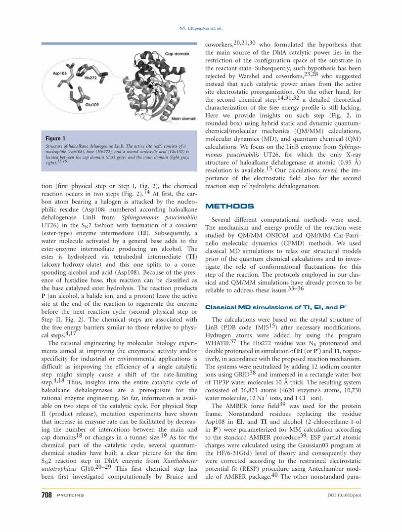
The structure of haloalkane dehalogenases is composed of main and
cap domains. An eight-stranded b-pleated sheet surrounded by a-helicesforms the main domain making-up the core of a protein.1 The a-helicalcap domain, which resembles uteroglobin fold,12 sits on the top of the
main domain. The active site is buried in between of two domains and is
connected with bulk solvent by several tunnels.13 Three residues, essential
for catalysis,4,14 aspartate, histidine, and aspartate/glutamate form the
catalytic triad in haloalkane dehalogenases (Fig. 1). The generic catalytic
triad made of a nucleophile (Asp, Ser, or Cys), a base (His), and a cata-
lytic acid (Asp or Glu) is shared by all enzymes of the a/b-hydrolase fam-
ily, containing enzymes of wide structural diversity and catalyzing a wide
range of chemical reactions.2 The triad is also functionally similar to triad
of proteases, such as serine protease,14 which employs an aspartic acid as
the nucleophile.
The hydrolytic dehalogenation reaction catalyzed by haloalkane dehalo-
genases consists of several steps. After enzyme-substrate complex forma-
The Supplementary Material referred to in this article can be found online at http://www.interscience.
wiley.com/jpages/0887-3585/suppmat.
Grant sponsor: The Ministry of Education, Young and Sport; Grant numbers: LC512, MSM6198959216
and MSM0021622413; Grant sponsor: Central European Initiative
*Correspondence to: Paolo Carloni, Department of Biophysics, SISSA/ISAS and CNR-INFM Democritos
National Simulation Center, Via Beirut 2-4, 34014 Trieste, Italy. E-mail: [email protected]
Received 28 August 2006; Revised 28 February 2007; Accepted 28 March 2007
Published online 29 August 2007 in Wiley InterScience (www.interscience.wiley.com).
DOI: 10.1002/prot.21523
ABSTRACT
Mechanistic studies on the hydrolytic deha-
logenation catalyzed by haloalkane dehalo-
genases are of importance for environmental
and industrial applications. Here, Car-Par-
rinello (CP) and ONIOM hybrid quantum-
mechanical/molecular mechanics (QM/MM)
are used investigate the second reaction step
of the catalytic cycle, which comprises a gen-
eral base-catalyzed hydrolysis of an ester in-
termediate (EI) to alcohol and free enzyme.
We focus on the enzyme LinB from Sphingo-
monas paucimobilis UT26, for which the X-ray
structure at atomic resolution is available. In
agreement with previous proposals, our calcu-
lations suggest that a histidine residue
(His272), polarized by glutamate (Glu132),
acts as a base, accepting a proton from the cat-
alytic water molecule and transferring it to an
alcoholate ion. The reaction proceeds through
a metastable tetrahedral intermediate, which
shows an easily reversed reaction to the EI. In
the formation of the products, the protonated
aspartic acid (Asp108) can easily adopt confor-
mation of the relaxed state found in the free
enzyme. The overall free energy barrier of the
reaction calculated by potential of the mean
force integration using CP-QM/MM calcula-
tions is equal to 19.5 6 2 kcal � mol21. The
lowering of the energy barrier of catalyzed
reaction with respect to the water reaction is
caused by strong stabilization of the reaction
intermediate and transition state and their
preorganization by electrostatic field of the
enzyme.
Proteins 2008; 70:707–717.VVC 2007 Wiley-Liss, Inc.
Key words: catalytic triad; enzyme dynam-
ics; tetrahedral intermediate; ester hydroly-
sis; reaction mechanism.
VVC 2007 WILEY-LISS, INC. PROTEINS 707
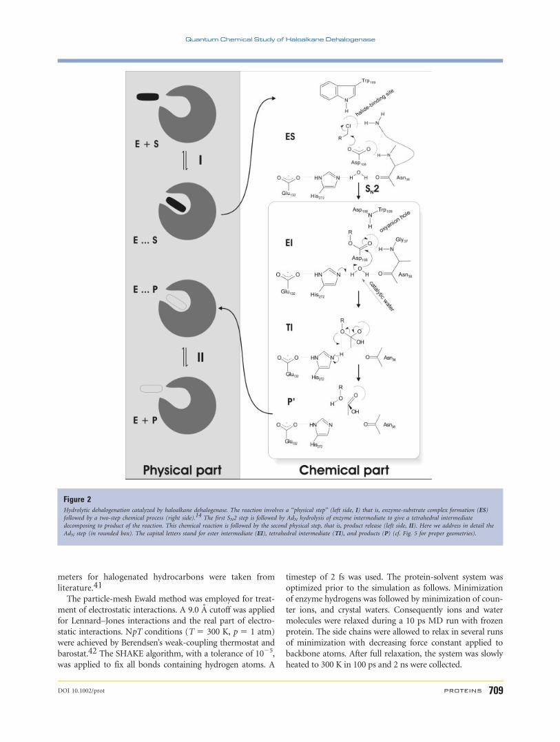
tion (first physical step or Step I, Fig. 2), the chemical
reaction occurs in two steps (Fig. 2).14 At first, the car-
bon atom bearing a halogen is attacked by the nucleo-
philic residue (Asp108; numbered according haloalkane
dehalogenase LinB from Sphingomonas paucimobilis
UT26) in the SN2 fashion with formation of a covalent
(ester-type) enzyme intermediate (EI). Subsequently, a
water molecule activated by a general base adds to the
ester-enzyme intermediate producing an alcohol. The
ester is hydrolyzed via tetrahedral intermediate (TI)
(alcoxy-hydroxy-olate) and this one splits to a corre-
sponding alcohol and acid (Asp108). Because of the pres-
ence of histidine base, this reaction can be classified as
the base catalyzed ester hydrolysis. The reaction products
P (an alcohol, a halide ion, and a proton) leave the active
site at the end of the reaction to regenerate the enzyme
before the next reaction cycle (second physical step or
Step II, Fig. 2). The chemical steps are associated with
the free energy barriers similar to those relative to physi-
cal steps.4,17
The rational engineering by molecular biology experi-
ments aimed at improving the enzymatic activity and/or
specificity for industrial or environmental applications is
difficult as improving the efficiency of a single catalytic
step might simply cause a shift of the rate-limiting
step.4,18 Thus, insights into the entire catalytic cycle of
haloalkane dehalogenases are a prerequisite for the
rational enzyme engineering. So far, information is avail-
able on two steps of the catalytic cycle. For physical Step
II (product release), mutation experiments have shown
that increase in enzyme rate can be facilitated by decreas-
ing the number of interactions between the main and
cap domains18 or changes in a tunnel size.19 As for the
chemical part of the catalytic cycle, several quantum-
chemical studies have built a clear picture for the first
SN2 reaction step in DhlA enzyme from Xanthobacter
autotrophicus GJ10.20–29 This first chemical step has
been first investigated computationally by Bruice and
coworkers,20,21,30 who formulated the hypothesis that
the main source of the DhlA catalytic power lies in the
restriction of the configuration space of the substrate in
the reactant state. Subsequently, such hypothesis has been
rejected by Warshel and coworkers,23,28 who suggested
instead that such catalytic power arises from the active
site electrostatic preorganization. On the other hand, for
the second chemical step,14,31,32 a detailed theoretical
characterization of the free energy profile is still lacking.
Here we provide insights on such step (Fig. 2, in
rounded box) using hybrid static and dynamic quantum-
chemical/molecular mechanics (QM/MM) calculations,
molecular dynamics (MD), and quantum chemical (QM)
calculations. We focus on the LinB enzyme from Sphingo-
monas paucimobilis UT26, for which the only X-ray
structure of haloalkane dehalogenase at atomic (0.95 A)
resolution is available.15 Our calculations reveal the im-
portance of the electrostatic field also for the second
reaction step of hydrolytic dehalogenation.
METHODS
Several different computational methods were used.
The mechanism and energy profile of the reaction were
studied by QM/MM ONIOM and QM/MM Car-Parri-
nello molecular dynamics (CPMD) methods. We used
classical MD simulations to relax our structural models
prior of the quantum chemical calculations and to inves-
tigate the role of conformational fluctuations for this
step of the reaction. The protocols employed in our clas-
sical and QM/MM simulations have already proven to be
reliable to address these issues.33–36
Classical MD simulations of TI, EI, and P0
The calculations were based on the crystal structure of
LinB (PDB code 1MJ515) after necessary modifications.
Hydrogen atoms were added by using the program
WHATIF.37 The His272 residue was Nd protonated and
double protonated in simulation of EI (or P0) and TI, respec-tively, in accordance with the proposed reaction mechanism.
The systems were neutralized by adding 12 sodium counter
ions using GRID38 and immersed in a rectangle water box
of TIP3P water molecules 10 A thick. The resulting system
consisted of 36,823 atoms (4620 enzyme’s atoms, 10,730
water molecules, 12 Na1 ions, and 1 Cl2 ion).
The AMBER force field39 was used for the protein
frame. Nonstandard residues replacing the residue
Asp108 in EI, and TI and alcohol (2-chloroethane-1-ol
in P0) were parameterized for MM calculation according
to the standard AMBER procedure39: ESP partial atomic
charges were calculated using the Gaussian03 program at
the HF/6-31G(d) level of theory and consequently they
were corrected according to the restrained electrostatic
potential fit (RESP) procedure using Antechamber mod-
ule of AMBER package.40 The other nonstandard para-
Figure 1Structure of haloalkane dehalogenase LinB. The active site (left) consists of a
nucleophile (Asp108), base (His272), and a second carboxylic acid (Glu132) is
located between the cap domain (dark gray) and the main domain (light gray,
right).15,16
M. Otyepka et al.
708 PROTEINS DOI 10.1002/prot
meters for halogenated hydrocarbons were taken from
literature.41
The particle-mesh Ewald method was employed for treat-
ment of electrostatic interactions. A 9.0 A cutoff was applied
for Lennard–Jones interactions and the real part of electro-
static interactions. NpT conditions (T 5 300 K, p 5 1 atm)
were achieved by Berendsen’s weak-coupling thermostat and
barostat.42 The SHAKE algorithm, with a tolerance of 1025,
was applied to fix all bonds containing hydrogen atoms. A
timestep of 2 fs was used. The protein-solvent system was
optimized prior to the simulation as follows. Minimization
of enzyme hydrogens was followed by minimization of coun-
ter ions, and crystal waters. Consequently ions and water
molecules were relaxed during a 10 ps MD run with frozen
protein. The side chains were allowed to relax in several runs
of minimization with decreasing force constant applied to
backbone atoms. After full relaxation, the system was slowly
heated to 300 K in 100 ps and 2 ns were collected.
Figure 2Hydrolytic dehalogenation catalyzed by haloalkane dehalogenase. The reaction involves a ‘‘physical step’’ (left side, I) that is, enzyme-substrate complex formation (ES)
followed by a two-step chemical process (right side).14 The first SN2 step is followed by AdN hydrolysis of enzyme intermediate to give a tetrahedral intermediate
decomposing to product of the reaction. This chemical reaction is followed by the second physical step, that is, product release (left side, II). Here we address in detail the
AdN step (in rounded box). The capital letters stand for ester intermediate (EI), tetrahedral intermediate (TI), and products (P) (cf. Fig. 5 for proper geometries).
Quantum Chemical Study of Haloalkane Dehalogenase
DOI 10.1002/prot PROTEINS 709
CP-QM/MM simulations
In the CPMD simulations, the QM region was treated
at the DFT-BLYP level of theory.43,44 A 70-Ry cut off
for plane-wave expansion was used. Normconserving
pseudopotentials of Troullier–Martins were employed for
all elements.45 As in the ONIOM calculations, the MM
region was treated with the AMBER force field.39 The
connection between both regions was realized using QM/
MM fully Hamiltonian coupling scheme as described in
literature.46,47 Here, the electrostatic interactions bet-
ween the MM and QM part are taken into account with
cutoff radii 10, 15, and 20 au for electrostatic coupling
(see Refs. 45–47 for a detailed description). The QM
supercell size was equal to 35.00 au 3 40.25 au 3 32.90
au with the orthorhombic symmetry. Capping hydrogen
atoms were added to saturate the valence of terminal car-
bons atoms.
The starting TI structure was the MD structure after
1 ns. The structure was then minimized, and relaxed by
CP-QM/MM using simulated annealing with 0.98 scaling
factor. Then the systems were subsequently heated to 300
K increasing the temperature stepwise using 50 K steps.
At the beginning of each heating-step the system wave
function was quenched to the Born–Oppenheimer sur-
face. The production part of dynamics was realized using
Nose–Hoover thermostat maintained at 300 K, 5 au time
integration step together with 800 au for fictitious elec-
tron mass were used. The periodic-boundary conditions
were employed in calculation.
The free energy profile of the reaction was calculated
by potential mean force (PMF) integration from con-
strained MD48–50 using Eq. (1)
DW ¼ W�n2��W
�n1� ¼ Z n2
n1
*@H
@n
+n0dn0 ð1Þ
where n represents reaction coordinate as a function of
configuration degrees of freedom n 5 n(r). The mean
force was time averaged over a constrained trajectory
with the reaction coordinate fixed at specific value, n0.The implementation employed provides DF value, which
is approximately equal to DG in the condensed phase.51
The total time of CP-QM/MM simulations used for PMF
integration is �12 ps and costs about 20,000 CPU hours
at IBM Power5 1.9 GHz.
ONIOM-QM/MM simulations
The QM/MM scheme implemented in Gaussian03
described as ONIOM model splits the calculated system
into two parts, model and real system. The total energy
of the system is given in Eq. (2)
Etot ¼ EMMreal þ EQM
model � EMMmodel ð2Þ
where, (i) EMMreal represents energy of the whole system
calculated at MM level, using the AMBER force field39
as above; (ii) EQMmodel energy of the model system calcu-
lated by a quantum mechanical (QM) method, namely at
the BLYP/6-31G(d) level of theory; (iii) EMMmodel energy
of the model calculated with the AMBER force field39 as
above. The mechanical electrostatic coupling between
QM and MM model was employed in our ONIOM cal-
culations. The minimized structure of TI, as obtained by
the MD calculations described above, was used as a start-
ing geometry. However, to decrease the computational
cost, only a 3.5 A thick water shell around the protein
was included. The resulting system consisted of 6223
atoms (enzyme, 530 water molecules, 12 Na1 ions, and 1
Cl2 ion). Esterified Asp108, His272 and backbone adja-
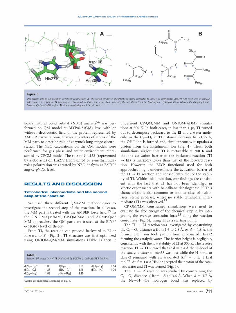
cent to Asn38 made the QM region (49 atoms, Fig. 3).
Hydrogen atoms were added to the dangling bonds.
ONIOM-ADMP simulations
Atom-centered density matrix propagator (ADMP)
method as implemented in Gaussian 03 was used to per-
form quantum MD of the studied combined QM/MM
system described by ONIOM model.52 ADMP method is
similar to CPMD scheme however, differs in usage of the
single particle density matrix in an orthogonal basis set
formed from Gaussian basis functions. This approach
dramatically increases effectiveness of ADMP over CPMD
when using hybrid functionals with explicit Hartree–Fock
(HF) exchange (such as B3LYP). The AMDP method
used here features the same coupling scheme between
QM and MM region as the ONIOM method does.
Prior the run, the system TI was geometrically mini-
mized at MM level as described earlier. BLYP with 6-
31G(d) basis set was used for the same QM level (Fig. 3)
and the AMBER force field was used to treat system at
the MM level. The whole protein was neutralized by 12
sodium ions and around the protein was surrounded by
a 3.5 A thick water shell. The production part of dynam-
ics was realized using thermostat maintained at 300 K,
0.2 fs long integration step together with 0.100 amu for
fictitious electron mass were used. The Frobenius norm
of the Fock and density matrix commutator [F,P] had a
mean value of 8.2 3 1023 Hartree. The small order of
magnitude of this value demonstrates that QM/MM-
ADMP dynamics remains close to Born–Oppenheimer
surface and produces stable simulation.
QM calculations of model systems
The QM calculation on model system was performed
by HF/6-31G(d), BLYP/6-31G(d), and MPW1K/6-31G(d)
in the gas phase to assess the dependence of the activa-
tion energy barrier on the functional used. The QM cal-
culation on model system in water represented by CPCM
method53 was performed at BLYP/6-31G(d) level. Wein-
M. Otyepka et al.
710 PROTEINS DOI 10.1002/prot
hold’s natural bond orbital (NBO) analysis54 was per-
formed on QM model at BLYP/6-31G(d) level with or
without electrostatic field of the protein represented by
AMBER partial atomic charges at centers of atoms of the
MM part, to describe role of enzyme’s long-range electro-
statics. The NBO calculations on the QM models were
performed for gas phase and water environment repre-
sented by CPCM model. The role of Glu132 (represented
by acetic acid) on His272 (represented by 2-methylimida-
zole) polarization was treated by NBO analysis at B3LYP/
aug-cc-pVDZ level.
RESULTS AND DISCUSSION
Tetrahedral intermediate and the secondstep of the reaction
We used three different QM/MM methodologies to
investigate the second step of the reaction. In all cases,
the MM part is treated with the AMBER force field.39 In
the ONIOM-QM/MM, CP-QM/MM, and ADMP-QM/
MM approaches, the QM parts are treated at the BLYP/
6-31G(d) level of theory.
From TI, the reaction can proceed backward to EI or
forward to P0 (Fig. 2). TI structure was first optimized
using ONIOM-QM/MM simulations (Table I) then it
underwent CP-QM/MM and ONIOM-ADMP simula-
tions at 300 K. In both cases, in less than 1 ps, TI turned
out to decompose backward to the EI and a water mole-
cule: as the C5��O4 at TI distance increases to �1.75 A,
the OH2 ion is formed and, simultaneously, it uptakes a
proton from the histidinium ion (Fig. 4). Thus, both
simulations suggest that TI is metastable at 300 K and
that the activation barrier of the backward reaction (TI
? EI) is markedly lower than that of the forward reac-
tion. However, the BLYP functional used for both
approaches might underestimate the activation barrier of
the TI ? EI reaction and consequently reduce the stabil-
ity of TI. Within this limitation, our findings are consist-
ent with the fact that TI has not been identified in
kinetic experiments with haloalkane dehalogenase.17 This
characteristic is also common to another class of hydro-
lases, serine protease, where no stable tetrahedral inter-
mediate (TI) was observed.55
CP-QM/MM constrained simulations were used to
evaluate the free energy of the chemical step 2, by inte-
grating the average constraint force48 along the reaction
coordinate (Fig. 5), using TI as a starting point.
The TI ? EI reaction was investigated by constraining
the C5��O4 distance d from 1.6 to 2.8 A. At d 5 1.8 A, the
formed OH2 ion took proton from protonated His272
forming the catalytic water. The barrier height is negligible,
consistently with the low stability of TI at 300 K. The reverse
reaction, EI ? TI showed that at d 5 2.4 A the H-bond of
the catalytic water to Asn38 was lost while the H-bond to
His272 remained with an associated DF{ 5 3 � 1 kcal
mol21. At d5 1.8 A His272 accepted the proton of the cata-
lytic water and TIwas formed (Fig. 4).
The TI ? P0 reaction was studied by constraining the
C5��O7 distance d from 1.5 to 3.6 A. When d 5 1.7 A,
the N1��H2���O5 hydrogen bond was replaced by
Figure 3QM region used in all quantum-chemistry calculations. A: The region consists of the backbone atoms connected to Asn38, of esterificated Asp108 side chain and of His272
side chain. The region in TI geometry is represented by sticks. The wires show some neighboring atoms from the MM region. Hydrogen atoms saturate the dangling bonds
between QM and MM region. B: Atom numbering used in this work.
Table ISelected Distances (A) of TI Optimized by BLYP/6-31G(d):AMBER Method
d(N1��H2)a 1.05 d(H3��O4) 0.99 d(O4��C5) 1.54
d(O6��C5) 1.33 d(O7��C5) 1.48 d(O6��H8) 1.70d(O6��H10) 1.68 d(H3��O12) 2.20
aAtoms are numbered according to Fig. 3.
Quantum Chemical Study of Haloalkane Dehalogenase
DOI 10.1002/prot PROTEINS 711
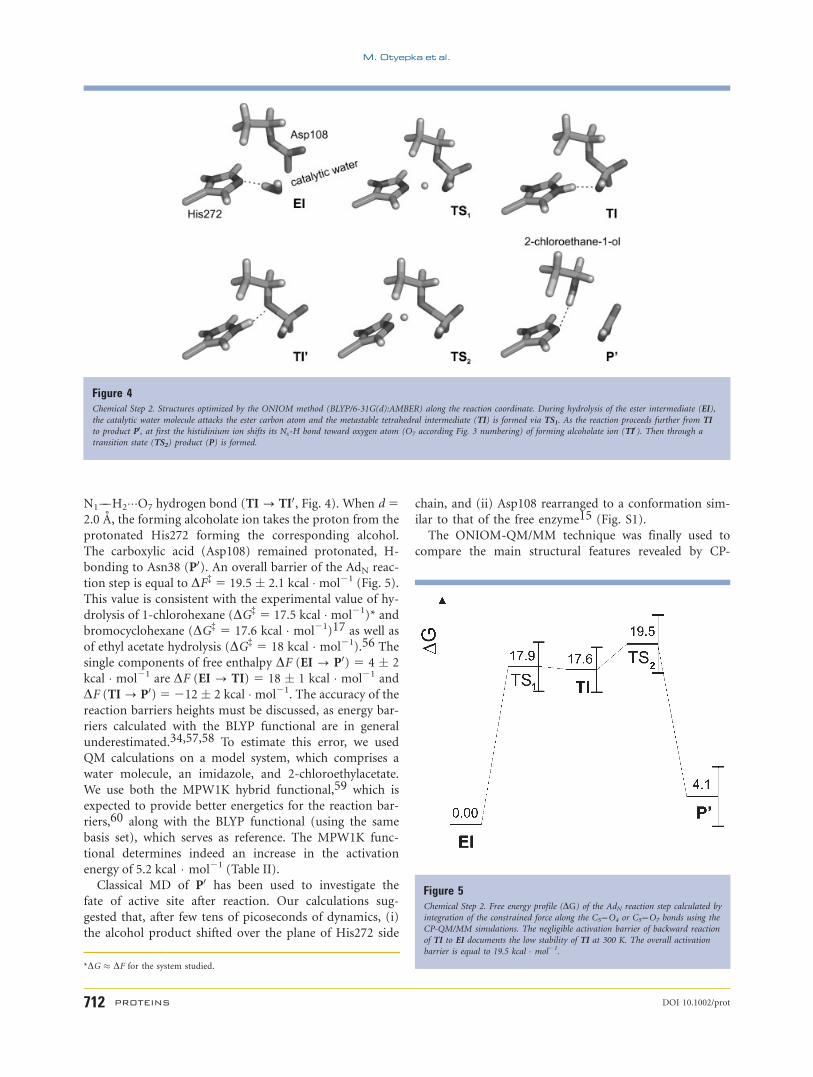
N1��H2���O7 hydrogen bond (TI ? TI0, Fig. 4). When d 52.0 A, the forming alcoholate ion takes the proton from the
protonated His272 forming the corresponding alcohol.
The carboxylic acid (Asp108) remained protonated, H-
bonding to Asn38 (P0). An overall barrier of the AdN reac-
tion step is equal to DF{ 5 19.5 � 2.1 kcal � mol21 (Fig. 5).
This value is consistent with the experimental value of hy-
drolysis of 1-chlorohexane (DG{ 5 17.5 kcal � mol21)* and
bromocyclohexane (DG{ 5 17.6 kcal � mol21)17 as well as
of ethyl acetate hydrolysis (DG{ 5 18 kcal � mol21).56 The
single components of free enthalpy DF (EI ? P0) 5 4 � 2
kcal � mol21 are DF (EI ? TI) 5 18 � 1 kcal � mol21 and
DF (TI? P0)5212 � 2 kcal �mol21. The accuracy of the
reaction barriers heights must be discussed, as energy bar-
riers calculated with the BLYP functional are in general
underestimated.34,57,58 To estimate this error, we used
QM calculations on a model system, which comprises a
water molecule, an imidazole, and 2-chloroethylacetate.
We use both the MPW1K hybrid functional,59 which is
expected to provide better energetics for the reaction bar-
riers,60 along with the BLYP functional (using the same
basis set), which serves as reference. The MPW1K func-
tional determines indeed an increase in the activation
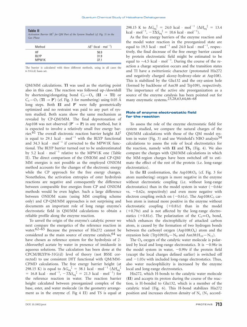
energy of 5.2 kcal � mol21 (Table II).
Classical MD of P0 has been used to investigate the
fate of active site after reaction. Our calculations sug-
gested that, after few tens of picoseconds of dynamics, (i)
the alcohol product shifted over the plane of His272 side
chain, and (ii) Asp108 rearranged to a conformation sim-
ilar to that of the free enzyme15 (Fig. S1).
The ONIOM-QM/MM technique was finally used to
compare the main structural features revealed by CP-
Figure 4Chemical Step 2. Structures optimized by the ONIOM method (BLYP/6-31G(d):AMBER) along the reaction coordinate. During hydrolysis of the ester intermediate (EI),
the catalytic water molecule attacks the ester carbon atom and the metastable tetrahedral intermediate (TI) is formed via TS1. As the reaction proceeds further from TI
to product P0, at first the histidinium ion shifts its Ne-H bond toward oxygen atom (O7 according Fig. 3 numbering) of forming alcoholate ion (TI0). Then through a
transition state (TS2) product (P) is formed.
Figure 5Chemical Step 2. Free energy profile (DG) of the AdN reaction step calculated by
integration of the constrained force along the C5��O4 or C5��O7 bonds using the
CP-QM/MM simulations. The negligible activation barrier of backward reaction
of TI to EI documents the low stability of TI at 300 K. The overall activation
barrier is equal to 19.5 kcal � mol21.
*DG � DF for the system studied.
M. Otyepka et al.
712 PROTEINS DOI 10.1002/prot
QM/MM calculations. TI was used as the starting point
also in this case. The reaction was followed up-/downhill
by shortening/elongating bond C5��O4 (EI ? TI) or
C5��O7 (TI ? P0) (cf. Fig. 3 for numbering) using 0.05 A
long steps. Both EI and P0 were fully geometrically
optimized and no restraint was paid to any part of sys-
tem studied. Both scans show the same mechanism as
revealed by CP-QM/MM. The final deprotonation of
Asp108 was not observed (P0 ? P) in any method, but it
is expected to involve a relatively small free energy bar-
rier.61 The overall electronic reaction barrier height DE{
is equal to 29.1 kcal � mol21 with the BLYP functional
and 34.3 kcal � mol21 if corrected to the MPW1K func-
tional. The BLYP barrier turned out to be underestimated
by 5.2 kcal � mol21 relative to the MPW1K one (Table
II). The direct comparison of the ONIOM and CP-QM/
MM energies is not possible as the employed ONIOM
method accounts for the changes of the electronic energy
while the CP approach for the free energy changes.
Nonetheless, the activation entropies of ester hydrolysis
reactions are negative and consequently the difference
between comparable free energies from CP and ONIOM
methods would be even higher. Such a large difference
between ONIOM scans (using MM coupling scheme
only) and CP-QM/MM approaches is not surprising and
documents an important role of long range enzyme’s
electrostatic field in QM/MM calculations to obtain a
reliable profile along the enzyme reaction.
To unveil the origin of the enzyme’s catalytic power we
next compare the energetics of the reference reaction in
water.62–65 Because the presence of His272 cannot be
considered as the main source of enzyme catalysis,64 we
have chosen as reference system for the hydrolysis of 2-
chloroethyl acetate by water in presence of imidazole in
aqueous solutions. The calculation has been done at the
CPCM/BLYP/6-31G(d) level of theory (not BSSE cor-
rected) to use consistent DFT functional with QM/MM-
CPMD calculations. The free energy barrier height (at
298.15 K) is equal to DGaq{ 5 38.1 kcal � mol21 (DHaq
{
5 16.8 kcal � mol21, 2TDSaq{ 5 21.3 kcal � mol21) for
the reference reaction in water. The reaction barrier
height calculated between preorganized complex of the
base, ester, and water molecule (in the geometry arrange-
ment as in the enzyme cf. Fig 4 EI) and TS is equal at
298.15 K to DGaq{ 5 24.0 kcal � mol21 (DHaq
{ 5 13.4
kcal � mol21, 2TDSaq{ 5 10.6 kcal � mol21).
As the free energy barriers of the enzyme reaction and
the model water reaction in the preorganized state are
equal to 19.5 kcal � mol21 and 24.0 kcal � mol21, respec-
tively, the final decrease of the free energy barrier caused
by protein electrostatic field might be estimated to be
equal to �4.5 kcal � mol21. During the course of the re-
action a charge separation occurs and the transition states
and TI have a zwitterionic character (protonated His272
and negatively charged alcoxy-hydroxy-olate at Asp108).
This is stabilized by the Glu132 and the oxy-anion hole
(formed by backbone of Asn38 and Trp109), respectively.
The importance of the active site preorganization as a
source of the enzyme catalysis has been pointed out for
many enzymatic systems.23,28,63,64,66–68
Role of enzyme electrostatic fieldfor the reaction
To assess the role of the enzyme electrostatic field for
system studied, we compare the natural charges of the
QM/MM calculations with those of the QM model sys-
tem in water (Fig. 3) and use Weinhold’s NBO analysis54
calculations to assess the role of local electrostatics for
the reaction, namely with EI and TS1 (Fig. 4). We also
compare the charges with QM/MM calculations in which
the MM-region charges have been switched off to esti-
mate the effect of the rest of the protein (i.e. long-range
electrostatics).
In the EI conformation, the Asp108:O6 (cf. Fig. 3 for
atom numbering) oxygen is more negative in the enzyme
without electrostatic coupling (i.e. without long-range
electrostatics) than in the model system in water (20.64e
vs. 20.62e, respectively) and even more negative with
electron coupling switch on (20.61e). The Asp108:C5 car-
bon atom is instead more positive in the enzyme without
electrostatic coupling (10.81e) than in the model
(10.79e) and is not affected by the long-range electro-
statics (10.81e). The polarization of the C5¼¼O6 bond,
which enhances the electrophilicity of attacked carbon
atom, is caused by the formation of two hydrogen bonds
between the carbonyl oxygen (Asp108:O6) atom and the
oxyanion hole (Trp109:H8��N9 and Asn38:H10��N11).
The O4 oxygen of the catalytic water molecule is polar-
ized by local and long-range electrostatics. It is 20.98e in
the model system in water, 20.99e if the protein field
(except the local charges defined earlier) is switched off
and 21.03e with included long-range electrostatics. Thus,
also water nucleophilicity is increased by the enzyme
local and long-range electrostatics.
His272, which H-bonds to the catalytic water molecule
(EI) and accepts its proton during the course of the reac-
tion, is H-bonded to Glu132, which is a member of the
catalytic triad (Fig. 6). This H-bond stabilizes His272
position and increases electron density of Ne (N1 accord-
Table IIActivation Barrier DE{ for QM Part of the System Studied (cf. Fig. 3) in the
Gas Phase
Method DE{ (kcal � mol21)
HF 56.9BLYP 31.9MPW1K 37.1
The barrier is calculated with three different methods, using in all cases the
6-31G(d) basis set.
Quantum Chemical Study of Haloalkane Dehalogenase
DOI 10.1002/prot PROTEINS 713
ing Fig. 3 numbering) atom and therefore His272 basic-
ity. The natural charge at His272:Ne atom increases from
20.53e to 20.58e in case of presence of Glu132 (on
model system, cf. methods). The same trend is seen in the
natural charges calculated by NBO analysis on QM part
without/with electrostatic field of the MM part. The natural
charge localized on His272:Ne atom increases from 20.52e
without to 20.58e with MM electrostatic field. Thus,
Glu132 polarizes His272 ring and it is expected to stabilize
electrostically the histidinium ion, which forms during reac-
tion. Similar function of the residue was described also for
the catalytic triad of serine proteases.69,70 In TS1, the
Asp108:O6 charge (20.84e) is more negative than EI
(20.64e), while the natural partial charges of surrounding
atoms do not change in both geometries (EI and TS1). The
earlier analysis underlines the importance of the two H-
bonds in the haloalkane dehalogenase’s oxyanion-hole for
the stabilization of the transition state.
Role of conformational fluctuationsfor the reaction
Conformational fluctuations have been proposed to
play a role in specific enzymes.71–78 Examples include
the dihydrofolate reductase enzyme,79 whose completion
of the catalytic cycle has been suggested to require
coupled motions, and the proteases superfamily,80–82
whose biological function has been proposed to be
affected by conformational fluctuations. To address this
issue, here we performed classical MD simulations with
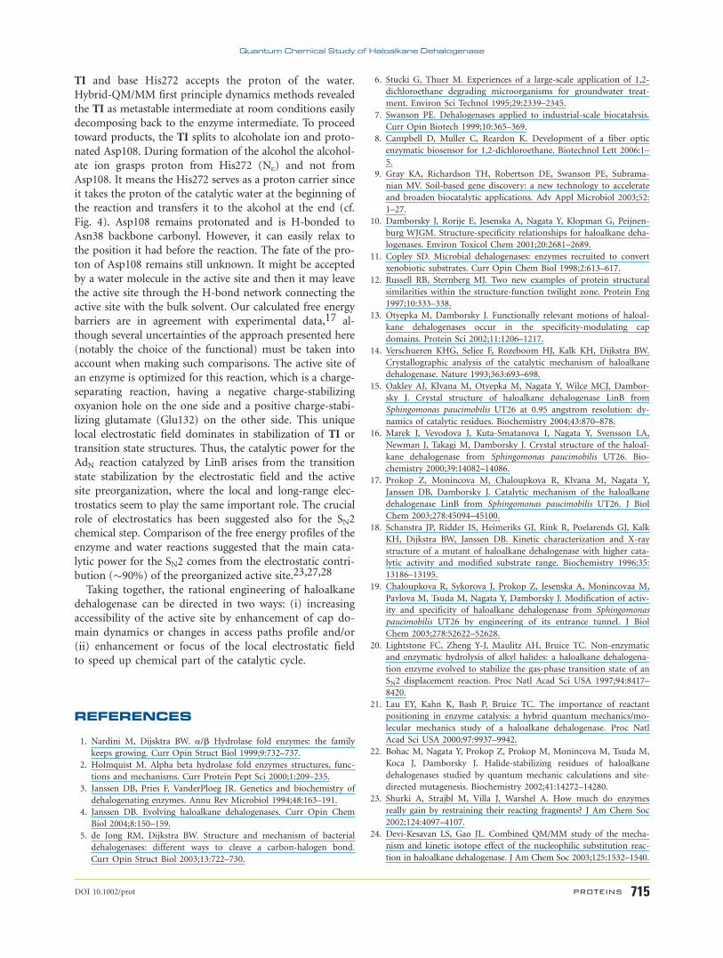
the AMBER force field39 and calculated the large-scale
motions of EI, TI, and P0 as the largest eigenvectors of
the covariance matrix of the Ca atoms.83 These eigenvec-
tors turned out to involve the LinB cap-domain, namely
the a4 helix and preceding loop. These motions well cor-
respond with correlated motions found in the free LinB
enzyme13 and no significant differences were found
among system studied (Fig. 7). As the correlated motions
are localized to the cap domain they influence active site
tunnels and do not include the active site. The absence
of any coupling between the large-scale motion of the
enzyme and the motion of the substrate suggest that
protein conformational fluctuations do not play a signifi-
cant role for the reaction step investigated here. Further
data on the MD are reported in the Supplementary
Information.
CONCLUSIONS
We have presented here an investigation of the second
chemical step, that is, enzyme intermediate hydrolysis
(Fig. 2) in haloalkane dehalogenase LinB by QM/MM
methods. The enzyme intermediate EI, our starting struc-
ture, is attacked by the catalytic water molecule clutched
between His272 and carbonyl group of Ans38 backbone
(Fig. 6). The catalytic water splits; the resulting OH2
group binds to ester carbon forming negatively charged
Figure 6Positioning of the catalytic water during the catalytic cycle. The water resides in
its position slightly fluctuating around it and having low B-factor (15 A2). Its
position is determined by three H-bonds. The third residue making the catalytic
triad of a haloalkane dehalogenase is Glu132, which is H-bonded to His272 and
increases its basicity.
Figure 7Projection of the first five essential motions to the structures of EI, TI, and P0 shows that the motions are located to the cap domain namely to a4 helix and preceding
loop in correspondence with motions found for the free enzyme.13
M. Otyepka et al.
714 PROTEINS DOI 10.1002/prot
TI and base His272 accepts the proton of the water.
Hybrid-QM/MM first principle dynamics methods revealed
the TI as metastable intermediate at room conditions easily
decomposing back to the enzyme intermediate. To proceed
toward products, the TI splits to alcoholate ion and proto-
nated Asp108. During formation of the alcohol the alcohol-
ate ion grasps proton from His272 (Ne) and not from
Asp108. It means the His272 serves as a proton carrier since
it takes the proton of the catalytic water at the beginning of
the reaction and transfers it to the alcohol at the end (cf.
Fig. 4). Asp108 remains protonated and is H-bonded to
Asn38 backbone carbonyl. However, it can easily relax to
the position it had before the reaction. The fate of the pro-
ton of Asp108 remains still unknown. It might be accepted
by a water molecule in the active site and then it may leave
the active site through the H-bond network connecting the
active site with the bulk solvent. Our calculated free energy
barriers are in agreement with experimental data,17 al-
though several uncertainties of the approach presented here
(notably the choice of the functional) must be taken into
account when making such comparisons. The active site of
an enzyme is optimized for this reaction, which is a charge-
separating reaction, having a negative charge-stabilizing
oxyanion hole on the one side and a positive charge-stabi-
lizing glutamate (Glu132) on the other side. This unique
local electrostatic field dominates in stabilization of TI or
transition state structures. Thus, the catalytic power for the
AdN reaction catalyzed by LinB arises from the transition
state stabilization by the electrostatic field and the active
site preorganization, where the local and long-range elec-
trostatics seem to play the same important role. The crucial
role of electrostatics has been suggested also for the SN2
chemical step. Comparison of the free energy profiles of the
enzyme and water reactions suggested that the main cata-
lytic power for the SN2 comes from the electrostatic contri-
bution (�90%) of the preorganized active site.23,27,28
Taking together, the rational engineering of haloalkane
dehalogenase can be directed in two ways: (i) increasing
accessibility of the active site by enhancement of cap do-
main dynamics or changes in access paths profile and/or
(ii) enhancement or focus of the local electrostatic field
to speed up chemical part of the catalytic cycle.
REFERENCES
1. Nardini M, Dijsktra BW. a/b Hydrolase fold enzymes: the family
keeps growing. Curr Opin Struct Biol 1999;9:732–737.
2. Holmquist M. Alpha beta hydrolase fold enzymes structures, func-
tions and mechanisms. Curr Protein Pept Sci 2000;1:209–235.
3. Janssen DB, Pries F, VanderPloeg JR. Genetics and biochemistry of
dehalogenating enzymes. Annu Rev Microbiol 1994;48:163–191.
4. Janssen DB. Evolving haloalkane dehalogenases. Curr Opin Chem
Biol 2004;8:150–159.
5. de Jong RM, Dijkstra BW. Structure and mechanism of bacterial
dehalogenases: different ways to cleave a carbon-halogen bond.
Curr Opin Struct Biol 2003;13:722–730.
6. Stucki G, Thuer M. Experiences of a large-scale application of 1,2-
dichloroethane degrading microorganisms for groundwater treat-
ment. Environ Sci Technol 1995;29:2339–2345.
7. Swanson PE. Dehalogenases applied to industrial-scale biocatalysis.
Curr Opin Biotech 1999;10:365–369.
8. Campbell D, Muller C, Reardon K. Development of a fiber optic
enzymatic biosensor for 1,2-dichloroethane. Biotechnol Lett 2006:1–
5.
9. Gray KA, Richardson TH, Robertson DE, Swanson PE, Subrama-
nian MV. Soil-based gene discovery: a new technology to accelerate
and broaden biocatalytic applications. Adv Appl Microbiol 2003;52:
1–27.
10. Damborsky J, Rorije E, Jesenska A, Nagata Y, Klopman G, Peijnen-
burg WJGM. Structure-specificity relationships for haloalkane deha-
logenases. Environ Toxicol Chem 2001;20:2681–2689.
11. Copley SD. Microbial dehalogenases: enzymes recruited to convert
xenobiotic substrates. Curr Opin Chem Biol 1998;2:613–617.
12. Russell RB, Sternberg MJ. Two new examples of protein structural
similarities within the structure-function twilight zone. Protein Eng
1997;10:333–338.
13. Otyepka M, Damborsky J. Functionally relevant motions of haloal-
kane dehalogenases occur in the specificity-modulating cap
domains. Protein Sci 2002;11:1206–1217.
14. Verschueren KHG, Seljee F, Rozeboom HJ, Kalk KH, Dijkstra BW.
Crystallographic analysis of the catalytic mechanism of haloalkane
dehalogenase. Nature 1993;363:693–698.
15. Oakley AJ, Klvana M, Otyepka M, Nagata Y, Wilce MCJ, Dambor-
sky J. Crystal structure of haloalkane dehalogenase LinB from
Sphingomonas paucimobilis UT26 at 0.95 angstrom resolution: dy-
namics of catalytic residues. Biochemistry 2004;43:870–878.
16. Marek J, Vevodova J, Kuta-Smatanova I, Nagata Y, Svensson LA,
Newman J, Takagi M, Damborsky J. Crystal structure of the haloal-
kane dehalogenase from Sphingomonas paucimobilis UT26. Bio-
chemistry 2000;39:14082–14086.
17. Prokop Z, Monincova M, Chaloupkova R, Klvana M, Nagata Y,
Janssen DB, Damborsky J. Catalytic mechanism of the haloalkane
dehalogenase LinB from Sphingomonas paucimobilis UT26. J Biol
Chem 2003;278:45094–45100.
18. Schanstra JP, Ridder IS, Heimeriks GJ, Rink R, Poelarends GJ, Kalk
KH, Dijkstra BW, Janssen DB. Kinetic characterization and X-ray
structure of a mutant of haloalkane dehalogenase with higher cata-
lytic activity and modified substrate range. Biochemistry 1996;35:
13186–13195.
19. Chaloupkova R, Sykorova J, Prokop Z, Jesenska A, Monincovaa M,
Pavlova M, Tsuda M, Nagata Y, Damborsky J. Modification of activ-
ity and specificity of haloalkane dehalogenase from Sphingomonas
paucimobilis UT26 by engineering of its entrance tunnel. J Biol
Chem 2003;278:52622–52628.
20. Lightstone FC, Zheng Y-J, Maulitz AH, Bruice TC. Non-enzymatic
and enzymatic hydrolysis of alkyl halides: a haloalkane dehalogena-
tion enzyme evolved to stabilize the gas-phase transition state of an
SN2 displacement reaction. Proc Natl Acad Sci USA 1997;94:8417–
8420.
21. Lau EY, Kahn K, Bash P, Bruice TC. The importance of reactant
positioning in enzyme catalysis: a hybrid quantum mechanics/mo-
lecular mechanics study of a haloalkane dehalogenase. Proc Natl
Acad Sci USA 2000;97:9937–9942.
22. Bohac M, Nagata Y, Prokop Z, Prokop M, Monincova M, Tsuda M,
Koca J, Damborsky J. Halide-stabilizing residues of haloalkane
dehalogenases studied by quantum mechanic calculations and site-
directed mutagenesis. Biochemistry 2002;41:14272–14280.
23. Shurki A, Strajbl M, Villa J, Warshel A. How much do enzymes
really gain by restraining their reacting fragments? J Am Chem Soc
2002;124:4097–4107.
24. Devi-Kesavan LS, Gao JL. Combined QM/MM study of the mecha-
nism and kinetic isotope effect of the nucleophilic substitution reac-
tion in haloalkane dehalogenase. J Am Chem Soc 2003;125:1532–1540.
Quantum Chemical Study of Haloalkane Dehalogenase
DOI 10.1002/prot PROTEINS 715
25. Kahn K, Bruice TC. Comparison of reaction energetics and leaving
group interactions during the enzyme-catalyzed and uncatalyzed
displacement of chloride from haloalkanes. J Phys Chem B
2003;107:6876–6885.
26. Hur S, Kahn K, Bruice TC. Comparison of formation of reactive
conformers for the S(N)2 displacements by CH3CO2- in water and
by Asp124-CO2- in a haloalkane dehalogenase. Proc Natl Acad Sci
USA 2003;100:2215–2219.
27. Soriano A, Silla E, Tunon I, Marti S, Moliner V, Bertran J. Electro-
static effects in enzyme catalysis: a quantum mechanics/molecular
mechanics study of the nucleophilic substitution reaction in haloal-
kane dehalogenase. Theor Chem Acc 2004;112:327–334.
28. Olsson MHM, Warshel A. Solute solvent dynamics and energetics
in enzyme catalysis: the SN2 reaction of dehalogenase as a general
benchmark. J Am Chem Soc 2004;126:15167–15179.
29. Rosta E, Klahn M, Warshel A. Towards accurate ab initio QM/MM
calculations of free-energy profiles of enzymatic reactions. J Phys
Chem B 2006;110:2934–2941.
30. Lightstone FC, Zheng YJ, Bruice TC. Molecular dynamics simula-
tions of ground and transition states for the S(N)2 displacement of
Cl2 from 1,2-dichloroethane at the active site of Xanthobacter auto-
trophicus haloalkane dehalogenase. J Am Chem Soc 1998;120:5611–
5621.
31. Kuty M, Damborsky J, Prokop M, Koca J. A molecular modeling
study of the catalytic mechanism of haloalkane dehalogenase. II.
Quantum chemical study of complete reaction mechanism. J Chem
Inf Comput Sci 1998;38:736–741.
32. Devi-Kesavan LS, Garcia-Viloca M, Gao J. Semiempirical QM/MM
potential with simple valence bond (SVB) for enzyme reactions.
Application to the nucleophilic addition reaction in haloalkane
dehalogenase. Theor Chem Acc 2003;109:133–139.
33. Piana S, Carloni P. Conformational flexibility of the catalytic Asp
dyad in HIV-1 protease: an ab initio study on the free enzyme. Pro-
teins: Struct Funct Genet 2000;39:26–36.
34. Piana S, Bucher D, Carloni P, Rothlisberger U. Reaction mechanism
of HIV-1 protease by hybrid Car-Parrinello/classical MD simula-
tions. J Phys Chem B 2004;108:11139–11149.
35. Piana S, Sebastiani D, Carloni P, Parrinello M. Ab initio molecular
dynamics-based assignment of the protonation state of pepstatin A/
HIV-1 protease cleavage site. J Am Chem Soc 2001;123:8730–8737.
36. Carloni P, Rothlisberger U, Parrinello M. The role and perspective
of ab-initio molecular dynamics in the study of biological systems.
Acc Chem Res 2002;35:455–464.
37. Vriend G. What if: a molecular modeling and drug design program.
J Mol Graphics 1990;8:52–56.
38. Goodford PJ. A computational procedure for determining energeti-
cally favorable binding sites on biologically important macromole-
cules. J Med Chem 1985;28:849–857.
39. Cornell WD, Cieplak P, Bayly CI, Gould IR, Merz KM, Ferguson
DM, Spellmeyer DC, Fox T, Caldwell JW, Kollman PA. A second
generation force field for the simulation of proteins, nucleic acids,
and organic molecules. J Am Chem Soc 1995;117:5179–5197.
40. Case DA, Cheatham TE, Darden T, Gohlke H, Luo R, Merz KM,
Onufriev A, Simmerling C, Wang B, Woods RJ. The amber biomo-
lecular simulation programs. J Comput Chem 2005;26:1668–1688.
41. Kmunicek J, Luengo S, Gago F, Ortiz AR, Wade RC, Damborsky J.
Comparative binding energy analysis of the substrate specificity of
haloalkane dehalogenase from Xanthobacter autotrophicus GJ10.
Biochemistry 2001;40:8905–8917.
42. Berendsen HJC, Postma JPM, van Gunsteren WF, DiNola A, Haak
JR. Molecular dynamics with coupling to an external bath. J Chem
Phys 1984;81:3684–3690.
43. Becke AD. Density-functional exchange-energy approximation with
correct asymptotic behavior. Phys Rev A 1988;38:3098–3100.
44. Lee CT, Yang WT, Parr RG. Development of the Colle-Salvetti cor-
relation-energy formula into a functional of the electron-density.
Phys Rev B 1988;37:785–789.
45. Troullier N, Martins JL. Efficient pseudopotentials for plane-wave
calculations. Phys Rev B 1991;43:1993–2006.
46. Laio A, VandeVondele J, Rothlisberger U. A hamiltonian electro-
static coupling scheme for hybrid Car-Parrinello molecular dynam-
ics simulations. J Chem Phys 2002;116:6941–6947.
47. Laio A, Gervasio FL, VandeVondele J, Sulpizi M, Rothlisberger U.
A variational definition of electrostatic potential derived charges.
J Phys Chem B 2004;108:7963–7968.
48. Carter EA, Ciccotti G, Hynes JT, Kapral R. Constrained reaction
coordinate dynamics for the simulation of rare events. Chem Phys
Lett 1989;156:472–477.
49. Ciccotti G, Ferrario M, Hynes JT, Kapral R. Constrained molecular
dynamics and the mean potential for an ion-pair in a polar solvent.
Chem Phys 1989;129:241–251.
50. Sprik M, Ciccotti G. Free energy from constrained molecular dy-
namics. J Chem Phys 1998;109:7737–7744.
51. Finkelstein AV. Protein physics. Amsterdam: Academic Press; 2002.
52. Rega N, Iyengar SS, Voth GA, Schlegel HB, Vreven T, Frisch MJ.
Hybrid ab-initio/empirical molecular dynamics: combining the
ONIOM scheme with the atom-centered density matrix propaga-
tion (ADMP) approach. J Phys Chem B 2004;108:4210–4220.
53. Cossi M, Rega N, Scalmani G, Barone V. Energies, structures, and
electronic properties of molecules in solution with the C-PCM sol-
vation model. J Comput Chem 2003;24:669–681.
54. Reed AE, Curtiss LA, Weinhold F. Intermolecular interactions from
a natural bond orbital, donor-acceptor viewpoint. Chem Rev 1988;
88:899–926.
55. Topf M, Richards WG. Theoretical studies on the deacylation step
of serine protease catalysis in the gas phase, in solution, and in elas-
tase. J Am Chem Soc 2004;126:14631–14641.
56. Isaacs N. Physical organic chemistry. Singapore: Longman; 1995.
57. Csonka GI, Johnson BG. Inclusion of exact exchange for self-inter-
action corrected H-3 density functional potential energy surface.
Theor Chem Acc 1998;99:158–165.
58. Gruning M, Gritsenko OV, Baerends EJ. Improved description of
chemical barriers with generalized gradient approximations (GGAs)
and meta-GGAs. J Phys Chem A 2004;108:4459–4469.
59. Lynch BJ, Fast PL, Harris M, Truhlar DG. Adiabatic connection for
kinetics. J Phys Chem A 2000;104:4811–4815.
60. Parthiban S, de Oliveira G, Martin JML. Benchmark ab initio
energy profiles for the gas-phase SN2 reactions Y- 1 CH3XCH3Y
1 X- (X,Y 5 F,Cl,Br). Validation of hybrid DFT methods. J Phys
Chem A 2001;105:895–904.
61. DalPeraro M, Llarrull LI, Rothlisberger U, Vila AJ, Carloni P.
Water-assisted reaction mechanism of monozinc b-lactamases.
J Am Chem Soc 2004;126:12661–12668.
62. Warshel A. Computer simulations of enzymatic reactions. Curr
Opin Struct Biol 1992;2:230–236.
63. Warshel A. Computer simulations of enzyme catalysis: methods, pro-
gress, and insights. Annu Rev Biophys Biomol Struct 2003;32:425–443.
64. Warshel A, Sharma PK, Kato M, Xiang Y, Liu HB, Olsson MHM. Elec-
trostatic basis for enzyme catalysis. Chem Rev 2006;106:3210–3235.
65. Olsson MHM, Parson WW, Warshel A. Dynamical contributions to
enzyme catalysis: critical tests of a popular hypothesis. Chem Rev
2006;106:1737–1756.
66. Warshel A, Levitt M. Theoretical studies of enzymic reactions:
dielectric, electrostatic and steric stabilization of the carbonium ion
in the reaction of lysozyme. J Mol Biol 1976;103:227–249.
67. Warshel A. Energetics of enzyme catalysis. Proc Natl Acad Sci USA
1978;75:5250–5254.
68. Warshel A, Papazyan A. Electrostatic effects in macromolecules:
fundamental concepts and practical modeling. Curr Opin Struct
Biol 1998;8:211–217.
69. Topf M, Varnai P, Richards WG. Ab initio QM/MM dynamics sim-
ulation of the tetrahedral intermediate of serine proteases: insights
into the active site hydrogen-bonding network. J Am Chem Soc
2002;124:14780–14788.
M. Otyepka et al.
716 PROTEINS DOI 10.1002/prot

70. Schutz CN, Warshel A. The low barrier hydrogen bond (LBHB)
proposal revisited: the case of the Asp�����His pair in serine pro-
teases. Proteins: Struct Funct Bioinform 2004;55:711–723.
71. Karplus M, McCammon JA. Dynamics of proteins: elements and
function. Annu Rev Biochem 1983;53:263–300.
72. Miller GP, Benkovic SJ. Deletion of a highly motional residue
affects formation of the Michaelis complex for Escherichia coli dihy-
drofolate reductase. Biochemistry 1998;37:6327–6335.
73. Kitao A, Go N. Investigating protein dynamics in collective coordi-
nate space. Curr Opin Struct Biol 1999;9:164–169.
74. Kohen A, Cannio R, Bartolucci S, Klinman JP. Enzyme dynamics
and hydrogen tunnelling in a thermophilic alcohol dehydrogenase.
Nature 1999;399:496–499.
75. Berendsen HJC, Hayward S. Collective protein dynamics in relation
to function. Curr Opin Struct Biol 2000;10:165–169.
76. Radkiewicz JL, Brooks CL. Protein dynamics in enzymatic catalysis:
exploration of dihydrofolate reductase. J Am Chem Soc 2000;122:
225–231.
77. Eisenmesser EZ, Bosco DA, Akke M, Kern D. Enzyme dynamics
during catalysis. Science 2002;295:1520–1523.
78. Daniel RM, Dunn RV, Finney JL, Smith JC. The role of dynamics in
enzyme activity. Annu Rev Biophys Biomol Struct 2003;32:69–92.
79. Brooks CL, Thorpe IF, Rod TH, Radkiewicz JL. Dynamics coupled
to protein catalysis? Factors affecting hydride transfer rates in
DHFR. Abstr Pap Am Chem Soc 2003;225:U747.
80. Piana S, Carloni P, Parrinello M. Role of conformational fluctua-
tions in the enzymatic reaction of HIV-1 protease. J Mol Biol 2002;
319:567–583.
81. Cascella M, Raugei S, Carloni P. Formamide hydrolysis investigated
by multiple-steering ab initio molecular dynamics. J Phys Chem B
2004;108:369–375.
82. Carnevale V, Raugei S, Micheletti C, Carloni P. Convergent dynam-
ics in the protease enzymatic superfamily. J Am Chem Soc 2006;
128:9766–9772.
83. Amadei A, Linssen ABM, Berenden HJC. Essential dynamics of pro-
tein. Proteins: Struct Funct Genet 1993;17:412–425.
Quantum Chemical Study of Haloalkane Dehalogenase
DOI 10.1002/prot PROTEINS 717
Copyright © 2022 FDOKUMEN