Stereoselectivity and conformational stability of haloalkane dehalogenase DbjA from Bradyrhizobium...
11
Stereoselectivity and conformational stability of haloalkane dehalogenase DbjA from Bradyrhizobium japonicum USDA110: the effect of pH and temperature Radka Chaloupkova 1,2 , Zbynek Prokop 1,2 , Yukari Sato 3 , Yuji Nagata 3 and Jiri Damborsky 1,2 1 Loschmidt Laboratories, Department of Experimental Biology, Faculty of Science, Masaryk University, Brno, Czech Republic 2 International Clinical Research Center, St Anne’s University Hospital Brno, Czech Republic 3 Graduate School of Life Sciences, Tohoku University, Sendai, Japan Keywords activity; enantioselectivity; haloalkane dehalogenase; oligomerization; pH; structure; thermostability Correspondence J. Damborsky, Loschmidt Laboratories, Department of Experimental Biology, Faculty of Science, Masaryk University, Kamenice 5 ⁄ A13, 625 00 Brno, Czech Republic Fax: +420549496302 Tel: +420549493467 E-mail: [email protected] (Received 6 February 2011, revised 15 May 2011, accepted 31 May 2011) doi:10.1111/j.1742-4658.2011.08203.x The effect of pH and temperature on structure, stability, activity and enantioselectivity of haloalkane dehalogenase DbjA from Bradyrhizobium japonicum USDA110 was investigated in this study. Conformational changes have been assessed by circular dichroism spectroscopy, functional changes by kinetic analysis, while quaternary structure was studied by gel filtration chromatography. Our study shows that the DbjA enzyme is highly tolerant to pH changes. Its secondary and tertiary structure was not affected by pH in the ranges 5.3–10.3 and 6.2–10.1, respectively. Oligomeri- zation of DbjA was strongly pH-dependent: monomer, dimer, tetramer and a high molecular weight cluster of the enzyme were distinguished in solu- tion at different pH conditions. Moreover, different oligomeric states of DbjA possessed different thermal stabilities. The highest melting tempera- ture (T m = 49.1 ± 0.2 °C) was observed at pH 6.5, at which the enzyme occurs in dimeric form. Maximal activity was detected at 50 °C and in the pH interval 7.7–10.4. While pH did not have any effect on enantiodiscri- minination of DbjA, temperature significantly altered DbjA enantioselectiv- ity. A decrease in temperature results in significantly enhanced enantioselectivity. The temperature dependence of DbjA enantioselectivity was analysed with 2-bromobutane, 2-bromopentane, methyl 2-bromopropi- onate and ethyl 2-bromobutyrate, and differential activation parameters D RS DH z and D RS DS z were determined. The thermodynamic analysis revealed that the resolution of b-bromoalkanes was driven by both enthal- pic and entropic terms, while the resolution of a-bromoesters was driven mainly by an enthalpic term. Unique catalytic activity and structural stabil- ity of DbjA in a broad pH range, combined with high enantioselectivity with particular substrates, make this enzyme a very versatile biocatalyst. Enzyme EC 3.8.1.5 haloalkane dehalogenase. Abbreviations CD, circular dichroism; MRE, mean residue ellipticity. 2728 FEBS Journal 278 (2011) 2728–2738 ª 2011 The Authors Journal compilation ª 2011 FEBS
Transcript of Stereoselectivity and conformational stability of haloalkane dehalogenase DbjA from Bradyrhizobium...

Stereoselectivity and conformational stability ofhaloalkane dehalogenase DbjA fromBradyrhizobium japonicum USDA110: the effect of pHand temperatureRadka Chaloupkova1,2, Zbynek Prokop1,2, Yukari Sato3, Yuji Nagata3 and Jiri Damborsky1,2
1 Loschmidt Laboratories, Department of Experimental Biology, Faculty of Science, Masaryk University, Brno, Czech Republic
2 International Clinical Research Center, St Anne’s University Hospital Brno, Czech Republic
3 Graduate School of Life Sciences, Tohoku University, Sendai, Japan
Keywords
activity; enantioselectivity; haloalkane
dehalogenase; oligomerization; pH;
structure; thermostability
Correspondence
J. Damborsky, Loschmidt Laboratories,
Department of Experimental Biology,
Faculty of Science, Masaryk University,
Kamenice 5 ⁄ A13, 625 00 Brno, Czech
Republic
Fax: +420549496302
Tel: +420549493467
E-mail: [email protected]
(Received 6 February 2011, revised 15 May
2011, accepted 31 May 2011)
doi:10.1111/j.1742-4658.2011.08203.x
The effect of pH and temperature on structure, stability, activity and
enantioselectivity of haloalkane dehalogenase DbjA from Bradyrhizobium
japonicum USDA110 was investigated in this study. Conformational
changes have been assessed by circular dichroism spectroscopy, functional
changes by kinetic analysis, while quaternary structure was studied by gel
filtration chromatography. Our study shows that the DbjA enzyme is
highly tolerant to pH changes. Its secondary and tertiary structure was not
affected by pH in the ranges 5.3–10.3 and 6.2–10.1, respectively. Oligomeri-
zation of DbjA was strongly pH-dependent: monomer, dimer, tetramer and
a high molecular weight cluster of the enzyme were distinguished in solu-
tion at different pH conditions. Moreover, different oligomeric states of
DbjA possessed different thermal stabilities. The highest melting tempera-
ture (Tm = 49.1 ± 0.2 �C) was observed at pH 6.5, at which the enzyme
occurs in dimeric form. Maximal activity was detected at 50 �C and in the
pH interval 7.7–10.4. While pH did not have any effect on enantiodiscri-
minination of DbjA, temperature significantly altered DbjA enantioselectiv-
ity. A decrease in temperature results in significantly enhanced
enantioselectivity. The temperature dependence of DbjA enantioselectivity
was analysed with 2-bromobutane, 2-bromopentane, methyl 2-bromopropi-
onate and ethyl 2-bromobutyrate, and differential activation parameters
DR�SDHz and DR�SDSz were determined. The thermodynamic analysis
revealed that the resolution of b-bromoalkanes was driven by both enthal-
pic and entropic terms, while the resolution of a-bromoesters was driven
mainly by an enthalpic term. Unique catalytic activity and structural stabil-
ity of DbjA in a broad pH range, combined with high enantioselectivity
with particular substrates, make this enzyme a very versatile biocatalyst.
Enzyme
EC 3.8.1.5 haloalkane dehalogenase.
Abbreviations
CD, circular dichroism; MRE, mean residue ellipticity.
2728 FEBS Journal 278 (2011) 2728–2738 ª 2011 The Authors Journal compilation ª 2011 FEBS

Introduction
Haloalkane dehalogenases (EC 3.8.1.5) make up an
important class of enzymes which are able to cleave car-
bon–halogen bonds in a broad range of halogenated ali-
phatic compounds. The hydrolytic dehalogenation
catalysed by these enzymes proceeds by nucleophilic
substitution of a halogen atom with a hydroxyl group
forming corresponding alcohols [1]. Haloalkanes, halo-
alcohols and alcohols are valuable building blocks in
organic and pharmaceutical synthesis [2–4], making
haloalkane dehalogenases potentially applicable in bio-
catalysis. We have recently shown that newly isolated
haloalkane dehalogenase DbjA from Bradyrhizobium ja-
ponicum USDA110 [5] possesses new substrate specific-
ity with high catalytic activity towards b-methylated
haloalkanes and sufficient enantioselectivity for indus-
trial scale synthesis of optically pure compounds [6].
Interestingly, the haloalkane dehalogenase DbjA (a) can
kinetically discriminate between enantiomers of two dis-
tinct groups of substrates, a-bromoesters and b-bro-moalkanes; (b) has enantioselectivity based on distinct
molecular interactions, which can be modified sepa-
rately by engineering of a surface loop; and (c) can
adopt an inverse temperature dependence of enantiose-
lectivity for b-bromoalkanes, but not a-bromoesters, by
mutating this surface loop and a flanking residue [7].
Use of enzymes in biocatalytic preparation of opti-
cally pure substances has been rapidly expanding in
recent years [8]. The efficient utilization of enzymes in
industrial processes requires that a number of criteria
are fulfilled, e.g. high activity, stability under process
conditions, appropriate substrate specificity and enanti-
oselectivity [9–11]. The manipulation of the physical
environment is an attractive way to provide additional
control of enzyme stereochemistry and catalytic func-
tionality alongside other methods, such as protein
engineering and directed evolution [12–14]. Under-
standing the effect of physical parameters on the struc-
ture and activity of an enzyme is important for
optimization of the operational conditions of a biocat-
alytic process, while knowledge of the structure–func-
tion relationships provides an essential theoretical
framework for modification of a biocatalyst by
rational protein design [15].
In this work we have systematically examined the
effects of pH and temperature on the stability, oligo-
merization state and functionality of the DbjA enzyme
using CD spectroscopy, size exclusion chromatogra-
phy, activity and enantioselectivity assays. Thermody-
namic analysis has been used to address the origin of
enantiomeric discrimination by determining differential
activation enthalpy and entropy for the enzymatic
reaction with racemic substrates 2-bromobutane,
2-bromopentane, ethyl 2-bromopropionate and methyl
2-bromobutyrate.
Results and Discussion
Conformational changes
CD spectroscopy was used for investigation of the sec-
ondary and tertiary structure of the DbjA enzyme at
pH conditions ranging from 1.7 to 11.5 in the far UV
and near UV spectral regions, respectively. The far UV
CD spectrum of native enzyme, measured in 50 mM
potassium phosphate buffer (pH 7.5 at 4 �C), exhibitedtwo negative features at 208 and 222 nm characteristic
of a-helical content (Fig. 1A, red bold curve). Similar
spectral features were found throughout the pH range
5.3–10.3, suggesting that enzyme secondary structure
remained preserved under these conditions. Calculated
Fig. 1. Far UV (A) and near UV (B) CD spectra of DbjA as a func-
tion of pH. The spectra shown represent the average of 10 consec-
utive scans.
R. Chaloupkova et al. Stereochemistry and conformational stability of DbjA
FEBS Journal 278 (2011) 2728–2738 ª 2011 The Authors Journal compilation ª 2011 FEBS 2729

a-helical content as a function of pH using the method
of Chen et al. [16], which is based on far UV CD data
at 222 nm, is presented in Fig. 2. Predicted a-helicalcontent at pH 5.3–10.3 was about 30.5%. The second-
ary structure of DbjA remains intact within five pH
units. At lower pH levels (pH < 5.0), the enzyme visu-
ally aggregates with simultaneous loss of UV signal.
On the other hand, at pH 11.0–11.4, the enzyme stays
in solution showing approximately a 42% loss in
a-helical content in comparison with its native state.
A strong negative band at 204 nm and a weak band
at 220 nm suggest that DbjA enzyme conformation
starts to be disordered at these extremely alkaline
conditions.
The near UV CD spectrum of the native state of the
enzyme reveals three negative ellipticity peaks at 259,
265 and 285 nm and a positive peak at 292 nm
(Fig. 1B, red bold curve). The ellipticity values at these
wavelengths remain preserved within the pH range
6.2–10.1. In acidic conditions, pH < 6.2, the CD
intensity at 285 and 292 nm slightly increases as a
result of the decreasing pH. The positive ellipticity at
292 nm can be attributed to a tryptophan environment,
since this region corresponds to the absorption band
for this residue [17]. The intensity changes observed at
292 nm might be related to a change in the tryptophan
environment as a result of the loss of some tertiary
interactions. This indicates that the enzyme starts to
lose its tertiary interactions without any secondary
structure loss before complete aggregation. In alkaline
conditions, pH > 10.7, the protein loses most of its
tertiary structure. A considerable increase in the ellip-
ticity at pH ‡ 10.7 is observed at 259 nm. This could
be caused by sudden exposure of phenylalanine resi-
dues in the extreme alkaline pH region. Comparison of
both near UV and far UV CD spectra determined at
various pH conditions revealed similar pH regions at
which the enzyme is structurally stable.
Changes in the structure could be attributed to a
change of ionization state of the enzyme at pH condi-
tions close to its isoelectric point (pI). The predicted pI
of DbjA is 5.89. Although many proteins demonstrate
a state of minimal solubility at their pI conditions,
DbjA remains soluble with a preserved secondary
structure. When pH is decreased below 5.3, the enzyme
suddenly passes from a nearly native state which is sol-
uble to a completely aggregated state. On the other
hand, alkalic denaturation of DbjA is accompanied by
significant modification of both secondary and tertiary
structure. At pH conditions 10.3–11.5, the enzyme
occurs in disordered conformation and remains
soluble.
Temperature dependence of conformational stability
was evaluated by performing a thermal unfolding
experiment at different pH conditions. Dependence of
the melting temperature on pH was monitored by CD
spectroscopy at 222 nm (Fig. 2). All thermal transi-
tions obtained were irreversible, possibly because of
the aggregation phenomena in the denatured state
where visible aggregates were observed after heating of
the enzyme sample up to 80 �C. The pH dependence
exhibits a bell-shaped curve with the highest Tm
(49.1 ± 0.2 �C) at pH 6.5. A decrease in DbjA ther-
mostability at pH below 6.5 possibly corresponds to
the loss of tertiary interactions, as indicated by CD
spectra determined in the near UV spectral region. On
the other hand, the decrease in the enzyme thermosta-
bility at a pH above 6.5 could be attributed to the
changes in the protonation state of the enzyme, since
no changes in enzyme structure were observed in this
pH region. Generally, two major factors are known to
determine optimal pH for protein stability: amino acid
composition and tertiary structure [18]. In addition, we
suggest that quaternary structure can also influence
thermal stability of proteins.
Oligomerization
Analytical gel filtration was used to quantitatively
assess the effect of pH on the oligomerization state of
DbjA. Monomer, dimer, tetramer and high molecular
weight clusters were distinguished by enzyme elution
Fig. 2. pH-dependent dissociation, deactivation and denaturation of
DbjA: , melting temperature evaluated from measured changes in
ellipticity at 222 nm with increasing temperature; m, relative activity
(in %) representing the portion of the maximal detected specific
activity (lmolÆs)1Æmg)1) at a particular pH; , near UV CD at
259 nm; h, a-helical content calculated by the method of Chen
et al. [16] based on far UV CD at 222 nm.
Stereochemistry and conformational stability of DbjA R. Chaloupkova et al.
2730 FEBS Journal 278 (2011) 2728–2738 ª 2011 The Authors Journal compilation ª 2011 FEBS
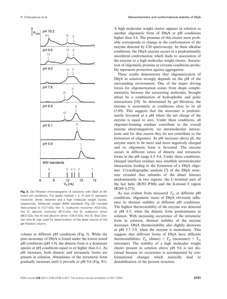
volume at different pH conditions (Fig. 3). While the
pure monomer of DbjA is found under the lowest tested
pH conditions (pH 5.9), the dimeric form is a dominant
species at pH conditions equal to or higher than 6.1. As
pH increases, both dimeric and tetrameric forms are
present in solution. Abundance of the tetrameric form
gradually increases until it prevails at pH 9.6 (Fig. S1).
A high molecular weight cluster appears in solution as
another oligomeric form of DbjA at pH conditions
higher than 9.6. The presence of this cluster most prob-
ably corresponds to change in the conformation of the
enzyme detected by CD spectroscopy. In these alkaline
conditions, the DbjA enzyme occurs in a predominantly
unordered conformation which leads to association of
the enzyme to a high molecular weight cluster. Associa-
tion of oligomeric proteins at extreme conditions proba-
bly represents protection against aggregation.
These results demonstrate that oligomerization of
DbjA in solution strongly depends on the pH of the
surrounding environment. One of the major driving
forces for oligomerization comes from shape comple-
mentarity between the associating molecules, brought
about by a combination of hydrophobic and polar
interactions [19]. As determined by gel filtration, the
enzyme is monomeric at conditions close to its pI
(5.89). This suggests that the monomer is predomi-
nantly favoured at a pH where the net charge of the
enzyme is equal to zero. Under these conditions, all
oligomer-forming residues contribute to the overall
enzyme electronegativity via intramolecular interac-
tions and for that reason they do not contribute to the
formation of oligomers. As pH increases above pI, the
enzyme starts to be more and more negatively charged
and its oligomeric form is favoured. The enzyme
occurs in different ratios of dimeric and tetrameric
forms in the pH range 6.5–9.6. Under these conditions,
charged interface residues may establish intermolecular
interactions leading to the formation of a DbjA oligo-
mer. Crystallographic analysis [7] of the DbjA struc-
ture revealed that subunits of the dimer interact
predominantly in two regions: the C-terminal part of
the last helix (R292–P306) and the b-strand 8 region
(R269–L275).
As was evident from measured Tm at different pH
conditions, oligomeric states of DbjA obviously influ-
ence its thermal stability at different pH conditions.
The highest thermostability of the enzyme was detected
at pH 6.5, when the dimeric form predominates in
solution. With increasing occurrence of the tetrameric
form in solution, thermal stability of the enzyme
decreases. DbjA thermostability also slightly decreases
at pH 5.7–5.9, when the enzyme is monomeric. This
suggests that different forms of DbjA have different
thermostabilities: Tm (dimer) > Tm (monomer) > Tm
(tetramer). The stability of a high molecular weight
cluster present in solution above pH 9.6 is not dis-
cussed because its occurrence is accompanied by con-
formational changes which naturally lead to
destabilization of the protein structure.
Fig. 3. Gel filtration chromatograms of solutions with DbjA at dif-
ferent pH conditions. The peaks marked I, II, III and IV represent
monomer, dimer, tetramer and a high molecular weight cluster,
respectively. Molecular weight (MW) standards (Fig. S2) included
ribonuclease A (13.7 kDa, line 1), ovalbumin monomer (43.0 kDa,
line 2), albumin monomer (67.0 kDa, line 3), ovalbumin dimer
(86.0 kDa, line 4) and albumin dimer (134.0 kDa, line 5). Blue Dex-
tran (line 6) was used for determination of the dead volume of the
gel filtration column.
R. Chaloupkova et al. Stereochemistry and conformational stability of DbjA
FEBS Journal 278 (2011) 2728–2738 ª 2011 The Authors Journal compilation ª 2011 FEBS 2731

pH profile
Measurement of DbjA activity was performed to
explore whether catalytic function directly relates to
conformational stability at various pH values. Experi-
ments were done under different pH conditions and
saturated concentrations of substrate 1-iodohexane for
which DbjA exhibited the highest catalytic efficiency
[5]. The activity profile of this enzyme shows a maxi-
mum at pH 9.7 (Fig. 2). However, the enzyme retains
at least 90% of its maximum activity at pH conditions
ranging from 7.7 to 10.4. DbjA thus possesses the
broadest pH optimum compared with other biochemi-
cally characterized haloalkane dehalogenases (Fig. 4).
This phenomenon is most likely related to the fact that
DbjA occurs as oligomer. The melting temperatures of
DbjA detected at optimal pH represent only 79.1% of
maximal Tm. For this reason, the pH interval at which
the enzyme possesses the highest activity and the high-
est thermostability simultaneously is narrowed to
between pH 7.4 and 8.7 (Fig. 2). DbjA activity
decreases below pH 7.0 and above pH 10.4 with no
activity detected below pH 5.0 and above pH 11.0.
These results correlate well with the conformational
stability as a function of pH observed by CD spectros-
copy. The loss of enzymatic activity at highly alkalic
conditions is caused by change from native to predom-
inantly disordered conformation. The drop in activity
below pH 7.0 is not induced by the structural changes
but by change in the protonation state of catalytic
amino acids.
Catalytic residues of DbjA comprise five key resi-
dues forming the so-called catalytic pentad [1]. The
catalytic pentad of DbjA consists of three residues
involved in the catalytic reaction, Asp103, Glu127 and
His280, and two H-bond donating residues, Asn38 and
Trp104, involved in stabilization of a halogen group
of the substrate. With respect to particular dissociation
constants of catalytic residues, pKAspa = 3.90
(b-COOH), pKGlua = 4.07 (c-COOH), pKHis
a = 6.04
(imidazol) [20], it is evident that the residue affecting
the enzyme activity below pH 7.0 is His280. At pH
6.1, the enzyme retains 50% of its maximal activity
which nicely corresponds to pKHisa . Under these condi-
tions, 50% of histidine is protonated and thus non-
reactive and 50% is still reactive. The imidazol ring of
His becomes protonated and the enzyme loses its activ-
ity when the pH decreases further. Knowledge of the
pH interval at which the enzyme retains its structure
but loses most of its activity due to protonation of cat-
alytic histidine is interesting for further detailed deter-
mination of its catalytic mechanism. An alkyl–enzyme
intermediate can be captured by protein crystallogra-
phy at these pH conditions as has been previously
described for the haloalkane dehalogenase DhlA [21].
The effect of pH on enantioselectivity
The dependence of DbjA enantioselectivity on pH was
tested in a reaction with 2-bromopentane. Although
the effect of pH on enzyme enantioselectivity has
already been described for both charged [22] and
uncharged [23] substrates, in the case of DbjA no sig-
nificant change in enantioselectivity was observed at
pH values ranging from 6.7 to 10.1 (data not shown).
Results indicated that ionization of the alkyl–enzyme
intermediate is the same for both enantiomers at all
tested pH values and corresponds with the theoretical
Enzymes
pH
6 7 8 9 10
DhlAa
DhaAb
LinBc
DhmAd
DmbAe
DmbBe
DmbCf
DrbAf
DbjAg
Fig. 4. Comparison of the pH profiles of biochemically characterized haloalkane dehalogenases. Enzyme activity was quantified as the spe-
cific enzyme activity in units of lmolÆs)1Æmg)1 under conditions corresponding to initial velocity measurements. Black boxes represent maxi-
mal dehalogenating activity. Grey boxes represent retained dehalogenating activity at the level of at least 90% of the maximal enzymatic
activity. a DhlA from Xanthobacter autotrophicus GJ10 [24]; b DhaA from Rhodococcus sp. [25]; c LinB from Sphingobium japonicum UT26
[39]; d DhmA from Mycobacterium avium N85 [26]; e DmbA and DmbB from Mycobacterium bovis 5033 ⁄ 66 [27]; f DmbC from Mycobacte-
rium bovis 5033 ⁄ 66 and DrbA from Rhodopirellula baltica SH1 [28]; g DbjA from Bradyrhizobium japonicum USDA110, this study.
Stereochemistry and conformational stability of DbjA R. Chaloupkova et al.
2732 FEBS Journal 278 (2011) 2728–2738 ª 2011 The Authors Journal compilation ª 2011 FEBS

rule that pH dependence of stereoselectivity can only
be observed around the pK values of groups in the
active site whose ionization controls the enzyme activ-
ity [23]. Ionization of the catalytic His of DbjA could
be reflected at tested pH conditions, although this
effect on enantioselectivity was not observed. The pKa
values of other catalytic amino acids of DbjA, i.e.
nucleophile Asp and catalytic acid Glu, are lower than
the pH conditions at which the enzyme aggregates.
Temperature profile
Measurement of enzymatic activity at different temper-
atures was carried out to study the effect of tempera-
ture on the rate of the dehalogenation reaction. The
enzyme exhibited the highest activity at 50 �C,although above this temperature it became rapidly
inactivated. This observation is in good agreement
with similar experiments previously described for other
haloalkane dehalogenases possessing the highest activ-
ity at temperatures ranging from 35 to 50 �C [24–28].
Thermodynamic analysis of enantioselectivity
The temperature dependence of DbjA enantioselectivi-
ty was studied to determine differential activation
parameters, enthalpy (DR�SDHz) and entropy
(DR�SDSz), contributing to the kinetic resolution of
selected b-bromoalkanes (2-bromobutane and 2-brom-
opentane) and a-bromoesters (methyl 2-bromopropio-
nate and ethyl 2-bromobutyrate). The temperature
dependence of DbjA enantioselectivity was measured
in the temperature range from 20 to 50 �C. The E val-
ues and the thermodynamic components of enantiose-
lectivity determined based on the linear relation of
ln E and T )1 are summarized in Table 1. Although
the studied temperature interval was relatively small,
highly significant changes in DbjA enantioselectivity
were observed. Variation of the reaction temperature
from 20 to 50 �C caused a decrease in E value of DbjA
from 174 to 13 in the reaction with 2-bromopentane,
from 474 to 197 with ethyl-2-bromopropionate and
from 225 to 83 with methyl 2-bromobutyrate. Since
enzyme enantioselectivity is defined as the ratio of the
specificity constants for (R)) and (S)) enantiomers,
the E value does not depend on the degree of conver-
sion or variation of the reaction mechanism of individ-
ual enantiomers with temperature. It should be noted
that the enthalpic and the entropic components of dif-
ferential activation free energy (DR�SDGz) both con-
tribute to the overall success of the kinetic resolution
of enantiomers [29,30]. All substrates have a racemic
temperature significantly above the experimental tem-
perature indicating that the entropic component coun-
teracts the enthalpic component of enantiomeric
discrimination. The linearity between ln E and T )1
observed from 20 to 50 �C suggested that a single tran-
sition state structure is held in this temperature range
for all tested substrates.
Enantiomeric discrimination of 2-bromobutane was
not observed at any tested temperature (Fig. 5). This
Table 1. Thermodynamic components for the dehalogenation of selected halogenated compounds catalyzed by DbjA. Errors were calculated
from the standard errors of the linear regression ln E versus T)1. Tr is the racemic temperature at which no stereochemical discrimination of
the enzyme between the (R)) and (S)) enantiomers occurs, E = 1 and DR�SDGz = 0. It is defined by the ratio of the differential activation
enthalpy and entropy, Tr ¼ DR�SDHz=DR�SDSz, and is constant for a particular racemic substrate converted by a particular enzyme [29,31].
No enantioselectivity was observed for 2-bromobutane.
Substrate E, 298 K
DR�SDHz(kJÆmol)1)
DR�SDSz(JÆmol)1ÆK)1)
TDR�SDSz,298 K (kJÆmol)1)
DR�SDGz,298 K (kJÆmol)1) Tr (�C)
2-Bromobutane 1 – – – – –
2-Bromopentane 132 )69.5 ± 2.6 )193.8 ± 8.4 )57.8 ± 2.5 )11.7 86
Ethyl 2-bromopropionate 392 )24.1 ± 1.8 )31.2 ± 5.9 )9.3 ± 1.8 )14.8 497
Methyl 2-bromobutyrate 209 )25.8 ± 2.2 )42.3 ± 7.2 )12.6 ± 2.1 )13.2 337
Fig. 5. The temperature dependence of enantiomeric ratios
determined for dehalogenation of selected b-bromoalkanes (2-bromo-
butane, 2-bromopentane) and a-brominated esters (ethyl 2-bromopro-
pionate, methyl 2-bromobutyrate) catalyzed by DbjA.
R. Chaloupkova et al. Stereochemistry and conformational stability of DbjA
FEBS Journal 278 (2011) 2728–2738 ª 2011 The Authors Journal compilation ª 2011 FEBS 2733
result excludes the possibility that the absence of DbjA
enantioselectivity towards 2-bromobutane is due to the
fact that the initial E value was determined at a tem-
perature (20 �C) close to the racemic temperature for
this particular enzymatic resolution. If this were the
case, the enantioselectivity of DbjA could be increased
with increasing reaction temperature, changing also the
enantio-preference of the enzyme [31]. However, our
measurements confirm that the absence of 2-bromobu-
tane discrimination is the effect of zero DR�SDGz at
all tested temperatures. (R)) and (S)) enantiomers of
this simple chiral molecule are probably too similar
to each other to be kinetically recognized by the
enzyme. Surprisingly, adding a single carbon atom to
a substrate molecule provided enough structural dif-
ference for high enantiomeric discrimination as was
seen in the case of 2-bromopentane (Fig. 5). This
finding indicates the importance of the length of the
b-substituted bromo-n-alkanes for their kinetic resolu-
tion. The temperature dependence of DbjA enantiose-
lectivity for 2-bromopentane revealed that both
thermodynamic parameters, DR�SDHz and DR�SDSz,where the entropic term represents 83% of the enthal-
pic term, are important for enantiodiscrimination
(Table 1). The high contribution of entropy indicates
the importance of solvation, conformational degrees
of freedom of the protein, or restriction of substrate
motion in the transition state of the reaction. b-bro-moalkanes display high flexibility within the enzyme
active site which is related to the significant influence
of DR�SDSz for their kinetic resolution by the DbjA
enzyme. This implies that enantiomeric recognition of
b-bromoalkanes by DbjA is mediated by the differen-
tial conformational freedom of enantiomers upon
binding and ⁄or a displacement of a different number
of active site water molecules by the (R)) and (S))enantiomer [32,33].
The temperature dependence of DbjA enantioselec-
tivity with ethyl 2-bromopropionate and methyl 2-bro-
mobutyrate revealed that differential activation
enthalpy represents a major contribution to their
discrimination (Table 1). The high contribution of
enthalpy is related to differences in the complementar-
ity of each enantiomer in the transition state compris-
ing steric and electrostatic interactions between the
enzyme active site, its substrate and the solvent.
a-bromoesters obviously possess limited flexibility
inside the active site cavity due to their ability to form
an additional hydrogen bond of a carboxylic oxygen
with halide stabilizing residues. This implies that DbjA
enantioselectivity towards a-bromoesters is due to
different interactions of individual enantiomers with
the residues of the enzyme active site in the Michaelis
complex and ⁄or the transition state of the dehalogen-
ation reaction [34].
The thermodynamic analysis showed that DbjA
enantioselectivity towards b-bromoalkanes and
a-bromoesters is differently influenced by individual
thermodynamic contributions, differential activation
enthalpy and entropy. The resolution of b-bromoalk-
anes was found to be driven by both enthalpic and
entropic terms, while the resolution of a-bromoesters
was driven mainly by an enthalpic term. These results
correspond well with the proposal that enantioselectivi-
ty of DbjA with b-bromoalkanes and a-bromoesters is
based on two distinct molecular interactions [7].
Conclusions
Here we show that DbjA possesses unusually high
structural and functional stability towards a broad
range of pH conditions. Oligomerization of DbjA is
strongly pH dependent. Monomer, dimer, tetramer and
a high molecular weight cluster of the enzyme were
distinguished in solution at different pH conditions and
each oligomeric state demonstrated different stability.
The highest thermostability occurred at pH conditions
when the enzyme occurs in its dimeric form. Tempera-
ture significantly alters enantioselectivity, but an effect
of pH on DbjA enantiodicrimination was not observed.
Lowering the temperature results in considerable
enhancement of enantioselectivity. The results from
thermodynamic analysis are in good agreement with the
proposal that enantiomeric discrimination of b-bromi-
nated alkanes and a-brominated esters by DbjA is
controlled by distinct molecular interactions [7]. These
results indicate unique properties of DbjA compared
with other known and characterized members of haloal-
kane dehalogenases. Catalytic activity and structural
stability in a broad range of pH conditions combined
with high enantioselectivity with selected substrates
make DbjA a very versatile biocatalyst.
Experimental procedures
Enzyme preparation
The His-tagged DbjA was overexpressed in Escherichia coli
BL21 using a previously described method [5] and purified
using the HighTrap Chelating HP 5-mL column charged
with Ni2+ ions (GE Healthcare, Uppsala, Sweden). The
enzyme was bound to the resin in equilibrating buffer
(20 mM potassium phosphate buffer, pH 7.5, containing
0.5 M sodium chloride and 10 mM imidazole). Unbound
and weakly bound proteins were washed out with the buf-
fer containing 10 mM imidazole. The target enzyme was
Stereochemistry and conformational stability of DbjA R. Chaloupkova et al.
2734 FEBS Journal 278 (2011) 2728–2738 ª 2011 The Authors Journal compilation ª 2011 FEBS

eluted by a buffer containing 500 mM imidazole. The active
fractions were pooled and dialysed against a 50 mM potas-
sium phosphate buffer (pH 7.5). The enzyme was kept at
4 �C during the purification procedure and stored in 50 mM
phosphate buffer at 4 �C until use.
CD spectroscopy
CD spectra were recorded at room temperature (22 �C)using a Jasco J-810 spectrometer (Jasco, Tokyo, Japan). All
the spectra were obtained at an interval of 0.1 nm with a
scanning speed of 100 nmÆmin)1, 1 s response time and
2 nm bandwidth. Cuvettes of 0.1 and 1 cm path length
were used in the far and near UV regions, respectively. The
protein concentrations for the far UV and the near UV
spectra acquisition were 0.23 mgÆmL)1 and 1.15 mgÆmL)1,
respectively. Each spectrum shown is the average of 10 indi-
vidual scans and has been corrected for baseline noise. CD
spectra were expressed in millidegrees. The a-helical contentof the enzyme was calculated from the mean residue ellip-
ticity (MRE) value at 222 nm using the following equation
as described by Chen et al. [16]:
a -helix% ¼MRE222�234030300� 100
ð1Þ
Thermal denaturation
Thermal unfolding of DbjA was followed at different pH
conditions by monitoring the ellipticity at 222 nm over the
temperature range 20–80 �C, with a resolution 0.2 �C, at aheating rate 0.5 �CÆmin)1. Recorded thermal denaturation
curves were roughly normalized to represent signal changes
between � 1 and 0, and fitted to sigmoidal curves using
software ORIGIN 6.1 (OriginLab, Northampton, MA, USA).
The melting temperatures (Tm) were evaluated as the mid-
point of the normalized thermal transition.
Prediction of the isoelectric point
The theoretical isoelectric point (pI) of DbjA was predicted
based on the amino acid sequence by using EXPASY SERVER
[35–37].
Effect of pH
DbjA activity and enantioselectivity were measured at dif-
ferent pH conditions. Britton–Robinson buffer solutions
were used to cover the pH range 1.7–11.5. The solutions
were prepared by mixing 0.04 M phosphoric, boric and ace-
tic acid with the appropriate volume of sodium hydroxide
(0.2 M) and sodium perchlorate monohydrate to get a con-
stant ionic strength of 0.15 M. The assays were performed
with 1-iodohexane as the substrate for activity measurement
at 37 �C or 2-bromopentane as the substrate for enantiose-
lectivity measurement at 25 �C.
Effect of temperature
The effect of temperature on DbjA activity and enantiose-
lectivity was determined by performing activity and enanti-
oselectivity assays at different temperatures. The activity
measurements were evaluated at temperatures ranging from
20 to 60 �C and the enantioselectivity of the DbjA enzyme
was monitored in the temperature range 20–50 �C, both in
50 mM glycin buffer at pH 8.6. Activity measurements were
performed with 1-iodohexane, and enantioselectivity mea-
surements with 2-bromobutane, 2-bromopentane, methyl
2-bromopropionate and ethyl 2-bromobutyrate.
Gel filtration chromatography
The molecular mass of DbjA enzyme at different pH condi-
tions was analysed using the FPLC system AKTA (GE
Healthcare) equipped with UV280 detection (GE Healthcare,
Uppsala, Sweden) and Superdex� 200 10 ⁄ 300 GL column
(GE Healthcare, Uppsala, Sweden). A total volume of
100 lL of each protein sample was applied to the column
and separated at a constant flow rate of 0.5 mLÆmin)1.
Britton–Robinson buffer with an appropriate pH value was
used as the mobile phase. The molecular weight standards
from the Gel Filtration Calibration Kit (GE Healthcare,
Uppsala, Sweden) included ribonuclease A (13.7 kDa), oval-
bumin monomer (43.0 kDa), albumin monomer (67.0 kDa),
ovalbumin dimer (86.0 kDa) and albumin dimer (134.0 kDa).
The dead volume of the Superdex� 200 10 ⁄ 300 GL column
was determined using the Blue Dextran of the calibration kit.
All protein standards as well as enzyme samples were trans-
ferred into the Britton–Robinson buffer by using a 5-mL
HighTrap Desalting Sephadex G-25 Superfine column (GE
Healthcare, Uppsala, Sweden).
Activity assay
DbjA activity was assayed by the colorimetric method
developed by Iwasaki et al. [38]. The halide ions released
were analysed after a reaction with mercuric thiocyanate
and ferric ammonium sulfate spectrophotometrically at
460 nm using the Sunrise microplate reader (Tecan,
Grodig ⁄ Salzburg, Austria). The dehalogenation reaction
was performed in 25-mL Reacti flasks closed by Miniert
valves at various temperatures. The reaction mixture was
composed of 15 mL of buffer and 2 lL of substrate 1-iod-
ohexane. The reaction was initiated by the addition of
enzyme in a final concentration of 0.15 lM. The reaction
was monitored by withdrawing 1 mL samples at 10, 20,
30, 40, 50 and 60 min from the reaction mixture. The reac-
tion mixture samples were immediately mixed with 0.1 mL
35% nitric acid to terminate the reaction. Dehalogenation
activity was quantified as a rate of product formation in
time. Each activity was measured in three to five indepen-
dent replicates and represented as mean values of relative
R. Chaloupkova et al. Stereochemistry and conformational stability of DbjA
FEBS Journal 278 (2011) 2728–2738 ª 2011 The Authors Journal compilation ª 2011 FEBS 2735
activity with plotted standard errors. Relative activities
represented a percentage of maximal specific activity
detected.
Enantioselectivity assay
Enantioselectivity was analysed in 25-mL Reacti flasks
closed by Miniert valves containing 20 mL of glycin buffer
(100 mM, pH 8.6). Chiral substrates were added to a final
concentration of 0.5–3.0 mM with regard to enzyme affinity.
The enzymatic reaction was initiated by the addition of
appropriate amounts of the DbjA enzyme depending on
enzyme activity (final concentration 0.2–2.0 lM). The reac-
tion was monitored by periodical withdrawing of 0.5 mL
sample aliquots from the reaction mixture. The reaction
was stopped by mixing the sample with 1 mL of diethyl
ether containing 1,2-dichloroethane as an internal standard.
After extraction, diethyl ether was anhydrated on a glass
column with sodium sulphate. The samples were automati-
cally analysed by using Hewlett-Packard 6890 gas chro-
matograph (Agilent, Santa Clara, USA) equipped with a
flame ionization detector and chiral capillary column Chi-
raldex B-TA and Chiraldex G-TA (Alltech, Deerfield,
USA). Michaelis–Menten parameters were derived by fitting
the progress curves obtained from kinetic resolution experi-
ments into a competitive kinetic pattern by numerical
integration using the software MICROMATH SCIENTIST
(ChemSW, Fairfield, USA). Enantioselectivity was deter-
mined as the enantiomeric ratio (E) defined by
E ¼ kRcat=KRm
kScat=KSm
ð2Þ
where kcat and Km represent the Michaelis–Menten parame-
ters of the two enantiomers.
Thermodynamic analysis
The difference in activation enthalpy and entropy between
enantiomers was determined by studying the variation of
the enzyme enantiomeric ratio with temperature:
lnE ¼ �DR�SDHzR
� 1
Tþ DR�SDSz
Rð3Þ
The enantiomeric ratio (or rather lnE) varied with recipro-
cal temperature to an extent determined by the enthalpic
term (the slope of Eqn 3, DR�SDHz ⁄R), at a level deter-
mined by the entropic term (the intercept of Eqn 3,
DR�SDSz ⁄R). A racemic temperature (Tr) was determined
as the ratio of the differential activation enthalpy and
entropy:
Tr ¼DR�SDHz
DR�SDSzð4Þ
Acknowledgements
This work was financially supported by the Grant
Agency of the Czech Academy of Sciences
(IAA401630901 to J.D.), the Czech Ministry of Educa-
tion (MSM0021622412 and LC06010 to J.D.), the
Grant Agency of the Czech Republic (203 ⁄ 08 ⁄ 0114 to
R.Ch.) and the European Regional Development Fund
(project FNUSA-ICRC no. CZ.1.05 ⁄ 1.1.00 ⁄ 02.0123 to
Z.P.). The authors thank Eva Chovancova for the pre-
diction of DbjA quaternary structure and Monika
Strakova for assistance with protein expression and
purification.
References
1 Janssen DB (2004) Evolving haloalkane dehalogenases.
Curr Opin Chem Biol 8, 150–159.
2 Fetzner S & Lingens F (1994) Bacterial dehalogenases:
biochemistry, genetics, and biotechnological applica-
tions. Microbiol Rev 58, 641–685.
3 Patel RN (2004) Biocatalytic synthesis of chiral phar-
maceutical intermediates. Food Technol Biotechnol 42,
305–325.
4 Patel RN (2006) Biocatalysis: synthesis of chiral inter-
mediates for drugs. Curr Opin Drug Discov Devel 9,
741–764.
5 Sato Y, Monincova M, Chaloupkova R, Prokop Z,
Ohtsubo Y, Minamisawa K, Tsuda M, Damborsky J &
Nagata Y (2005) Two rhizobial strains, Mesorhizobi-
um loti MAFF303099 and Bradyrhizobium japonicum
USDA110, encode haloalkane dehalogenases with novel
structures and substrate specificities. Appl Environ
Microbiol 71, 4372–4379.
6 Prokop Z, Damborsky J, Nagata Y & Janssen DB
(2009) Method of production of optically active
halohydrocarbons and alcohols using hydrolytic
dehalogenation catalysed by haloalkane dehalogenases.
US7, 632, 666.
7 Prokop Z, Sato Y, Brezovsky J, Mozga T, Chaloupkova
R, Koudelakova T, Jerabek P, Stepankova V, Natsume
R, Leeuwen JGE et al. (2010) Enantioselectivity of
haloalkane dehalogenases and its modulation by
surface loop engineering. Angew Chem Int Ed 49,
6111–6115.
8 Schoemaker HE, Mink D & Wubbolts MG (2003) Dis-
pelling the myths – biocatalysis in industrial synthesis.
Science 299, 1694–1697.
9 Bornscheuer UT & Pohl M (2001) Improved biocata-
lysts by directed evolution and rational protein design.
Curr Opin Chem Biol 5, 137–143.
10 Pollard DJ & Woodley JM (2007) Biocatalysis for
pharmaceutical intermediates: the future is now. Trends
Biotechnol 25, 66–73.
Stereochemistry and conformational stability of DbjA R. Chaloupkova et al.
2736 FEBS Journal 278 (2011) 2728–2738 ª 2011 The Authors Journal compilation ª 2011 FEBS
11 Woodley JM (2008) New opportunities for biocatalysis:
making pharmaceutical processes greener. Trends
Biotechnol 26, 321–327.
12 Jaeger KE & Eggert T (2004) Enantioselective biocatal-
ysis optimized by directed evolution. Curr Opin Biotech-
nol 15, 305–313.
13 Kazlauskas RJ (2005) Enhancing catalytic promiscuity
for biocatalysis. Curr Opin Chem Biol 9, 195–201.
14 Bornscheuer UT (2005) Trends and challenges in enzyme
technology. Adv Biochem Eng Biotechnol 100, 181–203.
15 Hult K & Berglund P (2003) Engineered enzymes for
improved organic synthesis. Curr Opin Biotech 14,
395–400.
16 Chen Y-H, Yang JT & Martinez HM (1972) Determi-
nation of the secondary structures of proteins by
circular dichroism and optical rotatory dispersion.
Biochemistry 11, 4120–4131.
17 Fasman GD (1996) Circular Dichroism and Conforma-
tional Analysis of Biomolecules. Plenum Press,
New York, NY.
18 Alexov E (2004) Numerical calculation of the pH of
maximal protein stability. Eur J Biochem 271, 173–185.
19 Shallom D, Golan G, Shoham G & Shoham Y (2004)
Effect of dimer dissociation on activity and thermostabil-
ity of the a-glucuronidase from Geobacillus stearothermo-
philus: dissecting the differnt oligomeric forms of family
67 glycoside hydrolases. J Bacteriol 186, 6928–6937.
20 Fersht A (1999) Structure and Mechanism in Protein
Science: A Guide to Enzyme Catalysis and Protein Fold-
ing. W.H. Freeman, New York, NY.
21 Verschueren KHG, Seljee F, Rozeboom HJ, Kalk KH
& Dijkstra BW (1993) Crystallographic analysis of the
catalytic mechanism of haloalkane dehalogenase. Nature
363, 693–698.
22 Lummer K, Rieks A, Galunsky B & Kasche V (1999)
pH dependence of penicillin amidase enantioselectivity
for charged substrates. Biochim Biophys Acta 1433,
327–334.
23 Secundo F & Phillips RS (1996) Effects of pH on enan-
tiospecificity of alcohol dehydrogenases from Thermo-
anaerobacter ethanolicus and horse liver. Enzyme
Microb Technol 19, 487–492.
24 Keuning S, Janssen DB & Witholt B (1985) Purification
and characterization of hydrolytic haloalkane dehalo-
genase from Xanthobacter autotrophicus GJ10. J Bacte-
riol 163, 635–639.
25 Yokota T, Omori T & Kodama T (1987) Purification
and properties of haloalkane dehalogenase from Coryne-
bacterium sp. strain m15-3. J Bacteriol 169, 4049–4054.
26 Jesenska A, Bartos M, Czernekova V, Rychlik I, Pavlik
I & Damborsky J (2002) Cloning and expression of
the haloalkane dehalogenase gene dhmA from
Mycobacterium avium N85 and preliminary character-
ization of DhmA. Appl Environ Microbiol 68,
3724–3730.
27 Jesenska A, Pavlova M, Strouhal M, Chaloupkova R,
Tesinska I, Monincova M, Prokop Z, Bartos M, Pavlik
I, Rychlik I et al. (2005) Mycobacterial haloalkane
dehalogenases: cloning, biochemical properties and
distribution. Appl Environ Microbiol 71, 6736–6745.
28 Jesenska A, Monincova M, Chrobakova T, Hasan K,
Chaloupkova R, Prokop Z, Geerlof A & Damborsky J
(2009) Isolation and biochemical characterization of
haloalkane dehalogenases DrbA and DmbC: representa-
tives of novel subfamily. Appl Environ Microbiol 75,
5157–5160.
29 Phillips RS (1996) Temperature modulation of the
stereochemistry of enzymatic catalysis: prospects for
exploitation. Trends Biotechnol 14, 13–16.
30 Ottosson J, Fransson L & Hult K (2002) Substrate
entropy in enzyme enantioselectivity: an experimental
and molecular modeling study. Protein Sci 11, 1462–
1471.
31 Pham VT, Phillips RS & Ljungdahl LG (1989) Temper-
ature-dependent enantiospecificity of secondary alcohol
dehydrogenase from Thermoanaerobacter ethanolicus.
J Am Chem Soc 111, 1935–1936.
32 Overbeeke PL, Orrenius C, Jongejan JA & Duine JA
(1998) Enthalpic and entropic contributions to lipase
enantioselectivity. Chem Phys Lipids 93, 81–93.
33 Phillips RS (2002) How does active site water affect
enzymatic stereorecognition? J Mol Catal B Enzym
19–20, 103–107.
34 Galunsky B, Ignatova S & Kasche V (1997) Tempera-
ture effects on S1- and S’1-enantioselectivity of
alpha-chymotrypsin. Biochim Biophys Acta 1343,
130–138.
35 Bjellqvist B, Hughes GJ, Pasquali C, Paquet N, Ravier
F, Sanchez J-C, Frutiger S & Hochstrasser DF (1993)
The focusing positions of polypeptides in immobilized
pH gradients can be predicted from their amino acid
sequences. Electrophoresis 14, 1023–1031.
36 Bjellqvist B, Basse B, Olsen E & Celis JE (1994)
Reference points for comparisons of two-dimensional
maps of proteins from different human cell types
defined in a pH scale where isoelectric points correlate
with polypeptide compositions. Electrophoresis 15, 529–
539.
37 Gasteiger E, Hoogland C, Gattiker A, Duvaud S,
Wilkins MR, Appel RD & Bairoch A (2005) The Prote-
omics Protocols Handbook. Humana Press, Totowa, NJ.
38 Iwasaki I, Utsumi S & Ozawa T (1952) New colorimet-
ric determination of chloride using mercuric thiocyanate
and ferric ion. Bull Chem Soc Jpn 25, 226.
39 Nagata Y, Miyauchi K, Damborsky J, Manova K,
Ansorgova A & Takagi M (1997) Purification and
characterization of haloalkane dehalogenase of a new
substrate class from a c-hexachlorocyclohexane-degrad-ing bacterium, Sphingomonas paucimobilis UT26. Appl
Environ Microbiol 63, 3707–3710.
R. Chaloupkova et al. Stereochemistry and conformational stability of DbjA
FEBS Journal 278 (2011) 2728–2738 ª 2011 The Authors Journal compilation ª 2011 FEBS 2737

Supporting information
The following supplementary material is available:
Fig. S1. Distribution of various forms of DbjA in
solution at different pH conditions.
Fig. S2. Gel filtration chromatogram of ribonuclease A
(13.7 kDa, line 1), ovalbumin monomer (43.0 kDa, line
2), albumin monomer (67.0 kDa, line 3), ovalbumin
dimer (86.0 kDa, line 4) and albumin dimer (134.0 kDa,
line 5) used as molecular weight standards. Blue Dex-
tran (line 6) was used for determination of dead volume
of the gel filtration column.
This supplementary material can be found in the
online version of this article.
Please note: As a service to our authors and readers,
this journal provides supporting information supplied
by the authors. Such materials are peer-reviewed and
may be reorganized for online delivery, but are not
copy-edited or typeset. Technical support issues arising
from supporting information (other than missing files)
should be addressed to the authors.
Stereochemistry and conformational stability of DbjA R. Chaloupkova et al.
2738 FEBS Journal 278 (2011) 2728–2738 ª 2011 The Authors Journal compilation ª 2011 FEBS