Stereoselectivity in the Rhodium-Catalysed Reductions of Non-Conjugated Dienes
Upload
independentCategory
view
0download
0
Bioorganic & Medicinal Chemistry 19 (2011) 1390–1399
Contents lists available at ScienceDirect
Bioorganic & Medicinal Chemistry
journal homepage: www.elsevier .com/locate /bmc
Brassinin oxidase mediated transformation of the phytoalexin brassinin:Structure of the elusive co-product, deuterium isotope effectand stereoselectivity
M. Soledade C. Pedras ⇑, Zoran Minic, Vijay K. Sarma-MamillapalleDepartment of Chemistry, University of Saskatchewan, 110 Science Place, Saskatoon, SK, Canada S7N 5C9
a r t i c l e i n f o
Article history:Received 24 November 2010Revised 4 January 2011Accepted 7 January 2011Available online 13 January 2011
Keywords:Brassinin oxidaseDeuterated brassininDetoxificationDeuterium isotope effectMethyl dithiocarbamateOxidoreductasePhytoalexin
0968-0896/$ - see front matter � 2011 Elsevier Ltd. Adoi:10.1016/j.bmc.2011.01.011
⇑ Corresponding author. Tel.: +1 306 966 4772; faxE-mail address: [email protected] (M. Sole
a b s t r a c t
Brassinin oxidase, a fungal detoxifying enzyme that mediates the conversion of the phytoalexin brassinininto indole-3-carboxaldehyde, is the first enzyme described to date that catalyzes the transformation of adithiocarbamate group into an aldehyde equivalent. Brassinin is an essential phytoalexin due to its anti-fungal activity and its role as biosynthetic precursor of other phytoalexins produced in plants of the fam-ily Brassicaceae (common name crucifer). In this report, the isolation, structure determination andsynthesis of the elusive co-product of brassinin transformation by brassinin oxidase, S-methyl dithiocar-bamate, the syntheses of dideuterated and (R) and (S) monodeuterated brassinins, kinetic analyses of iso-tope effects and chemical modifications of brassinin oxidase are described. The reaction of[10-2H2]brassinin was found to be slowed by a kinetic isotope effect of 5.3 on the value of kcat/Km. Thisresult indicates that the hydride/hydrogen transfer step preceding brassinin transformation is rate deter-mining in the overall reaction. In addition, the use of (R) and (S)-[10-2H]brassinins as substrates indicatedthat the hydride/hydrogen transfer step is ca. 88% stereoselective for the pro-R hydrogen. A detailedchemical mechanism of the enzymatic transformation of brassinin is proposed.
� 2011 Elsevier Ltd. All rights reserved.
1. Introduction brassinin detoxification by BOLm were devised as prospective crop
Brassinin oxidase (BOLm) is a fungal detoxifying enzyme thatcatalyzes the transformation of brassinin (1) into the non-toxic in-dole-3-carboxaldehyde (2).1–3 BOLm appears to be the first andonly enzyme described to date catalyzing the transformation of adithiocarbamate group into an aldehyde (Scheme 1). BOLm is pro-duced by the fungus Leptosphaeria maculans (Desm.) Ces. et deNot. [asexual stage Phoma lingam (Tode ex Fr.) Desm.], one of themost economically important pathogens of cruciferous oilseeds.4,5
Brassinin (1) is one of several crucifers’ defensive products knownas phytoalexins,6,7 that is, antimicrobial metabolites produced denovo in response to stress caused by pathogens and abiotic sources.Phytoalexins are not produced in healthy plant tissues and are partof general defense mechanisms to ward off plant invaders. The fun-gal detoxification of brassinin (1) in planta represents a com-pounded loss for infected plants because brassinin (1) is both anantifungal metabolite and a biosynthetic precursor of additionalantifungal phytoalexins. Hence, depletion of brassinin can lead toa fast decrease of several inducible plant defense metabolites,which facilitates pathogen invasion and subsequent plant death.Considering the biological importance of brassinin, inhibitors of
ll rights reserved.
: +1 306 966 4730.dade C. Pedras).
protection agents against L. maculans.3,8 Phytoalexin detoxificationinhibitors were coined paldoxins and proposed as alternative cropprotection agents against specific fungal pathogens.9 Interestingly,the crucifer phytoalexins, camalexin (3) and cyclobrassinin (4)were shown to inhibit competitively BOLm activity,2 indicating thatthe various constituents of phytoalexin blends produced by cruci-fers in response to stress have multiple physiological functions.
NH
NH
S
NSCH3S
N
34
NH
N O
O
H
CH3
5
1'34
1
To probe the substrate specificity of BOLm, various isostericgroups of dithiocarbamate (carbamate, thiolcarbamate, dithiocar-bonate, ester, amide, urea and thiourea) were screened using puri-

M. Soledade C. Pedras et al. / Bioorg. Med. Chem. 19 (2011) 1390–1399 1391
fied BOLm and phenazine methosulfate (PMS) as the electronacceptor.1 Results of this study showed that the catalytic transfor-mation of methyl N0-(3-indolylmethyl)carbamate (5) to indole-3-carboxaldehyde (2) was faster than that of any other functionalgroup, including the dithiocarbamate (brassinin (1)). In addition,essential structural features of substrates of BOLm were a methylor ethyl group attached to the thiol moiety of the (mono/di)thiocar-bamate group, a free –NH at the side-chain functional group, and amethylene bridge (–CH2–) between the C-3 of indole and the func-tional group. This last structural feature suggested that any BOLmsubstrate required a planar carbon (sp2) adjacent to the CH2 (C-10). Based on those results, we proposed a chemical mechanismfor the transformation of brassinin (1) to indole-3-carboxaldehyde(2) that suggested the formation of two imidodithiocarbamateintermediates I1 and I2, covalently bound to PMS (phenazinemethosulfate).1 However, the fate of the dithiocarbamate group ofbrassinin (1), the structure of the co-product(s) of the transforma-tion and the origin of the oxygen atom of the aldehyde group werenot identified (Scheme 2). BOLm accepted a wide range of cofactors(PMS, small quinones or flavin mononucleotide, FMN).
To design paldoxins effective against L. maculans, it is crucial tounderstand the chemical and catalytic mechanisms of the enzy-matic transformation of brassinin (1).10 Toward this end, we de-scribe the isolation and structure determination of the previouslyunknown co-product of the BOLm mediated transformation ofbrassinin (1), the syntheses of dideuterated brassinin and corre-sponding carbamate, and kinetic analyses of deuterium isotope ef-fects. A detailed chemical mechanism of the enzymatictransformation of brassinin (1) by BOLm that integrates these re-sults with previous work is discussed.
2. Results
2.1. Chemistry
2.1.1. Chemical structure of the co-product of brassininoxidation and origin of the aldehydic oxygen
The fate of the S-methyl dithiocarbamate group of brassinin (1)after transformation by BOLm has remained elusive until now.
N
N
CH3
NH
N S
S CH3
HR HS
N
N
CH3
NH
N S
S CH3
H
I1
H H
I2
Scheme 2. Proposed intermediates I1 and I2 of the catalytic transformation ofbrassinin (1) by BOLm using PMS as electron acceptor.1
NH
N S
S
H
CH3
brassinin (1)
1
341'
BOLm / PMS
NH
CHO
2
Scheme 1. Transformation of brassinin (1) to indole-3-carboxaldehyde (2) medi-ated by BOLm from the plant fungal pathogen Leptosphaeria maculans.
HPLC chromatograms of extracts of the reaction mixture of brass-inin and purified BOLm showed peaks due to the substrate (brass-inin, 1, tR = 19.8 min) and indole-3-carboxaldehyde (2, tR = 7.0 min)together with an additional peak due to an unknown component(tR = 4.4 min). To find the chemical structure of this component,the extract of a BOLm-brassinin reaction mixture (2 mM brassininwith 2 mM PMS, 4 lg of purified enzyme) was separated and ana-lyzed as described in Section 4. The 1H NMR spectral data of theunknown compound at tR = 4.4 min revealed the presence of a sin-glet at d 2.64 and its HRMS-ESI data suggested a molecular formulaof C2H5NS2. Altogether the spectroscopic data suggested that thesecond reaction product resulting from transformation of brassininwas S-methyl dithiocarbamate (6), the structure of which was con-firmed by chemical synthesis (Scheme 3)11 and direct comparisonof spectroscopic data, including UV spectrum (Fig. S1). Unlike otherdithiocarbamates, the antifungal activity of S-methyl dithiocarba-mate (6) was substantially lower than that of brassinin (100% at0.50 mM), causing only 18 ± 4% mycelial growth inhibition.
To establish the origin of the oxygen atom in indole-3-carboxal-dehyde (2), the catalytic transformation of brassinin (1) by BOLmwas carried out in H2
18O–H216O (ca. 1:1). The HRMS-ESI (nega-
tive-ion) spectrum of indole-3-carboxaldehyde (2) obtained in thisenzymatic transformation displayed peaks corresponding to natu-ral abundance [16O]indole-3-carboxaldehyde (2) (measured[M�1]� m/z = 144.0474; calculated for natural abundanceC9H6NO m/z = 144.0455) and to labeled [18O]indole-3-carboxalde-hyde (measured [M�1]� m/z = 146.0503; calculated for C9H7N18Om/z = 146.0498). In control reactions (carried out in non-labeledH2O), the [18O]indole-3-carboxaldehyde peak was not detected(Table 1). These results strongly suggested that the oxygen of in-dole-3-carboxaldehyde (2) originated from water.
2.1.2. Synthesis of deuterated brassinins and methyl [10,10-2H2]-N0-(3-indolylmethyl)carbamate (5a)
[10,10-2H2]Brassinin (1a) and methyl [10,10-2H2]-N0-(3-indolylm-ethyl)carbamate (5a) were obtained from [10,10-2H2]indolyl-3-methanamine (8a), which was prepared from 3-indolecarbonitrile(7) after reduction with lithium aluminum deuteride, as summa-rized in Scheme 4 and described in Section 4. The syntheses of(R)-[10-2H] and (S)-[10-2H]brassinins followed a straightforwardroute previously used in other chiral deuterated amine prepara-tions, where the amines were obtained from the correspondingalcohols.12 The enantiomeric purity of these alcohols was deter-mined to be >96%, after esterification with the Mosher’s acid chlo-ride13 and analysis of the purified reaction products by 1H NMRspectroscopy.14
2.2. Enzyme BOLm
2.2.1. Substrate kinetic isotope effectsTo determine the deuterium isotope effects of the methylene
group (–CH2–) on the rate of conversion of brassinin (1) to in-dole-3-carboxaldehyde (2), dideuterated ([10,10-2H2])brassinin(1a) was used as substrate and specific activities and kineticparameters were compared with those obtained for the transfor-mation of non-labeled brassinin (1). The relative specific activityof BOLm was about fourfold higher for the non-labeled substratethan for the dideuterated substrate (Table 2). HRMS-ESI analyses
H2N S
SCH3
H2N S
H3NS
CH3I
6
Scheme 3. Syntheses of S-methyl dithiocarbamate (6).
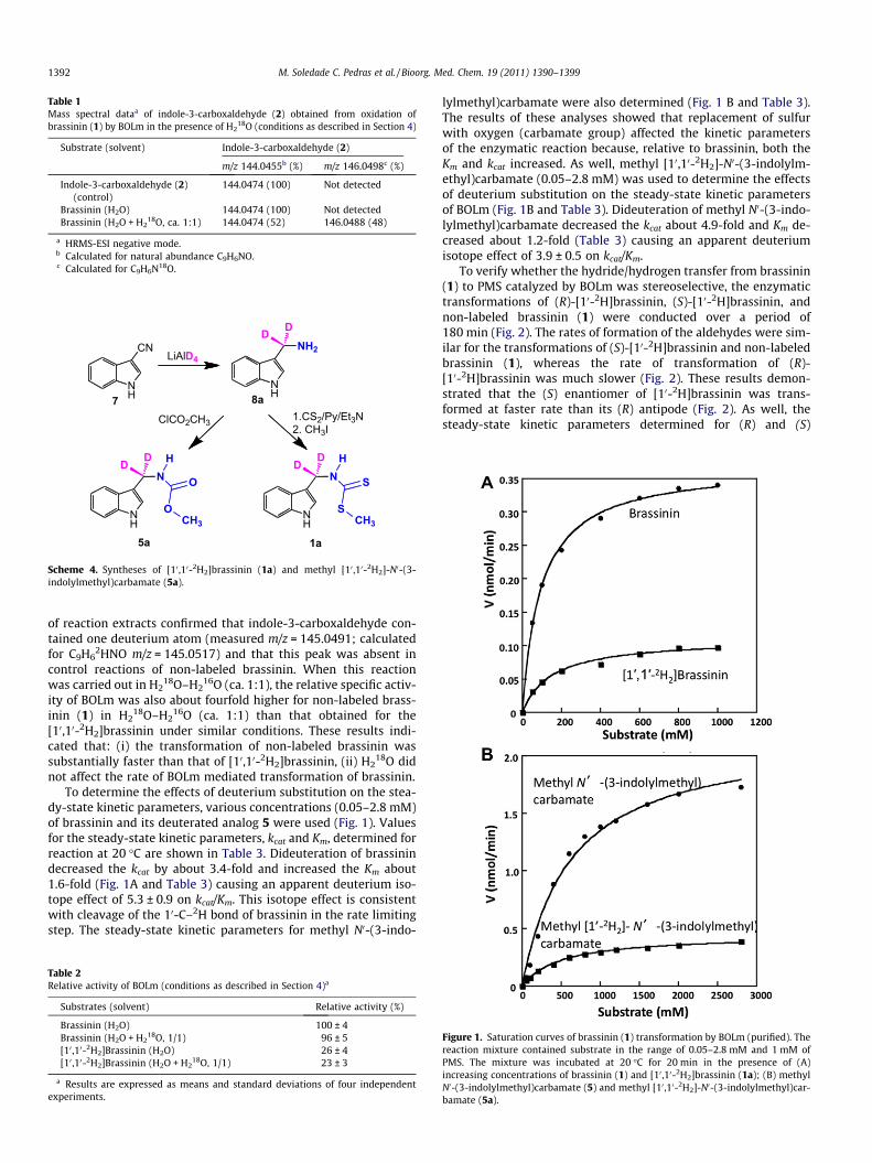
Table 1Mass spectral dataa of indole-3-carboxaldehyde (2) obtained from oxidation ofbrassinin (1) by BOLm in the presence of H2
18O (conditions as described in Section 4)
Substrate (solvent) Indole-3-carboxaldehyde (2)
m/z 144.0455b (%) m/z 146.0498c (%)
Indole-3-carboxaldehyde (2)(control)
144.0474 (100) Not detected
Brassinin (H2O) 144.0474 (100) Not detectedBrassinin (H2O + H2
18O, ca. 1:1) 144.0474 (52) 146.0488 (48)
a HRMS-ESI negative mode.b Calculated for natural abundance C9H6NO.c Calculated for C9H6N18O.
NH
N O
O
H
CH3
DD
NH
N S
S
H
CH3
DD
NH
CNLiAlD4
NH
NH2
DD
1a5a
ClCO2CH3 1.CS2/Py/Et3N2. CH3I
7 8a
Scheme 4. Syntheses of [10 ,10-2H2]brassinin (1a) and methyl [10 ,10-2H2]-N0-(3-indolylmethyl)carbamate (5a).
1392 M. Soledade C. Pedras et al. / Bioorg. Med. Chem. 19 (2011) 1390–1399
of reaction extracts confirmed that indole-3-carboxaldehyde con-tained one deuterium atom (measured m/z = 145.0491; calculatedfor C9H6
2HNO m/z = 145.0517) and that this peak was absent incontrol reactions of non-labeled brassinin. When this reactionwas carried out in H2
18O–H216O (ca. 1:1), the relative specific activ-
ity of BOLm was also about fourfold higher for non-labeled brass-inin (1) in H2
18O–H216O (ca. 1:1) than that obtained for the
[10,10-2H2]brassinin under similar conditions. These results indi-cated that: (i) the transformation of non-labeled brassinin wassubstantially faster than that of [10,10-2H2]brassinin, (ii) H2
18O didnot affect the rate of BOLm mediated transformation of brassinin.
To determine the effects of deuterium substitution on the stea-dy-state kinetic parameters, various concentrations (0.05–2.8 mM)of brassinin and its deuterated analog 5 were used (Fig. 1). Valuesfor the steady-state kinetic parameters, kcat and Km, determined forreaction at 20 �C are shown in Table 3. Dideuteration of brassinindecreased the kcat by about 3.4-fold and increased the Km about1.6-fold (Fig. 1A and Table 3) causing an apparent deuterium iso-tope effect of 5.3 ± 0.9 on kcat/Km. This isotope effect is consistentwith cleavage of the 10-C–2H bond of brassinin in the rate limitingstep. The steady-state kinetic parameters for methyl N0-(3-indo-
Table 2Relative activity of BOLm (conditions as described in Section 4)a
Substrates (solvent) Relative activity (%)
Brassinin (H2O) 100 ± 4Brassinin (H2O + H2
18O, 1/1) 96 ± 5[10 ,10-2H2]Brassinin (H2O) 26 ± 4[10 ,10-2H2]Brassinin (H2O + H2
18O, 1/1) 23 ± 3
a Results are expressed as means and standard deviations of four independentexperiments.
lylmethyl)carbamate were also determined (Fig. 1 B and Table 3).The results of these analyses showed that replacement of sulfurwith oxygen (carbamate group) affected the kinetic parametersof the enzymatic reaction because, relative to brassinin, both theKm and kcat increased. As well, methyl [10,10-2H2]-N0-(3-indolylm-ethyl)carbamate (0.05–2.8 mM) was used to determine the effectsof deuterium substitution on the steady-state kinetic parametersof BOLm (Fig. 1B and Table 3). Dideuteration of methyl N0-(3-indo-lylmethyl)carbamate decreased the kcat about 4.9-fold and Km de-creased about 1.2-fold (Table 3) causing an apparent deuteriumisotope effect of 3.9 ± 0.5 on kcat/Km.
To verify whether the hydride/hydrogen transfer from brassinin(1) to PMS catalyzed by BOLm was stereoselective, the enzymatictransformations of (R)-[10-2H]brassinin, (S)-[10-2H]brassinin, andnon-labeled brassinin (1) were conducted over a period of180 min (Fig. 2). The rates of formation of the aldehydes were sim-ilar for the transformations of (S)-[10-2H]brassinin and non-labeledbrassinin (1), whereas the rate of transformation of (R)-[10-2H]brassinin was much slower (Fig. 2). These results demon-strated that the (S) enantiomer of [10-2H]brassinin was trans-formed at faster rate than its (R) antipode (Fig. 2). As well, thesteady-state kinetic parameters determined for (R) and (S)
Figure 1. Saturation curves of brassinin (1) transformation by BOLm (purified). Thereaction mixture contained substrate in the range of 0.05–2.8 mM and 1 mM ofPMS. The mixture was incubated at 20 �C for 20 min in the presence of (A)increasing concentrations of brassinin (1) and [10 ,10-2H2]brassinin (1a); (B) methylN0-(3-indolylmethyl)carbamate (5) and methyl [10 ,10-2H2]-N0-(3-indolylmethyl)car-bamate (5a).
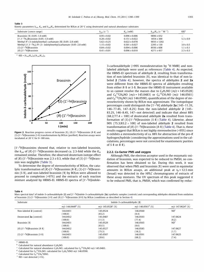
Table 3Kinetic parameters kcat, Km and kcat/Km determined for BOLm at 20 �C using deuterated and natural abundance substrates
Substrate (concn range) kcat (s�1) Km (mM) kcat/Km (s�1 M�1) KIEa
Brassinin (1) (0.05–1.0 mM) 0.95 ± 0.02 0.096 ± 0.006 9896 ± 652 —[10 ,10-2H2]Brassinin (0.05–1.0 mM) 0.28 ± 0.02 0.151 ± 0.022 1854 ± 300 5.3 ± 0.9Methyl N0-(3-indolylmethyl) carbamate (5) (0.05–2.8 mM) 5.62 ± 0.25 0.622 ± 0.078 9035 ± 1202 —Methyl [10 ,10-2H2]-N0-(3- indolylmethyl)carbamate (0.05–2.8 mM) 1.15 ± 0.02 0.501 ± 0.027 2295 ± 130 3.9 ± 0.5(S)-[10-2H]Brassinin 0.84 ± 0.02 0.094 ± 0.006 8936 ± 608 1.1 ± 0.1(R)-[10-2H]Brassinin 0.41 ± 0.01 0.096 ± 0.010 4271 ± 457 2.3 ± 0.3
a KIE = (kcat/Km)H/(kcat/Km)D.
Figure 2. Reaction progress curves of brassinin (1), (R)-[10-2H]brassinin (R-1) and(S)-[10-2H]brassinin (S-1) transformations by BOLm (purified). Reaction assays wereincubated at 20 �C for 0–180 min.
M. Soledade C. Pedras et al. / Bioorg. Med. Chem. 19 (2011) 1390–1399 1393
[10-2H]brassinins showed that, relative to non-labeled brassinin,the kcat of (R)-[10-2H]brassinin decreased ca. 2.3-fold while the Km
remained similar. Therefore, the observed deuterium isotope effectof (R)-[10-2H]brassinin was 2.3 ± 0.3, while that of (S)-[10-2H]brass-inin was negligible (Table 3).
To determine the degree of stereoselectivity of BOLm, the cata-lytic transformation of (R)-[10-2H]brassinin (R-1), (S)-[10-2H]brassi-nin (S-1), and non-labeled brassinin (1) by BOLm were allowed toproceed to completion (>97%) and the extracts of each reactionmixture analyzed by HRMS-EI. HRMS-EI spectra of [10-2H]indole-
Table 4Mass spectral dataa of indole-3-carboxaldehyde (2) and [10-2H]indole-3-carboxaldehyde (2of brassinin (S)-[10-2H]brassinin (S-1) and (R)-[10-2H]brassinin (R-1) by BOLm (conditions
Substrate
m/z 144.0449b (%) m/z 145
Non-labeled 2 (control) 144.0451(100.0)
145.052(83.2)
Deuterated 2a (control) 144.0452(100.0)
145.048(11.3)
Brassinin (1) 144.0451(100.0)
145.052(83.5)
(R)-[10-2H]Brassinin (R-1) 144.0452(100.0)
145.052(75.3)
(S)-[10-2H]Brassinin (S-1) 144.0451(100.0)
145.050(20.6)
a HRMS-EI.b Calculated for natural abundance C9H6NO.c Calculated for natural abundance C9H7NO; calculated for C8
13CH6NO m/z 145.0483.d Calculated for C8
13CH7NO; calculated for C9H62HNO m/z 146.0590.
e Calculated for C813CH6
2HNO.f ND = not detected (<1%).
3-carboxaldehyde (>99% monodeuteration by 1H NMR) and non-labeled aldehyde were used as references (Table 4). As expected,the HRMS-EI spectrum of aldehyde 2, resulting from transforma-tion of non-labeled brassinin (1), was identical to that of non-la-beled 2 (Table 4); however, the spectra of aldehydes 2 and 2awere different from the HRMS-EI spectra of aldehydes resultingfrom either R-1 or S-1. Because the HRMS-EI instrument availableto us cannot resolve the masses due to C9H7NO (m/z = 145.0528)and C8
13CH6NO (m/z = 145.0483) or C813CH7NO (m/z 146.0561)
and C813CH6NO (m/z 146.0590), quantification of the degree of ste-
reoselectivity shown by BOLm was approximate. The isotopologuepercentages could distinguish the [10-2H] aldehyde 2a (145–11.3%,146–77.4%, 147–8.2%) from the non-labeled aldehyde 2 (145–83.2%, 146–8.4%, 147—not detected) and indicate that about 88%(68.2/77.4 � 100) of deuterated aldehyde 2a resulted from trans-formation of (S)-[10-2H]brassinin (S-1) (Table 4). Likewise, about90% (75.3/83.2 � 100) of non-labeled aldehyde 2 resulted fromtransformation of (R)-[10-2H]brassinin (R-1) (Table 4). That is, theseresults suggest that BOLm is not highly stereoselective (<95%) sinceit exhibits a stereoselectivity of ca. 88% for abstraction of the pro-Rhydrogen/hydride (considering the approximations used in the cal-culations, percentages were not corrected for enantiomeric puritiesof S-1 or R-1).
2.2.2. Co-factor PMS and oxygenAlthough PMS, the electron acceptor used in the enzymatic oxi-
dation of brassinin, was expected to be reduced to PMSH, no con-firmation has been obtained so far. During this work, it wasobserved that when PMS and brassinin (1) were used in equimolaramounts in BOLm assays, an additional peak at tR = 12.5 min(broad) was detected in the HPLC chromatograms of extracts ofthese assay mixtures. The UV spectrum of this peak suggested itto be reduced PMS, that is, PMSH, which was confirmed by reduc-
a) synthetic samples (controls) and corresponding aldehydes obtained from oxidationas described in Section 4)
Indole-3-carboxaldehyde (2)
.0528c (%) m/z 146.0561d (%) m/z 147.0624e (%)
4 146.0560(8.4)
NDf
7 146.0593(77.4)
147.0624(8.2)
9 146.0565(8.6)
NDf
7 146.0585(18.2)
147.0627(1.7)
7 146.0591(68.2)
147.0625(7.4)
1394 M. Soledade C. Pedras et al. / Bioorg. Med. Chem. 19 (2011) 1390–1399
ing PMS with sodium dithionite, and analyzing the ethyl acetateextract of the reaction mixture by HPLC (peak retention time andUV spectrum identical to those of HPLC peak obtained in enzymeassay mixture). It was further noted that PMSH oxidized spontane-ously to PMS on exposure to air.
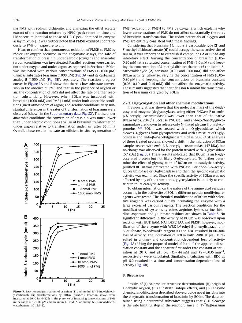
Next, to confirm that spontaneous oxidation of PMSH to PMS bymolecular oxygen occurred in the enzymatic assays, the rate oftransformation of brassinin under aerobic (oxygen) and anaerobic(argon) conditions was investigated. Parallel reactions were carriedout under oxygen and under argon, as reported in Section 4. BOLmwas incubated with various concentrations of PMS (1–1000 lM)using as substrates brassinin (1000 lM) (Fig. 3A) and its carbamateanalog 5 (1000 lM) (Fig. 3B), separately. The reaction progresscurves in Figure 3A and B show that there is low substrate conver-sion in the absence of PMS and that in the presence of oxygen orair, the concentration of PMS did not affect the rate of either reac-tion substantially. However, when BOLm was incubated withbrassinin (1000 mM) and PMS (1 mM) under both anaerobic condi-tions (inert atmosphere of argon) and aerobic conditions, very sub-stantial differences in the rates of transformation of brassinin wereobserved (shown in the Supplementary data, Fig. S2). That is, underanaerobic conditions the conversion of brassinin was much lowerthan under aerobic conditions (ca. 3% of brassinin transformationunder argon relative to transformation under air, after 65 min).Overall, these results indicate an efficient in situ regeneration of
Figure 3. Reaction progress curves of brassinin (1) and methyl N0-(3-indolylmeth-yl)carbamate (5) transformations by BOLm (purified). Reaction assays wereincubated at 20 �C for 0–22 h in the presence of increasing concentrations of PMSin the range of 1–1000 lM and brassinin 1.0 mM (A) or methyl N0-(3-indolylmeth-yl)carbamate 1.0 mM (B).
PMS (oxidation of PMSH to PMS by oxygen), which explains whylower concentrations of PMS do not affect substantially the ratesof brassinin transformation. The redox potentials of oxygen andPMS are entirely consistent with these findings.
Considering that brassinin (1), indole-3-carboxaldehyde (2) andS-methyl dithiocarbamate (6) could occupy the same active site ofBOLm, it was important to establish if compounds 2 or 6 had anyinhibitory effect. Varying the concentration of brassinin (0.05–0.30 mM) at a saturated concentration of PMS (1.0 mM) and keep-ing the concentration of S-methyl dithiocarbamate (6) or indole-3-carboxaldehyde (2) constant (0.30 and 0.60 mM) did not affectBOLm activity. Likewise, varying the concentration of PMS (0.05–0.30 lM) and keeping the concentration of brassinin constant(0.05, 0.10 and 0.15 mM) did not affect the enzymatic activity.These results suggested that neither 2 nor 6 inhibit the transforma-tion of brassinin catalyzed by BOLm.
2.2.3. Deglycosylation and other chemical modificationsPreviously, it was shown that the molecular mass of the degly-
cosylated enzyme (deglycosylated using either PNGase F or endo-b-N-acetylglucosaminidase) was lower than that of the nativeBOLm by ca. 20% (1). Because PNGase F and endo-b-N-acetylgluco-saminidase are known to release only N-linked glycans from glyco-proteins,15,16 BOLm was treated with an O-glycosidase, whichcleaves O-glycans from glycoproteins, and with a mixture of O-gly-cosidase and endo-b-N-acetylglucosaminidase. SDS/PAGE analysesof these treated proteins showed a shift in the migration of BOLmsample treated with endo-b-N-acetylglucosaminidase (47 kDa), butno change was observed for the protein treated with O-glycosidase(57 kDa) (Fig. S3). These results indicated that BOLm is an N-gly-cosylated protein but not likely O-glycosylated. To further deter-mine the effect of glycosylation of BOLm on its catalytic activity,purified BOLm was pretreated with PNGase F or endo-b-N-acetyl-glucosaminidase or O-glycosidase and then the specific enzymaticactivity was examined. Since the specific activity of BOLm was notaffected by any of the treatments, glycosylation is unlikely to con-tribute to its catalytic activity.
To obtain information on the nature of the amino acid residuesoccurring in the active site of BOLm, different protein modifying re-agents were tested. The chemical modification of BOLm with selec-tive reagents was carried out by incubating the enzyme with alarge excess of various reagents. The reaction conditions for themodifications of cysteine, tyrosine, arginine, lysine, serine, histi-dine, aspartate, and glutamate residues are shown in Table 5. Nosignificant difference in the activity of BOLm was observed uponreaction with BUT, EAM, NAI, DEPC, IAA and PMSF; however, mod-ification of the enzyme with WRK (N-ethyl-5-phenylisoxazolium-30-sulfonate, Woodward’s reagent K) and EDC resulted in 60–80%loss of activity. The incubation of BOLm with WRK at pH 6.0 re-sulted in a time- and concentration-dependent loss of activity(Fig. 4A). Using the proposed model of Petra,17 the apparent disso-ciation constant and the apparent first-order rate constant at satu-ration at 20 �C and pH 6.0 (KI = 44 mM and k2 = 0.035 min�1,respectively) were calculated. Similarly, incubation with EDC atpH 6.0 resulted in a time and concentration-dependent loss ofactivity (Fig. 4B).
3. Discussion
Results of (i) co-product structure determination, (ii) origin ofaldehydic oxygen, (iii) substrate isotope effects, and (iv) enzymechemical modifications described above provide novel insights intothe enzymatic transformation of brassinin by BOLm. The data ob-tained using dideuterated substrates suggests that C–H cleavageis the rate limiting step in the reaction, since [10,10-2H2]brassinin
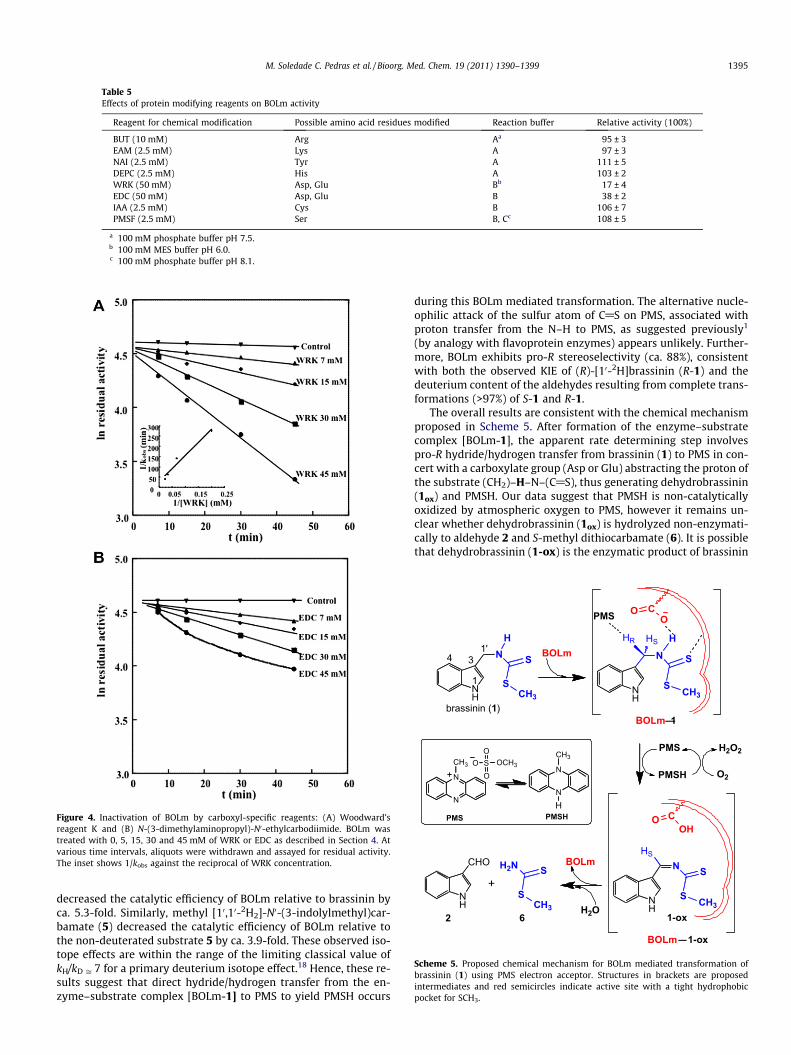
Table 5Effects of protein modifying reagents on BOLm activity
Reagent for chemical modification Possible amino acid residues modified Reaction buffer Relative activity (100%)
BUT (10 mM) Arg Aa 95 ± 3EAM (2.5 mM) Lys A 97 ± 3NAI (2.5 mM) Tyr A 111 ± 5DEPC (2.5 mM) His A 103 ± 2WRK (50 mM) Asp, Glu Bb 17 ± 4EDC (50 mM) Asp, Glu B 38 ± 2IAA (2.5 mM) Cys B 106 ± 7PMSF (2.5 mM) Ser B, Cc 108 ± 5
a 100 mM phosphate buffer pH 7.5.b 100 mM MES buffer pH 6.0.c 100 mM phosphate buffer pH 8.1.
Figure 4. Inactivation of BOLm by carboxyl-specific reagents: (A) Woodward’sreagent K and (B) N-(3-dimethylaminopropyl)-N0-ethylcarbodiimide. BOLm wastreated with 0, 5, 15, 30 and 45 mM of WRK or EDC as described in Section 4. Atvarious time intervals, aliquots were withdrawn and assayed for residual activity.The inset shows 1/kobs against the reciprocal of WRK concentration.
NH
N S
S NH
N S
S
H
NH
CHO
H
CH3 CH3
NH
N S
S CH3
HSHR
HS
2
BOLm
BOLm–1
BOLm–1-ox
N
N
CH3 SO
OO
OCH3
C
1
341'
PMS
PMS
PMSH
OO
COH
O
brassinin (1)
H2N S
SCH3
+
H2O
BOLm
O2
H2O2
6
N
N
CH3
HPMSH
1-ox
PMS
Scheme 5. Proposed chemical mechanism for BOLm mediated transformation ofbrassinin (1) using PMS electron acceptor. Structures in brackets are proposedintermediates and red semicircles indicate active site with a tight hydrophobicpocket for SCH3.
M. Soledade C. Pedras et al. / Bioorg. Med. Chem. 19 (2011) 1390–1399 1395
decreased the catalytic efficiency of BOLm relative to brassinin byca. 5.3-fold. Similarly, methyl [10,10-2H2]-N0-(3-indolylmethyl)car-bamate (5) decreased the catalytic efficiency of BOLm relative tothe non-deuterated substrate 5 by ca. 3.9-fold. These observed iso-tope effects are within the range of the limiting classical value ofkH/kD ffi 7 for a primary deuterium isotope effect.18 Hence, these re-sults suggest that direct hydride/hydrogen transfer from the en-zyme–substrate complex [BOLm-1] to PMS to yield PMSH occurs
during this BOLm mediated transformation. The alternative nucle-ophilic attack of the sulfur atom of C@S on PMS, associated withproton transfer from the N–H to PMS, as suggested previously1
(by analogy with flavoprotein enzymes) appears unlikely. Further-more, BOLm exhibits pro-R stereoselectivity (ca. 88%), consistentwith both the observed KIE of (R)-[10-2H]brassinin (R-1) and thedeuterium content of the aldehydes resulting from complete trans-formations (>97%) of S-1 and R-1.
The overall results are consistent with the chemical mechanismproposed in Scheme 5. After formation of the enzyme–substratecomplex [BOLm-1], the apparent rate determining step involvespro-R hydride/hydrogen transfer from brassinin (1) to PMS in con-cert with a carboxylate group (Asp or Glu) abstracting the proton ofthe substrate (CH2)–H–N–(C@S), thus generating dehydrobrassinin(1ox) and PMSH. Our data suggest that PMSH is non-catalyticallyoxidized by atmospheric oxygen to PMS, however it remains un-clear whether dehydrobrassinin (1ox) is hydrolyzed non-enzymati-cally to aldehyde 2 and S-methyl dithiocarbamate (6). It is possiblethat dehydrobrassinin (1-ox) is the enzymatic product of brassinin
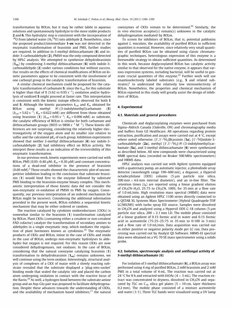
1396 M. Soledade C. Pedras et al. / Bioorg. Med. Chem. 19 (2011) 1390–1399
transformation by BOLm, but it may be rather labile in aqueoussolutions and spontaneously hydrolyze to the more stable products2 and 6. This hydrolytic step is consistent with the incorporation of18O from labeled water (H2
18O) into aldehyde 2. Nonetheless, sincethe proposed product/intermediate 1-ox was not detected in theenzymatic transformation of brassinin and PMS, further studiesare required. In addition to S-methyl dithiocarbamate (6) and in-dole-3-carboxaldehyde (2), PMSH was the only compound detectedby HPLC analysis. We attempted to synthesize dehydrobrassinin(1ox) by condensing S-methyl dithiocarbamate (6) with indole-3-carboxaldehyde (2) under various conditions but without success.Our results on the effects of chemical modifications of BOLm on ki-netic parameters appear to be consistent with the involvement ofone carboxyl group in the catalytic transformation of brassinin.
A similar chemical mechanism could be proposed for the cata-lytic transformation of carbamate 5; since the kcat for this substrateis higher than that of 1 (5.62 vs 0.95 s�1), oxidation and/or hydro-lysis of oxidized 5 might proceed at faster rate. This interpretationis consistent with the kinetic isotope effects observed for both 1and 5. Although the kinetic parameters kcat and Km obtained forBOLm using methyl N0-(3-indolylmethyl)carbamate (5)(kcat = 5.62 s�1; Km = 0.622 mM) are different from those obtainedusing brassinin (1) (kcat = 0.95 s�1; Km = 0.096 mM) as substrate,the catalytic efficiency of BOLm is similar for both carbamate anddithiocarbamate groups (9035 vs 9896 s�1 M�1). These kinetic dif-ferences are not surprising, considering the relatively higher elec-tronegativity of the oxygen atom and its smaller size relative tosulfur and the calculated pKa of each group. Inhibition experimentsshowed that neither S-methyl dithiocarbamate (6) nor indole-3-carboxaldehyde (2) had inhibitory effect on BOLm activity. Weinterpret these results as an indication of the irreversibility of thisenzymatic transformation.
In our previous work, kinetic experiments were carried out withBOLm, PMS (0.05–0.40 lM; Km = 0.30 lM) and constant concentra-tions of a dead-end inhibitor in the presence of brassinin(0.60 mM).2 Those results showed the characteristic plot of uncom-petitive inhibition leading to the conclusion that substrate brassi-nin (1) would bind first to the enzyme followed by substratePMS binding to the brassinin-enzyme binary complex. This mech-anistic interpretation of those kinetic data did not consider thenon-enzymatic re-oxidation of PMSH to PMS by oxygen. Conse-quently, our previous interpretation of an ordered mechanism forBOLm might be incorrect. Considering the additional informationprovided in the present work, BOLm exhibits a sequential kineticmechanism that may be either ordered or random.
The reaction catalyzed by cytokinin oxidoreductases (CKXs) issomewhat similar to the brassinin (1) transformation catalyzedby BOLm. Plant CKXs (containing either a covalent or non-covalentFAD cofactor) catalyze the transformation of secondary amines toaldehydes in a single enzymatic step, which mediates the regula-tion of plant hormones known as cytokinins.19 The enzymaticproducts of CKXs and BOLm, imine in the case of CKXs and imidoin the case of BOLm, undergo non-enzymatic hydrolyses to alde-hydes but oxygen is not required. For this reason CKXs are nowconsidered dehydrogenases, not oxidases. In the case of BOLm,considering that the natural coenzyme catalyzing brassinin (1)transformation to dehydrobrassinin (1ox) remains unknown, wewill continue using the term oxidase. Interestingly, structural anal-ysis of complexes of a CKX of maize with a slowly reacting sub-strate revealed that the substrate displayed a ‘plug-into-socket’binding mode that sealed the catalytic site and placed the carbonatom undergoing oxidation in contact with the reactive locus ofthe flavin.20 As well, a hydrogen bond between the substrate aminegroup and an Asp-Glu pair was proposed to facilitate dehydrogena-tion. Despite these advances towards the understanding of CKXs,and the availability of mechanism based inhibitors,21 the natural
coenzymes of CKXs remain to be determined.20 Similarly, thein vivo electron acceptor(s) remain(s) unknown in the catalyticdehydrogenation mediated by BOLm.
To screen for inhibitors of BOLm, that is, potential paldoxinsagainst L. maculans, the availability of purified BOLm in sufficientquantities is essential. However, since relatively very small quanti-ties of purified BOLm can be obtained using classic chromato-graphic techniques, heterologous expression of this enzyme is aforeseeable strategy to obtain sufficient quantities. As determinedin this work, because deglycosylated BOLm has catalytic activitysimilar to that observed for the native enzyme, it appears that var-ious expression systems, including bacterial, will be useful to gen-erate crucial quantities of this enzyme.22 Further work will useenantioselectively labeled substrates (e.g., 5 and related sub-strates)1 to understand the relatively low stereoselectivity ofBOLm. Nonetheless, the properties and chemical mechanism ofBOLm reported in this study will greatly assist the design of inhib-itors of BOLm.
4. Experimental
4.1. Materials and general procedures
Chemicals and deglycosylating enzymes were purchased fromSigma–Aldrich Canada (Oakville, ON) and chromatography mediaand buffers from GE Healthcare. All operations regarding proteinextraction, purification and assays were carried out at 4 �C, exceptwhere noted otherwise. [10,10-2H2]Brassinin (1a), [10-2H]indole-3-carboxaldehyde (2a), methyl [10,10-2H2]-N0-(3-indolylmethyl)car-bamate (5a), and S-methyl dithiocarbamate (6) were synthesizedas described below. All new compounds were characterized usingNMR spectral data (recorded on Bruker 500 MHz spectrometers)and HRMS data.
HPLC analysis was carried out with Agilent systems equippedwith a quaternary pump, an automatic injector, a photodiode arraydetector (wavelength range 190–600 nm), a degasser, a Hypersiloctadecylsilane (ODS) column (5 lm particle size silica,200 mm � 4.6 mm internal diameter), and an in-line filter. Theretention times (tR) are reported using a linear gradient elutionof CH3CN–H2O, 25:75 to CH3CN, 100%, for 35 min at a flow rateof 1.0 mL/min. High resolution mass spectral (HRMS) data wereobtained using an Agilent HPLC 1100 series directly connected toa QSTAR XL Systems Mass Spectrometer (Hybrid Quadrupole-TOFLC/MS/MS) with turbo spray ESI source. Samples were dissolvedin CH3CN and analyzed using a Hypersil ODS C-18 column (5 lmparticle size silica, 200 � 2.1 mm I.D. The mobile phase consistedof a linear gradient of 0.1% formic acid in water and 0.1% formicacid in acetonitrile (75:25–25:75 in 35 min to 0:100 in 5 min)and a flow rate of 1.0 mL/min. Data acquisition was carried outin either positive or negative polarity mode per LC run. Data pro-cessing was carried out by Analyst QS Software. HRMS-EI spectraldata were obtained on a VG 70 SE mass spectrometer using a solidsprobe.
4.2. Isolation, spectroscopic analysis and antifungal activity ofS-methyl dithiocarbamate (6)
For isolation of S-methyl dithiocarbamate (6), a BOLm assay wasperformed using 4 mg of purified BOLm, 2 mM brassinin and 2 mMPMS in a total volume of 4 mL. The reaction was carried out at24 �C for 8 h and extracted with EtOAc (4 � 3 mL). The reaction ex-tract was concentrated to dryness, dissolved in CH3CN and sepa-rated by TLC on C18 silica gel plates (5 � 10 cm, layer thickness0.2 mm). The mobile phase consisted of a mixture acetonitrileand water (1:1). Three TLC bands were visualized using a UV lamp

M. Soledade C. Pedras et al. / Bioorg. Med. Chem. 19 (2011) 1390–1399 1397
and the components eluted with CH3CN after scraping each band.The components eluted from the plate were analyzed by LC-HRMS,HPLC-DAD (see Fig. S1), and 1H NMR (CD2Cl2). S-Methyl dithiocar-bamate (6) was stable in the solvents used for TLC separation andextraction at least for 24 h.
Mycelial growth inhibition assays of L. maculans isolate BJ 125were carried out as previously described23 using S-methyl dithio-carbamate (6) at three concentrations (0.50, 0.25, 0.12 mM). Themycelial growth inhibition was determined relative to control cul-tures grown in the absence of compound 6, but otherwise identicalconditions.
4.3. Synthesis
4.3.1. [10,10-2H2]Brassinin (1a)A solution of 3-indolecarbonitrile (50 mg, 0.35 mmol) in THF
(0.5 mL) was added to a suspension of lithium aluminum deuteride(30 mg, 0.70 mmol) in dry THF (0.5 mL) and stirred at 55–60 �C for4 h. The reaction mixture was cooled to room temperature, dilutedwith NaOH (1 M, 0.5 mL) and filtered. The filtrate was diluted withwater (10 mL) and extracted with EtOAc (3 � 20 mL). The com-bined organic extract was dried over Na2SO4 and concentrated.The residue was separated (SiO2, CH2Cl2/MeOH/NH4OH) to afford[10,10-2H2]indolyl-3-methanamine as a white solid (27 mg, 55%yield). Carbon disulfide (12 mg, 0.152 mmol) was added to a solu-tion of [10,10-2H2]indolyl-3-methanamine (15 mg, 0.10 mmol) andEt3N (21 mg, 0.202 mmol) in pyridine (1 mL) and stirred at rt for15 min. Next, MeI (180 mg, 1.27 mmol) was added and stirringwas continued for another 20 min. The reaction mixture was di-luted with toluene (2 mL) and concentrated under reduced pres-sure. The crude product was separated (SiO2, EtOAC/hexanes) toyield [10,10-2H2]brassinin (14 mg, 58%). 1H NMR (500.3 MHz,CD3CN): d 9.26 (br s, NH), 8.24 (br s, NH), 7.63 (d, J = 8 Hz, 1H),7.42 (d, J = 8 Hz, 1H), 7.31 (s, 1H), 7.16 (dd, J = 8, 8 Hz, 1H), 7.07(dd, J = 8, 8 Hz, 1H), 2.55 (s, 3H). Minor peaks of a rotamer were ob-served at d 2.67 and 8.55 (only dideuterated brassinin was de-tected). 13C NMR (125.8 MHz, CD3CN): d 199.1 (s), 137.7 (s),128.1 (s), 126.1 (s), 123.2 (d), 120.7 (d), 120.0 (d), 112.8 (d),111.8 (s), 42.9 (quintet, JC–D = 21.5 Hz), 18.5 (q). HRMS: m/z mea-sured 238.0560 (238.0567 calcd for C11H10
2H2N2S2).
4.3.2. [10-2H]Indole-3-carboxaldehydeA solution of indole (200 mg, 1.71 mmol) in DMF-d7 (0.6 mL)
was added to a cooled solution of freshly distilled POCl3 (288 mg,1.88 mmol) in DMF-d7 (0.4 mL) and stirred at 40 �C for 60 min.The reaction mixture was poured into ice-cold water and addeddropwise to an ice-cold solution of NaOH (1%, w/v, 3 mL). The sus-pension was extracted with EtOAc (3 � 20 mL), the combined or-ganic extract was dried over Na2SO4 and concentrated to drynessto yield [10-2H]indole-3-carboxaldehyde (221 mg, 89% yield).24 1HNMR (CD3CN) d: 1H NMR (500.3 MHz, CD3CN): d 10.07 (br s, NH),8.17 (d, J = 7.6 Hz, 1H), 8.00 (s, 1H), 7.54 (d, J = 8 Hz, 1H), 7.31–7.24 (m, 2H). 13C NMR (125.8 MHz, CD3CN): d 186.3 (t, JC–
D = 25.6 Hz), 138.7, 138.4 (s), 125.7 (s), 125.2 (d), 123.8 (d), 122.5(d), 120.1 (t, 2JC–D = 3.7 Hz), 113.5 (d) HREIMS: m/z measured 144(144. calcd for C9H6
2HNO).
4.3.3. Methyl [10,10-2H2]-N0-(3-indolylmethyl)carbamate (5a)To a solution of [10,10-2H2]indolyl-3-methanamine (27 mg,
0.18 mmol) in CH2Cl2 (1 mL) was added methyl chloroformate(19 mg, 0.20 mmol) and triethylamine (39 mg, 0.39 mmol) andthe reaction mixture was stirred at rt for 20 min. The reaction mix-ture was diluted with water (10 mL), extracted with EtOAc(3 � 20 mL) and the combined organic extract was dried overNa2SO4 and concentrated to dryness. Purification of the crudeproduct (SiO2, EtOAC/hexanes) afforded methyl [10,10-2H2]-N0-(3-
indolylmethyl)carbamate (5a) as a white solid (27 mg, 73% yield).1H NMR (500.3 MHz, CD3CN): d 9.17 (br s, NH), 7.62 (d, J = 8 Hz,1H), 7.40 (d, J = 8 Hz, 1H), 7.18 (s, 1H), 7.14 (dd, J = 8, 8 Hz, 1H),7.06 (J = 8, 8 Hz, 1H), 5.78 (br s, NH), 3.6 (s, 3H) (only dideuterated5a was detected). 13C NMR (125.8 MHz, CD3CN): d 158.3 (s), 137.9(s), 127.9 (s), 124.8 (d), 123.0 (d), 120.4 (d), 120.0 (s), 114.3 (s),112.7 (d), 52.7 (quartet), 36.8 (quintet, JC–D = 21.5 Hz). HRMS-EI:m/z measured 206.1028 (206.1024 calcd for C11H10
2H2N2O2).
4.3.4. S-Methyl dithiocarbamate (6)S-Methyl dithiocarbamate was prepared after modification of a
previously published procedure.25 MeI (321 mg, 2.26 mmol) wasadded dropwise to a cooled suspension of ammonium dithiocarba-mate (250 mg, 1.97 mmol) in EtOH (3 mL) at 0 �C and the reactionmixture was stirred under argon for 4 h. The reaction mixture wasconcentrated (ca. 1/2 volume), diluted with water and the resultingsuspension was filtered. The solid was dissolved in Et2O (5 mL) andprecipitated by adding n-pentane. Filtration of the precipitateafforded S-methyl dithiocarbamate (152 mg, 72% yield) with amp 42–44 �C (lit value: 41 �C,25). HRMS-EI: m/z measured106.9865 (106.9863 calcd for C2H5NS2).
4.3.5. Co-factor PMSHPMS (ca. 1 mmol in H2O) was treated with an aqueous solution
of sodium dithionite (Na2S2O4, ca. 2 mmol) for 3 min, the reactionmixture was extracted with ethyl acetate and the extract analyzedby HPLC. PMSH oxidized spontaneously to PMS under exposure toaerobic conditions.
4.4. Fungal cultures and chromatographic purification of BOLm
Cultures of L. maculans (isolate BJ-125, IBCN collection, AAFC)were carried out as described previously.2 For isolation and puri-fication of BOLm, liquid cultures (600 mL) were induced with 3-phenylindole (0.05 mM), the cultures were incubated for an addi-tional 24 h and then gravity filtered to separate mycelia fromculture broth. The purification of BOLm was performed in foursteps as previously described.26 Protein concentrations weredetermined as described by Bradford26 using the Coomassie Bril-liant Blue method with bovine serum albumin (BSA) as astandard.
BOLm standard activity assay. The reaction mixture containedDEA (diethanol amine, 20 mM, pH 8.2), DTT (D,L-dithiothreitol,1.0 mM), 0.1% (v/v) Triton X-100, brassinin (1.0 mM), PMS(1.0 mM) and protein extract (50–100 lL) in a total volume of1000 lL. The reaction was carried out at 20 �C for 20 min. A controlreaction was stopped by addition of EtOAc (2 mL) at t = 0. Theproduct was extracted with EtOAc (2 mL) and concentrated to dry-ness. The extract was dissolved in CH3CN (200 lL) and analyzed byHPLC-DAD.2 The amounts of brassinin (1), indole-3-carboxalde-hyde (2), and S-methyl dithiocarbamate (6) in the reaction assaywere determined using calibration curves built with pure com-pounds. Brassinin (1), indole-3-carboxaldehyde (2), and S-methyldithiocarbamate (6) were stable in the buffer and solvents usedfor extraction and HPLC analysis at least for 24 h.
4.5. Origin of aldehydic oxygen
For the enzymatic experiments in H218O, all the reaction com-
ponents were dissolved in 50% of H218O. The incubation was car-
ried out in a saturated concentration of brassinin or dideuteratedbrassinin (1.0 mM). Incorporation of 18O from H2
18O into indole-3-carboxaldehyde was determined by HRMS. A control samplewas prepared using same experimental conditions but replacingH2
18O with natural abundance H2O.
1398 M. Soledade C. Pedras et al. / Bioorg. Med. Chem. 19 (2011) 1390–1399
4.6. Substrate kinetic isotope effects and data analysis
Deuterium kinetic isotope effects (KIE) have been determinedfrom the steady-state kinetic parameters: KIE = (kcat/Km)H/(kcat/Km)D. The isotope effects on the kinetic parameters were measuredby varying the concentration of substrate in the range of 0.05–2.8 mM at pH 8.2 and at saturating levels of PMS (1.0 mM).
For reaction progress curves of (R) and (S)-[10-2H]brassinins,reaction assays were incubated at 20 �C for 0–3 h in the presenceof R-1 and S-1 (0.20 mM), the reaction was stopped at varioustimes (5–180 min) and analyzed as described above for the stan-dard activity assay (Fig. 2).
The kinetic data were analyzed using the computer programKaleidaGraph. Km(app) and Vmax(app) were determined by measure-ment of the enzyme activity employing a concentration of brassi-nin (1) in the range of 0.05–1.0 mM and methyl [10,10-2H2]-N0-(3-indolylmethyl)carbamate (5) in the range of 0.05–2.8 mM. Datawere fitted to the equation: v = VmaxS/(Km + S), using a non-linearleast-squares approach.27 Kinetic constants possessed ±SE (stan-dard errors) of 3% of the experimental measurement and R2 valueswere between 0.980 and 0.998.
In order to analyze the mechanism of BOLm inactivation byWRK the following model was employed:17
Eþ I ¡KI
E � I!k2 E—I
where E is free enzyme, I is WRK concentration, E�I is the enzyme–WRK reversible complex; E–I is the enzyme–WRK covalent com-plex, KI is the apparent E�I dissociation constant, and k2 is the intrin-sic rate constant for covalent modification of BOLm. Theinactivation rate (kobs) was obtained by plotting the ln percentageof residual activity versus time (t). The observed inactivation rateconstant (kobs) can be expressed as:
1=kobs ¼ ðK I=k2½I� þ 1=k2Þ
Plotting 1/kobs, measured at various WRK concentrations (range 5–45 mM), used for calculation of KI and k2.
4.7. Effect of PMS and oxygen
The progress curves for the transformation of brassinin (1) ormethyl N0-(3-indolylmethyl)carbamate (5) by BOLm in the presenceof various concentrations of PMS were obtained by measuring thetime-dependent formation of indole-3-carboxaldehyde (2). The as-say mixture consisted of 1 mM brassinin or 1 mM methyl N0-(3-indo-lylmethyl)carbamate (5), 20 mM DEA (pH 8.2), 1 mM DTT, 0.1% (v/v)Triton X-100, and 50 lL protein extract in a total volume of 1000 lL.The concentrations of the PMS were 0, 1, 10, 1000 lM, respectively.The samples were incubated at 20 �C, and samples (200 ll) were ta-ken after incubation for 1, 2, 4, 7 and 22 h; the reaction was stoppedby the addition of 2 mL of EtOAc. The products were extracted andquantified as described above for BOLm standard assay.
Oxygen and argon were purchased from Praxair (USA, argoncontained traces of oxygen). The enzymatic assays were performedunder conditions similar to those described above for the BOLmstandard assay but in 5 mL vials containing 1 mM of brassinin,1 lM of PMS and 100 ll of purified enzyme in a total volume of2 mL. For enzymatic assays performed under argon or air, the reac-tion mixture stored in a capped and sealed vial was first purgedwith a stream of argon or air for 5 min and then kept under a stea-dy stream of either gas for the duration of the experiment; 200 lLof the reaction mixture were collected every 15 min using a needleand syringe and the reaction was immediately quenched by addingto 2 mL of EtOAc. The products were extracted and quantified asdescribed above for BOLm standard assay. Results obtained for
enzymatic assays performed under an oxygen atmosphere weresimilar to those obtained under normal aerobic conditions (Fig. S2).
4.8. Inhibition
Inhibition experiments were carried out using one substrate atsaturation level (brassinin or PMS) and varying the concentrationof the second substrate in the region of its Km (at different concen-tration of the product). Initial velocity experiments were conductedin the presence of S-methyl dithiocarbamate (6) or indole-3-car-boxaldehyde (2) at 0.30 mM and 0.60 mM and varying the concen-tration of brassinin (0.05–0.30 mM) at a saturated concentration ofPMS (1 mM). Next, experiments were carried out similarly butvarying the concentration of PMS (0.05–0.30 lM) and keeping theconcentration of brassinin constant (0.05, 0.10 and 0.15 mM).
4.9. Enzyme chemical modifications
The purified BOLm was incubated with 0.10 mL of correspond-ing reaction buffer for each chemical modification reagent and0.010 mL of 2,3-butanedione (BUT, 10 mM), ethyl acetimidate(EAM, 2.5 mM), N-acetylimidazole (NAI, 2.5 mM), diethyl pyrocar-bonate (DEPC, 2.5 mM), N-ethyl-5-phenylisoxazolium-30-sulfonate(Woodward’s reagent K, WRK, 5–50 mM), diazobenzenesulfonicacid (DBS, 10 mM), iodoacetamide (IAA, 2.5 mM), N-(3-dimethyl-aminopropyl)-N0-ethylcarbodiimide (EDC, 5–50 mM) and phen-ylmethanesulfonyl fluoride (PMSF, 2.5 mM). The enzyme activitywas measured and expressed as percentage specific activity calcu-lated as the ratio of specific activity of BOLm with chemical mod-ification reagents and that without chemical modification reagents.
4.10. Analysis of deglycosylated BOLm
Purified BOLm (1 lg) was treated with PNGase F (Sigma,G5166), endo-b-N-acetylglucosaminidase (Sigma, A-0810) and O-glycosidase (Sigma, G1163), separately and altogether, followingthe manufacturer’s protocols. Reactions were incubated at 37 �Cfor 3 h in non-denaturing conditions in the appropriate buffer(31 lL of total reaction volume). After incubation, enzymatic activ-ities were tested under conditions described for BOLm activity as-say or 3 lL of SDS/PAGE buffer was added to each reaction andsamples were analyzed by SDS/PAGE (sodium dodecyl sulfate/polyacrylamide gel electrophoresis).
Acknowledgements
We thank Professor D. E. Ward from the Department of Chem-istry, University of Saskatchewan for very insightful discussions.We also thank P. B. Chumala and K. Thoms from the Departmentof Chemistry, University of Saskatchewan for technical assistanceobtaining MS and HRMS data, respectively. Financial support fromNatural Sciences and Engineering Research Council of Canada(NSERC—Discovery Grant to M.S.C.P.), Canada Research Chairs Pro-gram (CRC—Tier I Grant to M.S.C.P.), and the University of Sas-katchewan (Teaching Assistantship to V.K.S.M.) is gratefullyacknowledged.
Supplementary data
Supplementary data associated with this article can be found, inthe online version, at doi:10.1016/j.bmc.2011.01.011.
References and notes
1. Pedras, M. S. C.; Jha, M.; Minic, Z.; Okeola, O. G. Bioorg. Med. Chem. 2007, 15,6054.
M. Soledade C. Pedras et al. / Bioorg. Med. Chem. 19 (2011) 1390–1399 1399
2. Pedras, M. S. C.; Minic, Z.; Jha, M. FEBS J. 2008, 275, 3691.3. Pedras, M. S. C.; Minic, Z.; Sarma-Mamillapalle, V. K.; Suchy, M. Bioorg. Med.
Chem. 2010, 18, 2456.4. Chen, Y.; Fernando, W. G. D. Can. J. Plant Pathol. 2006, 28, 533.5. Fitt, B. D. L.; Brun, H.; Barbetti, M. J.; Rimmer, S. R. Eur. J. Plant Pathol. 2006, 114,
3.6. Pedras, M. S. C.; Zheng, Q. A.; Sarma-Mamillapalle, V. K. Nat. Prod. Commun.
2007, 2, 319.7. Pedras, M. S. C.; Yaya, E. E. Phytochemistry 2010, 71, 1191.8. Pedras, M. S. C.; Minic, Z.; Sarma-Mamillapalle, V. K. J. Agric. Food Chem. 2009,
57, 2429.9. Pedras, M. S. C.; Jha, M.; Ahiahonu, P. W. K. Curr. Org. Chem. 2003, 7, 1635.
10. Pedras, M. S. C.; Jha, M. Bioorg. Med. Chem. 2006, 14, 4958.11. Gattow, G.; Gerner, R. Z. Anorg. Allg. Chem. 1982, 488, 94.12. Walker, J. R.; Robert, W. C. Tetrahedron 2001, 57, 6695.13. Dale, J. A.; Mosher, H. S. J. Am. Chem. Soc. 1973, 95, 512.14. Pedras, M. S. C. et al., to be published elsewhere.15. Morelle, W.; Michalski, J. C. Nat. Protocols 2007, 2, 1585.
16. Ghesquière, B.; Van Damme, J.; Martens, L.; Vandekerckhove, J.; Gevaert, K. J.Proteome Res. 2006, 5, 2438.
17. Petra, P. Biochemistry 1971, 10, 3163.18. Walsh, C. Enzymatic Reaction Mechanisms; W. H. Freeman and Company: San
Francisco, CA, 1979. p 113.19. Mattevi, A. Trends Biochem. Sci. 2006, 31, 276.20. Malito, E.; Coda, A.; Bilyeu, K. D.; Fraaije, M. W.; Mattevi, A. J. Mol. Biol. 2004,
341, 1237.21. Kopecny, D.; Sebela, M.; Briozzo, P.; Spichal, L.; Houba-Herin, N.; Masek, V.;
Joly, N.; Madzak, C.; Anzenbacher, P.; Laloue, M. J. Mol. Biol. 2008, 380, 886.22. The genome sequencing project of L. maculans is in progress http://
fungalgenomes.org/ and will facilitate cloning of the BOLm gene.23. Pedras, M. S. C.; Khan, A. Q. J. Agric. Food Chem. 1996, 44, 3403.24. Philip, N. J.; Snyder, H. R. Org. Synth. 1963, 4, 539.25. Gattow, G.; Gerner, R. Anorg. Allg. Chem. 1982, 488, 94.26. Bradford, M. M. Anal. Biochem. 1976, 72, 248.27. Atkins, G. L.; Nimmo, I. A. Anal. Biochem. 1980, 104, 1.
Copyright © 2022 FDOKUMEN


![The Elusive Presence of Multiculturalism [Hebrew]](https://static.fdokumen.com/doc/165x107/631cebfda906b217b907308a/the-elusive-presence-of-multiculturalism-hebrew.jpg)