
CFA-Level-1-QM-Lec-1-R6-Time-Value-of-Money.pdf - Infinity ...
Upload
independentCategory
view
3download
0
ORIGINAL PAPER
Tetrahydroisoquinolines acting as dopaminergic ligands.A molecular modeling study using MD simulationsand QM calculations
Sebastián Andujar & Fernando Suvire & Inmaculada Berenguer & Nuria Cabedo &
Paloma Marín & Laura Moreno & María Dolores Ivorra & Diego Cortes &
Ricardo D. Enriz
Received: 15 December 2010 /Accepted: 22 March 2011 /Published online: 27 April 2011# Springer-Verlag 2011
Abstract A molecular modeling study on 16 1-benzyltetrahydroisoquinolines (BTHIQs) acting as dopaminergicligands was carried out. By combining molecular dynamicssimulations with ab initio and density functional theory(DFT) calculations, a simple and generally applicableprocedure to evaluate the binding energies of BTHIQsinteracting with the human dopamine D2 receptor (D2 DR)is reported here, providing a clear picture of the bindinginteractions of BTHIQs from both structural and energeticviewpoints. Molecular aspects of the binding interactionsbetween BTHIQs and the D2 DR are discussed in detail. Asignificant correlation between binding energies obtainedfrom DFT calculations and experimental pKi values wasobtained, predicting the potential dopaminergic effect ofnon-synthesized BTHIQs.
Keywords 1-Benzyl-THIQ . Halogenated-1-benzyl-THIQ .
D2-dopamine receptor . Structure-activity relationship .
Molecular dynamic simulation . Ab initio and DFTcalculation
Introduction
The dopamine D2 receptor (D2 DR) has been implicated inthe mechanism of drugs used in the treatment of disorderssuch as schizophrenia and Parkinson’s disease. For thesereasons, a great deal of research has focused on thediscovery of novel dopaminergic ligands as potential drugcandidates [1]. DR can be classified into two pharmaco-logical families (D1 and D2-like) that are encoded by atleast five genes. Which receptor(s) needs to be activated toobtain therapeutic effects in Parkinson’s disease has beenthe subject of controversy [2]. The D2-like DR show highaffinities for drugs (antagonists) used in the treatment ofschizophrenia (antipsychotics) and those (agonists) utilizedto treat the Parkinson’s disease [3].
Tetrahydroisoquinolines (THIQs)—the most numerousnaturally occurring alkaloids—include 1-benzyl-THIQs andaporphines, both of which have structural similarities todopamine and can interact with DR [4]. Previous results inour group suggested that some natural and synthetic 1-benzyl-THIQs alkaloids were able to bind to DR [5–7]. Inthis way, we described the enantioselective syntheses of pairsof dopaminergic (1S)- and (1R)-benzyl-THIQs using (R)- and(S)-phenylglycinol as the chiral source, and we observedthat, in these series of 1-benzyl-THIQs, (1S)-enantiomerswere 5–15 times more effective at D1-like and D2-likedopamine receptors than (1R)-enantiomers [8] (Fig. 1). Onthe other hand, we described the preparation in a ‘one-pot’sequence of 1-cyclohexylmethyl 7,8-dioxygenated-THIQ,substituted and unsubstituted in the C ring by application ofthe photo–Fries transposition, followed by a tandem reduction-cyclization and further reduction. Indeed, we accomplished forthe first time a regioselective hydrogenation of the benzyl ringin the THIQ system. All 1-cyclohexylmethyl THIQs studied in
S. Andujar : F. Suvire :R. D. Enriz (*)Departamento de Química, Universidad Nacional de San Luis,San Luis, Argentinae-mail: [email protected]
S. Andujar :R. D. EnrizIMIBIO-SL (CONICET),Chacabuco 915,5700, San Luis, Argentina
I. Berenguer :N. Cabedo : P. Marín : L. Moreno :M. Dolores Ivorra :D. CortesDepartamento de Farmacología, Facultad de Farmacia,Universidad de Valencia,46100 Burjassot Valencia, Spain
J Mol Model (2012) 18:419–431DOI 10.1007/s00894-011-1061-0
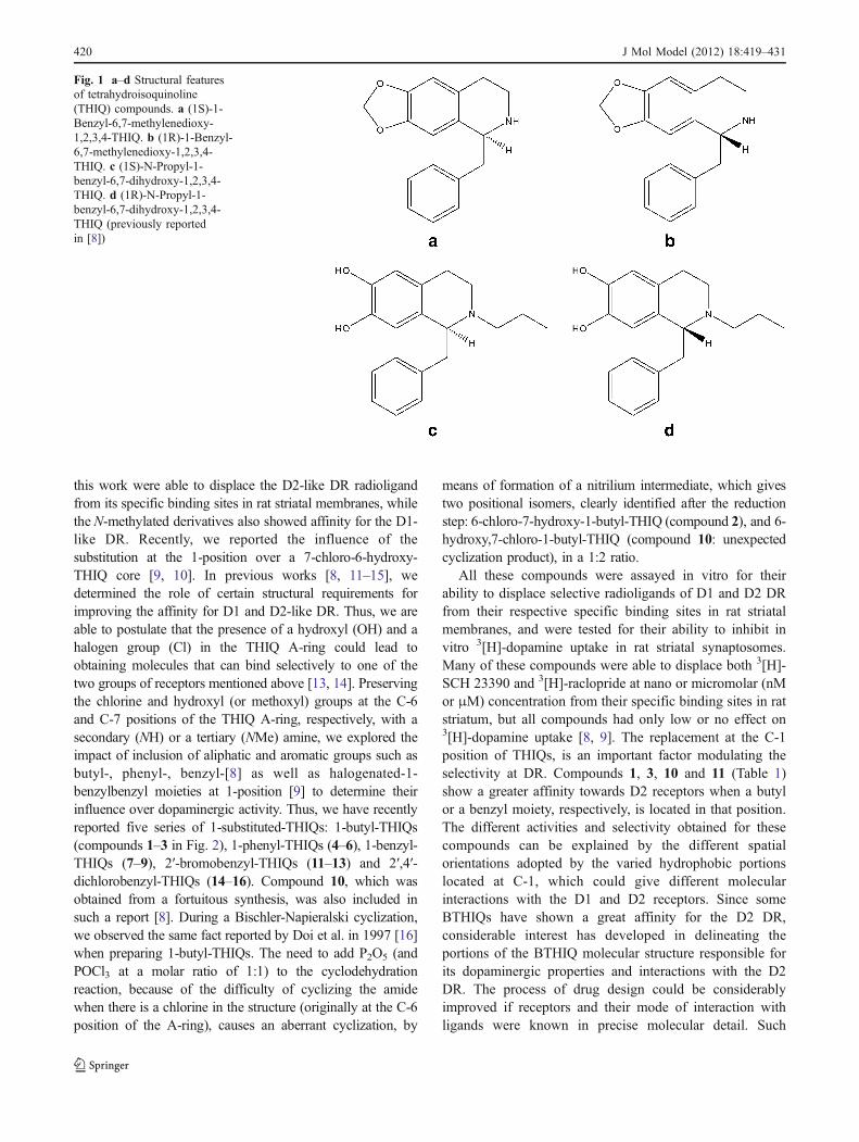
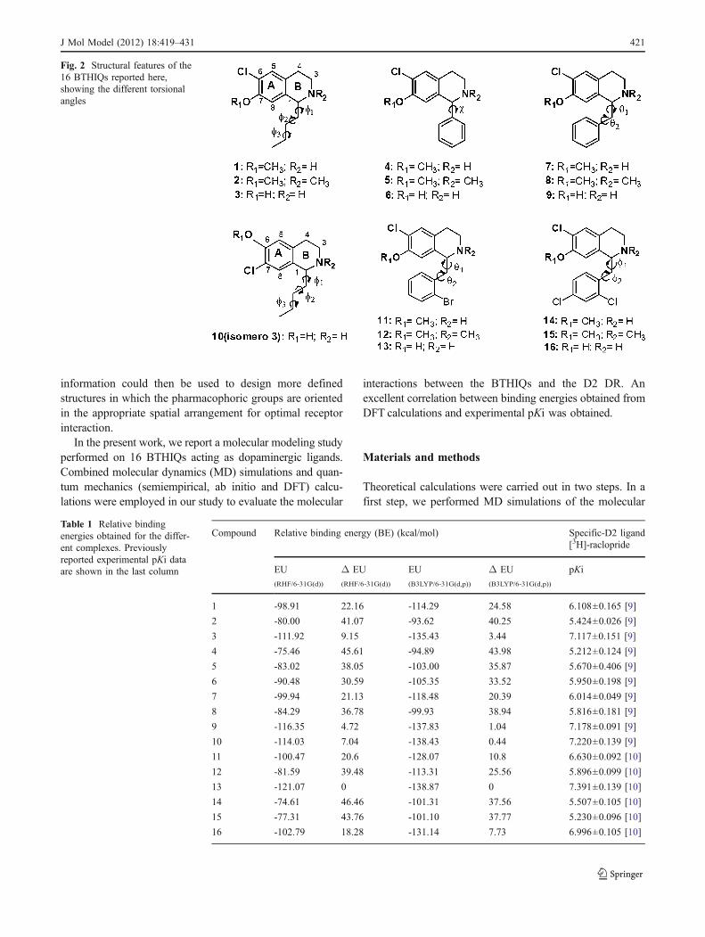
this work were able to displace the D2-like DR radioligandfrom its specific binding sites in rat striatal membranes, whilethe N-methylated derivatives also showed affinity for the D1-like DR. Recently, we reported the influence of thesubstitution at the 1-position over a 7-chloro-6-hydroxy-THIQ core [9, 10]. In previous works [8, 11–15], wedetermined the role of certain structural requirements forimproving the affinity for D1 and D2-like DR. Thus, we areable to postulate that the presence of a hydroxyl (OH) and ahalogen group (Cl) in the THIQ A-ring could lead toobtaining molecules that can bind selectively to one of thetwo groups of receptors mentioned above [13, 14]. Preservingthe chlorine and hydroxyl (or methoxyl) groups at the C-6and C-7 positions of the THIQ A-ring, respectively, with asecondary (NH) or a tertiary (NMe) amine, we explored theimpact of inclusion of aliphatic and aromatic groups such asbutyl-, phenyl-, benzyl-[8] as well as halogenated-1-benzylbenzyl moieties at 1-position [9] to determine theirinfluence over dopaminergic activity. Thus, we have recentlyreported five series of 1-substituted-THIQs: 1-butyl-THIQs(compounds 1–3 in Fig. 2), 1-phenyl-THIQs (4–6), 1-benzyl-THIQs (7–9), 2′-bromobenzyl-THIQs (11–13) and 2′,4′-dichlorobenzyl-THIQs (14–16). Compound 10, which wasobtained from a fortuitous synthesis, was also included insuch a report [8]. During a Bischler-Napieralski cyclization,we observed the same fact reported by Doi et al. in 1997 [16]when preparing 1-butyl-THIQs. The need to add P2O5 (andPOCl3 at a molar ratio of 1:1) to the cyclodehydrationreaction, because of the difficulty of cyclizing the amidewhen there is a chlorine in the structure (originally at the C-6position of the A-ring), causes an aberrant cyclization, by
means of formation of a nitrilium intermediate, which givestwo positional isomers, clearly identified after the reductionstep: 6-chloro-7-hydroxy-1-butyl-THIQ (compound 2), and 6-hydroxy,7-chloro-1-butyl-THIQ (compound 10: unexpectedcyclization product), in a 1:2 ratio.
All these compounds were assayed in vitro for theirability to displace selective radioligands of D1 and D2 DRfrom their respective specific binding sites in rat striatalmembranes, and were tested for their ability to inhibit invitro 3[H]-dopamine uptake in rat striatal synaptosomes.Many of these compounds were able to displace both 3[H]-SCH 23390 and 3[H]-raclopride at nano or micromolar (nMor μM) concentration from their specific binding sites in ratstriatum, but all compounds had only low or no effect on3[H]-dopamine uptake [8, 9]. The replacement at the C-1position of THIQs, is an important factor modulating theselectivity at DR. Compounds 1, 3, 10 and 11 (Table 1)show a greater affinity towards D2 receptors when a butylor a benzyl moiety, respectively, is located in that position.The different activities and selectivity obtained for thesecompounds can be explained by the different spatialorientations adopted by the varied hydrophobic portionslocated at C-1, which could give different molecularinteractions with the D1 and D2 receptors. Since someBTHIQs have shown a great affinity for the D2 DR,considerable interest has developed in delineating theportions of the BTHIQ molecular structure responsible forits dopaminergic properties and interactions with the D2DR. The process of drug design could be considerablyimproved if receptors and their mode of interaction withligands were known in precise molecular detail. Such
Fig. 1 a–d Structural featuresof tetrahydroisoquinoline(THIQ) compounds. a (1S)-1-Benzyl-6,7-methylenedioxy-1,2,3,4-THIQ. b (1R)-1-Benzyl-6,7-methylenedioxy-1,2,3,4-THIQ. c (1S)-N-Propyl-1-benzyl-6,7-dihydroxy-1,2,3,4-THIQ. d (1R)-N-Propyl-1-benzyl-6,7-dihydroxy-1,2,3,4-THIQ (previously reportedin [8])
420 J Mol Model (2012) 18:419–431
information could then be used to design more definedstructures in which the pharmacophoric groups are orientedin the appropriate spatial arrangement for optimal receptorinteraction.
In the present work, we report a molecular modeling studyperformed on 16 BTHIQs acting as dopaminergic ligands.Combined molecular dynamics (MD) simulations and quan-tum mechanics (semiempirical, ab initio and DFT) calcu-lations were employed in our study to evaluate the molecular
interactions between the BTHIQs and the D2 DR. Anexcellent correlation between binding energies obtained fromDFT calculations and experimental pKi was obtained.
Materials and methods
Theoretical calculations were carried out in two steps. In afirst step, we performed MD simulations of the molecular
Fig. 2 Structural features of the16 BTHIQs reported here,showing the different torsionalangles
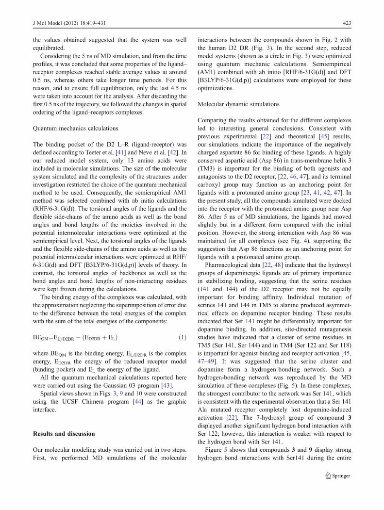
Compound Relative binding energy (BE) (kcal/mol) Specific-D2 ligand[3H]-raclopride
EU(RHF/6-31G(d))
Δ EU(RHF/6-31G(d))
EU(B3LYP/6-31G(d,p))
Δ EU(B3LYP/6-31G(d,p))
pKi
1 -98.91 22.16 -114.29 24.58 6.108±0.165 [9]
2 -80.00 41.07 -93.62 40.25 5.424±0.026 [9]
3 -111.92 9.15 -135.43 3.44 7.117±0.151 [9]
4 -75.46 45.61 -94.89 43.98 5.212±0.124 [9]
5 -83.02 38.05 -103.00 35.87 5.670±0.406 [9]
6 -90.48 30.59 -105.35 33.52 5.950±0.198 [9]
7 -99.94 21.13 -118.48 20.39 6.014±0.049 [9]
8 -84.29 36.78 -99.93 38.94 5.816±0.181 [9]
9 -116.35 4.72 -137.83 1.04 7.178±0.091 [9]
10 -114.03 7.04 -138.43 0.44 7.220±0.139 [9]
11 -100.47 20.6 -128.07 10.8 6.630±0.092 [10]
12 -81.59 39.48 -113.31 25.56 5.896±0.099 [10]
13 -121.07 0 -138.87 0 7.391±0.139 [10]
14 -74.61 46.46 -101.31 37.56 5.507±0.105 [10]
15 -77.31 43.76 -101.10 37.77 5.230±0.096 [10]
16 -102.79 18.28 -131.14 7.73 6.996±0.105 [10]
Table 1 Relative bindingenergies obtained for the differ-ent complexes. Previouslyreported experimental pKi dataare shown in the last column
J Mol Model (2012) 18:419–431 421

interactions between compounds 1–16 and D2 DR. In thesecond step, reduced model systems were optimized usingquantum mechanics calculations. Semiempirical (AM1)combined with ab initio [RHF/6-31G(d)] and B3LYP [6-31G(d,p)] calculations were employed in these optimizations.
Molecular dynamics simulations
It must be pointed out that the principal goal of the MDsimulations performed here was not to obtain a new D2 DRby homology. Our aim in this study was less ambitious; wewished to obtain a reasonable indication of the relationshipbetween the structures of compounds 5–7 and theirpotential affinities for the binding pocket of D2 DR. Thus,for this purpose, we considered it more appropriate to use apreviously reported and extensively tested model for D2DR [17]. In fact, there are many molecular modelingstudies in the literature reporting D2 DRs obtained byhomology, all of them structurally closely related [18–20].Thus, in the present study, we used the D2DR modelpreviously reported in reference [17]. The ligand topologieswere built using the mktop program [21]. For this purpose,we used the previously optimized geometry at RHF/6-31G(d) level of theory of the global minimum of each ligand. Inthe present study, we used an approach where manualdocking was guided by information from site-directedmutagenesis and short docking simulations, with both thereceptor and the ligand free to move. Structurally similarparts of the ligands were oriented in similar positions in thereceptor model, which was described by Mansour et al. [22]and Lan et al. [23]. Thus, receptor–ligand complexes wereprepared in order to obtain the input files forMD runs. Severaldocking positions were considered and the strongest receptorinteractions were examined in detail.
The MD simulations and analysis were performed usingthe GROMACS 3.2.1 simulation package [24, 25] with theOPLS-AA force field [26–30] and the rigid SPC watermodel [31, 32] in a cubic box with periodic boundaryconditions. Receptor–ligand complexes were embedded ina box containing the SPC water model that extended to atleast 1 nm between the receptor and the edge of the box,resulting in a box of 7.17 nm in side length. The totalnumber of water molecules was 11,330 for the differentsimulations. Three Na+ ions were then added to the systemsby replacing water in random positions, thus making thewhole system neutral. The time step for simulations was0.001 ps for a complete simulation time of 5 ns. For long-range interactions, the particle-mesh Ewald (PME) [33–35]method was used with a 1 nm cut-off and a Fourier spacingof 0.12 nm. The MD protocol consisted of severalpreparatory steps: energy minimization using the conjugategradient model [36, 37] density stabilization (NPT con-ditions), and finally production of the MD simulation
trajectory. All production simulations were performedunder NVT conditions at 310 K, using Berendsen’scoupling algorithm [38] for keeping the temperatureconstant. The compressibility was 4.8×10⁻⁵ bar−1. Allcoordinates are saved every 5 ps. The SETTLE [24]algorithm was used to keep water molecules rigid. TheLINCS [39] algorithm was also used to constrain all C-αatom positions for the receptor in order to avoid unfoldingproblems. The simulations were analyzed using the analysistools provided in the Gromacs package.
Histidine in the active site is a potential problem becausethe state of His (neutral or protonated) is a controversialtopic. We were particularly interested in performingsimulations under physiological conditions (pH≈7). Previ-ous reports have indicated that, under physiological con-ditions (pH≈7), histidine located in a hydrophobicenvironment (hydrophobic pocket without water molecules)is in neutral form [40]. In addition, previous simulationsperformed for D3DR by Micheli et al. [20] also consideredthe histidine residue to be neutral. Thus, on the basis ofthese results, we considered His in neutral form in ourcalculations. This amino acid was calculated as follow:protons were added using the program pdb2gmx, in theGROMACS suite of programs, for optimization of thehydrogen bond network. His protons were placed bydefault; these selections were done automatically (Hiswas in neutral form). This is based on an optimalhydrogen bonding conformation. Hydrogen bonds aredefined based on simple geometric criteria, specified bythe maximum hydrogen–donor–acceptor angle and donor–acceptor distance.
It should be noted that the compounds reported herepossess one chiral center, and are therefore enantiomericwith the possibility of two isomers (1-S and 1-R). However,we did not perform an enantiomeric resolution for previouslyreported biological assays; thus, only one isomer of eachcompound was evaluated in our MD simulations. To choosethe isomeric forms of each compound, we considered on theone hand previously reported results [15] and, on the other,preliminary and specially performed exploratory simulationsdetermining the spatially preferred form of each compound(results not shown). Our previous experimental results onstructurally related compounds suggested that the S formwould be the preferred isomer for these compounds [15].The preliminary and exploratory MD simulations are inagreement with these experimental data, indicating that thespatial ordering adopted by 1-S forms gives adequateorientation of the molecules to interact in the active site ofthe dopamine D2 receptor.
The equilibrium state of the complexes was observedfrom the onset of simulation until 5 ns. The temperaturewas stabilized at 310±4 K for all complexes. The potentialenergy stabilized in a short time period (around 0.5 ns), and
422 J Mol Model (2012) 18:419–431
the values obtained suggested that the system was wellequilibrated.
Considering the 5 ns of MD simulation, and from the timeprofiles, it was concluded that some properties of the ligand–receptor complexes reached stable average values at around0.5 ns, whereas others take longer time periods. For thisreason, and to ensure full equilibration, only the last 4.5 nswere taken into account for the analysis. After discarding thefirst 0.5 ns of the trajectory, we followed the changes in spatialordering of the ligand–receptors complexes.
Quantum mechanics calculations
The binding pocket of the D2 L–R (ligand-receptor) wasdefined according to Teeter et al. [41] and Neve et al. [42]. Inour reduced model system, only 13 amino acids wereincluded in molecular simulations. The size of the molecularsystem simulated and the complexity of the structures underinvestigation restricted the choice of the quantum mechanicalmethod to be used. Consequently, the semiempirical AM1method was selected combined with ab initio calculations(RHF/6-31G(d)). The torsional angles of the ligands and theflexible side-chains of the amino acids as well as the bondangles and bond lengths of the moieties involved in thepotential intermolecular interactions were optimized at thesemiempirical level. Next, the torsional angles of the ligandsand the flexible side-chains of the amino acids as well as thepotential intermolecular interactions were optimized at RHF/6-31G(d) and DFT [B3LYP/6-31G(d,p)] levels of theory. Incontrast, the torsional angles of backbones as well as thebond angles and bond lengths of non-interacting residueswere kept frozen during the calculations.
The binding energy of the complexes was calculated, withthe approximation neglecting the superimposition of error dueto the difference between the total energies of the complexwith the sum of the total energies of the components:
BEQM¼EL=D2DR � ED2DR þ ELð Þ ð1Þ
where BEQM is the binding energy, EL/D2DR is the complexenergy, ED2DR the energy of the reduced receptor model(binding pocket) and EL the energy of the ligand.
All the quantum mechanical calculations reported herewere carried out using the Gaussian 03 program [43].
Spatial views shown in Figs. 3, 9 and 10 were constructedusing the UCSF Chimera program [44] as the graphicinterface.
Results and discussion
Our molecular modeling study was carried out in two steps.First, we performed MD simulations of the molecular
interactions between the compounds shown in Fig. 2 withthe human D2 DR (Fig. 3). In the second step, reducedmodel systems (shown as a circle in Fig. 3) were optimizedusing quantum mechanic calculations. Semiempirical(AM1) combined with ab initio [RHF/6-31G(d)] and DFT[B3LYP/6-31G(d,p)] calculations were employed for theseoptimizations.
Molecular dynamic simulations
Comparing the results obtained for the different complexesled to interesting general conclusions. Consistent withprevious experimental [22] and theoretical [45] results,our simulations indicate the importance of the negativelycharged aspartate 86 for binding of these ligands. A highlyconserved aspartic acid (Asp 86) in trans-membrane helix 3(TM3) is important for the binding of both agonists andantagonists to the D2 receptor, [22, 46, 47], and its terminalcarboxyl group may function as an anchoring point forligands with a protonated amino group [23, 41, 42, 47]. Inthe present study, all the compounds simulated were dockedinto the receptor with the protonated amino group near Asp86. After 5 ns of MD simulations, the ligands had movedslightly but in a different form compared with the initialposition. However, the strong interaction with Asp 86 wasmaintained for all complexes (see Fig. 4), supporting thesuggestion that Asp 86 functions as an anchoring point forligands with a protonated amino group.
Pharmacological data [22, 48] indicate that the hydroxylgroups of dopaminergic ligands are of primary importancein stabilizing binding, suggesting that the serine residues(141 and 144) of the D2 receptor may not be equallyimportant for binding affinity. Individual mutation ofserines 141 and 144 in TM5 to alanine produced asymmet-rical effects on dopamine receptor binding. These resultsindicated that Ser 141 might be differentially important fordopamine binding. In addition, site-directed mutagenesisstudies have indicated that a cluster of serine residues inTM5 (Ser 141, Ser 144) and in TM4 (Ser 122 and Ser 118)is important for agonist binding and receptor activation [45,47–49]. It was suggested that the serine cluster anddopamine form a hydrogen-bonding network. Such ahydrogen-bonding network was reproduced by the MDsimulation of these complexes (Fig. 5). In these complexes,the strongest contributor to the network was Ser 141, whichis consistent with the experimental observation that a Ser 141Ala mutated receptor completely lost dopamine-inducedactivation [22]. The 7-hydroxyl group of compound 3displayed another significant hydrogen bond interaction withSer 122; however, this interaction is weaker with respect tothe hydrogen bond with Ser 141.
Figure 5 shows that compounds 3 and 9 display stronghydrogen bond interactions with Ser141 during the entire
J Mol Model (2012) 18:419–431 423

simulation period. Similar results were obtained for com-pounds 6, 10, 13 and 16. However, for the rest of theBTHIQs evaluated here, such interactions were slightlyweaker. It should be noted that in compounds 3, 6, 9, 10, 13and 16, the hydroxyl group on the ring-A is acting as aproton-donor; whereas the oxygen atom of the OH group ofSer141 is the proton-acceptor counterpart. In contrast, inthe case of compounds 1, 2, 4, 5, 7, 8, 11, 12, 14 and 15,the OH group of Ser 141 is the proton-donor and themethoxyl group on the ring-A is the acceptor counterpart.MD simulations predict that these interactions are weaker incomparison to those observed for hydroxyl ligands on thering-A.
Aromatic side chains are bulky, have low barriers forrotation, and are ideal for adjusting to the changingconformation of the hydrophobic moiety of the ligand. In
0 1000 2000 3000 4000 50000.2
0.3
0.4
0.5
0.6
0.7
0.8
9 3 6
dist
ance
(nm
)
time (ps)
Fig. 4 Bond lengths obtained for the salt bridge between Asp 86 andthe protonated amine group in compounds 3, 6 and 9
Fig. 3 Spatial view obtained forthe dopamine D2 receptor (D2DR) model. The plot was per-formed using the UCSFChimera program [44] programas a graphic interface. Confor-mations used as starting geome-tries for the molecular dynamics(MD) simulations of compounds3 (cyan), 6 (green) and 9(yellow) are shown. The bindingpocket optimized from quantummechanics calculations isdenoted with a circle. Thenumbers of the amino acidsincluded correspond to reference[17] and not to those given inthe crystal data
0 1000 2000 3000 4000 5000
0
1N
umbe
r of
hyd
roge
n bo
nds
time (ps)
time (ps)
a
0 1000 2000 3000 4000 5000
0
1
Num
ber
of h
ydro
gen
bond
s
b
Fig. 5 Hydrogen bonds obtained for compounds 9 (a) and 3 (b).These interactions are between Ser 141 and the catecholic hydroxyls
424 J Mol Model (2012) 18:419–431
the dopamine D2 receptor, the binding site proved to bealigned with aromatic side chains, and such residues canadjust to the different shapes and flexibility of the ligands inthe binding site. Thus, Phe 82, Val 83 and Val 87(TM3);Phe 145 (TM5); and Trp 182, Phe 185, Phe 186 and His189 (TM6) form a mostly hydrophobic pocket for ligands(Fig. 3).
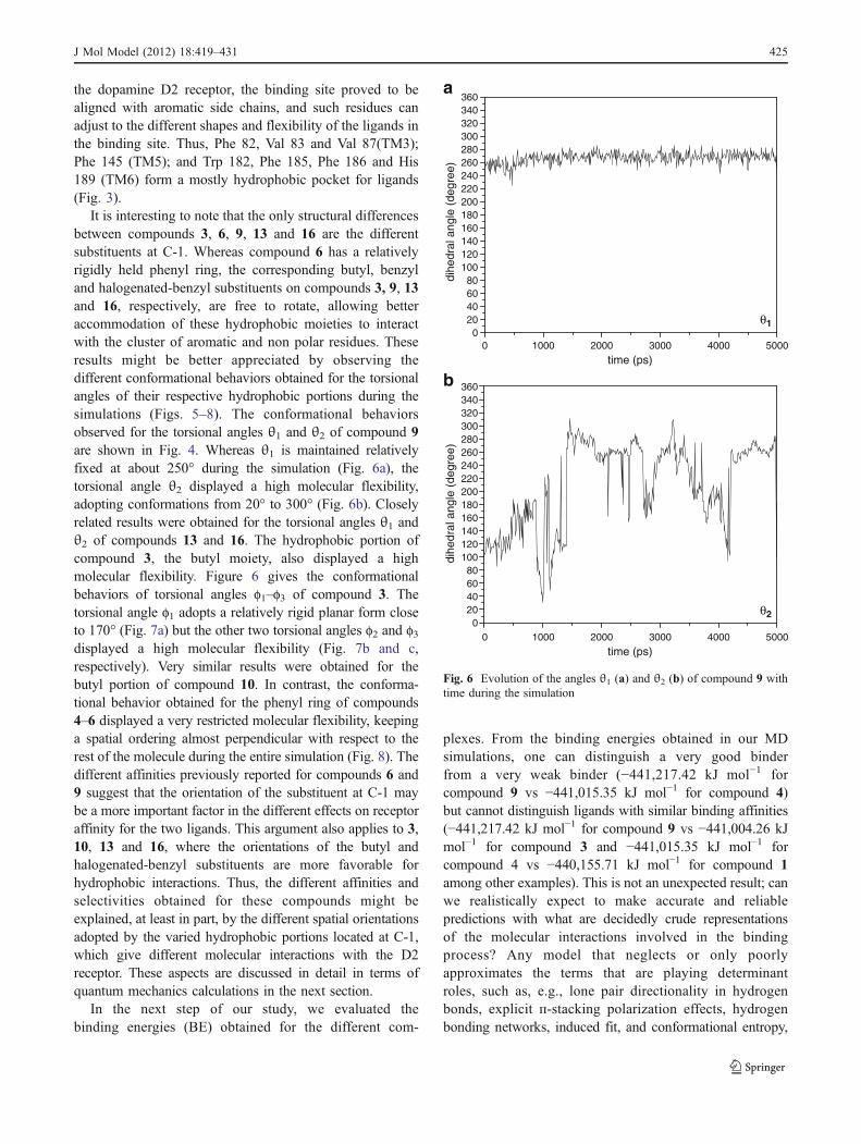
It is interesting to note that the only structural differencesbetween compounds 3, 6, 9, 13 and 16 are the differentsubstituents at C-1. Whereas compound 6 has a relativelyrigidly held phenyl ring, the corresponding butyl, benzyland halogenated-benzyl substituents on compounds 3, 9, 13and 16, respectively, are free to rotate, allowing betteraccommodation of these hydrophobic moieties to interactwith the cluster of aromatic and non polar residues. Theseresults might be better appreciated by observing thedifferent conformational behaviors obtained for the torsionalangles of their respective hydrophobic portions during thesimulations (Figs. 5–8). The conformational behaviorsobserved for the torsional angles θ1 and θ2 of compound 9are shown in Fig. 4. Whereas θ1 is maintained relativelyfixed at about 250° during the simulation (Fig. 6a), thetorsional angle θ2 displayed a high molecular flexibility,adopting conformations from 20° to 300° (Fig. 6b). Closelyrelated results were obtained for the torsional angles θ1 andθ2 of compounds 13 and 16. The hydrophobic portion ofcompound 3, the butyl moiety, also displayed a highmolecular flexibility. Figure 6 gives the conformationalbehaviors of torsional angles f1–f3 of compound 3. Thetorsional angle f1 adopts a relatively rigid planar form closeto 170° (Fig. 7a) but the other two torsional angles f2 and f3displayed a high molecular flexibility (Fig. 7b and c,respectively). Very similar results were obtained for thebutyl portion of compound 10. In contrast, the conforma-tional behavior obtained for the phenyl ring of compounds4–6 displayed a very restricted molecular flexibility, keepinga spatial ordering almost perpendicular with respect to therest of the molecule during the entire simulation (Fig. 8). Thedifferent affinities previously reported for compounds 6 and9 suggest that the orientation of the substituent at C-1 maybe a more important factor in the different effects on receptoraffinity for the two ligands. This argument also applies to 3,10, 13 and 16, where the orientations of the butyl andhalogenated-benzyl substituents are more favorable forhydrophobic interactions. Thus, the different affinities andselectivities obtained for these compounds might beexplained, at least in part, by the different spatial orientationsadopted by the varied hydrophobic portions located at C-1,which give different molecular interactions with the D2receptor. These aspects are discussed in detail in terms ofquantum mechanics calculations in the next section.
In the next step of our study, we evaluated thebinding energies (BE) obtained for the different com-
plexes. From the binding energies obtained in our MDsimulations, one can distinguish a very good binderfrom a very weak binder (−441,217.42 kJ mol−1 forcompound 9 vs −441,015.35 kJ mol−1 for compound 4)but cannot distinguish ligands with similar binding affinities(−441,217.42 kJ mol−1 for compound 9 vs −441,004.26 kJmol−1 for compound 3 and −441,015.35 kJ mol−1 forcompound 4 vs −440,155.71 kJ mol−1 for compound 1among other examples). This is not an unexpected result; canwe realistically expect to make accurate and reliablepredictions with what are decidedly crude representationsof the molecular interactions involved in the bindingprocess? Any model that neglects or only poorlyapproximates the terms that are playing determinantroles, such as, e.g., lone pair directionality in hydrogenbonds, explicit п-stacking polarization effects, hydrogenbonding networks, induced fit, and conformational entropy,
0 1000 2000 3000 4000 5000
dihe
dral
ang
le (
degr
ee)
time (ps)
0 1000 2000 3000 4000 5000
time (ps)
θ1
θ2
a
dihe
dral
ang
le (
degr
ee)
b
020406080
100120140160180200220240260280300320340360
020406080
100120140160180200220240260280300320340360
Fig. 6 Evolution of the angles θ1 (a) and θ2 (b) of compound 9 withtime during the simulation
J Mol Model (2012) 18:419–431 425

among others, cannot reasonably be expected to distinguishbetween compounds possessing relatively similar binding
energies. There are several works supporting this concept inthe literature [50, 51].
At this stage of our work, we considered the trendpredicted for the MD simulations as certainly significantbut, on the other hand, we might be reluctant to assign it aquantitative significance, because of the approximationsinvolved in this mode of approach. It should be noted thatwe are dealing with relatively weak interactions andtherefore MD simulations might underestimate such inter-actions. Thus, in the next step, we optimized reduced modelsystems using combined semiempirical, ab initio and DFTcalculations.
Quantum mechanics calculations
AM1 calculations combined with RHF/6-31G(d) and DFT[B3LYP/6-31G(d,p)] optimizations were performed byconsidering all receptor amino acids that could interactafter initial positioning of the ligands against Asp 86 andSer 141 residues. The binding pocket designed in this way(Fig. 3) provided data that matched experimental resultspreviously reported from binding assays [8, 9].
Figure 9a shows ligand 13 interactions with the D2 DRoptimized using quantum mechanical calculations. The saltbridge between the protonated amino group and thecarboxyl group of Asp 86, as well as the hydrogen bondbetween the 7-hydroxyl group with Ser 141 can be seen inthis figure. From Fig. 9a it is clear that a strong salt bridgeexists in this compound between the protonated aminogroups and the carboxyl group of Asp 86 (calculateddistance of 3.47Å). The hydrogen bond between 13 and Ser141 is a bifurcated interaction in which the oxygen atom ofthe hydroxyl group and the oxygen of carbonyl group ofSer 141 are the proton-acceptors, giving interatomic
0 1000 2000 3000 4000 50000
20406080
100120140160180200220240260280300320340360
dihe
dral
ang
le (
degr
ee)
dihe
dral
ang
le (
degr
ee)
dihe
dral
ang
le (
degr
ee)
time (ps)
0 1000 2000 3000 4000 5000time (ps)
0 1000 2000 3000 4000 5000time (ps)
φ1
φ2
φ3
a
b
c
020406080
100120140160180200220240260280300320340360
020406080
100120140160180200220240260280300320340360
Fig. 7 Evolution of the angles f1 (a), f2 (b) and f3 (c) of compound 3with time during the simulation
0 1000 2000 3000 4000 50000
20406080
100120140160180200220240260280300320340360
ángu
lo d
iedr
o (g
rado
s)
tiempo (ps)
χ
Fig. 8 Evolution of the angle χ of compound 6 with time during thesimulation
426 J Mol Model (2012) 18:419–431
distances of 2.28Å and 2.40Å, respectively. Figure 9bshows ligand 11 interaction with the D2 DR. In this case,the 7-methoxyl group acts as proton-acceptor while thehydroxyl group of Ser 141 is the proton-donor, displayingan interatomic distance of 2.32Å.
Table 1 gives the BE calculated for the differentcomplexes using RHF/6-31 G(d) and B3LYP/ 6-31 G(d,p)
calculations. All compounds possessing 7-methoxyl groupsdisplayed higher BE with respect to the 7- hydroxylhomologues (cf. 1 with 3; 4 with 6; 7 with 9; 11 with 13,and 14 with 16). Previously, we reported that a 7-hydroxylgroup acting as a proton-donor gives a stronger hydrogenbond than those derivatives possessing a 7-methoxyl group[16]. The present results are in agreement with previously
Fig. 9 Interactions ofcompound 13 (a) and 11 (b)with the binding pocket of D2DR. Spatial view of two inter-actions: salt bridge (Asp 86 withprotonated amino group) to theright and hydrogen bondbetween meta-hydroxyl groupwith Ser 141to the left
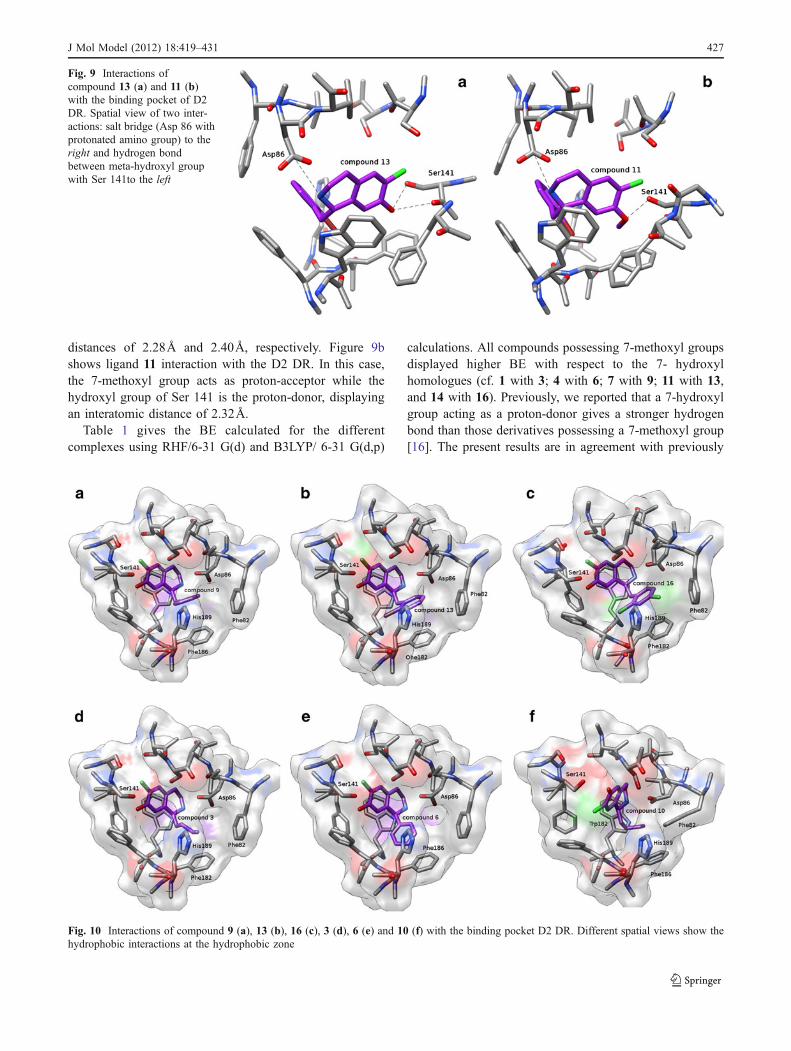
Fig. 10 Interactions of compound 9 (a), 13 (b), 16 (c), 3 (d), 6 (e) and 10 (f) with the binding pocket D2 DR. Different spatial views show thehydrophobic interactions at the hydrophobic zone
J Mol Model (2012) 18:419–431 427

reported calculations for isolated and solvated molecules, aswell as with previously reported experimental bindingaffinities [8, 9] (see Table 1).
Figure 10a shows ligand 9 interactions with the bindingpocket. In this case, a different spatial view with respect toFig. 9 is shown in order to better appreciate the hydropho-bic interactions. From this figure, we can observe that thebenzyl group of 9 adopts an adequate conformation tointeract with Phe 186, Phe 82 and His 189. A similar spatialordering was obtained for compounds 13 and 16 (Fig. 10band c, respectively). For compounds 13 and 16, the halogensubstituent confers a higher polarizability on the benzylgroup, allowing a stronger hydrophobic interaction. Inshould be noted that compound 13 displayed the highestof pKi value in this series. These hydrophobic interactionscould explain, at least in part, the strongest affinity obtainedfor this compound. The butyl group of 3 displays a spatialordering closely related to that of the benzyl group of 9, 13and 16, also giving closely related hydrophobic interactionswith the same hydrophobic residues (Fig. 10d). In contrast,the phenyl group of 6 displayed a different spatial ordering,giving adequate distance to interact only with Phe 186(Fig. 10e). Interestingly, the bonding energies obtained forthese complexes are: 13/D2 DR < 10/D2 DR < 9/D2 DR <3/D2 DR < 16/D2 DR, which are in complete agreementwith their respective pKi values obtained from our previousexperimental results (see Table 1). Compound 10 adopts adifferent spatial ordering at the binding site; thus, the butylportion of this compound interacts with three aromaticresidues: Trp 182, Phe 82 and Phe 186 (Fig. 10f).Compounds 4–6 possess a phenyl ring perpendicular tothe rest of the ligand from the ring containing theprotonated nitrogen [52]. These compounds docked in theD2 receptor model have few interactions in the bindingpocket because their 1-phenyl substituents extend towardthe extracellular surface of the receptor, parallel to the helixaxes. These results are in agreement with those previouslyreported [23]. Thus, it appears that the shape and flexibility ofthe side chain at the C-1 position affects the receptor subtypeselectivity of ligands to an extent that depends on thegeometry, flexibility and stacking potential of ligand sub-stituents. Lan et al. [23], reported that the D1 selective ligandSCH23390 contains a phenyl ring perpendicular to the restof the molecule and the membrane plane, and parallel to thehelix axes, which could explain its selectivity. Our results arein agreement with those results. Compounds type 4–6 in thisseries displayed a conformational behavior closely related tothat reported for SCH23390.
Regarding the general structure of BTHIQs reportedhere, it is reasonable to think that the presence of a chlorineatom at C6, and consequently halogen bonding interactions,could be operative for the ligand–receptor complex forma-tion. Thus, this chlorine possibly could be interacting
through either a positive sigma hole with a negative sitein its vicinity or through its negative lateral ring ofelectrostatic potential with a positive site in the vicinity. Acomprehensive study on electrostatically driven non-covalent interactions has been reported recently by Politzeret al. [53]. In this latter article, the possibility that halogenand other σ-hole interactions can be competitive withhydrogen bonding has been clearly established. Unfortunately,from the limited information obtained from our relatively low-level theory calculations, it is not possible to properlydetermine if the halogen bonding interactions could take placehere. It is clear that further, more accurate calculations, as wellas quantum atoms in molecules (QAIM) [54, 55] analysis arenecessary for a detailed description of these interactions.Such calculations are now in progress in our laboratory andwill be reported later in a separate paper.
0 10 20 30 40 505
6
7
8
14
415
12
2
5
86
17
11
16
310
9
pKi
pKi
ΔE (kcal/mol)
0 10 20 30 40 50
ΔE (kcal/mol)
R=0.9557 R2=0.9134
a
13
5
6
7
8
b
R=0.9726 R2=0.9460
13
109
316
11
1
76
8512
2
154
14
a
b
Fig. 11 Correlations obtained between the experimental pKi valuesversus the binding energies (BE) calculated from a ab initio [RHF/631G(d)] calculations, and b DFT (B3LYP/6-31G(d,p)) computations
428 J Mol Model (2012) 18:419–431

Figure 11a gives a graphical representation of thecalculated BEs obtained from RHF/6-31G(d) calculationsversus experimental pKi values, obtained in binding studiesin rat striatum [8, 9]. This figure has a correlation coefficientR2=0.9134. Theisresult is very satisfactory when oneconsiders the type of approximations used. Figure 11b showsthe same correlations but in this case using BEs obtainedfrom DFT [B3LYP/6-31G(d,p)] calculations. In this draft,the correlation coefficient is R2=0.9460 indicating that DFTcalculations give a better correlation with the experimentaldata. Although both linear correlations are good enough topredict the biological activity of BTHIQs, it is clear that DFTcalculations give a significantly better correlation withrespect to RHF computations. This is particularly evidentfrom the high squares of correlation coefficients, r2 obtainedusing RHF and DFT calculations (0.9134 and 0.9460,respectively). From our results, it is clear that the predictedfirst-principles structure of the primary binding pocket of D2DR leads to correct predictions of the critical residues forbinding THIQs, and gives relative binding affinities thatcorrelate fairly well with those obtained in experimentsperformed in native tissue. This good correlation providesadditional validation for the predicted structure and function.
It should be noted that the AM1 method it is notadequate to describe the hydrogen bonds. In addition, theab initio and DFT calculations performed here probably donot properly consider the dispersion interactions. Fortu-nately, in this case it appears that such limitations are notsevere enough to prevent us obtaining our objectives. Suchan assumption appears to be reasonable, considering thesignificant correlation obtained between the experimentaldata and the theoretical calculations performed. However,we cannot exclude that a kind of error-cancellation couldhave taken place in this case. Thus, it must be pointed outthat the approaches used in this study could be operativeonly for THIQs and structurally related compounds. Toextend these approaches to other compounds possessingdifferent structures would require additional validation andmore accurate calculations.
Conclusions
A molecular modeling study on 16 BTHIQs acting asdopaminergic ligands was carried out. By combining MDsimulations with ab initio and DFT calculations, a simpleand generally applicable procedure to evaluate the bindingenergies of BTHIQs interacting with the D2 DR is reportedhere, providing a clear picture of the binding interactions ofBTHIQs from both structural and energetic viewpoints.Thus, our results give interesting information that may behelpful in obtaining a better understanding of the molecularinteractions between BTHIQs and the D2 DR.
A significant correlation between binding energiesobtained from DFT calculations and experimental pKivalues was obtained. These results could predict thepotential dopaminergic effect of non-synthesized BTHIQswith an acceptable degree of accuracy. Such informationcould be essential in determining a priori the putativeactivity of new BTHIQ derivatives. It is prudent to remark thatthe excellent correlation obtained here between experimentaldata and the theoretical calculations performed here could belimited to BTHIQs and structurally related compounds.However, we believe our results may be helpful in thestructural identification and understanding of the mini-mum structural requirements for these molecules, and canprovide a guide to the design of BTHIQs with this biologicalactivity.
Acknowledgments Grants from Universidad Nacional de San Luis(UNSL) partially supported this work. This research was alsosupported by the Spanish “Ministerio de Educación y Ciencia” grantSAF 2007–63142. S.A.A. thanks a postdoctoral fellowship ofCONICET-Argentina. R.D.E. is a member of the Consejo Nacionalde Investigaciones Científicas y Técnicas (CONICET-Argentina) staff.
References
1. Oloff S, Mailman RB, Tropsha A (2005) Application of validatedQSAR models of D1 dopaminergic antagonist for databasemining. J Med Chem 48:7322–7332
2. Sit SY, Xie K, Jacutin-Porte S, Boy KM, Seanz J, Taber MT,Gulwadi AG, Korpinen CD, Burris KD, Molski TF, Ryan E, XuC, Verdoorn T, Johnson G, Nichols DE, Mailman RB (2004)Synthesis and SAR exploration of dinapsoline analogues. BioorgMed Chem 12:715–734
3. Strange PG (1997) Dopamine receptors. Tocris Reviews 15.Tocris Cookson, Bristol
4. Zhang A, Neumeyer JL, Baldessarini RJ (2007) Recent progressin development of dopamine receptor subtype-selectiveagents:potential therapeutics for neurological and psychiatric disorders.Chem Rev 107:274–302
5. Protais P, Arbaoui J, Bakkali EH, Bermejo A, Cortes D (1995)Effects of various isoquinoline alkaloids on in vitro 3H-dopamineuptake. J Nat Prod 58:1475–1484
6. Bermejo A, Protais P, Blázquez MA, Rao KS, Zafra-Polo MC,Cortes D (1995) Dopaminergic isoquinoline alkaloids from rotosof Xylopia papuana. Nat Prod Res 6:57–62
7. Cabedo N, Protais P, Cassels BK, Cortes D (1998) Synthesis anddopamine receptor selectivity of the benzyltetrahydroisoquinoline,(R)-(+)-nor-roefractine. J Nat Prod 61:709–712
8. Cabedo N, Andreu I, Ramírez de ArellanoMC, Chagraoui A, SerranoA, Bermejo A, Protais P, Cortes D (2001) Enantioselective synthesesof dopaminergic (R)- and (S)-benzyltetrahydroisoquinolines. J MedChem 44:1794–1801
9. Berenguer I, El Aouad N, Andujar S, Romero V, Suvire F, FreretT, Bermejo A, Ivorra MD, Enriz RD, Boulouard M, Cabedo N,Cortes D (2009) Tetrahydroisoquinolines as dopaminergicligands: 1-butyl-7-chloro-6-hydroxy-tetrahydroisoquinoline, anew compound with antidepressant-like activity in mice. BioorgMed Chem 17:4968–4980
10. El Aouad N, Berenguer I, Romero V, Marín P, Serrano A, AndujarS, Suvire F, Bermejo A, Ivorra MD, Enriz RD, Cabedo N, Cortes
J Mol Model (2012) 18:419–431 429

D (2009) Structure–activity relationship of dopaminergic haloge-nated 1-benzyl-tetrahydroisoquinoline derivatives. Eur J MedChem 44:4616–4621
11. Andreu I, Cabedo N, Torres G, Chagraoui A, Ramirez de ArellanoMC, Gil S, Bermejo A, Valpuesta M, Portais P, Cortes D (2002)Syntheses of dopaminergic 1-cyclohexylmethyl-78-dioxygenatedtetrahydroisoquinolines by selective heterogeneous tandem hydro-genation. Tetrahedron 58:10173–10179
12. Bermejo A, Andreu I, Suvire F, Léonce S, Caignard DH, RenardP, Pierré A, Enriz RD, Cortes D, Cabedo N (2002) Syntheses andantitumor targeting G1 phase of the cell cycle of benzoyldihy-droisoquinoline. J Med Chem 45:5058–5068
13. Suvire FD, Andreu I, Bermejo A, Zamora MA, Cortes D, EnrizRD (2003) Conformational study of N-alkyl-benzyltetrahydroiso-quinolines alkaloid. J Mol Struct THEOCHEM 666–667:109–116
14. Suvire FD, Cabedo N, Chagraoui A, Zamora MA, Cortes D, EnrizRD (2003) Molecular recognition and binding mechanism of N-alkyl-benzyltetrahydro-isoquinolines to the D1 dopamine receptor.A computational approach. J Mol Struc THEOCHEM 666–667:455–467
15. Andreu I, Cortes D, Protais P, Cassels BK, Chagraoui A, CabedoN (2000) Preparation of dopaminergic N-Alkyl-benzyltetrahydroi-soquinolines using a ‘One-Pot’ procedure in acid medium. BioorgMed Chem 8:889–895
16. Doi S, Shirai N, Sato Y (1997) Abnormal products in theBischler-Napieralski isoquinoline synthesis. J Chem Soc PerkinTrans 1:2217–2221
17. Andujar SA, Migliore de Angel BM, Charris JE, Israel A, Suárez-Roca H, López SE, Garrido MR, Cabrera EV, Visual G, Rosales C,Suvire FD, Enriz RD, Angel-Guío JE (2008) Synthesis dopaminergicprofile and molecular dynamics calculations of N-aralkyl substituted2-aminoindans. Bioorg Med Chem 16:3233–3244
18. Kalani MYS, Vaidehi N, Hall SE, Trabanino RJ, Freddolino PL,Kalani MA, Floriano WB, Wai Tak Kam V, Goddard WA III(2005) The predicted 3D structure of the human D2 dopaminereceptor and the binding site and binding affinities for agonistsand antagonists. Proc Natl Acad Sci USA 101:3815–3820
19. Becker OM, Marantz Y, Shacham S, Inbal B, Heifetz A, Kalid O,Bar-Haim S, Warshaviak D, Fichman M, Noiman SG (2004)Protein-coupled receptors: in silico drug discovery in 3D. ProcNatl Acad Sci USA 101:11304–11309
20. Micheli F, Bonanomi G, Blaney FE, Braggio S, Capelli AM,Checchia A, Curcuruto O, Damiani F, Di Fabio R, Donati D,Gentile G, Gribble A, Hamprecht D, Tedesco G, Terreni S, Tarsi L,Lightfoot A, Stemp G,MacDonald G, Smith A, Pecoraro M, PetroneM, Perini O, Piner J, Rossi T, Worby A, Pilla M, Valerio E, GriffanteC, Mugnaini M, Wood M, Scott C, Andreoli M, Lacroix L, SchwarzA, Gozzi A, Bifone A, Ashby CR Jr, Hagan JJ, Heidbreder C (2007)124-Triazol-3-yl-thiopropyl-tetrahydrobenzazepines: a series ofpotent and selective dopamine D3 receptor antagonists. J MedChem 50:5076–5089
21. Ribeiro AA, Horta BAC, de Alencastro RB (2008) MKTOP: aprogram for automatic construction of molecular topologies. JBraz Chem Soc 19:1433–1435
22. Manzour A, Meng F, Meador-Woodruff JH, Taylor LP, Civelli O,Akil H (1992) Site-directed mutagenesis of the human dopamineD2 receptor. Eur J Pharm Mol Pharmacol Sec 227:205–214
23. Lan H, DuRand CJ, Teeter MM, Neve KA (2006) Structuraldeterminants of pharmacological specificity between D1 and D2
dopamine receptors. Mol Pharmacol 69:185–19424. Berendsen HJC, Van der Spoel D, Van Drunen R (1995)
GROMACS: a message-passing parallel molecular dynamicsimplementations. Comput Phys Commun 91:43–56
25. Lindahl E, Hess B, van der Spoel D (2001) GROMACS 3.0: apackage for molecular simulations and trajectory analysis. J MolModel 7:306–317
26. van Buuren AR, Marrink SJ, Berendsen HJC (1993) A moleculardynamics study of the decane/water interface. J Phys Chem36:9206–9212
27. Mark AE, van Helden SP, Smith PE, Janssen LHM, vanGunsteren WF (1994) Convergence properties of free energycalculations. A-cyclodextrin complexes as a case study. J AmChem Soc 116:6293–6302
28. Jorgensen WL, Chandrasekhar J, Madura JD, Impey RW, KleinML (1983) Comparison of simple potential functions forsimulating liquid water. J Chem Phys 79:926–935
29. van Buuren AR, Berendsen HJC (1993) Molecular dynamicssimulation of the stability of a 22 residue alpha-helix in water and30% trifluoroethanol. Biopolymers 33:1159–1166
30. Liu H, Muller-Plathe F, van Gunsteren WF (1995) A force fieldfor liquid dimethyl sulfoxide and physical properties of liquiddimethyl sulfoxide calculated using molecular dynamics simulation.J Am Chem Soc 117:4363–4366
31. Miyamoto S, Kollman PA (1992) SETTLE—an analytical versionof the SHAKE and RATTLE algorithm for rigid water models. JComput Chem 13:952–962
32. Berendsen HJC, Postma HJC, Van Gunsteren WF, Hermans WF(1981) Interaction models for water in relation to proteinhydration. In: Pullman B (ed) Intermolecular forces. Reidel,Dordrecht, pp 331–342
33. Darden T, York D, Pedersen L (1993) Particle mesh Ewald—an N.log(n) method for Ewald sums in large systems. J Chem Phys98:10089–10092
34. Essmann U, Perera L, Berkowitz ML, Darden T, Lee H, PedersenLG (1995) A smooth particle mesh Ewald method. J Chem Phys103:8577–8593
35. Luty B, Tironi IG, van Gunsteren WF (1995) Lattice–summethods for calculating electrostatic interactions in molecularsimulations. J Chem Phys 103:3014–3021
36. Zimmerman K (1991) All purpose molecular mechanics simulatorand energy minimizer. J Comput Chem 12:310–319
37. Ferguson DM (1995) Parameterization and evaluation of a flexiblewater model. J Comput Chem 16:501–511
38. Berendsen HJC, Postma JPM, DiNola A, Haak JR (1984)Molecular dynamics with coupling to an external bath. J ChemPhys 81:3684–3690
39. Hess B, Bekker H, Berendsen HJC, Fraaije JG (1997) E.M.LINCS: a linear constraint solver for molecular simulations. JComput Chem 18:1463–1472
40. Kampmann T, Mueller DS, Mark AE, Young PR, Kobe B (2006)The role of histidine residues in low-pH-mediated viral membranefusion. Structure 14:1481–1487
41. Teeter MM, Froimowitz MF, Stec B, Durand CJ (1994)Homology modeling of the dopamine D2 receptor and its testingby docking of agonists and tricyclic antagonists. J Med Chem37:2874–2888
42. Neve KA, Cumbay MG, Thompson KR, Yang R, Buck DC, WattsVJ, Durand CJ, Teeter MM (2001) Modeling and mutationalanalysis of a putative sodium-binding pocket on the dopamine D2
receptor. Mol Pharmacol 60:373–38143. Frisch MJ, Trucks GW, Schlegel HB, Scuseria GE, Robb MA,
Cheeseman JR, Montgomery JA Jr, Vreven T, Kudin KN, BurantJC, Millam JM, Iyengar SS, Tomasi J, Barone V, Mennucci B,Cossi M, Scalmani G, Rega N, Petersson GA, Nakatsuji H, HadaM, Ehara M, Toyota K, Fukuda R, Hasegawa J, Ishida M,Nakajima T, Honda Y, Kitao O, Nakai H, Klene M, Li X, KnoxJE, Hratchian HP, Cross JB, Adamo C, Jaramillo J, Gomperts R,Stratmann RE, Yazyev O, Austin AJ, Cammi R, Pomelli C,Ochterski JW, Ayala PY, Morokuma K, Voth GA, Salvador P,Dannenberg JJ, Zakrzewski VG, Dapprich S, Daniels AD, StrainMC, Farkas O, Malick DK, Rabuck AD, Raghavachari K,Foresman JB, Ortiz JV, Cui Q, Baboul AG, Clifford S, Cioslowski
430 J Mol Model (2012) 18:419–431

J, Stefanov BB, Liu G, Liashenko A, Piskorz P, Komaromi I,Martin RL, Fox DJ, Keith T, Al-Laham MA, Peng CY,Nanayakkara A, Challacombe M, Gill PMW, Johnson B, ChenW, Wong MW, Gonzalez C, Pople JA (2003) Gaussian 03,Revision B.05. Gaussian Inc, Pittsburgh
44. Pettersen EF, Goddard TD, Huang CC, Couch GS, Greenblatt DM,Meng EC, Ferrin TE (2004) UCSF Chimera—a visualization systemfor exploratory research and analysis. J Comput Chem 25:1605–1612
45. Hjerde E, Dahl SG, Sylte I (2005) Atypical and typicalantipsychotic drug interactionswith the dopamine D2 receptor.Eur J Med Chem 40:185–194
46. Cho W, Taylor LP, Mansour A, Akil A (1995) Hydrophobicresidues of the D2 dopamine receptor are important for bindingand signal transduction. J Neurochem 65:2105–2115
47. Cox BA, Henningsen RA, Spanoyannis A, Neve RL, Neve KA(1992) Contributions of conserved serine residues to the interactionsof ligands with dopamine D2 receptors. J Neurochem 59:627–635
48. Wiens BL, Nelson CS, Neve KA (1998) Contribution of serineresidues to constitutive and agonist-induced signaling via theD2Sdopamine receptor: evidence for multiple agonist-specificactive conformations. Mol Pharmacol 54:435–444
49. Wilcox RE, Huang WH, Brusniak MYK, Wilcox DM, PearlmanRS, Teeter MM, Durand CJ, Wiens BL, Neve KA (2000)CoMFA-based prediction of agonist affinities at recombinant wildtype versus serine to alanine point mutated D2 dopaminereceptors. J Med Chem 43:3005–3019
50. Page CS, Bates PA (2008) Can MM-PBSA calculations Predictthe specifiicties of protein kinase inhibitors? J Comput Chem27:1990–2007
51. Mertz KM (2010) Limits of free energy computation for protein–ligand interactions. J Chem Theor Comput 6:1769–1776
52. Breneman CM, Wiberg KB (1990) Determining atom-centeredmonopoles from molecular electrostatic potentials. The need forhigh sampling density in formamide conformational analysis. JComput Chem 11:361–373
53. Politzer P, Murray JS, Clark T (2010) Halogen bonding: anelectrostatically-driven highly directional noncovalent interaction.Phys Chem Chem Phys 12:7748–7757
54. Bader RFW (1990) Atoms in molecules. A quantum theory.Oxford University Press, Oxford
55. Popelier PLA (1999) Atoms in molecules. An introduction.Pearson, Harlow
J Mol Model (2012) 18:419–431 431
Copyright © 2022 FDOKUMEN