co-regulation of endothelial and myogenic cell fates through ...
191
Acceptée sur proposition du jury pour l’obtention du grade de Docteur ès Sciences par CO-REGULATION OF ENDOTHELIAL AND MYOGENIC CELL FATES THROUGH INTERCELLULAR CROSSTALK IN SKELETAL MUSCLE Umji LEE Thèse n° 8400 2021 Présentée le 1 er octobre 2021 Prof. B. E. Ferreira De Sousa Correia, président du jury Dr J. Feige, Prof. B. D. Cosgrove, directeurs de thèse Prof. P. Muñoz-Cánoves, rapporteuse Prof. P. Castets, rapporteuse Prof. K. De Bock, rapporteuse Faculté des sciences de la vie Programme doctoral en biotechnologie et génie biologique
-
Upload
khangminh22 -
Category
Documents
-
view
1 -
download
0
Transcript of co-regulation of endothelial and myogenic cell fates through ...

Acceptée sur proposition du jury
pour l’obtention du grade de Docteur ès Sciences
par
CO-REGULATION OF ENDOTHELIAL AND MYOGENIC CELL FATES THROUGH INTERCELLULAR CROSSTALK IN SKELETAL MUSCLE
Umji LEE
Thèse n° 8400
2021
Présentée le 1er octobre 2021
Prof. B. E. Ferreira De Sousa Correia, président du juryDr J. Feige, Prof. B. D. Cosgrove, directeurs de thèseProf. P. Muñoz-Cánoves, rapporteuseProf. P. Castets, rapporteuseProf. K. De Bock, rapporteuse
Faculté des sciences de la vieProgramme doctoral en biotechnologie et génie biologique

1
Thesis Committee
Thesis Committee
Prof. Bruno Correia (President of Jury)
Dr. Jerome Feige (Director)
Prof. Benjamin Cosgrove (Director)
Prof. Katrien De Bock
Prof. Pura Muñoz-Canoves
Prof. Perrine Castets
Umji Lee
Ithaca 14850, New York
United States of America
05 August 2021 EPF Lausanne, Switzerland
bd
밑줄

2
Abstract
Co-regulation of Endothelial and Myogenic Cell Fates Through Intercellular Crosstalk in Skeletal Muscle
Abstract
Skeletal muscle is one of the most dynamic organs in the human body, which has a vigorous
potential to regenerate and adapt to environmental changes. Depending on environments,
skeletal muscle enables essential life activities to survive by controlling movement and
metabolism. The functional and structural adaptations of skeletal muscle crosstalk with other
physiological systems enabling nutrition and oxygen delivery, and the elimination of
metabolic waste products. The work in this dissertation focuses on the intercellular signals
produced by myogenic cells to dynamically remodel skeletal muscle during exercise or
muscle repair, and the reciprocal signals from the local niche and the systemic environment
that regulate myogenic cell fate and metabolism according to physiological needs. This
complex crosstalk is dissected in three specific chapters where we study the crosstalk of
myogenic cells with endothelial cells and the vasculature to coordinate local niche
interactions and systemic crosstalks. First, we demonstrate that an exercise-induced
myokine called apelin is produced by muscle fiber and mediates intercellular signaling to
endothelial cells through the apelin receptor. In addition, through a yeast one-hybrid screen
of transcription factor binding to the apelin promoter, we identified the myogenic transcription
factor TEA domain family member 1 (Tead1) as a regulator of Apln transcription. We
observed via single-cell transcriptomic analysis of regenerating skeletal muscle that Aplnr
(Apelin receptor) is enriched in muscle endothelial cells, whereas Tead1 is enriched in
myogenic cells. Myofiber-specific over-expression of Tead1 suppresses apelin secretion at
the whole-body level, and apelin secretion via Tead1 knock-down in muscle cells stimulates
endothelial cell proliferation in co-cultures. By showing that apelin peptide supplementation
in vivo enhances endothelial cell expansion following muscle injury, we conclude that
paracrine crosstalk in which apelin secretion controlled by Tead1 in myogenic cells
influences endothelial remodeling during skeletal muscle repair.
Secondly, we show how tissue-resident support cells affect muscle progenitor activation in
different metabolic environments. Skeletal muscle progenitors (SKMP) reside in close

3
Abstract
proximity to supportive cell types in the stem cell niche and dynamically interact with each
other to adapt to environmental changes. Here, we studied how different niche cell types
affect SKMPs in response to glycemic levels. Using co-cultures of SKMPs and niche cells,
we discovered that endothelial cells synergistically enhance SKMP proliferation in a low
glycemic environment, while fibroblasts and macrophages had no effect. We observed that
the crosstalk between SKMPs and endothelial cells was mediated by direct cell-cell contacts
independent of soluble paracrine signals. Transcriptomic analysis revealed that the
endothelial alpha/beta hydrolase N-Myc Downstream Regulated1 (NDRG1) is induced by a
low glycemic environment and is associated with biological adhesion. SKMPs co-cultured
with Ndrg1 knock-down endothelial cells lose their synergistic functional relationship in
response to glucose levels. Therefore, our findings suggest that Ndrg1 is a key mediator of
endothelial cell-mediated glycemic control of SKMPs and provides a link between systemic
energy levels and the skeletal muscle stem cell niche.
Lastly, I developed intravital imaging to monitor muscle stem cells and vasculature to
deepen the anatomical and kinetic understanding of the intercellular crosstalk between
vascular networks and muscle stem cells (MuSCs) in vivo. To that end, we optimized the
long-lasting- chronic window frame to optically access hindlimb muscle during recovery,
developed an endogenous endothelial cell labeling system, and monitored MuSC
engraftment after cell transplantation in a living animal. We discovered that most of the
muscle stem cells successfully engrafted were located in close proximity of the vasculature
and endothelial niche. This approach offers a great promise for the development of a
robust tool to monitor muscle repair dynamics in real-time with spatial information of the
vascular network. Altogether, my dissertation chapters bring together a complementary set
of cellular mechanisms with reciprocal co-regulatory effects between myogenic and
endothelial cell fates and established a competitive tool to investigate them in a living
animal.

4
Résumé
Co-régulation du destin des cellules endothéliales et myogéniques par diaphonie intercellulaire dans le muscle squelettique
Résumé
Le muscle squelettique est l'un des organes les plus dynamiques du corps humain, qui a un
potentiel vigoureux pour se régénérer et s'adapter aux changements environnementaux.
Selon les environnements, le muscle squelettique permet aux activités vitales essentielles
de survivre en contrôlant les mouvements et le métabolisme. Les adaptations fonctionnelles
et structurelles du muscle squelettique se croisent avec d'autres systèmes physiologiques
permettant la nutrition et l'apport d'oxygène, et l'élimination des déchets métaboliques. Le
travail de cette thèse se concentre sur les signaux intercellulaires produits par les cellules
myogéniques pour remodeler dynamiquement le muscle squelettique pendant l'exercice ou
la réparation musculaire, et les signaux réciproques de la niche locale et de l'environnement
systémique qui régulent le destin et le métabolisme des cellules myogéniques en fonction
des besoins physiologiques. Cette diaphonie complexe est disséquée dans trois chapitres
spécifiques où nous étudions la diaphonie des cellules myogéniques avec les cellules
endothéliales et le système vasculaire pour coordonner les interactions de niche locales et
les diaphonies systémiques. Premièrement, nous démontrons qu'une myokine induite par
l'exercice appelée apéline est produite par la fibre musculaire et médie la signalisation
intercellulaire vers les cellules endothéliales via le récepteur de l'apéline. En outre, grâce à
un criblage de levure mono-hybride du facteur de transcription se liant au promoteur de
l'apeline, nous avons identifié le membre de la famille du domaine du facteur de transcription
myogénique TEA 1 (Tead1) en tant que régulateur de la transcription Apln. Nous avons
observé via une analyse transcriptomique unicellulaire du muscle squelettique en
régénération que Aplnr (récepteur Apelin) est enrichi en cellules endothéliales musculaires,
tandis que Tead1 est enrichi en cellules myogéniques. La surexpression de Tead1
spécifique aux myofibres supprime la sécrétion d'apéline au niveau du corps entier, et la
sécrétion d'apéline via Tead1 knock-down dans les cellules musculaires stimule la
prolifération des cellules endothéliales dans les co-cultures. En montrant que la
supplémentation en peptide apéline in vivo améliore l'expansion des cellules endothéliales
après une lésion musculaire, nous concluons que la paracrine croisée dans laquelle la

5
Résumé
sécrétion d'apéline contrôlée par Tead1 dans les cellules myogéniques influence le
remodelage endothélial pendant la réparation des muscles squelettiques.
Deuxièmement, nous montrons comment les cellules de soutien résidentes dans les tissus
affectent l'activation des progéniteurs musculaires dans différents environnements
métaboliques. Les progéniteurs des muscles squelettiques (SKMP) résident à proximité
immédiate des types de cellules de soutien dans la niche des cellules souches et
interagissent dynamiquement les uns avec les autres pour s'adapter aux changements
environnementaux. Ici, nous avons étudié comment différents types de cellules de niche
affectent les SKMP en réponse aux niveaux glycémiques. En utilisant des co-cultures de
SKMP et de cellules de niche, nous avons découvert que les cellules endothéliales
améliorent de manière synergique la prolifération de SKMP dans un environnement à faible
indice glycémique, tandis que les fibroblastes et les macrophages n'ont aucun effet. Nous
avons observé que la diaphonie entre les SKMP et les cellules endothéliales était médiée
par des contacts directs cellule-cellule indépendants des signaux paracrines solubles.
L'analyse transcriptomique a révélé que l'hydrolase endothéliale alpha/bêta N-Myc
Downstream Regulated1 (NDRG1) est induite par un environnement à faible indice
glycémique et est associée à une adhésion biologique. Les SKMP co-cultivés avec des
cellules endothéliales knock-down Ndrg1 perdent leur relation fonctionnelle synergique en
réponse aux niveaux de glucose. Par conséquent, nos résultats suggèrent que Ndrg1 est un
médiateur clé du contrôle glycémique à médiation cellulaire endothéliale des SKMP et
fournit un lien entre les niveaux d'énergie systémique et la niche des cellules souches du
muscle squelettique.
Enfin, j'ai développé l'imagerie intravitale pour surveiller les cellules souches musculaires et
le système vasculaire afin d'approfondir la compréhension anatomique et cinétique de la
diaphonie intercellulaire entre les réseaux vasculaires et les cellules souches musculaires
(MuSC) in vivo. À cette fin, nous avons optimisé le cadre de la fenêtre chronique de longue
durée pour accéder optiquement au muscle des membres postérieurs pendant la
récupération, développé un système de marquage des cellules endothéliales endogènes et
surveillé la prise de greffe MuSC après transplantation cellulaire chez un animal vivant.
Nous avons découvert que la plupart des cellules souches musculaires greffées avec
succès étaient situées à proximité du système vasculaire et de la niche endothéliale. Cette
approche offre une grande promesse pour le développement d'un outil robuste pour

6
Résumé
surveiller la dynamique de réparation musculaire en temps réel avec des informations
spatiales du réseau vasculaire. Au total, les chapitres de ma thèse rassemblent un
ensemble complémentaire de mécanismes cellulaires avec des effets de corégulation
réciproques entre les destins des cellules myogéniques et endothéliales et ont établi un outil
compétitif pour les étudier chez un animal vivant.

7
Acknowledgments
Acknowledgments
You can not hope to build a better world without improving the individuals. To that
end, each of us must work for our own improvement and, at the same time share general
responsibility for all humanity, our particular duty being to aid those to whom we think we can
be most useful.
- Marie Sklodowska 1867-1984
As I remember, when I was at my elementary school, I often spoke that I would like to
become an artist in the future. A lot of accidental events in my life brought me into the art of
measuring life activity and converting the art into a scientific hypothesis. Five years of my
Ph.D. taught me that science is not just measurements and equations but is iterative
empirical observations and recursive corrections on my hypothesis. And I learned keeping
the enthusiasm during this iteration will make a scientist. However, to keep motivated amid
the small and big failures for 5 years was extremely hard. Every time I am facing the small
and big hurdles, I had so much luck with my mentors, teachers, colleagues, and friends who
help me keep going without losing my enthusiasm. I sincerely thank all the people who
worked with me and talked with me during this time.
My intellectual development and scientific thinking owe a huge deal to my two
advisors Dr. Jerome Feige and Dr. Benjamin Cosgrove. I sincerely thank Dr. Feige and
Prof. Cosgrove for allowing me to carry out my exciting Ph.D. projects under their guidance
and for providing the tools and invaluable advice to enrich my research and scientific
education. I am indebted to all their support and help on scientific growth but also for
showing me a lot of inspiration what is a good leader-like. It was a great privilege to work
with them. They have been the most influencing advisors in both professional and personal
aspects of my career and gave exceptional scientific supervision during my time at EPFL
and Cornell University. They advised me each study, which was not only for a big picture
design or results but also brings important details and messages from the moment I started
to design an experiment. I have learned a great deal from them how to logically and
constructively approach scientific questions. I believe their mentorship and advice are
necessary factors in finishing my Ph.D. Beyond my Ph.D. project supervision, their
encouragement and passion for learning and communicating with other scientists in the field,
have guided me in many aspects. The encouragement to attend scientific conferences
whenever there is a related conference or seminar and also leading the Muscle Science Talk
Series, which is not only helpful to broaden the knowledge but also it is a huge contribution

8
Acknowledgments
to the field of muscle biology. I sincerely learned a lot from my supervisors and feel lucky to
work at both places.
I would like to express my deep appreciation to Prof. Florian Bentzinger for
supporting and advising me with helpful discussion and collaboration. I feel extremely lucky
to meet him as my first mentor, which led me to muscle biology. Thanks to him, I could learn
not only basic muscle techniques and science but also the attitude to draw a big picture from
a sketch. He was my first mentor and will be my mentor forever. I will always admire him for
his energy and enthusiasm for science. Besides my mentors, I thank my thesis committee
members for their serve and contribution to my Ph.D. thesis. Prof. Bruno Correia, Prof. Pura Muñoz-Canoves, Prof. Katrien De Bock, and Prof. Perrine Castets, I am very
honored to have their input and have an opportunity to share my Ph.D. works with them as I
always admired their works in the field of study.
I also extremely thank Swiss National Science Foundation and the selection
committee for the mobility fellowship, which gave me an incredible opportunity to carry on
my Ph.D. with a unique project including multi-photon microscope modalities in pioneering
university. I had a lot of luck from many scientific foundations supporting my education from
my master's to here. Without their support, this higher education and studying abroad would
have just remained in my dream. I would like to thank Mogam Science Foundation to believe
and see my potential as well as Swiss National Science Foundation for allowing me to build
this amazing experience. I deeply acknowledge their decision.
To my colleagues as well as my friends, thank them for all their time and support,
encouragement, sharing mindful visions, and jokes during the experiments and my
endeavors. Sitting on the biosafety cabinet for a long time, fighting with so many different
cells could be joyful when we were doing it together. Both in and out of the lab, I have
learned a lot from each of them, they were my teachers and mentors. Hannah Fong, David McKellar, Charles Heinke, Emily Laurilliard, Paula Petella, Lauren Walter, Alex Loiben, and Andrea De Michele, I appreciate them for creating an environment where freely ask
and cooperate, and I truly learn synergy when people work together and help each other.
They were the people who made my Ph.D., which can be difficult sometimes, much easier. I
am deeply grateful to all the precious bonds forged at Cornell. In addition, I also would like
to thank Prof. Nishimura and her lab members Dr. Chi-Yong Eom, Nichole Biliotta, Dr. David Small, and Aron Mok in Chris Xu’s lab taught me and helped me with various steps
related to intravital imaging techniques in chapter 5.

9
Acknowledgments
I also would like to express special gratitude to all the Feige lab members and
collaborators at Nestle Institute of Health Sciences. I could be exposed to state-of-the-art
technology and various screening skills in muscle research thanks to them. Sonia Karaz, Omid Mashinchian, Tanja Sonntag, Gabriele Dammone, Dr. Eugenia Migliavacca, Dr. Gabriele Civiletto, Joris Michaud, Dr. Pascal Stuelsatz, Sara Ancel, Anna Weiser, Philip Rein, Maria Deak, Caterina Collodet, and Minji Huh. I sincerely thank them all who
not only taught me techniques but also gave priceless advice for my future career and gave
me enormous emotional support. They always gave me valuable advice and brought up
great ideas on my project. Without their detailed instructions, I would not have been able to
perform the projects properly in particular with Section 3. I appreciate their work which
became the basis of my project and helped my experiments using high-content screening.
Deplancke’s group, especially Julie Russeil and Prof. Bart Deplancke to give me
supervision and various feedback with helpful advice related to transcription factor study as
well as teaching me yeast one-hybrid techniques on Section 3. I would be able to start my
Ph.D. smoothly thanks to them.
Most of all, I sincerely thank my family and friends for their enormous support no
matter where I am, no matter what I try to do, and how I want to do it. Seongsik Lee, Inja Kim, Yeji Lee, Junglang Gu, Areum Yuhan, Jinhae Lee, Siyu Liu, Dr. Melina Scholze,
Garazi Monzo Contreras, Dr. Radek Jankele, Oh-Hyeon Choung, Jisoo Park, Dr. Jane Yi, and Changwoo Seo. I sincerely appreciate that you have been always on my side and
truly believed I can make my choice right. Without their supports and trust, I could not be
able to get back up again every time I stumble. Some of them I met as Ph.D. students or
master students became doctors. I do not recall how much coffee we have had so far, over
the coffee, they always inspired me in multiple aspects of life. Their scientific thoughts and
passion brought me a lot of crazy ideas and my fun life activities continuously and strongly,
which still make my scientific motivation to be going on. I would not have been able to
continue my Ph.D. or science without their support and love. I sincerely thank them all.
Now, I am exceptionally honored to announce that I will be moving to another step to
commence my new life as a scientist. These short paragraphs would not be sufficient to give
my acknowledgment to everyone who has been instrumental to my studying from the
moment I imagined 5 years ago until today. For all the people who constantly cheer me up, I
deeply bow my head to your dedication.

10

11
Contents
Contents
Abstract ............................................................................................................................................. 2
Résumé .............................................................................................................................................. 4
Acknowledgments ............................................................................................................................ 7
Contents .......................................................................................................................................... 11
Section 1: Introduction ................................................................................................................... 13
1.1 Skeletal Muscle Structure and Functions .................................................................... 13
1.2 Myokines: Endocrine Function of Skeletal Muscle ...................................................... 18
1.3 Muscle Stem Cells in Muscle Regeneration ................................................................ 20
1.4 Muscle Stem Cell Niche .............................................................................................. 23
1.5 Muscle Atrophy in Aging .............................................................................................. 34
Section 2: Statement of Research ................................................................................................. 38
2.1. Investigating transcriptional regulation of apelin ........................................................ 39
2.2 Investigating functional relationship between myogenic progenitors and supportive
cells with glycemic control ................................................................................................. 45
2.3 Developing intravital imaging of vasculature and satellite cells ................................... 50
Section 3 A Tead1-Apelin axis directs paracrine communcation from myogenic to endothelial cells in skeletal muscle ............................................................................................. 55
3.1 Introduction .................................................................................................................. 58
3.2 Results: ....................................................................................................................... 60
3.3 Discussion and Conclusion ......................................................................................... 79

12
Contents
Section 4 Endothelial-myogenic progenitor cell crosstalk is regulated by glycemic levels via endothelial Ndrg1 .......................................................................................................... 94
4.1 Introduction .................................................................................................................. 98
4.2 Results: ..................................................................................................................... 100
4.3 Discussion and Conclusion ....................................................................................... 112
Section 5 Intravital imaging of vasculature and muscle progenitors in skeletal muscle of live mice .................................................................................................................................... 125
5.1 Introduction ................................................................................................................ 128
5.2 Results: ..................................................................................................................... 130
5.3 Discussion and Conclusion ....................................................................................... 136
Section 6: Perspective .................................................................................................................. 138
6.1 Summary and Perspective ..................................................................................... 138
6.2 Future Directions ................................................................................................... 140
Appendix ........................................................................................................................................ 142
The Exerkine Apelin Reverses Age-associated Sarcopenia ........................................... 142
Bibliography .................................................................................................................................. 147
Curriculum Vitae ........................................................................................................................... 171

13
Section 1: Introduction
Section 1: Introduction
1.1 Skeletal Muscle Structure and Functions
1.1.1 Skeletal Muscles of Human Body
The musculoskeletal system is one of the most abundant components of vertebrate
animals and has evolved to enable movement specialization and adapt to environments
dynamically. It enables life activity to make it possible for them to survive in different
environments by controlling the movement of individuals to seek food and adapting
metabolism (Flück and Hoppeler, 2003). As a major component of the human body, skeletal
muscle accounts for approximately 40-50 percent of body mass and contains 50-70% of
body proteins (Frontera and Ochala, 2015; Karagounis and Hawley, 2010). Energy
expenditure of skeletal muscle varies dramatically depending on the physical activity rates of
individuals. During the resting time, it usually accounts for 20% of energy expenditure, but it
can account for 90% of whole-body O2 consumption during the high-loaded rates exercise
(Brooks, 1997; Wahren et al., 1971; Zurlo et al., 1990). In this regard, malfunctioning skeletal
muscle can skew the whole body bioenergetics and contribute to metabolic syndromes such
as obesity, hyperglycemia, and dyslipidemia, which increases the risk of type 2 diabetes
mellitus and cardiovascular diseases (Cornier et al., 2008; Kim and Kim, 2020; Lakka et al.,
2002; Rubio-Ruiz et al., 2019)
Around 650 types of skeletal muscles are spread symmetrically throughout the
human body. Individual skeletal muscle is connected to the skeletons and the somatic
nervous system, which is controlled by the central nervous system; hence it is contracted by
voluntary control under consciousness. It primarily controls body movement, sustains
posture, speaking, and breathing at will (Figure 1.1)(Frontera and Ochala, 2015). The
central nervous system control somatic peripheral motor neurons which recruit each muscle
independently. The disruption of the nervous system through motor neuron damages or
genetic mutations of motor-neuron-related proteins can lead to severe muscle fasciculations,
progressive muscle weakness, and muscle atrophy such as spinal muscular atrophy (SMA),
amyotrophic lateral sclerosis (ALS), and Charcot-Marie Tooth disease (CMT) (Bowerman et
al., 2018; Cafforio et al., 2005; Szigeti and Lupski, 2009; Zarei et al., 2015). Depending on its
supporting bones, degeneration or dysfunction of a certain muscle are also closely involved

14
Section 1: Introduction
in the pathogenesis of degenerative bone, joint and connective tissue disorders. For
instance, the interspinal and exterior erector lumbar muscles surrounding the lumbar spine
contribute to the stability of the lumbar spine, and improving those muscle functions could
prevent herniated disk (HNP) and chronic lower back pain (Deyo and Mirza, 2016; Hirayama
et al., 2006; Jeon et al., 2016; Stoll et al., 2001). In addition, periarticular and hip muscles
such as the gluteus maximus and gluteus medius muscles are closely involved in the
pathogenesis of osteoarthritis (OA) by contributing to joint stability while creating a vicious
cycle with diseased muscle mass from OA (Figure 1.1) (Hurley, 1999; Krishnasamy et al.,
2018; Pisters et al., 2014; Shorter et al., 2019).
Figure 1.1 Major Human Skeletal Muscular Anatomy (Source of the figures:
Encyclopaedia Britannica Inc.) Figure 1.1 This figure was reproduced from Encyclopaedia Britannica and is copyright of the Encyclopaedia
Britannica Inc.

15
Section 1: Introduction
1.1.2 Skeletal Muscle Contractility and Major Components
The functionality of skeletal muscle is integrally linked to the precisely organized
muscle structure, which enables muscle contraction-relaxation in a hundred milliseconds.
Each skeletal muscle is composed of muscle fibers, blood vessels, and connective tissue.
The outer connective tissue is called fascia or epimysium, which connects the muscle
fibers to tendons. Parts of epimysium infiltrate inward muscle and transforms into
perimysium or endomysium, covering the separate bundles of muscle fibers, called
fascicles which are grouped as hundred or a thousand muscle fibers (Figure 2). In the
endomysium area, blood vessels and nerves are largely abundant, which are essential for
the primary function of muscle, contraction. A set of arteries and veins is generally located
alongside a nerve that penetrates the entire muscle in the epimysium area. Endomysium
provides the pathway of capillaries and peripheral nerves, withholds the forces generated
from fibers, and protects the surrounding tissues.
A muscle fiber is a single cylindrical fused-muscle cells and an individual fiber is
composed of multiple micro-organelles, which provide critical elements for efficient
contraction-relaxation cycles. A fiber is enveloped by a transparent tubular sheath called
sarcolemma, and it separates the muscle cells from the endomysial connective tissue. Parts
of the sarcolemma branch and penetrate myofibril bundles and form transverse tubules (T-
tubules), sheathing around myofibrils and providing a specialized structure for excitation-
contraction coupling. Myofibrils are the basic unit of the myofiber which assemble in parallel
and grouped contractile elements. The gaps between the myofibril bundles are filled with
small organelles that provide necessary elements instantly for myofibril contraction such as
sarcoplasmic reticulum, mitochondria, and sarcoplasm (fluid) (Roberts et al., 2020). In
particular, the net structure of sarcoplasmic reticulum enables the recruitment of myofibril
bundles through Ca2+ release to contract simultaneously without delay when the motor unit is
polarized in a few milliseconds (Figure 2). Contractions occur when actin filaments (thin
filament) slide onto myosin filaments (thick filament) in a myofibril, and this actin-myosin
filament complex is called sarcomere and serves as a basic unit of contraction (Frontera
and Ochala, 2015). Sarcomere contraction is triggered by the Ca2+ diffusion emanated from
sarcoplasmic reticulum. Upon binding Ca2+, troponin moves tropomyosin away exposing the
head of myosin, which contains actin-binding site and ATPase, hence it acts as a cross-
bridge between the myofilaments (Figure 2) (Clarke, 2010; Huxley and Niedergerke, 1954;

16
Section 1: Introduction
Lehman et al., 1994). Because the pulling motion of actin filaments requires ATP to attach to
myosin heavy chains, the mitochondrial function is essential to provide sufficient energy to
myofibril contraction. Therefore, this topographical composition and its components are
critical to enabling rapid muscle recruitment and muscle function (Figure 2).
Figure 1.2. Geometry of skeletal muscle and components (This figure was modified from
figures from Frank Geisler and from (Bluce Blaus, 2014) and are copyright of MediDesign
and, Medical Gallery)
Figure 1.2 This figure was modified from Frank Geisler and Bluce Blaus (2014) and is copyright of the MediDesign
and Medical Gallery.

17
Section 1: Introduction
1.1.3 Physiological Function of Skeletal Muscle
As the most abundant organ in the human body, skeletal muscle plays multifaceted
roles in physiological functions that extend beyond the mechanical function to other various
vital functions including metabolic homeostasis. Mainly, skeletal muscles are defined by their
mechanical function, which converts chemical energy into physical energy such as
generating motion and sustaining body postures by contraction-relaxation of muscle fibers.
Moreover, skeletal muscle is largely responsible for whole-body metabolism (Bracy et al.,
1995; Mul et al., 2015; Wolfe, 2006; Yang, 2014). Together with the liver, skeletal muscle
plays a role in storing essential nutrients such as amino acids, fatty acids, and carbohydrates
while supplying them to other organs or catabolizing them to maintain body homeostasis
including blood glucose level (Mul et al., 2015; Wolfe, 2006). Furthermore, skeletal muscle
regulates body temperature by non-shivering thermogenesis, producing approximately 85
percent of the heat in a body (Periasamy et al., 2017).
Although its primary functions are mainly known to dictate mobility and metabolic
homeostasis, recent studies describe skeletal muscle as a secretory organ, releasing
hormone-like substances, which implies that skeletal muscle exerts endocrine and paracrine
functions to influence other physiological systems (Gomarasca et al., 2020; Pedersen, 2013;
Pedersen and Febbraio, 2012). Muscle fiber secretome includes several hundred peptides.
In particular, the small proteins or proteoglycan peptides released by muscle fibers during
contraction are called myokines or exerkines, and they exert various crucial functions on
other organs such as adipose tissue, liver, pancreas, bones, brain through blood vessel as
well as muscle itself (Eadie et al., 2005; Gomarasca et al., 2020; Pedersen, 2013; Pedersen
and Febbraio, 2012; Ramis et al., 2021; Yang, 2014). Therefore, studying the myokines is
logical to understand the health benefits of physical exercise.

18
Section 1: Introduction
1.2 Myokines: Endocrine Function of Skeletal Muscle
The combinatorial techniques of proteomics and bioinformatics are used to study
myokines and have allowed the identification of a hundred putative myokines (Catoire et al.,
2014; Kwon et al., 2020). In addition, the contractile in-vitro myotube model is often used to
validate the up-regulated proteins in response to electrical pulse stimulation (EPS) derived
from C2C12 or human myoblasts (Catoire et al., 2014; Farmawati et al., 2013; Kwon et al.,
2020; Pourteymour et al., 2017). Earlier, myostatin, LIF, IL-6, IL-7 were identified as
myokines, which exert autocrine effects of myogenesis and muscle hypertrophy or atrophy
(Broholm et al., 2008; Pedersen and Febbraio, 2012). Later, the sub-set of myokines such
as BDNF, IL-6, IGF-1, FGF-2, FSTL-1, and irisin were also found to exert systemic effects in
brain function, fat oxidation, brown fat development as well as endothelial function in the
vascular system (Miranda et al., 2019; Ouchi et al., 2008; Pedersen, 2013)
Although the detailed mechanism remains unclear, more recently, different types of
myokines were described to be up-regulated in response to the different types of exercise
training. For instance, after intensive-short term training, ADAMTS1, APOLD1, LIF, CCL2,
CX3CL1, FGF6, CXCL2 SERPINE1, ANGPTL4, CSF1, CYR61, and VEGFA are up-
regulated and these molecules are canonically characterized as cytokines, chemokines, or
ECM and vascular-related factors (Catoire et al., 2014; Görgens et al., 2015; Pourteymour et
al., 2017). On the other hand, long-term exercise largely induces various ECM-related
proteins such as COL1A, COL3A, COL4A, COL6A, OGN, BGN, ADAMTS7, MXRA5 taking
approximately 20 percent of total up-regulated genes (Hjorth et al., 2015).
However, there are discrepancies of selective myokines between the exercise
protocols, and only a limited number of myokines overlap among those that are previously
identified as up-regulated during exercise, which makes it difficult to find the best
pharmacological target for muscular or metabolic diseases. Furthermore, it is difficult to
know which types of physiological changes are critical to providing the health benefits of
physical exercise. Of relevance to disease intervention, focusing on the myokines that are
altered in aging could be an alternative strategy to find the best target of intervention, in
which muscular dysfunctionality is frequently observed.

19
Section 1: Introduction
Kwon et al described 13 myokines that are affected by aging (Kwon et al., 2020).
Alongside a muscular functional decline in aging, secretion of myokines including decorin,
IGF-1, IL-15, CXCL12, BMP-7, sestrin, SPARC, irisin, VEGF-A, and apelin are notably
reduced whereas IL-6 and myostatin increase (Görgens et al., 2015; Leal et al., 2018;
Puchert et al., 2016). Among those myokines, sestrin, IL-15, and irisin are known to increase
muscle performance by regulating metabolism in AMPK or PGC1α dependant pathways
(Crane et al., 2015; Lee et al., 2013). Interestingly, a subset of myokines such as decorin,
IGF-1, BMP-7, and SPARC are known to be primarily associated with cartilage, tendons, or
osteogenesis by controlling the extracellular matrix, yet also positively regulate skeletal
muscle hypertrophic pathways while mediating various myogenic factors like actin, follistatin,
Myod1, myogenin, atrogin1, and MuRF1 (Jørgensen et al., 2017; Kanzleiter et al., 2014; Kim
et al., 2016a; Liu et al., 2010; Ripamonti and Hari Reddi, 1997, 1997; Salmon and
Daughaday, 1957; Trombetta et al., 2010; Winbanks et al., 2013; Xu et al., 2018).
Similarly, several myokines such as CXCL12, VEGF-A, and apelin are well defined
predominantly as angiogenic factors, which regulate cardiovascular health through
controlling blood pressure, cardiac contractility, and angiogenesis, but also enhance muscle
function (Brzoska et al., 2012; Han et al., 2008a; Puchert et al., 2016; Szokodi et al., 2002a;
Tatemoto et al., 2001; Vinel et al., 2018; Yamada et al., 2019; Zhang et al., 2016). The
findings suggest that myokines have binary or multiple effects on the various muscle
resident cells and dynamically use muscle niche to improve myofibers in adaptation to
environments. Based on this notion, investigating more about the function of myokines in the
intercellular crosstalk must be intriguing and the underlying signal mediating these effects
remains to be further explained. In addition, from a therapeutic point of view, investigating
the mechanisms that decline in aged muscle in which muscle wasting and weakness are
frequently observed, is a potential approach to find targets that mitigate the relevant aging-
related symptoms by targeting autocrine and paracrine signals.

20
Section 1: Introduction
1.3 Muscle Stem Cells in Muscle Regeneration
Muscle contractile activities, accidents and, volumetric muscle loss by surgical
resection can cause local or massive muscle damage. Although small local damage on the
fiber can be healed by the adherence of membrane of muscle fibers, the repair capacity
largely relies on the function of adult stem cells that are undifferentiated myogenic cells
located around the muscle fibers called muscle stem cells (MuSCs) or satellite cells (Brack
and Rando, 2012; Kuang and Rudnicki, 2008). These satellite cells reside between the basal
lamina and the sarcolemma of muscle fibers in a typically quiescent state but can be
activated in response to muscle injury and environmental changes. Following activation,
MuSCs proliferate to amplify the pool of myogenic progeny that will then enter the
differentiation process and fuse with damaged myofibers or form new myofibers to repair
and maintain tissue homeostasis (Jejurikar and Kuzon, Jr., 2003; Yablonka-Reuveni, 2011).
While the vast majority of the MuSC progeny directly contributes to muscle repair, at least
some can self-renew, thereby meeting the criteria of bona fide resident stem cells (Collins et
al., 2005).
The states of MuSCs and their progenitors are defined at the molecular level by the
fine-tune temporal expression of transcription factors and cell cycle regulators, providing a
balance between quiescence, proliferation, differentiation, and renewal (Brack and Rando,
2012). This coordinated regulation involves the myogenic regulatory factors (MRFs) that are
expressed in a temporally ordered manner (Figure 1.3). MRFs are the master players, which
regulate myogenesis whether during embryogenesis or at the adult stage (Hernandez-Torres
et al., 2017). In the lineage of myogenic differentiation, the two paired box transcription
factors Pax3 and Pax7 are the most well-studied regulators of myogenesis (Buckingham,
2007; Halevy et al., 2004; Relaix et al., 2005)). In particular, transcription factor Pax7 is
expressed in adult myogenic progenitors and has been demonstrated to maintain the MuSC
stemness and stem cell pool (von Maltzahn et al., 2013). Many studies demonstrated that
transplantation of Pax7 expressing cells contribute to new and existing fibers and Pax7+ cells
are sufficient to repair muscle fibers after injury while the dysregulation of Pax7 impairs
MuSC survival and muscle regeneration; hence it has been conveniently used as a marker
of skeletal muscle progenitors (Halevy et al., 2004; Zammit et al., 2006; Lepper et al., 2011;
Sambasivan et al., 2011; Ancel et al., 2021).
21
Section 1: Introduction
Following stimulus, homeostatic Pax7+ cells undergo asymmetric division and start to
express myogenic transcription factors such as Myf5 or MyoD, which indicate the activated
state of MuSCs. A subset of the populations diverges into Pax7+Myf- or Pax7+MyoD-, which
are returning to quiescent MuSC. The activated Pax7+Myf5+ or Pax7+MyoD+ progenitors are
further differentiated to Pax7-Myf5-MyoD+Myog+ committing myocytes or expressing late
MRFs such as MRF4 and MRF6 (Ancel et al., 2021; Tedesco et al., 2010). At this stage,
myogenic progenitors exit from the active cell cycle and terminate differentiation, while being
aligned to fuse to existing myofibers or form new multi-nucleated syncytium (Chargé and
Rudnicki, 2004; Zammit et al., 2006). Ultimately, the functionally mature-myofibers re-
establish contractile components such as myosin heavy chain (Myh1), Desmin, and (Des) α-
actin (Acta1).

22
Section 1: Introduction
Figure 1.3. Adult myogenesis (Adapted from Hernandez-Torres et al., 2017) The overall
myogenic differentiation pathway includes the activation of quiescent SCs, commitment to
differentiation, proliferation, fusion to form myotubes, and ultimately maturation into
myofibers. SC, satellite cell; MN, myonucleus (Hernandez-Torres et al., 2017).
Figure 1.3 The figure was adapted from Frontier in Cell and Developmental Biology (Hernandez-Torres et al., 2017)
and is copyright of the Frontiers Media S.A.

23
Section 1: Introduction
1.4 Muscle Stem Cell Niche
1.4.1 Muscle stem cell membrane receptors
Skeletal muscle stem cells (MuSCs) are surrounded by a complex microenvironment
called niche, and the homeostasis and fate decisions of myogenic progenitors are
orchestrated by intrinsic and extrinsic signals that are mediated through cell-cell, cell-matrix,
and paracrine signals from the niche (Cosgrove et al., 2009; Kuang and Rudnicki, 2008;
Reilly and Engler, 2010). Particularly, MuSCs express various surface receptors and receive
these biologically active cues through ligand-receptor interactions that transduce intracellular
cell fate regulatory signals (Figure 1.4) (Abreu, 2018; Yin et al., 2013a). Quiescent MuSCs
are known to express several surface receptors such as integrin receptors α7 and β1 (Itga7,
Itgb2), which have been emphasized to develop cell-sorting and labeling strategies (Blanco-
Bose et al., 2001; Rozo et al., 2016; Yao et al., 1996). Beyond the use of purification MuSC
from muscle tissues, Itga7 and Itgb2 provide stability of adhesive molecules by dynamically
interacting with extracellular matrix (ECM) proteins such as laminin, collagen IV, fibronectin
and they dictate the cell quiescence. They also integrate with fibroblast growth factor 2 (Fgf2)
to regulate proliferation and myogenic differentiation (Urciuolo et al., 2013; Yao et al., 1996).
Notch (1-4) and adhesion-related receptors such as M-cad, N-cad, NCAM, and
VCAM-1, are highly abundant in MuSCs and tightly control satellite cell quiescence by
providing powerful adhesive molecules on the membrane (Figure 1.4). For instance, when
Notch receptors bind delta/jagged ligands (Dll1,4 and Jag1,2), MuSCs remain in the
quiescence state by transducing Hes and Hey which are repressors of MyoD but when it is
interrupted, MuSCs enter cell-cycle while promoting asymmetric division (Buas and Kadesch,
2010; Conboy and Rando, 2002). Recent studies showed an additional notch-regulatory
mechanism, explaining that kruffer-like factor 7 (KLF5) induced by Notch signaling up-
regulates cycle-dependent kinase inhibitor (CDKI) p21 in which p57 migrate to the nucleus
while triggering cell-cycle exit (Mademtzoglou et al., 2018; Relaix et al., 2021; Wang et al.,
2016). Similarly, M-cad, a calcium-dependent cell adhesion molecule, encoded by Cdh15
maintains MuSC quiescence by providing a homophilic interaction with myofiber (Goel et al.,
2017; Irintchev et al., 1994; Krauss et al., 2005). Conversely, other adhesion molecules such
as neural cell adhesion molecule (NCAM), N-cadherin (N-cad), and VCAM-1 provide MuSC

24
Section 1: Introduction
with tight-junction to other tissue-resident cells in niche while participating in cell migration or
activation in response to the tissue damage rather than supporting adhesion to myofibers
(Covault and Sanes, 1986; Irintchev et al., 1994; Jesse et al., 1998).
Several cytokine-related receptors are also found on the MuSC membrane and
regulate MuSC fates (Figure 1.4) (Abreu, 2018; Yin et al., 2013a). For example, C-X-C
chemokine receptor type 4 (CXCR4) that is expressed on the MuSC membrane, promotes
muscle regeneration by being involved in myogenic progenitor migration by binding its ligand
CXCL12 (SDF-1) (Brzoska et al., 2012). Intriguingly, CXCL12 has a dual binding affinity to
proteoglycan syndecan 4 (Sdc4) and is present in the bone marrow and hematopoietic stem
cells (Charnaux et al., 2005; Ratajczak, 2003). CXCL12 is previously noted as a myokine
and widely studied as an angiogenic factor, indicating its potential function in intercellular
crosstalk with the blood and myogenic populations (Brzoska et al., 2012; Collins et al., 2017;
Ho et al., 2010; Puchert et al., 2016; Ratajczak, 2003; Yamada et al., 2019). Moreover,
CD34, often used as a marker of MuSCs given its specificity, co-expresses with Myf5, which
indicates that they are activated MuSCs rather than quiescent MuSC although the initial
study suggests the majority of CD34+/Myf5+ cells are defined as quiescent satellite cells
(Beauchamp et al., 2000). CD34 can be activated by CXCL12 and promotes proliferation
and satellite cell migration (Alfaro et al., 2011; Brzoska et al., 2012). Furthermore, CD34
serves not only as a receptor but also has dual functions as a ligand for vascular selectins
on human hematopoietic progenitors (AbuSamra, 2015; Alfaro et al., 2011; Beauchamp et
al., 2000). Although the specific mechanism is unclear, accumulated evidence shows that
the niche ligand signals potentially coming from non-myogenic tissue-resident cells are
essential for myogenic cell fate decisions.

25
Section 1: Introduction
Figure 1.4. Muscle stem cell membrane surface expressing receptors (Abreu, 2018). Surface proteins of muscle stem cells that are identified to be involved in myogenic cell fate
decision. Lists of membrane proteins include Synd3/4 indicating syndecans 3 and 4, M-
cadherin (Mcad), calcitonin receptor (CalcR), C-X-C chemokine receptor type 4 (CXCR4),
calveolin-1(Cav1), α7- and β1- integrains (Itga7, Itb1) neural cell adhesion molecule1
(Ncam1), vascular cell adhesion molecule 1 (Vcam1) and CD34.
Figure 1.4 The figure was adapted from Frontier in Cell and Developmental Biology (Hernandez-Torres et al., 2017)
and is copyright of the Frontiers Media S.A.

26
Section 1: Introduction
1.4.2 Muscle stem cell niche and tissue-resident cells
Dynamic changes in tissue-resident cell population and myogenic cell fates are
notably observed in the muscle regeneration process upon tissue damage. Along the time of
recovery, different cells types are recruited at the site of injury to coordinate the distinctive
roles in muscle reconstitution (Figure 1.5) (Bentzinger et al., 2013; Yin et al., 2013a).
At the onset of injury, detection of necrotic lesions, activating neighboring cells by
multiple-inflammatory signaling, clearing the necrotic cells, and inflammation resolution are
coordinated by resident immune cells with the incorporation of circulating monocytes.
Initially, the injury site is instantly detected by the resident innate leukocytes such as mast cells and neutrophils while turning into Ly6G+/Cd11b+ phagocytic granulocytes and
secreting numerous pro-inflammatory cytokines such as TNF-α, histamine, IL-6, and IL-1β
(Auffray et al., 2007; Galli et al., 2011; Gibbs et al., 2001; Tidball, 2017; Ziemkiewicz et al.,
2021). Whereas the pro-inflammatory chemoattractants from neutrophils including IL-8, MIP-
1α, and MCP-1 promote other neutrophil and monocyte extravasation, the activated
phagocytes clear damaged cells and cellular debris (Scapini et al., 2000; Yang and Hu,
2018). Those inflammatory signals from neutrophils also serve dual functions in regulating
myogenic cells. Especially, adequate amounts of TNF-α and IL-6 are reported to provoke
satellite cell proliferation by activating the MAPK/p38a pathway, which leads to silencing
Pax7 and Notch1 thereby exit the cell cycle of quiescence (Chen et al., 2007a). Neutrophils
start to appear after an hour and can be present for a week (Ziemkiewicz et al., 2021).
Later around the first two days of recovery, the pro-inflammatory M1-like macrophages differentiated from Ly6C+ monocytes infiltrate and dominantly construct the
pro-inflammatory microenvironment in the lesion. Particularly, the polarized M1-like
macrophages emanate interferon-γ (IFN-γ), which inhibits the muscle differentiation by
activating the JAK-STAT1 pathway where Myog is down-regulated (Londhe and Davie, 2013;
Tidball, 2017). Subsequently or concurrently, the subset of monocytes with a lower level of
Ly6C differentiate to anti-inflammatory M2-like macrophages and secrete anti-
inflammatory cytokines which include IL-10, IL-4, TGF-β, IGF-1 as well as VEGF. These
anti-inflammatory factors are particularly involved in the progress of myogenesis while
closely interacting with differentiating myoblasts or myocytes (Saclier et al., 2013). In the

27
Section 1: Introduction
contrast to the similar result of the role of pro-inflammatory M1-like macrophages in
hindering myogenic differentiation and fusion, M2 polarized macrophages are secreting
factors that have a paradoxical function on myogenesis. For instance, while IGF-1 is
enhancing muscle stem cell proliferation in hypoxia, which is a close condition of the
regenerating muscle, under normoxia condition, it promotes myogenic differentiation (Duan
et al., 2010). Nevertheless, the majority of the anti-inflammatory signals such as IL-4 and
TGF- β and the consensus of anti-inflammatory cytokine functions have been established to
increase myogenic differentiation and reinforce the myogenic fusion while the protein
supplementation results in muscle hypertrophy (Chang et al., 2019; Girardi et al., 2021;
Saclier et al., 2013). The other regulatory mechanism of M2-like macrophage in muscle
regeneration presents in ECM proteins they exude. They produce a higher amount of ColVI,
fibronectin, which eventually link to muscle stem cell fate decisions (Gratchev et al., 2001;
Schnoor et al., 2008; Urciuolo et al., 2013).
Further, some evidence shows that Cd4+ Treg cells may express tissue-specific
amphiregulin which acts as an immune suppressor by directly binding to its receptor
epithelial growth factor receptor (EGFR) (Burzyn et al., 2013; Zaiss et al., 2015). Although
various other adaptive immune cells such as T cells, B cells, and NK cells are also found in
the regenerating muscle, the functional analysis showing which particular signals they
mediate and the direct relationship with myogenesis remains scant (McKellar et al., 2020a).
Collectively, the immune cells substantially contribute to the muscle regeneration throughout
the initial response to the injury to regulating myogenesis, which tightly adhered to the
transition of M1 and M2 polarization.

28
Section 1: Introduction
Figure 1.5. Participation of non‐myogenic cell types in muscle regeneration (Bentzinger et al., 2013). (A) The relative presence of immune, fibrotic, vascular, and
myogenic cell types after muscle injury. (B–D) Immunofluorescence images of muscle tissue
sections from regenerating mouse muscles. In their niche, Pax7‐positive cells (green) are
close to various non‐myogenic cells (red): (B) CD11b‐positive leukocytes; (C) Sca1‐positive
interstitial cells; and (D) VE‐Cad‐positive endothelial cells. Laminin staining is shown in
orange and nuclei are labeled with DAPI (blue). Scale bar, 10 μm.
Figure 1.5 The figure was reproduced from EMBO Reports (Bentzinger et al., 2013) and is copyright of the
European Molecular Biology Organization Journal.

29
Section 1: Introduction
Fibrogenic mesenchymal stromal cells are abundant cells in skeletal muscle and
dynamically transformed to sub-populations during muscle reconstruction while changing the
surrounding extracellular matrix composition thereby essential for connective tissue
remodeling and control muscle stem cell fate and myogenesis. The transition of ECM
components is particularly important because it preserves the structure of the anatomical
position of degenerative fibers until the formation of the muscle fibers and innervation is
complete. Intravital imaging showed that remnant ECM surrounding muscle fiber, which is
named as ‘ghost fiber’ after muscle injury can guide the directionality of myogenic
progenitors and newly forming muscle fiber during regeneration (Webster et al., 2016a)
The main populations of fibrogenic mesenchymal stromal cells are fibroblasts and
fibro/adipogenic progenitor cells (FAP). In particular, FAPs are capable to differentiate
into multi-distinctive cell types, which are not limited to fibroblasts, and adipocytes. They
even potentially diverge to tenocytes, which are further committed to forming cartilage or
bone (Uezumi et al., 2010; Wosczyna et al., 2012). FAPs are known to exclusively express
PDGFRα with CD34 and Sca-1 on the cell membrane but with the absence of CD45, CD31,
and α-7 integrin, thereby being distinguished by hematopoietic stem cells and satellite cells
(Low et al., 2017). FAPs are mainly in charge of fibrosis and fat accumulation and stay
closely adjacent to blood vessels in the interstitial endomysium area (Joe et al., 2010; Olson
and Soriano, 2009). Upon muscle injury, they are activated to proliferate rapidly and
maximized their population around 3-4 days of repair and return to the quiescent again after
a week to 9 days (Figure 1.5)(Joe et al., 2010; Pretheeban et al., 2012). During regeneration,
FAPs also positively contribute to myogenesis by supporting MuSC through various ECM
and secretory signals (Joe et al., 2010). These signals include collagen isoforms, connective
tissue growth factor (CTGF), WNT inducible signaling pathway protein (WISP-1), and TGF-β
which are pro-fibrotic but also act directly on satellite cells (Contreras et al., 2016; Joe et al.,
2010; Lukjanenko et al., 2019; Theret et al., 2021; Uezumi et al., 2011)
Similarly, fibroblasts are the main source of ECM proteins and play an essential role
in remodeling connective tissue by producing matrix metalloproteinases such as MMP1 and
3; hence controlling myogenesis (Lindner et al., 2012; Murphy et al., 2011; Tomasek et al.,
2002). They also express PDGFRα as FAPs, but co-express transcription factor 4 (TCF-4)
and have distinctive functional relationships with myogenic progenitors. Whereas FAPs are
more prone to facilitate myogenic differentiation to satellite cells, Tcf-4+ fibroblasts hinder
pre-mature differentiation of Pax7+ cells (Murphy et al., 2011). During the repair process,
fibroblasts are mostly populated at day 5 post-injury, in which satellite cells also start to

30
Section 1: Introduction
appear (Figure 1.6) (Bentzinger et al., 2013). Although they are a major part of the muscle
stem cell niche and crucial for muscle regeneration, the heterogeneity of fibroblasts and
FAPs and their complicated interplay with myogenic cells are often discrepant and largely
remain unknown.
Endothelial cells or blood vessel forming cells are one of the main muscle-tissue
resident cells. During homeostasis, they mainly reside as blood vessels and serve trophic
functions to skeletal muscle such as delivering oxygen, and nutrition, while eliminating the
toxic byproducts from heavy muscle activities such as carbon dioxide, lactic acids, and
excess inflammation (Clark et al., 2000; Duscha et al., 1999; Lin et al., 2014). Interestingly,
quiescent Pax7+ satellite cells were found very close to CD31+ or vWF+ endothelial cells in
histology (Christov et al., 2007a). In agreement with this finding, intravital imaging using
Pax7tdT: Flt1gfp labeled transgenic mice reveal that quiescent Pax7+ cells are highly
juxtavascular showing that around 80 percent of satellite cells are localized directly in the
contact with Flt1+ blood vessel cells (Figure 1.6)(Verma et al., 2018a). Additionally, the loss
of proximity resulted in the decreased number of muscle stem cells and thereby impair
muscle repair (Verma et al., 2018a). The intriguing part of this intercellular crosstalk between
Pax7+ muscle stem cells and Flt1+ endothelial cells is mediated by the combinatorial signals
between a paracrine signal of Vegfa and a juxtacrine signal of the Dll4-Notch pathway
(Verma et al., 2018a). During muscle reconstruction after muscle injury, capillary density was
highest at 7 days post-injury, and the delayed capillary formation due to lack of CCL2 lead to
a substantial decrease in the cross-section of muscle fibers, which is supported by the
proportional relationship between capillary density and muscle size in the previous study
(Luque et al., 1995; Ochoa et al., 2007). Additionally, endothelial cells also indirectly regulate
myogenesis by producing lactate which contributes to the polarization of macrophages to
M2-like macrophages (Zhang et al., 2020).
More direct evidence of crosstalk between endothelial cells and myogenic cells can
be found in an in-vitro experiment. Direct co-culture of satellite cells with endothelial cells
which express the high level of platelet-derived growth factor-BB (PDGF-BB) and bFGF
increased the proliferation of myogenic progenitors (Christov et al., 2007a). Moreover,
endothelial-related Ang1/Tie-2 signaling mediates the transition of satellite cells into
quiescence (Abou-Khalil et al., 2009; Mounier et al., 2011). Inversely, the differentiating
myogenic cells are highly angiogenic, which promote endothelial cell expansion and their
capillary structure formation (Christov et al., 2007a; Rhoads et al., 2009). This evidence
implicates they might have reciprocal co-regulation between myogenic and endothelial cells.

31
Section 1: Introduction
Figure 1.6. Tissue cleared MuSCs and ECs in skeletal muscle from genetic reporter mice (Pax7tdT:Flk1GFP)(Verma et al., 2018a). (Left) Maximum intensity project of reporter
mice for MuSCs (Pax7tdT) and capillary ECs (Flik1GFP) after tissue clearing: scale bar
50nm. (Right) Snapshot of segmented MuSCs (Pax7tdT), and ECs (Flk1gfp) in soleus
muscle. Immunofluorescence images of muscle tissue sections from regenerating mouse
muscles.
Figure 1.6 The figure was reproduced from Cell Stem Cell (Verma et al.,2018a) and is copyright of the Cell Press
Journal.

32
Section 1: Introduction
On these grounds, we learn that successful muscle repair is the consequence of the
precisely fine-tuned interplay between myogenic cells and non-myogenic cells in both
temporal and spatial ways. However, the understanding of complex ligand-receptor networks
driving heterogeneous cell type interactions in the skeletal muscle niche is still limited. Which
specific cell types produce the ligands and receptors? What are the conditions that activating
the ligands? How do they functionally interact with the receptors of which sub-populations in
muscle adaptation or regeneration? What are the consequences of the interaction? To
understand the complex functional relationship between niche and myogenic cells the field
needs to explore answer those questions.
1.4.3 Tissue-resident cells and single-cell RNA sequencing
The recent development of in-depth single-cell RNA sequencing analysis captured
and characterized extended sub-populations of tissue-resident cells and facilitate the
knowledge of the potential cell-stem cell interaction by deciphering the ligand-receptor
transcriptomics (Figure 1.7)(Armingol et al., 2021; De Micheli et al., 2020; Macosko et al.,
2015; Zheng et al., 2017). With that, we could add the muscle tissue-resident non-myogenic
cells which are defined by the various sub-types of immune cells like innate M1, M2 and
adaptive immune cells, fibroblast/adipogenic cells, neural cells, and various components of
vessel forming cells such as smooth muscle cells, and endothelial cells (Figure 1.7) (De
Micheli et al., 2020; Macosko et al., 2015; McKellar et al., 2020a; Saber et al., 2020; Zheng
et al., 2017). Notably, it also changes the conventional notions of the sub-cell types in the
tissue-resident cells and their marker expressions. In particular, fibroblasts and FAPs
populations are extended or merged by stem-like FAPs, adipogenic FAPs, and pro-
remodeling FAPs with a subtle difference of cell marker expression. Probably, what we have
known so far, might need to be annotated differently following further studies on their
functional relationship. Although the functional relationship between specific cell types and
their dynamic changes in transcriptomics during muscle repair has not been yet
unequivocally elucidated, it might be an advantageous strategy to take this advance of
technology to further dissect the signals related to intercellular crosstalk.

33
Section 1: Introduction
Figure 1.7. 102 scRNAseq and snRNAseq publically available dataset integration (McKellar et al., 2020b). (Left) UMAP projection of the 102 datasets colored by the source
with name and year of publication (Right) UMAP representation of sub-populations in
skeletal muscle after quality filtering and batch-correction (Harmony) colored by cell type.
Figure 1.7 The figure was reproduced from BioRxiv (McKellar et al., 2020b) and is copyright of the Cold Spring
Harbor Laboratory.

34
Section 1: Introduction
1.5 Muscle Atrophy in Aging
1.5.1 Prevalence of muscle atrophy in aging
With increasing life span, aging-related health issues became more prevalent. Aging
is a complex and multifactorial process leading to numerous chronic diseases such as
diabetes, cancers, cardiovascular, and arthritic disease (Rubio-Ruiz et al., 2019; Zierer et al.,
2016). Muscle-related pathologies account for 7.5% of the diseases over 60 years of age
and are estimated to affect 50% of people older than 80 years of age (Dawson A, 2016).
Sarcopenia, a gradual loss of skeletal muscle mass and function, is an inevitable process
associated with the loss of life quality while increasing the risk of mortality in elderly
individuals (Dorrens and Rennie, 2003; Janssen et al., 2004). Sarcopenia is clinically defined
as an age-dependent decline in walking speed or grip strength associated with low muscle
mass. From the ages of 20 to 80, Vastus lateralis muscle mass is diminished by 40 percent
and affects 5-30% of adults over 65 years with the prevalence increasing to as high as 50 %
in persons over the age of 80 years (von Haehling et al., 2010, Dorrens and Rennie, 2003;
Lexell, 1995).
Due to the various functions of skeletal muscle, a decline in skeletal muscle mass
and function could result in devasting life quality and often lead to life-threatening challenges
in addition to other negative pathogenic issues such as metabolic syndrome or impaired
glucose homeostasis (Marcell, 2003). Currently available treatments, only include a
moderate amount of exercise and nutritional management without any biological diagnoses
or medical treatments (Hawley et al., 2006; Parise and Yarasheski, 2000).
1.5.2 Causes of muscle atrophy in aging
Although specific biological diagnoses or medical treatments remain to be further
investigated, several negative changes in aging that are commonly found in sarcopenic
patients and diverse aging animal models have been reported. First, one’s lifestyle or physical activity is one of the most important factors associated with muscle wasting. For

35
Section 1: Introduction
example, individuals who have more sedentary behavior have a higher risk to develop
sarcopenia than individuals who have a vivacious lifestyle. In addition, physical inactivity is
closely linked with the pathogenesis of other chronic diseases. Negative energy balance or
energy depletion that leads to inactive behavior is often pointed out as a consequence of
imbalanced protein synthesis and degradation in skeletal muscle (Carbone et al., 2012).
Indeed, excessive protein consumption above the recommended protein allowance was
demonstrated to ameliorate the loss of muscle mass by regulating muscle anabolism and
proteolysis.
Perturbation in the nervous system and thereby the motor unit loss and
incomplete innervation with defective neuromuscular junction substantially affect the whole
muscle contractility in aging (Castets et al., 2020; Larsson and Ansved, 1995; Pannérec et
al., 2016). At a fiber level, atrophy of type II muscle fibers expressing fast myosin isoform
is a hallmark of aging and occurs at a higher frequency than in slow muscle fiber type (type I
fiber) (Brocca et al., 2017; Karlsen et al., 2019; Larsson et al., 1978; Vinel et al., 2018).
The gender differences and preferential atrophy between females and males come
from the alteration of sex hormones in aging. Particularly sex steroids such as androgens,
estrogen, and progesterone have been reported to decline in aging and directly influence the
pathophysiology of muscular function by systemically affecting multiple muscle function-
related organs and satellite cells (Collins et al., 2018, 2019; Kim et al., 2016b; Maggio et al.,
2012; Tanideh et al., 2014).
Furthermore, the multi-ethnic human transcriptomic profiles showed that a significant
reduction in mitochondrial-related genes such as ERRα/PGC-1α/NRF1 is commonly
featured in the biopsy from sarcopenia patients across their ethnicities (Migliavacca et al.,
2019). Given muscle contractility is highly dependent on bioenergetics and energy
availability, accumulated mutations in mitochondrial-related genes and, thereby, its
dysfunction leads to impairment of muscle function and the whole-body system.
Muscle stem cell depletion which leads to regeneration failure after volumetric
muscle injury is one of the causes of acute decline muscle mass in aging. Many studies
have shown that there is an age-associated decline in MuSC numbers, self-renewal and
differentiation function (Kuang and Rudnicki, 2008; Shefer et al., 2010). This decline causes
the impaired muscle regeneration, reduced muscle performance, and increased muscle
fibrosis, which slowly stiffens the tissue and leads to the loss of contractile function (Brack et

36
Section 1: Introduction
al., 2007; Fry et al., 2015). The impaired regenerative capability can not successfully be
recovered from the acute volumetric muscle injury from surgery or accidents which leave
post-traumatic atrophy. Other than regenerative capacity, muscle stem cell depletion can
also result in voluntary physical inactivity which creates a vicious cycle of muscle atrophy
although whether the increase in the number of muscle stem cells can change the elder’s
lifestyle remains to be investigated (Englund et al., 2020).
The aging of muscle stem cells is influenced by both complex relationships between
cell-intrinsic and extrinsic factors (Bengal et al., 2017; Blau et al., 2015; Brack and Rando,
2012; Cosgrove et al., 2014a). The perturbations in the p38 MAPK, SMAD, FGF, pRb, Stat3,
β-catenin, p16 together with senescence-related proteins such as p21 signaling have been
noted at a cell-intrinsic level while aberrant activation in MuSC niche is shown to impact the
MuSC function and muscle regeneration in aged MuSCs (Figure 1.8) (Blau et al., 2015;
Cosgrove et al., 2014a; Muñoz-Cánoves et al., 2020). Overall, dysfunction in aged muscle is
closely intertwined with diverse causes both at cell-intrinsic and -extrinsic factors, and the
future development of an effective intervention of sarcopenia should consider that systemic
and local alteration in aging.

37
Section 1: Introduction
Figure 1.8. The intrinsic and extrinsic changes of muscle stem cells in tissue homeostasis during aging (Blau et al., 2015). The comparison between young muscle
niche (Left) and aged muscle niche (Right) in quiescence (Upper) and in muscle
regeneration after muscle injury (Bottom). Aged muscle niche shows aberrant activation of
tissue-resident populations along the time after injury between young niche and aged niche.
Figure 1.8 The figure was reproduced from Nature Medicine (Blau et al., 2015) and is copyright of the Springer
Nature.

38
Section 2: Statement of Research
Section 2: Statement of Research

39
Section 2: Statement of Research
2.1. Investigating transcriptional regulation of apelin
2.1.1 Myokine apelin as anti-aging factor
Exercise is one of the most powerful ways to increase muscle function. Various
human training trials and animal experiments have shown that physical activity and
performance in exhaustion conditions increase running time and speed while reversing
aging-related phenotypes in various tissues. Overall, various animal experimental data
indicate that the exercise regimen promoted enhanced performance together with metabolic
improvement.
Apelin is produced in skeletal muscle in response to muscle contraction during
exercise. Apelin has recently emerged as a modulator of muscle physiology and as a
therapeutic target for muscle and metabolic diseases (Besse-Patin et al., 2014; Castan-
laurell et al., 2012; Kadoglou et al., 2012; Rai et al., 2017). Particularly, plasma apelin and
mRNA were significantly increased by long-term aerobic exercise such as cycling, running,
treadmill, ladder, and swimming after 8 weeks of training (Besse-Patin et al., 2014; Zhang et
al., 2006). Apelin regulates various physiological processes including metabolism, cardiac
contractility, blood pressure, and participates in several pathological processes of metabolic
syndrome such as diabetes and obesity (Boucher et al., 2005; Li et al., 2006; Szokodi et al.,
2002a; Tatemoto et al., 2001; Zhang et al., 2016). It increases blood glucose level stability
between rest, exhaustion, and recovery states, which improves the pre-diabetic condition
and also relieves hypertension homeostasis in rats (Zhang et al., 2006). Further, a
combination of apelin treatment and pre-exercise reduces fibrotic and inflammatory cells
after ischemia-reperfusion injury in rats, showing the protective function in cardiac cells
(Nazari and Chehelcheraghi, 2020).
In humans, apelin is produced as a 77 amino acid pre-pro peptide, which is cleaved
by endopeptidases, then converted into the mature peptides, apelin-36, apelin-17, and
apelin-13 (Japp and Newby, 2008; Nyimanu et al., 2019; Tatemoto et al., 1998). Apelin
signals by binding to the G protein-coupled apelin receptor (APLNR, angiotensin receptor-
like 1) and intra-cellular coupling to Gαi. Downstream to this receptor complex, apelin
signaling diverges on several signaling pathways such as phosphoinositol 3-kinase (Pl3K),

40
Section 2: Statement of Research
phospholipase C (PLC), AMPK, and mitogenic extracellular signal-regulated kinase (ERK) in
skeletal muscle (Figure 2.1) (Besse-Patin et al., 2014; Chapman et al., 2014; Szokodi et al.,
2002b).
Systemic apelin levels and local apelin and its receptor expression in cardiac, aorta,
renal, hepatic, lung tissue decline with aging (Rai et al., 2017). The exogenous
administration of the apelin peptide ameliorates various age-related pathologies such as
cardiac hypertrophy and insulin resistance (Attane et al., 2012; Rai et al., 2017; Vinel et al.,
2019). Particularly, apelin (-/-) deficient mice show much severe abnormality in muscle
function together with aging, and downregulation of apelin and its receptor (apelin receptor)
accelerated the age-related cardiovascular features as it mediates senescence-promoting
transcription factors (Rai et al., 2017). Taken together, apelin has the potential to be a strong
anti-aging factor while raising the investigation of using apelin as a pharmacological target to
extend healthspan.
As part of my Ph.D., we investigated the plasma level of apelin in sarcopenia patients
and the local production in skeletal muscle with aging (Appendix)(Vinel et al., 2018). We
investigated which specific phenotypes are related to the apelin signals in mice. In particular,
we demonstrate that apelin signaling is perturbed in myogenic cells in aging and the effect of
apelin in muscle regenerative function.

41
Section 2: Statement of Research
Figure 2.1. Signaling pathways induced by activation of APLNR (Chapman et al., 2014). Activated signals in the presence of apelin. Apelin receptor (APLNR) couples to Gi
protein and activates phosphoinositol 3-kinase (Pl3K), Akt, and Protein kinase C (PKC),
thereby leading to activation of mitogenic extracellular signal-regulated kinase (ERK)
pathway. Alternatively, activation of Gq protein through APJ activates phospholipase C (PLC)
inducing diacylglycerol (DAG) and inositol 1,4,5-triphosphate (Ins(1,4,5)P3) production.
Figure 2.1 The figure was reproduced from Biochemistry and Cell Biology (Chapman et al., 2014) and is copyright
of the Canadian Science Publishing.

42
Section 2: Statement of Research
2.1.2 Transcriptional regulation of apelin
The transcriptional regulation of the Apln promoter directly influences its circulating
levels. HIF and USF have been identified as transcription factors regulating apelin
transcription based on targeted hypotheses (Han et al., 2008b; Wang et al., 2006). For
instance, the APLN gene core promoter region has been characterized in humans and rats (-
100/+74bp for human -207/-1bp for rat) and a binding site for upstream stimulatory factor
(USF) has first been reported (Wang et al., 2006). Another identified activator of APLN
phospho-Stat3 was found at promoter between -198/-195bp, which implicates that apelin
expression can be induced by the inflammation-dependent manner within the
gastrointestinal tract (Han et al., 2008).
Notably, in skeletal muscle tissue, various dietary factors and metabolic signals are
reported to induce apelin expression (Bertrand et al., 2013; Lv et al., 2013; Mazzucotelli et
al., 2008). A significant increase in apelin expression was observed after 24 hours of
treatment with 100µM dietary eicosapentaenoic acid (EPA) supplement in ERK1/2
dependent manner both in vivo and in vitro using myotube (14days of C2C12
differentiated)(Bertrand et al., 2013). In another study, apelin mRNA and peptide secretion
was increased in response to PGC-1α in human white adipocytes (Mazzucotelli et al., 2008).
The induction of PGC-1α was either through forskolin and isobutylmethylaxhe (IBMX) or cold
exposure increased the apelin expression in a cAMP-dependent manner (Mazzucotelli et al.,
2008).
Furthermore, apelin precursor peptide expression is increased by synthetic retinoid
Am80 through recruiting retinoic acid receptor-α (RARα) to the apelin promoter by interacting
with Kruppel-like factor 5 (KLF5) and stimulating protein 1 (Sp1) in rat vascular smooth
muscle cells (VSMCs) (Lv et al., 2013). Hence, we could infer that the regulatory elements of
apelin transcription vary a lot ranging from cellular glucose and lipid metabolism to
inflammatory signals in various tissue cell types.
However, the backgrounds remain several questions to answer further. Which
specific network of transcription factors coordinates apelin expression in skeletal muscle?
What are the specific cell types in skeletal muscle that are most relevant to apelin production
in exercise? As a secreted protein, can endogenous regulation of apelin influence paracrine

43
Section 2: Statement of Research
communication? If apelin does, what are the specific cell types which serve as recipients,
and what are the consequences of the ligand-receptor signaling? My goal is, therefore, to
determine the function of apelin in skeletal muscle and the upstream target mediating apelin
expression to discover new strategies to target sarcopenia and muscle diseases.
2.1.3 Detailed research plan of project
To achieve this goal, we suggest two approaches. In the first investigation, we plan to
discover transcription factors interacting with the apelin promoter and driving apelin
transcription. In this study, we performed a systematic evaluation of transcription factors
regulating the apelin promoter using a yeast one-hybrid (Y1H) screen with a library
containing 745 mammalian transcription factors (Gubelmann et al., 2013). Through this
approach, we demonstrate a novel regulator of apelin expression in myofibers in vitro and in
vivo. Secondly, we suggest finding the major cell types expressing apelin and apelin
receptor to identify specific cell types that are involved in the paracrine function of apelin.
This is achieved by using single-cell RNAseq analysis of healthy and regenerating muscle.
Lastly, we present a specific phenotype that apelin treatment or modulation of apelin
production by a novel factor in a specific cell type drives a paracrine effect during muscle
repair.
Specifically, our detailed experimental aims include; the identification of putative
promoter sites driving active apelin expression by performing a dual luciferase assay [Aim 1]
(Figure 11). Then, we screen for 745 transcription factors for the discovery of novel
regulators interacting with the apelin promoter using Yeast 1 hybrid screening [Aim 2]. The
positive hits from Y1H are validated by ChIP-qPCR in-vitro and causative relationships are
determined both in-vitro and in-vivo [Aim 3]. Next, we analyze the single-cell RNAseq to
identify the major cell types expressing apelin and apelin receptor in regenerating muscle
[Aim 4] and elucidate the involved pathways from a transcription factor to phenotypes on
muscle in vitro [Aim 5] and find the phenotypes involved and therapeutic intervention in vivo
[Aim 6]. Ultimately, our strategies will increase our knowledge of the molecular regulation of
apelin signaling in health and disease and contribute to the development of novel
therapeutic approaches to treat sarcopenia and other conditions where apelin signaling is
defective.

44
Section 2: Statement of Research
Figure 2.2. Schematic illustration of specific aims of the project. [1] Dual-luciferase
assay to discover active promoter region regulating apelin transcription [2] Screening Y1H
for 760 transcription factors to discover new transcription factors interacting with the apelin
promoter and driving apelin transcription [3] Among the Hits from Y1H, validating the binding
affinity of TFs and promotor using ChIP-qPCR and finding causative relation with apelin
transcription in skeletal muscle [4] Finding the Apln/Aplnr signaling mediates cell-specific
phenotype by incorporating scRNAseq transcriptomics [5] Identifying the functional
relationship that Apln/Aplnr mediate cell-type-specific phenotypes in vitro. [6] Performing the
functional assay on myo-angiogenesis to validate in vivo.
45
Section 2: Statement of Research
2.2 Investigating functional relationship between myogenic progenitors and supportive cells with glycemic control
2.2.1 Satellite cells and the tissue-resident cells
Satellite cells or skeletal muscle progenitors (SKMPs) are located contiguously with
other muscle tissue-resident cells and dynamically interact with each other to maintain,
repair the tissue and adapt to environmental changes throughout life. Homeostasis and fate
decisions of SKMPs are orchestrated by various tissue-resident cells in the stem cell niche
(Bentzinger et al., 2013; Mashinchian et al., 2018; Thomas et al., 2015). Recent in-depth
single-cell transcriptomic analysis captured and characterized extended sub-populations of
tissue-resident cells such as immune cells, neurons, adipocytes, fibroblast-like cells, and
endothelial cells, and smooth muscle cells (De Micheli et al., 2020; Macosko et al., 2015;
McKellar et al., 2020a; Saber et al., 2020; Zheng et al., 2017). However, our mechanistic
understanding of how these specific cell types influence SKMP function remains limited.
2.2.2 Glucose metabolism in skeletal muscle and satellite cell activation
Skeletal muscle contraction is mediated by dynamic changes in diffusion and the
active transport of Ca2+, which requires a massive amount of adenosine triphosphate (ATP).
Energy supply is, therefore, critical to muscle function and subject to the metabolism of
glucose and fatty acid substrates (Berger et al., 1976; Hänninen and Atalay, 1998; Rose and
Richter, 2005). Depending on load and fitness, muscle fibers utilize different modes of ATP
synthesis either through anaerobic cytosolic glycolysis or mitochondrial oxidation (Brooks,
1998). The intensity and duration of exercise exponentially increase glucose utilization in
muscle and recruit fast-twitch muscle fibers (Brooks, 1997; Wahren et al., 1971; Williams et
al., 1995). Since skeletal muscle is one of the most glucose-consuming tissues, it can
account for 75-89% of total carbohydrate oxidation during high-intensity exercise (Brooks,
1997; Wahren et al., 1971).

46
Section 2: Statement of Research
Muscle stem cells can be activated in response to the different environments in
muscle adaptation such as mechanical stimulation from work-load endurance exercise and
metabolic pathways and oxygen availability. These environmental factors have been known
to provoke muscle stem cell activation with higher expression of Pax7, MyoD, Sirt1, Cxcr4,
and Spry1 (Abreu, 2018; Abreu et al., 2017; Crane et al., 2015; Fulco et al., 2008; Theret et
al., 2017). Current knowledge about satellite cell activation and expansion during the
endurance exercise mainly regards AMPK, Sirt1, Oxphos, and ROS-related pathways
through mitochondria, which then activates satellite cell self-renewal commitment (Abreu,
2018; Crane et al., 2015; Theret et al., 2017). Moreover, the metabolic adaptation mediated
by metabolites from non-myogenic cell types can indirectly be linked to the improvement of
satellite cell functions. For instance, lactate, pH, HIF related-signaling induction from
endothelial cells enhance satellite cell function through macrophage polarization while
different muscle adaptation processes (Figure 2.3) (Nalbandian et al., 2020; Zhang et al.,
2020). Although metabolism possesses a critical part of muscle stem cell fates, the specific
mechanism of muscle stem cell expansion from metabolic adaptation remains unclear.

47
Section 2: Statement of Research
Figure 2.3. Metabolism in muscle stem cell fate decision (Nalbandian et al., 2020). (A)
MuSC activation by pyruvate metabolism through TCA cycle activation, which produces
more citrate and acetyl-CoA (Yucel et al., 2019) (B) MuSC activation by pyruvate oxidation,
reducing the available NAD+ and hence reducing Sirt1 activity (Ryall et al., 2015) (C)
Endothelial-lactate mediated MuSC activation by M2-like macrophage polarization (Zhang et
al., 2020).
Figure 2.3 The figure was reproduced from Frontiers in Physiology (Nalbandian et al., 2020) and is copyright of the
Frontiers.

48
Section 2: Statement of Research
Interestingly, recent results suggest that SKMPs in-vitro do not require high levels of
glucose and become impaired under these conditions (Furuichi et al., 2021). In agreement
with these findings, excessive blood sugar levels, which are frequently observed in diabetic
patients, decrease the myogenic potential of SKMPs and can lead to the development of
myopathic changes (D’Souza et al., 2013). Conversely, in-vivo caloric restriction has been
shown to enhance SKMP function by boosting mitochondrial activity and stimulates muscle
regeneration (Cerletti et al., 2012). SKMPs undergo metabolic reprogramming depending on
their activation state and utilize distinct metabolic systems and substrates (Ryall, 2013; Ryall
et al., 2015). While proliferating SKMPs mainly rely on mitochondrial respiration for ATP
production, differentiating cells demand more glucose (Ahsan et al., 2020; Yucel et al.,
2019). Hence, glucose metabolism is closely linked to SKMP function.
Moreover, glucose metabolism impacts various functions of other cell populations
present in skeletal muscle. For instance, bone-marrow-derived macrophages become more
pro-inflammatory and show lower phagocytosis activity with high glucose treatment (Pavlou
et al., 2018; Van den Bossche et al., 2017). High glucose treatment decreases migration and
proliferation of fibroblasts, which causes the delayed wound healing (Buranasin et al., 2018;
Xuan et al., 2014). Endothelial proliferation, permeability, and regenerative function of EC
progenitors are also found to be altered by glycemic control (Baumgartner-Parzer et al.,
1995; Chen et al., 2007b; Hempel et al., 1997).
In this regard, my specific questions are straightforward yet to be determined. How
do glycemic changes affect skeletal muscle progenitors? How do the glycemic changes
affect skeletal muscle stem cell niche? What are the consequences in the functional
crosstalks between the non-myogenic tissue-resident cells and myogenic progenitors in
different metabolic environments? What is the specific mechanism in this metabolic
adaption? Can we use the inter-cellular mediators to boost the function of myogenic
progenitors? In this chapter, therefore, we proposed to investigate the niche-dependent
metabolic effects of glycemic levels.

49
Section 2: Statement of Research
2.2.3 Detailed research plan of project
To investigate which specific types of muscle resident cells are involved in this
muscle stem cell activation and how the crosstalk is mediated in different glycemic levels, we
utilized in-vitro co-culture models, engineered endothelial cell co-transplantation, and single-
cell RNAseq data. Our specific aims include identifying specific neighboring cells in muscle
tissue that directly give a positive effect on muscle stem cell function using a direct co-
culture model and high-content screening in-vitro (figure 2.4) [Aim 1]. Then, we find the
metabolic effect on SKMPs with its supportive cells by glycemic control [Aim 2]. Next, we
try to find the specific molecular signals that potentially mediate the intercellular crosstalk by
performing microarray and comparing scRNAseq data [Aim 3]. Then the potential mediators
are validated in-vitro and in-vivo [Aim 4].
Figure 2.4. Schematic illustration of specific aims of the project. [1] Identifying specific
neighboring cells in muscle tissue that directly give a positive effect on muscle stem cell
function using High-Content-Screening [2] Discovery of metabolic effect on SKMPs and its
supportive cells [3] Finding the specific molecular signals that mediate the crosstalk using
combinational analysis of microarray and [4] single-cell RNAseq [5] Validating the signals
using the loss of function models in vitro [6] Discovering the phenotypes in muscle in-vivo.

50
Section 2: Statement of Research
2.3 Developing intravital imaging of vasculature and satellite cells
2.3.1 Vasculature in skeletal muscle
Skeletal muscle is intensely vascularized and vasculature primarily gives a trophic
bed to skeletal muscle via local vasodilation depending on the demands of metabolism. The
vascular tone is responsively regulated by the instant and transient signals during exercise.
In particular, metabolic demands, oxygen levels, neuronal transmitters, and inner
polarization within the vessel compartment by mechanical compression from muscle are well
described to induce functional hyperemia (Bockman, 1983; Clifford et al., 2006; Hamann et
al., 2004). Not only the local hyperemia, but exercise can also regulate the remodeling of
vascularization by actively producing angiogenic signalings. Nitric oxide (NO) signaling
together with eNOS, nNOS, ROS from enzymatic production from NAD(P)H oxidase in
skeletal muscle remodels the microvasculature and the secretion of the vascular endothelial
growth factor (VEGF) by myofibers can stimulate endothelial cell expansion and regulates
myo-angiogenesis (Bloor, 2005; Gorski and De Bock, 2019).
2.3.2 Endothelial cells and satellite cells in muscle regeneration
The rapid remodeling of blood vessels during muscle repair is essential as the
spatiotemporal coordination of angiogenesis and myogenesis is key to rebuild functional
vascularized myofibers following muscle injury to ensure efficient oxygen and nutrient supply
to muscle fibers (Chazaud, 2020; Lazure et al., 2020; Whitham and Febbraio, 2016). In
recent discoveries, the other aspects of the roles of capillaries or vessel-forming cells were
highlighted in the maintenance and regulation of satellite cells. For instance, quiescent
muscle stem cells have been shown to attract endothelial cells by secreting local VEGF in
the niche to keep the proximity to the vasculature and thereby the quiescence (Verma et al.,
2018b). During regeneration, vascular remodeling supports muscle stem cell expansion and

51
Section 2: Statement of Research
commitment to myogenesis through the secretion of IGF-1, HGF, bFGF, and PDFG-BB
(Christov et al., 2007a). Beyond these signals, microvascular and skeletal muscle stem cell
expansion and hypertrophy after exercise are frequently observed but we still have the lack
of direct evidence in-vivo (Nederveen et al., 2017; Snijders et al., 2017). Therefore, the
understanding of the complex inter-cellular crosstalk between muscle stem cells and
microvasculature in skeletal muscle requires further investigation in a Spatio-temporal
manner.
2.3.3 Non-invasive imaging for in-vivo stem cell research
Recent advances in imaging techniques combined with genetically labeled
fluorescent markers have allowed closer inspections into the function of stem cells and
biological processes in vivo. For example, Cosgrove et al. showed that muscle stem cells
from older mice have intrinsic defects by monitoring transplanted stem cell engraftment
using in vivo bioluminescence imaging (Figure 2.5) (Cosgrove et al., 2014b). This study has
revealed that the intrinsic inhibition of p38α/β in MuSCs from old mice allows efficient
engraftment into the functional MuSC compartment without interfering viability of mice
(Cosgrove et al., 2014b). Such findings suggest a new strategy for quantitative
measurement of muscle stem cell function in a non-invasive way.
Figure 2.5. The measurement of engraftment success using bioluminescence (Cosgrove et al., 2014b) Figure 2.5 The figure was reproduced from Nature Medicine (Cosgrove et al., 2014a) and is copyright of the
Springer Nature.

52
Section 2: Statement of Research
Multiphoton microscopy has opened a new door for observation of stem cell growth,
proliferation, and stability without tissue disruption in a living animal, which was not possible
with conventional imaging techniques. For example, two-photon microscopy uses a near-
infrared (NIR) laser, which penetrates deep tissue by using the light excitation at 800 -
1300nm. A greater wavelength results in less scattering of photons on a specimen, which is
mainly caused by Rayleigh scattering (1/λ4) and Mie scattering (1/λ0.5), hence, it achieves a
deep real-time optical readout of living specimens to a micrometer scale level without
sacrificing the animal (Benninger and Piston, 2013).
Two-photon imaging has been applied to monitor diverse human and animal stem
cells. Researchers have successfully characterized cell morphology, behavior, and
metabolic activity in cancer cells and human mesenchymal stem cells (Rice et al., 2010;
Uchugonova et al., 2008). More recently, a joint work by Shen and Nishimura group’s at
Cornell University monitored the vasculature system to study cortical blood flow in
Alzheimer’s disease from seconds to several week-timescales (Figure 2.6)(Cruz Hernández
et al., 2019). Another example combining this approach showed a real-time recovery
process of injured intestinal stems cells using in vivo two-photon microscopy, which revealed
that local damage was recovered by the rearrangements of pre-existing cells, and this ability
was reduced in aged animals (Figure 2.7)(Choi et al., 2018).
This groundbreaking technical achievement is a perfect technique to answer our
biological questions. Can we observe the dynamics of muscle stem cells in regenerating
vasculature after muscle injury? How do MuSCs integrate the action from the regulatory
circuitry of the vascular niche? How stem cells change the behavior as incomplete vascular
remodeling is happening? Can we permanently restore the stem cells to a normal function
by using vascular niche? This is my vision for the future trajectory of studying muscle stem
cells and regeneration. My goal is, therefore, to build the imaging strategy to dynamically
and quantitatively evaluate tissue regenerative processes in living animals. This approach
will improve stem cell-targeted regenerative medicine therapies to treat muscle aging and
degeneration.

53
Section 2: Statement of Research
Figure 2.6. 2PEF imaging of mouse cortical vasculature revealed a higher fraction of plugged capillaries in APP/PS1 mice (Cruz Hernández et al., 2019). a, Rendering of 2PEF
image stack b, Individual brain capillaries (red; Texas Red dextran) in seconds c, 2PEF
images of the same capillary flow over several weeks.
Figure 2.7. Recovery dynamics of intestinal stem cells (GFP labeled) after laser ablation (at Baseline) (Choi et al., 2018).
Figure 2.6 was reproduced from Nature Neurosciences (Cruz Hernández et al., 2019) and Figure 2.7 was
reproduced from Scientific Reports (Choi et al., 2018) are copyright of the Springer Nature.

54
Section 2: Statement of Research
2.3.3 Detailed research plan of project
In this part of my project, I sought to develop intravital imaging to monitor muscle
stem cells and vasculature to deepen the anatomical and kinetic understanding of the
intercellular crosstalk between vascular networks and muscle stem cells. I propose to use
multiphoton microscopy and observe them in the skeletal muscle of mice. Our specific aims
included optimizing the chronic or long-lasting window to be able to optically access to
hindlimb muscle over the days of recovery [Aim 1], developing endogenous endothelial cells
labeling system [Aim 2], and optimizing visualize muscle stem cells and vasculature in
muscle tissue in a living animal [Aim 3]. This multidisciplinary approach offers a great
promise for the development of a robust in vivo tool to monitor muscle repair dynamics in
real-time and will give the basement of monitoring muscle stem cells and their interaction
with their native niche.

55
Section 3
Section 3
A Tead1-Apelin axis directs paracrine communication from myogenic to endothelial cells
in skeletal muscle

56
Section 3
A Tead1-Apelin axis directs paracrine communication from myogenic to endothelial cells in skeletal muscle
Umji Lee1,2,3, Pascal Stuelsatz1, Sonia Karaz1, David W. McKellar2, Julie Russeil3,
Maria Deak1, Iwijn De Vlaminck2, Christoph Lepper4, Bart Deplancke3, Benjamin D.
Cosgrove2, and Jerome N. Feige1,3,*
1 Nestlé Institute of Health Sciences,
Nestlé Research,
Lausanne, Switzerland
2 Meinig School of Biomedical Engineering,
Cornell University,
Ithaca, New York, USA
3 School of Life Sciences,
École Polytechnique Fédérale de Lausanne (EPFL),
Lausanne, Switzerland
4 Department of Physiology & Cell Biology,
The Ohio State University College of Medicine,
Columbus, Ohio, USA
* Corresponding author:

57
Section 3
Abstract
Apelin is a myokine that regulates skeletal muscle plasticity and metabolism and declines
during aging. The molecular mechanisms regulating Apelin levels and the paracrine actions
of Apelin signaling within the skeletal muscle are poorly understood. Through a yeast one-
hybrid screen of transcription factor binding to the Apelin promoter, we identified the
myogenic transcription factor TEA domain family member 1 (Tead1) as a novel regulator of
Apln transcription. We observed via single-cell transcriptomic analysis of regenerating
skeletal muscle that Aplnr (Apelin receptor) is enriched in muscle endothelial cells, whereas
Tead1 is enriched in myogenic cells. Knock-down of Tead1 stimulates the secretion of
Apelin from muscle cells in vitro and myofiber-specific over-expression of Tead1 suppresses
apelin secretion in vivo. Apelin secretion via Tead1 knock-down in muscle cells stimulates
endothelial cell proliferation in vitro co-cultures and Apelin peptide supplementation
enhances endothelial cell expansion following muscle injury in vivo. Our work describes
novel paracrine crosstalk in which Apelin secretion is controlled by Tead1 in myogenic cells
and influences endothelial remodeling during skeletal muscle repair.
Keywords: Apelin; Tead1; Skeletal muscle; regeneration; stem cells; endothelial cells; single-
cell RNAseq
Author contribution:
As a primary author, I have led the project by designing and performing the experiments,
interpreting the results, and writing the manuscript.
Current status of the project: Submitted to iScience Journal, in revision.

58
Section 3
3.1 Introduction
Skeletal muscle is a plastic tissue with intrinsic capacity for structural and functional
adaptations in response to nutrition, physical activity, and various physiological needs of the
body. In particular, muscle physiology adapts to different types of exercise training but also
declines in muscle-wasting conditions which can arise from genetic monogenic mutations
causing muscular dystrophies, chronic diseases such as cancer or COPD, aging, traumatic
or sports injuries, or simply inactivity during immobilization or prolonged bed rest (Egerman
and Glass, 2014; Sartori et al., 2021). At the cellular level, the ability to contract is tightly
controlled by the number of contractile proteins and the bioenergetic capacity of myofibers,
but also by local cellular interactions with motoneurons and blood vessels as well as
endocrine signals from the rest of the body. In addition, skeletal muscle can be repaired
when damaged during pathological conditions owing to a tissue-resident population of stem
cells called satellite cells (Ancel et al., 2021; Fuchs and Blau, 2020; Yin et al., 2013b).
Secretion of myokines by myofibers and paracrine communication within skeletal
muscle has emerged as an active mechanism through which muscle physiology and repair
are coordinated through cross-talk with satellite cells, immune cells, fibro-adipogenic
progenitors, and endothelial cells which regulate angiogenesis and remodel the vasculature
to ensure efficient oxygen and nutrient supply to muscle fibers (Chazaud, 2020; Lazure et al.,
2020; Mashinchian et al., 2018; Whitham and Febbraio, 2016). Exercise training is well
described to remodel vascularization of skeletal muscle via mechanisms involving paracrine
secretion of the vascular endothelial growth factor (VEGF) by myofibers, which stimulates
endothelial cells and regulates angiogenesis (Bloor, 2005; Gorski and De Bock, 2019). The
remodeling of blood vessels during muscle repair is also a topic of active investigation as the
spatiotemporal coordination of angiogenesis and myogenesis is key to rebuild functional
vascularized myofibers during tissue repair following muscle injury. Muscle stem cells have
been shown to attract endothelial cells by secreting local VEGF in the niche (Verma et al.,
2018b). Macrophages invading muscle after tissue injury were also recently described to
modulate endothelial cell remodeling via secretion of lactate (Zhang et al., 2020).
Conversely, vascular remodeling during muscle repair supports muscle stem cell expansion
and commitment to myogenesis (Christov et al., 2007a). Beyond the identification of these
first signals, the understanding of the complex inter-cellular crosstalk between muscle fibers,
muscle stem cells, and endothelial cells requires further investigation.

59
Section 3
Apelin (APLN) is a small peptide regulating paracrine and endocrine communication
in adipose tissue and the cardiovascular system. Apln is produced in skeletal muscle in
response to exercise and contraction and has recently emerged as a modulator of muscle
physiology and as a therapeutic target for muscle and metabolic diseases (Besse-Patin et al.,
2014; Castan-laurell et al., 2012; Kadoglou et al., 2012; Rai et al., 2017). In humans, apelin
is produced as a 77 amino acid pre-pro peptide, which is cleaved by endopeptidases, then
converted into the mature peptides, apelin-36, apelin-17, and apelin-13 (Japp and Newby,
2008; Nyimanu et al., 2019; Tatemoto et al., 1998). Apln signals by binding to the G protein-
coupled apelin receptor (APLNR, angiotensin receptor-like 1) and intra-cellular coupling to
Gαi. Downstream to this receptor complex, apelin signaling diverges on several signaling
pathways such as phosphoinositol 3-kinase (Pl3K), phospholipase C (PLC), AMPK, and
mitogenic extracellular signal-regulated kinase (ERK) in skeletal muscle (Besse-Patin et al.,
2014; Chapman et al., 2014; Szokodi et al., 2002b). Systemic apelin levels and local
production by muscle decline with aging and an exogenous administration of the apelin
peptide ameliorates age-associated pathologies such as cardiac hypertrophy, insulin
resistance, and sarcopenia (Attane et al., 2012; Rai et al., 2017; Vinel et al., 2018, 2019).
The transcriptional regulation of the Apln promoter directly influences its circulating
levels, and HIF and USF have been identified as transcription factors regulating apelin
transcription based on targeted hypotheses (Han et al., 2008b; Wang et al., 2006). It
remains, however, unclear which network of transcription factors coordinates apelin
expression in skeletal muscle and how the endogenous regulation of apelin expression
influences paracrine communication. In this study, we performed a systematic evaluation of
transcription factors regulating the apelin promoter using a yeast one-hybrid (Y1H) screen
with a library containing 745 mammalian transcription factors (Gubelmann et al., 2013).
Through this approach, we identified TEA domain family member 1 (Tead1) as a regulator of
apelin expression in myofibers in vitro and in vivo. Finally, single-cell RNAseq analysis of
healthy and regenerating muscle identified endothelial cells as the major cell type expressing
Aplnr. Based on this observation, we demonstrate that apelin treatment or modulation of
apelin production by Tead1 in myofibers drives endothelial remodeling and angiogenesis
during muscle repair through paracrine signaling.
60
Section 3
3.2 Results:
3.2.1 Tead1 interacts with the apelin gene promoter in myogenic cells.
To gain insight into the transcriptional mechanisms of Apln regulation, we
investigated cis-regulatory elements of the Apln precursor gene (Apln) and its regulatory
transcription factors. Classic methods to discover TF-DNA interactions, such as gel-shift
assay and reporter assay combined with promoter deletion do not easily scale to a high-
throughput evaluation of TF-DNA interactions. Hence, we adopted a yeast one-hybrid (Y1H)
method, which allows to probe interactions between TFs and DNA sequences of interest at a
large scale (Gubelmann et al., 2013). To identify a putative promoter region for Apln, we
examined the epigenetic regulatory signatures and sequence conservation of its 5’ upstream
region in the WashU Epigenome Browser (Figure 3.S1A). The conservation score between
20 mammalian species described in (Miller et al., 2007; Zhou and Wang, 2012) is high within
the -400bp region upstream of the Apln transcriptional start site (TSS). Similarly, this region
is enriched in H3K9ac and H3K4me3 histone marks and EP300 binding based on public
ChIP-seq analyses (Gates et al., 2017; Liang et al., 2004), suggesting that it may contain the
core Apln promoter. To determine the transcriptional activity of the Apln promoter region, we
performed dual reporter gene assays in C2C12 myoblast cells (Figure 3.S1B). -200/-1bp
and -400/-1bp fragments of the Apln promoter showed the highest transcriptional activity,
confirming that this proximal promoter contains the core activating elements for Apln
transcription.
We then performed Y1H screens of different Apln promoter regions. Yeast lines
containing the respective Apln promoter fragments were each mated with a library of yeast
strains, each containing a prey vector that encodes one out of 745 mouse transcription
factors that are fused to the Gal4 activation domain. Positive TF-Apln promoter fragment
interactions were then identified based on the ability of the respective diploid yeast to grow
on a selective plate (Figure 3.1A and (Gubelmann et al., 2013)). Among the positive Y1H
interactions with the -200/-1bp Apln promoter region, we identified six TFs (Tead1, Zic3,
Barx1, Zfp319, Gcm2, and Zdhhc9) out of the 745 candidates in the TF ORF library (Figure 3.1B-C). Binding of the same six TFs was detected for the -400/-1bp Apln promoter (Figure 3.S1C-D), and longer promoter fragments did not result in further binding of additional TFs
(data not shown).
61
Section 3
To prioritize the relevance of the candidate apelin promoter binding-TFs to skeletal
muscle physiology, we compared the mRNA expression of the six TFs in mouse and human
tissues using datasets in the bioGPA portal (Su et al., 2004; Wu et al., 2013). Tead1,
Zfp319, Zdhhc9, and Barx1 expression were all detectable in skeletal muscle from both mice
and humans (Figure 3.1D). Tead1, Zfp319, and Zdhhc9 were also expressed in C12C12
myoblasts (Figure 3.1E) and were selected for functional validation through siRNA knock-
down. Knock-down of Zdhhc9 did not influence Apln expression in C2C12 cells and knock-
down of Zfp319 reduced Apln mRNA but did not influence apelin peptide secretion (Figure 3.S2A-F).
Given that Zdhhc9 and Zfp319 have never been linked to muscle biology, while
Tead1 was previously established to influence myogenesis (Joshi et al., 2017; Southard et
al., 2016; Tsika et al., 2008), we then tested whether Tead1 binds and regulates the Apln
promoter in myogenic cells. In chromatin immunoprecipitation (ChIP) experiments, we found
that Tead1 specifically binds to the proximal Apln promoter (Figure 3.1G), as well as
regulatory regions of the previously reported TEAD1 target genes Ankrd1 and Ctgf (Stein et
al., 2015). The binding of Tead1 to the apelin promoter was reduced upon siRNA knockdown
of Tead1 (Figure 3.1F-G). Functionally, the siRNA knockdown of Tead1 increased the
activity of an apelin promoter-luciferase reporter (Figure 3.1H), highlighting that Tead1
represses the Apln promoter in muscle cells as also previously reported in other cell types
(Kim et al., 2015).

62
Section 3
Figure 3.1. A transcription factor screen identifies Tead1 as a direct regulator of apelin transcription. (A) Overview of the yeast one-hybrid (Y1H) assay to screen 745 transcription
factors for their ability to interact with the Apelin (Apln) promoter. Binding of a transcription
factor is read-out via expression of the HIS3 reporter which enables yeast growth on a
selective 3AT-containing medium plate. (B) Y1H screen results using the -200/-1bp fragment
of the mouse Apln promoter as bait and 745 mouse TFs as prey (n = 4 replicates per tested
TF, hence the formation of a quadrant in case of a positive interaction). (C) Z-score-
normalized Y1H spot intensities for all 745 TFs. Six TFs with Z-scores above the background
threshold (red line) are noted here and in (B). (D) Relative mRNA expression of the six TF
candidates in bulk mRNA profiling of human and mouse tissues. Microarray data for human
(left) from the GeneAtlas UI33A and mouse (right) from the GeneAtlas MOE430 of the
bioGPS gene annotation portal. n = 2 replicates per tissue. Expression data are normalized

63
Section 3
and presented in a log2-scaled heatmap by species. (E) mRNA expression of Apln and the
six TFs in C2C12 myoblasts measured by RT-qPCR relative to Hprt. n.d., not detected.
Mean ± standard error of mean (s.e.m.) of n = 16 replicates. (F) mRNA expression of Tead1
in C2C12 myoblasts transfected with scrambled control or Tead1 targeted siRNAs for 3 d. n
= 16 replicates. (G) ChIP-qPCR assay of Tead1 testing for binding to known target
promoters (Ctgf, Ankrd1), Apln promoter (-177/-77bp), or a random negative control in
C2C12 myoblasts treated with either scrambled control or Tead1-targeted siRNA for 3 d.
ChIP was performed with Tead1 or IgG control antibodies and qPCR was normalized to IP
input. n = 1 biological replicate. (H) Luciferase activity of five Apln promoter fragments
transfected into C2C12 myoblasts at D3 with scrambled or Tead1 siRNA. Vector control
contains no Apln promoter. Mean ± s.e.m. of n = 8 biological replicates. P values are
reported from two-tailed, unpaired t-tests between siRNA conditions.

64
Section 3
3.2.2 Tead1 suppresses apelin secretion in muscle.
The Apln peptide is a myokine previously documented to be produced by skeletal
muscle during contraction (Vinel et al., 2018). Apln is also expressed in myogenic cells
(Latroche et al., 2017)(Vinel et al., 2018), and induced at the transcript and protein levels
during myogenic differentiation (Figure 3.2A-B). To evaluate if Tead1 regulates the
endogenous Apln promoter, we performed Tead1 knockdown by siRNA in muscle cells. The
Apln transcript and peptide levels were significantly increased by 50 and 24%, respectively,
when Tead1 was knocked down (Figure3. 2C-D; Figure 3.1F).
The repression of apelin by Tead1 in myofibers was then tested in vivo by analyzing
Apln levels in mice overexpressing Tead1 specifically in mature myofibers under the control
of the muscle creatine kinase (MCK) promoter (Figure 3.2E-I) (Southard et al., 2016; Tsika
et al., 2008). Consistent with the in vitro data, myofiber-specific Tead1 overexpression
resulted in lower levels of Apln mRNA and peptide in skeletal muscle (Figure 3.2F-H). Overexpression of Tead1 in myofibers also reduced systemic Apln levels in serum (Figure 3.2I), demonstrating that the regulation of the Apln gene by Tead1 in muscle directly
influences systemic levels of Apln in the circulation and that skeletal muscle is a direct
contributor to circulating Apln levels.

65
Section 3
Figure 3.2. Apelin is repressed by Tead1 in muscle cells in vitro and in vivo. (A) Immunostaining of Apln protein during C2C12 myotube differentiation. Scale bars, 100 µm.
(B) Quantification of Apln mRNA by RT-qPCR during C2C12 myotube differentiation relative
to Hprt using 2–dCt method. n = 4 cell culture replicates per time point. (C-D) Quantification of
apelin mRNA by RT-qPCR (C) or apelin peptide in supernatant by ELISA (D) in C2C12
myoblasts transfected with scrambled control or Tead1 targeted siRNAs for 3d. n = 16-20
replicates per condition (E-I) Analysis of Tead1 and in adult mice over-expressiong Tead1 in
skeletal muscle myofibers under the muscle creatine kinase (MCK) promoter (MCK-OE-
Tead1 mice), compared to WT C57BL6 controls. (F-G)Tead1 mRNA (F) and Apln mRNA (G)
expression levels measured by RT-qPCR and normalized to Hprt in tibialis anterior (TA)
muscles. n = 6 mice per condition. (H-I) Apln peptide concentration measured by ELISA in
TA muscles (H) or serum (I). n = 5 mice per condition for TA ; n = 11 mice per condition for
serum. All data are presented as mean ± s.e.m., and P values are reported from two-tailed,
unpaired t-tests between conditions. * P < 0.05, ** P < 0.01, *** P < 0.001.

66
Section 3
3.2.3 Tead1-Apln-Aplnr expression patterns suggest paracrine signaling to endothelial cells.
The recovery of skeletal muscle following tissue damage is essential to maintain
muscle mass and strength and relies on coordinated expansion and differentiation of
myogenic and non-myogenic cell types (Bentzinger et al., 2013). Our previous work
demonstrating that apelin promotes muscle regeneration (Vinel et al., 2018) prompted us to
examine how Tead1 and Apln signaling crosstalk in the niche during muscle repair by
analyzing a notexin-induced muscle regeneration single-cell RNA sequencing dataset
previously reported (De Micheli et al., 2020). In this dataset analyzing mononucleated cells,
Apln expression in mature multinucleated myofibers cannot be captured as Apln is induced
during terminal phases of myogenic differentiation after myogenic fusion and maturation
(Figure 3.2A-B). Out of the mononucleated niche cells analyzed, low levels of apelin
expression were detected in endothelial and smooth muscle cells (Figure 3.3A-B).
Conversely, Tead1 was weakly expressed in most non-immune cell clusters,
including in the myogenic progenitor cell (Myod1+) cluster, compared to all non-myogenic
cells at 7 days post-injury (d.p.i.; false discovery-adjusted P = 2.2x10-164). To understand
how apelin secretion signals during muscle regeneration, we analyzed which recipient cells
from the muscle stem cell niche express the apelin receptor at 0-7 d.p.i. in this dataset
(Figures 3.3A-B and Figure 3.S3). Aplnr expression was detected in the capillary (Kdr+
Pecam1+) and vein (Vwf+ Pecam1+) endothelium cell clusters at most time points. At 7 d.p.i.,
Aplnr was enriched in all endothelial cells relative to all non-endothelial cells (false
discovery-adjusted P = 0.038).
To confirm the specificity of Apln and Aplnr expression in the niche and identify
simple cell culture models to study apelin signaling across the niche, we analyzed the
expression of Apln and Aplnr mRNA and protein in endothelial cells and myotubes in vitro.
As expected from the single-cell RNAseq, Aplnr is specifically expressed by mouse C166
yolk sac-derived endothelial cells (Wang et al., 1996), while apelin is specifically expressed
by C2C12 myotubes (Figure 3.3C-D). Given that Aplnr is highly enriched in endothelial cells,
we hypothesized that Apln may regulate endothelial cell behavior and angiogenesis following
muscle injury.
To evaluate this possibility, the dynamics of apelin production and endothelial cell
remodeling were measured in an in vivo model of muscle regeneration (Figure 3.3E-G).

67
Section 3
Transient activation of Apln expression during muscle regeneration directly mirrored the
dynamics of CD31/Pecam1+ endothelial cells remodeling, with both apelin expression and
CD31+ endothelial cells peaking in the initial phases of muscle repair at 3 d.p.i. (Figure 3.3F;H-I). Thus, apelin receptor is enriched in endothelial cells and the timing of apelin
production correlates with endothelial remodeling during tissue repair.

68
Section 3
Figure 3.3. Apln, Aplnr, and Tead1 expression dynamics in regenerating skeletal muscle. (A-B) Single-cell RNA-sequencing analysis of a notexin injury response in TA
muscle adult mice. TA muscle samples from 0, 2, 5, and 7 days post-injury (d.p.i.) with n = 2-
3 mice per time-point were analyzed from De Micheli et al (2020) (A) UMAP projection of
scRNAseq data demonstrating cell-type annotations of clusters using markers shown in
Figure S3. (B) Dot plots showing expression of Apln, Aplnr, and Tead1 by cell-type cluster
and time-point. Dot size shows frequency of cells expressing non-zero transcript level. Dot
color shows average expression level. (C-D) In vitro expression of Apln and Aplnr protein by
immunofluorescence (C) and mRNA by qRT-PCR (D) in C166 endothelial cells (ECs) and

69
Section 3
C2C12 myotubes differentiated for 8d. n = 5 for C166 ECs; n = 4 for C2C12 myotubes. Scale
bar, 100 µm. (E-I) Regeneration of TA muscles of adult WT mice after IM injection of glycerol
analyzed at 0, 3, 7, and 14 d.p.i. by gene expression microarray and immunohistology. (E) Experimental overview. (F-G) Apln and Aplnr mRNA levels from transcriptomic analyses,
normalized and presented as fold-change relative to mean of 0 d.p.i. Data are mean ± s.e.m.
of n = 5 (0, 14 d.p.i.) and n = 6 (3, 7 d.p.i.) mice. (H) Representative images of CD31 and
Laminin immunostaining in regenerating muscle samples at 0, 3, 7, and 14 d.p.i. Scale bar,
50 µm. (I) Quantification of CD31+ endothelial cells per cross-sectional area. Data are mean
± s.e.m. of n = 5 TA muscles. In (F-G) and (I), P values are reported by two-tailed unpaired t-
test compared to 0 d.p.i.; with * P < 0.05, ** P < 0.01, *** P < 0.001.
70
Section 3
3.2.4 Apelin stimulates endothelial remodeling during muscle regeneration.
To examine if Apln directly regulates endothelial cell remodeling, we treated C166 ECs as
well as the mouse EOMA hemangioendothelioma EC line with the recombinant bioactive
Apln-13 peptide for 5 days and observed increased endothelial cell expansion with both EC
lines (Figure 3.4A-B)(Obeso et al., 1990). We next asked if Apln signaling influences
angiogenesis during muscle regeneration in vivo. Based on the prior report of reduced Apln
production during aging (Vinel et al., 2018), we examined the effect of daily Apln-13
administration at 0.5 µmol/kg/day in aged mice following cardiotoxin-induced muscle injury
(Figure 3.4C). Apln-13 treatment increased the abundance of CD31+ endothelial cells by
IHC and elevated Pecam1 expression by bulk muscle RT-qPCR at both 3 and 7 d.p.i.
compared to vehicle-treated controls (Figure 3.4D-F). These data indicate that Apln
signaling induces the proliferation of endothelial cells and acts as a pro-angiogenic factor
during the early stage of muscle regeneration.

71
Section 3
Figure 3.4. Apln promotes endothelial cell remodeling in vitro and in vivo. (A-B) EC
expansion of C166 and EOMA lines treated for 5d with the recombinant Apln-13 peptide at
10µM. (A) Representative images of DAPI staining of C166 ECs. Scale bar, 100 µm. (B) Quantification of the total number of C166 and EOMA cells treated with Apln compared to
control medium. (n = 16 cell culture replicates per group). (C-F) Daily Apln-13 administration
at 0.5 µmol/kg/day for 7d in aged mice following cardiotoxin-induced injury in TA muscle. (D) Expression of Pecam1 mRNA by RT-qPCR in whole TA muscles at 3 and 7 d.p.i. and non-
injured TA muscles (n = 6-7 TA muscles per group). (E) Representative images of CD31 and
Laminin immunostaining in CTX-injured TA muscles at 3 d.p.i. Scale bar, 50 µm. (F) Quantification of CD31+ endothelial cells at 3 and 7 d.p.i. in regenerating regions (n = 5 TA
muscles per group). Dashed line indicates basal level of CD31+ cell per mm2 in non-injured
TA muscles.

72
Section 3
3.2.5 Tead1-Apelin axis regulates endothelial remodeling during muscle
regeneration.
Given the prior findings, we hypothesized that the paracrine communication between
myogenic cells and endothelial cells via Apln may be modulated by myogenic Tead1. We
tested this in a co-culture system with GFP-expressing C166 ECs cultured directly on pre-
differentiated C2C12 myotubes, with or without Tead1 depletion by siRNA (Figure 3.5A). After 3d of co-culture, EC growth reflected by total GFP+ cell area was enhanced in co-
cultures on si-Tead1 C2C12 myotubes compared to C2C12 myotubes (Figure 3.5B-C). To
test if this co-culture effect acts via direct or secreted factors, we assayed C166 EC
expansion in the presence of transferred conditioned media from C2C12 myotube mono-
cultures starting at 3 days of differentiation and continued daily (Figure 3.5D). In this system,
we found that the conditioned medium from si-Tead1 treated C2C12 myotubes enhanced
C166 EC cell growth compared to the conditioned medium from control C2C12 cultures,
suggesting that Tead1-regulated secreted factor(s) mediate the myogenic-endothelial cell
communication effect (Figure 3.5E). We next tested if this cell communication occurs
through Aplnr. To test, we treated si-Aplnr on GFP+ECs and ML221 compounds which is a
known Aplnr antagonist in the co-culture system (Maloney et al., 2010). In the presence of
si-Aplnr and ML221, the paracrine effect was attenuated in the co-culture with si-Tead1
myotube, suggesting that Tead1 regulated myogenic-endothelial crosstalk is partially
mediated by Apln-Aplnr signaling (Figure 3.5F-I).
Finally, we examined MCK-OE-Tead1 mice to test if Tead1-overexpressing myofibers, which
exhibit reduced Apln levels (Figure 3.2E-I), influence endothelial phenotypes in vivo after
muscle injury (Figure 3.5J-M). In MCK-OE-Tead1 transgenic muscles, the basal levels of
mRNA expression of the canonical endothelial genes Pecam1, Icam1, and Tek (also known
as Tie2) and the CD31+ ECs frequency in IHC were decreased (Figure 3.5K-M). Notably,
the endothelial infiltrated numbers in the regenerating region at 3 and 7 d.p.i were delayed in
the MCK-OE-Tead1 mice. This observation demonstrates that Tead1 acts by suppressing
pro-angiogenic paracrine secretion from myofibers, and establishes the Tead1-Apln
regulation in myofibers as a contributing mechanism to myogenic-endothelial cross-talk
during muscle regeneration.

73
Section 3
Figure 3.5. Myogenic Tead1 inhibits myogenic-endothelial cross-talk. (A-C) Co-culture
of GFP expressing C166 ECs with myotubes derived from C2C12 myoblasts transfected
with scrambled control or Tead1 targeted siRNAs for 3d. (B) Representative images of
C166-GFP EC and C2C12 co-cultures at 6d, with DAPI counter-stain. Scale bar, 100 µm. (C) Quantification of the total GFP+ cell area relative to total image area. (n = 24 cell culture
74
Section 3
replicates per group). (D-E) Culture of C166 ECs in presence of conditioned medium
harvested from myotubes derived from C2C12 myoblasts transfected with scrambled control
or Tead1 targeted siRNAs. Conditioned media were collected from myotube cultures and
applied daily to C166 EC cultures for 3 days. (E) Quantification of the number of C166 ECs
per well in conditioned medium from control and Tead1 siRNA treated C2C12 myotubes. (n
= 49 wells cell culture replicates per group). (F-I) Co-culture of GFP expressing C166 ECs
with myotubes derived from C2C12 myoblasts transfected with scrambled control or Tead1
targeted siRNAs in presence of Aplnr siRNAs or Aplnr inhibitor (ML221). (G) Aplnr
expression after transfection of siRNAs targeted Aplnr compared to si-scrambled control
(n=16 per group). (H) Quantification of the total GFP+ cell area relative to total image area
after siAplnr k/d (n = 24 per group). (I) Quantification of the total GFP+ cell area relative to
total image area after 10µM ML221 (Aplnr inhibitor) compared to DMSO control (n = 24 per
group). (J-M) Analysis of MCK-Tead1 overexpressing mice following cardiotoxin-induced
injury in TA muscle compared to WT controls. (K) Expression of Pecam1, Icam1, and Tek
(Tie2) mRNA by RT-qPCR in tibialis anterior muscles isolated from adult myofiber specific
Tead1-overexpressing mice (MCK-OE-Tead1) compared to WT C57BL6 controls. (n = 4
mice per group). (L) Representative images of CD31 immunostaining in CTX-injured TA
muscles at 3, 7 d.p.i. Scale bar, 50 µm. (M) Quantification of CD31+ endothelial cells at non-
injured, 3 and 7 d.p.i. in regenerating regions (n = 3-4 TA muscles per group). All graphs are
reported as mean ± s.e.m. and P values are reported from two-tailed, unpaired t-test
between the conditions.

75
Section 3
3.2.5 Supplementary Figures
Supplementary Figure 3.1. Characterization of the Apelin promoter and identification of transcription factors in Yeast 1 Hybrid screen. (A) Epigenomic annotation near the
TSS of Apelin precursor gene (Apln) in mouse ES-Bruce4 cells from the WashU Epigenome
Browser. Tag densities (blue) of proximal promoter markers H3K4me3, H3K9ac, EP300, and
POLR2A by ChIP-seq are highlighted (top). Placental Con represents the conservation score
between 20 mammalian species available from WashU Epigenome Browser (Miller et al.,
2007) (B) Nano luciferase activity of four putative Apln promoter regions and no-promoter
vector transfected into C2C12 cells, normalized by co-transfected firefly luciferase activity.
Mean ± s.e.m. of n = 4 cell culture replicates per group. (C) Y1H screen growth cluster
results using -400/-1bp fragment of Athe pln promoter mated with two TF libraries. (D) Z-
score-normalized Y1H spot intensities for all 745 TF-promoter interactions using -400/-1bp
promoter fragments.

76
Section 3
Supplementary Figure 3.2. Effects of Zfp319 and Zdhhc9 on apelin in C2C12 cells. (A-
B) Zfp319 (A) and Apln (B) mRNA expression measured by RT-qPCR in C2C12 myoblasts
transfected with scrambled control or Zfp319 targeted siRNA for 3 d,. n = 16 cell culture
replicates per condition. (C) Apln peptide concentration in culture medium measured by
enzyme immunoassay (EIA) after 3 d culture from the same experiment. n = 20 cell culture
supernatants per condition. (D-E) Zdhhc9 (D) and Apln (E) mRNA expression in C2C12
myoblasts transfected with scrambled control or Zdhhc9 targeted siRNA for 3 d, n = 16 cell
culture replicates per condition. (F) Apln peptide concentration in culture medium measured
by enzyme immunoassay (EIA) after 3 d culture from the same experiment. n = 20 from one
experiment. For all panels, data are presented as mean ± s.e.m., and P values are reported
from two-tailed, unpaired t-tests between conditions.

77
Section 3
Supplementary Figure 3.3. Expression profiles of single-cell RNA-sequencing clusters. (A) Dot plots representing marker gene expression used to characterize cell clusters in the
single-cell RNA-sequencing muscle regeneration data set from Figure 3C-E. Cell clustering
was performed using SNN and annotations were manually determined based on these gene
expression patterns. For each gene/cluster combination, the average expression level is
shown in red color and the fraction of cells with non-zero expression is shown as the dot size.
Some clusters shown here are not plotted in Figure 3E if not consistently observed at
different time-points in the dataset.

78
Section 3
Supplementary Figure 3.4. Expression profiles of six transcription factors in Y1H from single-cell and nucleus RNA-sequencing datasets. (A) Dot plots representing cell-
specific TF gene expression from integrated 102 publically-available single-cell or nucleus
RNA-sequencing datasets by harmony batch-correction (McKellar et al., 2020a).

79
Section 3
3.3 Discussion and Conclusion
Serum and muscle apelin decline during aging in humans and low apelin levels
associate with loss of muscle mass and strength in older people (Vinel et al., 2018). Pre-
clinical studies have shown the therapeutic potential of apelin supplementation as the apelin-
13 injection in aged mice could increase muscle strength and physical performance and
boost regeneration after muscle injury (Vinel et al., 2018). These proof-of-concept
observations bear promising therapeutic potential, but the applicability in humans is limited
by the short half-life of the apelin peptide and the fact that it is not orally bioavailable.
Stabilized apelin peptides and synthetic small molecule agonists of the Apln receptor
constitute possible alternatives but understanding the endogenous regulatory pathways that
modulate apelin production endogenously will allow developing parallel strategies to modify
local production specifically in target tissues.
In this study, we used a screen of 745 mammalian transcription factors to identify
novel transcriptional regulators of apelin expression. Out of 4 confirmed screening hits, we
characterize Tead1 as a bona fide regulator of apelin expression in myogenic cells using
functional assays in vitro and in vivo. Tead1 has been shown to regulate the expression of
several skeletal muscle-specific genes (Joshi et al., 2017; Qiu et al., 2011). Myofiber-specific
overexpression of Tead1 induces a switch to a slow muscle contractile phenotype in vivo
(Tsika et al., 2008), and induces hyperplasia of muscle stem cells (Southard et al., 2016).
Although Tead1 generally activates transcription by recruiting coactivators like Yap-Taz (Huh
et al., 2019; Stein et al., 2015), repressive actions of Tead1 have been previously reported
via co-repressor recruitment and coactivator squelching (Kim et al., 2015). In this study, we
demonstrated that Tead1 binds to the apelin promoter in myotubes and knock-down of
Tead1 is sufficient to boost apelin transcription and secretion, demonstrating that an
endogenous repressive tone limits apelin secretion in muscle cells. Conversely, mice with
myofiber-specific overexpression of Tead1 have reduced muscle and serum apelin
concentrations, highlighting that the regulation of apelin by Tead1 is also at play in vivo and
that skeletal muscle is a dominant contributor to systemic apelin levels.
To understand how apelin signals across the multiple cell types communicate to
efficiently maintain and repair skeletal muscle, we analyzed the Tead1-Apln-AplnR
regulatory network using recent single-cell RNAseq datasets of the muscle stem cell niche.
80
Section 3
Consistent with previous reports (Latroche et al., 2017; Vinel et al., 2018), apelin was
secreted by myogenic cells and myofibers and to some extent in endothelial and smooth
muscle populations. Although the AplnR mRNA was not detected in myogenic cells by
scRNAseq as the low expression of this GPCR cannot be captured with the sensitivity of
scRNAseq, Apelin can signal in an autocrine fashion in myogenic cells as these muscle stem
cells express the AplnR protein at sufficient levels to drive myogenic commitment (Latroche
et al., 2017; Vinel et al., 2018). Consistent with these observations, apelin production
increases in vivo after muscle injury, and systemic apelin injection further stimulates muscle
stem cell amplification and myofiber repair after a muscle injury (Vinel et al., 2018). Our
scRNAseq analysis also detected very strong enrichment of the AplnR mRNA in various
endothelial cell populations during muscle regeneration, highlighting that endothelial cells
are active receiving cells for Apelin signals in the niche.
The injured muscle stem cell niche is a hypoxic environment and the efficient
coupling of myogenesis and angiogenesis is required to rebuild functional myofibers with
adequate vascularization for oxygen and nutrient supply (Barnouin et al., 2017; Drouin et al.,
2019; Duscha et al., 1999; Latroche et al., 2015; Luque et al., 1995). In particular, muscle
stem cells and their myogenic progeny attract endothelial cells and orchestrate re-
vascularization during tissue repair (Christov et al., 2007a; Latroche et al., 2015, 2017).
Apelin has been previously reported to regulate angiogenesis and vascular formation during
development and adult physio-pathology in tissues like the heart or the retina (Kidoya and
Takakura, 2012; Wu et al., 2017), and was identified as a mediator of the cross-talk between
myogenic and endothelial cells in skeletal muscle using the loss of function approaches
(Latroche et al., 2017).
Our experiments with the recombinant apelin peptide further link apelin signaling to
endothelial remodeling in muscle, and open translational opportunities by proving that
apelin-mediated activation of angiogenesis can be further enhanced beyond its endogenous
tone through therapeutic activation of apelin signaling in vivo. Importantly, our co-culture,
conditioned medium and myofiber specific overexpression experiments also establish
directionality in the apelin-mediated myogenic-angiogenic crosstalk by proving that Tead1
regulation of apelin in myogenic cells directly modulates endothelial cells. Considering that
capillarization in skeletal muscle positively correlates with myofiber size and function
(Barnouin et al., 2017; Takahashi et al., 2002), our results support a model where the
regulation of endothelial cell remodeling and angiogenesis by apelin contributes to the

81
Section 3
reported benefits of apelin treatment on muscle mass and muscle strength (Vinel et al.,
2018).
Collectively, our experiments establish a novel regulatory pathway controlling apelin
secretion where Tead1 transcriptionally controls Apln expression in myofibers to regulate
endothelial remodeling via paracrine communication. This regulation highlights that the
beneficial effects of Apln during muscle regeneration are mediated by inter-cellular paracrine
communication across the niche and further strengthen the importance of myogenesis-
angiogenesis coupling during muscle repair.

82
Section 3
Section 3 Methods
Cell growth and differentiation C2C12 cells were harvested in a growth medium formulated with 20% of heat-inactivated
Fetal Bovine Serum (FBS) and 1% penicillin and streptomycin (Pen/Strep) in DMEM with or
without glutamax supplemented (Blau et al., 1985; Yaffe and Saxel, 1977). For the myotube
differentiation, 5% horse serum was added to DMEM with 1%Pen/Strep, and media was
changed daily. For the endothelial cell growth assay, we used C166 (ATCC) and EOMA
(ATCC) endothelial cells from ATCC and cultured them in DMEM supplemented with 20%
FBS and 1% Pen/Strep (Obeso et al., 1990; Wang et al., 1996).
Dual reporter assay
C2C12 cells were co-transfected with the apelin promoter plasmid fused with the nano-
luciferase encoding gene and a plasmid with the constitutively active cytomegalovirus (CMV)
promoter conjugated with firefly luciferase. Stop&Glo reagents were purchased from
Promega. Three days after transfection, cells were washed with PBS and lysed. After 15
minutes of incubation, 65μl of the lysate (supernatant) were mixed with ONE GLO EX firefly
luciferase detection reagent and measured luminescence with the integration time of one
minute. Subsequently, 65μl of NanoDLR STOP&GLO was added and luminescence was
measured after being mixed on an orbital shaker. The activity of NanoLuc was normalized
based on the constitutively active firefly luciferase activity.
Yeast one-hybrid screen
A large-scale library of TF Open Reading Frame (ORF) clones (768 mouse TFs) was
created as described previously (Gubelmann et al., 2013). For bait construction, four
different sizes of the Apln promoter region were selected as described in the promoter
characterization section. Those promoter fragments were inserted into a yeast-compatible
pMW2 vector containing the HIS3 gene with the Gibson assembly kit (NEB) after which bait-
HIS3 reporter yeast lines were generated, as described previously (Gubelmann et al., 2013). Yeast lines that showed minimal background reporter expression were then selected for
mating with the compatible mouse TF ORF yeast library whereby each interaction is tested
in quadruplicate, also as described (Gubelmann et al., 2013). After one week of incubation,
positive TF-DNA interactions were then identified in a semi-automated fashion based on
growth on a selective yeast plate containing 3-amino-1,2,4-triazole (3AT) using “transcription
factor-DNA interaction detection in yeast (TIDY)” software (Hens et al., 2011a). In short,
TIDY calculates the intensity values of each quadrant (i.e. four replicates of the same, tested

83
Section 3
protein-DNA interaction) and groups these into 10 clusters. Among these clusters, the
highest intensity value of the largest cluster, which most likely represents the bulk of
negative interactions, was used as the background threshold. Then, the TF yeast quadrants
whose intensity values are above at least 20% of the background value are selected as
positive hits.
Knock-down experiments
C2C12 myoblasts were harvested without pen/strep 24 hours before transfection and the
cells were split and distributed at 1000 per well in 96-well plate. When the cells were in the
suspension, the complex of transfection reagent RNAiMax (Thermofisher) with the siRNAs
was directly added to the cells. The siRNAs were synthesized by Dharmacon (cat# L-
048419-01-0005 for si-Tead1, L-058018-01-0005 for Zdhhc9, L-059731-01-0005 for Zfp319),
and we used scrambled siRNAs for the negative control (cat#D-001810-10-50). After 3 days
of incubation, mRNA or protein were extracted and the following experiments were
performed.
Chromatin Immunoprecipitation (ChIP) qPCR Assay
Cells were treated with siRNAs as described in Knock-down experiments and harvested until
100% confluency. Cells were fixed with 1% formaldehyde, and nuclei were prepared with
Covaris truChIP Chromatin Shearing Kit (Cat# SKU:500465) by following the manual for
“high cell” from the producer. The extracted chromatin was sonicated for 90 seconds using
Covaris E220 (5% duty cycle and intensity 4). Immunoprecipitation was performed using
Magna ChIP A/G kit from Millipore Sigma (Cat# 17-10085) and 10μg of chromatin was
incubated with 1μg of anti-Tead1 (Cat# 610922 BD Biosciences) or IgG (Cat# Magna 0014)
which were pre-incubated with 20μl of magnetic beads overnight at 4°C. Followed by low
salt, high salt, Lici, and TE washing steps with immunoprecipitation using a magnetic rack as
described in the manual of Magna ChIP kit, DNA was eluted, reverse-crosslinked at 65°C
overnight, and purified using Minielute PCR column purification from Qiagen (Cat#28004).
For quantitative PCR, Maxima Sybr Green/ROX qPCR master mix (K0221 Thermo scientific)
was used and performed on ViiA 7 Real-Time PCR system (Life Technologies). Ankrd1, Ctgf,
Apln, Neg primers were used for quantitative PCR detection as followed.
Gene Forward Primer Reverse Primer Genomic Coordinates
Ankrd GAGGGGAGGACAAGCTAACC CGATGTGATCACCACCAAAG chr10:92681001–92681083

84
Section 3
Ctgf GCCAATGAGCTGAATGGAGT CAATCCGGTGTGAGTTGATG Chr6:132272566-132272653
Apln CTCTCCCTCTTCCTGCCTC TTTTGTAGCTTGTGGTTTGGC chr X:48034975 -48034875
Neg- control
ACCAACACTCTTCCCTCAGC TTATTTTGGTTCAGGTGGTTGA Chr10:60902566-60902665
Real-Time quantitative PCR
Total RNA was extracted with the miRNeasy mini kit (Qiagen) or Fastlane cell multiplex kit
(Qiagen). TaqMan probes and the FastLane Cell Multiplex NR Kit (Qiagen) were used to
measure RNA levels. Following real-time quantitative PCR, the expression level of target
gene mRNA was analyzed with a ddCT algorithm and normalized to a reference gene, Hprt.
The TaqMan probes used are listed in the resources table.
Protein extraction After TA muscle is harvested, samples were weighed and calculated the lysis buffer as 10μl
per mg of tissue. The lysis buffer was formulated with 50mM Tris-HCl (pH 7.5), 1mM EDTA,
1mM EGFA, 0.27 M Sucrose, 1% Triton X-100, 20mM Glycerol-2-phosphate disodium,
50mM NaF, and 5mM Na4P2O7 with a proteinase inhibitor cocktail (Roche, #11697498001).
The samples were homogenized by polytron and incubated on ice with the lysis buffer for 30
mins. The protein was obtained by transferring the supernatant after spinning down the
samples with a centrifuge at 3500g with 4°C for 5 mins and the concentration of protein was
further determined by bicinchoninic acid assay (Peirce #23225)
Elisa detection of the apelin peptide Following protein extraction, the apelin peptide was measured with the EIA kit (Phoenix
pharmaceuticals cat# EK-057-23). 5mg of protein from samples in 50μl of lysis buffer were
distributed on the immuno-plates, which were pre-coated with secondary antibody and 25μl
of the biotinylated peptide. After 2 hours of incubation, the biotinylated peptide was catalyzed
by streptavidin-horseradish peroxidase solution for one hour. TMB substrate solution was
added for one hour at room temperature. Then, the reaction was terminated with 2N HCl.
The concentration of apelin peptide was detected by absorbance at 450nm and then
quantified based on a standard curve.
Quantification of myocyte and fusion index
C2C12 myoblasts or FACs-isolated satellite cells were cultured in a growth medium until 80%
confluent or 1000 cells per well, respectively. Media was then changed to the differentiation

85
Section 3
medium. After 5 days of incubation, the cells were fixed (4% paraformaldehyde for 15
minutes) and permeabilized (0.5% PBTX for 15 minutes). After blocking with 1% horse
serum, the primary antibody for anti-mouse myosin heavy chain (Millipore, 1/200) was
incubated for one hour at room temperature. Then, the secondary antibody with Hoechst
(goat anti-mouse A488 1:1000, Hoechst 33342 1:5000) was incubated for one hour. Images
were acquired by metaXpress (Molecular Devices) and quantified by the automated cell
counting software in the metaXpress platform. The margin of the plates was removed in the
software. Then, the fusion index was calculated as the number of MyHC expressing cells
divided by the total number of nuclei (DAPI).
Gene expression analysis with single-cell RNA sequencing data
Previously reported single-cell RNA sequencing data was prepared as described in McKellar
et al, bioRxiv, 2020 (McKellar et al., 2020a). Briefly, raw reads were aligned to the mm10
reference genome using Cellranger version 3.1.0. Count matrices generated by Cellranger
were analyzed using Seurat, version3.1.5 (Butler et al., 2018). Cells with fewer than 1000
transcripts detected or greater than 30% of transcripts mapping to mitochondrial genes were
removed from the analysis. The batch correction was performed using harmony, version 1.0
(Korsunsky et al., 2019a). After batch correction, clustering was performed via Seurat
(FindClusters) using default parameters. Each cluster was labeled based on canonical gene
expression. To minimize batch effects in gene expression values, but retain the clustering
resolution enabled through the large-scale resource, only samples from Gene Expression
Omnibus accession numbers GSE143435, GSE143437, and GSE159500 were subset out
and used for this analysis. After quality filtering and subsetting, 67,985 cells were used.
Histology of endothelial cell infiltration in muscle tissue
Mouse muscle samples were dissected, embedded in OCT, and frozen in liquid nitrogen-
cooled isopentane. Samples were then sectioned at 10μm on a cryostat and fixed with 4%
paraformaldehyde for 15min at room temperature. After permeabilization with cold 100%
methanol for 6 minutes, blocking was performed with 4% BSA for 3 hours. Following
blocking, primary antibodies, rat anti-mouse CD31 (BD Biosciences), chicken anti-mouse
laminin (LS Bio) were incubated at 4°C overnight at 1:500. Secondary antibodies with
Hoechst (goat anti-rat A488 1/2000, goat anti-chicken-A647 1/200, Hoechst 1/5000) were
incubated for one hour at room temperature. Subsequently, the stained sections were
mounted and imaged using a 10X objective on an Olympus VS120 fluorescence slide
scanner and quantified in three randomly selected injured areas of each sample using the

86
Section 3
VS-ASV 2.8 software. The total number of endothelial cells was quantified as the total
number of CD31+/DAPI+ nuclei per area analyzed.
Statistical Analysis
Statistical significance (P-value) for quantitative PCR, luciferase activity, cell quantification
data were assessed using Student’s t-test, two-way ANOVA in the software GraphPad Prism
7 to 9.
KEY RESOURCES TABLE
REAGENT or RESOURCE SOURCE IDENTIFIER Antibodies Anti-Myosin Heavy Chain antibody,clone A4.1025 Millipore Cat#05-716 RRID:AB_309930 Anti-Tead1 (TEF-1 Pure) BD Biosciences Cat# 610922 RRID:AB_398237 Anti-IgG Millipore Cat# Magna 0014 Anti-APJ receptor antibody Abcam Cat# ab214369 Anti-Apelin antibody Abcam Cat# ab125213
RRID:AB_10999708 Anti-Laminin antibody LS Bio Cat# LS-C96142
RRID:AB_2033342 Anti-CD31 (Pecam1) antibody BD Biosciences Cat# 557355 RRID:AB_396660 Critical Commercial Assays One-Glo Firefly Luciferase Detection Kit Promega Cat# E8120 Nano-Glo Dual-Luciferase Detection Kit Promega Cat# N1620 Glo-lysis Buffer Promega Cat# E2661 Lipofectamine RNAiMax Thermofisher Cat# S-006-100 Apelin EIA kit Phoenix
pharmaceuticals Cat# EK-057-23
Gibson assembly kit NEB Cat# E5510 FastLane cell multiplex NR kit Qiagen Cat# 216713 miRNeasy Mini Kit Qiagen Cat# 217004 Covaris truChIP chromatin shearing kit Covaris Cat# SKU:500465 Magna ChIP A/G kit Millipore/Sigma Cat#17-10085 Minielute DNA purification kit Qiagen Cat# 28004 Maxima Sybr Green/ROX master mix Thermo Scientific Cat# K0221 Chemicals, Peptides, and Recombinant Proteins Recombinant Phy Apelin-13 Bachem Cat# U-01260 Deposited Data GeneAtlas UI33A (gcrma) for human samples bioGPS N/A GeneAtlas MOE430 (gcrma) for mouse tissues bioGPS N/A GSE 81096 GEO (Lukjanenko et al., 2013) GSE 143435, GSE 143437, GSE 159500 GEO (McKellar et al., 2020a) Experimental Models: Cell Lines

87
Section 3
C2C12 myoblast cell line ATCC Cat# CRL1772 RRID:CVCL_0188
C166 endothelial cell line ATCC Cat# CRL2581 RRID:CVCL_6581
EOMA endothelial cell line ATCC Cat# CRL2586 RRID:CVCL_3507
C166GFP endothelial cell line ATCC Cat# CRL2583 RRID:CVCL_6582
Experimental Models: Organisms/Strains C57BL/6 Jackson Cat# 000664
RRID:IMSR_JAX:000664 Oligonucleotides si-RNA scramble pool Dharmacon Cat# D-001810-10-50 si-Tead1 pool Dharmacon Cat# L-048419-01-0005 si-Zdhhc9 pool Dharmacon Cat# L-058018-01-0005 si-Zfp319 pool Dharmacon Cat# L-059731-01-0005 Apln taqman probe Life Technologies Mm00443562_m1 Hprt taqman probe Life Technologies Mm00446968_m1 Tead1 taqman probe Life Technologies Mm00493507_m1 Barx1 taqman probe Life Technologies Mm01353100_m1 Zic3 taqman probe Life Technologies Mm00494362_m1 Zdhhc9 taqman probe Life Technologies Mm00552609_m1 Gcm2 taqman probe Life Technologies Mm00492312_m1 Pecam1 taqman probe Life Technologies Mm01242576_m1 Icam 1 taqman probe Life Technologies Mm00516023_m1 Tek (Tie2) taqman probe Life Technologies Mm00443243_m1 Recombinant DNA N/A Software and Algorithms SnapGene Snapgene N/A metaXpress Molecular
Devices N/A
VS-ASW FL software Nikon N/A MetaXpress software Molecular
Devices N/A
NIS-Elements Nikon N/A GraphPad Prism Software version 7-9 GraphPad N/A ApE ApE N/A Transcription factor-DNA interaction detection in yeast (TIDY)
(Hens et al., 2011b)
https://updeplasrv1.epfl.ch/software/
Other Seurat, version3.1.5 (Butler et al.,
2018)
Harmony, version 1.0 (Korsunsky et al., 2019a)

88
Section 3
Lead contact & material availability Further information and requests for resources and reagents should be directed to and will
be fulfilled where possible by the lead contact, Jerome Feige ([email protected]).
All unique/stable reagents generated in this study are available from the lead contact without
restriction.
ACKNOWLEDGEMENTS
This study was funded by Nestlé, EPFL Institutional Support, SNSF grant 310030_182655,
SNSF Doc. Mobility grant_P1ELP3_187970, and the National Institutes of Health award
R01AG058630. We also thank Joris Michaud, Eugenia Migliavacca, Cedric Gobet, Mathieu
Membrez, Omid Mashinchian, Gabrielle Dammone, Tanja Sonntag, Caterina Collodet,
Guillaume Jacot for technical supports and helpful discussions.
AUTHOR CONTRIBUTIONS U.L designed and performed experiments and analyzed results. B.D., B.D.C, and J.N.F.
designed and supervised the study. P.S., S.K., J.R., M.D., C.L. performed experiments and
generated critical experimental tools. D.W.M. performed single-cell RNAseq data analysis.
U.L., P.S., B.D.C, and J.N.F. wrote the manuscript.
COMPETING INTERESTS U.L., P.S., S.K., M.D. and J.N.F. are or were employees of the Société des Produits Nestlé
S.A.

89
Section 3
Section 3 References Ancel, S., Stuelsatz, P., and Feige, J.N. (2021). Muscle Stem Cell Quiescence: Controlling Stemness by Staying Asleep. Trends Cell Biol.
Attane, C., Foussal, C., Le Gonidec, S., Benani, A., Daviaud, D., Wanecq, E., Guzman-Ruiz, R., Dray, C., Bezaire, V., Rancoule, C., et al. (2012). Apelin Treatment Increases Complete Fatty Acid Oxidation, Mitochondrial Oxidative Capacity, and Biogenesis in Muscle of Insulin-Resistant Mice. Diabetes 61, 310–320.
Barnouin, Y., McPhee, J.S., Butler-Browne, G., Bosutti, A., De Vito, G., Jones, D.A., Narici, M., Behin, A., Hogrel, J.-Y., and Degens, H. (2017). Coupling between skeletal muscle fiber size and capillarization is maintained during healthy aging. J. Cachexia Sarcopenia Muscle 8, 647–659.
Bentzinger, C.F., Wang, Y.X., Dumont, N.A., and Rudnicki, M.A. (2013). Cellular dynamics in the muscle satellite cell niche. EMBO Rep. 14, 1062–1072.
Besse-Patin, A., Montastier, E., Vinel, C., Castan-Laurell, I., Louche, K., Dray, C., Daviaud, D., Mir, L., Marques, M.-A., Thalamas, C., et al. (2014). Effect of endurance training on skeletal muscle myokine expression in obese men: identification of apelin as a novel myokine. Int. J. Obes. 38, 707–713.
Blau, H.M., Pavlath, G.K., Hardeman, E.C., Chiu, C.-P., Silberstein, L., Webster, S.G., Miller, S.C., and Webster, C. (1985). Plasticity of the Differentiated State. Science 230, 758–766.
Bloor, C.M. (2005). Angiogenesis during exercise and training. Angiogenesis 8, 263–271.
Butler, A., Hoffman, P., Smibert, P., Papalexi, E., and Satija, R. (2018). Integrating single-cell transcriptomic data across different conditions, technologies, and species. Nat. Biotechnol. 36, 411–420.
Castan-laurell, I., Dray, C., Knauf, C., Kunduzova, O., and Valet, P. (2012). Apelin, a promising target for type 2 diabetes treatment? Trends Endocrinol. Metab. 23, 234–241.
Chapman, N.A., Dupré, D.J., and Rainey, J.K. (2014). The apelin receptor: Physiology, pathology, cell signalling, and ligand modulation of a peptide-activated class A GPCR. Biochem. Cell Biol. Biochim. Biol. Cell. 92, 431–440.
Chazaud, B. (2020). Inflammation and Skeletal Muscle Regeneration: Leave It to the Macrophages! Trends Immunol. 41, 481–492.
Christov, C., Chrétien, F., Abou-Khalil, R., Bassez, G., Vallet, G., Authier, F.-J., Bassaglia, Y., Shinin, V., Tajbakhsh, S., Chazaud, B., et al. (2007). Muscle Satellite Cells and Endothelial Cells: Close Neighbors and Privileged Partners. Mol. Biol. Cell 18, 1397–1409.
De Micheli, A.J., Laurilliard, E.J., Heinke, C.L., Ravichandran, H., Fraczek, P., Soueid-Baumgarten, S., De Vlaminck, I., Elemento, O., and Cosgrove, B.D. (2020). Single-Cell Analysis of the Muscle Stem Cell Hierarchy Identifies Heterotypic Communication Signals Involved in Skeletal Muscle Regeneration. Cell Rep. 30, 3583-3595.e5.

90
Section 3
Drouin, G., Couture, V., Lauzon, M.-A., Balg, F., Faucheux, N., and Grenier, G. (2019). Muscle injury-induced hypoxia alters the proliferation and differentiation potentials of muscle resident stromal cells. Skelet. Muscle 9, 18.
Duscha, B.D., Kraus, W.E., Keteyian, S.J., Sullivan, M.J., Green, H.J., Schachat, F.H., Pippen, A.M., Brawner, C.A., Blank, J.M., and Annex, B.H. (1999). Capillary density of skeletal muscle. J. Am. Coll. Cardiol. 33, 1956–1963.
Egerman, M.A., and Glass, D.J. (2014). Signaling pathways controlling skeletal muscle mass. Crit. Rev. Biochem. Mol. Biol. 49, 59–68.
Fuchs, E., and Blau, H.M. (2020). Tissue Stem Cells: Architects of Their Niches. Cell Stem Cell 27, 532–556.
Gates, L.A., Shi, J., Rohira, A.D., Feng, Q., Zhu, B., Bedford, M.T., Sagum, C.A., Jung, S.Y., Qin, J., Tsai, M.-J., et al. (2017). Acetylation on histone H3 lysine 9 mediates a switch from transcription initiation to elongation. J. Biol. Chem. 292, 14456–14472.
Gorski, T., and De Bock, K. (2019). Metabolic regulation of exercise-induced angiogenesis. Vasc. Biol. 1, H1–H8.
Gubelmann, C., Waszak, S.M., Isakova, A., Holcombe, W., Hens, K., Iagovitina, A., Feuz, J., Raghav, S.K., Simicevic, J., and Deplancke, B. (2013). A yeast one‐hybrid and microfluidics‐based pipeline to map mammalian gene regulatory networks. Mol. Syst. Biol. 9, 682.
Han, S., Wang, G., Qi, X., Lee, H.M., Englander, E.W., and Greeley, G.H. (2008). A possible role for hypoxia-induced apelin expression in enteric cell proliferation. Am. J. Physiol.-Regul. Integr. Comp. Physiol. 294, R1832–R1839.
Hens, K., Feuz, J.-D., Isakova, A., Iagovitina, A., Massouras, A., Bryois, J., Callaerts, P., Celniker, S.E., and Deplancke, B. (2011). Automated protein-DNA interaction screening of Drosophila regulatory elements. Nat. Methods 8, 1065–1070.
Huh, H.D., Kim, D.H., Jeong, H.-S., and Park, H.W. (2019). Regulation of TEAD Transcription Factors in Cancer Biology. Cells 8.
Japp, A.G., and Newby, D.E. (2008). The apelin–APJ system in heart failure: Pathophysiologic relevance and therapeutic potential. Biochem. Pharmacol. 75, 1882–1892.
Joshi, S., Davidson, G., Le Gras, S., Watanabe, S., Braun, T., Mengus, G., and Davidson, I. (2017). TEAD transcription factors are required for normal primary myoblast differentiation in vitro and muscle regeneration in vivo. PLOS Genet. 13, e1006600.
Kadoglou, N.P.E., Vrabas, I.S., Kapelouzou, A., Lampropoulos, S., Sailer, N., Kostakis, A., Liapis, C.D., and Angelopoulou, N. (2012). The impact of aerobic exercise training on novel adipokines, apelin and ghrelin, in patients with type 2 diabetes. Med. Sci. Monit. Int. Med. J. Exp. Clin. Res. 18, CR290–CR295.
Kidoya, H., and Takakura, N. (2012). Biology of the apelin-APJ axis in vascular formation. J. Biochem. (Tokyo) 152, 125–131.

91
Section 3
Kim, M., Kim, T., Johnson, R.L., and Lim, D.-S. (2015). Transcriptional Co-repressor Function of the Hippo Pathway Transducers YAP and TAZ. Cell Rep. 11, 270–282.
Korsunsky, I., Millard, N., Fan, J., Slowikowski, K., Zhang, F., Wei, K., Baglaenko, Y., Brenner, M., Loh, P., and Raychaudhuri, S. (2019). Fast, sensitive and accurate integration of single-cell data with Harmony. Nat. Methods 16, 1289–1296.
Latroche, C., Gitiaux, C., Chrétien, F., Desguerre, I., Mounier, R., and Chazaud, B. (2015). Skeletal Muscle Microvasculature: A Highly Dynamic Lifeline. Physiology 30, 417–427.
Latroche, C., Weiss-Gayet, M., Muller, L., Gitiaux, C., Leblanc, P., Liot, S., Ben-Larbi, S., Abou-Khalil, R., Verger, N., Bardot, P., et al. (2017). Coupling between Myogenesis and Angiogenesis during Skeletal Muscle Regeneration Is Stimulated by Restorative Macrophages. Stem Cell Rep. 9, 2018–2033.
Lazure, F., Blackburn, D.M., Corchado, A.H., Sahinyan, K., Karam, N., Sharanek, A., Nguyen, D., Lepper, C., Najafabadi, H.S., Perkins, T.J., et al. (2020). Myf6/MRF4 is a myogenic niche regulator required for the maintenance of the muscle stem cell pool. EMBO Rep. 21, e49499.
Liang, G., Lin, J.C.Y., Wei, V., Yoo, C., Cheng, J.C., Nguyen, C.T., Weisenberger, D.J., Egger, G., Takai, D., Gonzales, F.A., et al. (2004). Distinct localization of histone H3 acetylation and H3-K4 methylation to the transcription start sites in the human genome. Proc. Natl. Acad. Sci. U. S. A. 101, 7357–7362.
Luque, E., Peña, J., Martin, P., Jimena, I., and Vaamonde, R. (1995). Capillary Supply During Development of Individual Regenerating Muscle Fibers. Anat. Histol. Embryol. 24, 87–89.
Mashinchian, O., Pisconti, A., Le Moal, E., and Bentzinger, C.F. (2018). Chapter Two - The Muscle Stem Cell Niche in Health and Disease. In Current Topics in Developmental Biology, D. Sassoon, ed. (Academic Press), pp. 23–65.
McKellar, D.W., Walter, L.D., Song, L.T., Mantri, M., Wang, M.F.Z., Vlaminck, I.D., and Cosgrove, B.D. (2020). Strength in numbers: Large-scale integration of single-cell transcriptomic data reveals rare, transient muscle progenitor cell states in muscle regeneration. BioRxiv 2020.12.01.407460.
Miller, W., Rosenbloom, K., Hardison, R.C., Hou, M., Taylor, J., Raney, B., Burhans, R., King, D.C., Baertsch, R., Blankenberg, D., et al. (2007). 28-Way vertebrate alignment and conservation track in the UCSC Genome Browser. Genome Res. 17, 1797–1808.
Nyimanu, D., Kay, R.G., Sulentic, P., Kuc, R.E., Ambery, P., Jermutus, L., Reimann, F., Gribble, F.M., Cheriyan, J., Maguire, J.J., et al. (2019). Development and validation of an LC-MS/MS method for detection and quantification of in vivo derived metabolites of [Pyr 1 ]apelin-13 in humans. Sci. Rep. 9, 19934.
Obeso, J., Weber, J., and Auerbach, R. (1990). A hemangioendothelioma-derived cell line: its use as a model for the study of endothelial cell biology. Lab. Investig. J. Tech. Methods Pathol. 63, 259–269.
92
Section 3
Qiu, H., Wang, F., Liu, C., Xu, X., and Liu, B. (2011). TEAD1-dependent expression of the FoxO3a gene in mouse skeletal muscle. BMC Mol. Biol. 12, 1.
Rai, R., Ghosh, A.K., Eren, M., Mackie, A.R., Levine, D.C., Kim, S.-Y., Cedernaes, J., Ramirez, V., Procissi, D., Smith, L.H., et al. (2017). Downregulation of the Apelinergic Axis Accelerates Aging, whereas Its Systemic Restoration Improves the Mammalian Healthspan. Cell Rep. 21, 1471–1480.
Sartori, R., Romanello, V., and Sandri, M. (2021). Mechanisms of muscle atrophy and hypertrophy: implications in health and disease. Nat. Commun. 12, 330.
Southard, S., Kim, J.-R., Low, S., Tsika, R.W., and Lepper, C. (2016). Myofiber-specific TEAD1 overexpression drives satellite cell hyperplasia and counters pathological effects of dystrophin deficiency. ELife 5, e15461.
Stein, C., Bardet, A.F., Roma, G., Bergling, S., Clay, I., Ruchti, A., Agarinis, C., Schmelzle, T., Bouwmeester, T., Schübeler, D., et al. (2015). YAP1 Exerts Its Transcriptional Control via TEAD-Mediated Activation of Enhancers. PLoS Genet. 11.
Su, A.I., Wiltshire, T., Batalov, S., Lapp, H., Ching, K.A., Block, D., Zhang, J., Soden, R., Hayakawa, M., Kreiman, G., et al. (2004). A gene atlas of the mouse and human protein-encoding transcriptomes. Proc. Natl. Acad. Sci. U. S. A. 101, 6062–6067.
Szokodi, I., Tavi, P., Földes, G., Voutilainen-Myllylä, S., Ilves, M., Tokola, H., Pikkarainen, S., Piuhola, J., Rysä, J., Tóth, M., et al. (2002). Apelin, the novel endogenous ligand of the orphan receptor APJ, regulates cardiac contractility. Circ. Res. 91, 434–440.
Takahashi, A., Kureishi, Y., Yang, J., Luo, Z., Guo, K., Mukhopadhyay, D., Ivashchenko, Y., Branellec, D., and Walsh, K. (2002). Myogenic Akt Signaling Regulates Blood Vessel Recruitment during Myofiber Growth. Mol. Cell. Biol. 22, 4803–4814.
Tatemoto, K., Hosoya, M., Habata, Y., Fujii, R., Kakegawa, T., Zou, M.X., Kawamata, Y., Fukusumi, S., Hinuma, S., Kitada, C., et al. (1998). Isolation and characterization of a novel endogenous peptide ligand for the human APJ receptor. Biochem. Biophys. Res. Commun. 251, 471–476.
Verma, M., Asakura, Y., Murakonda, B.S.R., Pengo, T., Latroche, C., Chazaud, B., McLoon, L.K., and Asakura, A. (2018). Muscle Satellite Cell Cross-Talk with a Vascular Niche Maintains Quiescence via VEGF and Notch Signaling. Cell Stem Cell 23, 530-543.e9.
Vinel, C., Lukjanenko, L., Batut, A., Deleruyelle, S., Pradère, J.-P., Le Gonidec, S., Dortignac, A., Geoffre, N., Pereira, O., Karaz, S., et al. (2018). The exerkine apelin reverses age-associated sarcopenia. Nat. Med. 24, 1360–1371.
Vinel, C., Schanstra, J.P., Boizard, F., Péreira, O., Auriau, J., Dortignac, A., Breuil, B., Feuillet, G., Nkuipou-Kenfack, E., Zürbig, P., et al. (2019). Apelin affects the mouse aging urinary peptidome with minimal effects on kidney. Sci. Rep. 9, 10647.
Wang, G., Qi, X., Wei, W., Englander, E.W., and Greeley, G.H. (2006). Characterization of the 5’-regulatory regions of the rat and human apelin genes and regulation of breast apelin by USF. FASEB J. 20, 2639–2641.

93
Section 3
Wang, S.J., Greer, P., and Auerbach, R. (1996). Isolation and propagation of yolk-sac-derived endothelial cells from a hypervascular transgenic mouse expressing a gain-of-function fps/fes proto-oncogene. In Vitro Cell. Dev. Biol. Anim. 32, 292–299.
Whitham, M., and Febbraio, M.A. (2016). The ever-expanding myokinome: discovery challenges and therapeutic implications. Nat. Rev. Drug Discov. 15, 719–729.
Wu, C., Macleod, I., and Su, A.I. (2013). BioGPS and MyGene.info: organizing online, gene-centric information. Nucleic Acids Res. 41, D561-565.
Wu, L., Chen, L., and Li, L. (2017). Apelin/APJ system: A novel promising therapy target for pathological angiogenesis. Clin. Chim. Acta Int. J. Clin. Chem. 466, 78–84.
Yaffe, D., and Saxel, O. (1977). Serial passaging and differentiation of myogenic cells isolated from dystrophic mouse muscle. Nature 270, 725–727.
Yin, H., Price, F., and Rudnicki, M.A. (2013). Satellite cells and the muscle stem cell niche. Physiol. Rev. 93, 23–67.
Zhang, J., Muri, J., Fitzgerald, G., Gorski, T., Gianni-Barrera, R., Masschelein, E., D’Hulst, G., Gilardoni, P., Turiel, G., Fan, Z., et al. (2020). Endothelial Lactate Controls Muscle Regeneration from Ischemia by Inducing M2-like Macrophage Polarization. Cell Metab. 31, 1136-1153.e7.
Zhou, X., and Wang, T. (2012). Using the Wash U Epigenome Browser to examine genome-wide sequencing data. Curr. Protoc. Bioinforma. Ed. Board Andreas Baxevanis Al 0 10, Unit10.10.

94
Section 4
Section 4
Endothelial-myogenic progenitor cell crosstalk is regulated by glycemic levels via endothelial Ndrg1

95
Section 4
Endothelial-myogenic progenitor cell crosstalk is regulated by glycemic levels via endothelial Ndrg1
Umji Lee1,2,3, Hannah Fong1, David W. McKellar1, Charles Heinke1, Emily Laurillard1,
Jerome N. Feige2.3, C. Florian Bentzinger3,4, Benjamin D. Cosgrove1*
1. Meinig School of Biomedical Engineering Cornell University Ithaca, New York, USA
2. School of Life Sciences École Polytechnique Fédérale de Lausanne (EPFL) Lausanne, Switzerland
3. Néstle Institute of Health Sciences Lausanne, Switzerland
4. Department de phamacologie et physiologie Centre de Recherche du CHUS Faculté de médicine et des sciences de la santé Université de Sherbrooke Sherbrooke, QC, Canada
* Corresponding author:
Benjamin D. Cosgrove, PhD
Email: [email protected]

96
Section 4
Abstract
Background
Skeletal muscle progenitors (SKMPs) reside in close proximity to supportive cell types in the
stem cell niche and dynamically interact with each other to adapt to environmental changes.
Although systemic energy levels play a critical role in regulating muscle strength and SKMP
function, it remains unknown how these signals are integrated by the niche. Here, we
studied how different niche cell types affect SKMPs in response to glycemic levels.
Methods
To determine whether common niche cell types such as macrophages, fibroblasts, or
endothelial cells have differential effects on SKMPs in response to energy availability, we
analyzed co-cultures in low- and high-glucose media. Using transcriptomics we identified a
regulatory effector of the glycemic response in endothelial cells and studied its role in
controlling SKMP self-renewal function using siRNA.
Results
We discovered that endothelial cells synergistically enhanced SKMP proliferation in a low-
glycemic environment, while fibroblasts and macrophage co-culture had no effect. Crosstalk
between SKMPs and endothelial cells was mediated by direct cell-cell interaction and could
not be reproduced through conditioned media transfer of soluble paracrine factors.
Transcriptome analysis revealed that the endothelial α/β hydrolase N-Myc
downstreamregulated 1 (Ndrg1) is induced by a low-glycemic environment and is associated
with biological adhesion. SKMPs co-cultured with Ndrg1 knock-down endothelial cells lose
their synergistic functional relationship in response to altered glucose levels.
Conclusion
Our findings suggest that Ndrg1 is a key mediator of endothelial cell-mediated glycemic
control of SKMPs and provides a link between systemic energy levels and the skeletal
muscle stem cell niche.
Keywords: Skeletal Muscle Progenitors; Intercellular Metabolism; Cell-Stem Cell
Interaction; ar Metabolism; Cell-Stem Cell Interaction; Endothelial Ndrg1;

97
Section 4
Personal contribution:
As a first author, I had led the project, designed and performed the experiments, interpreted
the results, and wrote the manuscript.
Current status of the project: In submission to Skeletal Muscle Journal

98
Section 4
4.1 Introduction
Skeletal muscle is a dynamic tissue, which has a vigorous potential to regenerate and adapt
to environmental changes. Maintenance and regeneration of skeletal muscle primarily rely
on the function of undifferentiated adult stem cells that reside beneath the basal lamina of
muscle fibers in the satellite cell position. Skeletal muscle progenitors (SKMPs) are typically
quiescent but can be activated in response to external stimuli such as traumatic injury and
physical activity. In response, SKMPs proliferate and differentiate to form new myofibers and
maintain muscle homeostasis (Dumont et al., 2015; Chakkalakal et al., 2012, 2014; Shea et
al., 2010; Schultz, 1996). Defects in SKMP function associated with skeletal muscle disease
or aging can severely reduce the regenerative capability of the tissue.
Homeostasis and fate decisions of SKMPs are orchestrated by various tissue-
resident cells in the stem cell niche (Bentzinger et al., 2013; Mashinchian et al., 2018;
Thomas et al., 2015). Recent in-depth single-cell transcriptomic analyses captured and
characterized extended sub-populations of tissue-resident cells such as immune cells,
neurons, adipocytes, fibroblast-like cells and endothelial cells, and smooth muscle cells (De
Micheli et al., 2020; Macosko et al., 2015; McKellar et al., 2020a; Saber et al., 2020; Zheng
et al., 2017). However, our mechanistic understanding of how these specific cell types
influence SKMP function remains limited.
Skeletal muscle contraction is mediated by dynamic changes in diffusion and the
active transport of Ca2+, which requires a massive amount of adenosine triphosphate (ATP).
Thus, energy supply is critical to muscle function and subject to the metabolism of glucose
and fatty acid substrates (Berger et al., 1976; Hänninen and Atalay, 1998; Rose and Richter,
2005). Depending on load and fitness, muscle fibers utilize different modes of ATP synthesis
either through anaerobic cytosolic glycolysis or mitochondrial oxidation (Brooks, 1998). The
intensity and duration of exercise exponentially increase glucose utilization in muscle and
recruit fast-twitch muscle fibers (Brooks, 1997; Wahren et al., 1971; Williams et al., 1995).
Since skeletal muscle is one of the most glucose-consuming tissues, it can account for 75-
89% of total carbohydrate oxidation during high-intensity exercise (Brooks, 1997; Wahren et
al., 1971).

99
Section 4
Interestingly, recent results suggest that SKMPs in-vitro do not require high levels of
glucose and become impaired under these conditions (Furuichi et al., 2021). In agreement
with these findings, excessive blood sugar levels frequently observed in diabetic patients,
decrease the myogenic potential of SKMPs and can lead to the development of myopathic
changes (D’Souza et al., 2013). Conversely, in-vivo caloric restriction has been shown to
enhance SKMP function by boosting mitochondrial activity and stimulates muscle
regeneration (Cerletti et al., 2012). SKMPs undergo metabolic reprogramming depending on
their activation state and utilize distinct metabolic systems and substrates (Ryall, 2013; Ryall
et al., 2015). While proliferating SKMPs mainly rely on mitochondrial respiration for ATP
production, differentiating cells demand more glucose (Ahsan et al., 2020; Yucel et al.,
2019). Hence, glucose metabolism is closely linked to SKMP function.
Likewise, glucose metabolism impacts various functions of other cell populations
present in skeletal muscle. For instance, bone-marrow-derived macrophages become more
pro-inflammatory and show a lower phagocytosis activity under high glucose conditions
(Pavlou et al., 2018; Van den Bossche et al., 2017). High glucose treatment decreases the
migration and proliferation of fibroblasts, which causes delayed wound healing (Buranasin et
al., 2018; Xuan et al., 2014). Moreover, endothelial cell proliferation, permeability, and the
regenerative function of vascular progenitors depend on glucose levels (Baumgartner-Parzer
et al., 1995; Chen et al., 2007b; Hempel et al., 1997). These observations support the notion
that glycemic levels also affect regulatory cell types in the SKMP niche. Here, we
investigated niche-dependent metabolic effects of differential glucose levels and identify
alpha/beta hydrolase N-Myc Downstream Regulated1 (Ndrg1) in endothelial cells as a
regulator of SKMP function in low glycemic conditions.

100
Section 4
4.2 Results:
4.2.1 Endothelial cells stimulate SKMP proliferation in a low-glycemic environment
Immune cells, fibroblasts, and endothelial cells represent the majority of cell types
identified in single-cell transcriptomics of skeletal muscle tissue (De Micheli et al., 2020). To
assess whether macrophages (Macs), endothelial cells (ECs), or fibroblasts (Fibs) affect the
proliferation of Pax7 positive (Pax7+) SKMPs we maintained them for three days in co-
culture in standard myoblast media containing 4.5g glucose/L (Fig. 1a,b). We observed a
significant increase in both Pax7+ and proliferating EdU+/Pax7+ SKMPs were cultured in the
presence of ECs compared to SKMPs in mono-culture (Fig. 1c-e).
We next examined whether the altered availability of glucose affects Pax7+ SKMPs
in mono-culture. We observed that high glucose levels reduced total cell numbers and EdU
incorporation in Pax7+ SKMPs (Supplementary Fig. 1a-d). Interestingly, co-culture with
ECs stimulated the proliferation of Pax7+ SKMPs in response to decreasing levels of
glucose far beyond the effect observed in mono-cultures (Fig. 1f,l). While co-culture with
Fibs slightly increased total SKMP numbers it did not lead to increased EdU incorporation
(Fig. 1g,j). Conversely, co-culture of Macs inhibited EdU incorporation in SKMPs in media
with increased glucose levels (Fig. 1h,k). These observations demonstrate that ECs have
pro-proliferative effects on SKMPs in a low glycemic environment.

101
Section 4
Figure 4.1. Hypoglycemic conditions stimulate SKMP proliferation through endothelial cells. a,b. Overview of in-vitro co-culture experimental models of niche cell types with
SKMPs. c. Representative images of Pax7 immunostaining of SKMP mono- or co-cultures
with endothelial cells (ECs), fibroblasts (Fibs), and macrophages (Macs). Scale bar = 100
µm. d-e. Quantification of the number of Pax7+ and Edu+/Pax7+ SKMPs in mono- or co-
culture with ECs, Fibs, or Macs. f-k. Quantification of the number of Pax7+ and Edu+/Pax7+
SKMPs in mono- or co-culture with ECs, Fibs, or Macs depending on glucose concentration.
All data are presented as means ± s.e.m.

102
Section 4
4.2.2 SKMP and EC crosstalk under low glucose conditions is mediated by
direct cell-cell interactions
To test whether soluble factors are responsible for the interactions between niche cell
types and SKMPs, we prepared a conditioned medium from Macs, Fibros, and ECs.
Subsequently, we incubated SKMPs for 3 days with the conditioned media and assessed
effects on cell number and EdU incorporation (Fig. 2a). Interestingly, the number of Pax7+
and Pax7+/EdU+ SKMPs was not affected by any of the conditioned media (Fig. 2b-d). Even conditioned media that were four-fold concentrated did not affect SKMPs
(Supplementary Fig. 2a-c). These data suggest that crosstalk between niche cell types and
SKMPs occurs through insoluble factors or cell-cell contacts.
To better understand the nature of energy-dependent pro-proliferative signals
presented by ECs, we profiled them by microarray under low- and high-glucose conditions.
Genes with p-value <0.0001 and a fold change >3 were selected as differentially regulated
(Fig. 3e). Amongst 181 low glucose-induced genes, 17 genes were of the gene ontology
biological adhesion (Fig. 2f,g). To confirm their relevance in-vivo we compared these 17
genes to single-cell RNA-sequencing analysis of mouse skeletal muscle tissue (Fig. 2h). We
observed that the genes Ndrg1, Ackr3, Igf2, Gbp2 were expressed by skeletal muscle
resident ECs.

103
Section 4
Figure 4.2. Low glucose regulates adhesion-related mechanisms in ECs. a. Scheme
outlining SKMP culture in conditioned media collected from Macs, Fibs, and ECs. b-d. Quantification of the number of Pax7+, Edu+/Pax7+, Pax7+/total nuclei in SKMPs cultured in
conditioned media supernatant (Sup). Data are presented as means ± s.e.m. Statistical
significance was determined using the Student t-test compare with sup/myo control. * P <
0.05, ** P < 0.01, *** P < 0.001, , **** P < 0.0001. e. Volcano plot for differentially expressed
genes in ECs cultured in low- or high-glucose medium. Genes with a p-value <0.0001, fold
change >3 are colored in black and 17 genes related to the gene ontology (GO) biological
adhesion are colored in red. f. Venn diagram of the differentially expressed genes in ECs in
low- (LG) and high-glucose (HG) and the genes belonging to the GO term biological

104
Section 4
adhesion. g. Biological adhesion-related genes up-regulated in ECs in low glucose medium.
FC = Fold change. h. Dot plots showing the expression of biological adhesion-related genes
up-regulated in ECs under low-glucose conditions in-vitro by cell-type clusters from single-
cell RNA-sequencing analysis of adult mouse skeletal muscle from De Micheli et al. (2020).

105
Section 4
4.2.3 Ndrg1 expression in ECs is induced under low glycemic conditions
To test whether the mRNA levels of Ndrg1, Ackr3, Igf2, Gbp2 in ECs are responsive
to differential energy availability we assessed their induction under low- and high-glucose
conditions by quantitative PCR. Notably, expression of Ndrg1 expression was induced 6-fold
in ECs cultured under low glycemic conditions (Fig. 3a). Ndrg1 (N-Myc Downstream
Regulated 1) is annotated by alpha/beta hydrolase which involves in regulating tight and
adhesion junction molecules (Gon et al., 2017). Furthermore, Ndrg1 mRNA levels
progressively increased over 6 days of culture in low-glucose conditions, while it remained
stable in high-glycemic media (Fig. 3b,c). Western blots confirmed that low glucose also led
to higher Ndrg1 levels in ECs on the protein level (Fig. 3d,e). Lastly, we observed that Ndrg1
immunostaining was dramatically increased under low-glucose conditions in ECs (Fig. 3f).

106
Section 4
Figure 3. Endothelial Ndrg1 is upregulated under low-glycemic conditions. a. Glucose-
dependent gene expression of Ndrg1, Ackr3, Igf2, and Gbp2 in ECs measured by
quantitative PCR. b,c. Ndrg1 mRNA expression in ECs in low- or high-glucose media over
six days. Hprt. was used as a housekeeper. All graphs are presented as means ± s.e.m.
Statistics were analyzed using Students t-test compared to Day1 condition * P < 0.05, ** P <
0.01, *** P < 0.001, , **** P < 0.0001. d,e. Ndrg1 protein expression and gray value
quantification determined by western blot of ECs cultured in low- or high-glucose media.
Hsp90 is shown as a housekeeper. f. Representative images of Ndrg1 immunostaining in
ECs cultured in low- and high-glucose media. Scale bar = 100 µm. All graphs are presented
as means ± s.e.m. Binary comparisons were analyzed using Students t-test * P < 0.05, ** P
< 0.01, *** P < 0.001, , **** P < 0.0001.

107
Section 4
4.2.4 Ndrg1 is required for SKMP-EC crosstalk
To determine the role of Ndrg1 in EC-SKMP crosstalk, we performed siRNA
knockdown, which efficiently reduced mRNA and protein levels on ECs (Fig. 4a-c). Co-
cultured with Ndrg1 knock-down ECs in low glycemic medium significantly reduced the
number of Pax7+ cells when compared to scrambled siRNAs (siCtrl) or SKMP monoculture (Fig. 4d-f). Therefore, Ndrg1 in ECs is required for the pro-proliferative effects of a low
glycemic environment on SKMPs.
Validating our findings with closer models in skeletal muscle might be necessary to
recapitulate physiological relevance with in vivo systems. Hence, we tested our experiments
using primary endothelial cells (pECs) isolated from mouse skeletal muscle. Ndrg1 mRNA
levels were also found to be significantly higher in pECs cultured in low glucose and its
expression increased progressively during long-term culture under low glycemic conditions
(Fig. 5a,b). In agreement with these findings, immunostaining of pECs revealed increased
protein levels under low-glucose conditions (Fig. 5c). The siRNAs effectively downregulated
Ndrg1 expression in pECs compared to the control condition and prevented the pro-
proliferative effect in low glycemic co-culture with SKMPs (Fig. 5d-g). Altogether, we
demonstrate that ECs integrate energy levels through Ndrg1 to control SKMP proliferation

108
Section 4
Figure 4.4. Knockdown of Endothelial Ndrg1 under low-glucose conditions prevents the pro-proliferative effects of ECs on SKMPs. a. mRNA expression after transfection of
siNdrg1 or a scrambled siRNA (siCtrl) in ECs. b,c. Ndrg1 protein levels and western blot
gray value quantification after siNdrg1 or siCtrl transfection of ECs. d-f. Experimental outline,
representative images, and quantification of Pax7+ cells in SKMP mono-culture and in the
presence of ECs treated with siNdrg1 and siCtrl in low glucose medium. Scale bar = 100 µm.
All data are reported as means ± s.e.m. Binary comparisons were analyzed using Students
t-test. * P < 0.05, ** P < 0.01, *** P < 0.001, , **** P < 0.0001.

109
Section 4
Figure 4.5. Ndrg1 in primary endothelial response to low-glucose conditions and is required for controlling SKMP function. a,b. Glucose-dependent gene expression of
Ndrg1, primary ECs (pECs) measured by quantitative PCR. Hprt. was used as a
housekeeper. b. Representative images of Ndrg1 immunostaining in pECs cultured in low-
and high-glucose media d. mRNA expression after transfection of siNdrg1 or siCtrl in pECs
over five days of culture. e-g. Experimental outline, representative images, and quantification
of Pax7+ cells in SKMP mono-culture and the presence of pECs treated with siNdrg1 and
siCtrl in low glucose medium. Scale bar = 100 µm. All data are reported as means ± s.e.m.
Binary comparisons were analyzed using Students t-test and multiple comparisons using
ANOVA with Bonferroni correction. * P < 0.05, ** P < 0.01, *** P < 0.001, , **** P < 0.0001.

110
Section 4
Section 4. Supplementary Data
Supplementary Figure 1. High glucose affects SKMP proliferation. a. Representative
Pax7 immunostaining images of SKMPs cultured in low- (LG) or high-glucose media. b-d. Quantification of the number of Pax7+, Edu+/Pax7+, Pax7+/total nuclei in SKMPs cultured in
low- and high-glucose. Scale bar = 100 µm. All data are reported as means ± s.e.m.
Significance was determined using Students t-test. * P < 0.05, ** P < 0.01, *** P < 0.001, ,
**** P < 0.0001.

111
Section 4
Supplementary Figure 4.2. Four-fold concentrated niche-cell conditioned media do not affect SKMP proliferation. a-d. Quantification of the number of Pax7+, Edu+/Pax7+,
Pax7+/total nuclei in cultures exposed to conditioned media supernatant (Sup) collected
from Macs, Fibs, ECs, and SKMPs. Data are presented as means ± s.e.m.and n.s
represents non-significant with P values > 0.05, reported from two-tailed, unpaired t-test
between the conditions.

112
Section 4
4.3 Discussion and Conclusion
During adult myogenesis, supportive cells provide essential instructive signals to SKMPs. In
particular, blood vessel reconstitution by EC recruitment prior to fiber regeneration is critical
for shunting oxygen, nutrients, and growth factors from the systemic circulation (Barnouin et
al., 2017; Drouin et al., 2019; Duscha et al., 1999; Egginton, 2001; Luque et al., 1995). In
addition, reciprocal regulatory interactions between SKMPs and ECs have been reported.
SKMPs send angiogenic paracrine signals to ECs (Christov et al., 2007b; Verma et al.,
2018a). Vice-verca, ECs appear to regulate MuSC function through EC-derived delta-like
ligand, which mediates juxtacrine interaction with Notch signaling in MuSCs (Verma et al.,
2018a). Interestingly, changes in vascular density correlate with pathological skeletal
muscle diseases such as muscular dystrophy (Christov et al., 2007a; Ennen et al., 2013;
Latroche et al., 2015, 2017; Matsakas et al., 2013). Thus ECs represent potential therapeutic
targets to stimulate SKMP function.
Here, we report an EC-mediated metabolic control mechanism of SKMP proliferation.
Consistent with previous reports, we observed an inhibitory effect of glucose on SKMP
expansion in mono-culture (Furuichi et al., 2021). Moreover, the permissive effects of a low
glycemic environment on SKMP proliferation are dramatically enhanced in the presence of
ECs, while Fibs or Macs do not have such effects. Importantly, Cerletti et al. showed that
short-term caloric restriction enhances SKMP function in skeletal muscles of young and old
animals (Cerletti et al., 2012). Our results suggest that these effects are at least partially
mediated by ECs in the SKMP niche.
We observed that EC-dependent metabolic reprogramming of SKMPs does not
depend on soluble factors. Thus, juxtacrine signals appear to be more prominent in
endothelial cell-stem cell crosstalk. In agreement with this observation, in-vivo deep-tissue
imaging using Pax7 and the EC maker Flk1 showed that the function of Pax7+ cells depends
on their distance to microvessels (Verma et al., 2018a). Using transcriptomics, we identified
Ndrg1 as a low-glucose responsive gene required for EC-mediated SKMP stimulation. Ndrg1
is a highly conserved alpha/beta hydrolase and functions as a tumor suppressor by
regulating glucose metabolism-related enzymes in epithelial cells (Liu et al., 2017; Melotte et
al., 2010; Stein et al., 2004). Interestingly, live-cell imaging showed that Ndrg1 mediates
recycling of the adhesion molecule E-cadherin in the Golgi apparatus (Choudhury et al.,

113
Section 4
2005; Kachhap et al., 2007). Moreover, Ndrg1 can recruit the GTPase Rab4a and regulates
the extra- and intracellular trafficking of various membrane proteins including tight and
adhesion junction molecules (Goueli et al., 2012; Kachhap et al., 2007). Therefore, it is
conceivable that Ndrg1 also regulates adhesion molecules mediating EC-SKMP interactions
in response to low-glucose conditions.

114
Section 4
Conclusion
In conclusion, we demonstrate that endothelial Ndrg1 mediates EC-SKMP crosstalk
in in response to glucose levels. Our results support the notion that the vascular stem cell
niche integrates systemic energy levels through ECs to control SKMP function. Nevertheless,
we only focus on endothelial Ndrg1 fuction on EC-SKMP crosstalk in response to glucose
levels. To make the conclusive results, we will need to prove the solid mechanism how and
what specific adhesion molecules that Ndrg1 upregulates.

115
Section 4
Section 4 Methods Mice All animal protocols were approved by The Cornell University Institutional Animal Care and
Use Committee (IACUC) and experiments were performed in compliance with the
institutional guidelines. Animals used for SKMP isolation were 3-6 months old male and
female C57BL/6J mice (Jackson Laboratory, 000664).
SKMP isolation
Freshly dissected mouse hindlimb muscles were placed in 8 mg/mL collagenase D (Roche,
11088882001) in DMEM and incubated at 37C for 15 mins. The tissue was then
mechanically dissociated using the gentleMACS dissociator (Miltenyi Biotec, 130-093-235).
After adding 10 U/mL dispase II in DMEM (Roche, 39307800), the tissues were incubated
again at 37°C for 40 min. Digested tissues were filtered using a 100 μm strainer followed by
a 40 μm strainer. After the filtrate was spun down at 582 xg for 7 min at 4°C and the cells
were resuspended in RBC lysis buffer (IBI, 89135-030) for 5 min to remove red blood cells.
After spinning down again, cells were resuspended in FACS buffer (2.5% horse serum, 2mM
EDTA in PBS) containing biotinylated lineage antibodies: 1:500 biotin anti-CD45 (Biolegend,
103104-BL), 1:200 biotin anti-CD31 (Biolegend, 102404-BL), 1:200 biotin anti-CD11b
(Biolegend, 101204-BL), biotin anti-Sca1 (Biolegend, 108104-BL), and incubated for 25 mins
on ice. Streptavidin-coated magnetic microbeads (Miltenyi Biotec, 130-048-102) were added
to each tube at a dilution of 1:80 and incubated on ice for 10 mins. The cells were then were
loaded into prewashed LD separation columns (Miltenyi Biotec, 130-042-901) and placed in
a QuadroMACS magnetic separator (Miltenyi Biotec, 130-090-976). After two washes with
FACS buffer, the lineage negative cells were recovered and resuspended in fluorescent
antibody solution for 25 mins: 1:70 eFluor450 anti-CD34 (Invitrogen, 48-0341-80), 1:500
AlexaFluor647 anti-α7-integrin (Ablab, 67-0010-05), and 1:90 Streptavidin-PE-Cy7
(Biolegend, 405206). Cells were then diluted with FACS buffer, spun down, and
resuspended in 200-300 μL FACS buffer with 1 μL propidium iodide (Life Technologies,
P3566). Sorting was performed using a FACSAria Fusion Sorter (BD, 656700), selecting for
Lin-/PI-/Alpha 7+/CD34+ cells.
116
Section 4
Cell maintenance
Following FACS sorting, SKMPs were seeded and maintained in tissue culture plates pre-
coated with collagen (Life Technologies, A1048301). SKMPs were grown in Ham’s F-10
medium (Corning, 10-070-CV) supplemented with 20% heat-inactivated fetal bovine serum
(FBS), 5ng/ml b-FGF(Life Technologies, PMG0035), and 1% pen/strep (Thermo Scientific,
15140122). SKMPs were passaged with 0.25% Trypsin (Corning, 25-053-Cl) and seeded at
a density of 2.000 cells/cm2. Mouse skeletal muscle-derived fibroblasts (ATCC, NOR-10)
were cultured in DMEM (Corning, 10-0130-CM) containing 1% Pen/Strep and 20% FBS.
Mouse york sac-derived endothelial cells (ATCC, C166) were maintained in 10% FBS
containing growth medium DMEM (4.5g/L glucose). Mouse ascites-derived monocytes
(ATCC, J774.1A) were grown in DMEM with 10% FBS. Mouse muscle derived-primary
endothelial cells (Cell Biologics, C57-6220) were maintained with EndoGro-MV VEGF
medium (Cell Biologics, SCME003-M1168) on gelatine (Sigma, G1393) pre-coated plates.
Macrophage polarization
J774.1A monocytes were used and seeded at 0.2x10⁶/cm2. Macrophage polarization was
induced using 20ng/ml Interferon-gamma (INF-γ, Sigma-Aldrich, I4777) and 100ng/ml of
lipopolysaccharide (LPS) from E.coli 011184 (Sigma-Aldrich, L2630) for 24 hours in
monocyte media. Subsequently, the cells were washed for further co-culture experiments.
Co-culture assays and conditioned media
Endothelial cells, fibroblasts, or polarized macrophages were plated 96 well-plates at 1000
cells per well. After 24 hours of incubation, 1000 SKMPs per well were added to the different
cell types for co-culture. Subsequently, the cells were maintained SKMP medium for 24
hours. For low glucose conditions, no glucose medium (DMEM 0g glucose, Thermofisher,
11966025) with 20% FBS and 1mM of sodium pyruvate were supplemented. After 3-4 days
of treatment, the cells were fixed, stained, and analyzed. Conditioned media were obtained
from mono-cultures of the different cell types after 3 days in the cell-type growth medium.
Proliferation assay
Cells were treated with 5-ethynaly-2’-deoxyuridine (EdU, Life Technologies, C10086) for 2
hours at 10uM before fixation with 4% of Paraformaldehyde (PFA, Electron Microscopy
Sciences, 15710) for 15 mins. Fixed cells were blocked with 4% of bovine serum albumin

117
Section 4
(BSA, VWR, RLBSA50) following permeabilization for 15 mins. EdU staining was performed
as described in the Click-iT EdU imaging kit manual (Life technology, C10340).
Immunofluorescence and imaging
Cells were fixed with 4% of Paraformaldehyde (PFA) for 15 mins and blocked with 4% of
bovine serum albumin (BSA) following permeabilization with tween 20 (Millipore Sigma,
9005-64-5) or cold methanol for 15 mins. Primary antibodies of Pax7 (DSHB, Pax7-b) and
Ndrg1 (Abcam, ab124689) were diluted in blocking buffer at 1:200 and were incubated at
4oC overnight. After three PBS washes, the cells were exposed to secondary antibodies and
DAPI (ThermoFisher Scientific, 62248) on an agitating plate for 30 mins at room temperature.
Stained cells were imaged using a Nikon Ti-E Eclipse inverted confocal microscope (Nikon)
or the MetaXpress system (Molecular Devices) and analyzed using software provided by the
manufacturer.
Microarrays
RNA for microarray analysis was purified using the Agencourt RNAdvance extraction kit
(Beckman Coulter Life Sciences, A32649). Briefly, cells were treated with Proteinase K,
lysed, and nuclei acids were isolated with paramagnetic beads from other contaminants.
After several washing steps, DNase was added to digest genomic DNA and re-bind the RNA
to the beads. After additional washing, RNAs were eluted from magnetic particles.
Gene expression analysis and single-cell RNA sequencing data
Publicly available single-cell and single-nucleus RNA sequencing data was downloaded,
aligned, processed, and integrated as described in McKellar et al, bioRxiv, 2020 (McKellar et
al., 2020b). Briefly, samples were aligned to the mm10 reference genome using Cellranger
v3.1.0. Ambient RNA signatures were removed using SoupX v1.4.5 (Young and Behjati,
2018). Doublets were removed using DoubletFinder v2.0 (McGinnis et al., 2019). Samples
were then preprocessed using Seurat v3.2.1 (Stuart et al., 2019). Batch correction was
performed using Harmony v1.0 (Korsunsky et al., 2019b). Cell types were annotated based
on canonical marker gene expression. The code used for processing these data can be
found at https://github.com/mckellardw/scMuscle.

118
Section 4
Knock-down experiments
C166 endothelial cells at 50% confluence were harvested from 96 well plates and siRNAs
with transfection reagent RNAiMax (Life Technologies, 13778150) were added to the
suspended cells after 5 min incubation. siRNA SMART pools were synthesized by
Dharmacon (L-0455-01-0005), and non-specific target si-scramble was used as a negative
control (Dharmacon, D-001810-10-50). After 3 days of incubation, mRNA or protein were
extracted and quantified by RT-qPCR or western blot. Target sequences are listed in the
table below.
siRNA-Ndrg1 siRNA-scramble
ACGACAUGAACCCGAGCAA CGACCGAAAUUGCGAGGAC
UGGAGGUAGUGCACACGUA CAGAAUAGAUCUAGCUGAG
GCAAGAAGCAGUUCAGCGU CUACUUUGAAUGCGGACAA
UUAAAGUUCUGGACGCUGA CGUCCAUGCCGUUUGCUUU
Quantitative PCR
Total RNA was extracted using the mRNA extraction kit (Qiagen, 74106). The FastLane cell
multiplex NR kit (Qiagen, 216513) or Master mix (Life Technologies, 4444557) were used for
TaqMan qPCR using a Roche thermocycler. Taqman probes were purchased from Life
Technologies (Hprt: Mm00446968, Ndrg1: Mm00440447). Hprt was used as a housekeeper.
Western blot
Protein was extracted using a lysis buffer (Biorad, 171304011) containing the protease
inhibitor cocktail (0.5mM PMSF, 1mM Benzamidine HCl, 1 µg/ml Leupeptin, 1 µg/ml
Pepstatin A, 1mM DTT, 1mM Microcystin-LR, 1mM Sodium Orthovanadate, and 10 µg/ml
Aprotinin). After centrifugation of debris, protein concentration was measured using the
bicinchoninic acid assay (Pierce Chemical, 23235). SDS-PAGE was run on 10% acrylamide
gels. After transfer membranes were blocked using 4% BSA and incubated with primary
antibodies against anti-Ndrg1 (Abcam, ab124689) and anti-HSP90 (Cell signaling, 4875S).
After washing, the membranes were incubated with peroxidase- goat anti-rabbit antibody
(Jackson ImmunoResearch, 111-035-045). The Image Lab software (Bio-Rad) was used to
quantify protein expression levels.

119
Section 4
Statistical Analysis
Data were analyzed using Student’s t-test for binary and two-way ANOVA using GraphPad
Prism 9.
Lead contact & material availability Further information and requests for resources and reagents should be directed to and will
be fulfilled by the lead contact, Benjamin Cosgrove ([email protected]). All unique/stable
reagents generated in this study are available from the lead contact without restriction.
ACKNOWLEDGEMENTS
This study was funded by Nestlé and the National Institutes of Health (R01AG058630). U.L.
is supported by SNSF Doc. Mobility fellowship (P1ELP3_187970). C.F.B. is supported by the
Canadian Institutes of Health Research (CIHR, PJT-162442), the Natural Sciences and
Engineering Research Council of Canada (NSERC, RGPIN-2017-05490), the Fonds de
Recherche du Québec - Santé (FRQS, Dossier 36789 and 296357), the ThéCell Network
(supported by the FRQS), the Canadian Stem Cell Network, and a research chair of the
Centre de Recherche Médicale de l’Université de Sherbrooke (CRMUS). B.D.C. is supported
by the National Institutes of Health (R01AG058630). We also thank Genomics Group and
FACS Facility for technical supports and helpful discussions.
AUTHOR CONTRIBUTIONS U.L. designed and performed experiments and analyzed results. C.F.B and B.D.C designed
and supervised the study. U.L., H.F., C.H., E.L., performed experiments or generated critical
experimental tools. D.W.M. analyzed single-cell transcriptomics. U.L., J.N.F, C.F.B., and
B.D.C wrote the manuscript.
COMPETING INTERESTS J.N.F., U.L, and C.F.B are or were employees of the Société des Produits Nestlé S.A.,
Switzerland

120
Section 4
Section 4 References
1. Dumont NA, Wang YX, von Maltzahn J, Pasut A, Bentzinger CF, Brun CE, et al. Dystrophin expression in muscle stem cells regulates their polarity and asymmetric division. Nat Med. 2015;21:1455–63.
2. Chakkalakal JV, Jones KM, Basson MA, Brack AS. The aged niche disrupts muscle stem cell quiescence. Nature. 2012;490:355–60.
3. Chakkalakal JV, Christensen J, Xiang W, Tierney MT, Boscolo FS, Sacco A, et al. Early forming label-retaining muscle stem cells require p27kip1 for maintenance of the primitive state. Development. 2014;141:1649–59.
4. Shea KL, Xiang W, LaPorta VS, Licht JD, Keller C, Basson MA, et al. Sprouty1 Regulates Reversible Quiescence of a Self-Renewing Adult Muscle Stem Cell Pool during Regeneration. Cell Stem Cell. 2010;6:117–29.
5. Schultz E. Satellite Cell Proliferative Compartments in Growing Skeletal Muscles. Dev Biol. 1996;175:84–94.
6. Bentzinger CF, Wang YX, Dumont NA, Rudnicki MA. Cellular dynamics in the muscle satellite cell niche. EMBO Rep. 2013;14:1062–72.
7. Mashinchian O, Pisconti A, Le Moal E, Bentzinger CF. Chapter Two - The Muscle Stem Cell Niche in Health and Disease. In: Sassoon D, editor. Curr Top Dev Biol [Internet]. Academic Press; 2018 [cited 2021 Feb 26]. p. 23–65. Available from: https://www.sciencedirect.com/science/article/pii/S0070215317300431
8. Thomas K, Engler AJ, Meyer GA. Extracellular matrix regulation in the muscle satellite cell niche. Connect Tissue Res. 2015;56:1–8.
9. De Micheli AJ, Laurilliard EJ, Heinke CL, Ravichandran H, Fraczek P, Soueid-Baumgarten S, et al. Single-Cell Analysis of the Muscle Stem Cell Hierarchy Identifies Heterotypic Communication Signals Involved in Skeletal Muscle Regeneration. Cell Rep. 2020;30:3583-3595.e5.
10. Macosko EZ, Basu A, Satija R, Nemesh J, Shekhar K, Goldman M, et al. Highly Parallel Genome-wide Expression Profiling of Individual Cells Using Nanoliter Droplets. Cell. Elsevier; 2015;161:1202–14.
11. McKellar DW, Walter LD, Song LT, Mantri M, Wang MFZ, Vlaminck ID, et al. Strength in numbers: Large-scale integration of single-cell transcriptomic data reveals rare, transient muscle progenitor cell states in muscle regeneration. bioRxiv. Cold Spring Harbor Laboratory; 2020;2020.12.01.407460.
12. Saber J, Lin AYT, Rudnicki MA. Single-cell analyses uncover granularity of muscle stem cells. F1000Research. 2020;9.
13. Zheng GXY, Terry JM, Belgrader P, Ryvkin P, Bent ZW, Wilson R, et al. Massively parallel digital transcriptional profiling of single cells. Nat Commun. Nature Publishing Group; 2017;8:14049.

121
Section 4
14. Berger M, Hagg SA, Goodman MN, Ruderman NB. Glucose metabolism in perfused skeletal muscle. Effects of starvation, diabetes, fatty acids, acetoacetate, insulin and exercise on glucose uptake and disposition. Biochem J. 1976;158:191–202.
15. Hänninen O, Atalay M. Oxidative metabolism in skeletal muscle. In: Reznick AZ, Packer L, Sen CK, Holloszy JO, Jackson MJ, editors. Oxidative Stress Skelet Muscle [Internet]. Basel: Birkhäuser; 1998 [cited 2021 Jun 2]. p. 29–42. Available from: https://doi.org/10.1007/978-3-0348-8958-2_2
16. Rose AJ, Richter EA. Skeletal Muscle Glucose Uptake During Exercise: How is it Regulated? Physiology. American Physiological Society; 2005;20:260–70.
17. Brooks GA. Mammalian fuel utilization during sustained exercise. Comp Biochem Physiol B Biochem Mol Biol. 1998;120:89–107.
18. Brooks GA. Importance of the “crossover” concept in exercise metabolism. Clin Exp Pharmacol Physiol. 1997;24:889–95.
19. Wahren J, Felig P, Ahlborg G, Jorfeldt L. Glucose metabolism during leg exercise in man. J Clin Invest. 1971;50:2715–25.
20. Williams BD, Plag I, Troup J, Wolfe RR. Isotopic determination of glycolytic flux during intense exercise in humans. J Appl Physiol Bethesda Md 1985. 1995;78:483–90.
21. Furuichi Y, Kawabata Y, Aoki M, Mita Y, Fujii NL, Manabe Y. Excess Glucose Impedes the Proliferation of Skeletal Muscle Satellite Cells Under Adherent Culture Conditions. Front Cell Dev Biol [Internet]. Frontiers; 2021 [cited 2021 Mar 5];9. Available from: https://www.frontiersin.org/articles/10.3389/fcell.2021.640399/full
22. D’Souza DM, Al-Sajee D, Hawke TJ. Diabetic myopathy: impact of diabetes mellitus on skeletal muscle progenitor cells. Front Physiol [Internet]. Frontiers; 2013 [cited 2021 Jun 3];4. Available from: https://www.frontiersin.org/articles/10.3389/fphys.2013.00379/full
23. Cerletti M, Jang YC, Finley LWS, Haigis MC, Wagers AJ. Short-term calorie restriction enhances skeletal muscle stem cell function. Cell Stem Cell. 2012;10:515–9.
24. Ryall JG. Metabolic reprogramming as a novel regulator of skeletal muscle development and regeneration. FEBS J. 2013;280:4004–13.
25. Ryall JG, Dell’Orso S, Derfoul A, Juan A, Zare H, Feng X, et al. The NAD(+)-dependent SIRT1 deacetylase translates a metabolic switch into regulatory epigenetics in skeletal muscle stem cells. Cell Stem Cell. 2015;16:171–83.
26. Ahsan S, Raval MH, Ederer M, Tiwari R, Chareunsouk A, Rodgers JT. Metabolism of glucose and glutamine is critical for skeletal muscle stem cell activation. bioRxiv. Cold Spring Harbor Laboratory; 2020;2020.07.28.225847.
27. Yucel N, Wang YX, Mai T, Porpiglia E, Lund PJ, Markov G, et al. Glucose Metabolism Drives Histone Acetylation Landscape Transitions that Dictate Muscle Stem Cell Function. Cell Rep. 2019;27:3939-3955.e6.

122
Section 4
28. Luo W, Ai L, Wang B-F, Wang L-Y, Gan Y-M, Zhou Y. [Effects of mice macrophages on skeletal muscle cells under high glucose treatment]. Zhongguo Ying Yong Sheng Li Xue Za Zhi Zhongguo Yingyong Shenglixue Zazhi Chin J Appl Physiol. 2020;36:124–9.
29. Pavlou S, Lindsay J, Ingram R, Xu H, Chen M. Sustained high glucose exposure sensitizes macrophage responses to cytokine stimuli but reduces their phagocytic activity. BMC Immunol. 2018;19:24.
30. Van den Bossche J, O’Neill LA, Menon D. Macrophage Immunometabolism: Where Are We (Going)? Trends Immunol. 2017;38:395–406.
31. Buranasin P, Mizutani K, Iwasaki K, Pawaputanon Na Mahasarakham C, Kido D, Takeda K, et al. High glucose-induced oxidative stress impairs proliferation and migration of human gingival fibroblasts. PLoS ONE. 2018;13:e0201855.
32. Xuan YH, Huang BB, Tian HS, Chi LS, Duan YM, Wang X, et al. High-Glucose Inhibits Human Fibroblast Cell Migration in Wound Healing via Repression of bFGF-Regulating JNK Phosphorylation. PLoS ONE. 2014;9:e108182.
33. Baumgartner-Parzer SM, Wagner L, Pettermann M, Grillari J, Gessl A, Waldhäusl W. High-Glucose–Triggered Apoptosis in Cultured Endothelial Cells. Diabetes. American Diabetes Association; 1995;44:1323–7.
34. Chen Y-H, Lin S-J, Lin F-Y, Wu T-C, Tsao C-R, Huang P-H, et al. High Glucose Impairs Early and Late Endothelial Progenitor Cells by Modifying Nitric Oxide–Related but Not Oxidative Stress–Mediated Mechanisms. Diabetes. American Diabetes Association; 2007;56:1559–68.
35. Hempel A, Maasch C, Heintze U, Lindschau C, Dietz R, Luft FC, et al. High Glucose Concentrations Increase Endothelial Cell Permeability via Activation of Protein Kinase Cα. Circ Res. American Heart Association; 1997;81:363–71.
36. McKellar DW, Walter LD, Song LT, Mantri M, Wang MFZ, De Vlaminck I, et al. Strength in numbers: Large-scale integration of single-cell transcriptomic data reveals rare, transient muscle progenitor cell states in muscle regeneration. bioRxiv. Cold Spring Harbor Laboratory; 2020;2020.12.01.407460.
37. Young MD, Behjati S. SoupX removes ambient RNA contamination from droplet based single cell RNA sequencing data. bioRxiv. Cold Spring Harbor Laboratory; 2018;303727.
38. McGinnis CS, Murrow LM, Gartner ZJ. DoubletFinder: Doublet Detection in Single-Cell RNA Sequencing Data Using Artificial Nearest Neighbors. Cell Syst. Cell Press; 2019;8:329-337.e4.
39. Stuart T, Butler A, Hoffman P, Hafemeister C, Papalexi E, Mauck WM, et al. Comprehensive Integration of Single-Cell Data. Cell. Elsevier Inc.; 2019;177:1888-1902.e21.
40. Korsunsky I, Millard N, Fan J, Slowikowski K, Zhang F, Wei K, et al. Fast, sensitive and accurate integration of single-cell data with Harmony. Nat Methods. 2019;
41. Gon Y, Maruoka S, Kishi H, Kozu Y, Kazumichi K, Nomura Y, et al. NDRG1 is important to maintain the integrity of airway epithelial barrier through claudin-9 expression: NDRG1 maintains epithelial barrier integrity. Cell Biol Int. 2017;41:716–25.

123
Section 4
42. Barnouin Y, McPhee JS, Butler-Browne G, Bosutti A, De Vito G, Jones DA, et al. Coupling between skeletal muscle fiber size and capillarization is maintained during healthy aging. J Cachexia Sarcopenia Muscle. 2017;8:647–59.
43. Drouin G, Couture V, Lauzon M-A, Balg F, Faucheux N, Grenier G. Muscle injury-induced hypoxia alters the proliferation and differentiation potentials of muscle resident stromal cells. Skelet Muscle. 2019;9:18.
44. Duscha BD, Kraus WE, Keteyian SJ, Sullivan MJ, Green HJ, Schachat FH, et al. Capillary density of skeletal muscle. J Am Coll Cardiol. 1999;33:1956–63.
45. Egginton S. Unorthodox angiogenesis in skeletal muscle. Cardiovasc Res. 2001;49:634–46
46. Luque E, Peña J, Martin P, Jimena I, Vaamonde R. Capillary Supply During Development of Individual Regenerating Muscle Fibers. Anat Histol Embryol. 1995;24:87–9.
47. Verma M, Asakura Y, Murakonda BSR, Pengo T, Latroche C, Chazaud B, et al. Muscle Satellite Cell Cross-Talk with a Vascular Niche Maintains Quiescence via VEGF and Notch Signaling. Cell Stem Cell. 2018;23:530-543.e9.
48. Christov C, Chrétien F, Abou-Khalil R, Bassez G, Vallet G, Authier F-J, et al. Muscle Satellite Cells and Endothelial Cells: Close Neighbors and Privileged Partners. Bronner-Fraser M, editor. Mol Biol Cell. 2007;18:1397–409.
50. Ennen JP, Verma M, Asakura A. Vascular-targeted therapies for Duchenne muscular dystrophy. Skelet Muscle. 2013;3:9.
51. Latroche C, Gitiaux C, Chrétien F, Desguerre I, Mounier R, Chazaud B. Skeletal Muscle Microvasculature: A Highly Dynamic Lifeline. Physiology. American Physiological Society; 2015;30:417–27.
52. Latroche C, Weiss-Gayet M, Muller L, Gitiaux C, Leblanc P, Liot S, et al. Coupling between Myogenesis and Angiogenesis during Skeletal Muscle Regeneration Is Stimulated by Restorative Macrophages. Stem Cell Rep. 2017;9:2018–33.
53. Matsakas A, Yadav V, Lorca S, Narkar V. Muscle ERRγ mitigates Duchenne muscular dystrophy via metabolic and angiogenic reprogramming. FASEB J Off Publ Fed Am Soc Exp Biol. 2013;27:4004–16.
54. Melotte V, Qu X, Ongenaert M, van Criekinge W, de Bruïne AP, Baldwin HS, et al. The N-myc downstream regulated gene (NDRG) family: diverse functions, multiple applications. FASEB J. 2010;24:4153–66.
55. Stein S, Thomas EK, Herzog B, Westfall MD, Rocheleau JV, Jackson RS, et al. NDRG1 Is Necessary for p53-dependent Apoptosis*. J Biol Chem. 2004;279:48930–40.
56. Choudhury RR, Hyvola N, Lowe M. Phosphoinositides and membrane traffic at the trans-Golgi network. Biochem Soc Symp. 2005;31–8.
57. Kachhap SK, Faith D, Qian DZ, Shabbeer S, Galloway NL, Pili R, et al. The N-Myc down regulated Gene1 (NDRG1) Is a Rab4a effector involved in vesicular recycling of E-cadherin. PloS One. 2007;2:e844.

124
Section 4
58. Goueli BS, Powell MB, Finger EC, Pfeffer SR. TBC1D16 is a Rab4A GTPase activating protein that regulates receptor recycling and EGF receptor signaling. Proc Natl Acad Sci. 2012;109:15787–92.

125
Section 5
Section 5
Intravital Imaging of Vasculature and Muscle Progenitors in Skeletal Muscle of Live Mice
126
Section 5
Intravital Imaging of Vasculature and Muscle Progenitors
in Skeletal Muscle of Live Mice
Umji Lee1,2, Emily Laurilliard1, Hannah Fong1, Aron Mok1, Chiyong Eom1, Nicole Biliotta1,
Nozomi Nishimura1, Jerome N. Feige2, Benjamin D. Cosgrove1*
1 Meinig School of Biomedical Engineering,
Cornell University,
Ithaca, New York, USA
2 School of Life Sciences,
École Polytechnique Fédérale de Lausanne (EPFL),
Lausanne, Switzerland
* Corresponding author:
Benjamin D. Cosgrove, PhD
Email: [email protected]

127
Section 5
Abstract
Skeletal muscle is intensively surrounded by vessels that give a trophic bed to skeletal
muscle by controlling local hyperemia. Vasculature and vessel compartments in skeletal
muscle regulate the myogenesis of muscle progenitors through various signals involved in
secreting and adhesive factors. Although muscle stem cell-mediated therapy has been
successfully introduced as a model of muscle regeneration studies, these studies have
largely been relying on histological analysis at specific time points after sacrificing the
animals. Hence, a method for direct visualization of vasculature and muscle progenitors with
Spatio-temporal information in a live animal is necessary to understand the complex inter-
cellular crosstalk between muscle stem cells and microvasculature in skeletal muscle.
Recent advances in multiphoton microscopes combined with genetically labeled fluorescent
markers allow deep tissue intravital imaging to understand the function of stem cells and
biological processes in vivo. Here, we describe a method and the optimization process to
label microvasculature and myogenic progenitors during homeostasis after SKMP cell
transplantation using a multi-photon microscope.
Key words: Intravital Imaging; Vasculature; Skeletal muscle; Satellite cells; Endothelial cells;
Multi-photon microscope
Author contribution:
As a primary author, I designed and performed the experiments, interpreted the results and
wrote the manuscript.
Current status of the project: In preparation

128
Section 5
5.1 Introduction
Skeletal muscle is intensely vascularized, which gives a trophic bed to skeletal
muscle via local hyperemia depending on the metabolic demands of muscles. Vessels not
only supply oxygen and nutrition by the local hyperemia but also contribute to MuSC
functions (Latroche et al., 2017). In homeostasis, quiescent muscle stem cells attract
endothelial cells by secreting local VEGF in the niche to keep the proximity to the
vasculature (Verma et al., 2018b). During muscle reconstruction, re-vascularization supports
muscle stem cell expansion and commitment to myogenesis through IGF-1, HGF, bFGF,
and PDFG-BB (Christov et al., 2007a). Vascular remodeling and skeletal muscle stem cell
expansion after exercise are observed (Nederveen et al., 2017; Snijders et al., 2017).
Therefore, the understanding of the complex inter-cellular crosstalk between muscle stem
cells and microvasculature in skeletal muscle requires further investigation in a Spatio-
temporal manner.
Recent advances in imaging techniques combined with genetically labeled
fluorescent markers have allowed closer inspections into the function of stem cells and
biological processes in vivo. Multiphoton microscopy has been developed to observe stem
cell growth, proliferation, and stability without tissue disruption in a living animal.
Researchers have successfully characterized cell morphology, behavior, and metabolic
activity in cancer cells and human mesenchymal stem cells using two-photon microscopes
(Rice et al., 2010; Uchugonova et al., 2008). More recently, the cerebral-vasculature was
monitored to study cortical blood flow in Alzheimer’s disease from seconds to several week-
timescales (Cruz Hernández et al., 2019). Another example combining this approach
showed a real-time recovery process of injured intestinal stems cells using in vivo two-
photon microscopy, which revealed that local damage was recovered by the rearrangements
of pre-existing cells, and this ability was reduced in aged animals (Choi et al., 2018).
In this chapter, I optimized the intravital imaging to monitor muscle stem cells and
vasculature to deepen the anatomical and kinetical understanding of the intercellular
crosstalk between vascular networks and muscle stem cells. I propose to use multi-
modalities across two-, three-photon microscopy and observe microvasculature and

129
Section 5
myogenic progenitors in the hindlimb skeletal muscle of mice. We describe a step-by-step
protocol and optimization process of chronic or long-lasting window to be able to optically
access hindlimb muscle over the days of recovery. This multidisciplinary approach offers a
great promise for the development of a robust in vivo tool to monitor muscle repair dynamics
in real-time and will give the basement of monitoring muscle stem cells and their interaction
in their native niche.

130
Section 5
5.2 Results:
5.2.1 Window optimization for optical access
To develop intravital imaging to monitor muscle stem cells and vasculature in
hindlimb muscle, we need to optimize a window to be optically accessible over the days of
recovery in a living animal. Currently-available optical access is gained via a 3-mm
diameter silicone ring attached to a cover glass that is used for the cardiac muscle (Jones
et al., 2018). Webster et al., applied 2-3 cm diameter fluorodish that mounted over TA
muscle to gain optical access but it requires to sacrifice the mice after imaging due to the
restriction of mice movement after gluing the fluorodish window on hindlimb (Webster et al.,
2016b). Here, we modified the current system to be able to chronically access skeletal
muscles for a longer period without interfering with the mobility of mice.
In this step, we attempted to implant a small window that does not restrict the
movement of muscle by employing different types of materials such as PDMS, silicone, 3D
printed PLA (Figure 2). This step is particularly important to find the proper rigidity,
lightness, cover glass attachment, view area, accessibility for objectives, and ability to hold
water immersion. After implant several frames to the different locations in mice’s legs, we
traced the window if they are attached intact which is term as a chronic window. We
discovered that the vastus lateralis which is located on the quadricep is the optimal location
to insert the mice, minimizing the inconvenience in mice. Also, the 8 mm diameter silicone
ring was the optimal window type that allows a wide view area and a sufficient amount of
immersion on the glass cover (Figure 2). Detail procedure is commented on below.

131
Section 5
Procedure * All the animal engaged protocols were followed and approved by The Cornell University
Institutional Animal Care and Use Committee (IACUC).
1. Window Frame Preparation
a. Put glass cover on the frame with Loctite glue applied carefully only on the edge to
avoid excessive glue which leaves an opaque membrane on the cover glass.
b. Dip the frame with a glass cover in 70% ethanol and dry
2. Animal Preparation
a. Measure the weight of mouse for injection and calculate the injection volume
for glucose solution (5%), Dexamethason (0.1mg/mL), Bupivicaine (0.05%),
Ketoprofen (2mg/mL), Gycopyrrolate (0.2mg/mL).
b. Turn on the heating pad warm to 36.5 C֯.
c. Anesthetize the mouse and transfer the mouse to a heating pad-connected surgery
board with a nosecone. Connect oxygen and isoflurane tubes to the surgery board
d. Keep isoflurane level range from 1~2.5 during surgery.
* may be changed based on respiration and check the pinch-reaction.
e. Apply one drop of ointment eye gel on both eyes
f. Tape the mouse to fix (abdomen, one leg) and place a stage on the one leg which
we implant the window.
g. Shave leg hair and put 70% ethanol and inject the following solutions that are
required for a survival surgery:
- SC injection : Glucose solution (5%), Gycopyrrolate (0.2mg/mL),
- IM local injection : Bupivicaine (0.05%), Ketoprofen (2mg/mL),
Dexamethason (0.1mg/mL)

132
Section 5
3. Window Implantation
a. Place the entire heating plate and surgery board with the mouse
into clean surgery desk under a surgical microscope.
b. Tape again to fasten the mouse’s leg on a rubber stage that absorbs the pulsatile
movement and then sanitize the implantation part with ethanol, povidone-iodine
3 times each in turn.
c. Incise the skin roundly (minimize incision site) where you implant the window
and stretch the subcutaneous space by disconnecting the fascia. This part can be
done by putting and opening up the scissors under the skin.
d. Place the window set (frame with a coverglass) which the coverglass toward
the muscle. While holding the window set, apply the Vetbond adhesive under the
skin that is in contact with the window flame by stretching the skin toward the top
of the frame.
e. Apply UV cure adhesive on the skin covering the frame edge and expose the
gluing area to UV light for 30 seconds.
f. Prepare dental cement by mixing acrylic resin liquid and powder.
g. Place the dental cement on top of the frame edge before the dental cement is
solidified. Let it dry and solidified for 1-2 minutes.
h. Check if the coverglass is crystal clean with a surgical microscope.

133
Section 5
Figure 1. The scheme of mouse intravital imaging setup for hindlimb skeletal muscle using a multiphoton microscope. (A) The intravital imaging setup for hindlimb skeletal
muscle includes an anesthetic table with a heating pad under objective, a stage to fix the
muscle pulsatile movement of the hindlimb, a coverglass, window frame, glues, water
immersion, and multi-photon microscope. (B) A mouse is positioned in the microscope
setup.
Figure 2. The window implantation optimization for optical access on hindlimb skeletal muscle using different materials and gluing systems. (A) Manually carved
PDMS ring (upper) after implantation on hindlimb (bottom) (B) Punched PDMS ring (C) 3D
printed PLA ring (4 mm inner circle, 6mm outer circle) (D) 3D printed PLA ring (6 mm inner
circle, 7 mm outer circle) (E) 8mm diameter silicone ring and 8 mm glass cover

134
Section 5
5.2.2 Optimization of labeling vascular system
To probe the vascular system, we tested if FITC conjugated dextran can label blood
vessels, and compared with Cdh5-creERT;tdT mice using multiphoton microscopes (Cruz
Hernández et al., 2019). Cdh5creERT mice were floxed with Td-tomato mice, which
expresses a bright fluorescence signal in Cdh5 (Ve-cadherin) expressing vascular cells. The
dextran conjugated with FITC injection through intravenous allows visualizing bloodstream.
We found dextran-FITC injection was sufficiently colocalized with Cdh5-Tdt and sufficient
support to visualize vasculature for long hours of imaging (Figure 3). Unexpectedly, the
second harmonic generation which is previously described to visualize the fiber structure
was not seen as apparent when we used a two-photon microscope (Olympus 10X/1030nm
Satsuma laser) (Webster et al., 2016a).
5.2.3 Optimization of multiphoton microscopy for imaging MuSCs
Next, using the intravenous dextran fluorescence probe system, we labeled both
vascular network and muscle progenitors after transplanting the GFP expressing myogenic
progenitors (Figure 4). The myogenic progenitors were isolated from GFP-expressing mice
and 200.000 cells were injected into the vastus lateralis muscle after notexin-induced muscle
injury. To visualize the vasculature and skeletal muscle progenitors, we used a three-photon
microscope (Olympus 25X, water immersion/ 1320nm laser). With this modality, we could
reach 1-2 mm depth tissue from the cover glass, yet the best image can be acquired at -
0.9851mm. Nevertheless, we found that the successfully engrafted myogenic progenitors
were in contact with blood vessels and the myofiber by third-harmonic generation structured
with the vessels around (Figure 4).

135
Section 5
Figure 3. The intravital imaging of the vasculature in hindlimb skeletal muscle using Cdh5-cre Td-tomato expressing (A) Overlay of the images from FITC-dextran (green),
Cdh5-TdT (red), and second harmony channel (blue) (B) FITC-dextran labeled blood flux (C) Cdh5-Tdt (D) Second Harmony in gray
Figure 4. Images of the vasculature and myogenic progeny in hindlimb skeletal muscle. (A) Overlay of myoblasts engraftment (green), blood flow (red), and third harmonic
channel (blue). (B) Green fluorescent protein-expressing myoblasts engrafted in muscle
fiber. (C) Texas red dextran labeled blood flux. (D) Third harmonic in grey.

136
Section 5
5.3 Discussion and Conclusion
From our previous chapters described above, we learned about interesting
communications between vasculature or vessel forming cells and myogenic cells. We
observed that the myokine, apelin remodels vasculature upon muscle injury and endothelial
cells contribute to SKMP activation in a glucose-dependent manner. However, these
conclusions are largely derived by histological and in-vitro experimental analysis, and we still
lack an anatomical and kinetic understanding of how this relationship contributes to muscle
repair through vascular patterning and muscle stem cell (MuSC) activation. Using
multiphoton imaging is novel and differentiated, yet, essential for elucidating the role of
microvasculature in muscle regeneration. Therefore, I initiated to develop a non-invasive live
deep tissue imaging using multiphoton microscopy to probe the dynamics of vasculature and
single satellite cells in skeletal muscle.
The section of the study mainly described and focused on the protocol optimized a
window system to chronically monitor the vasculature and myogenic population in a living
animal using multi-photon microscopy. We were able to install a chronic window that is
attached intact in a hindlimb muscle without interfering with the mouse activity for longer
than 4 days. Using this method, we obtained the microvasculature visualization system in
skeletal muscle in vivo with and without transgenic reporter mice. Furthermore, we were able
to visualize the SKMPs by reporter cell injection with a muscle injury, which will give an
optimal system to monitor the engraftment process of skeletal muscle progenitors after cell
transplantation. This will allow tracing satellite cells and vascularization dynamics during
muscle regeneration in a live animal without sacrificing mice, which has not been possible in
the previous in-vivo imaging (Verma et al., 2018a; Webster et al., 2016a, 2016b).
Although there are many biological questions we want to answer using this system,
due to the Covid19 pandemic, the collaboration to use a multiphoton microscope had been
restricted and further testing and optimizing detection methodology of endogenous MuSCs
and vascular cells are postponed. The challenge was unforeseen and requires further
research to complete. Although this dissertation chapter did not answer one of my
fundamental biological questions, this project supports fundamental techniques to deepen
the understanding of temporal and spatial mechanisms of muscle repair not only relating to
muscle biology, but also for general research on cellular communication and vascular

137
Section 5
remodeling during tissue repair. I believe that this approach can shift methodological
paradigms to study other regulatory pathways during muscle regeneration as well as the role
of the vasculature in the other fields of regenerative biology.

138
Section 6: Perspective
Section 6: Perspective
6.1 Summary and Perspective
This dissertation focuses on the inter-cellular regulation between myogenic cells and
endothelial cells. By addressing the three specific chapters, we demonstrate the crosstalk of
myogenic cells with endothelial cells and the vasculature to coordinate local niche
interactions and systemic crosstalks. In the first chapter (Section 3), I demonstrated
transcriptional mechanisms of apelin production in myofibers and their role in muscle
regeneration through vascular remodeling. I addressed the following main outcomes in this
session.
• We identified the myogenic transcription factor TEA domain family member 1 (Tead1)
as a regulator of Apln transcription through a yeast one-hybrid screen of transcription
factor binding to the apelin promoter. Myofiber-specific over-expression of Tead1
suppresses apelin secretion at the whole-body level.
• Single-cell transcriptomic analysis of regenerating skeletal muscle revealed that
Aplnr (Apelin receptor) is enriched in muscle endothelial cells, whereas Tead1 is
enriched in myogenic cells. The elevated apelin secretion via Tead1 knock-down in
muscle cells stimulates endothelial cell proliferation in co-cultures.
• We showed that apelin peptide supplementation in vivo enhances endothelial cell
expansion following muscle injury. Altogether, we conclude that paracrine crosstalk in
which apelin secretion controlled by Tead1 in myogenic cells influences endothelial
remodeling during skeletal muscle repair.
Our experiments open translational opportunities by proving that apelin-mediated
activation of angiogenesis can be further enhanced beyond its endogenous tone through
therapeutic activation of apelin signaling in vivo. Importantly, our co-culture, conditioned
medium and myofiber specific overexpression experiments also establish directionality in the
apelin-mediated myogenic-angiogenic crosstalk by proving that Tead1 regulation of apelin in
myogenic cells directly modulates endothelial cells. Considering the importance of
capillarization in skeletal muscle in muscle size and function, our results support a model

139
Section 6: Perspective
where the regulation of endothelial cell remodeling and angiogenesis by apelin contributes to
the reported benefits of apelin treatment on muscle mass and muscle strength.
In the second chapter (Section 4), I show that Ndrg1 promotes muscle progenitor-
endothelial cell interactions that lead to enhanced stem cell activation. The main outcomes
are highlighted as follow:
• We discovered that endothelial cells synergistically enhance skeletal muscle
progenitor proliferation in a low glycemic environment, while fibroblasts and
macrophages had no effect.
• We observed that the crosstalk between SKMPs and endothelial cells was mediated
by direct cell-cell contacts independent of soluble paracrine signals.
• Transcriptomic analysis revealed that the endothelial alpha/beta hydrolase N-Myc
Downstream Regulated1 (NDRG1) is induced by a low glycemic environment and is
associated with biological adhesion.
• SKMPs co-cultured with Ndrg1 knock-down endothelial cells lose their synergistic
functional relationship in response to glucose levels. Collectively, our findings
suggest that Ndrg1 is a key mediator of endothelial cell-mediated glycemic control of
SKMPs and provides a link between systemic energy levels and the skeletal muscle
stem cell niche.
Overall, our results support the notion that the vascular stem cell niche integrates
systemic energy levels through ECs to control SKMP function. Our new findings raise the
therapeutic potential of endothelial cells using low-glucose conditions for muscular diseases.
As previous in-vivo experiments demonstrated that higher vascular density increases the
frequency of muscle stem cells in MDX mice, our new target Ndrg1 in endothelial cells could
be applicable to ameliorate muscular dystrophy which is caused by muscle stem cell
malfunctioning.
In the last chapter (Section 5), I describe the development process of in vivo multi-
photon intravital imaging of myogenic progenitors and microvessels to examine the effect of
engraftment of muscle progenitors in intact mice. The main accomplishments are pointed
below:

140
Section 6: Perspective
• We successfully optimized the long-lasting- chronic window frame to optically
access hindlimb muscle during recovery.
• We developed an endogenous endothelial cell labeling system and monitored
MuSC engraftment after cell transplantation in a living animal.
• We discovered that most of the muscle progenitors which successfully engrafted in
skeletal muscle were located in close proximity to the vasculature.
This approach offers a great promise for the development of a robust tool to monitor
muscle repair dynamics in real-time with spatial information of the vascular network.
Altogether, my dissertation chapters bring together a complementary set of cellular
mechanisms with reciprocal co-regulatory effects between myogenic and endothelial cell
fates and established a competitive tool to investigate them in a living animal. I believe that
this approach can shift methodological paradigms to study other regulatory pathways
during muscle regeneration as well as the role of the vasculature in the other fields of
regenerative biology.
6.2 Future Directions
From my dissertation chapters, we learned about intriguing reciprocal
communication between myogenic and endothelial cells and established a competitive tool
to monitor them in a living animal. Despite our findings, the observations open up
numerous subsequent questions.
Most of my thesis outcomes are relying on the co-culture of endothelial cells and
myogenic cells, which are mainly evaluated in a typical two-dimensional system in-vitro. To
find more physiologically relevant interaction, we would need the multiple cell types in
tissue with their topological locations. By using this conventional system, we can not
evaluate the secondary effects coming from other tissue-resident cells that can be initiated

141
Section 6: Perspective
by endothelial cells. Therefore, building a three-dimensional artificial niche and vessels with
their complete compartments could be useful to further dissect the mechanism of co-
regulation between myogenic cells and endothelial cells. In this context, engineering
artificial vessels with all the compartments such as smooth muscle cells and pericytes
could be an intriguing next step in the future.
Other important questions we must ask include; Can we observe the specific states
of myogenesis during different angiogenesis states in controlled environments? Can we
observe the local genetic expression dynamically changes during these adaptations? In my
dissertation, we could not profile the specific fates of myogenic cells in their crosstalk with
endothelial cells. I believe that the new technical advance in multiplex in-situ will enable us
to decode the specific regulatory circuits between the vascular niche and myogenic cell
fates.
Furthermore, the vasculature in skeletal muscle provides a trophic function by rapid
and transient vasodilation depending on the metabolic demands, which is possibly involved
in myogenic cell function. With the current technologies, monitoring myogenic cells in the
controlled vascular tone is still limited. Hence, developing a quantitative monitoring system
of local hyperemia would be necessary to understand the fundamental metabolic function
of vasculature in myogenic cell interaction.
Developing the therapeutic intervention using the intercellular targets must be one
of the main future directions. In the context of aging, finding small molecules, mRNAs,
microRNAs targeting intercellular signals is particularly beneficial as both vascular and
muscle health drastically decline in aging. In addition, myogenic cell transplantation is in
the active investigation in myogenic dystrophy diseases. However, people often face
engraftment failure due to the inadequate vascularization of the implanted cells. By
incorporating transplantation and angiogenic signals, we could potentially increase the
success rate of cell engraftment. Lastly, the intravital imaging techniques would be further
applicable to follow up the questions by providing temporal and spatial mechanisms of
cellular communication with the vasculature. I am planning to keep working on developing
bioimaging, asking stem cell biological questions, trying to solve them, and finding the
intervention by applying what I learned from my Ph.D. research.

142
Appendix
Appendix
The Exerkine Apelin Reverses Age-associated Sarcopenia
Claire Vinel1, Laura Lukjanenko2, Aurelie Batut1, Simon Deleruyelle1, Jean-Philippe Pradère1, Sophie Le Gonidec1, Alizée Dortignac1, Nancy Geoffre1, Ophelie Pereira1, Sonia Karaz2, Umji Lee2, Mylène Camus3, Karima Chaoui3, Etienne Mouisel1, Anne Bigot4, Vincent Mouly4, Mathieu Vigneau5, Allan F. Pagano6, Angèle Chopard6, Fabien Pillard1, Sophie Guyonnet7, Matteo Cesari7, Odile Burlet-Schiltz3, Marco Pahor8, Jerome N. Feige2, Bruno Vellas7, Philippe Valet1,9 and Cedric Dray1,9*
1Institut des Maladies Métaboliques et Cardiovasculaires, INSERM U1048, Université de
Toulouse, Université Paul Sabatier, Toulouse, France. 2Aging Department, Nestlé Institute of Health Sciences SA, Ecole Polytechnique Fédérale
de Lausanne Innovation Park, Lausanne, Switzerland. 3Institut de Pharmacologie et de Biologie Structurale–CNRS, Université de Toulouse,
Université Paul Sabatier, Toulouse, France. 4Institut de Myologie, Université Pierre et Marie Curie, Paris 6 UM76, Univ. Paris 6/U974,
UMR7215, CNRS, Pitié-Salpétrière–INSERM, UMRS 974, Paris, France. 5Institut des Technologies Avancées en Science du Vivant–USR3505 Centre Pierre Potier,
Toulouse, France. 6Université de Montpellier, Institut National de la Recherche Agronomique, UMR866
Dynamique Musculaire et Métabolisme, Montpellier, France. 7Gérontopole Toulouse-Purpan UMR 1027, Toulouse, France. 8Institute on Aging, College of Medicine, University of Florida, Gainesville, FL, USA.
Corresponding Author :

143
Appendix
As a collaborative work with Toulouse University, we demonstrated that the plasma
level of apelin was not only significantly declined with aging but was also dramatically
reduced in sarcopenia patients, suggesting that apelin can be a biomarker of sarcopenia.
Moreover, elderly patients with a higher level of apelin showed an increase in performance
after exercise. The supplementation of apelin peptide in mice was able to enhance
mitochondria biogenesis, autophagy and exerted anti-inflammatory effects. Particularly, we
further identified that apelin signaling was perturbed in muscle stem cells in aging, and
apelin restoration could enhance satellite cell function, enhancing muscle regeneration.
Altogether, these results indicate that apelin can be a target for restoring muscle function in
sarcopenia.
Status of the publication: Published July 30, 2018, in Nature Medicine
https://doi.org/10.1038/s41591-018-0131-6
Author contribution :
This project is added to my thesis as I started my Ph.D. by joining this project in the
Feige lab which leads muscle regeneration and muscle stem cell approaches in this
manuscript. During the revisions, I tested the role of apelin to enhance muscle stem cell
activity and regeneration in aged mice. I have characterized the histological phenotype in
aged mice treated with apelin during muscle regeneration by quantifying Pax7 expressing
MuSCs and proliferating Ki67+/Pax7+ cells (Figure 5 I-K). The systemic injection with apelin
at 0.5 mol/kg, I.P significantly increases the number of Pax7+ muscle progenitors as well as
the percentage of proliferating Pax7+/Ki67+ MuSCs at 7 days post-injury, at the peak of
MuSC amplification. This result confirms that apelin has a beneficial effect on muscle
regeneration in old mice by enhancing muscle stem cell activation.

144
Appendix
Abstract
Sarcopenia, the degenerative loss of skeletal muscle mass, quality, and strength, lacks
early diagnostic tools and new therapeutic strategies to prevent the frailty-to-disability
transition often responsible for the medical institutionalization of elderly individuals.
Herein we report that production of the endogenous peptide apelin, induced by muscle
contraction, is reduced in an age-dependent manner in humans and rodents and is
positively associated with the beneficial effects of exercise in older persons. Mice deficient
in either apelin or its receptor (APLNR) presented dramatic alterations in muscle function
with increasing age. Various strategies that restored apelin signaling during aging further
demonstrated that this peptide considerably enhanced muscle function by triggering
mitochondriogenesis, autophagy, and anti-inflammatory pathways in myofibers as well as
enhancing the regenerative capacity by targeting muscle stem cells. Taken together, these
findings revealed positive regulatory feedback between physical activity, apelin, and
muscle function and identified apelin both as a tool for diagnosis of early sarcopenia and
as the target of an innovative pharmacological strategy to prevent age-associated muscle
weakness and restore physical autonomy.
Keywords; Sarcopenia, Myokine, Exerkine, Apelin, Mitochondriogenesis,
Autophagy, Anti-inflammation

Articleshttps://doi.org/10.1038/s41591-018-0131-6
1Institut des Maladies Métaboliques et Cardiovasculaires, INSERM U1048, Université de Toulouse, Université Paul Sabatier, Toulouse, France. 2Aging Department, Nestlé Institute of Health Sciences SA, Ecole Polytechnique Fédérale de Lausanne Innovation Park, Lausanne, Switzerland. 3Institut de Pharmacologie et de Biologie Structurale–CNRS, Université de Toulouse, Université Paul Sabatier, Toulouse, France. 4Institut de Myologie, Université Pierre et Marie Curie, Paris 6 UM76, Univ. Paris 6/U974, UMR7215, CNRS, Pitié-Salpétrière–INSERM, UMRS 974, Paris, France. 5Institut des Technologies Avancées en Science du Vivant–USR3505 Centre Pierre Potier, Toulouse, France. 6Université de Montpellier, Institut National de la Recherche Agronomique, UMR866 Dynamique Musculaire et Métabolisme, Montpellier, France. 7Gérontopole Toulouse-Purpan UMR 1027, Toulouse, France. 8Institute on Aging, College of Medicine, University of Florida, Gainesville, FL, USA. 9These authors jointly directed this work: Philippe Valet, Cedric Dray. *e-mail: [email protected]
Maintaining the independence and physical functions of the elderly both preserves their lifestyle and dignity and con-siderably reduces the direct and indirect healthcare costs
associated with disability1,2. Sarcopenia, recently recognized as a disease by the World Health Organization3, is the degenerative loss of skeletal muscle strength and mass with aging, which contributes to the progressive loss of autonomy in the elderly. In addition, sarco-penia is tightly correlated with the development of other age-asso-ciated pathologies, such as osteoporosis, heart failure and cognitive disorders4,5. Consequently, the loss of mobility and locomotion is increasingly considered to be one of the strongest predictors of neg-ative health outcomes in the elderly population4,6–8. In this context, different strategies have been proposed to preserve these functional capacities. Because of its positive effects on protein turnover and improved metabolism in myofibers and activation of satellite cells, physical exercise is currently considered the most effective approach to impede age-associated muscle atrophy. However, exercise is often impractical or inefficient for individuals with reduced functional capacities. Several pharmacological approaches have been proposed to counteract or slow the onset of sarcopenia, mainly focused on treatments that provide sex hormones, such as testosterone, growth hormone or dehydroepiandrosterone (DHEA), but their results are variable and frequently have side effects and/or only lead to mar-ginal gains in muscle strength (for a review, see ref. 9). Thus the
development of new alternatives based on sarcopenia’s pathophysi-ological mechanisms are worthy of further investigation.
One such dysfunction, which contributes to the decline of skel-etal muscle during aging, is the mitochondrial-driven impairment of myofiber metabolism, characterized by decreasing energy sup-ply and protein turnover and increasing reactive oxygen species production10. A better understanding of these mechanisms has been achieved through experiments involving caloric restriction and exercise, which have identified cellular pathways, such as AMP kinase (AMPK), peroxisome proliferator–activated receptor gamma coactivator 1 (PGC-1α ) and autophagy and mitophagy, associated with the decline of mitochondrial biogenesis and activity during aging11–13. In parallel, physical exercise has revealed the importance of muscle cell progenitors (i.e., satellite cells) in muscle plasticity and that physical exercise contributes to muscle renewal by stimu-lating these stem cells14, although this renewal is limited in older individuals15,16. Mitochondrial homeostasis could also play a critical role in the functional decline of these stem cells, in a manner similar to that which is seen in myofibers12,13.
We identified apelin, a peptide that is 13 to 36 amino acids in length, as an exercise-induced myokine able to enhance both muscle cell metabolism and stem cell function during aging. Apelin has been used to induce beneficial metabolic effects by reducing obesity-related insulin resistance17–19. Apelin increased
The exerkine apelin reverses age-associated sarcopeniaClaire Vinel1, Laura Lukjanenko2, Aurelie Batut1, Simon Deleruyelle1, Jean-Philippe Pradère1, Sophie Le Gonidec1, Alizée Dortignac1, Nancy Geoffre1, Ophelie Pereira1, Sonia Karaz2, Umji Lee2, Mylène Camus3, Karima Chaoui3, Etienne Mouisel1, Anne Bigot4, Vincent Mouly4, Mathieu Vigneau5, Allan F. Pagano6, Angèle Chopard6, Fabien Pillard1, Sophie Guyonnet7, Matteo Cesari7, Odile Burlet-Schiltz3, Marco Pahor8, Jerome N. Feige2, Bruno Vellas7, Philippe Valet1,9 and Cedric Dray1,9*
Sarcopenia, the degenerative loss of skeletal muscle mass, quality and strength, lacks early diagnostic tools and new thera-peutic strategies to prevent the frailty-to-disability transition often responsible for the medical institutionalization of elderly individuals. Herein we report that production of the endogenous peptide apelin, induced by muscle contraction, is reduced in an age-dependent manner in humans and rodents and is positively associated with the beneficial effects of exercise in older persons. Mice deficient in either apelin or its receptor (APLNR) presented dramatic alterations in muscle function with increas-ing age. Various strategies that restored apelin signaling during aging further demonstrated that this peptide considerably enhanced muscle function by triggering mitochondriogenesis, autophagy and anti-inflammatory pathways in myofibers as well as enhancing the regenerative capacity by targeting muscle stem cells. Taken together, these findings revealed positive regula-tory feedback between physical activity, apelin and muscle function and identified apelin both as a tool for diagnosis of early sarcopenia and as the target of an innovative pharmacological strategy to prevent age-associated muscle weakness and restore physical autonomy.
NATUrE MEDiCiNE | www.nature.com/naturemedicine

Articles NATuRe MediciNe
mitochondrial function and biogenesis by stimulating AMPK and AKT through its Gi alpha subunit (Gαi)-coupled receptor APJ (also known as APLNR) in muscle cells19. In the present study, we further establish the strong links between apelin and physical exercise20,21 and report, for the first time to our knowledge, that in vivo apelin production by myofibers is stimulated by exercise-associated muscle contraction in rodents and humans. During aging, apelin synthesis in skeletal muscle is reduced and plasma apelin levels decrease, suggesting that the peptide may play a role in sarcopenia. In addition, we have observed an accelerated onset of sarcopenia using both whole-body and muscle-specific apelin-deficient mice. Conversely, aged mice, supplemented with a daily injection of apelin or overexpressing apelin via an adenovirus-mediated gene transfer to skeletal muscle, exhibited improved muscle capacities and myofiber hypertrophy. We found that apelin-induced muscle enhancement is achieved through the stimulation of metabolic (AMPK, mitochondriogenesis), trophic (Forkhead box O3 (FOXO3)–MURF-1–atrogin axis) and regen-erative pathways targeting either mature myofibers and/or satellite cells. Finally, this study clearly demonstrates that the impairment of the identified regulatory loop involving physical exercise, apelin and skeletal muscle dramatically contributes to sarcopenia while paving the way for the development of new diagnostic and thera-peutic applications.
resultsActivity-induced muscle apelin production is blunted dur-ing aging. We found that age-related loss of muscle assessed by appendicular lean mass evaluation (as previously proposed22) in the MAPT cohort23 of elderly people is associated with a specific decrease in plasma apelin immunoreactivity levels (Supplementary Fig. 1 and Supplementary Tables 1 and 2). Compared to other cyto-kines and hormones (i.e., leptin, insulin, interleukin-6 (IL-6) and IL-8, nerve growth factor (NGF) or monocyte chemoattractant protein-1 (MCP-1)), plasma apelin immunoreactivity was spe-cifically associated with sarcopenia independently of body weight and fat mass (Supplementary Fig. 1 and Supplementary Table 2). Consistent with these observations, in a mouse model of aging (at 3, 12 and 24 months of age), plasma apelin levels, as determined by immunoreactivity (Fig. 1a) or by the liquid chromatography–mass spectrometry (LC–MS) technique (Supplementary Fig. 2a–e), were lower in an age-dependent manner and were associated with a specific loss of skeletal muscle Apln and Aplnr mRNA expression (Fig. 1b and Supplementary Fig. 2f). The age-associated lower ape-lin expression was confirmed in isolated mouse muscle fibers from these groups of mice by mRNA expression and anti-apelin immu-nofluorescence labeling (Fig. 1c–e and Supplementary Fig. 2g), as well as in cultured human differentiated myocytes obtained from young (16–27 years old) and aged (68–83 years old) donors (Fig. 1f and Supplementary Fig. 2h).
Altogether, these data collected in humans and rodents highlight the tight relationship between aging and skeletal muscle apelin and led us to investigate the regulatory pathways underlying muscle ape-lin production. As we previously identified physical exercise as a potential enhancer of muscle apelin production in obese subjects20, we first measured apelin secretion by human muscle cell lines stim-ulated by forskolin (Fig. 1g) or by an electric stimulus (Fig. 1h) in order to mimic the effects of muscle contraction in vitro. Although both stimulations showed significantly increased apelin secretion in culture medium of human cells originating from young donors, this upregulation was reduced in muscle cells from aged donors (Fig. 1g,h). This observation was confirmed in vivo by evaluating plasma venous–arterial differences in apelin immunoreactivity lev-els following sciatic nerve electrical stimulation–induced muscle contraction in mice. Indeed, a dramatic age-dependent decrease in muscle apelin release into the bloodstream after contraction
was observed (Fig. 1i) and could explain, at least in part, the defi-ciency of increased plasma apelin immunoreactivity during acute physical exercise in middle-aged (12 months) and aged (24 months) mice (Fig. 1j). However, 4 weeks of training promoted muscle ape-lin expression (Fig. 1k) and subsequently increased plasma apelin immunoreactivity (Fig. 1l) in young and middle-aged mice, sug-gesting that loss of exercise-induced muscle apelin production could be reversed by chronic exercise until middle age. Similarly, 5 d of forskolin treatment induced a dose-dependent increase of Apln gene expression in myotubes from young and aged donors, indi-cating that the intrinsic capacity of muscle cells to produce apelin was conserved regardless of aging (Fig. 1m). Conversely, a definitive link between apelin and muscle activity was provided by hindlimb unloaded mice exhibiting a specific association between apelin expression decrease and the degrees of atrophy among the hindlimb muscles (Fig. 1n and Supplementary Fig. 2i–m). Such an age-depen-dent loss of muscle apelin production, combined with plasma apelin deregulation, led us to investigate the consequences of apelin or its receptor depletion in mice to better understand the role of apelin in muscle homeostasis and function during aging.
Apln and Aplnr deficiency dramatically accelerated muscle aging. Apelin-deficient mice (Apln–/–) died prematurely (Supplementary Fig. 3a) compared to wild-type littermates, probably owing to car-diac deficiencies24. Accordingly, we investigated the consequences of apelin deficiency on skeletal muscle in 12-month-old Apln–/– mice. At this age, the lack of apelin induced an acceleration of muscle loss, as demonstrated by a decrease of lean tissue mass resulting from skeletal muscle fiber hypoplasia and atrophy compared to wild-type mice (Fig. 2a–c and Supplementary Fig. 3b–d). Moreover, apelin deficiency affected similarly different muscle fibers types since gly-colytic and oxidative fiber areas were decreased to the same extent (Fig. 2c). Previous mechanistic studies have demonstrated that apelin plays a role in muscle mitochondriogenesis in the context of insulin resistance19,25. To further investigate whether mitochondria were targeted by apelin deficiency during aging, we analyzed the shape, quantity and function of the organelle in apelin-deficient mice (Fig. 2d–h and Supplementary Fig. 3e). Intramyofibrillar mitochondria number was lower in apelin-deficient mice than in wild-type mice (Fig. 2d–f), alongside the accumulation of cristae implying mitochondria alterations (Fig. 2d). Moreover, activities of enzymes related to mitochondria, such as citrate synthase or aconitase, were also dramatically lower, independent of the number of organelles (Fig. 2g,h). This suggests that apelin could promote mitochondriogenesis and provide a beneficial impact on oxidative stress during aging. We next performed in situ contractility tests consisting of the evaluation of strength development in plantaris or tibialis anterior muscles following electric-stimulated contrac-tion of the sciatic nerve. Compared to wild-type mice, we found that both muscles displayed a lower tetanic contractile capacity in apelin-deficient mice (Fig. 2i,j). Consequently, in vivo experiments demonstrated that Apln–/– mice exhibited a weaker strength, as measured by grip test, as well as decreased speed and grid-hanging capacities than wild-type mice of the same age (Fig. 2k–m). The crucial role of apelin in muscle physiology during aging was con-firmed by targeting Aplnr (Fig. 2k–m and Supplementary Fig. 3f). In contrast with apelin-deficient mice, maximal speed was not sig-nificantly altered by Aplnr reduction, whereas strength in the grip test and grid-hanging capacity were deteriorated to the same extent. Interestingly, this result suggests that a weak skeletal muscle expres-sion of Aplnr (Supplementary Fig. 3f) was sufficient to preserve certain muscle functions, such as running, but not hanging capac-ity or strength. Altogether, these results clearly demonstrate that, as observed during aging, systemic loss of apelin–APLNR signaling accelerates muscle atrophy and that apelin and its receptor are nec-essary to maintain muscle physiology during aging. However, the
NATUrE MEDiCiNE | www.nature.com/naturemedicine

ArticlesNATuRe MediciNe
use of whole-body apelin-knockout mice did not allow discrimina-tion between the local and systemic roles of apelin on muscle physi-ology during aging.
To that aim, we induced skeletal muscle–specific apelin knockdown in middle-aged mice by retro-orbitally delivering a GFP-expressing adeno-associated virus (AAV8-GFP) containing muscle creatine
500
a
f
j
n
k l m
g h
b c d
e
i
2.0 50
DA
PI
Ape
lin
3 mo.
5 × 10⁷
4 × 10⁷
Flu
ores
cenc
ede
nsity
(A
U)
3 × 10⁷
2 × 10⁷
1 × 10⁷
0
3 m
o.
12 m
o.
24 m
o.
3 mo.12 mo.24 mo.
3 mo.12 mo.24 mo.
3 mo.12 mo.24 mo.
3 mo.12 mo.24 mo.
12 mo. 24 mo.
40
30
20
10
0
3 m
o.
12 m
o.
24 m
o.
** ** * *** ***
*
*
*
1.5
1.0
0.5
0.0
Heart
Liver
Adipos
e
Gastro
c.
Soleus
Planta
ris
Quadr
i.
*****400
300
100
0
8
2.0
3 mo.12 mo.24 mo.
Pla
sma
apel
in v
aria
tion
Apln
mR
NA
expr
essi
on (
AU
)
Apln
mR
NA
expr
essi
on (
AU
)
Pla
sma
apel
in(f
old
incr
ease
)
1.5
1.0
0.5
0.0
3,000
0.04 2.0 8
6
4
2
0
1.5
1.0
0.5
0.0
0.03
0.02
0.01
0.00
Rest
Rest
Rest
Exerc
ise
Exerc
ise
Exerc
iseRes
t
Exerc
iseRes
t
Exerc
iseRes
t
Exerc
ise
2,000
1,000
0
Gastroc. Soleus Tibialis
CtH.U Ct
H.U CtH.U
0 30 60 120 0 30 60 120 0 30 60 120
300 800
600
400
200 200
0
– + – + – +Electricalstimulation
400
600 **
**
n.s.
00 5 10 30 60 0
Duration of electric pulses (min)
Duration of exercise (min)
5 10 30 60
****
******
***
**
**
*
*
Youngdonor cells
n.s.
Ageddonor cells
Youngdonor cellsAgeddonor cells
Youngdonor cellsAgeddonor cells
200
100
0–
1 ×
10–9
1 ×
10–6
1 ×
10–3
1 ×
10–9
1 ×
10–6
1 ×
10–3–
***
**
** * ******
**
###
****
##
** ** n.s.
**
6
4
2
0Youngdonorcells
Forskolin(M)
–
1 ×
10–6
1 ×
10–4
1 ×
10–6
1 ×
10–4–Forskolin
(M)
Ageddonorcells
3 m
o.
12 m
o.
24 m
o.
200
Pla
sma
apel
inim
mun
orea
ctiv
ity (
pg/m
l)
Apln
mR
NA
expr
essi
on (
AU
)
Apln
mR
NA
expr
essi
on (
AU
)
Apln
mR
NA
expr
essi
on (
AU
)
Ape
lin in
med
ium
(pg
/ml)
Ape
lin in
med
ium
(pg
/ml)
Ven
ous–
arte
rial
apel
indi
ffere
nce
(pg/
ml)
Apln
mR
NA
expr
essi
on (
AU
)
Fig. 1 | Aged skeletal muscle has a reduced capacity for apelin production. a,b, Plasma immunoreactivity (n = 7 mice per group) (a) and mRNA expression levels of apelin from the indicated tissues (n = 5 mice per group) (b) in middle-aged (12 months, gray circle) and aged (24 months, white circle) mice compared to young animals (3 months, black circle) (n = 7 mice per group). Gastroc., gastrocnemius muscle; Quadri., quadriceps muscle; AU, arbitrary units. c–e, Apln mRNA expression (n = 5 mice per group) (c), representative pictures (n = 5 mice per group for 3 mo. and n = 4 mice per group for 12 mo. and 24 mo.) of apelin immunolabeling (d) and associated fluorescence quantification in plantaris-isolated fibers in mice (n = 5 mice per group for 3 mo and n = 4 mice per group for 12 mo and 24 mo) (e). Scale bars, 30 μ m. f, Apelin expression in differentiated myotubes from aged and young human donors (n = 3 independent experiments from 3 different donors in each group). g,h, Apelin accumulation in medium from human myotubes after various doses of forskolin treatment (g) and duration of electrical pulse contractions (h) (n = 3 different donors in each group). i, Difference in plasma venous–arterial apelin immunoreactivity in the basal condition (–) or following electrical sciatic nerve stimulation (+ ) of young (3 mo., black circles), middle-aged (12 mo., gray circles) or aged (24 mo., white circles) mice (n = 5 mice per group). j, Ratio of plasma apelin immunoreactivity between baseline values and those after 30, 60 or 120 minutes of treadmill-mediated physical exercise in young (3 mo.), middle-aged (12 mo.) and aged (24 mo.) mice (n = 4 mice per group). Basal values of plasma apelin immunoreactivity are 385.2 ± 5.6 pg/ml; 260.4 ± 5.8 pg/ml and 211.6 ± 4.2 pg/ml for mice aged 3, 12 and 24 mo., respectively. k,l, Apln mRNA expression in tibialis muscle (n = 4 mice per group) (k) and plasma apelin protein levels compared to rest (n = 6 mice per group) (l) after a chronic bout of daily exercise (30 min treadmill per day, 28 d). Basal values of plasma apelin immunoreactivity are 306.9 ± 31.7 pg/ml; 258.2 ± 9.0 pg/ml and 208.2 ± 13.6 pg/ml for mice aged 3, 12 and 24 mo., respectively. m, Apln mRNA expression in differentiated human donor myotubes following a long-term forskolin treatment (5 d) (n = 3 independent experiments with 3 different donors in each group). n, Apln mRNA expression in skeletal muscles after 14 d of hindlimb unloading (H.U) versus normal loaded control mice (Ct) (n = 6 mice per group). Hprt was used as a standard housekeeping gene, and Apln mRNA expression is presented relative to Hprt expression (b,c,f,k,m,n). Data presented are means ± s.e.m. *P < 0.05; **P < 0.01 and ***P < 0.001; one-way ANOVA with post hoc Tukey’s multiple comparison test (a,c,e). *P < 0.05; **P < 0.01 and ***P < 0.001; one-way ANOVA with post hoc Dunnett’s multiple comparison test (b,g,h,j,m). * and #P < 0.05; **P < 0.01 and *** and ###P < 0.001; two-tailed Student’s t-test (f,i,k,l–n). n.s. (not significant) P > 0.05.
NATUrE MEDiCiNE | www.nature.com/naturemedicine

Articles NATuRe MediciNe
kinase (tMCK) promoter–driven shRNA targeting Apln (AAV8-GFP-tMCK-shApln), and we evaluated the consequences on muscle physiology (Fig. 2n–u and Supplementary Fig. 3g–l). As expected26–28, AAV8-GFP-tMCK-shApln induced skeletal muscle–specific
knockdown of apelin expression in both primarily slow- and fast-twitch muscle fibers compared to controls (Supplementary Fig. 3g), suggesting that despite the preferential expression of the MCK promoter in fast-twitch fibers, we were able to achieve a
80
60
40
20
0WT Apln–/–
WT Apln –/– WT Apln –/– WT Apln –/– WT Apln –/– WT Apln –/–W
T
Apln–/
–
Aplnr
+/–
WT
Apln–/
–
Aplnr
+/–
WT
Apln–/
–
Aplnr
+/–
WT WT
WT
Apln –/– Apln –/–
Apln –/–
WT
Apln
–/–
120
100
80
3020100
Plantaris
*** ** n.s. n.s.
Tibialis Soleus Heart
***
Lean
mas
s (%
)
Tis
sue
wei
ght d
iffer
ence
(%
)
1,500
1,000
500
0
*
Total Type lla Type I
150
100
50
0
**
150
100
50
0
CS
A (
µm2 )
Mito
chon
dria
l and
gen
omic
DN
A c
onte
nt
15
10
5
0
CS
act
ivity
(µm
ol/m
in/g
)
*
*0.004
0.003
0.002
0.001
0.000
* 40
30
20
10
0Pla
ntar
is m
ax. f
orce
(g)
* 80
60
40
20
0Tib
ialis
max
. for
ce (
g) ****
***1.0
0.5
0.0
Str
engt
h (A
U)
1.5
1.0
0.5
0.0
Max
. spe
ed (
AU
) 2.0
Grid
-han
ging
tim
e (A
U)
1.5
1.0
***
0.5
0.0
***** D0 D28 D40D47D54
WT 12 mo.
WT 12 mo.
AAV8-GFP-tMCK-shCt
AAV8-GFP-tMCK-shApln
150
100
50
0Single virus
InjectionIn vivo muscle
function assessmentsEuthanasia
Tibialis EDL Soleus Heart
Tis
sue
wei
ght d
iffer
ence
(%
)
* * *
AAV8-GFP-tMCK-shCtAAV8-GFP-tMCK-shApln
MAAV8-GFP-tMCK-shCtAAV8-GFP-tMCK-shApln
3,000
2,000
1,000
0Gastroc. Tibialis Soleus
** * ***
0.08
0.06
0.04
0.02
0.00shCtr shApln shCtr shApln shCtr shApln shCtr shApln shCtr shApln
Grip
test
str
engt
h(N
/g b
ody
wei
ght)
* 250
200
150
100
50
0
Grid
-han
ging
tim
e (s
ec)
60
40
20
0Max
. spe
ed (
cm/s
) * 1,500
1,000
500
0
Tim
e to
exh
aust
ion
(sec
)
* 400
300
200
100
0
* *D
ista
nce
to e
xhau
stio
n (m
)600
400
200
00 60
Time (min)600
Ape
linem
ia (
pg/m
l)IM
F m
itoch
ondr
ial n
umbe
rpe
r fie
ld
*
Aco
nita
se a
ctiv
ity(U
E/µ
g pr
otei
n)
CS
A (
μm2 )
a b c d e
f g h i j k
l m n o p
q r s t u v
Fig. 2 | Apln and Aplnr deficiency have consequences on muscle aging. a, Percentage of total lean mass normalized to body weight of Apln−/− mice (red squares) compared to control littermates (gray circles) at 12 months of age (n = 9 mice per group). b, Comparison of Apln−/− mice muscle mass with controls (n = 20 mice per group for plantaris and tibialis and 9 mice per group for soleus and heart). c, Cross-sectional area (CSA) of total, glycolytic and oxidative fibers of plantaris muscle (n = 6 mice per group). d, e Representative electron photomicrographs of longitudinal section of tibialis muscle myofibers from 12-month-old wild-type and Apln−/− mice (d) and associated quantification of intramyofibrillar (IMF) mitochondria number (e). White arrowheads indicate cristae accumulation in mitochondria. Scale bars: left panels, 5 μ m; right panels, 0.5 μ m (n = 6 mice per group, 3 pictures/animal). f, Mitochondrial DNA content evaluated by the ratio of a mitochondrial encoded gene (Cox1) and a nuclear-encoded gene (Ppia encoding for cyclophilin A) in tibialis muscle (n = 4 mice per group). g,h, Citrate synthase (CS) (g) and aconitase (h) activities in tibialis muscle (n = 4 mice per group). i,j, Specific plantaris (n = 9 WT and 13 Apln−/− mice) (i) and tibialis (n = 11 WT and 17 Apln−/− mice) (j) maximal strength evaluated in situ by electrical sciatic nerve stimulation. k–m, Grip test strength (k), maximal speed (l) and grid-hanging capacities (m) evaluated in Apln−/− (red boxes) and Aplnr+/− (orange boxes) mice relative to 12-month-old wild-type mice (gray boxes) (n = 8 mice per group). n–u, Muscle-specific apelin knock down accelerates age-related muscle weakness. Protocol for retro-orbital AAV-tMCK- shRNA injection and associated experiments (n). Muscle mass comparison of 12-month-old AAV8-GFP-tMCK-shApln-treated mice (yellow circles) with control-treated littermates (gray circles) (n = 8 mice per group). EDL, extensor digitorum longus. (o). Cross-sectional area of gastrocnemius, soleus, and tibialis in AAV8-GFP-tMCK-shApln-treated mice (yellow boxes) and control-treated littermates (gray boxes) (n = 8 mice per group) (p). Grip test strength (q), grid-hanging capacity (r), maximal speed (s) and time (t) and distance (u) to exhaustion evaluated in AAV8-GFP-tMCK-shApln-treated mice (yellow boxes) and control-treated littermates (gray boxes) (n = 8 mice per group). v, Blood apelin immunoreactivity before and after acute exercise (60 min) in AAV8-GFP-tMCK-shApln-treated mice (yellow boxes) and control-treated littermates (gray boxes) (n = 8 mice per group). Data presented are means ± s.e.m. for a, b, c, e, f, g, h, i, j and o. Boxplots reprent median (midline), 25% and 75% perecentiles (upper and lower perimeters) and maximum and minimum (tails) for k, l, m, p, q, r, s, t, u and v. *P < 0.05; **P < 0.01 and ***P < 0.001; two-tailed Student’s t-test (a–c,e–j,o–u). **P < 0.01 and ***P < 0.001; one-way ANOVA with post hoc Dunnett’s multiple comparison test (k–m). *P < 0.05; Paired two-tailed Student’s t-test (v). n.s P > 0.05.
NATUrE MEDiCiNE | www.nature.com/naturemedicine

ArticlesNATuRe MediciNe
considerable decrease of apelin expression in primarily slow mus-cles, such as soleus. Consequently, 54 days post injection (d.p.i.), mice treated with AAV8-GFP-tMCK-shApln exhibited a reduction
in muscle mass (Fig. 2o) and myofiber cross-sectional areas (Fig. 2p), without modification of regenerative processes (Supplementary Fig. 3h,i), fibrosis (Supplementary Fig. 3j,k) or number of fibers per
*****###
0
200
400
600
800
Num
ber
of fi
bers
per
mus
cle
Tibialis
**
###
PBSPBS
Apln PBSApln PBS
Apln0.00
0.03
0.06
0.09
Grip
test
str
engt
h (N
/g) *
#
*#
#
0
100
200
300
400
Num
ber
of fi
bers
per
mus
cle
Soleus
#
*
0 2 4 6 8 10 12 14 1640
60
80
100
Time (sec)
0 2 4 6 8 10 12 14 16
Time (sec)
Tib
iialis
forc
e de
crea
se (
%)
40
60
80
100
Pla
ntar
is fo
rce
decr
ease
(%
)
0
100
200
300
400
Dis
tanc
e to
exha
ustio
n (m
)
PBSPBS
Apln PBSApln PBS
Apln0
200
400
600
800
Grid
-han
ging
tim
e (s
ec)
*
####
###
**
0
20
40
60
D8 D35
* **
* *
0
500
1,000
1,500
2,000
2,500 Soleus
** ** ** * *
0
10
20
30
40
Num
ber
of fi
bers
per
sec
tion
##
0–50
0
500–
1,00
0
1,00
0–1,
500
>1,5
00
0–50
0
500–
1,00
0
1,00
0–1,
500
>1,5
00
0–50
0
500–
1,00
0
1,00
0–1,
500
>1,5
00
0–50
0
500–
1,00
0
1,00
0–1,
500
>1,5
00
0–50
0
500–
1,00
0
1,00
0–1,
500
>1,5
00
#*
#
#
#
#
****
3 mo.12 mo.24 mo.
12 mo. + Apelin24 mo. + Apelin
Laminin Type lType lla Type llb/x
PBSPBS
Apln AplnAplnPBSPBS
PBSPBS
Apln Apln AplnPBSPBS
PBSApln AplnPBS
PBS0.00
0.05
0.10
0.15
0.20
0.25
a
e
i
m
q r s t u
n o p
j k l
f g h
b c d
Tib
ialis
wei
ght (
%) **
**
**
######
###
0.00
0.02
0.04
0.06
0.08
Pla
ntar
is w
eigh
t (%
)
CS
A (
µm2 )
###* *
*
######
3 mo.
12 mo.
24 mo.
Apln–/–
3 mo.
12 mo.
24 mo.
3 mo.
12 mo.
24 mo.
Apln–/–
3 mo.
12 mo.
24 mo.
Apln–/–
3 mo.
12 mo.
24 mo.
Apln–/–
0
500
1,000
1,500
2,000 ###
*** ***
###
0
500
1,000
1,500
2,000
2,500
3 m
o.24
mo.
Type I Type IIa Type IIb/x Type I Type IIa Type IIb/x
Type I Type IIa Type IIb/x Type I Type IIa Type IIb/x
Tibialis
3 mo.12 mo.12 mo. + Apelin24 mo.24 mo. + Apelin
CS
A (
µm2 )
CS
A (
µm2 )
* *
##
#
##
*
*
PBSPBS
Apln PBSApln PBS
Apln0
20
40
60
Max
. spe
ed (
cm/s
)
Max
. spe
ed (
cm/s
)
** *
##
##
3 mo.12 mo.12 mo. + Apelin24 mo.24 mo. + Apelin
3 mo.12 mo.12 mo. + Apelin24 mo. 3 mo.
12 mo.
24 mo.
24 mo. + Apelin
3 mo.12 mo.12 mo. + Apelin24 mo.24 mo. + Apelin
3 mo.
12 mo.
24 mo.
Apln–/–
AAV-CtAAV-Apln
AAV-CtAAV-Apln
0
100
200
300
400
500
Grid
-han
ging
tim
e (s
ec) **
0.020.040.05
0.06
0.07
0.08
Grip
test
str
engt
h (N
/g)
D8 D21 D35 D8 D21 D35
*
AAV-Ct
AAV-AplnAAV-CtAAV-Apln
AAV-CtAAV-Apln
0
300
600
900
1,200
Tim
e to
exh
aust
ion
(sec
)
PBSPBS
Apln PBSApln
0
10
20
30
40
Pla
natr
is m
ax. f
orce
(g)
#
**
0
400
800
1,200
AU
CPBS
Apelin
PBS
Apelin
PBSApe
linPBS
Apelin
0
400
800
1,200
AU
C
3 mo.
12 mo. 12 mo.
12 mo. 24 mo.
D1
P1: Daily injection of apelin(0.5 µmol/kg/d)
P2: Single i.m. injectionof AAV-apelin-Luc
P3: Daily physical exercise+ injection of apelin
D28
24 mo.
Apln–/–
Apln–/–24 mo.
Fig. 3 | Chronic apelin supplementation reverses age-associated muscle weakness. a, Protocols for apelin supplementation. P1 consisted of a daily i.p. pyroglutamate-apelin-13 injection performed in wild-type (12 and 24 mo.) and in Apln−/− (12 mo.) mice (gray, white and red triangles, respectively). P2 aimed to specifically increased muscle apelin production in aged mice (24 mo.) by intramuscular (i.m.) (tibialis and gastrocnemius) injection of an AAV1-apelin-Luc (green circles). P3 was designed to study the potential consequences of an i.p. apelin (0.5 μ mol/kg/d, 28 d) injection on muscle physiology in aged mice (24 mo.) submitted to chronic exercise. b,c, Tibialis (b) and plantaris (c) masses relative to total body weight (n = 20 mice per group pooled from 3 different experiments). Apln, daily i.p. pyroglutamate apelin-13–injected mice; PBS, daily i.p. PBS-injected mice. d,e, Mean of fibers cross-sectional area (n = 10 mice per group; mean of 5 pictures per animal) (d) and distribution (in μ m) (n = 4 mice per group) (e) in tibialis. #P < 0.05; two-tailed Student’s t-test compared to 3-month-old mice. f, Representative pictures of myosin heavy chains isotypes (MHC) immunofluorescence in soleus muscle of young (3 mo., top) and aged (24 mo., bottom) mice. Scale bar, 100 μ m. Triple-labeling with α -laminin (blue), α -MHC-I (red, Type I fibers), α -MHC-IIa (green, Type IIa fibers) allows the estimation of the Type IIb/x fibers amount (black, nonlabeled) (n = 4 animals per group). g–j, Computational quantification of cross sectional area of each fiber type (g,h) and number of fibers per muscle (i,j) in tibialis andsoleus muscles (n = 4 mice per group). k, In situ specific plantaris maximal strength evaluated by electrical sciatic nerve stimulation (n = 8 for PBS treatment and n = 18 for apelin treatment). l–n, Grid-hanging capacity (l), maximal speed (m) and grip test strength (n) were evaluated in daily PBS- (circle) or apelin-treated (triangle) wild-type (3, 12 and 24 mo.; black, gray and white symbols, respectively) and Apln−/− (12 mo., red symbols) mice (n = 10 mice per group). o–s, Grip test strength (n = 6 mice per group) (o), grid-hanging capacity (n = 6 mice per group) (p), maximal speed (n = 6 mice per group) (q) and time (r) and distance (s) to exhaustion (n = 6 animals for AAV-Ct and n = 8 for AAV-Apln) evaluated in aged-mice (24 mo.) after intramuscular injection of AAV1-control-Luc (white bars) or AAV1-apelin-Luc (green bars). D8 (8 d post injection), D21 (21 d post injection), and D35 (35 d post injection) represent the selected time points after full expression of the virus. t,u, Tibialis (t) and plantaris (u) in vivo concentric torque of aged (24 mo.) PBS- (circles) and apelin-treated (triangles) mice submitted to chronic physical exercise. Insert represents area under curve (AUC) (n = 8 mice per group). Data are means ± s.e.m. * or #P < 0.05; ** or ##P < 0.01; *** or ###P < 0.001; two-tailed Student’s t-test.
NATUrE MEDiCiNE | www.nature.com/naturemedicine

Articles NATuRe MediciNe
muscle (Supplementary Fig. 3l). In vivo, the muscle-specific knock-down of Apln resulted in a significant decrease in all the tested parameters, such as strength in the grip test (Fig. 2q), grid-hanging time (Fig. 2r), maximal speed (Fig. 2s) and time and distance to exhaustion (Fig. 2t,u) compared to control (AAV8-GFP-tMCK-shCt)-injected mice. Interestingly, although basal values of plasma apelin immunoreactivity were unchanged, exercise-induced apelin production was dramatically lost in mice lacking apelin in skele-tal muscle (Fig. 2v). This result supports a strong impact of skel-etal muscle on the variation of plasma apelin levels in response to physical exercise. Finally, considering the age-dependent decrease of apelin and its receptor in muscle, all these results suggest that altered muscle apelin production could promote, at least partially, age-associated muscle weakness leading us to investigate the effects of apelin supplementation during aging.
Apelin supplementation reverses age-associated sarcopenia. We performed apelin supplementations in mice using a systemic pharmacological approach or by specifically targeting muscles (Fig. 3a). Middle-aged (12 months) and aged (24 months) mice were intraperitoneally (i.p.) injected with pyroglutamate ape-lin-13 (0.5 μ mol per kg body weight per d) daily for 28 d. I.p. injec-tion of apelin significantly promoted a physiological (600 pg/ml) and transient (5 min after injection) increase of plasma immuno-reactivity (Supplementary Fig. 4a). Despite the flash half-life of apelin, this increase is consistent with functional effects regard-ing the pharmacological characteristics of the peptide29–32 and the similar results obtained by chronic infusion or daily injection of apelin in the field of cardiovascular disease33,34. Apelin treat-ment increased the weight of both studied muscles (Fig. 3b,c) in aged wild-type and Apln−/− mice, which was associated with fiber hypertrophy (Fig. 3d,e). Apelin treatment also induced a shift toward a greater proportion of larger fibers (Fig. 3e). When compared to saline-treated mice, 12-month-old apelin-supplemented animals displayed a significant increase in the number of large diameter fibers (> 1,500 μ m) and a decrease in the number of smaller fibers (500–1,000 μ m) (Fig. 3e). The oldest apelin-treated mice exhibited an increase in the number of larger fibers (diameter between 500 μ m and 1,500 μ m) without develop-ing hypertrophic fibers (> 1,500 μ m) (Fig. 3e). Accordingly, area determination depending on fiber typology (Fig. 3f) measured in both glycolytic (tibialis, Fig. 3g) and oxidative (soleus, Fig. 3h) muscles, revealed that apelin promoted a strong increase in the cross sectional area of all the fibers types, independently of age. In parallel, apelin treatment promoted the emergence of type IIb/x fibers, which are the most sensitive to age-induced apoptosis35 (Fig. 3i,j and Supplementary Fig. 4b,c).
Considering that loss of fast-fiber type is considered to be a pro-tective mechanism during aging36 and that fiber size is not system-atically correlated with functional gain, we performed in situ and in vivo experiments to assess the consequence of such a restoration on muscle activity (Fig. 3k–n). In situ, plantaris and tibialis mus-cles from aged apelin-treated mice displayed an enhanced tetanic force related to age-matched controls (Fig. 3k and Supplementary Fig. 4d). In addition, daily apelin administration in aged mice led to a considerable increase in muscle function as evaluated by grid-hanging ability, maximal speed and strength measurements compared to PBS-treated mice (Fig. 3l–n). This observation was reinforced by the specific overexpression of apelin in gastrocne-mius and tibialis muscles from 24-month-old mice (protocol P2; Fig. 3a,o–s and Supplementary Fig. 4e–j) achieved by intramuscular injection of an adenovirus expressing apelin and luciferase (AAV1-apelin-Luc) (Supplementary Fig. 4e–g). Muscle-specific apelin overexpression induced a significant gain in strength (Fig. 3o), grid-hanging capacities (Fig. 3p), maximal speed (Fig. 3q) and time and distance to exhaustion (Fig. 3r,s) together with a specific increase
in the weight of injected muscles (Supplementary Fig. 4h–j). These results strongly suggest that local apelin production is sufficient to enhance muscle efficacy during aging, possibly via an autocrine regulatory loop.
Together with the regulation of muscle apelin production by exer-cise, we hypothesized that there was a potential cumulative effect of apelin and physical exercise during aging on muscle physiology. Therefore, we performed apelin supplementation treatments in conjunction with exercise over a 28 d period in aged mice (Protocol P3; Fig. 3a). No additive effect of apelin was observed on muscle function, as apelin-treated mice did not display any additional gain in muscle mass (Supplementary Fig. 4k,l) or force (Supplementary Fig. 4m,n) compared to exercised, PBS-treated controls. This result could be explained by the fact that, despite aging, physical exercise still enhanced gains in muscle mass and allowed muscles to functionally recover several capacities, as already described37,38. Nevertheless, beyond functional capacities, muscle homeostasis was still targeted by apelin treatment during physical exercise, as resistance to fatigue was improved in aged animals (Fig. 3t,u) and in 12-month-old mice supplemented with apelin (Supplementary Fig. 4o,p). Muscle fatigue is characterized by the accumulation of reactive oxygen species and alterations in metabolic and trophic functions in fibers, as well as a poor cell renewal due to modifica-tions in activation to satellite cells alteration39–41, suggesting a poten-tial role for apelin in these pathways.
Apelin promotes mitochondriogenesis and protein synthesis through AMPK, AKT and P70S6K activation in sarcopenic myofibers. In nonmuscle cells, apelin has been shown to be a potential trigger of various pathways that are also involved in muscle aging, such as AMPK, AKT, P70S6K or mTOR19,42,43. We first performed in vitro short-term apelin treatment of muscle cells from young and aged donors and assessed potential apelin-dependent signaling (Fig. 4a). Acute apelin (1 nM) treatment of cells from young donors increased AMPK and AKT phos-phorylation (Fig. 4a). Downstream of AKT, apelin triggered the mTOR–P70 S6 kinase (P70S6K)–4E-binding protein-1 (4E-BP1) cascade. Surprisingly, most of these effects were maintained and, moreover, sustained in cells from aged donors. Indeed, apelin tar-geted mTOR and P70S6K phosphorylation, which are involved in muscle anabolic pathways during aging. Unexpectedly, 4E-BP1 and FOXO-3 hyperphosphorylation was suppressed by apelin in cells from aged donors, evidencing a supplemental argument in favor of protein synthesis and revealing a new pathway for ape-lin in skeletal muscle. Validation of this hypothesis was provided by puromycin incorporation experiments with mature myotubes whereby apelin treatment was shown to be able to promote protein synthesis in young and aged cells (Fig. 4b). To confirm the crucial role of AMPK, we chronically treated 12-month-old mice, lack-ing AMPK activity specifically in skeletal muscle (AMPK-DN), with apelin (Fig. 4c,d). Cross-sectional area and grip test experi-ments validated the requirement of AMPK to promote apelin effects on muscle hypertrophy and strength during aging. Besides effects on protein synthesis and, accordingly, AMPK activation, chronic apelin treatment also enhanced mitochondria biogen-esis by increasing mitochondrial DNA synthesis (Fig. 4e) and function by improving citrate synthase and aconitase activities (Fig. 4f,g) in both aged and apelin-deficient mice. All these improvements were accompanied by a redefinition of mitochon-drial shape and size (Fig. 4h,i) and of their function via an increase in mitochondrial respiratory capacities in intramuscular AAV1-apelin-Luc-injected aged mice (Fig. 4j). These results defini-tively demonstrated a beneficial role for apelin in age-associated mitochondrial disorders; however, a broad range of intracellular pathways related to metabolism, oxidative stress, autophagy and inflammation could also be involved.
NATUrE MEDiCiNE | www.nature.com/naturemedicine
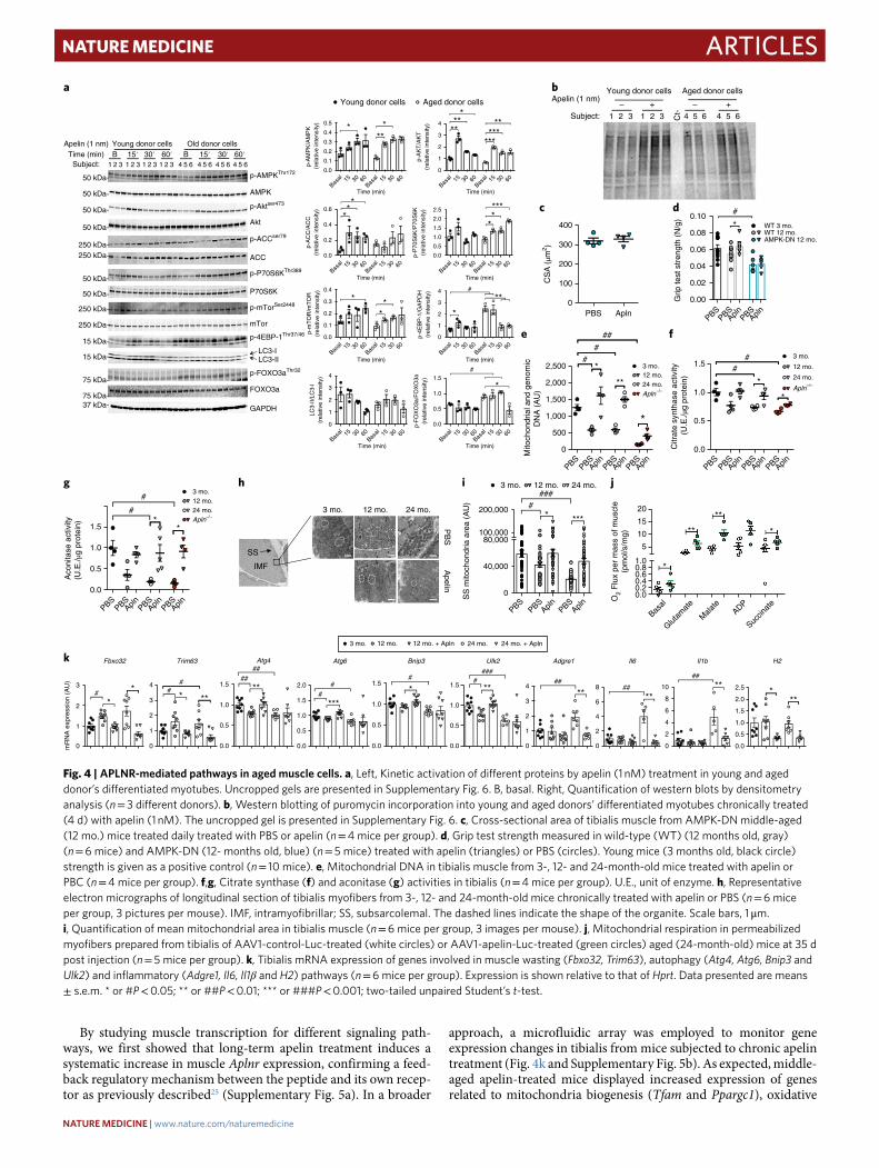
ArticlesNATuRe MediciNe
By studying muscle transcription for different signaling path-ways, we first showed that long-term apelin treatment induces a systematic increase in muscle Aplnr expression, confirming a feed-back regulatory mechanism between the peptide and its own recep-tor as previously described25 (Supplementary Fig. 5a). In a broader
approach, a microfluidic array was employed to monitor gene expression changes in tibialis from mice subjected to chronic apelin treatment (Fig. 4k and Supplementary Fig. 5b). As expected, middle-aged apelin-treated mice displayed increased expression of genes related to mitochondria biogenesis (Tfam and Ppargc1), oxidative
0.0
0.1
0.2
0.3
0.4
0.5
Time (min) Time (min)
Time (min)Time (min)
Time (min) Time (min)
Time (min)Time (min)
*** *
0.0
0.2
0.4
0.6 **
*
0.0
0.1
0.2
0.3
0.4
*
**
0
1
2
3
4
0
1
2
3
4****
*
p-A
KT
/AK
T(r
elat
ive
inte
nsity
)
******
**
0.0
0.5
1.0
1.5
2.0
2.5*
*
***
p-P
70S
6K/P
70S
6K(r
elat
ive
inte
nsity
)
0
1
2
3
4
*
**#
p-4E
BP
-1/G
AP
DH
(rel
ativ
e in
tens
ity)
0.0
0.5
1.0
1.5
*
***p-
FO
XO
3a/F
OX
O3a
(rel
ativ
e in
tens
ity)
#
0.00.20.40.60.81.0
5
10
15
20
O2
Flu
x pe
r m
ass
of m
uscl
e(p
mol
/s/m
g)
*
**
**
*
0.0
0.5
1.0
1.5
Aco
nita
se a
ctiv
ity(U
.E./µ
g pr
otei
n) *#
#
*
0
500
1,000
1,500
2,000
2,500
Mito
chon
dria
l and
gen
omic
DN
A (
AU
)
#*
#
##
**
*
PBS0
100
200
300
400
0.00
0.02
0.04
0.06
0.08
0.10
Grip
test
str
engt
h (N
/g)
*
#
WT 3 mo.WT 12 mo.AMPK-DN 12 mo.
3 mo.12 mo.24 mo.Apln–/–
0.0
0.5
1.0
1.5
Citr
ate
synt
hase
act
ivity
(U
.E./µ
g pr
otei
n) *#
#
*
3 mo.
12 mo.
24 mo.
Apln–/–
3 mo.12 mo.24 mo.Apln–/–
3 mo. 12 mo. 24 mo.
Young donor cells Aged donor cells
0
1
2
3
mR
NA
exp
ress
ion
(AU
)
#
*
Fbxo32
*
0
1
2
3
4
*
Trim63
#**
#
0.0
0.5
1.0
1.5
Atg4
##
**
##
0.0
0.5
1.0
1.5
2.0
Atg6
#
***
#
0.0
0.5
1.0
1.5
Bnip3
#
*
0.0
0.5
1.0
1.5
Ulk2
####
**
0
1
2
3
4
Adgre1
##
**
0
40,000
80,000100,000
200,000
SS
mito
chon
dria
are
a (A
U) #
***
###
*
0
2
4
6
8
Il6
##
**
0
2
4
6
8
10
Il1b
##
**
0.0
0.5
1.0
1.5
2.0
2.5
H2
***
3 mo. 12 mo. 12 mo. + Apln 24 mo. 24 mo. + Apln
Young donor cellsApelin (1 nm)
Subject:
Aged donor cells
– +
1 2 3 1 2 3 4 5 6 4 5 6Ct-
– +
CS
A (
µm2 )
SS
IMF
3 mo. 12 mo. 24 mo.
PB
SA
pelin
a
p-A
MP
K/A
MP
K (
rela
tive
inte
nsity
)
603015Bas
al
Basal 15 30 60 603015
Basal603015
Basal
p-A
CC
/AC
C(r
elat
ive
inte
nsity
)LC
3-II/
LC3-
I(r
elat
ive
inte
nsity
)
p-m
TO
R/m
TO
R(r
elat
ive
inte
nsity
)
603015Bas
al603015Bas
al 603015Bas
al603015Bas
al
603015Bas
al603015Bas
al 603015Bas
al603015Bas
al
603015Bas
al603015Bas
al 603015Bas
al603015Bas
al
b
AplnAplnPBS
AplnPBSPBS
c d
e f
PBSPBS
PBSApln Apln PBS
Apln PBSPBS
PBSApln Apln PBS
Apln
g
AplnPBSAplnPBS
AplnPBSPBS
h
AplnPBSAplnPBS
PBS
i
Succin
ate
Basal
Glutam
ate
Mala
teADP
j
k
50 kDa
50 kDa
50 kDa
50 kDa
250 kDa250 kDa
50 kDa
50 kDa
250 kDa
250 kDa
15 kDa
15 kDa
75 kDa
75 kDa37 kDa
p-AMPKThr172
AMPK
p-Aktser473
p-ACCser79
p-P70S6KThr389
P70S6K
p-mTorSer2448
p-4EBP-1Thr37/46
LC3-ILC3-II
p-FOXO3aThr32
FOXO3a
GAPDH
Akt
ACC
mTor
Young donor cellsApelin (1 nm)Time (min)
Subject: 2 31 2 31 2 31 2 31 5 64 5 64 5 64 5 64
B 15′ 30′ 60′Old donor cells
B 30′ 60′15′
Fig. 4 | APLNr-mediated pathways in aged muscle cells. a, Left, Kinetic activation of different proteins by apelin (1 nM) treatment in young and aged donor’s differentiated myotubes. Uncropped gels are presented in Supplementary Fig. 6. B, basal. Right, Quantification of western blots by densitometry analysis (n = 3 different donors). b, Western blotting of puromycin incorporation into young and aged donors’ differentiated myotubes chronically treated (4 d) with apelin (1 nM). The uncropped gel is presented in Supplementary Fig. 6. c, Cross-sectional area of tibialis muscle from AMPK-DN middle-aged (12 mo.) mice treated daily treated with PBS or apelin (n = 4 mice per group). d, Grip test strength measured in wild-type (WT) (12 months old, gray) (n = 6 mice) and AMPK-DN (12- months old, blue) (n = 5 mice) treated with apelin (triangles) or PBS (circles). Young mice (3 months old, black circle) strength is given as a positive control (n = 10 mice). e, Mitochondrial DNA in tibialis muscle from 3-, 12- and 24-month-old mice treated with apelin or PBC (n = 4 mice per group). f,g, Citrate synthase (f) and aconitase (g) activities in tibialis (n = 4 mice per group). U.E., unit of enzyme. h, Representative electron micrographs of longitudinal section of tibialis myofibers from 3-, 12- and 24-month-old mice chronically treated with apelin or PBS (n = 6 mice per group, 3 pictures per mouse). IMF, intramyofibrillar; SS, subsarcolemal. The dashed lines indicate the shape of the organite. Scale bars, 1 μ m. i, Quantification of mean mitochondrial area in tibialis muscle (n = 6 mice per group, 3 images per mouse). j, Mitochondrial respiration in permeabilized myofibers prepared from tibialis of AAV1-control-Luc-treated (white circles) or AAV1-apelin-Luc-treated (green circles) aged (24-month-old) mice at 35 d post injection (n = 5 mice per group). k, Tibialis mRNA expression of genes involved in muscle wasting (Fbxo32, Trim63), autophagy (Atg4, Atg6, Bnip3 and Ulk2) and inflammatory (Adgre1, Il6, Il1β and H2) pathways (n = 6 mice per group). Expression is shown relative to that of Hprt. Data presented are means ± s.e.m. * or #P < 0.05; ** or ##P < 0.01; *** or ###P < 0.001; two-tailed unpaired Student’s t-test.
NATUrE MEDiCiNE | www.nature.com/naturemedicine

Articles NATuRe MediciNe
damage response (Sod1 (Sox) and Cox5a (Cox)) and metabolism (Prkaa2 (Prkaa), Gsk3b, Srebf1, Slc2a1, Mixl1 (Mixpl), Sirt3, Sirt4 and Sirt5) (Supplementary Fig. 5b). Consistent with a reduction of FOXO-3a hyperphosphorylation in cells from aged donors, apelin blunted Fbxo32 and Trim63 overexpression in chronically treated aged mice (Fig. 4k). Notably, in muscles, apelin also promoted tran-scriptional activation of autophagy genes by increasing expression
of Atg4, Becn1 (Atg6), Bnip3 and Ulk2 (Fig. 4k). However, pharma-cological apelin treatment did not stimulate the transcription of these genes in 24-month-old mice, in which inflammation-related genes (Emr1 (Adgre), Il6, Il1b and H2) were found to be decreased in expression (Fig. 4k) suggesting that apelin could trigger differ-ent mechanisms depending on the age and/or state of the muscle and that a reduction in inflammation could be a prerequisite to the
a
e
j
o p q r s
k l m n
f g h i
b c d
25
1,500 60#
#####
##
######
####
##
#
#
## #
##
###### #####
40
20
0Ctl 3 7 14 3 7 14 3 7 14
D.p.i
Ctl 3 7 14 3 7 14 143 7
3 d.p.i. 7 d.p.i. 14 d.p.i. 3 d.p.i. 7 d.p.i. 14 d.p.i. 3 d.p.i. 7 d.p.i. 14 d.p.i.
CtlPBS
Apln PBSApln PBS
Apln CtlPBS
Apln PBSApln PBS
Apln CtlPBS
Apln PBSApln PBS
Apln
D.p.i
3 mo.
12 mo.
24 mo. Daily i.p. PBS orapelin injection
Regeneration process
3 d.p.i.i.m
. CTX in
jectio
n
7 d.p.i. 14 d.p.i.
**
1,000
500
03 mo. 24 mo.
Unstainedα-APJR-PEα-APJR + α-APJR–PE
19
13
72.99%
6
0
10
5 15
10
5
0
50
PB
SA
pelin
40
30
20
10
Myo
g m
RN
A e
xpre
ssio
nco
mpa
red
to c
ontr
ol (
AU
)
Myf5
mR
NA
exp
ress
ion
com
pare
d to
con
trol
(A
U)
0
4
3
2
1
0
Pax
7 m
RN
A e
xpre
ssio
nco
mpa
red
to c
ontr
ol (
AU
)8
6
4
2
0
3,000 8020 150
25
20
15
10
5
0
>200
200–
400
400–
600
600–
800
800–
1,00
0
1,00
0–1,
200
1,20
0–1,
400
1,40
0–1,
600
1,60
0–1,
800
>100
100–
200
200–
300
300–
400
400–
500
500–
600
600–
700
700–
800
800–
900
900–
1,00
0
1,00
0–1,
100
1,10
0–1,
200
1,20
0–1,
300
1,30
0–1,
400
1,40
0–1,
500
1,50
0–1,
600
1,60
0–1,
700
1,70
0–1,
800
1,80
0–2,
000
2,00
0–2,
200
2,20
0–2,
400
2,40
0–2,
600
2,60
0–2,
800
2,80
0–3,
000
3,00
0–3,
200
3,20
0–3,
400
100
50
0
15
10
5
0
60
40
20
0
40
20100
25
20
15
10
5
0
**
******
Young donor cellsAged donor cells
Myo
cyte
inde
x fu
sion
(%
)
Myo
tube
wid
th (
µm)
80
60
40
20
0
15
10
5
0
**
30
20
10
0
** **
* *
*
**
* *
*
2,000
1,000
0PBS
PB
S
Apln
Ape
lin
PBS Apln
PBSPBS
Apln (1
× 1
0–9 M
)
Apln (1
× 1
0–6 M
)PBS
WT
Aplnr
+/–
Apln–/
–
Apln (1
× 1
0–9 M
)
Apln (1
× 1
0–6 M
)
PBS
Apln
Apelin
PBSApelin
3 d.p.i 7 d.p.i 14 d.p.i 3 d.p.i 7 d.p.i 14 d.p.i
Fiber area (µm2)
Fiber area (µm2)
CtlPBS
Apln PBSApln PBS
Apln CtlPBS
Apln PBSApln PBS
Apln
PE fluorescence intensity
Apln
mR
NA
expr
essi
on in
MuS
C (
AU
)
Apln
mR
NA
exp
ress
ion
com
pare
d to
con
trol
(A
U)
Apln
mR
NA
exp
ress
ion
com
pare
d to
con
trol
(A
U)
Num
ber
of c
ells
Num
ber
of M
uSC
s(n
umbe
r of
PA
X7+
cel
ls/m
m2 )
Cen
tral
nucl
ei–p
ositi
ve fi
bers
(%
)
Cen
tral
nucl
ei–p
ositi
ve fi
bers
(%
)
Per
cent
age
of p
rolif
erat
ing
MuS
Cs
(% P
ax7+
ki67
+ c
ells
)
Myh
8 m
RN
A e
xpre
ssio
nco
mpa
red
to c
ontr
ol (
AU
)
Myh
3 m
RN
A e
xpre
ssio
nco
mpa
red
to c
ontr
ol (
AU
)
eMH
C-p
ositi
ve fi
bers
(%
)
1 ×
106
1 ×
107
PAX-7/DAPI
DAPI/Laminin
PAX-7/Ki-67/DAPI
Fig. 5 | Satellite cells are targeted by apelin during muscle regeneration in aged mice. a, Flow cytometry evaluation of APLNR (APJ) immunoreactivity on PAX-7+ cells in tibialis. Sorted muscle stem cells were unstained as a negative control (black curve) or stained with phycoerythrin (PE)-coupled APLNR antibody (red curve). Specificity of the staining was evaluated by preincubating sorted MuSCs for 30 min with the unlabeled APLNR antibody followed by an incubation with the APLNR-PE coupled antibody (blue curve). b, Aplnr mRNA expression of PAX-7+ cells in tibialis of young and aged mice (n = 6 mice per group). c, Protocol of cardiotoxin (CTX)-induced muscle regeneration experiments. d.p.i., days post injection. d,e, Expression of Apln (d) and Aplnr (e) mRNA in tibialis of 3-, 12- and 24-month-old mice (black, gray and white circles, respectively; n = 5 mice per group). Blue circles represent the mice not treated by cardiotoxin. f–h, Pax7 (f), Myf5 (g) and Myog (h) expression in tibialis of 24-month-old mice treated with apelin (triangles) or PBS (circles) (n = 6 mice per group). Blue circles represent the mice not treated by cardiotoxin and/or apelin. i, Representative immunofluorescence pictures of PAX-7 and PAX-7 and Ki-67 labeling in tibialis of cardiotoxin-injected aged mice (24 months old) treated with apelin or PBS. Yellow arrows represent the proliferative PAX-7–Ki67-double-positive cells. Scale bars, 25 μ m. (n = 5 mice per group, 1 picture per mouse). j,k, Number of PAX-7+ (j) and PAX-7+Ki-67+ (k) cells in tibialis of cardiotoxin-injected aged mice (24 months old) treated with apelin (0.5 μ mol/kg/day) or PBS over 7 d (n = 7 mice per group). l,m, Myh3 (l) and Myh8 (m) expression in tibialis of 24-month-old mice treated with apelin (triangles) or PBS (circles) (n = 6 mice per group). Blue circles represent the mice not treated by cardiotoxin and/or apelin. n, Distribution of embryonic myosin heavy chain (eMHC)-positive fibers in 24-month-old mice treated with apelin (hatched bars) or PBS (white bars) (n = 4 mice per group). o, Representative immunofluorescence labeling of laminin and DAPI for central nuclei quantification. White arrowheads indicate the presence of central nuclei (n = 4 mice per group, 3 pictures per mouse). Scale bars, 70 μ m. p, Mean percentage (n = 5 mice for PBS, n = 7 mice for Apln) (p) and distribution (q) of fibers with central nuclei (n = 4 mice) in 24-month-old mice treated with apelin (triangles) or PBS (circles). r, Myocyte index fusion calculated in young (black bars) and aged (white bar) donor muscle cells treated with apelin or PBS (1 × 10⁻⁹ and 1 × 10⁻⁶ mol/L) over 10 d (n = 3 independent experiments, 3 donors per group). s, Determination of myotube width in primary culture from WT (black circles), Aplnr–/+ (orange circles) and Apln−/− mice (red circles) hindlimb (n = 3 mice per group, 3 wells per mouse, 3 pictures per well). Apln mRNA expression is presented relative to Hprt expression (b,d,e,f,g,h,l,m). Data presented are means ± s.e.m. * or #P < 0.05; ** or ##P < 0.01; *** or ###P < 0.001; two-tailed unpaired Student’s t-test.
NATUrE MEDiCiNE | www.nature.com/naturemedicine

ArticlesNATuRe MediciNe
promotion of other pathways that enhance muscle physiology dur-ing late phases of aging.
Apelin targets muscle stem cells to enhance muscle regeneration. In adults, muscle stem cells (MuSCs) are quiescent but become acti-vated and proliferate to generate myoblasts that fuse into myofibers in response to physiological or pathological stimuli (for example, stretching, exercise or mechanical injury). Loss of MuSC function is a hallmark of aging and can emerge as defects in the maintenance of quiescence, self-renewal ability or activation–proliferation poten-tial44,45. In the context of a regulatory loop involving apelin, exercise and myogenesis, we hypothesized that apelin potentially plays a role in muscle regeneration during aging. By taking advantage of Pax7-ZsGreen reporter mice generated by inserting a T2A-ZsGreen-NLS reporter downstream of exon 9 of the Pax7 locus through homol-ogous recombination, we first evaluated the presence of Aplnr on Pax7-positive MuSCs by flow cytometry (Fig. 5a). MuSCs, sorted from young and old mice as previously described45, demonstrated a fourfold reduction in Aplnr expression in MuSCs from old mice (Fig. 5b). Cardiotoxin-mediated regeneration experiments, in which MuSCs are activated to induce the regenerative response (Fig. 5c), revealed a strong upregulation of Apln and Aplnr expression in reconstructing muscle during regeneration, providing evidence for a crucial role of apelin and its receptor in this process (Fig. 5d,e). However, this response was blunted in aged mice (Fig. 5d,e).
We next treated cardiotoxin-injected aged mice with apelin daily and analyzed the consequences of this supplementation on regenera-tion. Compared to controls, mice supplemented pharmacologically
with apelin had increased expression of satellite cell markers, such as Pax7, Myf5 and Myog (Fig. 5f–h). Additionally, apelin treatment of cardiotoxin-injected aged mice increased the number of MuSCs and their proliferative capacity, as shown by increased numbers of PAX-7-positive and PAX-7–Ki67-double-positive MuSCs (Fig. 5i–k). Moreover, apelin was able to significantly enhance myogenic dif-ferentiation and repair (Fig. 5l–s). Indeed, compared to PBS-treated mice, mRNA expression of Myh3 and Myh8 (markers transiently expressed in adult muscles during the regeneration process) was enhanced by apelin treatment (Fig. 5l,m) and associated with a shift toward larger positive embryonic MHC (eMHC) fibers (Fig. 5n), suggesting that the regenerative process was more active. To rein-force these results, we measured the diameter of newly synthesized centrally nucleated fibers, considered to be a marker of the regen-erative processes (Fig. 5o–q) and found that the number of centrally nucleated fibers (Fig. 5p), as well as the area of these regenerating fibers, was higher in apelin-treated mice compared to controls (Fig. 5q). Finally, the enhanced fusion index observed in human differentiated myotubes chronically treated with apelin (Fig. 5r), together with the reduced width of myotubes obtained from Apln−/− or Aplnr+/− MuSC primary cultures (Fig. 5s) definitively supported a prodifferentiating role for the apelin peptide in skeletal muscle.
Measuring apelin to assess the benefit of exercise in older indi-viduals. By acting simultaneously on both muscle fiber and satellite cell differentiation, contraction-induced muscle apelin could help mediate the benefit of physical exercise with increasing age. To eval-uate this hypothesis, we measured plasma apelin immunoreactivity
200
150
100
5050 100 150
Plasma apelin increase (%)
SP
PB
sco
re in
crea
se (
%)
r 2 = 0.3446
P = 0.0001
Young Old
Contraction-induced apelin production
Apelin acts autocrinally onmetabolic and regenerativeprocesses
Loss of contraction-inducedapelin production
Exogenous apelin substitutioninduced metbolic and autophagicand regenerative benefits
Muscle atrophyLoss of strengthSarcopein
Muscle hypertrophyStrength recovery
b
a
Fig. 6 | Apelin is correlated with beneficial exercise in humans. a, Correlation between the capacity of individuals from the LIFE-P cohort to increase their plasma apelin immunoreactivity level between 0 and 6 months (x axis) and the variation of SPPB score during this training phase (y axis). The line indicates linear regression, and the Spearman correlation coefficient is shown. b, Graphic description of apelin regulation and action in sarcopenic muscle. Apelin is produced and secreted by a contractile skeletal muscle. This regulation is dropped during aging, leading to muscle atrophy, loss of strength and age-associated sarcopenia. After exogenous apelin supplementation in aged muscle cells and mice, mitochondria functions are enhanced, as well as inflammation or autophagy. In parallel, apelin also acts on satellite cells to promote the regeneration process.
NATUrE MEDiCiNE | www.nature.com/naturemedicine

Articles NATuRe MediciNe
in a group of elderly people subjected to a 1-year program of regular physical exercise (LIFE-Study46). Compared to their basal level (i.e., before the study) of plasma apelin immunoreactivity, we observed that individuals who displayed increased plasma apelin levels 6 months after the beginning of the trial also demonstrated the most improved Short Physical Performance Battery test (SPPB) score (Fig. 6a).
This result evidences a positive regulatory loop between muscle contraction, apelin and muscle physiology, which may be altered during the aging process (Fig. 6b). More broadly, this result indicated that plasma apelin immunoreactivity level measured after acute physical exercise in the elderly could represent a potential diagnos-tic tool able to predict the benefit of physical exercise. Therefore, apelin supplementation in the elderly subpopulation that demon-strate reduced capacities toward apelin production represents an innovative approach that may partly limit or reverse age-associated muscle desensitization to the benefits of physical exercise.
DiscussionAge-related sarcopenia and loss of muscle strength are important risk factors for disabling conditions, such as cognitive decline, con-gestive heart failure and osteoporosis4,47. It is important to identify and characterize individuals at risk of developing sarcopenia and propose preventive strategies to avert transition to disability and medical institutionalization of older individuals. Our study has revealed the existence of a regulatory feedback mechanism between muscle contraction, apelin production and myogenesis. In addi-tion, we also demonstrated that these mechanisms are altered dur-ing aging, thus potentially contributing to the onset of sarcopenia. Finally, apelin may represent a promising therapeutic target in the field of aging, a potential new tool for the early diagnosis of sarco-penia and a prognostic marker for the benefits of exercise in the elderly.
A crucial role for the apelin peptide in muscle physiology dur-ing aging was first revealed by murine models deficient in apelin or its receptor. In the heart, it has recently been described that apelin deficiency leads to premature cardiac aging48. Herein we found loss of skeletal muscle mass and functional alterations in mice deficient for apelin (whole body and muscle-specific) or its receptor (heter-ologous knockout). As muscle atrophy was not observed in young knockout mice, we hypothesized that apelin-associated changes regarding metabolism, oxidative processes, innervation and angio-genesis were age dependent. It has been previously demonstrated that targeting of skeletal muscle cells by apelin improves metabolic functions through an AMPK–mitochondriogenesis-dependent axis19,49. Additionally, different Aplnr-expressing cell types exhibited apelin-induced activation of intracellular pathways such as AMPK, mTOR, P70S6 kinase and AKT42,43,50. The aging process has already been clearly shown to lead to alteration of mitochondria-related metabolism in muscle and that defects in AMPK, P70S6 kinase and AKT signaling are major mediators of this effect on the mitochon-dria51,52. Our results show that apelin supplementation in aged mice improved various processes related to muscle rejuvenation in an AMPK-dependent manner but without overt overactivation of this pathway. Apelin affected processes including mitochondrial func-tion and biogenesis, antioxidant enzymes, autophagy and inflamma-tion. Furthermore, apelin induced activation of anabolic pathways promoting protein synthesis (AKT, mTOR, P70S6K and 4E-BP1) and has been shown, for what we believe is the first time, to inhibit the activation of age-related proteolysis actors such as FOXO3a in aged, human differentiated myotubes. Together with the reduction of Fbxo32 and Trim63 expression in muscle observed in vivo, this result paves the way for new insight into the signaling pathway of apelin in the field of muscle atrophy. Although other muscle atro-phy-associated processes, such as innervation or angiogenesis36,53, also need to be investigated and could partly explain the observed
effects of apelin, our results highlight two fundamental mechanisms of muscle aging that are targeted by apelin: mitohormesis and pro-teogenesis–proteolysis. Consequently, we hypothesized that apelin could participate in type IIb/x fibers synthesis in glycolytic muscle by decreasing mitochondria-induced damage that is associated with aging, whereas fiber hypertrophy is most likely mediated by the effect of apelin on protein balance in both glycolytic and oxidative twitch muscles.
Our work also described the beneficial effects of apelin during muscle regeneration in aged mice and human cell lines. Apelin trig-gered mechanisms that permit the differentiation of MuSCs and led to an improvement of regenerative processes. It has been previ-ously shown that Gαi signaling acts as a strong inducer of muscle hypertrophy, myoblast differentiation and muscle regeneration54, thus coupling of APLNR to Gαi reinforced our results. By using a model of inducible depletion of satellite cells, it has been previously demonstrated that regenerative processes did not affect sarcopenia in mice16. However, in the context of resistance training associated with fiber damages and regeneration, apelin should promote the potentiation of cell renewal processes, congruent with our results demonstrating lower fatigability when the peptide was administered to mice together in conjunction with physical exercise. Additional experiments are necessary to determine whether the positive regu-latory loop involving muscle contraction, apelin and myogenesis is related to a particular form of exercise and to identify the underly-ing molecular mechanisms initiating the activation of satellite cells by apelin. Regardless, our results are supportive of the hypothesis that apelin both acts on mature muscle fibers to promote trophic benefits and participates in muscle reconstruction during acute phases of regeneration after damage due to different forms of resis-tance training or post trauma.
Contracting muscle cells, of both human and murine origin, pro-duce apelin. Although skeletal muscle has been previously shown to synthesize apelin20,55,56, we have now extended this observation by establishing a link between apelin and exercise and have shown, for we believe the first time, that muscle contraction promotes apelin secretion into bloodstream, although it was previously shown that chronic exercise leads to an increase in plasma apelin with a decrease in soleus apelin57. Our data confirm the increase in plasma and show that mRNA expression of the muscle tissue increases, suggesting that myocyte apelin content was probably emptied by exercise, as seen previously in enterocytes stimulated by glucose58. Basal apelin levels have been shown to be decreased with age in mice48 and humans, when adjusted for sex21, but our current study represents the first report of a reduction in exercise-stimulated muscle apelin production with age. We also show that muscle apelin production correlates with exercise efficiency during aging, indicating a potential use for apelin as a marker of the health benefits of exercise in the elderly. Compared to other exercise-induced muscle markers, such as IL-6 (ref. 59), ape-lin appears to be more specific, and its reduced production with age constitutes a powerful tool to identify muscle weakness. Thus, we propose apelin measurement after acute exercise as a new index of exercise achievement, which will potentially help clinicians better manage the physical exercise practice of the elderly in the future.
However, it is worth remarking that different apelin isoforms (apelin-13, 17 or 36 amino acids) are found in plasma or biological fluids, reflecting the need to better characterize the forms produced by contractile muscle through more accurate techniques in order to validate apelin production as a new biomarker of age-related exercise success. Finally, from a therapeutic perspective, it appears funda-mental to better understand the associated mechanisms regulating muscle contraction and apelin production and how this regulation is altered with age. Supporting this view, mechanic contraction itself could be involved, as cardiomyocyte stretching resulted in apelin production and Aplnr expression60 whereas inflammation and/or senescence could be potent repressors of muscle apelin or APLNR48.
NATUrE MEDiCiNE | www.nature.com/naturemedicine

ArticlesNATuRe MediciNe
Our findings directly implicate apelin signaling as a posi-tive factor relating to the benefit of exercise that deteriorates with aging, suggesting that it represents a target for various aspects in the development of health improvement strategies. To determine which part of the benefit is due to apelin, it will be interesting to perform different types of exercise in absence of apelin and measure the consequence on sarcopenia onset. In addition to approaches incorporating physical activity, which we show can bolster endog-enous apelin, it is possible that various nutritional approaches could be developed to either supply exogenous apelin or even stimulate endogenous production. In addition, development of APLNR ago-nists that provide both long-term activity and muscle specificity warrant further investigation as pharmacological strategies. Apelin, and possibly its receptor, represent future markers as well as thera-peutic targets for strategies that aim to ameliorate health problems related to age-associated sarcopenia.
MethodsMethods, including statements of data availability and any asso-ciated accession codes and references, are available at https://doi.org/10.1038/s41591-018-0131-6.
Received: 2 March 2017; Accepted: 8 June 2018; Published: xx xx xxxx
references 1. Janssen, I., Shepard, D. S., Katzmarzyk, P. T. & Roubenoff, R. The healthcare
costs of sarcopenia in the United States. J. Am. Geriatr. Soc. 52, 80–85 (2004). 2. Han, K. et al. Sarcopenia as a determinant of blood pressure in older
Koreans: findings from the Korea National Health and Nutrition Examination Surveys (KNHANES) 2008-2010. PLoS One 9, e86902 (2014).
3. Anker, S. D., Morley, J. E. & von Haehling, S. Welcome to the ICD-10 code for sarcopenia. J. Cachexia Sarcopenia Muscle 7, 512–514 (2016).
4. Pasco, J. A. et al. Sarcopenia and the common mental disorders: a potential regulatory role of skeletal muscle on brain function? Curr. Osteoporos. Rep. 13, 351–357 (2015).
5. Martinez, B. P. et al. Frequency of sarcopenia and associated factors among hospitalized elderly patients. BMC Musculoskelet. Disord. 16, 108 (2015).
6. Robertson, D. A., Savva, G. M. & Kenny, R. A. Frailty and cognitive impairment—a review of the evidence and causal mechanisms. Ageing Res. Rev. 12, 840–851 (2013).
7. Rockwood, K. & Mitnitski, A. Frailty in relation to the accumulation of deficits. J. Gerontol. A Biol. Sci. Med. Sci. 62, 722–727 (2007).
8. Samper-Ternent, R., Al Snih, S., Raji, M. A., Markides, K. S. & Ottenbacher, K. J. Relationship between frailty and cognitive decline in older Mexican Americans. J. Am. Geriatr. Soc. 56, 1845–1852 (2008).
9. Giannoulis, M. G., Martin, F. C., Nair, K. S., Umpleby, A. M. & Sonksen, P. Hormone replacement therapy and physical function in healthy older men. Time to talk hormones? Endocr. Rev. 33, 314–377 (2012).
10. Hepple, R. T. Mitochondrial involvement and impact in aging skeletal muscle. Front. Aging Neurosci. 6, 211 (2014).
11. Johnson, M. L., Robinson, M. M. & Nair, K. S. Skeletal muscle aging and the mitochondrion. TEM 24, 247–256 (2013).
12. Cartee, G. D., Hepple, R. T., Bamman, M. M. & Zierath, J. R. Exercise promotes healthy aging of skeletal muscle. Cell Metab. 23, 1034–1047 (2016).
13. Ryu, D. et al. Urolithin A induces mitophagy and prolongs lifespan in C. elegans and increases muscle function in rodents. Nat. Med. 22, 879–888 (2016).
14. Snijders, T. et al. The skeletal muscle satellite cell response to a single bout of resistance-type exercise is delayed with aging in men. Age (Dordr.) 36, 9699 (2014).
15. Bernet, J. D. et al. p38 MAPK signaling underlies a cell-autonomous loss of stem cell self-renewal in skeletal muscle of aged mice. Nat. Med. 20, 265–271 (2014).
16. Fry, C. S. et al. Inducible depletion of satellite cells in adult, sedentary mice impairs muscle regenerative capacity without affecting sarcopenia. Nat. Med. 21, 76–80 (2015).
17. Castan-Laurell, I. et al. Apelin, diabetes, and obesity. Endocrine 40, 1–9 (2011).
18. Dray, C. et al. Apelin and APJ regulation in adipose tissue and skeletal muscle of type 2 diabetic mice and humans. Am. J. Physiol. Endocrinol. Metab. 298, E1161–E1169 (2010).
19. Dray, C. et al. Apelin stimulates glucose utilization in normal and obese insulin-resistant mice. Cell Metab. 8, 437–445 (2008).
20. Besse-Patin, A. et al. Effect of endurance training on skeletal muscle myokine expression in obese men: identification of apelin as a novel myokine. Int. J. Obes. (Lond.) 38, 707–713 (2014).
21. Fujie, S. et al. Reduction of arterial stiffness by exercise training is associated with increasing plasma apelin level in middle-aged and older adults. PLoS One 9, e93545 (2014).
22. Newman, A. B. et al. Sarcopenia: alternative definitions and associations with lower extremity function. J. Am. Geriatr. Soc. 51, 1602–1609 (2003).
23. Vellas, B. et al. Mapt Study: a multidomain approach for preventing alzheimer’s disease: design and baseline data. J. Prev. Alzheimers Dis. 1, 13–22 (2014).
24. Kuba, K. et al. Impaired heart contractility in Apelin gene-deficient mice associated with aging and pressure overload. Circ. Res. 101, e32–e42 (2007).
25. Yamamoto, T. et al. Apelin-transgenic mice exhibit a resistance against diet-induced obesity by increasing vascular mass and mitochondrial biogenesis in skeletal muscle. Biochim. Biophys. Acta 1810, 853–862 (2011).
26. Wang, B. et al. Construction and analysis of compact muscle-specific promoters for AAV vectors. Gene Ther. 15, 1489–1499 (2008).
27. Tai, P. W. et al. Differentiation and fiber type-specific activity of a muscle creatine kinase intronic enhancer. Skelet. Muscle 1, 25 (2011).
28. Hauser, M. A. et al. Analysis of muscle creatine kinase regulatory elements in recombinant adenoviral vectors. Mol. Ther. 2, 16–25 (2000).
29. Katugampola, S. D., Maguire, J. J., Matthewson, S. R. & Davenport, A. P. [(125)I]-(Pyr(1))Apelin-13 is a novel radioligand for localizing the APJ orphan receptor in human and rat tissues with evidence for a vasoconstrictor role in man. Br. J. Pharmacol. 132, 1255–1260 (2001).
30. Pitkin, S. L., Maguire, J. J., Bonner, T. I. & Davenport, A. P. International Union of Basic and Clinical Pharmacology. LXXIV. Apelin receptor nomenclature, distribution, pharmacology, and function. Pharmacol. Rev. 62, 331–342 (2010).
31. Yang, P. et al. Elabela/toddler is an endogenous agonist of the apelin APJ receptor in the adult cardiovascular system, and exogenous administration of the peptide compensates for the downregulation of its expression in pulmonary arterial hypertension. Circulation 135, 1160–1173 (2017).
32. Medhurst, A. D. et al. Pharmacological and immunohistochemical characterization of the APJ receptor and its endogenous ligand apelin. J. Neurochem. 84, 1162–1172 (2003).
33. Jia, Y. X. et al. Apelin protects myocardial injury induced by isoproterenol in rats. Regul. Pept. 133, 147–154 (2006).
34. Chun, H. J. et al. Apelin signaling antagonizes Ang II effects in mouse models of atherosclerosis. J. Clin. Invest. 118, 3343–3354 (2008).
35. Jacobs, R. A. et al. Fast-twitch glycolytic skeletal muscle is predisposed to age-induced impairments in mitochondrial function. J. Gerontol. A Biol. Sci. Med. Sci. 68, 1010–1022 (2013).
36. Demontis, F., Piccirillo, R., Goldberg, A. L. & Perrimon, N. Mechanisms of skeletal muscle aging: insights from Drosophila and mammalian models. Dis. Model. Mech. 6, 1339–1352 (2013).
37. Stewart, V. H., Saunders, D. H. & Greig, C. A. Responsiveness of muscle size and strength to physical training in very elderly people: a systematic review. Scand. J. Med. Sci. Sports 24, e1–e10 (2014).
38. Pahor, M. et al. Effect of structured physical activity on prevention of major mobility disability in older adults: the LIFE study randomized clinical trial. JAMA 311, 2387–2396 (2014).
39. Westerblad, H. & Allen, D. G. Emerging roles of ROS/RNS in muscle function and fatigue. Antioxid. Redox Signal. 15, 2487–2499 (2011).
40. Snijders, T. et al. A single bout of exercise activates skeletal muscle satellite cells during subsequent overnight recovery. Exp. Physiol. 97, 762–773 (2012).
41. Allen, D. G., Lamb, G. D. & Westerblad, H. Skeletal muscle fatigue: cellular mechanisms. Physiol. Rev. 88, 287–332 (2008).
42. Masri, B., Morin, N., Cornu, M., Knibiehler, B. & Audigier, Y. Apelin (65–77) activates p70 S6 kinase and is mitogenic for umbilical endothelial cells. FASEB J. 18, 1909–1911 (2004).
43. Xie, F. et al. Apelin-13 promotes cardiomyocyte hypertrophy via PI3K–Akt–ERK1/2–p70S6K and PI3K-induced autophagy. Acta Biochim. Biophys. Sin. (Shanghai) 47, 969–980 (2015).
44. García-Prat, L., Sousa-Victor, P. & Muñoz-Cánoves, P. Functional dysregulation of stem cells during aging: a focus on skeletal muscle stem cells. FEBS J. 280, 4051–4062 (2013).
45. Lukjanenko, L. et al. Loss of fibronectin from the aged stem cell niche affects the regenerative capacity of skeletal muscle in mice. Nat. Med. 22, 897–905 (2016).
46. Pahor, M. et al. Effects of a physical activity intervention on measures of physical performance: results of the lifestyle interventions and independence for Elders Pilot (LIFE-P) study. J. Gerontol. A Biol. Sci. Med. Sci. 61, 1157–1165 (2006).
47. Papachristou, E. et al. The relationships between body composition characteristics and cognitive functioning in a population-based sample of older British men. BMC Geriatr. 15, 172 (2015).
NATUrE MEDiCiNE | www.nature.com/naturemedicine
Articles NATuRe MediciNe
48. Rai, R. et al. Downregulation of the apelinergic axis accelerates aging, whereas its systemic restoration improves the mammalian healthspan. Cell Rep. 21, 1471–1480 (2017).
49. Attané, C. et al. Apelin stimulates glucose uptake but not lipolysis in human adipose tissue ex vivo. J. Mol. Endocrinol. 46, 21–28 (2011).
50. Zhang, H. et al. Apelin inhibits the proliferation and migration of rat PASMCs via the activation of PI3K/Akt/mTOR signal and the inhibition of autophagy under hypoxia. J. Cell. Mol. Med. 18, 542–553 (2014).
51. Paturi, S. et al. Effects of aging and gender on muscle mass and regulation of Akt–mTOR–p70s6k related signaling in the F344BN rat model. Mech. Ageing Dev. 131, 202–209 (2010).
52. Sandri, M. et al. Signalling pathways regulating muscle mass in ageing skeletal muscle: the role of the IGF1–Akt–mTOR–FoxO pathway. Biogerontology 14, 303–323 (2013).
53. Ambrose, C. Muscle weakness during aging: a deficiency state involving declining angiogenesis. Ageing Res. Rev. 23(Pt B), 139–153 (2015).
54. Minetti, G. C. et al. Gαi2 signaling is required for skeletal muscle growth, regeneration, and satellite cell proliferation and differentiation. Mol. Cell. Biol. 34, 619–630 (2014).
55. Bertrand, C. et al. Effects of dietary eicosapentaenoic acid (EPA) supplementation in high-fat fed mice on lipid metabolism and apelin/APJ system in skeletal muscle. PLoS One 8, e78874 (2013).
56. Yue, P. et al. Apelin is necessary for the maintenance of insulin sensitivity. Am. J. Physiol. Endocrinol. Metab. 298, E59–E67 (2010).
57. Son, J. S. et al. Effects of exercise-induced apelin levels on skeletal muscle and their capillarization in type 2 diabetic rats. Muscle Nerve 56, 1155–1163 (2017).
58. Dray, C. et al. The intestinal glucose–apelin cycle controls carbohydrate absorption in mice. Gastroenterology 144, 771–780 (2013).
59. Pedersen, B. K. & Fischer, C. P. Beneficial health effects of exercise—the role of IL-6 as a myokine. Trends Pharmacol. Sci. 28, 152–156 (2007).
60. Chandrasekaran, B. et al. Myocardial apelin production is reduced in humans with left ventricular systolic dysfunction. J. Card. Fail. 16, 556–561 (2010).
AcknowledgementsWe thank V. Minville, I. Castan-Laurell, A. Yart, B. Masri, and L. Casteilla for their fruitful discussions. We also specially thank all of the personnel of the ANEXPLO animal facility (Toulouse, France) and transcriptomic GeTQ plateform (Toulouse, France);
J. Rouquette, head of the ITAV Imaging Service (Centre Pierre Potier, Toulouse, France); Federico S. and the NIHS flow cytometry facility (Lausanne, Switzerland). We thank J. Iacovani and J. Christensen for corrections to the article and M. Rossell for technical assistance. Mice deficient for AMPK activity (DN-AMPK) in skeletal muscles were kindly provided by the laboratory of M. J. Birnbaum (University of Pennsylvania Medical School, Philadelphia, USA). This work has been funded by INSERM (Institut National de la Santé et de la Recherche Médicale), the Région Occitanie and the Fondation de la Recherche Médicale (FRM). This project was supported in part by European funds (Fonds Européens de Développement Régional, FEDER), Toulouse Métropole, and the French Ministry of Research through the Investissement d’Avenir Infrastructures Nationales en Biologie et Santé program (ProFI, Proteomics French Infrastructure project, ANR-10-INBS-08).
Author contributionsC.D. and P.V. conceived the study. C.V., S.L.G., A.D., O.P. and S.D. performed all animal experiments. C.V., L.L., S.K., U.L. and J.F.N. designed, performed and analyzed the regeneration experiments. C.D., C.V. and J.-P.P. performed all the western blots. C.V., A.D., O.P. and N.G. performed all the transcriptomics. V.M. and A.B. provided human cells. A.B. performed all the culture cell experiments. B.V., M.C., M.P., F.P. and S.G. were involved in human samples collection and analysis. A.C. and A.F.P. performed the hindlimb unloading experiments. M.C., K.C. and O.S. designed and performed the HPLC experiments. M.V. analyzed muscle fiber composition. E.M. participated in performing the specific muscle contraction tests. C.D. supervised the design and execution of the study, interpreted the results and wrote the manuscript.
Competing interestsThe authors declare no competing interests.
Additional informationSupplementary information is available for this paper at https://doi.org/10.1038/s41591-018-0131-6.
Reprints and permissions information is available at www.nature.com/reprints.
Correspondence and requests for materials should be addressed to C.D.
Publisher’s note: Springer Nature remains neutral with regard to jurisdictional claims in published maps and institutional affiliations.
NATUrE MEDiCiNE | www.nature.com/naturemedicine

ArticlesNATuRe MediciNe
MethodsHuman cohorts. We determined the minimum number of participants according to the plasma apelin variability published by Castan-Laurell et al.61. A minimum sample size of 17 participants per group is necessary to detect a statistical difference of 0.1 ng/ml in apelin concentration between the groups, assuming α = 0.05, β = 0.90 and s.d. within each group = 0.02.
Multidomain Alzheimer Preventive Trial. Study design. MAPT23 was a phase 3, multicentered, randomized, placebo-controlled trial using a four-arm design. The study protocol has been approved by the French Ethical Committee located in Toulouse (CPP SOOM II) at the date of December 6, 2007 and was authorized by French health authority (Ministry of Health) on December 31, 2007. Written consent was obtained from all participants. The protocol is registered on a public-access clinical trial database (NCT00672685).
Participants. With respect to Newman’s index to characterize sarcopenia (see Supplementary Table 1), we only considered samples obtained before the intervention and collected 27 individuals without muscle weakness and 33 individuals with sarcopenia.
Lifestyle Interventions and Independence for Elders Pilot. Study Design. LIFE-P38 was a single-blind, multicentered, randomized controlled trial of physical activity or health education intervention in sedentary older adults. The study protocol was approved by the institutional review boards of the University of Florida and Wake Forest University Health Sciences. Participants gave written informed consent. The protocol is consistent with the principles of the Declaration of Helsinki and is registered at www.clinicaltrials.gov (NCT00116194). In our study, apelin measurements were taken from 34 individuals who performed physical exercise. SPPB score (based on chair stands, balance, and walk tests)was obtained as previously described.
Plasma sample assays. Arterial or venous total blood was centrifuged at 14,000 r.p.m., and then serum was collected and frozen at –80 °C. Serum apelin, insulin and leptin concentrations were measured using a nonselective apelin-12 EIA kit that recognizes the C-terminal sequence of 12 amino acids shared among all of the apelin isoforms (Phoenix Pharmaceuticals; Belmont, CA), an ultrasensitive mouse/human insulin ELISA (Mercodia, Uppsala, Sweden) and mouse/human leptin ELISA (R&D system), respectively. Samples for apelin quantification were supplemented with aprotinin (0.6 TIU/ml of total fluid) and purified by a 10-kDa cutoff column before snap freezing (Amicon Ultra, 10,000 molecular weight cutoff, Millipore, Temecula, CA). Plasma glucose was colorimetrically determined by RTU kit (Biomerieux). Nerve growth factor (NGF), IL-6, IL-8, tumor necrosis factor (TNF)-α , MCP-1 and hepatocyte growth factor (HGF) were measured using Luminex technology (Millipore).
Animals. All experimental procedures were performed in accordance with institutional guidelines for animal studies and were approved by an ethics committee (US006/CREFRE, CEEA-122 INSERM). C57Bl6/J WT mice aged 3–24 months were obtained from Janvier Laboratory (St-Berthevin, France). Apelin-deficient (Apln−/−) mice were generated as previously described and backcrossed to C57Bl6/J background mice ten times. Aplnr heterozygous (Aplnr+/−) mice were generated from a cross between C57Bl6/J WT littermates and mice heterozygous for the Aplnr mutation (Deltagen, San Carlos, CA, USA). Mice groups were randomly constituted. Mice were housed conventionally in a constant temperature (20–22 °C) and humidity (50–60%) animal room, with a 12 h–12 h light–dark cycle and free access to food and water. During the different protocols, mice were excluded if they died prematurely or if they displayed tumors after necropsy. All mice were fed a normal diet. All mice were euthanatized in a fed state by cervical vertebra dislocation.
Nanoflow liquid chromatography–tandem mass spectrometry analysis. Blood samples from young (3 months) and aged (24 months) mice were collected using a 22-gauge needle to avoid hemolysis and were immediately centrifuged at 14,000 r.p.m. over 30 min at 4 °C. After the centrifugation, the extracted serum was immediately acidified using HCl (12 M, 10%), and the proteases were neutralized by adding leupeptin (40 μ M), AEBSF-HCl (2 mM), bestatin (100 μ M), E64 (30μ M), EDTA (10 μ M), pepstatin A (100μ M) and aprotinin (5 μ M). The prepared serum was injected on a NanoRS 3500 chromatographic system (Dionex, Amsterdam, The Netherlands) coupled to a Q-Exactive HF-X mass spectrometer (Thermo Fisher Scientific, Bremen, Germany). 5 μ l of sample was loaded on a C18 precolumn (300-μ m inner diameter × 5 mm; Dionex) at 20 μ l/min in 5% acetonitrile, 0.05% TFA. After 3 min of desalting, the precolumn was switched online with the analytical C18 column (75-μ m inner diameter × 50 cm; in-house packed with Reprosil C18) equilibrated in 95% solvent A (5% acetonitrile, 0.2% formic acid) and 5% solvent B (80% acetonitrile, 0.2% formic acid). Peptides were eluted using a 2–40% gradient of solvent B over 60 min at a flow rate of 300 nL/min. Full mass spectrometry (MS) scans were acquired in the Orbitrap on the 350–1500 m/z range with the resolution set to 60,000. An inclusion list corresponding to several charge states (2 + , 3 + , 4 + ) of [Pyr-1]-apelin-13 was used to select these ions for HCD
fragmentation, and the resulting fragment ions were analyzed in the Orbitrap with the resolution set to 15,000. Dynamic exclusion was used within 20 s to prevent repetitive selection of the same peptide.
A standard sample of [Pyr-1]-apelin 13 was injected on a NanoRS 3500 chromatographic system (Dionex, Amsterdam, The Netherlands) coupled to an LTQ-Orbitrap Velos mass spectrometer (Thermo Fisher Scientific, Bremen, Germany). The chromatographic conditions were the same as above for serum samples, except that peptides were eluted using a 5–50% gradient of solvent B over 105 min. The LTQ-Orbitrap Velos was operated in data-dependent acquisition mode with the XCalibur software. Survey scan MS were acquired in the Orbitrap on the 350–1800 m/z range with the resolution set to a value of 60,000. The ten most intense ions per survey scan were selected for collision-induced dissociation (CID) fragmentation, and the resulting fragments were analyzed in the linear trap (LTQ).
The Mascot software (version 2.6.1) was used to automatically extract peak lists from.raw files. Data were searched against Mouse entries of the Swissprot protein database (UniProtKB/Swiss-Prot Knowledgebase release 2018_02, Mouse, https://www.uniprot.org/uniprot/Q9R0R4).
qPCR. Total RNA (1 μ g) was isolated from muscle using GeneJet RNA Purification kit (K0732, Fermentas, Thermo Scientific) and was reverse-transcribed using random hexamers and Superscript II reverse transcriptase (MultiScribe, Applied Biosystems). Real time PCR was performed as previously described starting with 6.25 ng cDNA and both sense and antisense oligonucleotides in a final volume of 10 µ l in a 384-well plate using the SYBR green universal PCR master mix (Eurogentec). Analysis of Hprt (Hypoxanthine-guanine phosphoribosyltransferase) mRNA was performed to normalize gene expression.
Immunohistology. Muscle frozen sections (10 µ m) were immunolabeled for the different myosin heavy chains (MHCs). Primary antibodies were MHC-1 (BA-D5), MHC-2a (SC-71), eMHC (F1.652) from Developmental Studies Hybridoma Bank (DSHB, University of Iowa, IA) and Laminin (Abcam, no. Ab11575). Briefly, sections were blocked for 1 h in PBS plus 4% BSA, 2% goat serum and 0.01% Triton X-100. Sections were then incubated overnight with primary antibodies. After washes in PBS, sections were incubated for 1 h with secondary antibodies anti-Ig2b AF 488, anti-IgG1 AF 546 and anti-rabbit AF 350 (Life Technology). Slides were finally mounted in ProLong Gold antifade Reagent (Molecular probes by Life Technology). Images were captured using a digital camera (Nanozoomer, Hamamatsu) attached to a motorized fluorescence microscope or using Olympus VS120 Virtual Microscopy Slide Scanning System. The area covered by eMHC-positive fibers and the degenerated area were manually determined across the entire sections using the VS-ASW FL software measurement tools. The size of myofibers with central nuclei was calculated from laminin–DAPI staining on all fibers of the section and area determination were performed across the entire sections using an automated image processing algorithm developed internally using the MetaXpress software (Molecular Devices). Finally, virus infection efficiency was assessed by labeling muscle frozen sections with GFP antibody (CS no. 2956, clone D5.1, Cell Signaling) for AAV8-eGFP-tMCK-shApln or with Luciferase pAb (G7451, Promega) for AAV1-apelin-Luc-CMV. Apelin immunostaining in isolated fibers was performed as previously described58.
Human cell culture. Human myoblasts isolated from muscle from three young healthy volunteers (subject 1: 25 years old; subject 2: 16 years old; subject 3: 27 years old) or from 3 aged healthy volunteers (subject 4: 79 years old; subject 5: 68 years old; subject 6: 83 years old) were immortalized as previously described62. Biopsies were provided anonymously by MYOBANK, a tissue bank affiliated to EUROBIOBANK. Human myoblasts were cultured in Skeletal Muscle Cell Media (PromoCell, no. C-23060) supplemented with 20% FBS at 37 °C in a humidified atmosphere containing 5% CO2. When cells were confluent, they were switched to the differentiation medium (DMEM with 50 μ g/ml Gentamicin (Thermo Fischer Scientific, no. 15750045), 10 μ g/ml insulin from bovine pancreas (Sigma-Aldrich, no. I5500), and 100 μ g/ml apo-Transferrin human (Sigma-Aldrich, no. T4382) without any further medium change. Mycoplasma contamination was checked monthly by specific qPCR and DAPI labeling.
Venous–arterial difference. Mice were anesthetized with pentobarbital (0.1 mg/g body weight, CEVA, France) and warmed on a 37 °C isothermal pad. Mice were shaved, and a skin incision was performed in the femoral area in order to identify the femoral vein. For arterial blood collection, carotid arteries were dissected. Indwelling catheters were gently placed in both vessels under microscope and fixed by surgical glue (3M-Vetbond). Sciatic nerve and tibialis tendon were then isolated to allow the electric-mediated (frequency of 75–150 Hz, train of stimulation of 200 msec, every 2 s) isotonic contraction (see the section ‘In situ tibialis anterior and plantaris specific strength’ below). After 10 min of recovery, arterial and venous blood was collected over 10 min through catheters (‘stimulation -’) and kept at 4 °C. Then, 10 min of electric contractions (every 2 seconds) were performed and blood was collected during this time (‘stimulation + ’) and kept at 4 °C.
Acute and chronic physical training. Prior to training, randomized mice were first submitted to the acclimation phase consisting of 5 min of treadmill
NATUrE MEDiCiNE | www.nature.com/naturemedicine

Articles NATuRe MediciNe
exploration (0 cm/second) followed by 5 min at slow speed (10 cm/second). The next day, maximal speed capacity was measured by rising the speed by 5 cm/second every 2 min until exhaustion. Mice were considered to be exhausted when the animal’s hindlimbs remained on the electric grid for more than 10 seconds. Acute exercise consisted of a 30-, 60- or 120-min bout of exercise (65% of maximal speed, 10% slope). Chronic training consisted of a daily 30 min bout of exercise (65% of maximal speed, 10% slope) for 6 d per week over 28 consecutive d.
Muscle atrophy induced by hindlimb unloading. Unloading associated muscle atrophy was performed according to the protocol described by McMahon et al.63. Briefly, tails of the mice were cleaned and were loosely surrounded by adhesive tapes cross-sectionally. A string was fixed at the dorsal side of the tail to maintain blood flow. The string was fastened to a mobile system fixed at the roof of the cage at a height allowing the forelimbs to support the mouse’s body weight yet preventing the hind limbs from touching the floor and the sides of the cage. The mice could reach food and water freely by using their forelimbs. The protocol was carried out over 14 consecutive d.
Body lean mass composition. To determine lean mass, mice were placed in a clear plastic holder, without anesthesia or sedation, and were inserted into the EchoMRI-3-in-1 system (Echo Medical Systems, Houston, TX).
Skeletal muscle function exploration. Time and distance to exhaustion. After two consecutive days of acclimation, running performance was tested in mice by measuring their ability to run until exhaustion. Mice were placed on a treadmill, the speed of which was increased every 2 min by 5 cm/second with the slope at 13%. Mice were considered to be exhausted when the animal’s hindlimbs remained on the electric grid for more than 10 s. Time and distance were automatically collected at the end of the protocol.
Strength by grip test. Mice were allowed to grasp a horizontal grid connected to a dynamometer with all four limbs and were pulled backwards five times. The force applied to the grid each time before the animal lost its grip was recorded in Newton. The average of the five measures is normalized to the whole body weight of each mouse.
Inverted grid test. Mice were placed in the center of the wire grid system, and the grid was rotated to an inverted position over 2 s, with the mouse head declining first. The grid was held steadily 40–50 cm above a padded surface. The grid-hanging time capacity was determined by the time the time the mice spent hanging on the grid.
Maximal speed by running performances. Maximal speed capacity was evaluated by placing mice on a treadmill and raising the treadmill speed 5 cm/second every 2 min until exhaustion. Mice were considered to be exhausted when the animal’s hindlimbs remained on the electric grid for more than 10 s.
In situ tibialis anterior and plantaris specific strength. Mice were anesthetized with pentobarbital i.p. injection (0.1 mg/g body weight; CEVA, France). Muscle distal tendons were isolated from other surrounded muscles, cut and bound with silk attached to a force transducer (50 g and 500 g respectively for plantaris and tibialis; AD Instruments, France) using the PowerLab system (26 T, AD Instruments) and software (LabChart 4, AD Instruments). Bipolar electrical probes were placed on isolated sciatic nerves, which were stimulated using the supramaximal square wave pulse. Force-generation capacity was evaluated by measuring the maximal force generated in response to isometric contraction under electrical stimulation (frequency of 75–150 Hz, train of stimulation of 200 milliseconds). Maximal isometric force was determined at L0 (length at which maximal tension was obtained during the tetanus). Force was normalized to the muscle mass (mg). Fatigue resistance was determined after a 2 minute resting period. Sciatic nerves were stimulated at 10 Hz over 500 milliseconds every 2 seconds for 1 minute. All contractions were made at the initial length L0. Contractions were expressed as a percentage of the initial maximal (isometric) force. Fatigability corresponded to the time allowing 50% decrease of force.
Microscopic image analysis and quantification. For electron microscope analysis, muscle was cut into small pieces and fixed in 1% glutaraldehyde solution. The tissue was then cut and mounted on copper grids and observed with a Hitachi HU 12 A transmission electron microscope equipped with a high-resolution camera. Mitochondria number was determined by counting the number of organelles per field. Mitochondria area was evaluated by measuring the average surface of the organelles per field.
Mitochondrial DNA content. The content of mitochondrial DNA (mtDNA) was calculated using real-time qPCR by measuring the threshold cycle ratio of a mitochondrial encoded gene (mt-Co1; Cox1) and a nuclear-encoded gene (Ppia).
Enzyme activities. Frozen muscle cryosections were dropped into 100 μ l of ice-cold homogenization buffer (0.25 M sucrose, 10 mM Tris, pH 7.4). Following
centrifugation (650 r.p.m., 4 °C, 15 min), supernatant was discarded, and samples were centrifuged again (25,000 r.p.m., 4 °C, 15 min). The pellet was suspended with homogenization buffer, before another centrifugation (25,000 r.p.m., 4 °C, 15 min). Finally, pellet-containing mitochondria were suspended with 0.1 M Tris, pH 8. For enzyme activities, samples were quickly frozen and defrosted three times in liquid nitrogen, centrifuged (5 min, 13,600 r.p.m., 4 °C), then supernatant was kept for enzyme activities. Citrate synthase activity was measured by adding to muscle mitochondria homogenate, 150 μ l of Tris Buffer 100 mM (pH 8), 25 μ l DTNB (1 mM, Sigma D-8130), 40 μ l Acetyl CoA (Sigma A-2056), 10% Triton and 15 μ l of Oxaloacetate 3 mM (Sigma O-4126). Absorbance was read at 412 nm, and citrate activity was assessed in μ mol/min/g = ((Abs/min) × total volume)/13.6 × sample volume × dilution factor). For aconitase activity, muscle mitochondria homogenate was mixed with aconitate (10 mM), Mg2+ (60 mM), β -NADP (0.1 mg/ml), tetrazolium salt (0.1 mg/ml), phenazine methosulfate (25 μ g/ml), Fe(NH4)2(SO4)2 (10μ g/ml), isocitrate dehydrogenase (0.04 U/ml) and Tris (150 mM, pH 8). Sample absorbance, as well as a standard curve of pig aconitase, was read at 575 nm for 30 min at 37 °C.
Muscle-specific apelin deletion. shRNA directed against murine apelin gene (NM_013912.3) was integrated into AAV8 under tMCK promoter dependency coupled with a triple enhancer and with eGFP (AAV8-eGFP-MCK-shApln) (Vector Biolabs). As already demonstrated26–28, high skeletal muscle specificity was obtained by adding a triple tandem of 2RS5 enhancer sequences (3-Ebox) ligated to the truncated regulation region of the MCK promoter, generating the tMCK promoter. The cardiac transcriptional factor Nkx-2.5 does not bind to the E-box, explaining why the multimerized E-boxes, especially the modified 2RS5 E-boxes, increased gene expression in skeletal muscle but not in the heart. Control mice were treated with a similar construction containing a scramble peptide sequence instead of shApln (AAV8-eGFP-MCK-shCt). After titration in mouse, 10-month-old C57Bl6/j randomized mice were submitted to a single retro-orbital injection (5 μ l) of the virus diluted in PBS to a final concentration of 5 × 10¹¹ particles. Forty d.p.i., blood was collected before and after a treadmill single boot exercise in order to measure plasma apelin immunoreactivity. Forty-seven d.p.i., mice were submitted to physical tests to evaluate strength, grid-hanging capacities, time and distance to exhaustion performed on treadmill and maximal speed. Finally, at 54 d.p.i., mice were euthanized, and tissues were collected.
Apelin supplementation. Chronic i.p. injection. 3-, 12- and 24-month-old C57Bl6/j mice, 12-month-old Apln–/–, Aplnr+/– and DN-AMPK mice randomized to different groups according to their weights were daily i.p injected with [pyr1]apelin-13 (Bachem, Switzerland) at 0.5 µ mol/kg/day for 28 d, as previously described. Age = and genotype-matched control mice were injected with PBS. Investigators were blinded to the group allocation (injection of apelin or PBS) during the protocol and the associated experiments. All mice were euthanatized 24 h after the last apelin or PBS injection. The investigators were blinded regarding the treatment.
AAV-mediated skeletal muscle apelin overexpression. Murine apelin sequence (NM_013912.3) was integrated to AAV1 under cytomegalovirus promoter dependency with secretion signal. Apelin was coexpressed with F-Luciferase interconnected by a p2A peptide (AAV1-apelin-Luc). Control mice were injected with a similar construction containing a scramble peptide sequence instead of apelin (AAV1-Luc-Ct). Randomized 24-month-old C57Bl6/j mice were injected in the tibialis anterior and gastrocnemius muscles with 1011 particles of AAV1-apelin-Luciferase in 50 μ l of 1 × PBS. Investigators were blinded to the group allocation during the protocol and the associated experiments. Mice were euthanized 8 weeks after injection.
In vivo animal imaging of luciferase expression. Seven days after AAV injection, mice were anesthetized with 2% isofluorane and oxygen. The d-luciferin substrate (Promega) was injected intraperitoneally at a dose of 150 μ g/g of body weight. The mice were then placed in a light-tight chamber, and images were generated using a cryogenically cooled charge-coupling device camera IVIS 100 (Xenogen, Alameda, CA). For each mouse, the images were taken 8–12 min after the substrate injection. Light and dark images of mice were collected, and the dark images were pseudocolored using the Xenogen software. The visual output represents the number of photons emitted/second/cm2 as a false color image where the maximum is red and the minimum is dark blue.
Western blotting. Muscle and cells samples were lysed (Precellys 24, Ozyme France) and loaded (30 mg protein per lane) on a 4–20% SDS-PAGE gel and transferred to nitrocellulose membrane (GE Healthcare, Life Sciences, Whatman). Membranes were blotted with anti-p-AMPKα Thr172 (CS no. 2535, clone 40H9), AMPKα 2 (CS no. 2757), p-AktSer473 (CS no. 4060, clone D9E), Akt (CS no. 4685, clone 11E7), p-mTORSer2448 (CS no. 5536, clone D9C2), mTOR (CS no. 2983, clone 7C10), p-p70S6 kinaseThr389 (CS no. 9234, clone 108D2), p70S6 kinase (CS no. 9202), p-FoxO1Thr24/FoxO3aThr32 (CS no. 9464), FoxO3a (CS no. 2497), LC3B (CS no. 2775), p- 46-4E-BP1Thr37 (CS no. 2855, clone 236B4), p-Acetyl-CoA CarboxylaseSer79 (CS no. 3661), Acetyl-CoA Carboxylase (CS no. 3662) and GAPDH (CS no. 5174, clone D16H11) (Cell Signaling Technology). MyHCIIa
NATUrE MEDiCiNE | www.nature.com/naturemedicine

ArticlesNATuRe MediciNe
western blotting was performed with MHC-2a (SC-71) antibody Developmental Studies Hybridoma Bank (DSHB, University of Iowa, IA). Immunoreactive proteins were detected using the Clarity Western ECL Substrate (BioRad) and quantified using Image Lab software (BioRad).
Protein synthesis by in vitro SUnSET. The SUnSET assay was used to monitor the rate of protein synthesis. Briefly, 10 min prior harvesting the cells, puromycin was added to culture medium at 1 μ g/ml. As a control, no puromycin was added. Cell extracts were then processed for western blotting using anti-puromycin 12D10 antibody (Millipore).
Mitochondrial respiration. The skeletal muscle tissue was subjected to mechanical dissection with pointed forceps in relaxing buffer on ice. The relaxing buffer contained 2.77 mM CaK2-EGTA, 7.23 mM K2EGTA, 20 mM imidazole, 20 mM taurine, 6.56 mM MgCl2, 5.77 mM ATP, 15 mM phosphocreatine, 0.5 mM dithiothreitol and 50 mM K-MES, pH 7.1. Skeletal muscle fibers were permeabilized by gentle agitation for 30 min at 4 °C in the relaxing solution supplemented with 50 μ g/mL of saponin. Fibers were then twice washed in ice-cold respiration medium. Respiration was measured at 30 °C in a high-resolution respirometer (Oroboros, Oxygraph; Innsbruck, Austria), and data are given as pmol O2/sec/mg (wet weight of muscle). The respiration medium consisted of 110 mM sucrose, 60 mM K-lactobionate, 0.5 mM EGTA, 1 g/L BSA essentially fatty acid free, 3 mM MgCl2, 20 mM taurine, 10 mM KH2PO4 and 20 mM K-HEPES, pH 7.1. The software DatLab (Oroboros Instruments) was used for data analysis. Respiration was assessed with glutamate (10 mM) and malate (2 mM), followed by ADP (3.5 mM). Rotenone (0.5 M) was added to inhibit complex I then succinate (10 mM) was added. Malate and glutamate provide NADH for complex I, succinate provides FADH2 for complex II.
Fluidigm by Biomark qPCR. qPCR was performed with 6.5 ng of cDNA and 100 µ M of both sense and antisense oligonucleotides as already described. The complete list of sequences and the results are available upon request.
Flow cytometry detection of APLNR. The rabbit anti–mouse Aplnr (Abcam ab214369, Cambridge, UK) was conjugated to PE by means of the Lightning Link PE-kit (Innova Biosciences, Cambridge, UK) following the manufacturer’s instructions. Muscle stem cells (MuSCs) were sorted from PAX-7–ZsGreen reporter mice, which were generated by inserting a T2A-ZsGreen-NLS reporter downstream of exon 9 of the Pax7 locus through homologous recombination. In this model, the ZsGreen fluorescent protein is expressed specifically in the nucleus of PAX-7+ muscle stem cells independently of the PAX-7 protein but under the control of the endogenous PAX-7 promoter. Correct transgene insertion was verified by long-range PCR and the neomycin selection cassette was removed through a Flp-mediated recombination system. The reporter was further validated by demonstrating that MuSCs sorted from skeletal muscle based on ZsGreen fluorescence are CD34–SCA1+α 7Int+ by flow cytometry, positive for PAX-7 by qPCR and myogenic ex vivo.
Hind-limb skeletal muscle was digested with Dispase II (2.5 U/ml) (Roche), Collagenase B (0.2%) (Roche) and MgCl2 (5 mM) at 37 °C after tissue mincing, and muscle stem cells were FACS-sorted at 4 °C using a Beckman Coulter Astrios Cell sorter using their positivity for the Zs-Green–PAX-7 reporter. Sorted muscle stem cells were then kept on ice and were left unstained as a negative control or were stained for 30 min on ice with PE-coupled APLNR antibody (APJ–PE; 0.5ug/uL), and washed with HBSS+. Specificity of the staining was evaluated by preincubating sorted MuSCs for 30 min with the unlabeled APLNR antibody followed by a 30 min incubation with the APLNR–PE coupled antibody at 1 μ g/μ L. Samples were then reanalyzed on the Beckman Coulter Astrios Cell sorter for APLNR detection based on PE fluorescence.
Cardiotoxin injury–induced skeletal muscle regeneration. Mice were i.p injected with buprenorphin (Centravet, 0.1 mg/kg) 30 min before injury and the day after. The day of the injury, mice were anesthetized with isoflurane inhalation and hindlimbs were shaved. Then, 10 µ M of cardiotoxin (CTX, Latoxan, no. L8102) was injected through two injections of 25 µ l into left tibialis muscle and two injections of 50 µ l into left gastrocnemius muscle, using a 22-gauge needle (Hamilton). Mice were euthanized 3, 7 and 14 d after injury by cervical dislocation, and muscles (PBS- and CTX-injected) were cut in two parts. One part was snap frozen into liquid nitrogen for total RNA extraction and the other part was embedded into
optimum cutting temperature (OCT) compound and frozen in isopentan cooled with liquid nitrogen for histological analysis.
Histology of muscle stem cells. Mouse muscle samples were dissected and cryopreserved in OCT frozen in liquid nitrogen–cooled isopentan. Samples were then sectioned at 10 μ m on a cryostat and post-fixed with 4% paraformaldehyde (PFA) for 15 min at room temperature. After permeabilization with cold 100% methanol for 6 min at –20 °C, sections were boiled in citrate buffer (10 mM, pH 6.0) for 10 min. Following blocking with 4% BSA and goat anti–mouse Fab (1/100) for 30 min, primary antibodies were incubated at 4 °C overnight (mouse anti-PAX-7 (purified IgG1 from DSHB hybridoma), 1:500 and rabbit anti-Ki-67 polyclonal (Abcam no. ab15580), 1:200). Secondary antibodies (biotinylated goat anti–mouse IgG, 1/1,000; goat anti–rabbit A648 1/2,000) were incubated for 1 h at room temperature. Subsequently, streptavidin–A555 was incubated at 1/2,000 for 30 min to amplify the PAX-7 signal, and Hoechst was used to detect nuclei. The stained sections were mounted, imaged using a 10 × objective on an Olympus VS120 fluorescence slide scanner and quantified manually in three randomly selected injured areas of each samples using the VS-ASV 2.8 software (Olympus). The total number of MuSCs was quantified as the total number PAX-7+DAPI+ nuclei per area analyzed, and the percentage of proliferating MuSCs was quantified as the number of PAX-7+Ki-67+DAPI+ nuclei normalized to the total number of MuSCs.
Mouse muscle stem cell primary culture. WT, Apln–/– and Aplnr–/+ mice were euthanized, and their hindlimbs were rapidly collected and dissected into cold PBS. Then, muscles were digested according to Rosenblatt’s protocol and stem cells (MuSCs) were purified by magnetic beads sorting using a commercial kit (Satellite Cell Isolation Kit, mouse, MACS, Miltenyl Biotec, Germany). MuSCs were then plated on 1% collagen-treated dishes in proliferation medium (40% DMEM, 40% HAM’s medium, 1% penicillin/streptomycin, 20% fetal bovine serum) supplemented with FGF2 (50 μ g/ml). After 5 d, myocytes were differentiated into myotubes adding DMEM with 1% horse serum. After 7 d of differentiation, myotubes were rinsed with PBS and fixed into 4% formalin. Fiber width was determined by averaging 10 different measures all along the myotube in a field of 20 × .
Fusion index. Cells were cultivated over 4 d (until confluence) and treated with apelin or PBS over 10 d. Then cells were fixed with 4% formalin (Sigma). DAPI coloration is done and myocytes index fusion is calculated by dividing the number of nuclei into a myotube by the total number of nuclei in a field × 100.
Statistical analysis. Data are expressed as the mean ± s.e.m. Statistical analyses were performed using Student’s t-test (two-tailed paired or unpaired) or one-way ANOVA followed by the post hoc Dunnett’s and Tukey’s test with GraphPad Prism 5 software (GraphPad Software). P values less than 0.05 (* or #), 0.01 (** or ##) or 0.001 (*** or ###) were statistically significant. A logistic regression was performed to discriminate if age, apelin, BMI and sex were independently correlated to the sarcopenic status. Goodness of fit of the model was assessed using the Hosmer–Lemeshow test. Statistical analysis was realized by MedCalc statistical software (Mariakerke, Belgium).
Reporting Summary. Further information on experimental design is available in the Nature Research Reporting Summary linked to this article.
Data availability. All of the data generated or analyzed during this study are included in this published article (and its Supplementary Information file) and are available from the corresponding author.
references 61. Castan-Laurell, I. et al. Effect of hypocaloric diet-induced weight loss in obese
women on plasma apelin and adipose tissue expression of apelin and APJ. Eur. J. Endocrinol. 158, 905–910 (2008).
62. Mamchaoui, K. et al. Immortalized pathological human myoblasts: towards a universal tool for the study of neuromuscular disorders. Skelet. Muscle 1, 34 (2011).
63. McMahon, C. D. et al. Myostatin-deficient mice lose more skeletal muscle mass than wild-type controls during hindlimb suspension. Am. J. Physiol. Endocrinol. Metab. 285, E82–E87 (2003).
NATUrE MEDiCiNE | www.nature.com/naturemedicine

1
nature research | reporting summ
aryM
arch 2018
Corresponding author(s): DBPR NMED-A85181C
Reporting SummaryNature Research wishes to improve the reproducibility of the work that we publish. This form provides structure for consistency and transparency in reporting. For further information on Nature Research policies, see Authors & Referees and the Editorial Policy Checklist.
Statistical parametersWhen statistical analyses are reported, confirm that the following items are present in the relevant location (e.g. figure legend, table legend, main text, or Methods section).
n/a Confirmed
The exact sample size (n) for each experimental group/condition, given as a discrete number and unit of measurement
An indication of whether measurements were taken from distinct samples or whether the same sample was measured repeatedly
The statistical test(s) used AND whether they are one- or two-sided Only common tests should be described solely by name; describe more complex techniques in the Methods section.
A description of all covariates tested
A description of any assumptions or corrections, such as tests of normality and adjustment for multiple comparisons
A full description of the statistics including central tendency (e.g. means) or other basic estimates (e.g. regression coefficient) AND variation (e.g. standard deviation) or associated estimates of uncertainty (e.g. confidence intervals)
For null hypothesis testing, the test statistic (e.g. F, t, r) with confidence intervals, effect sizes, degrees of freedom and P value noted Give P values as exact values whenever suitable.
For Bayesian analysis, information on the choice of priors and Markov chain Monte Carlo settings
For hierarchical and complex designs, identification of the appropriate level for tests and full reporting of outcomes
Estimates of effect sizes (e.g. Cohen's d, Pearson's r), indicating how they were calculated
Clearly defined error bars State explicitly what error bars represent (e.g. SD, SE, CI)
Our web collection on statistics for biologists may be useful.
Software and codePolicy information about availability of computer code
Data collection No sofware was used
Data analysis No sofware was used
For manuscripts utilizing custom algorithms or software that are central to the research but not yet described in published literature, software must be made available to editors/reviewers upon request. We strongly encourage code deposition in a community repository (e.g. GitHub). See the Nature Research guidelines for submitting code & software for further information.
DataPolicy information about availability of data
All manuscripts must include a data availability statement. This statement should provide the following information, where applicable: - Accession codes, unique identifiers, or web links for publicly available datasets - A list of figures that have associated raw data - A description of any restrictions on data availability
All the data will be available as raw data

2
nature research | reporting summ
aryM
arch 2018
Field-specific reportingPlease select the best fit for your research. If you are not sure, read the appropriate sections before making your selection.
Life sciences Behavioural & social sciences
For a reference copy of the document with all sections, see nature.com/authors/policies/ReportingSummary-flat.pdf
Life sciencesStudy designAll studies must disclose on these points even when the disclosure is negative.
Sample size Mouse sample sized was estimated by pilot experiments that showed trends of effects and their sizes. In most cases, an n=5 was the minimal amount used, only where effect size was large an n=3 yielded significant results. Human sample size See Methods, chapter “human cohorts”. We determined the minimal number of participants according to the plasma apelin variability published by Castan-Laurell et al. (Eur. J. Endocrinol., 2009) We calculated a sample size of 17 participants per group to detect a minimal difference of 0.1ng/ml in apelin concentration between the groups assuming alpha = 0.05, beta = 0.90, SD within each group = 0.02.
Data exclusions Because animals are gaed, mice were excluded for poor body condition (e.g.weight loose, important injuries). Exclusion crieria were pre-estblished before the study.
Replication All replications were successful.
Randomization Mice were randomly allocated to groups. To insure randomization, we weighted animals and distributed them equally in the different groups.
Blinding All the treatements and the functionnal experiments were done by blinded investigators
Materials & experimental systemsPolicy information about availability of materials
n/a Involved in the studyUnique materials
Antibodies
Eukaryotic cell lines
Research animals
Human research participants
Unique materials
Obtaining unique materials All unique materials are available
Antibodies
Antibodies used See Methods, chapter "western blotting" and "immunohistology". All the antibody used are commercial and validated excepted for apelin antibody which has been validated by incubation overnight with the antigenic peptide as demonstrated in Supplementary Figure 1g.
Validation Describe the validation of each primary antibody for the species and application, noting any validation statements on the manufacturer’s website, relevant citations, antibody profiles in online databases, or data provided in the manuscript.
Eukaryotic cell lines
Policy information about cell lines
Cell line source(s) See Methods , chapter "Human cell culture". The human cells are provided by EuroMyobank.
Authentication The cell lines have been described in Mamchaoui, K. et al. Immortalized pathological human myoblasts: towards a universal tool for the study of neuromuscular disorders. Skeletal muscle 1,6 34, doi:10.1186/2044-5040-1-34 (2011)
Mycoplasma contamination All the cell lines have been monthly tested for contamination specially for Mycoplasma by DAPI labelling and qPCR.

3
nature research | reporting summ
aryM
arch 2018Commonly misidentified lines(See ICLAC register)
Name any commonly misidentified cell lines used in the study and provide a rationale for their use.
Research animals
Policy information about studies involving animals; ARRIVE guidelines recommended for reporting animal research
Animals/animal-derived materials Mus musculus, C57Bl6/j background, male, aged from 3 to 24 month old
Human research participants
Policy information about studies involving human research participants
Population characteristics All the caracteristics of the human cohort are listed online MAPT: NCT00672685 LIFE-P: NCT00116194
Method-specific reportingn/a Involved in the study
ChIP-seq
Flow cytometry
Magnetic resonance imaging

147
Bibliography
Bibliography
1. Abou-Khalil, R., Le Grand, F., Pallafacchina, G., Valable, S., Authier, F.-J., Rudnicki, M.A., Gherardi, R.K., Germain, S., Chretien, F., Sotiropoulos, A., et al. (2009). Autocrine and Paracrine Angiopoietin 1/Tie-2 Signaling Promotes Muscle Satellite Cell Self-Renewal. Cell Stem Cell 5, 298–309.
2. Abreu, P. (2018). Bioenergetics mechanisms regulating muscle stem cell self-renewal commitment and function. Biomed. Pharmacother. 103, 463–472.
3. Abreu, P., Mendes, S.V.D., Ceccatto, V.M., and Hirabara, S.M. (2017). Satellite cell activation induced by aerobic muscle adaptation in response to endurance exercise in humans and rodents. Life Sci. 170, 33–40.
4. AbuSamra, D.B. (2015). CD34 Is a Ligand for Vascular Selectins on Human Hematopoietic Stem/Progenitor Cells. Blood 126, 2399–2399.
5. Ahsan, S., Raval, M.H., Ederer, M., Tiwari, R., Chareunsouk, A., and Rodgers, J.T. (2020). Metabolism of glucose and glutamine is critical for skeletal muscle stem cell activation. BioRxiv 2020.07.28.225847.
6. Alfaro, L.A.S., Dick, S.A., Siegel, A.L., Anonuevo, A.S., McNagny, K.M., Megeney, L.A., Cornelison, D.D.W., and Rossi, F.M.V. (2011). CD34 promotes satellite cell motility and entry into proliferation to facilitate efficient skeletal muscle regeneration. Stem Cells Dayt. Ohio 29, 2030–2041.
7. Ancel, S., Stuelsatz, P., and Feige, J.N. (2021). Muscle Stem Cell Quiescence: Controlling Stemness by Staying Asleep. Trends Cell Biol.
8. Armingol, E., Officer, A., Harismendy, O., and Lewis, N.E. (2021). Deciphering cell–cell interactions and communication from gene expression. Nat. Rev. Genet. 22, 71–88.
9. Attane, C., Foussal, C., Le Gonidec, S., Benani, A., Daviaud, D., Wanecq, E., Guzman-Ruiz, R., Dray, C., Bezaire, V., Rancoule, C., et al. (2012). Apelin Treatment Increases Complete Fatty Acid Oxidation, Mitochondrial Oxidative Capacity, and Biogenesis in Muscle of Insulin-Resistant Mice. Diabetes 61, 310–320.
10. Auffray, C., Fogg, D., Garfa, M., Elain, G., Join-Lambert, O., Kayal, S., Sarnacki, S., Cumano, A., Lauvau, G., and Geissmann, F. (2007). Monitoring of Blood Vessels and Tissues by a Population of Monocytes with Patrolling Behavior. Science 317, 666–670.
11. Barnouin, Y., McPhee, J.S., Butler-Browne, G., Bosutti, A., De Vito, G., Jones, D.A., Narici, M., Behin, A., Hogrel, J.-Y., and Degens, H. (2017). Coupling between skeletal muscle fiber size and capillarization is maintained during healthy aging. J. Cachexia Sarcopenia Muscle 8, 647–659.

148
Bibliography
12. Baumgartner-Parzer, S.M., Wagner, L., Pettermann, M., Grillari, J., Gessl, A., and Waldhäusl, W. (1995). High-Glucose–Triggered Apoptosis in Cultured Endothelial Cells. Diabetes 44, 1323–1327.
13. Beauchamp, J.R., Heslop, L., Yu, D.S.W., Tajbakhsh, S., Kelly, R.G., Wernig, A., Buckingham, M.E., Partridge, T.A., and Zammit, P.S. (2000). Expression of Cd34 and Myf5 Defines the Majority of Quiescent Adult Skeletal Muscle Satellite Cells. J. Cell Biol. 151, 1221–1234.
14. Bengal, E., Perdiguero, E., Serrano, A.L., and Muñoz-Cánoves, P. (2017). Rejuvenating stem cells to restore muscle regeneration in aging. F1000Research 6, 76.
15. Benninger, R.K.P., and Piston, D.W. (2013). Two‐Photon Excitation Microscopy for the Study of Living Cells and Tissues. Curr. Protoc. Cell Biol. 59.
16. Bentzinger, C.F., Wang, Y.X., Dumont, N.A., and Rudnicki, M.A. (2013). Cellular dynamics in the muscle satellite cell niche. EMBO Rep. 14, 1062–1072.
17. Berger, M., Hagg, S.A., Goodman, M.N., and Ruderman, N.B. (1976). Glucose metabolism in perfused skeletal muscle. Effects of starvation, diabetes, fatty acids, acetoacetate, insulin and exercise on glucose uptake and disposition. Biochem. J. 158, 191–202.
18. Bertrand, C., Pignalosa, A., Wanecq, E., Rancoule, C., Batut, A., Deleruyelle, S., Lionetti, L., Valet, P., and Castan-Laurell, I. (2013). Effects of dietary eicosapentaenoic acid (EPA) supplementation in high-fat fed mice on lipid metabolism and apelin/APJ system in skeletal muscle. PloS One 8, e78874.
19. Besse-Patin, A., Montastier, E., Vinel, C., Castan-Laurell, I., Louche, K., Dray, C., Daviaud, D., Mir, L., Marques, M.-A., Thalamas, C., et al. (2014). Effect of endurance training on skeletal muscle myokine expression in obese men: identification of apelin as a novel myokine. Int. J. Obes. 38, 707–713.
20. Blanco-Bose, W.E., Yao, C.-C., Kramer, R.H., and Blau, H.M. (2001). Purification of Mouse Primary Myoblasts Based on α7 Integrin Expression. Exp. Cell Res. 265, 212–220.
21. Blau, H.M., Pavlath, G.K., Hardeman, E.C., Chiu, C.-P., Silberstein, L., Webster, S.G., Miller, S.C., and Webster, C. (1985). Plasticity of the Differentiated State. Science 230, 758–766.
22. Blau, H.M., Cosgrove, B.D., and Ho, A.T.V. (2015). The central role of muscle stem cells in regenerative failure with aging. Nat. Med. 21, 854–862.
23. Bloor, C.M. (2005). Angiogenesis during exercise and training. Angiogenesis 8, 263–271.
24. Bockman, E.L. (1983). Blood flow and oxygen consumption in active soleus and gracilis muscles in cats. Am. J. Physiol. 244, H546-551.
25. Boucher, J., Masri, B., Daviaud, D., Gesta, S., Guigné, C., Mazzucotelli, A., Castan-Laurell, I., Tack, I., Knibiehler, B., Carpéné, C., et al. (2005). Apelin, a Newly

149
Bibliography
Identified Adipokine Up-Regulated by Insulin and Obesity. Endocrinology 146, 1764–1771.
26. Bowerman, M., Murray, L.M., Scamps, F., Schneider, B.L., Kothary, R., and Raoul, C. (2018). Pathogenic commonalities between spinal muscular atrophy and amyotrophic lateral sclerosis: Converging roads to therapeutic development. Eur. J. Med. Genet. 61, 685–698.
27. Brack, A.S., and Rando, T.A. (2012). Tissue-specific stem cells: Lessons from the skeletal muscle satellite cell. Cell Stem Cell 10, 504–514.
28. Brack, A.S., Conboy, M.J., Roy, S., Lee, M., Kuo, C.J., Keller, C., and Rando, T.A. (2007). Increased Wnt signaling during aging alters muscle stem cell fate and increases fibrosis. Science 317, 807–810.
29. Bracy, D.P., Zinker, B.A., Jacobs, J.C., Lacy, D.B., and Wasserman, D.H. (1995). Carbohydrate metabolism during exercise: influence of circulating fat availability. J. Appl. Physiol. Bethesda Md 1985 79, 506–513.
30. Brocca, L., McPhee, J.S., Longa, E., Canepari, M., Seynnes, O., De Vito, G., Pellegrino, M.A., Narici, M., and Bottinelli, R. (2017). Structure and function of human muscle fibres and muscle proteome in physically active older men: Skeletal muscle adaptations in human ageing. J. Physiol. 595, 4823–4844.
31. Broholm, C., Mortensen, O.H., Nielsen, S., Akerstrom, T., Zankari, A., Dahl, B., and Pedersen, B.K. (2008). Exercise induces expression of leukaemia inhibitory factor in human skeletal muscle. J. Physiol. 586, 2195–2201.
32. Brooks, G.A. (1997). Importance of the “crossover” concept in exercise metabolism. Clin. Exp. Pharmacol. Physiol. 24, 889–895.
33. Brooks, G.A. (1998). Mammalian fuel utilization during sustained exercise. Comp. Biochem. Physiol. B Biochem. Mol. Biol. 120, 89–107.
34. Brzoska, E., Kowalewska, M., Markowska-Zagrajek, A., Kowalski, K., Archacka, K., Zimowska, M., Grabowska, I., Czerwińska, A.M., Czarnecka-Góra, M., Stremińska, W., et al. (2012). Sdf-1 (CXCL12) improves skeletal muscle regeneration via the mobilisation of Cxcr4 and CD34 expressing cells. Biol. Cell 104, 722–737.
35. Buas, M.F., and Kadesch, T. (2010). Regulation of skeletal myogenesis by Notch. Exp. Cell Res. 316, 3028–3033.
36. Buckingham, M. (2007). Skeletal muscle progenitor cells and the role of Pax genes. C. R. Biol. 330, 530–533.
37. Buranasin, P., Mizutani, K., Iwasaki, K., Pawaputanon Na Mahasarakham, C., Kido, D., Takeda, K., and Izumi, Y. (2018). High glucose-induced oxidative stress impairs proliferation and migration of human gingival fibroblasts. PLoS ONE 13, e0201855.
38. Burzyn, D., Kuswanto, W., Kolodin, D., Shadrach, J.L., Cerletti, M., Jang, Y., Sefik, E., Tan, T.G., Wagers, A.J., Benoist, C., et al. (2013). A Special Population of Regulatory T Cells Potentiates Muscle Repair. Cell 155, 1282–1295.

150
Bibliography
39. Butler, A., Hoffman, P., Smibert, P., Papalexi, E., and Satija, R. (2018). Integrating single-cell transcriptomic data across different conditions, technologies, and species. Nat. Biotechnol. 36, 411–420.
40. Cafforio, G., Pistolesi, S., D’Avino, C., Galluzzi, F., Patricelli, A., Solito, B., Fontanini, G., and Siciliano, G. (2005). Inclusion body myopathy associated with motor neuron syndrome: three case reports. Clin. Neuropathol. 24, 36–41.
41. Carbone, J.W., McClung, J.P., and Pasiakos, S.M. (2012). Skeletal Muscle Responses to Negative Energy Balance: Effects of Dietary Protein. Adv. Nutr. 3, 119–126.
42. Castan-laurell, I., Dray, C., Knauf, C., Kunduzova, O., and Valet, P. (2012). Apelin, a promising target for type 2 diabetes treatment? Trends Endocrinol. Metab. 23, 234–241.
43. Castets, P., Ham, D.J., and Rüegg, M.A. (2020). The TOR Pathway at the Neuromuscular Junction: More Than a Metabolic Player? Front. Mol. Neurosci. 13, 162.
44. Catoire, M., Mensink, M., Kalkhoven, E., Schrauwen, P., and Kersten, S. (2014). Identification of human exercise-induced myokines using secretome analysis. Physiol. Genomics 46, 256–267.
45. Cerletti, M., Jang, Y.C., Finley, L.W.S., Haigis, M.C., and Wagers, A.J. (2012). Short-term calorie restriction enhances skeletal muscle stem cell function. Cell Stem Cell 10, 515–519.
46. Chakkalakal, J.V., Jones, K.M., Basson, M.A., and Brack, A.S. (2012). The aged niche disrupts muscle stem cell quiescence. Nature 490, 355–360.
47. Chakkalakal, J.V., Christensen, J., Xiang, W., Tierney, M.T., Boscolo, F.S., Sacco, A., and Brack, A.S. (2014). Early forming label-retaining muscle stem cells require p27kip1 for maintenance of the primitive state. Development 141, 1649–1659.
48. Chang, Y.-H., Tsai, J.-N., Chen, T.-L., Ho, K.-T., Cheng, H.-Y., Hsiao, C.-W., and Shiau, M.-Y. (2019). Interleukin-4 Promotes Myogenesis and Boosts Myocyte Insulin Efficacy. Mediators Inflamm. 2019, e4182015.
49. Chapman, N.A., Dupré, D.J., and Rainey, J.K. (2014). The apelin receptor: Physiology, pathology, cell signalling, and ligand modulation of a peptide-activated class A GPCR. Biochem. Cell Biol. Biochim. Biol. Cell. 92, 431–440.
50. Chargé, S.B.P., and Rudnicki, M.A. (2004). Cellular and Molecular Regulation of Muscle Regeneration. Physiol. Rev. 84, 209–238.
51. Charnaux, N., Brule, S., Hamon, M., Chaigneau, T., Saffar, L., Prost, C., Lievre, N., and Gattegno, L. (2005). Syndecan-4 is a signaling molecule for stromal cell-derived factor-1 (SDF-1)/ CXCL12. FEBS J. 272, 1937–1951.
52. Chazaud, B. (2020). Inflammation and Skeletal Muscle Regeneration: Leave It to the Macrophages! Trends Immunol. 41, 481–492.

151
Bibliography
53. Chen, S.-E., Jin, B., and Li, Y.-P. (2007a). TNF-alpha regulates myogenesis and muscle regeneration by activating p38 MAPK. Am. J. Physiol. Cell Physiol. 292, C1660-1671.
54. Chen, Y.-H., Lin, S.-J., Lin, F.-Y., Wu, T.-C., Tsao, C.-R., Huang, P.-H., Liu, P.-L., Chen, Y.-L., and Chen, J.-W. (2007b). High Glucose Impairs Early and Late Endothelial Progenitor Cells by Modifying Nitric Oxide–Related but Not Oxidative Stress–Mediated Mechanisms. Diabetes 56, 1559–1568.
55. Choi, J., Rakhilin, N., Gadamsetty, P., Joe, D.J., Tabrizian, T., Lipkin, S.M., Huffman, D.M., Shen, X., and Nishimura, N. (2018). Intestinal crypts recover rapidly from focal damage with coordinated motion of stem cells that is impaired by aging. Sci. Rep. 8.
56. Choudhury, R.R., Hyvola, N., and Lowe, M. (2005). Phosphoinositides and membrane traffic at the trans-Golgi network. Biochem. Soc. Symp. 31–38.
57. Christov, C., Chrétien, F., Abou-Khalil, R., Bassez, G., Vallet, G., Authier, F.-J., Bassaglia, Y., Shinin, V., Tajbakhsh, S., Chazaud, B., et al. (2007a). Muscle Satellite Cells and Endothelial Cells: Close Neighbors and Privileged Partners. Mol. Biol. Cell 18, 1397–1409.
58. Christov, C., Chrétien, F., Abou-Khalil, R., Bassez, G., Vallet, G., Authier, F.-J., Bassaglia, Y., Shinin, V., Tajbakhsh, S., Chazaud, B., et al. (2007b). Muscle Satellite Cells and Endothelial Cells: Close Neighbors and Privileged Partners. Mol. Biol. Cell 18, 1397–1409.
59. Clark, M.G., Rattigan, S., Clerk, L.H., Vincent, M.A., Clark, A.D., Youd, J.M., and Newman, J.M. (2000). Nutritive and non-nutritive blood flow: rest and exercise. Acta Physiol. Scand. 168, 519–530.
60. Clarke, M. (2010). Muscle sliding filaments: Sliding filament model for muscle contraction. Nat. Rev. Mol. Cell Biol. 9, s7–s7.
61. Clifford, P.S., Kluess, H.A., Hamann, J.J., Buckwalter, J.B., and Jasperse, J.L. (2006). Mechanical compression elicits vasodilatation in rat skeletal muscle feed arteries. J. Physiol. 572, 561–567.
62. Collins, B.C., Mader, T.L., Cabelka, C.A., Iñigo, M.R., Spangenburg, E.E., and Lowe, D.A. (2018). Deletion of estrogen receptor α in skeletal muscle results in impaired contractility in female mice. J. Appl. Physiol. 124, 980–992.
63. Collins, B.C., Arpke, R.W., Larson, A.A., Baumann, C.W., Xie, N., Cabelka, C.A., Nash, N.L., Juppi, H.-K., Laakkonen, E.K., Sipilä, S., et al. (2019). Estrogen Regulates the Satellite Cell Compartment in Females. Cell Rep. 28, 368-381.e6.
64. Collins, P.J., McCully, M.L., Martínez‐Muñoz, L., Santiago, C., Wheeldon, J., Caucheteux, S., Thelen, S., Cecchinato, V., Laufer, J.M., Purvanov, V., et al. (2017). Epithelial chemokine CXCL14 synergizes with CXCL12 via allosteric modulation of CXCR4. FASEB J. 31, 3084–3097.

152
Bibliography
65. Conboy, I.M., and Rando, T.A. (2002). The Regulation of Notch Signaling Controls Satellite Cell Activation and Cell Fate Determination in Postnatal Myogenesis. Dev. Cell 3, 397–409.
66. Contreras, O., Rebolledo, D.L., Oyarzún, J.E., Olguín, H.C., and Brandan, E. (2016). Connective tissue cells expressing fibro/adipogenic progenitor markers increase under chronic damage: relevance in fibroblast-myofibroblast differentiation and skeletal muscle fibrosis. Cell Tissue Res. 364, 647–660.
67. Cornier, M.-A., Dabelea, D., Hernandez, T.L., Lindstrom, R.C., Steig, A.J., Stob, N.R., Van Pelt, R.E., Wang, H., and Eckel, R.H. (2008). The metabolic syndrome. Endocr. Rev. 29, 777–822.
68. Cosgrove, B.D., Sacco, A., Gilbert, P.M., and Blau, H.M. (2009). A home away from home: challenges and opportunities in engineering in vitro muscle satellite cell niches. Differ. Res. Biol. Divers. 78, 185–194.
69. Cosgrove, B.D., Gilbert, P.M., Porpiglia, E., Mourkioti, F., Lee, S.P., Corbel, S.Y., Llewellyn, M.E., Delp, S.L., and Blau, H.M. (2014a). Rejuvenation of the aged muscle stem cell population restores strength to injured aged muscles. Nat. Med. 20, 255–264.
70. Cosgrove, B.D., Gilbert, P.M., Porpiglia, E., Mourkioti, F., Lee, S.P., Corbel, S.Y., Llewellyn, M.E., Delp, S.L., and Blau, H.M. (2014b). Rejuvenation of the muscle stem cell population restores strength to injured aged muscles. Nat. Med. 20, 255–264.
71. Covault, J., and Sanes, J.R. (1986). Distribution of N-CAM in synaptic and extrasynaptic portions of developing and adult skeletal muscle. J. Cell Biol. 102, 716–730.
72. Crane, J.D., MacNeil, L.G., Lally, J.S., Ford, R.J., Bujak, A.L., Brar, I.K., Kemp, B.E., Raha, S., Steinberg, G.R., and Tarnopolsky, M.A. (2015). Exercise-stimulated interleukin-15 is controlled by AMPK and regulates skin metabolism and aging. Aging Cell 14, 625–634.
73. Cruz Hernández, J.C., Bracko, O., Kersbergen, C.J., Muse, V., Haft-Javaherian, M., Berg, M., Park, L., Vinarcsik, L.K., Ivasyk, I., Rivera, D.A., et al. (2019). Neutrophil adhesion in brain capillaries reduces cortical blood flow and impairs memory function in Alzheimer’s disease mouse models. Nat. Neurosci. 22, 413–420.
74. De Micheli, A.J., Laurilliard, E.J., Heinke, C.L., Ravichandran, H., Fraczek, P., Soueid-Baumgarten, S., De Vlaminck, I., Elemento, O., and Cosgrove, B.D. (2020). Single-Cell Analysis of the Muscle Stem Cell Hierarchy Identifies Heterotypic Communication Signals Involved in Skeletal Muscle Regeneration. Cell Rep. 30, 3583-3595.e5.
75. Deyo, R.A., and Mirza, S.K. (2016). Herniated Lumbar Intervertebral Disk. N. Engl. J. Med. 374, 1763–1772.
76. Dorrens, J., and Rennie, M.J. (2003). Effects of ageing and human whole body and muscle protein turnover: Ageing and human whole body and muscle protein turnover. Scand. J. Med. Sci. Sports 13, 26–33.

153
Bibliography
77. Drouin, G., Couture, V., Lauzon, M.-A., Balg, F., Faucheux, N., and Grenier, G. (2019). Muscle injury-induced hypoxia alters the proliferation and differentiation potentials of muscle resident stromal cells. Skelet. Muscle 9, 18.
78. D’Souza, D.M., Al-Sajee, D., and Hawke, T.J. (2013). Diabetic myopathy: impact of diabetes mellitus on skeletal muscle progenitor cells. Front. Physiol. 4.
79. Duan, C., Ren, H., and Gao, S. (2010). Insulin-like growth factors (IGFs), IGF receptors, and IGF-binding proteins: Roles in skeletal muscle growth and differentiation. Gen. Comp. Endocrinol. 167, 344–351.
80. Dumont, N.A., Wang, Y.X., von Maltzahn, J., Pasut, A., Bentzinger, C.F., Brun, C.E., and Rudnicki, M.A. (2015). Dystrophin expression in muscle stem cells regulates their polarity and asymmetric division. Nat. Med. 21, 1455–1463.
81. Duscha, B.D., Kraus, W.E., Keteyian, S.J., Sullivan, M.J., Green, H.J., Schachat, F.H., Pippen, A.M., Brawner, C.A., Blank, J.M., and Annex, B.H. (1999). Capillary density of skeletal muscle. J. Am. Coll. Cardiol. 33, 1956–1963.
82. Eadie, B.D., Redila, V.A., and Christie, B.R. (2005). Voluntary exercise alters the cytoarchitecture of the adult dentate gyrus by increasing cellular proliferation, dendritic complexity, and spine density. J. Comp. Neurol. 486, 39–47.
83. Egerman, M.A., and Glass, D.J. (2014). Signaling pathways controlling skeletal muscle mass. Crit. Rev. Biochem. Mol. Biol. 49, 59–68.
84. Egginton, S. (2001). Unorthodox angiogenesis in skeletal muscle. Cardiovasc. Res. 49, 634–646.
85. Englund, D.A., Murach, K.A., Dungan, C.M., Figueiredo, V.C., Vechetti, I.J., Dupont-Versteegden, E.E., McCarthy, J.J., and Peterson, C.A. (2020). Depletion of resident muscle stem cells negatively impacts running volume, physical function, and muscle fiber hypertrophy in response to lifelong physical activity. Am. J. Physiol. Cell Physiol. 318, C1178–C1188.
86. Ennen, J.P., Verma, M., and Asakura, A. (2013). Vascular-targeted therapies for Duchenne muscular dystrophy. Skelet. Muscle 3, 9.
87. Farmawati, A., Kitajima, Y., Nedachi, T., Sato, M., Kanzaki, M., and Nagatomi, R. (2013). Characterization of contraction-induced IL-6 up-regulation using contractile C2C12 myotubes. Endocr. J. 60, 137–147.
88. Flück, M., and Hoppeler, H. (2003). Molecular basis of skeletal muscle plasticity-from gene to form and function. In Reviews of Physiology, Biochemistry and Pharmacology, (Berlin, Heidelberg: Springer Berlin Heidelberg), pp. 159–216.
89. Frontera, W.R., and Ochala, J. (2015). Skeletal muscle: a brief review of structure and function. Calcif. Tissue Int. 96, 183–195.
90. Fry, C.S., Lee, J.D., Mula, J., Kirby, T.J., Jackson, J.R., Liu, F., Yang, L., Mendias, C.L., Dupont-Versteegden, E.E., McCarthy, J.J., et al. (2015). Inducible depletion of satellite cells in adult, sedentary mice impairs muscle regenerative capacity without affecting sarcopenia. Nat. Med. 21, 76–80.

154
Bibliography
91. Fuchs, E., and Blau, H.M. (2020). Tissue Stem Cells: Architects of Their Niches. Cell Stem Cell 27, 532–556.
92. Fulco, M., Cen, Y., Zhao, P., Hoffman, E.P., McBurney, M.W., Sauve, A.A., and Sartorelli, V. (2008). Glucose Restriction Inhibits Skeletal Myoblast Differentiation by Activating SIRT1 through AMPK-Mediated Regulation of Nampt. Dev. Cell 14, 661–673.
93. Furuichi, Y., Kawabata, Y., Aoki, M., Mita, Y., Fujii, N.L., and Manabe, Y. (2021). Excess Glucose Impedes the Proliferation of Skeletal Muscle Satellite Cells Under Adherent Culture Conditions. Front. Cell Dev. Biol. 9.
94. Galli, S.J., Borregaard, N., and Wynn, T.A. (2011). Phenotypic and functional plasticity of cells of innate immunity: macrophages, mast cells and neutrophils. Nat. Immunol. 12, 1035–1044.
95. Gates, L.A., Shi, J., Rohira, A.D., Feng, Q., Zhu, B., Bedford, M.T., Sagum, C.A., Jung, S.Y., Qin, J., Tsai, M.-J., et al. (2017). Acetylation on histone H3 lysine 9 mediates a switch from transcription initiation to elongation. J. Biol. Chem. 292, 14456–14472.
96. Gibbs, B.F., Wierecky, J., Welker, P., Henz, B.M., Wolff, H.H., and Grabbe, J. (2001). Human skin mast cells rapidly release preformed and newly generated TNF-α and IL-8 following stimulation with anti-IgE and other secretagogues: Human skin mast cells and TNF-α and IL-8. Exp. Dermatol. 10, 312–320.
97. Girardi, F., Taleb, A., Ebrahimi, M., Datye, A., Gamage, D.G., Peccate, C., Giordani, L., Millay, D.P., Gilbert, P.M., Cadot, B., et al. (2021). TGFβ signaling curbs cell fusion and muscle regeneration. Nat. Commun. 12, 750.
98. Goel, A.J., Rieder, M.-K., Arnold, H.-H., Radice, G.L., and Krauss, R.S. (2017). Niche Cadherins Control the Quiescence-to-Activation Transition in Muscle Stem Cells. Cell Rep. 21, 2236–2250.
99. Gomarasca, M., Banfi, G., and Lombardi, G. (2020). Myokines: The endocrine coupling of skeletal muscle and bone. Adv. Clin. Chem. 94, 155–218.
100. Gon, Y., Maruoka, S., Kishi, H., Kozu, Y., Kazumichi, K., Nomura, Y., Takeshita, I., Oshima, T., and Hashimoto, S. (2017). NDRG1 is important to maintain the integrity of airway epithelial barrier through claudin-9 expression: NDRG1 maintains epithelial barrier integrity. Cell Biol. Int. 41, 716–725.
101. Görgens, S.W., Eckardt, K., Jensen, J., Drevon, C.A., and Eckel, J. (2015). Exercise and Regulation of Adipokine and Myokine Production. Prog. Mol. Biol. Transl. Sci. 135, 313–336.
102. Gorski, T., and De Bock, K. (2019). Metabolic regulation of exercise-induced angiogenesis. Vasc. Biol. 1, H1–H8.
103. Goueli, B.S., Powell, M.B., Finger, E.C., and Pfeffer, S.R. (2012). TBC1D16 is a Rab4A GTPase activating protein that regulates receptor recycling and EGF receptor signaling. Proc. Natl. Acad. Sci. 109, 15787–15792.

155
Bibliography
104. Gratchev, A., Guillot, P., Hakiy, N., Politz, O., Orfanos, C.E., Schledzewski, K., and Goerdt, S. (2001). Alternatively activated macrophages differentially express fibronectin and its splice variants and the extracellular matrix protein betaIG-H3. Scand. J. Immunol. 53, 386–392.
105. Gubelmann, C., Waszak, S.M., Isakova, A., Holcombe, W., Hens, K., Iagovitina, A., Feuz, J., Raghav, S.K., Simicevic, J., and Deplancke, B. (2013). A yeast one‐hybrid and microfluidics‐based pipeline to map mammalian gene regulatory networks. Mol. Syst. Biol. 9, 682.
106. Halevy, O., Piestun, Y., Allouh, M.Z., Rosser, B.W.C., Rinkevich, Y., Reshef, R., Rozenboim, I., Wleklinski-Lee, M., and Yablonka-Reuveni, Z. (2004). Pattern of Pax7 expression during myogenesis in the posthatch chicken establishes a model for satellite cell differentiation and renewal. Dev. Dyn. Off. Publ. Am. Assoc. Anat. 231, 489–502.
107. Hamann, J.J., Buckwalter, J.B., and Clifford, P.S. (2004). Vasodilatation is obligatory for contraction-induced hyperaemia in canine skeletal muscle. J. Physiol. 557, 1013–1020.
108. Han, S., Wang, G., Qi, X., Lee, H.M., Englander, E.W., and Greeley, G.H. (2008a). A possible role for hypoxia-induced apelin expression in enteric cell proliferation. Am. J. Physiol.-Regul. Integr. Comp. Physiol. 294, R1832–R1839.
109. Han, S., Wang, G., Qi, X., Lee, H.M., Englander, E.W., and Greeley, G.H. (2008b). A possible role for hypoxia-induced apelin expression in enteric cell proliferation. Am. J. Physiol.-Regul. Integr. Comp. Physiol. 294, R1832–R1839.
110. Hänninen, O., and Atalay, M. (1998). Oxidative metabolism in skeletal muscle. In Oxidative Stress in Skeletal Muscle, A.Z. Reznick, L. Packer, C.K. Sen, J.O. Holloszy, and M.J. Jackson, eds. (Basel: Birkhäuser), pp. 29–42.
111. Hawley, J.A., Tipton, K.D., and Millard-Stafford, M.L. (2006). Promoting training adaptations through nutritional interventions. J. Sports Sci. 24, 709–721.
112. Hempel, A., Maasch, C., Heintze, U., Lindschau, C., Dietz, R., Luft, F.C., and Haller, H. (1997). High Glucose Concentrations Increase Endothelial Cell Permeability via Activation of Protein Kinase Cα. Circ. Res. 81, 363–371.
113. Hens, K., Feuz, J.-D., Isakova, A., Iagovitina, A., Massouras, A., Bryois, J., Callaerts, P., Celniker, S.E., and Deplancke, B. (2011a). Automated protein-DNA interaction screening of Drosophila regulatory elements. Nat. Methods 8, 1065–1070.
114. Hens, K., Feuz, J.-D., Isakova, A., Iagovitina, A., Massouras, A., Bryois, J., Callaerts, P., Celniker, S.E., and Deplancke, B. (2011b). Automated protein-DNA interaction screening of Drosophila regulatory elements. Nat. Methods 8, 1065–1070.
115. Hernandez-Torres, F., Rodríguez-Outeiriño, L., Franco, D., and Aranega, A.E. (2017). Pitx2 in Embryonic and Adult Myogenesis. Front. Cell Dev. Biol. 5, 46.
156
Bibliography
116. Hirayama, J., Yamagata, M., Ogata, S., Shimizu, K., Ikeda, Y., and Takahashi, K. (2006). Relationship between low-back pain, muscle spasm and pressure pain thresholds in patients with lumbar disc herniation. Eur. Spine J. 15, 41–47.
117. Hjorth, M., Norheim, F., Meen, A.J., Pourteymour, S., Lee, S., Holen, T., Jensen, J., Birkeland, K.I., Martinov, V.N., Langleite, T.M., et al. (2015). The effect of acute and long-term physical activity on extracellular matrix and serglycin in human skeletal muscle. Physiol. Rep. 3, e12473.
118. Ho, T.K., Tsui, J., Xu, S., Leoni, P., Abraham, D.J., and Baker, D.M. (2010). Angiogenic effects of stromal cell-derived factor-1 (SDF-1/CXCL12) variants in vitro and the in vivo expressions of CXCL12 variants and CXCR4 in human critical leg ischemia. J. Vasc. Surg. 51, 689–699.
119. Huh, H.D., Kim, D.H., Jeong, H.-S., and Park, H.W. (2019). Regulation of TEAD Transcription Factors in Cancer Biology. Cells 8.
120. Hurley, M.V. (1999). THE ROLE OF MUSCLE WEAKNESS IN THE PATHOGENESIS OF OSTEOARTHRITIS. Rheum. Dis. Clin. N. Am. 25, 283–298.
121. Huxley, A.F., and Niedergerke, R. (1954). Structural Changes in Muscle During Contraction: Interference Microscopy of Living Muscle Fibres. Nature 173, 971–973.
122. Irintchev, A., Zeschnigk, M., Starzinski-Powitz, A., and Wernig, A. (1994). Expression pattern of M-cadherin in normal, denervated, and regenerating mouse muscles. Dev. Dyn. 199, 326–337.
123. Janssen, I., Shepard, D.S., Katzmarzyk, P.T., and Roubenoff, R. (2004). The healthcare costs of sarcopenia in the United States. J. Am. Geriatr. Soc. 52, 80–85.
124. Japp, A.G., and Newby, D.E. (2008). The apelin–APJ system in heart failure: Pathophysiologic relevance and therapeutic potential. Biochem. Pharmacol. 75, 1882–1892.
125. Jejurikar, S.S., and Kuzon, Jr., W.M. (2003). Satellite cell depletion in degenerative skeletal muscle. Apoptosis 8, 573–578.
126. Jeon, K., Kim, T., and Lee, S.-H. (2016). Effects of muscle extension strength exercise on trunk muscle strength and stability of patients with lumbar herniated nucleus pulposus. J. Phys. Ther. Sci. 28, 1418–1421.
127. Jesse, T.L., LaChance, R., Iademarco, M.F., and Dean, D.C. (1998). Interferon Regulatory Factor-2 Is a Transcriptional Activator in Muscle Where It Regulates Expression of Vascular Cell Adhesion Molecule-1. J. Cell Biol. 140, 1265–1276.
128. Joe, A.W.B., Yi, L., Natarajan, A., Le Grand, F., So, L., Wang, J., Rudnicki, M.A., and Rossi, F.M.V. (2010). Muscle injury activates resident fibro/adipogenic progenitors that facilitate myogenesis. Nat. Cell Biol. 12, 153–163.

157
Bibliography
129. Jones, J.S., Small, D.M., and Nishimura, N. (2018). In Vivo Calcium Imaging of Cardiomyocytes in the Beating Mouse Heart With Multiphoton Microscopy. Front. Physiol. 9, 969.
130. Jørgensen, L.H., Jepsen, P.L., Boysen, A., Dalgaard, L.B., Hvid, L.G., Ørtenblad, N., Ravn, D., Sellathurai, J., Møller-Jensen, J., Lochmüller, H., et al. (2017). SPARC Interacts with Actin in Skeletal Muscle in Vitro and in Vivo. Am. J. Pathol. 187, 457–474.
131. Joshi, S., Davidson, G., Le Gras, S., Watanabe, S., Braun, T., Mengus, G., and Davidson, I. (2017). TEAD transcription factors are required for normal primary myoblast differentiation in vitro and muscle regeneration in vivo. PLOS Genet. 13, e1006600.
132. Kachhap, S.K., Faith, D., Qian, D.Z., Shabbeer, S., Galloway, N.L., Pili, R., Denmeade, S.R., DeMarzo, A.M., and Carducci, M.A. (2007). The N-Myc down regulated Gene1 (NDRG1) Is a Rab4a effector involved in vesicular recycling of E-cadherin. PloS One 2, e844.
133. Kadoglou, N.P.E., Vrabas, I.S., Kapelouzou, A., Lampropoulos, S., Sailer, N., Kostakis, A., Liapis, C.D., and Angelopoulou, N. (2012). The impact of aerobic exercise training on novel adipokines, apelin and ghrelin, in patients with type 2 diabetes. Med. Sci. Monit. Int. Med. J. Exp. Clin. Res. 18, CR290–CR295.
134. Kanzleiter, T., Rath, M., Görgens, S.W., Jensen, J., Tangen, D.S., Kolnes, A.J., Kolnes, K.J., Lee, S., Eckel, J., Schürmann, A., et al. (2014). The myokine decorin is regulated by contraction and involved in muscle hypertrophy. Biochem. Biophys. Res. Commun. 450, 1089–1094.
135. Karagounis, L.G., and Hawley, J.A. (2010). Skeletal muscle: increasing the size of the locomotor cell. Int. J. Biochem. Cell Biol. 42, 1376–1379.
136. Karlsen, A., Bechshøft, R.L., Malmgaard-Clausen, N.M., Andersen, J.L., Schjerling, P., Kjaer, M., and Mackey, A.L. (2019). Lack of muscle fibre hypertrophy, myonuclear addition, and satellite cell pool expansion with resistance training in 83-94-year-old men and women. Acta Physiol. 227, e13271.
137. Kidoya, H., and Takakura, N. (2012). Biology of the apelin-APJ axis in vascular formation. J. Biochem. (Tokyo) 152, 125–131.
138. Kim, G., and Kim, J.H. (2020). Impact of Skeletal Muscle Mass on Metabolic Health. Endocrinol. Metab. 35, 1–6.
139. Kim, J.-S., Lee, Y.-H., and Yi, H.-K. (2016a). Gradual downhill running improves age-related skeletal muscle and bone weakness: implication of autophagy and bone morphogenetic proteins: Gradual downhill running improves age-related skeletal weakness. Exp. Physiol. 101, 1528–1540.
140. Kim, M., Kim, T., Johnson, R.L., and Lim, D.-S. (2015). Transcriptional Co-repressor Function of the Hippo Pathway Transducers YAP and TAZ. Cell Rep. 11, 270–282.

158
Bibliography
141. Kim, Y.J., Tamadon, A., Park, H.T., Kim, H., and Ku, S.-Y. (2016b). The role of sex steroid hormones in the pathophysiology and treatment of sarcopenia. Osteoporos. Sarcopenia 2, 140–155.
142. Korsunsky, I., Millard, N., Fan, J., Slowikowski, K., Zhang, F., Wei, K., Baglaenko, Y., Brenner, M., Loh, P., and Raychaudhuri, S. (2019a). Fast, sensitive and accurate integration of single-cell data with Harmony. Nat. Methods 16, 1289–1296.
143. Korsunsky, I., Millard, N., Fan, J., Slowikowski, K., Zhang, F., Wei, K., Baglaenko, Y., Brenner, M., and Loh, P. (2019b). Fast, sensitive and accurate integration of single-cell data with Harmony. Nat. Methods.
144. Krauss, R.S., Cole, F., Gaio, U., Takaesu, G., Zhang, W., and Kang, J.-S. (2005). Close encounters: regulation of vertebrate skeletal myogenesis by cell-cell contact. J. Cell Sci. 118, 2355–2362.
145. Krishnasamy, P., Hall, M., and Robbins, S.R. (2018). The role of skeletal muscle in the pathophysiology and management of knee osteoarthritis. Rheumatology 57, iv22–iv33.
146. Kuang, S., and Rudnicki, M.A. (2008). The emerging biology of satellite cells and their therapeutic potential. Trends Mol. Med. 14, 82–91.
147. Kwon, J.H., Moon, K.M., and Min, K.-W. (2020). Exercise-Induced Myokines can Explain the Importance of Physical Activity in the Elderly: An Overview. Healthcare 8, 378.
148. Lakka, H.-M., Laaksonen, D.E., Lakka, T.A., Niskanen, L.K., Kumpusalo, E., Tuomilehto, J., and Salonen, J.T. (2002). The metabolic syndrome and total and cardiovascular disease mortality in middle-aged men. JAMA 288, 2709–2716.
149. Larsson, L., and Ansved, T. (1995). Effects of ageing on the motor unit. Prog. Neurobiol. 45, 397–458.
150. Larsson, L., Sjödin, B., and Karlsson, J. (1978). Histochemical and biochemical changes in human skeletal muscle with age in sedentary males, age 22-65 years. Acta Physiol. Scand. 103, 31–39.
151. Latroche, C., Gitiaux, C., Chrétien, F., Desguerre, I., Mounier, R., and Chazaud, B. (2015). Skeletal Muscle Microvasculature: A Highly Dynamic Lifeline. Physiology 30, 417–427.
152. Latroche, C., Weiss-Gayet, M., Muller, L., Gitiaux, C., Leblanc, P., Liot, S., Ben-Larbi, S., Abou-Khalil, R., Verger, N., Bardot, P., et al. (2017). Coupling between Myogenesis and Angiogenesis during Skeletal Muscle Regeneration Is Stimulated by Restorative Macrophages. Stem Cell Rep. 9, 2018–2033.
153. Lazure, F., Blackburn, D.M., Corchado, A.H., Sahinyan, K., Karam, N., Sharanek, A., Nguyen, D., Lepper, C., Najafabadi, H.S., Perkins, T.J., et al. (2020). Myf6/MRF4 is a myogenic niche regulator required for the maintenance of the muscle stem cell pool. EMBO Rep. 21, e49499.

159
Bibliography
154. Leal, L.G., Lopes, M.A., and Batista, M.L. (2018). Physical Exercise-Induced Myokines and Muscle-Adipose Tissue Crosstalk: A Review of Current Knowledge and the Implications for Health and Metabolic Diseases. Front. Physiol. 9, 1307.
155. Lee, J.H., Budanov, A.V., and Karin, M. (2013). Sestrins Orchestrate Cellular Metabolism to Attenuate Aging. Cell Metab. 18, 792–801.
156. Lehman, W., Craig, R., and Vibert, P. (1994). Ca 2+ -induced tropomyosin movement in Limulus thin filaments revealed by three-dimensional reconstruction. Nature 368, 65–67.
157. Lepper, C., Partridge, T.A., and Fan, C.-M. (2011). An absolute requirement for Pax7-positive satellite cells in acute injury-induced skeletal muscle regeneration. Dev. Camb. Engl. 138, 3639–3646.
158. Li, L., Yang, G., Li, Q., Tang, Y., Yang, M., Yang, H., and Li, K. (2006). Changes and Relations of Circulating Visfatin, Apelin, and Resistin Levels in Normal, Impaired Glucose Tolerance, and Type 2 Diabetic Subjects. Exp. Clin. Endocrinol. Diabetes 114, 544–548.
159. Liang, G., Lin, J.C.Y., Wei, V., Yoo, C., Cheng, J.C., Nguyen, C.T., Weisenberger, D.J., Egger, G., Takai, D., Gonzales, F.A., et al. (2004). Distinct localization of histone H3 acetylation and H3-K4 methylation to the transcription start sites in the human genome. Proc. Natl. Acad. Sci. U. S. A. 101, 7357–7362.
160. Lin, R.-Z., Moreno-Luna, R., Li, D., Jaminet, S.-C., Greene, A.K., and Melero-Martin, J.M. (2014). Human endothelial colony-forming cells serve as trophic mediators for mesenchymal stem cell engraftment via paracrine signaling. Proc. Natl. Acad. Sci. 111, 10137–10142.
161. Lindner, D., Zietsch, C., Becher, P.M., Schulze, K., Schultheiss, H.-P., Tschöpe, C., and Westermann, D. (2012). Differential expression of matrix metalloproteases in human fibroblasts with different origins. Biochem. Res. Int. 2012, 875742.
162. Liu, M., Stevens-Lapsley, J.E., Jayaraman, A., Ye, F., Conover, C., Walter, G.A., Bose, P., Thompson, F.J., Borst, S.E., and Vandenborne, K. (2010). Impact of treadmill locomotor training on skeletal muscle IGF1 and myogenic regulatory factors in spinal cord injured rats. Eur. J. Appl. Physiol. 109, 709–720.
163. Liu, W., Zhang, B., Hu, Q., Qin, Y., Xu, W., Shi, S., Liang, C., Meng, Q., Xiang, J., Liang, D., et al. (2017). A new facet of NDRG1 in pancreatic ductal adenocarcinoma: Suppression of glycolytic metabolism. Int. J. Oncol. 50, 1792–1800.
164. Londhe, P., and Davie, J.K. (2013). Interferon-γ Resets Muscle Cell Fate by Stimulating the Sequential Recruitment of JARID2 and PRC2 to Promoters to Repress Myogenesis. Sci. Signal. 6, ra107–ra107.
165. Low, M., Eisner, C., and Rossi, F. (2017). Fibro/Adipogenic Progenitors (FAPs): Isolation by FACS and Culture. In Muscle Stem Cells: Methods and Protocols, E. Perdiguero, and D. Cornelison, eds. (New York, NY: Springer), pp. 179–189.

160
Bibliography
166. Lukjanenko, L., Brachat, S., Pierrel, E., Lach-Trifilieff, E., and Feige, J.N. (2013). Genomic Profiling Reveals That Transient Adipogenic Activation Is a Hallmark of Mouse Models of Skeletal Muscle Regeneration. PLoS ONE 8, e71084.
167. Lukjanenko, L., Karaz, S., Stuelsatz, P., Gurriaran-Rodriguez, U., Michaud, J., Dammone, G., Sizzano, F., Mashinchian, O., Ancel, S., Migliavacca, E., et al. (2019). Aging Disrupts Muscle Stem Cell Function by Impairing Matricellular WISP1 Secretion from Fibro-Adipogenic Progenitors. Cell Stem Cell 24, 433-446.e7.
168. Luo, W., Ai, L., Wang, B.-F., Wang, L.-Y., Gan, Y.-M., and Zhou, Y. (2020). [Effects of mice macrophages on skeletal muscle cells under high glucose treatment]. Zhongguo Ying Yong Sheng Li Xue Za Zhi Zhongguo Yingyong Shenglixue Zazhi Chin. J. Appl. Physiol. 36, 124–129.
169. Luque, E., Peña, J., Martin, P., Jimena, I., and Vaamonde, R. (1995). Capillary Supply During Development of Individual Regenerating Muscle Fibers. Anat. Histol. Embryol. 24, 87–89.
170. Lv, X., Zheng, B., Li, S., Han, A., Wang, C., Shi, J., Zhang, X., Liu, Y., Li, Y., and Wen, J. (2013). Synthetic retinoid Am80 up-regulates apelin expression by promoting interaction of RARα with KLF5 and Sp1 in vascular smooth muscle cells. Biochem. J. 456, 35–46.
171. Macosko, E.Z., Basu, A., Satija, R., Nemesh, J., Shekhar, K., Goldman, M., Tirosh, I., Bialas, A.R., Kamitaki, N., Martersteck, E.M., et al. (2015). Highly Parallel Genome-wide Expression Profiling of Individual Cells Using Nanoliter Droplets. Cell 161, 1202–1214.
172. Mademtzoglou, D., Asakura, Y., Borok, M.J., Alonso-Martin, S., Mourikis, P., Kodaka, Y., Mohan, A., Asakura, A., and Relaix, F. (2018). Cellular localization of the cell cycle inhibitor Cdkn1c controls growth arrest of adult skeletal muscle stem cells. ELife 7, e33337.
173. Maggio, M., Lauretani, F., and Ceda, G.P. (2012). Sex hormones and sarcopenia in older persons: Curr. Opin. Clin. Nutr. Metab. Care 1.
174. Maloney, P.R., Khan, P., Hedrick, M., Gosalia, P., Milewski, M., Li, L., Roth, G.P., Sergienko, E., Suyama, E., Sugarman, E., et al. (2010). Functional antagonists of the Apelin (APJ) receptor. In Probe Reports from the NIH Molecular Libraries Program, (Bethesda (MD): National Center for Biotechnology Information (US)), p.
175. von Maltzahn, J., Jones, A.E., Parks, R.J., and Rudnicki, M.A. (2013). Pax7 is critical for the normal function of satellite cells in adult skeletal muscle. Proc. Natl. Acad. Sci. 110, 16474–16479.
176. Marcell, T.J. (2003). Sarcopenia: causes, consequences, and preventions. J. Gerontol. A. Biol. Sci. Med. Sci. 58, M911-916.
177. Mashinchian, O., Pisconti, A., Le Moal, E., and Bentzinger, C.F. (2018). Chapter Two - The Muscle Stem Cell Niche in Health and Disease. In Current Topics in Developmental Biology, D. Sassoon, ed. (Academic Press), pp. 23–65.
161
Bibliography
178. Matsakas, A., Yadav, V., Lorca, S., and Narkar, V. (2013). Muscle ERRγ mitigates Duchenne muscular dystrophy via metabolic and angiogenic reprogramming. FASEB J. Off. Publ. Fed. Am. Soc. Exp. Biol. 27, 4004–4016.
179. Mazzucotelli, A., Ribet, C., Castan-Laurell, I., Daviaud, D., Guigné, C., Langin, D., and Valet, P. (2008). The transcriptional co-activator PGC-1alpha up regulates apelin in human and mouse adipocytes. Regul. Pept. 150, 33–37.
180. McGinnis, C.S., Murrow, L.M., and Gartner, Z.J. (2019). DoubletFinder: Doublet Detection in Single-Cell RNA Sequencing Data Using Artificial Nearest Neighbors. Cell Syst. 8, 329-337.e4.
181. McKellar, D.W., Walter, L.D., Song, L.T., Mantri, M., Wang, M.F.Z., Vlaminck, I.D., and Cosgrove, B.D. (2020a). Strength in numbers: Large-scale integration of single-cell transcriptomic data reveals rare, transient muscle progenitor cell states in muscle regeneration. BioRxiv 2020.12.01.407460.
182. McKellar, D.W., Walter, L.D., Song, L.T., Mantri, M., Wang, M.F.Z., De Vlaminck, I., and Cosgrove, B.D. (2020b). Strength in numbers: Large-scale integration of single-cell transcriptomic data reveals rare, transient muscle progenitor cell states in muscle regeneration. BioRxiv 2020.12.01.407460.
183. Melotte, V., Qu, X., Ongenaert, M., van Criekinge, W., de Bruïne, A.P., Baldwin, H.S., and van Engeland, M. (2010). The N-myc downstream regulated gene (NDRG) family: diverse functions, multiple applications. FASEB J. 24, 4153–4166.
184. Migliavacca, E., Tay, S.K.H., Patel, H.P., Sonntag, T., Civiletto, G., McFarlane, C., Forrester, T., Barton, S.J., Leow, M.K., Antoun, E., et al. (2019). Mitochondrial oxidative capacity and NAD + biosynthesis are reduced in human sarcopenia across ethnicities. Nat. Commun. 10, 5808.
185. Miller, W., Rosenbloom, K., Hardison, R.C., Hou, M., Taylor, J., Raney, B., Burhans, R., King, D.C., Baertsch, R., Blankenberg, D., et al. (2007). 28-Way vertebrate alignment and conservation track in the UCSC Genome Browser. Genome Res. 17, 1797–1808.
186. Miranda, M., Morici, J.F., Zanoni, M.B., and Bekinschtein, P. (2019). Brain-Derived Neurotrophic Factor: A Key Molecule for Memory in the Healthy and the Pathological Brain. Front. Cell. Neurosci. 13, 363.
187. Mounier, R., Chrétien, F., and Chazaud, B. (2011). Chapter five - Blood Vessels and the Satellite Cell Niche. In Current Topics in Developmental Biology, G. k. Pavlath, ed. (Academic Press), pp. 121–138.
188. Mul, J.D., Stanford, K.I., Hirshman, M.F., and Goodyear, L.J. (2015). Exercise and Regulation of Carbohydrate Metabolism. In Progress in Molecular Biology and Translational Science, (Elsevier), pp. 17–37.
189. Muñoz-Cánoves, P., Neves, J., and Sousa-Victor, P. (2020). Understanding muscle regenerative decline with aging: new approaches to bring back youthfulness to aged stem cells. FEBS J. 287, 406–416.

162
Bibliography
190. Murphy, M.M., Lawson, J.A., Mathew, S.J., Hutcheson, D.A., and Kardon, G. (2011). Satellite cells, connective tissue fibroblasts and their interactions are crucial for muscle regeneration. Dev. Camb. Engl. 138, 3625–3637.
191. Nalbandian, M., Radak, Z., and Takeda, M. (2020). Lactate Metabolism and Satellite Cell Fate. Front. Physiol. 0.
192. Nazari, A., and Chehelcheraghi, F. (2020). Using Apelin and exercise to protect the cardiac cells: synergic effect in ischemia reperfusion injuries treatment in rats. Bratisl. Med. J. 121, 14–21.
193. Nederveen, J.P., Snijders, T., Joanisse, S., Wavell, C.G., Mitchell, C.J., Johnston, L.M., Baker, S.K., Phillips, S.M., and Parise, G. (2017). Altered muscle satellite cell activation following 16 wk of resistance training in young men. Am. J. Physiol. Regul. Integr. Comp. Physiol. 312, R85–R92.
194. Nyimanu, D., Kay, R.G., Sulentic, P., Kuc, R.E., Ambery, P., Jermutus, L., Reimann, F., Gribble, F.M., Cheriyan, J., Maguire, J.J., et al. (2019). Development and validation of an LC-MS/MS method for detection and quantification of in vivo derived metabolites of [Pyr 1 ]apelin-13 in humans. Sci. Rep. 9, 19934.
195. Obeso, J., Weber, J., and Auerbach, R. (1990). A hemangioendothelioma-derived cell line: its use as a model for the study of endothelial cell biology. Lab. Investig. J. Tech. Methods Pathol. 63, 259–269.
196. Ochoa, O., Sun, D., Reyes-Reyna, S.M., Waite, L.L., Michalek, J.E., McManus, L.M., and Shireman, P.K. (2007). Delayed angiogenesis and VEGF production in CCR2-/- mice during impaired skeletal muscle regeneration. Am. J. Physiol. Regul. Integr. Comp. Physiol. 293, R651-661.
197. Olson, L.E., and Soriano, P. (2009). Increased PDGFRα Activation Disrupts Connective Tissue Development and Drives Systemic Fibrosis. Dev. Cell 16, 303–313.
198. Ouchi, N., Oshima, Y., Ohashi, K., Higuchi, A., Ikegami, C., Izumiya, Y., and Walsh, K. (2008). Follistatin-like 1, a Secreted Muscle Protein, Promotes Endothelial Cell Function and Revascularization in Ischemic Tissue through a Nitric-oxide Synthase-dependent Mechanism. J. Biol. Chem. 283, 32802–32811.
199. Pannérec, A., Springer, M., Migliavacca, E., Ireland, A., Piasecki, M., Karaz, S., Jacot, G., Métairon, S., Danenberg, E., Raymond, F., et al. (2016). A robust neuromuscular system protects rat and human skeletal muscle from sarcopenia. Aging 8, 712–728.
200. Parise, G., and Yarasheski, K.E. (2000). The utility of resistance exercise training and amino acid supplementation for reversing age-associated decrements in muscle protein mass and function. Curr. Opin. Clin. Nutr. Metab. Care 3, 489–495.
201. Pavlou, S., Lindsay, J., Ingram, R., Xu, H., and Chen, M. (2018). Sustained high glucose exposure sensitizes macrophage responses to cytokine stimuli but reduces their phagocytic activity. BMC Immunol. 19, 24.

163
Bibliography
202. Pedersen, B.K. (2013). Muscle as a secretory organ. Compr. Physiol. 3, 1337–1362.
203. Pedersen, B.K., and Febbraio, M.A. (2012). Muscles, exercise and obesity: skeletal muscle as a secretory organ. Nat. Rev. Endocrinol. 8, 457–465.
204. Periasamy, M., Herrera, J.L., and Reis, F.C.G. (2017). Skeletal Muscle Thermogenesis and Its Role in Whole Body Energy Metabolism. Diabetes Metab. J. 41, 327.
205. Pisters, M.F., Veenhof, C., van Dijk, G.M., and Dekker, J. (2014). Avoidance of activity and limitations in activities in patients with osteoarthritis of the hip or knee: a 5 year follow-up study on the mediating role of reduced muscle strength. Osteoarthritis Cartilage 22, 171–177.
206. Pourteymour, S., Eckardt, K., Holen, T., Langleite, T., Lee, S., Jensen, J., Birkeland, K.I., Drevon, C.A., and Hjorth, M. (2017). Global mRNA sequencing of human skeletal muscle: Search for novel exercise-regulated myokines. Mol. Metab. 6, 352–365.
207. Pretheeban, T., Lemos, D.R., Paylor, B., Zhang, R.-H., and Rossi, F.M. (2012). Role of stem/progenitor cells in reparative disorders. Fibrogenesis Tissue Repair 5, 20.
208. Puchert, M., Adams, V., Linke, A., and Engele, J. (2016). Evidence for the involvement of the CXCL12 system in the adaptation of skeletal muscles to physical exercise. Cell. Signal. 28, 1205–1215.
209. Qiu, H., Wang, F., Liu, C., Xu, X., and Liu, B. (2011). TEAD1-dependent expression of the FoxO3a gene in mouse skeletal muscle. BMC Mol. Biol. 12, 1.
210. Rai, R., Ghosh, A.K., Eren, M., Mackie, A.R., Levine, D.C., Kim, S.-Y., Cedernaes, J., Ramirez, V., Procissi, D., Smith, L.H., et al. (2017). Downregulation of the Apelinergic Axis Accelerates Aging, whereas Its Systemic Restoration Improves the Mammalian Healthspan. Cell Rep. 21, 1471–1480.
211. Ramis, M.R., Sarubbo, F., Moranta, D., Tejada, S., Lladó, J., Miralles, A., and Esteban, S. (2021). Neurochemical and Cognitive Beneficial Effects of Moderate Physical Activity and Catechin in Aged Rats. Antioxidants 10, 621.
212. Ratajczak, M.Z. (2003). Expression of Functional CXCR4 by Muscle Satellite Cells and Secretion of SDF-1 by Muscle-Derived Fibroblasts is Associated with the Presence of Both Muscle Progenitors in Bone Marrow and Hematopoietic Stem/Progenitor Cells in Muscles. Stem Cells 21, 363–371.
213. Reilly, G.C., and Engler, A.J. (2010). Intrinsic extracellular matrix properties regulate stem cell differentiation. J. Biomech. 43, 55–62.
214. Relaix, F., Rocancourt, D., Mansouri, A., and Buckingham, M. (2005). A Pax3/Pax7-dependent population of skeletal muscle progenitor cells. Nature 435, 948–953.

164
Bibliography
215. Relaix, F., Bencze, M., Borok, M.J., Der Vartanian, A., Gattazzo, F., Mademtzoglou, D., Perez-Diaz, S., Prola, A., Reyes-Fernandez, P.C., Rotini, A., et al. (2021). Perspectives on skeletal muscle stem cells. Nat. Commun. 12, 692.
216. Rhoads, R.P., Johnson, R.M., Rathbone, C.R., Liu, X., Temm-Grove, C., Sheehan, S.M., Hoying, J.B., and Allen, R.E. (2009). Satellite cell-mediated angiogenesis in vitro coincides with a functional hypoxia-inducible factor pathway. Am. J. Physiol. Cell Physiol. 296, C1321-1328.
217. Rice, W.L., Kaplan, D.L., and Georgakoudi, I. (2010). Two-Photon Microscopy for Non-Invasive, Quantitative Monitoring of Stem Cell Differentiation. PLoS ONE 5, e10075.
218. Ripamonti, U., and Hari Reddi, A. (1997). Tissue Engineering, Morphogenesis, and Regeneration of the Periodontal Tissues By Bone Morphogenetic Proteins. Crit. Rev. Oral Biol. Med. 8, 154–163.
219. Roberts, M.D., Haun, C.T., Vann, C.G., Osburn, S.C., and Young, K.C. (2020). Sarcoplasmic Hypertrophy in Skeletal Muscle: A Scientific “Unicorn” or Resistance Training Adaptation? Front. Physiol. 11, 816.
220. Rose, A.J., and Richter, E.A. (2005). Skeletal Muscle Glucose Uptake During Exercise: How is it Regulated? Physiology 20, 260–270.
221. Rozo, M., Li, L., and Fan, C.-M. (2016). Targeting β1-integrin signaling enhances regeneration in aged and dystrophic muscle in mice. Nat. Med. 22, 889–896.
222. Rubio-Ruiz, M.E., Guarner-Lans, V., Pérez-Torres, I., and Soto, M.E. (2019). Mechanisms Underlying Metabolic Syndrome-Related Sarcopenia and Possible Therapeutic Measures. Int. J. Mol. Sci. 20, E647.
223. Ryall, J.G. (2013). Metabolic reprogramming as a novel regulator of skeletal muscle development and regeneration. FEBS J. 280, 4004–4013.
224. Ryall, J.G., Dell’Orso, S., Derfoul, A., Juan, A., Zare, H., Feng, X., Clermont, D., Koulnis, M., Gutierrez-Cruz, G., Fulco, M., et al. (2015). The NAD(+)-dependent SIRT1 deacetylase translates a metabolic switch into regulatory epigenetics in skeletal muscle stem cells. Cell Stem Cell 16, 171–183.
225. Saber, J., Lin, A.Y.T., and Rudnicki, M.A. (2020). Single-cell analyses uncover granularity of muscle stem cells. F1000Research 9.
226. Saclier, M., Yacoub-Youssef, H., Mackey, A.L., Arnold, L., Ardjoune, H., Magnan, M., Sailhan, F., Chelly, J., Pavlath, G.K., Mounier, R., et al. (2013). Differentially activated macrophages orchestrate myogenic precursor cell fate during human skeletal muscle regeneration. Stem Cells Dayt. Ohio 31, 384–396.
227. Salmon, W.D., and Daughaday, W.H. (1957). A hormonally controlled serum factor which stimulates sulfate incorporation by cartilage in vitro. J. Lab. Clin. Med. 49, 825–836.

165
Bibliography
228. Sambasivan, R., Yao, R., Kissenpfennig, A., Van Wittenberghe, L., Paldi, A., Gayraud-Morel, B., Guenou, H., Malissen, B., Tajbakhsh, S., and Galy, A. (2011). Pax7-expressing satellite cells are indispensable for adult skeletal muscle regeneration. Dev. Camb. Engl. 138, 3647–3656.
229. Sartori, R., Romanello, V., and Sandri, M. (2021). Mechanisms of muscle atrophy and hypertrophy: implications in health and disease. Nat. Commun. 12, 330.
230. Scapini, P., Lapinet-Vera, J.A., Gasperini, S., Calzetti, F., Bazzoni, F., and Cassatella, M.A. (2000). The neutrophil as a cellular source of chemokines. Immunol. Rev. 177, 195–203.
231. Schnoor, M., Cullen, P., Lorkowski, J., Stolle, K., Robenek, H., Troyer, D., Rauterberg, J., and Lorkowski, S. (2008). Production of Type VI Collagen by Human Macrophages: A New Dimension in Macrophage Functional Heterogeneity. J. Immunol. 180, 5707–5719.
232. Schultz, E. (1996). Satellite Cell Proliferative Compartments in Growing Skeletal Muscles. Dev. Biol. 175, 84–94.
233. Shea, K.L., Xiang, W., LaPorta, V.S., Licht, J.D., Keller, C., Basson, M.A., and Brack, A.S. (2010). Sprouty1 Regulates Reversible Quiescence of a Self-Renewing Adult Muscle Stem Cell Pool during Regeneration. Cell Stem Cell 6, 117–129.
234. Shefer, G., Rauner, G., Yablonka-Reuveni, Z., and Benayahu, D. (2010). Reduced Satellite Cell Numbers and Myogenic Capacity in Aging Can Be Alleviated by Endurance Exercise. PLoS ONE 5, e13307.
235. Shorter, E., Sannicandro, A.J., Poulet, B., and Goljanek-Whysall, K. (2019). Skeletal Muscle Wasting and Its Relationship With Osteoarthritis: a Mini-Review of Mechanisms and Current Interventions. Curr. Rheumatol. Rep. 21, 40.
236. Snijders, T., Nederveen, J.P., Joanisse, S., Leenders, M., Verdijk, L.B., van Loon, L.J.C., and Parise, G. (2017). Muscle fibre capillarization is a critical factor in muscle fibre hypertrophy during resistance exercise training in older men. J. Cachexia Sarcopenia Muscle 8, 267–276.
237. Southard, S., Kim, J.-R., Low, S., Tsika, R.W., and Lepper, C. (2016). Myofiber-specific TEAD1 overexpression drives satellite cell hyperplasia and counters pathological effects of dystrophin deficiency. ELife 5, e15461.
238. Stein, C., Bardet, A.F., Roma, G., Bergling, S., Clay, I., Ruchti, A., Agarinis, C., Schmelzle, T., Bouwmeester, T., Schübeler, D., et al. (2015). YAP1 Exerts Its Transcriptional Control via TEAD-Mediated Activation of Enhancers. PLoS Genet. 11.
239. Stein, S., Thomas, E.K., Herzog, B., Westfall, M.D., Rocheleau, J.V., Jackson, R.S., Wang, M., and Liang, P. (2004). NDRG1 Is Necessary for p53-dependent Apoptosis*. J. Biol. Chem. 279, 48930–48940.
240. Stoll, T., Germann, D., and Hagmann, H. (2001). Physiotherapy in lumbar disc herniation. Ther. Umsch. 58, 487–492.

166
Bibliography
241. Stuart, T., Butler, A., Hoffman, P., Hafemeister, C., Papalexi, E., Mauck, W.M., Hao, Y., Stoeckius, M., Smibert, P., and Satija, R. (2019). Comprehensive Integration of Single-Cell Data. Cell 177, 1888-1902.e21.
242. Su, A.I., Wiltshire, T., Batalov, S., Lapp, H., Ching, K.A., Block, D., Zhang, J., Soden, R., Hayakawa, M., Kreiman, G., et al. (2004). A gene atlas of the mouse and human protein-encoding transcriptomes. Proc. Natl. Acad. Sci. U. S. A. 101, 6062–6067.
243. Szigeti, K., and Lupski, J.R. (2009). Charcot–Marie–Tooth disease. Eur. J. Hum. Genet. 17, 703–710.
244. Szokodi, I., Tavi, P., Földes, G., Voutilainen-Myllylä, S., Ilves, M., Tokola, H., Pikkarainen, S., Piuhola, J., Rysä, J., Tóth, M., et al. (2002a). Apelin, the Novel Endogenous Ligand of the Orphan Receptor APJ, Regulates Cardiac Contractility. Circ. Res. 91, 434–440.
245. Szokodi, I., Tavi, P., Földes, G., Voutilainen-Myllylä, S., Ilves, M., Tokola, H., Pikkarainen, S., Piuhola, J., Rysä, J., Tóth, M., et al. (2002b). Apelin, the novel endogenous ligand of the orphan receptor APJ, regulates cardiac contractility. Circ. Res. 91, 434–440.
246. Takahashi, A., Kureishi, Y., Yang, J., Luo, Z., Guo, K., Mukhopadhyay, D., Ivashchenko, Y., Branellec, D., and Walsh, K. (2002). Myogenic Akt Signaling Regulates Blood Vessel Recruitment during Myofiber Growth. Mol. Cell. Biol. 22, 4803–4814.
247. Tanideh, N., Shahin Sheikhani, H., Salesi, M., Tamadon, A., Rostamzad, K., and Kardeh, A. (2014). Effects of endurance exercise and estrogen supplementation on the proliferation of satellite cells. Comp. Clin. Pathol. 23, 1645–1649.
248. Tatemoto, K., Hosoya, M., Habata, Y., Fujii, R., Kakegawa, T., Zou, M.X., Kawamata, Y., Fukusumi, S., Hinuma, S., Kitada, C., et al. (1998). Isolation and characterization of a novel endogenous peptide ligand for the human APJ receptor. Biochem. Biophys. Res. Commun. 251, 471–476.
249. Tatemoto, K., Takayama, K., Zou, M.-X., Kumaki, I., Zhang, W., Kumano, K., and Fujimiya, M. (2001). The novel peptide apelin lowers blood pressure via a nitric oxide-dependent mechanism. Regul. Pept. 99, 87–92.
250. Tedesco, F.S., Dellavalle, A., Diaz-Manera, J., Messina, G., and Cossu, G. (2010). Repairing skeletal muscle: regenerative potential of skeletal muscle stem cells. J. Clin. Invest. 120, 11–19.
251. Theret, M., Gsaier, L., Schaffer, B., Juban, G., Ben Larbi, S., Weiss-Gayet, M., Bultot, L., Collodet, C., Foretz, M., Desplanches, D., et al. (2017). AMPKα1-LDH pathway regulates muscle stem cell self-renewal by controlling metabolic homeostasis. EMBO J. 36, 1946–1962.
252. Theret, M., Rossi, F.M.V., and Contreras, O. (2021). Evolving Roles of Muscle-Resident Fibro-Adipogenic Progenitors in Health, Regeneration, Neuromuscular Disorders, and Aging. Front. Physiol. 0.

167
Bibliography
253. Thomas, K., Engler, A.J., and Meyer, G.A. (2015). Extracellular matrix regulation in the muscle satellite cell niche. Connect. Tissue Res. 56, 1–8.
254. Tidball, J.G. (2017). Regulation of muscle growth and regeneration by the immune system. Nat. Rev. Immunol. 17, 165–178.
255. Tomasek, J.J., Gabbiani, G., Hinz, B., Chaponnier, C., and Brown, R.A. (2002). Myofibroblasts and mechano-regulation of connective tissue remodelling. Nat. Rev. Mol. Cell Biol. 3, 349–363.
256. Trombetta, J.M., Bradshaw, A.D., and Johnson, R.H. (2010). SPARC/Osteonectin Functions to Maintain Homeostasis of the Collagenous Extracellular Matrix in the Periodontal Ligament. J. Histochem. Cytochem. 58, 871–879.
257. Tsika, R.W., Schramm, C., Simmer, G., Fitzsimons, D.P., Moss, R.L., and Ji, J. (2008). Overexpression of TEAD-1 in Transgenic Mouse Striated Muscles Produces a Slower Skeletal Muscle Contractile Phenotype. J. Biol. Chem. 283, 36154–36167.
258. Uchugonova, A., Gorjup, E., Riemann, I., Sauer, D., and König, K. (2008). Two-photon imaging of stem cells. 68601W.
259. Uezumi, A., Fukada, S., Yamamoto, N., Takeda, S., and Tsuchida, K. (2010). Mesenchymal progenitors distinct from satellite cells contribute to ectopic fat cell formation in skeletal muscle. Nat. Cell Biol. 12, 143–152.
260. Uezumi, A., Ito, T., Morikawa, D., Shimizu, N., Yoneda, T., Segawa, M., Yamaguchi, M., Ogawa, R., Matev, M.M., Miyagoe-Suzuki, Y., et al. (2011). Fibrosis and adipogenesis originate from a common mesenchymal progenitor in skeletal muscle. J. Cell Sci. 124, 3654–3664.
261. Urciuolo, A., Quarta, M., Morbidoni, V., Gattazzo, F., Molon, S., Grumati, P., Montemurro, F., Tedesco, F.S., Blaauw, B., Cossu, G., et al. (2013). Collagen VI regulates satellite cell self-renewal and muscle regeneration. Nat. Commun. 4, 1964.
262. Van den Bossche, J., O’Neill, L.A., and Menon, D. (2017). Macrophage Immunometabolism: Where Are We (Going)? Trends Immunol. 38, 395–406.
263. Verma, M., Asakura, Y., Murakonda, B.S.R., Pengo, T., Latroche, C., Chazaud, B., McLoon, L.K., and Asakura, A. (2018a). Muscle Satellite Cell Cross-Talk with a Vascular Niche Maintains Quiescence via VEGF and Notch Signaling. Cell Stem Cell 23, 530-543.e9.
264. Verma, M., Asakura, Y., Murakonda, B.S.R., Pengo, T., Latroche, C., Chazaud, B., McLoon, L.K., and Asakura, A. (2018b). Muscle Satellite Cell Cross-Talk with a Vascular Niche Maintains Quiescence via VEGF and Notch Signaling. Cell Stem Cell 23, 530-543.e9.
265. Vinel, C., Lukjanenko, L., Batut, A., Deleruyelle, S., Pradère, J.-P., Le Gonidec, S., Dortignac, A., Geoffre, N., Pereira, O., Karaz, S., et al. (2018). The exerkine apelin reverses age-associated sarcopenia. Nat. Med. 24, 1360–1371.

168
Bibliography
266. Vinel, C., Schanstra, J.P., Boizard, F., Péreira, O., Auriau, J., Dortignac, A., Breuil, B., Feuillet, G., Nkuipou-Kenfack, E., Zürbig, P., et al. (2019). Apelin affects the mouse aging urinary peptidome with minimal effects on kidney. Sci. Rep. 9, 10647.
267. Wahren, J., Felig, P., Ahlborg, G., and Jorfeldt, L. (1971). Glucose metabolism during leg exercise in man. J. Clin. Invest. 50, 2715–2725.
268. Wang, G., Qi, X., Wei, W., Englander, E.W., and Greeley, G.H. (2006). Characterization of the 5’-regulatory regions of the rat and human apelin genes and regulation of breast apelin by USF. FASEB J. 20, 2639–2641.
269. Wang, S.J., Greer, P., and Auerbach, R. (1996). Isolation and propagation of yolk-sac-derived endothelial cells from a hypervascular transgenic mouse expressing a gain-of-function fps/fes proto-oncogene. In Vitro Cell. Dev. Biol. Anim. 32, 292–299.
270. Wang, X., Shen, Q.W., Wang, J., Zhang, Z., Feng, F., Chen, T., Zhang, Y., Wei, H., Li, Z., Wang, X., et al. (2016). KLF7 Regulates Satellite Cell Quiescence in Response to Extracellular Signaling. Stem Cells Dayt. Ohio 34, 1310–1320.
271. Webster, M.T., Manor, U., Lippincott-Schwartz, J., and Fan, C.-M. (2016a). Intravital Imaging Reveals Ghost Fibers as Architectural Units Guiding Myogenic Progenitors during Regeneration. Cell Stem Cell 18, 243–252.
272. Webster, M.T., Harvey, T., and Fan, C.-M. (2016b). Quantitative 3D Time Lapse Imaging of Muscle Progenitors in Skeletal Muscle of Live Mice. Bio-Protoc. 6, e2066.
273. Whitham, M., and Febbraio, M.A. (2016). The ever-expanding myokinome: discovery challenges and therapeutic implications. Nat. Rev. Drug Discov. 15, 719–729.
274. Williams, B.D., Plag, I., Troup, J., and Wolfe, R.R. (1995). Isotopic determination of glycolytic flux during intense exercise in humans. J. Appl. Physiol. Bethesda Md 1985 78, 483–490.
275. Winbanks, C.E., Chen, J.L., Qian, H., Liu, Y., Bernardo, B.C., Beyer, C., Watt, K.I., Thomson, R.E., Connor, T., Turner, B.J., et al. (2013). The bone morphogenetic protein axis is a positive regulator of skeletal muscle mass. J. Cell Biol. 203, 345–357.
276. Wolfe, R.R. (2006). The underappreciated role of muscle in health and disease. Am. J. Clin. Nutr. 84, 475–482.
277. Wosczyna, M.N., Biswas, A.A., Cogswell, C.A., and Goldhamer, D.J. (2012). Multipotent progenitors resident in the skeletal muscle interstitium exhibit robust BMP-dependent osteogenic activity and mediate heterotopic ossification. J. Bone Miner. Res. Off. J. Am. Soc. Bone Miner. Res. 27, 1004–1017.
278. Wu, C., Macleod, I., and Su, A.I. (2013). BioGPS and MyGene.info: organizing online, gene-centric information. Nucleic Acids Res. 41, D561-565.

169
Bibliography
279. Wu, L., Chen, L., and Li, L. (2017). Apelin/APJ system: A novel promising therapy target for pathological angiogenesis. Clin. Chim. Acta Int. J. Clin. Chem. 466, 78–84.
280. Xu, S., Liu, S., Xu, L., Deng, S., He, Y., Li, S., and Ni, G. (2018). Response of decorin to different intensity treadmill running. Mol. Med. Rep.
281. Xuan, Y.H., Huang, B.B., Tian, H.S., Chi, L.S., Duan, Y.M., Wang, X., Zhu, Z.X., Cai, W.H., Zhu, Y.T., Wei, T.M., et al. (2014). High-Glucose Inhibits Human Fibroblast Cell Migration in Wound Healing via Repression of bFGF-Regulating JNK Phosphorylation. PLoS ONE 9, e108182.
282. Yablonka-Reuveni, Z. (2011). The skeletal muscle satellite cell: still young and fascinating at 50. J. Histochem. Cytochem. Off. J. Histochem. Soc. 59, 1041–1059.
283. Yaffe, D., and Saxel, O. (1977). Serial passaging and differentiation of myogenic cells isolated from dystrophic mouse muscle. Nature 270, 725–727.
284. Yamada, M., Hokazono, C., Tokizawa, K., Marui, S., Iwata, M., Lira, V.A., Suzuki, K., Miura, S., Nagashima, K., and Okutsu, M. (2019). Muscle-derived SDF-1α/CXCL12 modulates endothelial cell proliferation but not exercise training-induced angiogenesis. Am. J. Physiol.-Regul. Integr. Comp. Physiol. 317, R770–R779.
285. Yang, J. (2014). Enhanced Skeletal Muscle for Effective Glucose Homeostasis. In Progress in Molecular Biology and Translational Science, (Elsevier), pp. 133–163.
286. Yang, W., and Hu, P. (2018). Skeletal muscle regeneration is modulated by inflammation. J. Orthop. Transl. 13, 25–32.
287. Yao, C.C., Ziober, B.L., Sutherland, A.E., Mendrick, D.L., and Kramer, R.H. (1996). Laminins promote the locomotion of skeletal myoblasts via the alpha 7 integrin receptor. J. Cell Sci. 109 ( Pt 13), 3139–3150.
288. Yin, H., Price, F., and Rudnicki, M.A. (2013a). Satellite Cells and the Muscle Stem Cell Niche. Physiol. Rev. 93, 23–67.
289. Yin, H., Price, F., and Rudnicki, M.A. (2013b). Satellite cells and the muscle stem cell niche. Physiol. Rev. 93, 23–67.
290. Young, M.D., and Behjati, S. (2018). SoupX removes ambient RNA contamination from droplet based single cell RNA sequencing data. BioRxiv 303727.
291. Yucel, N., Wang, Y.X., Mai, T., Porpiglia, E., Lund, P.J., Markov, G., Garcia, B.A., Bendall, S.C., Angelo, M., and Blau, H.M. (2019). Glucose Metabolism Drives Histone Acetylation Landscape Transitions that Dictate Muscle Stem Cell Function. Cell Rep. 27, 3939-3955.e6.
292. Zaiss, D.M.W., Gause, W.C., Osborne, L.C., and Artis, D. (2015). Emerging functions of amphiregulin in orchestrating immunity, inflammation and tissue repair. Immunity 42, 216–226.

170
Bibliography
293. Zammit, P.S., Relaix, F., Nagata, Y., Ruiz, A.P., Collins, C.A., Partridge, T.A., and Beauchamp, J.R. (2006). Pax7 and myogenic progression in skeletal muscle satellite cells. J. Cell Sci. 119, 1824–1832.
294. Zarei, S., Carr, K., Reiley, L., Diaz, K., Guerra, O., Altamirano, P., Pagani, W., Lodin, D., Orozco, G., and Chinea, A. (2015). A comprehensive review of amyotrophic lateral sclerosis. Surg. Neurol. Int. 6, 171.
295. Zhang, J., Ren, C.X., Qi, Y.F., Lou, L.X., Chen, L., Zhang, L.K., Wang, X., and Tang, C. (2006). Exercise training promotes expression of apelin and APJ of cardiovascular tissues in spontaneously hypertensive rats. Life Sci. 79, 1153–1159.
296. Zhang, J., Liu, Q., Fang, Z., Hu, X., Huang, F., Tang, L., and Zhou, S. (2016). Hypoxia induces the proliferation of endothelial progenitor cells via upregulation of Apelin/APLNR/MAPK signaling. Mol. Med. Rep. 13, 1801–1806.
297. Zhang, J., Muri, J., Fitzgerald, G., Gorski, T., Gianni-Barrera, R., Masschelein, E., D’Hulst, G., Gilardoni, P., Turiel, G., Fan, Z., et al. (2020). Endothelial Lactate Controls Muscle Regeneration from Ischemia by Inducing M2-like Macrophage Polarization. Cell Metab. 31, 1136-1153.e7.
298. Zheng, G.X.Y., Terry, J.M., Belgrader, P., Ryvkin, P., Bent, Z.W., Wilson, R., Ziraldo, S.B., Wheeler, T.D., McDermott, G.P., Zhu, J., et al. (2017). Massively parallel digital transcriptional profiling of single cells. Nat. Commun. 8, 14049.
299. Zhou, X., and Wang, T. (2012). Using the Wash U Epigenome Browser to examine genome-wide sequencing data. Curr. Protoc. Bioinforma. Ed. Board Andreas Baxevanis Al 0 10, Unit10.10.
300. Ziemkiewicz, N., Hilliard, G., Pullen, N.A., and Garg, K. (2021). The Role of Innate and Adaptive Immune Cells in Skeletal Muscle Regeneration. Int. J. Mol. Sci. 22, 3265.
301. Zierer, J., Pallister, T., Tsai, P.-C., Krumsiek, J., Bell, J.T., Lauc, G., Spector, T.D., Menni, C., and Kastenmüller, G. (2016). Exploring the molecular basis of age-related disease comorbidities using a multi-omics graphical model. Sci. Rep. 6, 37646.
302. Zurlo, F., Larson, K., Bogardus, C., and Ravussin, E. (1990). Skeletal muscle metabolism is a major determinant of resting energy expenditure. J. Clin. Invest. 86, 1423–1427.
303. (2014). Medical gallery of Blausen Medical 2014. WikiJournal Med. 1.
304. Muscle Groups | SEER Training.

171
Curriculum Vitae
Curriculum Vitae
Umji Lee Cornell University, Ithaca, NY, USA, 14850 +1-607-279-6184 [email protected] [email protected]
1. Education Cornell University, Ithaca, New York, USA (2019 – present) Visiting Ph.D. student in Biomedical Engineering Swiss Federal Institute of Technology, Lausanne, Switzerland (2016 – present) Ph.D. student in Bioengineering and Biotechnology Swiss Federal Institute of Technology, Lausanne, Switzerland (2013 – 2016) Master of Science in Life Sciences and Technologies, Molecular Medicine Seoul National University of Science and Technology, Korea (2008 – 2012) Bachelor of Engineering in Food Science and Technology 2. Research Experience Ph.D Student, Benjamin Cosgrove Laboratory, Cornell University, USA (2019 – present) -Investigating crosstalk between vascular niche and muscle stem cells in hyperglycemic and normal glycemic metabolic conditions
-Developing the intravital skeletal muscle imaging to tracing spatiotemporal in-vivo dynamics using a multi-photon microscope (Program funded by SNSF grant)
Ph.D Student, Jerome Feige Laboratory, Nestle Institute of Health Sciences, CH (2016 – 2019) -Investigated transcriptional mechanism of Apelin regulation in muscle regeneration. Master Student, Cowan Chad Laboratory, Harvard University USA (2016)
-Studied endothelial dysfunction induced by hyperglycemia through DUSP1 in MAPK signaling using human Pluripotent Stem Cells (hiPSC)

172
Curriculum Vitae
Research Assistant, Florian Bentzinger Laboratory, Nestle Institute of Health Sciences, CH (2015) - Studied niche dependent metabolic reprogramming of muscle stem cells Research Assistant, R&N Bio - Stem Cell Research Center, Seoul, South Korea (2013) - Optimization of method for culturing mesenchymal stem cells derived from amnion membrane
Publications
1. Lee, U., Laurillard, E., Hong, H., McKellar, D., Heinke C. J., Feige, J, Bentzinger, C.F., Cosgrove, B. Glycemic Levels Regulate Skeletal Muscle Progenitor Function through the Alpha/Beta Hydrolase Ndrg1 in Endothelial Cells (In submission Skeletal Muscle) (2021)
2. Lee, U., Stuelsatz, P., Karaz, S., McKellar, D., Russeil, J., Deak, M., Lepper, C., Deplancke, B., Cosgrove, B., Feige, J. Paracrine apelin promotes skeletal muscle vasculogenesis via myogenic Tead1 transcription factor (In revision iScience) (2021)
3. Vinel, C., Lukjanenko, L., Batut, A., Deleruyelle, S., Pradère, J.-P., Le Gonidec, S., Dortignac, A., Geoffre, N., Pereira, O., Karaz, S., Lee, U., Camus, M., Chaoui, K., Mouisel, E., Bigot, A., Mouly, V., Vigneau, M., Pagano, A.F., Chopard, A., Pillard, F., Guyonnet, S., Cesari, M., Burlet-Schiltz, O., Pahor, M., Feige, J.N., Vellas, B., Valet, P., Dray, C. The exerkine apelin reverses age-associated sarcopenia Nature Medicine (2018) https://doi.org/10.1038/s41591-018-0131-6
4. Lukjanenko, L., Jung, M.J., Hegde, N., Perruisseau-Carrier, C., Migliavacca, E., Rozo, M., Karaz, S., Jacot, G., Schmidt, M., Li, L., Metairon, S., Raymond, F., Lee, U., Sizzano, F., Wilson, D.H., Dumont, N.A., Palini, A., Fässler, R., Steiner, P., Descombes, P., Rudnicki, M.A., Fan, C.-M., von Maltzahn, J., Feige, J.N., Bentzinger, C.F., Loss of fibronectin from the aged stem cell niche affects the regenerative capacity of skeletal muscle in mice Nature Medicine (2016) 22, 897–905. https://doi.org/10.1038/nm.4126
5. Patent KR101588394B1 Media Composition for Improving Renewal Ability and Method for Culturing Stem Cells, R&N Bio Inc., Korea (2013)1
Teaching Activities & Memberships
Biomechanics in Physiology (Fall 2018)
Drug Discovery (Fall 2018)
Mise A Niveau : Mathematics 2 (Linear Algebra and Analytic Geometry) (Spring 2018)

173
Curriculum Vitae
Membership in Life Sciences Switzerland (LS2) (2017-2021)
Membership in Switzerland-Korea Science Association (2013-Current)
Awards & Fellowships
SNSF Mobility Grant, Fellowship (CHF75,000 corresponding $84,451) (2019-2021)
Travel Grant 2019 LS2 (CHF 1500) (2019)
Best Poster Award, Europe-Korea Conference, Glasgow, Scotland (2018, August)
Mogam Science Study Abroad Scholarship ($20,000), Korea (2013, Sep)
Czech Technical University Exchange Program Scholarship, Korea (2011-2012)
Posters & Oral Presentations
Apelin promotes capillary remodeling during muscle regeneration via Tead1 transcription factor, Myogenesis Gordon Conference, Lucca, Italy (June 2019) TEAD1 modulation on apelin directs myogenesis and vascular repatterning, Face Industry Research, Lausanne, Switzerland (Feb 2019)
A High-Throughput Screening of Transcription Factors Regulating Apelin to Reprogram Skeletal Muscle, Invited Talk at Cornell University, Ithaca, USA (Sep 2018) A High-Throughput Screening of Transcription Factors Regulating Apelin to Reprogram Skeletal Muscle Europe-Korea Conference, Glasgow, Scotland (August 2018) A High-Throughput Screening of Transcription Factors Regulating Apelin to Reprogram Skeletal Muscle by Inducing Apelinergic Nexus, LIMNA (Lausanne Integrative Metabolism Nutrition Alliance) Symposium, Lausanne (May 2018)
Language Korean (Native), English (proficient, C1), French (independent, B1), Czech (basic, A1) References
Benjamin Cosgrove (Ph.D); [email protected] Assistant Professor, Cornell University Jerome Feige (Ph.D); [email protected] Head of Musculoskeletal Health Department, Nestle Research

174
Curriculum Vitae
Florian Bentzinger (Ph.D); [email protected] Assistant Professor, University of Sherbrooke Cowan Chad (Ph.D); [email protected] Associate Professor, Harvard University