Geometric control of myogenic cell fate
10
203 NEW TECHNOLOGIES International Journal of Nanomedicine 2006:1(2) 203–212 © 2006 Dove Medical Press Limited. All rights reserved Abstract: This work combines expertise in stem cell biology and bioengineering to define the system for geometric control of proliferation and differentiation of myogenic progenitor cells. We have created an artificial niche of myogenic progenitor cells, namely, modified extracellular matrix (ECM) substrates with spatially embedded growth or differentiation factors (GF, DF) that predictably direct muscle cell fate in a geometric pattern. Embedded GF and DF signal progenitor cells from specifically defined areas on the ECM successfully competed against culture media for myogenic cell fate determination at a clearly defined boundary. Differentiation of myoblasts into myotubes is induced in growth-promoting medium, myotube formation is delayed in differentiation-promoting medium, and myogenic cells, at different stages of proliferation and differentiation, can be induced to coexist adjacently in identical culture media. This method can be used to identify molecular interactions between cells in different stages of myogenic differentiation, which are likely to be important determinants of tissue repair. The designed ECM niches can be further developed into a vehicle for transplantation of myogenic progenitor cells maintaining their regenerative potential. Additionally, this work may also serve as a general model to engineer synthetic cellular niches to harness the regenerative potential of organ stem cells. Keywords: satellite cell, myoblast, myotube, ECM, myogenic cell fate Introduction Skeletal muscle is maintained and repaired by endogenous stem cells, called satellite cells, which constitute all the regenerative potential in this organ (Zammit et al 2002; Sherwood et al 2004). Satellite cells reside in direct contact with the differentiated, multinucleated muscle cells (myofibers or myotubes), under the basal lamina. Two to five percent of all muscle nuclei are in satellite cells in the adult muscle (Morgan and Partridge 2003). In resting adult muscle, 99.9% of satellite cells are mitotically quiescent until muscle injury activates satellite cells to proliferate and differentiate along myogenic lineage into the myogenic progenitor cells, which progress to become fusion-competent myoblasts (Morgan and Partridge 2003). Myoblasts are still capable of division but can also fuse to form new multinucleated myofibers. This coordinated cell-fate determination, which consists of cell expansion followed by differentiation, serves to repair or replace the damaged muscle (Morgan and Partridge 2003). Muscle regeneration is a complex process of tissue remodeling that involves myogenesis, formation of new neuro-muscular junctions, and re-vascularization and is regulated by an intricate network of biochemical pathways, including those initiated by inflammatory cytokines, growth factors, integrins, and the evolutionarily conserved Notch, Wnt, and Shh signaling pathways (Husmann et al 1996; Yang et al 1996; Taverna et al 1998; Conboy and Rando 2002; Pola et al 2003; Seale et al 2003; Tidball 2005). Injury promotes the release of growth factors that bind to extracellular matrix Elena M de Juan-Pardo Mike Bao-Trong Hoang Irina M Conboy Department of Bioengineering, University of California at Berkeley, Berkley, CA, USA Correspondence: Irina M Conboy Department of Bioengineering, University of California at Berkeley, 479 Evans Hall 94720-1762, Berkeley, CA, USA Tel +1 510 665 3671 Fax +1 510 642 5835 Email [email protected] Geometric control of myogenic cell fate
-
Upload
independent -
Category
Documents
-
view
4 -
download
0
Transcript of Geometric control of myogenic cell fate

203
N E W T E C H N O L O G I E S
International Journal of Nanomedicine 2006:1(2) 203–212© 2006 Dove Medical Press Limited. All rights reserved
Abstract: This work combines expertise in stem cell biology and bioengineering to define the
system for geometric control of proliferation and differentiation of myogenic progenitor cells.
We have created an artificial niche of myogenic progenitor cells, namely, modified extracellular
matrix (ECM) substrates with spatially embedded growth or differentiation factors (GF, DF)
that predictably direct muscle cell fate in a geometric pattern. Embedded GF and DF signal
progenitor cells from specifically defined areas on the ECM successfully competed against
culture media for myogenic cell fate determination at a clearly defined boundary. Differentiation
of myoblasts into myotubes is induced in growth-promoting medium, myotube formation
is delayed in differentiation-promoting medium, and myogenic cells, at different stages of
proliferation and differentiation, can be induced to coexist adjacently in identical culture media.
This method can be used to identify molecular interactions between cells in different stages of
myogenic differentiation, which are likely to be important determinants of tissue repair. The
designed ECM niches can be further developed into a vehicle for transplantation of myogenic
progenitor cells maintaining their regenerative potential. Additionally, this work may also serve
as a general model to engineer synthetic cellular niches to harness the regenerative potential
of organ stem cells.
Keywords: satellite cell, myoblast, myotube, ECM, myogenic cell fate
IntroductionSkeletal muscle is maintained and repaired by endogenous stem cells, called satellite
cells, which constitute all the regenerative potential in this organ (Zammit et al 2002;
Sherwood et al 2004). Satellite cells reside in direct contact with the differentiated,
multinucleated muscle cells (myofibers or myotubes), under the basal lamina. Two
to five percent of all muscle nuclei are in satellite cells in the adult muscle (Morgan
and Partridge 2003). In resting adult muscle, 99.9% of satellite cells are mitotically
quiescent until muscle injury activates satellite cells to proliferate and differentiate
along myogenic lineage into the myogenic progenitor cells, which progress to become
fusion-competent myoblasts (Morgan and Partridge 2003). Myoblasts are still capable
of division but can also fuse to form new multinucleated myofibers. This coordinated
cell-fate determination, which consists of cell expansion followed by differentiation,
serves to repair or replace the damaged muscle (Morgan and Partridge 2003).
Muscle regeneration is a complex process of tissue remodeling that involves
myogenesis, formation of new neuro-muscular junctions, and re-vascularization and
is regulated by an intricate network of biochemical pathways, including those initiated
by inflammatory cytokines, growth factors, integrins, and the evolutionarily conserved
Notch, Wnt, and Shh signaling pathways (Husmann et al 1996; Yang et al 1996;
Taverna et al 1998; Conboy and Rando 2002; Pola et al 2003; Seale et al 2003; Tidball
2005). Injury promotes the release of growth factors that bind to extracellular matrix
Elena M de Juan-PardoMike Bao-Trong HoangIrina M Conboy
Department of Bioengineering, University of California at Berkeley, Berkley, CA, USA
Correspondence: Irina M Conboy Department of Bioengineering, University of California at Berkeley, 479 Evans Hall 94720-1762, Berkeley, CA, USA Tel +1 510 665 3671 Fax +1 510 642 5835 Email [email protected]
Geometric control of myogenic cell fate

International Journal of Nanomedicine 2006:1(2)204
Juan-Pardo et al
(ECM) proteins, such as proteoheparan sulfates (Husmann
et al 1996). During later stages of regeneration, interactions
between the remodeled ECM and cell-surface integrin
receptors play a key role in the adhesion and spreading of
newly generated myoblasts, and thus the organization of
the regenerated muscle fibers (Disatnik and Rando 1999;
Disatnik et al 2002; Zaidel-Bar et al 2004). Among the best-
characterized growth factors, the most important in muscle
repair are FGF-2, IGF-1, TGF-β, and GDF-8 (myostatin).
FGF-2 promotes the proliferation of myogenic progenitor
cells and delays their differentiation, in part by inhibiting the
expression of myogenic regulatory factors (Maley et al 1994;
Miller et al 2000). IGF-1 promotes myogenic differentiation
(Florini et al 1996) and is a key determinant of muscle mass,
as it enhances protein synthesis in differentiated myofibers
(Bodine et al 2001; Latres et al 2005). At the same time,
IGF-1 down-regulates protein degradation in muscle cells
(Sandri et al 2004; Stitt et al. 2004) and has anti-apoptotic
effects (Lawlor and Rotwein 2000; Downward 2004). Also,
IGF-1 reduces age-related muscle atrophy and attenuates
experimentally induced muscle wasting (Chakravarthy et
al 2001; Shavlakadze et al 2005). TGF-β and the muscle-
specific TGF-β family member, myostatin, are on the
opposite end of the proliferative spectrum. These factors
inhibit proliferation of myogenic progenitor cells during
both embryonic development and adult muscle regeneration
(McPherron et al 1997; Thomas et al 2000; Moustakas et al
2002; Zimmers et al 2002; McCroskery et al 2003). Myostatin
mRNA has been shown in vivo to accumulate progressively
during muscle repair (Armand et al 2003), while the mRNA
levels of its inhibitor, follistatin, has been shown to be present
in the mono-nucleated muscle cells located near the injury
site and in newly formed myofibers (Armand et al 2003).
This well-studied interplay of growth factors in regenerating
muscle serves to restore cellular homeostasis during injury
repair (Husmann et al 1996).
In stark contrast to young animals, aged organisms
produce very few myoblasts in response to muscle injury, and
thus not enough cells are available to form new myofibers
(Schultz and Lipton 1982; Bockhold et al 1998; Conboy
et al 2003). Decline in the generation of myoblasts in aged
muscle has been proven not to be caused by a physical loss of
satellite cells related to aging (Conboy et al 2003), but rather
by a failure in their ability to become activated and proliferate
in response to injury. Remarkably, the intrinsic satellite cell
regenerative potential is not irreversibly lost with age, but
rather is simply not triggered in old muscle due to extrinsic
systemic factors (Conboy et al 2005).
It is of great interest to the scientific community to be
able to control regeneration in chronically degenerating or
aged organs either by in situ activation of endogenous stem
cells or by stem cell transplantation. Satellite cells have often
been viewed as a promising source of regenerative reserves
in transplantation studies. These cells are numerous in adults,
readily available, and relatively easily harvested; they rapidly
expand in culture and their progeny myogenic progenitor
cells also proliferate and are able to differentiate into new
muscle tissue in vivo and in vitro (Morgan and Partridge
2003). However, despite the decades of attempts using
electroporation and other techniques, there is no known cell
transplantation-based cure for repair of aged or pathologically
degenerating muscle (Partridge 2004). Notably, taking into
account the dependence of the satellite cells’ regenerative
potential on the extrinsic environment described above, the
ability of transplanted cells to repair muscle efficiently is likely
to be inhibited in the aged environment of a degenerating
organ, even if the transplantation itself were successful.
Therefore, several key requirements appear to be
necessary for improving both the success of transplantation
and the regenerative outcome. We hypothesized that creating
an optimized micro-environment for myogenic progenitor
cells would provide these requirements by allowing
deliberate control of their expansion and differentiation,
thereby overcoming the negative effects of the endogenous
physiologic niche.
This study focuses on creating biologically active artificial
niches for myogenic progenitor cells – ECM substrates
containing spatially embedded growth factors (GF) and
differentiation factors (DF) – which predictably direct muscle
cell fate in a defined geometric pattern. The very first steps
for a successful design of such an artificial stem cell niche
are to test specific combinations of GF and DF that are able
to direct myogenic cell fate in vitro, as well as to create a
reliable method of spatial response of myogenic progenitor
cells adherent to ECM modified with GF and DF. These goals
have been accomplished in the present work.
The results presented here firstly demonstrate that it
is possible to create modified ECM adhesion substrates
with embedded GF or DF in a geometrically defined area.
Secondly, such modified ECM substrates override external
conditions imposed by culture media and promote specific
cell fate determination of myogenic progenitor cells. Finally,
our data directly show that cells with different cell fates
determined by their exposure to the geometrically embedded
GF or DF can coexist adjacent to each other on the same plate
sharing identical culture media.

International Journal of Nanomedicine 2006:1(2) 205
Myogenic cell fate
MethodsAnimal strain and primary myoblast culturesC57BL/6 mice were obtained from Jackson Laboratories and
housed at UC Berkeley Animal Care Facility. Both muscle
injury and acquisition of muscle progenitor cells from myofiber
fragments were performed as previously published (Conboy
and Rando 2002). Briefly, 3 days after muscle injury, hind leg
muscle was dissected and dissociated into myofibers, which
were cultured overnight, during which time activated satellite
cells gave rise to colonies of myogenic progenitor cells.
These cells, called primary myoblasts, were then expanded
and used in this work. Myofibers as well as myoblasts were
cultured on ECM-coated plates (1:500 ECM-phosphate
buffer solution [PBS]) in growth-promoting medium (GM).
ReagentsAntibodies to bromodeoxyuridine (BrdU) and to embryonic
myosin heavy chain (eMHC), and nuclear stain Hoechst were
obtained from Abcam Inc. (Cambridge, MA, USA), Vector
Laboratories (Burlingame, CA, USA), and Sigma (St Louis,
MO, USA), respectively. Secondary antibodies were obtained
from Molecular Probes (Eugene, OR, USA). TGF-β, GDF-8,
b-FGF, α-TGF-β, and IGF-1 were all obtained from R&D
Labs (Minneapolis, MN, USA). Follistatin was obtained from
Sigma (St Louis, MO, USA). Ham’s F10, DMEM medium,
and p/s were obtained from Mediatech Inc. (Herndon, VA,
USA) and OptiMEM medium and FBS were from Invitrogen
Corp. (Carlsbad, CA, USA). Horse serum (HS) was also
obtained from Mediatech Inc. (Herndon, VA, USA). PBS
was obtained from Fisher Scientific (Fairlawn, NJ, USA)
and ECM gel from Engelbreth Holm-Swarm (EHS) mouse
sarcoma from Sigma (St Louis, MO, USA). This ECM gel
contains collagens, non-collagenous glycoproteins, and
proteoglycans. Precisely, its major component is laminin,
and it also contains collagen type IV, heparan sulfate
proteoglycan, entactin, and other minor components.
Slides and cylindersTwo- and 4-chamber culture slides were obtained from BD
Biosciences (Bedford, MA, USA) and cloning cylinders were
obtained from VWR International (West Chester, PA, USA).
Slide preparationTwo-chamber slides were pre-coated with ECM gel 1 day
prior to experiments. To create a separate environment,
cloning cylinders were placed into the middle of each
chamber during pre-coating. The pre-coated slide with
a cylinder was allowed to congeal overnight at room
temperature. A seal between the interior and the exterior of
the cloning cylinder was formed during the gelation of the
ECM as the cylinder penetrated the ECM due to gravity. This
seal is vital in preventing exchange of GF or DF between
the interior and the exterior environment of the cylinder.
To facilitate uniform cell adhesion and proliferation, the
seal must also be such that it does not damage the ECM
underneath it when the cylinder is removed prior to cellular
seeding. To achieve these parameters, different concentrations
of ECM were tested and a concentration of 40 µg/ml was
finally selected.
For control experiments, 4-chamber slides were used and
pre-coated in similar fashion without the cylinders.
Growth and differentiation factor preparation and placementGF: b-FGF (0.05 µg/ml), follistatin (0.5 µg/ml), and α-TGF-β
(10 µg/ml); and DF: GDF-8 (0.1 µg/ml), IGF-1 (0.5 µg/ml),
and TGF-β (0.02 µg/ml) were prepared separately in a
ECM–PBS solution (12 µg/ml) and placed inside different
cylinders of each chamber. In order to maintain the seal
formed, pressure equilibrium between the outside and inside
environments of the cylinders was maintained while adding
factors. This allows us to confine the factors to a specific
area on the slide determined by the geometry and size of the
inner diameter of the cloning cylinders. Different cloning
cylinders were also tested and those with the best surface
finishing of the cross section (Scienceware cloning cylinders
from VWR International) produced the best seal and thus the
best geometric boundary.
This preparation was kept overnight (~24 hours) at 4°C
to allow integration of factors into the ECM layer.
Media preparation and cell placementGM consisted of Ham’s F10 + 20% FBS + FGF-2 (5 ng/
ml) + 1% p/s, differentiation-promoting medium (DM)
consisted of DMEM + 2% HS + 1% p/s, and neutral medium
(NM) consisted of OptiMEM + 5% FBS + 1% p/s. Prior to
seeding cells into each chamber, all residual liquid inside
chambers and cylinders was aspirated and afterwards
cloning cylinders were removed from the chambers, leaving
behind areas of modified ECM with embedded GF and DF.
Myoblasts were re-suspended into desired media (GM, DM,
or NM), seeded under each experimental condition into
corresponding chambers, and cultured for 48 hours.
International Journal of Nanomedicine 2006:1(2)206
Juan-Pardo et al
Immunofluorescence analysisIn order to measure cell proliferation or differentiation by
immunofluorescence as previously described (Conboy and
Rando 2002), myoblasts were fixed with 70% EtOH in PBS
after 36 or 48 hours of specific culture conditions. BrdU was
added to cultured media 2 hours prior to cell fixation in order
to label replicating cells. After fixing cells, they were washed
with staining buffer (PBS + 1% FBS + 0.5% Na azide) and
permeabilized in staining buffer containing 0.25% Triton X-
100. Afterwards, cells were incubated with antibodies specific
for both proliferation (BrdU) and differentiation (eMHC) for
1 hour at room temperature. Hoechst stain was added during
secondary antibody incubation. For BrdU detection, cells
were incubated with 4M HCl at room temperature prior to
permeabilization, to denature DNA. α-eMHC was used at
1:25 hybridoma supernatant dilution and α-BrdU at 2.5 µg/
ml. Secondary antibodies and Hoechst were used at 1:500
hybridoma supernatant dilution.
Quantification and statisticsCells were counted from triplicate experiments with at least
300 cells per experiment. Cells expressing BrdU proliferation
marker were counted as proliferating cells. Cells expressing
eMHC and containing 2 or less nuclei per fiber were counted
as early-differentiating cells D1 and those with more than 2
nuclei per fiber were D2. Statistical significance confidence
intervals were analyzed with p-value test (Anova: Single
Factor) and error bars.
ResultsThe main goal of this study was to direct myogenic
progenitor cells to either proliferation or differentiation
when these cells coexist on the same dish but are exposed
to different areas of their adhesion substrate. We have used
as the adhesion substrate ECM gels with geometrically
embedded factors that differ from each other in their
effects on cell fate (see references in Table 1). In this way,
cells are subjected to the same culture conditions imposed
by the media but to different culture conditions imposed
by the adhesion substrates. Specifically, we set out to
promote differentiation of myogenic progenitor cells into
myotubes in GM, growth of progenitor cells in DM, and
more efficient proliferation or robust differentiation in NM.
To do this, ECM gels have been modif ied with a
combination of GF and DF which had been previously shown
to direct myogenic proliferation or differentiation when added
to culture media (Husmann et al 1996; Conboy and Rando
2002; Pola et al 2003; Tidball 2005; Wagers and Conboy
2005). The factors used in this study and their main effects
on myogenesis have been summarized in Table 1.
Primary myogenic progenitor cell cultures have been
generated from satellite cells activated by muscle injury, as
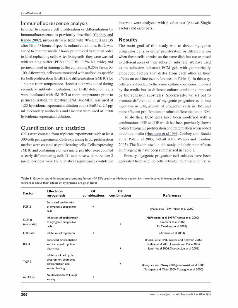
Table 1 Growth- and differentiation-promoting factors (GF, DF) used (see Methods section for more detailed information about these reagents; references about their effects on myogenesis are given here)
FactorEffects on myogenesis
GF combinations
DF combinations References
FGF-2Enhanced proliferation of myogenic progenitor cells
+(Maley et al 1994; Miller et al 2000)
GDF-8 (myostatin)
Inhibition of proliferation of myogenic progenitor cells
+
(McPherron et al 1997; Thomas et al 2000; Zimmers et al 2002;
McCroskery et al 2003)
Follistatin Inhibition of myostatin + (Armand et al 2003)
IGF-1Enhanced differentiation and increased myofiber size–mass +
(Florini et al 1996; Lawlor and Rotwein 2000; Bodine et al 2001; Heszele and Price 2004; Sandri et al 2004; Shavlakadze et al 2005)
TGF-β
Inhibitor of cell cycle progression, promotes differentiation and wound healing
+ (Derynck and Zhang 2003; Jakubowiak et al. 2000; Massague and Chen 2000; Massague et al 2000)
α-TGF-βNeutralization of TGF-β activity
+

International Journal of Nanomedicine 2006:1(2) 207
Myogenic cell fate
previously described (Conboy and Rando 2002; Wagers and
Conboy 2005). These myogenic cells or primary myoblasts
have been cultured in 3 specific media conditions: GM,
DM, and NM (see Methods section for media composition
details).
We embedded geometrically combinations of GF and
DF (Table 1) into ECM gel, which is frequently used
as a substrate for cell growth in vitro and is similar to
physiological substrate of myogenic progenitor cells in vivo.
We aimed to examine whether these factors would be able to
override the cell fate imposed by the aforementioned media
and, at the same time, whether a clearly defined boundary
between cells with alternative myogenic cell fates could be
created by their adhesion to modified ECM substrates which
contain either GF or DF.
To achieve these goals, we have developed the experi-
mental technique described in detail in the Methods section
and summarized in Scheme 1. Briefly, ECM gels were
divided into different geometric areas using cloning cylinders
and mixtures of either GF or DF were placed inside them,
creating modified areas of ECM which contained factors,
vs the unmodified ECM areas. Afterwards cells were
uniformly seeded onto the whole ECM substrate, so that after
approximately 1 hour cells adhered to both unmodified and
Scheme 1
1. ECM:PBS coating (1:100) + cylinder placement. Overnight @RT
2. GF/DF placement inside cylinder. Overnight @4°C
3. Removal of cylinders
4. Media and cells placement
5. Addition of BrdU 2 hours prior to fixation
6. Cells fixing after 48 hours
7. Indirect immunofluorescence (BrdU for proliferation, eMHC for differentiation, nuclear dye Hoechst)
modified ECM areas and shared the same media. Adherence
of cells to unmodified and modified ECM was simultaneous
and there was no difference in cell survival (data not shown).
Experiments with GF or DF uniformly embedded into the
whole ECM area have also been carried out as positive
controls.
Figure 1 Geometric control of terminal myogenic differentiation in GM. Myogenic progenitor cells have been plated at 50% confluency in chamber slides in GM.
(a) GM with unmodified ECM (GM) and GM with DF-modified ECM (GM + DF). (b) Quantification of P, differentiated cells with less than 2 nuclei (D1 = early stage of differentiation), and differentiated cells with more than 2 nuclei (D2 = later stage of differentiation). On unmodified ECM, there is a significantly higher percentage of proliferative cells vs differentiated cells. Looking at cells grown on DF-modified ECM, we see higher numbers of differentiated cells, but most cells do not form multinucleated myotubes. (c) The boundary between unmodified ECM substrate (outside) and DF-modified ECM (inside). Geometric boundary between DF-modified and unmodified ECM substrate was created as described in Methods. Cells were uniformly seeded throughout the ECM area, cultured for 48 hours and fixed. Immunofluorescence was performed with the indicated antibodies: α-BrdU (red), α-eMHC (green), and Hoechst (blue) was used to label all nuclei. Proliferation (incorporation of BrdU) is observed in GM with unmodified ECM and inhibited proliferation and differentiation (expression of eMHC) is observed in GM with DF-modified ECM. Similar results have been obtained in at least 3 independent experiments. Magnification: (a) 20x; (c) 10x.Abbreviations for Figures 1–3, Scheme 1, SOM 1: D, differentiated cells; DF, differentiation factors; DM, differentiation-promoting medium; ECM, extracellular matrix; GM, growth-promoting medium; GF, growth factors; NM, neutral medium; P, proliferating cells; PBS, phosphate buffered saline.
% c
ells

International Journal of Nanomedicine 2006:1(2)208
Juan-Pardo et al
Manipulating cell fate in GMFirstly, we have examined the behavior of cells in GM without
any factors embedded in ECM. Consistent with previously
published results (Morgan and Partridge 2003; Conboy and
Rando 2005), cells rapidly proliferate and do not differentiate
in GM, ie, they incorporate BrdU and only less than 1%
express the marker of differentiated myotubes, eMHC (Figure
1a-GM, quantified in Figure 1b-GM). In contrast, embedded
DF in ECM successfully promote myogenic differentiation of
primary myoblasts (Figure 1a-GM + DF), as shown by their
reduced proliferation and enhanced expression of eMHC.
Interestingly, such directed differentiation occurs even in
the presence of highly mitogenic GM, which contains 20%
FBS and FGF-2 (Figure 1a-GM + DF, quantified in Figure
1b-GM + DF). Figure 1b demonstrates quantification of
multiple experiments and statistically shows that the effects
caused by DF-modified ECM significantly promote the
differentiation of myogenic cells exposed to mitogenic media.
Specifically, cells attached to areas of DF-modified ECM
show higher expression of eMHC+ (number of cells at early
stage of differentiation, D1, significantly rises from 0.5% to
11.8%: p = 0.003). In addition, even though cells exposed
to DF-modified ECM continue to proliferate, the rate was
slower (Figure 1b: Number of proliferating cells P drops from
20.9% in GM to 9.6% in GM + DF; p = 0.077) and a higher
percentage of these cells expresses the differentiation marker,
eMHC (Figure 1b-GM + DF: D1 = 11.8%). Thus, these
findings reveal that it is possible to force the differentiation
of cells under proliferative media conditions through DF-
modified ECM substrates.
Moreover, we achieved geometric control of cell fate
determination, as clearly shown in Figure 1c, where an
obvious interface between eMHC+ and eMHC- myogenic
progenitor cells cultured under identical media conditions
was created by exposing these cells to the different regions
of ECM substrate (with vs without DF).
Manipulating cell fate in DMTo confirm and extrapolate these f indings, we tested
whether the reciprocal cell fate determination could also
be achieved within our experimental system. Specifically,
we cultured cells in DM with unmodified vs GF-modified
ECM. Unsurprisingly, fusion competent myoblasts terminally
differentiate and form eMHC+ myotubes when cultured in
DM (Conboy and Rando 2002; Morgan and Partridge 2003;
Conboy and Rando 2005) (Figure 2a-DM, quantified in
Figure 2b-DM). In contrast, Figure 2a-DM + GF shows that
much fewer eMHC+ myotubes are formed in the area where
Figure 2 Geometric delay of myotube formation in DM by locally embedded growth factors. Myogenic progenitor cells have been plated at 50% confluency in chamber slides in DM. Cells were cultured for 48 hours and fixed. Immunofluorescence was performed with the indicated antibodies: α-BrdU (red), α-eMHC (green), and Hoechst (blue) was used to label all nuclei. (a) DM with unmodified ECM (DM) and DM with GF-modified ECM (DM+GF). There is a clear difference in the fate of cells cultured in DM on unmodified ECM (terminally differentiated, multinucleated myotubes) vs on GF-modified ECM (higher numbers of proliferative cells and smaller myotubes). (b) Quantification of P, early stage-differentiated cells with less than 2 nuclei (D1) and later stage-differentiated cells with more than 2 nuclei (D2). Cells cultured on unmodified ECM in DM show low percentage of proliferating cells and high percentage of differentiated cells. Alternately, when cultured on GF-modified ECM, cells show higher percentage of proliferating cells and lower numbers of differentiated cells. (c) The boundary between unmodified ECM substrate and DF-modified ECM is shown (DM + GF interface). Magnified photographs (20x) of cells seeded on unmodified ECM (outside) and on GF-modified ECM (inside) areas of the culture plate are also shown. Cells were originally seeded at uniform confluency; however, as expected, cells adherent to the GF-modified ECM proliferated at a higher rate, resulting in a higher number of cells compared with those adherent to control ECM. Similar results have been obtained in at least 3 independent experiments. Magnification: (a) 20x; (c) “Outside”, “Inside” 20x; (c) “Interface” 10x.
cells are exposed to GF, compared with the area devoid of GF.
Additionally, a significant fraction of cells incorporate BrdU
(Figure 2a-DM + GF), thus overcoming the effect imposed by
highly differentiating media when attached to GF-modified
ECM substrates.
Both the robustness and reproducibility of the afore-
mentioned regulation of cell fate by GF-signaling from
specific areas of ECM were confirmed by the quantification
of at least 3 replicated experiments, as illustrated in Figure
2b-DM + DF. Proliferation (P) dramatically increases from
0.6% in DM to 16.7% in DM + GF (p = 0.003) and the number
% c
ells

International Journal of Nanomedicine 2006:1(2) 209
Myogenic cell fate
of cells at early stage of differentiation significantly drops
from 18.9% to 2.9% (p = 0.004)
Notably, similar to the data shown in Figure 1c, a clearly
defined interface was created between the region of modified
ECM, which contained embedded GF, and the unmodified
control ECM area, thus allowing cells with different fates to
coexist in the same culture medium (Figure 2c). This interface
is discernable not only because some cells incorporate
BrdU and some instead form myotubes, but also because of
different cell densities. Specifically, there are approximately
4 times more cells in the area of ECM embedded with GF.
Since cells were uniformly seeded on the plate at uniform
density, the higher number of cells can only be attributed to
a predictably higher rate of cell proliferation in regions of
GF-modified ECM.
It is well known that plating myoblasts at high density
will lead to exit of cell cycle and differentiation, even in
the presence of GM (Conboy and Rando 2002; Morgan
and Partridge 2003). Thus, we decided to test whether we
can inhibit differentiation under that specific condition.
Even under high cell density (80% confluency), myogenic
differentiation is delayed by GF-modified ECM, although
not completely avoided (Supplemental Online Material,
SOM 1). Remarkably, when plated at high density, cells
attached to GF-modified ECM area show higher levels of
both proliferation and differentiation (SOM 1). Therefore,
as cell numbers increase, differentiation seems inescapable,
despite initial placement of GF into ECM substrate.
Manipulating cell fate in NMAfter characterizing the effect of modified ECM substrates
on cell fate in strongly differentiating or mitogenic media,
we tested our GF- and DF-modified ECM substrates in NM
conditions. As expected, in NM condition myoblasts slowly
SOM 1 Delay of myogenic differentiation by locally embedded growth factors into ECM under high cell density (80% confluency). Myogenic progenitor cells have been plated at 80% confluency in chamber slides in DM for 36 hours. Immunofluorescence was performed after fixation with the indicated antibodies: α-Ki67 (red), α-eMHC (green), and Hoechst (blue) was used to label all nuclei. As specified in Table 1, GF were embedded into ECM in geometric fashion, as shown in Scheme 1. Consistent with the control DM shown in Figure 2a-DM, cells outside the geometric boundary (outside) form eMHC-positive robust myotubes and do not proliferate. Myogenic cell differentiation is diminished, as indicated by the lower number of nuclei per myotubes, and some Ki67+ proliferating cells persist inside the geometric boundary (inside). Thus, GF embedded in the ECM are capable of diminishing differentiation even when cell numbers increase, but differentiation seems inescapable despite initial placement of GF into ECM. Magnification: 10x.
Figure 3 Geometric control of proliferation or terminal differentiation in NM. Myogenic progenitor cells have been plated at 50% confluence in chamber slides in NM for 48 hours. Immunofluorescence was performed with the indicated antibodies: α-BrdU (red), α-eMHC (green), and Hoechst (blue) was used to label all nuclei.(a) Cells cultured in NM on unmodified ECM substrate (NM) show no distinct tendency towards proliferation or differentiation. When cultured on GF-modified ECM (NM + GF) cells proliferate (BrdU incorporation) without any tendency to form myotubes and differentiate, ie, eMHC+. Conversely, when exposed to DF-modified ECM (NM + DF), cells terminally differentiate (eMHC+) and form myotubes while proliferation is reduced. (b) Quantification of P, early stage-differentiated cells with less than 2 nuclei (D1), and later stage-differentiated cells with more than 2 nuclei (D2). When cultured in NM on unmodified ECM, cells infrequently differentiate and have slow proliferation rate. However, when cells are plated on GF-modified ECM (NM + GF) there is a much larger percentage of proliferating cells; and when they are plated on DF-modified ECM (NM + DF) there are higher percentages of not only eMHC+ differentiated cells but also yield higher percentages of multinucleated myotubes (D2). (c) Boundary between GF-modified ECM and unmodified ECM. There are higher numbers of proliferating cells on GF-modified ECM (inside) than on the unmodified ECM (outside). Similar results have been obtained in at least 3 independent experiments. Magnification: (a) 20x; (c) 10x.
% c
ells

International Journal of Nanomedicine 2006:1(2)210
Juan-Pardo et al
proliferate and infrequently produce eMHC+ terminally
differentiated cells, which usually have no more than 1 or
2 nuclei (Figure 3a-NM and quantified in Figure 3b-NM:
fraction of proliferation cells P = 9.8% and cells at early stage
of differentiation D1 = 9.1%). This verifies that NM does not
impose any strong determination of cell fate, thus potentially
allowing a more discernable effect of our selected factors
on the myogenic proliferation and differentiation. It has not
yet been determined whether the physiological environment
in regenerating muscle is either mitogenic (proliferating),
or differentiating or both–neutral. Since proliferation and
differentiation overlap during tissue repair, neutral media
conditions might mimic the environment of regenerating
muscle more accurately than GM or DM alone. As shown
in Figure 3a-NM + GF and quantified in Figure 3b-NM + GF,
cells cultured on modified ECM with embedded GF robustly
proliferate (fraction of proliferating cells P = 32.2%) and
do not significantly differentiate (fraction of cells at early
stage of differentiation D1 = 2.3%). This is confirmed by
the robust incorporation of BrdU and absence of eMHC
staining. In parallel, myoblasts attached to modified ECM
containing embedded DF efficiently differentiate and lack
proliferation (Figure 3a, NM + DF, quantified in Figure 3b,
NM + DF; fraction of proliferating cells P = 5.2%; fraction
of cells at early stage of differentiation D1 = 13.7%; fraction
of cells at late stage of differentiation D2 = 12.4%). As
above, quantification of at least 3 replicated experiments
demonstrated high reproducibility of this geometric
regulation of myogenic cell fate determination (Figure 3b).
Consistently with data shown above, Figure 3c demon-
strates a clearly defined interface between cells with different
rates of myogenic proliferation cultured in identical media
(NM), which was created by the exposure of cells to the
geometrically embedded GF into ECM.
In summary, data presented in this work demonstrate that
GF and DF geometrically placed in adhesion substrates of
myogenic progenitor cells compete against culture media for
myogenic cell fate determination. As expected, embedding
GF and DF into ECM substrate yields uniform and opposite
effects on the proliferation and differentiation of myogenic
progenitor cells. Importantly, the magnitude of the effects
on either proliferation or differentiation is identical between
uniformly modified ECM (Figures 1a, 2a, 3a) and the
spatially modified areas of ECM (Figures 1c, 2c, 3c). This
strongly suggests that geometrically embedded factors do not
significantly diffuse throughout the ECM and that their initial
concentrations are not diluted. Similar effects on proliferation
and differentiation of myoblasts have been observed when
these GF and DF (listed in Table 1) were added directly to
culture media (data not shown). Thus, the biological activity
of these factors remains the same whether they are embedded
in ECM or added to culture media. However, unlike GF- or
DF-modified ECM substrates, directly applied factors are, of
course, not capable of creating a geometric boundary between
cells with different fates coexisting in the same culture dish.
There is no doubt that these factors added to media signal via
their specific receptors on cells (Husmann et al 1996; Wagers
and Conboy 2005); thus, identical regulation of cell fate from
ECM-embedded factors shown here strongly suggests that
these factors also signal to cells attached to specific areas of
modified ECM.
DiscussionThis work demonstrates that myogenic cells at different
stages of proliferation and differentiation can deliberately
be orchestrated to coexist adjacent to each other on the
same plate with identical culture media by their attachment
to modified ECM substrates. Thus, cells with different fates
and at different stages of the cell cycle can interact, and
their direct interactions can be studied in the experimental
system developed. During embryonic organogenesis, as well
as in regenerating adult tissues, rapidly proliferating and
terminally differentiated cells coexist and signal via both
multiple cell–cell contacts and soluble molecules. Therefore,
our developed technique can be used to identify the important
molecular cross-talk regulating cell fate determination in
embryonic development and in adult tissue repair.
Yet another useful outcome of our work is the potential
to develop a better microenvironment for cell transplantation
studies. No existing method allows successful transplantation
of myogenic progenitor cells. In this study, we have defined
conditions that could improve the regenerative potential
of transplanted cells, by allowing local control of their
proliferation and terminal myogenic differentiation. In future
applications, it would also be interesting to test whether
negative effects of the aged or pathologic environments might
be overcome, and therefore muscle regenerative potential
could be controlled efficiently when myogenic progenitor
cells are transplanted in the context of the modified ECM
tested in this work. If necessary, the concentration and specific
combinations of growth-promoting and differentiation-
promoting factors in the ECM could be attenuated to produce
maximum myogenic potential in the presence of aged or
pathologic environments.
Our data strongly suggest that differentiation of myogenic
progenitor cells always prevails over their growth, even

International Journal of Nanomedicine 2006:1(2) 211
Myogenic cell fate
when GF are embedded in ECM (Figures 2a, c and SOM
1). Specifically, control cells cultured in DM on unmodified
ECM fuse into multinucleated myotubes (Figure 2b-
DM; fraction of cells at late stage of differentiation,
D2 = 27.9%), while cells cultured on DF-modified ECM
remain mono-nucleated in GM (Figure 1b-GM + DF; fraction
of cells at early stage of differentiation, D1 = 11.8%, and no
multinucleated cells have been found), which signifies a lower
degree of terminal myogenic differentiation (Morgan and
Partridge 2003). This suggests that DF-modified ECM have a
differentiating effect, which is not completely dominant over
the growth-promoting media (Figure 1b-GM + DF vs Figure
2b-DM). Therefore, there is no danger of continuous cell
proliferation or oncogenic phenotype from the exposure of
myogenic progenitor cells to exogenous GF in the described
method.
In contrast, we show that, once cells expand, new
myotubes are likely to be formed more efficiently and
robustly in the presence of GF-modified ECM (SOM 1).
Such data are physiologically significant, since the loss of
muscle strength and mass, known as muscle atrophy, often
accompanies old age and muscle dystrophies. Thus, cell
transplantation under conditions that are known to result in
muscle hypertrophy could be especially beneficial for aged
or pathologic organs.
In summary, the approach presented in this work combines
expertise in stem cell biology and bioengineering to create
ECM adhesion substrates for deliberate geometric control
of proliferation and differentiation of myogenic progenitor
cells, which overrides the cell fate determination imposed
by the culture media. Such modified ECM substrates could
be used as a supportive microenvironment able to promote
satellite cell expansion and differentiation in normal, aged,
and disease-afflicted muscle. Additionally, this work may
also serve as a model to engineer synthetic cellular niches
to control and harness optimally the regenerative potential
of stem cells in general.
AcknowledgmentsThis work was supported by Ellison’s Medical Foundation
and NIH KO1 AG 025652 grants to I.M.C.; and by the Torres
Quevedo Program from the Spanish Ministry of Education
and Science, by the European Social Fund and by CEIT and
Technum University of Navarra to E.M.J.P.
ReferencesArmand AS, Della GB, Launay T, et al. 2003. Expression and neural control
of follistatin vs myostatin genes during regeneration of mouse soleus. Dev Dyn, 227:256–65.
Bockhold KJ, Rosenblatt JD, Partridge TA. 1998. Aging normal and dystrophic mouse muscle: analysis of myogenicity in cultures of living single fibers. Muscle Nerve, 21:173–83.
Bodine SC, Stitt TN, Gonzalez M, et al. 2001. Akt/mTOR pathway is a crucial regulator of skeletal muscle hypertrophy and can prevent muscle atrophy in vivo. Nat Cell Biol, 3:1014–19.
Chakravarthy MV, Fiorotto ML, Schwartz RJ, et al. 2001. Long-term insulin-like growth factor-I expression in skeletal muscles attenuates the enhanced in vitro proliferation ability of the resident satellite cells in transgenic mice. Mech Ageing Dev, 122:1303–20.
Conboy IM, Conboy MJ, Smythe GM, et al. 2003. Notch-mediated restora-tion of regenerative potential to aged muscle. Science, 302:1575–7.
Conboy IM, Conboy MJ, Wagers AJ, et al. 2005. Rejuvenation of aged progenitor cells by exposure to a young systemic environment. Nature, 433:760–4.
Conboy IM, Rando TA. 2002. The regulation of Notch signaling controls satellite cell activation and cell fate determination in postnatal myogenesis. Dev Cell, 3:397–409.
Conboy IM, Rando TA. 2005. Aging, stem cells and tissue regeneration: lessons from muscle. Cell Cycle, 4:407–10.
Derynck R, Zhang YE. 2003. Smad-dependent and Smad-independent pathways in TGF-beta family signalling. Nature, 425:577–84.
Disatnik MH, Boutet SC, Lee CH, et al. 2002. Sequential activation of individual PKC isozymes in integrin-mediated muscle cell spreading: a role for MARCKS in an integrin signaling pathway. J Cell Sci, 115:2151–63.
Disatnik MH, Rando TA. 1999. Integrin-mediated muscle cell spreading. The role of protein kinase c in outside-in and inside-out signaling and evidence of integrin cross-talk. J Biol Chem, 274:32486–92.
Downward J. 2004. PI 3-kinase, Akt and cell survival. Semin Cell Dev Biol, 15:177–82.
Florini JR, Ewton DZ, Coolican SA. 1996. Growth hormone and the insulin-like growth factor system in myogenesis. Endocr Rev, 17:481–517.
Heszele MF, Price SR. 2004. Insulin-like growth factor I: the yin and yang of muscle atrophy. Endocrinology, 145:4803–5.
Husmann I, Soulet L, Gautron J, et al. 1996. Growth factors in skeletal muscle regeneration. Cytokine Growth Factor Rev, 7:249–58.
Jakubowiak A, Pouponnot C, Berguido F, et al. 2000. Inhibition of the transforming growth factor beta 1 signaling pathway by the AML1/ETO leukemia-associated fusion protein. J Biol Chem, 275:40282–7.
Latres E, Amini AR, Amini AA, et al. 2005. Insulin-like growth factor-1 (IGF-1) inversely regulates atrophy-induced genes via the phosphatidylinositol 3-kinase/Akt/mammalian target of rapamycin (PI3K/Akt/mTOR) pathway. J Biol Chem, 280:2737–44.
Lawlor MA, Rotwein P. 2000. Coordinate control of muscle cell survival by distinct insulin-like growth factor activated signaling pathways. J Cell Biol, 151:1131–40.
Maley MA, FanY, Beilharz MW, et al. 1994. Intrinsic differences in MyoD and myogenin expression between primary cultures of SJL/J and BALB/C skeletal muscle. Exp Cell Res, 211:99–107.
Massague J, Blain SW, Lo RS. 2000. TGFbeta signaling in growth control, cancer, and heritable disorders. Cell, 103:295–309.
Massague J, Chen YG. 2000. Controlling TGF-beta signaling. Genes Dev, 14:627–44.
McCroskery S, Thomas M, Maxwell L, et al. 2003. Myostatin negatively regulates satellite cell activation and self-renewal. J Cell Biol, 162:1135–47.
McPherron AC, Lawler AM, Lee SJ. 1997. Regulation of skeletal muscle mass in mice by a new TGF-beta superfamily member. Nature, 387:83–90.
Miller KJ, Thaloor D, Matteson S, et al. 2000. Hepatocyte growth factor affects satellite cell activation and differentiation in regenerating skeletal muscle. Am J Physiol Cell Physiol, 278:C174–181.

International Journal of Nanomedicine 2006:1(2)212
Juan-Pardo et al
Morgan JE, Partridge TA. 2003. Muscle satellite cells. Int J Biochem Cell Biol, 35:1151–6.
Moustakas A, Pardali K, Gaal A, et al. 2002. Mechanisms of TGF-beta signaling in regulation of cell growth and differentiation. Immunol Lett, 82:85–91.
Partridge TA. 2004. Stem cell therapies for neuromuscular diseases. Acta Neurol Belg, 104:141–7.
Pola R, Ling LE, Aprahamian TR, et al. 2003. Postnatal recapitulation of embryonic hedgehog pathway in response to skeletal muscle ischemia. Circulation, 108:479–85.
Sandri M, Sandri C, Gilbert A, et al. 2004. Foxo transcription factors induce the atrophy-related ubiquitin ligase atrogin-1 and cause skeletal muscle atrophy. Cell, 117:399–412.
Schultz E, Lipton BH. 1982. Skeletal muscle satellite cells: changes in proliferation potential as a function of age. Mech Ageing Dev, 20:377–83.
Seale P, Polesskaya A, Rudnicki MA. 2003. Adult stem cell specification by Wnt signaling in muscle regeneration. Cell Cycle, 2:418–19.
Shavlakadze T, White JD, Davies M. et al. 2005. Insulin-like growth factor I slows the rate of denervation induced skeletal muscle atrophy. Neuromuscul Disord, 15:139–46.
Sherwood RI, Christensen JL, Conboy IM, et al. 2004. Isolation of adult mouse myogenic progenitors: functional heterogeneity of cells within and engrafting skeletal muscle. Cell, 119:543–54.
Stitt TN, Drujan D, Clarke BA, et al. 2004. The IGF-1/PI3K/Akt pathway prevents expression of muscle atrophy-induced ubiquitin ligases by inhibiting FOXO transcription factors. Mol Cell, 14:395–403.
Taverna D, Disatnik MH, Rayburn H, et al. 1998. Dystrophic muscle in mice chimeric for expression of alpha5 integrin. J Cell Biol, 143:849–59.
Thomas M, Langley B, Berry C, et al. 2000. Myostatin, a negative regulator of muscle growth, functions by inhibiting myoblast proliferation. J Biol Chem, 275:40235–43.
Tidball JG. 2005. Inflammatory processes in muscle injury and repair. Am J Physiol Regul Integr Comp Physiol, 288:345–53.
Wagers AJ, Conboy IM 2005. Cellular and molecular signatures of muscle regeneration: current concepts and controversies in adult myogenesis. Cell, 122:659–67.
Yang JT, Rando TA, Mohler WA, et al. 1996. Genetic analysis of alpha 4 integrin functions in the development of mouse skeletal muscle. J Cell Biol, 135:829–35.
Zaidel-Bar R, Cohen M, Addadi L, et al. 2004. Hierarchical assembly of cell-matrix adhesion complexes. Biochem Soc Trans, 32:416–20.
Zammit PS, Heslop L, Hudon V, et al. 2002. Kinetics of myoblast proliferation show that resident satellite cells are competent to fully regenerate skeletal muscle fibers. Exp Cell Res, 281:39–49.
Zimmers TA, Davies MV, Koniaris LG, et al. J. 2002. Induction of cachexia in mice by systemically administered myostatin. Science, 296:1486–8.