Non-invasive assessment of coronary endothelial function in ...
14
RESEARCH ARTICLE Non-invasive assessment of coronary endothelial function in children and adolescents with type 1 diabetes mellitus using isometric handgrip exercise—MRI: A feasibility study Gae ¨ tan Zwingli 1 , Je ´ro ˆ me Yerly 2 , Yvan Mivelaz 3 , Sophie Stoppa-Vaucher 4,5 , Andrew A. Dwyer 6 , Nelly Pitteloud 5,7 , Matthias Stuber 2 , Michael Hauschild ID 5 * 1 Lausanne University (UNIL), Faculty of Biology and Medicine, Lausanne, Switzerland, 2 Department of Radiology, Lausanne University Hospital (CHUV), Center for Biomedical Imaging, Lausanne, Switzerland, 3 Pediatric Cardiology Unit, Service of Pediatrics, Lausanne University Hospital (CHUV), Lausanne, Switzerland, 4 Department of Pediatrics, Ho ˆ pital Neucha ˆ telois, Neucha ˆ tel, Switzerland, 5 Pediatric Endocrinology, Diabetology and Obesity Unit, Service of Pediatrics, Lausanne University Hospital (CHUV), Lausanne, Switzerland, 6 Boston College, William F.Connell School of Nursing, Chestnut Hill, MA, United States of America, 7 Service of Endocrinology, Diabetology and Metabolism, Lausanne University Hospital (CHUV), Lausanne, Switzerland * [email protected] Abstract Background Type 1 diabetes mellitus (T1DM) in children and adolescents is associated with significant cardiovascular morbidity and mortality. Early detection of vascular dysfunction is key to patient management yet current assessment techniques are invasive and not suitable for pediatric patient populations. A novel approach using isometric handgrip exercise during magnetic resonance imaging (IHE-MRI) has recently been developed to evaluate coronary endothelial function non-invasively in adults. This project aimed to assess endothelium- dependent coronary arterial response to IHE-MRI in children with T1DM and in age matched healthy controls. Materials and methods Healthy volunteers and children with T1DM (>5 years) were recruited. IHE-MRI cross-sec- tional coronary artery area measurements were recorded at rest and under stress. Carotid intima media thickness (CIMT) and aortic pulse wave velocity (PWV) were assessed for comparison. Student’s t-tests were used to compare results between groups. Results and discussion Seven children with T1DM (3 female, median 14.8 years, mean 14.8 ± 1.9 years) and 16 healthy controls (7 female, median 14.8 years, mean 14.2 ± 2.4 years) participated. A signifi- cant increase in stress-induced cross-sectional coronary area was measured in controls PLOS ONE | https://doi.org/10.1371/journal.pone.0228569 February 13, 2020 1 / 14 a1111111111 a1111111111 a1111111111 a1111111111 a1111111111 OPEN ACCESS Citation: Zwingli G, Yerly J, Mivelaz Y, Stoppa- Vaucher S, Dwyer AA, Pitteloud N, et al. (2020) Non-invasive assessment of coronary endothelial function in children and adolescents with type 1 diabetes mellitus using isometric handgrip exercise —MRI: A feasibility study. PLoS ONE 15(2): e0228569. https://doi.org/10.1371/journal. pone.0228569 Editor: Fang-Bao Tian, University of New South Wales, AUSTRALIA Received: July 25, 2019 Accepted: January 19, 2020 Published: February 13, 2020 Copyright: © 2020 Zwingli et al. This is an open access article distributed under the terms of the Creative Commons Attribution License, which permits unrestricted use, distribution, and reproduction in any medium, provided the original author and source are credited. Data Availability Statement: All relevant data are within the paper and its Supporting Information files. Funding: This project has been supported in part by the Swiss National Science Foundation Grants 320030_143923 and 326030_150828. The funder had no role in study design, data collection and analysis, decision to publish, or preparation of the manuscript.
-
Upload
khangminh22 -
Category
Documents
-
view
3 -
download
0
Transcript of Non-invasive assessment of coronary endothelial function in ...
RESEARCH ARTICLE
Non-invasive assessment of coronary
endothelial function in children and
adolescents with type 1 diabetes mellitus
using isometric handgrip exercise—MRI: A
feasibility study
Gaetan Zwingli1, Jerome Yerly2, Yvan Mivelaz3, Sophie Stoppa-Vaucher4,5, Andrew
A. Dwyer6, Nelly Pitteloud5,7, Matthias Stuber2, Michael HauschildID5*
1 Lausanne University (UNIL), Faculty of Biology and Medicine, Lausanne, Switzerland, 2 Department of
Radiology, Lausanne University Hospital (CHUV), Center for Biomedical Imaging, Lausanne, Switzerland,
3 Pediatric Cardiology Unit, Service of Pediatrics, Lausanne University Hospital (CHUV), Lausanne,
Switzerland, 4 Department of Pediatrics, Hopital Neuchatelois, Neuchatel, Switzerland, 5 Pediatric
Endocrinology, Diabetology and Obesity Unit, Service of Pediatrics, Lausanne University Hospital (CHUV),
Lausanne, Switzerland, 6 Boston College, William F.Connell School of Nursing, Chestnut Hill, MA, United
States of America, 7 Service of Endocrinology, Diabetology and Metabolism, Lausanne University Hospital
(CHUV), Lausanne, Switzerland
Abstract
Background
Type 1 diabetes mellitus (T1DM) in children and adolescents is associated with significant
cardiovascular morbidity and mortality. Early detection of vascular dysfunction is key to
patient management yet current assessment techniques are invasive and not suitable for
pediatric patient populations. A novel approach using isometric handgrip exercise during
magnetic resonance imaging (IHE-MRI) has recently been developed to evaluate coronary
endothelial function non-invasively in adults. This project aimed to assess endothelium-
dependent coronary arterial response to IHE-MRI in children with T1DM and in age matched
healthy controls.
Materials and methods
Healthy volunteers and children with T1DM (>5 years) were recruited. IHE-MRI cross-sec-
tional coronary artery area measurements were recorded at rest and under stress. Carotid
intima media thickness (CIMT) and aortic pulse wave velocity (PWV) were assessed for
comparison. Student’s t-tests were used to compare results between groups.
Results and discussion
Seven children with T1DM (3 female, median 14.8 years, mean 14.8 ± 1.9 years) and 16
healthy controls (7 female, median 14.8 years, mean 14.2 ± 2.4 years) participated. A signifi-
cant increase in stress-induced cross-sectional coronary area was measured in controls
PLOS ONE | https://doi.org/10.1371/journal.pone.0228569 February 13, 2020 1 / 14
a1111111111
a1111111111
a1111111111
a1111111111
a1111111111
OPEN ACCESS
Citation: Zwingli G, Yerly J, Mivelaz Y, Stoppa-
Vaucher S, Dwyer AA, Pitteloud N, et al. (2020)
Non-invasive assessment of coronary endothelial
function in children and adolescents with type 1
diabetes mellitus using isometric handgrip exercise
—MRI: A feasibility study. PLoS ONE 15(2):
e0228569. https://doi.org/10.1371/journal.
pone.0228569
Editor: Fang-Bao Tian, University of New South
Wales, AUSTRALIA
Received: July 25, 2019
Accepted: January 19, 2020
Published: February 13, 2020
Copyright: © 2020 Zwingli et al. This is an open
access article distributed under the terms of the
Creative Commons Attribution License, which
permits unrestricted use, distribution, and
reproduction in any medium, provided the original
author and source are credited.
Data Availability Statement: All relevant data are
within the paper and its Supporting Information
files.
Funding: This project has been supported in part
by the Swiss National Science Foundation Grants
320030_143923 and 326030_150828. The funder
had no role in study design, data collection and
analysis, decision to publish, or preparation of the
manuscript.
(5.4 mm2 at rest to 6.39 mm2 under stress, 18.8 ± 10.7%, p = 0.0004). In contrast, mean
area change in patients with T1DM was not significant (7.17 mm2 at rest to 7.59 mm2 under
stress, 10.5% ± 28.1%, p = n.s.). There was no significant difference in the results for neither
PWV nor CIMT between patients and controls, (5.3±1.5 m/s vs.4.8±0.7 m/s and 0.4
±0.03mm vs.0.46 mm ± 0.03 respectively, both p = n.s.).
Conclusions
Our pilot study demonstrates the feasibility of using a totally non-invasive IHE-MRI tech-
nique in children and adolescents with and without T1DM. Preliminary results suggest a
blunted endothelium-dependent coronary vasomotor function in children with T1DM (>5
years). Better knowledge and new methodologies may improve surveillance and care for
T1DM patients to reduce cardiovascular morbidity and mortality.
Introduction
Type 1 diabetes mellitus (T1DM) is a chronic disease characterized by immune-mediated
beta-cell destruction requiring lifelong insulin therapy. T1DM is most often diagnosed in
young children (2–3 years-old) and pre-teens/early adolescents (8–12 years-old). The inci-
dence and prevalence of T1DM in children and adolescents <16 years is rising and incidence
rates vary across countries [1]. Despite newly emerging treatment modalities [2], high rates of
mortality from acute and chronic complications persist [3]. Both disease duration and elevated
glycated hemoglobin (HbA1c) levels are associated with increased cardiovascular morbidity
and all-cause mortality [4]. The Diabetes Control and Complications Trial (DCCT) has used
magnetic resonance imaging (MRI) [5] to investigate cardiovascular effects of DM. The vascu-
lar endothelium serves as an important modulator of vasomotor tone and function via the
release of nitric oxide. The release of endothelium derived nitric oxide is thought to be neces-
sary to regulate vascular response to blood flow demands during exercise. Early studies showed
that exercise training improved endothelium-dependent vasodilatation as measured by
changes in coronary artery diameter through serial angiograms in response do intracoronary
infusion of increasing doses of acetylcholine [6]. Notably, end-stage lesions (e.g. atheroscle-
rotic plaques) have been observed in children and young adults [7]. Clinical detection of early
stages of vascular disease (e.g. endothelial dysfunction and arterial stiffness due to intima
media lesion) is therefore paramount, especially in individuals with risk factors such as obesity,
hypertension, and a positive familial history. Importantly, the availability and effects of early
secondary prevention accentuate the need for non-invasive studies to detect vascular disease
in children and young adults.
Both carotid intima media thickness (CIMT) and aortic pulse wave velocity (PWV) are the
standard non-invasive methods for investigating vascular effects of diabetes in children and
adolescents. Other techniques to directly assess coronary endothelial function often require
invasive coronary angiography and cannot be used in healthy subjects and children [8]. Mea-
surement of the carotid intima media thickness (CIMT) with B-mode ultrasonography [9] is
one of the most common used methods of vascular imaging studies and has been extensively
studied in adults with diabetes [10] and several reports suggest the presence of increased
CIMT values in a large proportion of children with T1DM ([11]. Aortic pulse wave velocity
(PWV) has also been used to assess aortic wall stiffness [12–14]. Recent studies point to the
PLOS ONE | https://doi.org/10.1371/journal.pone.0228569 February 13, 2020 2 / 14
Competing interests: The authors have declared
that no competing interests exist.
utility of assessing arterial stiffness by PWV, in children with varied congenital heart and vas-
cular malformations [15–17]. Studies reveal stiffer, less compliant thoracic aortas and impaired
arterial stiffness in adolescents with T1DM.
Recently, Hays et al. explored the use of MRI to measure cross-sectional coronary area and
blood flow changes in response to isometric handgrip exercise (IHE) in adults [18]. The
IHE-MRI technique was previously shown to be useful for assessing the effect of ischemia on
myocardial metabolism [19]. Indeed, in vivo studies demonstrate the vasomotor response to
IHE is primarily mediated by nitric oxide [20] and therefore, reflects endothelial function [6,
21–25]. The feasibility of the IHE-MRI technique to assess vasomotor reactivity in coronary
arteries has been studied in patients with coronary artery disease as well as patients with HIV
[18, 21, 26]. Phantom study results suggest that radial MRI is capable of distinguishing area
differences in the order of 0.2–0.3 mm2 (i.e. 3–4% difference for a 3 mm baseline diameter)
[27]. Thus, radial MRI is adequate for measuring area differences in the range of previously
reported endothelium dependent vasomotor response in healthy adult subjects (10–25%). Fur-
ther, these results also indicated that the smallest detectable area difference with radial MRI
was largely independent of pixel size in the resolution range investigated [27]. Coronary artery
diameter ranges from 2 mm in healthy infants to 5 mm in healthy teenagers [28]. However,
evaluation of the endothelial function by MRI has, to the best of our knowledge, not been stud-
ied in children.
This study aimed to test the feasibility of using IHE-MRI in children and adolescents by
assessing endothelial function in young patients with and without T1DM. Secondarily we
sought to compare IHE-MRI results with the standard CIMT and PWV techniques.
Materials and methods
This investigator-initiated, single-center pilot study was an unblinded parallel trial of
IHE-MRI versus CIMT and PWV examining blood vessel function in young children (adoles-
cent and pre-adolescent) at the University Hospital of Lausanne (CHUV) in Switzerland.
Prior to study enrollment, patients and their parents/legal representatives received oral and
written information about the study. Written informed consent was obtained from all parents/
guardians and also from participants over the age of 14 years.
Study participants
Two groups of children were recruited: patients with T1DM of at least 5 years duration, and
healthy community-dwelling children without any ongoing medication.
Participants ranged from 12–18 years of age, exhibiting Tanner II or greater pubertal devel-
opment. Exclusion criteria included smoking, obesity (BMI >97th percentile), systolic or dia-
stolic hypertension (>90th percentile), dyslipidemia (pathological HDL/total cholesterol), or
any known inflammatory process. Participants’ characteristics, such as age, duration of diabe-
tes, insulin therapies and recent Hb1AC levels were recorded for children with T1DM. On the
day of the exams, height, weight, heart rate and blood pressure were measured.
Imaging protocol
Patients with T1DM removed technical material (e.g. insulin pump, continuous glucose mea-
surement (CGM) devices) prior to imaging. This was planned in advance with the families in
order to minimize the study impact on T1DM management. Both CIMT and PWV were per-
formed by the same operator (YM) and IHE-MRI by another operator assisted by a technician.
Technicians varied according to the hospital schedule. All IHE-MRI analysis were performed
PLOS ONE | https://doi.org/10.1371/journal.pone.0228569 February 13, 2020 3 / 14
by the same investigator (JY). Exams took place in the afternoon, at least one-hour
postprandial.
To assess vascular function using CIMT and PVW, subjects were examined by ultrasound
using Philips iE33 (Philips Medical, Netherlands) echocardiograph with a linear L11-5 trans-
ducer. Data were saved digitally (Xcelera, Philips Medical, Netherlands) and analyzed off-line
using an automated measurement program (QLAB software, Philips Medical, Netherlands).
Images were acquired following the standards of the American Heart Association (AHA) [29].
IMT of the posterior wall was measured in three portions of the left and right carotid arteries:
common carotid, internal carotid and carotid bulb, in two different angles of insonation.
Based on these measurements, the maximum average value of carotid IMT was calculated and
expressed in mm ± standard deviation. PWV was measured with the Sphygmocor CPV System
(AtCor Medical, Sidney, Australia) [30, 31]. The tonometer was applied at the common carotid
artery and the femoral artery. Pulse wave was recorded simultaneously at both locations using
electrocardiogram. The latter was chosen as time of reference, enabling the device to deter-
mine the transit time of the pulse wave. The distance between the two sites is used to calculate
PWV. The device has fully-automated calculations and expresses an index of measurement
quality. Per manufacturer recommendations, measures with an index < 74% were not consid-
ered. The PWV value given by the apparatus was expressed in meter per second (± standard
deviation).
Isometric Handgrip Exercise—Magnetic Resonance Imaging (IHE-MRI). The imaging
studies done at baseline (at rest) and during IHE (under stress) for the coronary endothelial
function assessment were performed on a clinical 3T MR scanner (MAGNETOM Prisma; Sie-
mens AG, Healthcare Sector, Erlangen, Germany) with an 18-channel chest coil array and a
32-channel spine coil array for signal reception. A 2D radial retrospectively ECG-gated spoiled
gradient recalled echo sequence was used to acquire cine cardiac MR images. The relevant
imaging parameters included: field of view = 260 × 260 mm2, base resolution = 320 sample
points per radial line, pixel size = 0.8 × 0.8 mm2, slice thickness = 7.0 mm, echo time = 2.5 ms,
repetition time (TR) = 4.7 ms, receiver bandwidth = 580 Hz/pixel, and RF excitation
angle = 22 Deg. A product water-selective excitation pulse was used to suppress the signal
from epicardial fat and to improve the coronary artery conspicuity. Due to heart rate variation
among subjects, the number of readout lines per segment (or views per frame) was adjusted
for each subject such that a total of ~250 radial profiles per image were collected within a ~20 s
breath-hold duration. The data were acquired with a uniform radial trajectory and 40 cine
images were reconstructed at the console. Image quality was rated as poor if blurring due to
artifact/patient motion occurred, and as good or very good for optimal image quality.
Maximum grip strength was determined using an MRI-compatible dynamometer (Grip
Force Fiber Optic Response Pad, Current Designs Inc., Philadelphia, USA) prior to baseline
imaging. Following baseline imaging, IHE-MRI commenced. Handgrip exercise was started
one minute prior to data collection. Each subject held the handgrip at 30% of his/her maxi-
mum grip strength for approximately 4 minutes during image acquisition. A custom
MATLAB software provided real-time feedback to participants enabling them to visually
observe grip strength effort while in the magnet—allowing participants to adapt/maintain a
constant grip force (30% of maximal effort). Subjects were examined in the supine position
and all cine images were acquired during end expiratory breath hold to minimize respiratory
motion artifacts.
Bright blood cine MR images were acquired perpendicular to a linear segment of the right/
left coronary artery. We used this new technique to measure the changes of diameter of the
coronary in response to handgrip exertion, combined to a non-invasive measure of blood pres-
sure and heart rate to determine the rate-pressure product and evaluate the effect of handgrip
PLOS ONE | https://doi.org/10.1371/journal.pone.0228569 February 13, 2020 4 / 14
[20, 32]. As the cross-sectional area of the coronary arteries may change throughout the car-
diac cycle [33] measurements at rest and during stress were always made at the same time
point in one cardiac cycle, consistent with prior protocols. (18, 20–21, 28). This avoided con-
founding effects of coronary distensibility (or time phase differences) and minimized effects of
motion blurring.
Statistical analysis
All data were coded before statistical analysis. Assuming normal distribution, paired and non-
paired Student-test analysis were performed as appropriate. GraphPadPrim Software was
employed to chart data and compare group results. P values<0.05 were considered statistically
significant.
Ethics statement
The study protocol was approved by the institutional review board (Commission cantonaled'éthique de la recherche sur l'être humain [Vaud]) under approval number CERVD 213/15
and was conducted in accordance with the principles of the Declaration of Helsinki.
ClinicalTrials.gov identifier: NCT03506711
Grant information: This study was in part supported by SNF grant 143923.
The authors have declared that no competing interests exist.
Results
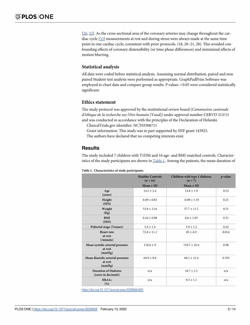
The study included 7 children with T1DM and 16 age- and BMI-matched controls. Character-
istics of the study participants are shown in Table 1. Among the patients, the mean duration of
Table 1. Characteristics of study participants.
Healthy Controls
(n = 16)
Children with type 1 diabetes
(n = 7)
p-value
Mean ± SD Mean ± SD
Age
(years)
14.2 ± 2.4 14.8 ± 1.9 0.53
Height
(SDS)
0.49 ± 0.81 -0.09 ± 1.35 0.21
Weight
(Kg)
53.8 ± 13.6 57.7 ± 11.1 0.51
BMI
(SDS)
0.16 ± 0.88 0.6 ± 1.03 0.31
Pubertal stage (Tanner) 3.4 ± 1.4 3.9 ± 1.2 0.42
Heart rate
at rest
(/minute)
72.8 ± 11.1 85 ± 6.9 0.014
Mean systolic arterial pressure
at rest
(mmHg)
110.6 ± 9 110.7 ± 10.4 0.98
Mean diastolic arterial pressure
at rest
(mmHg)
64.9 ± 8.8 66.1 ± 12.4 0.793
Duration of Diabetes
(years in decimals)
n/a 10.7 ± 3.1 n/a
HbA1c
(%)
n/a 9.3 ± 1.1 n/a
https://doi.org/10.1371/journal.pone.0228569.t001
PLOS ONE | https://doi.org/10.1371/journal.pone.0228569 February 13, 2020 5 / 14
T1DM was 10.7 ± 3.1 years, mean total daily insulin 0.9 ± 0.2 U insulin/kg/day, and mean
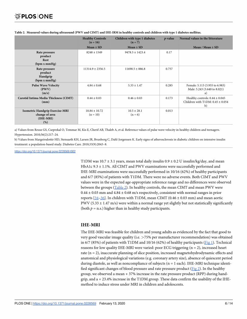
HbA1c 9.3 ± 1.1%. All CIMT and PWV examinations were successfully performed and
IHE-MRI examinations were successfully performed in 10/16 (62%) of healthy participants
and 6/7 (85%) of patients with T1DM. There were no adverse events. Both CIMT and PWV
values were in the expected age-appropriate reference range and no differences were observed
between the groups (Table 2). In healthy controls, the mean CIMT and mean PWV were
0.44 ± 0.03 mm and 4.84 ± 0.68 m/s respectively, consistent with normal ranges in prior
reports [34–36]. In children with T1DM, mean CIMT (0.46 ± 0.03 mm) and mean aortic
PWV (5.33 ± 1.47 m/s) were within a normal range yet slightly but not statistically significantly
(both p = n.s.) higher than in healthy study participants.
IHE-MRI
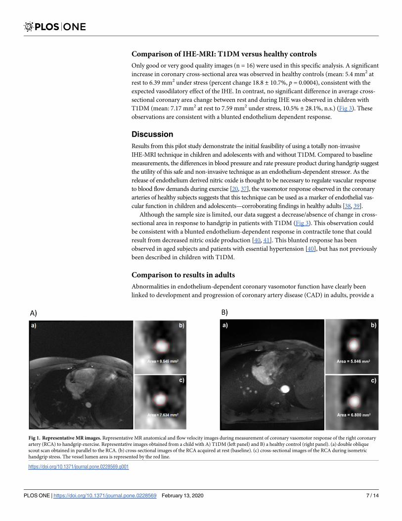
The IHE-MRI was feasible for children and young adults as evidenced by the fact that good to
very good vascular image quality (i.e. >75% per manufacturer recommendation) was obtained
in 6/7 (85%) of patients with T1DM and 10/16 (62%) of healthy participants (Fig 1). Technical
reasons for low quality IHE-MRI were varied: poor ECG triggering (n = 2), increased heart
rate (n = 2), inaccurate planning of slice position, increased magnetohydrodynamic effects and
anatomical and physiological variations (e.g. coronary artery size), absence of quiescent period
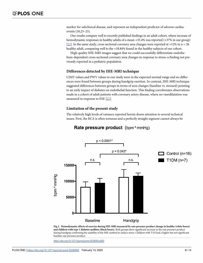
during diastole, as well as noncompliance of subjects (n = 1 each). IHE-MRI technique identi-
fied significant changes of blood pressure and rate pressure product (Fig 2). In the healthy
group, we observed a mean + 37% increase in the rate pressure product (RPP) during hand-
grip, and a + 23.4% increase in the T1DM group. These data confirm the usability of the IHE-
method to induce stress under MRI in children and adolescents.
Table 2. Measured values during ultrasound (PWV and CIMT) and IHE-IRM in healthy controls and children with type 1 diabetes mellitus.
Healthy Controls
(n = 16)
Children with type 1 diabetes
(n = 7)
p-value Normal values in the litterature
Mean ± SD Mean ± SD Mean / Mean ± SD
Rate pressure
product
Rest
(bpm x mmHg)
8248 ± 1549 9478.5 ± 1423.4 0.17
Rate pressure
product
Handgrip
(bpm x mmHg)
11314.9 ± 2356.5 11698.5 ± 886.8 0.757
Pulse Wave Velocity
(PWV)
(m/s)
4.84 ± 0.68 5.33 ± 1.47 0.285 Female: 5.113 (3.955 to 6.983)
Male: 5.243 (3.640 to 8.021)
a)
Carotid Intima Media Thickness (CIMT)
(mm)
0.44 ± 0.03 0.46 ± 0.03 0.173 Healthy controls: 0.44 ± 0.045
Children with T1DM: 0.45 ± 0.054
b)
Isometric Handgrip Exercise-MRI
change of area
(IHE-MRI)
(%)
18.84 ± 10.72
(n = 10)
10.5 ± 28.1
(n = 6)
0.013
a) Values from Reusz GS, Cseprekal O, Temmar M, Kis E, Cherif AB, Thaleb A, et al. Reference values of pulse wave velocity in healthy children and teenagers.
Hypertension. 2010;56(2):217–24.
b) Values from Margeirsdottir HD, Stensaeth KH, Larsen JR, Brunborg C, Dahl-Jorgensen K. Early signs of atherosclerosis in diabetic children on intensive insulin
treatment: a population-based study. Diabetes Care. 2010;33(9):2043–8.
https://doi.org/10.1371/journal.pone.0228569.t002
PLOS ONE | https://doi.org/10.1371/journal.pone.0228569 February 13, 2020 6 / 14
Comparison of IHE-MRI: T1DM versus healthy controls
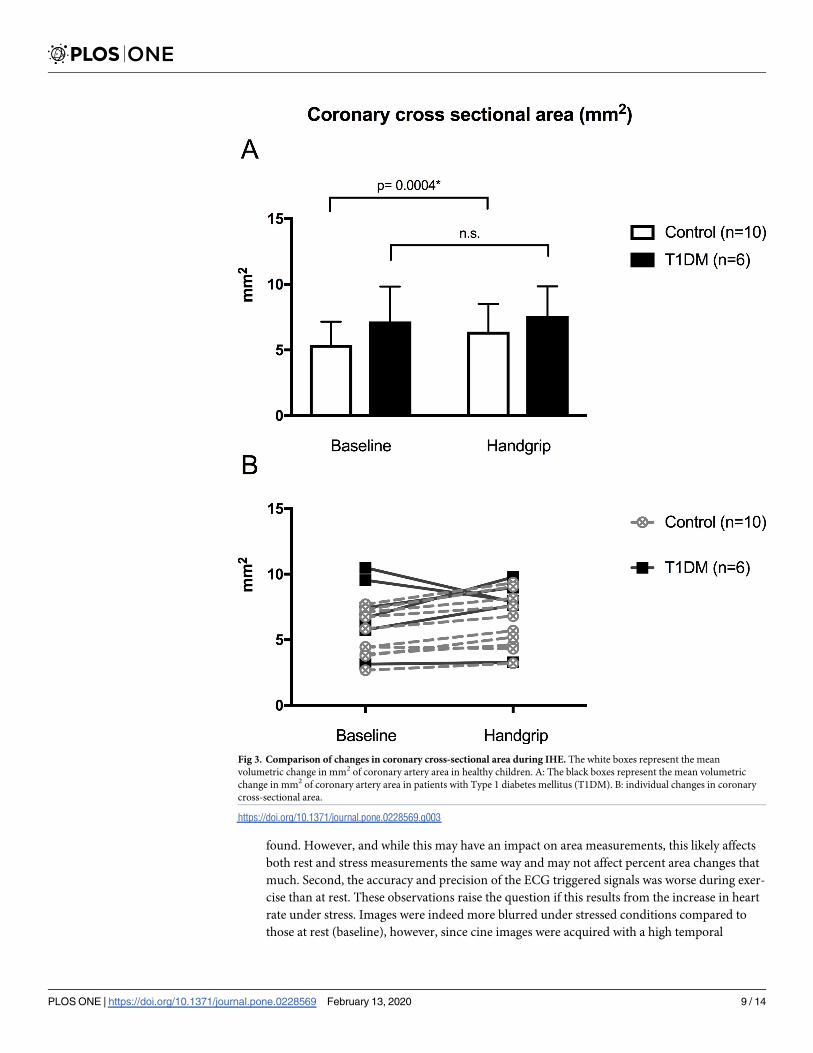
Only good or very good quality images (n = 16) were used in this specific analysis. A significant
increase in coronary cross-sectional area was observed in healthy controls (mean: 5.4 mm2 at
rest to 6.39 mm2 under stress (percent change 18.8 ± 10.7%, p = 0.0004), consistent with the
expected vasodilatory effect of the IHE. In contrast, no significant difference in average cross-
sectional coronary area change between rest and during IHE was observed in children with
T1DM (mean: 7.17 mm2 at rest to 7.59 mm2 under stress, 10.5% ± 28.1%, n.s.) (Fig 3). These
observations are consistent with a blunted endothelium dependent response.
Discussion
Results from this pilot study demonstrate the initial feasibility of using a totally non-invasive
IHE-MRI technique in children and adolescents with and without T1DM. Compared to baseline
measurements, the differences in blood pressure and rate pressure product during handgrip suggest
the utility of this safe and non-invasive technique as an endothelium-dependent stressor. As the
release of endothelium derived nitric oxide is thought to be necessary to regulate vascular response
to blood flow demands during exercise [20, 37], the vasomotor response observed in the coronary
arteries of healthy subjects suggests that this technique can be used as a marker of endothelial vas-
cular function in children and adolescents—corroborating findings in healthy adults [38, 39].
Although the sample size is limited, our data suggest a decrease/absence of change in cross-
sectional area in response to handgrip in patients with T1DM (Fig 3). This observation could
be consistent with a blunted endothelium-dependent response in contractile tone that could
result from decreased nitric oxide production [40, 41]. This blunted response has been
observed in aged subjects and patients with essential hypertension [40], but has not previously
been described in children with T1DM.
Comparison to results in adults
Abnormalities in endothelium-dependent coronary vasomotor function have clearly been
linked to development and progression of coronary artery disease (CAD) in adults, provide a
Fig 1. Representative MR images. Representative MR anatomical and flow velocity images during measurement of coronary vasomotor response of the right coronary
artery (RCA) to handgrip exercise. Representative images obtained from a child with A) T1DM (left panel) and B) a healthy control (right panel). (a) double oblique
scout scan obtained in parallel to the RCA. (b) cross-sectional images of the RCA acquired at rest (baseline). (c) cross-sectional images of the RCA during isometric
handgrip stress. The vessel lumen area is represented by the red line.
https://doi.org/10.1371/journal.pone.0228569.g001
PLOS ONE | https://doi.org/10.1371/journal.pone.0228569 February 13, 2020 7 / 14
marker for subclinical disease, and represent an independent predictor of adverse cardiac
events (18,23–25).
Our results compare well to recently published findings in an adult cohort, where increase of
hemodynamic responses in healthy adults of a mean +35.4% was reported (+37% in our group)
[21]. In the same study, cross sectional coronary area changes were reported at +12% in n = 26
healthy adult, comparing well to the +18.84% found in the healthy subjects of our cohort.
High-quality IHE-MRI images suggest that we could successfully differentiate endothe-
lium-dependent cross-sectional coronary area changes in response to stress–a finding not pre-
viously reported in a pediatric population.
Differences detected by IHE-MRI technique
CIMT values and PWV values in our study were in the expected normal range and no differ-
ences were found between groups during handgrip exertion. In contrast, IHE-MRI technique
suggested differences between groups in terms of area changes (baseline vs. stressed) pointing
to an early impact of diabetes on endothelial function. This finding corroborates observations
made in a cohort of adult patients with coronary artery disease, where no vasodilatation was
measured in response to IHE [21].
Limitation of the present study
The relatively high levels of variance reported herein draws attention to several technical
issues. First, the RCA is often tortuous and a perfectly straight segment cannot always be
Fig 2. Hemodynamic effects of exercise during IHE-MRI measured by rate pressure product change in healthy (white boxes)
and children with type 1 diabetes mellitus (black boxes). Both groups show significant increase in the rate pressure product
during handgrip confirming the usability of the IHE-method to induce stress. Children with T1D had a higher but not significant
baseline rate pressure product.
https://doi.org/10.1371/journal.pone.0228569.g002
PLOS ONE | https://doi.org/10.1371/journal.pone.0228569 February 13, 2020 8 / 14
found. However, and while this may have an impact on area measurements, this likely affects
both rest and stress measurements the same way and may not affect percent area changes that
much. Second, the accuracy and precision of the ECG triggered signals was worse during exer-
cise than at rest. These observations raise the question if this results from the increase in heart
rate under stress. Images were indeed more blurred under stressed conditions compared to
those at rest (baseline), however, since cine images were acquired with a high temporal
Fig 3. Comparison of changes in coronary cross-sectional area during IHE. The white boxes represent the mean
volumetric change in mm2 of coronary artery area in healthy children. A: The black boxes represent the mean volumetric
change in mm2 of coronary artery area in patients with Type 1 diabetes mellitus (T1DM). B: individual changes in coronary
cross-sectional area.
https://doi.org/10.1371/journal.pone.0228569.g003
PLOS ONE | https://doi.org/10.1371/journal.pone.0228569 February 13, 2020 9 / 14

resolution, the effects of motion blurring could be minimized. Still, rapid cardiac motion con-
sistent with stress and higher heart rates in children may have to be considered. Hays and col-
leagues demonstrated that vasodilation during handgrip exercise only occurs in healthy adult
subjects in control conditions with standard ECG-STD acquisitions and not during L-NMMA
infusion (to inhibit nitric oxide) [20].
Additional studies will be needed to clarify questions of imaging definition by including
participants with different coronary cross-sectional area. Second, the act of using the handgrip
may have influenced image quality. A number of participants moved their upper body during
exertion using the handgrip. Thus, brief orientation/training on handgrip use without moving
the upper body or the arm before entering the MRI seems both relevant and necessary. Alter-
natively, investigating the study subjects in the prone position as suggested by Hays et al. may
help mitigate that problem at the expense of patient comfort. Training young patients to hold
their breath during imaging may be another strategy to improve the success rate. The IHE pro-
tocol employed a unilateral handgrip. An alternative may be to develop a bilateral, symmetrical
handle that is fixed to the MRI bed–not for measuring grip strength per se, but rather to limit
upper limb movements. This would entail updating the IHE-MRI protocol and adapt it for
children.
Further studies
In this preliminary study in children and adolescents, we observed relatively large standard
deviations for mean difference in average cross-sectional coronary area (mean 1.0 mm2 (SD
0.57) in volunteers, and mean 0.41 mm2 (SD 2.2) in T1DM). This observation is likely due to
the limited sample size of this pilot study. A post hoc power calculation indicates that 37
healthy volunteers and 141 patients with T1DM would be required to identify statistically sig-
nificant differences with a power of 80% and alpha-level of 0.05 (two-sided). We presume that
by including young adult subjects up to 25 years of age with larger coronary arteries and lower
resting heartrate will help ameliorate some of the image quality concerns. Further, the sample
size would be sufficient to identify statistically significant differences in cIMT and PWV–based
on data reported herein (cIMT: 0.440±0.027mm, PWV: 4.84 ± 0.68 m/s).
The preliminary pilot imaging results reported herein suggest reduced endothelial function
in children with T1DM compared to healthy controls. Differences were identified using the
novel IHE-MRI protocol but not using standard CIMT and PWV exams. There are several
limitations to this study including small sample size and the fact that not all images obtained
were high quality. One possible bias is that investigators were not blinded to the participants’
condition and that normal dilatation with handgrip was expected in healthy controls while
milder effects were anticipated among children with T1DM. As this was a pilot feasibility
study, it is not powered to detect significant differences, and further studies are needed to con-
firm this finding and to relate it to disease duration and severity. The absence of ionizing radia-
tion will permit serial studies to measure progression/regression and help to monitor therapy,
which is not ethically justifiable with any invasive method or CT. As it is quantitative and pre-
cise, such serial studies are critically supported.
Conclusions
Data from the present study support the IHE-MRI as a safe, non-invasive and sensitive tool for
detecting coronary vascular complications in children and adolescents with T1DM. The
IHE-MRI technique described herein is feasible and is capable of producing high-quality
images. The observed differences in vascular response to IHE in children and adolescents with
T1DM compared to healthy controls were not identified by the clinically available CIMT and
PLOS ONE | https://doi.org/10.1371/journal.pone.0228569 February 13, 2020 10 / 14
PWV techniques. This novel use of IHE-MRI method in children may enable visualization of
vascular effects and changes in endothelial function—an early manifestation of coronary ath-
erosclerosis. Such an advance could help improve care for high risk patient populations.
Supporting information
S1 File. Summary of coronary cross-sectional area measurements.
(XLSX)
S1 Table.
(XLSX)
S2 Table.
(XLSX)
Author Contributions
Conceptualization: Andrew A. Dwyer, Nelly Pitteloud, Matthias Stuber, Michael Hauschild.
Data curation: Jerome Yerly.
Formal analysis: Gaetan Zwingli, Jerome Yerly, Michael Hauschild.
Funding acquisition: Matthias Stuber.
Investigation: Jerome Yerly, Yvan Mivelaz, Sophie Stoppa-Vaucher, Matthias Stuber, Michael
Hauschild.
Methodology: Jerome Yerly, Yvan Mivelaz, Sophie Stoppa-Vaucher, Matthias Stuber, Michael
Hauschild.
Project administration: Michael Hauschild.
Supervision: Sophie Stoppa-Vaucher, Nelly Pitteloud, Matthias Stuber, Michael Hauschild.
Validation: Nelly Pitteloud, Matthias Stuber.
Writing – original draft: Gaetan Zwingli.
Writing – review & editing: Jerome Yerly, Yvan Mivelaz, Sophie Stoppa-Vaucher, Andrew A.
Dwyer, Matthias Stuber, Michael Hauschild.
References1. Mayer-Davis EJ, Lawrence JM, Dabelea D, Divers J, Isom S, Dolan L, et al. Incidence Trends of Type 1
and Type 2 Diabetes among Youths, 2002–2012. N Engl J Med. 2017; 376(15):1419–29. Epub 2017/
04/14. https://doi.org/10.1056/NEJMoa1610187 PMID: 28402773; PubMed Central PMCID:
PMC5592722.
2. Nathan DM, Cleary PA, Backlund JY, Genuth SM, Lachin JM, Orchard TJ, et al. Intensive diabetes
treatment and cardiovascular disease in patients with type 1 diabetes. N Engl J Med. 2005; 353
(25):2643–53. Epub 2005/12/24. https://doi.org/10.1056/NEJMoa052187 PMID: 16371630; PubMed
Central PMCID: PMC2637991.
3. Gagnum V, Stene LC, Sandvik L, Fagerland MW, Njolstad PR, Joner G, et al. All-cause mortality in a
nationwide cohort of childhood-onset diabetes in Norway 1973–2013. Diabetologia. 2015; 58(8):1779–
86. Epub 2015/05/15. https://doi.org/10.1007/s00125-015-3623-7 PMID: 25972232.
4. Lind M, Svensson AM, Kosiborod M, Gudbjornsdottir S, Pivodic A, Wedel H, et al. Glycemic control and
excess mortality in type 1 diabetes. N Engl J Med. 2014; 371(21):1972–82. Epub 2014/11/20. https://
doi.org/10.1056/NEJMoa1408214 PMID: 25409370.
5. Armstrong AC, Ambale-Venkatesh B, Turkbey E, Donekal S, Chamera E, Backlund JY, et al. Associa-
tion of Cardiovascular Risk Factors and Myocardial Fibrosis With Early Cardiac Dysfunction in Type 1
PLOS ONE | https://doi.org/10.1371/journal.pone.0228569 February 13, 2020 11 / 14
Diabetes: The Diabetes Control and Complications Trial/Epidemiology of Diabetes Interventions and
Complications Study. Diabetes Care. 2017; 40(3):405–11. Epub 2016/12/18. https://doi.org/10.2337/
dc16-1889 PMID: 27986796; PubMed Central PMCID: PMC5319473.
6. Hambrecht R, Wolf A, Gielen S, Linke A, Hofer J, Erbs S, et al. Effect of exercise on coronary endothe-
lial function in patients with coronary artery disease. N Engl J Med. 2000; 342(7):454–60. Epub 2000/
02/17. https://doi.org/10.1056/NEJM200002173420702 PMID: 10675425.
7. Berenson GS, Wattigney WA, Tracy RE, Newman WP, 3rd, Srinivasan SR, Webber LS, et al. Athero-
sclerosis of the aorta and coronary arteries and cardiovascular risk factors in persons aged 6 to 30
years and studied at necropsy (The Bogalusa Heart Study). Am J Cardiol. 1992; 70(9):851–8. Epub
1992/10/11. https://doi.org/10.1016/0002-9149(92)90726-f PMID: 1529936.
8. Deanfield J, Donald A, Ferri C, Giannattasio C, Halcox J, Halligan S, et al. Endothelial function and dys-
function. Part I: Methodological issues for assessment in the different vascular beds: a statement by the
Working Group on Endothelin and Endothelial Factors of the European Society of Hypertension. J
Hypertens. 2005; 23(1):7–17. Epub 2005/01/12. https://doi.org/10.1097/00004872-200501000-00004
PMID: 15643116.
9. Mivelaz Y, Di Bernardo S, Boulos Ksontini T, Prsa M, Vial Y, Chiolero A, et al. Feasibility and reliability
of carotid intima-media thickness measurements in nonsedated infants. J Hypertens. 2016; 34
(11):2227–32. Epub 2016/08/05. https://doi.org/10.1097/HJH.0000000000001065 PMID: 27490951.
10. Nathan DM, Lachin J, Cleary P, Orchard T, Brillon DJ, Backlund JY, et al. Intensive diabetes therapy
and carotid intima-media thickness in type 1 diabetes mellitus. N Engl J Med. 2003; 348(23):2294–303.
Epub 2003/06/06. https://doi.org/10.1056/NEJMoa022314 PMID: 12788993; PubMed Central PMCID:
PMC2701300.
11. Dalla Pozza R, Bechtold S, Bonfig W, Putzker S, Kozlik-Feldmann R, Netz H, et al. Age of onset of type
1 diabetes in children and carotid intima medial thickness. J Clin Endocrinol Metab. 2007; 92(6):2053–
7. Epub 2007/03/22. https://doi.org/10.1210/jc.2006-2868 PMID: 17374703.
12. Kim WY, Astrup AS, Stuber M, Tarnow L, Falk E, Botnar RM, et al. Subclinical coronary and aortic ath-
erosclerosis detected by magnetic resonance imaging in type 1 diabetes with and without diabetic
nephropathy. Circulation. 2007; 115(2):228–35. Epub 2006/12/28. https://doi.org/10.1161/
CIRCULATIONAHA.106.633339 PMID: 17190865.
13. van Elderen SG, Brandts A, Westenberg JJ, van der Grond J, Tamsma JT, van Buchem MA, et al. Aor-
tic stiffness is associated with cardiac function and cerebral small vessel disease in patients with type 1
diabetes mellitus: assessment by magnetic resonance imaging. Eur Radiol. 2010; 20(5):1132–8. Epub
2009/11/17. https://doi.org/10.1007/s00330-009-1655-4 PMID: 19915847; PubMed Central PMCID:
PMC2850521.
14. Brandts A, van Elderen SG, Tamsma JT, Smit JW, Kroft LJ, Lamb HJ, et al. The effect of hypertension
on aortic pulse wave velocity in type-1 diabetes mellitus patients: assessment with MRI. Int J Cardio-
vasc Imaging. 2012; 28(3):543–50. Epub 2011/03/12. https://doi.org/10.1007/s10554-011-9841-2
PMID: 21394612; PubMed Central PMCID: PMC3326366.
15. Mivelaz Y, Leung MT, Zadorsky MT, De Souza AM, Potts JE, Sandor GG. Noninvasive Assessment of
Vascular Function in Postoperative Cardiovascular Disease (Coarctation of the Aorta, Tetralogy of Fal-
lot, and Transposition of the Great Arteries). Am J Cardiol. 2016; 118(4):597–602. Epub 2016/07/13.
https://doi.org/10.1016/j.amjcard.2016.05.055 PMID: 27401272.
16. McCulloch MA, Mauras N, Canas JA, Hossain J, Sikes KM, Damaso LC, et al. Magnetic resonance
imaging measures of decreased aortic strain and distensibility are proportionate to insulin resistance in
adolescents with type 1 diabetes mellitus. Pediatr Diabetes. 2015; 16(2):90–7. Epub 2014/12/20.
https://doi.org/10.1111/pedi.12241 PMID: 25524487; PubMed Central PMCID: PMC5646277.
17. Terlemez S, Bulut Y, Unuvar T, Tokgoz Y, Eryilmaz U, Celik B. Evaluation of arterial stiffness in children
with type 1 diabetes using the oscillometric method. J Diabetes Complications. 2016; 30(5):864–7.
Epub 2016/04/14. https://doi.org/10.1016/j.jdiacomp.2016.03.012 PMID: 27068268.
18. Hays AG, Hirsch GA, Kelle S, Gerstenblith G, Weiss RG, Stuber M. Noninvasive visualization of coro-
nary artery endothelial function in healthy subjects and in patients with coronary artery disease. J Am
Coll Cardiol. 2010; 56(20):1657–65. Epub 2010/11/06. https://doi.org/10.1016/j.jacc.2010.06.036
PMID: 21050976.
19. Weiss RG, Bottomley PA, Hardy CJ, Gerstenblith G. Regional myocardial metabolism of high-energy
phosphates during isometric exercise in patients with coronary artery disease. N Engl J Med. 1990; 323
(23):1593–600. Epub 1990/12/06. https://doi.org/10.1056/NEJM199012063232304 PMID: 2233948.
20. Hays AG, Iantorno M, Soleimanifard S, Steinberg A, Schar M, Gerstenblith G, et al. Coronary vasomo-
tor responses to isometric handgrip exercise are primarily mediated by nitric oxide: a noninvasive MRI
test of coronary endothelial function. Am J Physiol Heart Circ Physiol. 2015; 308(11):H1343–50. Epub
2015/03/31. https://doi.org/10.1152/ajpheart.00023.2015 PMID: 25820391; PubMed Central PMCID:
PMC4451304.
PLOS ONE | https://doi.org/10.1371/journal.pone.0228569 February 13, 2020 12 / 14
21. Iantorno M, Hays AG, Schar M, Krishnaswamy R, Soleimanifard S, Steinberg A, et al. Simultaneous
Noninvasive Assessment of Systemic and Coronary Endothelial Function. Circ Cardiovasc Imaging.
2016; 9(3):e003954. Epub 2016/02/28. https://doi.org/10.1161/CIRCIMAGING.115.003954 PMID:
26919997; PubMed Central PMCID: PMC4839535.
22. Anderson TJ, Meredith IT, Yeung AC, Frei B, Selwyn AP, Ganz P. The effect of cholesterol-lowering
and antioxidant therapy on endothelium-dependent coronary vasomotion. N Engl J Med. 1995; 332
(8):488–93. Epub 1995/02/23. https://doi.org/10.1056/NEJM199502233320802 PMID: 7830729.
23. Aad G, Abajyan T, Abbott B, Abdallah J, Abdel Khalek S, Abdelalim AA, et al. Search for magnetic
monopoles in sqrt[s] = 7 TeV pp collisions with the ATLAS detector. Phys Rev Lett. 2012; 109
(26):261803. Epub 2013/02/02. https://doi.org/10.1103/PhysRevLett.109.261803 PMID: 23368550.
24. Nitenberg A, Chemla D, Antony I. Epicardial coronary artery constriction to cold pressor test is predic-
tive of cardiovascular events in hypertensive patients with angiographically normal coronary arteries
and without other major coronary risk factor. Atherosclerosis. 2004; 173(1):115–23. Epub 2004/06/05.
https://doi.org/10.1016/j.atherosclerosis.2003.12.030 PMID: 15177131.
25. Schachinger V, Britten MB, Zeiher AM. Prognostic impact of coronary vasodilator dysfunction on
adverse long-term outcome of coronary heart disease. Circulation. 2000; 101(16):1899–906. Epub
2000/04/26. https://doi.org/10.1161/01.cir.101.16.1899 PMID: 10779454.
26. Iantorno M, Schar M, Soleimanifard S, Brown TT, Moore R, Barditch-Crovo P, et al. Coronary artery
endothelial dysfunction is present in HIV-positive individuals without significant coronary artery disease.
AIDS. 2017; 31(9):1281–9. Epub 2017/03/30. https://doi.org/10.1097/QAD.0000000000001469 PMID:
28353539; PubMed Central PMCID: PMC5626458.
27. Yerly J, Gubian D, Knebel JF, Schenk A, Chaptinel J, Ginami G, et al. A phantom study to determine
the theoretical accuracy and precision of radial MRI to measure cross-sectional area differences for the
application of coronary endothelial function assessment. Magn Reson Med. 2018; 79(1):108–20. Epub
2017/03/07. https://doi.org/10.1002/mrm.26646 PMID: 28261859.
28. Arjunan K, Daniels SR, Meyer RA, Schwartz DC, Barron H, Kaplan S. Coronary artery caliber in normal
children and patients with Kawasaki disease but without aneurysms: an echocardiographic and angio-
graphic study. J Am Coll Cardiol. 1986; 8(5):1119–24. Epub 1986/11/01. https://doi.org/10.1016/s0735-
1097(86)80390-4 PMID: 3760385.
29. Urbina EM, Williams RV, Alpert BS, Collins RT, Daniels SR, Hayman L, et al. Noninvasive assessment
of subclinical atherosclerosis in children and adolescents: recommendations for standard assessment
for clinical research: a scientific statement from the American Heart Association. Hypertension. 2009;
54(5):919–50. Epub 2009/09/05. https://doi.org/10.1161/HYPERTENSIONAHA.109.192639 PMID:
19729599.
30. Townsend RR, Wilkinson IB, Schiffrin EL, Avolio AP, Chirinos JA, Cockcroft JR, et al. Recommenda-
tions for Improving and Standardizing Vascular Research on Arterial Stiffness: A Scientific Statement
From the American Heart Association. Hypertension. 2015; 66(3):698–722. Epub 2015/07/15. https://
doi.org/10.1161/HYP.0000000000000033 PMID: 26160955; PubMed Central PMCID: PMC4587661.
31. Keehn L, Milne L, McNeill K, Chowienczyk P, Sinha MD. Measurement of pulse wave velocity in chil-
dren: comparison of volumetric and tonometric sensors, brachial-femoral and carotid-femoral pathways.
J Hypertens. 2014; 32(7):1464–9; discussion 9. Epub 2014/04/25. https://doi.org/10.1097/HJH.
0000000000000203 PMID: 24759123; PubMed Central PMCID: PMC4059550.
32. Yerly J, Ginami G, Nordio G, Coristine AJ, Coppo S, Monney P, et al. Coronary endothelial function
assessment using self-gated cardiac cine MRI and k-t sparse SENSE. Magn Reson Med. 2016; 76
(5):1443–54. Epub 2015/11/26. https://doi.org/10.1002/mrm.26050 PMID: 26597978.
33. Kelle S, Hays AG, Hirsch GA, Gerstenblith G, Miller JM, Steinberg AM, et al. Coronary artery distensibil-
ity assessed by 3.0 Tesla coronary magnetic resonance imaging in subjects with and without coronary
artery disease. Am J Cardiol. 2011; 108(4):491–7. Epub 2011/06/01. https://doi.org/10.1016/j.amjcard.
2011.03.078 PMID: 21624552; PubMed Central PMCID: PMC3159191.
34. Margeirsdottir HD, Stensaeth KH, Larsen JR, Brunborg C, Dahl-Jorgensen K. Early signs of atheroscle-
rosis in diabetic children on intensive insulin treatment: a population-based study. Diabetes Care. 2010;
33(9):2043–8. Epub 2010/06/10. https://doi.org/10.2337/dc10-0505 PMID: 20530748; PubMed Central
PMCID: PMC2928360.
35. Reusz GS, Cseprekal O, Temmar M, Kis E, Cherif AB, Thaleb A, et al. Reference values of pulse wave
velocity in healthy children and teenagers. Hypertension. 2010; 56(2):217–24. Epub 2010/06/23.
https://doi.org/10.1161/HYPERTENSIONAHA.110.152686 PMID: 20566959.
36. Brunvand L, Fugelseth D, Stensaeth KH, Dahl-Jorgensen K, Margeirsdottir HD. Early reduced myocar-
dial diastolic function in children and adolescents with type 1 diabetes mellitus a population-based
study. BMC Cardiovasc Disord. 2016; 16:103. Epub 2016/05/27. https://doi.org/10.1186/s12872-016-
0288-1 PMID: 27225446; PubMed Central PMCID: PMC4881039.
PLOS ONE | https://doi.org/10.1371/journal.pone.0228569 February 13, 2020 13 / 14
37. Higashi Y, Sasaki S, Kurisu S, Yoshimizu A, Sasaki N, Matsuura H, et al. Regular aerobic exercise aug-
ments endothelium-dependent vascular relaxation in normotensive as well as hypertensive subjects:
role of endothelium-derived nitric oxide. Circulation. 1999; 100(11):1194–202. Epub 1999/09/14.
https://doi.org/10.1161/01.cir.100.11.1194 PMID: 10484540.
38. Hays AG, Stuber M, Hirsch GA, Yu J, Schar M, Weiss RG, et al. Non-invasive detection of coronary
endothelial response to sequential handgrip exercise in coronary artery disease patients and healthy
adults. PLoS One. 2013; 8(3):e58047. Epub 2013/03/29. https://doi.org/10.1371/journal.pone.0058047
PMID: 23536782; PubMed Central PMCID: PMC3594224.
39. Iantorno M, Weiss RG. Using advanced noninvasive imaging techniques to probe the links between
regional coronary artery endothelial dysfunction and atherosclerosis. Trends in cardiovascular medi-
cine. 2014; 24(4):149–56. Epub 2013/12/04. https://doi.org/10.1016/j.tcm.2013.10.001 PMID:
24296299; PubMed Central PMCID: PMC4433143.
40. Vanhoutte PM, Shimokawa H, Tang EH, Feletou M. Endothelial dysfunction and vascular disease. Acta
Physiol (Oxf). 2009; 196(2):193–222. Epub 2009/02/18. https://doi.org/10.1111/j.1748-1716.2009.
01964.x PMID: 19220204.
41. Shi Y, Vanhoutte PM. Macro- and microvascular endothelial dysfunction in diabetes. Journal of diabe-
tes. 2017; 9(5):434–49. Epub 2017/01/04. https://doi.org/10.1111/1753-0407.12521 PMID: 28044409.
PLOS ONE | https://doi.org/10.1371/journal.pone.0228569 February 13, 2020 14 / 14