Peritruncal Coronary Endothelial Cells Contribute to Proximal Coronary Artery Stems and Their Aortic...
9
Peritruncal Coronary Endothelial Cells Contribute to Proximal Coronary Artery Stems and Their Aortic Orifices in the Mouse Heart Xueying Tian 1☯ , Tianyuan Hu 1☯ , Lingjuan He 1☯ , Hui Zhang 1 , Xiuzhen Huang 1 , Robert E. Poelmann 2 , Weibo Liu 3 , Zhen Yang 4 , Yan Yan 4* , William T. Pu 5 , Bin Zhou 1* 1 Key Laboratory of Nutrition and Metabolism, Institute for Nutritional Sciences, Shanghai Institutes for Biological Sciences, Graduate School of the Chinese Academy of Sciences, Chinese Academy of Sciences, Shanghai, China, 2 Department of Anatomy and Embryology, Leiden University Medical Center, Leiden, The Netherlands, 3 Ningbo 2nd Hospital, Ningbo, China, 4 Department of Cardiology, Zhongshan Hospital, Fu Dan University, Shanghai, China, 5 Department of Cardiology, Boston Children’s Hospital, Boston, Massachusetts, United States of America Abstract Avian embryo experiments proved an ingrowth model for the coronary artery connections with the aorta. However, whether a similar mechanism applies to the mammalian heart still remains unclear. Here we analyzed how the main coronary arteries and their orifices form during murine heart development. Apelin (Apln) is expressed in coronary vascular endothelial cells including peritruncal endothelial cells. By immunostaining, however, we did not find Apln expression in endothelial cells of the aorta during the period of coronary vessel development (E10.5 to E15.5). As a result of this unique expression difference, Apln CreERT2/+ genetically labels nascent coronary vessels forming on the heart, but not the aorta endothelium when pulse activated by tamoxifen injection at E10.5. This allowed us to define the temporal contribution of these distinct endothelial cell populations to formation of the murine coronary artery orifice. We found that the peritruncal endothelial cells were recruited to form the coronary artery orifices. These cells penetrate the wall of aorta and take up residence in the aortic sinus of valsalva. In conclusion, main coronary arteries and their orifices form through the recruitment and vascular remodeling of peritruncal endothelial cells in mammalian heart. Citation: Tian X, Hu T, He L, Zhang H, Huang X, et al. (2013) Peritruncal Coronary Endothelial Cells Contribute to Proximal Coronary Artery Stems and Their Aortic Orifices in the Mouse Heart. PLoS ONE 8(11): e80857. doi:10.1371/journal.pone.0080857 Editor: Jing-Wei Xiong, Peking University, China Received July 30, 2013; Accepted October 8, 2013; Published November 21, 2013 Copyright: © 2013 Zhou et al. This is an open-access article distributed under the terms of the Creative Commons Attribution License, which permits unrestricted use, distribution, and reproduction in any medium, provided the original author and source are credited. Funding: This work was supported by the Chinese Ministry of Science and Technology grant 2012CB945102, 2013CB945302, Chinese Academy of Sciences No. 874 and KSCX2- EW-R-09, and Shanghai PuJiang grant 11PJ1411400, National Natural Science Foundation of China 31271552 and 31222038. SA-SIBS Scholarship Program, SIBS-Postdoc Fund (2013KIP311), China Postdoc Science Fund (2013M541561). The funders had no role in study design, data collection and analysis, decision to publish, or preparation of the manuscript. Competing interests: The authors have declared that no competing interests exist. * E-mail: [email protected] (BZ); [email protected] (YY) ☯ These authors contributed equally to this work. Introduction Coronary arteries connect to the aorta and supply oxygenated blood to the heart muscle [1-3]. For most of the last century, the coronary arteries were believed to grow out of the aorta, forming the coronary artery orifices and proximal coronary arteries by sprouting angiogenesis [4,5]. Experiments in avian embryos later questioned this dogma and suggested a reverse model in which coronary arteries forms from the preliminary coronary plexuses of the developing heart and grow into the aortic wall to form the coronary orifices and proximal coronary arteries [6-9]. In the key experiment, quail proepicardium was transplanted into the pericardial cavity of chicken embryos. Quail endothelial cells from transplantation were found to form the proximal part of the two main coronary arteries connecting aorta endothelium[8]. However, at present there is no data about whether a similar mechanism accounts for the coronary artery orifice formation in mammalian hearts. In large part, this has been due to the lack of proper genetic tools that distinguish coronary from aortic endothelia. Here, we took advantage of a new inducible Cre genetic lineage tracing tool, Apln CreERT2 , that allows us to distinguish endothelial cells of the coronary vasculature of the developing heart from those of the aortic wall. Our data demonstrate that the coronary artery stems and orifices are largely derived from the coronary vasculature of the developing heart, which grow into the aorta to communicate with the systemic blood supply. PLOS ONE | www.plosone.org 1 November 2013 | Volume 8 | Issue 11 | e80857
Transcript of Peritruncal Coronary Endothelial Cells Contribute to Proximal Coronary Artery Stems and Their Aortic...

Peritruncal Coronary Endothelial Cells Contribute toProximal Coronary Artery Stems and Their Aortic Orificesin the Mouse HeartXueying Tian1☯, Tianyuan Hu1☯, Lingjuan He1☯, Hui Zhang1, Xiuzhen Huang1, Robert E. Poelmann2, WeiboLiu3, Zhen Yang4, Yan Yan4*, William T. Pu5, Bin Zhou1*
1 Key Laboratory of Nutrition and Metabolism, Institute for Nutritional Sciences, Shanghai Institutes for Biological Sciences, Graduate School of the ChineseAcademy of Sciences, Chinese Academy of Sciences, Shanghai, China, 2 Department of Anatomy and Embryology, Leiden University Medical Center, Leiden,The Netherlands, 3 Ningbo 2nd Hospital, Ningbo, China, 4 Department of Cardiology, Zhongshan Hospital, Fu Dan University, Shanghai, China, 5 Departmentof Cardiology, Boston Children’s Hospital, Boston, Massachusetts, United States of America
Abstract
Avian embryo experiments proved an ingrowth model for the coronary artery connections with the aorta. However,whether a similar mechanism applies to the mammalian heart still remains unclear. Here we analyzed how the maincoronary arteries and their orifices form during murine heart development. Apelin (Apln) is expressed in coronaryvascular endothelial cells including peritruncal endothelial cells. By immunostaining, however, we did not find Aplnexpression in endothelial cells of the aorta during the period of coronary vessel development (E10.5 to E15.5). As aresult of this unique expression difference, AplnCreERT2/+ genetically labels nascent coronary vessels forming on theheart, but not the aorta endothelium when pulse activated by tamoxifen injection at E10.5. This allowed us to definethe temporal contribution of these distinct endothelial cell populations to formation of the murine coronary arteryorifice. We found that the peritruncal endothelial cells were recruited to form the coronary artery orifices. These cellspenetrate the wall of aorta and take up residence in the aortic sinus of valsalva. In conclusion, main coronary arteriesand their orifices form through the recruitment and vascular remodeling of peritruncal endothelial cells in mammalianheart.
Citation: Tian X, Hu T, He L, Zhang H, Huang X, et al. (2013) Peritruncal Coronary Endothelial Cells Contribute to Proximal Coronary Artery Stems andTheir Aortic Orifices in the Mouse Heart. PLoS ONE 8(11): e80857. doi:10.1371/journal.pone.0080857
Editor: Jing-Wei Xiong, Peking University, China
Received July 30, 2013; Accepted October 8, 2013; Published November 21, 2013
Copyright: © 2013 Zhou et al. This is an open-access article distributed under the terms of the Creative Commons Attribution License, which permitsunrestricted use, distribution, and reproduction in any medium, provided the original author and source are credited.
Funding: This work was supported by the Chinese Ministry of Science and Technology grant 2012CB945102, 2013CB945302, Chinese Academy ofSciences No. 874 and KSCX2- EW-R-09, and Shanghai PuJiang grant 11PJ1411400, National Natural Science Foundation of China 31271552 and31222038. SA-SIBS Scholarship Program, SIBS-Postdoc Fund (2013KIP311), China Postdoc Science Fund (2013M541561). The funders had no role instudy design, data collection and analysis, decision to publish, or preparation of the manuscript.
Competing interests: The authors have declared that no competing interests exist.
* E-mail: [email protected] (BZ); [email protected] (YY)
☯ These authors contributed equally to this work.
Introduction
Coronary arteries connect to the aorta and supplyoxygenated blood to the heart muscle [1-3]. For most of the lastcentury, the coronary arteries were believed to grow out of theaorta, forming the coronary artery orifices and proximalcoronary arteries by sprouting angiogenesis [4,5]. Experimentsin avian embryos later questioned this dogma and suggested areverse model in which coronary arteries forms from thepreliminary coronary plexuses of the developing heart andgrow into the aortic wall to form the coronary orifices andproximal coronary arteries [6-9]. In the key experiment, quailproepicardium was transplanted into the pericardial cavity ofchicken embryos. Quail endothelial cells from transplantation
were found to form the proximal part of the two main coronaryarteries connecting aorta endothelium[8]. However, at presentthere is no data about whether a similar mechanism accountsfor the coronary artery orifice formation in mammalian hearts.In large part, this has been due to the lack of proper genetictools that distinguish coronary from aortic endothelia.
Here, we took advantage of a new inducible Cre geneticlineage tracing tool, AplnCreERT2, that allows us to distinguishendothelial cells of the coronary vasculature of the developingheart from those of the aortic wall. Our data demonstrate thatthe coronary artery stems and orifices are largely derived fromthe coronary vasculature of the developing heart, which growinto the aorta to communicate with the systemic blood supply.
PLOS ONE | www.plosone.org 1 November 2013 | Volume 8 | Issue 11 | e80857

Methods
Ethics StatementThis study was carried out in strict accordance with the
guidelines in the Institutional Animal Care and Use Committee(IACUC) of the Institute for Nutritional Sciences, ShanghaiInstitutes for Biological Sciences, Chinese Academy ofSciences (Approved protocol number 2011-AN-2).
MiceAplnCreERT2/+, AplnLacZ/+, Wt1CreERT2/+, and Rosa26RFP/+ mice
were described previously [10-13]. All mice were used inaccordance with the guidelines of the Institutional Animal Careand Use Committee of the Institute for Nutritional Sciences,Shanghai Institutes for Biological Sciences, Chinese Academyof Sciences. Mice were maintained on a C129/C57BL6/J mixedbackground. Tamoxifen (Sigma) was dissolved in corn oil (0.1 -0.15 mg/g). and introduced by gavage at the indicated time Thedate of the vaginal plug was designated as embryonic day 0.5(E0.5).
ImmunostainingImmunostaining was conducted as previously described [14].
Briefly, embryonic and adult hearts were fixed in 4% PFA inPBS for 30 min and 2 h at 4°C, respectively. After washing inPBS, hearts were dehydrated in 30% sucrose at 4°C overnight.Specimens were embedded in OCT and quickly frozen in dryice. Frozen tissues were sectioned at 10 μm thickness.Sections were blocked with 5% normal donkey serum in0.2%Triton X-100 PBS for 30 min at room temperature. Thespecimens were incubated with rabbit anti-estrogen receptor(ESR, Abcam, ab27595), mouse anti-alpha smooth muscleactin (aSMA, Sigma, F3777), rabbit anti-RFP (Rockland,600-401-379), rabbit anti-beta Gal (MP, 55976) and rat anti-PECAM antibodies (BD Pharmingen, 553370) in a humidchamber at 4°C overnight, and then washed three times in PBSfor 5 min. Alexa Fluor secondary antibodies (Invitrogen) wereadded for 30 min at room temperature. HRP-conjugated orbiotin-conjugated antibodies with tyramide signal amplification(PerkinElmer) were used to enhance signals of weakly stainingantibodies. After a final rinse in PBS, the specimens werecounterstained with 4’, 6-diamidino-2-phenylindole (DAPI) andcoverslipped. Images were acquired using a Zeiss LSM510confocal microscope, a Leica M165 FC stereo microscope, anda Olympus BX53 microscope.
X-gal stainingX-gal staining was conducted as previously described with
some modifications [15]. Embryos were dissected in PBS andfixed with 0.2% glutaraldehyde in PBS for 30 minutes. Thenhearts or thoraxes of embryos were washed in X-gal washingbuffer and stained in X-gal staining solution (1mg/ml X-gal) at37°C over night. Subsequently, embryos were washed 3 timesin washing buffer. Wholemount pictures were taken using astereo microscope (Leica, M165 FC).
Results
Apln is specifically expressed in vascular endothelial cells ofembryonic and adult hearts [10,16]. To examine Aplnexpression in embryos, we used AplnCreERT2/+ and AplnLacZ/+
knockin mouse lines [10,12], and stained for estrogen receptor(ESR) or beta-galactosidase as surrogates for Apln expression.ESR staining on embryonic sections and wholemount heartsshowed Apln is specifically expressed in vascular endothelialcells at examined stages (E10.5 to E15.5, Figure 1A-1C),which is in consistence with previous work [10,12,16]. ESRstaining of AplnCreERT2/+ embryonic hearts showed that Apln isnot expressed in endothelia of aorta through all examinedstages of coronary development (E10.5 to E15.5, Figure 1A-C).To confirm the immunostaining result, we used in situhybridization on wild type hearts from E10.5 to E13.5 andfound Apln is not expressed in endothelium of outflow tract oraorta at these stages (Figure 1D). These results wereindependently reinforced by wholemount X-gal staining ofAplnLacZ/+ embryonic hearts, which did not detect Apln-drivenLacZ expression in aortic endothelium (Figure 1E-1G).
Positive Apln expression in the developing coronary plexusover the heart and negative Apln expression in aorticendothelium indicated that AplnCreERT2/+ lineage tracing would beinformative on the contribution of the early coronary plexus ofthe developing heart to the coronary artery stems and orifices(Figure 2A). Based on models proposed by previous avianwork [4-8], we evaluated two models for coronary stem andorifice formation in mouse developing heart: an outgrowthmodel in which the unlabeled aortic endothelium spreads out toform the coronary artery stems and orifices throughangiogenesis; and an ingrowth model in which AplnCreERT2-labeled peritruncal coronary endothelial cells are first recruitedinto the aorta wall and then coalesce and remodel to form theproximal coronary arteries and their orifices in the sinuses ofValsalva of the aorta (Figure 2B). For all studies, we inducedCre-mediated recombination by E10.5 tamoxifen injection,which specifically label coronary vascular plexus by geneticmarker RFP at early stages (before E12.5) [12]. Byimmunostaining of the genetic lineage tracing marker (RFP)and endothelial cell marker (PECAM) on E12.5AplnCreERT2/+;Rosa26RFP/+ heart sections, we detected thegenetically labeled peritruncal capillary plexus around theaorta, especially in the region close to the right and left facingaortic sinuses [17] (Figure 2C). From E13.5 to postnatalstages, the main coronary arteries gradually emerge, and theybecome readily detectable after E14.5 (Figure 2D). By E10.5pulse tamoxifen induction, almost all endothelium of the maincoronary arteries and their branches were genetically labeled,as detected at later stages (Figure 2D), suggesting thatendothelial cells were first recruited to the peritruncal regionwhere they contributed to form the proximal coronary arteries.The coronary arteries marked by Apln+ coronary vascularplexus mature after E14.5 through vascular modeling andrecruitment of vascular smooth muscle cells from epicardiumderived cells, EPDCs labeled by Wt1-CreER (Figure 3). On theinner lining of the aorta, we detected labeled descendants ofperitruncal coronary endothelial cells (RFP+) at E15.5, E18.5,
Mammalian Coronary Artery Orifice Formation
PLOS ONE | www.plosone.org 2 November 2013 | Volume 8 | Issue 11 | e80857

Figure 1. Apln marks coronary vascular endothelial cells but not aortic endothelium. (A) Immunostaining of E10.5 to E12.5AplnCreERT2/+ hearts showed no Apln-driven estrogen receptor (ESR) expression (green) in outflow track or aortic endothelium(arrowheads). Myocardium and great vessel walls were labeled by alpha smooth muscle actin (aSMA, red). DAPI (blue), 4',6-diamidino-2-phenylindole. (B) Whole mount ESR staining on E13.5 AplnCreERT2/+ heart. IgG was used as a control. (C)Immunohistochemistry (DAB staining) of ESR on E13.5 to E15.5 AplnCreERT2/+ heart sections showed no ESR/Apln expression inaortic endothelium (arrows). Arrowheads denote coronary endothelial cells. (D) In situ hybridization of Apln on E10.5-E13.5 hearts.Arrows indicate the outflow tract or aorta, which did not detectably express Apln. (E-G) Whole mount and sections of AplnLacZ/+embryonic hearts after X-gal staining. Note dense staining of coronary endothelia cells but not the aorta. Arrows indicate aortaendothelium that is negative for Apln expression. White bar = 100 um; black bar = 200 um; red bar = 0.5 mm. At, atrium; RV, rightventricle; LV, left ventricle; A, atrium; V, ventricle; Ao, aorta; PA, pulmonary artery; VS, ventricle septum;OFT, outflow tract; dAo,dorsal aorta. Each representative of 3 - 4 hearts.doi: 10.1371/journal.pone.0080857.g001
Mammalian Coronary Artery Orifice Formation
PLOS ONE | www.plosone.org 3 November 2013 | Volume 8 | Issue 11 | e80857

postnatal day 7 (P7), P28 stages (Figure 2E and Figure 4 anddata not shown). The labeled coronary endothelial cellsspanned the thick aortic wall, constituting virtually the entirecoronary orifice (Figure 4). To provide further evidence atsingle cell resolution, we sectioned theseAplnCreERT2/+;Rosa26RFP/+ hearts for immunostaining of geneticand vascular markers. By studying serial sections, we verifiedthat the endothelium lining the orifices in the aorta was derivedfrom early Apln+ coronary vascular plexus (Figure 5).
Discussion
Previous work based on avian models suggested that theendothelium of the coronary artery originates from(pro)epicardium [18,19] and sinus Venosus [8], and eventuallygrows into the aorta, thereby forming the coronary arterialorifices [6,7,20]. However, studies of coronary artery formationin mammals do not often lead to same conclusions, so that it isimportant to revisit this question in mammals. Our work isconsistent with the coronary artery ingrowth model, andextends this model by showing that the peritruncal endothelialcells constitute virtually all coronary artery stems and twocoronary orifices in the aorta. Lineage tracing data alsosuggested that coronary artery stems initially form byrecruitment of peritruncal endothelial cells, subsequentlyfollowed by a remodeling and maturation process with additionof smooth muscle cells.
The peritruncal coronary plexus forms at E12.5, providingmultiple vessels that could potentially connect to the aorta. Inboth chicken and mouse models, peritruncal vascularendothelial cells penetrated the facing left and right sinuses.Only these two coronary artery strands penetrating the aorticwall finally develop into the coronary artery stems and orifices.In some chicken embryos, several endothelial strands
penetrated the posterior (noncoronary) sinus, and theyeventually disappear [8,9]. The regional signaling that governsthe establishment, penetration, maintenance, and growth ofthese two coronary artery strands remains largely unknown. Itwas indicated by chicken model that proper orificedevelopment is associated with penetration of epicardium-derived cells (EPDCs) that produce Fas ligand as an apoptoticinductor at sites of coronary ingrowth [20]. Since disruption ofEPDCs contribution show abnormalities in coronary ingrowthand orifice formation [20] and also EPDCs exhibit pro-angiogenic properties [14], it would be important to investigatethe secreting profile of penetrating EPDCs for peritruncalvascular angiogenesis. Previous work showed that EPDCscontribute to most smooth muscle cell fate [13,21,22], wereasoned that their in situ trans-differentiation into smoothmuscle could be responsible for the consolidation of the mainarteries. Alternatively, possible cues for guidance of ingrowthand subsequent persistence of the proximal coronary arterystems could be the parasympathic innervation by neural crestcells, as ablation of neural crest cells causes abnormalities incoronary orifice formation [23].
Our study provided a useful tool for genetically marking thecoronary plexus and subsequent coronary artery stems andorifices, allowing visualization of the entire process of coronaryartery orifice development under normal and pathologicalconditions such as anomalous origin of the right or left coronaryartery (AORCA or AOLCA) [24,25]. The ability to monitor thisprocess will facilitate future mechanistic studies of the signalsthat govern it. In addition, differential labeling of nascent versusestablished vasculature by AplnCreERT2 will likely make it usefulto study vascular growth patterns in other organs or tissuesunder normal and pathophysiological conditions such asischemic disease, vascular regeneration and tumorangiogenesis.
Mammalian Coronary Artery Orifice Formation
PLOS ONE | www.plosone.org 4 November 2013 | Volume 8 | Issue 11 | e80857
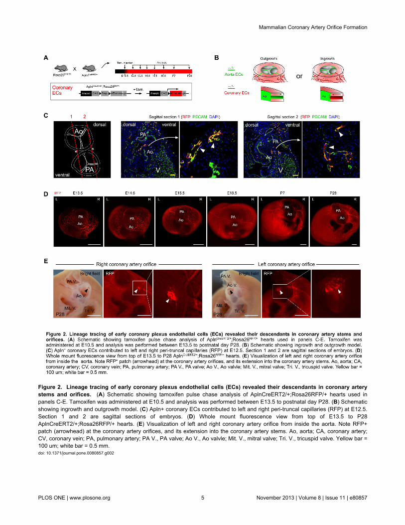
Figure 2. Lineage tracing of early coronary plexus endothelial cells (ECs) revealed their descendants in coronary arterystems and orifices. (A) Schematic showing tamoxifen pulse chase analysis of AplnCreERT2/+;Rosa26RFP/+ hearts used inpanels C-E. Tamoxifen was administered at E10.5 and analysis was performed between E13.5 to postnatal day P28. (B) Schematicshowing ingrowth and outgrowth model. (C) Apln+ coronary ECs contributed to left and right peri-truncal capillaries (RFP) at E12.5.Section 1 and 2 are sagittal sections of embryos. (D) Whole mount fluorescence view from top of E13.5 to P28AplnCreERT2/+;Rosa26RFP/+ hearts. (E) Visualization of left and right coronary artery orifice from inside the aorta. Note RFP+patch (arrowhead) at the coronary artery orifices, and its extension into the coronary artery stems. Ao, aorta; CA, coronary artery;CV, coronary vein; PA, pulmonary artery; PA V., PA valve; Ao V., Ao valvle; Mit. V., mitral valve; Tri. V., tricuspid valve. Yellow bar =100 um; white bar = 0.5 mm.doi: 10.1371/journal.pone.0080857.g002
Mammalian Coronary Artery Orifice Formation
PLOS ONE | www.plosone.org 5 November 2013 | Volume 8 | Issue 11 | e80857

Figure 3. Apln+ endothelial cells contribute to mature coronary arteries. (A) Immunostaining of genetic marker bet-Gal andextracellular marker periostin shows EPDCs were located around coronary vessels (white arrows). (B) Immunostaining of geneticmarker RFP and smooth muscle cell marker (aSMA) on AplnCreERT2/+;Rosa26RFP/+ heart sections. White bar = 50 um.doi: 10.1371/journal.pone.0080857.g003
Mammalian Coronary Artery Orifice Formation
PLOS ONE | www.plosone.org 6 November 2013 | Volume 8 | Issue 11 | e80857

Figure 4. Coronary orifice forms by recruiting coronary vascular endothelial cells into aorta. (A) Whole mount pictures ofE18.5 AplnCreERT2/+;Rosa26RFP/+ heart under fluorescence or bright field. Arrows indicate the left and right coronary orificeinside aorta. The zoomed out pictures are taken from base, and the magnified pictures are viewed from inside of aorta. Tamoxifenwas administered at E10.5. (B) Whole mount pictures of P7 AplnCreERT2/+;Rosa26RFP/+ heart under fluorescence or bright field.Arrowheads indicate RFP+ tubular structure representing coronary artery stem communicating with the aortic lumen (dotted line).The zoomed out and magnified pictures are viewed from right side of the heart and great vessels. Tamoxifen was injected at E10.5.Bar = 500 um.doi: 10.1371/journal.pone.0080857.g004
Mammalian Coronary Artery Orifice Formation
PLOS ONE | www.plosone.org 7 November 2013 | Volume 8 | Issue 11 | e80857

Figure 5. Apln+ peritruncal endothelial cells contribute to endothelia of coronary orifice. (A) Immunostaining of RFP andPECAM on serial sections (section 1 to 4) of E18.5 AplnCreERT2/+;Rosa26RFP/+ heart. Yellow arrows indicate labeled endothelialcells in coronary artery trunk and orifice, which are derived from Apln+ coronary endothelial cells. Tamoxfien was injected at E10.5.Serial sections were taken at 50 um intervals. (B) Immunostaining of aSMA (green) and genetic marker RFP (red) on serial sections(section 1 to 4) of P28 AplnCreERT2/+;Rosa26RFP/+ heart. Nuclei were stained with DAPI. Tamoxifen was administered at E10.5.Serial sections were taken at 50 um intervals. Inserts show magnified views of the coronary artery connection to aorta. Ao, aorta;CA, coronary artery. White bar = 100 um.doi: 10.1371/journal.pone.0080857.g005
Mammalian Coronary Artery Orifice Formation
PLOS ONE | www.plosone.org 8 November 2013 | Volume 8 | Issue 11 | e80857

Acknowledgements
We thank Thomas Quertermous for providing AplnLacZ/+ mice,and A.C.Gittenberger-de Groot for insightful comments onpaper.
Author Contributions
Conceived and designed the experiments: BZ YY. Performedthe experiments: XT TH LH HZ XH ZY. Analyzed the data: BZYY REP WL. Contributed reagents/materials/analysis tools:WTP. Wrote the manuscript: BZ WTP.
References
1. Reese DE, Mikawa T, Bader DM (2002) Development of the coronaryvessel system. Circ Res 91: 761-768. doi:10.1161/01.RES.0000038961.53759.3C. PubMed: 12411389.
2. Epstein JA (2010) Franklin H Epstein Lecture. Cardiac developmentand implications for heart disease. N Engl J Med 363: 1638-1647. doi:10.1056/NEJMra1003941. PubMed: 20961247.
3. Riley PR, Smart N (2011) Vascularizing the heart. Cardiovasc Res 91:260-268. doi:10.1093/cvr/cvr035. PubMed: 21282300.
4. Bennett HS (1936) The development of the blood supply to the heart inthe embryo pig. Am J Anat 60: 27-53. doi:10.1002/aja.1000600103.
5. Hutchins GM, Kessler-Hanna A, Moore GW (1988) Development of thecoronary arteries in the embryonic human heart. Circulation 77:1250-1257. doi:10.1161/01.CIR.77.6.1250. PubMed: 3286038.
6. Bogers AJ, Gittenberger-de Groot AC, Dubbeldam JA, Huysmans HA(1988) The inadequacy of existing theories on development of theproximal coronary arteries and their connexions with the arterial trunks.Int J Cardiol 20: 117-123. doi:10.1016/0167-5273(88)90321-X.PubMed: 3403075.
7. Bogers A, Gittenberger-de Groot A, Poelmann R, Peault B, HuysmansH (1989) Development of the origin of the coronary arteries, a matter ofingrowth or outgrowth? Anat Embryol (Berl) 180: 437-441. doi:10.1007/BF00305118.
8. Poelmann RE, Gittenberger-de Groot AC, Mentink MM, Bökenkamp R,Hogers B (1993) Development of the cardiac coronary vascularendothelium, studied with antiendothelial antibodies, in chicken-quailchimeras. Circ Res 73: 559-568. doi:10.1161/01.RES.73.3.559.PubMed: 8348697.
9. Ando K, Nakajima Y, Yamagishi T, Yamamoto S, Nakamura H (2004)Development of proximal coronary arteries in quail embryonic heart:multiple capillaries penetrating the aortic sinus fuse to form maincoronary trunk. Circ Res 94: 346-352. doi:10.1161/01.RES.0000112963.79064.09. PubMed: 14684625.
10. Sheikh AY, Chun HJ, Glassford AJ, Kundu RK, Kutschka I et al. (2008)In vivo genetic profiling and cellular localization of apelin reveals ahypoxia-sensitive, endothelial-centered pathway activated in ischemicheart failure. Am J Physiol Heart Circ Physiol 294: H88-H98. PubMed:17906101.
11. Madisen L, Zwingman TA, Sunkin SM, Oh SW, Zariwala HA et al.(2010) A robust and high-throughput Cre reporting and characterizationsystem for the whole mouse brain. Nat Neurosci 13: 133-140. doi:10.1038/nn.2467. PubMed: 20023653.
12. Tian X, Zhang H, He L, Huang X, Liu Q, Yu W, He L, Yang Z, Zhang Z,Zhong T, Yang X, Yang Z, Yan Y, Baldini A, Sun Y, Evans S, Red-Horse K, Zhou B (2013) Subepicardial Endothelial Cells Invade theEmbryonic Ventricle wall to Form Coronary Arteries. Cell Res 25 June2013. doi:10.1038/cr.2013.83.
13. Zhou B, Ma Q, Rajagopal S, Wu SM, Domian I et al. (2008) Epicardialprogenitors contribute to the cardiomyocyte lineage in the developing
heart. Nature 454: 109-113. doi:10.1038/nature07060. PubMed:18568026.
14. Zhou B, Honor LB, He H, Ma Q, Oh JH et al. (2011) Adult mouseepicardium modulates myocardial injury by secreting paracrine factors.J Clin Invest 121: 1894-1904. doi:10.1172/JCI45529. PubMed:21505261.
15. Zhou B, von Gise A, Ma Q, Hu YW, Pu WT (2010) Genetic fatemapping demonstrates contribution of epicardium-derived cells to theannulus fibrosis of the mammalian heart. Dev Biol 338: 251-261. doi:10.1016/j.ydbio.2009.12.007. PubMed: 20025864.
16. Red-Horse K, Ueno H, Weissman IL, Krasnow MA (2010) Coronaryarteries form by developmental reprogramming of venous cells. Nature464: 549-553. doi:10.1038/nature08873. PubMed: 20336138.
17. Smith A, Arnold R, Wilkinson JL, Hamilton DI, McKay R et al. (1986) Ananatomical study of the patterns of the coronary arteries and sinusnodal artery in complete transposition. Int J Cardiol 12: 295-307. doi:10.1016/0167-5273(86)90265-2. PubMed: 3759267.
18. Pérez-Pomares J, Phelps A, Sedmerova M, Carmona R, González-Iriarte M et al. (2002) Experimental studies on the spatiotemporalexpression of WT1 and RALDH2 in the embryonic avian heart: a modelfor the regulation of myocardial and valvuloseptal development byepicardially derived cells (EPDCs). Dev Biol 247: 307-326. doi:10.1006/dbio.2002.0706. PubMed: 12086469.
19. Ishii Y, Garriock RJ, Navetta AM, Coughlin LE, Mikawa T (2010) BMPsignals promote proepicardial protrusion necessary for recruitment ofcoronary vessel and epicardial progenitors to the heart. Dev Cell 19:307-316. doi:10.1016/j.devcel.2010.07.017. PubMed: 20708592.
20. Eralp I, Lie-Venema H, DeRuiter MC, van den Akker NM, Bogers AJ etal. (2005) Coronary artery and orifice development is associated withproper timing of epicardial outgrowth and correlated Fas-ligand-associated apoptosis patterns. Circ Res 96: 526-534. doi:10.1161/01.RES.0000158965.34647.4e. PubMed: 15705966.
21. Wilm B, Ipenberg A, Hastie ND, Burch JB, Bader DM (2005) Theserosal mesothelium is a major source of smooth muscle cells of thegut vasculature. Development 132: 5317-5328. doi:10.1242/dev.02141.PubMed: 16284122.
22. Cai CL, Martin JC, Sun Y, Cui L, Wang L et al. (2008) A myocardiallineage derives from Tbx18 epicardial cells. Nature 454: 104-108. doi:10.1038/nature06969. PubMed: 18480752.
23. Arima Y, Miyagawa-Tomita S, Maeda K, Asai R, Seya D et al. (2012)Preotic neural crest cells contribute to coronary artery smooth muscleinvolving endothelin signalling. Nat Commun 3: 1267. doi:10.1038/ncomms2258. PubMed: 23232397.
24. Wann S, Schuchard G (2006) Images in clinical medicine. Anomalousorigin of the right coronary artery. N Engl J Med 355: e8. doi:10.1056/NEJMicm050051. PubMed: 16943395.
25. Ou P, Iserin F, Vouhe P, Bonnet D (2006) Anomalous origin of the leftcoronary artery from the right aortic sinus: surgery based on diagnosisby 64-slice CT. Eur J Cardiothorac Surg 29: 610. doi:10.1016/j.ejcts.2005.12.031. PubMed: 16481182.
Mammalian Coronary Artery Orifice Formation
PLOS ONE | www.plosone.org 9 November 2013 | Volume 8 | Issue 11 | e80857