Cliona varians-Derived Actinomycetes as Bioresources of ...
24
marine drugs Article Cliona varians-Derived Actinomycetes as Bioresources of Photoprotection-Related Bioactive End-Products Jeysson Sánchez-Suárez 1,2 , Luisa Villamil 2 , Ericsson Coy-Barrera 3 and Luis Díaz 1,2, * Citation: Sánchez-Suárez, J.; Villamil, L.; Coy-Barrera, E.; Díaz, L. Cliona varians-Derived Actinomycetes as Bioresources of Photoprotection- Related Bioactive End-Products. Mar. Drugs 2021, 19, 674. https://doi.org/ 10.3390/md19120674 Academic Editor: Alexander N. Shikov Received: 28 October 2021 Accepted: 25 November 2021 Published: 27 November 2021 Publisher’s Note: MDPI stays neutral with regard to jurisdictional claims in published maps and institutional affil- iations. Copyright: © 2021 by the authors. Licensee MDPI, Basel, Switzerland. This article is an open access article distributed under the terms and conditions of the Creative Commons Attribution (CC BY) license (https:// creativecommons.org/licenses/by/ 4.0/). 1 Doctorate in Biosciences, School of Engineering, Universidad de La Sabana, Chía 250001, Colombia; [email protected] 2 Bioprospecting Research Group, School of Engineering, Universidad de La Sabana, Chía 250001, Colombia; [email protected] 3 Bioorganic Chemistry Laboratory, Universidad Militar Nueva Granada, Cajicá 250247, Colombia; [email protected] * Correspondence: [email protected] Abstract: Sunscreen and sunblock are crucial skincare products to prevent photoaging and photo- carcinogenesis through the addition of chemical filters to absorb or block ultraviolet (UV) radiation. However, several sunscreen and sunblock ingredients, mostly UV filters, have been associated with human and environmental safety concerns. Therefore, the exploration and discovery of promising novel sources of efficient and safer compounds with photoprotection-related activities are currently required. Marine invertebrates, particularly their associated microbiota, are promising providers of specialized metabolites with valuable biotechnological applications. Nevertheless, despite Actinobac- teria members being a well-known source of bioactive metabolites, their photoprotective potential has been poorly explored so far. Hence, a set of methanolic extracts obtained from Cliona varians- derived actinomycetes was screened regarding their antioxidant and UV-absorbing capacities (i.e., photoprotection-related activities). The active extract-producing strains were identified and classified within genera Streptomyces, Micrococcus, Gordonia, and Promicromonospora. This is the first report of the isolation of these microorganisms from C. varians (an ecologically important Caribbean coral reef-boring sponge). The in vitro cytotoxicity on dermal fibroblasts of oxybenzone and the selected active extracts revealed that oxybenzone exerted a cytotoxic effect, whereas no cytotoxic effect of test extracts was observed. Accordingly, the most active (SPFi > 5, radical scavenging > 50%) and nontoxic (cell viability > 75%) extracts were obtained from Streptomyces strains. Finally, LC-MS-based characterization suggested a broad chemical space within the test strains and agreed with the re- ported streptomycetes’ chemodiversity. The respective metabolite profiling exposed a strain-specific metabolite occurrence, leading to the recognition of potential hits. These findings suggest that marine Streptomyces produce photoprotectants ought to be further explored in skincare applications. Keywords: photoprotection; sunscreen; sponge; actinobacteria; Gordonia; Micrococcus; Promicromono- spora; Streptomyces 1. Introduction Chronic unprotected exposure to the sun is associated with the development of skin disorders such as altered immunity, photoaging, and cancer [1,2]. In the latter case, epi- demiological surveillance has shown alarming trends [3,4] that claim more attention to photoprotective behavior. One of the primary photoprotection strategies is the applica- tion of sunscreen and/or sunblock [5]. However, several sunscreening and sunblocking agents currently employed in photoprotective product manufacture have relevant safety concerns [6,7] and even adversely affect ecosystems [8,9]. The main goal of sun protectants is oriented to avoid the harmful effects of ultraviolet (UV) radiation (i.e., photodamage-related issues). Such a goal is commonly achieved by using chemical filters, whose purpose comprises specific actions based on absorbing, Mar. Drugs 2021, 19, 674. https://doi.org/10.3390/md19120674 https://www.mdpi.com/journal/marinedrugs
-
Upload
khangminh22 -
Category
Documents
-
view
5 -
download
0
Transcript of Cliona varians-Derived Actinomycetes as Bioresources of ...
marine drugs
Article
Cliona varians-Derived Actinomycetes as Bioresources ofPhotoprotection-Related Bioactive End-Products
Jeysson Sánchez-Suárez 1,2 , Luisa Villamil 2, Ericsson Coy-Barrera 3 and Luis Díaz 1,2,*
�����������������
Citation: Sánchez-Suárez, J.; Villamil,
L.; Coy-Barrera, E.; Díaz, L. Cliona
varians-Derived Actinomycetes as
Bioresources of Photoprotection-
Related Bioactive End-Products. Mar.
Drugs 2021, 19, 674. https://doi.org/
10.3390/md19120674
Academic Editor: Alexander N.
Shikov
Received: 28 October 2021
Accepted: 25 November 2021
Published: 27 November 2021
Publisher’s Note: MDPI stays neutral
with regard to jurisdictional claims in
published maps and institutional affil-
iations.
Copyright: © 2021 by the authors.
Licensee MDPI, Basel, Switzerland.
This article is an open access article
distributed under the terms and
conditions of the Creative Commons
Attribution (CC BY) license (https://
creativecommons.org/licenses/by/
4.0/).
1 Doctorate in Biosciences, School of Engineering, Universidad de La Sabana, Chía 250001, Colombia;[email protected]
2 Bioprospecting Research Group, School of Engineering, Universidad de La Sabana, Chía 250001, Colombia;[email protected]
3 Bioorganic Chemistry Laboratory, Universidad Militar Nueva Granada, Cajicá 250247, Colombia;[email protected]
* Correspondence: [email protected]
Abstract: Sunscreen and sunblock are crucial skincare products to prevent photoaging and photo-carcinogenesis through the addition of chemical filters to absorb or block ultraviolet (UV) radiation.However, several sunscreen and sunblock ingredients, mostly UV filters, have been associated withhuman and environmental safety concerns. Therefore, the exploration and discovery of promisingnovel sources of efficient and safer compounds with photoprotection-related activities are currentlyrequired. Marine invertebrates, particularly their associated microbiota, are promising providers ofspecialized metabolites with valuable biotechnological applications. Nevertheless, despite Actinobac-teria members being a well-known source of bioactive metabolites, their photoprotective potentialhas been poorly explored so far. Hence, a set of methanolic extracts obtained from Cliona varians-derived actinomycetes was screened regarding their antioxidant and UV-absorbing capacities (i.e.,photoprotection-related activities). The active extract-producing strains were identified and classifiedwithin genera Streptomyces, Micrococcus, Gordonia, and Promicromonospora. This is the first report ofthe isolation of these microorganisms from C. varians (an ecologically important Caribbean coralreef-boring sponge). The in vitro cytotoxicity on dermal fibroblasts of oxybenzone and the selectedactive extracts revealed that oxybenzone exerted a cytotoxic effect, whereas no cytotoxic effect oftest extracts was observed. Accordingly, the most active (SPFi > 5, radical scavenging > 50%) andnontoxic (cell viability > 75%) extracts were obtained from Streptomyces strains. Finally, LC-MS-basedcharacterization suggested a broad chemical space within the test strains and agreed with the re-ported streptomycetes’ chemodiversity. The respective metabolite profiling exposed a strain-specificmetabolite occurrence, leading to the recognition of potential hits. These findings suggest that marineStreptomyces produce photoprotectants ought to be further explored in skincare applications.
Keywords: photoprotection; sunscreen; sponge; actinobacteria; Gordonia; Micrococcus; Promicromono-spora; Streptomyces
1. Introduction
Chronic unprotected exposure to the sun is associated with the development of skindisorders such as altered immunity, photoaging, and cancer [1,2]. In the latter case, epi-demiological surveillance has shown alarming trends [3,4] that claim more attention tophotoprotective behavior. One of the primary photoprotection strategies is the applica-tion of sunscreen and/or sunblock [5]. However, several sunscreening and sunblockingagents currently employed in photoprotective product manufacture have relevant safetyconcerns [6,7] and even adversely affect ecosystems [8,9].
The main goal of sun protectants is oriented to avoid the harmful effects of ultraviolet(UV) radiation (i.e., photodamage-related issues). Such a goal is commonly achievedby using chemical filters, whose purpose comprises specific actions based on absorbing,
Mar. Drugs 2021, 19, 674. https://doi.org/10.3390/md19120674 https://www.mdpi.com/journal/marinedrugs
Mar. Drugs 2021, 19, 674 2 of 24
reflecting, and/or scattering UV radiation [10]. However, since photodamage can begenerated by different mechanisms [11], other agents such as antioxidants have recentlybeen added to improve the photoprotection efficacy of photoprotective products [12]. Thisrequirement for mitigating UV-induced skin damage might explain the growing demandfor topically applied biologically active ingredients [13,14]. Considering the limitationsregarding UV filters toxicity (for both human and environmental health) and their partialphotoprotection capability, there is a reasonable demand to find new photoprotectants.
During the search for new chemical entities, natural resources continue to offer apromising opportunity to discover biologically active compounds that satisfy humanity’sdemands [15]. Among the varied environment options to be explored, the marine bio-sphere comprises an attractive choice due to the biodiversity housed in these habitats [16].Marine sessile invertebrates have proved to be a priceless supply of compounds with acomprehensive chemodiversity and, consequently, a broad bioactivity profile [15]. In thissense, sponges are recognized as reliable reservoirs since respective chemical and biologicalcampaigns led to the isolation and characterization of several compounds that have beenthe direct or indirect basis for the development of important drugs [17].
Marine sessile invertebrates, particularly sponges, are undeniable sources of bioactivecompounds [18]. In several cases, the biosynthesis of these bioactive compounds has beendemonstrated to be associated with their symbiotic microorganisms [18]. Cliona varians is aboring encrusting sponge playing crucial ecological niches in coral reefs [19]. Bioprospect-ing studies on C. varians are scarce, and most of them have focused on their ecologicalroles. Recently, the bacterial diversity of C. varians was reported [19], and a high microbialabundance was revealed [20], including actinomycetes [19]. Despite the microbial andchemical richness exhibited by marine sponges, the development of commercially viableproducts (e.g., drugs, cosmetics) is limited by varied factors, namely complex structuresthat hinder their chemical synthesis, the low available amount of the targeted compoundand the “supply problem” [21–23]. These challenges have led to distinct approachesto exploit the respective chemical space available from these invertebrates, such as theisolation and fermentation of their associated microbiota for bio and chemoprospectinginitiatives [24,25]. In fact, this microbiota plays a relevant role in the metabolite regulationand chemodiversity described for the respective hosts (e.g., sponges) [26]. In several cases,the symbiotic microbes are the genuine source of the metabolite initially isolated from themacroorganism [24]. Consequently, bioprospecting research focused on microorganisms iscurrently attracting significant attention [27].
Regarding microorganisms, members of the phylum Actinobacteria are well-knownfor their contribution to the natural products field [28]. They are also recognized for estab-lishing symbiotic relationships [29], including sponge-associated microbiota that is knownto involve producers of active compounds of biotechnological interest [30]. Actinobacteriarepresent an intriguing taxon that encourages more research initiatives exploring theirspecialized metabolism. In this regard, the genus Streptomyces (class Actinobacteria) ispositioned within the most prolific microbes providing drugs, mostly antibiotic agents [28].Even though Streptomyces’ specialized metabolism has been intensely investigated, sev-eral niches remain unexamined, particularly the symbiotic ones [29,31]. Indeed, sinceStreptomyces have traditionally been those actinobacteria that have the largest record on asubstantial bioactive compounds’ repertoire, we have previously reviewed their potentialas a source of photoprotective substances [32].
Therefore, as part of our interest in bioactive compounds from actinomycetes associ-ated with marine holobionts, we aimed to identify actinobacteria strains isolated from themarine sponge Cliona varians, producing metabolites with photoprotection-related activities.The results revealed that different genera of the Actinobacteria class are part of the associ-ated microbiota of C. varians. Additionally, the strains exhibited strain-specific metaboliteoccurrence. In this regard, streptomycetes showed great potential in producing bioactivecompounds, specifically photoprotection-related activities. This study contributes to dis-
Mar. Drugs 2021, 19, 674 3 of 24
covering and recognizing new bioresources to explore the presence of metabolites with aninteresting bioactive profile applicable in the future to the skincare and cosmetics fields.
2. Results
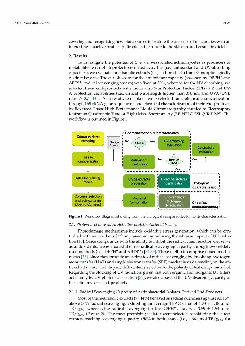
To investigate the potential of C. varians-associated actinomycetes as producers ofmetabolites with photoprotection-related activities (i.e., antioxidant and UV-absorbingcapacities), we evaluated methanolic extracts (i.e., end-products) from 35 morphologicallydistinct isolates. The cut-off score for the antioxidant capacity (assessed by DPPH• andABTS•+ radical scavenging assays) was fixed at 50%, whereas for the UV absorbing, weselected those end-products with the in vitro Sun Protection Factor (SPFi) > 2 and UV-A protection capabilities (i.e., critical wavelength higher than 370 nm and UVA/UVBratio ≥ 0.7 [33]). As a result, ten isolates were selected for biological characterizationthrough 16S rRNA gene sequencing and chemical characterization of their end-productsby Reversed-Phase High-Performance Liquid Chromatography coupled to ElectrosprayIonization Quadropole Time-of-Flight Mass Spectrometry (RP-HPLC-ESI-Q-ToF-MS). Theworkflow is outlined in Figure 1.
Mar. Drugs 2021, 19, x FOR PEER REVIEW 3 of 25
bioactive compounds, specifically photoprotection-related activities. This study contrib-utes to discovering and recognizing new bioresources to explore the presence of metabo-lites with an interesting bioactive profile applicable in the future to the skincare and cos-metics fields.
2. Results To investigate the potential of C. varians-associated actinomycetes as producers of
metabolites with photoprotection-related activities (i.e., antioxidant and UV-absorbing ca-pacities), we evaluated methanolic extracts (i.e., end-products) from 35 morphologically distinct isolates. The cut-off score for the antioxidant capacity (assessed by DPPH• and ABTS•+ radical scavenging assays) was fixed at 50%, whereas for the UV absorbing, we selected those end-products with the in vitro Sun Protection Factor (SPFi) > 2 and UV-A protection capabilities (i.e., critical wavelength higher than 370 nm and UVA/UVB ratio ≥ 0.7 [33]). As a result, ten isolates were selected for biological characterization through 16S rRNA gene sequencing and chemical characterization of their end-products by Reversed-Phase High-Performance Liquid Chromatography coupled to Electrospray Ionization Quadropole Time-of-Flight Mass Spectrometry (RP-HPLC-ESI-Q-ToF-MS). The workflow is outlined in Figure 1.
Figure 1. Workflow diagram showing from the biological sample collection to its characteriza-tion.
2.1. Photoprotection-Related Activities of Actinobacterial Isolates Photodamage mechanisms include oxidative stress generation, which can be con-
trolled with antioxidants [12] or prevented by reducing the adverse impact of UV radia-tion [10]. Since compounds with the ability to inhibit the radical chain reaction can serve as antioxidants, we evaluated the free radical scavenging capacity through two widely used methods (i.e., DPPH• and ABTS•+) [34,35]. These methods comprise mixed mecha-nisms [36], since they provide an estimate of radical scavenging by involving hydrogen atom transfer (HAT) and single electron transfer (SET) mechanisms depending on the an-tioxidant nature, and they are differentially selective to the polarity of test compounds [35]. Regarding the blocking of UV radiation, given that both organic and inorganic UV filters act mainly by UV photons absorption [37], we also assessed the UV-absorbing ca-pacity of the actinomycetes end-products.
2.1.1. Radical Scavenging Capacity of Actinobacterial Isolates-Derived End-Products Most of the methanolic extracts (77.14%) behaved as radical quenchers against
ABTS•+ above 50% radical scavenging, exhibiting an average TEAC value of 4.03 ± 1.18
Figure 1. Workflow diagram showing from the biological sample collection to its characterization.
2.1. Photoprotection-Related Activities of Actinobacterial Isolates
Photodamage mechanisms include oxidative stress generation, which can be con-trolled with antioxidants [12] or prevented by reducing the adverse impact of UV radia-tion [10]. Since compounds with the ability to inhibit the radical chain reaction can serveas antioxidants, we evaluated the free radical scavenging capacity through two widelyused methods (i.e., DPPH• and ABTS•+) [34,35]. These methods comprise mixed mecha-nisms [36], since they provide an estimate of radical scavenging by involving hydrogenatom transfer (HAT) and single electron transfer (SET) mechanisms depending on the an-tioxidant nature, and they are differentially selective to the polarity of test compounds [35].Regarding the blocking of UV radiation, given that both organic and inorganic UV filtersact mainly by UV photons absorption [37], we also assessed the UV-absorbing capacity ofthe actinomycetes end-products.
2.1.1. Radical Scavenging Capacity of Actinobacterial Isolates-Derived End-Products
Most of the methanolic extracts (77.14%) behaved as radical quenchers against ABTS•+
above 50% radical scavenging, exhibiting an average TEAC value of 4.03 ± 1.18 µmolTE/gDW, whereas the radical scavenging for the DPPH• assay was 3.59 ± 1.54 µmolTE/gDW (Figure 2). The most promising isolates were selected considering those testextracts reaching scavenging capacity >50% in both assays (i.e., 4.66 µmol TE/gDW for

Mar. Drugs 2021, 19, 674 4 of 24
DPPH assays and 3.35 µmol TE/gDW for ABTS assays), namely G6210, G6211, G1115,G11117, G11122, G11126, G11128, G1225, G1228, and G12218.
Mar. Drugs 2021, 19, x FOR PEER REVIEW 4 of 25
μmol TE/gDW, whereas the radical scavenging for the DPPH• assay was 3.59 ± 1.54 μmol TE/gDW (Figure 2). The most promising isolates were selected considering those test ex-tracts reaching scavenging capacity >50% in both assays (i.e., 4.66 μmol TE/gDW for DPPH assays and 3.35 μmol TE/gDW for ABTS assays), namely G6210, G6211, G1115, G11117, G11122, G11126, G11128, G1225, G1228, and G12218.
Figure 2. Antioxidant screening of actinobacterial methanolic extracts. Purple bars represent DPPH assays and green bars represent ABTS assays. Each TEAC (Trolox equivalent antioxidant capacity) value is expressed as the mean (n = 3), and error bars represent standard deviation (SD). Red lines indicate TEACs of 4.66 and 3.35 values for the DPPH and ABTS assays, respec-tively.
Since phenolics and flavonoids are compounds with well-recognized antioxidant ca-pacity, we estimated their total content in all test methanolic extracts to examine their plausible participation in the measured radical scavenging ability. Although actinomy-cetes can synthesize flavonoids, the results showed that the tested methanolic extracts, unlike G11126 (TFC = 55.57 ± 10.97 mg QE/100gDW), did not show detectable levels of fla-vonoids. In contrast, the methanolic extracts contained a broad content of phenolics (i.e., from 26.69 to 160.15 mg GA/100 gDW), enabling a correlation analysis against the antioxi-dant results (Figure 3). Although the Pearson correlation coefficient was low for both as-says (i.e., 0.353 and 0.250 for DPPH• and ABTS•+, respectively), the correlation calculated for DPPH• was statistically significant (p = 0.026; Figure 3).
Figure 3. Correlation between antioxidant capacity assays and phenolic content. The results of ABTS and DPPH assays are shown as green squares and purple circles, respectively. P-value was determined by the Pearson correlation coefficient. Total phenolic content is expressed as milli-grams of gallic acid equivalents per 100 g of dry weight (mg GAE/100gDW).
Figure 2. Antioxidant screening of actinobacterial methanolic extracts. Purple bars represent DPPHassays and green bars represent ABTS assays. Each TEAC (Trolox equivalent antioxidant capacity)value is expressed as the mean (n = 3), and error bars represent standard deviation (SD). Red linesindicate TEACs of 4.66 and 3.35 values for the DPPH and ABTS assays, respectively.
Since phenolics and flavonoids are compounds with well-recognized antioxidantcapacity, we estimated their total content in all test methanolic extracts to examine theirplausible participation in the measured radical scavenging ability. Although actinomycetescan synthesize flavonoids, the results showed that the tested methanolic extracts, unlikeG11126 (TFC = 55.57± 10.97 mg QE/100gDW), did not show detectable levels of flavonoids.In contrast, the methanolic extracts contained a broad content of phenolics (i.e., from 26.69to 160.15 mg GA/100 gDW), enabling a correlation analysis against the antioxidant results(Figure 3). Although the Pearson correlation coefficient was low for both assays (i.e., 0.353and 0.250 for DPPH• and ABTS•+, respectively), the correlation calculated for DPPH• wasstatistically significant (p = 0.026; Figure 3).
Mar. Drugs 2021, 19, x FOR PEER REVIEW 4 of 25
μmol TE/gDW, whereas the radical scavenging for the DPPH• assay was 3.59 ± 1.54 μmol TE/gDW (Figure 2). The most promising isolates were selected considering those test ex-tracts reaching scavenging capacity >50% in both assays (i.e., 4.66 μmol TE/gDW for DPPH assays and 3.35 μmol TE/gDW for ABTS assays), namely G6210, G6211, G1115, G11117, G11122, G11126, G11128, G1225, G1228, and G12218.
Figure 2. Antioxidant screening of actinobacterial methanolic extracts. Purple bars represent DPPH assays and green bars represent ABTS assays. Each TEAC (Trolox equivalent antioxidant capacity) value is expressed as the mean (n = 3), and error bars represent standard deviation (SD). Red lines indicate TEACs of 4.66 and 3.35 values for the DPPH and ABTS assays, respec-tively.
Since phenolics and flavonoids are compounds with well-recognized antioxidant ca-pacity, we estimated their total content in all test methanolic extracts to examine their plausible participation in the measured radical scavenging ability. Although actinomy-cetes can synthesize flavonoids, the results showed that the tested methanolic extracts, unlike G11126 (TFC = 55.57 ± 10.97 mg QE/100gDW), did not show detectable levels of fla-vonoids. In contrast, the methanolic extracts contained a broad content of phenolics (i.e., from 26.69 to 160.15 mg GA/100 gDW), enabling a correlation analysis against the antioxi-dant results (Figure 3). Although the Pearson correlation coefficient was low for both as-says (i.e., 0.353 and 0.250 for DPPH• and ABTS•+, respectively), the correlation calculated for DPPH• was statistically significant (p = 0.026; Figure 3).
Figure 3. Correlation between antioxidant capacity assays and phenolic content. The results of ABTS and DPPH assays are shown as green squares and purple circles, respectively. P-value was determined by the Pearson correlation coefficient. Total phenolic content is expressed as milli-grams of gallic acid equivalents per 100 g of dry weight (mg GAE/100gDW).
Figure 3. Correlation between antioxidant capacity assays and phenolic content. The results ofABTS and DPPH assays are shown as green squares and purple circles, respectively. P-value wasdetermined by the Pearson correlation coefficient. Total phenolic content is expressed as milligramsof gallic acid equivalents per 100 g of dry weight (mg GAE/100gDW).
2.1.2. UV-Absorbing Capacity of Actinobacterial Isolates-Derived Extracts
The bacterial extracts with the higher antioxidant capacity were assessed for the UV-absorbing profile. Oxybenzone was employed as a reference UV filter. The bacterial extracts

Mar. Drugs 2021, 19, 674 5 of 24
displayed diverse abilities to absorb UV radiation, ranging from 16.06% to 76.58% of theSPFi calculated for oxybenzone. Three isolates reached encouraging SPFis with valuesgreater than 10 (in increasing order: G6210, G1225, and G1228; Figure 4a). Most of theavailable UV filters offer better UV-B protection; hence, compounds with UV-A protectiondemand more attention [38]. Interestingly, the actinobacterial extracts showed a λC higherthan 370 nm, which is required to claim UV-A protection capability [33]. Additionally, theUVA/UVB ratio was the same or higher than the exhibited by oxybenzone (Figure 4b),achieving a 4-star rating according to the Boots Star Rating System [39]. These resultsindicate the occurrence of metabolites with a particular ability to absorb UV radiationbetween 320 and 400 nm over the UV-B spectrum.
Mar. Drugs 2021, 19, x FOR PEER REVIEW 5 of 25
2.1.2. UV-Absorbing Capacity of Actinobacterial Isolates-Derived Extracts The bacterial extracts with the higher antioxidant capacity were assessed for the UV-
absorbing profile. Oxybenzone was employed as a reference UV filter. The bacterial ex-tracts displayed diverse abilities to absorb UV radiation, ranging from 16.06% to 76.58% of the SPFi calculated for oxybenzone. Three isolates reached encouraging SPFis with val-ues greater than 10 (in increasing order: G6210, G1225, and G1228; Figure 4a). Most of the available UV filters offer better UV-B protection; hence, compounds with UV-A protection demand more attention [38]. Interestingly, the actinobacterial extracts showed a λC higher than 370 nm, which is required to claim UV-A protection capability [33]. Additionally, the UVA/UVB ratio was the same or higher than the exhibited by oxybenzone (Figure 4b), achieving a 4-star rating according to the Boots Star Rating System [39]. These results in-dicate the occurrence of metabolites with a particular ability to absorb UV radiation be-tween 320 and 400 nm over the UV-B spectrum.
Figure 4. UV-absorbing profile of the actinobacterial methanolic extracts: (a) Results of SPFi calculations; (b) Results of the UV-A protection profile; the left y-axis shows the critical wave-length (λC; the orange line indicates the threshold to claim broad-spectrum protection, i.e., 370 nm), and the right y-axis shows the UVA/UVB ratio. BP-3, which was used as a reference com-pound, is shown in gray.
2.2. In vitro Safety Evaluation of the Photoprotective Actinobacterial Extracts Most commercially available UV filters have been associated with toxic effects, in-
cluding oxybenzone [40]. We evaluated the cytotoxic effect of the selected photoprotective actinobacterial extracts (i.e., G6210, G6211, G1115, G11117, G11122, G11126, G11128, G1225, G1228, and G12218) on human dermal fibroblasts (HDFa cell line) and compared it to that shown by oxybenzone on the same cell line (Figure 5). Dimethyl sulfoxide (DMSO), at concentrations between 3 and 10% (v/v) (Figure 5a), was used as a cytotoxic reference substance. Oxybenzone displayed a cytotoxic effect with an IC50 of 75.93 μg/mL (Figure 5b). The cytotoxic effect was also observed at a morphological level, since HDFa cells exhibited a damaged cell morphology (i.e., loss of their spindly appearance, altera-tion in cell shape, membrane deformability; Figure 5e) compared with the usual visual appearance of these cells (i.e., crowded cells with elongated cell bodies and differentiated narrow ends; Figure 5c). A micrograph of HDFa after exposure to G1225 extract (i.e., the highest one antioxidant capacity) for 24 h is presented in Figure S1.
Regarding actinobacterial extracts, they were inactive (yielding far less than 50% cell viability reduction, Figure 6) and agree with drug discovery standards [41]. In addition, most of the extracts (90%) showed a cytotoxic effect higher than 12% on the HDFa cells. The G1115 extract exhibited the most cytotoxic action (reducing 23.84% of the cell viability at 500 μg/mL), whereas the cell viability was higher than 88% for the rest of the actinobac-terial extracts.
Figure 4. UV-absorbing profile of the actinobacterial methanolic extracts: (a) Results of SPFi calcula-tions; (b) Results of the UV-A protection profile; the left y-axis shows the critical wavelength (λC; theorange line indicates the threshold to claim broad-spectrum protection, i.e., 370 nm), and the righty-axis shows the UVA/UVB ratio. BP-3, which was used as a reference compound, is shown in gray.
2.2. In Vitro Safety Evaluation of the Photoprotective Actinobacterial Extracts
Most commercially available UV filters have been associated with toxic effects, in-cluding oxybenzone [40]. We evaluated the cytotoxic effect of the selected photoprotectiveactinobacterial extracts (i.e., G6210, G6211, G1115, G11117, G11122, G11126, G11128, G1225,G1228, and G12218) on human dermal fibroblasts (HDFa cell line) and compared it to thatshown by oxybenzone on the same cell line (Figure 5). Dimethyl sulfoxide (DMSO), atconcentrations between 3 and 10% (v/v) (Figure 5a), was used as a cytotoxic reference sub-stance. Oxybenzone displayed a cytotoxic effect with an IC50 of 75.93 µg/mL (Figure 5b).The cytotoxic effect was also observed at a morphological level, since HDFa cells exhib-ited a damaged cell morphology (i.e., loss of their spindly appearance, alteration in cellshape, membrane deformability; Figure 5e) compared with the usual visual appearance ofthese cells (i.e., crowded cells with elongated cell bodies and differentiated narrow ends;Figure 5c). A micrograph of HDFa after exposure to G1225 extract (i.e., the highest oneantioxidant capacity) for 24 h is presented in Figure S1.
Regarding actinobacterial extracts, they were inactive (yielding far less than 50% cellviability reduction, Figure 6) and agree with drug discovery standards [41]. In addition,most of the extracts (90%) showed a cytotoxic effect higher than 12% on the HDFa cells.The G1115 extract exhibited the most cytotoxic action (reducing 23.84% of the cell via-bility at 500 µg/mL), whereas the cell viability was higher than 88% for the rest of theactinobacterial extracts.

Mar. Drugs 2021, 19, 674 6 of 24Mar. Drugs 2021, 19, x FOR PEER REVIEW 6 of 25
Figure 5. Cytotoxic evaluation of oxybenzone: (a) concentration–response curve of DMSO (be-tween 10 and 3% v/v), (b) concentration–response curve of oxybenzone (between 400 and 3 μg/mL), (c) micrograph of HDFa cells in normal conditions, (d) micrograph of HDFa cells after 24 h the exposure to DMSO (9% v/v), (e) micrograph of HDFa cells exposed to oxybenzone (200 μg/mL) during 24 h.
Figure 6. Cytotoxic evaluation of the actinobacterial crude extracts. The crude extracts were evaluated at 5, 50, and 500 μg/mL. Each value represents the mean (n = 3), and error bars rep-resent standard deviation (SD). Cell viability was calculated with respect to untreated cells (control).
2.3. Identification of Isolates with Photoprotective Potential by 16S rRNA Gene Sequencing The 16S rRNA gene was sequenced to identify those selected strains producing ex-
tracts with potential photoprotective activity. Then, a phylogenetic analysis with highly similar bacterial strains (chosen after a BLAST (Basic Local Alignment Search Tool) search) was conducted. The cladogram led to classifying the bioactive isolates among the actino-bacterial genera Streptomyces (i.e., G11126, G1228, G1225, G11122, G6210, and G6211; Fig-ure 7), Micrococcus (i.e., G11117 and G11128; Figure 8a), Gordonia (i.e., G1115; Figure 8b),
Figure 5. Cytotoxic evaluation of oxybenzone: (a) concentration–response curve of DMSO (between10 and 3% v/v), (b) concentration–response curve of oxybenzone (between 400 and 3 µg/mL),(c) micrograph of HDFa cells in normal conditions, (d) micrograph of HDFa cells after 24 h theexposure to DMSO (9% v/v), (e) micrograph of HDFa cells exposed to oxybenzone (200 µg/mL)during 24 h.
Mar. Drugs 2021, 19, x FOR PEER REVIEW 6 of 25
Figure 5. Cytotoxic evaluation of oxybenzone: (a) concentration–response curve of DMSO (be-tween 10 and 3% v/v), (b) concentration–response curve of oxybenzone (between 400 and 3 μg/mL), (c) micrograph of HDFa cells in normal conditions, (d) micrograph of HDFa cells after 24 h the exposure to DMSO (9% v/v), (e) micrograph of HDFa cells exposed to oxybenzone (200 μg/mL) during 24 h.
Figure 6. Cytotoxic evaluation of the actinobacterial crude extracts. The crude extracts were evaluated at 5, 50, and 500 μg/mL. Each value represents the mean (n = 3), and error bars rep-resent standard deviation (SD). Cell viability was calculated with respect to untreated cells (control).
2.3. Identification of Isolates with Photoprotective Potential by 16S rRNA Gene Sequencing The 16S rRNA gene was sequenced to identify those selected strains producing ex-
tracts with potential photoprotective activity. Then, a phylogenetic analysis with highly similar bacterial strains (chosen after a BLAST (Basic Local Alignment Search Tool) search) was conducted. The cladogram led to classifying the bioactive isolates among the actino-bacterial genera Streptomyces (i.e., G11126, G1228, G1225, G11122, G6210, and G6211; Fig-ure 7), Micrococcus (i.e., G11117 and G11128; Figure 8a), Gordonia (i.e., G1115; Figure 8b),
Figure 6. Cytotoxic evaluation of the actinobacterial crude extracts. The crude extracts were evaluatedat 5, 50, and 500 µg/mL. Each value represents the mean (n = 3), and error bars represent standarddeviation (SD). Cell viability was calculated with respect to untreated cells (control).
2.3. Identification of Isolates with Photoprotective Potential by 16S rRNA Gene Sequencing
The 16S rRNA gene was sequenced to identify those selected strains producing extractswith potential photoprotective activity. Then, a phylogenetic analysis with highly similarbacterial strains (chosen after a BLAST (Basic Local Alignment Search Tool) search) wasconducted. The cladogram led to classifying the bioactive isolates among the actinobacterialgenera Streptomyces (i.e., G11126, G1228, G1225, G11122, G6210, and G6211; Figure 7),Micrococcus (i.e., G11117 and G11128; Figure 8a), Gordonia (i.e., G1115; Figure 8b), and

Mar. Drugs 2021, 19, 674 7 of 24
Promicromonospora (i.e., G12218; Figure 8c). The coding of the end-products and the namesof the identified strains are presented in Table S1. To the best of our knowledge, this studyis the first one reporting the isolation of these genera from C. varians.
Mar. Drugs 2021, 19, x FOR PEER REVIEW 7 of 25
and Promicromonospora (i.e., G12218; Figure 8c). The coding of the end-products and the names of the identified strains are presented in Table S1. To the best of our knowledge, this study is the first one reporting the isolation of these genera from C. varians.
Figure 7. Phylogenetic tree of the bioactive streptomycetes isolates. The cladogram was built using the sequence of the 16S rDNA gene. The optimal tree is shown. The accession code of the blasted strains is shown in parentheses. The percentage of replicate trees with associated taxa clustered together in the bootstrap test (1000 replicates) is displayed next to the branches. All ambiguous positions were removed for each sequence pair (pairwise deletion option).
Figure 8. Phylogenetic tree of the bioactive non-streptomycete isolates: (a) cladogram for strains of genus Micrococcus, (b) cladogram for the strain of genus Gordonia, (c) cladogram for the strain of genus Promicromonospora. The cladograms were built using the sequence of the 16S rDNA gene. The optimal tree is shown. The accession code of the blasted strains is shown in parentheses. The per-centage of replicate trees with associated taxa clustered together in the bootstrap test (1000 repli-cates) is displayed next to the branches. All ambiguous positions were removed for each sequence pair (pairwise deletion option).
Figure 7. Phylogenetic tree of the bioactive streptomycetes isolates. The cladogram was built usingthe sequence of the 16S rDNA gene. The optimal tree is shown. The accession code of the blastedstrains is shown in parentheses. The percentage of replicate trees with associated taxa clusteredtogether in the bootstrap test (1000 replicates) is displayed next to the branches. All ambiguouspositions were removed for each sequence pair (pairwise deletion option).
Mar. Drugs 2021, 19, x FOR PEER REVIEW 7 of 25
and Promicromonospora (i.e., G12218; Figure 8c). The coding of the end-products and the names of the identified strains are presented in Table S1. To the best of our knowledge, this study is the first one reporting the isolation of these genera from C. varians.
Figure 7. Phylogenetic tree of the bioactive streptomycetes isolates. The cladogram was built using the sequence of the 16S rDNA gene. The optimal tree is shown. The accession code of the blasted strains is shown in parentheses. The percentage of replicate trees with associated taxa clustered together in the bootstrap test (1000 replicates) is displayed next to the branches. All ambiguous positions were removed for each sequence pair (pairwise deletion option).
Figure 8. Phylogenetic tree of the bioactive non-streptomycete isolates: (a) cladogram for strains of genus Micrococcus, (b) cladogram for the strain of genus Gordonia, (c) cladogram for the strain of genus Promicromonospora. The cladograms were built using the sequence of the 16S rDNA gene. The optimal tree is shown. The accession code of the blasted strains is shown in parentheses. The per-centage of replicate trees with associated taxa clustered together in the bootstrap test (1000 repli-cates) is displayed next to the branches. All ambiguous positions were removed for each sequence pair (pairwise deletion option).
Figure 8. Phylogenetic tree of the bioactive non-streptomycete isolates: (a) cladogram for strains ofgenus Micrococcus, (b) cladogram for the strain of genus Gordonia, (c) cladogram for the strain of genusPromicromonospora. The cladograms were built using the sequence of the 16S rDNA gene. The optimaltree is shown. The accession code of the blasted strains is shown in parentheses. The percentageof replicate trees with associated taxa clustered together in the bootstrap test (1000 replicates) isdisplayed next to the branches. All ambiguous positions were removed for each sequence pair(pairwise deletion option).
Mar. Drugs 2021, 19, 674 8 of 24
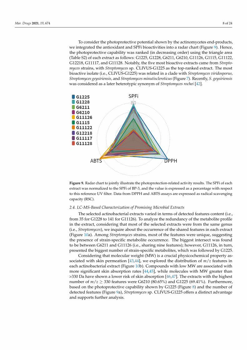
To consider the photoprotective potential shown by the actinomycetes end-products,we integrated the antioxidant and SPFi bioactivities into a radar chart (Figure 9). Hence,the photoprotective capability was ranked (in decreasing order) using the triangle area(Table S2) of each extract as follows: G1225, G1228, G6211, G6210, G11126, G1115, G11122,G12218, G11117, and G11128. Notably, the five most bioactive extracts came from Strepto-myces strains, with Streptomyces sp. CLIVUS-G1225 as the top-ranked extract. The mostbioactive isolate (i.e., CLIVUS-G1225) was related in a clade with Streptomyces viridosporus,Streptomyces geysiriensis, and Streptomyces minutiscleroticus (Figure 7). Recently, S. geysiriensiswas considered as a later heterotypic synonym of Streptomyces rochei [42].
Mar. Drugs 2021, 19, x FOR PEER REVIEW 8 of 25
To consider the photoprotective potential shown by the actinomycetes end-products, we integrated the antioxidant and SPFi bioactivities into a radar chart (Figure 9). Hence, the photoprotective capability was ranked (in decreasing order) using the triangle area (Table S2) of each extract as follows: G1225, G1228, G6211, G6210, G11126, G1115, G11122, G12218, G11117, and G11128. Notably, the five most bioactive extracts came from Strepto-myces strains, with Streptomyces sp. CLIVUS-G1225 as the top-ranked extract. The most bioactive isolate (i.e., CLIVUS-G1225) was related in a clade with Streptomyces viridosporus, Streptomyces geysiriensis, and Streptomyces minutiscleroticus (Figure 7). Recently, S. geysiriensis was considered as a later heterotypic synonym of Streptomyces rochei [42].
Figure 9. Radar chart to jointly illustrate the photoprotection-related activity results. The SPFi of each extract was normalized to the SPFi of BP-3, and the value is expressed as a percentage with respect to this reference UV filter. Data from DPPH and ABTS assays are expressed as radical scavenging capacity (RSC).
2.4. LC-MS-Based Characterization of Promising Microbial Extracts The selected actinobacterial extracts varied in terms of detected features content (i.e.,
from 35 for G1228 to 141 for G11126). To analyze the redundancy of the metabolite profile in the extract, considering that most of the selected extracts were from the same genus (i.e., Streptomyces), we inquire about the occurrence of the shared features in each extract (Figure 10a). Among Streptomyces strains, most of the features were unique, suggesting the presence of strain-specific metabolite occurrence. The biggest intersect was found to be between G6211 and G11126 (i.e., sharing nine features); however, G11126, in turn, pre-sented the biggest number of strain-specific metabolites, which was followed by G1225.
Considering that molecular weight (MW) is a crucial physicochemical property asso-ciated with skin permeation [43,44], we explored the distribution of m/z features in each actinobacterial extract (Figure 10b). Compounds with low MW are associated with more significant skin absorption rates [44,45], while molecules with MW greater than > 330 Da have shown a lower risk of skin absorption [46,47]. The extracts with the highest number of m/z ≥ 330 features were G6210 (80.65%) and G1225 (69.41%). Furthermore, based on the photoprotective capability shown by G1225 (Figure 8) and the number of detected fea-tures (Figure 9a), Streptomyces sp. CLIVUS-G1225 offers a distinct advantage and supports further analysis.
Figure 9. Radar chart to jointly illustrate the photoprotection-related activity results. The SPFi of eachextract was normalized to the SPFi of BP-3, and the value is expressed as a percentage with respectto this reference UV filter. Data from DPPH and ABTS assays are expressed as radical scavengingcapacity (RSC).
2.4. LC-MS-Based Characterization of Promising Microbial Extracts
The selected actinobacterial extracts varied in terms of detected features content (i.e.,from 35 for G1228 to 141 for G11126). To analyze the redundancy of the metabolite profilein the extract, considering that most of the selected extracts were from the same genus(i.e., Streptomyces), we inquire about the occurrence of the shared features in each extract(Figure 10a). Among Streptomyces strains, most of the features were unique, suggestingthe presence of strain-specific metabolite occurrence. The biggest intersect was foundto be between G6211 and G11126 (i.e., sharing nine features); however, G11126, in turn,presented the biggest number of strain-specific metabolites, which was followed by G1225.
Considering that molecular weight (MW) is a crucial physicochemical property as-sociated with skin permeation [43,44], we explored the distribution of m/z features ineach actinobacterial extract (Figure 10b). Compounds with low MW are associated withmore significant skin absorption rates [44,45], while molecules with MW greater than>330 Da have shown a lower risk of skin absorption [46,47]. The extracts with the highestnumber of m/z ≥ 330 features were G6210 (80.65%) and G1225 (69.41%). Furthermore,based on the photoprotective capability shown by G1225 (Figure 8) and the number ofdetected features (Figure 9a), Streptomyces sp. CLIVUS-G1225 offers a distinct advantageand supports further analysis.

Mar. Drugs 2021, 19, 674 9 of 24
Mar. Drugs 2021, 19, x FOR PEER REVIEW 9 of 25
Figure 10. Overview of the LC-MS-based characterization of the bioactive actinobacterial ex-tracts: (a) common and unique features are presented. The interconnected points indicate the extracts involved in the intersection; (b) m/z distribution of the features in each extract.
The resulting feature list from the MS-based annotation of the G1225 extract is sum-marized in Table 1. The features were identified at levels 3 (i.e., putative candidate) and 4 (i.e., unequivocal molecular formula), according to the confidence levels to communicate compound identity by high-resolution mass spectrometry (HRMS), as proposed by Schy-manski et al. [48]. The masses ranged from 310.23 to 949.52 Da, which were commonly found among Streptomyces-derived metabolites (percentile 34th = 330.7 Da, Figure S2). At level 3, we annotated eight compounds (Table 2; Figure A1), from which two were isolated from Streptomyces, namely the sideromycin A (10) from S. violaceus DSM 8286 [49] and the glomecidin (12) from S. lavendulae H698 SY2 [50]. Although tris(2,4-di-tert-bu-tylphenyl)phosphate (14) was annotated using StreptomeDB, it was isolated from a co-culture between the fungus Bionectria sp. and S. lividans [51]. In the same study, 14 was also isolated from a co-culture between Bionectria sp. and the bacterium B. subtilis, dis-carding the streptomycete as the source [51]. However, 14 has been previously identified in Streptomyces by LC-MS-based annotation [52]. In addition, we also putatively identified other compounds that were not previously reported in any streptomycete. Metabolites metacridamide A (7), periconiasin J (11), and icosalide B (15) were isolated from fungi species: Metarhizium acridum [53], Periconia sp. F-31 [54], and an unidentified fungus [55], respectively. Recent studies associated 15 with bacterial sources. It was firstly detected by MS-guide dereplication from the betaproteobacterium Burkholderia gladioli [56], and then, it was described that Streptomyces sp. JBS5-6 has the biosynthetic gene cluster of icosalide B [57]. For their part, bacillamidin C (4) and erythrazole A (13) were isolated from non-actinobacteria species, Bacillus pumilus RJA1515 [58] and Erythrobacter sp. SNB-035 [59], respectively.
Figure 10. Overview of the LC-MS-based characterization of the bioactive actinobacterial extracts:(a) common and unique features are presented. The interconnected points indicate the extractsinvolved in the intersection; (b) m/z distribution of the features in each extract.
The resulting feature list from the MS-based annotation of the G1225 extract is sum-marized in Table 1. The features were identified at levels 3 (i.e., putative candidate) and4 (i.e., unequivocal molecular formula), according to the confidence levels to commu-nicate compound identity by high-resolution mass spectrometry (HRMS), as proposedby Schymanski et al. [48]. The masses ranged from 310.23 to 949.52 Da, which werecommonly found among Streptomyces-derived metabolites (percentile 34th = 330.7 Da,Figure S2). At level 3, we annotated eight compounds (Table 2; Figure A1), from whichtwo were isolated from Streptomyces, namely the sideromycin A (10) from S. violaceus DSM8286 [49] and the glomecidin (12) from S. lavendulae H698 SY2 [50]. Although tris(2,4-di-tert-butylphenyl)phosphate (14) was annotated using StreptomeDB, it was isolated froma co-culture between the fungus Bionectria sp. and S. lividans [51]. In the same study, 14was also isolated from a co-culture between Bionectria sp. and the bacterium B. subtilis,discarding the streptomycete as the source [51]. However, 14 has been previously identifiedin Streptomyces by LC-MS-based annotation [52]. In addition, we also putatively identifiedother compounds that were not previously reported in any streptomycete. Metabolitesmetacridamide A (7), periconiasin J (11), and icosalide B (15) were isolated from fungispecies: Metarhizium acridum [53], Periconia sp. F-31 [54], and an unidentified fungus [55],respectively. Recent studies associated 15 with bacterial sources. It was firstly detectedby MS-guide dereplication from the betaproteobacterium Burkholderia gladioli [56], andthen, it was described that Streptomyces sp. JBS5-6 has the biosynthetic gene cluster oficosalide B [57]. For their part, bacillamidin C (4) and erythrazole A (13) were isolated fromnon-actinobacteria species, Bacillus pumilus RJA1515 [58] and Erythrobacter sp. SNB-035 [59],respectively.

Mar. Drugs 2021, 19, 674 10 of 24
Table 1. Mass spectra data of the annotated features in Streptomyces sp. strain CLIVUS-G1225.
ID a RT b MolecularFormula
Adduct Typem/z Isomer
Coincidence dIdentification
Level eExperimental Calculated ∆ (ppm) c
1 2.81 C28H38N4O6 [M+H]+ 527.2838 527.2864 5.02 4 42 6.37 C26H29NO5 [M+2ACN+H] + 518.2643 518.2650 1.39 7 43 14.50 C22H26O6 [M+H]+ 387.1814 387.1802 2.96 13 44 15.22 C17H30N2O3 [M+2ACN+H] + 393.2877 393.2860 4.22 1 35 15.69 C26H28O6 [M+H]+ 437.1947 437.1959 2.66 5 46 16.30 C24H30O6 [M+H]+ 415.2129 415.2115 3.35 9 47 20.00 C37H55NO6 [M+2ACN+H]+ 692.4602 692.4633 4.47 1 38 20.12 C32H54N4O7 [M+ACN+H] + 648.4338 648.4331 1.21 6 49 21.07 C35H63NO3 [M+H]+ 546.4901 546.4881 3.69 3 410 21.79 C42H75N7O17 [M+ACN+H] + 991.5576 991.5557 1.89 1 311 22.70 C21H31NO2 [M+2ACN+H] + 412.2968 412.2959 2.22 1 312 22.85 C27H37N7O7 [M+2ACN+H] + 654.3348 654.3358 1.48 1 313 22.93 C31H42N2O7S [M+2ACN+H] + 669.3346 669.3317 4.32 1 314 23.02 C42H63O4P [M+H]+ 663.4566 663.4537 4.38 1 315 23.34 C34H60N4O10 [M+H]+ 685.4385 685.4382 0.48 1 316 23.44 C22H38O2 [M+ACN+H] + 376.3202 376.321 2.19 3 417 25.29 C32H26O10 [M+ACN+H] + 612.1849 612.1864 2.49 8 4
a The annotated features were identified with consecutive Arabic numerals from 1 to 17 according to the elution order; b Retention time(min), c Mass measurement accuracy (ppm), d The number of isomers found in the databases; e Identification confidence levels according toSchymanski et al. [48]. Two-way profile (i.e., HPLC chromatogram ×mass spectra) of the G1225 extract is presented in Figure S3.
Considering the importance of low skin absorption in skincare products, we inquiredabout the lipophilicity of the level 3 identified compounds by calculating the partitioncoefficient between n-octanol and water (cLogP; a well-established descriptor of a com-pound permeability [60]). In this sense, 10 and 12 stand out as putatively low-permeabilitycompounds, which is a desired physicochemical characteristic focusing on the safety ofskincare products [46,47].
Table 2. Bioactivity records of the main identified compounds at level 3 in Streptomyces sp. strain CLIVUS-G1225.
ID a Name cLogP Previously Described Bioactivities
4 Bacillamidin C 3.191 Antimicrobial, non-cytotoxic (evaluated against HepG2, A549, MDA-MB-231,SGC7901) [58].
7 Metacridamide A 8.213 Cytotoxic against Caco-2, MCF-7, HepG2/C3A [53].10 Sideromycin A −1.133 Antimicrobial [49].11 Periconiasin J 3.132 Anti-HIV, non-cytotoxic (evaluated against MCF-7) [54].12 Glomecidin −2.695 Antifungal [50].13 Erythrazole A 6.946 Non-cytotoxic (evaluated against non-small cell lung cancer cell lines) [59].14 Tris(2,4-di-tert-
butylphenyl)phosphate 13.777 Anti-inflammatory [61].15 Icosalide B 2.551 Antiviral and moderate cytotoxic activities (evaluated against MDCK cells) [55].
a Only eight compounds, namely 4, 7, 10, 11, 12, 13, 14, and 15 could be putatively identified (i.e., level 3 according to Schymanski et al. [48])in the G1225 extract.
Concerning the compounds identified at level 4 (i.e., 1–3, 5, 6, 8, 9, 16, and 17), thenumber of isomers limited their further analysis (51 options). However, to reduce thepossibilities, closely related isomers were clustered to identify shared structural moietiesrepresenting each isomer (Figure 11). Hence, it was found that only for compound 17, allisomers clustered into one group, with isonigerone as the representative compound.
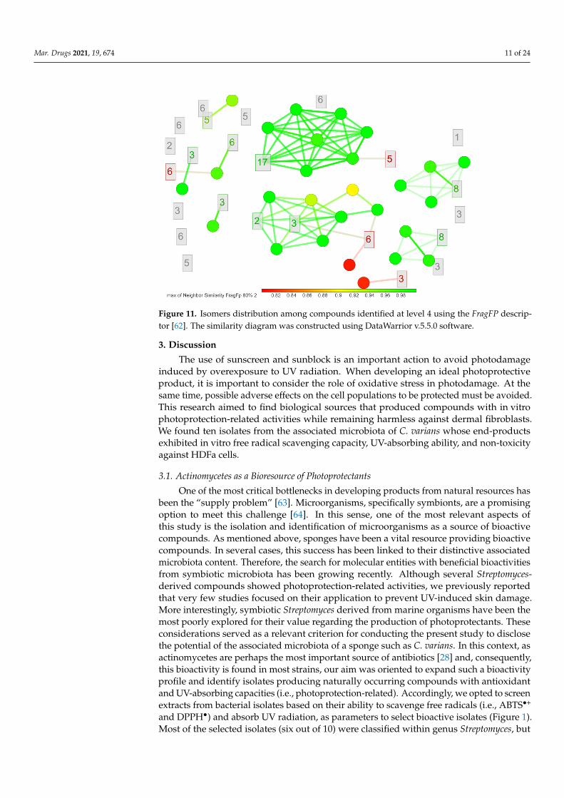
Mar. Drugs 2021, 19, 674 11 of 24Mar. Drugs 2021, 19, x FOR PEER REVIEW 11 of 25
Figure 11. Isomers distribution among compounds identified at level 4 using the FragFP de-scriptor [62]. The similarity diagram was constructed using DataWarrior v.5.5.0 software.
3. Discussion The use of sunscreen and sunblock is an important action to avoid photodamage in-
duced by overexposure to UV radiation. When developing an ideal photoprotective prod-uct, it is important to consider the role of oxidative stress in photodamage. At the same time, possible adverse effects on the cell populations to be protected must be avoided. This research aimed to find biological sources that produced compounds with in vitro photoprotection-related activities while remaining harmless against dermal fibroblasts. We found ten isolates from the associated microbiota of C. varians whose end-products exhibited in vitro free radical scavenging capacity, UV-absorbing ability, and non-toxicity against HDFa cells.
3.1. Actinomycetes as a Bioresource of Photoprotectants One of the most critical bottlenecks in developing products from natural resources
has been the “supply problem” [63]. Microorganisms, specifically symbionts, are a prom-ising option to meet this challenge [64]. In this sense, one of the most relevant aspects of this study is the isolation and identification of microorganisms as a source of bioactive compounds. As mentioned above, sponges have been a vital resource providing bioactive compounds. In several cases, this success has been linked to their distinctive associated microbiota content. Therefore, the search for molecular entities with beneficial bioactivi-ties from symbiotic microbiota has been growing recently. Although several Streptomyces-derived compounds showed photoprotection-related activities, we previously reported that very few studies focused on their application to prevent UV-induced skin damage. More interestingly, symbiotic Streptomyces derived from marine organisms have been the most poorly explored for their value regarding the production of photoprotectants. These considerations served as a relevant criterion for conducting the present study to disclose the potential of the associated microbiota of a sponge such as C. varians. In this context, as actinomycetes are perhaps the most important source of antibiotics [28] and, conse-quently, this bioactivity is found in most strains, our aim was oriented to expand such a bioactivity profile and identify isolates producing naturally occurring compounds with antioxidant and UV-absorbing capacities (i.e., photoprotection-related). Accordingly, we opted to screen extracts from bacterial isolates based on their ability to scavenge free rad-icals (i.e., ABTS•+ and DPPH•) and absorb UV radiation, as parameters to select bioactive isolates (Figure 1). Most of the selected isolates (six out of 10) were classified within genus
Figure 11. Isomers distribution among compounds identified at level 4 using the FragFP descrip-tor [62]. The similarity diagram was constructed using DataWarrior v.5.5.0 software.
3. Discussion
The use of sunscreen and sunblock is an important action to avoid photodamageinduced by overexposure to UV radiation. When developing an ideal photoprotectiveproduct, it is important to consider the role of oxidative stress in photodamage. At thesame time, possible adverse effects on the cell populations to be protected must be avoided.This research aimed to find biological sources that produced compounds with in vitrophotoprotection-related activities while remaining harmless against dermal fibroblasts.We found ten isolates from the associated microbiota of C. varians whose end-productsexhibited in vitro free radical scavenging capacity, UV-absorbing ability, and non-toxicityagainst HDFa cells.
3.1. Actinomycetes as a Bioresource of Photoprotectants
One of the most critical bottlenecks in developing products from natural resources hasbeen the “supply problem” [63]. Microorganisms, specifically symbionts, are a promisingoption to meet this challenge [64]. In this sense, one of the most relevant aspects ofthis study is the isolation and identification of microorganisms as a source of bioactivecompounds. As mentioned above, sponges have been a vital resource providing bioactivecompounds. In several cases, this success has been linked to their distinctive associatedmicrobiota content. Therefore, the search for molecular entities with beneficial bioactivitiesfrom symbiotic microbiota has been growing recently. Although several Streptomyces-derived compounds showed photoprotection-related activities, we previously reportedthat very few studies focused on their application to prevent UV-induced skin damage.More interestingly, symbiotic Streptomyces derived from marine organisms have been themost poorly explored for their value regarding the production of photoprotectants. Theseconsiderations served as a relevant criterion for conducting the present study to disclosethe potential of the associated microbiota of a sponge such as C. varians. In this context, asactinomycetes are perhaps the most important source of antibiotics [28] and, consequently,this bioactivity is found in most strains, our aim was oriented to expand such a bioactivityprofile and identify isolates producing naturally occurring compounds with antioxidantand UV-absorbing capacities (i.e., photoprotection-related). Accordingly, we opted to screenextracts from bacterial isolates based on their ability to scavenge free radicals (i.e., ABTS•+
and DPPH•) and absorb UV radiation, as parameters to select bioactive isolates (Figure 1).Most of the selected isolates (six out of 10) were classified within genus Streptomyces, but

Mar. Drugs 2021, 19, 674 12 of 24
these Streptomyces strains formed different clades (Figure 6). Considering that severalstudies have reported a wide chemodiversity in the Streptomyces genus [65], even withinstrains of the same species [66], a plausible estimation can be oriented to the fact that eachstrain could produce different metabolite profiles, involving relevant compounds withphotoprotective potential.
According to the List of Prokaryotic names with Standing in Nomenclature (LPNS) [67],there are 675 validly published and correct names of Streptomyces species (plus 816 syn-onyms). From these numbers, 44 have been reported between 2018 and 2021 (date access24 October 2021). This overview gives an idea of the vast biodiversity of the genus andclearly shows the importance of further exploration in the search for new streptomycetes.Except for strain CLIVUS-G1228 (forming a clade with Streptomyces coelicoflavus with abootstrap value of 100%), the bootstrap values calculated for the remaining strains did notallow defining the respective Streptomyces species. These strains could even be new species;however, this fact will need to be further studied.
On the other hand, although Streptomyces continue offering valuable chances to findnew bioactive compounds, rare actinomycetes (i.e., non-streptomycetes) are a distinct andrelevant bioresource to search for highly valuable bioactive compounds [68]. Herein, fourrare actinomycetes strains (i.e., Micrococcus sp. CLIVUS-G11128, Micrococcus sp. CLIVUS-G11117, Gordonia sp. CLIVUS-G1115, and Promicromonospora sp. CLIVUS-G12218) werealso isolated. Compared to Promicromonospora, Micrococcus, and Gordonia microorganisms,species of genus Streptomyces are by far a well-studied natural source of bioactive metabo-lites. For instance, using the genus name as a keyword in the Scopus database (searchperformed on 12/Oct/2021), Streptomyces yields 43,097 hits, Micrococcus yields 18,105 hits,Gordonia yields 1,243 hits (without excluded Gordonia plant genus coincidences), and Promi-cromonospora yields 117 hits. This fact indicates that Gordonia and Promicromonospora generaare underexplored biological sources and gives an added research value to the strains weidentify here (i.e., Gordonia sp. CLIVUS-G1115 and Promicromonospora sp. CLIVUS-G12218).
For the group of rare actinomycetes, compounds with antioxidant capacity, mainlypigments, have been reported in Micrococcus strains [69–71]. They have also been foundto produce exopolysaccharides that can be used in various industrial sectors that requireantioxidants [72,73]. A particularly interesting metabolite is 2,2′-[3-methoxy-1′-amyl-5′-methyl-4-(1′′-pyrryl)]dipyrryl methene [69], which is a prodigiosin-like compound. Thismetabolite, besides being derived from Streptomyces [74], is known for its photoprotec-tive properties [75]. Interestingly, a UV-specific repair enzyme has been isolated fromMicrococcus luteus, which represents another promising alternative to using this biore-source to find photoprotection hits [76]. As expected, in the case of Gordonia and Promi-cromonospra, no studies investigated their photoprotective potential to our knowledge.However, carotenoids have been isolated from Gordonia [77], which are known for their an-tioxidative properties [78]. This information supports the idea that although understudied,these genera have photoprotective potential that justifies further study.
According to the bioactivities radar chart (Figure 9), the most promising strain wasStreptomyces sp. CLIVUS-G1225. Although the strain types of the closest-related specieswere isolated from soil samples, some have also been isolated from marine sources: forinstance, S. rochei from the marine sponge Dysidea arenaria [79] and S. geysiriensis from themarine sponge Iotrochota sp. [80]. Nevertheless, as far as we know, this is the first report ofthese Streptomyces strains isolated from the boring sponge C. varians. The taxonomic identityof Streptomyces sp. CLIVUS-G1225 needs to be further investigated using a whole-genomesequencing approach.
3.2. Scavenging, UV-Absorbing, and Cytotoxicity Potential of Actinomycetes End-Products
It has been shown that topical antioxidants can prevent and promote the restorationof UV-induced damage [81]. The purpose of an antioxidant is to stabilize a radical throughHAT and/or SET mechanisms. Although the DPPH and ABTS assays are sensitive toboth mechanisms (i.e., mixed-mode methods [82]), a solvent effect favors one over the
Mar. Drugs 2021, 19, 674 13 of 24
other [83]. Hence, they allow the estimation of different types of radical scavengingcompounds. Additionally, although both radicals have their radical at a hidden site,these steric hindrances are different. For example, the mechanism of reaction for DPPHwith antioxidants is analogous to that of peroxyl radicals (ROO•; a type of UV-inducedreactive oxygen species [84]) [82]. This fact explains the differences between the DPPH andABTS results for the test extracts. In the case of the extracts of the selected isolates (i.e.,G6210, G6211, G1115, G11117, G11122, G11126, G11128, G1225, G1228, and G12218), theresults could be associated with the presence of compounds that are not limited by sterichindrance of either radical and might jointly exhibit radical scavenging by both HAT andSET mechanisms. We also found that the content of phenolics and flavonoids were notdeterminant in the antioxidant capacity of the test actinomycete-derived extracts. In fact,only one extract presented detectable levels of flavonoids, which agreed with previousstudies [85–87].
Most UV filters approved for sunscreens absorb UV-B radiation thoroughly; very feware broad-spectrum (UV-B and UV-A), and even fewer are UV-A absorbers [88]. BP-3 isconsidered broad spectrum, although it protects more against UV-B and UV-AII (i.e., 320to 340 nm) [89]. These features are consistent with the λC herein calculated (i.e., 362 nm,Figure 3b). As a result of its efficiency in absorbing UV radiation, it is one of the mostwidely used UV filters, not only in sunscreens [90], and consequently, it is considered abenchmark agent. However, a growing number of reports have raised concerns about thesafety of BP-3 [90]. Our results were consistent with this fact, and BP-3 showed cytotoxiceffects at the indicated concentrations. Indeed, the calculated IC50 of BP-3 corresponds to47.46 µg/cm2 on cell monolayer in the in vitro assay, which is lower than the maximumallowed amount (i.e., 120–200 µg/cm2 [91]) in a recommended dose of sunscreen (i.e.,2000 µg/cm2, the quantity of sunscreen per unit of skin surface [92]).
Compared to BP-3, SPFi results of isolates G6210, G1225, and G1225 suggest thepresence of compounds absorbing in the UV-B region (i.e., they reached 69.0, 79.1, and 76.3%of the SPFi calculated for BP-3). Furthermore, the λC values and the UVA/UVB ratio led toinfer that they behave as broad-spectrum UV filters. Remarkably, these extracts were foundas non-toxic against HDFa cells. UV-A injury is primarily produced in dermal tissue, wherefibroblasts (a significant dermal cell population) are more susceptible to UV-A radiationthan keratinocytes [93]. Therefore, selected actinobacterial extracts, with compoundsabsorbing preferably UV-A radiation and with an innocuous effect after exposure tofibroblast, constitute a starting point in the search for exploitable bioresources to obtainvaluable photoprotectant agents. However, since our results are based on in vitro assays,further steps are required to validate these findings with in vivo methods.
3.3. Metabolite Profile of Promising Actinomycetes End-Products
We considered the hypothesis that most metabolites among Streptomyces strainswould be shared. However, extracts contained mostly strain-specific metabolites, evenamong streptomycetes. This fact is consistent with the cladogram shown by Streptomycesstrains (Figure 7), where each strain was placed in separate clades and with the well-known chemodiversity reported for the genus, including marine sponge-derived strepto-mycetes [94]. We decided to focus on the Streptomyces sp. CLIVUS-G1225 end-product,since it was found to be the most active one (Figure 9). Its metabolite profile presented ahigh number of features; most of these exhibited a MW higher than 330 Da (i.e., associatedwith low skin absorption). Skin absorption is an undesirable property in topically appliedproducts, since it may result in interactions with unintended targets leading to side effects(e.g., endocrine disruptors [95]). These data located this Streptomyces strain in an advan-tageous position since the opportunity to find a biological producer of metabolites withpreferred photoprotection-related properties, such as antioxidant, UV-absorbing, and safercharacteristics (i.e., non-cytotoxic and high MW) could be favored.
Interestingly, several of the compounds annotated at level 3 in G1225 had not pre-viously been reported in streptomycetes (i.e., 11, 15, 4, and 13). Moreover, 7, 11, and 15
Mar. Drugs 2021, 19, 674 14 of 24
have been isolated from fungi. Even isonigerone (the compound that was found repre-sentative of the isomers associated with 17; Figure A1), a naphthopyrone, was isolatedfrom the marine-derived fungus Aspergillus carbonarius [96]. In Streptomyces, this type ofnaturally occurring compound has also been described [97]. There are other cases relatedto the biosynthesis of fungi-derived compounds by Streptomyces, such as aspergilazine A(isolated from the marine-derived fungus Aspergillus taichungensis [98] and Streptomycessp. NRRL S-1868 [99]) and violapyrone J (isolated from the fungus Cylindrocarpon [100]and Streptomyces somaliensis SCSIO ZH66 [101]). Other intriguing examples that linkedthe biosynthetic pathways of fungi and Streptomyces are the β-lactams cephalosporin C(isolated from the fungus Acremonium [102]) and O-carbamoyl-deacetylcephalosporin C(isolated from S. clavuligerus [103]). Further examples involving cephalosporin-type com-pounds are reported by Higgens et al. [104]. These data could add further evidence tobe further explored to the already recognized diversity in the biosynthetic machinery ofstreptomycetes and provide more relevance to the hypothesis of biosynthetic gene transferfrom actinomycetes to fungi [105].
Regarding the bioactivity potential of the identified compounds, they have beenpoorly explored (Table 2). Compound 15 has shown in vivo anti-inflammatory effects [61].Since inflammation is a pathophysiological response of UV insult [2], only 15 could beassociated with photoprotection-related activities. Nevertheless, all the metabolites arearomatic and aliphatic amides, and several nitrogen-containing compounds have been as-sociated with photoprotective capabilities, such as mycosporine-like amino acids [106] andbetacyanins [107]. In fact, we have previously reported that amide-containing compoundsare the most associated with the photoprotection-related activity, including antioxidantcapacity, for Streptomyces [32]. For instance, thiazole-containing compounds derived fromStreptomyces, such as 13, have exhibited promising antioxidant capacity [108]. Therefore,we hypothesized that Streptomyces sp. CLIVUS-G1225 is a promising bioresource of under-explored compounds.
Microorganisms offer advantageous opportunities as a bioresource of bioactive com-pounds. Biotechnological tools could improve the target metabolite biosynthesis moreeasily compared to macroorganisms. In the case of Streptomyces, the implementation ofapproaches such as media engineering [109] and genetic engineering [109] for enhancingthe production of value-added compounds have been successful. For instance, improve-ments of target metabolite production up to four-fold have been achieved in Streptomycesstrains [110,111]. Ribosomal engineering is another interesting approach that has beenproven to improve metabolite yield in Streptomyces [112]. Even though antibiotics arethe main bioactivity found in Streptomyces-derived metabolites, several antioxidants havealso been described [32], and only the undecylprodigiosinone has been characterized as aUV-absorbing compound from Streptomyces [113]. From these findings, Streptomyces is avaluable resource that can also be exploited to search for compounds with photoprotectiveproperties leading to the development of novel sunscreens.
4. Materials and Methods4.1. Chemicals and Reagents
The 2,2-diphenyl-1-picrylhydrazyl’(DPPH), 2,2′-azino-bis(3-ethylbenzothiazoline-6-sulfonic acid) (ABTS), L-ascorbic acid, gallic acid, Trolox, quercetin, Folin–Ciocalteu reagent(FCR), and methanol were purchased from MilliporeSigma (St. Louis, MO, USA). The3-(4,5-Dimethylthiazol-2-yl)-2,5-Diphenyltetrazolium Bromide (MTT) was acquired fromThermo Fisher Scientific Inc. (Waltham, MA USA).
4.2. Sample Collection and Actinobacteria Isolation
Cliona varians specimens were collected by scuba diving from the Colombian Caribbean,Bahía de Taganga, Punta Venado (11◦16′23.9′′ N 74◦12′24.9′′ W) and Bahía de Santa Marta,Punta Betín (11◦15′02.1′′ N 74◦13′16.0′′ W), Magdalena, Colombia, at 13 m and 9 m depth,respectively. Around 10 g per sample (two individuals on each location) were placed in
Mar. Drugs 2021, 19, 674 15 of 24
sterile plastic bags and transported on ice until processing. The taxonomic classificationwas performed by Dr. Sven Zea, confirming that all collected specimens were C. varians.The samples used in the present study have a Colombian origin, and they were obtainedaccording to Amendment No. 5 to ARG Master Agreement No. 117 of 26 May 2015, grantedby the Ministry of Environment and Sustainable Development, Colombia.
In the laboratory and aseptic conditions, the samples were quickly rinsed with abun-dant sterile seawater; then, the sponge tissue was homogenized using a mortar and pestlethat were previously sterilized. Serial dilutions (1/10) up to 10−6 were prepared fromthe homogenate, and 100 µL of the dilutions 100, 10−3, and 10−6 were plated, in dupli-cate, on Glucose Yeast Medium (GYM) and Zobell Marine Medium (Zobell) [114]. Mediacomposition can be consulted in Table S3. Media were supplemented with nalidixic acid(50 µg/mL). Cycloheximide (100 µg/mL) was used to prevent fungal growth. The plateswere incubated at 30 ◦C and carefully monitored for 8 weeks.
As actinobacteria-like colonies were picked, isolated, and individually grown to obtainpure cultures. Bacterial isolates were cryopreserved in their respective isolation mediumwith glycerol (30% v/v).
4.3. Submerged Fermentation and Crude Extracts Preparation
Based on 35 morphologically distinct actinomycetes isolates obtained from GYM (i.e.,G627, G6210, G6211, G1118, G1115, G11114, G11117, G11118, G11121, G11122, G11123,G11126, G11128, G11129, G1224, G1225, G1226, G1228, G1229, G12210, G12213, andG12218) and Zobell (i.e., Z616, Z6225, Z6231, Z6235, Z713, Z726, Z1119, Z1215, Z1216,Z12136, Z12141, Z1221, and Z1222) medium, 10 mL of liquid submerged fermentationwas done in the respective isolation media (i.e., GYM or Zobell). The seed culture wasprepared inoculating a 55.81 mm2 plug from a 7-day-old lawn growth plate in 3 mL ofbroth. This broth culture was incubated at 30 ºC, 200 rpm agitation, for 7 days. Fromthis seed culture, 1 mL was used to inoculate 9 mL of GYM or Zobell broth for 7 days at30 °C and 200 rpm agitation). Then, each culture was lyophilized and submitted to anultrasound-assisted extraction (frequency: 25 kHz; power effective: 200 W; temperature:<30 ◦C) with HPLC-grade methanol. Then, the extracted fraction was dried by a rotaryevaporator and stored at 4 °C until the in vitro assays were completed. Each dry extractwas resuspended in methanol according to the target tested concentrations.
4.4. Antioxidant Capacity Assays and Total Phenol and Flavonoid Content Measurements
The antioxidant capacity of the end-products was estimated using the 2,2′-azinobis-(3-ethylbenzothiazoline-6-sulfonic acid (ABTS) assay and the 2,2-di(4-tert-octylphenyl)-1-picrylhydrazyl (DPPH) assay, which are among the most used methods to estimateantioxidant capacity methods [34]. Assays were performed in 96-well plates in triplicate.Briefly, absorbance measurements were performed using an iMarkTM microplate reader(Bio-Rad Laboratories, Inc., Hercules, CA, USA), with Microplate Manager® Software v6.3(Bio-Rad Laboratories, Inc., Hercules, CA, USA).
4.4.1. DPPH and ABTS Radical Scavenging Assays
The radical scavenging assays were performed as described previously [115] withsome modifications. Briefly, a stock solution of DPPH at 0.2 mM in methanol and ABTS at7 mM in distilled water were prepared. For the DPPH assay, 100 µL of microbial extractswere added to the 96-well plate; then, 100 µL of DPPH at 0.2 mM were suspended on eachmicrobial extract (5 mg/mL, final concentration). The mixture was incubated for 30 min atroom temperature and protected from light, and absorbance was measured at 515 nm.
For the ABTS assay, we follow Re et al. [116] with some modifications. Firstly, amixture of ABTS with 2.45 mM potassium persulfate (final concentration) was allowed tostand under dark at room temperature for 16 h to generate ABTS radical cation (ABTS•+).The ABTS•+ solution was diluted with distilled water until an absorbance of 0.70 ± 0.02 at735 nm. Then, 190 µL of the adjusted ABTS•+ solution was added to 10 µL of microbial

Mar. Drugs 2021, 19, 674 16 of 24
extract (5 mg/mL, final concentration). Absorbance was recorded at 735 nm after 10 min ofincubation at room temperature and protected from light.
The antioxidant capacity was expressed as radical scavenging capacity (RSC) andTrolox equivalents antioxidant capacity (TEAC). RSC was expressed as percentage (%) andcalculated by Equation (1) as follows:
Radical scavenging capacity (%) =AC −AS
AC× 100 (1)
where AC is the absorbance of the blank (radical solution with solvent), and AS is theabsorbance of the radical solution mixed with the tested samples after incubation.
TEAC was expressed as µmol Trolox equivalents per gram of dry weight (µmolTE/gDW) and calculated by Equation (2) as follows:
TEAC (µmol TE/gDW) =T (µmol/L)
S (g/L)(2)
where T is the Trolox concentration obtained after interpolation using standard curves ofTrolox from both assays (DPPH: Y = 2.167X − 0.5203; ABTS: Y = 3.066X − 1.356) and S isthe concentration of the test sample.
4.4.2. Total Phenol and Flavonoid Content
Total phenol content (TPC) was measured following the protocol reported by Mag-alhães et al. [117]. Briefly, 50 µL of the sample were suspended in each well, which wasfollowed by 50 µL of the FCR (previously diluted 1:5 v/v in distilled water). Afterwards,100 µL of NaOH at 0.35 M was added. Finally, the mixture was incubated at room tempera-ture for 3 min, and the absorbance at 750 nm was recorded. The TPC was calculated usinga gallic acid standard curve between 20.00 and 1.25 µg/mL. Data were expressed as mgGAE/100gDW.
The total flavonoid content (TFC) was measured as described by Buitrago et al. [115].Briefly, 50 µL of ethanol was mixed with 10 µL of aluminum trichloride (10%) and 10 µLof sodium acetate (0.1 M). Once this mixture was dispersed in each well, we then added70 µL of the samples. After 40 min under darkness at room temperature, the flavonoidcontent was estimated by measuring the absorbance at 415 nm. The TFC was calculatedusing a quercetin standard curve between 30.00 and 0.47 µg/mL. Data were expressed asmg QE/100gDW.
4.4.3. Determination of the In Vitro Sun Protection Factor and UV-Absorbing Profile
The absorbance spectra of the microbial extracts and BP-3 (1 mg/mL and 30 µg/mL, re-spectively, in quartz cuvettes) in the 290–400 nm region were recorded using a GENESYS™10S UV-Vis Spectrophotometer (Thermo Fisher Scientific Inc., Waltham, MA, USA). Thein vitro Sun Protection Factor (SPFi) (Equation (3))was spectrophotometrically calculatedaccording to Mansur et al. [118].
SPFi = CF×320
∑290
EE(λ)× I(λ)× A(λ) (3)
where CF is the correction factor (i.e., 10), EE (λ) is the erythemal action spectrum, I (λ)is the solar intensity spectrum, and A (λ) is the absorbance value at wavelength λ. Thevalues of EE (λ) × I (λ) are constants and were obtained from [119].
For the calculation of the Critical Wavelength (λC), the following Equation (4) [39]was used: ∫ λc
290A(λ)dλ = 0.9
∫ 400
290A(λ)dλ (4)
where A is absorption and λ wavelength.
Mar. Drugs 2021, 19, 674 17 of 24
The UVA ratio was calculated using the following Equation (5) [39]:
UVA/UVBratio =
∫ 400320 A(λ)dλ∫ 320290 A(λ)dλ
. (5)
According to the UVA ratio result, the samples were classified using the start ratingsystem [39].
4.4.4. Sequencing of 16S rRNA Gene and Phylogenetic Analysis
Genomic DNA was extracted using a Quick-DNA Fungal/Bacterial Microprep kit(Zymo Research Corporation, Irvine, CA, USA) according to the manufacturer’s instruc-tions. The 16S rRNA gene was amplified using the universal primers 27F and 1492R underthe following PRC cycling conditions: initial denaturation at 94 ◦C for 3 min, followed by30 cycles of 94 ◦C for 1 min, 50 ◦C for 1 min, and 72 ◦C for 2 min, with a final extensionof 72 ◦C for 7 min. The amplification products were verified by agarose electrophoresis.The 16S rRNA gene sequences were blasted against the rRNA database with the megablastalgorithm [120]. Consensus sequences were deposited in GenBank under the accessionnumbers OK598071 to OK598080. To find the closest neighbors used as reference sequencesin the phylogenetic analysis, a Neighbor-Joining tree was conducted using MEGA versionX [121] using the Tamura 3-parameter model. The reliability of the phylogenetic treetopology was assessed by the bootstrap test (1000 replications).
4.4.5. Cytotoxicity Assay
The cytotoxic effect was evaluated on human primary dermal fibroblast (HDFa,ATCC® PCS-201-012™, Primary Dermal Fibroblast; Normal, Human, Adult). HDFa werecultured in Dulbecco’s Modified Eagle Medium supplemented (DMEM) with 10% fetalbovine serum at 37 ◦C and 5% CO2. Cells were seeded into a 96-well plate at a densityof 2 × 10⁴ cells/well and incubated overnight before exposure to treatment. The cellswere treated 24 h with microbial extracts at 500, 50, and 5 µg/mL. Dimethyl sulfoxide(DMSO, between 1 and 10% v/v), methanol (0.5% v/v), and oxybenzone (between 400.00and 3.13 µg/mL) were used as controls. After treatments, supernatants were replaced withfresh media with 3-(4,5-dimethylthiazol-2-yl)-2,5-diphenyltetrazolium bromide (MTT) at0.5 mg/mL and incubated for 4 h [122]. Then, the MTT was removed, and DMSO wasadded to each well to dissolve formazan crystals. The amount of formazan was mea-sured by its absorbance at 570 nm. Cell viability is calculated as the following formula(Equation (6)):
Cell viability (%) =ATAU× 100 (6)
where AT corresponds to the absorbance of treated cells and AU is the absorbance of theuntreated cells.
4.4.6. Data Analysis
Data are presented as mean ± standard deviation (SD). The antioxidant, cytotoxic,TPC, and TFC measurements were carried out in triplicate. For the inferential analysis,parametric statistics assumptions were verified; then, the differences were examinedapplying an ANOVA followed by a post hoc Tukey test at 95% confidence level. Finally,Pearson’s correlation coefficients were calculated to compare total contents and radicalscavenging abilities.
4.4.7. Metabolite Fingerprinting
The metabolite profiles of the extracts were recorded using an Agilent Technologies1260 Liquid Chromatography system coupled to a Q-ToF 6545 time-of-flight quadrupolemass analyzer with dual Agilent jet stream electrospray ionization (AJS ESI) (Agilent, SantaClara, CA, USA). The separation process consisted of an injection of 5 µL of the methanolic
Mar. Drugs 2021, 19, 674 18 of 24
extracts onto a C18 column (InfinityLab Poroshell 120 EC-C18; 100 × 3.0 mm, 2.7 µm)(Agilent, Santa Clara, CA, USA) at 30 ◦C and a gradient elution composed of 0.1% (v/v)formic acid in Milli-Q water (Phase A) and 0.1% (v/v) formic acid in acetonitrile (Phase B)with a constant flow rate at 0.4 mL/min. The chromatographic method started at 15% of Bfor 3 min, which was followed by an increase up to 40% of B in 5 min; this concentration ofB was held for 2 min, then, it was increased up to 70% B for 4 min with isocratic elutionfor additional 3 min, which was followed by a further increase up to 100% B over 4 minwith isocratic elution for an additional 4 min. Finally, elution was fixed at 15% B in 4 minand held at 15% B for an additional 3 min. Mass spectrometric detection was performed inpositive ion mode in a full scan from 70 to 1100 m/z. Two reference masses were used formass correction throughout the analysis, i.e., m/z 121.0509 (C5H4N4) and m/z 922.0098(C18H18O6N3P3F24). The AJS ESI source parameters comprised capillary voltage (3500 V),drying gas (8 L/min), gas temperature (325 ◦C), nebulizer pressure (50 psi), sheath gastemperature (350 ◦C), and sheath gas glow (11 L/min). The Q-ToF parameters involvedfragmentor voltage (175 V), skimmer voltage (65 V), and octapole radiofrequency peak-to-peak voltage (OCT RF Vpp) (750 V). Detected features were annotated using StreptomeDBv3.0 [123] and Natural Products Atlas v2.0 [124] databases. To analyze the degree ofstructural relatedness between the isomers of the compounds identified at level 4 accordingto Schymanski et al. [48], we constructed a similarity diagram using Osiris DataWarriorv.5.5.0 (Idorsia Pharmaceuticals Ltd., Allschwil, Switzerland) software based on the FragFpdescriptor [62].
5. Conclusions
This study is the first endeavor oriented to investigate the actinomycetes associatedwith the coral-reef boring sponge C. varians as a source of photoprotective compounds.Streptomyces, Micrococcus, Gordonia, and Promicromonospora were the genera with isolatesshowing promising results. Streptomyces sp. CLIVUS-G1225, Streptomyces sp. CLIVUS-G1228, Streptomyces sp. CLIVUS-G6211, Streptomyces sp. CLIVUS-G6210, and Streptomycessp. CLIVUS-G11126 were found to be the most active in terms of photoprotection. Notably,these strains were not phylogenetically closely related and could not be defined at thespecies level, showing the need for further genetic analysis through whole-genome sequenc-ing. Streptomyces sp. CLIVUS-G1225 exhibited higher performance in photoprotection-related activities at in vitro level, namely antioxidant (DPPH and ABTS radical scavengingcapacity) and UV-absorbing, with the advantage of showing a non-cytotoxic effect. LC-MS-based characterization of its end-product revealed several compounds not previouslyreported from Streptomyces strains. Therefore, further work should concentrate on theisolation of these compounds, followed by their identification or structural elucidation,and assess their activity using in vivo methods for exploring their potential use in topical-applied products. Our findings provide the basis of another route to exploit the specializedmetabolism of actinomycetes, especially concerning photoprotective compounds, postulat-ing Streptomyces as a promising alternative bioresource to find substitutes to oxybenzonefor sunscreens formulation.
Supplementary Materials: The following are available online at https://www.mdpi.com/article/10.3390/md19120674/s1, Figure S1: Micrograph of HDFa cells exposed to G1225 (500 µg/mL) during24 h, Figure S2: Distribution of Streptomyces-derived metabolite masses, Figure S3: Two-way profile(i.e., HPLC chromatogram × mass spectra) of the Streptomyces sp. CLIVUS-G1225 methanolic extract;Table S1: Identified actinomycetes strains, Table S2: Triangle area of the radar chart shown in theFigure 8, Table S3: Media composition.
Author Contributions: Conceptualization, J.S.-S., L.V. and L.D.; methodology, J.S.-S., L.V., E.C.-B.and L.D.; software, J.S.-S. and E.C.-B.; validation, J.S.-S., L.V., E.C.-B. and L.D.; formal analysis, J.S.-S.and E.C.-B.; investigation, J.S.-S., L.V. and L.D.; resources, L.V., E.C.-B. and L.D.; data curation, J.S.-S.and E.C.-B.; writing—original draft preparation, J.S.-S.; writing—review and editing, L.V., E.C.-B.and L.D.; visualization, J.S.-S. and E.C.-B.; supervision, L.V. and L.D.; project administration, L.V. and
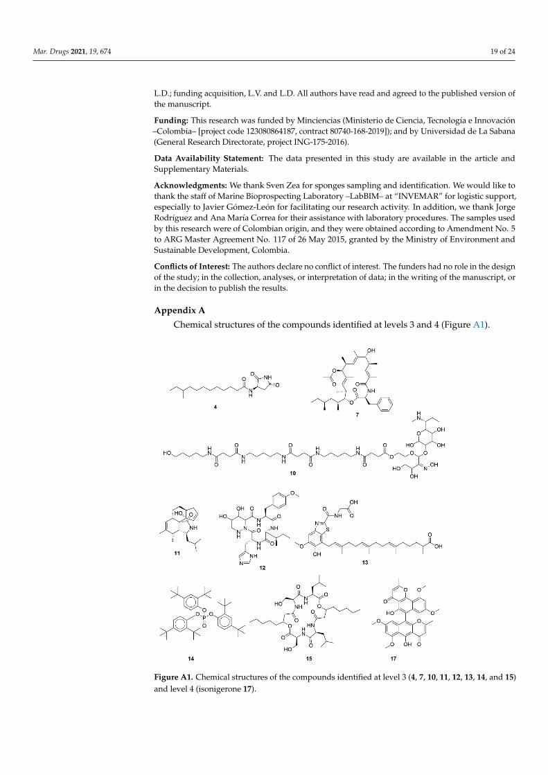
Mar. Drugs 2021, 19, 674 19 of 24
L.D.; funding acquisition, L.V. and L.D. All authors have read and agreed to the published version ofthe manuscript.
Funding: This research was funded by Minciencias (Ministerio de Ciencia, Tecnología e Innovación–Colombia– [project code 123080864187, contract 80740-168-2019]); and by Universidad de La Sabana(General Research Directorate, project ING-175-2016).
Data Availability Statement: The data presented in this study are available in the article andSupplementary Materials.
Acknowledgments: We thank Sven Zea for sponges sampling and identification. We would like tothank the staff of Marine Bioprospecting Laboratory –LabBIM– at “INVEMAR” for logistic support,especially to Javier Gómez-León for facilitating our research activity. In addition, we thank JorgeRodríguez and Ana María Correa for their assistance with laboratory procedures. The samples usedby this research were of Colombian origin, and they were obtained according to Amendment No. 5to ARG Master Agreement No. 117 of 26 May 2015, granted by the Ministry of Environment andSustainable Development, Colombia.
Conflicts of Interest: The authors declare no conflict of interest. The funders had no role in the designof the study; in the collection, analyses, or interpretation of data; in the writing of the manuscript, orin the decision to publish the results.
Appendix A
Chemical structures of the compounds identified at levels 3 and 4 (Figure A1).Mar. Drugs 2021, 19, x FOR PEER REVIEW 20 of 25
Figure A1. Chemical structures of the compounds identified at level 3 (4, 7, 10, 11, 12, 13, 14, and 15) and level 4 (isonigerone 17).
References 1. Gilchrest, B.A. Actinic Injury. Annu. Rev. Med. 1990, 41, 199–210. https://doi.org/10.1146/annurev.me.41.020190.001215. 2. D’Orazio, J.; Jarrett, S.; Amaro-Ortiz, A.; Scott, T. UV radiation and the skin. Int. J. Mol. Sci. 2013, 14, 12222–12248.
https://doi.org/10.3390/ijms140612222. 3. Bolick, N.L.; Geller, A.C. Epidemiology of Melanoma. Hematol. Oncol. Clin. N. Am. 2021, 35, 57–72.
https://doi.org/10.1016/j.hoc.2020.08.011. 4. Parker, E.R. The influence of climate change on skin cancer incidence—A review of the evidence. Int. J. Womens Dermatol. 2021,
7, 17–27. https://doi.org/10.1016/j.ijwd.2020.07.003. 5. Krutmann, J.; Passeron, T.; Gilaberte, Y.; Granger, C.; Leone, G.; Narda, M.; Schalka, S.; Trullas, C.; Masson, P.; Lim, H.W.
Photoprotection of the future: Challenges and opportunities. J. Eur. Acad. Dermatol. Venereol. 2020, 34, 447–454. https://doi.org/10.1111/jdv.16030.
6. Heurung, A.R.; Raju, S.I.; Warshaw, E.M. Adverse reactions to sunscreen agents: Epidemiology, responsible irritants and aller-gens, clinical characteristics, and management. Dermatitis 2014, 25, 289–326, https://doi.org/10.1097/DER.0000000000000079.
7. Hiller, J.; Klotz, K.; Meyer, S.; Uter, W.; Hof, K.; Greiner, A.; Göen, T.; Drexler, H. Systemic availability of lipophilic organic UV filters through dermal sunscreen exposure. Environ. Int. 2019, 132, 105068. https://doi.org/10.1016/j.envint.2019.105068.
8. Blasco, J.; Trombini, C.; Sendra, M.; Araujo, C.V.M. Environmental risk assessment of sunscreens. In Sunscreens in Coastal Eco-systems. Handbook of Environmental Chemistry; 2020; Springer International Publishing, Berlin, Germany, Volume 94, pp. 163–184.
9. Schneider, S.L.; Lim, H.W. Review of environmental effects of oxybenzone and other sunscreen active ingredients. J. Am. Acad. Dermatol. 2019, 80, 266–271. https://doi.org/10.1016/j.jaad.2018.06.033.
10. Mancuso, J.B.; Maruthi, R.; Wang, S.Q.; Lim, H.W. Sunscreens: An Update. Am. J. Clin. Dermatol. 2017, 18, 643–650. https://doi.org/10.1007/s40257-017-0290-0.
11. Seebode, C.; Lehmann, J.; Emmert, S. Photocarcinogenesis and Skin Cancer Prevention Strategies: An Update. Anticancer Res. 2018, 38, 1371–1378. https://doi.org/10.21873/anticanres.12334.
12. Pinnell, S.R. Cutaneous photodamage, oxidative stress, and topical antioxidant protection. J. Am. Acad. Dermatol. 2003, 48, 1–22. https://doi.org/10.1067/mjd.2003.16.
Figure A1. Chemical structures of the compounds identified at level 3 (4, 7, 10, 11, 12, 13, 14, and 15)and level 4 (isonigerone 17).
Mar. Drugs 2021, 19, 674 20 of 24
References1. Gilchrest, B.A. Actinic Injury. Annu. Rev. Med. 1990, 41, 199–210. [CrossRef]2. D’Orazio, J.; Jarrett, S.; Amaro-Ortiz, A.; Scott, T. UV radiation and the skin. Int. J. Mol. Sci. 2013, 14, 12222–12248. [CrossRef]3. Bolick, N.L.; Geller, A.C. Epidemiology of Melanoma. Hematol. Oncol. Clin. N. Am. 2021, 35, 57–72. [CrossRef]4. Parker, E.R. The influence of climate change on skin cancer incidence—A review of the evidence. Int. J. Womens Dermatol. 2021, 7,
17–27. [CrossRef]5. Krutmann, J.; Passeron, T.; Gilaberte, Y.; Granger, C.; Leone, G.; Narda, M.; Schalka, S.; Trullas, C.; Masson, P.; Lim, H.W.
Photoprotection of the future: Challenges and opportunities. J. Eur. Acad. Dermatol. Venereol. 2020, 34, 447–454. [CrossRef]6. Heurung, A.R.; Raju, S.I.; Warshaw, E.M. Adverse reactions to sunscreen agents: Epidemiology, responsible irritants and allergens,
clinical characteristics, and management. Dermatitis 2014, 25, 289–326. [CrossRef] [PubMed]7. Hiller, J.; Klotz, K.; Meyer, S.; Uter, W.; Hof, K.; Greiner, A.; Göen, T.; Drexler, H. Systemic availability of lipophilic organic UV
filters through dermal sunscreen exposure. Environ. Int. 2019, 132, 105068. [CrossRef] [PubMed]8. Blasco, J.; Trombini, C.; Sendra, M.; Araujo, C.V.M. Environmental risk assessment of sunscreens. In Sunscreens in Coastal
Ecosystems. Handbook of Environmental Chemistry; Springer International Publishing: Berlin, Germany, 2020; Volume 94, pp.163–184.
9. Schneider, S.L.; Lim, H.W. Review of environmental effects of oxybenzone and other sunscreen active ingredients. J. Am. Acad.Dermatol. 2019, 80, 266–271. [CrossRef] [PubMed]
10. Mancuso, J.B.; Maruthi, R.; Wang, S.Q.; Lim, H.W. Sunscreens: An Update. Am. J. Clin. Dermatol. 2017, 18, 643–650. [CrossRef][PubMed]
11. Seebode, C.; Lehmann, J.; Emmert, S. Photocarcinogenesis and Skin Cancer Prevention Strategies: An Update. Anticancer Res.2018, 38, 1371–1378. [CrossRef]
12. Pinnell, S.R. Cutaneous photodamage, oxidative stress, and topical antioxidant protection. J. Am. Acad. Dermatol. 2003, 48, 1–22.[CrossRef] [PubMed]
13. Velasco, M.V.R.; Sauce, R.; de Oliveira, C.A.; de Oliveira Pinto, C.A.S.; Martinez, R.M.; Baah, S.; Almeida, T.S.; Rosado, C.; Baby,A.R. Active ingredients, mechanisms of action and efficacy tests of antipollution cosmetic and personal care products. Braz. J.Pharm. Sci. 2018, 54, e01003. [CrossRef]
14. Cavinato, M.; Waltenberger, B.; Baraldo, G.; Grade, C.V.C.; Stuppner, H.; Jansen-Dürr, P. Plant extracts and natural compoundsused against UVB-induced photoaging. Biogerontology 2017, 18, 499–516. [CrossRef]
15. Atanasov, A.G.; Zotchev, S.B.; Dirsch, V.M.; Supuran, C.T. Natural products in drug discovery: Advances and opportunities. Nat.Rev. Drug Discov. 2021, 20, 200–216. [CrossRef] [PubMed]
16. Carroll, A.R.; Copp, B.R.; Davis, R.A.; Keyzers, R.A.; Prinsep, M.R. Marine natural products. Nat. Prod. Rep. 2019, 36, 122–173.[CrossRef]
17. Sipkema, D.; Franssen, M.C.R.; Osinga, R.; Tramper, J.; Wijffels, R.H. Marine sponges as pharmacy. Mar. Biotechnol. 2005, 7,142–162. [CrossRef]
18. Esposito, R.; Ruocco, N.; Viel, T.; Federico, S.; Zupo, V.; Costantini, M. Sponges and their symbionts as a source of valuablecompounds in cosmeceutical field. Mar. Drugs 2021, 19, 444. [CrossRef]
19. Sacristán-Soriano, O.; Turon, X.; Hill, M. Microbiome structure of ecologically important bioeroding sponges (family Clionaidae):The role of host phylogeny and environmental plasticity. Coral Reefs 2020, 39, 1285–1298. [CrossRef]
20. Ribes, M.; Dziallas, C.; Coma, R.; Riemann, L. Microbial diversity and putative diazotrophy in high- and low- microbial-abundancemediterranean sponges. Appl. Environ. Microbiol. 2015, 81, 5683–5693. [CrossRef]
21. Eastgate, M.D.; Schmidt, M.A.; Fandrick, K.R. On the design of complex drug candidate syntheses in the pharmaceutical industry.Nat. Rev. Chem. 2017, 1, 1–16. [CrossRef]
22. Atanasov, A.G.; Waltenberger, B.; Pferschy-Wenzig, E.-M.; Linder, T.; Wawrosch, C.; Uhrin, P.; Temml, V.; Wang, L.; Schwaiger, S.;Heiss, E.H.; et al. Discovery and resupply of pharmacologically active plant-derived natural products: A review. Biotechnol. Adv.2015, 33, 1582–1614. [CrossRef] [PubMed]
23. Molinski, T.F.; Dalisay, D.S.; Lievens, S.L.; Saludes, J.P. Drug development from marine natural products. Nat. Rev. Drug Discov.2009, 8, 69–85. [CrossRef]
24. Radjasa, O.K.; Vaske, Y.M.; Navarro, G.; Vervoort, H.C.; Tenney, K.; Linington, R.G.; Crews, P. Highlights of marine invertebrate-derived biosynthetic products: Their biomedical potential and possible production by microbial associants. Bioorg. Med. Chem.2011, 19, 6658–6674. [CrossRef]
25. Hug, J.J.; Krug, D.; Müller, R. Bacteria as genetically programmable producers of bioactive natural products. Nat. Rev. Chem.2020, 4, 172–193. [CrossRef]
26. Santos-Gandelman, J.; Giambiagi-deMarval, M.; Oelemann, W.; Laport, M. Biotechnological Potential of Sponge-AssociatedBacteria. Curr. Pharm. Biotechnol. 2014, 15, 143–155. [CrossRef]
27. Raimundo, I.; Silva, S.G.; Costa, R.; Keller-Costa, T. Bioactive secondary metabolites from octocoral-Associated microbes—Newchances for blue growth. Mar. Drugs 2018, 16, 485. [CrossRef]
28. Barka, E.A.; Vatsa, P.; Sanchez, L.; Gaveau-Vaillant, N.; Jacquard, C.; Meier-Kolthoff, J.P.; Klenk, H.-P.; Clément, C.; Ouhdouch,Y.; van Wezel, G.P. Taxonomy, Physiology, and Natural Products of Actinobacteria. Microbiol. Mol. Biol. Rev. 2016, 80, 1–43.[CrossRef] [PubMed]
Mar. Drugs 2021, 19, 674 21 of 24
29. Seipke, R.F.; Kaltenpoth, M.; Hutchings, M.I. Streptomyces as symbionts: An emerging and widespread theme? FEMS Microbiol.Rev. 2012, 36, 862–876. [CrossRef]
30. Thomas, T.R.A.; Kavlekar, D.P.; LokaBharathi, P.A. Marine Drugs from Sponge-Microbe Association—A Review. Mar. Drugs 2010,8, 1417–1468. [CrossRef]
31. Poulsen, M.; Oh, D.-C.; Clardy, J.; Currie, C.R. Chemical Analyses of Wasp-Associated Streptomyces Bacteria Reveal a ProlificPotential for Natural Products Discovery. PLoS ONE 2011, 6, e16763. [CrossRef]
32. Sánchez-Suárez, J.; Coy-Barrera, E.; Villamil, L.; Díaz, L. Streptomyces-Derived Metabolites with Potential PhotoprotectiveProperties—A Systematic Literature Review and Meta-Analysis on the Reported Chemodiversity. Molecules 2020, 25, 3221.[CrossRef]
33. Wang, S.Q.; Stanfield, J.W.; Osterwalder, U. In vitro assessments of UVA protection by popular sunscreens available in the UnitedStates. J. Am. Acad. Dermatol. 2008, 59, 934–942. [CrossRef] [PubMed]
34. Ilyasov, I.R.; Beloborodov, V.L.; Selivanova, I.A.; Terekhov, R.P. ABTS/PP Decolorization Assay of Antioxidant Capacity ReactionPathways. Int. J. Mol. Sci. 2020, 21, 1131. [CrossRef] [PubMed]
35. Huang, D.; Boxin, O.U.; Prior, R.L. The chemistry behind antioxidant capacity assays. J. Agric. Food Chem. 2005, 53, 1841–1856.[CrossRef] [PubMed]
36. Apak, R.; Gorinstein, S.; Böhm, V.; Schaich, K.M.; Özyürek, M.; Güçlü, K. Methods of measurement and evaluation of naturalantioxidant capacity/activity (IUPAC Technical Report). Pure Appl. Chem. 2013, 85, 957–998. [CrossRef]
37. Cole, C.; Shyr, T.; Ou-Yang, H. Metal oxide sunscreens protect skin by absorption, not by reflection or scattering. Photodermatol.Photoimmunol. Photomed. 2016, 32, 5–10. [CrossRef] [PubMed]
38. Sarkany, R. Sun protection. Medicine (Baltimore) 2021, 49, 453–456. [CrossRef]39. Springsteen, A.; Yurek, R.; Frazier, M.; Carr, K.F. In vitro measurement of sun protection factor of sunscreens by diffuse
transmittance. Anal. Chim. Acta 1999, 380, 155–164. [CrossRef]40. Paiva, J.P.; Diniz, R.R.; Leitão, A.C.; Cabral, L.M.; Fortunato, R.S.; Santos, B.A.M.C.; de Pádula, M. Insights and controversies on
sunscreen safety. Crit. Rev. Toxicol. 2020, 50, 707–723. [CrossRef] [PubMed]41. Hughes, J.P.; Rees, S.S.; Kalindjian, S.B.; Philpott, K.L. Principles of early drug discovery. Br. J. Pharmacol. 2011, 162, 1239–1249.
[CrossRef]42. Komaki, H. Reclassification of 15 Streptomyces species as synonyms of Streptomyces albogriseolus, Streptomyces althioticus,
Streptomyces anthocyanicus, Streptomyces calvus, Streptomyces griseoincarnatus, Streptomyces mutabilis, Streptomyces pilosusor Streptomyces r. Int. J. Syst. Evol. Microbiol. 2019, 71, 004718. [CrossRef] [PubMed]
43. Klimová, Z.; Hojerová, J.; Beránková, M. Skin absorption and human exposure estimation of three widely discussed UV filters insunscreens—In vitro study mimicking real-life consumer habits. Food Chem. Toxicol. 2015, 83, 237–250. [CrossRef] [PubMed]
44. Tampucci, S.; Burgalassi, S.; Chetoni, P.; Monti, D. Cutaneous Permeation and Penetration of Sunscreens: Formulation Strategiesand In Vitro Methods. Cosmetics 2017, 5, 1. [CrossRef]
45. Gilbert, E.; Pirot, F.; Bertholle, V.; Roussel, L.; Falson, F.; Padois, K. Commonly used UV filter toxicity on biological functions:Review of last decade studies. Int. J. Cosmet. Sci. 2013, 35, 208–219. [CrossRef]
46. Klinubol, P.; Asawanonda, P.; Wanichwecharungruang, S.P. Transdermal Penetration of UV Filters. Skin Pharmacol. Physiol. 2008,21, 23–29. [CrossRef]
47. Benech-Kieffer, F.; Meuling, W.J.A.; Leclerc, C.; Roza, L.; Leclaire, J.; Nohynek, G. Percutaneous Absorption of Mexoryl SX®inHuman Volunteers: Comparison with in vitro Data. Skin Pharmacol. Physiol. 2003, 16, 343–355. [CrossRef]
48. Schymanski, E.L.; Jeon, J.; Gulde, R.; Fenner, K.; Ruff, M.; Singer, H.P.; Hollender, J. Identifying small molecules via high resolutionmass spectrometry: Communicating confidence. Environ. Sci. Technol. 2014, 48, 2097–2098. [CrossRef]
49. Rivera, G.S.M.; Beamish, C.R.; Wencewicz, T.A. Immobilized FhuD2 Siderophore-Binding Protein Enables Purification of SalmycinSideromycins from Streptomyces violaceus DSM 8286. ACS Infect. Dis. 2018, 4, 845–859. [CrossRef]
50. Kunihiro, S.; Kaneda, M. Glomecidin, a Novel Antifungal Cyclic Tetrapeptide Produced by Streptomyces lavendulae H698 SY2. J.Antibiot. (Tokyo) 2003, 56, 30–33. [CrossRef] [PubMed]
51. Kamdem, R.S.T.; Wang, H.; Wafo, P.; Ebrahim, W.; Özkaya, F.C.; Makhloufi, G.; Janiak, C.; Sureechatchaiyan, P.; Kassack, M.U.;Lin, W.; et al. Induction of new metabolites from the endophytic fungus Bionectria sp. through bacterial co-culture. Fitoterapia2018, 124, 132–136. [CrossRef]
52. Apsari, P.P.; Budiarti, S.; Wahyudi, A.T. Actinomycetes of rhizosphere soil producing antibacterial compounds against urinarytract infection bacteria. Biodiversitas 2019, 20, 1259–1265. [CrossRef]
53. Krasnoff, S.B.; Englich, U.; Miller, P.G.; Shuler, M.L.; Glahn, R.P.; Donzelli, B.G.G.; Gibson, D.M. Metacridamides A and B,Macrocycles from Conidia of the Entomopathogenic Fungus Metarhizium acridum. J. Nat. Prod. 2012, 75, 175–180. [CrossRef]
54. Liu, J.; Zhang, D.; Zhang, M.; Liu, X.; Chen, R.; Zhao, J.; Li, L.; Wang, N.; Dai, J. Periconiasins I and J, two new cytochalasans froman endophytic fungus Periconia sp. Tetrahedron Lett. 2016, 57, 5794–5797. [CrossRef]
55. Boros, C.; Smith, C.J.; Vasina, Y.; Che, Y.; Dix, A.B.; Darveaux, B.; Pearce, C. Isolation and Identification of the Icosalides—CyclicPeptolides with Selective Antibiotic and Cytotoxic Activities. J. Antibiot. (Tokyo) 2006, 59, 486–494. [CrossRef] [PubMed]
56. Yoshimura, A.; Covington, B.C.; Gallant, É.; Zhang, C.; Li, A.; Seyedsayamdost, M.R. Unlocking Cryptic Metabolites with MassSpectrometry-Guided Transposon Mutant Selection. ACS Chem. Biol. 2020, 15, 2766–2774. [CrossRef] [PubMed]
Mar. Drugs 2021, 19, 674 22 of 24
57. Jing, T.; Zhou, D.; Zhang, M.; Yun, T.; Qi, D.; Wei, Y.; Chen, Y.; Zang, X.; Wang, W.; Xie, J. Newly Isolated Streptomyces sp. JBS5-6as a Potential Biocontrol Agent to Control Banana Fusarium Wilt: Genome Sequencing and Secondary Metabolite Cluster Profiles.Front. Microbiol. 2020, 11, 3036. [CrossRef] [PubMed]
58. Zhou, S.-Y.; Hu, Y.-J.; Meng, F.-C.; Qu, S.-Y.; Wang, R.; Andersen, R.; Liao, Z.-H.; Chen, M. Bacillamidins A–G from a Marine-Derived Bacillus pumilus. Mar. Drugs 2018, 16, 326. [CrossRef]
59. Hu, Y.; MacMillan, J.B. Erythrazoles A–B, Cytotoxic Benzothiazoles from a Marine-Derived Erythrobacter sp. Org. Lett. 2011, 13,6580–6583. [CrossRef]
60. Korinth, G.; Wellner, T.; Schaller, K.H.; Drexler, H. Potential of the octanol–water partition coefficient (logP) to predict the dermalpenetration behaviour of amphiphilic compounds in aqueous solutions. Toxicol. Lett. 2012, 215, 49–53. [CrossRef]
61. Vinuchakkaravarthy, T.; Kumaravel, K.P.; Ravichandran, S.; Velmurugan, D. Active compound from the leaves of vitex negundoL. shows anti-inflammatory activity with evidence of inhibition for secretory phospholipase A2 through molecular docking.Bioinformation 2011, 7, 199–206. [CrossRef]
62. Sander, T.; Freyss, J.; Von Korff, M.; Rufener, C. DataWarrior: An open-source program for chemistry aware data visualizationand analysis. J. Chem. Inf. Model. 2015, 55, 460–473. [CrossRef]
63. Cragg, G.M.; Newman, D.J. Natural products: A continuing source of novel drug leads. Biochim. Biophys. Acta—Gen. Subj. 2013,1830, 3670–3695. [CrossRef] [PubMed]
64. Haygood, M.G.; Schmidt, E.W.; Davidson, S.K.; Faulkner, D.J. Microbial symbionts of marine invertebrates: Opportunities formicrobial biotechnology. J. Mol. Microbiol. Biotechnol. 1999, 1, 33–43. [PubMed]
65. Omura, S.; Ikeda, H.; Ishikawa, J.; Hanamoto, A.; Takahashi, C.; Shinose, M.; Takahashi, Y.; Horikawa, H.; Nakazawa, H.; Osonoe,T.; et al. Genome sequence of an industrial microorganism Streptomyces avermitilis: Deducing the ability of producing secondarymetabolites. Proc. Natl. Acad. Sci. USA 2001, 98, 12215–12220. [CrossRef]
66. Seipke, R.F. Strain-level diversity of secondary metabolism in Streptomyces albus. PLoS ONE 2015, 10, e0116457. [CrossRef][PubMed]
67. Parte, A.C.; Sardà Carbasse, J.; Meier-Kolthoff, J.P.; Reimer, L.C.; Göker, M. List of Prokaryotic names with Standing inNomenclature (LPSN) moves to the DSMZ. Int. J. Syst. Evol. Microbiol. 2020, 70, 5607–5612. [CrossRef]
68. Jose, P.A.; Jebakumar, S.R.D. Non-streptomycete actinomycetes nourish the current microbial antibiotic drug discovery. Front.Microbiol. 2013, 4, 240. [CrossRef]
69. Variyar, P.S.; Chander, R.; Venkatachalam, S.R.; Bongirwar, D.R. A new red pigment from an alkalophilic Micrococcus species.Indian J. Chem.—Sect. B Org. Med. Chem. 2002, 41, 232–233.
70. Rostami, H.; Hamedi, H.; Yolmeh, M. Some biological activities of pigments extracted from Micrococcus roseus (PTCC 1411) andRhodotorula glutinis (PTCC 5257). Int. J. Immunopathol. Pharmacol. 2016, 29, 684–695. [CrossRef]
71. Karbalaei-Heidari, H.R.; Partovifar, M.; Memarpoor-Yazdi, M. Evaluation of the bioactive potential of secondary metabolitesproduced by a new marine Micrococcus species isolated from the Persian Gulf. Avicenna J. Med. Biotechnol. 2020, 12, 61–65.
72. Vinoshna, S.; Akula, M.; Mishra, B. In vitro antioxidant efficacy of EPS obtained from Micrococcus luteus snist-CM 02: A briefstudy. J. Microbiol. Biotechnol. Food Sci. 2017, 6, 1199–1202. [CrossRef]
73. Asker, M.M.S.; EL Sayed, O.H.; Mahmoud, M.G.; Ramadan, M.F. Chemical structure and antioxidant activity of a new exopolysac-charide produced from Micrococcus luteus. J. Genet. Eng. Biotechnol. 2014, 12, 121–126. [CrossRef]
74. Tsao, S.-W.; Rudd, B.A.M.; He, X.-G.; Chang, C.-J.; Floss, H.G. Identification of a red pigment from Streptomyces coelicolor A3(2)as a mixture of prodigiosin derivatives. J. Antibiot. (Tokyo) 1985, 38, 128–131. [CrossRef] [PubMed]
75. Suryawanshi, R.K.; Patil, C.D.; Borase, H.P.; Narkhede, C.P.; Stevenson, A.; Hallsworth, J.E.; Patil, S.V. Towards an understandingof bacterial metabolites prodigiosin and violacein and their potential for use in commercial sunscreens. Int. J. Cosmet. Sci. 2015,37, 98–107. [CrossRef] [PubMed]
76. Basílico, G.; Roger, C.A.; Seigelchifer, M.; Kerner, N. UV-specific DNA repair recombinant fusion enzyme: A new stablepharmacologically active principle suitable for photoprotection. J. Dermatol. Sci. 2005, 39, 81–88. [CrossRef] [PubMed]
77. Loh, W.L.C.; Huang, K.-C.; Ng, H.S.; Lan, J.C.-W. Exploring the fermentation characteristics of a newly isolated marine bacteriastrain, Gordonia terrae TWRH01 for carotenoids production. J. Biosci. Bioeng. 2020, 130, 187–194. [CrossRef]
78. Stahl, W.; Sies, H. Antioxidant activity of carotenoids. Mol. Aspects Med. 2003, 24, 345–351. [CrossRef]79. Li, Y.; Zhang, F.; Banakar, S.; Li, Z. Comprehensive optimization of precursor-directed production of BC194 by Streptomyces
rochei MB037 derived from the marine sponge Dysidea arenaria. Appl. Microbiol. Biotechnol. 2018, 102, 7865–7875. [CrossRef]80. Jiang, S.; Li, X.; Zhang, L.; Sun, W.; Dai, S.; Xie, L.; Liu, Y.; Lee, K.J. Culturable actinobacteria isolated from marine sponge
Iotrochota sp. Mar. Biol. 2008, 153, 945–952. [CrossRef]81. Burke, K.E. Photoaging: The role of oxidative stress. G. Ital. Dermatol. Venereol. 2010, 145, 445–459.82. Munteanu, I.G.; Apetrei, C. Analytical Methods Used in Determining Antioxidant Activity: A Review. Int. J. Mol. Sci. 2021, 22,
3380. [CrossRef]83. Barclay, L.R.C.; Edwards, C.E.; Vinqvist, M.R. Media Effects on Antioxidant Activities of Phenols and Catechols. J. Am. Chem. Soc.
1999, 121, 6226–6231. [CrossRef]84. Oresajo, C.; Pillai, S.; Manco, M.; Yatskayer, M.; McDaniel, D. Antioxidants and the skin: Understanding formulation and efficacy.
Dermatol. Ther. 2012, 25, 252–259. [CrossRef] [PubMed]
Mar. Drugs 2021, 19, 674 23 of 24
85. Tan, L.T.-H.; Chan, K.-G.; Chan, C.K.; Khan, T.M.; Lee, L.-H.; Goh, B.-H. Antioxidative Potential of a Streptomyces sp. MUM292Isolated from Mangrove Soil. Biomed Res. Int. 2018, 2018, 1–13. [CrossRef] [PubMed]
86. Tan, L.T.-H.; Chan, K.-G.; Khan, T.M.; Bukhari, S.I.; Saokaew, S.; Duangjai, A.; Pusparajah, P.; Lee, L.-H.; Goh, B.-H. Streptomycessp. MUM212 as a Source of Antioxidants with Radical Scavenging and Metal Chelating Properties. Front. Pharmacol. 2017, 8, 276.[CrossRef]
87. Tan, L.T.H.; Mahendra, C.K.; Yow, Y.Y.; Chan, K.G.; Khan, T.M.; Lee, L.H.; Goh, B.H. Streptomyces sp. MUM273b: A mangrove-derived potential source for antioxidant and UVB radiation protectants. Microbiologyopen 2019, 8, e859. [CrossRef] [PubMed]
88. Shaath, N.A. Ultraviolet filters. Photochem. Photobiol. Sci. 2010, 9, 464. [CrossRef] [PubMed]89. Mancebo, S.E.; Hu, J.Y.; Wang, S.Q. Sunscreens: A review of health benefits, regulations, and controversies. Dermatol. Clin. 2014,
32, 427–438. [CrossRef] [PubMed]90. DiNardo, J.C.; Downs, C.A. Dermatological and environmental toxicological impact of the sunscreen ingredient oxybenzone/ben-
zophenone-3. J. Cosmet. Dermatol. 2018, 17, 15–19. [CrossRef]91. Fivenson, D.; Sabzevari, N.; Qiblawi, S.; Blitz, J.; Norton, B.B.; Norton, S.A. Sunscreens: UV filters to protect us: Part 2-Increasing
awareness of UV filters and their potential toxicities to us and our environment. Int. J. Womens Dermatol. 2021, 7, 45–69. [CrossRef]92. Autier, P.; Boniol, M.; Severi, G.; Doré, J.F. Quantity of sunscreen used by European students. Br. J. Dermatol. 2001, 144, 288–291.
[CrossRef]93. Bernerd, F.; Asselineau, D. UVA exposure of human skin reconstructed in vitro induces apoptosis of dermal fibroblasts: Subse-
quent connective tissue repair and implications in photoaging. Cell Death Differ. 1998, 5, 792–802. [CrossRef] [PubMed]94. Jackson, S.A.; Crossman, L.; Almeida, E.L.; Margassery, L.M.; Kennedy, J.; Dobson, A.D.W. Diverse and abundant secondary
metabolism biosynthetic gene clusters in the genomes of marine sponge derived Streptomyces spp. Isolates. Mar. Drugs 2018, 16,67. [CrossRef] [PubMed]
95. Maipas, S.; Nicolopoulou-Stamati, P. Sun lotion chemicals as endocrine disruptors. Hormones 2015, 14, 32–46. [CrossRef] [PubMed]96. Zhang, Y.; Ling, S.; Fang, Y.; Zhu, T.; Gu, Q.; Zhu, W.-M. Isolation, Structure Elucidation, and Antimycobacterial Properties
of Dimeric Naphtho-γ-pyrones from the Marine-Derived FungusAspergillus carbonarius. Chem. Biodivers. 2008, 5, 93–100.[CrossRef]
97. Wang, X.; Shaaban, K.A.; Elshahawi, S.I.; Ponomareva, L.V.; Sunkara, M.; Zhang, Y.; Copley, G.C.; Hower, J.C.; Morris, A.J.; Kharel,M.K.; et al. Frenolicins C-G, Pyranonaphthoquinones from Streptomyces sp. RM-4-15. J. Nat. Prod. 2013, 76, 1441–1447. [CrossRef]
98. Cai, S.; Kong, X.; Wang, W.; Zhou, H.; Zhu, T.; Li, D.; Gu, Q. Aspergilazine A, a diketopiperazine dimer with a rare N-1 to C-6linkage, from a marine-derived fungus Aspergillus taichungensis. Tetrahedron Lett. 2012, 53, 2615–2617. [CrossRef]
99. Yu, H.; Li, S.-M. Two Cytochrome P450 Enzymes from Streptomyces sp. NRRL S-1868 Catalyze Distinct Dimerization ofTryptophan-Containing Cyclodipeptides. Org. Lett. 2019, 21, 7094–7098. [CrossRef]
100. Kamdem, R.S.T.; Pascal, W.; Rehberg, N.; van Geelen, L.; Höfert, S.-P.; Knedel, T.-O.; Janiak, C.; Sureechatchaiyan, P.; Kassack,M.U.; Lin, W.; et al. Metabolites from the endophytic fungus Cylindrocarpon sp. isolated from tropical plant Sapium ellipticum.Fitoterapia 2018, 128, 175–179. [CrossRef]
101. Hou, L.; Huang, H.; Li, H.; Wang, S.; Ju, J.; Li, W. Overexpression of a type III PKS gene affording novel violapyrones withenhanced anti-influenza A virus activity. Microb. Cell Fact. 2018, 17, 61. [CrossRef]
102. Newton, G.G.F.; Abraham, E.P. Isolation of cephalosporin C, a penicillin-like antibiotic containing D-α-aminoadipic acid. Biochem.J. 1956, 62, 651–658. [CrossRef]
103. Yu, H.; Serpe, E.; Romero, J.; Coque, J.-J.; Maeda, K.; Oelgeschlager, M.; Hintermann, G.; Liras, P.; Martin, J.-F.; Demain, A.L.; et al.Possible involvement of the lysine -aminotransferase gene (lat) in the expression of the genes encoding ACV synthetase (pcbAB)and isopenicillin N synthase (pcbC) in Streptomyces clavuligerus. Microbiology 1994, 140, 3367–3377. [CrossRef]
104. Higgens, C.E.; Hamill, R.L.; Sands, T.H.; Hoehn, M.M.; Davis, N.E.; Nagarajan, R.; Boeck, L.D. The occurrence of deacetoxy-cephalosporin c in fungi and streptomycetes. J. Antibiot. (Tokyo) 1974, 27, 298–300. [CrossRef] [PubMed]
105. Schmitt, I.; Lumbsch, H.T. Ancient Horizontal Gene Transfer from Bacteria Enhances Biosynthetic Capabilities of Fungi. PLoSONE 2009, 4, e4437. [CrossRef] [PubMed]
106. Singh, D.K.; Pathak, J.; Pandey, A.; Singh, V.; Ahmed, H.; Rajneesh; Kumar, D.; Sinha, R.P. Ultraviolet-screening compoundmycosporine-like amino acids in cyanobacteria: Biosynthesis, functions, and applications. In Advances in Cyanobacterial Biology;Elsevier: Amsterdam, The Netherlands, 2020; pp. 219–233.
107. Edreva, A. The importance of non-photosynthetic pigments and cinnamic acid derivatives in photoprotection. Agric. Ecosyst.Environ. 2005, 106, 135–146. [CrossRef]
108. Rouf, A.; Tanyeli, C. Bioactive thiazole and benzothiazole derivatives. Eur. J. Med. Chem. 2015, 97, 911–927. [CrossRef]109. Weber, T.; Charusanti, P.; Musiol-Kroll, E.M.; Jiang, X.; Tong, Y.; Kim, H.U.; Lee, S.Y. Metabolic engineering of antibiotic factories:
New tools for antibiotic production in actinomycetes. Trends Biotechnol. 2015, 33, 15–26. [CrossRef]110. Wang, W.; Wang, H.; Hu, H.; Peng, H.; Zhang, X. Overexpression of afsR and Optimization of Metal Chloride to Improve
Lomofungin Production in Streptomyces lomondensis S015. J. Microbiol. Biotechnol. 2015, 25, 672–680. [CrossRef]111. El-Housseiny, G.S.; Ibrahim, A.A.; Yassien, M.A.; Aboshanab, K.M. Production and statistical optimization of Paromomycin by
Streptomyces rimosus NRRL 2455 in solid state fermentation. BMC Microbiol. 2021, 21, 34. [CrossRef]112. Zhu, S.; Duan, Y.; Huang, Y. The Application of Ribosome Engineering to Natural Product Discovery and Yield Improvement in
Streptomyces. Antibiotics 2019, 8, 133. [CrossRef]
Mar. Drugs 2021, 19, 674 24 of 24
113. Stankovic, N.; Radulovic, V.; Petkovic, M.; Vuckovic, I.; Jadranin, M.; Vasiljevic, B.; Nikodinovic-Runic, J. Streptomyces sp. JS520produces exceptionally high quantities of undecylprodigiosin with antibacterial, antioxidative, and UV-protective properties.Appl. Microbiol. Biotechnol. 2012, 96, 1217–1231. [CrossRef] [PubMed]
114. Matobole, R.M.; Van Zyl, L.J.; Parker-Nance, S.; Davies-Coleman, M.T.; Trindade, M. Antibacterial Activities of Bacteria Isolatedfrom the Marine Sponges Isodictya compressa and Higginsia bidentifera Collected from Algoa Bay, South Africa. Mar. Drugs2017, 15, 47. [CrossRef] [PubMed]
115. Buitrago, D.; Buitrago-Villanueva, I.; Barbosa-Cornelio, R.; Coy-Barrera, E. Comparative examination of antioxidant capacity andfingerprinting of unfractionated extracts from different plant parts of quinoa (Chenopodium quinoa) grown under greenhouseconditions. Antioxidants 2019, 8, 238. [CrossRef] [PubMed]
116. Re, R.; Pellegrini, N.; Proteggente, A.; Pannala, A.; Yang, M.; Rice-Evans, C. Antioxidant activity applying an improved ABTSradical cation decolorization assay. Free Radic. Biol. Med. 1999, 26, 1231–1237. [CrossRef]
117. Magalhães, L.M.; Santos, F.; Segundo, M.A.; Reis, S.; Lima, J.L.F.C. Rapid microplate high-throughput methodology for assessmentof Folin-Ciocalteu reducing capacity. Talanta 2010, 83, 441–447. [CrossRef]
118. Mansur, J.D.S.; Breder, M.N.R.; Mansur, M.C.D.A.; Azulay, R.D. Determinação do fator de proteção solar por espectrofotometria.An. Bras. Dermatol. 1986, 61, 121–124.
119. Sayre, R.M.; Agin, P.P.; LeVee, G.J.; Marlowe, E. A comparison of in vivo and in vitro testing of sunscreening formulas. Photochem.Photobiol. 1979, 29, 559–566. [CrossRef]
120. Morgulis, A.; Coulouris, G.; Raytselis, Y.; Madden, T.L.; Agarwala, R.; Schäffer, A.A. Database indexing for productionMegaBLAST searches. Bioinformatics 2008, 24, 1757–1764. [CrossRef]
121. Kumar, S.; Stecher, G.; Li, M.; Knyaz, C.; Tamura, K. MEGA X: Molecular Evolutionary Genetics Analysis across ComputingPlatforms. Mol. Biol. Evol. 2018, 35, 1547–1549. [CrossRef]
122. Mosmann, T. Rapid colorimetric assay for cellular growth and survival: Application to proliferation and cytotoxicity assays. J.Immunol. Methods 1983, 65, 55–63. [CrossRef]
123. Moumbock, A.F.A.; Gao, M.; Qaseem, A.; Li, J.; Kirchner, P.A.; Ndingkokhar, B.; Bekono, B.D.; Simoben, C.V.; Babiaka, S.B.;Malange, Y.I.; et al. StreptomeDB 3.0: An updated compendium of streptomycetes natural products. Nucleic Acids Res. 2021, 49,D600–D604. [CrossRef] [PubMed]
124. Van Santen, J.A.; Poynton, E.F.; Iskakova, D.; McMann, E.; Alsup, T.A.; Clark, T.N.; Fergusson, C.H.; Fewer, D.P.; Hughes, A.H.;McCadden, C.A.; et al. The Natural Products Atlas 2.0: A database of microbially-derived natural products. Nucleic Acids Res.2021, gkab941. [CrossRef] [PubMed]