Isolation, Phylogenetic Analysis and Anti-infective Activity Screening of Marine Sponge-Associated...
14
Mar. Drugs 2010, 8, 399-412; doi:10.3390/md8030399 Marine Drugs ISSN 1660-3397 www.mdpi.com/journal/marinedrugs Article Isolation, Phylogenetic Analysis and Anti-infective Activity Screening of Marine Sponge-Associated Actinomycetes Usama Ramadan Abdelmohsen 1,2,3 , Sheila M. Pimentel-Elardo 1,2 , Amro Hanora 4 , Mona Radwan 5 , Soad H. Abou-El-Ela 5 , Safwat Ahmed 6 and Ute Hentschel 1,2, * 1 Julius-von-Sachs-Institute for Biological Sciences, University of Würzburg, Julius-von-Sachs-Platz 3, 97082 Würzburg, Germany; E-Mail: [email protected] (S.M.P.-E.); [email protected] (U.R.A.) 2 Research Center for Infectious Diseases, Josef-Schneider-Straße 2, 97080 Würzburg, Germany 3 Department of Pharmacognosy, Faculty of Pharmacy, Minia University, Minia, Egypt 4 Department of Microbiology, Faculty of Pharmacy, Suez Canal University, Ismailia, Egypt; E-Mail: [email protected] 5 Department of Biochemistry, Faculty of Pharmacy, Suez Canal University, Ismailia, Egypt; E-Mails: [email protected] (M.R.); [email protected] (S.H.A.-E.-E.) 6 Department of Pharmacognosy, Faculty of Pharmacy, Suez Canal University, Ismailia, Egypt; E-Mail: [email protected] * Author to whom correspondence should be addressed; E-Mail: [email protected]; Tel.: 0049-931-31-82581; Fax: 0049-931-31-86235. Received: 29 December 2009; in revised form: 3 February 2010 / Accepted: 5 February 2010 / Published: 26 February 2010 Abstract: Terrestrial actinomycetes are noteworthy producers of a multitude of antibiotics, however the marine representatives are much less studied in this regard. In this study, 90 actinomycetes were isolated from 11 different species of marine sponges that had been collected from offshore Ras Mohamed (Egypt) and from Rovinj (Croatia). Phylogenetic characterization of the isolates based on 16S rRNA gene sequencing supported their assignment to 18 different actinomycete genera representing seven different suborders. Fourteen putatively novel species were identified based on sequence similarity values below 98.2% to other strains in the NCBI database. A putative new genus related to Rubrobacter was isolated on M1 agar that had been amended with sponge extract, thus highlighting the need for innovative cultivation protocols. Testing for anti-infective activities was performed against clinically relevant, Gram-positive (Enterococcus faecalis, Staphylococcus aureus) and Gram-negative (Escherichia coli, Pseudomonas aeruginosa) OPEN ACCESS
-
Upload
independent -
Category
Documents
-
view
2 -
download
0
Transcript of Isolation, Phylogenetic Analysis and Anti-infective Activity Screening of Marine Sponge-Associated...
Mar. Drugs 2010, 8, 399-412; doi:10.3390/md8030399
Marine Drugs ISSN 1660-3397
www.mdpi.com/journal/marinedrugs
Article
Isolation, Phylogenetic Analysis and Anti-infective Activity Screening of Marine Sponge-Associated Actinomycetes
Usama Ramadan Abdelmohsen 1,2,3, Sheila M. Pimentel-Elardo 1,2, Amro Hanora 4,
Mona Radwan 5, Soad H. Abou-El-Ela 5, Safwat Ahmed 6 and Ute Hentschel 1,2,*
1 Julius-von-Sachs-Institute for Biological Sciences, University of Würzburg, Julius-von-Sachs-Platz
3, 97082 Würzburg, Germany; E-Mail: [email protected] (S.M.P.-E.);
[email protected] (U.R.A.) 2 Research Center for Infectious Diseases, Josef-Schneider-Straße 2, 97080 Würzburg, Germany 3 Department of Pharmacognosy, Faculty of Pharmacy, Minia University, Minia, Egypt 4 Department of Microbiology, Faculty of Pharmacy, Suez Canal University, Ismailia, Egypt;
E-Mail: [email protected] 5 Department of Biochemistry, Faculty of Pharmacy, Suez Canal University, Ismailia, Egypt;
E-Mails: [email protected] (M.R.); [email protected] (S.H.A.-E.-E.) 6 Department of Pharmacognosy, Faculty of Pharmacy, Suez Canal University, Ismailia, Egypt;
E-Mail: [email protected]
* Author to whom correspondence should be addressed; E-Mail: [email protected];
Tel.: 0049-931-31-82581; Fax: 0049-931-31-86235.
Received: 29 December 2009; in revised form: 3 February 2010 / Accepted: 5 February 2010 /
Published: 26 February 2010
Abstract: Terrestrial actinomycetes are noteworthy producers of a multitude of antibiotics,
however the marine representatives are much less studied in this regard. In this study,
90 actinomycetes were isolated from 11 different species of marine sponges that had been
collected from offshore Ras Mohamed (Egypt) and from Rovinj (Croatia). Phylogenetic
characterization of the isolates based on 16S rRNA gene sequencing supported their
assignment to 18 different actinomycete genera representing seven different suborders.
Fourteen putatively novel species were identified based on sequence similarity values
below 98.2% to other strains in the NCBI database. A putative new genus related to
Rubrobacter was isolated on M1 agar that had been amended with sponge extract, thus
highlighting the need for innovative cultivation protocols. Testing for anti-infective
activities was performed against clinically relevant, Gram-positive (Enterococcus faecalis,
Staphylococcus aureus) and Gram-negative (Escherichia coli, Pseudomonas aeruginosa)
OPEN ACCESS
Mar. Drugs 2010, 8
400
bacteria, fungi (Candida albicans) and human parasites (Leishmania major, Trypanosoma
brucei). Bioactivities against these pathogens were documented for 10 actinomycete
isolates. These results show a high diversity of actinomycetes associated with marine
sponges as well as highlight their potential to produce anti-infective agents.
Keywords: actinomycetes; marine sponges; anti-infective; anti-parasitic; phylogenetic
analysis
1. Introduction
Infectious disease is the number one cause of death in tropical countries accounting for
approximately half of all fatalities. In addition, infectious disease mortality rates are also increasing in
developed countries [1]. Emerging and re-emerging infections are thought to be driven largely by
socio-economic, environmental and ecological factors [2–4]. Jones et al. reported the emergence of
335 infectious diseases between 1940 and 2004 in the global human population [5]. These negative
health trends call for a renewed interest in infectious disease as well as effective strategies for
treatment and prevention. With respect to the development of new antimicrobials, the marine
environment holds great promise for the discovery of novel bioactive compounds.
Marine sponges (phylum Porifera) are among the most ancient multicellular animals (metazoans).
These sessile, filter feeding animals are a rich source of novel biologically active metabolites and offer
great potential for drug discovery and, in the long term, for treatment of cancer and infectious diseases [6].
Sponges are also known to have intimate contact with various types of microorganisms such as
viruses, bacteria, archaea, fungi, protozoa and single-celled algae, and the nature of the sponge-
microbe interaction is manifold [7,8]. In general terms, microorganisms serve as food particles, which
are retained from seawater in the choanocyte chambers, translocated into the mesohyl interior and
digested by phagocytosis. Many sponges contain furthermore symbiotic microbial consortia within
their mesohyl matrix that may amount up to nearly half of their biomass. The implementation of the
16S rRNA gene as a phylogenetic marker has, over the last decade, provided unprecedented insights
into the microbiology of sponges [7,8]. As many as 18 different prokaryotic phyla and one candidate
phylum were so far discovered from sponges, the vast majority of which remains unculturable to this
date. Finally, sponges can be overgrown by microbial biofilms and can even succumb to infections
much like has been reported for corals and other invertebrates [9].
Members of the phylum Actinobacteria and specifically the order Actinomycetales have been
identified as abundant members of sponge-associated microbial communities [10–15]. Their existence
in the marine environment has been further shown in marine sediments as well as in the deepest ocean
trenches [16–21]. Actinomycetes are of considerable interest owing to their ability to produce new
chemical entities with diverse pharmacological activities. Marine actinomycetes in particular have
yielded numerous novel secondary metabolites [22]. New actinomycete taxa of marine origin have also
been recovered as best exemplified by Salinispora, the first marine obligate actinomycete isolated
from ocean sediments [23] as well as from a sponge [24]. Our aim is to isolate and culture
Mar. Drugs 2010, 8
401
actinomycetes from marine sponges and to characterize their potential to produce bioactive
compounds, specifically those which inhibit the growth of human pathogens and parasites.
2. Results and Discussion
2.1. Diversity of Sponge-Associated Actinomycetes
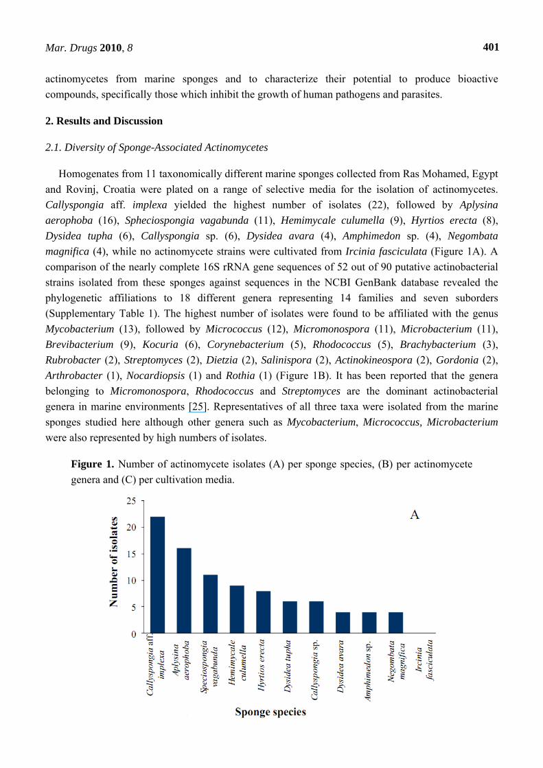
Homogenates from 11 taxonomically different marine sponges collected from Ras Mohamed, Egypt
and Rovinj, Croatia were plated on a range of selective media for the isolation of actinomycetes.
Callyspongia aff. implexa yielded the highest number of isolates (22), followed by Aplysina
aerophoba (16), Spheciospongia vagabunda (11), Hemimycale culumella (9), Hyrtios erecta (8),
Dysidea tupha (6), Callyspongia sp. (6), Dysidea avara (4), Amphimedon sp. (4), Negombata
magnifica (4), while no actinomycete strains were cultivated from Ircinia fasciculata (Figure 1A). A
comparison of the nearly complete 16S rRNA gene sequences of 52 out of 90 putative actinobacterial
strains isolated from these sponges against sequences in the NCBI GenBank database revealed the
phylogenetic affiliations to 18 different genera representing 14 families and seven suborders
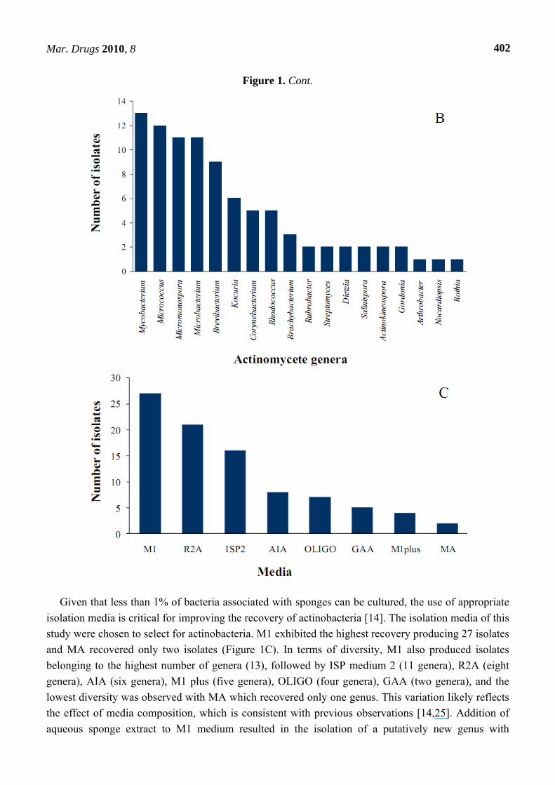
(Supplementary Table 1). The highest number of isolates were found to be affiliated with the genus
Mycobacterium (13), followed by Micrococcus (12), Micromonospora (11), Microbacterium (11),
Brevibacterium (9), Kocuria (6), Corynebacterium (5), Rhodococcus (5), Brachybacterium (3),
Rubrobacter (2), Streptomyces (2), Dietzia (2), Salinispora (2), Actinokineospora (2), Gordonia (2),
Arthrobacter (1), Nocardiopsis (1) and Rothia (1) (Figure 1B). It has been reported that the genera
belonging to Micromonospora, Rhodococcus and Streptomyces are the dominant actinobacterial
genera in marine environments [25]. Representatives of all three taxa were isolated from the marine
sponges studied here although other genera such as Mycobacterium, Micrococcus, Microbacterium
were also represented by high numbers of isolates.
Figure 1. Number of actinomycete isolates (A) per sponge species, (B) per actinomycete
genera and (C) per cultivation media.
Mar. Drugs 2010, 8
402
Figure 1. Cont.
Given that less than 1% of bacteria associated with sponges can be cultured, the use of appropriate
isolation media is critical for improving the recovery of actinobacteria [14]. The isolation media of this
study were chosen to select for actinobacteria. M1 exhibited the highest recovery producing 27 isolates
and MA recovered only two isolates (Figure 1C). In terms of diversity, M1 also produced isolates
belonging to the highest number of genera (13), followed by ISP medium 2 (11 genera), R2A (eight
genera), AIA (six genera), M1 plus (five genera), OLIGO (four genera), GAA (two genera), and the
lowest diversity was observed with MA which recovered only one genus. This variation likely reflects
the effect of media composition, which is consistent with previous observations [14,25]. Addition of
aqueous sponge extract to M1 medium resulted in the isolation of a putatively new genus with
Mar. Drugs 2010, 8
403
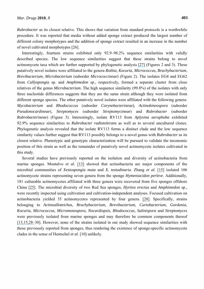
Rubrobacter as its closest relative. This shows that variation from standard protocols is a worthwhile
procedure. It was reported that media without added sponge extract produced the largest number of
different colony morphotypes and the addition of sponge extract resulted in an increase in the number
of novel cultivated morphotypes [26].
Interestingly, fourteen strains exhibited only 92.9–98.2% sequence similarities with validly
described species. The low sequence similarities suggest that these strains belong to novel
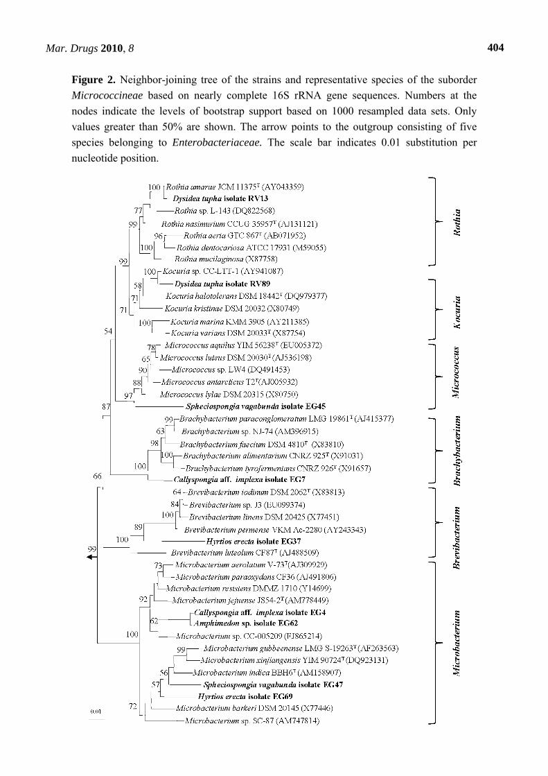
actinomycete taxa which are further supported by phylogenetic analysis [27] (Figures 2 and 3). These
putatively novel isolates were affiliated to the genera Rothia, Kocuria, Micrococcus, Brachybacterium,
Brevibacterium, Microbacterium (suborder Micrococcineae) (Figure 2). The isolates EG4 and EG62
from Callyspongia sp. and Amphimedon sp., respectively, formed a separate cluster from close
relatives of the genus Microbacterium. The high sequence similarity (99.8%) of the isolates with only
three nucleotide differences suggests that they are the same strain although they were isolated from
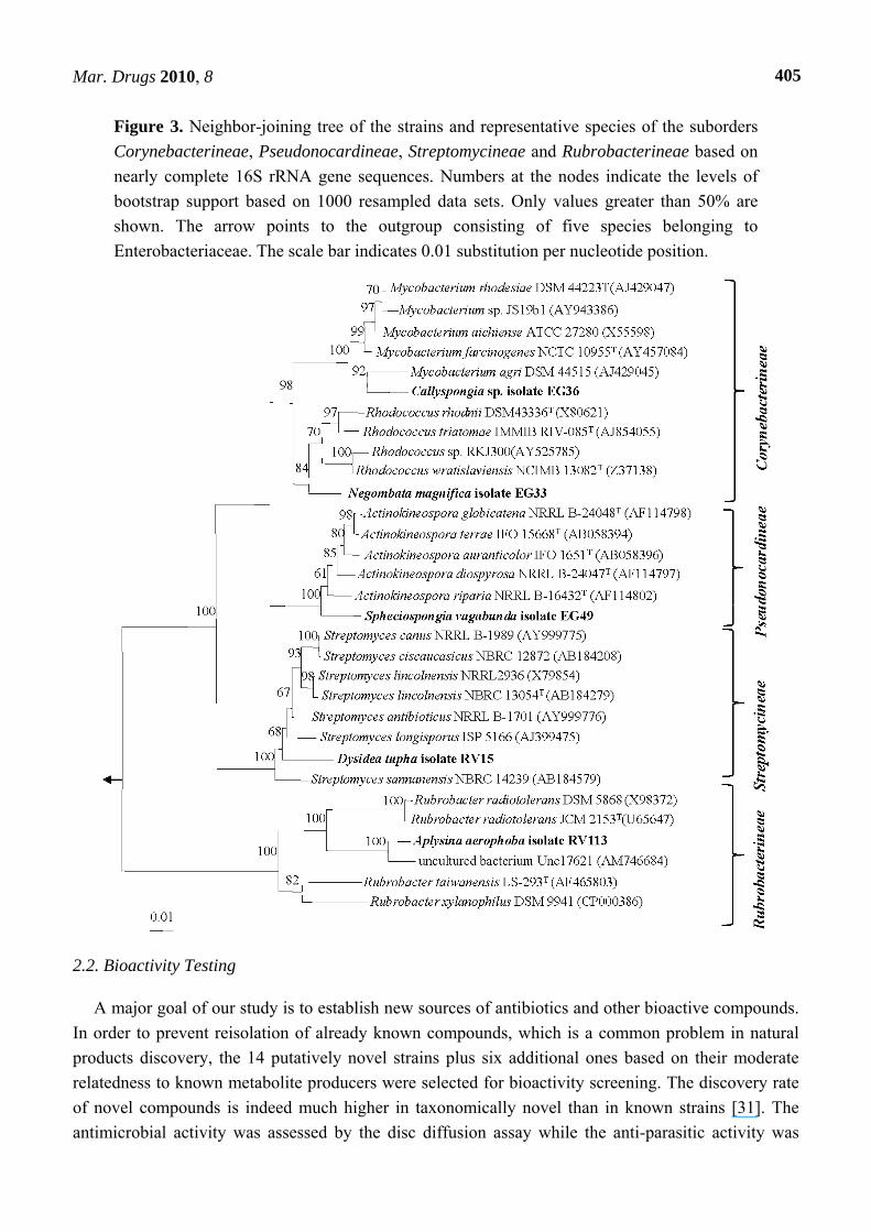
different sponge species. The other putatively novel isolates were affiliated with the following genera:
Mycobacterium and Rhodococcus (suborder Corynebacterineae), Actinokineospora (suborder
Pseudonocardineae), Streptomyces (suborder Streptomycineae) and Rubrobacter (suborder
Rubrobacterineae) (Figure 3). Interestingly, isolate RV113 from Aplysina aerophoba exhibited
92.9% sequence similarities to Rubrobacter radiotolerans as well as to several uncultured clones.
Phylogenetic analysis revealed that the isolate RV113 forms a distinct clade and the low sequence
similarity values further suggest that RV113 possibly belongs to a novel genus with Rubrobacter as its
closest relative. Phenotypic and genotypic characterization will be pursued to validate the taxonomic
position of this strain as well as the remainder of putatively novel actinomycete isolates cultivated in
this study.
Several studies have previously reported on the isolation and diversity of actinobacteria from
marine sponges. Montalvo et al. [13] showed that actinobacteria are major components of the
microbial communities of Xestospongia muta and X. testudinaria. Zhang et al. [15] isolated 106
actinomycete strains representing seven genera from the sponge Hymeniacidon perleve. Additionally,
181 culturable actinomycetes affiliated with three genera were recovered from five sponges offshore
China [25]. The microbial diversity of two Red Sea sponges, Hyrtios erectus and Amphimedon sp.,
were recently inspected using cultivation and cultivation-independent analyses. Focused cultivation on
actinobacteria yielded 35 actinomycetes represented by four genera. [28]. Specifically, strains
belonging to Actinoalloteichus, Brachybacterium, Brevibacterium, Curtobacterium, Gordonia,
Kocuria, Micrococcus, Micromonospora, Nocardiopsis, Rhodococcus, Salinispora and Streptomyces
were previously isolated from marine sponges and may therefore be common components thereof
[13,15,28–30]. However, none of the strains isolated in our study showed sequence similarities with
those previously reported from sponges, thus rendering the existence of sponge-specific actinomycete
clades in the sense of Hentschel et al. [10] unlikely.
Mar. Drugs 2010, 8
404
Figure 2. Neighbor-joining tree of the strains and representative species of the suborder
Micrococcineae based on nearly complete 16S rRNA gene sequences. Numbers at the
nodes indicate the levels of bootstrap support based on 1000 resampled data sets. Only
values greater than 50% are shown. The arrow points to the outgroup consisting of five
species belonging to Enterobacteriaceae. The scale bar indicates 0.01 substitution per
nucleotide position.
Mar. Drugs 2010, 8
405
Figure 3. Neighbor-joining tree of the strains and representative species of the suborders
Corynebacterineae, Pseudonocardineae, Streptomycineae and Rubrobacterineae based on
nearly complete 16S rRNA gene sequences. Numbers at the nodes indicate the levels of
bootstrap support based on 1000 resampled data sets. Only values greater than 50% are
shown. The arrow points to the outgroup consisting of five species belonging to
Enterobacteriaceae. The scale bar indicates 0.01 substitution per nucleotide position.
2.2. Bioactivity Testing
A major goal of our study is to establish new sources of antibiotics and other bioactive compounds.
In order to prevent reisolation of already known compounds, which is a common problem in natural
products discovery, the 14 putatively novel strains plus six additional ones based on their moderate
relatedness to known metabolite producers were selected for bioactivity screening. The discovery rate
of novel compounds is indeed much higher in taxonomically novel than in known strains [31]. The
antimicrobial activity was assessed by the disc diffusion assay while the anti-parasitic activity was
Mar. Drugs 2010, 8
406
measured in multi-well plates and is represented as the percentage of growth inhibition in comparison
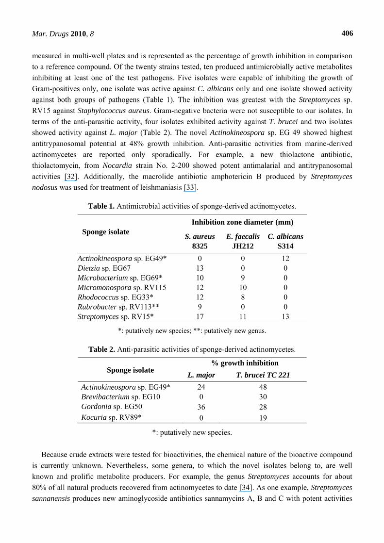
to a reference compound. Of the twenty strains tested, ten produced antimicrobially active metabolites
inhibiting at least one of the test pathogens. Five isolates were capable of inhibiting the growth of
Gram-positives only, one isolate was active against C. albicans only and one isolate showed activity
against both groups of pathogens (Table 1). The inhibition was greatest with the Streptomyces sp.
RV15 against Staphylococcus aureus. Gram-negative bacteria were not susceptible to our isolates. In
terms of the anti-parasitic activity, four isolates exhibited activity against T. brucei and two isolates
showed activity against L. major (Table 2). The novel Actinokineospora sp. EG 49 showed highest
antitrypanosomal potential at 48% growth inhibition. Anti-parasitic activities from marine-derived
actinomycetes are reported only sporadically. For example, a new thiolactone antibiotic,
thiolactomycin, from Nocardia strain No. 2-200 showed potent antimalarial and antitrypanosomal
activities [32]. Additionally, the macrolide antibiotic amphotericin B produced by Streptomyces
nodosus was used for treatment of leishmaniasis [33].
Table 1. Antimicrobial activities of sponge-derived actinomycetes.
Inhibition zone diameter (mm) Sponge isolate
C. albicans S314
E. faecalis JH212
S. aureus 8325
12 0 0 Actinokineospora sp. EG49* 0 0 13 Dietzia sp. EG67 0 9 10 Microbacterium sp. EG69* 0 10 12 Micromonospora sp. RV115 0 8 12 Rhodococcus sp. EG33* 0 0 9 Rubrobacter sp. RV113** 13 11 17 Streptomyces sp. RV15*
*: putatively new species; **: putatively new genus.
Table 2. Anti-parasitic activities of sponge-derived actinomycetes.
Sponge isolate % growth inhibition
L. major T. brucei TC 221
Actinokineospora sp. EG49* 24 48 Brevibacterium sp. EG10 0 30 Gordonia sp. EG50 36 28 Kocuria sp. RV89* 0 19
*: putatively new species.
Because crude extracts were tested for bioactivities, the chemical nature of the bioactive compound
is currently unknown. Nevertheless, some genera, to which the novel isolates belong to, are well
known and prolific metabolite producers. For example, the genus Streptomyces accounts for about
80% of all natural products recovered from actinomycetes to date [34]. As one example, Streptomyces
sannanensis produces new aminoglycoside antibiotics sannamycins A, B and C with potent activities
Mar. Drugs 2010, 8
407
against Gram-positive bacteria [35]. First organic chemistry analyses of the novel isolate RV15 points
to entirely novel cyclic peptides which are currently being characterized further. Rhodococcus species,
of which one putatively novel and bioactive strain was isolated in this study, produce a number of
commercially interesting and potentially useful products including various types of steroids and
peptides [36]. For example, two antimycobacterial cyclic peptides, lariatins A and B, were isolated
from the culture broth of Rhodococcus sp. K01-B0171 [37]. The genus Microbacterium, of which two
putatively new species were isolated in this study, produces different glycolipids. For example, five
anti-tumor glycoglycerolipids were obtained from Microbacterium sp., which was isolated from the
sponge Halichondria panicea [38]. Antimicrobial activities have, to our knowledge, not been reported
for the genera Actinokineospora, Kocuria and Rubrobacter and the identification of metabolites
produced by the putative new species of these genera is currently underway.
In conclusion, considerable actinobacterial diversity was recovered from various marine sponges.
Altogether 90 actinomycete isolates were affiliated with 18 different genera including 14 putatively
novel species. One possibly new genus related to Rubrobacter was isolated on M1 agar amended with
sponge extract, thus highlighting the need for innovative cultivation media. Antibacterial, antifungal,
antitrypanosomal and antileishmanial activities were reported for 10 of the isolates. Marine sponges
represent therefore a still largely untapped resource for novel actinomycete diversity as well as for new
secondary metabolites of therapeutic value.
3. Experimental
3.1. Sponge Collection
The first group of sponges (Aplysina aerophoba, Dysidea avara, D. tupha, Hemimycale culumella,
Ircinia fasciculata) was collected by SCUBA diving at depths of 3–20 m in the Mediterranean Sea
(Rovinj, Croatia, (GPS: 27˚47.655 N; 34˚12.904 W) in August 2008. Taxonomic identification was
performed by W.E.G. Müller and I. Müller (University of Mainz, Germany). The second group
(Amphimedon sp., Callyspongia sp., C. aff. implexa, Hyrtios erecta, Negombata magnifica,
Spheciospongia vagabunda) was collected at a depth of 10 m in the Red Sea (Ras Mohamed, Sinai,
Egypt; (GPS: 27˚47.655 N; 34˚12.904 W) in August 2006. Amphimedon sp. and Hyrtios erecta were
identified by M. Kelly (National Institute of Water and Atmospheric Research (NIWA) Auckland,
New Zealand) and the remaining sponges by R.W.M. van Soest (University of Amsterdam,
Netherlands). Sponges were transferred to plastic bags containing seawater and transported to the
laboratory. Sponge specimens were rinsed in sterile seawater, cut into pieces of ca. 1 cm3, and then
thoroughly homogenized in a sterile mortar with 10 volumes of sterile seawater. The supernatant was
diluted in ten-fold series (10-1, 10-2, 10-3) and subsequently plated out on agar plates.
3.2. Actinomycete Isolation
Eight different media [M1 [19], ISP medium 2 [39], Oligotrophic medium (OLIGO) [40], M1 plus [14],
Actinomycete Isolation Agar (AIA) [41], Marine Agar (MA) [42], Glycerol Asparagine Agar (GAA)
[41] and R2A Agar [43]] were used for the isolation of actinobacteria. All media were supplemented
with 0.2 µm pore size filtered cycloheximide (100 µg/mL), nystatin (25 µg/mL) and nalidixic acid
Mar. Drugs 2010, 8
408
(25 µg/mL) to facilitate the isolation of slow-growing actinobacteria. Cycloheximide and nystatin
inhibit fungal growth, while nalidixic acid inhibits many fast-growing Gram-negative bacteria [14]. All
media contained Difco Bacto agar (18 g/L) and were prepared in 1 L artificial sea water (NaCl 234.7 g,
MgCl2.6 H2O 106.4 g, Na2SO4 39.2 g, CaCl2 11.0 g, NaHCO3 1.92 g, KCl 6.64 g, KBr 0.96 g, H3BO3
0.26 g, SrCl2 0,24 g, NaF 0.03 g and ddH2O to 10.0 L) [44]. To promote the growth of selected
sponge-associated actinobacteria, 1% “aqueous sponge extract” was added to the autoclaved medium.
Aqueous sponge extract was prepared by grinding 20 g of sponge biomass in a mortar containing
20 mL of sterile seawater followed by centrifugation (5000 rpm, 10 min) and sterilized by filtration
through a 0.2 µm pore size filter. The freshly prepared supernatant served as aqueous sponge extract.
The inoculated plates were incubated at 30 °C for 6–8 weeks. Distinct colony morphotypes were
picked and re-streaked until visually free of contaminants. Isolates were inoculated into liquid media
(M1 or the medium on which colonies were initially isolated). The isolates were maintained on plates
for short-term storage and long-term strain collections were set up in medium supplemented with 30%
glycerol at −80 °C. The isolates from Egypt are abbreviated as “EG” and from Rovinj as “RV”.
3.3. Molecular Identification and Phylogenetic Analysis
16S rRNA gene amplification, cloning and sequencing were performed according to Hentschel et
al. [45] using the universal primers 27F and 1492R [46]. Chimeric sequences were identified by using
the Pintail program [47]. The genus-level affiliation of the sequences was validated using the
Ribosomal Database Project Classifier [48]. Sequence alignment and phylogenetic analysis were
performed using the ARB software package [49]. Tree construction was conducted using neighbour-
joining algorithm (Jukes-Cantor correction) with bootstrap values based on 1000 replications. The 16S
rRNA gene sequences of the putatively novel isolates were deposited in GenBank under the accession
numbers indicated in parentheses: EG4 (GU318354), EG7 (GU318355), EG33 (GU318356), EG36
(GU318357), EG37 (GU318358), EG45 (GU318359), EG47 (GU318360), EG49 (GU318361), EG62
(GU318362), EG69 (GU318363), RV15 (GU318364), RV113 (GU318365), RV13 (GU318366) and
RV89 (GU318367).
3.4. Extract Preparation and Anti-infective Activity Screening
Fourteen strains selected based on phylogenetic novelty and six selected based on their
affiliation to known metabolite-producers were cultured in 100 mL Erlenmeyer flasks containing
50 mL of 5 different production media (M1, ISP2, OLIGO, AIA and R2A) for each isolate. The liquid
cultures were grown for 7–14 days depending on their growth rate at 30 °C while shaking at 150 rpm.
An equal volume of methanol was added to the liquid cultures for cell lysis and shaking was continued
(150 rpm, 1 h at room temperature; Shaker SM 30, E. Bühler). The broth was centrifuged in
50 mL falcon tubes (5000 rpm, 15 min at room temperature; Megafuge 1.0R, Heraeus) and the
supernatant was stored at 4 °C.
The in vitro antimicrobial activity testing was carried out using the standard disk diffusion assay [50]
against pathogenic bacteria (Staphylococcus aureus strain 8325, Enterococcus faecalis strain JH212,
Escherichia coli strain 536, Pseudomonas aeruginosa strain Nr. 3) and yeast (Candida albicans strain
S314). Sterile filter disks (6 mm) impregnated with actinomycete extracts were placed on agar plates
Mar. Drugs 2010, 8
409
that had been inoculated with the test pathogen. After 24 h incubation at 37 °C (bacteria) and 30 °C
(yeast), the antimicrobial potential was quantitatively assessed as diameter of the inhibition zone
(n = 2). Anti-leishmanial activity was tested following the method of Ponte-Sucre et al. [51]. Briefly,
this involved the incubation of Leishmania major promastigotes for 24 h at 26 °C, 5% CO2, and 95%
humidity in the absence or presence of the extracts. Following the addition of Alamar Blue, the plates
were incubated again and the optical densities were determined after 48 h with a Multiskan Ascent
enzyme-linked immunosorbent assay (ELISA) reader (Thermo Electron Corporation, Dreieich,
Germany). Absorbance in the absence of compounds was set as 100% of growth. Amphotericin B was
used as a reference compound and positive control. Each extract was assayed in duplicate from two
independent experiments. Anti-trypanosomal activity was tested following the protocols of Huber and
Koella [52]. Following cultivation of Trypanosoma brucei brucei strain TC 221 in Complete Baltz
Medium, a defined number of parasites were exposed in 96-well plate test chambers to the extracts.
The plates were then incubated at 37 °C in an atmosphere of 5% CO2 for 24 h. After addition of
Alamar Blue, the activity of the extracts was measured by light absorption using MR 700 Microplate
Reader after 48 h. Absorbance in the absence of compounds was set as 100% of growth. Each extract
was assayed in duplicate from two independent experiments.
Acknowledgments
We gratefully acknowledge H. Angermeier and P. Tabares for sponge collections from Rovinj, C.
Gernert for technical assistance in the laboratory and P. Tabares for introduction into the molecular
techniques. We thank H. Bruhn, A. Stich (SFB 630 TP Z1) and H. Moll (SFB 630 TP B3) for
performing the anti-parasitic screening assays. Financial support was provided by the Egyptian
government to U. R. and the DFG (SFB 630 TP A5) to U. H.
References
1. Pinner, R.; Teutsch, S.; Simonsen, L.; Klug, L., Graber, J.; Clarke, M.; Berkelman, R. Trends in
infectious diseases mortality in the United States. J. Am. Med. Assoc. 1996, 275, 189–193.
2. Woolhouse, M.E.J. Epidemiology: Emerging diseases go global. Nature 2008, 451, 898–899.
3. Morens, D.M.; Folkers, G.K.; Fauci, A.S. The challenge of emerging and re-emerging infectious
diseases. Nature 2004, 430, 242–249.
4. Daszak, P.; Cunningham, A.A.; Hyatt, A.D. Emerging infectious diseases of wildlife-threats to
biodiversity and human health. Science 2000, 287, 443–449.
5. Jones, K.E.; Patel N.G.; Levy, M.A.; Storeygard, A.; Balk, D.; Gittleman J.L.; Daszak, P. Global
trends in emerging infectious diseases. Nature 2008, 451, 990–993.
6. Blunt, J.W.; Copp, B.R.; Hu, V.P.; Munro, M.H.; Northcote, P.T.; Prinsep, M.R. Marine natural
products. Nat. Prod. Rep. 2007, 24, 31–86.
7. Hentschel, U.; Usher, K.M.; Taylor, M.W. Marine sponges as microbial fermenters. FEMS
Microbiol. Ecol. 2006, 55, 167–177.
8. Taylor, M.W.; Radax, R.; Steger, D.; Wagner, M. Sponge-associated microorganisms: Evolution,
ecology, and biotechnological potential. Microbiol. Mol. Biol. Rev. 2007, 71, 295–347.
9. Webster, N.S. Sponge disease: A global threat? Environ. Microbiol. 2007, 9, 1363–1375.
Mar. Drugs 2010, 8
410
10. Hentschel, U.; Hopke, J.; Horn, M.; Friedrich, A.B.; Wagner, M.; Hacker, J.; Moore, B.S.
Molecular evidence for a uniform microbial community in sponges from different oceans.
Appl. Environ. Microbiol. 2002, 68, 4431–4440.
11. Jiang, S.; Li, X.; Zhang, L.; Sun, W.; Dai, S.; Xie, L.; Liu, Y.; Lee, K. Culturable actinobacteria
isolated from marine sponge Iotrochota sp. Mar. Biol. 2008, 153, 945–952.
12. Kim, T.K.; Garson, M.J.; Fuerst, J.A. Marine actinomycetes related to the 'Salinospora' group
from the great barrier reef sponge Pseudoceratina clavata. Environ. Microbiol. 2005, 7, 509–518.
13. Montalvo, N.F.; Mohamed, N.M.; Enticknap, J.J.; Hill, R.T. Novel actinobacteria from marine
sponges. Anton. Leeuwenhoek 2005, 87, 29–36.
14. Webster, N.S.; Wilson, K.J.; Blackall, L.L.; Hill, R.T. Phylogenetic diversity of bacteria
associated with the marine sponge Rhopaloeides odorabile. Appl. Environ. Microbiol. 2001,
67, 434–444.
15. Zhang, H.T.; Lee, Y.K.; Zhang, W., Lee, H.K. Culturable actinobacteria from the marine sponge
Hymeniacidon perleve: Isolation and phylogenetic diversity by 16S rRNA Gene-RFLP analysis.
Anton. Leeuwenhoek 2006, 90, 159–169.
16. Bredholdt, H.; Galatenko, O.A.; Engelhardt, K.; Fjærvik, E.; Terekhova, L.P.; Zotchev, S.B. Rare
actinomycete bacteria from the shallow water sediments of the trodheim fjord, norway: Isolation,
diversity and biological activity. Environ. Microbiol. 2007, 9, 2756–2764.
17. Fenical, W.; Jensen, P.R. Developing a new resource for drug discovery: Marine actinomycete
bacteria. Nat. Chem. Biol. 2006, 2, 666–673.
18. Grein, A.; Meyers, S.P. Growth characteristics and antibiotic production of actinomycetes isolated
from littoral sediments and materials suspended in sea water. J. Bacteriol. 1958, 76, 457–463.
19. Mincer, T.J.; Jensen, P.R.; Kauffman, C.A.; Fenical, W. Widespread and persistent populations of
a major new marine actinobacteria taxon in ocean sediments. Appl. Environ. Microbiol. 2002,
68, 5005–5011.
20. Panthom-Aree, W.; Stach, J.E.M.; Ward, A.C.; Horikoshi, K.; Bull, A.T.; Goodfellow, M.
Diversity of actinomycetes isolated from challenger deep sediment (10,898 m) from the Mariana
Trench. Extremophiles 2006, 10, 181–189.
21. Maldonado, L.A.; Fenical, W.; Jensen, P.R.; Kauffman, C.A.; Mincer, T.J.; Ward, A.C.; Bull,
A.T.; Goodfellow, M. Salinispora arenicola gen. nov., sp. nov. and Salinispora tropica sp. nov.,
obligate marine actinomycetes belonging to the family Micromonosporaceae. Int. J. Syst. Evol.
Microbiol. 2005, 55, 1759–1766.
22. Lam, K.S. Discovery of novel metabolites from marine actinomycetes. Curr. Opin. Microbiol.
2006, 9, 245–251.
23. Maldonado, L.A.; Stach, J.E.M.; Pathom-aree, W.; Ward, A.C.; Bull, A.T.; Goodfellow, M.
Diversity of cultivable actinobacteria in geographically widespread marine sediments.
Anton. Leeuwenhoek 2005, 87, 11–18.
24. Kim, T.K.; Hewavitharana, A.K.; Shaw, P.N.; Fuerst, J.A. Discovery of a new source of
rifamycin antibiotics in marine sponge actinobacteria by phylogenetic prediction. Appl. Environ.
Microbiol. 2006, 72, 2118–2125.
Mar. Drugs 2010, 8
411
25. Zhang, H.T.; Zhang, W.; Jin, Y.; Jin, M; Yu, X. A comparative study on the phylogenetic
diversity of culturable actinobacteria isolated from five marine sponge species.
Anton. Leeuwenhoek 2008, 93, 241–248.
26. Webster, N.S.; Hill, R.T. The culturable microbial community of the great barrier reef sponge
Rhopaloeides odorabile. Appl. Environ. Microbiol. 2001, 138, 843–851.
27. Stackebrandt, E.; Ebers, J. Taxonomic parameters revisited: Tarnished gold standards.
Microbiol. Today 2006, 33, 152–155.
28. Radwan, M.; Hanora, A.; Zan, J.; Mohamed, N.M.; Abo-Elmatty, D.M.; Abou-El-Ela, S.H.; Hill,
R.T. Bacterial community analyses of two Red Sea sponges. Mar. Biotechnol. 2009, DOI:
10.1007/s10126-009-9239-5.
29. Bultel-Ponce, V.C.; Debitus, J.P.; Berge, G.P.; Guyot, M. Metabolites from the marine sponge-
associated bacterium Micrococcus luteus. J. Mar. Biotech. 1998, 6, 233–236.
30. Lee, H.K.; Lee, D.S.; Lim, J.; Kim, J.S.; Im, K.S.; Jung, J.H. Topoisomerase I inhibitors from the
Streptomyces sp. Strain KM86–9B isolated from a marine sponge. Arch. Pharm. Res. 1998, 2,
729–733.
31. Pimentel-Elardo, S.M. Novel Anti-infective secondary metabolites and biosynthetic gene clusters
from actinomycetes associated with marine sponges. Dissertation thesis, University of Würzburg,
Würzburg, Germany, 2008.
32. Oishi, H.; Noto, T.; Sasaki, H.; Suzuki, K.; Hayashi, T.; Okazaki, H.; Ando, K.; Sawada, M.
Thiolactomycin, a new antibiotic. 1. taxonomy of the prodrug organism, fermentation and
biological properties. J. Antibiot. 1982, 35, 391–395.
33. Loiseau, P.M.; Imbertie, L.; Bories, C.; Betbeder, D.; De Miguel, V. Design and antileishmanial
activity of amphotericin b-loaded stable ionic amphiphile biovector formulations. Antimicrob.
Agents Chemother. 2002, 46 (5), 1597–1601.
34. Bull, A.T.; Stach, J.E. Marine actinobacteria: New opportunities for natural product search and
discovery. Trends Microbiol. 2007, 15, 491–499.
35. Deushi, T.; Yamaguchi, T.; Kamiya, K.; Iwasaki, A.; Mizoguchi, T.; Nakayama, M.; Watanabe, I;
ITOH, H.; Mri, T. A new aminoglycoside antibiotic, sannamycin C and its 4-N-glycyl derivative.
J. Antibiot. 1980, 33, 1274–1280.
36. Bell, K.S.; Philp, J.C.; Aw, D.W.J.; Christofi, N. The genus Rhodococcus. J. Appl. Microbiol.
1998, 85, 195–210.
37. Iwatsuki, M; Tomoda, H.; Uchida, R.; Gouda, H.; Hirono, S.; Ōmura, S. Lariatins,
antimycobacterial peptides produced by Rhodococcus sp. K01−B0171, have a lasso structure. J.
Am. Chem. Soc. 2006, 128, 74, 86–7491.
38. Wicke, C.; Hüners, M.; Wray, V.; Nimtz, M.; Bilitewski, U.; Lang, S. Production and structure
elucidation of glycoglycerolipids from a marine sponge-associated Microbacterium species. J.
Nat. Prod. 2000, 63, 621–626.
39. Shirling, E.B.; Gottlieb, D. Methods for characterization of Streptomyces species. Int. J. Syst.
Bacteriol. 1966, 16, 313–340.
40. Olson, J.B.; Lord, C.C.; McCarthy, P.J. Improved recoverability of microbial colonies from
sponge samples. Mar. Microb. Ecol. 2000, 40, 139–147.
Mar. Drugs 2010, 8
412
41. Lechevalier, M.P. Actinomycetes of sewage-treatment plants. Environ. Protection Technol. Ser.
1975, EPA-600/2-75-031.
42. Weiner, R.M.; Segall, A.M.; Colwell, R.R. Characterization of a marine bacterium associated
with Crassostrea virginica (the eastern oyster). Appl. Environ. Microbiol. 1985, 49, 83–90.
43. Reasoner, D.J.; Geldreich, E.E. A new medium for the enumeration and subculture of bacteria
from potable water. Appl. Environ. Microbiol. 1985, 49, 1–7.
44. Lyman, J.; Fleming, R. Composition of seawater. J. Mar. Res. 1940, 3, 134–146.
45. Hentschel, U.; Schmid, M.; Wagner, M.; Fieseler, L.; Gernert, C.; Hacker, J. Isolation and
phylogenetic analysis of bacteria with antimicrobial activities from the mediterranean sponges
Aplysina aerophoba and Aplysina cavernicola. FEMS Microbiol. Ecol. 2001, 35, 305–312.
46. Lane, D.J. 16S/23S rRNA Sequencing. In Nucleic Acid Techniques in Bacterial Systematic;
Stackebrandt, E., Goodfellow, M., Eds.; John Wiley: Chichester, UK, 1991; pp. 115–175.
47. Ashelford, K.E.; Chuzhanova, N.A.; Fry, J.C.; Jones, A.J.; Weightman, A.J. At least 1 in 20 16S
rRNA sequence records currently held in public repositories is estimated to contain substantial
anomalies. Appl. Environ. Microbiol. 2006, 71, 7724–7736.
48. Wang, Q.; Garrity, G.M.; Tiedje, J.M.; Cole, J.R. Naïve Bayesian classifier for rapid assignment
of rRNA sequences into the new bacterial taxonomy. Appl. Environ. Microbiol. 2007, 73(16),
5261–5267.
49. Ludwig, W.; Strunk, O.; Westram, R.; Richter, L.; Meier, H.; Yadhukumar, Buchner, A.; Lai, T.;
Steppi, S.; Jobb. G.; et al. ARB: A software environment for sequence data. Nuc. Acid Res. 2004,
32, 1363–1371.
50. Inderlied, C.B.; Salfinger, M. Antimicrobial agents and susceptibility tests: Mycobacteria. In
Manual of Clinical Microbiology, 6th ed.; Murray, P.R., Baron, E.J., Pfaller, M.A., Tenover, F.C.,
Yolken, R.H., Eds.; American Society for Microbiology: Washington DC, USA, 1995;
pp. 1385–1404.
51. Ponte-Sucre, A.; Vicik, R.; Schultheis, M.; Schirmeister, T.; Moll, H. Aziridine-2,3-
dicarboxylates, peptidomimetic cysteine protease inhibitors with antileishmanial activity.
Antimicrob. Agents Chemother. 2006, 50, 2439–2447.
52. Huber, W.; Koella, J.C. A Comparison of three methods of estimating ec50 in studies of drug
resistance of malaria parasites. Acta Trop. 1993, 55, 257–261.
Samples Availability: Available from the authors.
© 2010 by the authors; licensee Molecular Diversity Preservation International, Basel, Switzerland.
This article is an open-access article distributed under the terms and conditions of the Creative
Commons Attribution license (http://creativecommons.org/licenses/by/3.0/).