Genus Diversity of Actinomycetes in Cibinong Science Center, West Java, Indonesia
8
Available online at http://jurnal.permi.or.id/index.php/mionline DOI: 10.5454/mi.6.4.4 ISSN 1978-3477, eISSN 2087-8587 Vol 6, No 4, December 2012, p 165-172 *Corresponding author; Phone: , Fax: 21-8754587, E-mail: [email protected] +62-21-8754587 +62- Microorganisms play important role in our activities, although some have pathogenic properties but some have positive role to support human life. Actinomycetes are microorganisms that play important role especially in pharmaceutical industry to support human health. They are potential as a novel source for the discovery of new bioactive compounds. Activities that focused on discovery of new bioactive compound Actinomycetes are microorganisms that play important role to support human health and known as soil microorganisms. The aim of the research was to describe genus diversity of actinomycetes in Cibinong Science Center (CSC), West Java. Samples for isolation were soil and plant litters. The samples were air dried and ground. We employed isolation methods: dry heat (DH), sodium dodecyl sulphates-yeast extract (SDS-YE), rehydration and centrifugation (RC), and oil separation (OS). A total of 263 isolates of actinomycetes were isolated in CSC, in 2004-2006. Totally 58, 144, 50, and 11 isolates were isolated under each isolation methods, respectively. All isolates were identified using the 16S rRNA gene sequencing method. The results showed that the isolates were belonged to the family Kineosporiaceae, Micromonosporaceae, Nocardiaceae, Pseudonocardiaceae, Streptomycetaceae, Streptosporangiaceae, Mycobacteriaceae, Nocardioidaceae, Nocardiopsaceae, and Thermomonosporaceae. There were 23 genera under those families. Homology value of the isolates based on BLAST search using 16S rRNA gene sequence data as queries showed that 136, 91, 30, and 6 isolates were ≥99, 98, 97, and ≤ 96%, respectively, compared to the known sequence in data base. The later 6 isolates were interesting for further identification leading to new taxa. Recognized species of Streptomyces genera under the member of the Streptomycetaceae were dominant among other isolates. Keywords: 16S rRNA gene sequencing, actinomycetes, diversity, Indonesia Actinomycetes merupakan mikroorganisma tanah yang mempunyai peran pada bidang kesehatan. Oleh karena itu, upaya pencarian species actinomycetes baru banyak dilakukan. Tujuan penelitian ini adalah melihat keanekargaman actinomycetes tingkat genus di Cibinong Science Center (CSC), Jawa Barat. Sampel untuk isolasi actinomycetes adalah tanah dan serasah. Metode isolasi yang digunakan adalah dry heat (DH), sodium dodesil sulfat-yeast extract (SDS-YE), rehydration and centrifugation (RC), dan oil separation (OS). Sebanyak 263 isolat actinomycetes telah diperoleh dari CSC pada tahun 2004-2006. Dari jumlah tersebut 58 dari metode DH, 144 isolat dari SDS-YE, 50 dari RC dan 11 dari OS isolat. Identifikasi isolat dilakukan menggunakan metoda sekuen gen 16S rRNA. Hasilnya menunjukkan adanya famili Kineosporiaceae, Micromonosporaceae, Nocardiaceae, Pseudonocardiaceae, Streptomycetaceae, Streptosporangiaceae, Mycobacteriaceae, Nocardioidaceae, Nocardiopsaceae, dan Thermomonosporaceae. Dari famili tersebut diperoleh 23 genus. Nilai kesesuaian isolat berdasarkan analisis BLAST menggunakan sekuen 16S gen rRNA sebagai queri dibandingkan dengan species yang terdaftar pada data base menunjukkan bahwa 131, 91, 30, dan 6 isolat masing-masing mempunyai kesamaan sebesar ≤ 99, 98, 97, dan ≤ 96%. Enam isolat dengan kesesuaian ≤ 96% sangat menarik untuk diungkap sebagai taxa baru. Pada penelitian ini, diperoleh isolat terbanyak dari genus Streptomyces. Kata kunci: actinomycetes, Indonesia, keragaman, sekuensing gen 16S rRNA Genus Diversity of Actinomycetes in Cibinong Science Center, West Java, Indonesia 1 1 1 YANTYATI WIDYASTUTI *, PUSPITA LISDIYANTI , SHANTI RATNAKOMALA , GINA 1 1 1 1 KARTINA , RONI RIDWAN , ROHMATUSSOLIHAT, NITA ROSALINDA PRAYITNO , EVI 2 2 3 3 TRIANA , NUNUK WIDHYASTUTI , RASTI SARASWATI , RATIH DEWI HASTUTI , 4 5 5 5 YULIN LESTARI , MISA OTOGURO , SHINJI MIYADOH , HIDEKI YAMAMURA , 5 5 TOMOHIKO TAMURA , AND KATSUHIKO ANDO 1 Research Center for Biotechnology, Indonesian Institute of Sciences (LIPI), Jalan Raya Bogor Km 46, Bogor 16911, Indonesian; 2 Research Center for Biology, Indonesian Institute of Sciences (LIPI), Jalan Raya Jakarta-Bogor Km 46, Bogor 16911, Indonesia; 3 Indonesia Soil Research Institute, Departement of Agricultutre, Jalan Tentara Pelajar No 12, Bogor 16114, Indonesia; 4 Faculty of Mathematics and Sciences, Institut Pertanian Bogor, Dramaga Campus, Bogor 16680, Indonesia; 5 NITE Biological Resource Center, National Instituite of Technology and Evaluation, Kazusakamatari, Kisarazu-shi, Chiba, Japan
-
Upload
independent -
Category
Documents
-
view
0 -
download
0
Transcript of Genus Diversity of Actinomycetes in Cibinong Science Center, West Java, Indonesia
Available online athttp://jurnal.permi.or.id/index.php/mionline
DOI: 10.5454/mi.6.4.4ISSN 1978-3477, eISSN 2087-8587Vol 6, No 4, December 2012, p 165-172
*Corresponding author; Phone: , Fax:21-8754587, E-mail: [email protected]
+62-21-8754587 +62-
Microorganisms play important role in our
activities, although some have pathogenic properties
but some have positive role to support human life.
Actinomycetes are microorganisms that play important
role especially in pharmaceutical industry to support
human health. They are potential as a novel source for
the discovery of new bioactive compounds. Activities
that focused on discovery of new bioactive compound
Actinomycetes are microorganisms that play important role to support human health and known as soil microorganisms. The aim of the research was to describe genus diversity of actinomycetes in Cibinong Science Center (CSC), West Java. Samples for isolation were soil and plant litters. The samples were air dried and ground. We employed isolation methods: dry heat (DH), sodium dodecyl sulphates-yeast extract (SDS-YE), rehydration and centrifugation (RC), and oil separation (OS). A total of 263 isolates of actinomycetes were isolated in CSC, in 2004-2006. Totally 58, 144, 50, and 11 isolates were isolated under each isolation methods, respectively. All isolates were identified using the 16S rRNA gene sequencing method. The results showed that the isolates were belonged to the family Kineosporiaceae, Micromonosporaceae, Nocardiaceae, Pseudonocardiaceae, Streptomycetaceae, Streptosporangiaceae, Mycobacteriaceae, Nocardioidaceae, Nocardiopsaceae, and Thermomonosporaceae. There were 23 genera under those families. Homology value of the isolates based on
BLAST search using 16S rRNA gene sequence data as queries showed that 136, 91, 30, and 6 isolates were ≥99,
98, 97, and ≤ 96%, respectively, compared to the known sequence in data base. The later 6 isolates were interesting for further identification leading to new taxa. Recognized species of Streptomyces genera under the member of the Streptomycetaceae were dominant among other isolates.
Keywords: 16S rRNA gene sequencing, actinomycetes, diversity, Indonesia
Actinomycetes merupakan mikroorganisma tanah yang mempunyai peran pada bidang kesehatan. Oleh karena itu, upaya pencarian species actinomycetes baru banyak dilakukan. Tujuan penelitian ini adalah melihat keanekargaman actinomycetes tingkat genus di Cibinong Science Center (CSC), Jawa Barat. Sampel untuk isolasi actinomycetes adalah tanah dan serasah. Metode isolasi yang digunakan adalah dry heat (DH), sodium dodesil sulfat-yeast extract (SDS-YE), rehydration and centrifugation (RC), dan oil separation (OS). Sebanyak 263 isolat actinomycetes telah diperoleh dari CSC pada tahun 2004-2006. Dari jumlah tersebut 58 dari metode DH, 144 isolat dari SDS-YE, 50 dari RC dan 11 dari OS isolat. Identifikasi isolat dilakukan menggunakan metoda sekuen gen 16S rRNA. Hasilnya menunjukkan adanya famili Kineosporiaceae, Micromonosporaceae, Nocardiaceae, Pseudonocardiaceae, Streptomycetaceae, Streptosporangiaceae, Mycobacteriaceae, Nocardioidaceae, Nocardiopsaceae, dan Thermomonosporaceae. Dari famili tersebut diperoleh 23 genus. Nilai kesesuaian isolat berdasarkan analisis BLAST menggunakan sekuen 16S gen rRNA sebagai queri dibandingkan dengan species yang terdaftar pada data base menunjukkan bahwa 131, 91, 30, dan 6 isolat masing-masing
mempunyai kesamaan sebesar ≤99, 98, 97, dan ≤96%. Enam isolat dengan kesesuaian ≤96% sangat menarik untuk diungkap sebagai taxa baru. Pada penelitian ini, diperoleh isolat terbanyak dari genus Streptomyces.
Kata kunci: actinomycetes, Indonesia, keragaman, sekuensing gen 16S rRNA
Genus Diversity of Actinomycetes in Cibinong Science Center, West Java, Indonesia
1 1 1YANTYATI WIDYASTUTI *, PUSPITA LISDIYANTI , SHANTI RATNAKOMALA , GINA 1 1 1 1KARTINA , RONI RIDWAN , ROHMATUSSOLIHAT , NITA ROSALINDA PRAYITNO , EVI
2 2 3 3TRIANA , NUNUK WIDHYASTUTI , RASTI SARASWATI , RATIH DEWI HASTUTI , 4 5 5 5YULIN LESTARI , MISA OTOGURO , SHINJI MIYADOH , HIDEKI YAMAMURA ,
5 5TOMOHIKO TAMURA , AND KATSUHIKO ANDO
1Research Center for Biotechnology, Indonesian Institute of Sciences (LIPI), Jalan Raya Bogor Km 46, Bogor 16911, Indonesian;
2 Research Center for Biology, Indonesian Institute of Sciences (LIPI), Jalan Raya Jakarta-Bogor Km 46,
Bogor 16911, Indonesia;3Indonesia Soil Research Institute, Departement of Agricultutre, Jalan Tentara Pelajar No 12, Bogor 16114, Indonesia;
4Faculty of Mathematics and Sciences, Institut Pertanian Bogor, Dramaga Campus, Bogor 16680, Indonesia;5 NITE Biological Resource Center, National Instituite of Technology and Evaluation,
Kazusakamatari, Kisarazu-shi, Chiba, Japan
for new products of antibiotics are carried out in some
parts of the world recently. Different ecological niches
showed many interesting new taxa of actinomycetes
and soil has been reported as the natural habitat of
actinomycetes. Results of isolation of actinomycetes
differed depend on condition of soil and vegetation
above the soil (Li et al. 2006; Jiang et al. 2008) or
mangrove ecosystem (Tamura et al. 2005; Tamura et
al. 2006).
Indonesia is located in an equatorial area and most
of the area is covered by its tropical rain forests.
Tropical rain forests has been known to be the most
biologically diverse ecosystems for plants, animals and
microorganims on earth. Compared to that of plants
and animals, information on diversity microorganisms
is very limited. So far, there is no report on diversity of
actinomycetes from Indonesia. During the
collaborative research on ecology and taxonomy of
actinomycetes in Indonesia between Indonesia and
Japan, we have collected thousands of actinomycetes
isolates from several sites in Indonesia. Some
interesting new taxa has been reported, including
Streptomyces baliensis sp nov. from Bali (Otoguro et
al. 2009), Dietzia timorensis from Timor (Yamamura
et al. 2010); Actinokineospora baliensis sp. nov.,
Actinokineospora cibodasensis sp. nov.,
Actinokineospora cianjurensis sp. nov. from Bali and
Cibodas Botanic Garden (Lisdiyanti et al. 2010); and
Act inophytocola t imorensis sp . nov. and
Actinophytocola coralina sp. nov., from Kupang, East
Nusa Tenggara (Otoguro et al. 2011). Cibinong
Science Center (CSC), West Java, was selected as one
site during the study. The aim of the research was to
describe genus diversity of actinomycetes in CSC
during the period of 2004-2006.
MATERIALS AND METHODS
Sample Collection and Preparation. Soil and
plant litter samples were collected from different
locations in CSC in the year of 2004, 2005, and 2006.
Soil samples were taken by digging about 15 cm from
the top soil and and put into plastic bag to keep their
humidity while plant litter samples were kept in a paper
bag. All samples were air dried for 5-7 d, then ground
and sieved through a 2 mm sieve.
Isolation of Actinomycetes. Four different
isolation methods for actinomycetes were employed
with different target of isolates first Sodium dodecyl
sulphate-yeast extract (SDS-YE) (Hayakawa and
Nonomura 1989) for general actinomycetes. One g of a
166 WIDYASTUTI ET AL. Microbiol Indones
dried soil sample was suspended in 10 mL of water and
stirred for 1 min using a thermo mixer. Then, 1 mL of
this suspension was transferred to SDS-YE solution
(0.05% of SDS and 6% of yeast extract were dissolved
in 50 mM P-buffer pH 7.0). The suspension was heated
at 40 °C for 20 min. SDS-YE solution was serially
diluted and spread each 0.1 mL solutions to Humic
acid-Vitamin (HV) medium (Hayakawa and
Nonomura 1987). Second Rehydration-centrifugation
(RC) (Hayakawa et al. 2000) for motile bearing
arthrospore actinomycetes. Five hundred mg of dried
soil sample were suspended in 10% of soil extract and
P-buffer and kept at 28 °C for 1 h, to release the
zoospores. Elimination of non-motile actinomyetes
was carried out by centrifugation at 3000 g for 20 min.
After centrifugation, the buffer was incubated at 28 °C
for 30 min, to sediment the non-motile actinomycetes,
while motile actinomycetes are swimming up in the
supernatant. Supernatant was serially diluted and
spread 0.1 mL solutions to HV medium. The third dry
heat (DH)(Nonomura and Ohara 1969) for heat
resistant and rare actinomycetes. One gram of air dried
soil samples were put in a glass petri dish and heated in
oven at 100-120 °C for 1 h to eliminate filamentous
bacteria and Streptomyces. The sample (0.1 mL
solutionswas) serially diluted and spread to HV
medium. Fourth, oil separation (OS) as modification of
Ishigami's water-hexane distribution method use of
olive-oil instead of hexane (Ishigami et al. 2004) for
lypolytic actinomycetes.This method is based on
distribution with oil and water. Five hundred mg of soil
sample were suspended in 5 mL of olive oil and mixed
for 5 min. Then, 5 mL of water were added to oil and
mixed again. Oil and water were distributed by
centrifugation for 10 min. After centrifugation, 0.1 mL
of oil was spread to HV medium with addition of -1 -1Kabicidin (0.75 mg L ), Nalidixic acid (10 mg L ), and
-1Chroltetracyclin (50 mg L ).
HV Medium Preparation. HV agar plates -1supplemented with kabicidin (0.75 mg L ) and
-1nalidixic acid (10 mg L ) were prepared at least 4 d
before used, for optimum absorption of inoculated
samples. Diluted samples were inoculated and spread
until dry. Incubation was done at room temperature for
5-20 d, with occasionally observation after 5 d. Plates
were kept in a plastic bag and put in paper box.
Isolation and Selection of Actinomycetes.
Colonies appeared around 5 d of incubation represent
fast growing and the rest were slow growing
actinomycetes. Colonies of interest were picked up
using sterile woody tooth pick and inoculated to yeast
extract-starch (YS) agar plates. Incubation was carried
out at room temperature. Morphological observation of
colonies grown in YS agar was done under a
microscope. Detection of pigment produced by the
isolates was observed from both upper and lower sides
of the plates. Selection of the isolates was based on
their different morphological appearance.
Identification of Isolates Based on 16S rRNA
Gene Sequencing. Selected isolates were subjected
for sequence analysis of 16S rRNA gene. Genomic
DNA of the isolates was isolated using DNA extraction
kit (Promega, USA). The 16S rRNA gene of the
isolates was amplified by PCR. PCR condition was as
follows: pre denaturation 1.5 min at 96 °C;
denaturation 10 sec at 96 °C, annealing 5 sec at 50 °C,
elongation 4 min at 60 °C (denaturation, annealing and
elongation were run for 25 cycles) and final extension 5
min at 72 °C. The PCR product was sequenced using an
ABI PRISM 3130 Genetic Analyzer (Applied
Biosystem) according to the manufacturer's protocol.
Cycle sequencing was performed using 6 primers, i.e.
9F (5'-GAGTTTGATCCTGGCTCAG-3’), 515F (5'-
GTGCCAGCAGCCGCGGT-3’),
GAGCGCAACCC-3’),
CTGCTG-3’), 1510R (5'-GGCTACCTTGTTACGA-
3’), and 1541R (5'AAGGAGGTGATCCAGCC-3’).
The 16S rRNA gene sequence data were aligned with
published sequences of species of the related genus
with validly published names available from
EMBL/GenBank/DDBJ by using BLAST Search
program (Altschul et al. 1990).
Phylogenetic Analysis. The neighbour-joining
(Saitou and Nei 1987), maximum-likelihood
(Felsenstein 1981) and maximum- parsimony (Fitch
1971) algorithms of the Clustal_X 1.8 program
(Thompson et al. 1997) and MEGA version 3.1
(Kumar et al. 2004) were used for constructing a
phylogenetic tree. The robustness for individual
branches were estimated by bootstrapping withh 1000
replicates (Felsenstein 1985). Phylogenetic tree was
constructed based on several genera within a family.
RESULTS
Isolation and Selection of Actinomycetes. CSC is
one campus area of the Indonesian Institute of Sciences
(LIPI) of about 190 Ha, consisting of buildings, roads,
ponds and gardens with several vegetations.
Characteristic of the area including altitude 161-170 m,
temperature 29.9-33.7 °C, humidity 46.9-63% and pH
of soil 6-6.5. Collection of samples in CSC was carried
out almost in the same season between June and
September each year. Although there is global climate
change, the period of sample collection may represents
end of dry season and beginning of rainy season in
Indonesia. Different number of soil and plant litter
samples from several rhizospheres and vegetations
were collected. We used several isolation methods for
soil samples and isolated many isolates compare to that
of plant litter samples. Number of plant litter samples
collected is also less than soil samples. Many colonies
grew on HV medium from DH and SDS-YE isolation
method and they appeared to be different each other so
that we obtained many isolates from these isolation
methods. Only RC method was used for isolation of
actinomycetes from both soil and plant litter samples.
There is no isolate obtained from both soil and plant
litter by RC method in 2004, however we obtained
many interested isolates by this method in 2005 and
2006. Actinomycetes isolated from plant litter by RC
method did not grow well and difficult to purified,
therefore no selected representative isolates in 2004
and 2006. We selected 6 isolates from plant litter by RC
method in 2005. OS is a new modification method and
was employed for soil samples (Table 1).
Selection of interest isolates of actinomycetes was
based on morphological observation and selected as
representative of those isolates with similar appearance.
We tried to select as diverse as possible to describe the
genus diversity of actinomycetes in CSC during the study.
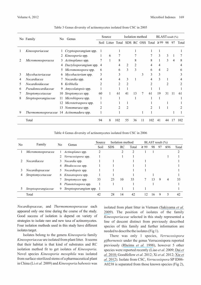
Identification of Actinomycetes. We identified
119 isolates of actinomycetes and they belong to 10
genera and 6 families in 2004 (Table 2), there are more
genera and families in 2005 (Table 3) and less genera
and families in 2006 (Table 4). A total of 263 isolates
represented of 10 genera and 10 families of
actinomycetes were identified from this study (Table 5).
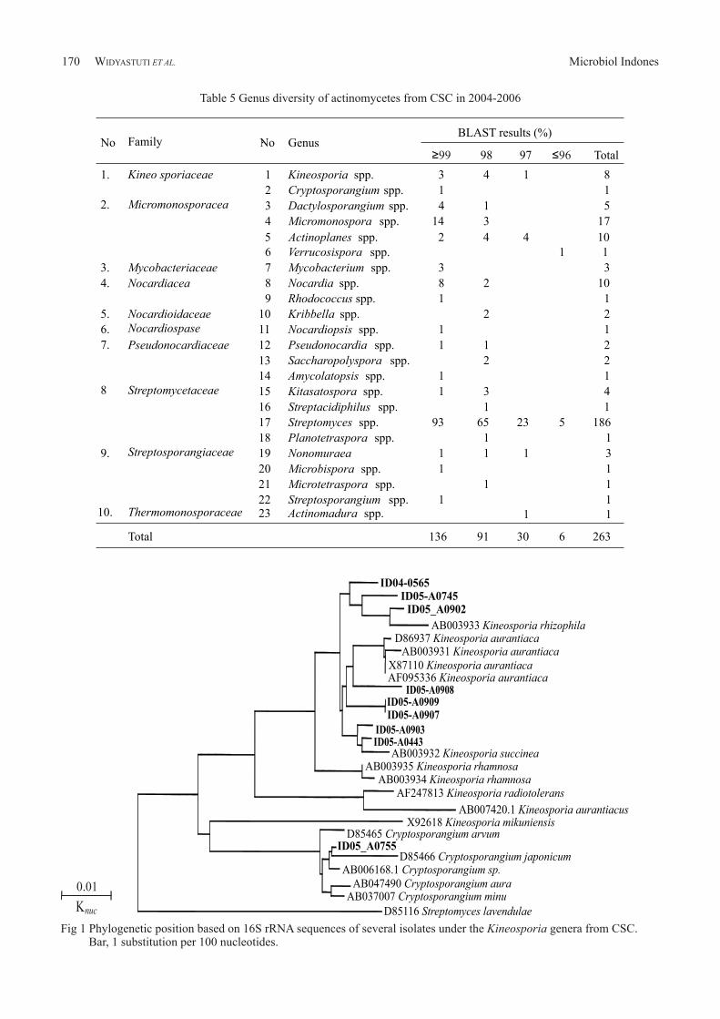
Phylogenetic Tree of Interesting Isolates.
Identification of the interesting isolates and their
position in the phylogenetic tree showed that most of
the isolates were separated from the known species of
actinomycetes. Among the families of actinomycetes
present in CSC, family Kineosporiaceae showed a
cluster of interesting isolates as candidates of new
genera (Fig 1). We found a canditate of new taxa of
Verrucosispora from CSC in 2005 (Fig 2).
DISCUSSION
We collected more soil samples than plant litter. So
far actinomycetes were reported mostly isolated from
the soil, although some actinomycetes were reported
Volume 6, 2012 Microbiol Indones 167
1099F (5'-GCAAC536R (5'GTATTACCGCGG
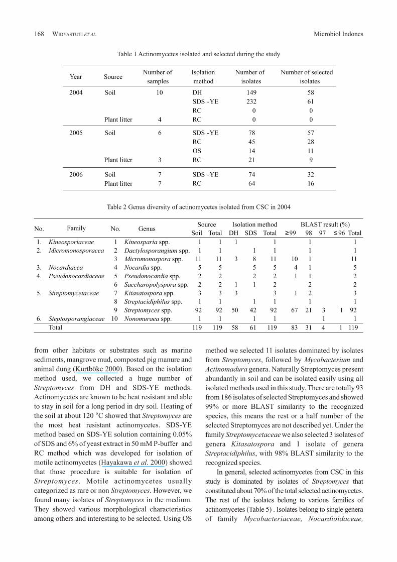
method we selected 11 isolates dominated by isolates
from Streptomyces, followed by Mycobacterium and
Actinomadura genera. Naturally Streptomyces present
abundantly in soil and can be isolated easily using all
isolated methods used in this study. There are totally 93
from 186 isolates of selected Streptomyces and showed
99% or more BLAST similarity to the recognized
species, this means the rest or a half number of the
selected Streptomyces are not described yet. Under the
family Streptomycetaceae we also selected 3 isolates of
genera Kitasatospora and 1 isolate of genera
Streptacidiphilus, with 98% BLAST similarity to the
recognized species.
In general, selected actinomycetes from CSC in this
study is dominated by isolates of Streptomyces that
constituted about 70% of the total selected actinomycetes.
The rest of the isolates belong to various families of
actinomycetes (Table 5) . Isolates belong to single genera
of family Mycobacteriaceae, Nocardioidaceae,
from other habitats or substrates such as marine
sediments, mangrove mud, composted pig manure and
animal dung (Kurtböke 2000). Based on the isolation
method used, we collected a huge number of
Streptomyces from DH and SDS-YE methods.
Actinomycetes are known to be heat resistant and able
to stay in soil for a long period in dry soil. Heating of
the soil at about 120 °C showed that Streptomyces are
the most heat resistant actinomycetes. SDS-YE
method based on SDS-YE solution containing 0.05%
of SDS and 6% of yeast extract in 50 mM P-buffer and
RC method which was developed for isolation of
motile actinomycetes (Hayakawa et al. 2000) showed
that those procedure is suitable for isolation of
Streptomyces. Motile actinomycetes usually
categorized as rare or non Streptomyces. However, we
found many isolates of Streptomyces in the medium.
They showed various morphological characteristics
among others and interesting to be selected. Using OS
Microbiol Indones168 WIDYASTUTI ET AL.
Table 1 Actinomycetes isolated and selected during the study
No. Family No. Genus Isolation method BLAST result (%) Source
Soil Total DH SDS Total
1. Kineosporiaceae 1 Kineosparia spp. 1 1 1 1 1 1
2. Micromonosporacea 2 Dactylosporangium spp. 1 1 1 1 1 1
3 Micromonospora spp. 11 11 3 8 11 10 1 11
3. Nocardiacea 4 Nocardia spp. 5 5 5 5 4 1 5
4. Pseudonocardiaceae 5 Pseudonocardia spp. 2 2 2 2 1 1 2
6 Saccharopolyspora spp. 2 2 1 1 2 2 2
5. Streptomycetaceae 7 Kitasatospora spp. 3 3 3 3 1 2 3
8 Streptacidiphilus spp. 1 1 1 1 1 1
9 Streptomyces spp. 92 92 50 42 92 67 21 3 1 92
6. Steptosporangiaceae 10 Nonomuraea spp. 1 1 1 1 1 1
Total 119 119 58 61 119 83 31 4 1 119
≥ 98 97 ≤ Total 99 96
Table 2 Genus diversity of actinomycetes isolated from CSC in 2004
Year SourceIsolation
method
Number of
isolates
Number of selected
isolates
2004 Soil 10 DH 149 58
Number of
samples
SDS -YE 232 61
RC 0 0
Plant litter 4 RC 0 0
2005 Soil 6 SDS -YE 78 57
RC 45 28
OS 14 11
Plant litter 3 RC 21 9
2006 Soil 7 SDS -YE 74 32
Plant litter 7 RC 64 16
isolated from plant litter in Vietnam (Sakiyama et al.
2009). The position of isolates of the family
Kineosporiaceae selected in this study represented a
line of descent distinct from previously described
species of this family and further information are
needed to describe the isolates (Fig 1).
There was only 1 species, Verrucosispora
gifhornensis under the genus Verrucosispora reported
previously (Rheims et al. 1998), however 5 other
species were reported recently (Liao et al. 2009; Dai et
al. 2010; Goodfellow et al. 2012; Xi et al. 2012; Xie et
al. 2012). Isolate from CSC, Verrucosispora SP ID06-
A0238 is separated from those known species (Fig 2),
Volume 6, 2012 Microbiol Indones 169
Nocardiopsaceae, and Thermomonosporaceae each
appeared only one time during the course of the study.
Good success of isolation is depend on variety of
strategies to isolate rare and new taxa of actinomycetes.
Four isolation methods used in this study have different
isolates target.
Isolates belong to the genera Kineosporia family
Kineosporiaceae are isolated from plant litter. It seems
that their habitat is that kind of substrates and RC
isolation method fit to get isolates of Kineosporia.
Novel species Kineosporia mesophila was isolated
from surface-sterilized stems of a pharmaceutical plant
in China (Li et al. 2009) and Kineosporia babensis was
No Family No Genus Source Isolation method BLAST result (%)
Total SDS RC OSS Total ≥ 98 97 Total99Soil Litter
1 Kineosporiaceae 1 Cryptosporangium spp. 1 1 1 1 1 1
2 Kineosporia spp. 1 6 7 7 7 3 3 1 7
2 Micromonosporacea 3 Actinoplanes spp. 7 1 8 8 8 1 3 4 8
4 Dactylosporangium spp. 4 4 2 2 4 4 4
5 Micromonospora spp. 6 6 3 3 6 4 2 6
3 Mycobacteriaceae 6 Mycobacterium spp. 3 3 3 3 3 3
4 Nocardiacea 7 Nocardia spp. 4 4 3 1
4 3 1 4
5 Nocardioidaceae 8 Kribbella 2 2 2 2 2 2
6 Pseudonocardiaceae 9 Amycolatopsis spp. 1 1 1 1 1 1
7 Streptomycetaceae 10 Streptomyces spp. 60 1 61 41 13 7 61 19 31 11 61
8 Streptosporangiaceae 11 Microbispora spp. 1 1 1 1 1 1
12 Microtetraspora spp. 1 1 1 1 1 1
13 Nonomuraea spp. 2 2 2 2 1 1 2
9 Thermomonosporaceae 14 Actinomadura spp. 1 1 1 1 1 1
Total 94 8 102 55 36 11 102 41 44 17 102
Table 3 Genus diversity of actinomycetes isolated from CSC in 2005
Table 4 Genus diversity of actinomycetes isolated from CSC in 2006
No Family No Genus Isolation method BLAST result (%) Source
Soil SDS RC Total
1 Micromonosporacea 1 Actinoplanes spp. 2 2 2 1 1 2 2 Verrucosispora spp. 1 1 1 1 1
2 Nocardiacea 3 Nocardia spp. 1 1 1 1 1 4 Rhodococcus spp. 1 1 1 1 1
3 Nocardiopsaceae 5 Nocardiopsis spp. 1 1 1 1 1
4 Streptomycetaceae 6 Kitasatospora spp. 1 1 1 1 1 7 Streptomyces spp. 33 23 10 33 7 13 9 4 33 8 Planotetraspora spp. 1 1 1 1 1
5 Streptosporangiaceae 9 Streptosporangium spp. 1 1 1 1 1
Total 42 28 14 42 12 16 9 5 42
≥ 99 98 97 ≤ 96 Total
Microbiol Indones170 WIDYASTUTI ET AL.
Table 5 Genus diversity of actinomycetes from CSC in 2004-2006
Fig 1 Phylogenetic position based on 16S rRNA sequences of several isolates under the Kineosporia genera from CSC. Bar, 1 substitution per 100 nucleotides.
No Family No. Genus BLAST results (%)
≥99 98 97 ≤96 Total
1. Kineo sporiaceae 1 Kineosporia spp. 3 4 1 8
2 Cryptosporangium spp. 1 1 2. Micromonosporacea 3 Dactylosporangium spp. 4 1 5
4 Micromonospora spp. 14 3 17 5
Actinoplanes spp.
2
4
4
10
6 Verrucosispora spp. 1 1
3. Mycobacteriaceae 7 Mycobacterium spp. 3 3
4. Nocardiacea 8 Nocardia spp. 8 2 10
9 Rhodococcus spp. 1 1
5. Nocardioidaceae 10 Kribbella spp. 2 2 6. Nocardiospase 11 Nocardiopsis spp. 1 1
7. Pseudonocardiaceae 12 Pseudonocardia spp. 1 1 2
13 Saccharopolyspora spp. 2 2
14 Amycolatopsis spp. 1 1
8
Streptomycetaceae 15 Kitasatospora spp. 1 3 4
16 Streptacidiphilus spp. 1 1
17 Streptomyces spp. 93 65 23 5 186
18 Planotetraspora spp. 1 1
9. Streptosporangiaceae 19 Nonomuraea 1 1 1 3
20 Microbispora spp. 1
1
21 Microtetraspora spp. 1
1
22 Streptosporangium spp. 1 1
10. Thermomonosporaceae 23 Actinomadura spp. 1 1
Total 136 91 30 6 263
AB007420.1 Kineosporia aurantiacus
D85116 Streptomyces lavendulae
ID05-A0908ID05-A0909ID05-A0907
ID05-A0903ID05-A0443
ID04-0565ID05-A0745
ID05_A0902
AB003933 Kineosporia rhizophilaD86937 Kineosporia aurantiaca
AB003931 Kineosporia aurantiaca X87110 Kineosporia aurantiaca AF095336 Kineosporia aurantiaca
AB003932 Kineosporia succineaAB003935 Kineosporia rhamnosa
AB003934 Kineosporia rhamnosaAF247813 Kineosporia radiotolerans
X92618 Kineosporia mikuniensisD85465 Cryptosporangium arvum
ID05_A0755D85466 Cryptosporangium japonicum
AB006168.1 Cryptosporangium sp.AB047490 Cryptosporangium aura
AB037007 Cryptosporangium minu Knuc
0.01
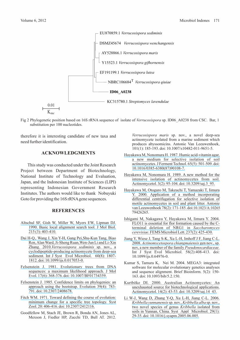
therefore it is interesting candidate of new taxa and
need further identification.
ACKNOWLEDGMENTS
This study was conducted under the Joint Research
Project between Department of Biotechnology,
National Institute of Technology and Evaluation,
Japan, and the Indonesian Institute of Sciences (LIPI)
representing Indonesian Government Research
Institutes. The authors would like to thank Nobuyuki
Goto for providing the 16S rRNA gene sequences.
REFERENCES
Altschul SF, Gish W, Miller W, Myers EW, Lipman DJ. 1990. Basic local alignment search tool. J Mol Biol. 215 (3): 403-410.
Dai H-Q,. Wang J, Xin Y-H, Gang Pei,Shu-Kun Tang, Biao Ren, Alan Ward, Ji-Sheng Ruan,Wen-Jun Li and Li-Xin Zhang. 2010.Verrucosispora sediminis sp. nov., a cyclodipeptide-producing actinomycete from deep-sea sediment. Int J Syst Evol Microbiol. 60(8): 1807-1812. doi: 10.1099/ijs.0.017053-0.
Felsenstein J. 1981. Evolutionary trees from DNA sequences: a maximum likelihood approach. J Mol Evol. 17(6): 368-376. doi: 10.1007/BF01734359.
Felsenstein J. 1985. Confidence limits on phylogenies: an approach using the bootstrap. Evolution 39(4): 783-791. doi: 10.2307/2408678.
Fitch WM. 1971. Toward defining the course of evolution: minimum change for a specific tree topology. Syst Zool. 20: 406-416. doi: 10.2307/2412116.
Goodfellow M, Stach JE, Brown R, Bonda AN, Jones AL, Mexson J, Fiedler HP, Zucchi TD, Bull AT. 2012.
Volume 6, 2012 Microbiol Indones 171
Fig 2 Phylogenetic position based on 16S rRNA sequence of isolate of Verrucosispora sp. ID06_A0238 from CSC. substitution per 100 nucleotides.
Bar, 1
Verrucosispora maris sp. nov., a novel deep-sea actinomycete isolated from a marine sediment which produces abyssomicins. Antonie Van Leeuwenhoek, 101(1): 185-193. doi: 10.1007/s10482-011-9651-5.
Hayakawa M, Nonomura H. 1987. Humic acid-vitamin agar, a new medium for selective isolation of soil actinomycetes. J Ferment Technol. 65(5): 501-509. doi: 10.1016/0385-6380(87)90108-7.
Hayakawa M, Nonomura H. 1989. A new method for the intensive isolation of actinomycetes from soil. Actinomycetol. 3(2): 95-104. doi: 10.3209/saj.3_95.
Hayakawa M, Otoguro M, Takeuchi T, Yamazaki T, Iimura Y. 2000. Application of a method incorporating differential centrifugation for selective isolation of motile actinomycetes in soil and plant litter. Antonie van Leeuwenhoek 78(2): 171-185. doi:
Ishigami M, Nakagawa Y, Hayakawa M, Iimura Y. 2004. FLO11 is essential for flor formation caused by the C-terminal deletion of NRG1 in Saccharomyces cerevisiae. FEMS Microbiol Lett. 237(2): 425-430.
Jiang Y, Wiese J, Tang S-K, Xu L-H, Imhoff J F, Jiang C-L. 2008. Actinomycetospora chiangmaiensis gen nov., sp. nov, a new member of the family Pseudonocardiaceae. Int J Syst Evol Microbiol. 58(2):408-413. doi: 10.1099/ijs.0.64976-0.
Kumar S, Tamura K, Nei M. 2004. MEGA3: integrated software for molecular evolutionary genetics analyses and sequence alignment. Brief Bioinform. 5(2): 150-163. doi: 10.1093/bib/5.2.150.
Kurtböke DI. 2000. Australian Actinomycetes: An unexhausted source for biotechnological applications. Actinomycetol. 14(2): 43-53. doi: 10.3209/saj.14_43.
Li W-J, Wang D, Zhang Y-Q, Xu L-H, Jiang C-L. 2006. Kribbella yunnanensis sp. nov., Kribbella alba sp. nov., two novel species of genus Kribbella isolated from soils in Yunnan, China. Syst Appl Microbiol. 29(1): 29-35. doi: 10.1016/j.syapm.2005.06.005.
10.1023/A:1026579426265.
KC315780.1 Streptomyces lavendulae
ID06_A0238
NBRC106684 Verrucosispora qiuiaeT
EF191199.1 Verrucosispora lutea
Y15523.1 Verrucosispora gifhornensis
AY528866.1 Verrucosispora maris
DSMZ45674 Verrucosispora wenchangensisT
EU870859.1 Verrucosispora sediminis
Knuc
0.01
Evol. 4(4): 406-425.
Sakiyama Y, Thao NKN, Giang NM, Miyadoh S, Hop DV, Ando K. 2009. Kineosporia babensis sp. nov., isolated from plant litter in Vietnam. Int J Syst Evol Microbiol. 59 (3): 550-554. doi: 10.1099/ijs.0.002907-0.
Rheims H, Schumann P, Rohde M, Stackebrandt E. 1998. Verrucosispora gifhornensis gen. nov., sp. nov., a new member of the actinobacterial family Micromonosporaceae. Int J Syst Evol Microbiol. 48: 1119-1127.
Tamura T, Sakane T. 2005. Asanoa iriomotensis sp. nov., isolated from mangrove soil. Int J Syst Evol Microbiol. 55(2): 725-727. doi: 10.1099/ijs.0.02982-0.
Tamura T, Hatano K, Suzuki K. 2006. A new genus of the family Micromonosporaceae, Polymorphospora gen. nov., with description of Polymorphospora rubra sp. nov. Int J Syst Evol Microbiol. 56(8): 1959-1964. doi: 10.1099/ijs.0.64046-0.
Xie QY, Lin HP, Li L, Brown R, Goodfellow M, Deng Z, Hong K.2012. Verrucosispora wenchangensis sp. nov., isolated from mangrove soil. Antonie van Leeuwenhoek 102(1):1-7. doi: 10.1007/s10482-012-9707-1.
Xi L, Zhang L, Ruan J, Huang Y. 2012. Description of Verrucosispora qiuiae sp. nov., isolated from mangrove swamp sediment, and emended description of the genus Verrucosispora. Int J Syst Evol Microbiol. 62(7):1564-1569. doi: 10.1099/ijs.0.033787-0.
Yamamura H, Lisdiyanti P, Ridwan R, Ratnakomala S, Saraswati R, Lestari Y, Triana E, Kartina G, Widyastuti Y, Ando K. 2010. Dietzia timorensis sp. nov., isolated from soil. Int J Syst Evol Microbiol. 60(2): 451-454. doi: 10.1099/ijs.0.012229-0 .
Li J, Zhao g-Z, Huang H-Y, Qin S, Zhu W-Y, Xu L-H, Li W-J. 2009. Kineosporia mesophila sp. nov., isolated from surface-sterilized stems of Tripterygium wilfordii. Int J Syst Evol Microbiol. 59(12):3150-3154. doi: 10.1099/ijs.0.012021-0.
Liao ZL, Tang SK, Guo L, Zhang YQ, Tian XP, Jiang CL, Xu LH, Li WJ.2009.Verrucosispora lutea sp. nov., isolated from a mangrove sediment sample. Int J Syst Evol Microbiol. 59(9): 2269-2273. doi:10.1099/ijs.0.008813-0 .
Lisdiyanti P, Otoguro M, Ratnakomala S, Lestari Y, Hastuti RD, Triana E, Ando K, Widyastuti Y. 2010. Actinokineospora baliensis sp. nov., Actinokineospora cibodasensis sp. nov., Actinokineospora cianjurensis sp. nov., isolated from soil and plant litter. Int J Syst Evol Microbiol. 60(1): 2331-2335. doi:10.1099/ijs.0.013276-0 .
Nonomura H, Ohara Y. 1969. Distribution of actinomycetes in soil. VII. A culture method effective for both preferential isolation and enumeration of Microbispora and Streptosporangium strains in soil. (Part 1) Classification of the isolates. J Ferment Technol. 47: 463-469.
Otoguro M, Ratnakomala S, Lestari Y, Hastuti RD, Triana E, Widyastuti Y, Ando K. 2009. Streptomyces baliensis sp. nov., isolated from Balinese soil. Int J Syst Evol Microbiol. 59(2): 2158-2161. doi:10.1099/ijs.0.007179-0.
Otoguro M, Yamamura H, Tamura T, Irzaldi R, Ratnakomala S, Ridwan R, Kartina G, Triana E, Nurkanto A, Lestari Y, Lisdiyanti P, Widyastuti Y, Katsuhiko A. 2011. Act inophytocola t imorens is sp . nov. and Actinophytocola coralina sp. nov., isolated from soil. Intl J Syst Evol Microbiol. 61(4): 834-838. doi: 10.1099/ijs.0.023432-0.
Saitou N, Nei M. 1987. The neighbor-joining method: a new method for reconstructing phylogenetic trees. Mol Biol
Microbiol Indones172 WIDYASTUTI ET AL.