Preferential use of Nearshore Kelp Habitats by Juvenile Salmon and Forage Fish
R E S EA RCH AR T I C L E
Targeted search for actinomycetes from nearshore anddeep-sea marine sediments
Alejandra Prieto-Dav�o1,2, Luis J. Villarreal-G�omez3, Stephanie Forschner-Dancause4, Alan T. Bull5,James E.M. Stach6, David C. Smith7, Dave C. Rowley4 & Paul R. Jensen1
1Scripps Institution of Oceanography, University of California San Diego, San Diego, CA, USA; 2Facultad de Qu�ımica, Unidad Sisal, Universidad
Nacional Aut�onoma de M�exico, Sisal Yuc, M�exico; 3Universidad Aut�onoma de Baja California, Campus Las Palmas, Tijuana, M�exico; 4Department
of Biomedical and Pharmaceutical Sciences, University of Rhode Island, Kingston, RI, USA; 5School of Biosciences, University of Kent, Canterbury,
UK; 6School of Biology, University of Newcastle, Newcastle upon Tyne, UK; and 7Graduate School of Oceanography, University of Rhode Island,
Narragansett, RI, USA
Correspondence: Alejandra Prieto-Dav�o,
Puerto de Abrigo s/n, Municipio de
Hunucm�a, Sisal CP 97356, M�exico.
Tel.: +52 988 9311013;
fax: +52 988 931-1015; e-mails:
[email protected]; alejandra.
Received 8 November 2012; revised 16
January 2013; accepted 21 January 2013.
DOI: 10.1111/1574-6941.12082
Editor: Max H€aggblom
Keywords
marine-derived actinomycetes; Salinispora;
marine microbial ecology; cultivation
independent.
Abstract
Sediment samples collected off the coast of San Diego were analyzed for
actinomycete diversity using culture-independent techniques. Eight new opera-
tional taxonomic units (OTUs) in the Streptomycetaceae were identified as well
as new diversity within previously cultured marine OTUs. Sequences belonging
to the marine actinomycete genus Salinispora were also detected, despite the
fact that this genus has only been reported from more tropical environments.
Independent analyses of marine sediments from the Canary Basin (3814 m)
and the South Pacific Gyre (5126 and 5699 m) also revealed Salinispora
sequences providing further support for the occurrence of this genus in deep-
sea sediments. Efforts to culture Salinispora spp. from these samples have yet
to be successful. This is the first report of Salinispora spp. from marine sedi-
ments > 1100 m and suggests that the distribution of this genus is broader
than previously believed.
Introduction
The phylum Actinobacteria is extraordinarily diverse
(Gao & Gupta, 2012) and well represented in the marine
environment (Rapp�e et al., 1999). Of the five subclasses
that comprise this phylum (the Acidomicrobidae, Actino-
bacteridae, Coriobacteridae, Nitriliruptoridae, and Rubro-
bacteridae), sequences belonging to the Acidomicrobidae
are commonly observed when culture-independent tech-
niques are applied (Jensen & Lauro, 2008). Conversely,
cultured Actinobacteria often fall within the subclass
Actinobacteridae and, more specifically, within the order
Actinomycetales. These bacteria are commonly referred to
as actinomycetes and have been targeted from marine
samples for their ability to produce structurally novel
secondary metabolites (Zotchev, 2012). While a number
of marine actinomycete species and genera have been
described (Tian et al., 2009a; Tian et al., 2009b; Zhao
et al., 2009; Goodfellow et al., 2012) it is not clear how
well these cultured strains represent the extant diversity
present in the marine environment.
Culture-independent studies have revealed the presence
of actinomycetes in seawater (Yoshida et al., 2008) and
deep-sea marine sediments (Stach et al., 2003a, b). Less
abundant taxa such as the marine actinomycete genus
Salinispora (Maldonado et al., 2005) have been detected
when specific primers targeting this group were applied
(Mincer et al., 2005). Actinomycetes have also been
detected in marine sponges, facilitating the selection of
culture media and further increasing the diversity of iso-
lates recovered (Webster et al., 2001). In a separate study
of two sponges from China, a wide difference between
the genera observed using actinomycete specific primers
and cultivation-based methods was observed (Xin et al.,
2008). Further studies on one of these sponges revealed
the importance of using both culture and culture-inde-
pendent methods when studying actinomycete diversity
(Sun et al., 2010). While all methods suffer from
FEMS Microbiol Ecol && (2013) 1–9 ª 2013 Federation of European Microbiological SocietiesPublished by Blackwell Publishing Ltd. All rights reserved
MIC
ROBI
OLO
GY
EC
OLO
GY
inherent biases, culture-independent techniques can help
establish the occurrence of bacteria in specific environ-
ments.
In a prior study of sediment samples collected off
the coast of California, culture-dependent actinomycete
diversity was assessed between nearshore and offshore
sites (Prieto-Dav�o et al., 2008). The results revealed con-
siderable, marine-specific diversity and high levels of
terrestrial influence out to 125 km from shore. The pres-
ent study was undertaken to provide a culture-indepen-
dent assessment of the collective actinomycete diversity
present in five of these samples. These studies were com-
plimented by independent analyses of deep-sea sediment
samples collected from the Canary Basin and the South
Pacific Gyre (SPG).
Materials and methods
Sample collection
To further explore the diversity of actinomycetes present
in marine sediments collected off the coast of California,
five of eleven sediment samples previously employed for
cultivation studies (Prieto-Dav�o et al., 2008) were used to
generate 16S rRNA gene clone libraries targeting the
order Actinomycetales. All of these samples were collected
using an untethered coring device designed and con-
structed at the Scripps Institution of Oceanography
(SIO). The depths and collection sites are provided in
Supporting Information Table S1. Each core was divided
into five or six sections as previously described (Prieto-
Dav�o et al., 2008). Approximately 1 g of wet sediment
from each section was placed in a 1.5 mL Eppendorf tube
containing 1 mL of sucrose lysis buffer (50 mM Tris–HCl,
150 mM NaCl, 5 mM EDTA, 0.25 M sucrose) and immedi-
ately stored on ice for transportation to shore. Long-term
storage was at �20 °C.Samples from the Canary Basin and the SPG were ana-
lyzed specifically for the presence of Salinispora spp. The
Canary Basin sample was collected as previously described
(Stach et al., 2003b). The SPG samples were collected
using gravity or piston cores during the KNOX-02RR
expedition (D’Hondt et al., 2009). A total of 11 cores
were sectioned and subsampled from 3 to 5 times at vari-
ous depths from the sediment surface to the bottom of
the core generating a total of 51 samples (Table S2). Sub-
cores were taken from each section by first removing the
top layer of sediment with a sterile spatula. Sterilized cut-
off syringes were then pushed into the core resulting in
an uncontaminated subcore. The syringe containing the
subcore was stored intact in heat sealed bags at �80 °Cprior to molecular analysis.
DNA extraction, PCR amplification, and cloning
Environmental DNA (eDNA) was extracted from the sed-
iment samples collected off the coast of California using a
soil DNA extraction kit (cat. No 69506) according to
manufacturer’s protocol (Qiagen, Valencia, CA). 16S
rRNA gene primers targeting the order Actinomycetales
(Stach et al., 2003a, b) and the families Streptomycetaceae
and Micromonosporaceae (Monciardini et al., 2002) were
used (Table S3). PCR amplification of 1–4 lL of eDNA
(18–20 ng mL�1) was carried out in triplicate for each
sample as follows: initial denaturation at 95 °C for
10 min followed by 30 cycles of 94 °C for 45 s, 65 °C for
45 s, and 72 °C for 1 min, followed by a 10 min exten-
sion at 72 °C. Triplicate PCR products were pooled and
purified using MiniElute PCR purification columns
according to the manufacturer’s instructions (Qiagen).
Purified DNA was ligated to the plasmid vector pCR� 2.1-
TOPO� and used to transform One-Shot� Mach1TM -T1�
chemically competent cells using a Topo TA� cloning kit
according to the manufacturer’s protocol (Invitrogen,
Carlsbad, CA). Transformed clones were identified using
white–blue selection and inoculated into 10 mL Falcon
tubes containing 3 mL of LB broth and 50 lg mL�1 kana-
mycin. Plasmid DNA was extracted using the QiaPrep�
MiniPrep extraction kit according to manufacturer’s
instructions (Qiagen), digested with BstX I (New England
BioLabs, Ipswich, MA), and run on a 1% agarose gel to
confirm the presence of the correct-sized insert.
eDNA was extracted from 10 g of Canary Basin sedi-
ment using an UltraClean Mega DNA soil kit (Mo Bio Lab-
oratories, Solana Beach, CA). The primers (Supplemental
Table S3) and PCR conditions are as previously described
(Stach et al., 2003a, b). PCR products were purified using
QIAquick gel extraction columns according to the manu-
facturer’s instructions (Qiagen, Crawley, UK). Purified
DNA was blunt-end-ligated into the plasmid vector pST-
Blue-1 and used to transform NovaBlue competent cells
using a Perfectly Blunt cloning kit (Novagen, Madison,
WI). Cloned plasmid DNA was extracted as described
above and the presence of inserts confirmed by PCR using
previously described primers (Stach et al., 2003a, b).
For the SPG sediments, a modified FastDNA� SPIN kit
for soil protocol (Qbiogene, Irvine, CA), followed by
ChromaSpinTM TE-100 columns (Clontech, Mountain
View, CA), was used to ensure high eDNA yields and
purity (Froschner et al., 2009). The eDNA samples were
subjected to whole-genome amplification using the
REPLI-g� Midi kit (Qiagen) as previously described (Fro-
schner et al., 2009) and analyzed for the presence of
Salinispora spp. using Salinispora-specific 16S rRNA gene
PCR primers (Table S3). The reactions included 200 ng
ª 2013 Federation of European Microbiological Societies FEMS Microbiol Ecol && (2013) 1–9Published by Blackwell Publishing Ltd. All rights reserved
2 A. Prieto-Dav�o et al.
of whole-genome amplification product and consisted of
an initial 5-min denaturation step at 94 °C followed by
30 cycles of 30 s at 94 °C, 90 s at 60 °C, and 90 s at 72 °C followed by a final 10 min extension at 72 °C. The
reaction products were purified using a QIAquick PCR
purification kit (Qiagen) following manufacturer’s guide-
lines and quantified using a NanoDrop Spectrophotome-
ter (NanoDrop, Wilmington, DE). PCR amplicons were
cloned using the pGEM�-T Easy Vector System (Pro-
mega, Madison, WI) at a 1 : 1 insert to vector ratio per
the manufacturer’s protocol. Transformed clones were
grown overnight in LB broth and the plasmids extracted
using Wizard� Plus SV Minipreps (Promega).
Terminal restriction fragment length
polymorphism (T-RFLP) analysis
T-RFLP was used to probe the SPG samples for the pres-
ence of Salinispora spp. Whole-genome amplification
products were PCR amplified as described above with the
addition of a 6-FAM label to the forward primer. The
Salinispora-specific primers were designed as part of a
prior study and shown to be > 98% specific for this
taxon (Mincer et al., 2005). Approximately 40 ng of flu-
orescently labeled PCR product was digested indepen-
dently with 4 U of the restriction enzymes AluI, AvaII,
and EcoO109I in 10-lL reaction volumes. Reference T-
RFLs were generated for the three Salinispora species by
digesting labeled PCR products from cultured strains
(Supplemental Table S5). The restriction digests were
incubated at 37 °C for 16 h to ensure complete digestion
prior to being deactivated at 65 °C for 20 min. The
digests were precipitated and resuspended in Hi-Di form-
amide (Applied Biosystems, Carlsbad, CA) to a final con-
centration of approximately 0.6 ng lL�1. About 0.5 lLGeneScan Liz 1200 size standard (Applied Biosystems)
was added to each sample. The samples were denatured
at 95 °C for 5 min and placed immediately on ice. The
terminally labeled restriction fragments (T-RFs) were
visualized with the Applied Biosystems 3130xl genetic
analysis system.
Sequencing and phylogenetic analyses
Plasmid inserts from the California samples were
sequenced at the UCSD Rebecca and John Moore Cancer
Center using the M13 primer included in the vector
(Invitrogen). Sequences were analyzed using the Basic
Local Alignment Search Tool (BLAST) function of GenBank
(Altschul et al., 1990), and strains with the highest level
of sequence identity were recorded as top BLAST matches
regardless of whether or not they were associated with a
formal publication. Top matches and cloned sequences
belonging to the Micromonosporaceae (560 bp) and Strep-
tomycetaceae (533 bp) were clustered into OTUs based on
� 99% 16S rRNA gene sequence identity using the java
application Clusterer (http://www.bugaco.com/mioritic/
clusterer_jlp.php). In addition, OTU representatives from
cultured strains derived from all 11 samples used in a
prior study (Prieto-Dav�o et al., 2008) were included in
the clustering analyses. Sequences were then aligned using
ClustalX (Larkin et al., 2007) and imported into MacC-
lade for manual curation of the alignment (http://macc-
lade.org).
Plasmid inserts derived from the Canary Basin PCR
products were sequenced by Qiagen using the SP6 univer-
sal primer. Plasmid inserts from the SPG samples were
sequenced using the 16S primers or vector primers
(T7, SP6). Sequencing was performed on an Applied Bio-
systems 3130xl Genetic Analyser (Applied Biosystems) at
the University of Rhode Island’s Genomics and Sequenc-
ing Center (Kingston, RI). The resulting sequences were
trimmed and contigs built using SequencherTM (Gene
Codes, Ann Arbor, MI).
Maximum-Likelihood rooted phylogenetic trees
(HKY85 substitution model, 1000 bootstraps) were con-
structed for the Streptomycetaceae and Micromonospora-
ceae using PhyML (Guindon et al., 2010) via http://www.
phylogeny.fr/ and http://www.atgc-montpellier.fr/phyml/
(Dereeper et al., 2008). Trees were edited using Inkscape
(inkscape.org). For both phylogenetic trees, top BLAST
matches, as well as previously cultivated OTU representa-
tives (Prieto-Dav�o et al., 2008), were included.
Diversity estimations
Estimates of the diversity present in the California clone
libraries were performed using the statistical package Esti-
mateS (http://purl.oclc.org/estimates) with 1000 runs for
each library.
Results
Clone libraries targeting the order Actinomycetales
(actinomycetes) and the families Streptomycetaceae and
Micromonosporaceae were generated from five indepen-
dent sediment cores collected off the coast of California
(Table S1). The Actinomycetales-specific primers yielded
largely nonactinobacterial sequences and Actinobacteria in
the subclass Acidomicrobidae, which is outside of the Ac-
tinomycetales. In response, primers specific for the Strep-
tomycetaceae and Micromonosporaceae were used because
these were the dominant families cultured previously
from these samples. Clone libraries were generated from
two of the sediment cores using the Streptomycetaceae
primers, two using the Micromonosporaceae primers, and
FEMS Microbiol Ecol && (2013) 1–9 ª 2013 Federation of European Microbiological SocietiesPublished by Blackwell Publishing Ltd. All rights reserved
Marine-derived actinomycetes 3

one using both primer sets (Table S4). All libraries were
generated from the surface section of the cores, with the
exception of sample SD06-13, from which libraries were
generated from all five sections. Seventy-seven of the
combined 79 clones obtained using the Streptomycetaceae
primers belonged to this family confirming the specificity
of that primer set. The cloning efficiency of the Micromo-
nosporaceae primers was poor and yielded only 23
sequences, of which 22 belonged to the targeted family.
The 99 Streptomycetaceae and Micromonosporaceae
clones were clustered based on � 99% sequence identity
(Table 1). This yielded a total of 41 OTUs, each of which
contained 1–10 clones (average = 2). Of these, representa-
tives of eight OTUs were cultured as part of a prior study
(Prieto-Dav�o et al., 2008; Table 1, Figs 1 and 2). When
compared more broadly to GenBank, which includes the
sequences from the prior study, 33 of the 41 OTUs from
this study have cultured representatives, and thus, eight can
be considered new in terms of publically available sequence
data. The majority of the OTUs (37) and all of the eight
new OTUs belong to the Streptomycetaceae. The OTUs in
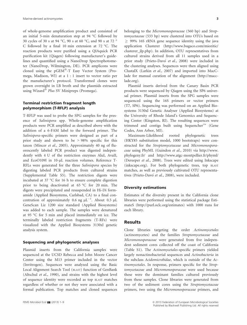
this family are scattered throughout the phylogenetic tree;
however, two of the new OTUs (represented by clones
SD06-09A-02 and SD06-13C-01) form a well-supported
clade with the marine-derived actinomycete strain CNQ-
085 (Fig. 1). Two more of the new OTUs, represented by
SD06-07A-01 and SD06-13E-03, belong to a much larger
and previously identified marine clade that includes the
new species Streptomyces marinus (Prieto-Dav�o et al.,
2008) and the cloned OTU (SD06-13A-10). One additional
OTU (SD06-13B-01) belongs to another cultivated marine
clade (Prieto-Dav�o et al., 2008), while the two remaining
new OTUs (SD06-09A-06 and SD06-13A-02) are distantly
related to previously observed sequences.
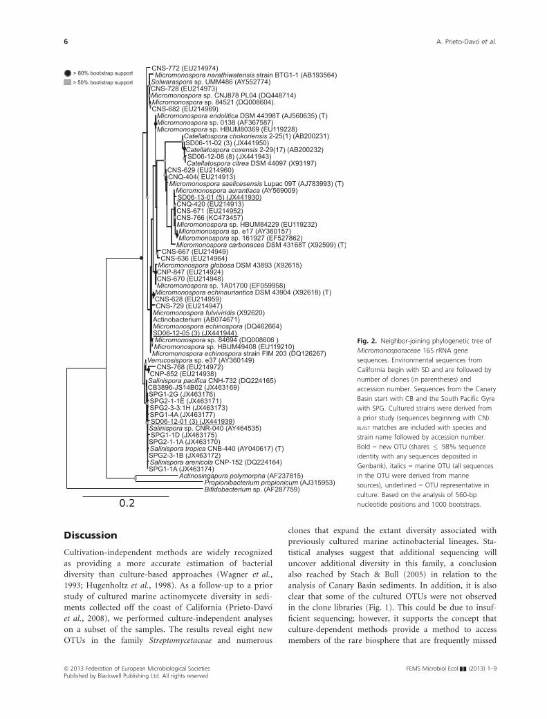
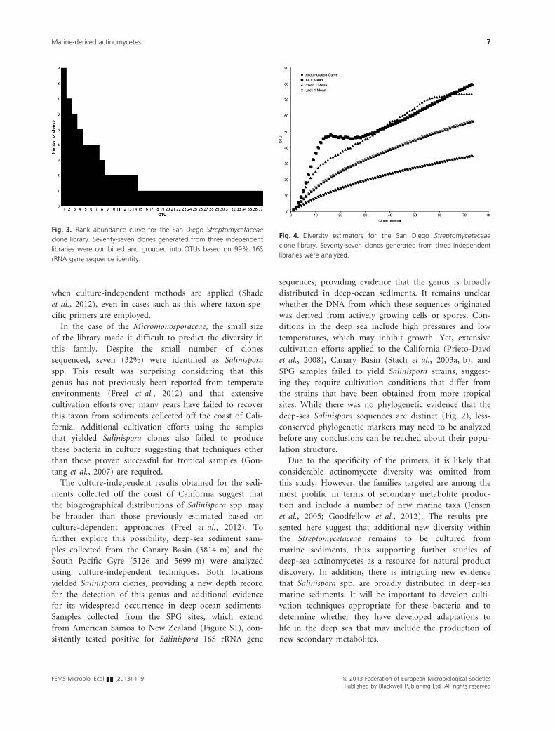
In an effort to estimate the total diversity in the sedi-
ments sampled, a rank abundance curve was generated
from the combined Streptomycetaceae clone libraries. This
curve shows little duplication and a long right-hand tail,
as is characteristic of a highly diverse community (Fig. 3).
This is supported by three diversity estimators (ACE,
Chao 1, and Jackknife 1), which predict that, on average,
70 OTUs were present in the three samples analyzed
(Fig. 4). The shape of the accumulation curve provides
clear evidence that additional sequencing could reveal
additional diversity. The Micromonosporaceae clones
formed only four OTUs, and thus, a rank abundance
curve was not generated. The poor cloning efficiency and
low number of sequences analyzed suggest that additional
studies of this family are warranted. Although none of
the Micromonosporaceae OTUs were considered new, one
claded with Catellatospora spp. (clones SD06-11-02 and
SD06-12-08) and a second, which included three cloned
sequences, claded with Salinispora spp. (clone SD06-
12-01; Fig. 2). The other two OTUs claded with Micro-
monospora spp. (clones SD06-12-05 and SD06-13-01).
This is the first evidence that Salinispora spp. occur in
temperate marine environments (Jensen & Mafnas,
2006; Freel et al., 2012). Given that neither Catellatos-
pora nor Salinispora spp. had previously been cultured
from these samples, cultivation techniques specific for
both genera were applied (Ara & Kudo, 2006; Freel
et al., 2012). However, only Micromonospora spp. were
recovered.
Independent analyses of deep-sea marine sediment sam-
ples collected from the Canary Basin and the South Pacific
Gyre (SPG) were also performed to test for the presence
of Salinispora sequences. A clone library generated from a
Canary Basin sediment sample collected at a depth of
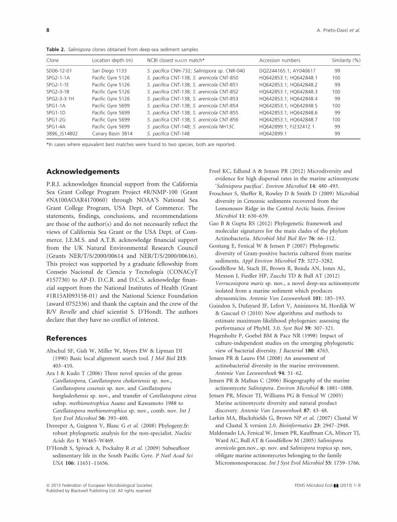
3814 m revealed the presence of one Salinispora sequence
(Table 2). Eleven deep-sea sediment cores obtained from
the SPG were subdivided into 51 sections and analyzed by
T-RFLP. PCR products were obtained using Salinispora-
specific primers (Mincer et al., 2005) from 25 of the 51
subsamples including samples from each of the 11 sites
(Table S2). The second depth below the sediment surface
(0.3–0.89 m) yielded the largest number of successful PCR
amplifications. T-RFLP analyses were performed on the 25
PCR products, and all 25 yielded T-RFs of the expected
size for Salinispora spp. (Supplemental Table S5). Clone
libraries generated from three of the PCR-positive samples
(SPG1, SPG2-1, and SPG2-3) yielded eight Salinispora
sequences and confirmed the presence of the genus in sed-
iments collected at a depth of 5699 m (Table 2). Addi-
tional sequencing would be required to confirm the
presence of the genus in the remaining 22 samples. All of
these sequences clade with Salinispora spp. (Fig. 2) and
fall within a single 99% OTU. However, due to the lack of
species-specific nucleotides in the region sequenced, it is
not possible to assign species level identifications. Collec-
tively, these results provide the first evidence that Salinis-
pora spp. occur at depths > 1100 m (Mincer et al., 2005)
and outside of the geographical range from which the
genus has been reported using culture-dependent methods
(Freel et al., 2012).
Table 1. Clustering of Streptomycetaceae and Micromonosporaceae
clones into OTUs based on > 99% 16S rRNA gene sequence identity
over 560 bp (Streptomycetaceae) and 533-bp (Micromonosporaceae)
Family
Cloned
OTUs
Cultured
OTUs*
GenBank cultured
OTUs
Streptomycetaceae 37 5 29
Micromonosporaceae 4 3 4
Total 41 8 33
*Based on a prior analysis of the same sediment samples (Prieto-Dav�o
et al., 2008).
ª 2013 Federation of European Microbiological Societies FEMS Microbiol Ecol && (2013) 1–9Published by Blackwell Publishing Ltd. All rights reserved
4 A. Prieto-Dav�o et al.
Fig. 1. Neighbor-joining phylogenetic tree of
Streptomycetaceae 16S rRNA gene sequences.
Environmental sequences from California
begin with SD and are followed by number of
clones (in parentheses) and accession number.
Cultured strains were derived from a prior
study (sequences beginning with CN). BLAST
matches are included with species and strain
name followed by accession number.
Bold = new OTU (shares � 98% sequence
identity with any sequences deposited in
Genbank), italics = marine OTU (all sequences
in the OTU were derived from marine
sources), underlined = OTU representative in
culture. Boxed clade = marine clade (all
sequences in the OTU plus top BLAST match
and cultivars were derived from marine
sources). Based on the analysis of 533-bp
nucleotide positions and 1000 bootstraps.
FEMS Microbiol Ecol && (2013) 1–9 ª 2013 Federation of European Microbiological SocietiesPublished by Blackwell Publishing Ltd. All rights reserved
Marine-derived actinomycetes 5
Discussion
Cultivation-independent methods are widely recognized
as providing a more accurate estimation of bacterial
diversity than culture-based approaches (Wagner et al.,
1993; Hugenholtz et al., 1998). As a follow-up to a prior
study of cultured marine actinomycete diversity in sedi-
ments collected off the coast of California (Prieto-Dav�o
et al., 2008), we performed culture-independent analyses
on a subset of the samples. The results reveal eight new
OTUs in the family Streptomycetaceae and numerous
clones that expand the extant diversity associated with
previously cultured marine actinobacterial lineages. Sta-
tistical analyses suggest that additional sequencing will
uncover additional diversity in this family, a conclusion
also reached by Stach & Bull (2005) in relation to the
analysis of Canary Basin sediments. In addition, it is also
clear that some of the cultured OTUs were not observed
in the clone libraries (Fig. 1). This could be due to insuf-
ficient sequencing; however, it supports the concept that
culture-dependent methods provide a method to access
members of the rare biosphere that are frequently missed
Fig. 2. Neighbor-joining phylogenetic tree of
Micromonosporaceae 16S rRNA gene
sequences. Environmental sequences from
California begin with SD and are followed by
number of clones (in parentheses) and
accession number. Sequences from the Canary
Basin start with CB and the South Pacific Gyre
with SPG. Cultured strains were derived from
a prior study (sequences beginning with CN).
BLAST matches are included with species and
strain name followed by accession number.
Bold = new OTU (shares � 98% sequence
identity with any sequences deposited in
Genbank), italics = marine OTU (all sequences
in the OTU were derived from marine
sources), underlined = OTU representative in
culture. Based on the analysis of 560-bp
nucleotide positions and 1000 bootstraps.
ª 2013 Federation of European Microbiological Societies FEMS Microbiol Ecol && (2013) 1–9Published by Blackwell Publishing Ltd. All rights reserved
6 A. Prieto-Dav�o et al.
when culture-independent methods are applied (Shade
et al., 2012), even in cases such as this where taxon-spe-
cific primers are employed.
In the case of the Micromonosporaceae, the small size
of the library made it difficult to predict the diversity in
this family. Despite the small number of clones
sequenced, seven (32%) were identified as Salinispora
spp. This result was surprising considering that this
genus has not previously been reported from temperate
environments (Freel et al., 2012) and that extensive
cultivation efforts over many years have failed to recover
this taxon from sediments collected off the coast of Cali-
fornia. Additional cultivation efforts using the samples
that yielded Salinispora clones also failed to produce
these bacteria in culture suggesting that techniques other
than those proven successful for tropical samples (Gon-
tang et al., 2007) are required.
The culture-independent results obtained for the sedi-
ments collected off the coast of California suggest that
the biogeographical distributions of Salinispora spp. may
be broader than those previously estimated based on
culture-dependent approaches (Freel et al., 2012). To
further explore this possibility, deep-sea sediment sam-
ples collected from the Canary Basin (3814 m) and the
South Pacific Gyre (5126 and 5699 m) were analyzed
using culture-independent techniques. Both locations
yielded Salinispora clones, providing a new depth record
for the detection of this genus and additional evidence
for its widespread occurrence in deep-ocean sediments.
Samples collected from the SPG sites, which extend
from American Samoa to New Zealand (Figure S1), con-
sistently tested positive for Salinispora 16S rRNA gene
sequences, providing evidence that the genus is broadly
distributed in deep-ocean sediments. It remains unclear
whether the DNA from which these sequences originated
was derived from actively growing cells or spores. Con-
ditions in the deep sea include high pressures and low
temperatures, which may inhibit growth. Yet, extensive
cultivation efforts applied to the California (Prieto-Dav�o
et al., 2008), Canary Basin (Stach et al., 2003a, b), and
SPG samples failed to yield Salinispora strains, suggest-
ing they require cultivation conditions that differ from
the strains that have been obtained from more tropical
sites. While there was no phylogenetic evidence that the
deep-sea Salinispora sequences are distinct (Fig. 2), less-
conserved phylogenetic markers may need to be analyzed
before any conclusions can be reached about their popu-
lation structure.
Due to the specificity of the primers, it is likely that
considerable actinomycete diversity was omitted from
this study. However, the families targeted are among the
most prolific in terms of secondary metabolite produc-
tion and include a number of new marine taxa (Jensen
et al., 2005; Goodfellow et al., 2012). The results pre-
sented here suggest that additional new diversity within
the Streptomycetaceae remains to be cultured from
marine sediments, thus supporting further studies of
deep-sea actinomycetes as a resource for natural product
discovery. In addition, there is intriguing new evidence
that Salinispora spp. are broadly distributed in deep-sea
marine sediments. It will be important to develop culti-
vation techniques appropriate for these bacteria and to
determine whether they have developed adaptations to
life in the deep sea that may include the production of
new secondary metabolites.
Fig. 3. Rank abundance curve for the San Diego Streptomycetaceae
clone library. Seventy-seven clones generated from three independent
libraries were combined and grouped into OTUs based on 99% 16S
rRNA gene sequence identity.
Fig. 4. Diversity estimators for the San Diego Streptomycetaceae
clone library. Seventy-seven clones generated from three independent
libraries were analyzed.
FEMS Microbiol Ecol && (2013) 1–9 ª 2013 Federation of European Microbiological SocietiesPublished by Blackwell Publishing Ltd. All rights reserved
Marine-derived actinomycetes 7
Acknowledgements
P.R.J. acknowledges financial support from the California
Sea Grant College Program Project #R/NMP-100 (Grant
#NA100AOAR4170060) through NOAA’S National Sea
Grant College Program, USA Dept. of Commerce. The
statements, findings, conclusions, and recommendations
are those of the author(s) and do not necessarily reflect the
views of California Sea Grant or the USA Dept. of Com-
merce. J.E.M.S. and A.T.B. acknowledge financial support
from the UK Natural Environmental Research Council
(Grants NER/T/S/2000/00614 and NER/T/S/2000/00616).
This project was supported by a graduate fellowship from
Consejo Nacional de Ciencia y Tecnolog�ıa (CONACyT
#157730) to AP-D. D.C.R. and D.C.S. acknowledge finan-
cial support from the National Institutes of Health (Grant
#1R15AI093158-01) and the National Science Foundation
(award 0752336) and thank the captain and the crew of the
R/V Revelle and chief scientist S. D’Hondt. The authors
declare that they have no conflict of interest.
References
Altschul SF, Gish W, Miller W, Myers EW & Lipman DJ
(1990) Basic local alignment search tool. J Mol Biol 215:
403–410.Ara I & Kudo T (2006) Three novel species of the genus
Catellatospora, Catellatospora chokoriensis sp. nov.,
Catellatospora coxensis sp. nov. and Catellatospora
bangladeshensis sp. nov., and transfer of Catellatospora citrea
subsp. methionotrophica Asano and Kawamoto 1988 to
Catellatospora methionotrophica sp. nov., comb. nov. Int J
Syst Evol Microbiol 56: 393–400.Dereeper A, Guignon V, Blanc G et al. (2008) Phylogeny.fr:
robust phylogenetic analysis for the non-specialist. Nucleic
Acids Res 1: W465–W469.
D’Hondt S, Spivack A, Pockalny R et al. (2009) Subseafloor
sedimentary life in the South Pacific Gyre. P Natl Acad Sci
USA 106: 11651–11656.
Freel KC, Edlund A & Jensen PR (2012) Microdiversity and
evidence for high dispersal rates in the marine actinomycete
‘Salinispora pacifica’. Environ Microbiol 14: 480–493.Froschner S, Sheffer R, Rowley D & Smith D (2009) Microbial
diversity in Cenozoic sediments recovered from the
Lomonosov Ridge in the Central Arctic basin. Environ
Microbiol 11: 630–639.Gao B & Gupta RS (2012) Phylogenetic framework and
molecular signatures for the main clades of the phylum
Actinobacteria. Microbiol Mol Biol Rev 76: 66–112.Gontang E, Fenical W & Jensen P (2007) Phylogenetic
diversity of Gram-positive bacteria cultured from marine
sediments. Appl Environ Microbiol 73: 3272–3282.Goodfellow M, Stach JE, Brown R, Bonda AN, Jones AL,
Mexson J, Fiedler HP, Zucchi TD & Bull AT (2012)
Verrucosispora maris sp. nov., a novel deep-sea actinomycete
isolated from a marine sediment which produces
abyssomicins. Antonie Van Leeuwenhoek 101: 185–193.Guindon S, Dufayard JF, Lefort V, Anisimova M, Hordijk W
& Gascuel O (2010) New algorithms and methods to
estimate maximum-likelihood phylogenies: assessing the
performance of PhyML 3.0. Syst Biol 59: 307–321.Hugenholtz P, Goebel BM & Pace NR (1998) Impact of
culture-independent studies on the emerging phylogenetic
view of bacterial diversity. J Bacteriol 180: 4765.
Jensen PR & Lauro FM (2008) An assessment of
actinobacterial diversity in the marine environment.
Antonie Van Leeuwenhoek 94: 51–62.Jensen PR & Mafnas C (2006) Biogeography of the marine
actinomycete Salinispora. Environ Microbiol 8: 1881–1888.Jensen PR, Mincer TJ, Williams PG & Fenical W (2005)
Marine actinomycete diversity and natural product
discovery. Antonie Van Leeuwenhoek 87: 43–48.Larkin MA, Blackshields G, Brown NP et al. (2007) Clustal W
and Clustal X version 2.0. Bioinformatics 23: 2947–2948.Maldonado LA, Fenical W, Jensen PR, Kauffman CA, Mincer TJ,
Ward AC, Bull AT & Goodfellow M (2005) Salinispora
arenicola gen.nov., sp. nov. and Salinispora tropica sp. nov,
obligate marine actinomycetes belonging to the family
Micromonosporaceae. Int J Syst Evol Microbiol 55: 1759–1766.
Table 2. Salinispora clones obtained from deep-sea sediment samples
Clone Location depth (m) NCBI closest BLASTn match* Accession numbers Similarity (%)
SD06-12-01 San Diego 1133 S. pacifica CNH-732; Salinispora sp. CNR-040 DQ2244165.1; AY040617 99
SPG2-1-1A Pacific Gyre 5126 S. pacifica CNT-138; S. arenicola CNT-850 HQ642853.1; HQ642848.1 100
SPG2-1-1E Pacific Gyre 5126 S. pacifica CNT-138; S. arenicola CNT-851 HQ642853.1; HQ642848.2 99
SPG2-3-1B Pacific Gyre 5126 S. pacifica CNT-138; S. arenicola CNT-852 HQ642853.1; HQ642848.3 100
SPG2-3-3:1H Pacific Gyre 5126 S. pacifica CNT-138; S. arenicola CNT-853 HQ642853.1; HQ642848.4 99
SPG1-1A Pacific Gyre 5699 S. pacifica CNT-138; S. arenicola CNT-854 HQ642853.1; HQ642848.5 100
SPG1-1D Pacific Gyre 5699 S. pacifica CNT-138; S. arenicola CNT-855 HQ642853.1; HQ642848.6 99
SPG1-2G Pacific Gyre 5699 S. pacifica CNT-138; S. arenicola CNT-856 HQ642853.1; HQ642848.7 100
SPG1-4A Pacific Gyre 5699 S. pacifica CNT-148; S. arenicola NH13C HQ642899.1; FJ232412.1 99
3896_JS14B02 Canary Basin 3814 S. pacifica CNT-148 HQ642899.1 99
*In cases where equivalent best matches were found to two species, both are reported.
ª 2013 Federation of European Microbiological Societies FEMS Microbiol Ecol && (2013) 1–9Published by Blackwell Publishing Ltd. All rights reserved
8 A. Prieto-Dav�o et al.
Mincer TJ, Fenical W & Jensen PR (2005) Culture-dependent
and culture-independent diversity within the obligate
marine actinomycete genus Salinispora. Appl Environ
Microbiol 71: 7019–7028.Monciardini P, Sosio M, Cavaletti L, Chiocchini C & Donadio
S (2002) New PCR primers for the selective amplification of
16S rDNA from different groups of actinomycetes. FEMS
Microbiol Ecol 42: 419–429.Prieto-Dav�o A, Fenical W & Jensen PR (2008) Comparative
actinomycete diversity in marine sediments. Aquat Microb
Ecol 52: 1–11.Rapp�e MS, Gordon DA, Vergin KL & Giovannoni S (1999)
Phylogeny of actinobacteria small subunit (SSU) rRNA gene
clones recovered from marine bacterioplankton. Syst Appl
Microbiol 22: 106–112.Shade A, Hogan CS, Klimowicz AK, Linske M, McManus PS &
Handelsman J (2012) Culturing captures members of soil rare
biosphere. Environ Microbiol 14: 2247–2252.Stach JEM & Bull AT (2005) Estimating and comparing the
diversity of marine actinobacteria. Antonie Van Leeuwenhoek
87: 3–9.Stach J, Maldonado L, Ward AC, Goodfellow M & Bull A
(2003a) New primers for the class Actinobacteria:
application to marine and terrestrial environments. Environ
Microbiol 5: 828–841.Stach JE, Maldonado LA, Masson DG, Ward AC, Goodfellow
M & Bull AT (2003b) Statistical approaches to estimating
bacterial diversity in marine sediments. Appl Environ
Microbiol 69: 6189–6200.Sun W, Dai S, Jiang S, Wang G, Liu G, Wu H & Li X (2010)
Culture-dependent and culture-independent diversity of
Actinobacteria associated with the marine sponge
Hymeniacidon perleve from the South China Sea. Antonie
Van Leeuwenhoek 98: 65–75.Tian XP, Zhi XY, Qiu YQ, Zhang YQ, Tang SK, Xu LH,
Zhang S & LiWJ (2009a) Sciscionella marina gen.nov., sp. nov.,
a marine actinomycete isolated from a sediment in the northern
South China Sea. Int J Syst Evol Microbiol 59: 222–228.Tian XP, Tang SK, Dong JD, Zhang YQ, Xu LH & Li WJ (2009b)
Marinactinospora thermotolerans gen.nov., sp. nov., a marine
actinomycete isolated from a sediment in the northern south
China sea. Int J Syst Evol Microbiol 59: 948–952.Wagner M, Amann R, Lemmer H & Schleifer KH (1993)
Probing activated sludge with oligonucleotides specific for
proteobacteria: inadequacy of culture-dependent methods
for describing microbial community structure. Appl Environ
Microbiol 59: 1520–1525.Webster NS, Wilson KJ, Blackall LL & Hill TR (2001)
Phylogenetic diversity of bacteria associated with the marine
sponge Rhopaloeides odorabile. Appl Environ Microbiol 67:
434–444.Xin Y, Huang J, Deng M & Zhang W (2008) Culture-
independent nested PCR method reveals high diversity of
actinobacteria associated with the marine sponges
Hymeniacidon perleve and Sponge sp. Antonie Van
Leeuwenhoek 94: 533–542.Yoshida A, Seo Y, Suzuki S, Nishino T, Kobayashi T, Hamada-
Sato N, Kogure K & Imada C (2008) Actinomycetal
community structures in seawater and freshwater examined
by DGGE analysis of 16S rRNA gene fragments. Mar
Biotechnol 10: 554–563.Zhao XQ, Li WJ, Jiao WC, Li Y, Yuan WJ, Zhang QY, Klenk
HP, Suh JW & Bai FW (2009) Streptomyces xinghaiensis sp.
nov., isolated from marine sediment. Int J Syst Evol
Microbiol 59: 2870–2874.Zotchev SB (2012) Marine actinomycetes as an emerging
resource for the drug development pipelines. J Biotechnol
158: 168–175.
Supporting Information
Additional Supporting Information may be found in the
online version of this article:
Fig. S1. Sampling locations for South Pcific Gyre (SPG)
sediments.
Table S1. Sample depth and location.
Table S2. South Pacific Gyre sampling sites (see Table S1
for locations) and depths (m) below the sediment surface
(in parentheses) from which the cores were sub-sampled.
Table S3. PCR primers. *New to this study.
Table S4. Culture-independent analyses of sediments
collected off the coast of California.
Table S5. T-RFLs in base pairs of cultured Salinispora
species.
FEMS Microbiol Ecol && (2013) 1–9 ª 2013 Federation of European Microbiological SocietiesPublished by Blackwell Publishing Ltd. All rights reserved
Marine-derived actinomycetes 9
Copyright © 2022 FDOKUMEN