Twenty-four hour rhythm of plasma prolactin in female rabbit pups
Upload
independentCategory
view
0download
0
Clinicopathological significance of prolactinreceptor expression in colorectal carcinomaand corresponding metastases
Lars Harbaum1, Marion J Pollheimer1, Thomas Bauernhofer2, Peter Kornprat3,Richard A Lindtner1, Andrea Schlemmer4, Peter Rehak5 and Cord Langner1
1Institute of Pathology, Medical University of Graz, Graz, Austria; 2Division of Oncology, Department of
Internal Medicine, Medical University of Graz, Graz, Austria; 3Division of General Surgery, Department of
Surgery, Medical University of Graz, Graz, Austria; 4Institute for Medical Informatics, Statistics and
Documentation, Medical University of Graz, Graz, Austria and 5Department of Surgery, Research Unit for
Biomedical Engineering and Computing, Medical University of Graz, Graz, Austria
The role of human prolactin and its receptor, the prolactin receptor, in colorectal cancer is largely unknown. Our
study aimed to assess the prevalence of prolactin receptor expression, its association with clinicopathological
variables, as well as its prognostic value, comparing results of primary tissues with those of corresponding
metastases. In all, 373 primary colorectal cancer and 171 corresponding metastases were evaluated for
prolactin receptor expression by immunohistochemistry using a tissue microarray technique. Immunoreactivity
was semiquantitatively scored as either focal (o10% of tumor cells positive), moderate (10–50%), or extensive
(450%). Prolactin receptor expression was related to clinicopathological parameters as well as patient
outcome. To substantiate our findings, prolactin receptor expression was additionally assessed in HT-29 and
SW-480 colorectal cancer cell lines using western blot. Prolactin receptor expression was observed in 360 out
of 373 (97%) primary tumors, with 21 (6%) cases showing focal, 55 (15%) moderate, and 284 (76%) extensive
expression, respectively. Extensive prolactin receptor expression was significantly associated with tumor size
(P¼ 0.002) and grade (Po0.001) as well as histological subtype (Po0.001). Somer’s D coefficients for
concordance of primary tumors with corresponding lymph node and distant metastases were D¼ 0.719
(Po0.001) and D¼ 0.535 (P¼ 0.001), respectively. Extensive prolactin receptor expression was significantly
associated with disease progression (P¼ 0.03) and cancer-specific survival (P¼ 0.04) in patients with high-
grade cancers. In conclusion, prolactin receptor expression is common in colorectal cancer, with high
concordance between primary tumors and corresponding metastases. In view of evolving targeted therapy
concepts in colorectal cancer, widespread prolactin receptor expression may offer a therapeutic perspective in
affected patients.Modern Pathology (2010) 23, 961–971; doi:10.1038/modpathol.2010.83; published online 7 May 2010
Keywords: colorectal cancer; prolactin receptor; immunohistochemistry; cell line; survival; clinical significance;targeted therapy
Incidence of colorectal cancer ranks fourth in men(after lung, prostate, and stomach) and third inwomen (after breast and cervix uteri) accounting forabout 1 million new cancer cases occurring every
year worldwide with a similar number of cases inmen and women for colon cancer and a maleprevalence for rectal cancer.1 In the United States,colorectal cancer accounts for B10% of annual newcancer cases. About 150 000 newly diagnosed casesand nearly 50 000 deaths of the disease have beenestimated for 2008.2 The overall lifetime risk in theUnited States for developing colorectal cancer is ashigh as 5.3%.3
Owing to advanced surgical techniques, newdrugs, and multimodal therapy regimens, outcome
Received 8 February 2010; revised and accepted 9 April 2010;published online 7 May 2010
Correspondence: Dr C Langner, MD, Institute of Pathology,Medical University of Graz, Auenbruggerplatz 25, A-8036 Graz,Austria.E-mail: [email protected]
Modern Pathology (2010) 23, 961–971
& 2010 USCAP, Inc. All rights reserved 0893-3952/10 $32.00 961
www.modernpathology.org

and prognosis of colorectal cancer patients havemarkedly improved. Thus, chemotherapy based on5-fluoruracil has decreased tumor recurrence inpatients with nodal disease, whereas neoadjuvantchemoradiotherapy and total mesorectal excisionhave improved local control of rectal cancer.4–6 Theintroduction of new drugs, the so-called biologicals,which selectively target pathways implicated intumor growth and development, such as epidermalgrowth factor receptor or vascular endothelialgrowth factor antagonists, has further improvedoutcome of affected patients.7–9
The role of human prolactin and its receptor, theprolactin receptor, in cancer has been analyzed foralmost two decades. Prolactin receptor is expressedin various extrapituitary cells, including breast,liver, pancreas, and gastrointestinal tissues. In thelatter, prolactin has been reported to act on waterand electrolyte transport through the mucosa.10,11
Numerous studies have provided evidence implicat-ing a pathogenetic role of prolactin in breast cancer.On prolactin binding, the receptor exerts mitogeniceffects involving various intracellular signalingcascades, such as JAK-STAT, ras-MAPK, and SHC-Grb pathways.12–14 Thus, prolactin and its receptorare promising therapeutic targets, wherein prolactinreceptor antagonism appears to be the most promis-ing interventional approach.15–17
The significance of prolactin and its receptor incolorectal cancer, however, is largely unknown. Theexpression of prolactin receptor in primary colo-rectal cancer has so far been analyzed by threestudies, reporting significant higher level of prolac-tin receptor protein and mRNA expression in cancertissue as compared with normal colorectal tissue.18–20
A systematic correlation of prolactin receptor ex-pression with other tumor variables includinganalysis of prognostic impact is currently lacking.
Our study aimed to assess the prevalence ofprolactin receptor expression, its association withother clinicopathological variables, as well as itsprognostic value in a large cohort of colorectalcancer patients. Herein, we evaluated the expressionprofile of primary tumor tissues and correspondingmetastases, which appears to be of particularinterest regarding the possible therapeutic role ofprolactin receptor antagonists in patients withmetastatic disease. To substantiate our immunohis-tochemical findings in cancer tissues, we assessedprolactin receptor expression in HT-29 and SW-480colorectal cancer cell lines with different methodo-logical approaches.
Materials and methods
Patient Selection
During the period from January 1, 1984 to December31, 2005, a total of 7909 colorectal cancers from 7564patients (4095 males, 3469 females; ratio 1.2:1) wereidentified in the colorectal cancer database of the
Institute of Pathology, Medical University of Graz,Austria. Of these, 400 (5%) patients were randomlysampled from January 1992 through December 2000with the aim of obtaining identical adjuvant treat-ment modalities (see below) as well as at least5 years’ follow-up.
The following patients were excluded: (i) thosewho underwent endoscopic polypectomy for low-risk T1 cancer because of missing data regardingnodal status; (ii) patients who underwent neo-adjuvant chemotherapy because of presumptivetreatment-related changes in T classification; (iii)patients with synchronous or metachronous second-ary colorectal cancer; and (iv) patients with compe-titive invasive cancers originating from other sites ifmetastatic deposits were not assessed by histology.
In total, 381 specimens from 400 patients (95%)were available for review pathology. There were 215males (56%) and 166 females (44%) with a medianage of 68.5 (range 27.6–93.1) years. Of these, 191(50%) were older than 70 years.
Tumors were located in the cecum in 49 patients(13%), in the ascending colon in 27 (7%), at thehepatic flexure in 18 (5%), in the transverse colonin 13 (3%), at the splenic flexure in 13 (3%), in thedescending colon in 15 (4%), in the sigmoid colonin 82 (22%), at the rectosigmoid junction in 15 (4%),and in the rectum in 149 patients (39%). Thus, 107tumors (28%) were found on the right side, 110(29%) on the left side, and 164 (43%) in the rectum(including rectosigmoid junction).
Adjuvant chemotherapy was guided by AJCC/UICC stage: stage I (T1 N0 M0 or T2 N0 M0) andstage II (T3 N0 M0 or T4 N0 M0) patients did notreceive adjuvant therapy, whereas, stage III (anyT N1 M0 or any T N2 M0) patients were given5-fluorouracil/folinic acid according to the MayoClinic protocol.4
Follow-up included laboratory testing (includingblood count, liver enzymes, and tumor markers(CEA and CA19-9) at 3-month intervals; after 3 yearsthe interval was extended to 6 months. Chest X-rayand abdominal ultrasound were performed at6-month intervals; after 3 years the interval wasextended to 12 months. Patients with rectal cancerunderwent computerized tomography every 12months.
Institutional review board approval was receivedfrom the Ethic’s Committee of the Medical Uni-versity of Graz, Austria.
Histopathology
Slide review was independently performed bytwo investigators (MP and CL). Discrepancies wereresolved by simultaneous re-examination of theslides by both investigators using a double-headedmicroscope. T and N classifications were adjusted tothe 2002 edition of the AJCC/UICC TNM system.21
Prolactin receptor in colorectal cancer
962 L Harbaum et al
Modern Pathology (2010) 23, 961–971

Histological tumor type and tumor grades wereassessed according to the WHO classification.22
Immunohistochemistry
Using a tissue microarray technique provided theimmunohistochemical evaluation. The details ofthis technique have been described earlier.23 Briefly,tissue microarrays were constructed using a manualtissue-arraying instrument (Beecher, Silver Spring,MD, USA). To account for tumor heterogeneity,between 3 and 14 (mean 5.03, median 5) cylindricalcore biopsies, 0.6 mm in diameter, were taken fromdifferent sites of each tumor and arrayed in recipientparaffin tissue microarray block. Correspondinglymph node and distant metastases were includedin 143 and 42 cases, respectively. In all, 4mm tissuemicroarrays sections were stained using an auto-mated staining system (BenchMarkTM VentanaMedical Systems, SA, Illkirch, Cedex, France).Enzymatic digestion was performed using ProteaseType 1 (concentration 0.5 enzyme unit/ml, CatalogNo. 760-2018, Ventana) for 32 min. The primaryprolactin receptor antibody (Ab-1, Clone B6.2;Thermo Fisher Scientific, Fremont, CA, USA) wasapplied at 1:400 dilution, and the reaction wasvisualized using the ultraVIEW Universal DABDetection kitTM (Catalog No. 760-500, Ventana).
Immunoreactivity was independently assessed bytwo investigators (LH and CL), who were blindedto clinicopathologic data, using a semiquantitativescoring system. Discrepancies were resolved bysimultaneous re-examination of the slides by bothinvestigators using a double-headed microscope.
A distinct membranous and/or granular cytoplas-mic staining was considered positive, and immu-noreactivity was semiquantitatively categorized as‘focal’ (o10% of tumor cells positive), ‘moderate’(10–50%), or ‘extensive’ (450%). Each tumor wasscored assessing the average positivity of the corebiopsies. Slides of breast cancer, known to expressprolactin receptor, served as positive control. Nega-tive controls included omission of the primaryantibody and incubation with Ventana AntibodyDiluent (Catalog No. 251–018).
Cell Lines
Commercially available colon cancer cell lines HT-29 and SW-480 and the breast cancer cell line T47Dwere used for experiments. HT-29 and T47D werepurchased from American Type Culture Collection(Manassas, VA, USA, http://www.atcc.org; HT-29Catalog No. HTB-38, T47D Catalog No. HTB-133)and SW-480 from DSMZ (Deutsche Sammlungvon Mikroorganismen und Zellkulturen GmbH,Braunschweig, Germany, http://www.dsmz.de; Cat-alog No. ACC 313). Cells were cultivated accordingto the manufacturer’s instructions. The media, thefetal bovine serum ‘Gold’ EU approved (FBS ‘Gold’)
as well as the Penstrep were obtained from PAALaboratories (Pasching, Austria). Twenty-fourhours before collection, cells were cultivated inappropriate media supplemented with charcoalstripped 10% fetal bovine serum (Bio Products,Grossmugl, Austria) to avoid blocking of prolactinreceptor on cell surface by prolactin contained inunmanipulated fetal bovine serum. For prolactinreceptor immunostaining, cells were collected andcell pellets were embedded in bacto agar (BectonDickinson and Company, Franklin Lakes, NJ, USA),fixed in formalin, and embedded in paraffin follow-ing standard procedures as published earlier.24
Western Blot
Cell lines were collected and lysed using RIPABuffer (Radioimmunoprecipitation Buffer, formula-tion according to Abcam, Cambridge, MA, USA)supplemented with 1 mM phenylmethanesulfonylfluoride, 10 mM DTT (both purchased from Sigma-Aldrich, St Louis, MO, USA) and 1� ProteaseInhibitor Cocktail (Invitrogen Leek, the Nether-lands). The amount of extracted protein was de-tected with 660 nm Protein Assay kit (ThermoFisher Scientific). SDS–page electrophoresis wasperformed using 10% polyacrylamide dissolvinggels with 2.5–20mg protein per lane. Proteins weretransferred onto a nitrocellulose membrane (Appli-chem, Darmstadt, Germany). After the membranewas blocked with 5% skim milk powder in TBS-T(Abcam), the primary prolactin receptor antibody(Ab-1, Clone B6.2; Thermo Fisher Scientific) wasincubated at a dilution of 1:1500 in 0.5% skim milkpowder TBS-T (at 41C, overnight). Anti-b-actin(Sigma-Aldrich) was incubated at 1:20000 underthe same conditions as described above and servedas a positive loading control. As an isotype controluniversal mouse negative control IgG (Dako, Glostr-up, Denmark) was diluted at 1:100. Next, themembrane was washed and incubated with thesecondary antibody (polyclonal rabbit anti-mouseimmunoglobulins/HRP, Dako) at a dilution of 1:1000(at room temperature, for 2 h). Visualization of X-rayfilms was performed using SuperSignal West FemtoChemiluminescent Substrate (Thermo Scientific,Rockford, IL, USA) and Photo developer Curix 60(Wiroma, Niederscherli, Switzerland) according tothe manufacturer’s instructions. T47D cell line wasused as positive control and supernatant of the cellcultures as well as the cell culture media servedas negative controls. The molecular weight of theprotein bands was analyzed using SeeBlue Plus2Pre-Stained Standard (Invitrogen).
Statistical Analysis
Progressive disease was defined as either localrecurrence (any detectable local disease at follow-up, occurring either alone or in conjunction with
Prolactin receptor in colorectal cancer
L Harbaum et al 963
Modern Pathology (2010) 23, 961–971

generalized recurrence) or systemic recurrence (asany detectable disease at follow-up, except localdisease).
Associations between prolactin receptor expres-sion and other tumor parameters, such as Tclassification, N classification, tumor differentia-tion, angioinvasion, and AJCC/UICC stage, wereanalyzed using w2 or Fisher’s exact test, respectively.Disease-free (progression-free) and cancer-specificsurvival were assessed with the Kaplan–Meiermethod and compared by the log-rank test. Formultivariate testing, Cox’s proportional hazardsregression model was used. All reported P-valueswere two-sided with significance at Po0.05.
To assess concordance of immunostaining resultsbetween primary and corresponding lymph nodeand/or distant metastases the Somer’s D rank-ordercorrelation coefficient was used.
All statistical calculations were performed usingNCSS (Hintze, 2007; NCSS, LLC, Kaysville, UT,USA; www.ncss.com) and StatXact (Cytel SoftwareCorporation, Cambridge, MA, USA).
Results
Histopathology
The tumor distribution according to the T and Nclassifications, AJCC/UICC stage, and tumor grade isshown in Table 1. The majority of tumors extendedbeyond the bowel wall. Positive lymph nodes weredetected in more than 40% of cases. Tumor gradeswere G1 in 121 (32%), G2 in 138 (36%), G3 in 99(26%), and G4 in 23 (6%) cases, respectively.
Overall, 316 (83%) tumors were adenocarcino-mas, 45 (12%) mucinous adenocarcinomas, and 13(3%) undifferentiated carcinomas. Seven cases pre-sented rare histological subtypes, including threesignet-ring cell, two medullary, one adenosqua-mous, and one amphicrine carcinoma, respectively.Lymph vessel and blood vessel invasion weredetected in 126 (33%) and 87 (23%) cases.
Immunohistochemistry
Prolactin receptor expression was observed in 360/373 (97%) evaluable primary tumors, with 21 (6%)
cases showing focal, 55 (15%) moderate, and 284(76%) extensive expression, respectively. Stainingwas homogenous, mainly cytoplasmic, but withmembranous accentuation (Figure 1a–c). Adjacentnon-neoplastic epithelial cells showed compara-tively weak immunoreactivity with staining accent-uation at the mucosal surface (Figure 1d).
Extensive prolactin receptor expression in pri-mary tumors was significantly associated withhistological subtype, tumor size, and grade(Table 2). Thus, extensive prolactin receptor expres-sion was observed in 205/254 (81%) low-grade (G1/G2), yet in only 79/119 (66%) high-grade (G3/G4)cancers (P¼ 0.004). This finding, however, is mostprobably related to the fact, that extensive prolactinreceptor expression was comparably low in muci-nous adenocarcinomas, which, according to thecurrent WHO classification,22 are by conventionconsidered poorly differentiated (G3). Thus, restrict-ing analysis to classical (non-mucinous) adenocar-cinomas, extensive prolactin receptor expressionwas observed in similar amounts in low-grade (G1/G2: 205/254 or 81%) and high-grade (G3/G4: 45/58or 78%) cancers (P¼ 0.59). Anyhow, it should benoted that only about 50% of undifferentiatedtumors showed extensive prolactin receptor expres-sion. Sample size, however, was small (Table 2).
Immunoreactivity of metastatic tissues matchedwell with that of corresponding primary tumors. Ifprimary tumors were negative for prolactin receptor,metastatic sites similarly lacked immunolabeling. Ifprimary tumors, however, showed extensive prolac-tin receptor expression, 96/104 (92%) correspond-ing nodal and 28/32 (86%) corresponding distantmetastases also showed high expression (Table 3).Somer’s D coefficients for concordance of primarytumors with corresponding lymph node and distantmetastases were D¼ 0.719 (Po0.001) and D¼ 0.535(P¼ 0.001), respectively.
Survival Analysis
Follow-up data were available for 350 (92%)patients. Median follow-up was 45 months (mean56, range 0–180). At the time of last follow-up, 173(49%) patients showed no evidence of disease.Progressive disease was observed in 141 (40%)patients including 117 (33%) patients who diedfrom cancer and 11 (3%) patients who are currentlyalive with metastatic disease. Median time toprogression was 7 months (mean 15, range 0–88).In all, 49 (14%) patients died from causes not relatedto colorectal cancer.
Disease progression occurred in 24/78 (31%)patients with tumors either lacking prolactinreceptor expression or showing only focal/moderateexpression and 116/264 (44%) patients withtumors showing extensive prolactin receptor expres-sion (P¼ 0.08, log-rank test; Figure 2a). Actuarial5-year progression-free (disease-free) survival ratesfor patients with tumors either lacking prolactin
Table 1 Patient cohort: distribution of T and N classifications,AJCC/UICC stage, and tumor grade
T n (%) N n (%) Stage n (%) G n (%)
T1 28 (7) N0 213 (56) I 81 (21) 1 121 (32)T2 70 (18) N1 83 (22) IIA 110 (29) 2 138 (36)T3 218 (57) N2 85 (22) IIB 10 (3) 3 99 (26)T4 65 (17) IIIA 5 (1) 4 23 (6)
IIIB 61 (16)IIIC 60 (16)IV 54 (14)
Prolactin receptor in colorectal cancer
964 L Harbaum et al
Modern Pathology (2010) 23, 961–971

receptor expression or showing only focal/moderateexpression and patients with tumors with extensiveprolactin receptor expression were 68 and 56%,respectively.
In addition, 19/78 (24%) patients with tumorseither lacking prolactin receptor expression orshowing only focal/moderate expression and 98/264 (37%) patients with tumors showing extensiveprolactin receptor died of disease (P¼ 0.08, log-ranktest; Figure 2b). Actuarial 5-year cancer-specificsurvival rates for patients with tumors either lackingprolactin receptor expression or showing only focal/moderate expression and patients with tumors withextensive prolactin receptor expression were 73 and62%, respectively.
Regarding only patients with high-grade (G3/G4)tumors, 43/73 (59%) patients with tumors showingextensive prolactin receptor expression experienceddisease progression compared with 11/33 (33%)patients with tumors either lacking prolactin recep-tor expression or showing only focal/moderateexpression (P¼ 0.03, log-rank test; Figure 3a).Similarly, 39/73 (53%) patients with tumors with
extensive prolactin receptor expression died ofdisease in this group compared with 9/33 (27%)patients with tumors either lacking prolactin recep-tor expression or showing only focal/moderateexpression (P¼ 0.04, log-rank test; Figure 3b).
In Cox’s proportional hazards regression modelsrestricted to patients with high-grade (G3/G4)tumors presence of lymph node metastasis provedto be the only independent predictor of bothprogression-free and cancer-specific survival. Pa-tients with tumors showing extensive prolactinreceptor expression were more likely to experiencedisease progression (hazard ratio 1.62, 95% con-fidence interval 0.77–3.40) or to die of disease(hazard ratio 2.01, 95% confidence interval 0.89–4.53), but differences were not statistically signifi-cant (Table 4).
To appraise the possible impact of histologicalsubtype on analysis, we recalculated data inclassical (non-mucinous) adenocarcinomas andand mucinous adenocarcinomas separately.
In the group of classical adenocarcinomas forwhich follow-up data were available (n¼ 289)
Figure 1 Expression of prolactin receptor in classical (non-mucinous) colorectal adenocarcinoma (a, original �100), in mucinouscolorectal adenocarcinoma (b, original �100), and in lymph node metastasis of colorectal cancer (c, original �100); note comparablyweak prolactin receptor expression in adjacent non-neoplastic mucosa (d, original � 100).
Prolactin receptor in colorectal cancer
L Harbaum et al 965
Modern Pathology (2010) 23, 961–971

99/233 (42%) patients with tumors showingextensive prolactin receptor expression experienceddisease progression compared with 17/56 (30%)patients with tumors either lacking prolactin recep-tor expression or showing only focal/moderateexpression (P¼ 0.19, log-rank test). In addition,84/233 (36%) patients with tumors with extensiveprolactin receptor expression died of disease com-pared with 14/56 (25%) patients with tumorseither lacking prolactin receptor expression orshowing only focal/moderate expression (P¼ 0.23,log-rank test).
In the much smaller group of mucinous adeno-carcinomas (n¼ 39), data were nearly significant,
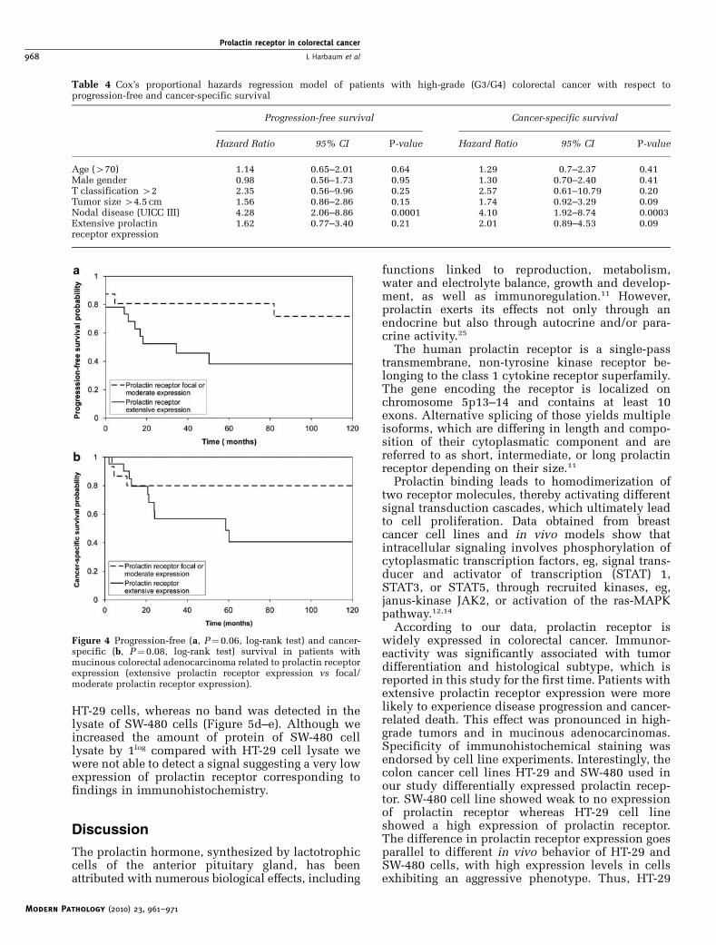
thus indicating stronger prognostic value of prolac-tin receptor immunostaining in this distinct type ofcancer. In all, 12/23 (52%) patients with tumorsshowing extensive prolactin receptor expressionexperienced disease progression compared with4/16 (25%) patients with tumors either lackingprolactin receptor expression or showing onlyfocal/moderate expression (P¼ 0.06, log-rank test;Figure 4a). Similarly, 10/23 (43%) patients withtumors with extensive prolactin receptor expressiondied of disease compared with 3/16 (19%) patientswith tumors either lacking prolactin receptor ex-pression or showing only focal/moderate expression(P¼ 0.08, log-rank test; Figure 4b).
Table 2 Association of prolactin receptor expression with different tumor characteristics
Prolactin receptor immunoreactivity P-value
Positive (%) Extensive expression (%)
T classificationT1 24/26 (92) 20/26 (77) 0.15T2 66/69 (96) 48/69 (70)T3 206/213 (97) 160/213 (75)T4 64/65 (98) 56/65 (86)
N classificationN0 199/208 (96) 152/208 (73) 0.29N1 80/82 (98) 66/82 (80)N2 81/83 (98) 66/83 (75)
AJCC/ UICC StageI 75/80 (88) 55/80 (69) 0.30II 112/116 (97) 88/116 (76)III 121/124 (98) 99/124 (80)IV 52/53 (98) 42/53 (79)
GradeG1 114/118 (97) 87/118 (74) o0.001G2 135/136 (99) 118/136 (87)G3 90/97 (93) 7/97 (69)G4 21/22 (95) 12/22 (55)
HistologyAdenocarcinoma 304/312 (97) 250/312 (80) o0.001Mucinous Ca. 37/42 (88) 23/42 (55)Undifferentiated Ca. 12/12 (100) 7/12 (58)Other Ca. 7/7 (100) 4/7 (57)
Tumor sizer4.5 cm 191/194 (98) 161/194 (83) 0.00244.5 cm 148/157 (94) 108/157 (69)
Lymphatic invasionL0 241/250 (96) 190/250 (76) 1L1 119/123 (97) 94/123 (76)
Venous invasionV0 277/286 (97) 216/286 (76) 0.67V1 83/87 (95) 68/87 (78)
Ager70 179/184 (97) 139/184 (76) 0.81470 181/189 (96) 145/189 (77)
GenderFemale 161/164 (98) 123/164 (75) 0.71Male 199/209 (95) 161/209 (77)
Prolactin receptor in colorectal cancer
966 L Harbaum et al
Modern Pathology (2010) 23, 961–971

Cell Lines
To substantiate the specificity of prolactin receptorstaining results we thought to examine prolactinreceptor protein expression in the colon cancer celllines HT-29 and SW-480 using immunohistochem-istry as well as western blotting. Intensive prolactin
receptor staining was observed in HT-29 cells (andin T47D breast cancer cells, serving as positivecontrol), whereas SW-480 cells showed weak tomoderate immunolabeling of single cancer cells(Figure 5a–c). In accordance, western blottingshowed a prominent band at 78 kDa correspondingto the prolactin receptor protein in the lysate of
Table 3 Concordance of prolactin receptor expression in primary and corresponding metastatic tumor tissues (n¼ 151 primary tumorswith lymph node (n¼132) and/or distant (n¼39) metastases)
Primary tumor Lymph node metastasis (%) Distant metastasis (%)
T negative Negative 4/4 (100) Negative —4/151 (3%) Focal — Focal —
Moderate — Moderate —Extensive — Extensive —
T focal Negative — Negative —9/151 (6%) Focal 3/6 (50) Focal 1/3 (33
Moderate 1/6 (17) Moderate 2/3 (66)Extensive 2/6 (33) Extensive —
T moderate Negative — Negative —19/151 (13%) Focal — Focal —
Moderate 15/18 (83) Moderate 2/4 (50)Extensive 3/18 (17) Extensive 2/4 (50)
T extensive Negative — Negative —119/151 (79%) Focal 1/104 (1) Focal 1/32 (3)
Moderate 7/104 (7) Moderate 3/32 (9)Extensive 96/104 (92) Extensive 28/32 (86)
Figure 2 Progression-free (a, P¼ 0.08, log-rank test) and cancer-specific (b, P¼ 0.08, log-rank test) survival in patients withcolorectal carcinoma related to prolactin receptor expression(extensive prolactin receptor expression vs focal/moderate pro-lactin receptor expression).
Figure 3 Progression-free (a, P¼0.03, log-rank test) and cancer-specific (b, P¼0.04, log-rank test) survival in patients with high-grade (G3/G4) colorectal carcinoma related to prolactin receptorexpression (extensive prolactin receptor expression vs focal/moderate prolactin receptor expression).
Prolactin receptor in colorectal cancer
L Harbaum et al 967
Modern Pathology (2010) 23, 961–971
HT-29 cells, whereas no band was detected in thelysate of SW-480 cells (Figure 5d–e). Although weincreased the amount of protein of SW-480 celllysate by 1log compared with HT-29 cell lysate wewere not able to detect a signal suggesting a very lowexpression of prolactin receptor corresponding tofindings in immunohistochemistry.
Discussion
The prolactin hormone, synthesized by lactotrophiccells of the anterior pituitary gland, has beenattributed with numerous biological effects, including
functions linked to reproduction, metabolism,water and electrolyte balance, growth and develop-ment, as well as immunoregulation.11 However,prolactin exerts its effects not only through anendocrine but also through autocrine and/or para-crine activity.25
The human prolactin receptor is a single-passtransmembrane, non-tyrosine kinase receptor be-longing to the class 1 cytokine receptor superfamily.The gene encoding the receptor is localized onchromosome 5p13–14 and contains at least 10exons. Alternative splicing of those yields multipleisoforms, which are differing in length and compo-sition of their cytoplasmatic component and arereferred to as short, intermediate, or long prolactinreceptor depending on their size.11
Prolactin binding leads to homodimerization oftwo receptor molecules, thereby activating differentsignal transduction cascades, which ultimately leadto cell proliferation. Data obtained from breastcancer cell lines and in vivo models show thatintracellular signaling involves phosphorylation ofcytoplasmatic transcription factors, eg, signal trans-ducer and activator of transcription (STAT) 1,STAT3, or STAT5, through recruited kinases, eg,janus-kinase JAK2, or activation of the ras-MAPKpathway.12,14
According to our data, prolactin receptor iswidely expressed in colorectal cancer. Immunor-eactivity was significantly associated with tumordifferentiation and histological subtype, which isreported in this study for the first time. Patients withextensive prolactin receptor expression were morelikely to experience disease progression and cancer-related death. This effect was pronounced in high-grade tumors and in mucinous adenocarcinomas.Specificity of immunohistochemical staining wasendorsed by cell line experiments. Interestingly, thecolon cancer cell lines HT-29 and SW-480 used inour study differentially expressed prolactin recep-tor. SW-480 cell line showed weak to no expressionof prolactin receptor whereas HT-29 cell lineshowed a high expression of prolactin receptor.The difference in prolactin receptor expression goesparallel to different in vivo behavior of HT-29 andSW-480 cells, with high expression levels in cellsexhibiting an aggressive phenotype. Thus, HT-29
Figure 4 Progression-free (a, P¼0.06, log-rank test) and cancer-specific (b, P¼0.08, log-rank test) survival in patients withmucinous colorectal adenocarcinoma related to prolactin receptorexpression (extensive prolactin receptor expression vs focal/moderate prolactin receptor expression).
Table 4 Cox’s proportional hazards regression model of patients with high-grade (G3/G4) colorectal cancer with respect toprogression-free and cancer-specific survival
Progression-free survival Cancer-specific survival
Hazard Ratio 95% CI P-value Hazard Ratio 95% CI P-value
Age (470) 1.14 0.65–2.01 0.64 1.29 0.7–2.37 0.41Male gender 0.98 0.56–1.73 0.95 1.30 0.70–2.40 0.41T classification 42 2.35 0.56–9.96 0.25 2.57 0.61–10.79 0.20Tumor size 44.5 cm 1.56 0.86–2.86 0.15 1.74 0.92–3.29 0.09Nodal disease (UICC III) 4.28 2.06–8.86 0.0001 4.10 1.92–8.74 0.0003Extensive prolactinreceptor expression
1.62 0.77–3.40 0.21 2.01 0.89–4.53 0.09
Prolactin receptor in colorectal cancer
968 L Harbaum et al
Modern Pathology (2010) 23, 961–971

cells have been reported to show local invasion andmarked metastatic capacity after subcutaneous andintracaecal xenografting in nude mice, whereas SW-480 cells showed solid growth without invasion inthe bowel wall or the development of metastases.26
This pair of cell lines may consequently be used as amodel system for further research of the role ofprolactin in the biology of colon cancer.
To the best of our knowledge, only three studieshave so far addressed the topic of prolactin receptorexpression in colorectal cancer.18–20 The first studyassessed prolactin receptor protein by radioligandassay using 125I-hGH. Tumor specimens of 71 malepatients were analyzed and prolactin receptor wasfound to be positive in 36 (51%) patients.18 Noassociations with clinicopathological variables werenoted and prolactin receptor protein content wasindependent from outcome. In a second study, thesame group of authors noted prolactin receptorimmunoreactivity in 26 out of 56 (46%) tumors.19
The authors used a monoclonal mouse anti-human
prolactin receptor antibody different from the oneused in our study, which may have accounted forthe low prevalence of prolactin receptor expression.Data were not correlated with follow-up in thisanalysis.
In the third study by Otte et al20 the long transcriptof prolactin receptor mRNA was detected in 45out of 48 (94%) of cancer specimens as well as in 22out of 23 (96%) normal colonic tissues. Expressionin cancer tissue did not differ significantly betweentumors of different grades. mRNA encoding for theshort form of prolactin receptor was not found inany samples analyzed. In addition, prolactin recep-tor protein was detected by western blotting in 39out of 45 (87%) cancer specimens and in 17 out of 22(77%) normal tissues. Similar to our data, theprolactin receptor was detected on the membraneand in the cytoplasm of epithelial cells in normaland cancer tissue. Data regarding the percentage oftumors stained, however, were not provided andfollow-up information was not given.
Figure 5 Intensive prolactin receptor staining in HT-29 colon cancer cells (a) and in T47D breast cancer cells serving as positive control(b). Weak staining of single cells in SW-480 colon cancer cells (c). Western blotting shows a prominent band at 80 kDa corresponding tothe prolactin receptor protein in the lysate of HT-29 cells, whereas no band was detected in the lysate of SW-480 cells. The expression ofprolactin receptor in HT-29 cells was B10-fold compared to T47D cells (d). Anti-b-actin served as a positive loading control. Please notethat the protein concentration of SW-480 and T47D cell lysates was 20mg, and that of HT-29 was 2.5mg, respectively, because of the 1log
difference in prolactin receptor expression. The western blot presented is representative for five experiments.
Prolactin receptor in colorectal cancer
L Harbaum et al 969
Modern Pathology (2010) 23, 961–971

Remarkably, in all the three studies the patientcohorts consisted only of male patients and/orpostmenopausal women. Thus, our study is the firstto analyze an unselected cohort of patients, includ-ing a random sample of female and male patientsof all ages. It is known that prolactin secretion isgreatly affected by the menstrual cycle.11 It is,however, largely unknown whether prolactin recep-tor expression, particularly in the gastrointestinalmucosa, varies accordingly in female patients.Of note, we did not find an association betweenprolactin receptor expression and gender. Moreover,there was no difference in immunoreactivity amongpre- and postmenopausal women (data not shown).
Data in the literature regarding the ectopicexpression of prolactin in colorectal cancer arecontroversial, with prevalence rates ranging from0 to 77%.20,27–32 Of note, compared with normalcolorectal tissue, cancer tissues show a significantlyhigher rate of co-expression of prolactin and itsreceptor. Thus, in addition to systemic effects,prolactin presumably acts through an auto-/para-crine loop in promoting cell proliferation, as shownin colorectal cancer cell lines (CaCO-2, HT-29, andLoVo cells).20,25
As pointed out above, prolactin and its receptorare promising anti-cancer targets, wherein prolactinreceptor antagonism appears to be the most promis-ing interventional approach.15–17,33 First, resultsshowing a beneficial effect of prolactin/prolactinreceptor-targeted therapy in breast cancer have beenreported from both in vitro and in vivo models usingeither pure prolactin receptor antagonists, such asD1–9-G129R-hPRL or D1–14-G129R-hPRL, or fusionpeptides between a prolactin receptor antagonistand potential anti-tumor peptides, such as G129R-hPRL-IL2, G129R-hPRL-endostatin, or G129R-hPRL-PE40-KDEL.12,34–36 However, these drugs are in earlystages of development and application, and diffi-culties regarding stability and binding affinity ofprolactin receptor antagonist must be overcomebefore entering the clinic.33 Nevertheless, a singleclinical therapy trial using the anti-prolactinemicdrug cabergoline combined with docetaxel in wo-man with metastatic breast cancer and concomitanthyperprolactinemia showed that tumor regressionrate was significantly higher in patients treated withcabergoline than in those who received docetaxelalone.37 To the best of our knowledge, other cancertrials targeting the prolactin/prolactin receptor axis,in breast cancer or even other types of cancer, havenot been published.
According to Carver et al33 the relatively restricteddistribution of prolactin receptor as well as thelimited disturbance in phenotype of the prolactinreceptor�/� transgenic mice apart from morpholo-gical changes occurring within the mammary glandsuggests that therapeutics directed toward prolac-tin/prolactin receptor should be well tolerated,increasing its appeal. According to our data, thewidespread expression of prolactin receptor in
primary tissues as well as in regional and distantmetastatic sites of colorectal cancer makes this typeof cancer an ideal candidate for prolactin/prolactinreceptor-targeted therapy. Moreover, the concordantexpression profiles of primary tumors and corre-sponding metastases facilitate the promise of in-dividualized treatment for metastatic disease, astailored medicine can be accomplished withouttaking additional biopsies from metastatic deposits.
In conclusion, prolactin receptor is widely ex-pressed in colorectal cancer. High concordancerates between primary tumor and metastatic tissuesmake this protein a promising target in patientswith advanced disease. Moreover, prolactin receptorexpression may be an additional prognosticvariable, particularly in high-grade and mucinouscancers.
Acknowledgement
We are grateful to Mrs M Schuller, Mrs J Strutz, andMrs A Kaps for excellent technical assistance.
Disclosure/conflict of interest
The authors declare no conflict of interest.
References
1 Boyle P, Levin B. World Health Organization.World Cancer Report 2008. IARC Press: Lyon, 2008,,pp 374–378.
2 Jemal A, Siegel R, Ward E, et al. Cancer statistics, 2008.CA Cancer J Clin 2008;58:71–96.
3 Hampton T. New therapies for GI cancers fall short.JAMA 2009;302:373–374.
4 Moertel CG, Fleming TR, MacDonald JS, et al.Levamisole and fluorouracil for adjuvant therapy ofresected colon carcinoma. N Engl J Med 1990;322:352–358.
5 MacFarlane JK, Ryall RD, Heald RJ. Mesorectal exci-sion for rectal cancer. Lancet 1993;341:457–460.
6 Sauer R, Becker H, Hohenberger W, et al. Preoperativeversus postoperative chemoradiotherapy for rectalcancer. N Engl J Med 2004;351:1731–1740.
7 Chau I, Cunningham D. Treatment in advanced color-ectal cancer: what, when and how? Br J Cancer2009;100:1704–10719.
8 Ciardiello F, Tortora G. EGFR antagonists in cancertreatment. N Engl J Med 2008;358:1160–1174.
9 Allegra CJ, Yothers G, O’Connell MJ, et al. Initial safetyreport of NSABP C-08: A randomized phase III study ofmodified FOLFOX6 with or without bevacizumab forthe adjuvant treatment of patients with stage II or IIIcolon cancer. J Clin Oncol 2009;27:3385–3390.
10 Nagano M, Chastre E, Choquet A, et al. Expression ofprolactin and growth hormone receptor genes andtheir isoforms in the gastrointestinal tract. Am JPhysiol 1995;268:431–442.
11 Bole-Feysot C, Goffin V, Edery M, et al. Prolactin (PRL)and its receptor: actions, signal transduction pathways
Prolactin receptor in colorectal cancer
970 L Harbaum et al
Modern Pathology (2010) 23, 961–971

and phenotypes observed in PRL receptor knockoutmice. Endocr Rev 1998;19:225–268.
12 Clevenger CV, Zheng J, Jablonski EM, et al. From benchto bedside: future potential for the translation ofprolactin inhibitors as breast cancer therapeutics.J Mammary Gland Biol Neoplasia 2008;13:147–156.
13 Reynolds C, Montone KT, Powell CM, et al. Expressionof prolactin and its receptor in human breast carcino-ma. Endocrinology 1997;138:5555–5560.
14 Vonderhaar BK. Prolactin involvement in breastcancer. Endocr Relat Cancer 1999;6:389–404.
15 Tallet E, Rouet V, Jomain JB, et al. Rational design ofcompetitive prolactin/growth hormone receptorantagonists. J Mammary Gland Biol Neoplasia 2008;13:105–117.
16 Walker AM. Therapeutic potential of S179D prolactin–from prostate cancer to angioproliferative disorders:the first selective prolactin receptor modulator. ExpertOpin Investig Drugs 2006;15:1257–1267.
17 Goffin V, Bernichtein S, Touraine P, et al. Developmentand potential clinical uses of human prolactin receptorantagonists. Endocr Rev 2005;26:400–422.
18 Bhatavdekar J, Patel D, Ghosh N, et al. Interrelation-ship of prolactin and its receptor in carcinoma of colonand rectum: a preliminary report. J Surg Oncol1994;55:246–249.
19 Bhatavdekar JM, Patel DD, Chikhlikar PR, et al. Ectopicproduction of prolactin by colorectal adenocarcinoma.Dis Colon Rectum 2001;44:119–127.
20 Otte JM, Otte C, Beckedorf S, et al. Expression offunctional prolactin and its receptor in colorectalcancer. Int J Coloretal Dis 2003;18:86–94.
21 Sobin LH, Wittekind C, (eds) TNM Classificationof Malignant Tumors, 6th edn. Wiley-Liss: New York,2002.
22 Hamilton SR, Vogelstein B, Kudo S, et al. Carcinoma ofthe colon and rectum In: Hamilton SR, Aaltonen LA(eds). World Health Organization Classificationof Tumours. Pathology and Genetics. Tumours ofthe Digestive System. IARC Press: Lyon, 2000, pp105–119.
23 Kononen J, Bubendorf L, Kallioniemi A, et al. Tissuemicroarrays for high-throughput molecular profiling oftumor specimens. Nat Med 1998;4:844–847.
24 Kersten HM, Robben JC, Poddinghe PJ, et al. AgarCyto:a novel cell processing method for multiple moleculardiagnostic analyses of uterine cervix. J HistochemCytochem 2000;48:709–718.
25 Ben-Jonathan N, Mershon JL, Allen DL, et al.Extrapituitary prolactin: distribution, regulation,functions, and clinical aspects. Endocr Rev 1996;17:639–669.
26 De Vries JE, Dinjens WN, De Bruyne GK, et al. In vivoand in vitro invasion in relation to phenotypiccharacteristics of human colorectal carcinoma cells.Br J Cancer 1995;71:271–277.
27 Baert D, Matthys C, Gillardin JP, et al. Prolactin andcolorectal cancer: is there a connection? Acta Gastro-enterol Belg 1998;61:407–409.
28 Carlson HE, Zarrabi MH, Lyubsky SL. Lack of associa-tion between hyperprolactinemia and colon carcino-ma. Cancer Invest 2000;18:130–134.
29 Wood AJ, Thomas CM, Baumforth KRN, et al. Absenceof prolactin gene expression in colorectal cancer. J ClinPathol 1999;52:135–139.
30 Bhatavdekar JM, Patel DD, Chikhlikar PR, et al.Molecular markers are predictors of recurrence andsurvival in patients with Dukes B and Dukes Ccolorectal adenocarcinoma. Dis Colon Rectum2001;44:523–533.
31 Ilan Y, Sibirsky O, Livni N, et al. Plasma and tumorprolactin in colorectal cancer patients. Dig Dis Sci1995;40:2010–2015.
32 Indinnimeo M, Cicchini C, Memeo L, et al. Plasma andtissue prolactin detection in colon carcinoma. OncolRep 2001;8:1351–1353.
33 Carver KC, Arendt LM, Schuler LA. Complex prolactincrosstalk in breast cancer: new therapeutic implica-tions. Mol Cell Endocrinol 2009;307:1–7.
34 Bernichtein S, Kayser C, Dillner K, et al. Developmentof pure prolactin receptor antagonists. J Biol Chem2003;278:35988–35999.
35 Beck MT, Chen NY, Franek KJ, et al. Prolactinantagonist-endostatin fusion protein as a targeteddual-functional therapeutic agent for breast cancer.Cancer Res 2003;63:3598–3604.
36 Langenheim JF, Chen WY. Development of a prolactinreceptor-targeting fusion toxin using a prolactinantagonist and a recombinant form of Pseudomonasexotoxin A. Breast Cancer Res Treat 2005;90:281–293.
37 Frontini L, Lissoni P, Vaghi M, et al. Enhancement ofthe efficacy of weekly low-dose taxotere by the longacting anti-prolactinemic drug cabergoline in pre-treated metastatic breast cancer. Anticancer Res2004;24:4223–4226.
Prolactin receptor in colorectal cancer
L Harbaum et al 971
Modern Pathology (2010) 23, 961–971
Copyright © 2022 FDOKUMEN