Juvenile Administration of Methylphenidate Attenuates Adult Hippocampal Neurogenesis
Upload
independentCategory
view
0download
0
Cell Therapy Attenuates Cardiac Dysfunction PostMyocardial Infarction: Effect of Timing, Routes ofInjection and a Fibrin ScaffoldJuliana S. Nakamuta1, Maria E. Danoviz1, Fabio L. N. Marques2, Leonardo dos Santos1,3, Claudia Becker1,
Giovana A. Goncalves1, Paula F. Vassallo1, Isolmar T. Schettert1, Paulo J. F. Tucci3, Jose E. Krieger1*
1 Heart Institute (InCor), University of Sao Paulo Medical School, Sao Paulo, Sao Paulo, Brazil, 2 Radiopharmacy Laboratory, Nuclear Medicine Center, University of Sao
Paulo Medical School, Sao Paulo, Sao Paulo, Brazil, 3 Cardiac Physiology and Pathophysiology Laboratory, Cardiology Division, Federal University of Sao Paulo, Sao Paulo,
Sao Paulo, Brazil
Abstract
Background: Cell therapy approaches for biologic cardiac repair hold great promises, although basic fundamental issuesremain poorly understood. In the present study we examined the effects of timing and routes of administration of bonemarrow cells (BMC) post-myocardial infarction (MI) and the efficacy of an injectable biopolymer scaffold to improve cardiaccell retention and function.
Methodology/Principal Findings: 99mTc-labeled BMC (66106 cells) were injected by 4 different routes in adult rats:intravenous (IV), left ventricular cavity (LV), left ventricular cavity with temporal aorta occlusion (LV+) to mimic coronaryinjection, and intramyocardial (IM). The injections were performed 1, 2, 3, or 7 days post-MI and cell retention was estimatedby c-emission counting of the organs excised 24 hs after cell injection. IM injection improved cell retention and attenuatedcardiac dysfunction, whereas IV, LV or LV* routes were somewhat inefficient (,1%). Cardiac BMC retention was notinfluenced by timing except for the IM injection that showed greater cell retention at 7 (16%) vs. 1, 2 or 3 (average of 7%)days post-MI. Cardiac cell retention was further improved by an injectable fibrin scaffold at day 3 post-MI (17 vs. 7%), eventhough morphometric and function parameters evaluated 4 weeks later displayed similar improvements.
Conclusions/Significance: These results show that cells injected post-MI display comparable tissue distribution profileregardless of the route of injection and that there is no time effect for cardiac cell accumulation for injections performed 1to 3 days post-MI. As expected the IM injection is the most efficient for cardiac cell retention, it can be further improved byco-injection with a fibrin scaffold and it significantly attenuates cardiac dysfunction evaluated 4 weeks post myocardialinfarction. These pharmacokinetic data obtained under similar experimental conditions are essential for furtherdevelopment of these novel approaches.
Citation: Nakamuta JS, Danoviz ME, Marques FLN, dos Santos L, Becker C, et al. (2009) Cell Therapy Attenuates Cardiac Dysfunction Post Myocardial Infarction:Effect of Timing, Routes of Injection and a Fibrin Scaffold. PLoS ONE 4(6): e6005. doi:10.1371/journal.pone.0006005
Editor: Alicia J. Kowaltowski, Instituto de Quımica, Universidade de Sao Paulo, Brazil
Received March 8, 2009; Accepted May 6, 2009; Published June 23, 2009
Copyright: � 2009 Nakamuta et al. This is an open-access article distributed under the terms of the Creative Commons Attribution License, which permitsunrestricted use, distribution, and reproduction in any medium, provided the original author and source are credited.
Funding: This study was funded by grants from public Brazilian Agencies Fundacao de Amparo a Pesquisa do Estado de Sao Paulo (FAPESP # 01/0009-0),Ministerio da Ciencia e Tecnologia/Conselho Nacional de Desenvolvimento Cientifico e Tecnologico/Ministerio da Saude/Departamento Ciencia e Tecnologia(MCT/CNPq/MS/DECIT #552324/20005-1 and 10120104096700). JSN, MED, CB, and GAG were recipients of fellowships from FAPESP (04/06784-4, 05/54695-3, 03/02671-8 and 03/02672-4, respectively). The funders had no role in study design, data collection and analysis, decision to publish, or preparation of the manuscript.
Competing Interests: The authors have declared that no competing interests exist.
* E-mail: [email protected]
Introduction
Transplantation of stem and progenitor cells is emerging as a
promising therapeutic option for repair of ischemic and infarcted
myocardium [1–3]. Nevertheless, the implementation of this novel
approach in a clinical setting requires the understanding of a
number of key aspects that remain poorly understood. Among the
main issues, the optimal timing for therapy and the most
appropriate cell delivery route might be of particular importance
in order to maximize cell transplantation efficiency.
Studies investigating the kinetics of cytokines production and
mobilization of stem cells to the injured myocardium provide
evidence that these processes occur within a limited time window
after infarction [4–6]. This provides a rational for identification of
the ideal timing for cell transplantation. Moreover, recent
evidence suggests that the combination of cells with biopolymers
such as fibrin, collagen and matrigel can improve cell survival,
angiogenesis and cardiac function [7–9].
Different routes for cell administration have been proposed to
deliver cells including transepicardic [10–14], systemic [15–17]
and intracoronary balloon catheter-mediated cell delivery [18–
21], however, comparative studies designed to evaluate both their
efficiencies and the effect of timing are scarce. The optimal timing
for therapy may vary depending on the route used to administer
the cells and factors associated to cell in situ retention at the
damaged cardiac tissue may be essential for the intramuscular (IM)
injection, whereas cell recruitment should be equally important for
intravenous injection. Consequently, the optimal time to trans-
PLoS ONE | www.plosone.org 1 June 2009 | Volume 4 | Issue 6 | e6005
plant the cells should be assessed within the context of the delivery
modality in order to optimize the efficiency of the predominant
underlying mechanism(s) related to each route.
In the present study, we examined the effect of timing and
routes of administration of 99mTc-labeled bone marrow cells
(BMCs) on cardiac cell retention, as well as the efficacy of a
biopolymer used as vehicle to improve retention and cardiac
function in rats submitted to experimental myocardium infarction
(MI).
Results
Labeling efficiency and stabilityBMCs showed an average labeling efficiency of 14.963.5%,
which means that approximately 15% of the total radioactivity
labeled uniformly the cell pool, resulting in 1.98 MBq per 106
BMCs. Moreover, the 99mTc radioactivity detected in BMCs and
in the supernatant revealed that only 33.062.5% of the
radioactivity initially incorporated remained within the cells
24 hours after labeling. The absence of significant deterioration
of cell viability (80% for labeled vs. 87.5% for unlabeled BMCs,
P.0.05), as indicated by the Trypan blue dye exclusion test,
suggests that the radioactivity detected in the supernatant is
secondary to 99mTc leakage from labeled BMCs rather than cell
death. Therefore, to determine the transplanted cell accumulation,
heart radioactivity values were corrected according to the rate of99mTc leakage from BMCs in vitro (33.0%) assuming an equivalent
rate of leakage in vivo.
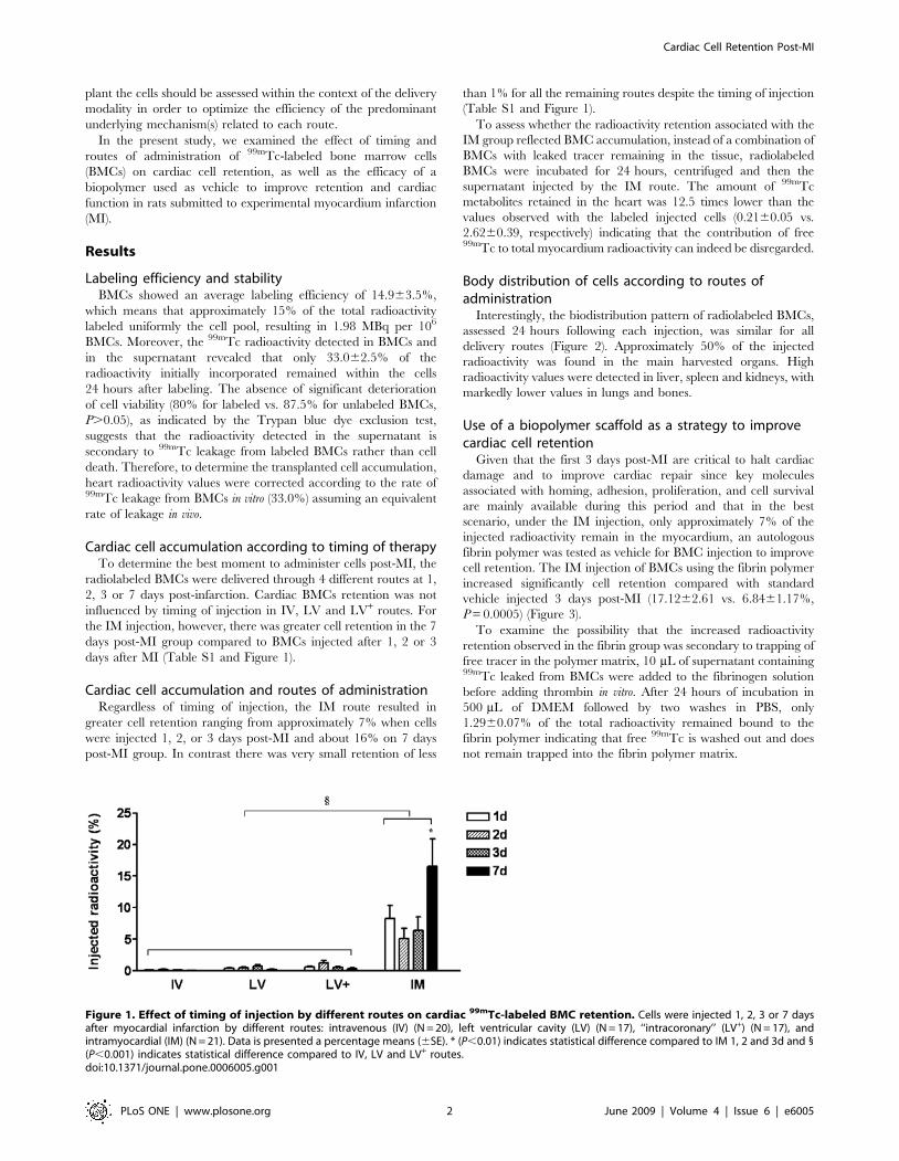
Cardiac cell accumulation according to timing of therapyTo determine the best moment to administer cells post-MI, the
radiolabeled BMCs were delivered through 4 different routes at 1,
2, 3 or 7 days post-infarction. Cardiac BMCs retention was not
influenced by timing of injection in IV, LV and LV+ routes. For
the IM injection, however, there was greater cell retention in the 7
days post-MI group compared to BMCs injected after 1, 2 or 3
days after MI (Table S1 and Figure 1).
Cardiac cell accumulation and routes of administrationRegardless of timing of injection, the IM route resulted in
greater cell retention ranging from approximately 7% when cells
were injected 1, 2, or 3 days post-MI and about 16% on 7 days
post-MI group. In contrast there was very small retention of less
than 1% for all the remaining routes despite the timing of injection
(Table S1 and Figure 1).
To assess whether the radioactivity retention associated with the
IM group reflected BMC accumulation, instead of a combination of
BMCs with leaked tracer remaining in the tissue, radiolabeled
BMCs were incubated for 24 hours, centrifuged and then the
supernatant injected by the IM route. The amount of 99mTc
metabolites retained in the heart was 12.5 times lower than the
values observed with the labeled injected cells (0.2160.05 vs.
2.6260.39, respectively) indicating that the contribution of free99mTc to total myocardium radioactivity can indeed be disregarded.
Body distribution of cells according to routes ofadministration
Interestingly, the biodistribution pattern of radiolabeled BMCs,
assessed 24 hours following each injection, was similar for all
delivery routes (Figure 2). Approximately 50% of the injected
radioactivity was found in the main harvested organs. High
radioactivity values were detected in liver, spleen and kidneys, with
markedly lower values in lungs and bones.
Use of a biopolymer scaffold as a strategy to improvecardiac cell retention
Given that the first 3 days post-MI are critical to halt cardiac
damage and to improve cardiac repair since key molecules
associated with homing, adhesion, proliferation, and cell survival
are mainly available during this period and that in the best
scenario, under the IM injection, only approximately 7% of the
injected radioactivity remain in the myocardium, an autologous
fibrin polymer was tested as vehicle for BMC injection to improve
cell retention. The IM injection of BMCs using the fibrin polymer
increased significantly cell retention compared with standard
vehicle injected 3 days post-MI (17.1262.61 vs. 6.8461.17%,
P = 0.0005) (Figure 3).
To examine the possibility that the increased radioactivity
retention observed in the fibrin group was secondary to trapping of
free tracer in the polymer matrix, 10 mL of supernatant containing99mTc leaked from BMCs were added to the fibrinogen solution
before adding thrombin in vitro. After 24 hours of incubation in
500 mL of DMEM followed by two washes in PBS, only
1.2960.07% of the total radioactivity remained bound to the
fibrin polymer indicating that free 99mTc is washed out and does
not remain trapped into the fibrin polymer matrix.
Figure 1. Effect of timing of injection by different routes on cardiac 99mTc-labeled BMC retention. Cells were injected 1, 2, 3 or 7 daysafter myocardial infarction by different routes: intravenous (IV) (N = 20), left ventricular cavity (LV) (N = 17), ‘‘intracoronary’’ (LV+) (N = 17), andintramyocardial (IM) (N = 21). Data is presented a percentage means (6SE). * (P,0.01) indicates statistical difference compared to IM 1, 2 and 3d and 1(P,0.001) indicates statistical difference compared to IV, LV and LV+ routes.doi:10.1371/journal.pone.0006005.g001
Cardiac Cell Retention Post-MI
PLoS ONE | www.plosone.org 2 June 2009 | Volume 4 | Issue 6 | e6005

To further confirm the efficacy of this strategy, we tested the
injectable fibrin scaffold with other more homogeneous cell types
including rat cardiac fibroblasts expressing b-galactosidase and rat
adipose stem cells (see supplementary Methods S1). Twenty-four
hours after injection of cardiac fibroblasts there was higher cell
retention in the fibrin compared to the control group (supple-
mentary Figure S1). Furthermore, adipose mesenchymal stem cells
injected with fibrin survived and remained in the myocardium for
at least 30 days (supplementary Figure S2).
Effect of increased BMC accumulation on cardiacmorphometry and performance
To examine if greater BMC cardiac retention translated into
improved cardiac repair, morphometric and functional analyses
were performed in 5 groups of rats administered with BMCs IV or
IM and for the former in the presence or absence of fibrin 4 weeks
after myocardial infarction (Table 1).
Infarct size, LV cavity perimeter and scar thicknessHistological analyses revealed no significant differences in
percentage of left ventricle perimeter occupied by fibrosis between
the 5 groups (31.2161.99 vs. 24.7662.27 vs. 27.5162.67 vs.
28.2563.41 vs. 26.6661.04% for M-IM, F-IM, BMC-IV, BMC-
IM, and BMC+F-IM, respectively, P = 0.6384) (Figure 4A).
The LV perimeter increased in all MI groups compared to the
sham operated animals (27.560.18 vs. 36,9860,78; 37,2360,91,
39,4860,77; 34.5360.96; and 33,2060.38 mm for Sham vs. M-
IM, F-IM, BMC-IV, BMC-IM, and BMC+F-IM, respectively,
P,0.001), but was significantly reduced in the groups that
received BMC injected IM, with or without the fibrin scaffold
matrix, compared to the other experimental groups (Figure 4B).
Similarly, the scar thickness tended to be thicker in the animals
injected with BMC IM, but reached significant values only in the
group injected with the fibrin scaffold (1.5860.15 vs. 0.7060.13,
0.9460.20 0.8560,16, 1.360.14 for BMC+F-IM vs. M-IM, F-
IM, BMC-IV, and BMC-IM, respectively, p,0.05) (Figure 4C).
Interestingly, the linear regression model from data of scar
thickness and LV perimeter shows a significant inverse correlation
between LV cavity dilatation and scar thickness (Pearson’s
r = 20.65; slope = 25.9161.31, P,0.001) (Figure 4D).
Figure 2. Effect of different routes of injection of 99mTc-labeled BMC on radioactivity tissue distribution profile. Cells were injectedpost-MI via intravenous (IV) (N = 20), left ventricular cavity (LV) (N = 17), ‘‘intracoronary’’ (LV+) (N = 17), and intramyocardial (IM) (N = 21) routes. Dataare expressed as percentage means (6SE).doi:10.1371/journal.pone.0006005.g002
Figure 3. Effect of intramyocardial co-injection of a fibrinscaffold on cardiac cell retention post-myocardial infarction.99mTc-labeled BMC were injected post-myocardial infarction directlyinto the myocardium in vehicle (DMEM, N = 22) or in a fibrin scaffold(Fibrin, N = 15). Cell retention was assessed 24 hours later by the level ofradioactivity in the heart shown as percentage means (6SE). * denotesstatistical significance for P,0.01.doi:10.1371/journal.pone.0006005.g003
Table 1. Experimental design of functional study.
Group MI Content Route
SHAM (N = 5) 2 2 2
M - IM (N = 4) + culture medium intramyocardial (IM)
F - IM (N = 4) + fibrin intramyocardial (IM)
BMC - IV (N = 5) + BMC intravenous (IV)
BMC - IM (N = 4) + BMC intramyocardial (IM)
BMC+F - IM (N = 4) + BMC+fibrin (16106) intramyocardial (IM)
N indicates the number of animals in each group. + and 2 indicates thepresence or absence of myocardial infarction (MI).doi:10.1371/journal.pone.0006005.t001
Cardiac Cell Retention Post-MI
PLoS ONE | www.plosone.org 3 June 2009 | Volume 4 | Issue 6 | e6005

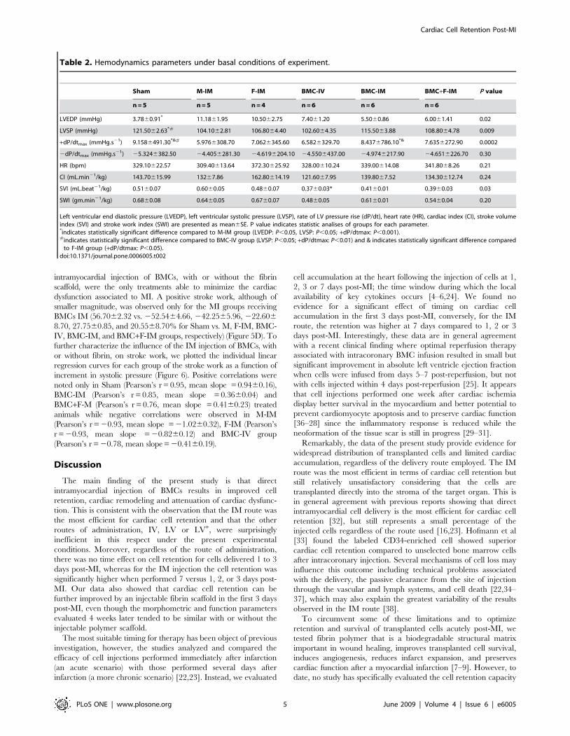
Hemodynamic assessment of ventricular functionWe next examined whether the improvement in cardiac
remodeling associated with BMC administered IM, with or
without fibrin scaffold, translated into functional benefits. As a
show in table 2, the indices of ventricular function under basal
conditions were generally worse in M-IM and F-IM groups
compared to sham group. LVEDP was increased in M-IM group
and the LVSP diminished in M-IM and BMC-IV groups.
Moreover, low values of +dP/dtmax were observed in M-IM, F-
IM and BMC-IV compared to sham animals. The other
hemodynamic parameters exhibited similar profile in all experi-
mental groups.
More informative data to evaluate overall cardiac function,
however, were obtained during the pharmacologic pressure stress
with phenilefrine. The animals that received BMC by intramyo-
cardial route exhibited a clear improvement in myocardial
function especially considering the discrete basal hemodynamic
and morphometric changes described before.
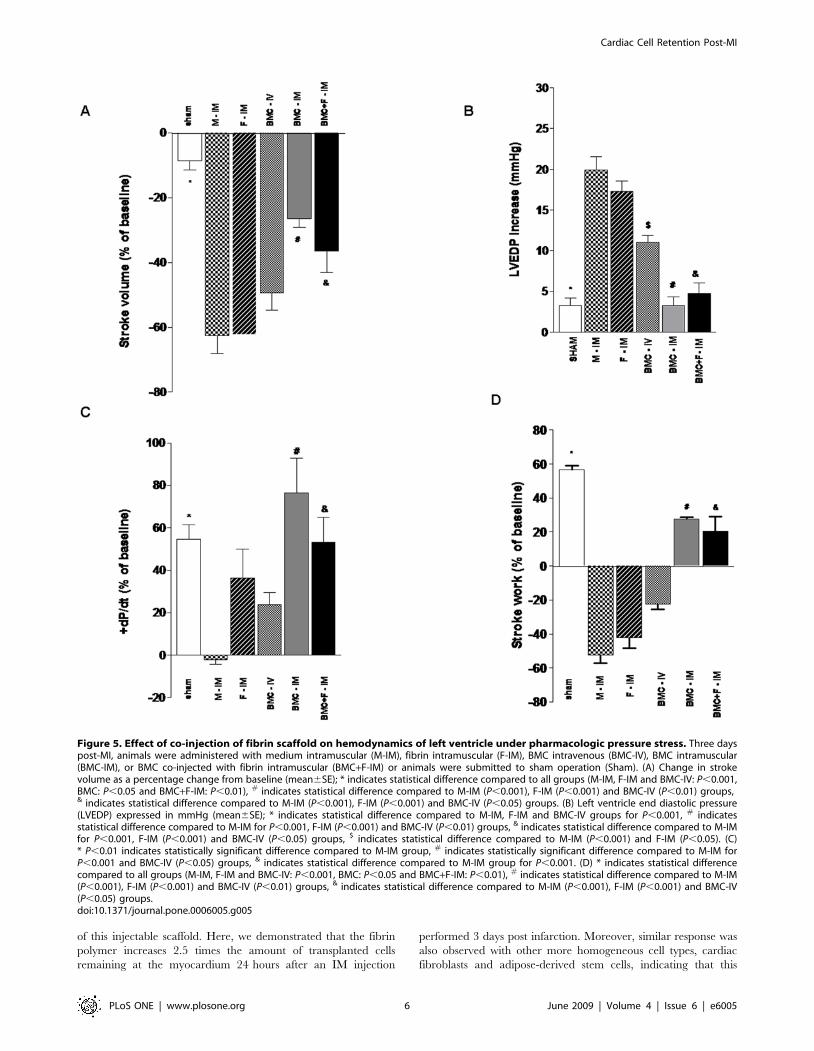
Stroke volume in response to pressure overload, depicted as %
change, decreased in all infarcted groups, but it was better preserved
in the groups receiving BMC IM either with or without the fibrin
scaffold matrix compared to the other experimental groups
(24.5562.19 vs. 262.5065.50, 262.2563.70, 249.4065.26,
226.5062.63, and 228.5065.75% for sham vs. M-IM, F-IM,
BMC-IV, BMC-IM, and BMC+F-IM, respectively, p,0.05)
(Figure 5A). LVEDP increased only in the control experimental
groups injected with medium and fibrin IM or BMC intravenously
compared to sham animals or the IM BMC groups (3.2660.97 vs.
19.9061.66, 17.2561.31, 11.0060.89, 3.2561.10, and
4.7561.31 mmHg for Sham vs. M-IM, F-IM, BMC-IV, BMC-IM,
and BMC+F-IM groups, respectively) (Figure 5B). The changes in
+dP/dtmax were not as consistent as the other variables, but it can be
noted that the index of contractility tended to reduce in the three
control groups compared to sham, reaching significance only for the
M-IM comparison, whereas it remained unchanged in the two BMC-
IM groups (54.56+6.94 vs. 22.0262.37, 36.25+13.64, 23.8065.82,
76.50616.36, 53.40611.75% for Sham vs. M-IM, F-IM, BMC-IV,
BMC-IM, and BMC+F-IM, respectively, P,0.01) (Figure 5c).
Finally, the work generation under pressure overload, which
represents a global index of cardiac function, showed clearly that
Figure 4. Effect of intramyocardial co-injection of fibrin scaffold 3 days post-MI on cardiac morphometry. (A) Myocardial infarction estimatedas a percentage of left ventricle (LV) perimeter with a scar (mean6SE). (B) Left ventricle (LV) perimeter in millimeters (mean6SE); * indicates statisticaldifference compared to all groups for P,0.001, # indicates statistical difference compared to BMC-IV group for P,0.001 and & indicates statistical differencecompared to F-IM and BMC-IV for P,0.01. (C) Scar thickness in millimeters (mean6SE); * indicates statistical difference compared to M-IM and BMC-IVgroups for P,0.05. (D) Linear regression of scar thickness as a function a LV perimeter (Pearson’s r = 20.65; slope = 25.9161.31, P,0.001).doi:10.1371/journal.pone.0006005.g004
Cardiac Cell Retention Post-MI
PLoS ONE | www.plosone.org 4 June 2009 | Volume 4 | Issue 6 | e6005
intramyocardial injection of BMCs, with or without the fibrin
scaffold, were the only treatments able to minimize the cardiac
dysfunction associated to MI. A positive stroke work, although of
smaller magnitude, was observed only for the MI groups receiving
BMCs IM (56.7062.32 vs. 252.5464.66, 242.2565.96, 222.606
8.70, 27.7560.85, and 20.5568.70% for Sham vs. M, F-IM, BMC-
IV, BMC-IM, and BMC+F-IM groups, respectively) (Figure 5D). To
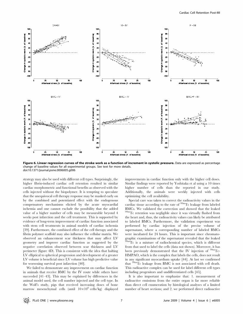
further characterize the influence of the IM injection of BMCs, with
or without fibrin, on stroke work, we plotted the individual linear
regression curves for each group of the stroke work as a function of
increment in systolic pressure (Figure 6). Positive correlations were
noted only in Sham (Pearson’s r = 0.95, mean slope = 0.9460.16),
BMC-IM (Pearson’s r = 0.85, mean slope = 0.3660.04) and
BMC+F-M (Pearson’s r = 0.76, mean slope = 0.4160.23) treated
animals while negative correlations were observed in M-IM
(Pearson’s r = 20.93, mean slope = 21.0260.32), F-IM (Pearson’s
r = 20.93, mean slope = 20.8260.12) and BMC-IV group
(Pearson’s r = 20.78, mean slope = 20.4160.19).
Discussion
The main finding of the present study is that direct
intramyocardial injection of BMCs results in improved cell
retention, cardiac remodeling and attenuation of cardiac dysfunc-
tion. This is consistent with the observation that the IM route was
the most efficient for cardiac cell retention and that the other
routes of administration, IV, LV or LV+, were surprisingly
inefficient in this respect under the present experimental
conditions. Moreover, regardless of the route of administration,
there was no time effect on cell retention for cells delivered 1 to 3
days post-MI, whereas for the IM injection the cell retention was
significantly higher when performed 7 versus 1, 2, or 3 days post-
MI. Our data also showed that cardiac cell retention can be
further improved by an injectable fibrin scaffold in the first 3 days
post-MI, even though the morphometric and function parameters
evaluated 4 weeks later tended to be similar with or without the
injectable polymer scaffold.
The most suitable timing for therapy has been object of previous
investigation, however, the studies analyzed and compared the
efficacy of cell injections performed immediately after infarction
(an acute scenario) with those performed several days after
infarction (a more chronic scenario) [22,23]. Instead, we evaluated
cell accumulation at the heart following the injection of cells at 1,
2, 3 or 7 days post-MI; the time window during which the local
availability of key cytokines occurs [4–6,24]. We found no
evidence for a significant effect of timing on cardiac cell
accumulation in the first 3 days post-MI, conversely, for the IM
route, the retention was higher at 7 days compared to 1, 2 or 3
days post-MI. Interestingly, these data are in general agreement
with a recent clinical finding where optimal reperfusion therapy
associated with intracoronary BMC infusion resulted in small but
significant improvement in absolute left ventricle ejection fraction
when cells were infused from days 5–7 post-reperfusion, but not
with cells injected within 4 days post-reperfusion [25]. It appears
that cell injections performed one week after cardiac ischemia
display better survival in the myocardium and better potential to
prevent cardiomyocyte apoptosis and to preserve cardiac function
[36–28] since the inflammatory response is reduced while the
neoformation of the tissue scar is still in progress [29–31].
Remarkably, the data of the present study provide evidence for
widespread distribution of transplanted cells and limited cardiac
accumulation, regardless of the delivery route employed. The IM
route was the most efficient in terms of cardiac cell retention but
still relatively unsatisfactory considering that the cells are
transplanted directly into the stroma of the target organ. This is
in general agreement with previous reports showing that direct
intramyocardial cell delivery is the most efficient for cardiac cell
retention [32], but still represents a small percentage of the
injected cells regardless of the route used [16,23]. Hofmann et al
[33] found the labeled CD34-enriched cell showed superior
cardiac cell retention compared to unselected bone marrow cells
after intracoronary injection. Several mechanisms of cell loss may
influence this outcome including technical problems associated
with the delivery, the passive clearance from the site of injection
through the vascular and lymph systems, and cell death [22,34–
37], which may also explain the greatest variability of the results
observed in the IM route [38].
To circumvent some of these limitations and to optimize
retention and survival of transplanted cells acutely post-MI, we
tested fibrin polymer that is a biodegradable structural matrix
important in wound healing, improves transplanted cell survival,
induces angiogenesis, reduces infarct expansion, and preserves
cardiac function after a myocardial infarction [7–9]. However, to
date, no study has specifically evaluated the cell retention capacity
Table 2. Hemodynamics parameters under basal conditions of experiment.
Sham M-IM F-IM BMC-IV BMC-IM BMC+F-IM P value
n = 5 n = 5 n = 4 n = 6 n = 6 n = 6
LVEDP (mmHg) 3.7860.91* 11.1861.95 10.5062.75 7.4061.20 5.5060.86 6.0061.41 0.02
LVSP (mmHg) 121.5062.63*# 104.1062.81 106.8064.40 102.6064.35 115.5063.88 108.8064.78 0.009
+dP/dtmax (mmHg.s21) 9.1586491.30*&# 5.9766308.70 7.0626345.60 6.5826329.70 8.4376786.10*& 7.6356272.90 0.0002
2dP/dtmax (mmHg.s21) 25.3246382.50 24.4056281.30 24.6196204.10 24.5506437.00 24.9746217.90 24.6516226.70 0.30
HR (bpm) 329.10622.57 309.40613.64 372.30625.92 328.00610.24 339.00614.08 341.8068.26 0.21
CI (mL.min21/kg) 143.70615.99 13267.86 162.80614.19 121.6067.95 139.8067.52 134.30612.74 0.24
SVI (mL.beat21/kg) 0.5160.07 0.6060.05 0.4860.07 0.3760.03* 0.4160.01 0.3960.03 0.03
SWI (gm.min21/kg) 0.6860.08 0.6460.05 0.6760.07 0.4860.05 0.6160.01 0.5460.04 0.20
Left ventricular end diastolic pressure (LVEDP), left ventricular systolic pressure (LVSP), rate of LV pressure rise (dP/dt), heart rate (HR), cardiac index (CI), stroke volumeindex (SVI) and stroke work index (SWI) are presented as mean6SE. P value indicates statistic analises of groups for each parameter.*indicates statistically significant difference compared to M-IM group (LVEDP: P,0.05, LVSP: P,0.05; +dP/dtmax: P,0.001).#indicates statistically significant difference compared to BMC-IV group (LVSP: P,0.05; +dP/dtmax: P,0.01) and & indicates statistically significant difference compared
to F-IM group (+dP/dtmax: P,0.05).doi:10.1371/journal.pone.0006005.t002
Cardiac Cell Retention Post-MI
PLoS ONE | www.plosone.org 5 June 2009 | Volume 4 | Issue 6 | e6005
of this injectable scaffold. Here, we demonstrated that the fibrin
polymer increases 2.5 times the amount of transplanted cells
remaining at the myocardium 24 hours after an IM injection
performed 3 days post infarction. Moreover, similar response was
also observed with other more homogeneous cell types, cardiac
fibroblasts and adipose-derived stem cells, indicating that this
Figure 5. Effect of co-injection of fibrin scaffold on hemodynamics of left ventricle under pharmacologic pressure stress. Three dayspost-MI, animals were administered with medium intramuscular (M-IM), fibrin intramuscular (F-IM), BMC intravenous (BMC-IV), BMC intramuscular(BMC-IM), or BMC co-injected with fibrin intramuscular (BMC+F-IM) or animals were submitted to sham operation (Sham). (A) Change in strokevolume as a percentage change from baseline (mean6SE); * indicates statistical difference compared to all groups (M-IM, F-IM and BMC-IV: P,0.001,BMC: P,0.05 and BMC+F-IM: P,0.01), # indicates statistical difference compared to M-IM (P,0.001), F-IM (P,0.001) and BMC-IV (P,0.01) groups,& indicates statistical difference compared to M-IM (P,0.001), F-IM (P,0.001) and BMC-IV (P,0.05) groups. (B) Left ventricle end diastolic pressure(LVEDP) expressed in mmHg (mean6SE); * indicates statistical difference compared to M-IM, F-IM and BMC-IV groups for P,0.001, # indicatesstatistical difference compared to M-IM for P,0.001, F-IM (P,0.001) and BMC-IV (P,0.01) groups, & indicates statistical difference compared to M-IMfor P,0.001, F-IM (P,0.001) and BMC-IV (P,0.05) groups, $ indicates statistical difference compared to M-IM (P,0.001) and F-IM (P,0.05). (C)* P,0.01 indicates statistically significant difference compared to M-IM group, # indicates statistically significant difference compared to M-IM forP,0.001 and BMC-IV (P,0.05) groups, & indicates statistical difference compared to M-IM group for P,0.001. (D) * indicates statistical differencecompared to all groups (M-IM, F-IM and BMC-IV: P,0.001, BMC: P,0.05 and BMC+F-IM: P,0.01), # indicates statistical difference compared to M-IM(P,0.001), F-IM (P,0.001) and BMC-IV (P,0.01) groups, & indicates statistical difference compared to M-IM (P,0.001), F-IM (P,0.001) and BMC-IV(P,0.05) groups.doi:10.1371/journal.pone.0006005.g005
Cardiac Cell Retention Post-MI
PLoS ONE | www.plosone.org 6 June 2009 | Volume 4 | Issue 6 | e6005
strategy may also be used with different cell types. Surprisingly, the
higher fibrin-induced cardiac cell retention resulted in similar
cardiac morphometric and functional benefits as observed with the
cells injected without the biopolymer. It is tempting to speculate
that the unequivocal cell therapy response may be masked early on
by the combined and potentiated effect with the endogenous
compensatory mechanisms elicited by the acute myocardial
ischemia and one cannot exclude the possibility that the added
value of a higher number of cells may be measurable beyond 4
weeks post infarction and the cell treatment. This is supported by
evidence of long-term improvement of cardiac function associated
with stem cell treatments in animal models of cardiac ischemia
[39]. Furthermore, the combined effect of the cell therapy and the
fibrin polymer scaffold may also influence the cellular matrix. We
observed an enhancement scar thickness that may affect LV
geometry and improve cardiac function as suggested by the
negative correlation observed between scar thickness and LV
perimeter (figure 4D). This is consistent with the idea that halting
LV elliptical to spherical progression and development of a greater
LV volume is beneficial since LV volume has high predictive value
for worsening survival post infarction [40].
We failed to demonstrate any improvement on cardiac function
in animals that receive BMC by the IV route while others have
succeeded [41–43]. This may be explained by differences in the
animal model used, the cell number injected and the cell type. In
the Wolf’s study, pigs that received increasing doses of bone
marrow mesenchymal cells (until 106106 cells/kg) displayed
improvements in cardiac function only with the higher cell doses.
Similar findings were reported by Yoshitaka et al using a 10 times
higher number of cells than the reported in our study.
Additionally, the animals were weekly injected with cells
optimizing the cell availability.
Special care was taken to correct the radioactivity values in the
cardiac tissue according to the rate of 99mTc leakage from labeled
BMCs. We validated the correction and showed that the leaked99mTc retention was negligible since it was virtually flushed from
the heart and, thus, the radioactivity values can likely be attributed
to labeled BMCs. Furthermore, the validation experiment was
performed by cardiac injection of the precise volume of
supernatant, where a corresponding number of labeled BMCs
were incubated for 24 hours. This is important since chromato-
graphic examinations of the supernatant revealed that the leaked99mTc is a mixture of radiochemical species, which is different
from that used to label the cells (data not shown). Moreover, it has
been previously demonstrated that the IV injection of 99mTc-
HMPAO, which is the complex that labels the cells, does not result
in any significant myocardium uptake [44]. At last we confirmed
that 99mTc leakage from BMC is not associated with cell death.
This radioactive complex can be used for label different cell types
including progenitors and undifferentiated cells [45].
It is also important to emphasize that: 1. measurement of
radioactive emissions from the entire organ is far more reliable
than direct cell enumeration by histological analyses of a limited
number of heart sections; and 2. we performed direct radioactive
Figure 6. Linear regression curves of the stroke work as a function of increment in systolic pressure. Data are expressed as percentagechange of baseline values for all experimental groups. See text for more details.doi:10.1371/journal.pone.0006005.g006
Cardiac Cell Retention Post-MI
PLoS ONE | www.plosone.org 7 June 2009 | Volume 4 | Issue 6 | e6005

tissue counts and not indirect imaging assessment so the effect of
analyzing the samples 24 h later (approximately 4 isotopic 99mTc
half-lives), does not limit our measurements. Consequently, we
believe that the use of radioactivity instead of direct enumeration
techniques represents an improvement on the estimation of
cardiac cell retention delivered by different routes.
Taken together, our data show a clear pattern of cell body
distribution post-MI using four different routes. There is no time
effect for cardiac cell retention when cells are administered 1 to 3
days post-MI, but for the IM route, injection at 7 days is more
efficient than injections at 1, 2, or 3 days post MI. The retention of
cells by the IM administration 3 days post-MI can be significantly
improved by an injectable fibrin scaffold, even though the cardiac
morphometric and functional parameters evaluated 4 weeks later
were similar. Finally, we provided evidence that direct BMC
intramyocardial injection is associated with more efficient cell
retention than the IV, LV and LV+ routes and it lead to a
significant attenuation of cardiac dysfunction associated with
myocardial infarction.
Materials and Methods
Ethics Statement and Animal Ischemia ModelThe experimental procedures followed the institutional guidelines
for care and use of laboratory animals and were approved by the
Institutional Review Board of the University of Sao Paulo Medical
School, Brazil (#527/04). Ten-week-old male inbred Lewis rats
were kept on a rat chow diet and water ad libitum and housed under
an alternating 12-h light–dark cycle. Experimental myocardial
ischemia was produced by ligation of the descending left coronary
artery as described before [46]. Briefly, a lateral thoracotomy was
performed under anesthesia and the left coronary artery was looped
by a single nylon suture (5.0) at approximately 1 mm from its origin
and gently tied for 45 min and then released. This procedure
produced a clearly demarcated (cyanotic and bulging) area of acute
ischemia corresponding to the distribution of the left coronary
artery distal to the occlusion. The chest was closed and rats were
individually caged during a 24-hour period for recovery.
Cell isolation and radiolabelingUnder sterile conditions, the femur and tibia of ten-week-old
male Lewis rats were excised, and connective tissue was removed.
Bone marrow (BM) plugs were extracted from the bones by
flushing their cavities with Dulbecco’s modified Eagle’s medium
(DMEM). The resulting BM suspension was carefully minced by
passing it through subsequent pipettes of decreasing sizes. The red
blood cells were removed by density-gradient centrifugation at
829 g for 30 minutes after adding an equal volume of Ficoll-
PaqueTM Plus (Amersham Bio Sciences AB, Uppsala, Sweden)
solution to the BM suspension. Following centrifugation, the low
density fraction, composed of the so-called fresh unfractioned bone
marrow cells (BMCs), was collected and rinsed with phosphate-
buffered saline (PBS).
CeretecH lyophilized kit (Amersham) was reconstituted in 2 mL
of 0.9% NaCl solution containing 1.48 GBq (40mCi) of sodium
pertecnetate (IPEN-CNEN, Brazil). Radiochemical purity of the
labeled product [technetium 99m–hexamethylpropylene amine
oxime (99mTc-HMPAO)] was determined by ethyl acetate/saline
extraction procedure. BMCs were labeled with 99mTc-HMPAO,
with a minimum changes according to the fabricant recommen-
dation for labeling leucocytes [47]. Briefly, the suspension of
BMCs was centrifuged, supernatant was removed and the pellet
was resuspended in 1 mL of 99mTc-HMPAO solution and
incubated for 15 min at 37uC. Plasma was then added to interrupt
cell tagging and the suspension was centrifuged at 466 g for 10
minutes. The supernatant was discarded and the pellet resus-
pended in PBS. The centrifugation and suspension procedure was
repeated. The labeling efficiency was assessed and calculated as
the ratio of the activity in 99mTc-BMCs to the total radioactivity
(radioactivity in cells plus in discarded supernatant).
To evaluate BMC labeling stability, a suspension of 66106 cells
was incubated for 24 hours and then centrifuged. Radioactivity
was measured in the pellet and in the supernatant and the rate of
leakage was calculated as labeling efficiency. In order to examine
the potentially harmful effects of radiation or chemical compo-
nents of 99mTc-HMPAO, BMCs viability was determined by the
trypan blue dye exclusion test.
Cell Delivery MethodsOne, 2, 3 or 7 days after the myocardial ischemia, the animals
were subjected to another surgical procedure for a single injection
of BMCs (66106 cells/100 mL of serum-free DMEM) by one of
the following experimental routes: intravenous route (IV), injection of
the cell suspension into the tail vein; left ventricular cavity route (LV),
transepicardial injection directly into the cavity of the left ventricle;
left ventricular cavity/intracoronary route (LV+), transepicardial injection
into left ventricular cavity with aorta occlusion for 20 seconds in
order to mimic an intracoronary infusion; and intramyocardial route
(IM), transepicardial injection within the infarct border zone of the
anterior left ventricular free wall. Organs were harvested for
analysis twenty four hours after cell transplantation.
Nuclear radiometry of harvested organsThe animals were euthanized and the heart, lungs, liver, spleen,
kidneys, femur and a sample of blood (approximately 3 mL) were
harvested and weighed. The radioactivity of the whole isolated
organs was determined in a gamma counter (Automatic Gamma
Counter 1480 – Perkin Elmer). The radioactivity values of blood
and bones were estimated from the amount of radioactivity in
samples of these tissues, considering them as 7% and 10%,
respectively, of the mass of the entire animal [48]. The results were
expressed as a percentage of total injected radioactivity after
correction for radioactive decay.
Heart uptake of 99mTc metabolitesIn order to evaluate heart retention and uptake of 99mTc
metabolites leaked from BMCs, 306106 cells were incubated in
500 mL of culture medium for 24 hours. The cell suspension was
then centrifuged and 100 mL of the supernatant were injected in
the myocardium of 5 animals, 24 hours after infarction. After
another 24 hours, animals were euthanized and the hearts
harvested for nuclear radiometry.
Fibrin polymerThe fibrin polymer was prepared by combining fibrinogen and
thrombin at the time of injection. Fibrinogen was obtained by
separating the plasma from 50 mL of rat whole blood and then
adding 5 mL of 3.8% sodium citrate. Finally, the fibrinogen was
isolated using the cryoprecipitation technique [49] and diluted to a
final concentration of 316 mg/dL. Human thrombin (Baxter
Healthcare, Inc.) was used to catalyze fibrin polymerization.
Cells were resuspended in 80 ml of the fibrinogen solution and, a
few seconds before injection in the tissue, 20 ml of thrombin
(250 UI/ml) were added to the syringe containing the cell
suspension. This combination allowed a suitable time window of
20 seconds to perform the myocardial injection before polymer-
ization.
Cardiac Cell Retention Post-MI
PLoS ONE | www.plosone.org 8 June 2009 | Volume 4 | Issue 6 | e6005
Hemodynamic study and pharmacologic stressFour weeks after myocardial infarction, invasive hemodynamic
evaluation was performed on a heated rodent operating table
(37uC), under adjusted anesthesia (urethane 1.2 g/kg), and oxygen-
enriched mechanic ventilation. Left femoral vein was accessed to
supplement anesthesia and drugs or saline administration. Bilateral
vagothomy was produced to prevent changes on heart rate as a
result of the barorreflex in response to pressure pharmacologic
stress. A Millar micro manometer (MikroTipTM 2F, Millar
Instruments Inc., Houston, TX, USA) was inserted from the right
carotid artery to the LV cavity to access intraventricular pressure. A
blood flow ultrasound probe (Transonic Systems Inc. NY, USA) was
positioned on the ascending aorta to access stroke volume
(excluding coronary flow), through right thoracotomy. Data were
acquired by the software Acknowledge for windows (Biopac
Systems, CA, USA) to get systolic (LVSP) and end-diastolic LV
pressures (LVEDP), rate of LV pressure rise (dP/dt), heart rate
(HR), cardiac output (CO) and stroke volume (SV). Stroke work
(SW) was estimated offline as a product of stroke volume6developed
pressure (LVSP – LVEDP)6constant 0.0136.
Hemodynamic parameters were determined under basal
condition and after an afterload stress induced by sudden
pressure-overload with a vasoconstrictive phenylephrine bolus
injection (PHE, 25–75 mg/kg body wt) through the left femoral
vein. PHE doses were adjusted for each animal to produce
comparable blood pressure increases (60 to 80% of baseline).
Morphometric AnalysisAt the end of experimental procedures, hearts were fixed in
10% formalin for 24 hours, embedded in paraffin and cut into
5 mm sections that were mounted onto slides and stained with
Picrossirius Red for measurement of collagen scar, left ventricular
cavity perimeter (LV cavity) and thickness scar. This procedure
was performed in samples obtained from middle and apical
transversal segments of the heart. Images of slices were obtained
by a software system of image acquisition (NIS - Elements AR
2.30) and the measurements by UTHSCSA ImageTollH software.
Infarct size was quantified by the percentage of left ventricular
perimeter containing scar tissue and the mean of values from
segments was utilized; LV cavity and thickness scar were expressed
in millimeters (mm).
Statistical AnalysisDifferences among groups were compared by two way ANOVA
test for 3 or more groups and one way ANOVA to compare 2
groups. The Bonferroni Post hoc analysis was performed when the
P value was lower than 0.05 with a compliance interval of 95%.
Results are expressed as means6standard errors of the mean
(SEM).
Supporting Information
Methods S1
Found at: doi:10.1371/journal.pone.0006005.s001 (0.04 MB
DOC)
Table S1 Cardiac Retention of 99mTc- BMCs 1, 2, 3 or 7 days
after myocardial infarction according to the route for cell injection
Found at: doi:10.1371/journal.pone.0006005.s002 (0.03 MB
DOC)
Figure S1 Transverse sections of hearts from animals that
received IM injections of cardiac fibroblasts. A and C show a heart
that received cells in standard vehicle, while B and D show a heart
that received cells mixed with fibrin. In A and B the heart sections
were assessed by b-galactosidase assay (green). In C and D,
sections were observed under fluorescence microscopy for nuclei
visualization by DAPI staining (blue).
Found at: doi:10.1371/journal.pone.0006005.s003 (0.37 MB TIF)
Figure S2 Survival of transplanted cells for 30 days. Heart
sections from animals that received 30 days earlier IM injection of
ASC (adipose stem cells) labeled with CM-DiI (shown in red and
indicated by arrows) mixed with fibrin (A and B, 2006; C and D,
4006magnification, respectively). Nuclei are shown in blue (DAPI
staining).
Found at: doi:10.1371/journal.pone.0006005.s004 (0.60 MB TIF)
Acknowledgments
We thank Tiago V. Pereira for the assistance in statistical data analyses. We
also thank Miriam R. Y. Okamoto for technical assistance with cell labeling
and radiometry determination.
Author Contributions
Conceived and designed the experiments: JSN MED FLM JEK.
Performed the experiments: JSN MED FLM LdS CB GAG PFV ITS.
Analyzed the data: JSN MED FLM LdS CB GAG PFV ITS PJT JEK.
Contributed reagents/materials/analysis tools: PJT JEK. Wrote the paper:
JSN PJT JEK.
References
1. Reinlib L, Field L (2000) Cell transplantation as future therapy for
cardiovascular disease? : A workshop of the National Heart, Lung, and Blood
Institute. Circulation 101(18): E182–7.
2. Melo LG, Pachori AS, Kong D, Gnecchi M, Wang K, Pratt RE, et al. (2004)
Molecular and cell-based therapies for protection, rescue, and repair of ischemic
myocardium: reasons for cautious optimism. Circulation 109(20): 2386–93.
3. Collins SD, Baffour R, Waksman R (2007) Cell therapy in myocardial infarction.
Cardiovasc Revasc Med 8(1): 43–51.
4. Askari AT, Unzek S, Popovic ZB, Goldman CK, Forudi F, Kiedrowski M, et al.
(2003) Effect of stromal-cell-derived factor 1 on stem-cell homing and tissue
regeneration in ischaemic cardiomyopathy. Lancet 362(9385): 697–703.
5. Abbott JD, Huang Y, Liu D, Hickey R, Krause DS, Giordano FJ (2004) Stromal
cell-derived factor-1alpha plays a critical role in stem cell recruitment to the
heart after myocardial infarction but is not sufficient to induce homing in the
absence of injury. Circulation 110(21): 3300–5.
6. Roy S, Khanna S, Kuhn DE, Rink C, Williams WT, Zweier JL, et al. (2006)
Transcriptome analysis of the ischemia-reperfused remodeling myocardium:
temporal changes in inflammation and extracellular matrix. Physiol Genomics
25(3): 364–74.
7. Christman KL, Vardanian AJ, Fang Q, Sievers RE, Fok HH, Lee RJ (2004)
Injectable fibrin scaffold improves cell transplant survival, reduces infarct
expansion, and induces neovasculature formation in ischemic myocardium. J Am
Coll Cardiol 44(3): 654–60.
8. Christman KL, Fok HH, Sievers RE, Fang Q, Lee RJ (2004) Fibrin glue alone
and skeletal myoblasts in a fibrin scaffold preserve cardiac function after
myocardial infarction. Tissue Eng 10(3–4): 403–9.
9. Huang NF, Yu J, Sievers R, Li S, Lee RJ (2005) Injectable biopolymers enhance
angiogenesis after myocardial infarction. Tissue Eng 11(11–12): 1860–6.
10. de Oliveira SA, Gowdak LH, Buckberg G, Krieger JE (2006) Cell biology, MRI
and geometry: insight into a microscopic/macroscopic marriage.
Eur J Cardiothorac Surg 29 Suppl 1: S259–65.
11. Gowdak LH, Schettert IT, Rochitte CE, Rienzo M, Lisboa LA, Dallan LA, et al.
(2007) Transmyocardial laser revascularization plus cell therapy for refractory
angina. Int J Cardiol 127(2): 295–97.
12. Hodgson DM, Behfar A, Zingman LV, Kane GC, Perez-Terzic C, Alekseev AE,
et al. (2004) Stable benefit of embryonic stem cell therapy in myocardial
infarction. Am J Physiol Heart Circ Physiol 287(2): H471–9.
13. Liu Y, Guo J, Zhang P, Zhang S, Chen P, Ma K, et al. (2004) Bone marrow
mononuclear cell transplantation into heart elevates the expression of angiogenic
factors. Microvasc Res 68(3): 156–60.
14. Olivares EL, Ribeiro VP, Werneck de Castro JP, Ribeiro KC, Mattos EC,
Goldenberg RC, et al. (2004) Bone marrow stromal cells improve cardiac
Cardiac Cell Retention Post-MI
PLoS ONE | www.plosone.org 9 June 2009 | Volume 4 | Issue 6 | e6005
performance in healed infarcted rat hearts. Am J Physiol Heart Circ Physiol
287(2): H464–70.15. Chin BB, Nakamoto Y, Bulte JW, Pittenger MF, Wahl R, Kraitchman DL
(2003) 111In oxine labelled mesenchymal stem cell SPECT after intravenous
administration in myocardial infarction. Nucl Med Commun 24(11): 1149–54.16. Aicher A, Brenner W, Zuhayra M, Badorff C, Massoudi S, Assmus B, et al.
(2003) Assessment of the tissue distribution of transplanted human endothelialprogenitor cells by radioactive labeling. Circulation 107(16): 2134–9.
17. Schuster MD, Kocher AA, Seki T, Martens TP, Xiang G, Homma S, et al.
(2004) Myocardial neovascularization by bone marrow angioblasts results incardiomyocyte regeneration. Am J Physiol Heart Circ Physiol 287(2): H525–32.
18. Strauer BE, Brehm M, Zeus T, Kostering M, Hernandez A, Sorg RV, et al.(2002) Repair of infarcted myocardium by autologous intracoronary mononu-
clear bone marrow cell transplantation in humans. Circulation 106(15): 1913–8.19. Assmus B, Schachinger V, Teupe C, Britten M, Lehmann R, Dobert N, et al.
(2002) Transplantation of Progenitor Cells and Regeneration Enhancement in
Acute Myocardial Infarction (TOPCARE-AMI). Circulation 106(24): 3009–17.20. Perin EC, Dohmann HF, Borojevic R, Silva SA, Sousa AL, Mesquita CT, et al.
(2003) Transendocardial, autologous bone marrow cell transplantation forsevere, chronic ischemic heart failure. Circulation 107(18): 2294–302.
21. Tse HF, Kwong YL, Chan JK, Lo G, Ho CL, Lau CP (2003) Angiogenesis in
ischemic myocardium by intramyocardial autologous bone marrow mononu-clear cell implantation. Lancet 361(9351): 47–9.
22. Zhang M, Methot D, Poppa V, Fujio Y, Walsh K, Murry CE (2001)Cardiomyocyte grafting for cardiac repair: graft cell death and anti-death
strategies. J Mol Cell Cardiol 33(5): 907–21.23. Barbash IM, Chouraqui P, Baron J, Feinberg MS, Etzion S, Tessone A, et al.
(2003) Systemic delivery of bone marrow-derived mesenchymal stem cells to the
infarcted myocardium: feasibility, cell migration, and body distribution.Circulation 108(7): 863–8.
24. Vandervelde S, van Luyn MJ, Tio RA, Harmsen MC (2005) Signaling factors instem cell-mediated repair of infarcted myocardium. J Mol Cell Cardiol 39(2):
363–76.
25. Schachinger V, Erbs S, Elsasser A, Haberbosch W, Hambrecht R,Holschermann H, et al. (2006) Intracoronary bone marrow-derived progenitor
cells in acute myocardial infarction. N Engl J Med 355(12): 1210–21.26. Jiang C, Gui C, He A, Hu X, Chen J, Jiang Y, Wang J (2008) Optimal time for
mesenchymal stem cells transplantation in rats with myocardial infarction.J Zhejiang Univ Sci 9(8): 630–637.
27. Li RK, Mickle DA, Weisel RD, Rao V, Jia ZQ (2001) Optimal time for
cardiomyocytes transplantation to maximize myocardial function after leftventricular injury. J Ann Thorac Surg 72(6): 1957–63.
28. Hu X, Wang J, Chen J, Luo R, He A, Xie X, Li J (2007) Optimal temporaldelivery of bone marrow stem cells in rats with myocardial infarction. Eur J of
Cardio-thorac Surg 31: 438–443.
29. Frangogiannis NG, Smith CW, Entman ML (2002) The inflammatory responsein myocardial infarction. Cardiovasc Res 53(1): 31–47.
30. Lu L, Zhang JQ, Ramires FJ, Sun Y (2004) Molecular and cellular events at thesite of myocardial infarction: from the perspective of rebuilding myocardial
tissue. Biochem Biophys Res Commun 320(3): 907–13.31. Virag JI, Murry CE (2003) Myofibroblast and endothelial cell proliferation
during murine myocardial infarct repair. Am J of Pathol 163: 2433–2440.
32. Hou D, Yossef EA, Brinton TJ, Zhang P, Rogers P, Price ET, Yeung AC,Johnstone BH, Yock PG, March KL (2005) Radiolabeled cell distribution after
intramyocardial, intracoronary, and interstitial retrograde coronary venousdelivery. Implication for current clinical trials. Circulation [suppl I]: I-150–
I-156.
33. Hofmann M, Wollert KC, Meyer GP, Menke A, Arseniev L, Hertenstein B,
Ganser A, Knapp WH, Drexler H (2005) Monitoring of bone marrow hominginto the infarcted human myocardium. Circulation 111: 2198–2202.
34. Muller-Ehmsen J, Whittaker P, Kloner RA, Dow JS, Sakoda T, Long TI, et al.
(2002) Survival and development of neonatal rat cardiomyocytes transplantedinto adult myocardium. J Mol Cell Cardiol 34(2): 107–16.
35. Suzuki K, Murtuza B, Beauchamp JR, Smolenski RT, Varela-Carver A,Fukushima S, et al. (2004) Dynamics and mediators of acute graft attrition after
myoblast transplantation to the heart. Faseb J 18(10): 1153–5.
36. Dow J, Simkhovich BZ, Kedes L, Kloner RA (2005) Washout of transplantedcells from the heart: a potential new hurdle for cell transplantation therapy.
Cardiovasc Res 67(2): 301–7.37. Yasuda T, Weisel RD, Kiani C, Mickle DA, Maganti M, Li RK (2005)
Quantitative analysis of survival of transplanted smooth muscle cells with real-time polymerase chain reaction. J Thorac Cardiovasc Surg 129(4): 904–11.
38. Hou D, Youssef EA, Brinton TJ, Zhang P, Rogers P, Price ET, et al. (2005)
Radiolabeled cell distribution after intramyocardial, intracoronary, andinterstitial retrograde coronary venous delivery: implications for current clinical
trials. Circulation 112(9 Suppl): I150–6.39. Zhang S, Guo J, Zhang P, Liu Y, Jia Z, Ma K, Li W, Li L, Zhou C (2004) Long-
term effects of bone marrow mononuclear cell transplantation on left ventricular
function and remodeling in rats. Life Sci (74): 2853–2864.40. White HD, Norris RM, Brown MA, Brandt PW, Whitlock RM, Wild CJ (1987)
Left ventricular end-systolic volume as the major determinant of survival afterrecovery from myocardial infarction. Circulation 76: 44–51.
41. Iso Y, Spees JL, Serrano C, Bakondi B, Pochampally R, Song Y, Sobel BE,Delafontaine P, Prockop DJ (2007) Multipotent human stromal cells improve
cardiac function after myocardial infarction in mice without long-term
engraftment. Biochem and Biophys Res Comm 354: 700–706.42. Halkos ME, Zhao Z, Kerendi F, Wang N, Jiang R, Schmarkey LS, Martin BJ,
Quyyumi AA, Few WL, Kin H, Guyton RA, Vinten-Johansen J (2008)Intravenous infusion of mesenchymal stem cells enhances regional perfusion and
improves ventricular function in a porcine model of myocardial infarction. Basic
Res Cardiol 103(6): 525–36.43. Wolf D, Reinhard A, Katus HA, Kuecherer H, Hansen A (2008) Dose-
dependent effects of intravenous allogeneic mesenchymal stem cells in theinfarcted porcine heart. Stem Cells Develop 24: [Epub ahead of print].
44. Neirinckx RD, Canning LR, Piper IM, Nowotnik DP, Pickett RD, Holmes RA,et al. (1987) Technetium-99 m d,l-HM-PAO: a new radiopharmaceutical for
SPECT imaging of regional cerebral blood perfusion. J Nucl Med 28(2):
191–202.45. Fernandez P, Bordenave L, Celerier C, Bareille R, Brouillaud B (1999) A novel
potential application for 99mTc-HMPAO endothelial cell labeling for in ivtroinvestigation of cell-biomaterial interactions. The J Nuclear Med 40(10):
1756–1763.
46. Becker C, Lacchini S, Muotri AR, da Silva GJ, Castelli JB, Vassallo PF,Menck CF, Krieger JE (2006) Skeletal muscle cells expressing VEGF induce
capillary formation and reduce cardiac injury in rats. Int J Cardiol 113(3):348–54.
47. Insert packdge CeretecH product monograph. Little Chalfont - Buckingham-shire: Amersham plc.
48. Brown RP, Delp MD, Lindstedt SL, Rhomberg LR, Beliles RP (1997)
Physiological parameter values for physiologically based pharmacokineticmodels. Toxicol Ind Health 13(4): 407–84.
49. Radosevich M, Goubran HI, Burnouf T (1997) Fibrin sealant: scientificrationale, production methods, properties, and current clinical use. Vox Sang
72(3): 133–43.
Cardiac Cell Retention Post-MI
PLoS ONE | www.plosone.org 10 June 2009 | Volume 4 | Issue 6 | e6005
Copyright © 2022 FDOKUMEN