Applying terrestrial LiDAR to derive gap fraction distribution time series during bud break
Upload
independentCategory
view
1download
0
0270~6474/81/0102-0141$02.00/0 Copyright 0 Society for Neuroscience Printed in U.S.A.
The Journal of Neuroscience Vol. 1, No. 2, pp. 141-151
February 1981
CELL DEATH OF MOTONEURONS IN THE CHICK EMBRYO SPINAL CORD
V. Evidence on the Role of Cell Death and Neuromuscular Function in the Formation of Specific Peripheral Connections’
RONALD W. OPPENHEIM
Neuroembryology Laboratory, Research Section, North Carolina DilGion of Mental Health, Dorotheu Dix Hospital, Raleigh, North Carolina 27611
Abstract
Previous reports from this laboratory have shown that the chronic treatment of chick embryos with neuromuscular blocking agents (e.g., curare, a-bungarotoxin), during the period of naturally occurring cell death of spinal motoneurons (days 5 to lo), greatly reduces the amount of cell death in this system. The surviving motoneurons continue differentiation and innervate the peripheral musculature. Since cell death has been prevented in these preparations from the earliest stages of limb innervation on day 4 or 5, it was expected that any inappropriate synaptic connections present at that time, or formed later, would be retained as long as the cells and their axons were prevented from regressing. To test this possibility, small injections of horseradish peroxidase were made into specific leg and wing muscles on embryonic day 10 in order to label retrogradely motoneuron pools in the spinal cord.
The location of labeled motoneurons was found to be the same in control and experimental embryos. The specific muscles examined included the gastrocnemius, peroneus, adductor, and sartorius in the leg and the biceps, triceps, extensor metacarpi radialis, and flexor carpi ulnaris in the wing. In virtually all cases, there was a greater number of labeled motoneurons in the experimental cases. Despite this difference, the location of motoneuron pools in the rostra&caudal and transverse planes were remarkably similar in control and experimental embryos. Thus, natural cell death in this system is not primarily designed to remove errors in synaptic connectivity.
Since a normal pattern of neuromuscular connections was formed in the virtual absence of functional synaptic interactions between motoneurons and their targets, these data do not support the contention that function is involved in the developmental specificity of peripheral connectivity.
The widespread and large scale regression of neurons by cell death during normal development has now been reported in several different species and for many differ- ent kinds of neurons (see Oppenheim, 1981 for review). Although the mechanisms involved in this rather cata- clysmic phenomenon are becoming increasingly clear in some systems (e.g., trophic substances such as nerve growth factor), the adaptive value of normally occurring (or natural) cell death in the nervous system remains an enigma. Among the various explanations which have been suggested, the one which has the most intuitive
’ The research reported herein was supported by National Science Foundation Grant BNS 79 24088 and by research funds provided by the North Carolina Division of Mental Health Services. I thank Rod- crick Jones, Carol Willard, and I-Wu Chu-Wang for their expert assist- ance, Lynn Landmesser for advice in setting up the organ bath proce- dure, and Rubenia Daniels for her competent clerical aid. Lanny Haverkamp and Jerome Maderdrut provided helpful comments and ideas on the contents of an early draft of this paper.
appeal-but not necessarily the most support-is that cell death is an error correction mechanism for eliminat- ing aberrant synaptic connections. As first proposed by Hughes (1965), cell death acting in this role has been considered to be either the primary, or at least a subsid- iary, mechanism in the establishment of specific synaptic connections (Lamb, 1980; Pettigrew et al., 1979; Land- messer, 1980 (review)). An alternative and more widely favored suggestion involves the notion of redundancy, with primary emphasis on either the ontogenetic or phy- logenetic advantage of an overproduction of neurons (Hamburger, 1975; Katz and Lasek, 1978).
Since we have been able to prevent the cell death of chick spinal motoneurons by chronic treatment with neuromuscular blocking agents (Pittman and Oppen- heim, 1978,1979; Oppenheim and Majors-Willard, 1978), it occurred to us that these preparations might be of some use in elucidating the possible role of cell death in the development of mature connectivity patterns be- tween specific motoneurons and muscles. Each muscle in
142 Oppenheim Vol. 1, No. 2, Feb. 1981
the leg and wing of the chick is known to be innervated by motoneurons located in a specific region of the spinal cord (Hollyday et al., 1977; Landmesser, 1978a, b; Petti- grew et al., 1979; Stirling and Summerbell, 1980). Con- sequently, if, during normal development, aberrant con- nections are formed and then removed by cell death, then, following the prevention of cell death, these aber- rant connections should be retained and their presence should be revealed by horseradish peroxidase (HRP) injections into specific muscles and the retrograde label- ing of the associated motoneurons.
In addition to the possibility of providing some insight into the role of neuronal cell death in establishing specific synaptic connections, these preparations also offer the potential to elucidate the related question of the role of functional interactions between neurons and their tar- gets in the formation of specific connectivity patterns. Since functional interactions between motoneurons and muscles will have been virtually eliminated during the period when innervation occurs in this system, these same HRP retrograde tracer techniques should reveal whether a normal pattern of neuromuscular connections develops despite the absence of function. According to some recent speculations (e.g., Changeux and Danchin, 1976), one might expect that, in the absence of function, synaptic connections would lack specificity.
Materials and Methods Embryos. Approximately 150 white Leghorn chick em-
bryos were used in various phases of this study. Eggs were incubated in the laboratory under standard condi- tions, including automated turning of eggs every hour up to the ages at which experimental treatment began, which was usually on embryonic day 5 (stage 26, Ham- burger and Hamilton, 1951).
Drug administration and motility recordings. On em- bryonic day 5, eggs were removed from the incubator, opened by the lateral window technique (Oppenheim et al., 1973) and injected with Tyrode solution (controls), d- tubocurarine (curare; Sigma, St. Louis), or cr-bungaro- toxin ((r-BTX, Miami Serpentarium). Daily injections were continued through embryonic day 9. The injections consisted of dropping 100 to 200 ~1 of solution onto the vascularized chorioahantoic membrane (CAM) beneath the window. The curare-treated embryos received 2 to 2.5 mg of the drug per day. For the Lu-BTX-treated embryos, the daily amounts varied: days 5 and 6, 100 pg; days 7 and 8,85 pg; day 9,75 pg. Following each injection, the window in the egg was covered and sealed with Parafilm and the eggs were returned to the incubator. After the initiation of drug administration on day 5, the eggs were no longer turned.
Beginning 6 to 8 hr after drug administration on day 5, and once or twice per day thereafter, 5-min recordings of embryonic movements (motility) were taken for each embryo. Embryos were observed in a temperature- and humidity-controlled observation box with the aid of a dissecting microscope (magnification X 20), and active movements of any part of the embryo were recorded on a hand counter. Any curare or o-BTX embryos that were moving substantially more than expected on any one day (Pittman and Oppenheim, 1979) were discarded. Since
there were no significant differences in rates of motility between the curare- and cu-BTX-treated embryos, these data were pooled for subsequent analyses.
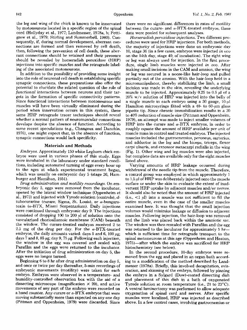
Horseradish peroxidase injections. Two different pro- cedures were used for HRP injections. For both methods, the majority of injections were done on embryonic day 10, stage 36 (in a few cases, embryos were injected in ouo on the 14th day, stage 40, of incubation). The right wing or leg was always used for injection. In the first proce- dure, single limb muscles were injected in ouo. After making small incisions in the CAM and amnion, the wing or leg was secured in a noose-like hair-loop and pulled partially out of the amnion. With the hair-loop held in a micromanipulator, thereby stabilizing the limb, a small incision was made in the skin, revealing the underlying muscle to be injected. Approximately 0.25 to 0.5 ~1 of a 20% w/v solution of HRP was injected into the belly of a single muscle in each embryo using a 30 gauge, lo+1 Hamilton microsyringe fitted with a 40- to 60-pm glass pipette tip. Since chronic immobilization results in a 30 to 40% reduction of muscle size (Pittman and Oppenheim, 1979), an attempt was made to inject smaller volumes of HRP into the curare and a-BTX embryos, in order to roughly equate the amount of HRP available per unit of muscle mass in control and treated embryos. The injected muscles included the gastrocnemius, peroneus, sartorius, and adductor in the leg and the biceps, triceps, flexor carpi ulnaris, and extensor metacarpi radialis in the wing (Fig. 1). Other wing and leg muscles were also injected, but complete data are available only for the eight muscles listed above.
Varying amounts of HRP leakage occurred during withdrawal of the needle tip from the muscle. Therefore, a control group was employed in which approximately 1 to 1.5 ~1 of HRP was deliberately injected onto the muscle surface or under the skin to evaluate the extent of inad- vertent HRP uptake by adjacent muscles and/or nerves. It should also be noted that the amount of HRP injected (i.e., <l ~1) into a muscle was not sufficient to fill the entire muscle, even in the case of the smaller muscles examined here. It was thought that the small injection volumes would minimize leakage or diffusion to adjacent muscles. Following injection, the hair-loop was removed and the limb was placed back within the amniotic sac. The window was then resealed with Parafilm and the egg was returned to the incubator for approximately 5 hr- which is sufficient time for retrograde transport to the spinal motoneurons at this age (Oppenheim and Heaton, 1975)-after which the embryo was sacrificed for HRP histochemistry (see below).
In the second procedure, lo-day embryos were re- moved from the egg and placed in an organ bath accord- ing to a modification of the method described by Land- messer (1978a). Briefly, this involved decapitation, evis- eration, and skinning of the embryo, followed by pinning the embryo in a Sylgard (Dow)-coated dissecting dish and placement of this dish in a bath of oxygenated Tyrode solution at room temperature (i.e., 21 to 23°C). A ventral laminectomy was performed to allow adequate oxygenation of spinal cord and nerves. After specific muscles were localized, HRP was injected as described above. In a few control cases, involving gastrocnemius or
The Journal of Neuroscience Cell Death of Motoneurons in the Chick Embryo Spinal Cord 143
Biceps E. melacarpi radialis
Triceps
Figure I. Schematic drawings of leg and wing muscles of the chick. A, Medial view of leg; B, lateral view of leg; C, ventral view of wing.
biceps injections, most of the adjacent muscles were removed prior to HRP injection in order to evaluate the extent to which HRP leakage might contribute to the results. Following the injection, the temperature of the bath was raised to 33 to 35°C and the embryos were left at this temperature for 4 to 5 hr. Just prior to removal from the bath, the peripheral nerves of control embryos were stimulated electrically (see Landmesser and Morris, 1975) to determine the viability of the preparation. If brisk muscle contractions occurred, the embryo was re- tained for HRP histochemistry or for celI counts (see below); if no response could be elicited, the embryo was discarded. Since the in ouo and organ bath procedures gave similar results.with regard to the location of moto- neuron pools (see “Results”), these data were pooled for subsequent analyses.
Horseradish peroxidase histochemistry and histolog- ical procedures. Embryos were prepared for HRP his- tochemistry by removing the entire lumbar or brachial spinal cord with spinal ganglia intact. In the case of the in ouo injections, the spinal cord was exposed at this time by removing the dorsal vertebra to allow for better pen- etration of the fixative. The spinal cords were fixed overnight in a phosphate-buffered solution of 2% glutar- aldehyde in 5% sucrose. Following fixation, the spinal cords were processed according to the en bloc technique
described previously (Oppenheim and Heaton, 1975), using 3,3’-diaminobenzidine tetrahydrochloride (DAB). The blocks were embedded in paraffin, sectioned at 10 to 12 pm, and lightly counterstained with a 3% cresyl violet solution.
For total cell counts of motoneurons in control and treated embryos, all motoneurons in every 10th section in the lateral motor column (LMC) of the lumbar (seg- ments 23 to 30) or brachial (segments 13 to 16) spinal cord were included, according to criteria described pre- viously (Hamburger, 1975; Chu-Wang and Oppenheim, 1978). Cell counts at 5 and 10 days were done at a magnification of x 400 and at x 312 for 14-day embryos. Cell counts were always restricted to the right side of the spinal cord. All cells were indicated on drawings of the spinal cord by the use of a Leitz drawing tube. Finally, because tissue processed for HRP histochemistry is not always adequate for carrying out reliable cell counts (e.g., even after counterstaining with cresyl violet, the nucleo- lus isn’t always clearly defined), the total cell counts (see Table I, “Results”) were obtained from HRP-injected embryos that were treated similarly to other HRP-in- jetted embryos except that instead of being processed for HRP, they were fixed in Carnoy’s solution and stained with thionine (Chu-Wang and Oppenheim, 1978).
Following HRP histochemistry, motoneurons in the LMC were found to contain heavy, medium, or light amounts of reaction product (see “Results”). For reasons described below, however, in most cases, only heavily and medium labeled cells were included in the data analysis. Spinal cord segments were defined by adjacent spinal ganglia such that lumbosacral segment 23 began midway between the end of the last thoracic and the beginning of the first lumbar ganglion and ended midway between the first and second lumbar ganglia. Each seg- ment was arbitrarily subdivided into thirds by dividing the total number of sections in each segment by three. For each embryo, the rostral-caudal extent of sections containing cells with HRP reaction product was indicated on the slides and the number of heavily, medium, and lightly labeled cells in every third section in this region was counted and drawn at a magnification of x 400 with the aid of a Leitz drawing attachment. The values pre- sented in the graphs (see “Results”) represent the aver- age number of heavily and medium labeled cells per 30- to 36-pm section in each third of the appropriate seg- ments and are based on pooled data from five control and five treated embryos.
For the location of labeled cells in the dorsal-ventral and medial-lateral axes, all heavily, medium, and lightly labeled cells in the LMC of every other section in the 10 central-most sections of each segment were indicated on drawings of the spinal cord. (Only segments in which labeled cells were found in all three arbitrary subdivisions were included in this analysis.) Since the position of motor pools in the transverse plane varies only slightly throughout a segment (e.g., Landmesser, 1978a), this was considered a valid sampling procedure. For each segment, these five drawings were then pooled by superimposing them onto a single drawing. For each HRP-injected muscle, three embryos were analyzed this way and the data were superimposed giving a single drawing for the
144 Oppenheim Vol. 1, No. 2, Feb. 1981
motoneurons supplying each muscle. A line then was drawn around the main dense cluster of cells resulting from this procedure such that approximately 5 to 15% of the cells were excluded from the main cluster due to their ectopic position. The excluded cells usually represented lightly labeled neurons.
Results
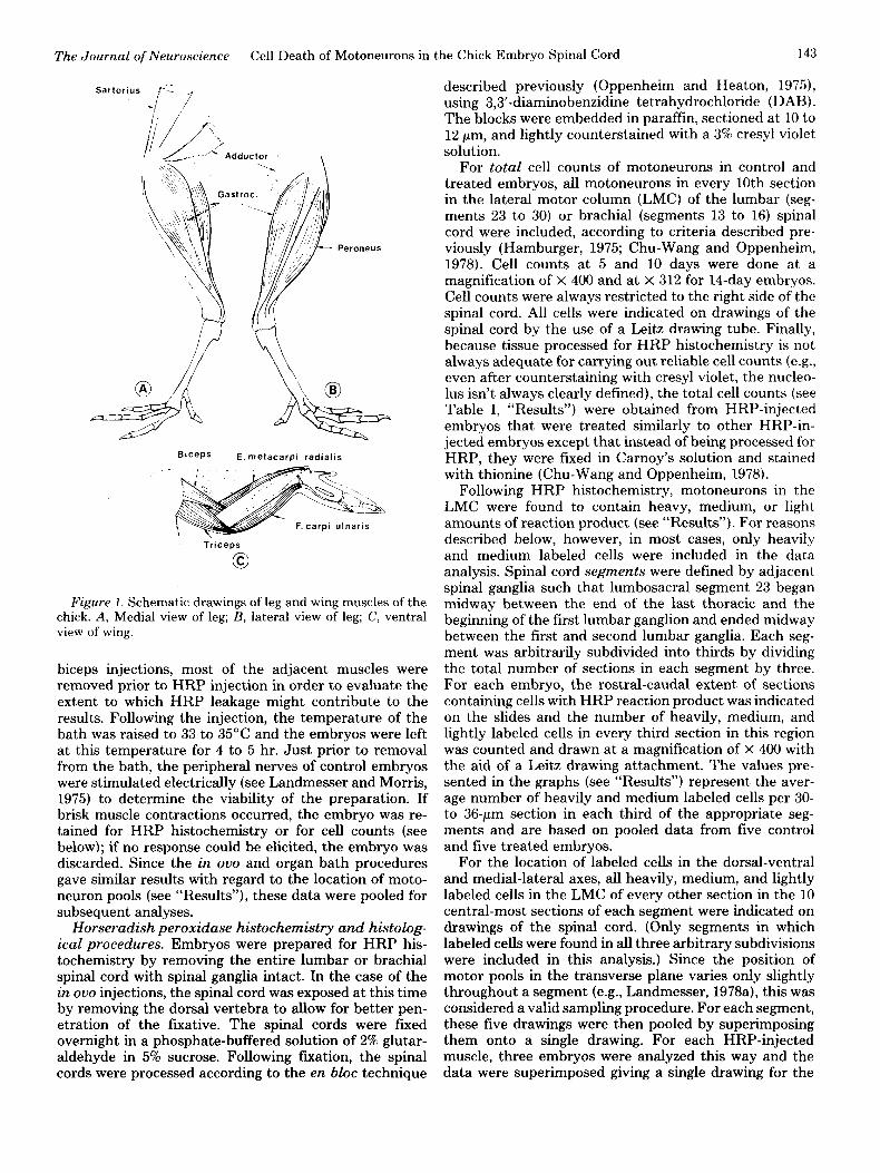
Motility and the prevention of cell death. As indicated in Table I, beginning about 6 to 8 hr following curare or (u-BTX treatment on day 5 (and with daily treatment thereafter), the motility of the experiment.al embryos was markedly reduced throughout the remainder of the treat- ment period and up to day 10. During this period, motility levels were chronically reduced by at least 85 to 90% compared to controls. Even this marked reduction prob- ably represents an underestimate. Motility recordings were typically made 22 to 24 hr after the previous day’s injection (or just prior to the daily injection) and t.hus probably reflect the maximum rate of motility for the preceding 24 hr. Based on the results from a previous experiment (Pittman and Oppenheim, 1979) and on the estimated rate of turnover of acetylcholine receptors (Burden, 1977)-and thus of n-BTX degradation-it was expected that, by 14 days of incubation (i.e., 5 days after the last injection of cu-BTX), the motility of the a-BTX group would recover and approach control levels. Al- though motility was still slightly lower than controls at this time, this was, in fact, the case (Table I).
Consistent with previous reports (Chu-Wang and Op- penheim, 1978; Pitt.man and Oppenheim, 1979), cell counts of motoneurons in the brachial and lumbar spinal cord were at a peak at 5 days, with no differences found between control and treated embryos at this time (5-day embryos were sacrificed 6 to 8 hr after the initial drug or control injection). Also in agreement with previous re- ports (Hamburger, 1975; Chu-Wang and Oppenheim, 1978; Oppenheim and Majors-Willard, 1978), there was
a 44% decrease in the number of lumbar motoneurons between days 5 and 10 in control embryos (from 23,150 to 12,845) and a 31% decrease in brachial motoneurons (from 16,270 to 11,085). In contrast, the curare- and ct- BTX-treated embryos had an average of 19,787 (lumbar) and 14,200 (brachial) neurons present on day 10. Thus, cell death was prevented to an extent equal to or greater than that previously reported (Pittman and Oppenheim. 1979). In the lumbar spinal cord, natural cell death was reduced from 44% to about 9% and in the brachial region, from 31% to approximately 10%. Following the cessation of drug treatment on day 9 and the subsequent gradual recovery of motility, by day 14, cell number was reduced in the curare- and cu-BTX-treated embryos. This was especially true in the case of the lumbar spinal cord (19,787 on day 10 versus 13,907 on day 14). There was a slight, but consistent drop in cell number in the control lumbar spinal cord between days 10 and 14, and a some- what greater drop in the control brachial spinal cord. This is consistent with a previous report in which it was found that natural cell death is more protracted in the brachial region (Oppenheim and Majors-Willard, 19’i8), extending beyond day 10. However, cell number on day 14 remained significantly elevated compared to controls (Table I).
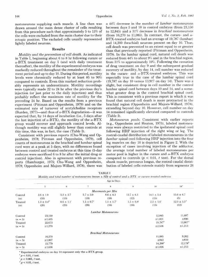
Motoneuron pools. Consistent with earlier reports (e.g., Oppenheim and Heaton, 1975), labeled motoneu- rons were always restricted to the ipsilateral spinal cord following HRP injection of the right wing or leg. The rostral-caudal distribution of labeled motoneurons in the lumbar spinal cord following HRP injection into the four leg muscles on day 10 is depicted in Figure 2. With the exception of cases involving injection of the adductor, the average total number of labeled motoneurons per motor pool is higher in the curare and (u-BTX embryos compared to controls (p < 0.01, t test). For the dorsal shank muscle, peroneus longus, the rostral-caudal distri- bution of labeled cells extends mainly from segments 26
TABLE I Motility and total number of motoneurons (mean + SD) of control and a-BTX- or curare-treated embryos
Age in Da.vs “--- ----. .--..-- ~~ ~~~
5 6 7 a 9 10
Movements per Min Control 3.6 Icr 1.0 5.2 + 2.7 5.7 + 3.0 8.6 + 4.3 13.7 f 6.1 14.1 + 5.4
(4 (10) (10) (10) (10) (1‘3 (15) Treated 1.3 + 0.4h 0.6 + 1.1“ 1.5 It 0.7’ 1.1 + 1.7’ 1.5 + 0.8” 2.2 -f. 1.f’
(n) cm cm (20) (16) (18) (15)
Lumbar Motoneurons Control 23,150 12,845 (n = 5) * 1,435 -+I,021 Treated 22,673 19,787” (n = 5) +1,270 f2,536
Brachial Motoneurons Control 16,270 11,085 (n = 5) +1,807 +843 Treated 15,779 14,200h (n = 5) i1,636 L1.026
’ Experimental embryos on day 14 represent only the a-BTX group. *p < 0.01, t test. ‘p < 0.005, t test. dp < 0.05, t test.
14”
15.6 zk 6.7 (5)
12.0 + 5.5” (12)
11.687 33390
13,907” k1.213
9,262 +810
12,178’ +1,213
The Journal of Neuroscience Cell Death of Motoneurons in the Chick Embryo Spinal Cord 145
to 28 for both control and experimental cases. The dis- tribution of labeled cells for the ventral shank muscle, gastrocnemius, extends from segments 26 to 30, with a similar distribution for control and experimental cases. Following HRP injection into the two thigh muscles, sartorius and adductor, labeled cells are localized in the rostra1 segments, with no apparent differences between the respective control and experimental groups. Sartorius neurons were restricted to the first two lumbar segments (23 and 24), whereas adductor neurons were mainly found in segments 24 and 25, with a small extension into seg- ments 23 and 26.
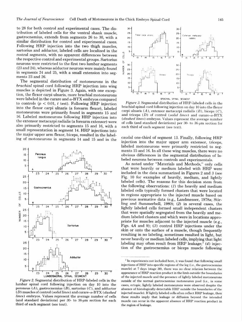
The segmental distribution of motoneurons in the brachial spinal cord following HRP injection into wing muscles is depicted in Figure 3. Again, with one excep- tion, the flexor carpi ulnaris, more brachial motoneurons were labeled in the curare and a-BTX embryos compared to controls (p < 0.01, t test). Following HRP injection into the flexor carpi ulnaris (a forearm flexor), labeled motoneurons were primarily found in segments 15 and 16. Labeled motoneurons following HRP injection into the extensor metacarpi radialis (a forearm extensor) were also primarily restricted to segments 15 and 16, with a small representation in segment 14. HRP injections into the major upper arm flexor, biceps, resulted in the label- ing of motoneurons in segments 14 and 15 and in the
--I-
-I ; : D I u-r ’ 23 ’ 24 ’ 25 ’ 26 ’ 27 ’ 28 ’ 29 ’ 30 ’
Sortorius
’ 23 ’ 24 ’ 25 ’ 26 ’ 27 ’ 28 ’ 29 ’ 30 ‘-
25 -
20 -
Adductor
5-
I I 1 i 23 24 25 I I 1 26 27 28 29 ’ 30 ’ LUMBOSACRAL SPINAL SEGMENT
Figure 2. Segmental distribution of HRP-labeled cells in the lumbar spinal cord following injection on day 10 into the peroneus (A), gastrocnemius (B), sartorius (C), and adductor (D) muscles of control (solid lines) and curare-a-BTX (dashed lines) embryos. Values represent the average number of cells (and standard deviations) per 30- to 36-pm section for each third of each segment (see text).
BRACHIAL WNAL SEGMENT
Figure 3. Segmental distribution of HRP-labeled cells in the brachial spinal cord following injection on day 10 into the flexor carpi ulnaris (A), extensor metacarpi radialis (B), biceps (C), and triceps (D) of control (solid lines) and curare-cu-BTX (dashed lines) embryos. Values represent the average number of cells (and standard deviations) per 30- to 36-pm section for each third of each segment (see text).
caudal one-third of segment 13. Finally, following HRP injection into the major upper arm extensor, triceps, labeled motoneurons were primarily restricted to seg- ments 15 and 16. In all these wing muscles, there were no obvious differences in the segmental distribution of la- beled neurons between controls and experimentals.
As noted under “Materials and Methods,” only cells that were heavily or medium labeled with HRP were included in the data summarized in Figures 2 and 3 (see Fig. 10 for examples of heavily, medium, and lightly labeled cells). The reasons for this decision stem from the following observations: (I) the heavily and medium labeled cells typically formed clusters that were located in regions appropriate to the injected muscle based on previous normative data (e.g., Landmesser, 1978a; Stir- ling and Summerbell, 1980); (2) in several cases, the lightly labeled cells formed small independent clusters that were spatially segregated from the heavily and me- dium labeled clusters and which were in locations appro- priate for muscles adjacent to the injected muscle (e.g., Figs. 4A and 9); (3) control HRP injections under the skin or onto the surface of a muscle, though frequently resulting in no labeling, sometimes resulted in light, but never heavily or medium labeled cells, implying that light labeling may often result from HRP leakage;2 (4) injec- tion of the gastrocnemius or biceps muscle following
’ In experiments not included here, it was found that following small injections of HRP into specific regions of the leg (i.e., the gastrocnemius muscle) at 7 days (stage 30), there was no clear relation between the appearance of HRP reaction product in the limb outside the boundaries of the injected muscle and the presence of lightly labeled motoneurons outside of the normal gastrocnemius motoneuron pool (Le., in some cases, ectopic, lightly labeled motoneurons were observed despite the absence of histologically detectable HRP outside the boundaries of the injected muscle). If lightly labeled cells often reflect HRP leakage, then these results imply that leakage or diffusion beyond the intended muscle can occur in the apparent absence of HRP reaction product in the region of leakage.
146 Oppenheim Vol. I, No. 2, Feb. 1981
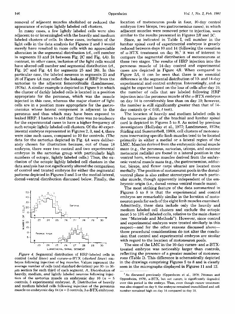
removal of adjacent muscles abolished or reduced the appearance of ectopic lightly labeled cell clusters.
In many cases, a few lightly labeled cells were also adjacent to or intermingled with the heavily and medium labeled clusters of cells. In these cases, inclusion of the light cells in the data analysis for Figures 2 and 3 would merely have resulted in more cells with no appreciable alteration in the segmental distribution (cf., cell number in segments 23 and 24 between Fig. 2C and Fig. 4A). In contrast, in other cases, inclusion of the light cells would have altered cell number and segmental distribution (cf., Fig. 2C and Fig. 4A for segments 25 and 26). In this particular case, the labeled neurons in segments 25 and 26 of Figure 4A may reflect the leakage of HRP from the sartorius to the adjacent femorotibialis (Landmesser, 1978a). A similar example is depicted in Figure 9 in which the cluster of darkly labeled cells is located in a position appropriate for the peroneus, which was the muscle injected in this case, whereas the major cluster of light cells are in a position more appropriate for the gastro- cnemius whose lateral component is adjacent to the peroneus and thus which may have been exposed to leaked HRP. I hasten to add that there was no tendency for the experimental cases to have a higher frequency of such ectopic lightly labeled cell clusters. Of the 40 exper- imental embryos represented in Figures 2,3, and 4, there were nine such cases, compared to 10 for controls. (The data for the sartorius depicted in Fig. 4A were deliber- ately chosen for illustration because, out of these 18 embryos, there were two control and two experimental embryos in the sartorius group with particularly high numbers of ectopic, lightly labeled cells.) Thus, the ex- clusion of the ectopic lightly labeled cell clusters in the data analysis has not significantly altered the comparison of control and treated embryos for either the segmental patterns depicted in Figures 2 and 3 or the medial-lateral, dorsal-ventral distribution discussed below. Finally, the
!:p-:‘L:~;, , , r
!;$y’~“s, 2!,‘: 2g , 3o r
23 24 25 26 27 28 29 30
LUMBOSACRAL SPINAL SEGMENT
Figure 4. Segmental distribution of HRP-labeled cells in control (solid lines) and curare-a-BTX (dashed lines) em- bryos following injection of leg muscles. Values represent the average number of cells (and standard deviation) per 30- to 36- pm section for each third of each segment. A, Distribution of heavily, medium, and lightly labeled neurons following injec- tion of the sartorius muscle on embryonic day 10 (n = 5 controls, 5 experimental embryos). B, Distribution of heavily and medium labeled cells following injection of the peroneus muscle on embryonic day 14 (n = 2 controls, 3 (Y-BTX embryos).
location of motoneuron pools in four, lo-day control embryos (two biceps, two gastrocnemius cases), in which adjacent muscles were removed prior to injection, were similar to the results presented in Figures 2B and 3C.
Since, as indicated in Table I, cell number in the lumbar spinal cord of experimental embryos is greatly reduced between days 10 and 14 (following the cessation of a-BTX treatment on day 9),” it was of interest to compare the segmental distribution of motoneurons at these two stages. The results of HRP injection into the peroneus muscle of 14-day control and experimental cases are depicted in Figure 4B. When compared to Figure 2A, it can be seen that there is no essential difference in the segmental distribution of lo- and 14-day experimental and control embryos. Interestingly, and as might be expected based on the loss of cells after day 10, the number of cells that are labeled following HRP injection into the peroneus muscle of the a-BTX embryos on day 14 is considerably less than on day 10; however, the number is still significantly greater than that of 14- day controls 0, < 0.05, t test).
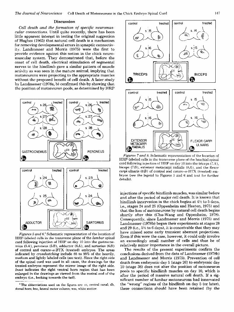
The location of heavily and medium labeled cells in the transverse plane of the brachial and lumbar spinal cord is depicted in Figures 5 to 8. As noted by previous investigators (Hollyday et al., 1977; Landmesser, 1978a; Stirling and Summerbell, 1980), cell clusters of motoneu- rons innervating specific limb muscles tend to be located primarily in either a medial or a lateral region of the LMC. Muscles derived from the embryonic dorsal muscle mass (e.g., the peroneus, sartorius, triceps, and extensor metacarpi radialis) are found in a lateral position in the ventral horn, whereas muscles derived from the embry- onic ventral muscle mass (e.g., the gastrocnemius, adduc- tor, biceps, and flexor carpi ulnaris) are located more medially. The position of motoneuron pools in the dorsal- ventral plane is also rather stereotyped for each partic- ular muscle, though apparently independent of the em- bryonic origin (i.e., dorsal versus ventral muscle mass).
The most striking feature of the data summarized in Figures 5 to 8 is that the experimental and control embryos are remarkably similar in the location of moto- neuron pools for each of the eight limb muscles examined. Admittedly, these data include only the heavily and medium labeled cell clusters and exclude the ectopic most 5 to 15% of labeled cells, relative to the main cluster (see “Materials and Methods”). However, since control and experimental embryos were treated similarly in this respect-and for the other reasons discussed above- these procedural considerations do not alter the conclu- sion that control and experimental embryos are similar with regard to the location of motoneuron pools.
The size of the LMC in the IO-day curare- and a-BTX- treated embryos was noticeably larger than controls, reflecting the presence of a greater number of motoneu- rons (Table I). This difference is schematically depicted in the drawings comprising Figures 5 to 8 and is clearly seen in the micrographs displayed in Figures 11 and 12.
“As discussed previously (Oppenheim et al., 1978; Pittman and Oppenheim, 1979), a-BTX, but not curare, is significantly degraded over this period in the embryo. Thus, even though curare treatment was also stopped on day 9, the embryos remained immobilized and cell number remained high on day 14 compared to day 10.
The Journal of Neuroscience Cell Death of Motoneurons in the Chick Embryo Spinal Cord 147
Discussion
Cell death and the formation of specific neuromus- cular connections. Until quite recently, there has been little apparent interest in testing the original suggestion of Hughes (1965) that natural cell death is a mechanism for removing developmental errors in synaptic connectiv- ity. Landmesser and Morris (1975) were the first to provide evidence against this notion in the chick neuro- muscular system. They demonstrated that, before the onset of cell death, electrical stimulation of segmental nerves to the hindlimb gave a similar pattern of muscle activity as was seen in the mature animal, implying that motoneurons were projecting to the appropriate muscles without the proposed benefit of ceil death. A later study by Landmesser (1978a, b) confirmed this by showing that the position of motoneuron pools, as determined by HRP
- (
ml dh
ted
;ASTROCNEMIUS 1 m ] GERONEUS
:ontrol treated control treated
Figures 5 and 6.’ Schematic representation of the location of HRP-labeled cells in the transverse plane of the lumbar spinal cord following injection of HRP on day 10 into the gastrocne- mius (5A), peroneus (5B), adductor (6A), and sartorius (6B) of control and curare-cu-BTX (treated) embryos. The areas indicated by crosshatching include 85 to 95% of the heavily, medium and lightly labeled cells (see text). Since the right side of the spinal cord was used in all cases, the drawings for the treated embryos represent the mirror image of the right side. Inset indicates the right ventral horn region that has been enlarged in the drawings as viewed from the rostra1 end of the embryo (i.e., looking towards the tail).
‘The abbreviations used on the figures are: cc, central canal; dh, dorsal horn; Imc, lateral motor column; wm, white matter.
control treated 1
TRICEPS
control treated
control treated
control treated
EXTENSOR FLEXOR CARPI METACARPI
@ ULNARIS
Figures 7 and 8. Schematic representation of the location of HRP-labeled cells in the transverse plane of the brachial spinal cord following injection of HRP on day 10 into the triceps (7A ), biceps (7B), extensor metacarpi radialis (8A). and the flexor carpi ulnaris (8B) of control and curare-cu-BTX (treated) em- bryos (see the legend to Figures 5 and 6 and text for further details).
injections of specific hindlimb muscles, was similar before and after the period of major ceil death. It is known that hindlimb innervation in the chick begins at 4% to 5 days, i.e., stages 24 and 25 (Oppenheim and Heaton, 1975) and that the loss of motoneurons by natural cell death begins shortly after this (Chu-Wang and Oppenheim, 1978). Consequently, since Landmesser and Morris (1975) and Landmesser (1978b) began their experiments at stages 28 and 29 (i.e., 5% to 6 days), it is conceivable that they may have missed some early transient aberrant projections. Even if this were the case, however, it could only involve an exceedingly small number of cells and thus be of relatively minor importance in the overall picture.
The results of the present experiments confirm the conclusions derived from the data of Landmesser (1978b) and Landmesser and Morris (1975). Prevention of cell death from embryonic day 5 (stage 26) to embryonic day 10 (stage 36) does not alter the position of motoneuron pools to specific hindlimb muscles on day 10, which is after the period of massive natural cell death. If a sig- nificant number of lumbar motoneurons had innervated the “wrong” regions of the hindlimb on day 5 (or later), these connections should have been retained (by the
148 Oppenheim Vol. 1, No. 2, Feb. 1981
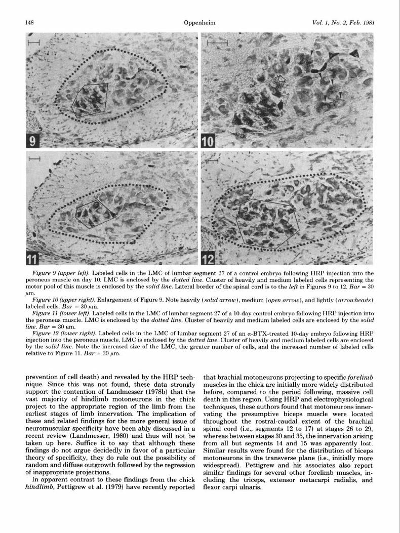
Figure 9 (upper left). Labeled cells in the LMC of lumbar segment 27 of a control embryo following HRP injection into the peroneus muscle on day 10. LMC is enclosed by the dotted line. Cluster of heavily and medium labeled cells representing the motor pool of this muscle is enclosed by the solid line. Lateral border of the spinal cord is to the left in Figures 9 to 12. Bar = 30 pm.
Figure 10 (upper right). Enlargement of Figure 9. Note heavily (solid arrow), medium (open arrow), and lightly (arrowheads) labeled cells. Bar = 30 pm.
Figure 11 (lower left). Labeled cells in the LMC of lumbar segment 27 of a lo-day control embryo following HRP injection into the peroneus muscle. LMC is enclosed by the dotted line. Cluster of heavily and medium labeled cells are enclosed by the solid line. Bar = 30 pm.
Figure 12 (lower right). Labeled cells in the LMC of lumbar segment 27 of an cY-BTX-treated lo-day embryo following HRP injection into the peroneus muscle. LMC is enclosed by the dotted line. Cluster of heavily and medium labeled cells are enclosed by the solid line. Note the increased size of the LMC, the greater number of cells, and the increased number of labeled cells relative to Figure 11. Bar = 30 pm.
prevention of cell death) and revealed by the HRP tech- nique. Since this was not found, these data strongly support the contention of Landmesser (1978b) that the vast majority of hindlimb motoneurons in the chick project to the appropriate region of the limb from the earliest stages of limb innervation. The implication of these and related findings for the more general issue of neuromuscular specificity have been ably discussed in a recent review (Landmesser, 1980) and thus will not be taken up here. Suffice it to say that although these findings do not argue decidedly in favor of a particular theory of specificity, they do rule out the possibility of random and diffuse outgrowth followed by the regression of inappropriate projections.
In apparent contrast to these findings from the chick hindlimb, Pettigrew et al. (1979) have recently reported
that brachial motoneurons projecting to specific forelimb muscles in the chick are initially more widely distributed before, compared to the period following, massive cell death in this region. Using HRP and electrophysiological techniques, these authors found that motoneurons inner- vating the presumptive biceps muscle were located throughout the rostral-caudal extent of the brachial spinal cord (i.e., segments 12 to 17) at stages 26 to 29, whereas between stages 30 and 35, the innervation arising from all but segments 14 and 15 was apparently lost. Similar results were found for the distribution of biceps motoneurons in the transverse plane (i.e., initially more widespread). Pettigrew and his associates also report similar findings for several other forelimb muscles, in- cluding the triceps, extensor metacarpi radialis, and flexor carpi ulnaris.
The Journal of Neuroscience Cell Death of Motoneurons in the Chick Embryo Spinal Cord 149
Pettigrew et al. (1979) do report that there was a tendency for more motoneurons to be labeled in the appropriate spinal cord regions at all stages, implying a certain degree of initial specificity. Nonetheless, the per- centage of labeled neurons that they found outside this region at the earliest stages (-50%) represents a signifi- cant proportion of the total number of labeled neurons seen following injection of the presumptive biceps muscle at stages 26 to 28, the time of initial wing innervation (Roncali, 1970; Bennett et al., 1979; R. W. Oppenheim, unpublished observations). Thus, it is exceedingly un- likely that such a substantial number of “ectopic” neu- rons would have been missed in the present experiments following the prevention of cell death from stage 26. Although it seems unlikely that the small number of neurons (i.e., 10%) which died in the present study, despite chronic treatment with neuromuscular blocking agents, represents neurons that had made inappropriate connections at stages 26 to 28, this possibility cannot be excluded. Another possible explanation for this apparent discrepancy is that, although chronic neuromuscular blockade prevents cell death, under these conditions, neurons with initially incorrect connections may be in- duced to retract their axons and continue growing until they find the appropriate muscle. Although this seems unlikely, it can’t be entirely ruled out.
A more likely explanation for the different results of Pettigrew et al. (1979) and the present report, however, is that the techniques (anatomical and physiological) that they used for identifying motor pools and segmental innervation of specific muscles at the earliest stages (i.e., stages 26 to 28) were not sufficiently precise. For instance, despite precautions against leakage or diffusion, it is possible that HRP injections made at stages before cleav- age of the primary muscle masses into individual wing muscles occurs (which begin at stage 30) could result in HRP diffusion to wider regions than intended. Small injection volumes and the reported absence of HRP reaction product in muscles outside of the intended re- gion in the wing may not be sufficient evidence against this possibility (e.g., see Footnote 2). Furthermore, since the axons of brachial neurons are still growing at the earliest stages of wing innervation and since growth cones in. uivo are known to take up HRP (Heaton, 1977; Chu- Wang and Oppenheim, 1980), some of the motoneurons which are labeled at these stages may not have reached their final target. If this is the case, then the distribution of motoneurons labeled by the techniques of Pettigrew et al. (1979) would be misleading with respect to their final projection patterns. By contrast, in the present study, which used stage 36, or lo-day, embryos, virtually all motoneurons have probably reached their mature target and specific muscles at this stage are easily iden- tified and large enough so that the possibility of signifi- cant diffusion or leakage between muscles, though by no means eliminated, is lessened considerably. Thus, there can be little question that the labeled clusters (see “Re- sults”) represent the motor pool of the injected muscles.
Although an equally plausible alternative argument cannot presently be used to explain the electrophysiolog- ical data of Pettigrew et al. (1979), it is worth noting that these experiments were conducted primarily at stages 29 to 35, when the HRP data indicate that the distribution
of motoneurons innervating the biceps muscle is already similar to the mature distribution. Although the reported electrophysiological data from stage 28 embryos are con- sistent with the HRP data (i.e., that there is a more diffuse distribution of motoneurons relative to later stages), it is conceivable that the techniques were not sufficiently precise to rule out the spread of electrical activity to adjacent nerves and muscles.
In the present study, motoneuron cell death has been prevented from the earliest stages of wing and leg inner- vation. For both the leg and wing, approximately 90% of the neurons present on day 5 (prior to the onset of natural cell death in these populations) were maintained up to day 10. Despite the virtual absence of cell death in these two populations, the rostral-caudal, medial-lateral, and dorsal-ventral locations of motaneuron pools on day 10, for the muscles examined, were similar to controls. Following the cessation of neuromuscular blockade on day 9, and the subsequent resumption of neuromuscular activity, a substantial number of neurons underwent a delayed cell death. Although cell number was consider- ably lower in treated embryos on day 14 compared to day 10 (especially in the lumbar region), the location of motoneuron pools in experimental and control embryos remained similar. Thus, consistent with previous reports (Landmesser, 1978b; Stirling and Summerbell, 1980), but against the findings of Pettigrew et al. (1979), naturally occurring cell death in the brachial and lumbar spinal cord of the chick does not appear to act primarily as a mechanism for removing inappropriate neuromuscular connections during development. Although a small per- centage of neurons in these as well as in other systems (Clarke and Cowan, 1976; Lamb, 1979) may die as a result of having innervated the wrong target, such a mechanism was probably of little significance in the evolution of naturally occurring neuron death. It seems more likely that the overproduction of neurons followed by regres- sion evolved primarily as a compensatory mechanism in the event of ontogenetic variability or genetic changes in the number of postsynaptic targets (e.g., see Katz and Lasek, 1978; Oppenheim, 1981).”
Functional interactions and the formation of specific neuromuscular connections. The suggestion that func- tion or use, i.e., electrical transmission and/or synaptic activity, may be important factors in the ontogenetic organization of the nervous system has a long history reaching at least as far back as the British and French associationists in the 18th century (e.g., see Oppenheim, 1979). In modern times, a rather large literature has accumulated showing that various aspects of neural de- velopment can be modified by varying either the amount, the kind of, or the time of exposure to, sensory input during the postnatal period (see Jacobson, 1978 for re- view). Therefore, although one may argue over whether
5 It is important to note that my conclusions with regard to cell death and neuronal specificity are only valid for mechanisms acting at the level of entire muscles (i.e., between motoneurons innervating different muscles). My experiments do not address the issue of whether there is a specific topographic relationship between motoneurons and particular regions uGthin a muscle and, if so, whether cell death is involved at this level. Since there is evidence for such “intramuscle” specificity in adult cat (Burke et al., 1977; Swett et al., 1970). it is conceivable that a similar phenomenon occurs in the chick.
a particular experiment illustrates functional adaptations (in the sense that the anitnal is rendered more or less biologically fit as a result of the neural modification). there can be no doubt that, in some cases. such phenom- ena occur in the postnatal period. In contrast, the pre- vailing view concerning the embr?sonic orprerzutnl period has been that. “Neurogenesis does not make use of itnpulse transmission and its electrical and other corre- lates as an instrument of differentiation” (Hamburger, 195”. p. 131 J.
This view. which has been expressed by many neu- roembryologists, was ne\.er meant to exclude the possi- bility that certain aspects of s>-napse formation or main- tenance lvere under some form of functional control (e.g.. see Sperry. 1951. 19%: Hamburger, 1957: \Veiss. 1939). It tnerel>. referred to the fact that neural function did not appear to be involved in the proliferation. migration, or initial differentiation of neurons, including the formation of specific neuronal pathways. Xithough there ha\-e been attempts to explain some or all of these neurogenetic processes by reference to neural function (e.g., Xriens- Kappers. 1936: Bok, 1915: Holt. 1931~, these attempts were vague and largely de\-oicl of experimental support and thus were never widely accepted by the majority of neuroembryologists.
Despite the lack of support concerning the role of function in early neurogenesis. it has long been recog- nized that synaptic activity has nel-er been rigorousl) excluded as playing a role in the terminal phases of neurogenesis; namelJ-. in the formation. stabilization, maintenance. or physiological efficacy of specific synaptic connections. Changeus and Danchin (1976), for instance, have recently proposed a model whereby synaptic trans- mission is involved in the stctbilization (i.e.. whether a synaptic contact is maintained or rejected) as xell as the specificit?, of synaptic connecti\-ity. They suggest that. through trial and error. only a felt- of the transient contacts made b>- the growth cone are selected and that the function of the growing processes play a role in this selection. As they have expressed it. “. the precocious activity of the cleveloping network would stabilize selec- tively particular synapses among the many equivalent cant acts which etnerge during growth CIR~ thereb>’ crente dic~ersity rind specificit?” (Changeus and Danchin. 1976, 1~. 706. italics added).” Although Changeux and Danchin
There appears to he some confunon Loncerning the qxwtic prw wsses actually included in the proposal of C‘hangcu\ and 11anchin , 19;61, The problem arlsex over the !a(~ that thehe authors ha\ e focll>ed on the elimmatlon of polyne~ronal innen ation. which occ11rh at an advanced stage of developmrnt. ax a paratilgm cabe in Ivhich functional activity at the ne~rom~~c~lar junction II thought to control the clim- natlon of redundant synapses. Although there is e\idence supporting . this argmnent (Pun-es and Lichtman, 19801. it is not clear that this has anything to do with neuronal specificity in the traditional sense. It is known that cell death F not in\,ol\ ed in thw phenomenon I Opprnheim and Majors-\Villard, 19781. However. (‘hanpens and 11anchln (197Cil propose that function is involved in the stabilization ihut not the initial formation) of synapses durmp the earlier phases of lllaturatlon--u-hen natural cell death is occurring-and they imply that function is alho mvolved in this process. \l’ith regard to this early period. they state that, “, in the case of the neuromuscular junction the stabilization of developing nerve terminals requires the functional interaction of the neurotransmitter with its postsynaptic receptor” ~Changeus and I1an- chin. 1976. p. 708).
are not explicit in their definition of specificit>-. tradi- tional conceptions of specificity. in the neuromusculat system (*Jacobson. 1978; Lanclmesser. 1980) have allva1.s been concerned with ho\\- motoneurons form c>onnectiok with one muscle and not \vith others as Lye11 as with bon selective central connections are formed between moto- neurons and specific afferents.
Changeux and Uanchin ( 1976) have f’ocusrcl on the developing neuromuscular ,qxeni for exemplifying thcit tnoclel. Thus. assuming that the,v we the term specif’icit> in its traditional sense. the present experiments, whic~h involi-e the virtual absence of functional ittterac-tions during the growth and formation of’ yxaptic connecti\-it> between motoneurons and limb ntuwle~. pro\-ide a twt of the correctness of their model. These esperimrnt~ indicate that specific and normal ~~ouro~~~~~~c~tlat. inner- vation patterns can form and perhaps even become sta- bilked despite the \.irtual absence of peripheral function. Furthermore. the observation that the number of’ Httf-‘- labeled motoneurons i.;: increased follon ing the pre\.ett- tion of cell death implies that many of’ the neurons that normally die can project axons to their apl,roI,riatc s:\-n- aptic targets (but see Footnote .5).
Since preliminary experiments (Il. if-. Oppetiheim and J. I,. Maderdrut, manuscript in preparation 1 indicate that behar~iornl immobilization may be a necessar?- bit1 not sufficient condition for the prevention of cell death. it remains to be seen Mhether synaptic function, trophic, interactions. or some combination of the ti\-o represents the critical factor in the prel-ention of cell death in thi5 q-stem. Furthermore, whereas the present results are reasonably clear in demonstrating that specific syiaptic connections can be formed in the periphery in thr al)- sence of neuromuscular function. they shed no light on whether the central connectix-it? of’ qinal moroneurons is normal or altered under these condition5 nor do thc,v rule out the role of other kinds of’ functional interactions (e.g.. between nerve fibers or nerve endingsr in these phenomena.
References
Burden. S. (19771 De\-elopment ot’ the neurotnwxular junction
Chronic treatment ot rmbr? o- from d;ly.k a to 111 xvlth concenira- tions of eserine. nicotine. ~~arhachol. or he\;ararhoc,holine sufficrent to produce behavioral immobilization nm~llnr to that cawed t)y aware OI n-BI’S does not prc\-cnt motonewon cell death. Indeed. MC hai e to~ntl that chronic eserine treatment tiwlng ttli:, lx,rioti may actually enhance cell death rewlting in signlticantly T’~U PT motoneuron., than normal on day 10. Since chronic eherine treatment after da! 10 II c.. after the normal cell death period) rehlllts in an e\en greater 105.5 of nlotOIieur~)~~.~. however, it is not prehentlx clear hou. or e\en if, thi.+ phenomenon 1> related to naturally occurrtnp cell death. It may onl> he a manife.-tatlon of the well known neuropathological effects of chronic treatment M ,t h anticholinesterase pgents (e.g.. Ixonard and Salpeter 193).
The Journal of Neuroscience Cell Death of Motoneurons in the Chick Embryo Spinal Cord 151
in the chick embryo: The number, distribution, and stability of acetylcholine receptors. Dev. Biol. 57: 317-329.
Burke, R. E., P. L. Strick, K. Kanda, C. C. Kim, and B. Walmsley (1977) Anatomy of medial gastrocnemius and so- leus motor nuclei in cat spinal cord. J. Neurophysiol. 40: 667- 680.
Changeux, J. P., and A. Danchin (1976) Selective stabilization of developing synapses as a mechanism for the specification of neuronal networks. Nature 264: 705-711.
Chu-Wang, I. W., and R. W. Oppenheim (1978) Cell death of motoneurons in the chick embryo spinal cord. I. A light and electron microscopic study of naturally occurring and induced cell loss during development. J. Comp. Neurol. 277: 33-58.
Chu-Wang, I. W., and R. W. Oppenhiem (1980) Uptake, intraax- onal transport and fate of horseradish peroxidase in embry- onic spinal neurons of the chick. J. Comp. Neurol. 193: 753- 776.
Clarke, P. G. H., and W. M. Cowan (1976) The development of the isthmo-optic tract in the chick, with special reference to the occurrence and correction of developmental errors in the location and connections of isthmo-optic neurons. J. Comp. Neurol. 167: 143-164.
Hamburger, V. (1952) Development of the nervous system. Ann. N. Y. Acad. Sci. 55: 117-132.
Hamburger, V. (1957) The concept of development in biology. In The Concept of Development, D. B. Harris, ed., pp. 49-58, University of Minnesota Press, Minneapolis.
Hamburger, V. (1975) Cell death in the development of the lateral motor column of the chick embryo. J. Comp. Neurol. 160: 535-546.
Hamburger, V., and H. L. Hamilton (1951) A series of normal stages in the development of the chick embryo. J. Morphol. 88: 49-92.
Heaton, M. B. (1977) Retrograde axonal transport in lateral motor neurons of the chick embryo prior to limb bud inner- vation. Dev. Biol. 58: 421-427.
Hollyday, M., V. Hamburger, and J. M. G. Farris (1977) Local- ization of motor neuron pools supplying identified muscles in normal and supernumerary legs of chick embryos. Proc. Natl. Acad. Sci. U. S. A. 74: 3582-3586.
Holt, E. B. (1931) Animal Drive and the Learning Process, Holt, New York.
Hughes, A. (1965) A quantitative study of the development of the nerves in the high-limb of Eleutherodactylus martinicen- sis. J. Embryol. Exp. Morphol. 13: 9-34.
Jacobson, M. (1978) Developmental Neurobiology, Plenum, New York.
Katz, M. J., and R. J. Lasek (1978) Evolution of the nervous system: Role of ontogenetic mechanisms in the evolution of matching populations. Proc. Natl. Acad. Sci. U. S. A. 75: 1349-1352.
Lamb, A. H. (1979) Evidence that some developing limb mo- toneurons die for reasons other than peripheral competition. Dev. Biol. 71: 8-21.
Lamb, A. H. (1980) Motoneurone counts in Xenopus frogs reared with one bilaterally-innervated hindlimb. Nature 284: 347-350.
Landmesser, L. (1978a) The distribution of motoneurones sup- plying chick hind limb muscles. J. Physiol. (Lond.) 284: 371- 389.
Landmesser, L. (1978b) The development of motor projection patterns in the chick hind limb. J. Physiol. (Lond.) 284: 391- 414.
Landmesser, L. (1980) The generation of neuromuscular speci- ficity. Annu. Rev. Neurosci. 3: 279-302.
Landmesser, L., and D. G. Morris (1975) The development of functional innervation in the hind limb of the chick embryo. J. Physiol. (Lond.) 249: 301-326.
Leonard, J. P., and M. M. Salpeter (1979) Agonist induced myopathy at the neuromuscular junction is mediated by calcium. J. Cell Biol. 82: 811-819.
Oppenheim, R. W. (1979) Laura Bridgemans brain: An early consideration of functional adaptations in neural develop- ment. Dev. Psychobiol. 12: 533-537.
Oppenheim, R. W. (1981) Neuronal cell death and some related regressive phenomena during neurogenesis: A selective his- torical review and progress report. In Studies in Develop mental Neurobiology: Essays in Honor of Viktor Ham- burger, W. M. Cowan, ed., Oxford University Press, in press.
Oppenheim, R. W., and M. B. Heaton (1975) The retrograde transport of horseradish peroxidase from the developing limb of the chick embryo. Brain Res. 98: 291-302.
Oppenheim, R. W., and C. Majors-Willard (1978) Neuronal cell death in the brachial spinal cord of the chick is unrelated to the loss of polyneuronal innervation in wing muscle. Brain Res. 154: 148-152.
Oppenheim, R. W., H. L. Levin, and M. S. Harth (1973) An investigation of various egg-opening techniques for use in avian behavioral embryology. Dev. Psychobiol. 6: 53-68.
Oppenheim, R. W., R. Pittman, M. Gray, and J. I,. Maderdrut (1978) Embryonic behavior, hatching and neuromuscular de- velopment in the chick following a transient reduction of spontaneous motility and sensory input by neuromuscular blocking agents. J. Comp. Neurol. 179: 619-640.
Pettigrew, A. G., R. Lindeman, and M. R. Bennett (1979) Development of the segmental innervation of the chick forelimb. J. Embryol. Exp. Morphol. 49: 115-137.
Pittman, R., and R. W. Oppenheim (1978) Neuromuscular blockade increases motoneurone survival during normal cell death in the chick embryo. Nature 271: 364-366.
Pittman, R., and R. W. Oppenheim (1979) Cell death of moto- neurons in the chick embryo spinal cord. IV. Evidence that a functional neuromuscular interaction is involved in the regulation of naturally occurring cell death and the stabili- zation of synapses. J. Comp. Neurol. 187: 425-446.
Purves, D., and J. W. Lichtman (1980) Elimination of synapses in the development of the nervous system. Science 210: 153- 157.
Roncali, L. (1970) The brachial plexus and the wing nerve pattern during early developmental phases in chick embryo. Monit. Zool. Ital. 4: 81-98.
Sperry, R. W. (1951) Mechanisms of neural maturation. In Handbook of Experimental Psychology, S. S. Stevens, ed., pp. 236-280, Wiley, New York.
Sperry, R. W. (1965) Embryogenesis of behavioral nerve nets. In Organogenesis, R. L. DeHaan and H. Ursprung, eds., pp. 236-280, Holt, New York.
Stirling, R. V., and D. Summerbell (1980) Evidence for non- selective motor innervation of rotated chick limbs. J. Physiol. (Land.) 300: 7-SP.
Swett, J., E. Eldred, and J. S. Buchwald (1970) Somatotopic cord-to-muscle relations in efferent innervation of cat gastro- cnemius. Am. J. Physiol. 219: 762-766.
Weiss, P. A. (1939) Principles of Development, a Text on Experimental Embryology, Hafner, New York (1969 reprint).
Copyright © 2022 FDOKUMEN