Refractive error study in children: results from La Florida, Chile
Upload
independentCategory
view
5download
0
www.elsevier.com/locate/visres
Vision Research 47 (2007) 2465–2472
Emmetropization and optical aberrations in a myopic cornealrefractive surgery chick model
E. Garcıa de la Cera a, G. Rodrıguez b, A. de Castro a, J. Merayo b, S. Marcos a,*
a Instituto de Optica, Consejo Superior de Investigaciones Cientıficas, Serrano 121, 28006 Madrid, Spainb Instituto Universitario de Oftalmobiologıa Aplicada (IOBA), Universidad de Valladolid, Valladolid, Spain
Received 29 March 2007; received in revised form 13 June 2007
Abstract
We studied the potential of myopic corneal refractive laser surgery to induce myopia (axial elongation) and potential interactionsbetween aberrations (generally resulting from the procedure) and myopia development in chicks (Gallus domesticus). Ten white Leghornchicks were monolaterally treated one day post-hatching with photorefractive keratectomy (PRK), with a nominal dioptric change of�9.9 D (imposed hyperopia). Axial length was measured using an adapted ultrasonic biometer; corneal radius of curvature was measuredusing a custom-built videokeratometer and spherical error and high order aberrations were measured using custom-built Hartmann–Shack aberrometer post-operatively on days 9, 12, 14 and 16 after hatching. Two-weeks after surgery, there were no significant differ-ences in corneal radius of curvature between treated and control eyes. Astigmatism increased on average by a factor of 2.6 and 3rd andhigher order aberrations by a factor of 4.3 after PRK. Both treated and control eyes were close to emmetropia, and no axial elongationwas found in the treated eyes. The inability of the refractive procedure to achieve significant reductions in the corneal power could beattributed to the biomechanical properties of the chick eye. High order aberrations induced significant contrast decrease (by a factor of1.7 at 4.5 c/deg). However, reduced image quality neither produced myopic refractive error nor axial elongation in the treated eyes. Bothnormal and treated eyes emmetropized, indicating that increased amounts of aberrations do not appear to be a risk factor for myopia.� 2007 Elsevier Ltd. All rights reserved.
Keywords: Emmetropization; Experimental myopia; Refractive surgery; Optical aberrations; Refractive error
1. Introduction
The quality of visual experience in early stages ofpost-natal development is critical for proper eye growthand normal emmetropization. Abnormal visual experi-ence has been associated to the development of myopia(Smith, 1998; Wallman, 1993; Wildsoet, 1997). Animalmodels in which retinal image has been degraded exhibitabnormal eye growth. It is well established that formdeprivation imposed by diffusers (Hayes, Fitzke, Hodos,& Holden, 1986; Troilo & Wallman, 1991; Wallman &Adams, 1987) and defocus imposed by negative lenses(Diether & Schaeffel, 1997; Schaeffel, Glasser, & How-land, 1988; Schaeffel & Howland, 1991) cause excessive
0042-6989/$ - see front matter � 2007 Elsevier Ltd. All rights reserved.doi:10.1016/j.visres.2007.06.005
* Corresponding author. Fax: +34 915645557.E-mail address: [email protected] (S. Marcos).
elongation in chick eyes and therefore myopia. Treat-ments with lenses in young chicks produce a change ofthe refractive state of the treated eye, which adjusts itsgrowth to compensate for the imposed defocus (Kee,Marzani, & Wallman, 2001; Schaeffel et al., 1988). Forexample, Schaeffel et al. (1988) achieved �2.2 D after15 days of treatment with �4 D lenses placed at day 9post hatching, while control eyes with a normal visualenvironment remained slightly hyperopic in the same per-iod of time. In humans, several pathologies that producean absence of normal visual experience, such as congen-ital cataracts, eyelid closure or corneal opacities havebeen associated to myopia (Gee & Tabbara, 1988; Hoyt,Stone, Fromer, & Billson, 1981; Robb, 1977).
An increasingly popular technique to correct refractiveerrors in humans is corneal refractive surgery. Cornealpower is changed using excimer laser, reshaping the
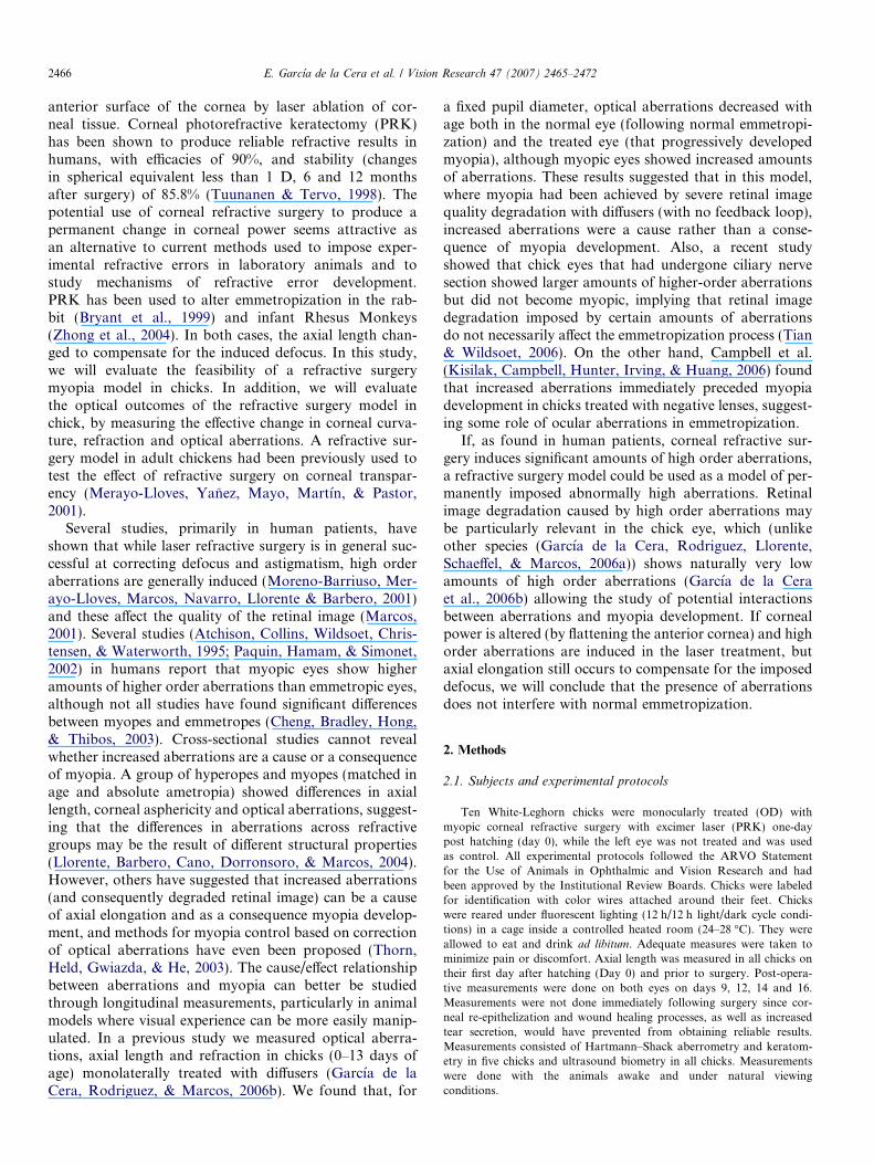
2466 E. Garcıa de la Cera et al. / Vision Research 47 (2007) 2465–2472
anterior surface of the cornea by laser ablation of cor-neal tissue. Corneal photorefractive keratectomy (PRK)has been shown to produce reliable refractive results inhumans, with efficacies of 90%, and stability (changesin spherical equivalent less than 1 D, 6 and 12 monthsafter surgery) of 85.8% (Tuunanen & Tervo, 1998). Thepotential use of corneal refractive surgery to produce apermanent change in corneal power seems attractive asan alternative to current methods used to impose exper-imental refractive errors in laboratory animals and tostudy mechanisms of refractive error development.PRK has been used to alter emmetropization in the rab-bit (Bryant et al., 1999) and infant Rhesus Monkeys(Zhong et al., 2004). In both cases, the axial length chan-ged to compensate for the induced defocus. In this study,we will evaluate the feasibility of a refractive surgerymyopia model in chicks. In addition, we will evaluatethe optical outcomes of the refractive surgery model inchick, by measuring the effective change in corneal curva-ture, refraction and optical aberrations. A refractive sur-gery model in adult chickens had been previously used totest the effect of refractive surgery on corneal transpar-ency (Merayo-Lloves, Yanez, Mayo, Martın, & Pastor,2001).
Several studies, primarily in human patients, haveshown that while laser refractive surgery is in general suc-cessful at correcting defocus and astigmatism, high orderaberrations are generally induced (Moreno-Barriuso, Mer-ayo-Lloves, Marcos, Navarro, Llorente & Barbero, 2001)and these affect the quality of the retinal image (Marcos,2001). Several studies (Atchison, Collins, Wildsoet, Chris-tensen, & Waterworth, 1995; Paquin, Hamam, & Simonet,2002) in humans report that myopic eyes show higheramounts of higher order aberrations than emmetropic eyes,although not all studies have found significant differencesbetween myopes and emmetropes (Cheng, Bradley, Hong,& Thibos, 2003). Cross-sectional studies cannot revealwhether increased aberrations are a cause or a consequenceof myopia. A group of hyperopes and myopes (matched inage and absolute ametropia) showed differences in axiallength, corneal asphericity and optical aberrations, suggest-ing that the differences in aberrations across refractivegroups may be the result of different structural properties(Llorente, Barbero, Cano, Dorronsoro, & Marcos, 2004).However, others have suggested that increased aberrations(and consequently degraded retinal image) can be a causeof axial elongation and as a consequence myopia develop-ment, and methods for myopia control based on correctionof optical aberrations have even been proposed (Thorn,Held, Gwiazda, & He, 2003). The cause/effect relationshipbetween aberrations and myopia can better be studiedthrough longitudinal measurements, particularly in animalmodels where visual experience can be more easily manip-ulated. In a previous study we measured optical aberra-tions, axial length and refraction in chicks (0–13 days ofage) monolaterally treated with diffusers (Garcıa de laCera, Rodriguez, & Marcos, 2006b). We found that, for
a fixed pupil diameter, optical aberrations decreased withage both in the normal eye (following normal emmetropi-zation) and the treated eye (that progressively developedmyopia), although myopic eyes showed increased amountsof aberrations. These results suggested that in this model,where myopia had been achieved by severe retinal imagequality degradation with diffusers (with no feedback loop),increased aberrations were a cause rather than a conse-quence of myopia development. Also, a recent studyshowed that chick eyes that had undergone ciliary nervesection showed larger amounts of higher-order aberrationsbut did not become myopic, implying that retinal imagedegradation imposed by certain amounts of aberrationsdo not necessarily affect the emmetropization process (Tian& Wildsoet, 2006). On the other hand, Campbell et al.(Kisilak, Campbell, Hunter, Irving, & Huang, 2006) foundthat increased aberrations immediately preceded myopiadevelopment in chicks treated with negative lenses, suggest-ing some role of ocular aberrations in emmetropization.
If, as found in human patients, corneal refractive sur-gery induces significant amounts of high order aberrations,a refractive surgery model could be used as a model of per-manently imposed abnormally high aberrations. Retinalimage degradation caused by high order aberrations maybe particularly relevant in the chick eye, which (unlikeother species (Garcıa de la Cera, Rodriguez, Llorente,Schaeffel, & Marcos, 2006a)) shows naturally very lowamounts of high order aberrations (Garcıa de la Ceraet al., 2006b) allowing the study of potential interactionsbetween aberrations and myopia development. If cornealpower is altered (by flattening the anterior cornea) and highorder aberrations are induced in the laser treatment, butaxial elongation still occurs to compensate for the imposeddefocus, we will conclude that the presence of aberrationsdoes not interfere with normal emmetropization.
2. Methods
2.1. Subjects and experimental protocols
Ten White-Leghorn chicks were monocularly treated (OD) withmyopic corneal refractive surgery with excimer laser (PRK) one-daypost hatching (day 0), while the left eye was not treated and was usedas control. All experimental protocols followed the ARVO Statementfor the Use of Animals in Ophthalmic and Vision Research and hadbeen approved by the Institutional Review Boards. Chicks were labeledfor identification with color wires attached around their feet. Chickswere reared under fluorescent lighting (12 h/12 h light/dark cycle condi-tions) in a cage inside a controlled heated room (24–28 �C). They wereallowed to eat and drink ad libitum. Adequate measures were taken tominimize pain or discomfort. Axial length was measured in all chicks ontheir first day after hatching (Day 0) and prior to surgery. Post-opera-tive measurements were done on both eyes on days 9, 12, 14 and 16.Measurements were not done immediately following surgery since cor-neal re-epithelization and wound healing processes, as well as increasedtear secretion, would have prevented from obtaining reliable results.Measurements consisted of Hartmann–Shack aberrometry and keratom-etry in five chicks and ultrasound biometry in all chicks. Measurementswere done with the animals awake and under natural viewingconditions.
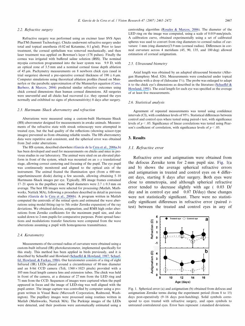
ig. 1. Spherical error (a) and astigmatism (b) obtained from defocus andstigmatism Zernike terms during the experiment period (from 8 to 15)ays post-operatively (9–16 days post-hatching). Solid symbols corre-pond to eyes treated with refractive surgery, and open symbols tontreated contralateral eyes. Error bars represent ±standard deviations.
E. Garcıa de la Cera et al. / Vision Research 47 (2007) 2465–2472 2467
2.2. Refractive surgery
Refractive surgery was performed using an excimer laser SVS ApexPlusTM (Summit Technology). Chicks underwent refractive surgery undertotal and topical anesthesia (0.02 ml Ketamine, 0.1 g/ml). Prior to lasertreatment, the corneal epithelium was removed mechanically, and thenlaser treatment was applied on Bowman’s layer (178 pulses). Finally thecornea was irrigated with buffered saline solution (BSS). The nominalmyopia correction programmed into the laser system was �9.9 D, withan optical zone of 3.5 mm and a nominal corneal tissue depth ablationof 45 lm. Pachymetry measurements on 8 newborn chick eyes (used intrial surgeries) showed a pre-operative corneal thickness of 190 ± 6 lm.Computer simulations using theoretical ablation profiles (based on Mun-nerlyn or the parabolic approximation of the Munnerlyn equation (Cano,Barbero, & Marcos, 2004) predicted similar refractive outcomes usingchick corneal dimensions than human corneal dimensions. All surgerieswere uneventful and all chicks had recovered (i.e. they opened the eyesnormally and exhibited no signs of photosensitivity) 8 days after surgery.
2.3. Hartmann–Shack aberrometry and refraction
Aberrations were measured using a custom-built Hartmann–Shack(HS) aberrometer designed for measurements in awake animals. Measure-ments of the refractive state with streak retinoscopy were attempted intreated eyes, but the bad quality of the reflections (showing scissor-typeimages) prevented us from obtaining reliable results. The HS aberrometrydata were repetitive and consistent, and the spherical error was obtainedfrom 2nd order aberrations.
The HS system, described elsewhere (Garcıa de la Cera et al., 2006a,b)has been developed and used for measurements on chicks and mice in pre-vious studies in our laboratory. The animal were sited on an elevated plat-form in front of the system, which was mounted on an x–y translationalstage, allowing correct centering and focusing of the pupil. The eye pupilwas continuously monitored and aligned to the optical axis of theinstrument. The animal fixated the illumination spot (from a 680 nm-superluminescent diode) during a few seconds, allowing obtaining 5–10Hartmann–Shack images per eye. Typically, HS image frames contained17–21 spots in the pupillary zone. Pupil diameters were 2.7 ± 0.3 mm onaverage. The best HS images were selected for processing (Matlab, Math-works, Nattick MA), following a procedure described in detail in previousstudies (Garcıa de la Cera et al., 2006b). A program written in Matlabcomputed the centroids of the retinal spots and estimated the wave aber-rations using modal fitting (up to 5th order Zernike expansion) of the raydeviations. We obtained defocus, astigmatism, and RMS high order aber-rations from Zernike coefficients for the maximum pupil size, and alsoscaled down to 2-mm pupils for comparative purposes. Point spread func-tions and modulations transfer functions were computed from the waveaberrations assuming a pupil with homogeneous transmittance.
2.4. Keratometry
Measurements of the corneal radius of curvature were obtained using acustom-built infrared (IR) photokeratometer, implemented specifically forthis study. This method has been applied in birds previously, and it isdescribed by Schaeffel and Howland (Schaeffel & Howland, 1987; Schaef-fel, Howland, & Farkas, 1986). Our keratometer consists of a ring of eightInfrared (IR) LEDs placed around a circumference of 80 mm diameterand an 8-bit CCD camera (Teli, 1360 · 1023 pixels) provided with a105 mm focal length camera lens and extension tubes. The chick was heldin front of the camera, at a distance of 27 mm from the LED ring and71 mm from the CCD. Sequences of images were captured when the pupilappeared in focus and the image of LED-ring was well aligned with thepupil center. The image capture was controlled by computer using a pro-gram written in Visual Basic (Microsoft Corporation, Redmond, Wash-ington). The pupillary images were processed using routines written inMatlab (Mathworks, Nattick MA). The Purkinje images of the LEDswere detected, and their positions were automatically estimated using a
centroiding algorithm (Rosales & Marcos, 2006). The diameter of theLED ring on the image was computed, using a scale of 0.019 mm/pixels.A calibration curve, obtained experimentally using a set of calibratedspheres was used to convert from ring diameters to corneal radius of cur-vature: 1 mm (ring diameter)/3.9 mm (corneal radius). Differences in cor-neal curvature across 4 meridians (45, 90, 135, and 180 deg) allowedestimation of corneal astigmatism.
2.5. Ultrasound biometry
Axial length was obtained by an adapted ultrasound biometer (Aller-gan Humphrey Mod. 826). Measurements were conducted under topicalanesthesia with a drop of (lidocaine 1%). The probe was enlarged to adaptit to the chick eye’s dimensions as described in the literature (Schaeffel &Howland, 1991). The axial length for each eye was specified as the averageof at least five measurements.
2.6. Statistical analysis
Agreement of repeated measurements was tested using confidenceintervals (CI), with confidence levels of 95%. Statistical differences betweencontrol and control eyes where tested using paired-t test, with significancelevels of p < .05. Significance of linear correlations was tested using Pear-son’s coefficient of correlation, with significance levels of p < .05.
3. Results
3.1. Refractive error
Refractive error and astigmatism were obtained fromthe defocus Zernike term for 2 mm pupil size. Fig. 1(aand b) shows the average spherical refractive errorsand astigmatism in treated and control eyes on 4 differ-ent days, starting 8 days after surgery. Both eyes wereclose to emmetropia, and although spherical refractiveerror tended to decrease slightly with age (�0.03 D/day and in control eye and �0.07 D/day) these changeswere not statistically significant. There were no statisti-cally significant differences in refractive error (paired t-test) between the treated and control eyes in any of
Fadsu

2468 E. Garcıa de la Cera et al. / Vision Research 47 (2007) 2465–2472
the days. Individually, we only found significant differ-ences in chick #1, day 14 (p = .0014), chick #3, day16 (p = .0176) and chick #4, day 12 (p = .0243). Thechanges and amounts in refractive state were consistentwith previous data in the literature, and surprisingly,these were not modified by refractive surgery. Measure-ments tended to be slightly noisier in treated eyes thanin the control eyes, with average standard deviations forrepeated measurements of 0.99 D and 0.57 D, respec-tively. The 95% confidence interval (CI) for repeatedmeasurements was ±0.97 D and ±0.57 D, respectively.Inter-subject variability was also larger in treated eyesthan in the control eyes (0.98 D in treated eye and0.66 D in control eyes), and 95% CI were ±1.26 and±0.62, respectively. Astigmatism was almost constantthroughout the measurement period. Treated eyesshowed higher values of astigmatism (2.6 ± 0.5 D) on
Fig. 2. Examples of wave aberration maps from all chicks (treated and contrcorresponding PSF for 3rd and higher order aberrations. Data are for 2-mm
average than the control eyes (1.0 ± 0.2 D), and thesedifferences were statistically significant in all days(p = .0013).
3.2. Optical aberrations
Fig. 2 shows examples of wave aberrations for 3rd andhigher order in the treated and control eye of the samechick, on day 16 and their corresponding PSFs for 2-mmpupils. The higher number of contour lines in the waveaberration map and larger PSF in the treated eye wereindicative of larger optical degradation. Fig. 3 shows aver-age 3rd and higher order (a), 3rd order aberrations (b) andspherical aberrations (Z40 term) (c) in treated and controleyes on 4 different days, starting 9 days after hatching (8after surgery). Third and higher order root-mean-squarewave front error (RMS) was higher in the surgical eyes
ol eyes) for 3rd and higher order Zernike coefficients at day 16 and theirpupil diameters. Map contour lines are plotted in 0.01 lm steps.
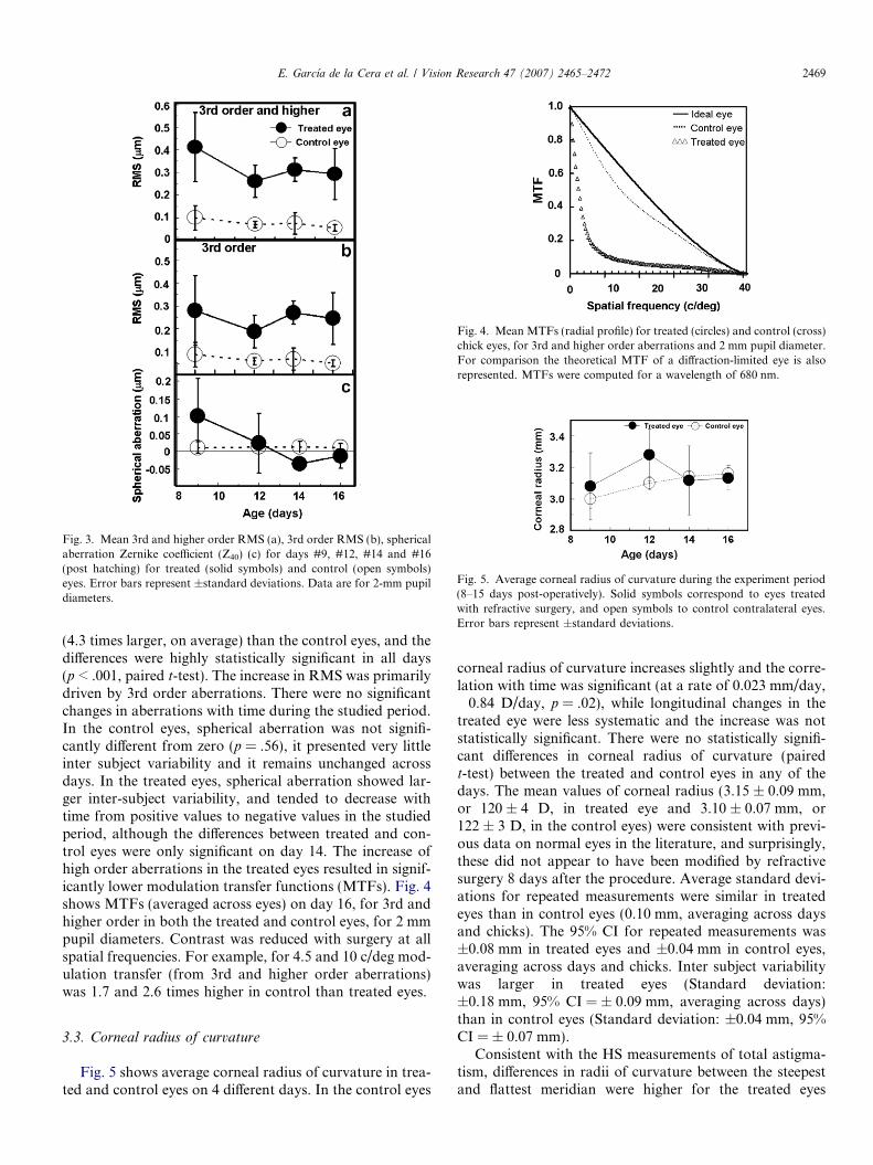
Fig. 3. Mean 3rd and higher order RMS (a), 3rd order RMS (b), sphericalaberration Zernike coefficient (Z40) (c) for days #9, #12, #14 and #16(post hatching) for treated (solid symbols) and control (open symbols)eyes. Error bars represent ±standard deviations. Data are for 2-mm pupildiameters.
Fig. 4. Mean MTFs (radial profile) for treated (circles) and control (cross)chick eyes, for 3rd and higher order aberrations and 2 mm pupil diameter.For comparison the theoretical MTF of a diffraction-limited eye is alsorepresented. MTFs were computed for a wavelength of 680 nm.
Fig. 5. Average corneal radius of curvature during the experiment period(8–15 days post-operatively). Solid symbols correspond to eyes treatedwith refractive surgery, and open symbols to control contralateral eyes.Error bars represent ±standard deviations.
E. Garcıa de la Cera et al. / Vision Research 47 (2007) 2465–2472 2469
(4.3 times larger, on average) than the control eyes, and thedifferences were highly statistically significant in all days(p < .001, paired t-test). The increase in RMS was primarilydriven by 3rd order aberrations. There were no significantchanges in aberrations with time during the studied period.In the control eyes, spherical aberration was not signifi-cantly different from zero (p = .56), it presented very littleinter subject variability and it remains unchanged acrossdays. In the treated eyes, spherical aberration showed lar-ger inter-subject variability, and tended to decrease withtime from positive values to negative values in the studiedperiod, although the differences between treated and con-trol eyes were only significant on day 14. The increase ofhigh order aberrations in the treated eyes resulted in signif-icantly lower modulation transfer functions (MTFs). Fig. 4shows MTFs (averaged across eyes) on day 16, for 3rd andhigher order in both the treated and control eyes, for 2 mmpupil diameters. Contrast was reduced with surgery at allspatial frequencies. For example, for 4.5 and 10 c/deg mod-ulation transfer (from 3rd and higher order aberrations)was 1.7 and 2.6 times higher in control than treated eyes.
3.3. Corneal radius of curvature
Fig. 5 shows average corneal radius of curvature in trea-ted and control eyes on 4 different days. In the control eyes
corneal radius of curvature increases slightly and the corre-lation with time was significant (at a rate of 0.023 mm/day,�0.84 D/day, p = .02), while longitudinal changes in thetreated eye were less systematic and the increase was notstatistically significant. There were no statistically signifi-cant differences in corneal radius of curvature (pairedt-test) between the treated and control eyes in any of thedays. The mean values of corneal radius (3.15 ± 0.09 mm,or 120 ± 4 D, in treated eye and 3.10 ± 0.07 mm, or122 ± 3 D, in the control eyes) were consistent with previ-ous data on normal eyes in the literature, and surprisingly,these did not appear to have been modified by refractivesurgery 8 days after the procedure. Average standard devi-ations for repeated measurements were similar in treatedeyes than in control eyes (0.10 mm, averaging across daysand chicks). The 95% CI for repeated measurements was±0.08 mm in treated eyes and ±0.04 mm in control eyes,averaging across days and chicks. Inter subject variabilitywas larger in treated eyes (Standard deviation:±0.18 mm, 95% CI = ± 0.09 mm, averaging across days)than in control eyes (Standard deviation: ±0.04 mm, 95%CI = ± 0.07 mm).
Consistent with the HS measurements of total astigma-tism, differences in radii of curvature between the steepestand flattest meridian were higher for the treated eyes
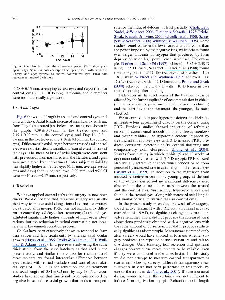
Fig. 6. Axial length during the experiment period (8–15 days post-operatively). Solid symbols correspond to eyes treated with refractivesurgery, and open symbols to control contralateral eyes. Error barsrepresent ±standard deviations.
2470 E. Garcıa de la Cera et al. / Vision Research 47 (2007) 2465–2472
(0.28 ± 0.13 mm, averaging across eyes and days) than forcontrol eyes (0.08 ± 0.06 mm), although the differenceswere not statistically significant.
3.4. Axial length
Fig. 6 shows axial length in treated and control eyes on 4different days. Axial length increased significantly with agefrom Day 0 (measured just before treatment, not shown inthe graph, 7.39 ± 0.09 mm in the treated eyes and7.35 ± 0.03 mm in the control eyes) and Day 16 (7.8 ±0.6 mm in the treated eyes and 8.16 ± 0.16 mm in the controleyes). Differences in axial length between treated and controleyes were not statistically significant (paired t-test) in any ofthe days. The mean values of axial length were consistentwith previous data on normal eyes in the literature, and againwere not altered by the treatment. Inter subject variabilitywas slightly higher in treated eyes (0.11 mm, average acrosseyes and days) than in control eyes (0.08 mm) and 95% CIwere ±0.14 and ±0.17 mm, respectively.
4. Discussion
We have applied corneal refractive surgery to new bornchicks. We did not find that refractive surgery was an effi-cient way to induce axial elongation: (1) corneal curvatureeyes treated with myopic PRK was not significantly differ-ent to control eyes 8 days after treatment; (2) treated eyesexhibited significantly higher amounts of high order aber-rations, but the reduction in retinal contrast did not inter-fere with the emmetropization process.
Chicks have been extensively shown to respond to formdeprivation and lens treatments by altering axial oculargrowth (Hayes et al., 1986; Troilo & Wallman, 1991; Wall-man & Adams, 1987). In a previous study using the samechick strain, from the same hatchery as that used in thepresent study, and similar time course for treatment andmeasurements, we found interocular differences betweeneyes treated with frosted occluders and control contralat-eral eyes of �16 ± 3 D for refraction and of treatmentand axial length of 0.81 ± 0.3 mm by day 13. Numerousstudies have shown that functional hyperopia induced bynegative lenses induces axial growth that tends to compen-
sate for the induced defocus, at least partially (Choh, Lew,Nadel, & Wildsoet, 2006; Diether & Schaeffel, 1997; Priolo,Sivak, Kuszak, & Irving, 2000; Schaeffel et al., 1988; Schip-pert & Schaeffel, 2006; Wildsoet & Wallman, 1995). Somestudies found consistently lower amounts of myopia thanthe power imposed by the negative lens, while others foundeven larger amounts of myopia that produced by formdeprivation when high power lenses were used. For exam-ple, Diether and Schaeffel (1997) achieved �3.82 ± 2.48 Dusing �7.5 D lenses; Schaeffel, Glasser et al. (1988) foundsimilar myopia (�1.5 D) for treatments with either �4 or�8 D while Wildsoet and Wallman (1995) achieved �8.6D after treatment with �15 D lenses and Priolo and Sivak(2000) achieved �12.8 ± 0.7 D with �10 D lenses in eyestreated one day after hatching.
Differences in the effectiveness of the treatment can beaffected by the large amplitude of accommodation in chicks(in the experiments performed under natural conditions)and the start day of the treatment (the younger, the moreeffective).
We attempted to impose hyperopic defocus in chicks (asin negative lens experiments) directly on the cornea, usingPRK. Previous studies showed induction of refractiveerrors in experimental models in infant rhesus monkeysand young rabbits. The hyperopic defocus imposed bytreating infant monkey eyes with 3 D myopic PRK, pro-duced consistent hyperopic shifts, corneal flattening andcompensatory axial elongation (Zhong et al., 2004).Results from a study in which rabbits (5 and 10 weeks ofage) monocularly treated with 5–6 D myopic PRK showedalso initially refractive changes which tended to be com-pensated by increased rate in axial length in the treated eyes(Bryant et al., 1999). In addition to the regression frominduced refractive errors in the young group, at the endof the observation period no significant differences wereobserved in the corneal curvatures between the treatedand the control eyes. Surprisingly, hyperopic errors werefound in the treated eyes, along with increased axial lengthsand similar corneal curvature than in control eyes.
In the present study in chicks, one week after surgery,the refractive treatment with PRK with a nominal negativecorrection of �9.9 D, no significant change in corneal cur-vature remained and it did not produce the increased axialelongations previously obtained with a negative lens withthe same amount of correction, nor did it produce statisti-cally significant anisometropia. Measurements immediatelyafter surgery would have allowed us to assess whether sur-gery produced the expected corneal curvature and refrac-tive changes. Unfortunately, tear secretion and epithelialchanges prevent those measurements to be reliable (evenif they were conducted under anesthesia). In this studywe did not attempt to measure corneal transparency orscattering following surgery (although transparency mea-surements in vitro had been performed in this model byone of the authors, del Val et al., 2001). If haze increasedduring wound healing, this certainly was not sufficient toinduce form deprivation myopia. Refraction, axial length
E. Garcıa de la Cera et al. / Vision Research 47 (2007) 2465–2472 2471
and corneal radius of curvature in the control eyes in thisstudy were similar to previous studies. For example, refrac-tion and axial length of untreated 13-day old chick eyesfrom a previous study on the same chick strain (0.9 ± 0.7D and 7.9 ± 0.2 mm) (Garcıa de la Cera et al., 2006b) weresimilar to those find here despite the differences in therefraction measurement techniques (retinoscopy in the pre-vious study, and Hartmann–Shack here). Published cornealradius of curvature of untreated 2-week old chicks(3.18 ± 0.03 mm) (Li, Howland, & Troilo, 2000) were sim-ilar to these of our study. While some corneal flatteningwas observed in the treated eyes during the first days ofthe observation period, the change in corneal power wasconsistently below the accommodation ability of chicksand in most cases not statistically significantly differentfrom the corneal curvature of the control eyes. If the treat-ment was effective in reshaping the cornea at all, regressionin less than two weeks following surgery may have can-celled the nominally imposed corneal curvature. This effect,also described in a PRK rabbit model, may have occurredmore rapidly in chicks for several reasons: (1) the treatmentwas applied earlier –one day after hatching-, and regressionhad been associated with earlier treatment (5 versus 10weeks in the rabbit experiment); (2) chick corneas exhibithigher elasticity than mammalian corneas (Glasser, Troilo,& Howland, 1994; Troilo & Wallman, 1987). It has beenproved that under normal physiological conditions, a pres-sure-mediated mechanism would be able to alter cornealcurvature in chicks by about only 3 D (Glasser et al.,1994). However it is likely that the decreased intraocularpressure and decreased corneal thickness following PRK(Schipper, Senn, Thomann, & Suppiger, 1995) play a majorrole in increasing corneal curvature and cause regression.
While we have found that, unlike other species, PRKwas not effective in changing corneal power of chicks,and therefore as an alternative to spectacle-rearing proce-dures, high order aberrations were systematically inducedby the procedure. As a result, modulation transfer func-tions in treated eyes were significantly lower than in con-trol eyes. Unlike in human eyes (Marcos, Barbero,Llorente, & Merayo-Lloves, 2001; Moreno-Barriusoet al., 2001), spherical aberration did not increase signifi-cantly with the procedure (although longitudinal varia-tions were found), perhaps as a result of regressionmechanisms similar to defocus. Astigmatism was signifi-cantly higher in treated than control eyes (see Fig. 1b).Other asymmetric aberrations such as coma increased sig-nificantly, producing increased blur in the retinal images(see Fig. 2) and consistently decreased contrast (seeFig. 4—MTF) in the treated eyes with respect to controleyes. Bartman and Schaeffel (Bartmann & Schaeffel, 1994)found 9 D of induced myopia in chicks wearing diffusersthat caused a 4-time decrease in the modulation transferat 4.5 c/deg. For the same frequency, in this experiment,high order aberrations decreased modulation transferfunctions by 2. When astigmatism was considered, theMTF decreased from 0.69 (normal eyes) to 0.21 (treated
eyes) for this spatial frequently. Previous studies in chickshad shown that induced astigmatism actually resulted inlow but significant hyperopic (and not myopic) refractiveerror (McLean & Wallman, 2003). In infant monkeys ithas been shown that induced astigmatism produces bothhyperopic and myopic refractive errors (Kee, Hung,Qiao-Grider, Roorda, & Smith, 2004). Thus, presence oflaser induced astigmatism could prevent myopia develop-ment in the treated eye. We did not find that the contrastdegradation produced by high order aberrations inducedneither refractive changes nor significant changes in axiallength. This was consistent with recent findings in chicksthat had undergone ciliary nerve section (Tian & Wild-soet, 2006). The treated chicks showed higher amountsof higher-order aberrations but they did not become myo-pic. For the same pupil size (2-mm) we found slightlylower HOA aberration values than (Tian & Wildsoet,2006) for the control eyes and of the same order of mag-nitude for the treated eyes (0.53 D versus 2–3 D using theequivalent defocus power metric (Cheng, Bradley, & Thi-bos, 2004). On the other hand, this was in contrast withstudies suggesting that increased aberrations may precedemyopia development (Kisilak et al., 2006). Along with dif-ferences in magnitude which may set a threshold forimage blur below which the emmetropization processwas not affected, the nature of the image degradationinduced by diffusers (scattering) may be different fromthat induced by aberrations. We found that predominantinduced aberrations were non-rotationally symmetric. Itcould be that this type of aberrations (as previously foundfor astigmatism, (McLean & Wallman, 2003) may notnecessarily trigger myopia development. Future researchon the potential involvement of specific high order aberra-tions (i.e. spherical aberration) in myopia developmentcould be addressed by using phase-plates or customizedcontact lenses with a better a priori control on the magni-tude and type of aberration induced.
Acknowledgments
The authors acknowledge funding from FIS2005-04382(Ministerio de Educacion y Ciencia, Spain), and an Euro-pean Young Investigator Award (EURHORCs) to SusanaMarcos. The authors also acknowledge support from theUnidad Asociada IO-CSIC/IOBA-Universidad deValladolid.
References
Atchison, D., Collins, M., Wildsoet, C., Christensen, J., & Waterworth,M. (1995). Measurement of monochromatic ocular aberrations ofhuman eyes as a function of accommodation by the Howlandaberroscope technique. Vision Research, 35, 313–323.
Bartmann, M., & Schaeffel, F. (1994). A simple mechanism for emme-tropization without cues from accommodation or colour. Vision
Research, 34(7), 873–876.Bryant, M. R., Kampmeier, J., Er, H., Sanchez-DiMartino, D., Shah, S.
S., & McDonnell, P. J. (1999). PRK-induced anisometropia in the

2472 E. Garcıa de la Cera et al. / Vision Research 47 (2007) 2465–2472
rabbit as a model of myopia. Graefes Archive for Clinical and
Experimental Ophthalmology, 237(2), 161–165.Cano, D., Barbero, B., & Marcos, S. (2004). Comparison of real and
computer-simulated outcomes of LASIK refractive surgery. Journal of
the Optical Society of America A, 21, 926–936.Cheng, X., Bradley, A., Hong, X., & Thibos, L. (2003). Relationship
between refractive error and monochromatic aberrations of the eye.Optometry and Vision Science, 80, 43–49.
Cheng, X., Bradley, A., & Thibos, L. N. (2004). Related articles.Predicting subjective judgment of best focus with objective imagequality metrics. Journal of Vision, 23(4), 310–321.
Choh, V., Lew, M. J. Y., Nadel, M. W., & Wildsoet, C. F. (2006). Effectsof interchanging hyperopic defocus and form deprivation stimuli innormal and optic nerve-sectioned chicks. Vision Research, 46(6–7),1070–1079.
del Val, J. A., Barrero, S., Yanez, B., Merayo, J., Aparicio, J. A.,Gonzalez, V. R., Pastor, J. C., & Mar, S. (2001). Experimentalmeasurement of corneal haze after excimer laser keratectomy. Applied
Optics, 40(10), 1727–1734.Diether, S., & Schaeffel, F. (1997). Local changes in eye growth induced by
imposed local refractive error despite active accommodation. Vision
Research, 37(6), 659–668.Garcıa de la Cera, E., Rodriguez, G., Llorente, L., Schaeffel, F., &
Marcos, S. (2006a). Optical aberrations in the mouse eye. Vision
Research, 46(16), 2546–2553.Garcıa de la Cera, E., Rodriguez, G., & Marcos, S. (2006b). Longitudinal
changes of optical aberrations in normal and form-deprived myopicchick eyes. Vision Research, 46(4), 579–589.
Gee, S., & Tabbara, K. (1988). Increase of ocular axial length in patientswith corneal pacification. Ophthalmology, 1988(95), 1276–1278.
Glasser, A., Troilo, D., & Howland, H. C. (1994). The mechanism ofcorneal accommodation in chicks. Vision Research, 34(12), 1549–1566.
Hayes, B. P., Fitzke, F. W., Hodos, W., & Holden, A. L. (1986). Amorphological analysis of experimental myopia in young chickens.Investigative of Ophthalmology and Visual Science, 27, 981–991.
Hoyt, C. S., Stone, R. D., Fromer, C., & Billson, F. A. (1981). Monocularaxial myopia associated with neonatal eyelid closure in human infants.American Journal of Ophthalmology, 91(2), 197–200.
Kee, C., Hung, L., Qiao-Grider, Y., Roorda, A., & Smith, E. L. (2004).Effects of optically imposed astigmatism on emmetropization in infantmonkeys. Investigative Ophthalmology & Visual Science, 45(6),1647–1659.
Kee, C. S., Marzani, D., & Wallman, J. (2001). Differences in time courseand visual requirements of ocular responses to lenses and diffusers.Investigative Ophthalmology & Visual Science, 42(3), 575–583.
Kisilak, M. L., Campbell, M. C. W., Hunter, J. J., Irving, E. L., & Huang,L. (2006). Aberrations of chick eyes during normal growth and lensinduction of myopia. Journal of Comparative Physiology A, Neuroe-
thology Sensory Neural and Behavioral Physiology, 192(8), 845–855.Li, T., Howland, H. C., & Troilo, D. (2000). Diurnal illumination patterns
affect the development of the chick eye. Vision Research, 40(18),2387–2393.
Llorente, L., Barbero, S., Cano, D., Dorronsoro, C., & Marcos, S. (2004).Myopic versus hyperopic eyes: axial length, corneal shape and opticalaberrations. http://journalofvision.org/4/4/5/. Journal of Vision, 4,288.
Marcos, S. (2001). Aberrations and visual performance following standardlaser vision correction. Journal of Refractive Surgery, 17, 596–601.
Marcos, S., Barbero, B., Llorente, L., & Merayo-Lloves, J. (2001). Opticalresponse to LASIK for myopia from total and corneal aberrations.Investigative Ophthalmology and Visual Science, 42, 3349–3356.
McLean, R., & Wallman, J. (2003). Severe astigmatic blur does notinterfere with spectacle lens compensation. Investigative Ophthalmol-
ogy Visual Science, 44(2), 449–457.Merayo-Lloves, J., Yanez, B., Mayo, A., Martın, R., & Pastor, J. C.
(2001). Experimental model of corneal haze. Journal of Refractive
Surgery, 17, 696–699.
Moreno-Barriuso, E., Merayo-Lloves, J., Marcos, S., Navarro, R.,Llorente, L., & Barbero, S. (2001). Ocular aberrations before andafter myopic corneal refractive surgery: LASIK-induced changesmeasured with laser ray tracing. Investigative Ophthalmology and
Visual Science, 42, 1396–1403.Paquin, M. P., Hamam, H., & Simonet, P. (2002). Objective measurement
of optical aberrations in myopic eyes. Optometry and Vision Science,
79(5), 285–291.Priolo, S., Sivak, J. G., Kuszak, J. R., & Irving, E. L. (2000). Effects of
experimentally induced ametropia on the morphology and opticalquality of the avian crystalline lens. Investigative Ophthalmology &
Visual Science, 41(11), 3516–3522.Robb, R. M. (1977). Refractive errors associated with Hemangiomas of
eyelids and orbit in infancy. American Journal of Ophthalmology, 83(1),52–58.
Rosales, P., & Marcos, S. (2006). Phakometry and lens tilt anddecentration using a custom-developed Purkinje imaging apparatus:validation and measurements. Journal of the Optical Society of
America A, Optics Image Science and Vision, 23(3), 509–520.Schaeffel, F., Glasser, A., & Howland, H. (1988). Accommodation,
refractive error and eye growth in chickens. Vision Research, 28,639–657.
Schaeffel, F., & Howland, H. C. (1987). Corneal accommodation in chickand pigeon. Journal of Comparative Physiology A, Sensory Neural and
Behavioral Physiology, 160(3), 375–384.Schaeffel, F., & Howland, H. C. (1991). Properties of the feedback loops
controlling eye growth and refractive state in the chicken. Vision
Research, 31, 717–734.Schaeffel, F., Howland, H. C., & Farkas, L. (1986). Natural accommo-
dation in the growing chicken. Vision Research, 26(12), 1977–1993.Schipper, I., Senn, P., Thomann, U., & Suppiger, M. (1995). Intraocular-
pressure after excimer-laser photorefractive keratectomy for myopia.Journal of Refractive Surgery, 11(5), 366–370.
Schippert, R., & Schaeffel, F. (2006). Peripheral defocus does notnecessarily affect central refractive development. Vision Research,
46(22), 3935–3940.Smith, E. III, (1998). Environmentally induced refractive errors in
animals. Myopia and Nearwork, 57–90.Thorn, R., Held, J., Gwiazda, & He, J. C. U. (2003). Patent Application
US 2003/0058404 A1. US.Tian, Y. B., & Wildsoet, C. F. (2006). Diurnal fluctuations and
developmental changes in ocular dimensions and optical aberrationsin young chicks. Investigative Ophthalmology & Visual Science, 47(9),4168–4178.
Troilo, D., & Wallman, J. (1987). Changes in corneal curvature duringaccommodation in chicks. Vision Research, 27(2), 241–247.
Troilo, D., & Wallman, J. (1991). The regulation of eye growth andrefractive state: an experimental study of emmetropization. Vision
Research, 31, 1237–1250.Tuunanen, T. H., & Tervo, T. T. (1998). Results of photorefractive
keratectomy for low, moderate, and high myopia. Journal of Refractive
Surgery, 14(4), 437–446.Wallman, J. (1993). Retinal control of eye growth and refraction. Progress
in Retinal Research, 12, 133–153.Wallman, J., & Adams, J. I. (1987). Developmental aspects of exper-
imental myopia in chicks: susceptibility, recovery and relation toemmetropization. Vision Research, 27, 1139–1163.
Wildsoet, C., & Wallman, J. (1995). Choroidal and scleral mechanisms ofcompensation for spectacle lenses in chicks. Vision Research(35),1175–1194.
Wildsoet, C. F. (1997). Active emmetropization—Evidence for itsexistence and ramifications for clinical practice. Ophthalmic and
Physiological Optics, 17(4), 279–290.Zhong, X. W., Ge, J., Nie, H. H., Chen, X. L., Huang, J., & Liu, N.
(2004). Effects of photorefractive keratectomy-induced defocus onemmetropization of infant rhesus monkeys. Investigative Ophthalmol-
ogy & Visual Science, 45(10), 3806–3811.
Copyright © 2022 FDOKUMEN