Saturation of the nonlinear refractive index in atomic gases
8
300 AJR:186, February 2006 AJR 2006; 186:300–307 0361–803X/06/1862–300 © American Roentgen Ray Society M E D I C A L I M A G I N G A C E N T U R Y O F Mukundan et al. Nanoscale, Liposomal CT Contrast Agent Molecular Imaging • Original Research A Liposomal Nanoscale Contrast Agent for Preclinical CT in Mice Srinivasan Mukundan, Jr. 1,2 Ketan B. Ghaghada 3 Cristian T. Badea 1 Chen-Yu Kao 4 Laurence W. Hedlund 1 James M. Provenzale 1 G. Allan Johnson 1,2 Emmanuel Chen 3 Ravi V. Bellamkonda 4 Ananth Annapragada 5 Mukundan S Jr, Ghaghada KB, Badea CT, et al. Keywords: animal studies, contrast media, CT, molecular imaging DOI:10.2214/AJR.05.0523 Received March 24, 2005; accepted after revision June 13, 2005. All imaging was performed at the Duke Center for In Vivo Microscopy, an NCRR/NCI National Resource (P41 05959/R24-CA92656). Project support for S. Mukundan, Jr., and J. M. Provenzale was provided by the Chandran Foundation. Project support for R. V. Bellamkonda and A. Annapragada was provided by the National Science Foundation grant BES 0401627. Partial support for G. Allan Johnson was provided by the National Cancer Institute (R24-CA 092656). 1 Department of Radiology, Duke University Medical Center, Box 3808, Durham, NC 27705. Address correspondence to S. Mukundan, Jr. 2 Department of Biomedical Engineering, Duke University Medical Center, Durham, NC 27710. 3 Department of Chemical Engineering, University of Houston, Houston, TX 77204. 4 Department of Biomedical Engineering, Georgia Institute of Technology/Emory University, Atlanta, GA 30332. 5 Department of Bioinformatics, University of Texas, Health Science Center at Houston, Houston, TX 77030. OBJECTIVE. The goal of this study was to determine if an iodinated, liposomal contrast agent could be used for high-resolution, micro-CT of low-contrast, small-size vessels in a murine model. MATERIALS AND METHODS. A second-generation, liposomal blood pool contrast agent encapsulating a high concentration of iodine (83–105 mg I/mL) was evaluated. A total of five mice weighing between 20 and 28 g were infused with equivalent volume doses (500 µL of contrast agent/25 g of mouse weight) and imaged with our micro-CT system for intervals of up to 240 min postinfusion. The animals were anesthetized, mechanically ventilated, and vital signs monitored allowing for simultaneous cardiac and respiratory gating of image acquisition. RESULTS. Initial enhancement of about 900 H in the aorta was obtained, which decreased to a plateau level of approximately 800 H after 2 hr. Excellent contrast discrimination was shown between the myocardium and cardiac blood pool (650–700 H). No significant nephro- gram was identified, indicating the absence of renal clearance of the agent. CONCLUSION. The liposomal-based iodinated contrast agent shows long residence time in the blood pool, very high attenuation within submillimeter vessels, and no significant renal clearance rendering it an effective contrast agent for murine vascular imaging using a mi- cro-CT scanner. he introduction of micro-CT has brought about substantial im- provements in anatomic imaging of small animals. However, with the introduction of this technology, new methodologic issues have arisen. Some of the issues faced include that the acquisition time in a typical micro-CT study is around 30 min and can be as long as 1 hr. Such long scan times necessitate a long-residence-time contrast agent unless one resorts to continu- ous infusion of contrast material or the per- formance of postmortem studies. Another limitation introduced by imaging small ani- mals on micro-CT scanners is that small an- imals have correspondingly small anatomic features, and imaging them with acceptable conspicuity requires high relative image contrast (often approximately several hun- dred Hounsfield units) [1]. Liposomes are spherical vesicles com- posed of a lipid bilayer envelope, surrounding a central aqueous core (Fig. 1). In a previous study, we showed the use of a novel, long-cir- culating liposome-based blood pool contrast agent for CT in a rabbit model using a clinical CT scanner for humans [2]. The agent, which consisted of a conventional, nonionic, iodi- nated vascular contrast agent that was encap- sulated within polyethylene glycol–stabilized (PEGylated) liposomes, showed stable en- hancement of the intravascular space for more than 3 hr. Because the usual renal clearance mechanism of the iodinated agent was altered by encapsulation, the agent was primarily cleared via the reticuloendothelial system (RES), consistent with the known mechanism of clearance for stealth liposomes [3]. Although that study proved adequate for imaging moderate-size animals, the smaller anatomic features in the mouse and the poorer noise profile of micro-CT systems in compar- ison with whole-body systems suggested that the 200-H enhancement of the vascular bed was unlikely to be adequate for imaging the very small (i.e., submillimeter-size) struc- tures in the mice. With this in mind, we have synthesized a second-generation, iodinated li- posomal blood pool contrast agent that encap- sulates a payload of > 100 mg I/mL for testing in an animal model. We hypothesized that a well-tolerated injection volume (500 µL in a T
-
Upload
univ-lyon1 -
Category
Documents
-
view
4 -
download
0
Transcript of Saturation of the nonlinear refractive index in atomic gases

300 AJR:186, February 2006
AJR 2006; 186:300–307
0361–803X/06/1862–300
© American Roentgen Ray Society
M E D I C A L I M A G I N G
A C E N T U R Y O F
Mukundan et al.Nanoscale, Liposomal CT Contrast Agent
M o l e c u l a r I m ag i n g • O r i g i n a l R e s e a rc h
A Liposomal Nanoscale Contrast Agent for Preclinical CT in Mice
Srinivasan Mukundan, Jr.1,2
Ketan B. Ghaghada3
Cristian T. Badea1
Chen-Yu Kao4
Laurence W. Hedlund1
James M. Provenzale1
G. Allan Johnson1,2
Emmanuel Chen3
Ravi V. Bellamkonda4
Ananth Annapragada5
Mukundan S Jr, Ghaghada KB, Badea CT, et al.
Keywords: animal studies, contrast media, CT, molecular imaging
DOI:10.2214/AJR.05.0523
Received March 24, 2005; accepted after revision June 13, 2005.
All imaging was performed at the Duke Center for In Vivo Microscopy, an NCRR/NCI National Resource (P41 05959/R24-CA92656).
Project support for S. Mukundan, Jr., and J. M. Provenzale was provided by the Chandran Foundation. Project support for R. V. Bellamkonda and A. Annapragada was provided by the National Science Foundation grant BES 0401627. Partial support for G. Allan Johnson was provided by the National Cancer Institute (R24-CA 092656).
1Department of Radiology, Duke University Medical Center, Box 3808, Durham, NC 27705. Address correspondence to S. Mukundan, Jr.
2Department of Biomedical Engineering, Duke University Medical Center, Durham, NC 27710.
3Department of Chemical Engineering, University of Houston, Houston, TX 77204.
4Department of Biomedical Engineering, Georgia Institute of Technology/Emory University, Atlanta, GA 30332.
5Department of Bioinformatics, University of Texas, Health Science Center at Houston, Houston, TX 77030.
OBJECTIVE. The goal of this study was to determine if an iodinated, liposomal contrastagent could be used for high-resolution, micro-CT of low-contrast, small-size vessels in amurine model.
MATERIALS AND METHODS. A second-generation, liposomal blood pool contrastagent encapsulating a high concentration of iodine (83–105 mg I/mL) was evaluated. A total offive mice weighing between 20 and 28 g were infused with equivalent volume doses (500 µL ofcontrast agent/25 g of mouse weight) and imaged with our micro-CT system for intervals of upto 240 min postinfusion. The animals were anesthetized, mechanically ventilated, and vital signsmonitored allowing for simultaneous cardiac and respiratory gating of image acquisition.
RESULTS. Initial enhancement of about 900 H in the aorta was obtained, which decreasedto a plateau level of approximately 800 H after 2 hr. Excellent contrast discrimination wasshown between the myocardium and cardiac blood pool (650–700 H). No significant nephro-gram was identified, indicating the absence of renal clearance of the agent.
CONCLUSION. The liposomal-based iodinated contrast agent shows long residencetime in the blood pool, very high attenuation within submillimeter vessels, and no significantrenal clearance rendering it an effective contrast agent for murine vascular imaging using a mi-cro-CT scanner.
he introduction of micro-CT hasbrought about substantial im-provements in anatomic imagingof small animals. However, with
the introduction of this technology, newmethodologic issues have arisen. Some ofthe issues faced include that the acquisitiontime in a typical micro-CT study is around30 min and can be as long as 1 hr. Such longscan times necessitate a long-residence-timecontrast agent unless one resorts to continu-ous infusion of contrast material or the per-formance of postmortem studies. Anotherlimitation introduced by imaging small ani-mals on micro-CT scanners is that small an-imals have correspondingly small anatomicfeatures, and imaging them with acceptableconspicuity requires high relative imagecontrast (often approximately several hun-dred Hounsfield units) [1].
Liposomes are spherical vesicles com-posed of a lipid bilayer envelope, surroundinga central aqueous core (Fig. 1). In a previousstudy, we showed the use of a novel, long-cir-culating liposome-based blood pool contrastagent for CT in a rabbit model using a clinical
CT scanner for humans [2]. The agent, whichconsisted of a conventional, nonionic, iodi-nated vascular contrast agent that was encap-sulated within polyethylene glycol–stabilized(PEGylated) liposomes, showed stable en-hancement of the intravascular space for morethan 3 hr. Because the usual renal clearancemechanism of the iodinated agent was alteredby encapsulation, the agent was primarilycleared via the reticuloendothelial system(RES), consistent with the known mechanismof clearance for stealth liposomes [3].
Although that study proved adequate forimaging moderate-size animals, the smalleranatomic features in the mouse and the poorernoise profile of micro-CT systems in compar-ison with whole-body systems suggested thatthe 200-H enhancement of the vascular bedwas unlikely to be adequate for imaging thevery small (i.e., submillimeter-size) struc-tures in the mice. With this in mind, we havesynthesized a second-generation, iodinated li-posomal blood pool contrast agent that encap-sulates a payload of > 100 mg I/mL for testingin an animal model. We hypothesized that awell-tolerated injection volume (500 µL in a
T

Nanoscale, Liposomal CT Contrast Agent
AJR:186, February 2006 301
25-g mouse) would provide at least 500-Hvascular enhancement and that this degree ofenhancement would enable imaging of fea-tures in the mouse model.
Materials and MethodsLiposomal Formulations
Iodixanol (Visipaque 320, GE Healthcare) wasconcentrated using a FreeZone 4.5-L BenchtopFreeze Dry System (Labconco). Liposomal iodix-anol formulations were fabricated by methods sim-ilar to those described previously [2]. Briefly, alipid mixture (200 mmol/L) consisting of 1,2-di-
palmitoyl-sn-glycero-3-phosphocholine (DPPC),cholesterol, and 1,2-distearoyl-sn-glycero-3-phos-phoethanolamine-N-[methoxy (polyethylene gly-col)-2000] (DSPE-MPEG 2000) in a 55:40:5 molarratio was dissolved in ethanol at 70°C. The ethanolsolution was then hydrated with concentrated io-dixanol (480 mg I/mL) for 2 hr.
Liposomes were extruded with a 10-mL LipexThermoline extruder (Northern Lipids) with fivepasses through a 0.2-µm Nuclepore membrane(Waterman) and seven passes through a 0.1-µm Nu-clepore membrane (Waterman). Liposomes werethen dialyzed overnight in a 100,000-molecular
Fig. 1—Schematic diagram of liposome.
Fig. 2—In vitro stability of liposomal iodixanol dialyzed against phosphate-buffered saline at 40°C. Initial iodine concentration in sample is 107.5 mg I/mL. Concentration at 8 hr is 104.3 mg I/mL, and at 7 days it had dropped to 104.1 mg I/mL.
Time (hr)
Iod
ine
Lea
kag
e (%
of
init
ial)
0 30 60 90 120 150 1800.0
0.5
1.0
1.5
2.0
2.5
3.0
3.5
weight cutoff (MWCO) dialysis bag against phos-phate-buffered saline to remove ethanol and free io-dixanol. The resulting liposomal iodixanol formu-lations (43.8 mg I/mL) were then concentratedusing a Pellicon tangential flow filtration cassetteand Labscale TFF system (Millipore) to a final con-centration of 118.6 mg I/mL and stored in phos-phate-buffered saline at pH 7.2.
The size of the resultant liposomal formulationsobtained was determined by dynamic light scatter-ing (DLS) using a BI-9000AT Digital Autocorrela-tor (Brookhaven Instruments), a BI-200SM goni-ometer (JDS Uniphase), and a Hamamatsuphotomultiplier (Brookhaven). Mean size of lipo-somes as determined by DLS was found to be 112.9nm with an SE of 1 nm. The polydispersity indexfor the formulation was 0.107.
Assessment of In Vitro StabilityThe iodine concentrations of the liposomal for-
mulations were determined by measuring the ab-sorption of ultraviolet (UV) light at 246 nm with aUV-visible light spectrophotometer. The in vitrostability of liposomal iodixanol formulations wasdetermined by measuring the leakage of iodixanolfrom the liposomes in phosphate-buffered saline at4°C. In storage, leakage stopped after a few hoursdue to equilibration of the internal and externalphases. To quantitate the rate of equilibration and toshow the long-term stability after equilibration, thestored preparation was dialyzed for 1 hr to removepreviously leaked iodixanol (about 3–4%) and thentested. In the test, 1 mL of liposomal iodixanol for-mulation was placed in a 100,000-MWCO dialysisbag and dialyzed against 250 mL of phosphate-buffered saline. At each time point, 1 mL of the di-alysate was removed for UV absorption–based io-dixanol measurement. After measurement, thesamples were added back to the buffer solution tomaintain constant volume. The interval of samplingvaried over the course of the experiment; frequentsampling was conducted initially, with longer inter-vals used during the final phases of the experiment(Fig. 2). The experiment was continued until theamount of iodine detected in the external phasereached a constant value.
In Vivo StudiesAll animal studies were performed on mice
weighing between 20 and 28 g under a protocol ap-proved by the Institutional Animal Care and UseCommittee at Duke University. A total of five micewere studied. Four of these mice were C57BL/6mice and the remaining animal (animal 2) was anathymic nude mouse in which a xenograft of the hu-man squamous cell carcinoma (FaDu) could be im-planted. This animal was used to assess the viabilityof implanting this tumor in our animal model for

Mukundan et al.
302 AJR:186, February 2006
future studies. Anesthesia was induced with a 50mg/kg intraperitoneal injection of sodium pento-barbital and 2 mg/kg butorphanol. After endotra-cheal intubation, anesthesia was maintained with2–3% isoflurane. A custom-made ventilator wasused to deliver the isoflurane [4]. Animals weremechanically ventilated at a rate of 90 breaths/minand at a tidal volume of 0.4 mL. Pancuronium bro-mide was used to arrest free breathing.
Iodinated liposomes were infused via a tail veincannula at a volume dose of 0.5 mL/25 g of mouseweight. This was chosen as a standard volume for in-fusion because it was well tolerated by the animals.A total of five mice were independently studied ondifferent dates. The formulations used were all de-signed to show large degrees of contrast enhance-ment but varied slightly due to differences in the fab-rication process (83–105 mg I/mL). A total imagingtime of 2 hr was planned for all experiments. All an-imals were imaged at 60 min. In addition, four ani-mals were also imaged immediately after infusion(designated time 0), four animals were imaged at 30min, three animals were imaged at 90 min, and threeanimals were imaged at 120 min (Table 1). One ani-mal (animal 5) died 60 min after imaging began,likely due to complications from anesthesia. Foreach animal, we recorded attenuation coefficientsover time in the following locations: descendingaorta, myocardium within the interventricular sep-tum, within the blood-filled chamber of the left ven-tricle, kidney, liver, and spleen.
Animal monitoring consisted of an airway pres-sure tracing monitored with a solid-state pressuretransducer on the breathing valve and ECG record-ing measured by electrodes taped to the footpads.Body temperature was recorded using a rectal (orperoral) thermistor that was also used to controlheat lamps that maintained the core body tempera-ture at 36.5°C. The animals were placed in the cra-dle in a vertical position with flexible tubes andwires carrying anesthesia gas and physiologic sig-nals suspended from above to allow free rotation.All physiologic signals were displayed on a com-puter using a LabVIEW application (National In-struments). After the study, the animals were sacri-ficed with an overdose of pentobarbital sodium(Nembutal, Abbott Laboratories).
CTMicro-CT system—The micro-CT system used
in this work has previously been described [5, 6].Animals were positioned vertically in a rotatingcradle placed immediately in front of a stationarydetector. A high-flux rotating anode X-ray tube(SRO 09 50, Philips Medical Systems) with a dual0.3/1.0 mm focal spot was used, which is sufficientto support exposures as short as 9 msec required tolimit cardiac motion blur. A high-resolution detec-tor with 50 × 50 µm pixels covering an image ma-trix of 2,048 × 2,048 (Microphotonics X-ray ImageStar camera, Photonics Science) was used over anactive area input of 106 × 106 mm. We used a hard-ware feature that combined pixels to a 2 × 2 arraythat reduced the effective detector pitch to 100 µm.
Image acquisition—Imaging was performed us-ing the following parameters: 80 kVp, 170 mA, and9-msec exposure. Projections were acquired over acircular orbit of 189° (i.e., 180° + fan angle) with astep angle of 0.75° using a total of 252 projections.Acquisition time of each projection set was approx-imately 8–10 min; therefore a 12-phase cardiacscan took approximately 120 min total to acquire.Scanning was done with the animal placed at asource-to-object distance of 480 mm, an object-to-detector distance of 40 mm, and a source-to-detec-tor distance of 520 mm resulting in a geometric blurof the focal spot that matched the Nyquist sample atthe detector [5]. This resulted in measured exposurefor each image set of 16 R (4.13 × 10⁻³ C/kg).
Image reconstruction—Projection images wereused to reconstruct tomograms with a Feldkamp al-gorithm using Parker weighting [7]. Cobra EXXIMsoftware package (EXXIM Computing) was used.Data were reconstructed as isotropic 1,024 × 1,024× 1,024 arrays with effective digital sampling in theimage plane of 92 µm because the magnificationfactor for the geometry used was 1.08. All data setswere acquired with ventilatory synchronization (onend-expiration) and cardiac gating on differentpoints of the ECG cycle. Both temperature (36.5 ±1°C) and heart rate (R-R interval = 90–100 msec)were relatively stable during imaging.
Blood pool imaging—Contrast enhancementwas measured at regions of interest in the descend-ing aorta, myocardium within the interventricular
TABLE 1: Experimental Parameters
Animal No. Animal TypeAnimal Weight
(g)Dose
(mg I/kg)
LiposomalFormulation
(mg I/mL)
TotalScanning Time
(hr)
ScanningTime Points
(min)
1 C57BL/6 25 1,660 83 2 0, 30, 60, 90, 120
2 Athymic nude 20 2,100 105 1.5 0, 30, 60, 90
3 C57BL/6 28 2,054 100 2 0, 30, 60, 90, 120
4 C57BL/6 25 2,000 100 4 0, 60, 120
5 C57BL/6 25 1,800 90 1 30, 60
septum, within the blood-filled chamber of the leftventricle, kidney, liver, and spleen in Hounsfieldunits (H). The relative enhancement, defined as themean difference in attenuation postinjection andpreinjection of the contrast agent was measured.Results were plotted as time–attenuation curves.
ResultsIn Vitro Stability
A decrease in the iodine concentration ofthe liposomal iodixanol formulation after the1-hr dialysis cleaning step to 107.5 mg I/mLfrom the initial 118.6 mg I/mL was observed.The 107.5 mg I/mL formulation was thenused to perform the in vitro stability study.Cumulative leakage of approximately 3.2%of the total iodine content, which is a smallamount that is acceptable for storage pur-poses, was seen (Fig. 2). The leakage began tostabilize at approximately 8 hr and eventuallyreached a steady state at the end of day 7.Based on these data, the in vivo sample wasdialyzed for 8 hr before use, achieving a finalconcentration of 105 mg I/mL.
Blood Pool ImagingTime–attenuation curves in the regions of
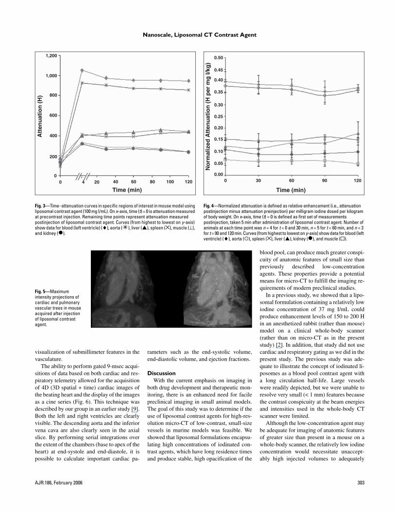
interest over time showed an initial enhance-ment of about 900 H in the aorta, indicating ahigh blood pool iodine concentration (Fig. 3).CT density slightly decreased over time to alevel of approximately 800 H after 2 hr. Excel-lent contrast discrimination was seen betweenthe myocardium and blood in the left ventricle(650–700 H). Enhancement of renal arteriesand veins was seen, but no significant nephro-gram was identified, indicating the absence ofrenal clearance of the agent. Liver and spleenenhancement was substantially lower than vas-cular enhancement and remained relativelystable over 120 min, consistent with the ex-pected delayed clearance of the liposomal io-dixanol formulation via the RES due to thePEGylated formulation (Fig. 4).
Because the agent could produce highopacity for several hours, a number of ana-tomic structures were clearly visible. The vol-ume-rendered image of the cardiac and pul-monary vascular trees acquired after injectionof the liposomal contrast agent shows theright and left ventricles, aorta, pulmonarytrunk, and inferior vena cava (Fig. 5). Thelong residence time at stable, high opacity en-ables simultaneous respiratory- and cardiac-gated image acquisition, thus limiting motionartifacts to less than 100 µm [8] and enablingthe extremely high resolution of these images(100-µm isotropic voxels) and, in turn, the
Nanoscale, Liposomal CT Contrast Agent
AJR:186, February 2006 303
Fig. 3—Time–attenuation curves in specific regions of interest in mouse model using liposomal contrast agent (100 mg I/mL). On x-axis, time (t) = 0 is attenuation measured at precontrast injection. Remaining time points represent attenuation measured postinjection of liposomal contrast agent. Curves (from highest to lowest on y-axis) show data for blood (left ventricle) (♦), aorta ( ), liver ( ), spleen (×), muscle (⊥), and kidney ( ).
Fig. 4—Normalized attenuation is defined as relative enhancement (i.e., attenuation postinjection minus attenuation preinjection) per milligram iodine dosed per kilogram of body weight. On x-axis, time (t) = 0 is defined as first set of measurements postinjection, taken 5 min after administration of liposomal contrast agent. Number of animals at each time point was n = 4 for t = 0 and 30 min, n = 5 for t = 60 min, and n = 3 for t = 90 and 120 min. Curves (from highest to lowest on y-axis) show data for blood (left ventricle) (♦), aorta ( ), spleen (×), liver ( ), kidney ( ), and muscle ( ).
Time (min)
Att
enu
atio
n (
H)
00
200
400
600
800
1,000
1,200
4 20 40 60 80 100 120
Time (min)
No
rmal
ized
Att
enu
atio
n (
H p
er m
g I/
kg)
0 30 60 90 1200.00
0.05
0.10
0.15
0.20
0.25
0.30
0.35
0.40
0.45
0.50
Fig. 5—Maximum intensity projections of cardiac and pulmonary vascular trees in mouse acquired after injection of liposomal contrast agent.
visualization of submillimeter features in thevasculature.
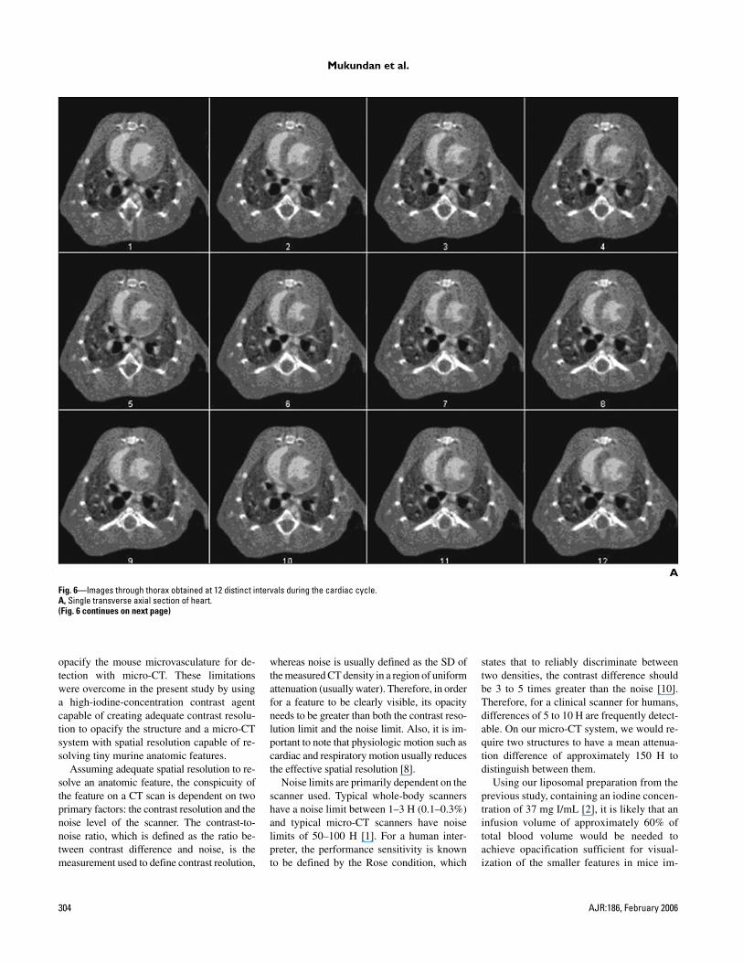
The ability to perform gated 9-msec acqui-sitions of data based on both cardiac and res-piratory telemetry allowed for the acquisitionof 4D (3D spatial + time) cardiac images ofthe beating heart and the display of the imagesas a cine series (Fig. 6). This technique wasdescribed by our group in an earlier study [9].Both the left and right ventricles are clearlyvisible. The descending aorta and the inferiorvena cava are also clearly seen in the axialslice. By performing serial integrations overthe extent of the chambers (base to apex of theheart) at end-systole and end-diastole, it ispossible to calculate important cardiac pa-
rameters such as the end-systolic volume,end-diastolic volume, and ejection fractions.
DiscussionWith the current emphasis on imaging in
both drug development and therapeutic mon-itoring, there is an enhanced need for facilepreclinical imaging in small animal models.The goal of this study was to determine if theuse of liposomal contrast agents for high-res-olution micro-CT of low-contrast, small-sizevessels in murine models was feasible. Weshowed that liposomal formulations encapsu-lating high concentrations of iodinated con-trast agents, which have long residence timesand produce stable, high opacification of the
blood pool, can produce much greater conspi-cuity of anatomic features of small size thanpreviously described low-concentrationagents. These properties provide a potentialmeans for micro-CT to fulfill the imaging re-quirements of modern preclinical studies.
In a previous study, we showed that a lipo-somal formulation containing a relatively lowiodine concentration of 37 mg I/mL couldproduce enhancement levels of 150 to 200 Hin an anesthetized rabbit (rather than mouse)model on a clinical whole-body scanner(rather than on micro-CT as in the presentstudy) [2]. In addition, that study did not usecardiac and respiratory gating as we did in thepresent study. The previous study was ade-quate to illustrate the concept of iodinated li-posomes as a blood pool contrast agent witha long circulation half-life. Large vesselswere readily depicted, but we were unable toresolve very small (< 1 mm) features becausethe contrast conspicuity at the beam energiesand intensities used in the whole-body CTscanner were limited.
Although the low-concentration agent maybe adequate for imaging of anatomic featuresof greater size than present in a mouse on awhole-body scanner, the relatively low iodineconcentration would necessitate unaccept-ably high injected volumes to adequately
Mukundan et al.
304 AJR:186, February 2006
A
Fig. 6—Images through thorax obtained at 12 distinct intervals during the cardiac cycle.A, Single transverse axial section of heart.(Fig. 6 continues on next page)
opacify the mouse microvasculature for de-tection with micro-CT. These limitationswere overcome in the present study by usinga high-iodine-concentration contrast agentcapable of creating adequate contrast resolu-tion to opacify the structure and a micro-CTsystem with spatial resolution capable of re-solving tiny murine anatomic features.
Assuming adequate spatial resolution to re-solve an anatomic feature, the conspicuity ofthe feature on a CT scan is dependent on twoprimary factors: the contrast resolution and thenoise level of the scanner. The contrast-to-noise ratio, which is defined as the ratio be-tween contrast difference and noise, is themeasurement used to define contrast reolution,
whereas noise is usually defined as the SD ofthe measured CT density in a region of uniformattenuation (usually water). Therefore, in orderfor a feature to be clearly visible, its opacityneeds to be greater than both the contrast reso-lution limit and the noise limit. Also, it is im-portant to note that physiologic motion such ascardiac and respiratory motion usually reducesthe effective spatial resolution [8].
Noise limits are primarily dependent on thescanner used. Typical whole-body scannershave a noise limit between 1–3 H (0.1–0.3%)and typical micro-CT scanners have noiselimits of 50–100 H [1]. For a human inter-preter, the performance sensitivity is knownto be defined by the Rose condition, which
states that to reliably discriminate betweentwo densities, the contrast difference shouldbe 3 to 5 times greater than the noise [10].Therefore, for a clinical scanner for humans,differences of 5 to 10 H are frequently detect-able. On our micro-CT system, we would re-quire two structures to have a mean attenua-tion difference of approximately 150 H todistinguish between them.
Using our liposomal preparation from theprevious study, containing an iodine concen-tration of 37 mg I/mL [2], it is likely that aninfusion volume of approximately 60% oftotal blood volume would be needed toachieve opacification sufficient for visual-ization of the smaller features in mice im-

Nanoscale, Liposomal CT Contrast Agent
AJR:186, February 2006 305
aged on a micro-CT instrument. Therefore,the goal of this study was to determine if theuse of liposomal contrast agents at accept-able infusion volumes (i.e., < 30% of totalblood volume) for high-resolution micro-CTof low-contrast, small-size vessels in murinemodels was feasible. The data in the presentstudy show that the infused dose (0.5 mL/25g) of the approximately 100 mg I/mL liposo-mal preparation elicited nearly 800-H rela-tive enhancement, which proved sufficientfor the much greater conspicuity of anatomic
features of small size and higher resolutionimages needed for micro-CT.
Torchilin et al. [11] investigated the use ofmicelle-encapsulated iodinated contrast agentsto take advantage of the intrinsic long circulat-ing time of these particles. They speculated thatthe monolayer boundary or envelope of the mi-celles was more efficient than the bilayer com-position of liposomes. They synthesized male-imide-polyethylene glycol-iodine-polylysinemicelles with an iodine content of 34%, whichresulted in a low overall concentration of 22 mg
I/mL that required large injection volumes ofapproximately several milliliters in rats. Al-though these investigators found a threefold en-hancement in the aorta over a 3-hr period (i.e.,approximately 250 H at 170 mg I/kg dose), theintrinsic inability of these particles to hold largeconcentrations of iodine and the resultant nec-essarily large infusion volumes have resulted inlow enthusiasm for this approach.
Desser et al. [12] attempted to produce a li-posomal CT contrast agent by preparing largeliposomes (550 nm in diameter compared
B
Fig. 6 (continued)—Images through thorax obtained at 12 distinct intervals during the cardiac cycle.B, Coronal section of heart.

Mukundan et al.
306 AJR:186, February 2006
with the 100-nm liposomes used in our study)that encapsulated iodixanol. Although thepreparation contained about 200 mg I/mL,only 40% of it was encapsulated, providing aliposomal concentration of 80 mg I/mL butresulting in an unacceptably large unencapsu-lated fraction. Furthermore, at this large size,vesicles were cleared rapidly by the RES de-spite a hydrophilic (PEGylated) coating, andthe agent had poor circulation time. In theirstudy, a dose of 150 mg I/kg produced initialenhancements of about 360 H, which rapidlydropped below 80 H in less than 5 min be-cause the unencapsulated iodine cleared viarenal excretion whereas the encapsulated por-tion cleared via the RES. This preparationtherefore suffered from potential renal toxic-ity problems based on the unencapsulated io-dine fraction and also failed to enhance circu-lation time. Thus, it provided no practicaladvantage over conventional contrast agents.
Using another approach, Sachse et al. [13]incorporated iopromide into 150-nm stealth(PEG-coated) liposomes. However, at thissize, the clearance rate of liposomes allowedonly 71-H enhancement after 45 min despitea relatively high dose of 250 mg I/kg. Leander[14] developed liposome-encapsulated iodix-anol particles with a particle size of 350 nmand an iodine content of 200 mg I/mL. How-ever, the formulation consisted of 40% of thetotal iodine within the liposomal interior andthe remaining 60% in the external phase. Ahuman phase 1 clinical study of the formula-tion indicated rapid clearance of the agentfrom blood circulation due to the large parti-cle size of the nanoparticles [15]. This re-sulted in a maximum initial blood pool en-hancement of about 150 H, which droppedbelow 80 H in less than 10 min.
In another study, Bakan et al. [16] tested alipid emulsion containing polyiodinated tri-glycerides for hepatocyte-specific delivery. Al-though these emulsions had a nominal particlesize of 100 nm, they were sequestered in theliver quite rapidly, possibly due to the lack of ahydrophilic coating. Also, with a maximum io-dine content of 50 mg I/mL, they exhibited rel-atively low, transient blood pool enhancementand a maximum hepatic enhancement of onlyapproximately 120 H. The same investigatorshave since tested a PEGylated version of thelipid emulsion with longer circulation time andhigher iodine concentrations [17]. The circula-tion time is still modest, approximately 60 min,and the blood pool enhancement is modest.However, the particles appear to localize quitewell in inflammatory lymph nodes, thought by
the authors to be due to the chylomicron-rem-nant-like structure of the lipid molecule. Thispreparation was used to depict lymph nodes ina dog model and exhibited about 200-H en-hancement in some nodes. Based on the re-search, a blood pool contrast agent is commer-cially available as Fenestra VC (AlerionBiomedical) containing iodine in a concentra-tion of 50 mg/mL. This contrast agent was re-cently used for a cardiac micro-CT study byBadea et al. [9] and provided a relatively con-stant enhancement over 3 hr, with maximumenhancement of approximately 620 H (aorta)and 90 H (kidney cortex). The maximum en-hancement difference between blood and myo-cardium in the heart was approximately 500 H.
Measurement of the degree of iodine leak-age in our formulation was important to as-sess the risk of excessive exposure of the sub-ject to free iodine and the attendant risks ofrenal toxicity as well as anaphylaxis in sensi-tive individuals. Our in vitro stability studyshowed that the formulation is stable withinliposomes and hence has a shelf life of at least7 days in phosphate buffer solution at 4°C.Leakage of only approximately 3% of the to-tal iodine was found, and that leakage waslikely due to the osmolarity or concentrationdifference between the interior of liposomesand the external environment. This degree ofleakage is sufficiently low to practically elim-inate renal toxicity as a realistic concern.Thus, in clinical use, agents such as ourswould not likely be contraindicated in pa-tients with renal insufficiency, as is the situa-tion with conventional iodinated agents.
At equivalent doses of iodine, conventionalagents would expose the renal medulla tomore than 30 times the iodine dose associatedwith the use of our encapsulated agents. Thus,the liposomal encapsulation process greatlyreduces overall iodine exposure. In fact, it ispossible that these agents may be adminis-tered preferentially in individuals with renalinsufficiency. Although it is also possible thatthe liposomes described here may confersome protection against anaphylaxis com-pared with standard iodinated contrast agents,further work along this line of investigation isneeded before definitive claims can be made.
In summary, our study shows that a liposo-mal formulation encapsulating a high concen-tration of iodine molecules has many proper-ties that optimize its use as a contrast agent forsmall animal imaging on a micro-CT scanner,including a long residence time and a high de-gree of opacification of very small blood ves-sels. The long residence time eliminates the
need for bolus infusion and rapid scanning,which are presently substantial limitations fordynamic contrast-enhanced imaging on mi-cro-CT scanners imposed by the long scantimes. Many features of our liposomal con-trast agent formulation, including the lowlevel of free iodine (thereby minimizing riskof renal toxicity) and long residence time(making them ideal blood pool contrastagents), render them potentially highly usefulimaging agents and deserving of consider-ation for experimental trials in humans.
References1. Paulus MJ, Gleason SS, Kennel SJ, Hunsicker PR,
Johnson DK. High resolution X-ray computed to-
mography: an emerging tool for small animal can-
cer research. Neoplasia 2000; 2:62–70
2. Kao CY, Hoffman EA, Beck KC, Bellamkonda RV,
Annapragada AV. Long-residence-time nano-scale
liposomal iohexol for X-ray-based blood pool im-
aging. Acad Radiol 2003; 10:475–483
3. Gabizon A, Huang L, Martin F, Barenholz Y.
Doxorubicin encapsulated in polyethylene-gly-
col coated liposomes: initial clinical-pharmaco-
kinetic studies in solid tumors. In: Lasic DD,
Martin FJ, eds. Stealth liposomes. Boca Raton,
FL: CRC Press, 1995:245–257
4. Hedlund LW, Cofer GP, Owen SJ, Johnson GA.
MR-compatible ventilator for small animals:
computer-controlled ventilation for proton and
noble gas imaging. Magn Reson Imaging 2000;
18:753–759
5. Badea CT, Hedlund LW, Wheeler CT, Mai W,
Johnson GA. Volumetric micro-CT system for in-
vivo microscopy. In: IEEE International Sympo-
sium on Biomedical Imaging: from nano to macro.
Arlington, VA: Institute of Electrical and Electron-
ics Engineers, 2004:1377–1381
6. Badea C, Hedlund LW, Johnson GA. Micro-CT
with respiratory and cardiac gating. Med Phys 2004;
31:3324–3329
7. Feldkamp LA, Davis LC, Kress JW. Practical cone-
beam algorithm. J Opt Soc Am 1984; 1:612–619
8. Mai W, Badea CT, Wheeler CT, Hedlund LW,
Johnson GA. Effects of breathing and cardiac mo-
tion on spatial resolution in the microscopic imag-
ing of rodents. Magn Reson Med 2005; 53:858–865
9. Badea CT, Fubara B, Hedlund LW, Johnson GA. 4-
D micro-CT of the mouse heart. Mol Imaging 2005;
4:110–116
10. Rose A. The sensitivity performance of the human
eye on an absolute scale. J Opt Soc Am 1948;
38:196–208
11. Torchilin VP, Frank-Kamenetsky MD, Wolf GL.
CT visualization of blood pool in rats by using long-
circulating, iodine-containing micelles. Acad Ra-
Nanoscale, Liposomal CT Contrast Agent
AJR:186, February 2006 307
diol 1999; 6:61–65
12. Desser TS, Rubin DL, Muller H, McIntire GL, Ba-
con ER, Toner JL. Blood pool and liver enhance-
ment in CT with liposomal iodixanol: comparison
with iohexol. Acad Radiol 1999; 6:176–183
13. Sachse A, Leike JU, Schneider T, et al. Biodis-
tribution and computed tomography blood-pool
imaging properties of polyethylene glycol-
coated iopromide-carrying liposomes. Invest Ra-
diol 1997; 32:44–50
14. Leander P. A new liposomal contrast medium for
CT of the liver: an imaging study in a rabbit tumour
model. Acta Radiol 1996; 37:63–68
15. Leander P, Hoglund P, Borseth A, Kloster Y, Berg
A. A new liposomal liver-specific contrast agent for
CT: first human phase-I clinical trial assessing effi-
cacy and safety. Eur Radiol 2001; 11:698–704
16. Bakan DA, Longino MA, Weichert JP, Counsell
RE. Physicochemical characterization of a syn-
thetic lipid emulsion for hepatocyte-selective
delivery of lipophilic compounds: application to
polyiodinated triglycerides as contrast agents
for computed tomography. J Pharm Sci 1996;
85:908–914
17. Wisner ER, Weichert JP, Longino MA, Counsell
RE, Weisbrode SE. A surface-modified chylomi-
cron remnant-like emulsion for percutaneous com-
puted tomography lymphography: synthesis and
preliminary imaging findings. Invest Radiol 2002;
37:232–239