Fibroblast growth factor maintains chondrogenic potential of limb bud mesenchymal cells by...
13
Cell Reports Article Fibroblast Growth Factor Maintains Chondrogenic Potential of Limb Bud Mesenchymal Cells by Modulating DNMT3A Recruitment Deepak Kumar 1,2 and Andrew B. Lassar 1, * 1 Department of Biological Chemistry and Molecular Pharmacology, Harvard Medical School, 240 Longwood Avenue, Boston, MA 02115, USA 2 Present address: EMD Serono Research & Development Institute, Inc., 45A Middlesex Turnpike, Billerica, MA 01821, USA *Correspondence: [email protected] http://dx.doi.org/10.1016/j.celrep.2014.07.038 This is an open access article under the CC BY-NC-ND license (http://creativecommons.org/licenses/by-nc-nd/3.0/). SUMMARY The formation of cartilage is restricted to the core of the limb bud mesenchyme by ectodermal Wnts, which can irreversibly silence expression of the pro- chondrogenic transcription factor Sox9. In contrast, fibroblast growth factor (FGF) signals from the apical ectodermal ridge maintain the competence of chon- drogenic precursors to undergo chondrogenesis once these cells go out of the range of ectodermal Wnt signals. We have found that Wnt signals induce both a repressive chromatin mark (H3K27me3) and DNA methylation over the Sox9 promoter and that Wnt-induced irreversible silencing of the Sox9 gene requires DNA methylation of this locus, which is spe- cifically countered by FGF signals. FGF blocks the recruitment of the de novo DNA methyltransferase, DNMT3A, to the Sox9 promoter by inducing the inter- action and phosphorylation of DNMT3A by ERK1/ ERK2 and thereby controls whether expression of Sox9 is either irreversibly or reversibly silenced by Wnt signals in limb bud mesenchymal cells. INTRODUCTION Vertebrate long bones are formed through a process of endo- chondral ossification. During this process, bone formation be- gins with the establishment of mesenchymal condensations, which serve as a template for the adult skeletal elements. Chon- drocytes then differentiate within the aggregated mesenchyme. Sox9, Sox5, and Sox6 play an essential role in the regulation of chondrogenesis (reviewed in Lefebvre and Bhattaram, 2010). Indeed, Sox9 directly activates cartilage differentiation markers, as this transcription factor has been shown to bind to the regu- latory elements that drive expression of these genes (reviewed in Lefebvre and Bhattaram, 2010). The formation of cartilage in the limb bud is restricted to the core of the limb bud mesen- chyme by signals from the ectoderm that block cartilage forma- tion in the periphery of this tissue (Solursh, 1984). Whereas ectopic expression of Wnts that signal via beta-catenin/LEF1/ TCF block cartilage formation in the limb bud, conditional loss of beta-catenin expression in the limb bud mesenchyme in- creases the expression of Sox9 in this tissue (Hill et al., 2005). Together, these findings suggest that Wnts secreted by the ecto- derm act via a beta-catenin-dependent pathway to block Sox9 expression and cartilage formation in limb bud mesenchymal cells that lie in the peripheral regions of the limb bud. In addition to Wnts secreted by limb bud ectoderm, fibroblast growth fac- tors (FGFs) secreted by the apical ectodermal ridge (AER) are necessary to maintain (1) limb bud outgrowth, (2) the viability of chondrogenic precursors that give rise to the cartilage templates of the limb, and (3) the competence for limb bud mesenchymal cells to undergo chondrogenesis once the Wnt signals are removed (ten Berge et al., 2008). RESULTS Transient Wnt Signals Irreversibly Block Induction of Sox9 Expression and Chondrogenesis Only in the Absence of FGF Signals To further elucidate how Wnt and FGF signaling modulates the competence of limb bud mesenchymal cells (LBMCs) to undergo chondrogenesis, we evaluated the expression of Sox9, colla- gen II, and aggrecan in micromass cultures of chicken LBMCs in response to these signals. We observed that Sox9, collagen II, and aggrecan were all robustly expressed in LBMCs after 8 days culture in control medium (Figure 1Aa). If, however, soluble Wnt3A was transiently administered to the explants during only the first 4 days of culture, the expression of Sox9, collagen II, and aggrecan was extinguished at day 4 (Figure 2A, compare a and b), and expression of these genes continued to be silenced 4 days after Wnt3A was removed from the culture medium (i.e., at day 8; Figure 1Ab). In concert with prior observations (ten Berge et al., 2008), we observed that transient administration of Wnt3A in the presence of FGF8 for 4 days allowed the subse- quent expression of Sox9, collagen II, and aggrecan in cultures harvested at day 8 (Figure 1Ad). FGF2, which can substitute for the AER to sustain relatively normal limb development, could similarly restore subsequent chondrogenesis when adminis- tered together with Wnt3A (Figure 1Ac). In contrast, FGF10 (which is expressed in limb bud mesenchyme), when adminis- tered together with Wnt3A, was unable to restore chondrogenic Cell Reports 8, 1419–1431, September 11, 2014 ª2014 The Authors 1419
-
Upload
mobilelandus -
Category
Documents
-
view
0 -
download
0
Transcript of Fibroblast growth factor maintains chondrogenic potential of limb bud mesenchymal cells by...

Cell Reports
Article
Fibroblast Growth Factor Maintains ChondrogenicPotential of Limb Bud Mesenchymal Cellsby Modulating DNMT3A RecruitmentDeepak Kumar1,2 and Andrew B. Lassar1,*1Department of Biological Chemistry and Molecular Pharmacology, Harvard Medical School, 240 Longwood Avenue, Boston,MA 02115, USA2Present address: EMD Serono Research & Development Institute, Inc., 45A Middlesex Turnpike, Billerica, MA 01821, USA
*Correspondence: [email protected]
http://dx.doi.org/10.1016/j.celrep.2014.07.038This is an open access article under the CC BY-NC-ND license (http://creativecommons.org/licenses/by-nc-nd/3.0/).
SUMMARY
The formation of cartilage is restricted to the core ofthe limb bud mesenchyme by ectodermal Wnts,which can irreversibly silence expression of the pro-chondrogenic transcription factor Sox9. In contrast,fibroblast growth factor (FGF) signals from the apicalectodermal ridge maintain the competence of chon-drogenic precursors to undergo chondrogenesisonce these cells go out of the range of ectodermalWnt signals. We have found that Wnt signals induceboth a repressive chromatin mark (H3K27me3) andDNA methylation over the Sox9 promoter and thatWnt-induced irreversible silencing of the Sox9 generequires DNA methylation of this locus, which is spe-cifically countered by FGF signals. FGF blocks therecruitment of the de novo DNA methyltransferase,DNMT3A, to the Sox9 promoter by inducing the inter-action and phosphorylation of DNMT3A by ERK1/ERK2 and thereby controls whether expression ofSox9 is either irreversibly or reversibly silenced byWnt signals in limb bud mesenchymal cells.
INTRODUCTION
Vertebrate long bones are formed through a process of endo-
chondral ossification. During this process, bone formation be-
gins with the establishment of mesenchymal condensations,
which serve as a template for the adult skeletal elements. Chon-
drocytes then differentiate within the aggregated mesenchyme.
Sox9, Sox5, and Sox6 play an essential role in the regulation of
chondrogenesis (reviewed in Lefebvre and Bhattaram, 2010).
Indeed, Sox9 directly activates cartilage differentiation markers,
as this transcription factor has been shown to bind to the regu-
latory elements that drive expression of these genes (reviewed
in Lefebvre and Bhattaram, 2010). The formation of cartilage in
the limb bud is restricted to the core of the limb bud mesen-
chyme by signals from the ectoderm that block cartilage forma-
tion in the periphery of this tissue (Solursh, 1984). Whereas
ectopic expression of Wnts that signal via beta-catenin/LEF1/
Cell Re
TCF block cartilage formation in the limb bud, conditional loss
of beta-catenin expression in the limb bud mesenchyme in-
creases the expression of Sox9 in this tissue (Hill et al., 2005).
Together, these findings suggest thatWnts secreted by the ecto-
derm act via a beta-catenin-dependent pathway to block Sox9
expression and cartilage formation in limb bud mesenchymal
cells that lie in the peripheral regions of the limb bud. In addition
to Wnts secreted by limb bud ectoderm, fibroblast growth fac-
tors (FGFs) secreted by the apical ectodermal ridge (AER) are
necessary to maintain (1) limb bud outgrowth, (2) the viability of
chondrogenic precursors that give rise to the cartilage templates
of the limb, and (3) the competence for limb bud mesenchymal
cells to undergo chondrogenesis once the Wnt signals are
removed (ten Berge et al., 2008).
RESULTS
Transient Wnt Signals Irreversibly Block Inductionof Sox9 Expression and Chondrogenesis Only inthe Absence of FGF SignalsTo further elucidate how Wnt and FGF signaling modulates the
competence of limb budmesenchymal cells (LBMCs) to undergo
chondrogenesis, we evaluated the expression of Sox9, colla-
gen II, and aggrecan in micromass cultures of chicken LBMCs
in response to these signals. We observed that Sox9, collagen II,
and aggrecan were all robustly expressed in LBMCs after 8 days
culture in control medium (Figure 1Aa). If, however, soluble
Wnt3A was transiently administered to the explants during only
the first 4 days of culture, the expression of Sox9, collagen II,
and aggrecan was extinguished at day 4 (Figure 2A, compare
a and b), and expression of these genes continued to be silenced
4 days after Wnt3A was removed from the culture medium (i.e.,
at day 8; Figure 1Ab). In concert with prior observations (ten
Berge et al., 2008), we observed that transient administration
of Wnt3A in the presence of FGF8 for 4 days allowed the subse-
quent expression of Sox9, collagen II, and aggrecan in cultures
harvested at day 8 (Figure 1Ad). FGF2, which can substitute
for the AER to sustain relatively normal limb development, could
similarly restore subsequent chondrogenesis when adminis-
tered together with Wnt3A (Figure 1Ac). In contrast, FGF10
(which is expressed in limb bud mesenchyme), when adminis-
tered together with Wnt3A, was unable to restore chondrogenic
ports 8, 1419–1431, September 11, 2014 ª2014 The Authors 1419

Figure 1. Transient Wnt Signals Irreversibly Block Induction of Sox9 Expression and Chondrogenesis Only in the Absence of FGF Signals(A) LBMCs were isolated from day 4 chicken embryos and cultured in either control medium or medium containing either Wnt3A or FGF2/FGF8/FGF10 for the
days indicated. Cells were harvested after 8 days of culture, and gene expression was assayed by quantitative RT-PCR (qRT-PCR). In this and subsequent
figures, significance was calculated using Student’s t test; *p < 0.05, **p < 0.01, ***p < 0.001, and ns (nonsignificant difference) are indicated; and error bar
indicates SEM. The relative expression in b–f was compared with that in a.
(B) Wnt signals from the ectoderm block Sox9 expression (and chondrogenesis) in the peripheral regions of the limb bud; FGF signals from the AERmaintain both
the viability and chondrogenic competence of limb bud mesenchymal cells.
gene expression in these cultures (Figure 1Ae). Thus, FGF family
members (i.e., FGF2 and FGF8) that can substitute for the AER to
maintain limb bud outgrowth can alsomaintain the chondrogenic
potential of limb bud mesenchymal cells transiently exposed
to Wnt signals, which are secreted by the overlying ectoderm
(displayed schematically in Figure 1B). Interestingly, FGF2 could
only restore chondrogenesis when administered simultaneously
with Wnt3A but failed to do so when administered subsequent to
Wnt exposure (Figure 1A, compare c and f), indicating that FGF
signals must occur concomitant with Wnt signals to maintain
chondrogenic competence. Importantly, simultaneous adminis-
tration of FGF did not disrupt Wnt signaling, as inclusion of FGF2
for the first 4 days of culture did not block the ability of Wnt3A to
either induce target genes such as Twist2 (Figure S1A) or repress
expression of Sox9, collagen II, and aggrecan in cultures har-
vested at day 4 (Figure 2Ac). To begin to explore how FGF main-
tains the competence for LBMCs to express Sox9 following
exposure to transient Wnt signals, we simultaneously treated
micromass cultures of LBMCswith Wnt3A and FGF2 in the pres-
ence of the MEK1/MEK2 antagonist, U0126. Inhibition of MEK1/
1420 Cell Reports 8, 1419–1431, September 11, 2014 ª2014 The Au
MEK2 activity severely blocked the ability of FGF2 to restore
chondrogenic gene expression following removal of Wnt3A (Fig-
ures 2A, compare g and h, and S1B). Thus, FGF signaling blocks
the ability of transient Wnt signals to stably repress the expres-
sion of Sox9, collagen II, and aggrecan via a MEK1/MEK2-
dependent pathway and can do so even in the absence of
DNA replication (Figures S2A–S2C).
Transient Wnt Signals Induce Stable H3K27me3Modification over the Sox9 Promoter Only in theAbsence of FGF SignalsBecause Sox9 is the earliest marker of chondrogenic differenti-
ation, we evaluated whether Wnt and FGF signals may modulate
chondrogenesis by inducing epigenetic modification of this lo-
cus. We first evaluated whether these signals alter histone
methylation over the Sox9 promoter. The Sox9 gene in LBMCs
cultured in control medium for 4 days is expressed (Figure 2Aa),
and its promoter is accordingly marked by both H3K4me3 (Fig-
ure 2Ba) and H3K9Ac (Figure S2Da) modifications, which are
both indicative of active transcription. Correspondingly, this
thors

Figure 2. Transient Wnt Signals Induce Stable H3K27me3 Modification and CpGMethylation over the Sox9 Promoter Only in the Absence of
FGF Signals
LBMCs were cultured in medium containing either Wnt3A, FGF2, or the MEK1/MEK2 antagonist, U0126, for the days indicated. Cells were harvested after 4 or
8 days of culture and either (A) gene expression was assayed by qRT-PCR, (B) chromatin IP (ChIP) was performed to assay H3K27me3 and H3K4me3 modi-
fications over the Sox9 promoter, or (C) methyl-DNA IP (MeDIP) was performed to assay methyl CpG modification in two CpG islands (CpG1 and CpG2) that
encompass the Sox9 promoter. In (B) and (C), isotype control (IC) immunoglobulin G (IgG) IPs are also displayed. In each section (A–C) of this figure, b–d were
compared with a and f–h were compared with e. *p < 0.05, **p < 0.01, ***p < 0.001, and ns (nonsignificant difference) are indicated, and error bar indicates SEM.
promoter lacks H3K27me3 modification (Figure 2Ba), which is
associated with transcriptional silencing. In contrast, the Sox9
gene in LBMCs cultured in the presence of Wnt3A for 4 days
(in either the absence or presence of FGF2) is transcriptionally
silent (Figures 2Ab and 2Ac), and its promoter is accordingly
marked by significantly increased levels of H3K27me3 and
decreased levels of both H3K4me3 and H3K9Ac modifications
(Figures 2Bb, 2Bc, S2Db, and S2Dc). Thus, concomitant admin-
istration of FGF2 does not block the ability of WntA3 to initially
Cell Re
establish a repressive chromatin structure over the Sox9
promoter.
As discussed above, a transient 4-day exposure of LBMCs to
Wnt3A (in the absence of exogenous FGF) resulted in stable
repression of Sox9 gene expression in cells harvested at day 8
(Figure 2Af). Consistent with this stable repression of Sox9
gene expression, the Sox9 promoter displayed a relatively high
level of H3K27me3 modification and a relatively low level of
both H3K4me3 and H3K9Ac modifications (at day 8) in LBMCs
ports 8, 1419–1431, September 11, 2014 ª2014 The Authors 1421

that had been cultured for the initial 4 days in Wnt3A-containing
medium (Figures 2Bf and S2De). In contrast, a transient 4-day
exposure of LBMCs to Wnt3A in the presence of FGF2 resulted
in the subsequent expression of Sox9 (at day 8; Figure 2Ag),
coupled with a significant decrease in H3K27me3 modification,
and corresponding increases in both H3K4me3 and H3K9Ac
modifications of the Sox9 promoter (at day 8; Figures 2Bg and
S2Df). Taken together, these findings suggest that, whereas
concurrent FGF2 administration does not block the initiation of
Wnt3A-induced H3K27me3 modification of the Sox9 promoter
(at day 4), it disrupts the stability of this histone modification,
such that it is lost in the absence of continued Wnt signaling
(by day 8 of culture).
Administration of the MEK1/MEK2 antagonist, U0126,
blocked the ability of FGF2 to restore Sox9 gene expression
(at 8 days) in cells exposed to both Wnt3A and FGF2 for the first
4 days of culture (Figure 2A, compare g and h). Thus, MEK1/
MEK2 activity is necessary for FGF signals to maintain the chon-
drogenic competence of LBMCs exposed transiently to Wnt
signals. Interestingly, however, U0126 failed to significantly alter
either H3K4me3 or H3K27me3 modification of the Sox9 pro-
moter after culture of LBMCs inWnt3A plus FGF2 for 4 days (Fig-
ure 2B, compare c and d). Thus, the potential to express the
Sox9 gene (after 8 days culture) in LBMCs transiently exposed
to Wnt3A, Wnt3A/FGF2, or Wnt3A/FGF2/U0126 (for 4 days) did
not correlate with the relative levels of either H3K4me3 or
H3K27me3 modification of this locus (after 4 days culture).
Transient Wnt Signals Induce CpG Methylation of theSox9 Promoter Only in the Absence of FGF SignalsWe have found that FGF/MEK1/MEK2 signaling does not sig-
nificantly affect the ability of Wnt3A to induce H3K27me3 mo-
dification over the Sox9 promoter but specifically blocks the
maintenance of this histone modification after withdrawal of
Wnt signaling. Thus, maintenance of the H3K27me3 modifica-
tion (after transient Wnt signaling) requires another event that is
inhibited by FGF signaling. Because DNA methylation has
been noted to in some cases reinforce repressive histone mod-
ifications, we investigated whether FGF might alter the ability of
Wnt signals to induce DNA methylation of two CpG islands that
encompass the Sox9 promoter. By employing a methylated
DNA-immunoprecipitation (IP) assay, we found that a 4-day
exposure of LBMCs to soluble Wnt3A increased DNA methyl-
ation of both CpG islands surrounding the Sox9 promoter (Fig-
ure 2C, compare a and b). Interestingly, the relative level of
DNA methylation that was initially established on the Sox9
promoter in the presence of Wnt3A (at 4 days) was stably main-
tained after withdrawal of Wnt3A for an additional 4 days (Fig-
ure 2C, compare e and f). In marked contrast, in the presence
of FGF2, Wnt3A administration failed to increase CpG methyl-
ation of the Sox9 promoter (Figure 2Cc), and this relative deficit
of CpG methylation was maintained after withdrawal of both
Wnt3A and FGF2 for an additional 4 days (Figure 2Cg).We exam-
ined whether MEK signaling was necessary for FGF2 to inhibit
Wnt3A-induced DNA methylation of the Sox9 promoter in
LBMCs. Indeed, administration of the MEK1/MEK2 antagonist,
U0126, blocked the ability of FGF2 to inhibit Wnt3A-induced
DNA methylation of the Sox9 promoter, resulting in increased
1422 Cell Reports 8, 1419–1431, September 11, 2014 ª2014 The Au
CpG methylation of this sequence (Figure 2C, compare c and
d and g and h). The increased DNA methylation of the Sox9 pro-
moter in LBMCs treated with Wnt3A/FGF2/U0126 (for 4 days)
correlated with the subsequent repression of Sox9 expression
in cells harvested 4 days following the removal of Wnt3A/
FGF2/U0126 (Figure 2Ah). Thus, MEK signaling is essential for
FGF signals to block Wnt-induced DNA methylation of the
Sox9 promoter.
To confirm these methyl-DNA IP results, we performed bisul-
fite sequencing of the second Sox9 CpG island (CpG2) in
genomic DNA isolated from LBMCs that had been cultured for
4 days in either control medium, medium containing soluble
Wnt3A, or medium containing bothWnt3A and FGF2. Consistent
with ourmethyl-DNA IP results, we found thatWnt3A administra-
tion increased cytosine methylation of CpG residues located
throughout the Sox9 CpG2, with the greatest effect occurring
in the 30 region of this CpG island (Figure S3A; Table S1). Most
notably, concurrent administration of FGF2 (together with
Wnt3A) decreased cytosine methylation of CpG residues
throughout the Sox9 CpG2 (Figure S3A; Table S1). To further
evaluate whether the competence for subsequent Sox9 gene
expression in LBMCs transiently exposed to both Wnt and
FGF signals inversely correlates with DNA methylation of the
Sox9 promoter, we treated LBMCs with Wnt3A plus differing
levels of FGF2. We found that levels of FGF2 that were sufficient
to block Wnt3A-induced DNA methylation of the Sox9 promoter
(Figure S3B) correlated with FGF2 levels necessary to maintain
the competence to express both Sox9 and other chondrogenic
differentiation markers, following transient exposure to Wnt3A
plus FGF2 (Figure S3C). Taken together, our findings indicate
that the relative level of DNA methylation that is established
over the Sox9 promoter by culture of LBMCs in either control
medium, medium containing solubleWnt3A, or medium contain-
ing both Wnt3A and FGF2 inversely correlates with the subse-
quent expression of this gene 4 days after these growth factors
are removed.
FGF Signals Block Recruitment of DNMT3A to theSox9 PromoterBecause transient Wnt signals induce DNA methylation of
the Sox9 promoter and FGF signals block this effect, we inv-
estigated both whether Wnt signals recruit a DNA methyltrans-
ferase to the Sox9 promoter and whether FGF signals might
block this recruitment. LBMCs were infected with retrovirus en-
coding either Flag/hemagglutinin (HA) (FH)-tagged hDNMT1,
hDNMT3A, or hDNMT3B and cultured in either control medium,
medium containing soluble Wnt3A, or medium containing both
Wnt3A and FGF2. After 4 days of culture, the cells were har-
vested and the association of exogenous FH-tagged DNMT
with the Sox9 promoter was assayed by chromatin IP with
anti-HA antibody. We found that Wnt3A specifically induced
the association of FH-DNMT3A with the Sox9 promoter and
that Wnt-induced recruitment of FH-DNMT3A did not occur in
the presence of FGF2 (Figure 3A). Importantly, neither Wnt nor
FGF administration altered expression of the exogenous
DNMTs (Figures S4A and S4B), indicating that the effects of
these ligands on HA-DNMT3A:Sox9 promoter interaction
were posttranscriptional. In addition, we observed that Wnt3A
thors
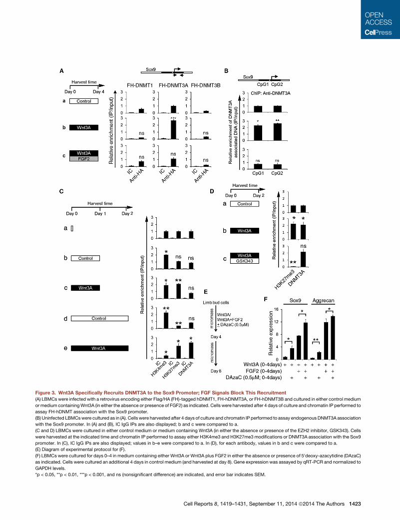
Figure 3. Wnt3A Specifically Recruits DNMT3A to the Sox9 Promoter; FGF Signals Block This Recruitment
(A) LBMCs were infected with a retrovirus encoding either Flag/HA (FH)-tagged hDNMT1, FH-hDNMT3A, or FH-hDNMT3B and cultured in either control medium
or medium containingWnt3A (in either the absence or presence of FGF2) as indicated. Cells were harvested after 4 days of culture and chromatin IP performed to
assay FH-hDNMT association with the Sox9 promoter.
(B) Uninfected LBMCswere cultured as in (A). Cells were harvested after 4 days of culture and chromatin IP performed to assay endogenous DNMT3A association
with the Sox9 promoter. In (A) and (B), IC IgG IPs are also displayed; b and c were compared to a.
(C and D) LBMCs were cultured in either control medium or medium containing Wnt3A (in either the absence or presence of the EZH2 inhibitor, GSK343). Cells
were harvested at the indicated time and chromatin IP performed to assay either H3K4me3 and H3K27me3 modifications or DNMT3A association with the Sox9
promoter. In (C), IC IgG IPs are also displayed; values in b–e were compared to a. In (D), for each antibody, values in b and c were compared to a.
(E) Diagram of experimental protocol for (F).
(F) LBMCs were cultured for days 0–4 in medium containing either Wnt3A or Wnt3A plus FGF2 in either the absence or presence of 50deoxy-azacytidine (DAzaC)
as indicated. Cells were cultured an additional 4 days in control medium (and harvested at day 8). Gene expression was assayed by qRT-PCR and normalized to
GAPDH levels.
*p < 0.05, **p < 0.01, ***p < 0.001, and ns (nonsignificant difference) are indicated, and error bar indicates SEM.
Cell Reports 8, 1419–1431, September 11, 2014 ª2014 The Authors 1423
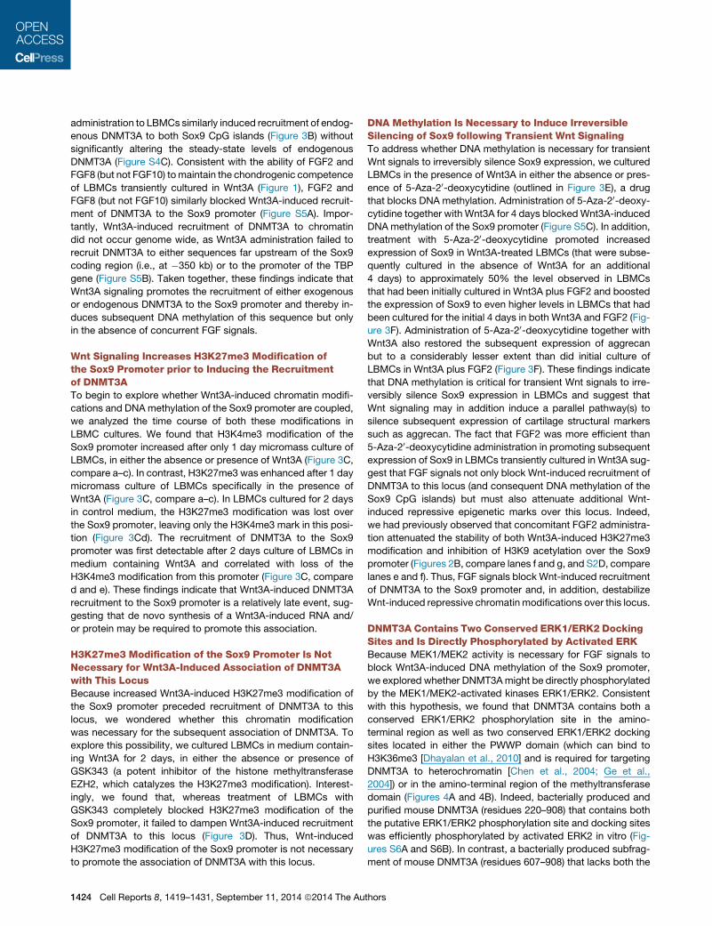
administration to LBMCs similarly induced recruitment of endog-
enous DNMT3A to both Sox9 CpG islands (Figure 3B) without
significantly altering the steady-state levels of endogenous
DNMT3A (Figure S4C). Consistent with the ability of FGF2 and
FGF8 (but not FGF10) tomaintain the chondrogenic competence
of LBMCs transiently cultured in Wnt3A (Figure 1), FGF2 and
FGF8 (but not FGF10) similarly blocked Wnt3A-induced recruit-
ment of DNMT3A to the Sox9 promoter (Figure S5A). Impor-
tantly, Wnt3A-induced recruitment of DNMT3A to chromatin
did not occur genome wide, as Wnt3A administration failed to
recruit DNMT3A to either sequences far upstream of the Sox9
coding region (i.e., at �350 kb) or to the promoter of the TBP
gene (Figure S5B). Taken together, these findings indicate that
Wnt3A signaling promotes the recruitment of either exogenous
or endogenous DNMT3A to the Sox9 promoter and thereby in-
duces subsequent DNA methylation of this sequence but only
in the absence of concurrent FGF signals.
Wnt Signaling Increases H3K27me3 Modification ofthe Sox9 Promoter prior to Inducing the Recruitmentof DNMT3ATo begin to explore whether Wnt3A-induced chromatin modifi-
cations and DNA methylation of the Sox9 promoter are coupled,
we analyzed the time course of both these modifications in
LBMC cultures. We found that H3K4me3 modification of the
Sox9 promoter increased after only 1 day micromass culture of
LBMCs, in either the absence or presence of Wnt3A (Figure 3C,
compare a–c). In contrast, H3K27me3 was enhanced after 1 day
micromass culture of LBMCs specifically in the presence of
Wnt3A (Figure 3C, compare a–c). In LBMCs cultured for 2 days
in control medium, the H3K27me3 modification was lost over
the Sox9 promoter, leaving only the H3K4me3 mark in this posi-
tion (Figure 3Cd). The recruitment of DNMT3A to the Sox9
promoter was first detectable after 2 days culture of LBMCs in
medium containing Wnt3A and correlated with loss of the
H3K4me3 modification from this promoter (Figure 3C, compare
d and e). These findings indicate that Wnt3A-induced DNMT3A
recruitment to the Sox9 promoter is a relatively late event, sug-
gesting that de novo synthesis of a Wnt3A-induced RNA and/
or protein may be required to promote this association.
H3K27me3 Modification of the Sox9 Promoter Is NotNecessary for Wnt3A-Induced Association of DNMT3Awith This LocusBecause increased Wnt3A-induced H3K27me3 modification of
the Sox9 promoter preceded recruitment of DNMT3A to this
locus, we wondered whether this chromatin modification
was necessary for the subsequent association of DNMT3A. To
explore this possibility, we cultured LBMCs in medium contain-
ing Wnt3A for 2 days, in either the absence or presence of
GSK343 (a potent inhibitor of the histone methyltransferase
EZH2, which catalyzes the H3K27me3 modification). Interest-
ingly, we found that, whereas treatment of LBMCs with
GSK343 completely blocked H3K27me3 modification of the
Sox9 promoter, it failed to dampen Wnt3A-induced recruitment
of DNMT3A to this locus (Figure 3D). Thus, Wnt-induced
H3K27me3 modification of the Sox9 promoter is not necessary
to promote the association of DNMT3A with this locus.
1424 Cell Reports 8, 1419–1431, September 11, 2014 ª2014 The Au
DNA Methylation Is Necessary to Induce IrreversibleSilencing of Sox9 following Transient Wnt SignalingTo address whether DNA methylation is necessary for transient
Wnt signals to irreversibly silence Sox9 expression, we cultured
LBMCs in the presence of Wnt3A in either the absence or pres-
ence of 5-Aza-20-deoxycytidine (outlined in Figure 3E), a drug
that blocks DNA methylation. Administration of 5-Aza-20-deoxy-cytidine together with Wnt3A for 4 days blocked Wnt3A-induced
DNA methylation of the Sox9 promoter (Figure S5C). In addition,
treatment with 5-Aza-20-deoxycytidine promoted increased
expression of Sox9 in Wnt3A-treated LBMCs (that were subse-
quently cultured in the absence of Wnt3A for an additional
4 days) to approximately 50% the level observed in LBMCs
that had been initially cultured in Wnt3A plus FGF2 and boosted
the expression of Sox9 to even higher levels in LBMCs that had
been cultured for the initial 4 days in both Wnt3A and FGF2 (Fig-
ure 3F). Administration of 5-Aza-20-deoxycytidine together with
Wnt3A also restored the subsequent expression of aggrecan
but to a considerably lesser extent than did initial culture of
LBMCs in Wnt3A plus FGF2 (Figure 3F). These findings indicate
that DNA methylation is critical for transient Wnt signals to irre-
versibly silence Sox9 expression in LBMCs and suggest that
Wnt signaling may in addition induce a parallel pathway(s) to
silence subsequent expression of cartilage structural markers
such as aggrecan. The fact that FGF2 was more efficient than
5-Aza-20-deoxycytidine administration in promoting subsequent
expression of Sox9 in LBMCs transiently cultured in Wnt3A sug-
gest that FGF signals not only block Wnt-induced recruitment of
DNMT3A to this locus (and consequent DNA methylation of the
Sox9 CpG islands) but must also attenuate additional Wnt-
induced repressive epigenetic marks over this locus. Indeed,
we had previously observed that concomitant FGF2 administra-
tion attenuated the stability of both Wnt3A-induced H3K27me3
modification and inhibition of H3K9 acetylation over the Sox9
promoter (Figures 2B, compare lanes f and g, and S2D, compare
lanes e and f). Thus, FGF signals block Wnt-induced recruitment
of DNMT3A to the Sox9 promoter and, in addition, destabilize
Wnt-induced repressive chromatin modifications over this locus.
DNMT3A Contains Two Conserved ERK1/ERK2 DockingSites and Is Directly Phosphorylated by Activated ERKBecause MEK1/MEK2 activity is necessary for FGF signals to
block Wnt3A-induced DNA methylation of the Sox9 promoter,
we explored whether DNMT3Amight be directly phosphorylated
by the MEK1/MEK2-activated kinases ERK1/ERK2. Consistent
with this hypothesis, we found that DNMT3A contains both a
conserved ERK1/ERK2 phosphorylation site in the amino-
terminal region as well as two conserved ERK1/ERK2 docking
sites located in either the PWWP domain (which can bind to
H3K36me3 [Dhayalan et al., 2010] and is required for targeting
DNMT3A to heterochromatin [Chen et al., 2004; Ge et al.,
2004]) or in the amino-terminal region of the methyltransferase
domain (Figures 4A and 4B). Indeed, bacterially produced and
purified mouse DNMT3A (residues 220–908) that contains both
the putative ERK1/ERK2 phosphorylation site and docking sites
was efficiently phosphorylated by activated ERK2 in vitro (Fig-
ures S6A and S6B). In contrast, a bacterially produced subfrag-
ment of mouse DNMT3A (residues 607–908) that lacks both the
thors

Figure 4. DNMT3A Contains Conserved ERK1/ERK2 Phosphorylation and Docking Sites and Is Directly Phosphorylated by Activated ERK(A) Schematic diagram of human DNMT3A depicting the ERK phosphorylation and docking sites and the PWWP and ADD (ATRX-DNMT3-DNMT3L) domains.
(B) ERK1/ERK2 phosphorylation and docking sites are conserved in vertebrate DNMT3A (Ec, Equus caballus; Gg, Gallus gallus; Hs, Homo sapiens; MAPK,
mitogen-activated protein kinase; Mm, Mus musculus; Rn, Rattus norvegicus).
(C) ERK2 kinase assay (employing g�32P-ATP) and either WT or mutant forms of GST-hDNMT3A, as indicated. WB, western blot.
(D) Western analysis (employing either anti-hDNMT3A-(pS255) or anti-DNMT3A antibodies) of in vitro ERK2-phosphorylated WT or mutant forms of GST-
hDNMT3A.
(E) Limb buds (isolated from day 4 chicken embryos) were separated into proximal (P), middle (M), and distal (D) regions. Equal protein amounts of each limb bud
region were loaded onto SDS-PAGE, and western analysis was performed with either anti-DNMT3A-(pS220), anti-DNMT3A, anti-pERK, anti-ERK, or anti-beta
actin.
(F) Limb buds (isolated from day 4 chicken embryos) were cultured for 2 hr in either control medium or medium containing either soluble Wnt3A, Wnt3A/FGF2, or
Wnt3A/FGF2/U0126 as indicated. Equal protein amounts of each explant culture were loaded onto SDS-PAGE, and western analysis was performed with either
anti-DNMT3A-(pS220) (in the presence of competitor nonphosphorylated hDNMT3A peptide), anti-DNMT3A, anti-pERK, anti-ERK, or anti-beta actin.
putative ERK1/ERK2 phosphorylation site and one of the
ERK docking sites was not phosphorylated by activated ERK2
(Figure S6B). To better map the ERK2 phosphorylation site in
DNMT3A, we made glutathione S-transferase (GST)-fusion
proteins with either human DNMT3A-wild-type (WT) or human
DNMT3A-(S255A), which contains a serine to alanine mutation
in the putative ERK1/ERK2 phosphorylation site. Whereas
activated ERK2 efficiently phosphorylated GST-hDNMT3A-WT,
this kinase failed to significantly phosphorylate GST-hDNMT3A-
(S255A) (Figure 4C, compare lanes 1 and 3), suggesting that
S255 in hDNMT3A is a key residue necessary for ERK2 phos-
phorylation. We generated affinity-purified polyclonal antibodies
in rabbits that specifically recognize phosphorylated S255 in hu-
man DNMT3A. This affinity-purified antibody, which was made
against a 17-amino-acid peptide (246aa-AVQQPTDPA-phos-
pho-S255PTVATTC-262aa) including phosphorylated S255 in
humanDNMT3A, only recognizedGST-hDNMT3A-WTafter incu-
Cell Re
bation with ATP and activated ERK2 (Figure 4D, compare lanes 1
and 2) and specifically did not recognizeGST-hDNMT3A-(S255A)
even after incubation with ATP and activated ERK2 (Figure 4D,
lanes 5 and 6). Importantly, interaction of anti-phospho-S255
with GST-hDNMT3A-WT that had been phosphorylated in vitro
by activated ERK2was specifically competed by aDNMT3Apep-
tide (246aa-AVQQPTDPASPTVATTC-262aa) containing phos-
pho-S255, but not by a peptide containing nonphosphorylated
S255 (Figure S6C). Together, these findings indicate that the prin-
cipal ERK2 phosphorylation site in hDNMT3A is S255.
ERK1/ERK2 docking sites identified in substrates for these
kinases have previously been noted to be necessary for efficient
phosphorylation of these substrates by ERK1/ERK2. To assay
whether the ERK1/ERK2 docking sites located in the PWWP
domain and in the methyltransferase domain of DNMT3A are
required for efficient phosphorylation of this protein by activated
ERK2, we assayed the relative ability of activated ERK2 to
ports 8, 1419–1431, September 11, 2014 ª2014 The Authors 1425

A
D E F
B C
Figure 5. DNMT3A Is Phosphorylated at the ERK1/ERK2 Phosphorylation Site in a Proximal-Distal Gradient in the Limb Bud
Cryosections of limb buds (isolated from day 4 chicken embryos) were immunostained with either anti-phospho-S220-DNMT3A (A–C) or anti-total-DNMT3A
(D–F); nuclei were visualized by DAPI staining. In (A)–(C), a DNMT3A peptide lacking serine phosphorylation (at the ERK phosphorylation site) was added along
with anti-phospho-S220-DNMT3A. The distal region of the limb bud (containing the AER) is shown on the left.
phosphorylate either GST-hDNMT3A-WT versus GST-
hDNMT3A-(L373A, L637A), which contains point mutations in
both ERK1/ERK2 docking sites. Notably, in vitro phosphorylation
of GST-hDNMT3A-(L373A, L637A) by activated ERK2 (as as-
sayed either by labeling with g-32P-ATP or by western blotting
with anti-phospho-S255 antibody) was markedly attenuated
relative to GST-hDNMT3A-WT phosphorylation (Figures 4C,
compare lanes 1 and 2, and 4D, compare lanes 2 and 4). These
findings suggest that ERK2 interaction with either L373 and/or
L637 in DNMT3A is critical to support efficient phosphorylation
of S255A in hDNMT3A by this kinase.
DNMT3A Is Phosphorylated at the ERK1/ERK2Phosphorylation Site in Response to FGF Signals inLimb BudsTo investigate whether endogenous DNMT3A is phosphorylated
at the ERK1/ERK2 phosphorylation site, we dissected limb buds
from H. H. stage 20–22 chicken embryos and isolated either the
proximal, middle, or distal regions of this structure. Western
analysis revealed that these three regions of the limb bud ex-
press equivalent levels of DNMT3A but that this protein is prefer-
entially phosphorylated at the ERK1/ERK2 phosphorylation site
(S220 in chicken DNMT3A) specifically in distal limb bud cells,
which also express relatively higher levels of activated phos-
pho-ERK1/ERK2 (Figures 4E and S6D). To determine whether
FGF signals specifically induce phosphorylation of DNMT3A at
the ERK1/ERK2 phosphorylation site via a MEK1/MEK2-depen-
dent pathway, we cultured limb bud explants for 2 hr in either
control medium or medium containing soluble Wnt3A, Wnt3A/
1426 Cell Reports 8, 1419–1431, September 11, 2014 ª2014 The Au
FGF2, or Wnt3A/FGF2/U0126. Whereas culture of the intact
limb buds in Wnt3A alone only slightly boosted phosphorylation
of DNMT3A at the ERK1/ERK2 phosphorylation site, culture of
limb bud explants in both Wnt3A and FGF2 significantly
increased phosphorylation of DNMT3A at this site (Figure 4F,
compare lanes 1–3). Inclusion of the MEK1/MEK2 inhibitor
U0126 abrogated the increased phosphorylation of both ERK
and DNMT3A (at the ERK1/ERK2 phosphorylation site) induced
by FGF2 administration. (Figure 4F, lane 4). To assay the spatial
localization of ERK-phosphorylated DNMT3A in the limb bud, we
assayed its expression by immunofluorescence in cryosections
of limb buds. Interestingly, we found that, whereas anti-total
DNMT3A recognized approximately equal levels of this protein
in proximal and distal regions of the limb bud (Figure 5D), anti-
phospho-S220-DNMT3A recognized this protein most predom-
inantly in the distal region of the limb bud mesenchyme adjacent
to the AER (Figure 5A). Most importantly, we found that the
immunoreactivity of ERK-phosphorylated DNMT3A in limb
buds was specifically competed by the phosphorylated
DNMT3A peptide (Figure S6E), but not by the peptide lacking
serine phosphorylation (at the ERK phosphorylation site;
Figure 5A).
ERK-DNMT3A Interaction and Phosphorylation Are BothNecessary for FGF Signals to Block DNMT3ARecruitment to the Sox9 PromoterTo investigate whether mutation of either the ERK1/ERK2 phos-
phorylation site and/or the ERK1/ERK2 docking sites in DNMT3A
would alter the ability of FGF2 to modulate Wnt-induced
thors

0
1
2
3
4
5
CTL
WN
T3A
WN
T3A
+FG
F2
CTL
WN
T3A
WN
T3A
+FG
F2
CTL
WN
T3A
WN
T3A
+FG
F2
HA-DNMT3A HA-DNMT3A (S255D) HA-DNMT3A (S255E)
Rel
ativ
e en
richm
ent (
IP/ i
nput
)
A
0
1
2
3
4
CTL
Wnt
3A
Wnt
3A+F
GF2
CTL
Wnt
3A
Wnt
3A+F
GF2
CTL
Wnt
3A
Wnt
3A+F
GF2
CTL
Wnt
3A
Wnt
3A+F
GF2
HA-DNMT3A HA-DNMT3A (L373,637A)
HA-DNMT3A (S255A)
HA-DNMT3A (S255A;
L373,637A)
Rel
ativ
e en
richm
ent (
IP/ i
nput
) B
0
1
2
3
4
5
CTL
WN
T3A
WN
T3A
+FG
F2
CTL
WN
T3A
WN
T3A
+FG
F2
CTL
WN
T3A
WN
T3A
+FG
F2
HA-DNMT3A HA-DNMT3A (S255A,L373A)
HA-DNMT3A (S255A,L673A)
Rel
ativ
e en
richm
ent (
IP/ i
nput
)
1 2 3 4 5 6 7 8 9 10 11 12 1 2 3 4 5 6 7 8 9 C ** **
* * * *
0
1
2
3
4
5
CTL WNT3A WNT3A + FGF2
WNT3A + FGF2 + U0126
WNT3A + FGF2 +
FR180204
HA-DNMT3A (S255D)
Rel
ativ
e en
richm
ent (
IP/in
put)
D
** **
*** ns
ChIP of HA-DNMT3A-(S255D)
** ** ** * * * ns ns ** ** ** ** ** * Figure 6. Phosphorylation/Interaction of
DNMT3A with Activated ERK Attenuates
Binding of DNMT3A to the Sox9 Promoter
LBMCs were infected with retrovirus encoding
either WT or mutant forms of hDNMT3A, as indi-
cated, and cultured in either control medium or
medium containingWnt3A in either the absence or
presence of FGF2. In (D), either a MEK1/MEK2
inhibitor (U0126) or an ERK inhibitor (FR180204)
was also added, where indicated. Cells were har-
vested after 4 days of culture, and ChIP was per-
formed (and normalized to input DNA) to assay
HA-hDNMT3A association with the Sox9 pro-
moter. CTL, control. *p < 0.05, **p < 0.01, ***p <
0.001, and ns (nonsignificant difference) are indi-
cated, and error bar indicates SEM.
recruitment of DNMT3A to the Sox9 promoter, we engineered
retroviruses encoding either hDNMT3A-WT, hDNMT3A-
(S255A), or hDNMT3A-(L373A, L637A). We infected micromass
cultures of LBMCs with these retroviruses and cultured the cells
in either control medium or in medium containingWnt3A alone or
Wnt3A plus FGF2. Like hDNMT3A-WT, we found that Wnt3A
administration increased binding of either hDNMT3A-(S255A)
or hDNMT3A-(L373A, L637A) to the Sox9 promoter (Figure 6A,
lanes 2, 5, and 8). Interestingly, however, both mutant forms of
DNMT3A showed higher occupancy (than the WT protein) on
the Sox9 promoter in LBMCs cultured in control medium (Fig-
ure 6A, compare lanes 1, 4, and 7) and less eviction from this pro-
moter (than the WT protein) by FGF2 administration (Figure 6A,
compare lanes 3, 6, and 9). Consistent with these results, we
observed that a mutant form of hDNMT3A that lacked both the
ERK1/ERK2 phosphorylation site and ERK1/ERK2 docking sites
(hDNMT3A-(S255A, L373A, L637A)) was constitutively bound to
the Sox9 promoter in LBMCs cultured in control medium and
that administration of Wnt3A only slightly augmented the associ-
ation of this triple-mutant form of hDNMT3A with the Sox9 pro-
moter (Figure 6A, lanes 10 and 11). In addition, the binding of
hDNMT3A-(S255A, L373A, L637A) to the Sox9 promoter was
onlymarginally attenuated in LBMCs cultured in the combination
of Wnt3A and FGF2 (Figure 6A, lane 12). Importantly, WT and
mutant forms of DNMT3A were all expressed at approximately
equal levels in LBMCs cultured in either control medium or inme-
dium supplemented with Wnt3A or Wnt3A/FGF2 (Figure S7A).
To discern whether one or both of the ERK docking sites
was necessary for FGF signaling to evict DNMT3A from the
Sox9 promoter, we engineered retroviruses encoding DNMT3A
with mutations in both the ERK1/ERK2 phosphorylation site
and only one of the two ERK1/ERK2 docking sites (i.e.,
hDNMT3A-(S255A, L373A) and hDNMT3A-(S255A, L637A)). In
contrast to hDNMT3A-WT, we found that hDNMT3A-(S255A,
L373A) was significantly associated with the Sox9 promoter in
Cell Reports 8, 1419–1431, Sep
LBMCs cultured in control medium and
that administration of Wnt3A only slightly
promoted further recruitment of this
mutant form of DNMT3A to this locus
(Figure 6B, lanes 4 and 5). Addition of
FGF2 was able to counter the effect of
Wnt3A but was considerably attenuated in its ability to evict
hDNMT3A-(S255A, L373A) from the Sox9 promoter (Figure 6B,
lane 6). These findings suggest that interaction of activated
ERK1/ERK2 with the ERK1/ERK2 docking site located in the
PWWP domain of DNMT3A (which is mutated by L373A) is
crucial for FGF signals to fully inhibit Wnt3A-induced recruitment
of DNMT3A to the Sox9 promoter.
Phosphomimetic Mutations at the ERK PhosphorylationSite in DNMT3A Are Poorly Recruited by Wnt3A,and Very Efficiently Evicted by FGF2, from theSox9 PromoterTo further investigate the role of the ERK phosphorylation site in
DNMT3A, we generated phosphomimetic mutations converting
S255 of hDNMT3A to either aspartic (D) or glutamic (E) acid.
Relative to DNMT3A-WT, we found that both DNMT3A-
(S255D) and DNMT3A-(S255E) were poorly recruited to the
Sox9 promoter following Wnt3A administration (Figure 6C,
compare lane 2 with lanes 5 and 8). Conversely, we also noted
that either phosphomimetic mutation promoted more efficient
eviction of the mutant form of DNMT3A from the Sox9 promoter,
upon concurrent administration of FGF2 together with Wnt3A
(Figure 6C, compare lane 3 with lanes 6 and 9). Importantly,
WT and mutant forms of DNMT3A containing the phosphomi-
metic mutations were all expressed at approximately equal
levels in LBMCs (Figures S7B and S7C). Together, these findings
suggest that a negative charge at S255 (as would occur after
ERK1/ERK2 phosphorylation) significantly destabilizes Wnt-
induced recruitment of DNMT3A to the Sox9 promoter.
Activated ERK1/ERK2 Blocks Recruitment of DNMT3Ato the Sox9 Promoter by Both Catalytic andStoichiometric MeansOur findings suggest that FGF disrupts DNMT3A association
with the Sox9 promoter via two parallel pathways: one involving
tember 11, 2014 ª2014 The Authors 1427

the ERK1/ERK2 phosphorylation site and the other dependent
upon the ERK1/ERK2 docking site(s). To investigate whether
ERK1/ERK2 works in a stoichiometric fashion (in addition to a
catalytic one) to block the association of DNMT3A with the
Sox9 promoter, we investigated whether ERK1/ERK2 catalytic
activity was necessary for FGF signals to evict hDNMT3A-
(S255D) from the Sox9 promoter. hDNMT3A-(S255D) contains
a phosphomimetic mutation at the ERK1/ERK2 phosphorylation
site and is only weakly recruited by Wnt signals, and very effi-
ciently evicted by FGF signals, from the Sox9 promoter (Fig-
ure 6D, lanes 1–3). Interestingly, whereas inclusion of a MEK1/
MEK2 inhibitor (U0126) completely blocked FGF-induced evic-
tion of this mutant form of hDNMT3A from the Sox9 promoter
(Figure 6D, lane 4), inclusion of an ERK1/ERK2 inhibitor
(FR180204) only partially attenuated the ability of FGF2 to evict
this mutant form of hDNMT3A from the Sox9 promoter (Fig-
ure 6D, lane 5). Consistent with these findings, we found that in-
clusion of the MEK1/MEK2 inhibitor (U0126) completely blocked
expression of Sox9, collagen II, and aggrecan in LBMCs (pro-
grammed to express hDNMT3A-(S255D)) that had been cultured
in Wnt3A plus FGF2 for the first 4 days of an 8-day culture period
(Figure S7D, lane 4). In contrast, inclusion of the ERK1/ERK2
inhibitor (FR180204) allowed the expression of these chondro-
genic markers in LBMCs (programmed to express hDNMT3A-
(S255D)) that had been cultured in Wnt3A plus FGF2 for the first
4 days of an 8-day culture period (Figure S7D, lane 5). Impor-
tantly, U0126 and FR180204 equally blocked FGF2-induced
phosphorylation of DNMT3A in chicken limb bud explants (Fig-
ure S7E, lanes 3–5), indicating that both reagents can efficiently
inhibit ERK1/ERK2-mediated phosphorylation of DNMT3A.
Whereas the MEK1/MEK2 inhibitor (U0126) blocks both MEK1/
MEK2-mediated phosphorylation of ERK1/ERK2 and accumula-
tion of nuclear-localized ERK1/ERK2; the ERK1/ERK2 inhibitor
(FR180204) allows MEK1/MEK2-mediated phosphorylation of
ERK1/ERK2 and consequent nuclear localization of ERK1/
ERK2 (albeit at a reduced level, due to diminished ERK1/ERK2
autophosphorylation; see Plotnikov et al., 2011). Thus, we sus-
pect that the ability of FGF2 to promote reversible silencing of
Sox9 by Wnt3A in the presence of the ERK1/ERK2 inhibitor
FR180204 (and not in the presence of the MEK1/MEK2 inhibitor
U0126) may reflect the accumulation of catalytically inactive
ERK1/ERK2 in the nucleus (specifically in the presence of
FR180204), which acts to stoichiometrically block the as-
sociation of both endogenous cDNMT3A and exogenous
hDNMT3A-(S255D) with the Sox9 promoter.
WntSignalsModulate theCatalytic Activity of aDNMT3AComplex that Contains a Mutant Form of This Proteinthat Is Constitutively Bound to the Sox9 PromoterExpression of exogenous hDNMT3A-WT did not alter the relative
levels of DNA methylation (as assayed by methyl-DNA IP) of the
Sox9 CpG islands in LBMCs cultured in either control medium,
medium supplemented with Wnt3A, or medium supplemented
with both Wnt3A plus FGF2 (Figure 7, lanes 1–6). Thus, CpG
methylation of the Sox9 promoter in LBMCs is primarily
controlled by the relative levels of Wnt3A and FGF signaling,
rather than by the absolute level of DNMT3A in the cells. Consis-
tent with this idea, Wnt3A administration boosted DNA methyl-
1428 Cell Reports 8, 1419–1431, September 11, 2014 ª2014 The Au
ation of the Sox9 CpG islands to approximately the same level
in LBMCs programmed to express either hDNMT3A-WT,
hDNMT3A-(S255A), hDNMT3A-(L373A, L637A), or hDNMT3A-
(S255A, L373A, L637A) (Figure 7A, lanes 5, 8, 11, and 14). In
contrast, the ability of FGF2 administration to reverse Wnt3A-
induced DNA methylation of the Sox9 CpG islands was either
significantly attenuated or completely abrogated in LBMCs pro-
grammed to express mutant forms of DNMT3A that were poor
substrates for ERK interaction/phosphorylation (Figure 7A,
compare lanes 6, 9, 12, and 15). Thus, mutation of either the
ERK phosphorylation site and/or ERK docking sites in DNMT3A
either attenuate (in the single mutants) or block (in the combined
mutant) the ability of FGF signals to counterWnt3A-inducedDNA
methylation of the Sox9 promoter. The inability of FGF signals to
alter DNA methylation of the Sox9 promoter in LBMCs pro-
grammed to express hDNMT3A-(S255A, L373A, L637A) strongly
support the notion that this mutant form of hDNMT3A is either
itself catalytically active or serves as a tether to constitutively re-
cruit endogenous cDNMT3A to this locus (as an oligomer with
endogenous cDNMT3A). Thus, whereas FGF/ERK signaling
can disrupt Wnt3A-induced interaction of DNMT3A-WT with
the Sox9 promoter, this signaling pathway is unable to disrupt
Wnt3A-induced interaction of a catalytically active complex con-
taining hDNMT3A-(S255A, L373A, L637A) from the Sox9
promoter.
Interestingly, whereas association of hDNMT3A-(S255A,
L373A, L637A) with the Sox9 promoter was only slightly boosted
by Wnt signals (Figure 6A, lanes 10 and 11), Wnt-induced CpG
methylation of this promoter was significantly enhanced in
LBMCs programmed to express this mutant form of DNMT3A
(Figure 7A, lanes 13 and 14). Thus, Wnt signals both induce
the recruitment of hDNMT3A-WT to the Sox9 promoter and
also promote the catalytic activity of an oligomeric complex con-
taining hDNMT3A-(S255A, L373A, L637A) (which is constitu-
tively bound to the Sox9 promoter) to induce CpG methylation
of this locus. Prior work has indicated that H3K4me0 enhances
(and that H3K4me3 modification inhibits) DNMT3A-mediated
CpG methylation (Li et al., 2011). Thus, we think it is relevant
in this regards that Wnt3A administration robustly inhibits
H3K4me3 modification of the Sox9 promoter in LBMCs
programmed to express either DNMT3A-WT or hDNMT3A-
(S255A, L373A, L637A) (Figure 7B, compare lanes 1 and 3 and
lanes 5 and 7). Taken together, our findings suggest that
Wnt3A signaling promotes DNA methylation of the Sox9 CpG
islands by both recruiting DNMT3A to this locus and inducing
DNMT3A catalytic activity by repressing H3K4me3 modification
of this promoter.
Expression of a Mutant Form of DNMT3A that LacksERK Kinase and Docking Sites Can MediateIrreversible Silencing of Sox9 byWnt Signals even in thepresence of FGF2Because recruitment of hDNMT3A-(S255A, L373A, L637A) to the
Sox9 promoter was not blocked by FGF2 administration, we
investigated whether expression of this mutant form of DNMT3A
in LBMCs would also inhibit the subsequent induction of Sox9
expression following transient exposure to both Wnt3A and
FGF2. Following a 4-day exposure to both Wnt3A and FGF2
thors

A B
C D
Figure 7. FGF Signals Promote DNMT3A
Phosphorylation by ERK1/ERK2 in Distal
LimbBudMesenchymeandTherebyMaintain
Competence for Eventual Sox9 Gene Expres-
sion by Blocking Wnt Signals from Inducing
Stable CpG Methylation of the Sox9 Locus
(A–C) LBMCs were infected with retrovirus encod-
ing either WT or mutant forms of DNMT3A and
cultured in either control medium or medium con-
taining Wnt3A in either the absence or presence of
FGF2. Cells were harvested after 4 days of culture,
and either MeDIP was performed to assay methyl
CpG modification (A) or chromatin IP was per-
formed to assay H3K4me3 and H3K9Ac modifica-
tion of the Sox9 promoter (B). (C) Cells were
cultured for another 4 days (in control medium) and
harvested at day 8. Gene expression was assayed
by qRT-PCR. (B): lanes 3 and 4 were compared to
lanes 1 and 2; lanes 7 and 8 were compared
to lanes 5 and 6; and lanes 5 and 6 were compared
to lanes 1 and 2, respectively. (A)–(C): *p < 0.05,
**p < 0.01, ***p < 0.001, and ns (nonsignificant dif-
ference) are indicated and error bar indicates SEM.
(D) Wnt signals recruit DNMT3A to the Sox9 pro-
moter; FGF signals block recruitment of DNMT3A
to the Sox9 promoter by promoting activated
ERK1/ERK2-mediated phosphorylation of (and
interaction with) DNMT3A (see text for details).
and a subsequent 4-day culture in the absence of these signals,
Sox9, collagen II, and aggrecan expression were induced in
micromass cultures of either uninfected LBMCs or in cultures in-
fected with virus encoding either hDNMT3A-WT, hDNMT3A-
(S255A), or hDNMT3A-(L373A, L637A) (Figure 7C). In contrast,
Sox9, collagen II, and aggrecan failed to be expressed in similar
cultures infected with virus encoding hDNMT3A-(S255A, L373A,
L637A) (Figure 7C). Thus, because FGF signals cannot block the
association of hDNMT3A-(S255A, L373A, L637A) with the Sox9
promoter (Figure 6A, lane 12), in LBMCs expressing this mutant
form of DNMT3A, Sox9 expression is irreversibly silenced by
transient Wnt signals in both the absence or presence of FGF2
(Figure 7C).
Interestingly, the expression of Sox9 was also extinguished in
LBMCs programmed to express hDNMT3A-(S255A, L373A,
L637A) when cultured in control medium (Figure 7C), consistent
with the constitutive recruitment of this mutant form of DNMT3A
to the Sox9 promoter, even in the absence of Wnt signals (Fig-
ure 6A, lane 10). In this instance, transcriptional silencing of
Sox9 expression occurred in the absence of increased DNA
methylation of its promoter (Figure 7A, lane 13) and may reflect
the ability of DNMT3A to recruit histone deacetylases (HDACs)
to target genes and thereby repress their expression (Fuks
et al., 2001). Indeed, we observed that expression of
hDNMT3A-(S255A, L373A, L637A) significantly decreased
H3K9Ac modification of the Sox9 promoter (by approximately
60%) in LBMCs cultured in control medium (Figure 7B, compare
lanes 2 and 6). These findings suggest that recruitment of
DNMT3A to the Sox9 promoter blocks expression of this gene
by both increasing DNA methylation and attenuating histone
acetylation of this locus (in the latter case, via interaction with
HDAC1; Fuks et al., 2001).
Cell Re
DISCUSSION
FGF Signals Maintain a Competence for Eventual Sox9Gene Expression by Blocking Wnt Signals from InducingStable CpG Methylation of the Sox9 LocusIn the absence of FGF signaling, transient Wnt signals sent from
the ectoderm both induce H3K27me3 modification over the
Sox9 promoter and simultaneously recruit DNMT3A association
to this gene, which promotes subsequent CpG methylation of
this promoter in LBMCs. Interestingly, both the H3K27me3
modification and DNAmethylation of the Sox9 promoter is stably
maintained after the withdrawal of Wnt signals and correlates
with irreversible silencing of Sox9 gene expression in LBMCs
(outlined in Figure 7D, left). In the presence of both Wnt and
FGF signals (as occurs beneath the AER), transient Wnt signals
induce H3K27me3 modification over the Sox9 promoter but
neither recruit DNMT3A to the Sox9 promoter nor induce CpG
methylation of this sequence (Figure 7D, right). Our findings indi-
cate that MEK1/MEK2 activity is necessary for FGF signals to
block de novoDNAmethylation of the Sox9CpG islands. In addi-
tion, it seems likely that this effect is mediated via direct ERK1/
ERK2-mediated interaction with and phosphorylation of
DNMT3A based on four lines of evidence: (1) DNMT3A is a sub-
strate for activated ERK2 phosphorylation in vitro, (2) FGF pro-
motes phosphorylation of endogenous DNMT3A (at the ERK
phosphorylation site) in a MEK1/MEK2-dependent fashion
in vivo, (3) FGF signals fail to block DNA methylation of the
Sox9 CpG islands in LBMCs programmed to express a mutant
form of DNMT3A with mutations in both the ERK phosphoryla-
tion and docking sites (which is constitutively bound to the
Sox9 promoter), and (4) phosphomimetic mutations at the
ERK phosphorylation site in DNMT3A strongly impede
ports 8, 1419–1431, September 11, 2014 ª2014 The Authors 1429

Wnt3A-induced recruitment of these mutant forms of DNMT3A
to the Sox9 promoter. In addition, our findings suggest that acti-
vated ERK1/ERK2 (which translocates into the nucleus) blocks
recruitment of DNMT3A to the Sox9 promoter by both phos-
phorylating DNMT3A and by binding to the ERK1/ERK2 docking
sites in DNMT3A. Thus, nuclear-localized ERK1/ERK2 acts in
both a catalytic and stoichiometric capacity to block Wnt-
induced recruitment of DNMT3A to the Sox9 promoter.
We speculate that, by uncoupling H3K27me3 modification
from simultaneous CpG methylation, FGF signals render the
H3K27me3 modification unstable, allowing the loss of this chro-
matin modification from the Sox9 promoter and consequent
activation of this gene, when cells go out of range of Wnt
signaling (outlined in Figure 7D, right). In this model, FGF signals
maintain the competence of limb bud mesenchymal cells to un-
dergo chondrogenesis (once the cells have moved away from
the ectodermal Wnt signal) by blocking CpG methylation of
the Sox9 promoter and thereby maintain the reversibility of a
Wnt-induced H3K27me3 modification over this sequence.
Interestingly, we observed that FGF2 was more efficient than
5-Aza-20-deoxycytidine administration in promoting subsequent
expression of Sox9 in LBMCs transiently cultured in Wnt3A,
suggesting that FGF signals not only block Wnt3A-induced
recruitment of DNMT3A to the Sox9 promoter butmay also block
the stabilization of repressive chromatin marks on this locus.
Consistent with this notion, we found that FGF signals attenuate
the stability of bothWnt3A-induced H3K27me3modification and
inhibition of H3K9 acetylation over the Sox9 promoter.
Wnt Signals Both Recruit DNMT3A to the Sox9Promoter and Induce Its DNA MethyltransferaseActivity at This LocusWe observed that Wnt signals specifically recruited DNMT3A,
but not DNMT3B nor DNMT1, to the Sox9 promoter in chicken
limb bud mesenchymal cells, suggesting that DNMT3A plays a
unique role in stably silencing Sox9 expression in this tissue.
On the other hand, DNMT3A-null mice appear normal at birth
but become runted and die at about 4 weeks of age (Okano
et al., 1999). Thus, tissue-specific differentiation and limb patter-
ing can take place (in mice) in the absence of DNMT3A function,
and a requirement for DNMT3A activity is manifest after birth.
Perhaps chromatin-based transcriptional repressive mecha-
nisms serve to temporarily silence inappropriate gene expres-
sion during tissue patterning prior to birth, but these eventually
degrade in the absence of appropriate DNMT3A-mediated
DNA methylation.
We have found that amutant form of hDNMT3A that lacks both
the ERK1/ERK2 phosphorylation and docking sites (hDNMT3A-
(S255A, L373A, L637A)) is constitutively bound to the Sox9
promoter in LBMCs programmed to express this protein. Inter-
estingly, however, the ability of hDNMT3A-(S255A, L373A,
L637A) (either alone or in complex with endogenous cDNMT3A)
to induce CpG methylation of the Sox9 promoter is restrained in
the absence of Wnt signals and is specifically induced by admin-
istration of Wnt3A (in either the absence or presence of FGF). We
have found that Wnt3A administration to LBMCs inhibits
H3K4me3 modification of the Sox9 promoter, which in turn has
been shown to inhibit the DNA methyltransferase activity of
1430 Cell Reports 8, 1419–1431, September 11, 2014 ª2014 The Au
DNMT3A (Li et al., 2011). Thus, we speculate that the presence
of the H3K4me3 chromatin modification at the Sox9 promoter
in cells cultured in control medium may act to block the enzy-
matic activity of the DNMT3A complex containing hDNMT3A-
(S255A, L373A, L637A) (which is constitutively recruited to the
Sox9 locus but does not increase DNA methylation of this locus
in the absence of Wnt signals). Taken together, our findings sug-
gest that Wnt signals induce both the recruitment of hDNMT3A-
WT to the Sox9 promoter and, in addition, promote the catalytic
activity of either WT or mutant forms of hDNMT3A to induce CpG
methylation of this locus by erasing the H3K4me3 modification
over the Sox9 promoter.
Our current working hypothesis is that Wnt signals recruit
DNMT3A to the Sox9 promoter by promoting the expression of
a DNMT3A docking partner (termed ‘‘X’’ in Figure 7D) that is tar-
geted to the Sox9 promoter. In contrast, FGF signals block
recruitment of DNMT3A to the Sox9 promoter by promoting acti-
vated ERK1/ERK2-mediated phosphorylation of (and interaction
with) DNMT3A, which in turn blocks the interaction of DNMT3A
with this docking partner. Interestingly, even in the absence of
exogenous Wnt3A, a basal level of DNMT3A is present at the
promoter of the Sox9 gene in LBMCs cultured in control medium,
and additional DNMT3A is recruited to this locus in LBMCs
cultured in Wnt3A (Figure S5B). Indeed, hDNMT3A-(S255A,
L373A, L637A) (which is immune to ERK signaling) is constitu-
tively recruited to the Sox9 promoter in LBMCs, where it re-
presses both H3K9Ac modification of the Sox9 promoter and
expression of this gene in LBMCs cultured in control medium,
presumably by recruiting class I HDACs to this locus (Fuks
et al., 2001). Taken together, these findings suggest that basal
levels of the DNMT3A docking partner X are present at the
Sox9 promoter, even in the absence of exogenous Wnt3A
administration, and that Wnt signaling increases the expression
of this docking partner.
EXPERIMENTAL PROCEDURES
Cell Culture
All animal experiments were approved by the Institutional Animal Care andUse
Committee of Harvard Medical School. Chicken LBMCs were harvested from
H. H. stage 22–24 chicken embryos. Micromass cultures were generated by
plating 2 3 105 LBMCs (pooled from 30 to 60 chicken embryos) into a 10 ml
spot (i.e., 23 107 cells/ml) as described in ten Berge et al. (2008). Either micro-
mass cultures or intact limb buds (isolated from H. H. stage 20–22 chicken
embryos) were maintained in a 50:50 ratio of Dulbecco’s modified Eagle’s me-
dium:F12 (Invitrogen) supplemented with 10% fetal bovine serum (Invitrogen),
50 mg/ml streptomycin, and 50 U penicillin and either Wnt3A-conditioned me-
dia or control conditioned medium. Culture medium was changed every 24 hr.
In some explants, either 50 ng/ml FGF2 (Sigma), 150 ng/ml FGF8b (R&D Sys-
tems), 50 ng/ml FGF10 (R&D Systems), 500 ng/ml 5-Aza-20-deoxycytidine(Sigma), 500 nM aphidicolin (Sigma), 20 nM U0126 (a MEK1/MEK2 inhibitor;
Calbiochem), 100 nM FR180204 (an ERK inhibitor; R&D Systems), or
500 ng/ml GSK343 (an EZH2 inhibitor; Glaxo-Smith Kline) were added to the
culturemedium.Wnt3A-conditionedmediumwasmade as described inWillert
et al. (2003).
Plasmids and Viruses
MSCV-FH-hDNMT1, MSCV-FH-hDNMT3A, and MSCV-FH-hDNMT3B plas-
mids encoding human DNMT1, DNMT3A, and DNMT3B cDNAs were obtained
fromDr. Yang Shi (Children’s Hospital). For FH-hDNMT3A, site-directedmuta-
genesis was carried out to obtainmutations in either the ERK2 phosphorylation
thors

site (S255) and/or in the ERK1/ERK2 docking sites (L373 and L637) to generate
MSCV-FH-hDNMT3A-(S255A), MSCV-FH-hDNMT3A-(L373A, L637A), MSCV-
FH-hDNMT3A-(S255A, L373A, L637A),MSCV-FH-hDNMT3A-(S255A, L373A),
MSCV-FH-hDNMT3A-(S255A, L637A), MSCV-FH-hDNMT3A-(S255D), or
MSCV-FH-hDNMT3A-(S255E) using a Quikchange multisite-directed muta-
genesis kit (Stratagene). Retroviruses expressing either wild-type hDNMT3A
or itsmutant formswere prepared by cotransfecting 293T cells with expression
vehicles encodingGAG-POLand theVSVGprotein. For bacterial expression of
either wild-type or mutant hDNMT3A, the respective cDNAs were cloned into
the pGEX-KG vector and GST-tagged proteins were purified using a B-Per
GST fusion protein purification kit (Thermo Scientific).
Generation of Anti-phospho-S255 hDNMT3A Antibody
Anti-phospho-S255 hDNMT3A antibody was produced in rabbits (Covance
Research Products). Briefly, a 17-amino-acid peptide (246aa-AVQQPTD-
PApSPTVATTC-262aa) of hDNMT3A containing phosphorylated serine was
conjugated with keyhole limpet hemocyanin and injected subcutaneously as
an emulsion with Freund’s Complete Adjuvant followed by Freund’s Incom-
plete Adjuvant every 3 weeks. Animals were bled 10 days after the final
booster. The serum was affinity purified using both negative and positive se-
lection columns containing either the nonphosphorylated or phosphorylated
peptide, respectively.
Western Analysis
Western blotting was done as previously described (Kumar and Lassar, 2009).
The following primary antibodies were used: anti-beta actin (50 ng/ml; ab6276;
Abcam), anti-Flag-horseradish peroxidase (HRP) (1 mg/ml; A8592; Sigma),
anti-HA-HRP (1 mg/ml; sc-805; Santa Cruz Biotechnology), anti-DNMT3A
(1 mg/ml; ab13888; Abcam), and anti-phospho-S255 hDNMT3A (1 mg/ml;
described above). For anti-phospho-S255 hDNMT3A western blots, in some
cases, we added competitor nonphosphorylated or phosphorylated DNMT3A
peptide (246aa-AVQQPTDPApSPTVATTC-262aa) at 5 mg/ml during primary
antibody incubation. Horseradish peroxidase conjugated either donkey anti-
mouse or anti-rabbit secondary antibodies (Jackson ImmunoResearch Labo-
ratories) were used. The interactions were detected using enhanced chemilu-
minescence (Pierce).
SUPPLEMENTAL INFORMATION
Supplemental Information includes Supplemental Experimental Procedures,
seven figures, and one table and can be found with this article online at
http://dx.doi.org/10.1016/j.celrep.2014.07.038.
AUTHOR CONTRIBUTIONS
A.B.L. and D.K. were together responsible for the conception and design of the
study, analysis and interpretation of data, and drafting the article. D.K. was
responsible for acquisition of data.
ACKNOWLEDGMENTS
The authors thank Yang Shi (Children’s Hospital, Boston, MA) for generously
supplying us with the DNMT retroviral expression vehicles, Xing Zhang and
Xiaodong Cheng (Emory University School of Medicine, Atlanta, GA) for gener-
ously providing us with bacterially purified fragments of DNMT3A, and Steve
Cell Re
Buratowski for his advice during the course of this work. In addition, we thank
the Nikon Imaging Facility at HarvardMedical School for the use of their micro-
scopes and Jennifer Waters for her assistance with photomicroscopy. This
work was supported by grants to A.B.L. from the Harvard Stem Cell Institute
and the NIH (AR055552 and AR060735).
Received: October 12, 2012
Revised: May 11, 2014
Accepted: July 23, 2014
Published: August 21, 2014
REFERENCES
Chen, T., Tsujimoto, N., and Li, E. (2004). The PWWP domain of Dnmt3a and
Dnmt3b is required for directing DNAmethylation to the major satellite repeats
at pericentric heterochromatin. Mol. Cell. Biol. 24, 9048–9058.
Dhayalan, A., Rajavelu, A., Rathert, P., Tamas, R., Jurkowska, R.Z., Ragozin,
S., and Jeltsch, A. (2010). The Dnmt3a PWWP domain reads histone 3 lysine
36 trimethylation and guides DNA methylation. J. Biol. Chem. 285, 26114–
26120.
Fuks, F., Burgers, W.A., Godin, N., Kasai, M., and Kouzarides, T. (2001).
Dnmt3a binds deacetylases and is recruited by a sequence-specific repressor
to silence transcription. EMBO J. 20, 2536–2544.
Ge, Y.Z., Pu, M.T., Gowher, H., Wu, H.P., Ding, J.P., Jeltsch, A., and Xu, G.L.
(2004). Chromatin targeting of de novo DNAmethyltransferases by the PWWP
domain. J. Biol. Chem. 279, 25447–25454.
Hill, T.P., Spater, D., Taketo, M.M., Birchmeier, W., and Hartmann, C. (2005).
Canonical Wnt/beta-catenin signaling prevents osteoblasts from differenti-
ating into chondrocytes. Dev. Cell 8, 727–738.
Kumar, D., and Lassar, A.B. (2009). The transcriptional activity of Sox9 in chon-
drocytes is regulated by RhoA signaling and actin polymerization. Mol. Cell.
Biol. 29, 4262–4273.
Lefebvre, V., and Bhattaram, P. (2010). Vertebrate skeletogenesis. Curr. Top.
Dev. Biol. 90, 291–317.
Li, B.Z., Huang, Z., Cui, Q.Y., Song, X.H., Du, L., Jeltsch, A., Chen, P., Li, G., Li,
E., and Xu, G.L. (2011). Histone tails regulate DNAmethylation by allosterically
activating de novo methyltransferase. Cell Res. 21, 1172–1181.
Okano, M., Bell, D.W., Haber, D.A., and Li, E. (1999). DNA methyltransferases
Dnmt3a and Dnmt3b are essential for de novo methylation and mammalian
development. Cell 99, 247–257.
Plotnikov, A., Chuderland, D., Karamansha, Y., Livnah, O., and Seger, R.
(2011). Nuclear extracellular signal-regulated kinase 1 and 2 translocation is
mediated by casein kinase 2 and accelerated by autophosphorylation. Mol.
Cell. Biol. 31, 3515–3530.
Solursh, M. (1984). Ectoderm as a determinant of early tissue pattern in the
limb bud. Cell Differ. 15, 17–24.
ten Berge, D., Brugmann, S.A., Helms, J.A., and Nusse, R. (2008). Wnt and
FGF signals interact to coordinate growth with cell fate specification during
limb development. Development 135, 3247–3257.
Willert, K., Brown, J.D., Danenberg, E., Duncan, A.W., Weissman, I.L., Reya,
T., Yates, J.R., 3rd, and Nusse, R. (2003). Wnt proteins are lipid-modified
and can act as stem cell growth factors. Nature 423, 448–452.
ports 8, 1419–1431, September 11, 2014 ª2014 The Authors 1431