Soluble signalling factors derived from differentiated cartilage tissue affect chondrogenic...
131
Influencing CHONDROGENESIS in bone marrow STROMAL CELLS Dissertation to obtain the Ph.D. degree in Natural Sciences (Dr.rer.nat.) from the Faculty of Chemistry and Pharmacy University of Regensburg By Nazish Ahmed of Karachi, Pakistan -2006-
-
Upload
independent -
Category
Documents
-
view
1 -
download
0
Transcript of Soluble signalling factors derived from differentiated cartilage tissue affect chondrogenic...

Influencing CHONDROGENESIS in bone marrow STROMAL CELLS
Dissertation to obtain the Ph.D. degree in Natural Sciences (Dr.rer.nat.)
from the Faculty of Chemistry and Pharmacy University of Regensburg
By
Nazish Ahmed of Karachi, Pakistan
-2006-

This work was carried out between May 2003 and September 2006 at the Department of Experimental Orthopaedics of the University Hospital Regensburg, Germany. Under the supervision of Prof. Dr. Achim Göpferich and PD Dr. Susanne Grässel Request for examination submitted on: 02.10.2006 Date of examination: 31.10.2006 Examination board: Chairman: Prof. Dr. Sigurd Elz
First reviewer: Prof. Dr. Achim Göpferich Second reviewer: PD. Dr. Susanne Grässel External examiner: Prof. Dr. Jörg Heilmann

A good word is like a good tree, whose roots are firmly fixed and whose top is in the sky - Quran
There is no higher or lower knowledge, but one only, flowing out of experimentation. - Leonardo da Vinci (1452-1519)


i
Contents
Prologue …………………………………………………………………………………………… iii
Acknowledgments …………………………………………………………………………………… vi
Abbreviations ………………………………………………………………………………………… vii Chapter 1 State of the Art General introduction …………………………………………………… 1 Chapter 2 Chondrogenesis and marrow stromal cells
Gene expression and cytokine secretion profile of osteo-chondro progenitor adult rat marrow stromal cells …………………………… 19
Chapter 3 Chondrogenesis and bone marrow microenvironment
CD45-positive cells of haematopoietic origin enhance chondrogenic gene expression in rat marrow stromal cells ………… 43
Chapter 4 Chondrogenesis and articular cartilage
Soluble signalling factors from differentiated cartilage tissue affect chondrogenic differentiation of adult marrow stromal cells ………… 63
Chapter 5 Chondrogenesis and master regulator Sox9
Retrovirus based knockdown of transcription factor sox9 with RNA interference ………………………………………………… 91
Chapter 6 Conclusion ………………………………………………………… 107 Abstract ……… …………………………………………………………………………………… 112 Zusammenfassung ………………………………………………………………………………… 113 Curriculum Vitae ………………………………………………………………………………… 114 List of Publications ……………………………………………………………………………… 116

iii
Prologue
In the beginning there were stem cells. This is how every story of organogenesis start.
Chondrogenesis is one of the most unique amongst them involving extremely fascinating
characters and stages of development. Condensation of mesenchymal stem cells with
epithelial cells, commitment to the lineage, formation of chondrocytes which in turn generate
cartilage specific extracellular matrix (ECM); then the cells attain prehypertrophic stage
which leads to hypertrophy and in the end a phoenix like death of chondrocytes giving way to
the birth of bone, and the life goes on.
This thesis deals with one small part of the chondrogenesis story, i.e. the beginning. It probes
the biological factors which may have an influence on induction and maintenance of
chondrogenesis in vitro. The rationale for this and other similar studies lies in the
irreparableness of the cartilage tissue. Cartilage does not repair itself neither offers any easy
way for aided repair. Thus, comes tissue engineering in to the picture. Like every other
engineering, tissue engineering also needs at first a blue print of the structure to be
constructed. Construction materials and tools are required and everything has to be done
efficiently in the most cost effective manner as quickly as possible. For cartilage tissue
engineering, time limitation is further intense because a human being is suffering while we are
tinkering in the lab. Joint forces of biomechanics, biomaterials and cell biology provide us
with the tools. Immense concentrated effort is directed to develop an ideal material to be used
as anlage, and to attain enough cells to start the in vitro synthesis of cartilage. We are trying
our best to engineer cartilage tissue as efficiently as possible but our knowledge of the blue
print is limited and incomplete; we know a lot, still there are mysteries unknown to us. We
know growth factors like IGF´s, TGF´s and BMP´s help chondrogenesis but we are struggling
to employ them fruitfully. We have identified some biomaterials like fibrin and have
constructed many synthetic biosorbable scaffolds but we have still not optimized the use of
these materials to obtain the desired type of cartilage tissue in vivo. We can isolate
chondrocytes and embed them in the scaffolds to tip off cartilage construction but the
chondrocytes tend to become fibroblasts in our labs. We know that mesenchymal stem cells
(MSCs) are chondroprogenitor cells and that they reside in the bone marrow but we do not
know sufficiently how their proliferation and differentiation is regulated by the factors from
their native environment. In short, there are many open issues and a collective effort is called

iv
for to provide pain free and agile life for osteoarthritis patients and injured sportsmen. This
thesis is yet another effort to fill some of the gaps in our collective knowledge of
chondrogenesis induction and maintenance of the desired phenotype.
The focus of this study is the regulatory effect of the surrounding environment on
chondrogenic differentiation of MSCs. We have been able to show that both the bone marrow
microenvironment and cartilage tissue influence chondrogenesis at different stages. The effect
itself could be shown at molecular as well as on biochemical level. The involved cell types
and various paracrine factors were also identified. At the last stage, one of the effected
molecules, a major transcription factor was knocked down and an experimental study model
was setup for future studies on chondrogenesis related genes.
This thesis is structured in a series of four major titles (chapter 2-5), each title is a short
complete account based on different aspects of chondrogenesis in MSCs. In the sixth and last
chapter the data presented in the preceding chapters are collectively concluded and analyzed
in the light of ‘influencing chondrogenesis’. The first chapter is a prelude to the main topic,
an in depth introduction of the molecules and processes appearing in the following chapters.
Here, we have discussed articular cartilage and related molecules and the physiological
process of chondrogenesis. The importance of MSCs pertaining chondrogenesis is also in
detail explored along with the bone marrow microenvironment. MSCs are the main tools of
this study therefore; in the second chapter MSCs are investigated in depth. Here we establish
osteo-chondro progenitor status of MSCs by doing osteogenic and chondrogenic
differentiation studies. The genes and proteins which may predestine MSCs to become
committed chondroprogenitor cells are screened by quantitative PCR (qPCR) and antibody
microarray. Thus, this chapter constitutes a foundation on which the next chapters are
constructed. The third chapter deals specifically with chondrogenesis of MSCs in 3-D high
density alginate cultures in vitro. MSCs source of origin, native environment and in vitro
behaviour is explored with immunofluorescence and FACS. With the help of MACS and
qPCR we demonstrate how the other cells of bone marrow microenvironment influence
chondrogenesis. In the fourth chapter effect of cartilage tissue on the differentiating MSCs
is studied. The behaviour of chondrogenically differentiating MSCs under the influence of
articular cartilage explants is investigated in a novel coculture model. The differences and the
putative responsible factors have been identified by qPCR, antibody arrays, zymography,
immunobloting and collagen preparations. The fourth chapter revealed transcriptional factor
Sox9 as an effected molecule therefore, in the fifth chapter a method was developed to knock
down Sox9 by RNA interference. Sox9 is an integral regulator of chondrogenic lineage

v
differentiation therefore, this retroviral based Sox9 gene silencing experimental model system
can be used to identify direct and indirect role of Sox9 in chondrogenic regulation.
The methods and identified molecules described in this thesis collectively make up one more
step in the direction of successful regenerative therapy for damaged cartilage.
Specific questions
• Do undifferentiated multipotent MSCs express osteo-chondro lingeage specific genes?
• Does the native bone marrow environment influence
chondrogenesis in MSCs? • Does cartilage affect chondrogenic differentiation of MSCs? • Can we achieve efficient chondrogenesis via biological factors?
• How integral is Sox9 for chondrogenesis?
vi
Acknowledgments
This work will never have seen light of the day without Priv. Doz. Dr. Susanne Grässel,
Department of Orthopaedics, University Hospital Regensburg. Supervision not only requires
intellectual guidance and constructive criticism but also open discussions, freedom of thought
and motivation, Susanne gave me all. I am grateful to her for being a true mentor. Special
thanks are due to Prof. Achim Göpferich, Department of Pharmaceutical Technology,
University of Regensburg, for allowing me to undertake the thesis under his flagship with
unconditional support. I also want to convey my gratitude to Prof. Joachim Grifka, Director
of the Department of Orthopaedics, University Hospital Regensburg, for the financial
support, lab space and specially for his keen interest in the project. Grateful thanks are due to
the collaboration partners Dr. Rita Dreier of University of Münster, Germany for MMPs
studies presented in chapter 4 and Dr. Breda Vogel, Mr. Thomas Vogel and Prof. Dr.
Michaela B. Schulz of University of Graz, Austria for FACS analysis in chapter 3. I am also
grateful to Dr. Daniela Eyrich of University of Regensburg for stimulating discussions and
for practical advice during compilation of this thesis.
The invaluable technical knowledge and assistance which I gained from Ms. Anja Pasoldt,
Ms. Maren Marschner and Ms. Claudia Göttl cannot be thanked enough; neither can be
“Frauenabends” in various “BeerGartens” of Regensburg. My lab mate Ms. Sabine
Ratzinger is specially acknowledged not only for the fruitful discussions and relaxed lab
environment but also for the friendship and experiences we enjoyed outside the lab. This band
of four has also earned my deepest gratification for never leaving me alone in the most
difficult part of my work as “death of rats”.
On personal note, I sincerely thank Kallol Biswas for being a shock absorber of my life
during the last four years. I also thank my family back home for their encouragement and
support. On the top of my list are my parents Nasreen Talat and Shakil Ahmed, who taught
me to dream and then gave me strength and freedom to follow it. Thank you, this is for you.
Nazish Ahmed Regensburg, Germany October 2006
vii
Abbreviations
3-D Three dimensional
α-MEM Minimum Essential Medium Eagle, alpha modification
Amp Ampicillin
bFGF Basic fibroblast growth factor
ßNGF ß Nerve growth factor
BMP Bone morphogenic protein
BSA Bovine serum albumin
Cbfa-1 Core binding factor-1, a transcription factor
CD Cluster of differentiation
CINC-2 Cytokine induced neutrophil chemoattractant
DMEM Dulbecco’s modified eagle’s medium
DNA Deoxyribonucleic acid
ECM Extracellular matrix
EDTA Ethylene-diamine-tetra acetic acid
FACS Fluorescence activate cell sorting
FCS Fetal calf serum
FGF Fibroblast growth factor
FITC Fluorescein isothyocyanate
GM-CSF Granulocyte-macrophage colony stimulating factor
HC Haematopoietic cells
Hox Homeobox containing transcription factors
HSCs Haematopoietic stem cells
IFN Interferon
Ig Immunoglobulin
IGF Insulin-like growth factor
Ihh Indian hedgehog
IL Interleukin
ITS™ Insulin, transferrin and selenious acid containing supplement
Km Kanamycin
MACS Magnetic activated cell sorting, Miltenyi Biotech™
viii
MAPC Multipotent adult progenitor cells
MCP Monocyte chemoattractant protein, aka CCL2
MMPs Matrix metalloproteinases
mRNA messenger RNA
MSCs Marrow stromal cells/ Mesenchymal stem cells
PAGE Polyacrylamide gel electrophoresis
PBS Phosphate buffered saline
PCR Polymerase chain reaction
PTHrP Parathyroid hormone-related peptide
Puro Puromycin
qPCR Quantitative PCR
RNA Ribonucleic acid
RNAi RNA interference
RNase Riboneuclease (RNA degrading enzymes)
Rpm Rotations per minute
RT-PCR Reverse transcriptase PCR
SD Standard deviation
SDS Sodium dodecyl sulphate
Sox Sex related homeobox containing transcription factors
shRNA Short hairpin loop containing RNA
TGF-ß Transforming growth factor ß
TEMED Tetramethylethylenediamine
TIMPs Tissue inhibitors of matrix metalloproteinases
TNFα Tumor necrosis factor α
VEGF Vascular endothelial growth factor


1
Chapter 1
State of the art
Abstract Articular cartilage disorders and injuries often end up as life long chronic pain and
compromised quality of life. When it comes to local articular cartilage defects modern
medicine is limited to short term pain relief and inflammation control. In extreme cases the
affected tissue is surgically removed and replaced by synthetic prostheses carrying an expiry
date. Cell based therapies to regenerate articular cartilage are in use since 1994. Such
therapies provide a healthy population of cells to the injured site and require differentiated
chondrocytes from uninjured site as base material. Use of healthy chondrocytes often lead to
donor side morbidity and generate rigid fibrous cartilage when more flexible hyaline cartilage
is required. The major restrictive factor for such methods is inadequate number and limited
proliferation capacity of chondrocytes in vitro. The discovery of adult marrow stromal cells
/mesenchymal stem cells (MSCs), their unlimited proliferation potential and proven capability
to differentiate into chondrocytes is therefore significant. However, for optimal harnessing of
MSCs as chondroprogenitor cells basic background information regarding commitment to the
lineage, cartilage differentiation and the regulatory factors and molecules is essential. The
current knowledge of cartilage differentiation has lots of open ends. This review covers the
latest information regarding cartilage developmental pattern. Though MSCs are no longer
considered as panacea, still the vision of autologous ex-vivo created hyaline cartilage tissue
may come true with tissue engineering of MSCs.
Key words: Chondrogenesis, mesenchymal stem cells, articular cartilage, Sox9, bone marrow

Chapter 1 State of the art Ahmed 2006
2
Introduction Tissue engineering uses living cells, biomatrices and signalling molecules to provide new
functional tissue hence it combines cell biology, engineering, material sciences and surgery.
The potential of tissue engineering is endless and ranges from cardiac valve generation to ex-
vivo cartilage construction. Damaged articular cartilage specifically requires tissue
engineering based therapeutic methods because of its minimal self-repair capacity (Solchaga
et al., 2004).
Defects in cartilage structure, biosynthesis, and assembly lead to severe diseases or
abnormalities such as osteoarthritis, campomelic dysplasias or multiple epiphyseal dysplasia
etc (Ge et al., 2006; Mansour et al., 1995; Thur et al., 2001). Any defect or external injury
leading only to chondral lesions (more then 5 mm) is detrimental to normal living because it
does not spontaneously heal due to the avascular nature of the tissue and, more importantly,
because of chondroprogenitor cells deficiency. Even if the injury is osteochondral in nature
and penetrates through the vascularized subchondral bone it usually results into fibrous
cartilage formation which is quite rigid and lacks the characteristic features of chondrocytes
derived hyaline matrix. Therapies are usually restricted to surgical intervention as autologous
osteochondral transfer (OCT), fresh osteochondral allograft and microfracturing. All of the
surgical methods have limitations and high risk potential to the donor and recipient tissue.
OCT or mosaicplasty is limited to the damaged area of less then 2 cm2, lack of compatible
donor tissue is the major limitation of allogenic osteochondral graft and microfracturing cues
off fibrocartilage formation (Cancedda et al., 2003). A classical cell based therapeutic option
in use since 12 years is autologous chondrocyte transfer (ACT). The most important factor for
this and any other tissue engineering method is the source and origin of the appropriate cells.
For ACT autologous chondrocytes from healthy tissue of minor weight bearing areas are
isolated by enzymatic digestion, expanded in culture and then injected at the injured cartilage
site under a periosteal flap or a synthetic matrix (Brittberg et al., 1994). However, the
generated tissue is mostly fibrous and rigid and the risk of permanent damage to the donor site
is too large. Furthermore, differentiated chondrocytes do not proliferate in vitro and attempts
of induction of proliferation leads to dedifferentiate towards fibroblast like cells. Thus, the
need of specific chondroprogenitor cells, with high proliferation capacity in combination with
good differentiation potential, is evident for regenerative therapy. The discovery of skeletal
stem cells or MSCs has opened new horizons for bone/cartilage reconstructive procedures
(Cancedda et al., 2003).

Ahmed 2006 State of the Art Chapter 1
3
1. Cartilage Cartilage is an essentially avascular highly specialized connective tissue of mesenchymal
lineage. It is widely distributed throughout the body and has multiple pre- and post-natal
functions. In adults the most highly manifested function is in assisting bones to withstand
compressive forces; in addition it has a vital role in skeletal development and growth. During
embryogenesis most of the bones, with exception of craniofacial bones are developed by a
process called “endochondral ossification” in which first a cartilaginous mould is formed
which is later converted into bone (Goldring et al., 2006).
Cartilage comprises chondrocytes embedded in self contrived extracellular matrix (ECM).
Primary molecules of ECM are collagens which constitute 60% of cartilage protein bulk. The
most dominant (90-95%) form of cartilaginous collagen is collagen type II which forms a
heterofibrillar structure together with (~5-10%) type XI and type IX collagens (Mendler et al.,
1989). In addition, cartilage ECM also contains large proteoglycans as aggrecan and
hyaluronic acid and several small leucine rich proteoglycans (SLRPs) e.g. decorin and
biglycan (Iozzo, 1999). Non-collagenous adhesive glycoproteins as fibronectin, tenascin,
matrilins and cartilage oligometric protein (COMP) often function as adapter proteins
connecting fibrillar structures with the extra fibrillar matrix that may contribute to the stability
and structural integrity of the ECM (Budde et al., 2005).
On the basis of biochemical composition, morphology and composition of ECM cartilage is
divided into three divisions of hyaline, elastic and fibrous cartilage (Eikenberry and Bruckner,
1999).
Fibro cartilage Elastic cartilage Hyaline cartilage
Figure 1: Histological analysis of different cartilage types. The three kinds of cartilage can be differentiated by haematoxylin eosin (H&E) staining. Fibrous cartilage shows bundles of fibres, elastic cartilage shows elastic fibres. Hyaline cartilage exhibits characteristic ECM structures with intermittent chondrocytes in the lacunae. The bar represents 250µm. Courtesy of http://www.kumc.edu/instruction/medicine/anatomy/histoweb/cart/cart.htm

Chapter 1 State of the art Ahmed 2006
4
Hyaline cartilage is the most abundant type of cartilage present in the skeleton of all
vertebrates. It is mainly found in diarthrodial (synovial) joints forming a smooth surface to
reduce friction; this hyaline cartilage is termed as articular cartilage. Hyaline cartilage also
exists inside the bones and forms the growth plate which serves as the template for
endochondral ossification. Articular cartilage is optically uniform mainly composed of
chondrocytes and extracellular matrix. It is avascular thus diffusion of nutrients from the
surrounding diarthrodial fluid is the only means of sustenance. Oxygen tension in articular
cartilage gets as low as 1-3% compared with 24% in the normal atmosphere (Eikenberry and
Bruckner, 1999). The more flexible elastic cartilage is mainly found in the pinna of ear and
lining of the tubes like larynx; it keeps the tubes permanently open. It is similar to hyaline
cartilage but contains more elastin in the matrix. Fibrous cartilage is tougher and is found in
the areas which require more tensile strength as intervertebral discs. It contains denser
collagenous fibrillar network as compared with the hyaline cartilage and lacks perichondrium
(Eikenberry and Bruckner, 1999) (Fig.1).
ECM turnover by MMPs and Inhibitors For stability of functionally competent ECM different proteases and their inhibitors play a
regulatory role. Among them different matrix metalloproteinases (MMPs) and their inhibitors,
tissue inhibitors of matrix metalloproteinases (TIMPs) are crucial. MMPs are Ca2+- and Zn2+-
dependent endopeptidases which cleave most of the ECM components. About 25 different
MMPs have been so far identified and according to the substrate preference have been divided
into four major classes: collagenases, gelatinases, stromelysins, membrane-type (MT-MMP).
In contrast, only four TIMPs are known, but they control the activities of all the MMPs.
TIMPs are significant for tissue development and remodelling. Not well explored hints of
regulatory activity of TIMPs in cell growth and mesenchymal growth also exist (Mannello et
al., 2005). Both the MMPs and TIMPs have a large activity portfolio, from ECM regulation to
embryonic development, morphognesis and cells and tissue development and modulation of
gene expression. They have also been known to affect cell differentiation (Vu and Werb,
2000).
Development of axial and appendicular skeleton during embryogenesis occurs via
endochondral ossification and depends on chondrocytes fashioned cartilaginous template.
Ossification begins with chondrocyte hypertrophy and cell death; parallel to severe
calcification and partial degradation of the template which facilitates vascular invasion,
essential for skeletogenesis (Haeusler et al., 2005). The remodelling of the cartilaginous

Ahmed 2006 State of the Art Chapter 1
5
template is mainly achieved by MMPs employing their proteolytic activity. MMP-13 or
collagenase-3, a highly expressed collagenolytic MMP detected in primary centre of
ossification during embryonic development has a critical role in cartilage turnover (Mitchell et
al., 1996). Absence of MMP-13 hinders hypertrophic differentiation of chondrocytes and
causes increased length of growth plates and complete distortion in alignment of rows of
chondrocytes leading to delayed ossification (Inada et al., 2004). Therefore, for normal
skeletal development and maturation coordinated regulation of the anabolic and catabolic
ECM associated genes is critical (Von der, 1999).
2. Marrow stromal cells All cells arise from a single population of progenitor cells traceable to the fertilized egg or
zygote, the totipotent embryonic stem cells (ES). In 1970s Friedenstein et al isolated
pluripotent MSCs with ES properties from adult tissues. The colonogenic adherent cells could
replicate many folds in culture while retaining their differentiation competence (Friedenstein
et al., 1976).
Figure 2: Multilineage potential of MSCs: MSCs have multiple lineage potential to differentiate into bone, cartilage, muscle, marrow stroma, tendon/ligament, fat and other connective tissues. Each differentiation involves multiple steps controlled by growth factors and cytokines. Reproduced from Caplan 2005 (Caplan, 2005).

Chapter 1 State of the art Ahmed 2006
6
The original discovery was brought in lime light by Pittenger et al in 1999 who showed the
true promise of MSCs; in their multi-differentiation potential and in the fact that they do not
harbour ethical questions related with embryonic stem cells research (Pittenger et al., 1999).
MSCs are a fundamental unit of bone marrow not only as mesenchymal progenitors but also
as support for haematopoiesis (Bianco et al., 2001). They possess three distinctive
characteristics of a stem cell; they can be expanded in vitro, they have an unlimited
proliferation capacity and they can differentiate into multiple lineages namely, osteocytes,
chondrocytes, adipocytes, astrocytes and myocytes (Fig.2) (Caplan, 2005). The greatest
importance of MSCs lie in the fact that under specific culture and physical conditions a
particular differentiation pathway can be induced at will in vitro as well as in vivo (Pittenger
et al., 1999; Cancedda et al., 2003).
Isolation: Initially MSCs were discovered in bone marrow as part of the marrow stroma. It is now
known that MSCs also exist in umbilical cord blood, teeth, skin, adipose tissue, periosteum,
trabecular bone and peripheral blood. However, bone marrow still remains the major source
for MSCs which retain their pluripotency even after 6-10 passages in vitro (Magne et al.,
2005). Iliac crest of pelvis is usually the site of bone marrow extraction in humans and other
larger animals (Pittenger et al., 1999). In rodents they are easily harvested from mid-diaphysis
of the tibiae and femora and the marrow extract is directly cultured in tissue culture flasks
(Maniatopoulos et al., 1988). The bone marrow aspirate from iliac crest is first subjected to
density gradient centrifugation for separation of mono-nucleated cells and only the MSCs
fraction is cultured. So method of choice for MSCs isolation mainly depends upon the species
and source of extraction (Pittenger et al., 1999). Using low seeding density and proper culture
conditions MSCs can be separated from the other cells of bone marrow due to their adherent
nature. Distinct colonies of spindle shaped fibroblast like cells termed as colony forming unit-
fibroblast (CFU-F) are major characterization criteria for MSCs (Bianco et al., 2001). Further
characterization is based on their antigenic profile and differentiation potential. Under proper
culture conditions and mechanical stimulus MSCs can be induced to differentiate towards
terminally differentiated cell lineages of mesenchyme in vitro. However most of the MSCs
populations are heterogenic, comprising of naïve MSCs with multidifferentiation capacity and
progenitor MSCs which have reached different stages of commitment to a particular lineage.
Depending on the commitment status these cells exhibit restricted lineage potential (Bianco et
al., 2001).

Ahmed 2006 State of the Art Chapter 1
7
Surface markers: Not all stromal cells are stem cells however, until to date no unique antigenic cell surface
marker has been discovered which can positively identify MSCs. Therefore, to characterize
marrow derived MSCs a consortium of positive and negative markers is required. Stro-1,
CD29, CD44, CD49a, CD71, CD73, CD90, CD106 are some of the generally accepted
positive markers (Baksh et al., 2004; Pittenger et al., 1999; Barry, 2003). The consensus is
that MSCs stain negative for markers of haematopoietic lineage like CD4, CD14, CD34 and
CD45 (Magne et al., 2005; Baksh et al., 2004). Different techniques like fluorescence
associated cell sorting (FACS) and magnetic associated cell sorting (MACS) are used to sort
the cells on the basis of their surface marker profile. Both the techniques are crucial to
establish the so called “stemness” of the cultured MSCs. An advantage of MACS over FACS
is that after MACS separation cells can be further cultured and their differentiation and
proliferation behaviour can be monitored (Majumdar et al., 2000). MACS is routinely used for
negative selection of bone marrow extracted cells, such cell populations are cultured and
studied to decipher involvement of other cells on MSCs proliferation and differentiation in
vitro.
3. Bone marrow microenvironment in vivo The bone cavity of mammalian bone is filled with soft bone marrow (BM) and blood vessels.
BM is the only organ so far identified which is host of two types of functionally cooperating
stem cells. The main population of haematopoietic stem cells (HSCs) is supported by bone
marrow stroma containing a small population of non-blood forming MSCs. In the stroma or
bone marrow microenvironment, MSCs coexist with endothelial cells, macrophages,
adipocytes, fibroblasts, osteoprogenitor cells and HSCs and their progeny etc. (Dorshkind,
1990; Yin and Li, 2006).
Stem cell niche is where the stem cells reside and undergo self-renewal and/ or differentiation,
the MSCs niche in the marrow is not well explored. However, existence of two distinct stem
cell niches has been well argued; an osteoblastic niche for osteoprogenitor cells and a vascular
niche for HSCs where the mature haematopoietic cells are released into the vascular system
(Fig 3). Since 1978 HSCs niche has been known and since then the role of their physiological
microenvironment as structural support and in mediation of cell signalling has been studied in
depth. MSCs exist in different commitment and differentiation states most likely the so called
naïve MSCs with true stem cell attribute reside as part of the stroma but the MSCs with

Chapter 1 State of the art Ahmed 2006
8
committed osteoblastic progenitor status reside in the osteoblastic niche (Moore and
Lemischka, 2006).
Figure 3. Bone marrow microenvironment. Bone marrow contains 99% HSCs and <1% MSCs. HSCs reside mostly in the osteoblastic niche and move towards the vascular niche at the time of differentiation to enter the circulation. Multipotent naïve MSCs are part of the stroma however, as the MSCs become committed progenitors they move towards the osteoblastic niche. Exact spatial relationships of the bone marrow cells are not well defined (Moore and Lemischka, 2006; Yin and Li, 2006).
Both in vivo and in vitro plasticity of MSCs greatly depends on the microenvironment. It has
been convincingly shown that heterogeneity of the extracted cell population determines their
differentiation potential. Historically, MSCs as part of BM stroma have been known to
support haematopoiesis even before their mesodermal progenitor cells status was established
(Bianco and Robey, 2001). Now it is also known that removal of the native soluble and cell-
contact signalling network of the bone marrow reduces plasticity and proliferation capacity of
Tibia Bone
EndostealBone marrow
Sinusoidal vessel
Fibroblasts
MSCsHSCs
OsteoclastsOsteoblasts
Endothelial cells
Progenitors
Vascular niche Osteoblastic niche
Adipocytes

Ahmed 2006 State of the Art Chapter 1
9
MSCs in vitro. Such discoveries indicate that the signalling cues, cytokines and growth
factors from the environment are vital for differentiation, proliferation and maintenance of
differentiation status of HSCs and MSCs (Bianchi et al., 2001). Thus, to provide an optimal
chondrogenic favourable culture microenvironment in vitro it is necessary to characterize the
bone marrow microenvironment in vivo.
4. Chondrogenic favorable microenvironment in vitro: For in vitro chondrogenic differentiation MSCs are needed to be kept in high density 3-D
environment. This can be attained by aggregating the cells in mircomass pellets or as
suspension in alginate. Different synthetic or biological scaffolds like agarose, collagen
suspensions, fibrin gels and biopolymers can also be used, depending upon the aim of study
(Bruckner et al., 1989; Kavalkovich et al., 2002; Hunziker, 2002). Alginate bead culture is an
excellent tool for chondrogenic differentiation studies in vitro. Alginate is a linear
polysaccharide which is soluble in aqueous solutions and cells can be homogenously
suspended in it. It is cross-linked in presence of calcium or other divalent ions to form a
polymerized hydrogel. Importantly, it can be easily resolubilized by a chelating agent (EDTA)
to separate cells from the ECM (Hauselmann et al., 1994).
Chondrogenic differentiation is a complicated process requiring well defined conditions,
therefore, external fetal calf serum (FCS) normally used for proliferation of MSCs has to be
substituted by defined medium supplement, like widely used ITS™. It contains insulin,
transferrin, selenious acid and linoleic acid suspended in solution of bovine serum albumin.
Insulin as a hormone supplement is necessary for survival of cells as it is involved in fatty
acid and glycogen synthesis. Transferrin is an iron-binding protein for hormones and
nutrients, therefore, it is vital for in vitro cell growth and selenious acid is a cofactor for
glutathione peroxidase necessary for cell membrane integrity. Linoleic acid is an integral
component of chondrogenic medium, like many unsaturated fatty acids it is an integral
membrane component and important for cell growth. It is not a specific chondrogenic
differentiation factor but in combination with Tgfß has profound stimulatory effect on
chondrogenesis. Tgfß alone and in combination with dexamethasone exerts profound
stimulatory effect on chondrogenesis (Johnstone et al., 1998; Lennon et al., 1995). The
chondrogenic medium has to be enriched with proline because this amino acid is found in
very high concentrations in extracellular matrices. Pro-x-gly-pro sequence motif appears very
frequently in collagens where x is usually a neutral amino acid. Ascorbic acid is required as an
electron donor, thus it contributes to collagen synthesis by acting as a co factor for lysine and

Chapter 1 State of the art Ahmed 2006
10
proline hydroxylation essential for the formation of typical-triple helical collagen structures
(Chepda et al., 2001).
As all the required conditions are met MSCs start to loose their fibroblast like characteristics
and start expressing chondrocyte specific ECM which can be monitored by expression of
chondrogenic markers.
5. Chondrogenesis Chondrogenesis, one of the major differentiation pathways of MSCs, is the process which
leads to formation of cartilage anlagen during endochondral ossification in skeletal
development. It occurs autonomously in three separate mesenchymal lineages, cranial neural
crest, sclerotome cells and lateral plate mesoderm cells. From cranial neural crest cartilage
and bone of the head region arise, sclerotome gives rise to vertebrae and ribs while the lateral
mesoderm generates limb cartilage (Goldring et al., 2006). It is important to note that in every
case the same chondrogenic differentiation program is used independent of cell source or type
of cartilage. Subsequently the same program is adapted for skeletal regeneration following
bone fractures in adults (Ferguson et al., 1999).
Figure 4. Chondrogenic differentiation during endochondral bone formation. Schematic representation of different stages; growth and differentiation factors are listed on top of the arrows and transcription factors below the arrows. Stage specific ECM marker proteins are listed at the bottom of the figure. Reproduced from Goldring 2006 (Goldring et al., 2006).

Ahmed 2006 State of the Art Chapter 1
11
This osteochondral differentiation is a multistep process where every succeeding step is
guided by the predecessor and its integrity is controlled by interplay between genetic and
biomechanical forces. Every stage of the process is identifiable because of differentiation
stage specific and tissue specific biochemical markers. Defined cell surface markers may be
employed to identify a specific differentiation stage on the cellular level while mRNA can be
utilized for gene expression analysis (Hall and Miyake, 2000). The first stage of chondrogenic
differentiation is conversion of undifferentiated MSCs to committed osteochondroprogenitor
cells and migration to the site of differentiation where they interact with epithelial cells. This
leads to cell condensation (formation of high cell density regions) and growth arrest.
Condensation on its own is a multistep process and involves intiation, establishment of
boundary conditions, cells adhesion, proliferation, growth and eventual cessation of growth.
Cells present at the centre of this condensation nodule first form pre-chondrocytes and then
chondrocytes which start to produce cartilage matrix. In the growth plate chondrocytes
undergo successive changes and after unidirectional proliferation become hypertrophic and
attain the ability to calcify the matrix which is followed by cell death and replacement by
osteoblasts (Fig.4) (Goldring et al., 2006; de Crombrugghe et al., 2000). Progression from
condensation to overt chondrogenic differentiation requires down regulation of proliferation
associated genes (e.g. N-CAM) and up regulation of differentiation associated genes as
Col2a1, Col9a2, Col11a2, COMP and aggrecan. It is governed by many extracellular ligands
and their receptors, nuclear receptors, transcription factors, DNA-binding proteins, matrix
proteins, matrix modifiers as metalloproteinases and adhesion molecules (de Crombrugghe et
al., 2000; Mundlos and Olsen, 1997; Lefebvre et al., 2001). Collagen X is the distinguishing
marker of terminal differentiated chondrocytes, able to synthesize mineralized matrix
followed by vascular invasion and cell death (Von der Mark et al., 1992). Ossification can be
overtly detected by collagen I and osteocalcin gene expression (Fig.4).
The family of homeobox containing (Hox) transcription factors play integral roles in
epithelial-mesenchymal interaction. Major growth factors involved in condensation initiation
are members of the transforming growth factor ß family (TGFß-1, 2 and 3) and fibronectin.
Up regulation of differentiation after condensation is directly under influence of bone
morphogenic proteins (BMP2-7) and fibroblast growth factors (FGF1, 2 and 9). Parathyroid
hormone and parathyroid hormone-related peptide receptor (PTHrP) are induced by Indian
hedgehog (Ihh) for transition of chondrocytes to hypertrophic chondrocytes.Transcription
factor families with paired type DNA-binding homeodomain (Pax) and SRY-type high
mobility group box DNA binding domain (Sox) have been shown to play important roles in
Chapter 1 State of the art Ahmed 2006
12
condensation and transition from condensation to overt differentiation (Hall and Miyake,
2000; Shum and Nuckolls, 2002). For chondrogenic differentiation the most important
transcription factor by far is Sox9 (Akiyama et al., 2002; Bi et al., 1999).
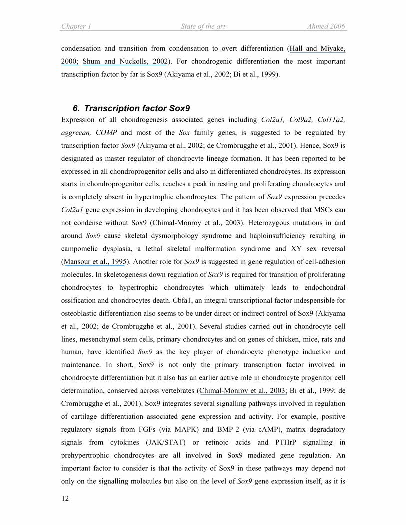
6. Transcription factor Sox9 Expression of all chondrogenesis associated genes including Col2a1, Col9a2, Col11a2,
aggrecan, COMP and most of the Sox family genes, is suggested to be regulated by
transcription factor Sox9 (Akiyama et al., 2002; de Crombrugghe et al., 2001). Hence, Sox9 is
designated as master regulator of chondrocyte lineage formation. It has been reported to be
expressed in all chondroprogenitor cells and also in differentiated chondrocytes. Its expression
starts in chondroprogenitor cells, reaches a peak in resting and proliferating chondrocytes and
is completely absent in hypertrophic chondrocytes. The pattern of Sox9 expression precedes
Col2a1 gene expression in developing chondrocytes and it has been observed that MSCs can
not condense without Sox9 (Chimal-Monroy et al., 2003). Heterozygous mutations in and
around Sox9 cause skeletal dysmorphology syndrome and haploinsufficiency resulting in
campomelic dysplasia, a lethal skeletal malformation syndrome and XY sex reversal
(Mansour et al., 1995). Another role for Sox9 is suggested in gene regulation of cell-adhesion
molecules. In skeletogenesis down regulation of Sox9 is required for transition of proliferating
chondrocytes to hypertrophic chondrocytes which ultimately leads to endochondral
ossification and chondrocytes death. Cbfa1, an integral transcriptional factor indespensible for
osteoblastic differentiation also seems to be under direct or indirect control of Sox9 (Akiyama
et al., 2002; de Crombrugghe et al., 2001). Several studies carried out in chondrocyte cell
lines, mesenchymal stem cells, primary chondrocytes and on genes of chicken, mice, rats and
human, have identified Sox9 as the key player of chondrocyte phenotype induction and
maintenance. In short, Sox9 is not only the primary transcription factor involved in
chondrocyte differentiation but it also has an earlier active role in chondrocyte progenitor cell
determination, conserved across vertebrates (Chimal-Monroy et al., 2003; Bi et al., 1999; de
Crombrugghe et al., 2001). Sox9 integrates several signalling pathways involved in regulation
of cartilage differentiation associated gene expression and activity. For example, positive
regulatory signals from FGFs (via MAPK) and BMP-2 (via cAMP), matrix degradatory
signals from cytokines (JAK/STAT) or retinoic acids and PTHrP signalling in
prehypertrophic chondrocytes are all involved in Sox9 mediated gene regulation. An
important factor to consider is that the activity of Sox9 in these pathways may depend not
only on the signalling molecules but also on the level of Sox9 gene expression itself, as it is

Ahmed 2006 State of the Art Chapter 1
13
seen in human single allele mutation studies (Bi et al., 2001). Due to post natal fatality,
attempts for generation of Sox9 null homozygous transgenic mice have been abandoned thus,
Sox9 expression studies are limited to mouse embryo chimeras derived from Sox9-/- ES
(Akiyama et al., 2002). Hence, use of new technologies and novel ideas for understanding
Sox9 function and mode of action is vital.
7. Animal models Many aspects of biology are similar in most or all organisms and
often it is easier to study a particular aspect in one organism than
in others. These much-studied organisms are commonly referred
to as model organisms. Mammals share many basic biological
functions, such as the regulation of cell division, the development of organ systems, and
immune response. The rat is a principal model organism to link function to genes. The large
number of inbred rat models and the vast amount of data available are helpful for studies of
human physiology and pathology. These animals are a unique resource for studying and
identifying genetic pathways relevant to some human diseases. Tissue engineering based
approaches require pre clinical in vitro and in vivo studies. Though the mouse model is better
characterized and is less cost and space intensive, a big advantage of the rat over the mouse
model is bigger animal size which results in more cellular material. The rat system is also
ideal for tissue engineering studies due to less inter-individual variation and easy availability
of the cells. (http://www.ncbi.nlm.nih.gov/About/model/mammal.html).
8. Future MSCs based therapeutic approaches for cartilage repair exhibit ample promise. The more
effort goes in deciphering regulatory mechanisms of stem cells proliferation and
differentiation the quicker we will reach the goal. Optimal application of MSCs for cartilage
repair requires biodegradable scaffolds and proper signalling to maintain the articular
phenotype of the newly formed cartilage. Factors and the involved pathways which influence
geno- and phenotypical changes in MSCs during differentiation could be vital for ex vivo
cartilage regeneration. Hence, studies like this are important for the future of cartilage tissue
engineering.
Chapter 1 State of the art Ahmed 2006
14
Reference List
1. Akiyama,H., Chaboissier,M.C., Martin,J.F., Schedl,A., and de Crombrugghe,B. (2002). The transcription factor Sox9 has essential roles in successive steps of the chondrocyte differentiation pathway and is required for expression of Sox5 and Sox6. Genes Dev. 16, 2813-2828.
2. Baksh,D., Song,L., and Tuan,R.S. (2004). Adult mesenchymal stem cells: characterization, differentiation, and application in cell and gene therapy. J. Cell Mol. Med. 8, 301-316.
3. Barry,F.P. (2003). Biology and clinical applications of mesenchymal stem cells. Birth Defects Res. C. Embryo. Today 69, 250-256.
4. Bi,W., Deng,J.M., Zhang,Z., Behringer,R.R., and de Crombrugghe,B. (1999). Sox9 is required for cartilage formation. Nat. Genet. 22, 85-89.
5. Bi,W., Huang,W., Whitworth,D.J., Deng,J.M., Zhang,Z., Behringer,R.R., and de Crombrugghe,B. (2001). Haploinsufficiency of Sox9 results in defective cartilage primordia and premature skeletal mineralization. Proc. Natl. Acad. Sci. U. S. A 98, 6698-6703.
6. Bianchi,G., Muraglia,A., Daga,A., Corte,G., Cancedda,R., and Quarto,R. (2001). Microenvironment and stem properties of bone marrow-derived mesenchymal cells. Wound. Repair Regen. 9, 460-466.
7. Bianco,P., Riminucci,M., Gronthos,S., and Robey,P.G. (2001). Bone marrow stromal stem cells: nature, biology, and potential applications. Stem Cells 19, 180-192.
8. Bianco,P. and Robey,P.G. (2001). Stem cells in tissue engineering. Nature 414, 118-121.
9. Brittberg,M., Lindahl,A., Nilsson,A., Ohlsson,C., Isaksson,O., and Peterson,L. (1994). Treatment of deep cartilage defects in the knee with autologous chondrocyte transplantation. N. Engl. J. Med. 331, 889-895.
10. Bruckner,P., Horler,I., Mendler,M., Houze,Y., Winterhalter,K.H., Eich-Bender,S.G., and Spycher,M.A. (1989). Induction and prevention of chondrocyte hypertrophy in culture. J. Cell Biol. 109, 2537-2545.
11. Budde,B., Blumbach,K., Ylostalo,J., Zaucke,F., Ehlen,H.W., Wagener,R., Ala-Kokko,L., Paulsson,M., Bruckner,P., and Grassel,S. (2005). Altered integration of matrilin-3 into cartilage extracellular matrix in the absence of collagen IX. Mol. Cell Biol. 25, 10465-10478.
12. Cancedda,R., Dozin,B., Giannoni,P., and Quarto,R. (2003). Tissue engineering and cell therapy of cartilage and bone. Matrix Biol. 22, 81-91.

Ahmed 2006 State of the Art Chapter 1
15
13. Caplan,A.I. (2005). Review: mesenchymal stem cells: cell-based reconstructive therapy in orthopedics. Tissue Eng 11, 1198-1211.
14. Chepda,T., Cadau,M., Girin,P., Frey,J., and Chamson,A. (2001). Monitoring of ascorbate at a constant rate in cell culture: effect on cell growth. In Vitro Cell Dev. Biol. Anim 37, 26-30.
15. Chimal-Monroy,J., Rodriguez-Leon,J., Montero,J.A., Ganan,Y., Macias,D., Merino,R., and Hurle,J.M. (2003). Analysis of the molecular cascade responsible for mesodermal limb chondrogenesis: Sox genes and BMP signaling. Dev. Biol. 257, 292-301.
16. de Crombrugghe,B., Lefebvre,V., Behringer,R.R., Bi,W., Murakami,S., and Huang,W. (2000). Transcriptional mechanisms of chondrocyte differentiation. Matrix Biol. 19, 389-394.
17. de Crombrugghe,B., Lefebvre,V., and Nakashima,K. (2001). Regulatory mechanisms in the pathways of cartilage and bone formation. Curr. Opin. Cell Biol. 13, 721-727.
18. Dorshkind,K. (1990). Regulation of hemopoiesis by bone marrow stromal cells and their products. Annu. Rev. Immunol. 8, 111-137.
19. Eikenberry,E.F. and Bruckner,P. (1999). Supromolecular structure of cartilage matrix. In Dynamics of bone and cartilage metabolism, M.J.Seibel, S.P.Robins, and J.P.bilezikian, eds. (California: Academic Press,USA.), pp. 289-300.
20. Ferguson,C., Alpern,E., Miclau,T., and Helms,J.A. (1999). Does adult fracture repair recapitulate embryonic skeletal formation? Mech. Dev. 87, 57-66.
21. Friedenstein,A.J., Gorskaja,J.F., and Kulagina,N.N. (1976). Fibroblast precursors in normal and irradiated mouse hematopoietic organs. Exp. Hematol. 4, 267-274.
22. Ge,Z., Hu,Y., Heng,B.C., Yang,Z., Ouyang,H., Lee,E.H., and Cao,T. (2006). Osteoarthritis and therapy. Arthritis Rheum. 55, 493-500.
23. Goldring,M.B., Tsuchimochi,K., and Ijiri,K. (2006). The control of chondrogenesis. J. Cell Biochem. 97, 33-44.
24. Haeusler,G., Walter,I., Helmreich,M., and Egerbacher,M. (2005). Localization of matrix metalloproteinases, (MMPs) their tissue inhibitors, and vascular endothelial growth factor (VEGF) in growth plates of children and adolescents indicates a role for MMPs in human postnatal growth and skeletal maturation. Calcif. Tissue Int. 76, 326-335.
25. Hall,B.K. and Miyake,T. (2000). All for one and one for all: condensations and the initiation of skeletal development. Bioessays 22, 138-147.
26. Hauselmann,H.J., Fernandes,R.J., Mok,S.S., Schmid,T.M., Block,J.A., Aydelotte,M.B., Kuettner,K.E., and Thonar,E.J. (1994). Phenotypic stability of bovine articular chondrocytes after long-term culture in alginate beads. J. Cell Sci. 107 ( Pt 1), 17-27.
27. Hunziker,E.B. (2002). Articular cartilage repair: basic science and clinical progress. A review of the current status and prospects. Osteoarthritis. Cartilage. 10, 432-463.
Chapter 1 State of the art Ahmed 2006
16
28. Inada,M., Wang,Y., Byrne,M.H., Rahman,M.U., Miyaura,C., Lopez-Otin,C., and Krane,S.M. (2004). Critical roles for collagenase-3 (Mmp13) in development of growth plate cartilage and in endochondral ossification. Proc. Natl. Acad. Sci. U. S. A 101, 17192-17197.
29. Iozzo,R.V. (1999). The biology of the small leucine-rich proteoglycans. Functional network of interactive proteins. J. Biol. Chem. 274, 18843-18846.
30. Johnstone,B., Hering,T.M., Caplan,A.I., Goldberg,V.M., and Yoo,J.U. (1998). In vitro chondrogenesis of bone marrow-derived mesenchymal progenitor cells. Exp. Cell Res. 238, 265-272.
31. Kavalkovich,K.W., Boynton,R.E., Murphy,J.M., and Barry,F. (2002). Chondrogenic differentiation of human mesenchymal stem cells within an alginate layer culture system. In Vitro Cell Dev. Biol. Anim 38, 457-466.
32. Lefebvre,V., Behringer,R.R., and de Crombrugghe,B. (2001). L-Sox5, Sox6 and Sox9 control essential steps of the chondrocyte differentiation pathway. Osteoarthritis. Cartilage. 9 Suppl A, S69-S75.
33. Lennon,D.P., Haynesworth,S.E., Young,R.G., Dennis,J.E., and Caplan,A.I. (1995). A chemically defined medium supports in vitro proliferation and maintains the osteochondral potential of rat marrow-derived mesenchymal stem cells. Exp. Cell Res. 219, 211-222.
34. Magne,D., Vinatier,C., Julien,M., Weiss,P., and Guicheux,J. (2005). Mesenchymal stem cell therapy to rebuild cartilage. Trends Mol. Med. 11, 519-526.
35. Majumdar,M.K., Banks,V., Peluso,D.P., and Morris,E.A. (2000). Isolation, characterization, and chondrogenic potential of human bone marrow-derived multipotential stromal cells. J. Cell Physiol 185, 98-106.
36. Maniatopoulos,C., Sodek,J., and Melcher,A.H. (1988). Bone formation in vitro by stromal cells obtained from bone marrow of young adult rats. Cell Tissue Res. 254, 317-330.
37. Mannello,F., Tonti,G.A., Bagnara,G.P., and Papa,S. (2005). Role and function of matrix metalloproteinases in the differentiation and biological characterization of mesenchymal stem cells. Stem Cells.
38. Mansour,S., Hall,C.M., Pembrey,M.E., and Young,I.D. (1995). A clinical and genetic study of campomelic dysplasia. J. Med. Genet. 32, 415-420.
39. Mendler,M., Eich-Bender,S.G., Vaughan,L., Winterhalter,K.H., and Bruckner,P. (1989). Cartilage contains mixed fibrils of collagen types II, IX, and XI. J. Cell Biol. 108, 191-197.
40. Mitchell,P.G., Magna,H.A., Reeves,L.M., Lopresti-Morrow,L.L., Yocum,S.A., Rosner,P.J., Geoghegan,K.F., and Hambor,J.E. (1996). Cloning, expression, and type II collagenolytic activity of matrix metalloproteinase-13 from human osteoarthritic cartilage. J. Clin. Invest 97, 761-768.

Ahmed 2006 State of the Art Chapter 1
17
41. Moore,K.A. and Lemischka,I.R. (2006). Stem cells and their niches. Science 311, 1880-1885.
42. Mundlos,S. and Olsen,B.R. (1997). Heritable diseases of the skeleton. Part I: Molecular insights into skeletal development-transcription factors and signaling pathways. FASEB J. 11, 125-132.
43. Pittenger,M.F., Mackay,A.M., Beck,S.C., Jaiswal,R.K., Douglas,R., Mosca,J.D., Moorman,M.A., Simonetti,D.W., Craig,S., and Marshak,D.R. (1999). Multilineage potential of adult human mesenchymal stem cells. Science 284, 143-147.
44. Shum,L. and Nuckolls,G. (2002). The life cycle of chondrocytes in the developing skeleton. Arthritis Res. 4, 94-106.
45. Solchaga,L., Welter,J., Lennon,D.P., and Caplan,A.I. (2004). Generation of pluripotent stem cells and their differentiation to the chondrocytic phenotype. In Cartilage and Osteoarthritis, D.M.Sabatini, p.pastoureau, and F.D.ceunink, eds. Humana Press, Totowa, New Jersey), pp. 53-67.
46. Thur,J., Rosenberg,K., Nitsche,D.P., Pihlajamaa,T., Ala-Kokko,L., Heinegard,D., Paulsson,M., and Maurer,P. (2001). Mutations in cartilage oligomeric matrix protein causing pseudoachondroplasia and multiple epiphyseal dysplasia affect binding of calcium and collagen I, II, and IX. J. Biol. Chem. 276, 6083-6092.
47. Von der,M.K. (1999). Structure and biosynthesis of collagens. In Dynamics of bone and cartilage metabolism, M.J.Seibel, S.P.Robins, and J.P.bilezikian, eds. (California: Academic Press,USA.), pp. 3-18.
48. Von der,M.K., Kirsch,T., Nerlich,A., Kuss,A., Weseloh,G., Gluckert,K., and Stoss,H. (1992). Type X collagen synthesis in human osteoarthritic cartilage. Indication of chondrocyte hypertrophy. Arthritis Rheum. 35, 806-811.
49. Vu,T.H. and Werb,Z. (2000). Matrix metalloproteinases: effectors of development and normal physiology. Genes Dev. 14, 2123-2133.
50. Yin,T. and Li,L. (2006). The stem cell niches in bone. J. Clin. Invest 116, 1195-1201.


19
Chapter 2
Gene expression and protein secretion
profile of naïve and differentiated rat
marrow stromal cells
Nazish Ahmed1,2, Joachim Grifka1 and Susanne Grässel1,2*
1Experimental Orthopaedics, Dept. of Orthopaedics, University of Regensburg, Kaiser Karl V
Allee-3, 93077 Bad Abbach, Germany. 2Experimental Orthopaedics, Centre for
Biotechnology, BioPark I, University of Regensburg, 93053 Regensburg, Germany.
*Corresponding author
Submitted: Cell and tissue research, 2006
Abstract
Adult mesenchymal stem cells (MSCs) are adherent stromal cells of non-haematopoietic
origin. Upon in vitro expansion they retain their self renewal capacity as well as their potential
to differentiate into tissues of mesenchymal lineage including bone, cartilage, muscle, tendon
and connective tissue. Amongst these cartilage is the only tissue which lacks self renewal
capacity thus MSCs are an excellent tool for therapeutic regeneration of focal cartilage
lesions. For optimal manipulation of MSCs identification and better understanding of
Chapter 2 Profile of marrow stromal cells Ahmed 2006
20
molecular mechanisms regulating differentiation pathways is needed. While many studies on
genetic profiles of human MSCs exist; basal gene and protein profiles of adult rat MSCs has
rarely being investigated. Rat is a widely used mammalian experimental model for preclinical
studies. Therefore, this study undertook a comprehensive profiling of mRNA expression of
osteo- / chondrogenesis related genes in undifferentiated and differentiated rat adult MSCs by
using quantitative RT-PCR technology. A differential gene expression pattern was observed
depending on osteo-chondral differentiation status of the cells. At protein level TIMP-1,
MCP-1 and VEGF164α were detected in culture supernatant and CINC-2 and ß-NGF in the
cell lysate of MSCs after an antibody array analysis. Our results provide a foundation for a
more reproducible and reliable quality control of rat bone marrow derived MSCs used for
osteo-chondro differentiation studies.
Key words: rat marrow stromal cells, profiling, cytokines, gene expression, antibody array
Introduction
Cartilage cannot regenerate upon injury or degeneration, the exact reason of this incapacity is
unclear, but lack of access to progenitor cells is often pointed out as a main reason. Adult
osteo-chondroprogenitor bone marrow stromal or mesenchymal stem cells (MSCs) are known
progenitors of tissue of mesenchymal lineage. These chondroprogenitor MSCs residing as part
of bone marrow stroma in the medullary cavity of the bone are inaccessible to the avascular
cartilage (Cancedda et al., 2003). They make up less then 1% of the bone marrow cellular
population which is otherwise dominated by haematopoietic cells. In vitro expansion of bone
marrow cells leads to separation of the stromal MSCs relying on their capacity of adherence to
the plastic surface of the tissue culture vessels. Upon expansion these cells retain not only
their self renewal capacity but also their differentiation competence (Friedenstein et al., 1976).
This intrinsic competence can be invoked under controlled nutritional and mechanical
conditions to differentiate MSCs into bone, cartilage, muscle, tendon and connective tissue
(Bruder et al., 1994). MSCs have shown promise for ex-vivo cartilage regenerative medicine
and for treatment of large bone defects (Quarto et al., 2001; Horwitz et al., 1999).
Lack of common standards and of a precise definition of initial cell preparations is a major
obstacle for MSCs based research and application. Genetic profiling of human MSCs for
Ahmed 2006 Profile of marrow stromal cells Chapter 2
21
trans-differentiation capacities has provided data on differential gene expression depending on
origin and commitment status of MSCs from different tissue sources (Wagner et al., 2005).
However, very little is known about the proteomic and genomic profile of MSCs which can
qualify as osteo-chondroprogenitor cells. Knowledge of changes in expression pattern of
matrix associated structural genes, of proteases and their inhibitors, transcription factors and
adhesion molecules during osteogenesis and chondrogenesis are essential for providing proper
conditions for in vitro differentiation of MSCs.
It has been known that together with the other cells of bone marrow microenvironment MSCs
contribute to an interactive network of cytokines, growth factors and matrix proteins to sustain
existence and differentiation of haematopoietic stem cells (Dorshkind, 1990). However, what
role the network plays in proliferation and differentiation of MSCs is still unknown. Some
reports indicate differential gene expression of cytokines as IL-1, IL-6 and GM-CSF in MSCs.
However, the studies are mainly restricted to gene expression analysis and do not address the
protein. (Majumdar et al., 1998; Kim et al., 2005). Knowledge of a basal cytokine protein
profile in MSCs is essential to understand the effect of these cytokines on MSCs and their
differentiation.
Mammalian animal models like rat share many aspects of the human genomic, cellular and
immunological structure. Large numbers of inbred models and subsequent vast amount of
available data parallel to the small size and rapid development are advantages of employing
rat MSCs for in-vitro studies. However, genetic and proteomic expression profiling of rat
MSCs is incomplete. Keeping in mind that the basic background profile of human MSCs
cannot be taken at face value for rat MSCs, this study undertook a comprehensive profiling of
baseline mRNA level of osteo-chondro related genes in undifferentiated adult rat MSCs which
is subsequently compared with the changes in gene expression upon osteo-chondrogenic
differentiation. Secondly, a basic protein profile of factors generated from undifferentiated
MSCs was compared with that of the differentiated cells.

Chapter 2 Profile of marrow stromal cells Ahmed 2006
22
Material and Methods
Isolation and cell culture of MSCs
MSCs were isolated from rat bone marrow as described earlier (Ahmed et al., 2006). Briefly,
bone marrow was removed from tibiae and femora of 6 week old male Sprague-Dawley rats
by centrifugation (2000rpm for 3min). Homogenized bone marrow was cultured in 175cm²
tissue culture flasks in proliferation medium containing 5% glutamate, 1%
antibiotics/antimycotics and 10% FBS (Gibco, Invitrogen, UK) in α-MEM (Sigma Aldrich,
Germany). Non-adherent cells were removed on the 3rd day and the adherent CFU-cells were
proliferated until reaching 70% confluence. Magnetic associated cell sorting procedure
(MACS- Miltenyi Biotech, Germany) was carried out according to the manufactures
instructions; in short, expanded cells were incubated in suspension with 4µl of selected
antibody / 106cells for 5min at 37°C followed by washing and incubation with goat anti-
mouse secondary antibody coupled with magnetic beads for 15min at 4°C. The suspension
was passed through a magnetic column (Miltenyi Biotec´s LS-MACS columns); while
labelled cells were retained by the magnetic field the flow through containing the unlabelled
negative fraction was collected. After removing the column from the magnetic field the
antibody labelled cells (positive fraction) was also flushed out. For both fractions cells were
counted and stained. Antibodies used for MACS were directed against CD45 and CD49a
(Chemicon, Germany), CD71 and CD106 (BD Bioscience, USA).
Chondrogenic and osteogenic differentiation of MSCs
For chondrogenesis, cells were cultured for 21 days in high density 3-D alginate cultures. To
prepare the culture 107 cells/ml were suspended in 1.2% alginate. The cell- alginate amalgam
was dropped into 102 mM CaCl2 solution via a syringe which resulted in formation of beads
with a diameter of 2-3mm containing approx. 105 cells/bead. Beads were cultured in 2.3ml
chondrogenic medium in 12 well tissue culture plates. 10 alginate beads were used for RNA
isolation and gene expression analysis. Cells were released from alginate by incubation at
37°C for 30min in 55mM sodium citrate and 0.15M sodium chloride buffer followed by cell
recovery by 3min spin at 750xg. Chondrogenic medium contained: ITS+ premix (6.25µg/ml
insulin, 6.25ng/ml selenium acid, 6.25µg/ml transferrin, 1.25mg/ml BSA and 5.35µg/ml
linoleic acid (BD Biosciences, USA.), 110µg/ml pyruvate, 40µg/ml proline, 0.1µM
dexamethasone, 50µg/ml ascorbic acid and 10ng/ml TGFß-3 (Johnstone et al., 1998) (R&D
Systems) in α MEM high glucose (Gibco, Invitrogen, UK). Alginate sodium salts were

Ahmed 2006 Profile of marrow stromal cells Chapter 2
23
acquired from Sigma Aldrich, Germany (Cat# A0682-100G). Osteogenesis was induced for
15 days in 6 well culture plates, 250,000 cells/ well were cultured as monolayer in osteogenic
medium containing, 10% FCS, 1% pen/strep, 10nM dexamethasone, 50µg/ml Ascorbate 2-
PO4 and 10mM ß-Na glycerophosphate.
Immunofluorescence
For immunofluorescence analysis alginate beads were irreversibly polymerized by replacing
CaCl2 with 100mM BaCl2 (Hauselmann et al., 1994). The beads were fixed with 4%
paraformaldehyde and after sequential dehydration embedded in paraffin. 4µm sections were
acquired and the deparaffinized and rehydrated sections were used for staining after
hyaluronidase digestion. Undifferentiated MSCs were fixed with 4% paraformaldehyde.
Slides were blocked for 1h at 37°C in 5% normal goat serum and 1% BSA in PBS containing
Complete Mini 1:5 protease inhibitor solution (Roche, Germany). After washing with PBS
cells were stained overnight at 4°C with monoclonal antibodies directed against collagen II,
1:1000 diluted (Acris, Germany), CD49a, diluted 1:50 (Chemicon, Germany) and D7fib,
diluted 1:50 (Acris, Germany). The appropriate Alexa568 or Alexa488 conjugated secondary
antibodies (goat anti mouse, 5 µg/ml; Molecular Probes, USA) were added for 1h at RT. After
washing slides were permanently mounted with DAKO fluorescent mounting medium
(DAKO, USA) and covered with cover slips. Slides were evaluated with scanning laser
microscopy (C1 confocal microscope from Nikon, Germany) and photos were taken with a
Nikon C4 camera and software.
Histological analysis
Osteogenically differentiated cells were washed with PBS and fixed with methanol for 10
min. After rinsing with water staining was carried out for 2min with 1% alizarin red prepared
in 25% ammonia. Stained cells were thoroughly washed and photographed with a Nikon C4
camera after complete drying.
RNA Isolation and reverse transcription
RNA was isolated by an affinity column chromatography method with Ambion’s
RNAqueous4-PCR kit according to the manufacturer’s protocol. For removal of possible
DNA contamination DNaseI enzyme (DNA-free, Ambion, USA) was used. RNA
concentration was determined at 585nm wavelength with RiboGreen RNA quantification kit
(Molecular Probes, USA). Conversion of 0.5-1µg of RNA to cDNA was done with

Chapter 2 Profile of marrow stromal cells Ahmed 2006
24
SuperScript II reverse transcriptase kit (Invitrogen, UK) in 20µl of total reaction volume in
the presence of 40 units/µl recombinant ribonuclease inhibitor (RNase OUT®), 500µg/ml of
Oligo-dT primers, 10mM dNTPs and 200 units of SuperScriptII enzyme in First-Strand
Buffer and 0.1M DTT for 50min at 42°C followed by an extension period of 15min at 70°C.
Relative quantitative PCR
Relative quantitative PCR was performed using the SYBR Green Dye I on ABI 7000 Prism
Sequence detection system (AB Systems, USA) according to manufacturer’s instructions.
Briefly, 1µl of cDNA was amplified in 50µl final volume of 0.2µM of each primer suspended
in SYBR green master mix (AB Systems, USA). Amplification parameters were identical for
all primer pairs and were repeated for 40 cycles, denaturation occurred at 95°C for 0.15min
and annealing at 60°C for 1min. ∆Ct values, i.e. difference in mRNA expression level of
genes of interest to that of an endogenous control, ß-actin, was evaluated from three
independent experiments and plotted on an inverse scale so that 0 indicates expression of the
endogenous control and thus taken as the reference point for the genes of interest. Mean
relative quantification (RQ) values were calculated by the software “RQ study application
v1.1” (ABI Prism 7000 SDS software v1.1) according to the ∆∆Ct method using ß-Actin as
endogenous control and undifferentiated MSCs (day 0) as calibrator. Primers were designed
either with freeware Primer3 (http://frodo.wi.mit.edu/cgi-bin/primer3/primer3_www.cgi) or
“Primer Express” software supplied by AB. All primers were manufactured at MWG –
Biotech, Germany and are listed in table 1.
Antibody array analysis
RayBio™ (Tebu-bio, France) rat cytokine antibody array I for conditioned medium was used
according to the manufacturer’s instructions. Briefly, first the membranes were blocked for
30min in 5% BSA in 0.01M Tris buffer with 0.15M NaCl (pH 7.6) followed by overnight
incubation at 4°C in 1ml of cell culture supernatant or cell lysates. After washing and
incubation with biotin coupled anti-cytokine antibodies for 2h a second series of washing was
performed and then the membranes were incubated with horse reddish peroxidase-conjugated
streptavidin for 2h. Signals were detected with the provided detection solution using a CURIX
60 film developer (Agfa, Germany).

Ahmed 2006 Profile of marrow stromal cells Chapter 2
25
Statistical Analysis
One way ANOVA and student t test were employed for quantitative PCR experiments
performed in triplicate. The level of significance was determined by Turkey´s test with 2
group comparison.
Table 1: Primers used for quantitative PCR
Gene RefSeq# Amplicon Forward primer Reverse primer
(bp) (5´ - 3´) (5´ - 3´)
Alpha-10 XM_001063132 188 -tttcttcgggaaatcagagc -tggatggagaagccaatctc
Alpha-11 XM_001075650 170 -tggaggtccaacacttcctc -gggtttcagtccctcctctc
Aggrecan NM_022190 224 -ggccttccctctggatttag -ccgcactactgtccaac
BMP-7 AF100787 167 -gaaaacagcagcagtgacca -gtggcgttcatgtaggagt
ß-Actin NM_031144 104 -gtagccatccaggctgtgtt -ccctcatagatgggcagagt
Cbfa1 XM_34016 86 -gccgggaatgatgagaacta -agatcgttcaacctggccact
Col1a1 RGD61817* 59 -tccagggctccaacgaga -ctgtaggtgaatccactgttgc
Col2a1 NM_012929 60 -cccctgcagtacatgcgg -ctcgacgtcatgctgtctcaag
Col10a1 AJ131848* 247 -ccctattggaccaccaggta -tctctgtccgctctttgtga
Col16 a1 M92642 97 -gcctggtaccaaaggtgaaa -catagcctggaggaccttga
COMP NM_012834 167 -tgacttcgatgctgacaagg -gaacgatctccattccctga
Ihh XM_343590 103 -atgaagacggccatcactcag -cgcgccagtagtccgtacttat
MMP-2 NM_031054 111 -gaccggtttatttggcgga -ggcctcatacacagcgtcaat
MMP-13 XM_343345 93 -acctgggatttccaaaagagg -acacgtccttccctgagaaga
Sox4 XM-344594 58 -ggcccatgaacgcctttat -ctggatgaacgggatcttgtc
Sox6 XM_215016 51 -gaaatccatgtccaaccaggac -cgggcctgctcttcatagtaag
Sox9 XM_343981 140 -ctgaagggctacgactggac -tactggtctgccagcttcct
Tbox2 XM_220810 71 -gcccactctccgtttgtatgag -aggacgaggcatcggattc
TIMP-1 NM_053819 136 -gattcgacgctgtgggaaat -tttccgttccttaaacggcc
TIMP-2 NM_021989 140 -ggcaagatgcacattaccctct -atgtagcatgggatcatagggc
Tgfß-3 NM_013174 86 -ttccttcttggccgtatttcc -tgtgtgggatccagaatcca
VEGFα NM_031836 71 -tggctttactgctgtacctcca -tttctgctccccttctgtcgt
VEGFR-2 NM_013062 95 -ttgcctagtcaagcagctcgt -cgatggtctcaccaatggttg
*Locus ID

Chapter 2 Profile of marrow stromal cells Ahmed 2006
26
Osteogenesis
c d.
f.e
40µm 10µm
MSCs
a b.
200µm 200µm
100µm 100µm
Chondrogenesis
Results
Cellular morphology and surface antigen markers of MSCs
Formation of CFU-F is one of the basic classification of bone marrow derived stem cells
along with rapid adhesion and extended proliferation (Bianco et al., 2001). After in-vitro
expansion of bone marrow isolated cells, colony forming units of fibroblast like cells (CFU-F)
designated as MSCs readily adhered to the culture flasks and showed typical fibroblast like
morphology (Fig 1a). Immunofluorescence analysis of the adherent cells exhibited positive
staining for fibroblast marker D7fib and alpha 1 integrin marker CD49a (Fig. 1b).
Figure 1: Morphological characterization of MSCs CFU-F like marrow derived adherent cells (a) double stained with D7fib (red) and CD49a (green) by immunofluorescence (b) exhibit typical stem cells morphology. Dark red staining with alizarin red indicates calcified matrix formation after 15 days of osteogenesis in monolayer while the control culture remained unstained (c, d).
Chondrogenically differentiated cells kept in alginate were stained with collagen II antibody. After 21 days (f) the positive signal was stronger when compared with the beads stained on day 0 (e).
One of the major characterization criteria for MSCs is the retained differentiation potential
upon expansion. MSCs were induced for 15 days in monolayer culture for osteogenesis and
21 days for chondrogenesis in 3-D high density alginate cultures. Osteogenesis cultures
stained positive for alizarin red indicating formation of calcified matrix while the un-induced
Ahmed 2006 Profile of marrow stromal cells Chapter 2
27
control cultures did not stain (Fig 1c-d). Chondrogenically induced cells exhibited clear
collagen II staining of the surrounding matrix on day 21 of the culture as compared with day 0
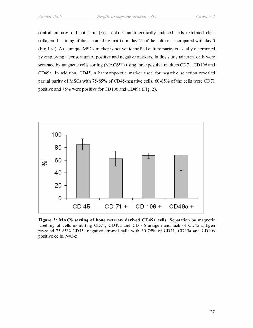
(Fig 1e-f). As a unique MSCs marker is not yet identified culture purity is usually determined
by employing a consortium of positive and negative markers. In this study adherent cells were
screened by magnetic cells sorting (MACS™) using three positive markers CD71, CD106 and
CD49a. In addition, CD45, a haematopoietic marker used for negative selection revealed
partial purity of MSCs with 75-85% of CD45-negative cells. 60-65% of the cells were CD71
positive and 75% were positive for CD106 and CD49a (Fig. 2).
Figure 2: MACS sorting of bone marrow derived CD45+ cells Separation by magnetic labelling of cells exhibiting CD71, CD49a and CD106 antigen and lack of CD45 antigen revealed 75-85% CD45- negative stromal cells with 60-75% of CD71, CD49a and CD106 positive cells. N=3-5

Chapter 2 Profile of marrow stromal cells Ahmed 2006
28
Gene expression profiling
With the help of quantitative RT-PCR and specific primers (Table 1) baseline mRNA
expression of selected genes was determined. Subsequently, the MSCs were induced to
differentiate into osteogenic and chondrogenic lineages and mRNA level was compared with
the expression in un-induced undifferentiated cells.
Among the most highly expressed genes in undifferentiated MSCs were integrin Alpha-11,
VEGFa, Sox9, Sox4, Cbfa1,TGFß-3, TIMP-1, TIMP-2 and MMP-13; Col1a1 exhibited the
highest expression of all analysed genes. Lowest expressed genes were VEGFr-2 (kdr), Ihh,
Col2a1, and Col10a1. Expression of integrin Alpha-10, Sox6, Col16a1, Aggrecan, COMP and
MMP-2 was also observed (Fig. 3a).
Figure 3a: Selected gene expression profile of adherent undifferentiated MSCs Quantitative mRNA expression level of selected groups of genes in undifferentiated MSCs (black bars) was normalized to ß-actin employing the ∆CT method using SYBR I Green dye. The calculated ∆CT value was subtracted from 40, the total number of PCR cycles. This value was plotted into a graph to show a positive correlation between bar length and gene expression. N = 3.

Ahmed 2006 Profile of marrow stromal cells Chapter 2
29
Figure 3b: Selected gene expression profile of adherent undifferentiated MSCs compared with differentiated cells Relative quantitative mRNA expression level of selected genes was determined by the ∆∆CT method using ß-actin gene expression as endogenous control. Expression level of each gene was compared after osteogenesis (stripped grey bars) and chondrogenesis (grey bars) with that of undifferentiated MSCs used as calibrator (set to 1). N=3
Upon induction of osteogenic and chondrogenic differentiation gene expression of Alpha 11,
TIMP-1 and TIMP-2 and Col10a1 remained mostly unaltered. After 21 days of chondrogenic
favourable conditions mRNA level of integrin Alpha-10, Sox9, Sox6, Ihh, Col2a1, Col16a1,
COMP, and MMP-2 were up regulated compared with uninduced MSCs. Culturing MSCs for
15 days in osteogenic medium induced mRNA level of VEGF, VEGFr-2,Col1a1, and MMP-
13. Differentiation in general caused about 10 folds increase in mRNA level of Tbox2 and
Cbfa1 independent of the differentiation pathway while Sox4 and Aggrecan expression was
slightly higher at the end of chondrogenic differentiation. Chondrogenesis reduced the mRNA
level of VEGFa and MMP-13 while osteogenesis had no significant negative effects on gene
expression level (Fig. 3b).

Chapter 2 Profile of marrow stromal cells Ahmed 2006
30
TIMP-1 MCP-1
MSC+ + +
- + - VEGFα-164 CINC-2
+ - - - + +
ß-NGF
Osteo Chondro
+ + -
Cytokine and growth factor profile of undifferentiated and differentiated MSCs
Cell culture supernatants and cell lysates from undifferentiated and differentiated MSCs were
analyzed for secretory molecules using an antibody array covering 15 cytokines plus ß-NGF,
TIMP-1, CNTF, leptin and VEGFα-164. Only tissue inhibitor of matrix metalloproteases
(TIMP-1) was secreted from undifferentiated as well as from differentiated MSCs, while
monocyte chemoattractant protein (MCP-1) was exclusively secreted from undifferentiated
MSCs. Secretion of vascular endothelial growth factor (VEGFα-164) was observed after
induction of osteogenic and chondrogenic differentiation. When the same antibody array was
examined with the cell lysate two additional molecules were identified. Nerve growth factor
(ß-NGF) was detected in undifferentiated as well as in cells after osteogenic differentiation
and cytokine induced neutrophil chemoattractant (CINC-2) was detected in low amounts after
osteogenesis, both factors were undetectable after chondrogenic induction (Fig. 4).
A. MSCs Osteogenic Chondrogenic
Cell lysate
Medium
Negative Control
for medium
B.
Figure 4: Comparison of cytokine profile of undifferentiated and differentiated MSCs Antibody array containing 19 different cytokines was used to compare protein profile of proliferated MSCs with MSCs after 15 days of osteogenic induction and after 21 days of chondrogenic induction (A.). Dot intensities higher than the negative control were accepted as positive expression. Among the 5 molecules with
differential induction pattern TIMP-1 was present in all three conditions, MCP-1 was specific to undifferentiated MSCs. The induction profile of the remaining three factors was influenced by the differentiation state (B.). N=2
* ** ς
∆
* TIMP-1 ∆ MCP-1 ** VEGFα-164 + CINC-2 ς ß-NGF
+

Ahmed 2006 Profile of marrow stromal cells Chapter 2
31
The remaining 14 proteins were undetectable in all culture conditions analysed, these include,
CINC-3, Ciliary neurotrophic factor (CNTF), fractalkine, GM-CSF, IFN-γ, IL-1α, IL-1ß, IL-
4, IL-6, IL-10, lipoplysacchride induced c-x-c chemokine (LIX), leptin, macrophage
inflammatory protein (MIP-3α), and TNF-α.
Discussion
Cellular morphology and surface antigen markers of MSCs
In the medullary cavity of bone marrow MSCs coexist with haematopoietic stem cells. In-
vitro the two cell populations can be easily separated because MSCs tend to adhere to the
tissue culture plastic and form small colony like units arising from single cells (Pittenger et
al., 1999). Formation of these colony forming units of fibroblast like cells (CFU-F) is one of
the basic classification of bone marrow derived stem cells parallel to rapid adhesion and
extended proliferation (Bianco et al., 2001). The CFU-F forming rat bone marrow derived
MSCs used in this study showed CD 45-/low, CD106+. CD49a+ and CD 71+ antigen profiles, as
determined by MACS after 7 days of expansion. CD106 (V-CAM) and CD71 (transferring)
are well established positive markers for bone marrow derived osteo-chondro progenitor cells.
CD49a alpha 1 integrin is expressed only on MSCS and not on other cells of bone marrow.
CD45 is mainly expressed on haematopoietic cells and MSCs are known not to express this
antigen.
Immunofluorescence analysis further confirmed the identity of MSCs which stained positive
for fibroblast marker D7fib and CD49a. The D7fib antigen is described as a specific marker
for MSCs as suitable as STRO-1 and CD105 (Jones et al., 2002).
BM-derived cells obtained by sole adherance to plastic surfaces are not a pure population with
regard to their CD marker profile. MACS™ cell sorting shows that the cells are a
heterogeneous population comprising of a small proportion of CD45-positive cells of
haematopoietic origin and a large fraction of CD45-negative cells. It is described that both cell
populations exert a mutual influence important for differentiation pathways. The role of
MSCs in providing a supportive microenvironment for HSCs is well established and in some
cases even direct interaction has been observed (Prockop, 1997). However, vice versa
influence of haematopoietic cells in the differentiation of MSCs is still not answered
satisfactorily, co transplantation of HSCs with MSCs in hematologic malignancy patients
have indicated faster engraftment and low graft versus host disease (Lazarus et al., 2005). In

Chapter 2 Profile of marrow stromal cells Ahmed 2006
32
addition, we could demonstrate a positive effect of haematopoietic cells on the chondrogenic
differentiation of MSCs in vitro (Ahmed et al., 2006). Therefore, investigation of gene and
protein expression profile of MSC populations frequently isolated solely according to plastic
adherance will be helpful for basic characterization.
Gene expression profile
Little information is available about baseline mRNA expression profile of rat multipotent
MSCs. Here, we present profiling of osteo-chondrogenic related gene expression in
undifferentiated and differentiated MSCs including growth factors, transcription factors,
proteases and their inhibitors, structural components and adhesion molecules.
Integrin subunit Alpha-11 was amongst the highly expressed genes, whereas only little Alpha-
10 mRNA was detected in undifferentiated MSCs. Integrin Alpha-11 is highly expressed in
various mesenchymal tissues around the cartilage anlage in the developing skeleton. Its
additional expression in MSCs of the intervertebral discs and in keratinocytes in the cornea is
compatible with its role as receptor for interstitial collagens (Tiger et al., 2001). Integrin
Alpha-10 is a chondrocyte specific major collagen II and IX -binding integrin integral to
cartilage development and mature hyaline cartilage (Camper et al., 1998; Camper et al.,
2001). In rat MSCs neither osteogenic nor chondrogenic differentiation alter Alpha-11 gene
expression, contrary to Alpha-10 mRNA level which increased about 1000 folds during
chondrogenesis. The ratio between Alpha-11 / Alpha-10 integrin gene expression makes these
genes useful markers for monitoring the progression of MSCs differentiation towards
particular lineages.
Transcription factors which regulate osteo- and chondrogenic lineage as, Tbox2 (Brachyury),
Sox9, Sox4 and Cbfa1 were also expressed in un-induced naïve MSCs. In early vertebrate
embryonic development Tbox2 is involved in determination of mesodermal cell fates and
patterning during limb bud formation, where it is restricted to subridge mesoderm (Liu et al.,
2003). Both, Sox9 and Tbox2 are essential components for the BMP-dependent onset of
chondrogenesis (Hoffmann et al., 2002). Increase in Tbox2 gene expression after
differentiation substantiates its role in osteo-chondrogenic cell fate determination. So far
involvement of Tbox2 in the osteogenic pathway of MSCs has not been described and needs
further investigations. Sox9 is expressed in all essential stages of chondrogenic development
starting from pre-condensation nodules and including proliferating chondrocytes, however,
the expression is abrogated in hypertrophic chondrocytes (Zhao et al., 1997). At that terminal
differentiation stage inhibition is possibly regulated due to a negative feed back mechanism

Ahmed 2006 Profile of marrow stromal cells Chapter 2
33
accompanied by the activation of the PTHrP and Ihh signal cascade in pre-hypertrophic
chondrocytes (Akiyama et al., 2002); (de Crombrugghe et al., 2000). After chondrogenic
differentiation gene expression of both, Sox9 and Ihh was upregulated, indicating cells might
be arrested in a pre-hypertrophic stage, corroborated by lack of gene expression of collagen X
which is restricted to terminally differentiated hypertrophic chondrocytes (Schmid et al.,
1990). Interestingly, master regulators of both the osteogenic pathway, Cbfa1, and
chondrogenic lineage, Sox9, are both already expressed in un-induced MSCs. This implicates
that the progenitor cells probably have entered a specific commitment stage which may have
been triggered by cell culture conditions. Differential expression of both the master
transcription factors might serve as marker for harvesting osteo-chondroprogenitor cells at
different states of commitment thus allowing a separation of these populations.
Factors of the Tgfß super family have been identified as important inducers and promoters of
differentiation in human MSCs (Barry et al., 2001a). Therefore, high expression of Tgfß-3 in
un-induced MSCs might be considered as a prerequisite for ECM structural components
regulation during differentiation. Structural components of the cartilaginous extracellular
matrix like collagen II (Mendler et al., 1989), aggrecan and COMP (Hedbom et al., 1992;
DiCesare et al., 1994) are known to be upregulated in MSCs during the chondrogenic
differentiation pathway (Barry et al., 2001b). This makes them suitable as markers for
discriminating between undifferentiated and chondrogenically differentiated MSCs. In
undifferentiated rat MSCs collagen II and X displayed low basal gene expression indicating
lack of chondrogenic differentiation, however, gene expression of aggrecan and COMP was
observed. On the other hand collagen I showed undoubtedly the highest gene expression
amongst all screened molecules which further increased during differentiation. Reduction of
relative gene expression of collagen I during chondrogenic differentiation is desirable and
underscores the development of a cartilaginous matrix. Little is known about expression
profile of fibril-associated collagens with interrupted triple helices (FACIT) in MSCs, gene
expression of collagen XVI in undifferentiated MSCs indicates a yet unknown role in
proliferation or differentiation processes. Notably, collagen XVI mRNA level is upregulated
during chondrogenesis compared with osteogenesis implicating a specific role in
chondrogenesis.
The results demonstrate high gene expression of VEGFα in un-induced MSCs with an
increase during osteogenic differentiation. During osteoblasts differentiation angiogenic factor
VEGF modulates matrix mineralization in an autocrine manner and up regulates the
expression of its receptor (Mayer et al., 2005). VEGFα gene expression level appears as a

Chapter 2 Profile of marrow stromal cells Ahmed 2006
34
suitable marker for osteogenic committed MSCs while chondrogenic differentiation requires
downregulation of VEGFα mRNA.
The ability of MMPs to alter the activity or function of numerous proteins by proteolytic
procession suggests that they are also involved in various cellular functions and matrix
formation during development. Gene expression of MMPs and TIMPs in MSCs cultures has
been reported earlier in studies on human MSCs of different origin (Silva, Jr. et al., 2003;
Mannello et al., 2005). MMP-13 together with its inhibitors, TIMP-1 and -2 were highly
expressed in un-induced MSCs indicating a role in stem cell migration as assigned to MMP-2
(Cronwright et al., 2005). While TIMP-1 and -2 were not regulated during MCSs
differentiation MMP-13 was controversely regulated during osteo-chondrogenensis.
Downregulation of MMP-13 gene expression after chondrogenesis correlates with arrest of
cells in a pre-hyperthrophic stage. Relatively higher MMP-13 mRNA level after osteogenesis
indicates progressing mineralization as MMP-13 along with alkaline phosphatase and
osteocalcin is known to be involved in endochondral and intramembranous ossification
(Tuckermann et al., 2000). Notably, MMP-2 gene expression was elevated during
chondrogenesis which may indicate beginning of late stage chondrogenesis as proteolytic
activity of MMP-2 is vital for ECM turnover prior to hypertrophy (Aigner et al., 2001).
Protein profiling of undifferentiated and differentiated MSCs
Secretory cytokines, proteases and growth factors are key players in cell proliferation and
differentiation. Mixed cultures of BM isolated MSCs are known to secrete haematopoiesis
supportive cytokines. We speculated that the protein profile should differ according to the
differentiation state of the MSCs. TIMP-1 was the only identified molecule to be secreted
from undifferentiated as well as differentiated MSCs while monocyte chemoattractant protein
(MCP-1) was exclusively secreted from undifferentiated MSCs. TIMP-1 is a multifunctional
molecule apart from its biological activity as inhibitor of metalloproteinases,it is thought to be
involved in regulation of cell growth and differentiation. Cell surface binding of TIMP-1 and -
4 has been reported, but the identification of specific cell surface receptors is missing (Stetler-
Stevenson and Seo, 2005). Therefore, TIMP-1 secreted by MSCs may act as inducer or
repressor of specific factors necessary for commitment to specific differentiation pathways of
MSCs. MCP-1, which recruits monocytes into ischemic tissue and stimulates chemotaxis, is
secreted exclusively from un-induced MSCs. MSCs are described to secrete this chemokine
which is speculated to be involved in homing of circulating stem and progenitor cells to the
region of injuries and thus contribute to healing (Kinnaird et al., 2004; Liu and Hwang, 2005)

Ahmed 2006 Profile of marrow stromal cells Chapter 2
35
Notably, osteoarthritic chondrocytes secrete MCP-1 and it is suggested that it contributes in
triggering inflammatory mechanisms involved in persistence of joint diseases (Pulsatelli et al.,
1999). MCP-1 secretion could be used as a marker for un-induced MCSs which becomes
attenuated during chondrogenic and osteogenic differentiation processes.
Secretion of VEGFα-164 was induced only after the cells were subjected to osteogenic and
chondrogenic differentiation. VEGF influences vascular permeability and is a strong
angiogenic protein indispensable for neovascularisation during embryonic skeletal
development (Goldring et al., 2006). VEGF as a multifaceted protein may have a role in
induction of metalloproteinases during late stage of chondrogenesis marking the onset of
hypertrophy (Pufe et al., 2004). Lack of VEGF secretion in undifferentiated MSCs might be
taken as an indicator for a relatively pure MSC population moistly devoid of hematopoietic
and endothelial progenitor cell contamination. Latter stem cells are known to express and
secrete VEGF (Rehman et al., 2003; Iba et al., 2002) The discrepancy in low gene and high
protein secretion profile of VEGFα after chondrogenic differentiation is very likely due to
post translational regulation. VEGF production is known to be regulated at translational level
as overexpression of transcription factor c-Myc related with rapid induction of tumors, also
leads to 10 fold higher VEGF production while the gene expression level remains unaltered.
The mechanism of action is most probably higher ribosome biogenesis which increases
translation efficiency of selected mRNA (Mezquita et al., 2005).
When the same antibody array was examined with the cellular lysates, two additional
molecules were identified, ß-NGF was detected in undifferentiated as well as in cells after
osteogenic differentiation and cytokine induced neutrophil chemoattractant (CINC-2) was
detected only after osteogenesis. CINC-2 attracts neutrophils and has been suggested to
inhibit acute inflammation indicating its role is osteogenesis could be anti-inflammatory
(Nakagawa et al., 1994). So far this chemkine has not been determined in MSCs but in bone
marrow derived macrophages exposed to pro-inflammamtory cytokins (Crippen et al., 1998)
It is known that early non-committed MSCs with high cologenic potential bind to antibodies
against NGF receptor (Quirici et al., 2002) and that undifferentiated bone marrow derived
cells express NGF together with other neurotrophic factors (Yaghoobi and Mowla, 2006).
This is in line with the production of ß-NGF in undifferentiated MSCs. The naïve MSCs loose
ß-NGF upon chondrogenic differentiation but keep it under osteogenic conditions which
suggests a role of ß-NGF in osteogenesis as a trophic factor stimulating collagen synthesis and
expression of specific osteogenic marker as alkaline phosphatase (Yada et al., 1994). In

Chapter 2 Profile of marrow stromal cells Ahmed 2006
36
mandibular distraction osteogenesis elevated expression of NGF has been demonstrated (Park
et al., 2006).
In conclusion, we were able to demonstrate gene expression and secretion of molecules from a
mostly CD45-negative bone marrow derived cell population which may be crucial for MSCs
fate determination and sustenance. Identification and better understanding of the molecular
events characterizing differentiation of MSCs which allows discrimination among specific
differentiation stages is a prerequisite for manipulation of MSCs to regenerate complex tissues
as cartilage and bone.
Acknowledgements
We thank Maren Marschner for her excellent technical assistance. Evy Lundgren-Akerlund of
Cartella AG, Sweden is gratefully acknowledged for providing Alpha-10 and Alpha-11
primers. Special thanks go to Prof. Dr. Achim Göpferich (Pharmaceutical Technology,
University of Regensburg) for his unconditional support.

Ahmed 2006 Profile of marrow stromal cells Chapter 2
37
Reference List
1. Ahmed,N., Vogel,B., Rohde,E., Strunk,D., Grifka,J., Schulz,M.B., and Grassel,S. (2006). CD45-positive cells of haematopoietic origin enhance chondrogenic marker gene expression in rat marrow stromal cells. Int. J. Mol. Med. 18, 233-240.
2. Aigner,T., Zien,A., Gehrsitz,A., Gebhard,P.M., and McKenna,L. (2001). Anabolic and catabolic gene expression pattern analysis in normal versus osteoarthritic cartilage using complementary DNA-array technology. Arthritis Rheum. 44, 2777-2789.
3. Akiyama,H., Chaboissier,M.C., Martin,J.F., Schedl,A., and de Crombrugghe,B. (2002). The transcription factor Sox9 has essential roles in successive steps of the chondrocyte differentiation pathway and is required for expression of Sox5 and Sox6. Genes Dev. 16, 2813-2828.
4. Barry,F., Boynton,R.E., Liu,B., and Murphy,J.M. (2001b). Chondrogenic differentiation of mesenchymal stem cells from bone marrow: differentiation-dependent gene expression of matrix components. Exp. Cell Res. 268, 189-200.
5. Barry,F., Boynton,R.E., Liu,B., and Murphy,J.M. (2001a). Chondrogenic differentiation of mesenchymal stem cells from bone marrow: differentiation-dependent gene expression of matrix components. Exp. Cell Res. 268, 189-200.
6. Bianco,P., Riminucci,M., Gronthos,S., and Robey,P.G. (2001). Bone marrow stromal stem cells: nature, biology, and potential applications. Stem Cells 19, 180-192.
7. Bruder,S.P., Fink,D.J., and Caplan,A.I. (1994). Mesenchymal stem cells in bone development, bone repair, and skeletal regeneration therapy. J. Cell Biochem. 56, 283-294.
8. Camper,L., Hellman,U., and Lundgren-Akerlund,E. (1998). Isolation, cloning, and sequence analysis of the integrin subunit alpha10, a beta1-associated collagen binding integrin expressed on chondrocytes. J. Biol. Chem. 273, 20383-20389.
9. Camper,L., Holmvall,K., Wangnerud,C., Aszodi,A., and Lundgren-Akerlund,E. (2001). Distribution of the collagen-binding integrin alpha10beta1 during mouse development. Cell Tissue Res. 306, 107-116.
10. Cancedda,R., Dozin,B., Giannoni,P., and Quarto,R. (2003). Tissue engineering and cell therapy of cartilage and bone. Matrix Biol. 22, 81-91.
11. Crippen,T.L., Klasing,K.C., and Hyde,D.M. (1998). Regulation of cytokine-induced neutrophil chemoattractant in rat bone marrow-derived macrophages by inflammatory mediators. Pathobiology 66, 293-301.
12. Cronwright,G., Le Blanc,K., Gotherstrom,C., Darcy,P., Ehnman,M., and Brodin,B. (2005). Cancer/testis antigen expression in human mesenchymal stem cells: down-regulation of SSX impairs cell migration and matrix metalloproteinase 2 expression. Cancer Res. 65, 2207-2215.
Chapter 2 Profile of marrow stromal cells Ahmed 2006
38
13. de Crombrugghe,B., Lefebvre,V., Behringer,R.R., Bi,W., Murakami,S., and Huang,W. (2000). Transcriptional mechanisms of chondrocyte differentiation. Matrix Biol. 19, 389-394.
14. DiCesare,P., Hauser,N., Lehman,D., Pasumarti,S., and Paulsson,M. (1994). Cartilage oligomeric matrix protein (COMP) is an abundant component of tendon. FEBS Lett. 354, 237-240.
15. Dorshkind,K. (1990). Regulation of hemopoiesis by bone marrow stromal cells and their products. Annu. Rev. Immunol. 8, 111-137.
16. Friedenstein,A.J., Gorskaja,J.F., and Kulagina,N.N. (1976). Fibroblast precursors in normal and irradiated mouse hematopoietic organs. Exp. Hematol. 4, 267-274.
17. Goldring,M.B., Tsuchimochi,K., and Ijiri,K. (2006). The control of chondrogenesis. J. Cell Biochem. 97, 33-44.
18. Hauselmann,H.J., Fernandes,R.J., Mok,S.S., Schmid,T.M., Block,J.A., Aydelotte,M.B., Kuettner,K.E., and Thonar,E.J. (1994). Phenotypic stability of bovine articular chondrocytes after long-term culture in alginate beads. J. Cell Sci. 107 ( Pt 1), 17-27.
19. Hedbom,E., Antonsson,P., Hjerpe,A., Aeschlimann,D., Paulsson,M., Rosa-Pimentel,E., Sommarin,Y., Wendel,M., Oldberg,A., and Heinegard,D. (1992). Cartilage matrix proteins. An acidic oligomeric protein (COMP) detected only in cartilage. J. Biol. Chem. 267, 6132-6136.
20. Hoffmann,A., Czichos,S., Kaps,C., Bachner,D., Mayer,H., Kurkalli,B.G., Zilberman,Y., Turgeman,G., Pelled,G., Gross,G., and Gazit,D. (2002). The T-box transcription factor Brachyury mediates cartilage development in mesenchymal stem cell line C3H10T1/2. J. Cell Sci. 115, 769-781.
21. Horwitz,E.M., Prockop,D.J., Fitzpatrick,L.A., Koo,W.W., Gordon,P.L., Neel,M., Sussman,M., Orchard,P., Marx,J.C., Pyeritz,R.E., and Brenner,M.K. (1999). Transplantability and therapeutic effects of bone marrow-derived mesenchymal cells in children with osteogenesis imperfecta. Nat. Med. 5, 309-313.
22. Iba,O., Matsubara,H., Nozawa,Y., Fujiyama,S., Amano,K., Mori,Y., Kojima,H., and Iwasaka,T. (2002). Angiogenesis by implantation of peripheral blood mononuclear cells and platelets into ischemic limbs. Circulation 106, 2019-2025.
23. Johnstone,B., Hering,T.M., Caplan,A.I., Goldberg,V.M., and Yoo,J.U. (1998). In vitro chondrogenesis of bone marrow-derived mesenchymal progenitor cells. Exp. Cell Res. 238, 265-272.
24. Jones,E.A., Kinsey,S.E., English,A., Jones,R.A., Straszynski,L., Meredith,D.M., Markham,A.F., Jack,A., Emery,P., and McGonagle,D. (2002). Isolation and characterization of bone marrow multipotential mesenchymal progenitor cells. Arthritis Rheum. 46, 3349-3360.
25. Kim,D.H., Yoo,K.H., Choi,K.S., Choi,J., Choi,S.Y., Yang,S.E., Yang,Y.S., Im,H.J., Kim,K.H., Jung,H.L., Sung,K.W., and Koo,H.H. (2005). Gene expression profile of cytokine and growth factor during differentiation of bone marrow-derived mesenchymal stem cell. Cytokine 31, 119-126.

Ahmed 2006 Profile of marrow stromal cells Chapter 2
39
26. Kinnaird,T., Stabile,E., Burnett,M.S., Shou,M., Lee,C.W., Barr,S., Fuchs,S., and Epstein,S.E. (2004). Local delivery of marrow-derived stromal cells augments collateral perfusion through paracrine mechanisms. Circulation 109, 1543-1549.
27. Lazarus,H.M., Koc,O.N., Devine,S.M., Curtin,P., Maziarz,R.T., Holland,H.K., Shpall,E.J., McCarthy,P., Atkinson,K., Cooper,B.W., Gerson,S.L., Laughlin,M.J., Loberiza,F.R., Jr., Moseley,A.B., and Bacigalupo,A. (2005). Cotransplantation of HLA-identical sibling culture-expanded mesenchymal stem cells and hematopoietic stem cells in hematologic malignancy patients. Biol. Blood Marrow Transplant. 11, 389-398.
28. Liu,C., Nakamura,E., Knezevic,V., Hunter,S., Thompson,K., and Mackem,S. (2003). A role for the mesenchymal T-box gene Brachyury in AER formation during limb development. Development 130, 1327-1337.
29. Liu,C.H. and Hwang,S.M. (2005). Cytokine interactions in mesenchymal stem cells from cord blood. Cytokine 32, 270-279.
30. Majumdar,M.K., Thiede,M.A., Mosca,J.D., Moorman,M., and Gerson,S.L. (1998). Phenotypic and functional comparison of cultures of marrow-derived mesenchymal stem cells (MSCs) and stromal cells. J. Cell Physiol 176, 57-66.
31. Mannello,F., Tonti,G.A., Bagnara,G.P., and Papa,S. (2005). Role and function of matrix metalloproteinases in the differentiation and biological characterization of mesenchymal stem cells. Stem Cells.
32. Mayer,H., Bertram,H., Lindenmaier,W., Korff,T., Weber,H., and Weich,H. (2005). Vascular endothelial growth factor (VEGF-A) expression in human mesenchymal stem cells: autocrine and paracrine role on osteoblastic and endothelial differentiation. J. Cell Biochem. 95, 827-839.
33. Mendler,M., Eich-Bender,S.G., Vaughan,L., Winterhalter,K.H., and Bruckner,P. (1989). Cartilage contains mixed fibrils of collagen types II, IX, and XI. J. Cell Biol. 108, 191-197.
34. Mezquita,P., Parghi,S.S., Brandvold,K.A., and Ruddell,A. (2005). Myc regulates VEGF production in B cells by stimulating initiation of VEGF mRNA translation. Oncogene 24, 889-901.
35. Nakagawa,H., Komorita,N., Shibata,F., Ikesue,A., Konishi,K., Fujioka,M., and Kato,H. (1994). Identification of cytokine-induced neutrophil chemoattractants (CINC), rat GRO/CINC-2 alpha and CINC-2 beta, produced by granulation tissue in culture: purification, complete amino acid sequences and characterization. Biochem. J. 301 ( Pt 2), 545-550.
36. Park,B.W., Kim,J.R., Lee,J.H., and Byun,J.H. (2006). Expression of nerve growth factor and vascular endothelial growth factor in the inferior alveolar nerve after distraction osteogenesis. Int. J. Oral Maxillofac. Surg. 35, 624-630.
37. Pittenger,M.F., Mackay,A.M., Beck,S.C., Jaiswal,R.K., Douglas,R., Mosca,J.D., Moorman,M.A., Simonetti,D.W., Craig,S., and Marshak,D.R. (1999). Multilineage potential of adult human mesenchymal stem cells. Science 284, 143-147.
Chapter 2 Profile of marrow stromal cells Ahmed 2006
40
38. Prockop,D.J. (1997). Marrow stromal cells as stem cells for nonhematopoietic tissues. Science 276, 71-74.
39. Pufe,T., Harde,V., Petersen,W., Goldring,M.B., Tillmann,B., and Mentlein,R. (2004). Vascular endothelial growth factor (VEGF) induces matrix metalloproteinase expression in immortalized chondrocytes. J. Pathol. 202, 367-374.
40. Pulsatelli,L., Dolzani,P., Piacentini,A., Silvestri,T., Ruggeri,R., Gualtieri,G., Meliconi,R., and Facchini,A. (1999). Chemokine production by human chondrocytes. J. Rheumatol. 26, 1992-2001.
41. Quarto,R., Mastrogiacomo,M., Cancedda,R., Kutepov,S.M., Mukhachev,V., Lavroukov,A., Kon,E., and Marcacci,M. (2001). Repair of large bone defects with the use of autologous bone marrow stromal cells. N. Engl. J. Med. 344, 385-386.
42. Quirici,N., Soligo,D., Bossolasco,P., Servida,F., Lumini,C., and Deliliers,G.L. (2002). Isolation of bone marrow mesenchymal stem cells by anti-nerve growth factor receptor antibodies. Exp. Hematol. 30, 783-791.
43. Rehman,J., Li,J., Orschell,C.M., and March,K.L. (2003). Peripheral blood "endothelial progenitor cells" are derived from monocyte/macrophages and secrete angiogenic growth factors. Circulation 107, 1164-1169.
44. Schmid,T.M., Popp,R.G., and Linsenmayer,T.F. (1990). Hypertrophic cartilage matrix. Type X collagen, supramolecular assembly, and calcification. Ann. N. Y. Acad. Sci. 580, 64-73.
45. Silva,W.A., Jr., Covas,D.T., Panepucci,R.A., Proto-Siqueira,R., Siufi,J.L., Zanette,D.L., Santos,A.R., and Zago,M.A. (2003). The profile of gene expression of human marrow mesenchymal stem cells. Stem Cells 21, 661-669.
46. Stetler-Stevenson,W.G. and Seo,D.W. (2005). TIMP-2: an endogenous inhibitor of angiogenesis. Trends Mol. Med. 11, 97-103.
47. Tiger,C.F., Fougerousse,F., Grundstrom,G., Velling,T., and Gullberg,D. (2001). alpha11beta1 integrin is a receptor for interstitial collagens involved in cell migration and collagen reorganization on mesenchymal nonmuscle cells. Dev. Biol. 237, 116-129.
48. Tuckermann,J.P., Pittois,K., Partridge,N.C., Merregaert,J., and Angel,P. (2000). Collagenase-3 (MMP-13) and integral membrane protein 2a (Itm2a) are marker genes of chondrogenic/osteoblastic cells in bone formation: sequential temporal, and spatial expression of Itm2a, alkaline phosphatase, MMP-13, and osteocalcin in the mouse. J. Bone Miner. Res. 15, 1257-1265.
49. Wagner,W., Wein,F., Seckinger,A., Frankhauser,M., Wirkner,U., Krause,U., Blake,J., Schwager,C., Eckstein,V., Ansorge,W., and Ho,A.D. (2005). Comparative characteristics of mesenchymal stem cells from human bone marrow, adipose tissue, and umbilical cord blood. Exp. Hematol. 33, 1402-1416.
50. Yada,M., Yamaguchi,K., and Tsuji,T. (1994). NGF stimulates differentiation of osteoblastic MC3T3-E1 cells. Biochem. Biophys. Res. Commun. 205, 1187-1193.

Ahmed 2006 Profile of marrow stromal cells Chapter 2
41
51. Yaghoobi,M.M. and Mowla,S.J. (2006). Differential gene expression pattern of neurotrophins and their receptors during neuronal differentiation of rat bone marrow stromal cells. Neurosci. Lett. 397, 149-154.
52. Zhao,Q., Eberspaecher,H., Lefebvre,V., and de Crombrugghe,B. (1997). Parallel expression of Sox9 and Col2a1 in cells undergoing chondrogenesis. Dev. Dyn. 209, 377-386.


43
Chapter 3
CD 45-positive cells of haematopoietic
origin enhance chondrogenic marker gene
expression in rat marrow stromal cells
Nazish Ahmed1*, Breda Vogel2,3*, Eva Rohde4, Dirk Strunk4, Joachim Grifka1 Michaela B.
Schulz3 and Susanne Grässel1.
1Experimental Orthopaedics, Dept. of Orthopaedics, University of Regensburg, Kaiser Karl V
Allee-3, 93077 Bad Abbach, Germany. 2Pharmaceutical Technology, University of
Regensburg, 93053 Regensburg, Germany. 3Institute of Pharmaceutical Sciences,
Pharmaceutical Technology, Karl-Franzens University, 8010 Graz, Austria. 4Division of
Hematology and Department of Transfusion Medicine, Medical University, 8010 Graz,
Austria
*Contributed equally
Published: International Journal of Molecular Medicine 18:233-240, 2006
Abstract Adult mesenchymal stem cells (MSCs) can be readily isolated from bone marrow, expanded
in culture and subsequently subjected to differentiation into various connective tissue
lineages. In general, for animal studies separation of MSCs from other bone marrow derived

Chapter 3 Effect of haematopoietic cells on chondrogenesis Ahmed 2006
44
cells is achieved by sole adherence to plastic surface of tissue culture flasks, however, this
procedure produces a heterogeneous cell population containing CD45-positive haematopoietic
cells (HCs) and haematopoietic stem cells (HSCs) as well. It is well known, that such mixed
cell cultures consisting of cocultures of differentiated somatic cells with adult stem cells
promote differentiation towards specific cell lineages. For determining the effect of the CD45-
positive cell population on the differentiation potential of MSCs we have sorted out the bone
marrow derived adherent cells by immunomagnetic technique (MACSTM) to attain a
subpopulation of CD45-depleted cells. Here, we show that the presence of adherent CD45-
positive HCs not only promote expression of the chondrogenic marker genes Col2a1, COMP
and Sox9, but also of Col1a1, Col10a1 and to a certain degree Cbfa1 in MSCs when cultured
in an appropriate three-dimensional environment. These observations constitute a step
towards unravelling the influence of haematopoietic cells on chondrogenic differentiation of
MSCs.
Key words: rat marrow stromal cells, MACS, chondrogenic differentiation, haematopoietic
cells, CD45
Introduction Adult bone marrow stromal cells regarded as mesenchymal stem cells are progenitors of
connective tissue cells, thus are excellent candidates for chondrogenic differentiation studies.
The progeny of adult stem cells includes both daughter stem cells and committed progenitors
with a more restricted differentiation potential. These progenitors in turn give rise to
differentiated cell types. Mesenchymal stem cells (MSCs) can differentiate under specific
cultural and physical conditions into multiple mesenchymal lineages namely, osteocytes,
chondrocytes, adipocytes, astrocytes and myocytes (Pittenger et al., 1999b; Cancedda et al.,
2003). Bone marrow consists of primarily non-adherent haematopoietic cells and
haematopoietic stem cells (99%) along with a minor population of MSCs (≤1 %). Both of the
stem cell types are known to co-exist and have been suggested to cooperate in one another’s
differentiation (Bianco et al., 2001). The role of MSCs in haematopoietic microenvironment
formation is beginning to unravel and it is assumed that the presence of adherent MSCs and
their progeny facilitates HSCs differentiation into granulocytes and erythrocytes both in vivo
and in vitro (Galmiche et al., 1993; Prockop, 1997a; Majumdar et al., 1998b). Conversely, in

Ahmed 2006 Effect of haematopoietic cells on chondrogenesis Chapter 3
45
depth studies concerning the role of HSCs in MSCs commitment and differentiation towards a
distinct lineage are scarce.
Adult rat MSCs are routinely isolated from tibio-femoral bone marrow by relying solely on
their adherence to the plastic surface of tissue culture flasks (Friedenstein et al., 1976; Caplan
and Bruder, 2001). The isolated cells constitute a heterogeneous population which always
contains HCs therefore; obtaining a pure population of non-haematopoietic cells remains
difficult. This haematopoietic cell fraction varies depending on the species; being relatively
high in initial cultures of mouse marrow cells (Phinney et al., 1999) and less than 30% in
human cell cultures. In rat marrow stromal cells this trend has not been studied thoroughly,
although the presence of haematopoietic cells in primary (7 days) and secondary (18 days)
bone marrow cultures has been indicated (Herbertson and Aubin, 1997). Human and murine
MSC studies show subsequent loss of haematopoietic cell surface markers when cultures are
maintained for 2 or 3 weeks. Prior to differentiation experiments the haematopoietic cell
population is often separated from MSCs by employing a magnetic associated cell sorting
(MACS) system. The system immunologically separates different cell types by labelling cell
surface antigens with magnetic beads followed by sorting through a magnetic column. As a
unique identifying marker for MSCs is still lacking a negative selection protocol is carried out
using CD34 (My10) or CD45 (leukocyte common antigen) for exclusion of undesired
positively labelled haematopoietic cells.
One of the major differentiation pathways of MSCs occurs along the chondrogenic lineage
which can happen autonomously in three separate mesenchymal cell lines, cranial neural
crest, sclerotome cells and lateral plate mesoderm cells (von der, 1999). The first stage of
chondrogenic differentiation is conversion of undifferentiated MSCs to committed
osteochondroprogenitor cells leading to cell condensation and growth arrest. The major
proteins involved in condensation initiation are either fibronectin or belong to the
transforming growth factor ß family of proteins (TGFß-1, -2 and -3). Cells present at the
centre of the condensation nodules first form pre-chondrocytes and then chondrocytes which
start to produce cartilage matrix marked by up regulation of structural protein genes as
Col2a1, Col9a2, Col11a2, aggrecan and COMP. Expression of chondrocyte marker genes is
controlled by members of the Sox-family, in particular Sox9 has been characterized as a
master transcription factor with essential direct or indirect regulatory effects exerted along the
entire chondrogenic differentiation pathway. It is expressed in all chondroprogenitor cells and
also in differentiated chondrocytes (Bi et al., 1999; de Crombrugghe et al., 2000b; Mundlos
and Olsen, 1997; Lefebvre et al., 2001) .

Chapter 3 Effect of haematopoietic cells on chondrogenesis Ahmed 2006
46
In this study we have evaluated the degree of haematopoietic cell coexistence in primary
cultures of rat MSCs and their influence on the chondrogenic differentiation potential of
MSCs. We have determined the difference in chondrogenic marker gene expression of
heterogeneous MSCs isolated by sole adherence to plastic surfaces compared to that of a more
homogeneous subpopulation of MACS sorted CD45-depleted cells.
Material and Methods MSC isolation and primary cell culture
A modified version of the MSCs isolation procedure from rat bone marrow was optimized
(Dobson et al., 1999; Maniatopoulos et al., 1988). Briefly, 6 week old male Sprague - Dawley
rats were killed with CO2 and tibiae and femora were aseptically removed. The bones were
cut from the middle and centrifuged in a 1.5ml tube at 2000 rpm for 3min (mini centrifuge-
Eppendorf). The centrifuged bone marrow cells were collected and homogenized with 18g,
21g and 23g needles and seeded at the rate of 2.5 x 10⁵ cells/cm2 in 175cm² tissue culture
flasks in proliferation medium containing 5% glutamate, 1% antibiotics/antimycotics (Gibco.)
and 10%FBS (Gibco, Invitrogen, UK; Lot No.: 40F7430K) in α-MEM (Sigma Aldrich,
Germany). All non-adherent cells were removed on the 3rd day and the adherent cells were
proliferated.
Magnetic separation of cells (MACS)
When the flasks were ~75% confluent (9-11 days) cells were harvested with trypsin digestion.
Separation procedure was carried out according to the manufactures instructions; in short,
cells were incubated with 4µl of CD45 antibody/106cells for 5min at 37°C followed by
washing and a second incubation with goat anti-mouse secondary antibody coupled with
magnetic beads (Miltenyi Biotech) for 15min at 4°C. The suspension was passed through a
magnetic column (Miltenyi Biotec´s LS-MACS columns); while labelled cells got coupled to
the magnetic field the flow through containing unlabelled CD45-negative fraction was
collected.
Immunofluorescence
20,000 cells/chamber were seeded on chamber slides (Biocoat slides, BD, Bioscience) and
incubated overnight at 37°C and 5% CO2. Cells were blocked for 1h at 37°C in Complete
Mini 1:5 (Roche, Germany) plus 5% normal goat serum and 1% BSA in PBS. After washing

Ahmed 2006 Effect of haematopoietic cells on chondrogenesis Chapter 3
47
with PBS cells were stained with monoclonal antibodies directed against CD45 (Cb1 1502,
Chemicon), CD68 and D7Fib (Acris, Germany) in 1:50 dilution ratio overnight at 4°C. The
appropriate Alexa568 or Alexa488 conjugated secondary antibodies (goat anti mouse, 5
µg/ml; Molecular Probes, US) were added for 1h at RT. After washing slides were
permanently mounted with DAKO fluorescent mounting medium (DAKO, USA) and covered
with coverslips. Slides were evaluated with scanning laser microscopy (C1 confocal
microscope from Nikon) and photos were taken with a Nikon C4 camera and software.
Flow cytometric analysis
Cells obtained from 1 animal (approx. 8x 10⁷ cells, as determined after NH4Cl lysis of the
erythrocytes) were seeded in 4x 150cm2 flasks (5x 10⁵ cells/cm2). The cells were left to
adhere 1, 2, 3 or 4 days before non adherent cells were removed. Cells were trypsinized on
day 7 after the isolation and resuspended in PBS. The cell suspension was first blocked for 15
min at 4°C with sheep serum followed by incubation with antibodies against rat CD45-PE
(mouse IgG1 against rat CD45, Acris Antibodies, Germany), CD29-FITC (Hamster IgM
against rat CD29, BD Pharmingen, USA) and with corresponding isotype controls for 30min
at 4°C. Cells were washed with PBS containing sodium azide and bovine serum albumin and
resuspended. Shortly before the FACS – analysis propidium iodide was added to each sample
in order to discriminate the dead cells which were excluded from the evaluation. All
measurements were performed on a FACSCalibur® instrument (Becton Dickenson, USA)
with CellQuest data acquisition software (Becton Dickenson, USA). Data analysis was
preformed with WinMDI 2.8 software (free access on http://facs.scripps.edu/software.html).
A minimum of 1x10⁴ viable cells was acquired per data set.
Chondrogenic differentiation
A batch of adherent cells (undepleted) was subjected directly to chondrogenesis favourable
condition, the other batch was depleted of CD45-positive cells by MACS prior to
chondrogenic differentiation experiments. For chondrogenic differentiation, high density 3-D
cultures were attained by preparing cell-alginate amalgam in 1.2% alginate at a concentration
of 107 cells/ml. 2-3mm cell-alginate beads containing approx.105 cells/bead, were formed by
dropping the suspension into 102 mM CaCl2 solution. The culture was carried out in 12 well
tissue culture plates for 4 weeks; medium was changed every 2-3 days. 10 alginate beads were
removed every 7th day for RNA isolation and gene expression analysis. To release the cells
from alginate the beads were incubated for 30min at 37°C on a shaker in 55mM sodium

Chapter 3 Effect of haematopoietic cells on chondrogenesis Ahmed 2006
48
citrate and 0.15M sodium chloride buffer; cells were recovered after 3min spin at 750xg. The
chondrogenic medium contained; ITS+ premix (6.25µg/ml insulin, 6.25ng/ml selenium acid,
6.25µg/ml transferrin, 1.25mg/ml BSA and 5.35µg/ml linoleic acid. BD Biosciences, USA.),
110µg/ml pyruvate, 40µg/ml proline, 0.1µM dexamethasone, 50µg/ml ascorbic acid and
10ng/ml TGFß-3 (Johnstone et al., 1998) (R&D Systems) in α MEM high glucose (Gibco).
Alginate sodium salts were acquired from Sigma Aldrich, Germany (Cat# A0682-100G).
RNA Isolation and reverse transcription
RNA was isolated by an affinity column chromatography method with Ambion’s
RNAqueous4-PCR kit according to the manufacturer’s protocol. For removal of possible
DNA contamination, isolated RNA was incubated for 1 h at 37°C in 2 Units of DNaseI
enzyme (DNA-free, Ambion). RNA integrity was determined on 0.8% Agarose-formaldehyde
gels. The RiboGreen RNA quantification kit (Molecular Probes) was used for determination
of RNA concentration at 585nm wavelength. For RNA conversion to cDNA, Invitrogen´s
SuperScript II reverse transcriptase kit was used with 0.5-1µg of RNA in 20µl of total
reaction volume in presence of 40units/µl recombinant ribonuclease inhibitor (RNase OUT®).
Reverse transcription was carried out with 500µg/ml of Oligo-dT primers, 10mM dNTPs and
200 units of SuperScriptII enzyme in First-Strand Buffer and 0.1M DTT for 50min at 42°C
followed by an extension period of 15min at 70°C.
Relative quantitative PCR
Relative quantitative PCR was carried out with SYBR Green Dye I on ABI 7000 Prism
Sequence detection system (Applied Biosystems) according to manufacturer’s instructions.
Briefly, 1µl of cDNA was amplified in 50µl final volume of 0.2µM of each primer suspended
in SYBR green master mix (AB Systems).Amplification parameters were same for all primer
pairs and were repeated for 40 cycles, denaturation at 95°C for 0.15min and annealing at 60°C
for 1min. Mean relative quantification (RQ) value evaluated from three independent
experiments was plotted on a semi logarithmic graph. RQ values were calculated by the
software “RQ study application v1.1” (ABI Prism 7000 SDS software v1.1) according to the
∆∆Ct method using ß-Actin as endogenous control and undifferentiated MSCs (day 0) as
calibrator. Primers were designed with either Primer3 (http://frodo.wi.mit.edu/cgi-
bin/primer3/primer3_www.cgi) or “Primer Express” software supplied by AB. All primers
were manufactured at MWG – Biotech and are listed in table 1. Prior to RQ studies, a CT
value (cycle threshold) standard curve, with 10 folds diluted cDNA, was plotted to determine
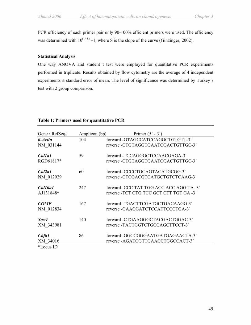
Ahmed 2006 Effect of haematopoietic cells on chondrogenesis Chapter 3
49
PCR efficiency of each primer pair only 90-100% efficient primers were used. The efficiency
was determined with 10(1/-S) –1, where S is the slope of the curve (Ginzinger, 2002).
Statistical Analysis
One way ANOVA and student t test were employed for quantitative PCR experiments
performed in triplicate. Results obtained by flow cytometry are the average of 4 independent
experiments ± standard error of mean. The level of significance was determined by Turkey´s
test with 2 group comparison.
Table 1: Primers used for quantitative PCR
Gene / RefSeq# Amplicon (bp) Primer (5´ - 3´) ß-Actin 104 forward -GTAGCCATCCAGGCTGTGTT-3´ NM_031144 reverse -CTGTAGGTGAATCGACTGTTGC-3´ Col1a1 59 forward -TCCAGGGCTCCAACGAGA-3´ RGD61817* reverse -CTGTAGGTGAATCGACTGTTGC-3´ Col2a1 60 forward -CCCCTGCAGTACATGCGG-3´ NM_012929 reverse -CTCGACGTCATGCTGTCTCAAG-3´ Col10a1 247 forward -CCC TAT TGG ACC ACC AGG TA -3´ AJ131848* reverse -TCT CTG TCC GCT CTT TGT GA -3´ COMP 167 forward -TGACTTCGATGCTGACAAGG-3´ NM_012834 reverse -GAACGATCTCCATTCCCTGA-3´ Sox9 140 forward -CTGAAGGGCTACGACTGGAC-3´ XM_343981 reverse -TACTGGTCTGCCAGCTTCCT-3´ Cbfa1 86 forward -GGCCGGGAATGATGAGAACTA-3´ XM_34016 reverse -AGATCGTTGAACCTGGCCACT-3´ *Locus ID

Chapter 3 Effect of haematopoietic cells on chondrogenesis Ahmed 2006
50
D7Fib ICD 68 G CD 45 H
CD 68 D F D7Fib E CD 45
B CD 45 A CD 68 D7Fib C
CD 68 J CD 45 K D7Fib L
Results MSC isolation procedure and CD expression profile
To determine the influence of adherence time of rat bone marrow cells on the presence of
CD45-positive haematopoietic cells we left the cells to adhere for 3, 4 and 5 days before non-
adherent and weakly attached cells were removed. Upon immunofluorescence analysis the
adherent cells showed a D7fib+, CD68+ and CD45+/low expression profile regardless of the
time of removal of non-adherent cells. Figure 1 shows the representative immunomicrographs
of the cells left to adhere for 3 days and compares the staining of non-sorted and MACS-
sorted cells after the first passage. Non-sorted cells, left for 3 days to adhere before medium
removal, are contaminated with CD45-positive cells (Fig.1A-C). After magnetic sorting, most
of the CD45-positive cells were excluded, leaving a CD45-depleted cell fraction (Fig.1D-F).
However, we still observe few rounded cells among the CD68 positively stained fraction
(Fig.1D) of unknown surface antigen characterization. The CD45-positive cell fraction
includes cells which stain for CD68 and D7Fib (Fig.1G-I).
Figure 1: Morphological characterization of MSCs
Immunofluorescence imaging of heterogeneous adherent MSCs (A, B, C) and CD45-depleted MACS sorted cells (D, E, F), showing typical fibroblast like spindle shaped cells, while CD45-positive cells (G, H, I) are small and rounded in appearance. The negative controls for all three antibodies used are unstained (J, K, L). All 3 cell types are CD68- and D7fib- positive while the CD45-depleted cell population does not exhibit CD45 harbouring cells (E). Magnification is 20x for all panels, bars are 10µm.
D7Fib

Ahmed 2006 Effect of haematopoietic cells on chondrogenesis Chapter 3
51
0
1
1 2 3 4day of non-adherent cell removal
Nor
mal
ised
cel
l num
ber o
n da
y 7
***
*
C
Quantification of CD45-positive cell contamination of primary MSC cultures
In order to quantitate the ratio between haematopoietic CD45-positive cells and MSCs we
employed three colour flow cytometry (FACS) which allowed for a distinction between the
two different cell types according to their surface antigens (CD45, CD29) and dead cells
(Fig.2A).
Figure 2: FACS analysis of bone marrow derived stem cells prepared by adherence to plastic Representative dot-plot (out of 3 experiments) of rat MSCs isolated by adherence to plastic after 7 days of cell culture. The lower right quadrant contains CD29 –highly positive and CD45-depleted MSCs while the upper right quadrant contains HCs with a CD45 – positive and CD29-low expression profile (A). Proportion of CD45-positive (black bars) and CD45-depleted (open bars) cells from rat MSCs isolated by adherence to plastic in relation to the time of the first medium change and therefore, removal of non-adherent cells (B). Total yield of CD45-depleted (open bars) and CD45-positive (black bars) bone marrow derived cells after 7 days of cell culture (*p< 0,05; **p< 0,01). Yield of cells is normalized to cell number achieved when the non-adherent cells were removed on day 4 (C).
A B
0
10
20
30
40
50
60
70
80
90
1 2 3 4
day o f non-adherent cell rem oval
% o
f cel
ls o
n da
y 7

Chapter 3 Effect of haematopoietic cells on chondrogenesis Ahmed 2006
52
Figure 2B shows that the fraction of CD45-positive haematopoietic cells was not influenced
by the adherence time after cell isolation. We mostly found about 20-30 % of the attached
cells in bone marrow cell cultures to be CD45-positive when selected according to sole
adherence to plastic surfaces. Flow cytometric results were concurrent with the observations
made after immunofluorescent staining. In one out of four experiments we detected variations
between 10 - 40% in individual flasks. We observed that CD45-depleted cells expressed
CD29 stronger than CD45-positive cells (Fig.2A) which offers an additional selection
parameter. In cultures where non-adherent cells were removed on days 3 and 4, we obtained a
significantly increased number of desired CD45-depleted MSCs compared to removal on day
1 and 2 (Fig.2C). Apparently, 3 to 4 days were a more appropriate duration for the adherent
fraction to get acclimated to the cell culture conditions, hence exhibit a firmer attachment to
the cell culture plastics.
Effect of CD45-positive cells on gene expression levels of Col1a1, Col2a1, Col10a1,
COMP, Sox9 and Cbfa1
During the time course of cell culture we observed a strong increase in relative mRNA levels
of COMP and collagen II, while Sox9 mRNA levels exhibited a moderate up regulation
(Fig.3A, B, D). Collagen X gene expression remained undetectable the first three weeks of
cell culture, however, at day 28 expression of this collagen was found to be induced (Fig.3C).
The internal control gene, ß-Actin, remained unaltered during the complete time course of cell
culturing (Fig. 3G). Comparing gene expression of CD45-depleted cells and the non-sorted
population, we found collagen I gene expression to be increased in the non-sorted cell
population during the culture period, while it became decreased in CD45-depleted MSCs.
However, in CD45-depleted cells collagen I was still expressed at a relative higher level as in
freshly isolated hip articular chondrocytes (Fig.3E). Notably, the non-sorted cells displayed a
strikingly higher up regulation of collagen II, COMP and Sox9 gene expression compared to
the CD45-depleted MSCs throughout the culture period. In the non-sorted population collagen
II gene expression is up regulated at average about 25 fold, COMP gene expression about 10
fold, collagen X expression about 23 fold and Sox9 mRNA levels about 3 fold if compared to
CD45-depleted MSCs. For collagen I gene expression we have observed a significant
difference only between the two groups at day 14 and 28, where the adherent MSCs
demonstrated an up regulation resulting in a 3-5 fold higher gene expression if compared to
CD45-depleted MSCs.

Ahmed 2006 Effect of haematopoietic cells on chondrogenesis Chapter 3
53
Figure 3: Chondrogenic gene expression profile of adherent MSCs, CD45-depleted MSCs and remixed control cells. Relative quantitative mRNA expression level of chondrogenesis specific genes as COMP (A) Col2a1(B) and Col10a1 (C), Sox9 (D), Col1a1(E), and Cbfa1 (F) was determined by employing the ∆∆CT method using SYBR I Green dye. Expression level of housekeeping gene, ß-actin was determined as a controlling indicator of cell viability (G) and is expressed as positive logarithm of CT-values. Expression level of each gene was compared between adherent (black bars), CD45-depleted (open bars) and remixed control cells (crossed bars). Grey shaded bars indicate relative gene expression level of freshly isolated hip articular chondrocytes. Number of experiments = 3

Chapter 3 Effect of haematopoietic cells on chondrogenesis Ahmed 2006
54
Collagen I remained up regulated from day 14 on in adherent cells while CD45-depleted
MSCs exhibit an increasingly down regulation during the time course of cell culture. We also
observed a striking difference in the collagen X expression pattern between non-sorted and
CD45-depleted cells. The collagen X mRNA expression profile of CD45-depleted cells
experienced only a moderate increase of about 3.3 fold at the end of the culture period while
adherent cells up regulated their mRNA level about 76 fold compared to day 0 (Fig. 3C).
Notably, Cbfa1 gene expression is differentially regulated with respect to the two cell
populations (Fig.3F). On day 1 Cbfa1 mRNA level is significantly higher in adherent cells
(4.7 fold) and also in chondrocytes (ca. 2.0 fold) compared to CD45-depleted cells. This
pattern became inverted at day 28 with CD45-depleted cells exhibiting an elevated expression
(1.7 fold) compared to the adherent MSCs. In between these time points, relative gene
expression of Cbfa1 is similarly regulated in both cell populations.
In remixed control cell populations (30% CD45-positive cells with 70% CD45-depleted cells)
collagen II, X, I, Sox9 and Cbfa1 gene expression profile resembles closely that of adherent
cells (Fig.3B, C, D, E, F) and exhibits no significant differences. Only COMP gene expression
at day 14 of the culture period significantly differs from adherent cells but the level at day 21
and 28 shows no difference to non-sorted cells (Fig. 3A). ß-actin gene expression profile is
similar for all three culture conditions (Fig.3G).
Discussion Adult bone marrow is the major source of haematopoietic stem cells and mesenchymal stem
cells (Pittenger et al., 1999a). In animal studies, adherence to tissue culture plastic is accepted
as a satisfactory parameter for attaining marrow stromal cell population depleted of
haematopoietic cells. However, when employing FACS analysis for surface antigen
characterization we observed a contamination of these non-sorted MSCs with 20-30% of
CD45-positive cells. For additional characterization of sorted and non-sorted cell population
we applied immunofluorescence staining for CD68 and D7Fib. The D7Fib antigen constitutes
a fibroblast specific molecule of yet unknown function, supposedly a specific marker for
MSCs as suitable as STRO-1 and CD105 (Jones et al., 2002). We observed expression of the
D7Fib antigen on adherent CD45-positive cells; however, these cells displayed a rounded
morphology compared to the fibroblast-like CD45-depleted cells. CD68 and its murine
analogue macrosialin, a member of the lamp family, appear to have a rather limited tissue
distribution, being found on macrophages, Langerhans cells and dendritic cells (Ramprasad et

Ahmed 2006 Effect of haematopoietic cells on chondrogenesis Chapter 3
55
al., 1996). Notably, we have found all adherent cells CD68-positive suggesting that in non-
committed, undifferentiated cells, this protein might exhibit a different distribution pattern
and possibly exert functions related to cell–cell or cell–ligand interactions (Fukuda, 1991).
CD45-positive haematopoietic cells were always detected in MSC cultures, regardless of the
time of the first change of culture medium. This indicated a persistent presence and
attachment of CD45-positive cells in the MSC environment when no further selection based
on specific surface antigens is carried out. Adherence to the tissue culture plastic alone is a
weak discriminating criterion for a homogeneous MSC population, because neural cells,
monocytes and macrophages are also known to be isolated according to their adherence to
plastic surfaces (Delles et al., 2002; Kiefer et al., 1991; Majumdar et al., 1998a). The presence
of CD45-positive haematopoietic cells and the variation of their proportion in primary
cultures of MSCs could explain the occurrence of extensive variations usually seen in
differentiation experiments with bone marrow derived cells.
The fact, that CD45-depleted cells expressed CD29 stronger than CD45-positive cells offered
us an easier separation of two populations and limited the possible existence of more than two
major subpopulations in our cultures. The integrin ß1 subunit CD29 exhibits a broad tissue
distribution, including lymphocytes, endothelia, smooth muscle, and epithelia (Springer,
1990) and plays an important role in cellular processes, e.g. embryogenesis and HSC
development. Although we did not find the haematopoietic cell fraction to vary according to
the time of removal of non-adherent cells we observed a correlation between adhesion time
and yield of CD45-depleted marrow stromal cells. In parallel, cell adhesion plus proliferation
could also be promoted by the prolonged presence of the non-adherent fraction via cytokine
release and / or cell-cell contacts. A comparable effect of haematopoietic cells on MSC
proliferation has been observed for megakaryocytes and platelets (Castro-Malaspina et al.,
1981). According to our results we decided to leave the cells to adhere for 3 days before
performing differentiation experiments.
Differentiation studies of haematopoietic stem cells have demonstrated, that the small number
of adherent MSCs present in total bone marrow aspirates provide an important
microenvironment for growth of HSCs and their differentiation into several blood borne cell
types. In long-term cultures of HSCs, these adherent cells even interact directly with the
haematopoietic precursors (Prockop, 1997b). Here, we could show that vice versa adherent
CD45-positive haematopoietic precursor cells create a microenvironment which may enhance
expression of particular marker genes in MSCs. For studying the effects of contaminating
CD45-positive cells we prepared a relatively homogeneous population of CD45-depleted

Chapter 3 Effect of haematopoietic cells on chondrogenesis Ahmed 2006
56
MSCs by immunogenic depletion of CD45-positive cells. Both, the heterogenic adherent cells
and the CD45-depleted subpopulation were subjected to chondrogenic favourable 3-D
environment by formation of a cell alginate amalgam. For control purposes in order to assure
that difference in gene expression profile between sorted and unsorted MSCs is due to
depletion of CD45-positive cells and not because of general depletion of differentiation
competent cells or due to cell damage caused by the sorting process, we prepared a third
population of remixed cells (30% CD45-positive mixed with 70% CD45-depleted cells)
following MACS preparation which was treated according to the differentiation protocol used
for adherent and sorted cells.
We monitored relative gene expression of collagen II (Mendler et al., 1989) and COMP
(Hedbom et al., 1992; DiCesare et al., 1994) as examples for structural extracellular matrix
proteins highly specific for hyaline cartilage. Additionally, we have investigated Sox9 as a
master transcription factor imperative for chondrogenic differentiation (de Crombrugghe et
al., 2000a) and collagen I as differentiation control for a gene normally not active in healthy
hyaline cartilage (Rossert et al., 2000) but occurring together with collagen II in fibrocartilage
(Nerlich et al., 1998). Gene expression of collagen X, specific for terminal differentiated
hypertrophic chondrocytes in the calcifying zone of cartilage and secondary ossification
centres within the growth plate (Schmid et al., 1990) indicates maturation of MSCs towards
an undesired cartilage condition.
Gene expression of Cbfa1, a transcriptional activator for osteoblastic differentiation during the
process of endochondral ossification is analyzed for monitoring differentiation towards
osteogenic condition. Cbfa1 is also expressed in pre-condensation stage during early
development in a cell type with the potential to become either a chondrocyte or an osteoblast.
In later developmental stages expression of this transcription factor is restricted to
prehypertrophic and hypertrophic chondrocytes where it acts as a hypertrophic differentiation
inducer (Karsenty, 2001; Takeda et al., 2001).
The observed gene expression profile indicates not only a beneficial influence of CD45-
positive cells on the differentiation process of MSCs towards the chondrogenic lineage. It also
shows that when culture time is extended beyond 28 days chondrogenically differentiating
MSCs might enter the prehypertrophic / hypertrophic differentiation cascade and proceed
towards terminal differentiation. Interestingly, usage of chondrogenic culture medium does
not prevent up regulation of Cbfa1 and collagen X. This indicates either a not yet defined
influence of medium components (i.e. TGF-ß or dexamethasone) on intrinsic cell derived
factors (i. e. signalling molecules, proteases, transcription factors) which are involved in

Ahmed 2006 Effect of haematopoietic cells on chondrogenesis Chapter 3
57
regulation of the chondrogenic differentiation cascade or a medium independent process
occurring by default through paracrine interaction of MSCs. A variety of growth factors i.e.
members of the TGF-ß superfamily, IGF-1 and bFGF could account for gene modulating
effects in our system. (Bohme et al., 1995) (Recklies et al., 1998) (Murakami et al., 2000)
(Horton, Jr. et al., 1989).
In conclusion, we demonstrated that bone marrow derived fibroblast like CD45-depleted rat
MSCs share a heterogeneous cellular environment with ~20-30% CD45-positive HCs, if no
other selection procedure than adherence to plastic is employed. Our differentiation results
indicate that adherent CD45-positive haematopoietic cells may create a microenvironment
promoting differentiation of MSCs towards the chondrogenic lineage by up regulating
specific genes. However, expression of undesired genes as collagen I and collagen X is
likewise promoted by HCs. On the contrary, at early condensation stage HCs strongly
increase gene expression of Cbfa1 thus supporting chondrogenic differentiation of MSCs,
while at a later differentiation stage when Cbfa1 plays an integral role in hypertrophy the
regulation appears to be independent of the presence of HCs.
We speculate that the stromal CD45-positive population in toto is responsible for regulation
of gene expression. The heterotrophic nature of bone marrow derived cells suggest that in vivo
interactions of different cell types is very likely and should be addressed accordingly.
Characterization of additional subpopulations of CD45-positive cells according to their
surface antigens and determination of their potential influence on gene expression may
elaborate this point further more. Studies using conditioned media are needed to clarify
whether physical contact between cells or paracrine factors, alone or in concurrence, are
necessary for promoting gene expression leading to chondrogenic differentiation and
maturation of MSCs. Identification and isolation of factors which are capable of modulating
genes involved in differentiation and dedifferentiation processes of chondrocytes is vital for in
vitro engineering of cartilage tissue repair.
Acknowledgements We want to thank Maren Marschner for her excellent technical assistance and support. We
also acknowledge Thomas Vogel for his generous help and scientific discussion. Special
thanks go to Prof. Dr. Achim Göpferich for his advisement and support. The study was funded
by the Bavarian Research Foundation (Bayerische Forschungsstiftung).
Chapter 3 Effect of haematopoietic cells on chondrogenesis Ahmed 2006
58
Reference List
1. Bi,W., Deng,J.M., Zhang,Z., Behringer,R.R., and de Crombrugghe,B. (1999). Sox9 is required for cartilage formation. Nat. Genet. 22, 85-89.
2. Bianco,P., Riminucci,M., Gronthos,S., and Robey,P.G. (2001). Bone marrow stromal stem cells: nature, biology, and potential applications. Stem Cells 19, 180-192.
3. Bohme,K., Winterhalter,K.H., and Bruckner,P. (1995). Terminal differentiation of chondrocytes in culture is a spontaneous process and is arrested by transforming growth factor-beta 2 and basic fibroblast growth factor in synergy. Exp. Cell Res. 216, 191-198.
4. Cancedda,R., Dozin,B., Giannoni,P., and Quarto,R. (2003). Tissue engineering and cell therapy of cartilage and bone. Matrix Biol. 22, 81-91.
5. Caplan,A.I. and Bruder,S.P. (2001). Mesenchymal stem cells: building blocks for molecular medicine in the 21st century. Trends Mol. Med. 7, 259-264.
6. Castro-Malaspina,H., Rabellino,E.M., Yen,A., Nachman,R.L., and Moore,M.A. (1981). Human megakaryocyte stimulation of proliferation of bone marrow fibroblasts. Blood 57, 781-787.
7. de Crombrugghe,B., Lefebvre,V., Behringer,R.R., Bi,W., Murakami,S., and Huang,W. (2000b). Transcriptional mechanisms of chondrocyte differentiation. Matrix Biol. 19, 389-394.
8. de Crombrugghe,B., Lefebvre,V., Behringer,R.R., Bi,W., Murakami,S., and Huang,W. (2000a). Transcriptional mechanisms of chondrocyte differentiation. Matrix Biol. 19, 389-394.
9. Delles,A.M., Rittenhouse-Olson,K., Morgan,J., and Oseroff,A.R. (2002). A simple method for the purification of human peripheral blood antigen presenting cells (dendritic cells, monocytes/macrophages, and B lymphocytes). Immunol. Invest 31, 233-245.
10. DiCesare,P., Hauser,N., Lehman,D., Pasumarti,S., and Paulsson,M. (1994). Cartilage oligomeric matrix protein (COMP) is an abundant component of tendon. FEBS Lett. 354, 237-240.
11. Dobson,K.R., Reading,L., Haberey,M., Marine,X., and Scutt,A. (1999). Centrifugal isolation of bone marrow from bone: an improved method for the recovery and quantitation of bone marrow osteoprogenitor cells from rat tibiae and femurae. Calcif. Tissue Int. 65, 411-413.
12. Friedenstein,A.J., Gorskaja,J.F., and Kulagina,N.N. (1976). Fibroblast precursors in normal and irradiated mouse hematopoietic organs. Exp. Hematol. 4, 267-274.
13. Fukuda,M. (1991). Lysosomal membrane glycoproteins. Structure, biosynthesis, and intracellular trafficking. J. Biol. Chem. 266, 21327-21330.

Ahmed 2006 Effect of haematopoietic cells on chondrogenesis Chapter 3
59
14. Galmiche,M.C., Koteliansky,V.E., Briere,J., Herve,P., and Charbord,P. (1993). Stromal cells from human long-term marrow cultures are mesenchymal cells that differentiate following a vascular smooth muscle differentiation pathway. Blood 82, 66-76.
15. Ginzinger,D.G. (2002). Gene quantification using real-time quantitative PCR: an emerging technology hits the mainstream. Exp. Hematol. 30, 503-512.
16. Hedbom,E., Antonsson,P., Hjerpe,A., Aeschlimann,D., Paulsson,M., Rosa-Pimentel,E., Sommarin,Y., Wendel,M., Oldberg,A., and Heinegard,D. (1992). Cartilage matrix proteins. An acidic oligomeric protein (COMP) detected only in cartilage. J. Biol. Chem. 267, 6132-6136.
17. Herbertson,A. and Aubin,J.E. (1997). Cell sorting enriches osteogenic populations in rat bone marrow stromal cell cultures. Bone 21, 491-500.
18. Horton,W.E., Jr., Higginbotham,J.D., and Chandrasekhar,S. (1989). Transforming growth factor-beta and fibroblast growth factor act synergistically to inhibit collagen II synthesis through a mechanism involving regulatory DNA sequences. J. Cell Physiol 141, 8-15.
19. Johnstone,B., Hering,T.M., Caplan,A.I., Goldberg,V.M., and Yoo,J.U. (1998). In vitro chondrogenesis of bone marrow-derived mesenchymal progenitor cells. Exp. Cell Res. 238, 265-272.
20. Jones,E.A., Kinsey,S.E., English,A., Jones,R.A., Straszynski,L., Meredith,D.M., Markham,A.F., Jack,A., Emery,P., and McGonagle,D. (2002). Isolation and characterization of bone marrow multipotential mesenchymal progenitor cells. Arthritis Rheum. 46, 3349-3360.
21. Karsenty,G. (2001). Minireview: transcriptional control of osteoblast differentiation. Endocrinology 142, 2731-2733.
22. Kiefer,F., Wagner,E.F., and Keller,G. (1991). Fractionation of mouse bone marrow by adherence separates primitive hematopoietic stem cells from in vitro colony-forming cells and spleen colony-forming cells. Blood 78, 2577-2582.
23. Lefebvre,V., Behringer,R.R., and de Crombrugghe,B. (2001). L-Sox5, Sox6 and Sox9 control essential steps of the chondrocyte differentiation pathway. Osteoarthritis. Cartilage. 9 Suppl A, S69-S75.
24. Majumdar,M.K., Thiede,M.A., Mosca,J.D., Moorman,M., and Gerson,S.L. (1998a). Phenotypic and functional comparison of cultures of marrow-derived mesenchymal stem cells (MSCs) and stromal cells. J. Cell Physiol 176, 57-66.
25. Majumdar,M.K., Thiede,M.A., Mosca,J.D., Moorman,M., and Gerson,S.L. (1998b). Phenotypic and functional comparison of cultures of marrow-derived mesenchymal stem cells (MSCs) and stromal cells. J. Cell Physiol 176, 57-66.
26. Maniatopoulos,C., Sodek,J., and Melcher,A.H. (1988). Bone formation in vitro by stromal cells obtained from bone marrow of young adult rats. Cell Tissue Res. 254, 317-330.

Chapter 3 Effect of haematopoietic cells on chondrogenesis Ahmed 2006
60
27. Mendler,M., Eich-Bender,S.G., Vaughan,L., Winterhalter,K.H., and Bruckner,P. (1989). Cartilage contains mixed fibrils of collagen types II, IX, and XI. J. Cell Biol. 108, 191-197.
28. Mundlos,S. and Olsen,B.R. (1997). Heritable diseases of the skeleton. Part I: Molecular insights into skeletal development-transcription factors and signaling pathways. FASEB J. 11, 125-132.
29. Murakami,S., Kan,M., McKeehan,W.L., and de Crombrugghe,B. (2000). Up-regulation of the chondrogenic Sox9 gene by fibroblast growth factors is mediated by the mitogen-activated protein kinase pathway. Proc. Natl. Acad. Sci. U. S. A 97, 1113-1118.
30. Nerlich,A.G., Boos,N., Wiest,I., and Aebi,M. (1998). Immunolocalization of major interstitial collagen types in human lumbar intervertebral discs of various ages. Virchows Arch. 432, 67-76.
31. Phinney,D.G., Kopen,G., Isaacson,R.L., and Prockop,D.J. (1999). Plastic adherent stromal cells from the bone marrow of commonly used strains of inbred mice: variations in yield, growth, and differentiation. J. Cell Biochem. 72, 570-585.
32. Pittenger,M.F., Mackay,A.M., Beck,S.C., Jaiswal,R.K., Douglas,R., Mosca,J.D., Moorman,M.A., Simonetti,D.W., Craig,S., and Marshak,D.R. (1999a). Multilineage potential of adult human mesenchymal stem cells. Science 284, 143-147.
33. Pittenger,M.F., Mackay,A.M., Beck,S.C., Jaiswal,R.K., Douglas,R., Mosca,J.D., Moorman,M.A., Simonetti,D.W., Craig,S., and Marshak,D.R. (1999b). Multilineage potential of adult human mesenchymal stem cells. Science 284, 143-147.
34. Prockop,D.J. (1997a). Marrow stromal cells as stem cells for nonhematopoietic tissues. Science 276, 71-74.
35. Prockop,D.J. (1997b). Marrow stromal cells as stem cells for nonhematopoietic tissues. Science 276, 71-74.
36. Ramprasad,M.P., Terpstra,V., Kondratenko,N., Quehenberger,O., and Steinberg,D. (1996). Cell surface expression of mouse macrosialin and human CD68 and their role as macrophage receptors for oxidized low density lipoprotein. Proc. Natl. Acad. Sci. U. S. A 93, 14833-14838.
37. Recklies,A.D., Baillargeon,L., and White,C. (1998). Regulation of cartilage oligomeric matrix protein synthesis in human synovial cells and articular chondrocytes. Arthritis Rheum. 41, 997-1006.
38. Rossert,J., Terraz,C., and Dupont,S. (2000). Regulation of type I collagen genes expression. Nephrol. Dial. Transplant. 15 Suppl 6, 66-68.
39. Schmid,T.M., Popp,R.G., and Linsenmayer,T.F. (1990). Hypertrophic cartilage matrix. Type X collagen, supramolecular assembly, and calcification. Ann. N. Y. Acad. Sci. 580, 64-73.
40. Springer,T.A. (1990). Adhesion receptors of the immune system. Nature 346, 425-434.

Ahmed 2006 Effect of haematopoietic cells on chondrogenesis Chapter 3
61
41. Takeda,S., Bonnamy,J.P., Owen,M.J., Ducy,P., and Karsenty,G. (2001). Continuous expression of Cbfa1 in nonhypertrophic chondrocytes uncovers its ability to induce hypertrophic chondrocyte differentiation and partially rescues Cbfa1-deficient mice. Genes Dev. 15, 467-481.
42. von der,M.K. (1999). Structure and Biosynthesis of Collagens. In Bone and Cartilage Metabolism, M.J.Seibel, S.P.Robins, and J.P.Bilezikian, eds. Academic Press), pp. 3-29.


63
Chapter 4
Soluble signalling factors derived from
differentiated cartilage tissue affect
chondrogenic differentiation of adult
marrow stromal cells
Nazish Ahmed1, 2, Rita Dreier3, Achim Göpferich4, Joachim Grifka1 and Susanne Grässel1, 2*
1Dept. of Orthopaedic Surgery, Experimental Orthopaedics, University Hospital of
Regensburg, Kaiser Karl V Allee-3, 93077 Bad Abbach, Germany, 2Experimental
Orthopaedics, Centre for Medical Biotechnology, BioPark I, Regensburg, Germany, 3Institute
for Physiological Chemistry and Pathobiochemistry, University Hospital of Münster,
Germany, 4Dept. of Pharmaceutical Technology, University of Regensburg, Germany.
*Corresponding author
Submitted: Osteoarthritis and Cartilage, 2006
Abstract Limited regeneration capacity of cartilage is mainly due to lack of access to progenitor cells.
The only known chondroprogenitor cells for adult cartilage tissues are multipotent marrow

Chapter 4 Effect of cartilage on chondrogenesis Ahmed 2006
64
stromal or mesenchymal stem cells (MSCs). In vitro lineage differentiation studies on these
precursor cells usually require a cue from externally added growth factors of the TGFß
superfamily. We hypothesise that signalling factors secreted by cartilage tissue may substitute
for external growth factors. To study such paracrine communication between allogenic rat
articular cartilage and MSCs embedded in alginate beads a novel coculture system without
external growth factors has been established. Successful chondrogenic differentiation in
MSCs was induced and the impact of cartilage on differentiating MSCs was observed at two
culture stages. Firstly, elevated expression of Sox9 at the initial stage indicated early
induction of chondrogenic differentiation. Secondly, late stage repression of collagen X
indicated arrest of differentiation prior to hypertrophy. Four soluble signalling molecules were
identified in the culture supernatant: vascular endothelial growth factor alpha (VEGF-164α),
matrix metalloprotease (MMP) -13 and tissue inhibitors of MMPs (TIMP-1 and TIMP-2).
These factors could be traced back either directly to the cartilage tissue or to the MSCs
influenced by the presence of cartilage. The identified factors may have a mutual impact on
the control and regulation of differentiation of both the chondrocytes and MSCs.
Key words: rat mesenchymal stem cells, chondrogenic differentiation, coculture, signalling
factors, cartilage, MMP-13, collagen X
Introduction A defect in articular cartilage caused by aging, chondral injury or a degenerative disease is
seldom repaired, causes persistent pain and restricts mobility. Generally, the avascular nature
of cartilage is held responsible for the lack of spontaneous rejuvenation; more specifically it is
probably due to the deficiency of chondroprogenitor cells (Magne et al., 2005). As
chondrocytes are the only cell type of cartilage tissues and bone marrow derived MSCs are the
only known chondroprogenitor cells, it is in interest of cartilage regenerative medicine to
understand and identify molecular events characterising their growth and differentiation
(Lefebvre and Smits, 2005).
Under specific culture conditions and proper stimulation MSCs can differentiate into multiple
mesenchymal lineages including osteocytes and chondrocytes. The first stage of chondrogenic
differentiation is conversion of undifferentiated MSCs to committed osteo-chondroprogenitor
cells leading to cell condensation and growth arrest. The major proteins involved in
condensation initiation are fibronectin and proteins of the transforming growth factor ß

Ahmed 2006 Effect of cartilage on chondrogenesis Chapter 4
65
superfamily (TGFß1-3 and BMP1-7) (Barry et al., 2001). Cells present at the centre of the
condensation nodules first form pre-chondrocytes and then chondroblasts / chondrocytes
which start to produce cartilaginous extracellular matrix (ECM) marked by upregulation of
structural protein genes as col2a1, col9a2, col11a2 and aggrecan. Collagens make up to 60%
of the hyaline cartilage protein bulk; among them the most dominant (90-95%) form of
cartilaginous collagen is collagen type II which forms a fibrillar alloy structure together with
type XI and IX collagens and non-collagenous proteins (Mendler et al., 1989). Gene
expression of collagen type X which according to the concept of endochondral ossification
indicates the final step of chondrocyte maturation serves as a marker for hypertrophic and
terminally differentiated chondrocytes (Reddi, 1994).
The molecular basis of regulatory mechanisms involved in chondrogenic lineage signalling
cascade are not sufficiently understood, however, they are known to involve numerous growth
factors, proteases and cytokines in a strictly controlled time dependent manner. Major players
imperative for proper skeletogenesis belong to proteases of MMP-family and growth factors
of VEGF family. For example, activity of MMPs and their endogenous inhibitors (TIMPs) is
not only fundamental to skeletal growth and repair, but also to their pathology. One member
of this large family of matrix degrading MMPs, MMP-13 is predominantly expressed both, in
osteoarthritic cartilage and in hypertrophic and calcifying cartilage of the mammalian growth
plate (Wu et al., 2002; Johansson et al., 1997). Together with other collagenases, and their
endogenous inhibitors like TIMP-1, MMP-13 plays a significant role in collagen turnover by
cleaving collagen II, additionally, it also degrades collagen X (Knauper et al., 1996; Mitchell
et al., 1996). VEGF, expressed by hypertrophic chondrocytes, is considered as an essential
coordinator of chondrocyte death, chondroclast function, ECM remodelling, angiogenesis and
bone formation in the growth plate thereby hold responsible for proper endochondral
ossification (Gerber et al., 1999). Little is known how these soluble factors affect
differentiation pathways decisions and integration processes of chondroprogenitor MSCs.
Chondrogenesis is controlled by several transcription factors, important players are members
of the Sox- and Cbfa- family. In particular, Sox9 has been characterized as a master
transcription factor with essential direct or indirect regulatory effects along the entire
chondrogenic differentiation pathway. It is expressed in all chondroprogenitor cells and in
differentiated chondrocytes but not in hypertrophic cells (Bi et al., 1999; de Crombrugghe et
al., 2000; Mundlos and Olsen, 1997; Lefebvre et al., 2001). Direct control of hypertrophy
involves Cbfa1, also known as RUNX2, the major osteogenic transcription factor. Expression
of Cbfa1 increases during maturation of chondrocytes and is necessary for inducing gene

Chapter 4 Effect of cartilage on chondrogenesis Ahmed 2006
66
expression of collagen X (Inada et al., 1999; Goldring et al., 2006) and MMP-13 in terminally
differentiated chondrocytes (Porte et al., 1999). For a normal skeletal development and
maintenance a co-ordinated regulation of these chondrogenic differentiation associated genes
is critical.
This in vitro study aimed to identify soluble signalling factors mediating a paracrine cross talk
of differentiated cartilage tissue with undifferentiated MSCs. We have designed and set up a
co-culture system of rat MSCs and allogenic articular cartilage chips under serum-free
chondrogenesis favourable 3D- conditions. Our results indicate that externally added growth
factors are dispensable for the induction of the chondrogenic differentiation pathway in rat
MSCs embedded in alginate beads. Coculture of MSCs with differentiated cartilage induces
up regulation of Sox9 gene expression in MSCs and causes suppression of collagen X
biosynthesis and secretion presumably mediated by cartilage secreted factors. Using
zymographic techniques and an antibody array we have detected VEGF-164α, TIMP-1, -2 and
MMP-13 in the coculture supernatant. These molecules originate in part from cartilage
explants and could be important players in regulation and control of chondrogenic
differentiation.
Materials and methods Cell isolation, coculture and differentiation
MSCs from rat bone marrow were isolated as described earlier (Ahmed et al., 2006c). Briefly,
bone marrow removed by centrifugation from tibiae and femora of 6 weeks old male rats was
seeded at a concentration of 2.5x 10⁵ cells/cm2 and proliferated in α MEM medium containing
5% glutamate, 1% pen/strep and 10% FBS (Invitrogen, UK). Cells from ~75% confluent
flasks were harvested and high density 3-D cultures were attained by preparing a cell-alginate
amalgam in 1.2% alginate (Sigma, USA) at a concentration of 107 cells/ml. Alginate beads of
105 cells/bead were formed by dropping the suspension into 102mM CaCl2 solution and 10
beads per insert (1µm pore size, BD Bioscience, USA) were placed in 12-well tissue culture
plates. One hip cartilage explant cut in 3 pieces was placed in the bottom well. The culture
was carried out for 3 weeks in serum-free chondrogenic medium with medium change every
2-3 days. The chondrogenic medium contained ITS+ premix (6.25µg/ml insulin, 6.25ng/ml
selenium acid, 6.25µg/ml transferrin, 1.25mg/ml BSA and 5.35µg/ml linoleic acid; BD
Biosciences, USA), 110µg/ml pyruvate, 40µg/ml proline, 0.1µM dexamethasone, 50µg/ml

Ahmed 2006 Effect of cartilage on chondrogenesis Chapter 4
67
ascorbic acid (Johnstone et al., 1998) in α MEM high glucose (Invitrogen, UK). Additionally,
10ng/ml TGFß-3 (R&D Systems, USA) was added to the culture medium of control
monocultures. Samples of cells, tissue and culture supernatant were taken once a week for
gene and protein expression analysis. Alginate beads were depolymerised by 30min
incubation in 55mM sodium citrate and 0.15M sodium chloride buffer at 37°C on a shaker;
cells were recovered after 3min centrifugation at 750xg. Osteogenesis was induced for 19
days in 6 well culture plates, 250,000 cells/well were cultured in osteogenic medium
containing, 10% FCS, 1% pen/strep, 10nM dexamethasone, 50µg/ml ascorbate 2-PO4 and
10mM ß-Na glycerophosphate. At the end of the culture time cells were fixed with methanol
for 10min and stained for 2min with 1% Alizarin Red.
RNA isolation, reverse transcription and quantitative PCR analyses
All molecular biology techniques were carried out as described earlier (Ahmed et al., 2006b).
Briefly, RNA was isolated by an affinity column chromatography method with Ambion’s
RNAqueous4-PCR kit according to the manufacturer’s protocol. Contaminating DNA was
removed with DNaseI enzyme (DNA-free, Ambion, USA). Invitrogen´s SuperScript II
reverse transcriptase kit was used for conversion of 0.5-1µg of RNA into cDNA with
500µg/ml of oligo-dT primers and 200 units of SuperScriptII enzyme. Relative quantitative
PCR was carried out in triplicate with SYBR Green Dye I master mix using an ABI 7000
Prism Sequence detection system (Applied Biosystems, USA). Amplification parameters were
as follows, denaturation at 95°C for 15sec, annealing at 60°C for 1min in 40 cycles repeats.
Relative quantification (RQ) values were calculated by using ABI Prism 7000 SDS software
v1.1 employing the ∆∆Ct method with ß-actin as endogenous control and undifferentiated
MSCs freshly suspended in alginate (day 0) as calibrator. Mean RQ values were plotted on a
semi-logarithmic graph. Primers (table 1) were designed with either Primer3 or “Primer
Express” software (Applied Biosystems, USA). All primers were manufactured at MWG –
Biotech, Germany.
Immunofluorescence
Alginate beads were incubated for 30min in 100mM BaCl2 for irreversible polymerization
(Mok et al., 1994). Then the beads were fixed with 3% paraformaldehyde (PFA), sequentially
dehydrated with alcohol and then embedded in paraffin. 4-8µm sections were cut with a
microtome (Leica, Germany) and 2 sections per slide were mounted. Slides were fixed again
in 3% PFA and submerged in xylol for 5min to remove paraffin. Sections were sequentially
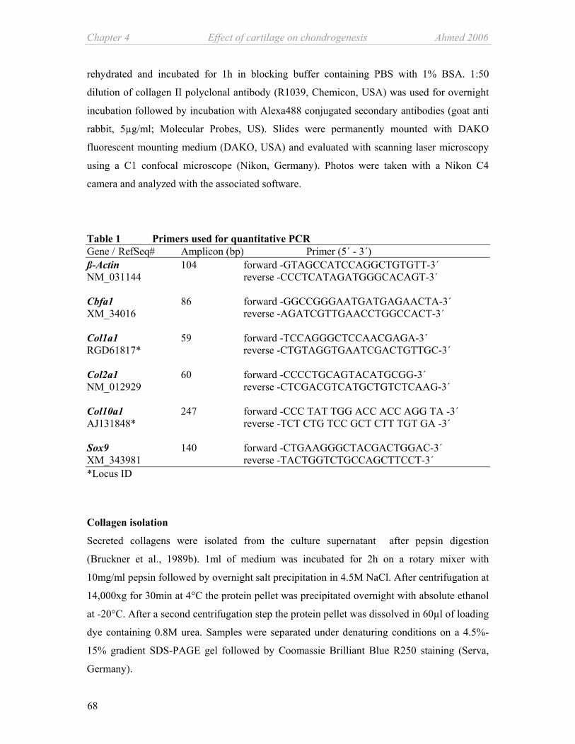
Chapter 4 Effect of cartilage on chondrogenesis Ahmed 2006
68
rehydrated and incubated for 1h in blocking buffer containing PBS with 1% BSA. 1:50
dilution of collagen II polyclonal antibody (R1039, Chemicon, USA) was used for overnight
incubation followed by incubation with Alexa488 conjugated secondary antibodies (goat anti
rabbit, 5µg/ml; Molecular Probes, US). Slides were permanently mounted with DAKO
fluorescent mounting medium (DAKO, USA) and evaluated with scanning laser microscopy
using a C1 confocal microscope (Nikon, Germany). Photos were taken with a Nikon C4
camera and analyzed with the associated software.
Table 1 Primers used for quantitative PCR Gene / RefSeq# Amplicon (bp) Primer (5´ - 3´) ß-Actin 104 forward -GTAGCCATCCAGGCTGTGTT-3´ NM_031144 reverse -CCCTCATAGATGGGCACAGT-3´ Cbfa1 86 forward -GGCCGGGAATGATGAGAACTA-3´ XM_34016 reverse -AGATCGTTGAACCTGGCCACT-3´ Col1a1 59 forward -TCCAGGGCTCCAACGAGA-3´ RGD61817* reverse -CTGTAGGTGAATCGACTGTTGC-3´ Col2a1 60 forward -CCCCTGCAGTACATGCGG-3´ NM_012929 reverse -CTCGACGTCATGCTGTCTCAAG-3´ Col10a1 247 forward -CCC TAT TGG ACC ACC AGG TA -3´ AJ131848* reverse -TCT CTG TCC GCT CTT TGT GA -3´ Sox9 140 forward -CTGAAGGGCTACGACTGGAC-3´ XM_343981 reverse -TACTGGTCTGCCAGCTTCCT-3´ *Locus ID
Collagen isolation
Secreted collagens were isolated from the culture supernatant after pepsin digestion
(Bruckner et al., 1989b). 1ml of medium was incubated for 2h on a rotary mixer with
10mg/ml pepsin followed by overnight salt precipitation in 4.5M NaCl. After centrifugation at
14,000xg for 30min at 4°C the protein pellet was precipitated overnight with absolute ethanol
at -20°C. After a second centrifugation step the protein pellet was dissolved in 60µl of loading
dye containing 0.8M urea. Samples were separated under denaturing conditions on a 4.5%-
15% gradient SDS-PAGE gel followed by Coomassie Brilliant Blue R250 staining (Serva,
Germany).

Ahmed 2006 Effect of cartilage on chondrogenesis Chapter 4
69
Pulse-Chase
After 21 days in chondrogenic favourable environment alginate suspended cells cultured in
the presence of TGFß-3 were metabolically labelled for 1h in labelling DMEM-HG medium
without methionine and cystein (#21013-024, Invitrogen, UK), as described previously
(Grassel et al., 1996). Briefly, cell culture inserts containing the alginate beads were removed
from cocultures and pre-incubated in the labelling medium without radioactive isotopes for
1h. Afterwards beads were labelled in absence of cartilage with freshly added 2ml labelling
medium plus 3.7MBq of [35S]- methionine and cystein (Tran 35S- label from Qbiogene/ICN-
USA) for 1h at 37°C (pulse). Labelling medium was then removed and after 2 washing steps
with PBS the labelled cells were placed back in the culture vessel; in case of coculture
together with cartilage. Newly secreted collagens were chased by continuing the culture in
chondrogenic medium for 4h at 37°C. 1ml cell culture supernatant was retrieved after 0.5h,
1h, 2h and 4h of chase time. Secreted collagens were extracted, as explained above and
samples (each ~155 cpm) were separated on a 4.5-15% gradient SDS-PAGE under reducing
conditions followed by Coomassie Brilliant Blue R250 staining. The destained and fixed gels
were incubated for 30min in Amplify™ solution (Amersham Biosciences, UK), dried and
developed for max. 21 days at -80°C. The radiographic images (Hyperfilm™ MP- Amersham
Biosciences, UK) were acquired with CURIX 60 film developer (Agfa, Germany).
Immunoblotting
Western blotting was carried out with MSCs, cartilage explants and alginate cell fraction
lysates. 0.5 mg cartilage explants were pulverized in a freezer mill and the finely powder
extract was treated similar as the cells. Briefly, ~106 cells were lysed by suspending the cell
pellet in 80µl of 250mM Tris buffer (pH 6.8) containing 6% EDTA, 4% SDS, 40mM DTT,
20% glycerine, 0.002% bromophenol blue and 1:10 protease inhibitors (Complete Mini,
Roche, Germany). Cells were heated for 5min at 60°C immediately followed by incubation
for 10min at 100°C. Cell debris was precipitated by centrifugation at 14000xg for 30min at
4°C and protein containing supernatant was TCA-precipitated and subjected to 4.5-15% SDS-
polyacrylamide gel electrophoresis. Prestained protein standards within a range of 7.5 to
207kDa (BioRad, Germany) were used as molecular weight markers. Proteins were
electrotransfered to a nitrocellulose membrane for 3h at 4°C. To reduce unspecific binding the
membrane was blocked for 1h in blocking buffer (5% dry milk in PBS containing 0.1%
Tween 20). Blots were incubated overnight with polyclonal antibodies against collagen I,
(R1038 Acris, Germany), MMP-2 (AB 809, Chemicon, Germany, 1:100) and MMP-13

Chapter 4 Effect of cartilage on chondrogenesis Ahmed 2006
70
(RP1MMP13, Triple Point Biologics, USA, 1:1000). Detection was performed using
Horseradish-peroxidase coupled secondary anti-rabbit antibodies (A2074, Sigma, USA;
1:50000). Signals were visualized with ECL reagent (Pierce, USA) and the membrane was
exposed to blue light sensitive autoradiography film (Pierce, USA). For dot blot analysis, 1ml
of cell culture supernatant was extracted with 20% TCA solution for 30min at 4°C. Extracted
protein was pelleted by centrifugation at 14000xg for 5min and washed twice with chilled
acetone. After the final washing step the pellet was resuspended in 40µl PBS. 1µl of the
resuspended protein was spotted and air dried on a nitrocellulose membrane. After blocking
for 1h the membrane was incubated overnight with an antibody against TGFß-1/2/3 (sc-7892,
Santa Cruz, USA, 1:200) and developed as above.
Antibody array analysis
RayBio™ rat cytokine antibody array I for conditioned medium (Tebu-bio, France) was used
according to the manufacturer’s instructions. Briefly, the membranes were blocked for 30min
in 5% BSA in 0.01M Tris buffer with 0.15M NaCl (pH 7.6) followed by overnight incubation
at 4°C in 1ml of cell culture supernatant. After washing and incubation with biotin coupled
anti-cytokine antibodies for 2h membranes were again washed and afterwards incubated with
HRP-conjugated streptavidin for 2h. Signals were detected with the solution provided with the
kit, exposed ECL films (Amersham Bioscience, UK) were developed with CURIX 60 film
developer.
Zymography
For gelatine and reverse gelatine zymography, aliquots of culture medium were mixed with
equal volumes of two fold concentrated sample loading buffer (2mM EDTA, 2% SDS, 20%
glycerol, 0.02% bromophenol blue, 20mM Tris/HCl, pH 8.0) and subjected to electrophoresis
on 1% gelatine containing SDS-polyacrylamide gels. For gelatine zymography 4.5 - 15%
gradient gels were used. Reverse zymography was carried out on 15% SDS-polyacrylamide
gels in which 25% conditioned macrophage medium was included as MMP-source. Both
types of gels were washed twice for 30min in 2.5% Triton-X 100, rinsed in distilled water,
and developed for 16h at 37˚C in 50mM Tris/HCl pH 8.5 containing 5mM CaCl2. Finally, the
gels were stained with Coomassie Brilliant Blue R250 (Serva, Germany) to visualize protease
activity and were photographed.

Ahmed 2006 Effect of cartilage on chondrogenesis Chapter 4
71
a. b.
c. d.
a. b.
c. d.
Incubation of 14C-labelled collagen X with culture supernatant from MSCs
(14C)-proline labelled collagen X was produced and isolated from IGF-I stimulated cranial
chicken sternum chondrocytes as described elsewhere (Bohme et al., 1995b). Equal amounts
(1000 cpm) were incubated with aliquots of differently conditioned cell culture media or PBS
for 7h at 37°C and analysed by 4.5-15 % gradient SDS-PAGE gel electrophoresis under
reducing conditions followed by fluorgraphy as described (Bruckner et al., 1989a).
Statistical Analysis
One way ANOVA and student t test were employed for quantitative PCR experiments which
were performed in triplicate.
Results The coculture model
Differentiation potential of the isolated MSCs was determined by cell culture under
osteogenic and chondrogenic favourable conditions. Positive Alizarin Red staining indicating
osteogenesis and collagen II staining indicating chondrogenesis confirmed differentiation
potential of the multipotent MSCs (Fig. 1). Cellular composition of the MSCs was screened
previously by immunofluorescence and magnetic sorting for selected surface markers. These
MSCs were shown to be CD45low, D7Fib+ and CD49a+ (Ahmed et al., 2006a).
Figure 1. Multidifferentiation potential of bone marrow extracted MSCs. After 19 days culture in osteogenic differentiation medium accumulation of calcified ECM is evident by positive staining with Alizarin Red (a) compared with the MSCs cultured in proliferation medium (b). Chondrogenic differentiation potential of MSCs was established by positive collagen II staining after 21 days in chondrogenic favourable 3-dimensional environment (c) compared with day 0 culture (d). Bars represent 50µm.

Chapter 4 Effect of cartilage on chondrogenesis Ahmed 2006
72
MSCs/ alginateporousmembraneRat cartilage chips
Chondrogenicmedium
A. D.
C.
ß-Actin
CaMSCs
1 7 14 21Collagen I
Ca.
MS
Cs
B.
1 2 3 4 5 6
Coculture model
MSCs/ alginateporousmembraneRat cartilage chips
Chondrogenicmedium
A. D.
C.
ß-Actin
CaMSCs
1 7 14 21Collagen I
Ca.
MS
Cs
B.
1 2 3 4 5 6
Coculture model
Figure 2. The coculture model Schematic representation of the coculture model carried out in serum-free chondrogenic medium (A). Dot blot analysis of TGFß secretion into the culture supernatant by monocultured cartilage and monocultured MSCs during the period of culture (B). mRNA expression levels of ß-actin in the cartilage explants and in alginate suspended MSCs were determined by RT-PCR as an indicator of cell viability. Lane 1-3: coculture MSCs from day 7, 14 and 21. Lane 4-6: cartilage explants from day 7, 14 and 21 (C). Western blot analysis of cartilage explant shows lack of collagen I secretion; in comparison MSCs secrete collagen I (D). Ca = explant culture of rat hip cartilage. A coculture system of cartilage tissue explants and multipotent MSCs suspended in 1.2%
alginate was set up in tissue culture inserts placed into 12 well culture plates under serum-free
conditions. The inserts separated MSCs from cartilage chips by a 1µm porous membrane, thus
restricting the contact between the explant and MSCs to soluble diffusible molecules (Fig.
2A). Cartilage explants secrete growth factor TGFß until day 14, from day 21 on TGFß
secretion is strongly reduced and becomes undetectable in culture supernatant. MSCs totally
lack TGFß secretion (Fig. 2B). The viability of both, the cartilage explants and MSCs was
determined at mRNA level with RT-PCR. The unchanged band intensities for the
housekeeping gene ß-actin indicate sustained vitality of MSCs and cartilage tissue throughout
the culture period (Fig. 2C). Cartilage tissue samples were screened for contaminating bone
tissue by western blot analysis of collagen I in culture supernatant. Lack of collagen I
secretion in cartilage explants indicate absence of bone tissue (Fig. 2D).
Effect of cartilage on gene expression profile of Sox9 and Cbfa1
To account for the regulatory effect of Sox9 and Cbfa1 in chondrogenic differentiation of
MSCs, expression of these transcription factors was investigated by qRT-PCR. Application of
chondrogenic culture conditions resulted in an early increase in gene expression level of
transcription factor Sox9 on the first day of culture (day 1). Cartilage exerted an impact on

Ahmed 2006 Effect of cartilage on chondrogenesis Chapter 4
73
Sox9 gene expression within 7 days as the cocultured MSCs showed significant increase in
Sox9 mRNA expression when compared with the monocultures. The increment was
equivalent to the mRNA level of control monocultures of MSCs carried out with TGFß-3.
0.1
1
10
1 7 14 21
0.1
1
10
100
1 7 14 21
A.Sox9 Cbfa1/Runx2
B.
Log1
0 of
RQ
****P ≤0.01; *P ≤0.05
Chondrogenic control
Coculture
Monoculture
Chondrocytes
DaysDays 0.1
1
10
1 7 14 21
0.1
1
10
100
1 7 14 21
A.Sox9 Cbfa1/Runx2
B.
Log1
0 of
RQ
****P ≤0.01; *P ≤0.05
Chondrogenic control
Coculture
Monoculture
Chondrocytes
**P ≤0.01; *P ≤0.05
Chondrogenic control
Coculture
Monoculture
Chondrocytes
DaysDays
Figure 3. Relative changes in gene expression of transcription factor Sox9 and Cbfa1 Relative gene expression level of Sox 9 (A) and Cbfa1 (B) assessed during culture time by qRT-PCR employing the ∆∆CT method using SYBR I Green dye. Target gene expression was normalized to gene expression in MSCs on day 0, which was used as a calibrator and constitutes the baseline at 1. Expression level of each gene from coculture (dotted bars) and monoculture (white bars) was compared with MSCs cultured in chondrogenic medium with addition of 10ng / ml TGFß-3 (chondrogenesis control) (black bars). Grey bar indicates relative gene expression level of freshly isolated hip articular chondrocytes used as positive control.
However, from day 14 onwards Sox9 gene expression was reduced below the level of
undifferentiated MSCs (Fig. 3A). Cbfa-1 gene expression was induced about 3-5 fold at day 1
in alginate cultures of MSCs, remained stable until day 14 and became reduced to starting
values after 21 days. The presence of cartilage did not influence relative induction of gene
expression level of Cbfa-1 (Fig. 3B). Induction of Sox9 and Cbfa1 gene expression did not
require external TGFß-3 as the mRNA levels during the initial (Sox9) and the middle (Cbfa1)
stages of the culture were equivalent to the control culture.
Effect of cartilage on gene expression profile of Col1a1, Col2a1 and Col10a1 and their
biosynthesis
To identify the effect of cartilage explants on chondrogenesis of MSCs, expression and
biosynthesis of different collagens were monitored. Altogether Col1a1 gene expression

Chapter 4 Effect of cartilage on chondrogenesis Ahmed 2006
74
MC CC MC CC MC CC
D.1hr. 2hr. 4hr.
α1(II)/α1(I)α2(I)
MC CC MC CC MC CC
D.1hr. 2hr. 4hr.
α1(II)/α1(I)α2(I)
remained suppressed throughout the 3 weeks of culture became gradually reduced during the
timeline of culture independent of cartilage (Fig. 4A).
Figure 4. Relative gene expression of Col1a1 and Col2a1 and subsequent protein secretion Relative quantitative mRNA expression level of a marker for fibroblast-like cells, Col1a1 (A) was compared with a marker for chondrogenesis, Col2a1 (B) by qRT-PCR employing the ∆∆CT method using SYBR I Green dye. As calibrator gene expression of day 0 MSCs constitutes the baseline at 1. Expression level of each gene was compared between coculture (dotted bars) and monoculture (white bars). MSCs cultured in chondrogenic medium with addition of 10 ng / ml TGFß-3 were regarded as chondrogenesis control (black bars). Grey bar indicates relative gene expression level of freshly isolated hip articular chondrocytes serving as positive control. N=3.
Analysis of collagen chain secretion (C). Collagens were precipitated from culture medium, pepsin digested, separated on a 4.5%-15% gradient SDS-PAGE under reduced conditions and stained with Coomassie Blue. N=3.
Assessment of collagen secretion from mono- and cocultured MSCs by employing the pulse-chase method (D). After 21 days in presence of TGFß-3 cocultured cells were separated from cartilage explants and like monocultured
cells metabolically labelled with [35S]- methionine / cystein for 1h. During the chase period from 0.5h up to 4h secreted collagens were extracted from the culture supernatant. Samples were separated on a 4.5-15% gradient SDS-PAGE under reducing conditions. MC = monocultured MSCs, CC = cocultures. N=2
C.+ TGFß-3 - TGFß-3
Day 7 Day 14 Day 21CC MC CC MC CC MCCollagen
α1(II) / α1(I) α2(I)
Day 7 Day 14 Day 21CC MC CC MC CC MC
C.+ TGFß-3 - TGFß-3
Day 7 Day 14 Day 21CC MC CC MC CC MCCollagen
α1(II) / α1(I) α2(I)
Day 7 Day 14 Day 21CC MC CC MC CC MC
0.1
1
10
100
1000
10000
100000
1 7 14 210.001
0.01
0.1
1
10
1 7 14 21
B.Collagen IICollagen I
A.
Log1
0 of
RQ
**
Days
Days
**P ≤0.01; *P ≤0.05
Chondrogenic controlCocultureMonocultureChondrocytes
0.1
1
10
100
1000
10000
100000
1 7 14 210.001
0.01
0.1
1
10
1 7 14 21
B.Collagen IICollagen I
A.
Log1
0 of
RQ
**
Days
Days
**P ≤0.01; *P ≤0.05
Chondrogenic controlCocultureMonocultureChondrocytes
**P ≤0.01; *P ≤0.05
Chondrogenic controlCocultureMonocultureChondrocytes

Ahmed 2006 Effect of cartilage on chondrogenesis Chapter 4
75
On the contrary, mRNA expression of Col2a1 was progressively elevated in both mono- and
cocultured MSCs similarly cartilage exhibited no influence on gene expression level of
Col2a1 (Fig. 4B). However, secretion of α1/2 (I) and α1 (II) collagen chains was positively
affected by cartilage when compared with the monocultured MSCs. Notably, in absence of
external TGFß-3, neither cartilage nor MSCs secreted detectable amounts of collagen I,
indicated by the lack of the α2 (I) chain while addition of TGFß-3 induced secretion of
collagen I (Fig. 4C). To deduce the origin of collagens detected in coculture medium, both
mono and cocultured MSCs (day 21 in presence of TGFß-3) were metabolically labelled with 35[S] Met/Cys in a pulse/chase regimen after removal of the cartilage explants. After 1 hour of
chase newly synthesized collagens were detected in the medium of MSCs which were
previously cultured as cocultures with a secretion peak at 4 hours of chase. We assume most
of the collagen constitutes collagen II, because of the poor intensity of the lower α2 (I) chain
compared to the strong signal of the upper α1 (II) band (Fig. 4D). Minuscule collagen
secretion in monocultured MSCs corroborated the non-radioactive collagen preparation results
of day 21 (Fig. 4C and D). These data suggest that the factors derived from cartilage explants
influence collagen II secretion of MSCs indicating a positive influence on biosynthesis of
collagen II.
Compared with freshly isolated chondrocytes undifferentiated MSCs exhibit a very low
baseline expression of Col10a1 mRNA. Induction of chondrogenic differentiation did not
alter this expression pattern during the first two weeks of culture. Elevation of Col10a1
mRNA was observed at the terminal stage of the culture (Day 21 and 28). While relative gene
expression in the chondrogenic control cultures (+ TGFß-3) increased about 100x on day 28
gene expression in both, monocultured and cocultured MSCs remained suppressed.
Interestingly, this strong upregulation of collagen X gene expression in MSCs under the
influence of external TGFß-3 is profoundly suppressed in the presence of cartilage (Fig. 5A).
Concurrently, collagen X secretion and biosynthesis was strongly suppressed in the presence
of cartilage compared to the monocultured MSCs which released high amounts of collagen X
into the culture supernatant. Little collagen X was detected in the cellular fraction of
monocultures, however, none was found in cell layers of cocultured MSCs. (Fig. 5B). To
exclude protease induced degradation of collagen X in the cell culture supernatant of
cocultures 14C-labelled collagen X was incubated with the cell culture supernatants of mono-
and cocultured MSCs from day 21 for 7 hours at 37°C. No proteolytic activity was observed
in mono- or cocultured MSCs supernatants that generated degradation products of collagen X
or reduced signal intensity of the bands. Therefore, we exclude protease induced degradation

Chapter 4 Effect of cartilage on chondrogenesis Ahmed 2006
76
(Fig. 5C) and attribute this strong impact of cartilage derived soluble factors on suppression of
collagen X secretion from MSCs to the biosynthesis level.
0.01
0.1
1
10
100
1 7 14 21 28
Collagen XA.
Log1
0 of
RQ
b.
**P ≤0.01; *P ≤0.05
Chondrogenic control (+Tgfß-3)
CocultureMonocultureChondrocytes
MC CC
α1(X)
Pepsin
- TGFß-3+ TGFß-3
α1(II)
α1(X)
+TGFß -TGFßMC CC MC CC TBS
MC CC
B. a.
MC CC MC CCS CF S CF
Coculture(+Tgfß-3)
0.01
0.1
1
10
100
1 7 14 21 28
Collagen XA.
Log1
0 of
RQ
b.
**P ≤0.01; *P ≤0.05
Chondrogenic control (+Tgfß-3)
CocultureMonocultureChondrocytes
MC CC
α1(X)
Pepsin
- TGFß-3+ TGFß-3
α1(II)
α1(X)
+TGFß -TGFßMC CC MC CC TBS
MC CC
B. a.
MC CC MC CCS CF S CF
Coculture(+Tgfß-3)
Figure 5: Relative gene expression, biosynthesis and secretion of collagen X Relative mRNA expression level of Col10a1 is assayed by qRT-PCR employing the ∆∆CT method using SYBR I Green dye (A). Calibrator was gene expression of day 0 MSCs which constitutes the baseline at 1. Expression level of each gene was compared between coculture (dotted bars) and monoculture (white bars). Gene expression of monocultured (black bar) and cocultured (mosaic bar) MSCs cultured in chondrogenic medium with addition of 10 ng / ml TGFß-3 was determined in parallel. Grey bar indicates relative gene expression level of freshly isolated hip articular chondrocytes serving as positive control. Analysis of collagen X protein biosynthesis and secretion (B): Collagens were precipitated from the alginate beads and culture medium, pepsin digested, separated on a 4.5%-15% gradient SDS-PAGE under reduced conditions and stained with Coomassie Blue (a). Collagen X degradation assay: (14C)-labelled collagen X, isolated from metabolically labelled chicken chondrocyte cultures, was incubated with supernatants from mono- and cocultured MSCs (day 21) (b). MC = monocultured MSCs, CC = cocultures, S = culture supernatant, CF = cellular fraction. N = 3
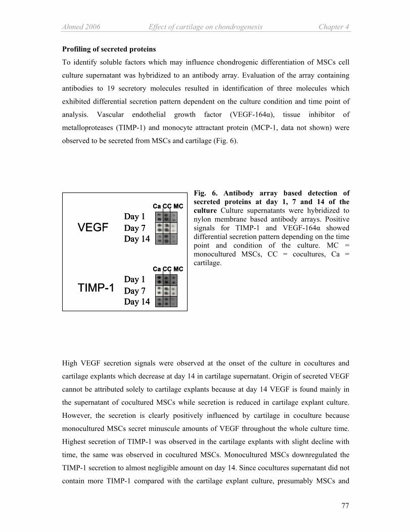
Ahmed 2006 Effect of cartilage on chondrogenesis Chapter 4
77
Ca CC MC
Ca CC MC
VEGF
TIMP-1Day 1Day 7Day 14
Day 1Day 7Day 14
Ca CC MC
Ca CC MC
VEGF
TIMP-1Day 1Day 7Day 14
Day 1Day 7Day 14
Day 1Day 7Day 14
Day 1Day 7Day 14
Profiling of secreted proteins
To identify soluble factors which may influence chondrogenic differentiation of MSCs cell
culture supernatant was hybridized to an antibody array. Evaluation of the array containing
antibodies to 19 secretory molecules resulted in identification of three molecules which
exhibited differential secretion pattern dependent on the culture condition and time point of
analysis. Vascular endothelial growth factor (VEGF-164α), tissue inhibitor of
metalloproteases (TIMP-1) and monocyte attractant protein (MCP-1, data not shown) were
observed to be secreted from MSCs and cartilage (Fig. 6).
Fig. 6. Antibody array based detection of secreted proteins at day 1, 7 and 14 of the culture Culture supernatants were hybridized to nylon membrane based antibody arrays. Positive signals for TIMP-1 and VEGF-164α showed differential secretion pattern depending on the time point and condition of the culture. MC = monocultured MSCs, CC = cocultures, Ca = cartilage.
High VEGF secretion signals were observed at the onset of the culture in cocultures and
cartilage explants which decrease at day 14 in cartilage supernatant. Origin of secreted VEGF
cannot be attributed solely to cartilage explants because at day 14 VEGF is found mainly in
the supernatant of cocultured MSCs while secretion is reduced in cartilage explant culture.
However, the secretion is clearly positively influenced by cartilage in coculture because
monocultured MSCs secret minuscule amounts of VEGF throughout the whole culture time.
Highest secretion of TIMP-1 was observed in the cartilage explants with slight decline with
time, the same was observed in cocultured MSCs. Monocultured MSCs downregulated the
TIMP-1 secretion to almost negligible amount on day 14. Since cocultures supernatant did not
contain more TIMP-1 compared with the cartilage explant culture, presumably MSCs and

Chapter 4 Effect of cartilage on chondrogenesis Ahmed 2006
78
cartilage are collectively responsible for the secretion in the cocultures. Furthermore, based on
the antibody array results secretion of the following pro-inflammatory cytokines and growth
factors could be excluded at any stage of the culture. These non-secreted factors included
CINC-2, CNTF, Fractalkine, GM-CSF, IFN-γ, IL-1α, IL-1β, IL-4, IL-6, IL-10, LIX, Leptin,
MIP-3α, ß-NGF and TNF-α.
Analysis of secretion pattern of MMP-13 and TIMP-2 by zymography
Reverse zymography was used to detect TIMP-2 in the culture system. Monocultured MSCs
did not secrete TIMP-2 at any time point; in contrast secretion of TIMP-2 by cartilage
explants started at day 7 and remained detectable until the end of the culture period. Initially,
less TIMP-2 secretion was detected in cocultures when compared with cartilage explant
cultures however, at the end of the culture period the pattern was reversed (Fig. 7).
TIMP-2 (21 KDa)
MC CC Ca MC CC Ca MC CC Ca MC CC Ca
Day 1 Day 14Day 7 Day 21
TIMP-2 (21 KDa)
MC CC Ca MC CC Ca MC CC Ca MC CC Ca
Day 1 Day 14Day 7 Day 21
Figure 7. Reverse zymography for TIMP-2 Aliquots of culture supernatants from cells were subjected to SDS-PAGE under non-reducing conditions on a 4.5-15% gradient gel containing 1 % gelatine and 25% conditioned macrophage medium. This reverse zymographic analysis was applied for detection of TIMP-2. Ca = monocultured cartilage, MC = monocultured MSCs, CC = cocultures. N= 3
Gelatine zymogram results indicated secretion of MMP-13 and MMP-9. From the beginning
of the culture cartilage and cocultures secreted increasing concentration of pro-MMP-13
without decline till day 21. In contrast, release of pro-enzyme from monocultured MSCs
remained largely unchanged during the culture period. Notably, the active form of MMP-13 in
coculture and cartilage culture became reduced in relation to the strongly increasing pro-form
(Fig. 8A). In coculture and cartilage culture we have observed an additional gelatinolytic
activity, which we assume to be pro-MMP-9 according to the molecular weight (black arrow
in fig. 8A). Western blot analysis of culture supernatants confirmed the identity of MMP-13 in
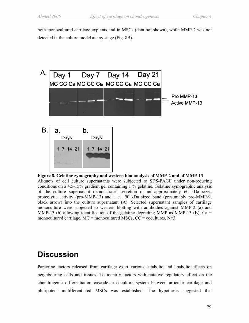
Ahmed 2006 Effect of cartilage on chondrogenesis Chapter 4
79
both monocultured cartilage explants and in MSCs (data not shown), while MMP-2 was not
detected in the culture model at any stage (Fig. 8B).
A.MC CC Ca MC CC Ca MC CC CaDay 1 Day 14Day 7
MC CC CaDay 21
Active MMP-13Pro MMP-13
B.
1 7 14 21
Days
1 7 14 21
Daysa. b.
A.MC CC Ca MC CC Ca MC CC CaDay 1 Day 14Day 7
MC CC CaDay 21
Active MMP-13Pro MMP-13
B.
1 7 14 21
Days
1 7 14 21
Daysa. b.
Figure 8. Gelatine zymography and western blot analysis of MMP-2 and of MMP-13 Aliquots of cell culture supernatants were subjected to SDS-PAGE under non-reducing conditions on a 4.5-15% gradient gel containing 1 % gelatine. Gelatine zymographic analysis of the culture supernatant demonstrates secretion of an approximately 60 kDa sized proteolytic activity (pro-MMP-13) and a ca. 90 kDa sized band (presumably pro-MMP-9, black arrow) into the culture supernatant (A). Selected supernatant samples of cartilage monoculture were subjected to western blotting with antibodies against MMP-2 (a) and MMP-13 (b) allowing identification of the gelatine degrading MMP as MMP-13 (B). Ca = monocultured cartilage, MC = monocultured MSCs, CC = cocultures. N=3
Discussion Paracrine factors released from cartilage exert various catabolic and anabolic effects on
neighbouring cells and tissues. To identify factors with putative regulatory effect on the
chondrogenic differentiation cascade, a coculture system between articular cartilage and
pluripotent undifferentiated MSCs was established. The hypothesis suggested that

Chapter 4 Effect of cartilage on chondrogenesis Ahmed 2006
80
differentiation of chondroprogenitor MSCs into chondrocytes is controlled and affected by
soluble factors secreted by cartilage.
In the current culture system where MSCs were kept in alginate stabilized 3D environment
known to induce chondrogenesis in MSCs, Sox9 gene expression was prematurely up
regulated which indicates early onset of chondrogenesis (Majumdar et al., 2000). Elevated
gene expression of Sox9 particular on day 7 under the influence of cartilage suggests
involvement of chondrocyte released signalling factors in regulation of the chondrogenic
inducer activity of Sox9. Several signalling pathways are involved in regulating Sox9 and its
downstream genes during cartilage differentiation. TGFß induces Sox9 gene expression and
transcriptional activity through Smad3 (Furumatsu et al., 2005) and BMP-2 enhances Sox9
gene expression level during murine fracture healing (Uusitalo et al., 2001). TGFß, released
from cartilage explants until day 14, presumably accounts in part for the observed increase in
Sox9 gene expression. On the other hand, VEGF has been described to suppress Sox9 mRNA
levels during treatment of chick embryo limb buds (Yin and Pacifici, 2001). In our system
Sox9 gene expression is reduced below initial levels after day 7. Possibly, high secretion level
of VEGF from MSCs themselves plus cartilage explants induces this suppression of Sox9 in
MSCs, thereby counteracting the influence of TGFß. Despite suppression of Sox9 mRNA
below baseline level, gene expression of Col2a1 remains up regulated. This observation
indicates that an active Sox9 gene above basal level of un-induced MSCs might not be
mandatory in later stages of chondrogenic differentiation of MSCs for regulation of
chondrogenesis associated genes as Col2a1, but is imperative at initiation of culture.
The most profound effect of cartilage derived factors was observed on suppression of collagen
X gene expression and protein secretion. MSCs, regardless of the presence of cartilage exhibit
no collagen X secretion before day 21 corroborated by low gene expression until that time
point. However, from 21 days on monocultured MSCs release high amounts of collagen X
into the culture supernatant, while cocultures do not. Suppression of collagen X secretion in
the presence of cartilage depicts delayed hypertrophy in our coculture model. We suggest that
cartilage derived soluble factors have the ability to reduce collagen X secretion compared to a
strongly elevated secretion by monocultured MSCs at that stage of culture. As we did not
observe collagen X degradation products in culture supernatant we deduce that absence of
collagen X secretion is due to suppression at the biosynthesis level. This suggestion is
substantiated by significantly reduced gene expression in cocultured MSCs compared with the
monocultured MSCs. Secondly; we did not detect collagen X protein in the cellular fraction of
cocultured MSCs while small amounts were retained in monocultured MSCs.

Ahmed 2006 Effect of cartilage on chondrogenesis Chapter 4
81
Paracrine factors derived from surrounding chondrocytes and endothelial cells alter
differentiation status of chondrocytes. Chondrocytes of normal articular cartilage do not
undergo terminal differentiation whereas growth plate chondrocytes do. Articular
chondrocytes in osteoarthritic joints however, express markers of terminal differentiation like
collagen X and Alkaline Phosphatase (AP) (von der et al., 1992). By coculturing articular and
growth plate chondrocytes Jikko et al. could demonstrate that soluble paracrine factors alone
are responsible for inhibition of terminal differentiation. Articular chondrocytes were able to
suppress calcification and AP activity in cocultured growth plate chondrocytes, indicating that
articular chondrocytes produce factors which inhibit terminal differentiation (Jikko et al.,
1999). Also during endochondral ossification cartilage maturation and conversion of
hypertrophic cartilage to bone is delayed by paracrine signals derived from
periosteal/perichondrial tissue or from the cartilage proper itself. Formation of new blood
vessels and their penetration into hypertrophic cartilage appear to be essential for the
elimination of this developmental barrier. Consistently with this notion, the terminal
differentiation barrier in chondrocytes from the caudal part of 17-day old chick embryo
sternum can be eliminated by coculturing them with endothelial cells in-vitro. The humoral
activity secreted by endothelial cells consists of proteinases sufficient to break down the
terminal differentiation barrier inherent to this chondrocyte population (Babarina et al., 2001;
Bittner et al., 1998; Bohme et al., 1995a). In addition, articular chondrocytes obtained from
osteoarthritic joints are known to secrete factors which activate pro-MMP-9 derived from
cocultured macrophages. The stepwise activation process of pro-MMP-9 involves MMP-13,
secreted from chondrocytes and MT1-MMP, also expressed by chondrocytes which converts
pro-MMP-13 to its active form (Dreier et al., 2001; Dreier et al., 2004; Knauper et al., 2002).
TIMP-1, TIMP-2, VEGF-164α and MMP13 were the four prominent secreted molecules
identified in the supernatant of cocultures and cartilage explants. Except VEGF-164α which
seems to be mainly secreted by cocultured MSCs at least at the final culture stage, a
reasonable amount of TIMP-1,-2 and MMP13 is released by differentiated chondrocytes of
the cartilage explants. We observed a profound increase in pro-MMP-13 secretion in coculture
and cartilage monocultures during the culture period; however with declining activation
status. This effect might be due to a concurrent secretion of TIMP-1 and -2, both known as
efficient inhibitors of MMP-13 activity (Will et al., 1996; Knauper et al., 1997b; Cowell et al.,
1998). MMP-13 is known to degrade collagen X (Knauper et al., 1997a) however, lack of
collagen X in supernatant of cocultured MSCs can not be due to proteolytic activities because
collagen X degradation products or reduced signal intensities were never detected in the

Chapter 4 Effect of cartilage on chondrogenesis Ahmed 2006
82
cultures. Additionally, in absence of cartilage MMP-13 activation rate was not higher whereas
collagen X secretion was profoundly increased. In MMP-13 knockout mice Inada et al. have
demonstrated a profound enlargement of the anatomic domain where collagen X is expressed
by hypertrophic growth plate chondrocytes and an increase of collagen X deposition in this
domain. In addition to proteolytic activity of MMP-13 this protease might exert a yet
unknown regulatory impact on collagen X biosynthesis (Inada et al., 2004; Stickens et al.,
2004). Therefore, suppression of collagen X gene expression and accordingly biosynthesis in
cocultures might be also due to a regulatory effect of MMP-13 and not to a proteolytic effect.
Multiple Cbfa1 consensus binding elements are located in the collagen X promoter region
(Kim et al., 1999; Zheng et al., 2003). Despite induction of Cbaf1 gene expression early in
MSCs differentiation pathway (day 1) collagen X expression is not significantly induced
before day 28 while Cbfa1 gene expression is downregulated after 21 days prior to
upregulation of the col10a1 gene. Also, relative gene expression of Cbfa1 is not significantly
influenced by cocultured cartilage in our system during the whole culture period whereas gene
expression of collagen X is. In case Cbfa1 influences collagen X mRNA level we would not
expect such a profound difference between cocultured and monocultured MSCs therefore, we
cannot attribute an influence of this transcription factor on mRNA expression or eventual
transcription of collagen X.
Another key player in this system might be VEGF-164α, released by cartilage and cocultured
MSCs. VEGF as one of the most important mediators of angiogenesis is abundantly expressed
during embryogenesis but in adults it is only found in restricted tissues and cell types, as in
osteoarthritic chondrocytes, but not in resting chondrocytes (Pufe et al., 2001; Pfander et al.,
2001). Interestingly, we have detected VEGF-164α in increasing amounts in cocultures and
cartilage, however, main source seems to be cocultured MSCs. VEGF is known to stimulate
receptor phosphorylation of its VEGF-receptor 2 (flk-1 /KDR), thereby activating the mitogen
activated protein kinases ERK/1/2 inducing long-lasting activation of the transcription factor
AP-1 plus expression of c-fos mRNA in dental pulp cells (Pufe et al., 2004b; Matsushita et al.,
2000). As a consensus site for AP-1, a dimeric transcription factor composed of the two
oncogenic products jun and fos, has been identified within the promoter of collagen X, VEGF
binding to its receptor (VEGFR1) might contribute to suppression of gene expression of
collagen X, while at the same time it is known to induce secretion of MMP-13 (Pufe et al.,
2004a).
All four members of the TIMP-family function as inhibitors of angiogenesis by blocking
MMP activity. TIMP-1 and -2, both present in cocultures, presumably account for inhibiting
Ahmed 2006 Effect of cartilage on chondrogenesis Chapter 4
83
generation of active MMP-13. Besides, TIMPs can exert MMP-independent functions, which
might be important in our system. TIMP-2 exhibits a growth suppressive activity by
interacting with the α3ß1 integrin on endothelial cells thereby inhibiting cell proliferation
(Seo et al., 2003). This shows that in addition to inhibition of MMPs TIMPs also regulate cell
growth and differentiation. Cell surface binding of TIMP-1 and -4 has been reported, but the
identification of specific cell surface receptors is missing (Stetler-Stevenson and Seo, 2005).
Therefore, TIMPs secreted by differentiated chondrocytes and MSCs may act as inducers or
repressors of specific factors necessary for chondrogenic differentiation of MSCs.
Two additional molecules are described in literature with collagen X regulatory properties.
PTH / PTHrP expressed in perichondrium and in resting and proliferating chondrocytes
during endochondral growth (Lee et al., 1996; Iwamoto et al., 1994) strongly represses
collagen X biosynthesis in chick embryonic hypertrophic chondrocytes thus inhibits matrix
mineralization and apoptosis of chondrocytes (Zerega et al., 1999). It has been demonstrated
that PTH / PTHrP fragments suppress collagen X gene expression in vitro by acting on two
responsive elements within the promoter (Gebhard et al., 2004; Riemer et al., 2002). The
second molecule is, osteogenic protein 1 (OP-1 or BMP-7) which belongs to the TGFß super
family (Kingsley, 1994) can induce collagen X gene expression via a cell type specific
response element that contains an A/T rich and a MEF-2 like sequence which react with yet
unidentified nuclear proteins in response to OP-1(Harada et al., 1997).
In conclusion, we have demonstrated that the coculture system including differentiated
cartilage tissue and undifferentiated MSCs is suitable for studying paracrine communication
between both the components. Notably, external growth factors are not necessary for
induction of chondrogenic marker genes and biosynthesis of the corresponding proteins.
However, for long term maintenance of the gene expression level growth factors like TGFß
are required as internal secretion by cartilage ceases from day 14 on. Cartilage derived soluble
factors are sufficient to induce Sox9 gene expression and suppress collagen X
expression/secretion. MMP-13 and VEGF may have a regulatory role in collagen X
expression and secretion. Besides the identified secreted proteins, MMP-13, TIMP-1 and -2
and VEGF-164α other, yet unidentified signalling molecules surely have an impact on
regulating and directing the differentiation process of MSCs. It shall be kept in mind that in
the coculture on one hand cartilage derived factors influence fate of MSCs; on the other hand
MSCs released molecules might have an impact on cartilage metabolism as well. These
putative proteins may even abrogate the intrinsic blockade which prevents hypertrophy and
eventual ossification in articular cartilage. Discovering the mutual paracrine influence of

Chapter 4 Effect of cartilage on chondrogenesis Ahmed 2006
84
differentiated and undifferentiated cells will have direct consequences on therapeutic use of
either undifferentiated or ex vivo differentiated MSCs for cartilage replacement. For MSCs
based cartilage repair, inhibition of hypertrophy and stabilization of the cartilaginous
phenotype in explants as well as in the host is a prerequisite for success and long lasting
vitality.
Acknowledgements We want to thank Maren Marschner and Marianne Ahler for their excellent technical
assistance. This work was in part supported by the Deutsche Forschungsgemeinschaft (SFB
492: Extrazelluläre Matrix: Biogenese, Assemblierung und zelluläre Wechselwirkungen, B18
assigned to R.D.).

Ahmed 2006 Effect of cartilage on chondrogenesis Chapter 4
85
Reference List
1. Ahmed,N., Vogel,B., Rohde,E., Strunk,D., Grifka,J., Schulz,M.B., and Grassel,S. (2006a). CD45-positive cells of haematopoietic origin enhance chondrogenic marker gene expression in rat marrow stromal cells. Int. J. Mol. Med. 18, 233-240.
2. Ahmed,N., Vogel,B., Rohde,E., Strunk,D., Grifka,J., Schulz,M.B., and Grassel,S. (2006b). CD45-positive cells of haematopoietic origin enhance chondrogenic marker gene expression in rat marrow stromal cells. Int. J. Mol. Med. 18, 233-240.
3. Ahmed,N., Vogel,B., Rohde,E., Strunk,D., Grifka,J., Schulz,M.B., and Grassel,S. (2006c). CD45-positive cells of haematopoietic origin enhance chondrogenic marker gene expression in rat marrow stromal cells. Int. J. Mol. Med. 18, 233-240.
4. Babarina,A.V., Mollers,U., Bittner,K., Vischer,P., and Bruckner,P. (2001). Role of the subchondral vascular system in endochondral ossification: endothelial cell-derived proteinases derepress late cartilage differentiation in vitro. Matrix Biol. 20, 205-213.
5. Barry,F., Boynton,R.E., Liu,B., and Murphy,J.M. (2001). Chondrogenic differentiation of mesenchymal stem cells from bone marrow: differentiation-dependent gene expression of matrix components. Exp. Cell Res. 268, 189-200.
6. Bi,W., Deng,J.M., Zhang,Z., Behringer,R.R., and de Crombrugghe,B. (1999). Sox9 is required for cartilage formation. Nat. Genet. 22, 85-89.
7. Bittner,K., Vischer,P., Bartholmes,P., and Bruckner,P. (1998). Role of the subchondral vascular system in endochondral ossification: endothelial cells specifically derepress late differentiation in resting chondrocytes in vitro. Exp. Cell Res. 238, 491-497.
8. Bohme,K., Winterhalter,K.H., and Bruckner,P. (1995a). Terminal differentiation of chondrocytes in culture is a spontaneous process and is arrested by transforming growth factor-beta 2 and basic fibroblast growth factor in synergy. Exp. Cell Res. 216, 191-198.
9. Bohme,K., Winterhalter,K.H., and Bruckner,P. (1995b). Terminal differentiation of chondrocytes in culture is a spontaneous process and is arrested by transforming growth factor-beta 2 and basic fibroblast growth factor in synergy. Exp. Cell Res. 216, 191-198.
10. Bruckner,P., Horler,I., Mendler,M., Houze,Y., Winterhalter,K.H., Eich-Bender,S.G., and Spycher,M.A. (1989a). Induction and prevention of chondrocyte hypertrophy in culture. J. Cell Biol. 109, 2537-2545.
11. Bruckner,P., Horler,I., Mendler,M., Houze,Y., Winterhalter,K.H., Eich-Bender,S.G., and Spycher,M.A. (1989b). Induction and prevention of chondrocyte hypertrophy in culture. J. Cell Biol. 109, 2537-2545.
12. Cowell,S., Knauper,V., Stewart,M.L., d'Ortho,M.P., Stanton,H., Hembry,R.M., Lopez-Otin,C., Reynolds,J.J., and Murphy,G. (1998). Induction of matrix metalloproteinase activation cascades based on membrane-type 1 matrix metalloproteinase: associated activation of gelatinase A, gelatinase B and collagenase 3. Biochem. J. 331 ( Pt 2), 453-458.

Chapter 4 Effect of cartilage on chondrogenesis Ahmed 2006
86
13. de Crombrugghe,B., Lefebvre,V., Behringer,R.R., Bi,W., Murakami,S., and Huang,W. (2000). Transcriptional mechanisms of chondrocyte differentiation. Matrix Biol. 19, 389-394.
14. Dreier,R., Grassel,S., Fuchs,S., Schaumburger,J., and Bruckner,P. (2004). Pro-MMP-9 is a specific macrophage product and is activated by osteoarthritic chondrocytes via MMP-3 or a MT1-MMP/MMP-13 cascade. Exp. Cell Res. 297, 303-312.
15. Dreier,R., Wallace,S., Fuchs,S., Bruckner,P., and Grassel,S. (2001). Paracrine interactions of chondrocytes and macrophages in cartilage degradation: articular chondrocytes provide factors that activate macrophage-derived pro-gelatinase B (pro-MMP-9). J. Cell Sci. 114, 3813-3822.
16. Furumatsu,T., Tsuda,M., Taniguchi,N., Tajima,Y., and Asahara,H. (2005). Smad3 induces chondrogenesis through the activation of SOX9 via CREB-binding protein/p300 recruitment. J. Biol. Chem. 280, 8343-8350.
17. Gebhard,S., Poschl,E., Riemer,S., Bauer,E., Hattori,T., Eberspaecher,H., Zhang,Z., Lefebvre,V., de Crombrugghe,B., and von der,M.K. (2004). A highly conserved enhancer in mammalian type X collagen genes drives high levels of tissue-specific expression in hypertrophic cartilage in vitro and in vivo. Matrix Biol. 23, 309-322.
18. Gerber,H.P., Vu,T.H., Ryan,A.M., Kowalski,J., Werb,Z., and Ferrara,N. (1999). VEGF couples hypertrophic cartilage remodeling, ossification and angiogenesis during endochondral bone formation. Nat. Med. 5, 623-628.
19. Goldring,M.B., Tsuchimochi,K., and Ijiri,K. (2006). The control of chondrogenesis. J. Cell Biochem. 97, 33-44.
20. Grassel,S., Timpl,R., Tan,E.M., and Chu,M.L. (1996). Biosynthesis and processing of type XVI collagen in human fibroblasts and smooth muscle cells. Eur. J. Biochem. 242, 576-584.
21. Harada,S., Sampath,T.K., Aubin,J.E., and Rodan,G.A. (1997). Osteogenic protein-1 up-regulation of the collagen X promoter activity is mediated by a MEF-2-like sequence and requires an adjacent AP-1 sequence. Mol. Endocrinol. 11, 1832-1845.
22. Inada,M., Wang,Y., Byrne,M.H., Rahman,M.U., Miyaura,C., Lopez-Otin,C., and Krane,S.M. (2004). Critical roles for collagenase-3 (Mmp13) in development of growth plate cartilage and in endochondral ossification. Proc. Natl. Acad. Sci. U. S. A 101, 17192-17197.
23. Inada,M., Yasui,T., Nomura,S., Miyake,S., Deguchi,K., Himeno,M., Sato,M., Yamagiwa,H., Kimura,T., Yasui,N., Ochi,T., Endo,N., Kitamura,Y., Kishimoto,T., and Komori,T. (1999). Maturational disturbance of chondrocytes in Cbfa1-deficient mice. Dev. Dyn. 214, 279-290.
24. Iwamoto,M., Jikko,A., Murakami,H., Shimazu,A., Nakashima,K., Iwamoto,M., Takigawa,M., Baba,H., Suzuki,F., and Kato,Y. (1994). Changes in parathyroid hormone receptors during chondrocyte cytodifferentiation. J. Biol. Chem. 269, 17245-17251.

Ahmed 2006 Effect of cartilage on chondrogenesis Chapter 4
87
25. Jikko,A., Kato,Y., Hiranuma,H., and Fuchihata,H. (1999). Inhibition of chondrocyte terminal differentiation and matrix calcification by soluble factors released by articular chondrocytes. Calcif. Tissue Int. 65, 276-279.
26. Johansson,N., Saarialho-Kere,U., Airola,K., Herva,R., Nissinen,L., Westermarck,J., Vuorio,E., Heino,J., and Kahari,V.M. (1997). Collagenase-3 (MMP-13) is expressed by hypertrophic chondrocytes, periosteal cells, and osteoblasts during human fetal bone development. Dev. Dyn. 208, 387-397.
27. Johnstone,B., Hering,T.M., Caplan,A.I., Goldberg,V.M., and Yoo,J.U. (1998). In vitro chondrogenesis of bone marrow-derived mesenchymal progenitor cells. Exp. Cell Res. 238, 265-272.
28. Kim,I.S., Otto,F., Zabel,B., and Mundlos,S. (1999). Regulation of chondrocyte differentiation by Cbfa1. Mech. Dev. 80, 159-170.
29. Kingsley,D.M. (1994). The TGF-beta superfamily: new members, new receptors, and new genetic tests of function in different organisms. Genes Dev. 8, 133-146.
30. Knauper,V., Bailey,L., Worley,J.R., Soloway,P., Patterson,M.L., and Murphy,G. (2002). Cellular activation of proMMP-13 by MT1-MMP depends on the C-terminal domain of MMP-13. FEBS Lett. 532, 127-130.
31. Knauper,V., Cowell,S., Smith,B., Lopez-Otin,C., O'Shea,M., Morris,H., Zardi,L., and Murphy,G. (1997a). The role of the C-terminal domain of human collagenase-3 (MMP-13) in the activation of procollagenase-3, substrate specificity, and tissue inhibitor of metalloproteinase interaction. J. Biol. Chem. 272, 7608-7616.
32. Knauper,V., Cowell,S., Smith,B., Lopez-Otin,C., O'Shea,M., Morris,H., Zardi,L., and Murphy,G. (1997b). The role of the C-terminal domain of human collagenase-3 (MMP-13) in the activation of procollagenase-3, substrate specificity, and tissue inhibitor of metalloproteinase interaction. J. Biol. Chem. 272, 7608-7616.
33. Knauper,V., Lopez-Otin,C., Smith,B., Knight,G., and Murphy,G. (1996). Biochemical characterization of human collagenase-3. J. Biol. Chem. 271, 1544-1550.
34. Lee,K., Lanske,B., Karaplis,A.C., Deeds,J.D., Kohno,H., Nissenson,R.A., Kronenberg,H.M., and Segre,G.V. (1996). Parathyroid hormone-related peptide delays terminal differentiation of chondrocytes during endochondral bone development. Endocrinology 137, 5109-5118.
35. Lefebvre,V., Behringer,R.R., and de Crombrugghe,B. (2001). L-Sox5, Sox6 and Sox9 control essential steps of the chondrocyte differentiation pathway. Osteoarthritis. Cartilage. 9 Suppl A, S69-S75.
36. Lefebvre,V. and Smits,P. (2005). Transcriptional control of chondrocyte fate and differentiation. Birth Defects Res. C. Embryo. Today 75, 200-212.
37. Magne,D., Vinatier,C., Julien,M., Weiss,P., and Guicheux,J. (2005). Mesenchymal stem cell therapy to rebuild cartilage. Trends Mol. Med. 11, 519-526.

Chapter 4 Effect of cartilage on chondrogenesis Ahmed 2006
88
38. Majumdar,M.K., Banks,V., Peluso,D.P., and Morris,E.A. (2000). Isolation, characterization, and chondrogenic potential of human bone marrow-derived multipotential stromal cells. J. Cell Physiol 185, 98-106.
39. Matsushita,K., Motani,R., Sakuta,T., Yamaguchi,N., Koga,T., Matsuo,K., Nagaoka,S., Abeyama,K., Maruyama,I., and Torii,M. (2000). The role of vascular endothelial growth factor in human dental pulp cells: induction of chemotaxis, proliferation, and differentiation and activation of the AP-1-dependent signaling pathway. J. Dent. Res. 79, 1596-1603.
40. Mendler,M., Eich-Bender,S.G., Vaughan,L., Winterhalter,K.H., and Bruckner,P. (1989). Cartilage contains mixed fibrils of collagen types II, IX, and XI. J. Cell Biol. 108, 191-197.
41. Mitchell,P.G., Magna,H.A., Reeves,L.M., Lopresti-Morrow,L.L., Yocum,S.A., Rosner,P.J., Geoghegan,K.F., and Hambor,J.E. (1996). Cloning, expression, and type II collagenolytic activity of matrix metalloproteinase-13 from human osteoarthritic cartilage. J. Clin. Invest 97, 761-768.
42. Mok,S.S., Masuda,K., Hauselmann,H.J., Aydelotte,M.B., and Thonar,E.J. (1994). Aggrecan synthesized by mature bovine chondrocytes suspended in alginate. Identification of two distinct metabolic matrix pools. J. Biol. Chem. 269, 33021-33027.
43. Mundlos,S. and Olsen,B.R. (1997). Heritable diseases of the skeleton. Part I: Molecular insights into skeletal development-transcription factors and signaling pathways. FASEB J. 11, 125-132.
44. Pfander,D., Kortje,D., Zimmermann,R., Weseloh,G., Kirsch,T., Gesslein,M., Cramer,T., and Swoboda,B. (2001). Vascular endothelial growth factor in articular cartilage of healthy and osteoarthritic human knee joints. Ann. Rheum. Dis. 60, 1070-1073.
45. Porte,D., Tuckermann,J., Becker,M., Baumann,B., Teurich,S., Higgins,T., Owen,M.J., Schorpp-Kistner,M., and Angel,P. (1999). Both AP-1 and Cbfa1-like factors are required for the induction of interstitial collagenase by parathyroid hormone. Oncogene 18, 667-678.
46. Pufe,T., Harde,V., Petersen,W., Goldring,M.B., Tillmann,B., and Mentlein,R. (2004a). Vascular endothelial growth factor (VEGF) induces matrix metalloproteinase expression in immortalized chondrocytes. J. Pathol. 202, 367-374.
47. Pufe,T., Harde,V., Petersen,W., Goldring,M.B., Tillmann,B., and Mentlein,R. (2004b). Vascular endothelial growth factor (VEGF) induces matrix metalloproteinase expression in immortalized chondrocytes. J. Pathol. 202, 367-374.
48. Pufe,T., Petersen,W., Tillmann,B., and Mentlein,R. (2001). The splice variants VEGF121 and VEGF189 of the angiogenic peptide vascular endothelial growth factor are expressed in osteoarthritic cartilage. Arthritis Rheum. 44, 1082-1088.
49. Reddi,A.H. (1994). Bone and cartilage differentiation. Curr. Opin. Genet. Dev. 4, 737-744.

Ahmed 2006 Effect of cartilage on chondrogenesis Chapter 4
89
50. Riemer,S., Gebhard,S., Beier,F., Poschl,E., and von der,M.K. (2002). Role of c-fos in the regulation of type X collagen gene expression by PTH and PTHrP: localization of a PTH/PTHrP-responsive region in the human COL10A1 enhancer. J. Cell Biochem. 86, 688-699.
51. Seo,D.W., Li,H., Guedez,L., Wingfield,P.T., Diaz,T., Salloum,R., Wei,B.Y., and Stetler-Stevenson,W.G. (2003). TIMP-2 mediated inhibition of angiogenesis: an MMP-independent mechanism. Cell 114, 171-180.
52. Stetler-Stevenson,W.G. and Seo,D.W. (2005). TIMP-2: an endogenous inhibitor of angiogenesis. Trends Mol. Med. 11, 97-103.
53. Stickens,D., Behonick,D.J., Ortega,N., Heyer,B., Hartenstein,B., Yu,Y., Fosang,A.J., Schorpp-Kistner,M., Angel,P., and Werb,Z. (2004). Altered endochondral bone development in matrix metalloproteinase 13-deficient mice. Development 131, 5883-5895.
54. Uusitalo,H., Hiltunen,A., Ahonen,M., Gao,T.J., Lefebvre,V., Harley,V., Kahari,V.M., and Vuorio,E. (2001). Accelerated up-regulation of L-Sox5, Sox6, and Sox9 by BMP-2 gene transfer during murine fracture healing. J. Bone Miner. Res. 16, 1837-1845.
55. von der,M.K., Kirsch,T., Nerlich,A., Kuss,A., Weseloh,G., Gluckert,K., and Stoss,H. (1992). Type X collagen synthesis in human osteoarthritic cartilage. Indication of chondrocyte hypertrophy. Arthritis Rheum. 35, 806-811.
56. Will,H., Atkinson,S.J., Butler,G.S., Smith,B., and Murphy,G. (1996). The soluble catalytic domain of membrane type 1 matrix metalloproteinase cleaves the propeptide of progelatinase A and initiates autoproteolytic activation. Regulation by TIMP-2 and TIMP-3. J. Biol. Chem. 271, 17119-17123.
57. Wu,C.W., Tchetina,E.V., Mwale,F., Hasty,K., Pidoux,I., Reiner,A., Chen,J., Van Wart,H.E., and Poole,A.R. (2002). Proteolysis involving matrix metalloproteinase 13 (collagenase-3) is required for chondrocyte differentiation that is associated with matrix mineralization. J. Bone Miner. Res. 17, 639-651.
58. Yin,M. and Pacifici,M. (2001). Vascular regression is required for mesenchymal condensation and chondrogenesis in the developing limb. Dev. Dyn. 222, 522-533.
59. Zerega,B., Cermelli,S., Bianco,P., Cancedda,R., and Cancedda,F.D. (1999). Parathyroid hormone [PTH(1-34)] and parathyroid hormone-related protein [PTHrP(1-34)] promote reversion of hypertrophic chondrocytes to a prehypertrophic proliferating phenotype and prevent terminal differentiation of osteoblast-like cells. J. Bone Miner. Res. 14, 1281-1289.
60. Zheng,Q., Zhou,G., Morello,R., Chen,Y., Garcia-Rojas,X., and Lee,B. (2003). Type X collagen gene regulation by Runx2 contributes directly to its hypertrophic chondrocyte-specific expression in vivo. J. Cell Biol. 162, 833-842.


91
Chapter 5
Retrovirus based gene silencing of Sox9
Nazish Ahmed1,2, Joachim Grifka1 and Susanne Grässel1,2*
1Experimental Orthopaedics, Dept. of Orthopaedics, University of Regensburg, Kaiser Karl V
Allee-3, 93077 Bad Abbach, Germany. 2Experimental Orthopaedics, Centre for
Biotechnology, BioPark I, University of Regensburg, 93053 Regensburg, Germany.
*Corresponding author
Abstract Transcription factor Sox9 designated as master regulator of the chondrocytes lineage regulates
the whole cascade of chondrogenic differentiation. It is also identified as the first transcription
factor to be expressed during chondrogenesis. Heterozygous Sox9 knockout mice do not
survive due to lack of rib cartilage and lung collapse which limits Sox9 functional studies.
Many unclear aspects related to Sox9 and its downstream differentiation specific pathways
include Sox9 gene regulation, redundancies among Sox family genes, feedback mechanisms,
effect of level of Sox9 expression on differentiation stage and direct and indirect impact of
Sox9 activities etc. Unavailability of Sox9 homozygous knockout mice due to postnatal
morbidity calls for new techniques for Sox9 functional analysis. One option for development
of a suitable model system would be in vitro knockdown of genes by RNA interference

Chapter 5 Sox9gene silencing Ahmed 2006
92
(RNAi). RNAi is a post-transcriptional RNA mediated gene silencing via small interfering
RNA (siRNA) or short hairpin loop RNA (shRNA). In this study we have knocked down the
Sox9 gene in murine fibroblast cell line NIH3T3 by using shRNAs specific for Sox9
employing a retroviral gene delivery system. After 48 hours of transduction 80% knockdown
at gene expression level and nearly 100% knockdown at protein level were observed. A
decrease in gene expression of Sox6 was demonstrated 48 hours after knockdown of Sox9. On
the contrary, Sox4 and Tbox2 mRNA expression level remained unaffected. This pilot in vitro
gene silencing study serves as a foundation for research on Sox9 silenced primary
mesenchymal stem cells to identify hithero unknown signalling factors, redundant genes and
unidentified genes involved in chondrogenesis.
Key words: rat marrow stromal cells, RNAi, Sox9, chondrogenic differentiation
Introduction Several studies carried out in chondrocyte cell lines, mesenchymal stem cells, primary
chondrocytes and on genes of chicken, mice, rats and human, have identified Sox9 as the key
player of chondrocyte phenotype induction and maintenance. Expression of most of the
chondrogenesis associated genes including Col2a1, Col9a2, Col11a2, aggrecan, COMP and
other Sox-family genes, is suggested to be regulated by Sox9 (Akiyama et al., 2002; de
Crombrugghe et al., 2001; Chimal-Monroy et al., 2003). Sox9 is expressed in all
chondroprogenitor cells and also in differentiated chondrocytes. The expression starts in
chondroprogenitor cells, reaches a peak in resting and proliferating chondrocytes and becomes
completely absent in hypertrophic chondrocytes. Sox9 expression precedes Col2a1 gene
expression in developing chondrocytes and it is described that MSCs can not condense in
absence of Sox9 (Chimal-Monroy et al., 2003). Several other transcription factors of the Sox-
family as Sox4, L-Sox5, Sox6, Sox8, Sox9 and Sox10 are also known to be involved in
chondrogenesis. L-Sox5 and Sox-6 have redundant functions and have been specifically
implied to be involved in overt chondrogenic differentiation. L-Sox5 and Sox6 double-null
mice show virtual absence of cartilage and total arrest of chondrogenic differentiation at the
stage of mesenchymal condensation. Both of them are suggested to be downstream in the
Sox9 regulatory pathway. In skeletogenesis down regulation of Sox9 is required for transition
of proliferating chondrocytes to hypertrophic chondrocytes which ultimately leads to bone
formation in the process of endochondral ossification. Cbfa1, an integral transcription factor

Ahmed 2006 Sox9 gene silencing Chapter 5
93
of osteoblast differentiation also appears to be under direct and/ or indirect control of Sox9
(Akiyama et al., 2002; de Crombrugghe et al., 2001). In short, Sox9 is the primary
transcription factor involved in chondrocyte differentiation conserved across vertebrates, and
it actively determines the fate of chondrocyte progenitor cells (Healy et al., 1999; Chimal-
Monroy et al., 2003; Bi et al., 1999; de Crombrugghe et al., 2001). Attempts for Sox9
conventional genetic studies employing transgenic mice have proven fruitless because of post
natal fatality of Sox9 heterozygous null mutants leaving no hope for generation of Sox9 null
homozygous animals. The Sox9 heterozygous mice show malformation of cartilage primordia
and premature mineralization of bones and other typical signs of the human skeletal
malformation syndrome campomelic dysplasia (Bi et al., 2001). Therefore, studies to
understand Sox9 expression and activity in chondrogenesis have been limited to mouse
embryo chimeras derived from Sox9-/- ES or on conditional inactivation of Sox9 by using
Cre/loxP recombination system (Bi et al., 1999; Akiyama et al., 2002). In conditional
knockout study, heterozygous transgenic mice containing loxP flanking sites on exon 2 and 3
of Sox9 gene, which carry the DNA-binding domain, were mated with Cre-containing mice.
The resultant Sox9 conditional deletion indicated absolute necessity of Sox9 for overt
differentiation of chondrocytes (Akiyama et al., 2002). However, the level of Sox9 required
for regulation of other genes of the chondrogenic cascade is still unresolved. Hence, use of
new technologies and novel ideas for understanding Sox9 function and mode of action is vital.
RNA interference is a powerful technique for analysis of loss-of-function phenotype. It
induces degradation of complementary mRNA by introducing double-stranded (ds) RNA into
organisms as C.elegans and in eukaryotic cells (Rene F.Ketting et al., 2003). These dsRNAs
are cleaved into 21-25 nucleotide duplexes of short small interfering RNAs (siRNA) which
initiate sequence-specific degradation of mRNA by mimicking endogenous microRNA
(miRNA).Both the endogenous miRNA and exogenous siRNA are taken up by RNA-induced
silencing complex (RISC) which then attaches to the complementary mRNA resulting in gene
silencing (McManus and Sharp, 2002). The role of miRNA in animals is still not identified
however; they have been suggested to be involved in developmental regulation. Studies on
Arabidopsis indicate transcription factors involved in patterning of development to be the
targets of miRNA directed endonucleolytic cleavage of mRNA. As most of the miRNA are
evolutionary conserved from worms to mammals their developmental regulation may have
been a key on the route to multicellular life (Gregory J.Hannon and and Phillip D.Zamore,
2003). Short hairpin (sh)RNA based interference is a more stable RNAi approach in which
~50 bp long RNA hairpins consisting of a stem of 19-26 bp are linked to a small ~6 bp

Chapter 5 Sox9gene silencing Ahmed 2006
94
terminal loop and ~2 bp 3´-overhangs. Inside the cytoplasm these shRNA are treated as pre-
miRNA by RNaseIII enzyme called Dicer resulting in siRNA/miRNA duplexes (Paddison et
al., 2002; Cullen, 2006).
In this study we have employed shRNA designed to specifically and stably knockdown Sox9
gene expression by inducing RNAi using a retroviral based vector system.
Material and Methods Cell Culture
NIH3T3 fibroblast cells were proliferated in DMEM F-12 with 10% FCS (Invitrogen, UK)
and 1% Penicillin-Streptomycin (PAA, Germany). Cells were cultured in 6 well plates for
transduction. Packaging cell line EcoPack2-293 was cultured in 10% FCS containing DMEM
F-12 with 4mM L-Glutamine, 1% antibiotics and 1mM Sodium Pyruvate (Sigma, USA). For
long term storage in liquid N2 2x10⁶ cells were suspended in 70% FCS and 10% DMSO in
DMEM. Trypsin-EDTA (Invitrogen, UK; 0.25% Trypsin and 1mM EDTA) was used to
detach cells from culture vessels.
Viral vector constructs:
Three individual shRNAs were selected using algorithm promoted by Clonetech, BD
Bioscience (Table 1). ~69 basepair hairpin loop containing three shRNA pairs were
manufactured at MWG, Germany. 100µM of top and bottom strands shRNA were each mixed
in 1:1 ratio and incubated for 30 sec at 95°C and then annealed for 2 min at sequential
temperature of 72°C, 37°C and 25°C. The annealed top and bottom strands of each shRNA
were ligated into a shRNA expression vector (RNAi-ready pSIREN retro Q plasmid
expression vector system, Clonetech, BD, Bioscience) according to the manufacturer’s
protocol. Briefly, 0.5µM of annealed shRNA were ligated to 25ng/µl of linearized pSIREN
vector by 400/µl of T4 DNA ligase in presence of 1.5µl of 10x T4 DNA ligase buffer
containing 10mg/ml BSA, reaction volume was made up to 15µl with nuclease free H2O.
After 3 h incubation at RT 2µl of the ligated vector was transformed into 50µl of E.coli K-12
competent cells (Fusion-Blue competent cells, Clonetech, BD Bioscience ,USA). After 30
min incubation on ice the K-12 cells were subjected to heat shock for 45 sec at 42°C in a
water bath and placed back on ice; 950µl of SOC medium was added and transformation was
carried out at 37°C for 1 h with shaking at 250 rpm. 50µl of the transformation mix was plated

Ahmed 2006 Sox9 gene silencing Chapter 5
95
on LB agar plates containing ampicillin and incubated overnight at 37°C. Plasmids were
isolated by an endotoxin free plasmid extraction kit (NecleoBond Plasmid Maxi EF kit,
Clonetech, BD Bioscience, USA) from overnight culture of bacterial colonies in 3 ml of liquid
LB medium at 37°C with shaking. Positive clones were identified by restriction and sequence
analysis. Restriction with BamHI and EcoRI was carried out at 37°C overnight and the
restricted plasmids were resolved on a 2% agarose gel. Sequence analysis was carried out at
GeneArt, Germany. Control vector containing a luciferase shRNA was handled exactly in the
same way as the Sox9 shRNA containing viral constructs.
Generation of shRNA expressing retrovirus:
Endotoxin-free positively identified plasmids were transfected into EcoPak 293-2 packaging
cell line with Lipofectamine2000 (Invitrogen, UK) after 24 h 6µg/ml of puromycine was
added and transfected colonies were selected for ~9 days based on their puromcyin resistance.
Each selected clone was transferred to a 25 cm2 flask and cells were passaged twice in 75cm2
flask to obtain enough plasmid harbouring transfected cells for long term storage and
transduction experiments. For transduction the culture medium containing shRNA expressing
virus was collected after 48 h of culture until flasks reached confluence. Virus titre was
determined as advised by the manufacturer and 105 CFU were used for transduction.
Transduction of target cells:
15,000 of NIH3T3 cells /well were seeded in proliferation medium on a 6 well plate. After 24
hours nearly 40% confluent wells were tranduced with 1 ml of fresh filtered (0.45µm filter)
virus mixed with 1 ml of fresh medium containing 6µg/ml polybrene (final concentration).
After 24 h of transduction cells were harvested by trypsin digestion, pooled and used for RNA
and protein analysis.
RNA Isolation and reverse transcriptase-polymerase chain reaction:
RNA was isolated by an affinity column chromatography method with Ambion’s
RNAqueous4-PCR kit according to the manufacturer’s protocol. For removal of possible
DNA contamination, isolated RNA was incubated for 1 h at 37°C in 2 units of DNaseI
enzyme (DNA-free, Ambion, USA). The RiboGreen RNA quantification kit (Molecular
Probes, Invitrogen, UK) was used for determination of RNA concentration at 585nm
wavelength. For RNA conversion to cDNA, Invitrogen´s SuperScript II reverse transcriptase
kit was used with 0.5-1 µg of RNA in 20µl of total reaction volume in presence of 40units/µl

Chapter 5 Sox9gene silencing Ahmed 2006
96
recombinant ribonuclease inhibitor (RNase OUT®). Reverse transcription was carried out
with 500µg/ml of Oligo-dT primers, 10mM dNTPs and the 200 units of SuperScriptII enzyme
in First-Strand Buffer and 0.1M DTT for 50 min at 42°C followed by an extension period of
15 min at 70°C.
Table 1: Sox9 shRNA sequences
shSOX9#1 Top strand
5'-gatccGGAGGAAGTCGGTGAAGAATGTTCAAGAGACATTCTTCACCGACTTCCTC
CTTTTTTACGCGTg-3´
Bottom strand
5'-aattcACGCGTAAAAAAGGAGGAAGTCGGTGAAGAATGTCTCTTGAACATTCTT
CACCGACTTCCTCCg-3´
shSOX9#2 Top strand
5'-gatccGCTGGTAGTCCCGTTGATATTCAAGAGATATCAACGGGACTACCAGCTTT
TTTACGCGTg-3´
Bottom strand
5'- aattcACGCGTAAAAAAGCTGGTAGTCCCGTTGATATCTCTTGAATATCAAC
GGGACTACCAGCg
shSOX9#3 Top strand
5'- gatccGCGGCTCCAGCAAGAACAAGTTCAAGAGACTTGTTCTTGCTGGAGCC
GTTTTTTACGCGTg
Bottom strand
5'- aattcACGCGTAAAAAACGGCTCCAGCAAGAACAAGTCTCTTGAACTTGTTC
TTGCTGGAGCCGCg
Ahmed 2006 Sox9 gene silencing Chapter 5
97
Quantitative PCR
Absolute and relative qPCR was carried out with SYBR Green Dye I on ABI 7000 Prism
Sequence detection system (Applied Biosystems, USA) according to manufacturer’s
instructions. Briefly, 0.2µM of each primer was suspended in SYBR green master mix (AB
Systems) and the reaction was carried out in 96 well plates with 1µl of cDNA in a 50µl final
volume. For all primer pairs same amplification parameters were used and repeated for 40
cycles, denaturation at 95°C for 0.15 min and annealing at 60°C for 1 min. Triplicate values
from three independent experiments were evaluated and mean relative quantification (RQ)
value was plotted on a semi-logarithmic graph. RQ values were calculated by the software
“RQ study application v1.1” (ABI Prism 7000 SDS software v1.1) according to the ∆∆Ct
method using ß-Actin as endogenous control and vector control as calibrator. For absolute
quantification a standard curve of serially diluted quantified Sox9 plasmid was plotted and
sample Ct values were used to determine exact copy numbers of Sox9. Primers were designed
with either Primer3 or Primer express software supplied by Applied Biosystems and
manufactured at MWG – Biotech as listed in table 2.
Table 2: Primers used for quantitative PCR
Gene / RefSeq# Amplicon (bp) Primer (5´ - 3´)
ß-Actin 104 forward -GTAGCCATCCAGGCTGTGTT-3´
NM_031144 reverse -CCCTCATAGATGGGCACAGT-3´
Sox4 58 forward -GGCCCATGAACGCCTTTAT-3´
XM-344594 reverse -CTGGATGAACGGGATCTTGTC-3´
Sox6 51 forward-GAAATCCATGTCCAACCAGGAG-3´
XM_215016 reverse -CGGGCCTGCTCTTCATAGTAAG-3´
Sox9 140 forward -CTGAAGGGCTACGACTGGAC-3´
XM_343981 reverse -TACTGGTCTGCCAGCTTCCT-3´
Tbox2 71 forward -GCCCACTCTCCGTT TGTATGAG-3´
XM_220810 reverse -AGGACGAGGCATCGGATTC-3´

Chapter 5 Sox9gene silencing Ahmed 2006
98
Immunoblotting:
Western blotting was carried out with a lysate of ~106 cells prepared by suspending the cell
pellet in 1% NP40 Buffer containing 150mM NaCl, 50mM Tris pH 8.0 and 1:10 protease
inhibitors (Complete Mini, Roche, Germany) in water. Cells were heated for 5 min at 60°C
immediately followed by incubation for 10 min at 100°C. Cell debris was precipitated by
centrifugation at 14000xg for 30 min at 4°C and the concentration of protein containing
supernatant was determined. A protein aliquot of 5µg was suspended in equal volume of
0.1M Tris/HCl buffer (pH 6.8) loading buffer containing 4% SDS, 20% glycerine, 5% ß-
Mercaptoethanol and 0.01% bromophenol blue and subjected to 4.5-15% SDS-
polyacrylamide gel electrophoresis. Proteins were electrotransfered to a nitrocellulose
membrane for 3 h at 4°C. To reduce unspecific binding the membrane was blocked for 1h in
blocking buffer (5% dry milk in PBS containing 0.1% Tween 20). Blots were incubated
overnight with 1:500 diluted polyclonal antibodies against Sox9 (AB5535 Chemicon, USA).
Detection was performed using horseradish-peroxidase coupled secondary antibodies against
rabbit (A2074, Sigma, USA; 1:50000). Signals were visualized with ECL reagent (Pierce,
USA) and the membrane was exposed to autoradiography film (Pierce, USA). For dot blot
analysis, 5µg of protein was spotted and air dried on a nitrocellulose membrane. After
blocking for 1h the membrane was incubated overnight with an antibody against ß-Actin
(AB978, Chemicon, USA; 1:200) and developed as above.
Statistical Analysis: one way ANOVA and student t test were employed for quantitative PCR
experiments performed in triplicate. The level of significance was determined by Turkey´s test
with 2 group comparison.
Results and Discussion: Cloning and generation of virus particles: One of the preferred gene delivery systems used
for mammalian cells is retroviral transduction. High transduction efficiency achieved with
difficult to transfect primary cells as stem cells parallel to long lasting genetic stability are the
biggest advantages. The annealed Sox9 shRNAs were subcloned into low copy number RNAi-
ready pSIREN retro Q plasmid expression vector system (BD, Bioscience.) suitable for long
term studies up to several weeks (Fig 1).
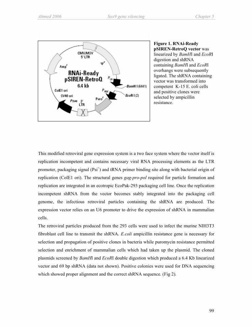
Ahmed 2006 Sox9 gene silencing Chapter 5
99
Figure 1. RNAi-Ready pSIREN-RetroQ vector was linearized by BamHI and EcoRI digestion and shRNA containing BamHI and EcoRI overhangs were subsequently ligated. The shRNA containing vector was transformed into competent K-15 E. coli cells and positive clones were selected by ampicillin resistance.
This modified retroviral gene expression system is a two face system where the vector itself is
replication incompetent and contains necessary viral RNA processing elements as the LTR
promoter, packaging signal (Psi+) and tRNA primer binding site along with bacterial origin of
replication (ColE1 ori). The structural genes gag-pro-pol required for particle formation and
replication are integrated in an ecotropic EcoPak-293 packaging cell line. Once the replication
incompetent shRNA from the vector becomes stably integrated into the packaging cell
genome, the infectious retroviral particles containing the shRNA are produced. The
expression vector relies on an U6 promoter to drive the expression of shRNA in mammalian
cells.
The retroviral particles produced from the 293 cells were used to infect the murine NIH3T3
fibroblast cell line to transmit the shRNA. E.coli ampicillin resistance gene is necessary for
selection and propagation of positive clones in bacteria while puromycin resistance permitted
selection and enrichment of mammalian cells which had taken up the plasmid. The cloned
plasmids screened by BamHI and EcoRI double digestion which produced a 6.4 Kb linearized
vector and 69 bp shRNA (data not shown). Positive colonies were used for DNA sequencing
which showed proper alignment and the correct shRNA sequence. (Fig 2).

Chapter 5 Sox9gene silencing Ahmed 2006
100
Figure 2. Sequence analysis of one of the cloned shRNA (shSox9#2) shows proper alignment with the plasmid construct. Arrows show BamHI and EcoRI restriction overhangs integrated to the shSox9 sequence for linear ligation into the vector.
Transduction and evaluation of the knockdown efficiency
For the evaluation of transfection efficiency cells were harvested after 48 hours of
transduction. Readout for all tested parameters was the gene expression level of Sox9
normalized to endogenous control ß-actin and analysed by quantitative PCR. 80% knockdown
of Sox9 was observed in comparison with the control cells which were transduced with viral
particles containing an unrelated luciferase shRNA. Evaluation of the knockdown was
performed at the protein level with western blot and dot blot analysis. Nearly 100% absence
of Sox9 protein confirmed the knockdown effect. Housekeeping protein ß-actin remained
unaffected indicating Sox9 gene silencing to be specific and not due to RNAi provoked
unspecific silencing or viral interferon mediated “off target” mechanism (Fig 3).
BamHI EcoRI
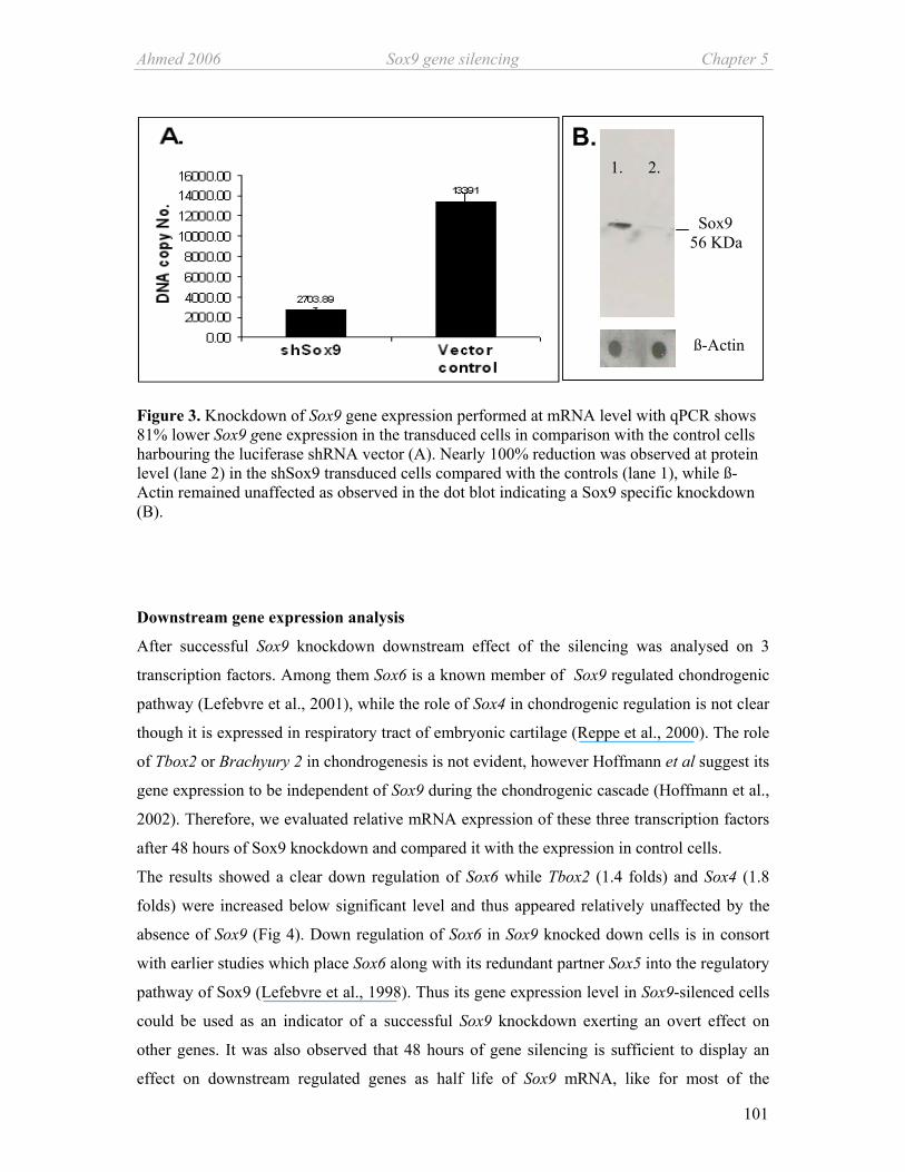
Ahmed 2006 Sox9 gene silencing Chapter 5
101
Figure 3. Knockdown of Sox9 gene expression performed at mRNA level with qPCR shows 81% lower Sox9 gene expression in the transduced cells in comparison with the control cells harbouring the luciferase shRNA vector (A). Nearly 100% reduction was observed at protein level (lane 2) in the shSox9 transduced cells compared with the controls (lane 1), while ß-Actin remained unaffected as observed in the dot blot indicating a Sox9 specific knockdown (B).
Downstream gene expression analysis
After successful Sox9 knockdown downstream effect of the silencing was analysed on 3
transcription factors. Among them Sox6 is a known member of Sox9 regulated chondrogenic
pathway (Lefebvre et al., 2001), while the role of Sox4 in chondrogenic regulation is not clear
though it is expressed in respiratory tract of embryonic cartilage (Reppe et al., 2000). The role
of Tbox2 or Brachyury 2 in chondrogenesis is not evident, however Hoffmann et al suggest its
gene expression to be independent of Sox9 during the chondrogenic cascade (Hoffmann et al.,
2002). Therefore, we evaluated relative mRNA expression of these three transcription factors
after 48 hours of Sox9 knockdown and compared it with the expression in control cells.
The results showed a clear down regulation of Sox6 while Tbox2 (1.4 folds) and Sox4 (1.8
folds) were increased below significant level and thus appeared relatively unaffected by the
absence of Sox9 (Fig 4). Down regulation of Sox6 in Sox9 knocked down cells is in consort
with earlier studies which place Sox6 along with its redundant partner Sox5 into the regulatory
pathway of Sox9 (Lefebvre et al., 1998). Thus its gene expression level in Sox9-silenced cells
could be used as an indicator of a successful Sox9 knockdown exerting an overt effect on
other genes. It was also observed that 48 hours of gene silencing is sufficient to display an
effect on downstream regulated genes as half life of Sox9 mRNA, like for most of the
B.
ß-Actin
Sox9 56 KDa
1. 2.

Chapter 5 Sox9gene silencing Ahmed 2006
102
transcription factors, is relatively short and has been reported to be between 1.2- 2.5 h (Sekiya
et al., 2001; Kinkel and Horton, Jr., 2003).
0.1
1
10
sox4 sox6 tbox2
Figure 4. Relative qPCR analysis after 48 hours of Sox9 knockdown shows reduction in Sox6 mRNA level while Tbox2 and Sox4 expression became not significantly elevated.
Sox4 is known to be expressed in osteoblast cell lines and has been localized to the zone of
hypertrophic chondrocytes in the growth plates of developing foetal mouse hind limbs. This
suggests an integral role of Sox4 in maturation of osteoblasts (Reppe et al., 2000). Here we
indicate for the first time that Sox4 gene expression is not influenced by absence of Sox9.
Tbox2 is a transcription factor containing a T-box DNA binding domain. This family of
transcription factors is implicated in regulation of the FGF receptor 3 signalling pathway
which is involved in induction of chondrogenesis in MSCs. In addition it contains receptors
which bind to FGFs and TGFß, both of these growth factors are known to influence skeletal
development. On these grounds Hoffman et al have argued a role for Tbox2 in the
chondrogenic differentiation pathway independent or even prior to Sox9 (Hoffmann et al.,
2002). Our results are in consort with this study which places Tbox2 not in the Sox9
regulatory cascade but rather parallel to it.

Ahmed 2006 Sox9 gene silencing Chapter 5
103
In conclusion, we have successfully silenced chondrogenic master regulator Sox9 in a mouse
fibroblast cell line by inducing RNAi employing shRNA placed into a retroviral gene delivery
vector. We could demonstrate effect on Sox6 gene expression level, one of the major
regulatory transcription factors downstream of Sox9. We also suggest Sox4 and Tbox2 not to
be directly regulated by Sox9. This study provides a gene silencing system for future studies
on regulation of chondrogenesis and early stages of osteogenesis. The system was designed to
knockdown Sox9 in MSCs to study possible redundancies in Sox9 driven chondrogenic gene
cascade. At a later stage the system is to be adapted to an inducible knockdown system where
Sox9 gene expression will be knocked down during different stages of chondrogenesis to
identify the role of Sox9 in maintenance of the chondrogenic phenotype. An inducible in vitro
knockdown system for Sox9 has a wide application for osteo- chondrogenic regulation
studies.
Acknowledgements We are thankful to Maren Marschner and Claudia Göttl for their excellent technical
assistance.
Chapter 5 Sox9gene silencing Ahmed 2006
104
Reference List
Akiyama,H., Chaboissier,M.C., Martin,J.F., Schedl,A., and de Crombrugghe,B. (2002). The transcription factor Sox9 has essential roles in successive steps of the chondrocyte differentiation pathway and is required for expression of Sox5 and Sox6. Genes Dev. 16, 2813-2828.
Bi,W., Deng,J.M., Zhang,Z., Behringer,R.R., and de Crombrugghe,B. (1999). Sox9 is required for cartilage formation. Nat. Genet. 22, 85-89.
Bi,W., Huang,W., Whitworth,D.J., Deng,J.M., Zhang,Z., Behringer,R.R., and de Crombrugghe,B. (2001). Haploinsufficiency of Sox9 results in defective cartilage primordia and premature skeletal mineralization. Proc. Natl. Acad. Sci. U. S. A 98, 6698-6703.
Chimal-Monroy,J., Rodriguez-Leon,J., Montero,J.A., Ganan,Y., Macias,D., Merino,R., and Hurle,J.M. (2003). Analysis of the molecular cascade responsible for mesodermal limb chondrogenesis: Sox genes and BMP signaling. Dev. Biol. 257, 292-301.
Cullen,B.R. (2006). Enhancing and confirming the specificity of RNAi experiments. Nat. Methods 3, 677-681.
de Crombrugghe,B., Lefebvre,V., and Nakashima,K. (2001). Regulatory mechanisms in the pathways of cartilage and bone formation. Curr. Opin. Cell Biol. 13, 721-727.
Gregory J.Hannon and and Phillip D.Zamore (2003). Small RNAs, Big Biology: Biochemical studies of RNA interference. In RNAi A guide to gene silencing, Gregory J.Hannon, ed. (New York, USA: Cold Spring Harobor Laboratory Press), pp. 87-108.
Healy,C., Uwanogho,D., and Sharpe,P.T. (1999). Regulation and role of Sox9 in cartilage formation. Dev. Dyn. 215, 69-78.
Hoffmann,A., Czichos,S., Kaps,C., Bachner,D., Mayer,H., Kurkalli,B.G., Zilberman,Y., Turgeman,G., Pelled,G., Gross,G., and Gazit,D. (2002). The T-box transcription factor Brachyury mediates cartilage development in mesenchymal stem cell line C3H10T1/2. J. Cell Sci. 115, 769-781.
Kinkel,M.D. and Horton,W.E., Jr. (2003). Coordinate down-regulation of cartilage matrix gene expression in Bcl-2 deficient chondrocytes is associated with decreased SOX9 expression and decreased mRNA stability. J. Cell Biochem. 88, 941-953.
Lefebvre,V., Behringer,R.R., and de Crombrugghe,B. (2001). L-Sox5, Sox6 and Sox9 control essential steps of the chondrocyte differentiation pathway. Osteoarthritis. Cartilage. 9 Suppl A, S69-S75.
Lefebvre,V., Li,P., and de Crombrugghe,B. (1998). A new long form of Sox5 (L-Sox5), Sox6 and Sox9 are coexpressed in chondrogenesis and cooperatively activate the type II collagen gene. EMBO J. 17, 5718-5733.
McManus,M.T. and Sharp,P.A. (2002). Gene silencing in mammals by small interfering RNAs. Nat. Rev. Genet. 3, 737-747.

Ahmed 2006 Sox9 gene silencing Chapter 5
105
Paddison,P.J., Caudy,A.A., Bernstein,E., Hannon,G.J., and Conklin,D.S. (2002). Short hairpin RNAs (shRNAs) induce sequence-specific silencing in mammalian cells. Genes Dev. 16, 948-958.
Rene F.Ketting, Marcel Tijsterman, and and Ronald H.A.Plasterk (2003). RNAi in Caenorhabditis elegans. In RNAi A guide to gene silencing, Gregory J.Hannon, ed. (New York, USA: Cold Spring Harbor Laboratory Press), pp. 65-86.
Reppe,S., Rian,E., Jemtland,R., Olstad,O.K., Gautvik,V.T., and Gautvik,K.M. (2000). Sox-4 messenger RNA is expressed in the embryonic growth plate and regulated via the parathyroid hormone/parathyroid hormone-related protein receptor in osteoblast-like cells. J. Bone Miner. Res. 15, 2402-2412.
Sekiya,I., Koopman,P., Tsuji,K., Mertin,S., Harley,V., Yamada,Y., Shinomiya,K., Niguji,A., and Noda,M. (2001). Transcriptional suppression of Sox9 expression in chondrocytes by retinoic acid. J. Cell Biochem. Suppl Suppl 36, 71-78.


107
Chapter 6
Conclusions
Arguably one of the most challenging complex tissues for regenerative medicine is articular
cartilage. It’s avascular nature, dense extracellular matrix, few cells, and no known specific
progenitor cells are but a few issues (Cancedda et al., 2003; Hunziker, 2002). For ex vivo
cartilage regeneration multipotent adult stromal MSCs are the tool of choice because of their
in vitro pliability and retention of differentiation potential (Barry, 2003). In addition, for cell
based cartilage therapy use of MSCs instead of chondrocytes as cell source also avoids donor
site morbidity (Cancedda et al., 2003). However, production of tissue engineered functional
cartilage with articular characteristics is still a challenge. To effectively address this problem
employment of the two elements of tissue engineering, the scaffolding material and the cells,
are needed to be optimized. Thus, studies aiming at better understanding of the chondrogenic
differentiation cascade and the molecular regulatory mechanisms are essential.
This thesis concentrated on external regulation and influence on chondrogenesis in MSCs.
Indisputable effects of microenvironment on proliferation, growth and differentiation of
MSCs were the intellectual background. The experimental approach was coculture of different
cell types to explore the impact of microenvironment on chondrogenically differentiating
MSCs in vitro. Regulatory effects of both the native and artificial environment were reported
after analyzing the gene expression and protein translation data. The study was carried out in a
rat model system which is not well characterized for MSCs based differentiation studies;
therefore, pre-screening studies were a prerequisite. Often in literature it has been pointed out
that MSCs change their antigenic profile and subsequently loose plasticity upon in vitro
expansion (Bianco and Robey, 2001). Thus, prior to differentiation studies, MSCs were
characterized upon their antigenic profile and CFU-F forming capability in chapter 2. The
colonogenic cells were expanded and found to be predominantly positive for the known
positive markers of cells of stromal origin as CD71, CD106, CD49a and D7Fib (Pittenger et
al., 1999). However magnetic and fluorescence sorting of the cell population also revealed
presence of 20-30% CD45-positive cells of haematopoietic origin (chapter 2, 3). The
Chapter 6 Conclusions Ahmed 2006
108
expanded MSCs were screened for osteo-chondro related basal gene expression and secretion
profiles of cytokines, growth factors and proteases and the differences among this basal
profile were compared with that of the differentiated cells. Col1a1 was identified as the most
highly expressed gene in undifferentiated MSCs which could be used as one of the markers of
marrow derived MSCs. The high expression of master regulators of chondrogenesis and
osteogenesis, Sox9 and Cbfa1 respectively, in the un-induced MSCs could be an indicator of
commitment to differentiation towards osteo-/chondrogenic lineages (Bianco et al., 2001).
These cells also showed retained osteogenic and chondrogenic potential as demonstrated by
histology. This comprehensive screening of rat marrow derived MSCs serves as a continual
reference point for differentiation and gene regulation studies.
In chapter 3 the effect of bone marrow native environment was determined on
chondrogenically differentiating MSCs in vitro. Bone marrow microenvironment is a complex
structure where HSCs are retained until they mature and are released into circulation. The
structural support to these cells is provided by stroma and stromal cells as MSCs, known to be
precursors of osteoblasts, chondrocytes, fibroblasts, adipocytes, endothelial cells and
myocytes (Pittenger et al., 1999). Osteogenesis and heamtopoiesis occurs in close proximity;
osteoblast progenitors influence haematopoiesis and haematopoietic precursor cells generate
osteoclasts essential for bone resorption (Begg et al., 1993; Yin and Li, 2006). Therefore, the
in vitro chondrogenic capacity of the mixed population of MSCs (CD45-negative) and HCs
(CD45-positive) was compared with that of the pure MSCs population to determine the effect
of CD45- positive bone marrow cells on CD45-negative stromal cells or MSCs. The gene
expression analysis revealed an up regulation in mRNA level of chondrogenic marker genes
as Sox9, col2a1 col10a1 and COMP in the mixed population. The results indicate that CD45-
positive cells may create a microenvironment which helps in promotion of chondrogenesis
under specified culture conditions and thus support chondrogenesis in MSCs in vitro. Stem
cells are regulated by environmental signals and intrinsic regulatory mechanisms; this in vitro
study indicates potential involvement of HSCs/ HCs in regulation of chondrogenesis.
Chapter 4 dealt with articular cartilage released factors and their putative paracrine effect on
early and late chondrogenic cascade in MSCs. Articular cartilage is a known paracrine organ
as it maintains its differentiation status via continual regulation through extracellular signals.
Here, it was shown that the articular cartilage when cultured in the vicinity of MSCs
influences chondrogenesis in MSCs. The cartilage explant and MSCs were cocultured in
absence of external growth factors in chambers separated by a 1µm porous membrane which
limited the contact to paracrine level. The most curious and exciting discovery was

Ahmed 2006 Conclusions Chapter 6
109
suppression of collagen X expression and release from the MSCs under the influence of
cartilage. Collagen X is accepted as beacon of hypertrophy (Reddi, 1994; Goldring et al.,
2006) its lack indicates delayed hypertrophy due to signalling factors secreted by cartilage.
Appearance of collagen X is indicative of progressing endochondral ossification while
maintenance of stable chondrogenic phenotype requires suppression of collagen X. For MSCs
based ex vivo regeneration of articular cartilage with sustained phenotype, it is vital to
understand and control collagen X regulation via extrinsic factors. The identified soluble
signaling molecules including MMP-13, TIMP-2 and VEGFα are proposed to be partially
responsible for this delay by effecting the gene regulation and/ or biosynthesis. MMP-13 has a
dual role during hypertrophy, first is the conventional role of the collagenase involved in
matrix turnover secondly, it is indicated to be a regulator of collagen X gene expression
(Inada et al., 2004). VEGFα can also activate collagen X inhibiting DNA binding proteins;
additionally it indirectly promotes MMP-13. The identified molecules may create a chain of
events causing overt collagen X suppression. Interestingly expression of Sox9, the main
transcriptional factor of chondrogenesis, was found to be stimulated by the factors released by
cartilage tissue. This effect was observed in absence of external growth factors required for
chondrogenic differentiation in vitro. Most likely growth factor Tgfß-3, secreted by cartilage
is responsible for this early up regulation. After one week of culture an overall decline in Sox9
mRNA was observed irrespective of Tgfß-3, however, this decline did not hinder the
progressive up regulation of pro cartilage structural genes. Sox9 is integral for induction of
chondrogenic differentiation, however, its role in sustenance of chondrogenic phenotype in
our system remained ambiguous (Lefebvre and Smits, 2005). To find out true hierarchy of
Sox9 in chondrogenesis in chapter 5 a method was developed to knock down Sox9 by RNA
interference. The silencing of Sox9 was successfully established in the NIH3T3 fibroblast cell
line by shRNA delivered by a retroviral expression system. The initial effect on downstream
regulatory genes indicated a decline in mRNA of Sox6 while Sox4 and Tbox2 remained
uninfluenced. The establishment of this system proves that the major chondrogenesis
regulator transcription factor Sox9 could be knocked down in cell culture. The importance of
this system is reflected by the fact that Sox9 homozygous knockout mouse system is
unachievable due to prenatal mortality (Akiyama et al., 2002). Studies have also indicated that
over-expression of Sox9 does not directly relate to over induction of chondrogenesis, therefore
involvement of other signalling molecules is likely (Furumatsu et al., 2005). This knockdown
system will be of valuable help in future to identify direct and indirect role of Sox9 in
chondrogenic regulation.

Chapter 6 Conclusions Ahmed 2006
110
In conclusion, this study on paracrine effects of microenvironment during MSCs commitment
and differentiation demonstrated that the chondrogenic differentiation pathway of rat bone
marrow derived MSCs is influenced by the microenvironment (Fig 1). The observation of the
paracrine effect of cartilage tissue on collagen X is a foundation for future studies on the
identified soluble factors to discover a direct correlation between the effect and the putative
effectors. The presented paracrine coculture system between cartilage and MSCs and the
coculture of different bone marrow populations could be used for gene and protein regulation
studies in future. Identification of signalling molecules responsible for cell fate determination
are empirical for cartilage regenerative medicine.
Figure 1: ‘Influencing chondrogenesis’ in a nutshell: A summarized schematic diagram of main results acquired during this study shows chondrogenic differentiation of rat bone marrow derived MSCs upon suspension in alginate and culture in defined chondrogenic medium (A) (Chapters 2 and 3). The up regulatory effect of CD45-positive cells on chondrogenic differentiation of MSCs (B) (Chapter 3). Early induction and delayed hypertrophy induced on differentiating MSCs via soluble factors secreted by cartilage tissue (C) (Chapter 4).
MSCs/ alginate suspension and chondrogenic medium
Sox9
Collagen X
Collagen II
Sox9
Collagen X VEGF
Cartilage
TIMP-1 MMP-13
TIMP-2
CD45+ cells
MSCs
B.
C.
A.

Ahmed 2006 Conclusions Chapter 6
111
Reference List
1. Akiyama,H., Chaboissier,M.C., Martin,J.F., Schedl,A., and de Crombrugghe,B. (2002). The transcription factor Sox9 has essential roles in successive steps of the chondrocyte differentiation pathway and is required for expression of Sox5 and Sox6. Genes Dev. 16, 2813-2828.
2. Barry,F.P. (2003). Biology and clinical applications of mesenchymal stem cells. Birth Defects Res. C. Embryo. Today 69, 250-256.
3. Begg,S.K., Radley,J.M., Pollard,J.W., Chisholm,O.T., Stanley,E.R., and Bertoncello,I. (1993). Delayed hematopoietic development in osteopetrotic (op/op) mice. J. Exp. Med. 177, 237-242.
4. Bianco,P., Riminucci,M., Gronthos,S., and Robey,P.G. (2001). Bone marrow stromal stem cells: nature, biology, and potential applications. Stem Cells 19, 180-192.
5. Bianco,P. and Robey,P.G. (2001). Stem cells in tissue engineering. Nature 414, 118-121.
6. Cancedda,R., Dozin,B., Giannoni,P., and Quarto,R. (2003). Tissue engineering and cell therapy of cartilage and bone. Matrix Biol. 22, 81-91.
7. Furumatsu,T., Tsuda,M., Taniguchi,N., Tajima,Y., and Asahara,H. (2005). Smad3 induces chondrogenesis through the activation of SOX9 via CREB-binding protein/p300 recruitment. J. Biol. Chem. 280, 8343-8350.
8. Goldring,M.B., Tsuchimochi,K., and Ijiri,K. (2006). The control of chondrogenesis. J. Cell Biochem. 97, 33-44.
9. Hunziker,E.B. (2002). Articular cartilage repair: basic science and clinical progress. A review of the current status and prospects. Osteoarthritis. Cartilage. 10, 432-463.
10. Inada,M., Wang,Y., Byrne,M.H., Rahman,M.U., Miyaura,C., Lopez-Otin,C., and Krane,S.M. (2004). Critical roles for collagenase-3 (Mmp13) in development of growth plate cartilage and in endochondral ossification. Proc. Natl. Acad. Sci. U. S. A 101, 17192-17197.
11. Lefebvre,V. and Smits,P. (2005). Transcriptional control of chondrocyte fate and differentiation. Birth Defects Res. C. Embryo. Today 75, 200-212.
12. Pittenger,M.F., Mackay,A.M., Beck,S.C., Jaiswal,R.K., Douglas,R., Mosca,J.D., Moorman,M.A., Simonetti,D.W., Craig,S., and Marshak,D.R. (1999). Multilineage potential of adult human mesenchymal stem cells. Science 284, 143-147.
13. Reddi,A.H. (1994). Bone and cartilage differentiation. Curr. Opin. Genet. Dev. 4, 737-744.
14. Yin,T. and Li,L. (2006). The stem cell niches in bone. J. Clin. Invest 116, 1195-1201.
112
Abstract Cartilage damaging injuries or degenerative diseases like osteoarthritis are restrictive and
painful for normal mobility. Damaged articular cartilage has limited regeneration capacity
mainly due to lack of chondroprogenitor cells. Chondrocytes are the only cell type of cartilage
and pluripotent mesenchymal stem cells (MSCs) are the only known adult chondrocyte
progenitor cells. MSCs have shown promise for tissue engineered therapeutic approach for
focal defects in articular cartilage. However, production of functional cartilage with articular
characteristics is still a challenge. Therefore, it is important to identify the molecular events
and regulatory mechanisms characterizing chondrogenesis in MSCs.
This thesis has addressed external regulation of chondrogenesis in MSCs. Paracrine signaling
factors from the surrounding cells are known to modulate differentiation and proliferation in
surrounding cells, thus, the impact of native and acquired microenvironment on
differentiation of MSCs was investigated in vitro. Two interesting observations concerning
chondrogenesis were reported. Firstly the major population of bone marrow the
haematopoietic cells support chondrogenesis in MSCs in vitro. Gene expression of all the
chondrogenesis related genes was found to be many folds increased when the MSCs were
cocultured with 20-30% CD45-positive haematopoietic cells. Secondly we found out that
articular cartilage when cultured in the vicinity of MSCs also exerts affect on chondrogenesis
in MSCs. Both of the findings indicate that signaling molecules originating from
differentiated cells influence chondrogenesis. We also identified some of the soluble signaling
molecules including MMP-13 TIMP-1, TIMP-2, Tgfß-3 and VEGFα. Interestingly Sox9, the
main transcriptional factor of chondrogenesis, was found to be stimulated by factors released
from cartilage. The most curious observation was suppression of collagen X secretion from
MSCs under the influence of cartilage. The identified soluble molecules like MMP-13,
VEGFA and TIMP-2 may have a role to play in suppression of collagen X while Tgfß-3 can
be cause of induction of Sox9.
The presented paracrine coculture system between cartilage and MSCs and the coculture of
different bone marrow populations could be used for gene and protein regulation studies in
future. Identification of signalling molecules responsible for cell fate determination and
molecular control are empirical for cartilage regenerative medicine.

113
Zusammenfassung Degenerative Erkrankungen des Knorpels wie Osteoarthritis oder Verletzungen im Gelenk
sind sehr schmerzhaft und schränken die normale Beweglichkeit und Lebensqualität ein. Die
natürliche Regenerationskapazität geschädigten Gelenkknorpels ist jedoch aufgrund des
Mangels an Knorpelvorläuferzellen stark eingeschränkt. Knorpel wird nur von Chondrozyten
aufgebaut und pluripotente mesenchymale Stammzellen (MSCs) sind die einzigen bekannten
adulten Vorläuferzellen von Chondrozyten. MSCs bilden einen vielversprechenden
therapeutischen Ansatz um mit Hilfe des Tissue Engineering Defekte im artikulären Knorpel
zu behandeln. Es stellt jedoch eine große Herausforderung dar, funktionellen Knorpel mit
artikulärer Charakteristik herzustellen. Deshalb ist es von großer Bedeutung, Abläufe auf
molekularer Ebene und Regulationsmechanismen der Chondrogenese zu identifizieren.
Diese Arbeit beschäftigt sich mit der externen Reglulation der Chondrogenese in MSCs. Es ist
bekannt, dass parakrine Faktoren der umgebenden Zellen die Differenzierung beeinflussen;
darum wurde die Rolle von nativem Knorpel und dessen Mikoumgebung in der
Differenzierung von MSCs in vitro untersucht. Dabei wurden zwei interessante
Beobachtungen gemacht: 1. Den Großteil der Zellpopulation des Knochenmarks bilden
hämatopoetische Zellen welche die Chondrogenese in MSCs in vitro unterstützen. Die
Expression aller Gene, die in Zusammenhang mit der Chrondrogenese stehen, wurde durch
die Kokultur mit 20-30% CD45-positiven hämatopoetischen Zellen um ein vielfaches erhöht.
2. Ausserdem fanden wir heraus, dass artikulärer Knorpel in Kokultur mit MSCs einen
positiven Effekt auf die Chondrogenese der MSCs ausübt. Diese beiden Resultate zeigen, dass
Faktoren, die von differenzierten Zellen stammen, auf die Chondrogenese Einfluss nehmen.
Wir konnten des Weiteren einige lösliche Signalmoleküle identifizieren wie z.B. MMP-13,
TIMP-1, TIMP-2, Tgfß-3 and VEGFα. Interessanterweise wird Sox9, der wichtigste
Transkriptionsfaktor der Chondrogenese durch von Knorpel sezernierte Faktoren stimuliert.
Eine aussergewöhnliche Beobachtung war, dass in der Anwesenheit von Knorpel die
Kollagen X Sekretion durch MSCs unterdrückt wurde. Die löslichen Faktoren wie MMP-13,
VEGFα und TIMP-2 spielen möglicherweise eine Rolle in der Suppression von Kollagen X,
während Tgfß-3 Sox9 induzieren kann.
Das dargestellte parakrine Kokulturmodell zwischen Knorpel und MSCs und der Kokultur
verschiedener Knochenmarks-Zellpopulationen kann in der Zukunft für Gen- und Protein-
Regulationsstudien verwendet werden.

114
Nazish Ahmed Curriculum Vitae
Personal Information:
Family Name Ahmed
First Name Nazish
Birth Date April 08, 1973
Nationality Pakistani
Marital Status Single
Educational Background:
2003- Ph.D. Pharmaceutical technology, University of
Regensburg, Germany.
1996-1998 M.Phil Biotechnology, NIBGE / Quaid-i-Azam
University,Islamabad, Pakistan. Thesis: Development of
polyclonal antibodies against AC1 and AV2 genes of
CLCuV. Superviosr: Dr. Aftab Bashir
1994-1995 Masters in Human Physiology, University of Karachi,
Karachi, Pakistan. Major: Molecular Endocrinology.
1991-1994 B.Sc. (Honors), University of Karachi, Karachi, Pakistan.
Courses: Physiology, Biochemistry, and Microbiology
1988-1990 Higher Secondary School, Karachi, Pakistan
1986- 1988 Secondary School, Karachi. Pakistan
1978- 1986 Primary School, Riyadh, Saudi Arabia

115
Curriculum Vitae Nazish Ahmed
Work Experience:
May 2003 - Doctoral research at the Department of Pharmaceutical
Technology, University of Regensburg, Germany, lead by
Prof. Dr. Achim Goepferich under the direct supervision of
PD Dr. Susanne Grassel. Thesis: Influencing
chondrogenesis in bone marrow derived stromal cells.
April 2001- April 2003 Scientific researcher at Technical Microbiology, Chemical
Engineering, University of Dortmund, Germany.
May 1999- Jan.2001 Research Officer at the Surgery department, Aga Khan
University Hospital, Karachi, Pakistan.
Academic Awards and honours:
July 2006 Travel fellowship from “Friends of University of Regensburg”
March 2006 GlaxoSmithKline travel fellowship
October 2004 Wilhelm-Roux-Prize 2004 for “Best basic science project” “A
coculture model: Paracrine interaction between cartilage and
mesenchymal stem cells” by Ahmed N, Volk B, Schulz M, Grifka
J and Grassel S. From: German orthopaedics society (DGOOC)
1997 One year full “Scholarship for higher studies”
from University grants commission (UGC) of Pakistan

116
Nazish Ahmed List of Publication Papers:
Ahmed N., Grifka J. and Grassel S. (2006) Cytokine secretion and gene expression profile of rat chondro-progenitor marrow stromal cells. Submitted: Cell and tissue research
Ahmed N., Dreier, R., Goepferich A., Grifka J. and Grassel S. (2006) Soluble signalling factors from differentiated cartilage tissue influence chondrogenic differentiation in rat marrow stromal cells. Submitted: Osteoarthritis and cartilage Ahmed N., Vogel B., Rohde, E., Strunk D., Grifka J., Schulz M.B. and Grassel S. (2006) CD 45-positive cells of haematopoietic origin enhance chondrogenic marker gene expression in rat marrow stromal cells. Int.J. mol. Med. 2006 Aug; 18(2):233-40.
Ahmed M., Ahmed N., Khan K. M., Umer M., Rashid H., Hashmi P. and Umar M. (2004). Sensory hypoinnervation in club foot. J.Bone Joint Surg. Br. 86, 1163-1169.
Abstracts presented at conferences: Oral presentations: Ahmed N., Grifka J and Grassel S. Molecular and cellular profiling of multipotent rat mesenchymal stem cells. XXth FECTS- Federation of European Connective Tissue Society meeting. Oulu, Finland, 1st- 5th July, 2006 Ahmed N., Dreier R., Grifka J. and Grassel S.Cartilage derived paracrine factors influence chondrogenesis in mesenchymal stem cells. European Orthopaedic Research Society 16th Annual meeting. Bologna, Italy, 7th - 9th June 2006. Ahmed N., Dreier R., Grifka J and Grassel S. Role of MMPs and TIMPs in chondrogenic differentiation of mesenchymal stem cells. Strategies in Tissue engineering. Würzburg, Germany. 31st May- 2nd June 2006 Ahmed N., Grifka J and Grassel S. Gene and protein expression profiling of multipotent rat mesenchymal stem cells. Strategies in Tissue engineering. Würzburg, Germany. 31st May- 2nd June 2006 Ahmed N., Goepfrich,A, Grifka J and Grassel S. Paracrine influence of cartilage on chondrogenic differentiation of rat mesenchymal stem cells. 4th annual meeting of the European tissues engineering society. Munich, Germany. 31st Aug - 03. Sept. 2005

117
List of Publication Nazish Ahmed Ahmed N., Volk B., Schulz M., Grifka J. and Grassel S. Chondrogenic differentiation of rat mesenchymal stem cells in a novel coculture system. XIX FECTS Meeting Sicily, Italy, 9th -13th July 2004 Ahmed N, Volk B, Schulz M, Grifka J,Grassel S. A coculture model: Paracrine interaction between cartilage and mesenchymal stem cells. DGOOC, Deutscher Orthpadenkongress. Berlin,Germany. 21th – 23rd October 2004 Ahmed N and Ahmed M. Innervation pattern in the capsule of club foot. 14th International Orthopaedic Conf. Peshawar, Pakistan, 6th -9th Nov, 1999 Poster presentations: Ahmed N., Dreier R., Grifka J. and Grassel S.Role of cartilage derived factors in chondrogenic differentiation of mesenchymal stem cells. XXth FECTS- Federation of European Connective Tissue Society meeting. Oulu, Finland, 1st- 5th July, 2006 Ahmed N., Lundgren-Akerlund E., Grifka J. and Grassel S Gene expression profiling of multipotent adult rat mesenchymal stem cells. European Orthopaedic Research Society 16th Annual meeting. Bologna, Italy, 7th - 9th June 2006. Ahmed N., Goepfrich,A, Grifka J and Grassel S. Effect of cartilage on chondrogenic differentiation of rat mesenchymal stem cells. DGOOC Deutscher Orthpadenkongress Berlin, Germany. 19th – 22nd October 2005. Ahmed N., Goepfrich A., Grifka J., Grassel S Chondrogenic differentiation profile of adult rat mesenchymal stem cells under paracrine influence of articular cartilage signals. Gordon Research Conference on Cartilage biology and pathology. Lucca, Italy. 5th- 9th June 2005 Ahmed N., Volk B., Schulz M., Grifka J. and Grassel S. Rat mesenchymal stem cells isolation and chondrogenic differentiation in a novel coculture system. Strategies in Tissue engineering. Würzburg, Germany. 17th-19th June 2004 Volk B., Rohde E., Vogel T., Ahmed N., Grassel S., Kunz-Schughart L.A., Strunk D., Goepferich A., Schulz M.B. FACS analysis of cultured adherent rat bone marrow cells used for tissue engineering of bone. Strategies in Tissue engineering. Würzburg, Germany. 17th-19th June 2004