Cell adhesion to plasma electrolytic oxidation (PEO) titania coatings, assessed using a centrifuging...
10
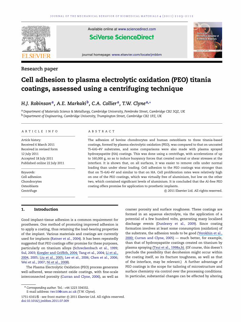
JOURNAL OF THE MECHANICAL BEHAVIOR OF BIOMEDICAL MATERIALS 4 (2011) 2103–2112 Available online at www.sciencedirect.com journal homepage: www.elsevier.com/locate/jmbbm Research paper Cell adhesion to plasma electrolytic oxidation (PEO) titania coatings, assessed using a centrifuging technique H.J. Robinson a , A.E. Markaki b , C.A. Collier a , T.W. Clyne a,∗ a Department of Materials Science & Metallurgy, Cambridge University, Pembroke Street, Cambridge CB2 3QZ, UK b Department of Engineering, Cambridge University, Trumpington Street, Cambridge CB2 1PZ, UK ARTICLE INFO Article history: Received 6 March 2011 Received in revised form 12 July 2011 Accepted 18 July 2011 Published online 22 July 2011 Keywords: Cell adhesion Chondrocytes Osteoblasts Centrifuge ABSTRACT The adhesion of bovine chondrocytes and human osteoblasts to three titania-based coatings, formed by plasma electrolytic oxidation (PEO), was compared to that on uncoated Ti–6Al–4V substrates, and some comparisons were also made with plasma sprayed hydroxyapatite (HA) coatings. This was done using a centrifuge, with accelerations of up to 160,000 g, so as to induce buoyancy forces that created normal or shear stresses at the interface. It is shown that, on all surfaces, it was easier to remove cells under normal loading than under shear loading. Cell adhesion to the PEO coatings was stronger than that on Ti–6Al–4V and similar to that on HA. Cell proliferation rates were relatively high on one of the PEO coatings, which was virtually free of aluminium, but low on the other two, which contained significant levels of aluminium. It is concluded that the Al-free PEO coating offers promise for application to prosthetic implants. c ⃝ 2011 Elsevier Ltd. All rights reserved. 1. Introduction Good implant-tissue adhesion is a common requirement for prostheses. One method of promoting improved adhesion is to apply a coating, thus retaining the load-bearing properties of the implant. Various materials and coatings are currently used for implants (Ratner et al., 2004). It has been repeatedly suggested that PEO coatings offer promise for these purposes, particularly on titanium alloys (Schreckenbach et al., 1999; Sul, 2003; Koegler and Griffith, 2004; Tang et al., 2004; Li et al., 2004, 2005; Liu et al., 2005; Lee et al., 2006; Chen et al., 2006; Wei et al., 2007; Ni et al., 2008). The Plasma Electrolytic Oxidation (PEO) process generates well-adhered, wear-resistant oxide coatings, with fine-scale interconnected porosity (Curran and Clyne, 2006), as well as ∗ Corresponding author. Tel.: +44 1223 334332. E-mail address: [email protected] (T.W. Clyne). coarser porosity and surface roughness. These coatings are formed in an aqueous electrolyte, via the application of a potential of a few hundred volts, generating many localised discharge events (Dunleavy et al., 2009). Since coating formation involves at least some consumption (oxidation) of the substrate, the adhesion tends to be good (Yerokhin et al., 2000; Curran and Clyne, 2005) — much better, for example, than that of hydroxyapatite coatings created on titanium by plasma spraying (Tsui et al., 1998a,b). (Of course, this doesn’t preclude the possibility that decohesion might occur within the coating itself, so its fracture toughness, as well as that of the interface, may be relevant.) A further advantage of PEO coatings is the scope for tailoring of microstructure and surface chemistry via control over the processing conditions. In particular, substantial changes can be effected by altering 1751-6161/$ - see front matter c ⃝ 2011 Elsevier Ltd. All rights reserved. doi:10.1016/j.jmbbm.2011.07.009
Transcript of Cell adhesion to plasma electrolytic oxidation (PEO) titania coatings, assessed using a centrifuging...
J O U R N A L O F T H E M E C H A N I C A L B E H AV I O R O F B I O M E D I C A L M A T E R I A L S 4 ( 2 0 1 1 ) 2 1 0 3 – 2 1 1 2
Available online at www.sciencedirect.com
journal homepage: www.elsevier.com/locate/jmbbm
Research paper
Cell adhesion to plasma electrolytic oxidation (PEO) titaniacoatings, assessed using a centrifuging technique
H.J. Robinsona, A.E. Markakib, C.A. Colliera, T.W. Clynea,∗
aDepartment of Materials Science & Metallurgy, Cambridge University, Pembroke Street, Cambridge CB2 3QZ, UKbDepartment of Engineering, Cambridge University, Trumpington Street, Cambridge CB2 1PZ, UK
A R T I C L E I N F O
Article history:
Received 6 March 2011
Received in revised form
12 July 2011
Accepted 18 July 2011
Published online 22 July 2011
Keywords:
Cell adhesion
Chondrocytes
Osteoblasts
Centrifuge
A B S T R A C T
The adhesion of bovine chondrocytes and human osteoblasts to three titania-based
coatings, formed by plasma electrolytic oxidation (PEO), was compared to that on uncoated
Ti–6Al–4V substrates, and some comparisons were also made with plasma sprayed
hydroxyapatite (HA) coatings. This was done using a centrifuge, with accelerations of up
to 160,000 g, so as to induce buoyancy forces that created normal or shear stresses at the
interface. It is shown that, on all surfaces, it was easier to remove cells under normal
loading than under shear loading. Cell adhesion to the PEO coatings was stronger than
that on Ti–6Al–4V and similar to that on HA. Cell proliferation rates were relatively high
on one of the PEO coatings, which was virtually free of aluminium, but low on the other
two, which contained significant levels of aluminium. It is concluded that the Al-free PEO
coating offers promise for application to prosthetic implants.c⃝ 2011 Elsevier Ltd. All rights reserved.
d
1. Introduction
Good implant-tissue adhesion is a common requirement forprostheses. One method of promoting improved adhesion isto apply a coating, thus retaining the load-bearing propertiesof the implant. Various materials and coatings are currentlyused for implants (Ratner et al., 2004). It has been repeatedlysuggested that PEO coatings offer promise for these purposes,particularly on titanium alloys (Schreckenbach et al., 1999;Sul, 2003; Koegler and Griffith, 2004; Tang et al., 2004; Li et al.,2004, 2005; Liu et al., 2005; Lee et al., 2006; Chen et al., 2006;Wei et al., 2007; Ni et al., 2008).
The Plasma Electrolytic Oxidation (PEO) process generateswell-adhered, wear-resistant oxide coatings, with fine-scaleinterconnected porosity (Curran and Clyne, 2006), as well as
∗ Corresponding author. Tel.: +44 1223 334332.E-mail address: [email protected] (T.W. Clyne).
1751-6161/$ - see front matter c⃝ 2011 Elsevier Ltd. All rights reservedoi:10.1016/j.jmbbm.2011.07.009
coarser porosity and surface roughness. These coatings areformed in an aqueous electrolyte, via the application of apotential of a few hundred volts, generating many localiseddischarge events (Dunleavy et al., 2009). Since coatingformation involves at least some consumption (oxidation) ofthe substrate, the adhesion tends to be good (Yerokhin et al.,2000; Curran and Clyne, 2005) — much better, for example,than that of hydroxyapatite coatings created on titanium byplasma spraying (Tsui et al., 1998a,b). (Of course, this doesn’tpreclude the possibility that decohesion might occur withinthe coating itself, so its fracture toughness, as well as thatof the interface, may be relevant.) A further advantage ofPEO coatings is the scope for tailoring of microstructure andsurface chemistry via control over the processing conditions.In particular, substantial changes can be effected by altering
.
2104 J O U R N A L O F T H E M E C H A N I C A L B E H AV I O R O F B I O M E D I C A L M A T E R I A L S 4 ( 2 0 1 1 ) 2 1 0 3 – 2 1 1 2
the composition of the electrolyte. As an example for thecase of titanium substrates, Tang et al. (2004) have shownthat a combination of pre-anodic oxidation and subsequentsequence of PEO treatments, carried out using two differentelectrolytes, can create a porous titania film which is largelyanatase.
Several techniques have been employed for characterisa-tion of the strength of cell adhesion. In many cases, theforces tending to cause debonding have been generated byfluid flow (Mohandas et al., 1974; Truskey and Pirone, 1990;Cozensroberts et al., 1990; Anamelechi et al., 2005; Fu et al.,2009), although other techniques, such as the use of ultra-sound (Debavelaere-Callens et al., 2007), have also been used.Assessment of the forces generated on individual cells in suchtests is fraught with difficulties and in general they can onlybe used for ranking purposes with a given set of surfaces. Pro-cedures in which the mechanical forces created are some-what better-defined include those based on spinning disks(Horbett et al., 1988), although, since cells located at differ-ent radial distances from the rotation axis experience differ-ent forces, population changes have to be assessed locally andthe accuracy of such measurements can be doubtful.
There have also been several studies (Hertl et al., 1984;Thoumine et al., 1996; Reyes and Garcia, 2003; Harberset al., 2005; Harbers and Healy, 2005) in which centrifugalacceleration is employed, such that all cells experiencethe same force. The procedure is in general regarded asconvenient and robust. One issue with this type of test iswhether body forces induced in this way are likely to affectthe metabolic activity of the cell. In fact, it’s fairly clear thatthis will occur. Even small accelerations of a few g havebeen shown (Koyama et al., 2009) to have detectable effects,whereas centrifugal adhesion testing commonly involvesaccelerations of at least several tens of thousand g. However,the tests are normally of short duration, during whichmetabolic activity is unlikely to be of great importance andit seems unlikely that the strength or nature of the bondingwould change during this period.
All of these tests are based on the simultaneous treatmentof a large number of cells, and indeed there are obviousadvantages in doing this, given that individual cells willalmost certainly exhibit a range of adhesive strengths. Whilethere have been attempts (Yamamoto et al., 1998; Thoumineet al., 2008) to apply forces to individual cells (mainly usingmicro-pipettes or optical tweezers) and hence to assesstheir adhesion to surfaces, in general these approaches aredifficult, time-consuming and relatively unreliable.
Various outcomes have been noted from the applicationof (cell population) adhesion tests to particular surfaces,including (plasma-treated) PET (Pratt et al., 1989), fibronectin(Lotz et al., 1989) and titanium (alloys) (Anselme et al.,2000a,b; Anselme and Bigerelle, 2005), with the latterwork covering the effects of both topography and surfacechemistry. However, very few of these studies have yieldedmeaningful values for an interfacial strength or fractureenergy. Lotz et al. (1989) concluded that forces of the orderof 0.1 mN were needed to detach fibroblasts and gliomacells from extra-cellular matrix proteins, during centrifuging.For the cell sizes concerned, assuming that they are bondedover the complete contact area, this suggests that interfacial
stresses of the order of 1 MPa are required to causedebonding. However, other experiments, such as those ofThoumine et al., using centrifuging (Thoumine et al., 1996)and optical tweezer (Thoumine et al., 2008) techniques,indicate the forces required, and corresponding nominalstresses, to be several orders of magnitude smaller than this.In fact, there are virtually no data available which allow cell-substrate adhesive strengths to be compared with those ofconventional macroscopic interfaces, such as those producedby glues or other bonding techniques.
There is also a shortage of systematic qualitativeinformation about the factors that favour strong bonding ofcells to surfaces of interest. It is well known, of course, that Tialloys exhibit good general biocompatibility, oftenmanifestedas good cell proliferation and adhesion, although it should benoted that it is a (thin) titania layer that is in actual contactwith the cells. It’s therefore evident that the details of howcells interact with titania surfaces are likely to be highlyimportant, for nominally uncoated titanium substrates, aswell as for titania coatings. In this context, it may be relevantthat several different phases can be present under differentcircumstances (notably anatase, rutile, aluminium titanateand amorphous phases), and that their stoichiometry andcomposition may vary appreciably. There is very little clearinformation in the literature about which of these phasesmight more effectively promote cell activity favourable tostrong bonding, although it may be noted that the oxide layeron Ti–6Al–4V, the alloy most commonly used in biomedicalapplications, is reported (Roessler et al., 2002; Variola et al.,2008) to be composed of amorphous and rutile phases.
Regarding topographic effects, studies such as those ofAnselme et al. (2000a,b); Anselme and Bigerelle (2005) havetended to confirm that adhesion is in general superioron relatively rough surfaces, particularly when the surfaceintrusions are in a size range suited to allowing cellpenetration — typically of the order of 100 µm. On a relatednote, pore sizes above ∼100 µm are reported as beingnecessary to promote the ingrowth of bone tissue into animplant (Rea, 2003). These larger surface features may helpan implant ‘key’ into a bone, helping to create a strongmechanical bond — for example Bobyn et al. (1980) reportedthat pore sizes between 100 and 400 µm provided the bestresults in mechanical removal tests for porous metal testpieces implanted in vivo.
However, there is in general rather little that iswell-established concerning optimisation of either surfaceroughness or porosity, in terms of measuring the mechanicalstrength cell adhesion. Some work, such as that of Murphyet al. (2010), has examined how pore size can affect initialcell adhesion in terms of the proportion of cells thatremain attached to a surface after a period of time inculture, and longer term proliferation, showing that oncollagen–glycosaminoglycan scaffolds variations in pore sizebetween 85 and 325 µm can have significant effects.
In the present work, a centrifuge has been used to generatewell-defined buoyancy forces, in either normal or sheardirections relative to the plane of the interface, and applied toboth bovine chondrocytes and human osteoblasts, attachedto a range of PEO titania-based coatings, and also to untreatedTi–6Al–4V substrates (with their normal native titania layerson the surface).
J O U R N A L O F T H E M E C H A N I C A L B E H AV I O R O F B I O M E D I C A L M A T E R I A L S 4 ( 2 0 1 1 ) 2 1 0 3 – 2 1 1 2 2105
Table 1 – Electrolyte compositions used to make the PEOcoatings.
Coating Electrolyte compositionPhosphate
(Na3PO4) contentAluminate(NaAlO2)
content
PEO(phosphate) 0.03 M (4.5 g dm−3) –PEO(mixed) 0.03 M (4.5 g dm−3) 0.02M (2.0 g dm−3)
PEO(aluminate) 0.009 M (1.5 g dm−3) 0.15M (12.5 g dm−3)
Table 2 – Processing parameters for vacuum plasmaspraying of hydroxyapatite (HA) coatings.
Processing parameter Spraying condition
Chamber pressure 200 mbarStand-off distance 260 mmNozzle diameter 8 mmPlasma gas flow rates Ar: 50 SLPM
H2: 6 SLPMPlasma power 40 kW
2. Experimental procedures
2.1. Specimen production
Samples of Ti–6Al–4V, in the form of discs of diameter12.5 mm, were polished with 1200 SiC grit and cleanedfor 30 min in an ultrasonic bath of distilled water,followed by 30 min in acetone. Samples to be coated weresubjected to PEO processing, using three different (aqueous)electrolyte compositions. These are given in Table 1. Theseelectrolytes, and the corresponding coatings, are designatedPEO(phosphate), which was phosphate-rich, PEO(mixed),which contained a mixture of phosphate and aluminateions, and PEO(aluminate), which was aluminate-rich. Theprocessing was carried out in a 10 kW constant power facilitysupplied by Keronite Ltd, operating at 50 Hz AC. Samples wereprocessed for 1 h. The system capacitance was adjusted toachieve a final current (RMS) density of ∼20 A dm−2. Othersamples were coated with hydroxyapatite by vacuum plasmaspraying. The spraying conditions are given in Table 2.
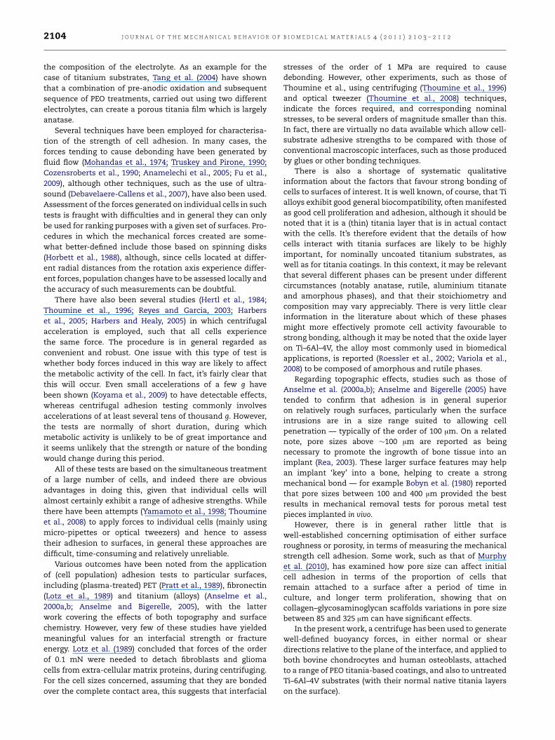
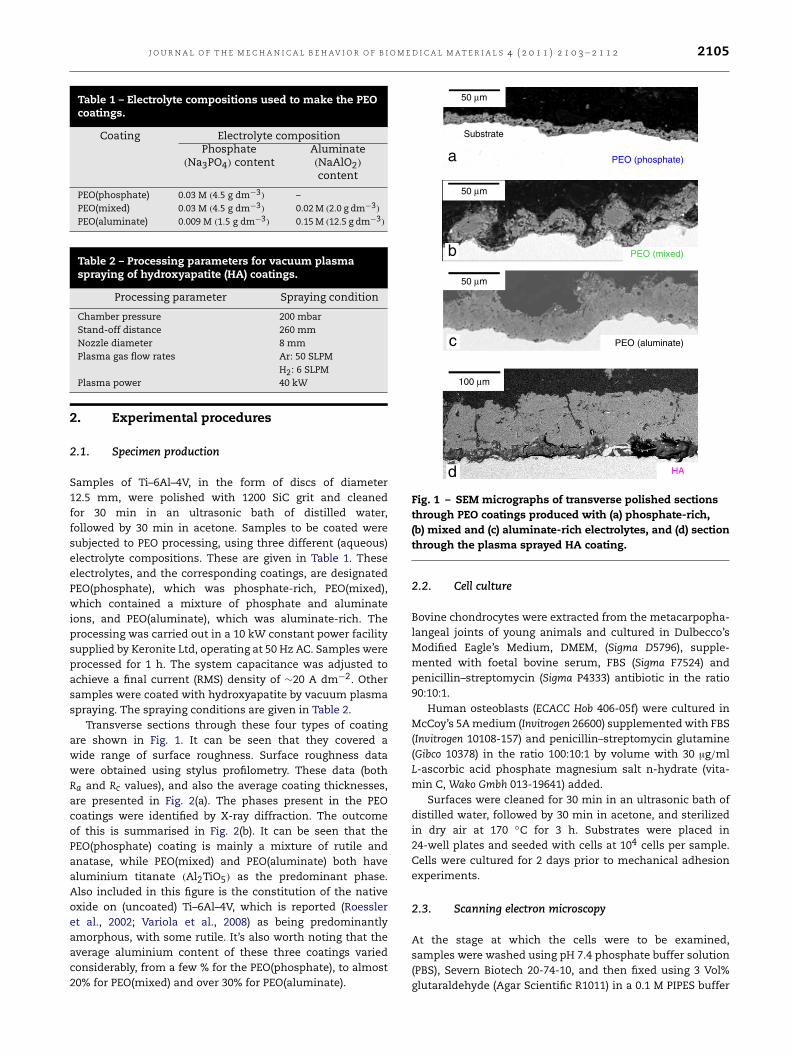
Transverse sections through these four types of coatingare shown in Fig. 1. It can be seen that they covered awide range of surface roughness. Surface roughness datawere obtained using stylus profilometry. These data (bothRa and Rc values), and also the average coating thicknesses,are presented in Fig. 2(a). The phases present in the PEOcoatings were identified by X-ray diffraction. The outcomeof this is summarised in Fig. 2(b). It can be seen that thePEO(phosphate) coating is mainly a mixture of rutile andanatase, while PEO(mixed) and PEO(aluminate) both havealuminium titanate (Al2TiO5) as the predominant phase.Also included in this figure is the constitution of the nativeoxide on (uncoated) Ti–6Al–4V, which is reported (Roessleret al., 2002; Variola et al., 2008) as being predominantlyamorphous, with some rutile. It’s also worth noting that theaverage aluminium content of these three coatings variedconsiderably, from a few % for the PEO(phosphate), to almost20% for PEO(mixed) and over 30% for PEO(aluminate).
50 µm
50 µm
50 µm
100 µm
HA
PEO (aluminate)
PEO (mixed)
PEO (phosphate)
Substrate
a
b
c
d
Fig. 1 – SEM micrographs of transverse polished sectionsthrough PEO coatings produced with (a) phosphate-rich,(b) mixed and (c) aluminate-rich electrolytes, and (d) sectionthrough the plasma sprayed HA coating.
2.2. Cell culture
Bovine chondrocytes were extracted from the metacarpopha-langeal joints of young animals and cultured in Dulbecco’sModified Eagle’s Medium, DMEM, (Sigma D5796), supple-mented with foetal bovine serum, FBS (Sigma F7524) andpenicillin–streptomycin (Sigma P4333) antibiotic in the ratio90:10:1.
Human osteoblasts (ECACC Hob 406-05f) were cultured inMcCoy’s 5Amedium (Invitrogen 26600) supplemented with FBS(Invitrogen 10108-157) and penicillin–streptomycin glutamine(Gibco 10378) in the ratio 100:10:1 by volume with 30 µg/mlL-ascorbic acid phosphate magnesium salt n-hydrate (vita-min C, Wako Gmbh 013-19641) added.
Surfaces were cleaned for 30 min in an ultrasonic bath ofdistilled water, followed by 30 min in acetone, and sterilizedin dry air at 170 ◦C for 3 h. Substrates were placed in24-well plates and seeded with cells at 104 cells per sample.Cells were cultured for 2 days prior to mechanical adhesionexperiments.
2.3. Scanning electron microscopy
At the stage at which the cells were to be examined,samples were washed using pH 7.4 phosphate buffer solution(PBS), Severn Biotech 20-74-10, and then fixed using 3 Vol%glutaraldehyde (Agar Scientific R1011) in a 0.1 M PIPES buffer
2106 J O U R N A L O F T H E M E C H A N I C A L B E H AV I O R O F B I O M E D I C A L M A T E R I A L S 4 ( 2 0 1 1 ) 2 1 0 3 – 2 1 1 2
1 0
20
40
60
80
100
120
140
10
PEO(phosphate)
PEO(mixed)
PEO(aluminate)
PEO(phosphate)
PEO(mixed)
PEO(aluminate)
Ti-6Al-4V(oxide)
HA
Pha
se p
ropo
rtio
n (%
)
Leng
th s
cale
(µm
)
100a b
Fig. 2 – Coating characteristics: (a) thickness and roughness, for PEO and HA coatings, and (b) phase proportions for thePEO coatings and the oxide on Ti–6Al–4V.
Fig. 3 – XPS wide-scan spectra from the free surfaces of (a) PEO(phosphate) (b) PEO(mixed), and (c) PEO(aluminate) coatings.
(Sigma P6757), with 2 µM CaCl2 added. Samples were criticalpoint dried and then sputter coated with ∼50 nm of gold.Samples were viewed using a variable pressure tungstensource JEOL 5800LV SEM, with an accelerating voltage of∼5 kV.
2.4. X-ray photoelectron spectroscopy (XPS)
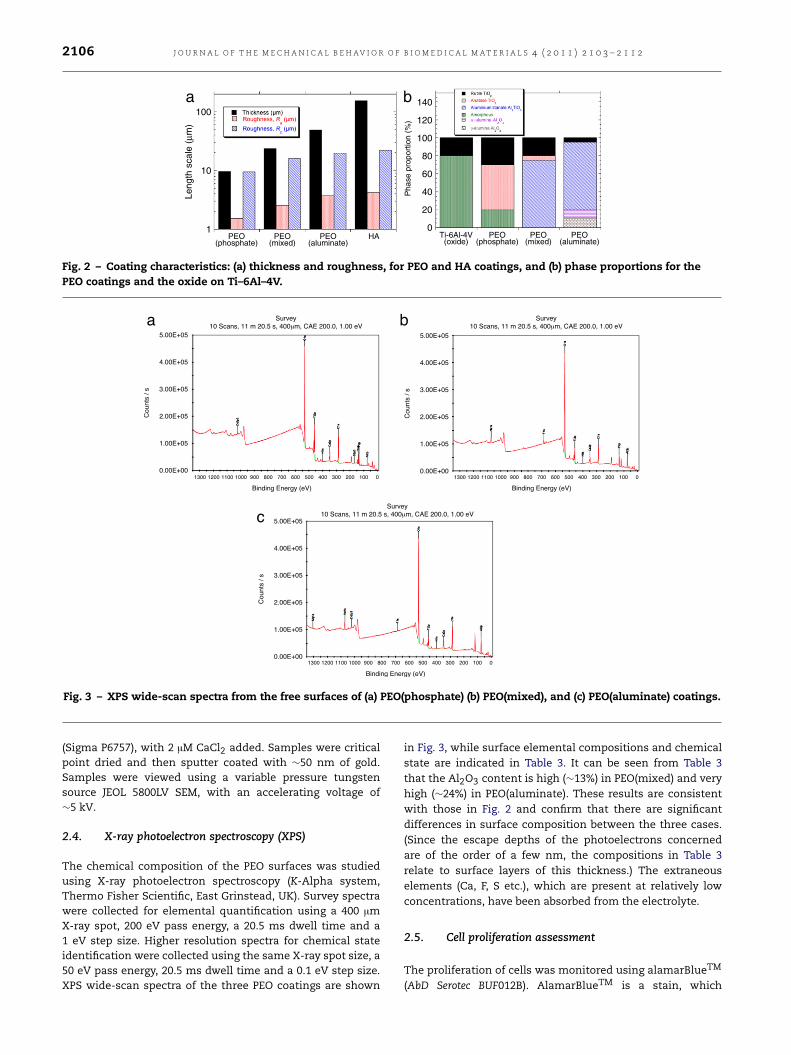
The chemical composition of the PEO surfaces was studiedusing X-ray photoelectron spectroscopy (K-Alpha system,Thermo Fisher Scientific, East Grinstead, UK). Survey spectrawere collected for elemental quantification using a 400 µmX-ray spot, 200 eV pass energy, a 20.5 ms dwell time and a1 eV step size. Higher resolution spectra for chemical stateidentification were collected using the same X-ray spot size, a50 eV pass energy, 20.5 ms dwell time and a 0.1 eV step size.XPS wide-scan spectra of the three PEO coatings are shown
in Fig. 3, while surface elemental compositions and chemicalstate are indicated in Table 3. It can be seen from Table 3that the Al2O3 content is high (∼13%) in PEO(mixed) and veryhigh (∼24%) in PEO(aluminate). These results are consistentwith those in Fig. 2 and confirm that there are significantdifferences in surface composition between the three cases.(Since the escape depths of the photoelectrons concernedare of the order of a few nm, the compositions in Table 3relate to surface layers of this thickness.) The extraneouselements (Ca, F, S etc.), which are present at relatively lowconcentrations, have been absorbed from the electrolyte.
2.5. Cell proliferation assessment
The proliferation of cells was monitored using alamarBlueTM
(AbD Serotec BUF012B). AlamarBlueTM is a stain, which
J O U R N A L O F T H E M E C H A N I C A L B E H AV I O R O F B I O M E D I C A L M A T E R I A L S 4 ( 2 0 1 1 ) 2 1 0 3 – 2 1 1 2 2107
Table 3 – XPS elemental composition and chemical state identification.
Chemical state Elemental composition (At. %)PEO(phosphate) PEO(mixed) PEO(aluminate)
O1s Metal oxide, carbonate and C–Ocontributions
45.5 48.21 43.72
C1s C–H, C–C, C–O, C=O, C–N orcarbonate contributions
30.8 21.22 24.01
Al2p Oxide state (Al2O3) 3.3 13.13 24.18Ca2p Metal carbonate 2.6 1.95 2.14P2p Metal phosphate 8.47 9.61 –Ti2p Oxide state (TiO2) 7.19 2.87 2.11Na1s 0.85 2.09N1s 1.08 0.52 0.66F1s – 1.65 0.45S2p3 0.49 –Pb4f Oxide state 0.36 –Mg1s – – 0.39Zn2p3 0.21 – 0.25
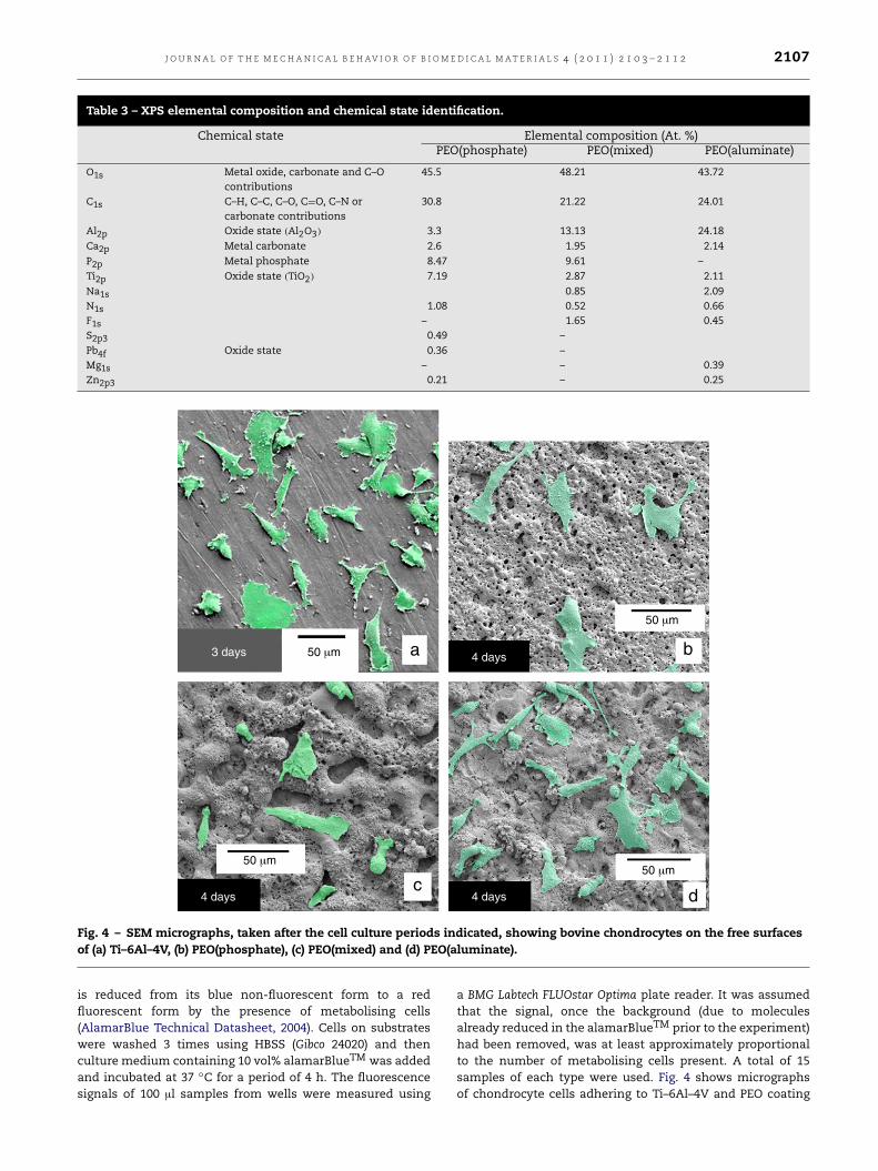
Fig. 4 – SEM micrographs, taken after the cell culture periods indicated, showing bovine chondrocytes on the free surfacesof (a) Ti–6Al–4V, (b) PEO(phosphate), (c) PEO(mixed) and (d) PEO(aluminate).
is reduced from its blue non-fluorescent form to a redfluorescent form by the presence of metabolising cells(AlamarBlue Technical Datasheet, 2004). Cells on substrateswere washed 3 times using HBSS (Gibco 24020) and thenculture medium containing 10 vol% alamarBlueTM was addedand incubated at 37 ◦C for a period of 4 h. The fluorescencesignals of 100 µl samples from wells were measured using
a BMG Labtech FLUOstar Optima plate reader. It was assumedthat the signal, once the background (due to moleculesalready reduced in the alamarBlueTM prior to the experiment)had been removed, was at least approximately proportionalto the number of metabolising cells present. A total of 15samples of each type were used. Fig. 4 shows micrographsof chondrocyte cells adhering to Ti–6Al–4V and PEO coating
2108 J O U R N A L O F T H E M E C H A N I C A L B E H AV I O R O F B I O M E D I C A L M A T E R I A L S 4 ( 2 0 1 1 ) 2 1 0 3 – 2 1 1 2
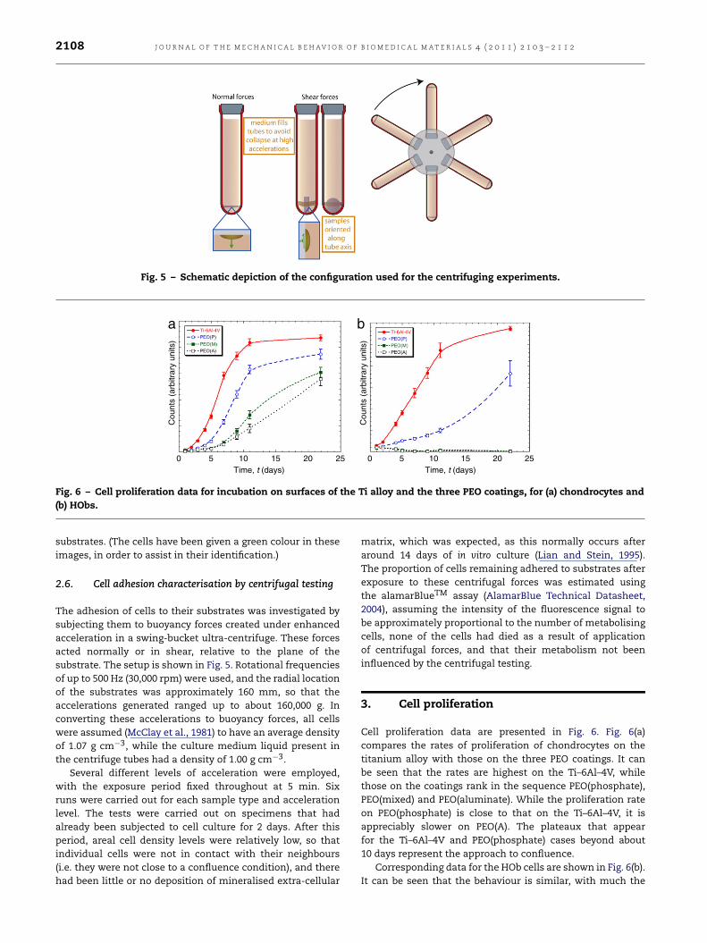
Fig. 5 – Schematic depiction of the configuration used for the centrifuging experiments.
0
Cou
nts
(arb
itrar
y un
its)
Cou
nts
(arb
itrar
y un
its)
5 10 15Time, t (days)
20 25 0 5 10 15Time, t (days)
20 25
a b
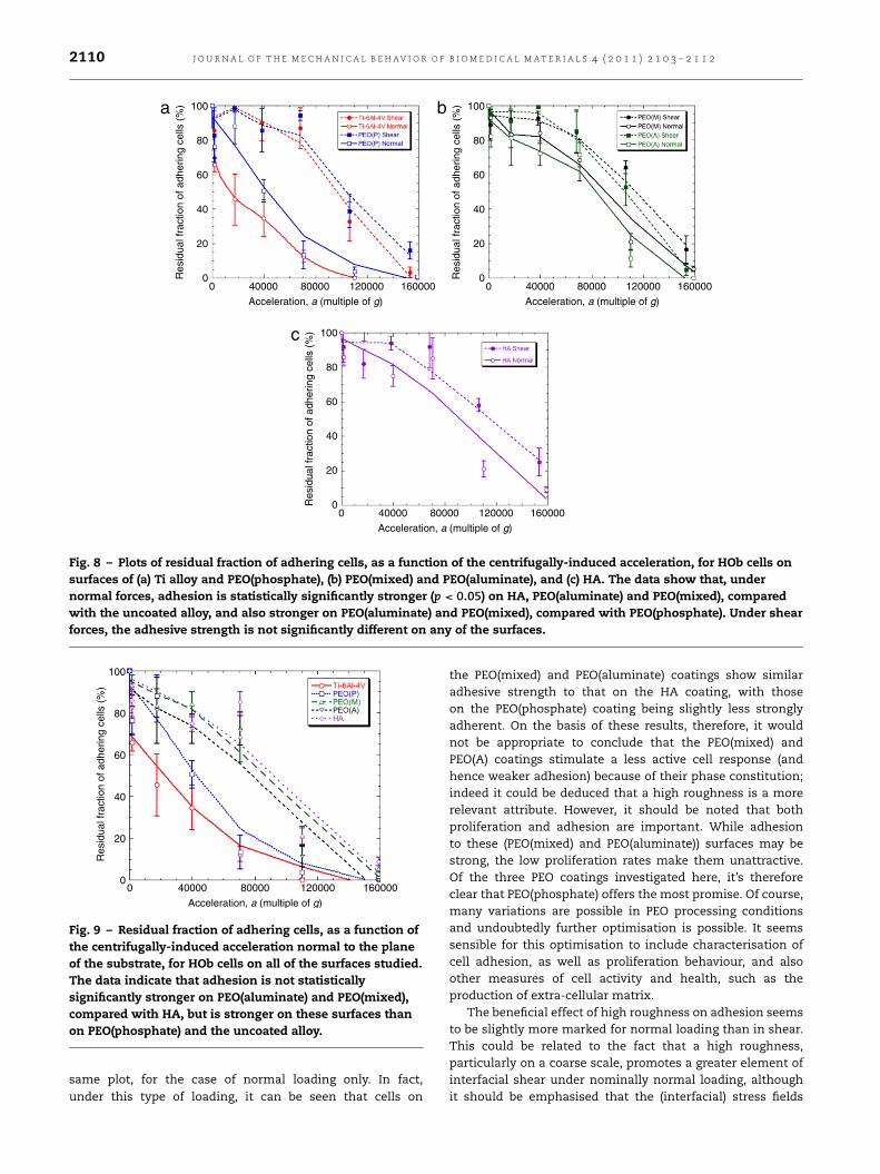
Fig. 6 – Cell proliferation data for incubation on surfaces of the Ti alloy and the three PEO coatings, for (a) chondrocytes and(b) HObs.
substrates. (The cells have been given a green colour in theseimages, in order to assist in their identification.)
2.6. Cell adhesion characterisation by centrifugal testing
The adhesion of cells to their substrates was investigated bysubjecting them to buoyancy forces created under enhancedacceleration in a swing-bucket ultra-centrifuge. These forcesacted normally or in shear, relative to the plane of thesubstrate. The setup is shown in Fig. 5. Rotational frequenciesof up to 500 Hz (30,000 rpm) were used, and the radial locationof the substrates was approximately 160 mm, so that theaccelerations generated ranged up to about 160,000 g. Inconverting these accelerations to buoyancy forces, all cellswere assumed (McClay et al., 1981) to have an average densityof 1.07 g cm−3, while the culture medium liquid present inthe centrifuge tubes had a density of 1.00 g cm−3.
Several different levels of acceleration were employed,with the exposure period fixed throughout at 5 min. Sixruns were carried out for each sample type and accelerationlevel. The tests were carried out on specimens that hadalready been subjected to cell culture for 2 days. After thisperiod, areal cell density levels were relatively low, so thatindividual cells were not in contact with their neighbours(i.e. they were not close to a confluence condition), and therehad been little or no deposition of mineralised extra-cellular
matrix, which was expected, as this normally occurs afteraround 14 days of in vitro culture (Lian and Stein, 1995).The proportion of cells remaining adhered to substrates afterexposure to these centrifugal forces was estimated usingthe alamarBlueTM assay (AlamarBlue Technical Datasheet,2004), assuming the intensity of the fluorescence signal tobe approximately proportional to the number of metabolisingcells, none of the cells had died as a result of applicationof centrifugal forces, and that their metabolism not beeninfluenced by the centrifugal testing.
3. Cell proliferation
Cell proliferation data are presented in Fig. 6. Fig. 6(a)compares the rates of proliferation of chondrocytes on thetitanium alloy with those on the three PEO coatings. It canbe seen that the rates are highest on the Ti–6Al–4V, whilethose on the coatings rank in the sequence PEO(phosphate),PEO(mixed) and PEO(aluminate). While the proliferation rateon PEO(phosphate) is close to that on the Ti–6Al–4V, it isappreciably slower on PEO(A). The plateaux that appearfor the Ti–6Al–4V and PEO(phosphate) cases beyond about10 days represent the approach to confluence.
Corresponding data for the HOb cells are shown in Fig. 6(b).It can be seen that the behaviour is similar, with much the

J O U R N A L O F T H E M E C H A N I C A L B E H AV I O R O F B I O M E D I C A L M A T E R I A L S 4 ( 2 0 1 1 ) 2 1 0 3 – 2 1 1 2 2109
0
Res
idua
l fra
ctio
n of
adh
erin
g ce
lls (
%)
0
20
40
60
80
100
40000 80000
Acceleration, a (multiple of g)
120000 160000 0
Res
idua
l fra
ctio
n of
adh
erin
g ce
lls (
%)
0
20
40
60
80
100
40000 80000
Acceleration, a (multiple of g)
120000 160000
a b
Fig. 7 – Plots of residual fraction of adhering cells, as a function of the centrifugally-induced acceleration, for chondrocyteson surfaces of (a) Ti alloy and PEO(phosphate), and (b) PEO(mixed) and PEO(aluminate). The data show that, under normalforces, adhesion is statistically significantly stronger (p < 0.05) on all PEO coatings, compared with the uncoated alloy, andalso stronger on PEO(aluminate) and PEO(mixed), compared with PEO(phosphate). Under shear forces, adhesion toPEO(phosphate) and PEO(mixed) is significantly stronger than to PEO(aluminate) and the uncoated alloy.
same ranking order. However, it is clear that the proliferationrates on both the PEO(mixed) and PEO(aluminate) coatingsbecame very low after a few days.
4. Cell adhesion
Cell adhesion data are presented in Figs. 7–9, in the form ofresidual proportions of cells remaining adherent after variouscentrifuging treatments. Fig. 7 shows data for chondrocytes,under both normal and shear loading, for the titanium alloyand for the three PEO coatings. Firstly, it can be seen that,in all cases, cells resist debonding under shear forces morestrongly thanwhen subjected to normal forces. In general, thetoughness (resistance to fracture) of most interfaces is higherin shear (mode II) than in tension (mode I), so this observationappears unsurprising.
There are also differences apparent in Fig. 7 betweenadhesive strengths on the different surfaces, with that onPEO(phosphate) coatings being stronger than on Ti–6Al–4V(Fig. 7(a)) and that on the other two PEO coatings beingintermediate between the two (Fig. 7(b)). Of course, all threePEO surfaces are much rougher than the Ti–6Al–4V substrate,but it may beworth noting that the roughness increases in thesequence PEO(phosphate), PEO(mixed), PEO(aluminate) — seeFig. 2(a). Other things being equal, it might be expected thatadhesion would be stronger on rougher surfaces, particularlyunder shear loading, whereas in fact it is strongest onPEO(phosphate) coatings. However, this assumes that theconformity of the cells to the contours of the surface isequally good in all cases, and the mechanism of bonding issimilar for them all. In reality, this is unlikely to be the case,and differences in these factors are presumably responsiblefor at least some of the observed effects.
It may be that there are effects associated with localcharacteristics other than the macroscopic roughness, suchas benefits from the very fine scale porosity in the PEOcoatings, or differences in the response of the cells to thephases that are present, or the surface chemistry. These could
affect the spreading of focal adhesions and/or the local bondstrength. In general, for surfaces that are nominally titania,there is very little information in the literature concerningthe effects of phase constitution or chemical composition,on either cell proliferation or cell adhesion. The oxide onTi–6Al–4V is predominantly amorphous (Fig. 2(b)), with somerutile, so it seems likely that these phases elicit a healthy cellresponse. The fact that both the amorphous phase and rutileare present in the PEO(phosphate) coating may be relevant inexplaining the good cell behaviour that occurs on its surface.On the other hand, both PEO(mixed) and PEO(aluminate)coatings predominantly comprise the aluminium titanatephase, with no amorphous phase present in either caseand very little rutile (in PEO(mixed) only). The results couldtherefore be taken as indicating that the response of thecells to contact with the Al2TiO5 phase, and hence theiradhesion to it, is inferior to that of pure titania (amorphousand rutile) phases. It may also be noted that, for PEO(mixed)and, particularly, PEO(aluminate) coatings, the proliferationrates were relatively low for chondrocyte cells and verypoor for osteoblast cells. This is consistent with a generalperception that high levels of aluminium in a surface canbe detrimental to a wide range of cells — although somealuminium-containing surfaces, such as alumino-silicateshave been reported as suitable substrates for osteoblasts(Leivo et al., 2006), so this was not a foregone conclusion
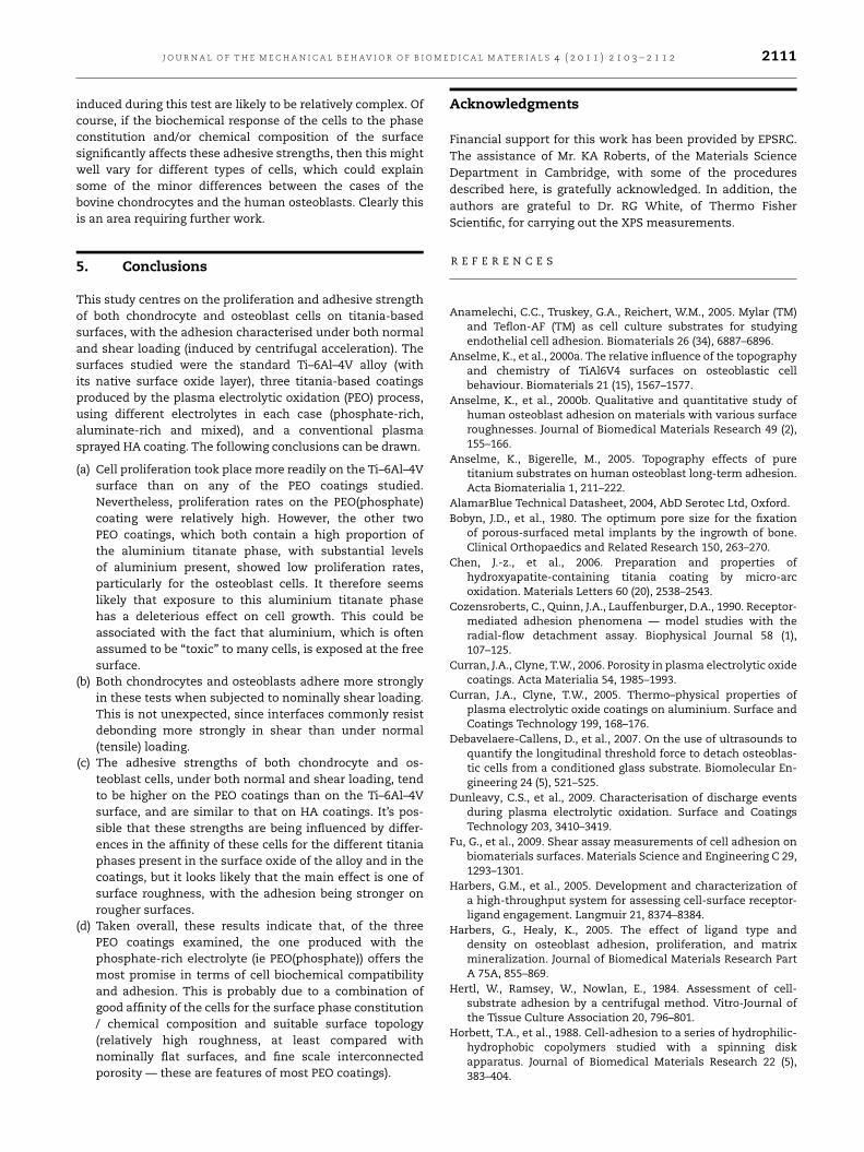
Fig. 8 shows residual adhering proportions for HObcells, on Ti–6Al–4V, PEO coatings and the HA coating(included solely for comparative purposes). These resultsshow similarities to those for the chondrocyte cells, withthe resistance to debonding in shear being consistentlyhigher than that under normal loading, and the cells onPEO(phosphate) coatings showing superior adhesion to thoseon the titanium alloy. Cells on the other two PEO coatingsagain showed similar adhesive strengths, although in thiscase they were also similar to that on the PEO(phosphate)coating. These strengths were also similar to that on theHA coating. Comparing the five cases is made a littleeasier by inspecting Fig. 9, which includes them all on the
2110 J O U R N A L O F T H E M E C H A N I C A L B E H AV I O R O F B I O M E D I C A L M A T E R I A L S 4 ( 2 0 1 1 ) 2 1 0 3 – 2 1 1 2
0
Res
idua
l fra
ctio
n of
adh
erin
g ce
lls (
%)
0
20
40
60
80
100
40000 80000
Acceleration, a (multiple of g)
120000 160000
a
0
Res
idua
l fra
ctio
n of
adh
erin
g ce
lls (
%)
0
20
40
60
80
100
40000 80000
Acceleration, a (multiple of g)
120000 160000
b
0
Res
idua
l fra
ctio
n of
adh
erin
g ce
lls (
%)
0
20
40
60
80
100
40000 80000
Acceleration, a (multiple of g)
120000 160000
c
Fig. 8 – Plots of residual fraction of adhering cells, as a function of the centrifugally-induced acceleration, for HOb cells onsurfaces of (a) Ti alloy and PEO(phosphate), (b) PEO(mixed) and PEO(aluminate), and (c) HA. The data show that, undernormal forces, adhesion is statistically significantly stronger (p < 0.05) on HA, PEO(aluminate) and PEO(mixed), comparedwith the uncoated alloy, and also stronger on PEO(aluminate) and PEO(mixed), compared with PEO(phosphate). Under shearforces, the adhesive strength is not significantly different on any of the surfaces.
0
Res
idua
l fra
ctio
n of
adh
erin
g ce
lls (
%)
0
20
40
60
80
100
40000 80000
Acceleration, a (multiple of g)
120000 160000
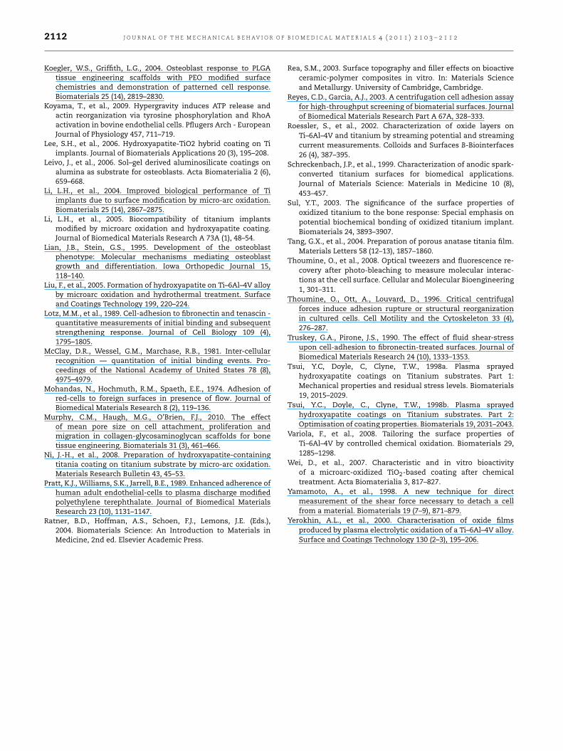
Fig. 9 – Residual fraction of adhering cells, as a function ofthe centrifugally-induced acceleration normal to the planeof the substrate, for HOb cells on all of the surfaces studied.The data indicate that adhesion is not statisticallysignificantly stronger on PEO(aluminate) and PEO(mixed),compared with HA, but is stronger on these surfaces thanon PEO(phosphate) and the uncoated alloy.
same plot, for the case of normal loading only. In fact,under this type of loading, it can be seen that cells on
the PEO(mixed) and PEO(aluminate) coatings show similaradhesive strength to that on the HA coating, with thoseon the PEO(phosphate) coating being slightly less stronglyadherent. On the basis of these results, therefore, it wouldnot be appropriate to conclude that the PEO(mixed) andPEO(A) coatings stimulate a less active cell response (andhence weaker adhesion) because of their phase constitution;indeed it could be deduced that a high roughness is a morerelevant attribute. However, it should be noted that bothproliferation and adhesion are important. While adhesionto these (PEO(mixed) and PEO(aluminate)) surfaces may bestrong, the low proliferation rates make them unattractive.Of the three PEO coatings investigated here, it’s thereforeclear that PEO(phosphate) offers the most promise. Of course,many variations are possible in PEO processing conditionsand undoubtedly further optimisation is possible. It seemssensible for this optimisation to include characterisation ofcell adhesion, as well as proliferation behaviour, and alsoother measures of cell activity and health, such as theproduction of extra-cellular matrix.
The beneficial effect of high roughness on adhesion seemsto be slightly more marked for normal loading than in shear.This could be related to the fact that a high roughness,particularly on a coarse scale, promotes a greater element ofinterfacial shear under nominally normal loading, althoughit should be emphasised that the (interfacial) stress fields
J O U R N A L O F T H E M E C H A N I C A L B E H AV I O R O F B I O M E D I C A L M A T E R I A L S 4 ( 2 0 1 1 ) 2 1 0 3 – 2 1 1 2 2111
induced during this test are likely to be relatively complex. Ofcourse, if the biochemical response of the cells to the phaseconstitution and/or chemical composition of the surfacesignificantly affects these adhesive strengths, then this mightwell vary for different types of cells, which could explainsome of the minor differences between the cases of thebovine chondrocytes and the human osteoblasts. Clearly thisis an area requiring further work.
5. Conclusions
This study centres on the proliferation and adhesive strengthof both chondrocyte and osteoblast cells on titania-basedsurfaces, with the adhesion characterised under both normaland shear loading (induced by centrifugal acceleration). Thesurfaces studied were the standard Ti–6Al–4V alloy (withits native surface oxide layer), three titania-based coatingsproduced by the plasma electrolytic oxidation (PEO) process,using different electrolytes in each case (phosphate-rich,aluminate-rich and mixed), and a conventional plasmasprayed HA coating. The following conclusions can be drawn.
(a) Cell proliferation took place more readily on the Ti–6Al–4Vsurface than on any of the PEO coatings studied.Nevertheless, proliferation rates on the PEO(phosphate)coating were relatively high. However, the other twoPEO coatings, which both contain a high proportion ofthe aluminium titanate phase, with substantial levelsof aluminium present, showed low proliferation rates,particularly for the osteoblast cells. It therefore seemslikely that exposure to this aluminium titanate phasehas a deleterious effect on cell growth. This could beassociated with the fact that aluminium, which is oftenassumed to be “toxic” to many cells, is exposed at the freesurface.
(b) Both chondrocytes and osteoblasts adhere more stronglyin these tests when subjected to nominally shear loading.This is not unexpected, since interfaces commonly resistdebonding more strongly in shear than under normal(tensile) loading.
(c) The adhesive strengths of both chondrocyte and os-teoblast cells, under both normal and shear loading, tendto be higher on the PEO coatings than on the Ti–6Al–4Vsurface, and are similar to that on HA coatings. It’s pos-sible that these strengths are being influenced by differ-ences in the affinity of these cells for the different titaniaphases present in the surface oxide of the alloy and in thecoatings, but it looks likely that the main effect is one ofsurface roughness, with the adhesion being stronger onrougher surfaces.
(d) Taken overall, these results indicate that, of the threePEO coatings examined, the one produced with thephosphate-rich electrolyte (ie PEO(phosphate)) offers themost promise in terms of cell biochemical compatibilityand adhesion. This is probably due to a combination ofgood affinity of the cells for the surface phase constitution/ chemical composition and suitable surface topology(relatively high roughness, at least compared withnominally flat surfaces, and fine scale interconnectedporosity — these are features of most PEO coatings).
Acknowledgments
Financial support for this work has been provided by EPSRC.The assistance of Mr. KA Roberts, of the Materials ScienceDepartment in Cambridge, with some of the proceduresdescribed here, is gratefully acknowledged. In addition, theauthors are grateful to Dr. RG White, of Thermo FisherScientific, for carrying out the XPS measurements.
R E F E R E N C E S
Anamelechi, C.C., Truskey, G.A., Reichert, W.M., 2005. Mylar (TM)and Teflon-AF (TM) as cell culture substrates for studyingendothelial cell adhesion. Biomaterials 26 (34), 6887–6896.
Anselme, K., et al., 2000a. The relative influence of the topographyand chemistry of TiAl6V4 surfaces on osteoblastic cellbehaviour. Biomaterials 21 (15), 1567–1577.
Anselme, K., et al., 2000b. Qualitative and quantitative study ofhuman osteoblast adhesion on materials with various surfaceroughnesses. Journal of Biomedical Materials Research 49 (2),155–166.
Anselme, K., Bigerelle, M., 2005. Topography effects of puretitanium substrates on human osteoblast long-term adhesion.Acta Biomaterialia 1, 211–222.
AlamarBlue Technical Datasheet, 2004, AbD Serotec Ltd, Oxford.Bobyn, J.D., et al., 1980. The optimum pore size for the fixation
of porous-surfaced metal implants by the ingrowth of bone.Clinical Orthopaedics and Related Research 150, 263–270.
Chen, J.-z., et al., 2006. Preparation and properties ofhydroxyapatite-containing titania coating by micro-arcoxidation. Materials Letters 60 (20), 2538–2543.
Cozensroberts, C., Quinn, J.A., Lauffenburger, D.A., 1990. Receptor-mediated adhesion phenomena — model studies with theradial-flow detachment assay. Biophysical Journal 58 (1),107–125.
Curran, J.A., Clyne, T.W., 2006. Porosity in plasma electrolytic oxidecoatings. Acta Materialia 54, 1985–1993.
Curran, J.A., Clyne, T.W., 2005. Thermo–physical properties ofplasma electrolytic oxide coatings on aluminium. Surface andCoatings Technology 199, 168–176.
Debavelaere-Callens, D., et al., 2007. On the use of ultrasounds toquantify the longitudinal threshold force to detach osteoblas-tic cells from a conditioned glass substrate. Biomolecular En-gineering 24 (5), 521–525.
Dunleavy, C.S., et al., 2009. Characterisation of discharge eventsduring plasma electrolytic oxidation. Surface and CoatingsTechnology 203, 3410–3419.
Fu, G., et al., 2009. Shear assay measurements of cell adhesion onbiomaterials surfaces. Materials Science and Engineering C 29,1293–1301.
Harbers, G.M., et al., 2005. Development and characterization ofa high-throughput system for assessing cell-surface receptor-ligand engagement. Langmuir 21, 8374–8384.
Harbers, G., Healy, K., 2005. The effect of ligand type anddensity on osteoblast adhesion, proliferation, and matrixmineralization. Journal of Biomedical Materials Research PartA 75A, 855–869.
Hertl, W., Ramsey, W., Nowlan, E., 1984. Assessment of cell-substrate adhesion by a centrifugal method. Vitro-Journal ofthe Tissue Culture Association 20, 796–801.
Horbett, T.A., et al., 1988. Cell-adhesion to a series of hydrophilic-hydrophobic copolymers studied with a spinning diskapparatus. Journal of Biomedical Materials Research 22 (5),383–404.
2112 J O U R N A L O F T H E M E C H A N I C A L B E H AV I O R O F B I O M E D I C A L M A T E R I A L S 4 ( 2 0 1 1 ) 2 1 0 3 – 2 1 1 2
Koegler, W.S., Griffith, L.G., 2004. Osteoblast response to PLGAtissue engineering scaffolds with PEO modified surfacechemistries and demonstration of patterned cell response.Biomaterials 25 (14), 2819–2830.
Koyama, T., et al., 2009. Hypergravity induces ATP release andactin reorganization via tyrosine phosphorylation and RhoAactivation in bovine endothelial cells. Pflugers Arch - EuropeanJournal of Physiology 457, 711–719.
Lee, S.H., et al., 2006. Hydroxyapatite-TiO2 hybrid coating on Tiimplants. Journal of Biomaterials Applications 20 (3), 195–208.
Leivo, J., et al., 2006. Sol–gel derived aluminosilicate coatings onalumina as substrate for osteoblasts. Acta Biomaterialia 2 (6),659–668.
Li, L.H., et al., 2004. Improved biological performance of Tiimplants due to surface modification by micro-arc oxidation.Biomaterials 25 (14), 2867–2875.
Li, L.H., et al., 2005. Biocompatibility of titanium implantsmodified by microarc oxidation and hydroxyapatite coating.Journal of Biomedical Materials Research A 73A (1), 48–54.
Lian, J.B., Stein, G.S., 1995. Development of the osteoblastphenotype: Molecular mechanisms mediating osteoblastgrowth and differentiation. Iowa Orthopedic Journal 15,118–140.
Liu, F., et al., 2005. Formation of hydroxyapatite on Ti–6Al–4V alloyby microarc oxidation and hydrothermal treatment. Surfaceand Coatings Technology 199, 220–224.
Lotz, M.M., et al., 1989. Cell-adhesion to fibronectin and tenascin -quantitative measurements of initial binding and subsequentstrengthening response. Journal of Cell Biology 109 (4),1795–1805.
McClay, D.R., Wessel, G.M., Marchase, R.B., 1981. Inter-cellularrecognition — quantitation of initial binding events. Pro-ceedings of the National Academy of United States 78 (8),4975–4979.
Mohandas, N., Hochmuth, R.M., Spaeth, E.E., 1974. Adhesion ofred-cells to foreign surfaces in presence of flow. Journal ofBiomedical Materials Research 8 (2), 119–136.
Murphy, C.M., Haugh, M.G., O’Brien, F.J., 2010. The effectof mean pore size on cell attachment, proliferation andmigration in collagen-glycosaminoglycan scaffolds for bonetissue engineering. Biomaterials 31 (3), 461–466.
Ni, J.-H., et al., 2008. Preparation of hydroxyapatite-containingtitania coating on titanium substrate by micro-arc oxidation.Materials Research Bulletin 43, 45–53.
Pratt, K.J., Williams, S.K., Jarrell, B.E., 1989. Enhanced adherence ofhuman adult endothelial-cells to plasma discharge modifiedpolyethylene terephthalate. Journal of Biomedical MaterialsResearch 23 (10), 1131–1147.
Ratner, B.D., Hoffman, A.S., Schoen, F.J., Lemons, J.E. (Eds.),2004. Biomaterials Science: An Introduction to Materials inMedicine, 2nd ed. Elsevier Academic Press.
Rea, S.M., 2003. Surface topography and filler effects on bioactiveceramic-polymer composites in vitro. In: Materials Scienceand Metallurgy. University of Cambridge, Cambridge.
Reyes, C.D., Garcia, A.J., 2003. A centrifugation cell adhesion assayfor high-throughput screening of biomaterial surfaces. Journalof Biomedical Materials Research Part A 67A, 328–333.
Roessler, S., et al., 2002. Characterization of oxide layers onTi–6Al–4V and titanium by streaming potential and streamingcurrent measurements. Colloids and Surfaces B-Biointerfaces26 (4), 387–395.
Schreckenbach, J.P., et al., 1999. Characterization of anodic spark-converted titanium surfaces for biomedical applications.Journal of Materials Science: Materials in Medicine 10 (8),453–457.
Sul, Y.T., 2003. The significance of the surface properties ofoxidized titanium to the bone response: Special emphasis onpotential biochemical bonding of oxidized titanium implant.Biomaterials 24, 3893–3907.
Tang, G.X., et al., 2004. Preparation of porous anatase titania film.Materials Letters 58 (12–13), 1857–1860.
Thoumine, O., et al., 2008. Optical tweezers and fluorescence re-covery after photo-bleaching to measure molecular interac-tions at the cell surface. Cellular and Molecular Bioengineering1, 301–311.
Thoumine, O., Ott, A., Louvard, D., 1996. Critical centrifugalforces induce adhesion rupture or structural reorganizationin cultured cells. Cell Motility and the Cytoskeleton 33 (4),276–287.
Truskey, G.A., Pirone, J.S., 1990. The effect of fluid shear-stressupon cell-adhesion to fibronectin-treated surfaces. Journal ofBiomedical Materials Research 24 (10), 1333–1353.
Tsui, Y.C, Doyle, C, Clyne, T.W., 1998a. Plasma sprayedhydroxyapatite coatings on Titanium substrates. Part 1:Mechanical properties and residual stress levels. Biomaterials19, 2015–2029.
Tsui, Y.C., Doyle, C., Clyne, T.W., 1998b. Plasma sprayedhydroxyapatite coatings on Titanium substrates. Part 2:Optimisation of coating properties. Biomaterials 19, 2031–2043.
Variola, F., et al., 2008. Tailoring the surface properties ofTi–6Al–4V by controlled chemical oxidation. Biomaterials 29,1285–1298.
Wei, D., et al., 2007. Characteristic and in vitro bioactivityof a microarc-oxidized TiO2-based coating after chemicaltreatment. Acta Biomaterialia 3, 817–827.
Yamamoto, A., et al., 1998. A new technique for directmeasurement of the shear force necessary to detach a cellfrom a material. Biomaterials 19 (7–9), 871–879.
Yerokhin, A.L., et al., 2000. Characterisation of oxide filmsproduced by plasma electrolytic oxidation of a Ti–6Al–4V alloy.Surface and Coatings Technology 130 (2–3), 195–206.