The Analysis of Pendolino (peo) Mutants Reveals Differences in the Fusigenic Potential among...
31
RESEARCH ARTICLE The Analysis of Pendolino (peo) Mutants Reveals Differences in the Fusigenic Potential among Drosophila Telomeres Giovanni Cenci 1,2☯ , Laura Ciapponi 1,2☯ , Marta Marzullo 1,2 , Grazia D. Raffa 1,2 , Patrizia Morciano 1,2 , Domenico Raimondo 3 , Romina Burla 1,2 , Isabella Saggio 1,2,4 , Maurizio Gatti 1,2,4 * 1 Dipartimento di Biologia e Biotecnologie, Sapienza—Università di Roma, Roma, Italy, 2 Istituto Pasteur Fondazione Cenci Bolognetti, Sapienza—Università di Roma, Roma, Italy, 3 Dipartimento di Fisica, Sapienza—Università di Roma, Roma, Italy, 4 IBPM CNR, Sapienza—Università di Roma, Roma, Italy ☯ These authors contributed equally to this work. * [email protected] Abstract Drosophila telomeres are sequence-independent structures that are maintained by transpo- sition to chromosome ends of three specialized retroelements (HeT-A, TART and TAHRE; collectively designated as HTT) rather than telomerase activity. Fly telomeres are protected by the terminin complex (HOAP-HipHop-Moi-Ver) that localizes and functions exclusively at telomeres and by non-terminin proteins that do not serve telomere-specific functions. Al- though all Drosophila telomeres terminate with HTT arrays and are capped by terminin, they differ in the type of subtelomeric chromatin; the Y, XR, and 4L HTT are juxtaposed to constitutive heterochromatin, while the XL, 2L, 2R, 3L and 3R HTT are linked to the TAS re- petitive sequences; the 4R HTT is associated with a chromatin that has features common to both euchromatin and heterochromatin. Here we show that mutations in pendolino (peo) cause telomeric fusions (TFs). The analysis of several peo mutant combinations showed that these TFs preferentially involve the Y, XR and 4th chromosome telomeres, a TF pattern never observed in the other 10 telomere-capping mutants so far characterized. peo en- codes a non-terminin protein homologous to the E2 variant ubiquitin-conjugating enzymes. The Peo protein directly interacts with the terminin components, but peo mutations do not affect telomeric localization of HOAP, Moi, Ver and HP1a, suggesting that the peo-depen- dent telomere fusion phenotype is not due to loss of terminin from chromosome ends. peo mutants are also defective in DNA replication and PCNA recruitment. However, our results suggest that general defects in DNA replication are unable to induce TFs in Drosophila cells. We thus hypothesize that DNA replication in Peo-depleted cells results in specific fusi- genic lesions concentrated in heterochromatin-associated telomeres. Alternatively, it is pos- sible that Peo plays a dual function being independently required for DNA replication and telomere capping. PLOS Genetics | DOI:10.1371/journal.pgen.1005260 June 25, 2015 1 / 31 OPEN ACCESS Citation: Cenci G, Ciapponi L, Marzullo M, Raffa GD, Morciano P, Raimondo D, et al. (2015) The Analysis of Pendolino (peo) Mutants Reveals Differences in the Fusigenic Potential among Drosophila Telomeres. PLoS Genet 11(6): e1005260. doi:10.1371/journal. pgen.1005260 Editor: Yikang Rong, NIH, UNITED STATES Received: October 20, 2014 Accepted: May 4, 2015 Published: June 25, 2015 Copyright: © 2015 Cenci et al. This is an open access article distributed under the terms of the Creative Commons Attribution License, which permits unrestricted use, distribution, and reproduction in any medium, provided the original author and source are credited. Data Availability Statement: All relevant data are within the paper and its Supporting Information files. Funding: This work was supported by grants from Italian Association for Cancer Research to MG (AIRC, IG10793) and GC (AIRC, IG12749). The funders had no role in study design, data collection and analysis, decision to publish, or preparation of the manuscript. Competing Interests: The authors have declared that no competing interests exist.
Transcript of The Analysis of Pendolino (peo) Mutants Reveals Differences in the Fusigenic Potential among...

RESEARCH ARTICLE
The Analysis of Pendolino (peo) MutantsReveals Differences in the Fusigenic Potentialamong Drosophila TelomeresGiovanni Cenci1,2☯, Laura Ciapponi1,2☯, Marta Marzullo1,2, Grazia D. Raffa1,2,Patrizia Morciano1,2, Domenico Raimondo3, Romina Burla1,2, Isabella Saggio1,2,4,Maurizio Gatti1,2,4*
1 Dipartimento di Biologia e Biotecnologie, Sapienza—Università di Roma, Roma, Italy, 2 Istituto PasteurFondazione Cenci Bolognetti, Sapienza—Università di Roma, Roma, Italy, 3 Dipartimento di Fisica,Sapienza—Università di Roma, Roma, Italy, 4 IBPM CNR, Sapienza—Università di Roma, Roma, Italy
☯ These authors contributed equally to this work.* [email protected]
AbstractDrosophila telomeres are sequence-independent structures that are maintained by transpo-
sition to chromosome ends of three specialized retroelements (HeT-A, TART and TAHRE;
collectively designated as HTT) rather than telomerase activity. Fly telomeres are protected
by the terminin complex (HOAP-HipHop-Moi-Ver) that localizes and functions exclusively at
telomeres and by non-terminin proteins that do not serve telomere-specific functions. Al-
though all Drosophila telomeres terminate with HTT arrays and are capped by terminin,
they differ in the type of subtelomeric chromatin; the Y, XR, and 4L HTT are juxtaposed to
constitutive heterochromatin, while the XL, 2L, 2R, 3L and 3R HTT are linked to the TAS re-
petitive sequences; the 4R HTT is associated with a chromatin that has features common to
both euchromatin and heterochromatin. Here we show that mutations in pendolino (peo)cause telomeric fusions (TFs). The analysis of several peomutant combinations showed
that these TFs preferentially involve the Y, XR and 4th chromosome telomeres, a TF pattern
never observed in the other 10 telomere-capping mutants so far characterized. peo en-
codes a non-terminin protein homologous to the E2 variant ubiquitin-conjugating enzymes.
The Peo protein directly interacts with the terminin components, but peomutations do not
affect telomeric localization of HOAP, Moi, Ver and HP1a, suggesting that the peo-depen-dent telomere fusion phenotype is not due to loss of terminin from chromosome ends. peomutants are also defective in DNA replication and PCNA recruitment. However, our results
suggest that general defects in DNA replication are unable to induce TFs in Drosophilacells. We thus hypothesize that DNA replication in Peo-depleted cells results in specific fusi-
genic lesions concentrated in heterochromatin-associated telomeres. Alternatively, it is pos-
sible that Peo plays a dual function being independently required for DNA replication and
telomere capping.
PLOS Genetics | DOI:10.1371/journal.pgen.1005260 June 25, 2015 1 / 31
OPEN ACCESS
Citation: Cenci G, Ciapponi L, Marzullo M, Raffa GD,Morciano P, Raimondo D, et al. (2015) The Analysisof Pendolino (peo) Mutants Reveals Differences inthe Fusigenic Potential among Drosophila Telomeres.PLoS Genet 11(6): e1005260. doi:10.1371/journal.pgen.1005260
Editor: Yikang Rong, NIH, UNITED STATES
Received: October 20, 2014
Accepted: May 4, 2015
Published: June 25, 2015
Copyright: © 2015 Cenci et al. This is an openaccess article distributed under the terms of theCreative Commons Attribution License, which permitsunrestricted use, distribution, and reproduction in anymedium, provided the original author and source arecredited.
Data Availability Statement: All relevant data arewithin the paper and its Supporting Information files.
Funding: This work was supported by grants fromItalian Association for Cancer Research to MG(AIRC, IG10793) and GC (AIRC, IG12749). Thefunders had no role in study design, data collectionand analysis, decision to publish, or preparation ofthe manuscript.
Competing Interests: The authors have declaredthat no competing interests exist.

Author Summary
Telomeres are specialized structures that protect chromosome ends from incomplete repli-cation, degradation and end-to-end fusion. Abnormalities in telomere structure or mainte-nance can promote a variety of human diseases including premature aging and cancer.Although all human telomeres contain the same DNA sequences, they differ from eachother in the subtelomeric regions or subtelomeres. Recent work has shown that humansubtelomeres control telomere replication and that abnormalities in these structures canlead to localized chromosome instability and disease. However, the relationships betweensubtelomeres and telomeres are currently poorly understood. Here, we have addressed thisproblem using the fruit fly Drosophila melanogaster as model system. Drosophila subtelo-mers are very different from each other as they contain different types of chromatin. Wehave found that mutations in a gene we called pendolino (peo) cause telomeric fusions(TFs) and that these fusions preferentially involve the telomeres associated with a tightlypacked form of chromatin called heterochromatin. Interestingly, none of the 10 mutantswith TFs so far described in Drosophila shows the pattern of TFs observed in peomutants.Thus, our data provide the first demonstration that subtelomeres can affect telomere fu-sion. We believe that these results will stimulate further studies on the role of subtelomeresin the maintenance of genome stability.
IntroductionTelomeres are nucleoprotein complexes that counterbalance incomplete replication of terminalDNA and protect chromosome ends preventing both activation of cell cycle checkpoints andfusion events. In most organisms, the end replication problem is solved by telomerase, whichmediates the addition of short GC-rich repeats to chromosome ends. These repeats specificallybind a discrete number of proteins, which recruit a series of additional factors to form largeprotein assemblies that ensure proper telomere function and homeostasis (reviewed in [1–4]).Drosophila telomeres are not elongated by telomerase but by targeted transposition of threespecialized retroelements called HeT-A, TART and TAHRE (collectively abbreviated withHTT). However, Drosophila telomere formation does not require the HTT arrays; abundantevidence indicates that fly telomeres are epigenetically-determined structures that can assembleat the ends of the chromosomes independently of their terminal DNA sequence (reviewed in[5–7]).
With the exception of budding yeast, in organisms with telomerase telomeres are protectedby the conserved shelterin complex. Human shelterin is a six-protein complex (TRF1, TRF2,POT1, TIN2, TPP1 and Rap1) that specifically associates with the telomeric TTAGGG repeats.TRF1, TRF2 directly bind the TTAGGG duplex and POT1 the single stranded overhang. TIN2and TPP1 do not bind DNA and interconnect TRF1 and TRF2 with POT1. TRF2 interactswith hRap1, a distant homologue of S. cerevisiae Rap1. The shelterin subunits share propertiesthat distinguish them from the non-shelterin telomere-associated proteins: they are specificallyenriched at telomeres throughout the cell cycle and appear to function only at telomeres [1].The human non-shelterin proteins, which are not telomere-specific in localization and func-tion, include the conserved CST complex, HP1a, and proteins involved in DNA repair and/orreplication such as the ATM kinase, the Ku70/80 heterodimer, the MRE11/RAD50/NBS1(MRN) complex, Rad51, the ERCC1/XPF endonuclease, the Apollo exonuclease, the FEN1 nu-clease, the RecQ family members WRN and BLM; the RTL1 helicase, RPA70, the Timelesscomponent of the replisome, and the subunits of the conserved ORC prereplication complex.
Peo Protects Heterochromatic Telomeres in Drosophila
PLOSGenetics | DOI:10.1371/journal.pgen.1005260 June 25, 2015 2 / 31

Depletion of any one of the shelterin subunits or the shelterin-associated proteins leads to visi-ble telomere defects ranging from altered packaging of telomeric chromatin (multi telomeresignals after FISH), telomere loss and telomere fusion (reviewed in [1–4]; see also [8, 9]).
Most of the Drosophila telomere-capping proteins have been identified by molecular clon-ing of genes specified by mutations that cause telomeric fusions (TFs) in larval brain cells. Ge-netic and molecular analyses have thus far identified 11 loci that are required to prevent TF(henceforth they will be designated as TF genes). These are effete (eff; also called UbcD1) thatencodes a highly conserved E2 enzyme that mediates protein ubiquitination [10, 11], Su(var)205 that encodes Heterochromatin Protein 1a (HP1a) [12], the Drosophila homologues of theATM, RAD50,MRE11 and NBS1 DNA repair genes [13–19], without children (woc) that speci-fies a transcription factor [20]; caravaggio (cav),modigliani (moi), verrocchio (ver) and hiphopthat encode the components of the terminin complex [21–25]. HOAP (the cav product HP1/ORC-Associated Protein [21–26]), Moi and HipHop are fast evolving non-conserved proteinsthat do not share homology with any known telomere-associated protein [22–24]Ver is also afast evolving protein; however, it contains an OB-fold motif that is structurally homologous tothe OB fold of the Stn1 protein of the conserved CST complex [23]. Although the structuralcharacterization of terminin is still incomplete, the extant data suggest that HOAP and Hip-Hop are primarily bound to the DNA duplex while Ver is associated with the single-strandedoverhang [6, 22, 23, 26]. In contrast with the other telomere capping proteins (Eff, HP1a,ATM, Mre11, Rad50, Nbs, and Woc) that have multiple localizations and functions, HOAP,HipHop, Moi and Ver localize only at telomeres and appear to function only in telomere main-tenance. These properties are similar to the shelterin properties, suggesting that terminin is afunctional analog of shelterin [25]. Furthermore, the findings that shelterin subunits are notconserved in flies, that terminin components have no homologues (with the possible exceptionof Ver) outside Drosophilidae, and that terminin subunits are encoded by fast evolving geneshave suggested a hypothesis on terminin evolution. We proposed that the transition between atelomerase-driven and a transposon-driven telomere elongation mechanism generated a diver-gence in terminal DNA sequences, which exerted a strong selective pressure towards the evolu-tion of sequence-independent telomere binding proteins such as those that comprise terminin.We also hypothesized that non-terminin Drosophila telomere-capping proteins with multiplelocalizations and functions correspond to ancestral telomere components that did not evolveas rapidly as terminin because of the functional constraints imposed by their participation indiverse cellular processes [23–25].
In addition to their peculiar telomere elongation mechanism, Drosophila telomeres are alsocharacterized by striking variations in their subtelomeric regions. Recent work has shown thatsubtelomeric regions play important regulatory roles in mammalian telomere behavior. For ex-ample, it has been reported that most human telomeres are replicated by forks progressingfrom subtelomere to telomere [27] and that the timing of telomere replication depends on thetype of subtelomeric DNA; the telomeres associated with satellite-like subtelomeric sequencesreplicate later than telomeres that are not associated with this type of subtelomeric DNA [28].Furthermore, recent work has shown that chimpanzee telomeres carrying subtelomeric hetero-chromatin replicate later than telomeres devoid of heterochromatic subtelomeres [29]. Howev-er, the fusigenic properties of mammalian telomeres carrying different subtelomeres havenever been investigated.
Drosophila is an ideal model organism for investigating the influence of subtelomeric re-gions on telomere behavior. All Drosophila telomeres terminate with HTT arrays that arecapped by terminin; these HTT arrays are juxtaposed to different types of chromatin: canonicalconstitutive heterochromatin (the Y, XR, and 4L telomeres), clusters of repetitive telomere-as-sociated sequences (designated as TAS, and present at the XL, 2L, 2R, 3L and 3R telomeres), or
Peo Protects Heterochromatic Telomeres in Drosophila
PLOSGenetics | DOI:10.1371/journal.pgen.1005260 June 25, 2015 3 / 31
sequences with both euchromatic and heterochromatic features (4R telomeres). Here we de-scribe a Drosophila gene, pendolino (peo), identified by mutations that preferentially induceTFs between telomeres associated with constitutive heterochromatin. The Peo protein bindsterminin but does not have the typical terminin properties, as it is conserved in mammals andassociates with several chromosomal sites. In addition, Peo is required for PCNA recruitmentand for general DNA replication. However, both the present and previous results strongly sug-gest that telomere lesions generated by general defects in DNA replication are unable to induceTFs in Drosophila cells. We thus propose that loss of peo function results in specific fusigeniclesions concentrated in heterochromatin-associated telomeres, and that these lesions might begenerated during telomere replication.
Results
Isolation and characterization of pendolino (peo)The pendolino1 (peo1)mutation was isolated by a cytological screen of 120 late lethal mutantsmapping to the second chromosome, recovered after I element mobilization by I-R dysgeniccrosses (see Materials and Methods). Mitotic cells of DAPI-stained brain preparations frompeo1/peo1 larvae displayed very frequent telomeric fusions (TFs; Fig 1A and 1B), often resultingin multicentric linear chromosomes that resemble little “trains” of chromosomes. The pendo-lino gene was named after this phenotype just as caravaggio,modigliani and verrocchio, whichare all names of Italian trains.
Recombination analysis with visible markers and deficiency mapping placed peo1 in the46B-C polytene chromosome interval uncovered by Df(2R)X3 and Df(2R)B5 but not by Df(2R)X1 (Fig 1C). Previous studies mapped to this interval 3 lethal complementation groups [30].peo1 failed to complement the 1527 and 2723mutant alleles (henceforth designated as peo1527
and peo2723) of group III for both the lethality and the TF phenotype but complemented repre-sentative alleles of groups I and II (Fig 1D). peo1 also failed to complement the P element inser-tion p112 (henceforth peop112) and the small p221 and p520 deficiencies (henceforth peoΔ221
and peoΔ520) all generated by the remobilization of the P{w+, ry+}AJN2 insertion [31], originallylocalized proximally to the Df(2R)X3 breakpoint (Fig 1C and 1D). Finally, we identified anoth-er peomutant allele (peoh) by a cytological screen of a collection of 193 late lethal mutants thatarose in the Zucker’s collection of heavily mutagenized viable lines (see Materials and Methodsfor details). peoh is homozygous viable but male and female sterile, and it is lethal in combina-tion with peo1 (peo1/peoh); the lethal phases of selected combinations of peomutant alleles anddeficiencies are reported in Fig 1D.
To identify the peo gene at the molecular level we exploited the peop112 allele that carries aP{w+, ry+}construct inserted into the gene [30]. Using inverse PCR we found that the P constructis inserted into the 5’UTR of the longest transcript of the CG10536 gene (Fig 2). CG10536 wasoriginally namedms(2)46C [32] an then renamed crossbronx (cbx) [33]. However, CG10536 doesnot correspond toms(2)46C/cbx. The phenotype associated with thems(2)46C/cbxmutation wasattributed to the P{PZ}05704 insertion that maps just proximal to the UTR region of CG10536(Fig 2). However, this attribution was only tentative because the male sterile phenotype was nei-ther mapped over deficiency nor reverted by P element excision [32]. We found that maleshomozygous for the P{PZ}05704 insertion are sterile and show the spermatid abnormalities pre-viously described [32]. In contrast, males bearing the same insertion overDf(2R)B5 that uncoverthe 46B-C interval were fully fertile and displayed normal spermatids (Fig 1D). We thus concludethat thems(2)46C/cbxmutation maps outside the 46B-C region, and that CG10536 actually cor-responds to pendolino.
Peo Protects Heterochromatic Telomeres in Drosophila
PLOSGenetics | DOI:10.1371/journal.pgen.1005260 June 25, 2015 4 / 31

The organization of the peo locus is rather complex. The gene encodes three different tran-scripts; the long introns of two of these transcripts contain genes (Ntmt and CG18446) with anopposite transcriptional orientation to peo/CG10536 (FlyBase; Fig 2). The three putative peo
Fig 1. Mutations in peo cause telomeric fusions. (A) Examples of TFs in peomutant neuroblasts. (A1-2) Control (Oregon-R) male (A1) and female (A2)metaphases; (A3-A5) peo1/peo1metaphases showing: (A3) a ring autosome (abbreviated with A; asterisk) and, starting from the arrow, a multicentricchromosome A-XL�XR-4-4-XR�XL-A-A; (A4) an XR-4 TF (arrowhead) and an A-A-A-Amulticentric chromosome (arrow); (A5) a 4 (arrow)-XR�XL-A-A-A-A-XL�XR-4multicentric chromosome including the entire female complement. (A6-A9) peoh/peohmetaphases showing: (A6) a ring Y chromosome (arrowhead) and a 4(arrow)-4-XR tricentric chromosome; (A7) a 4 (arrow)-YS�YL-XR tricentric chromosome; (A8) two 4-XR TFs (arrows); (A9) an XR-XR TF (arrow). (B) Frequenciesof TFs in peomutant combinations. hs-peo and peo-HA are rescued by a construct carrying a wild type copy of the peo gene. At least 250 cells from at least 4brains were scored for each genotype. (C) Deficiency mapping of peo. (D) Complementation analysis showing the phenotypes of animals heterozygous for theindicated genes/alleles.
doi:10.1371/journal.pgen.1005260.g001
Peo Protects Heterochromatic Telomeres in Drosophila
PLOSGenetics | DOI:10.1371/journal.pgen.1005260 June 25, 2015 5 / 31

transcripts encode proteins of 183, 214 and 244 aa and are identified by 2 cDNAs (FlyBase, Fig2). These proteins share homology with the E2 variant ubiquitin-conjugating enzymes, whichare devoid of the catalytic cysteine that mediates ubiquitin transfer [34]. Interestingly, peo hasan intronless paralogue (CG16894) that maps to the 56F9 polytene chromosome region. TheCG16894 protein shares 35.4 identity and 51.6 similarity with Peo and its function is currentlyunknown (FlyBase).
Sequencing of peo1 did not revealed nonsense, frameshift, splice-site or missense mutationsin the protein-coding sequences of CG10536 with respect to the FlyBase sequences. In addition,sequencing of approximately 2000 bp upstream of the gene ATG and in situ hybridization ex-periments did not reveal I element insertions. Nonsense, frameshift or splice-site mutationswere also absent from the protein coding sequences of peo1527 and peohmutant genes. All peomutant alleles displayed several genomic single nucleotide polymorphisms (SNPs) with respectto the Fly Base sequence. However these SNPs were all present in DPGP natural populations(www.dpgp.org/dpgp3), suggesting that they are associated with little if any phenotypic conse-quences. Collectively, these results suggest that peo1, peo1527 and peoh are regulatory mutationsthat lower the intracellular amount of the Peo protein (see below). However the molecular le-sions in these mutant alleles remain to be identified.
To unambiguously determine the identity of peo we performed rescue experiments usingthe LD08052 cDNA clone (Fig 2; see also FlyBase). Sequencing confirmed that this cDNA con-tains the entire coding sequence of the CG10536-RB transcript, which produces the larger pro-tein isoform encoded by the locus (Fig 2). The LD08052 cDNA was fused in frame with theheat shock (hsp70) promoter or used to make a construct containing the tubulin promoter anda 3HA sequence at the 3’ of the gene. Both constructs were then used to make transgenic fliesand both rescued the lethality and the TF phenotype of the peo1 mutant flies. In hs-CG10536;peo1/peo1 flies exposed to heat shocks (1h at 37°C every 24 h throughout development), surviv-al of peo1/peo1 individuals with respect their peo1/CyO siblings was 18% of the Mendelian ex-pectation, and the TF frequency dropped to less than 1 per cell from more than 5 per cell(Fig 1B). In the presence of the Tub-CG10536-3HA construct, the survival rate of peo1/peo1 ho-mozygotes with respect to peo1/CyO heterozygotes was 10% and the TF frequency less than1/cell (Fig 1B). To confirm these rescue data we generated peoh/peoh larvae bearing the Tub-
Fig 2. Structure of the peo (CG10536) transcripts and available cDNAs. The triangles indicate P element insertions; P[PZ]05704 is not responsible for thecrossbronx (cbx) phenotype (see text for detailed explanation). TheNtmt andCG18446 genes are nested into the introns defined by the peo-RA and peo-RCtranscripts. The asterisks indicate the positions of ATG start codons.
doi:10.1371/journal.pgen.1005260.g002
Peo Protects Heterochromatic Telomeres in Drosophila
PLOSGenetics | DOI:10.1371/journal.pgen.1005260 June 25, 2015 6 / 31

CG10536-3HA construct. The brains of these larvae displayed a 5-fold reduction in TF frequen-cy compared to those of peoh/peoh larvae (0.2 vs 1 TF/ cell). Thus, our results collectively indi-cate that CG10536 corresponds to peo.
Peo specifically affects heterochromatin-associated telomeresIn most colchicine-treated metaphases from peo1/peo1, peo1/ peo1527, peo1/peo2723, peo1/peop112,peo1/ peoΔ221, peo1/peoΔ520 and peo1/Df(2R)BSC298 (henceforth Df(2R)BSC298 will be abbrevi-ated with Df), the majority of telomeres were involved in fusions, often forming tangles of chro-mosomes difficult to resolve (Fig 1A and 1B). However, a careful examination of these tanglesled us to estimate the average number of TFs per cell and to determine the relative frequenciesof single telomere associations (STAs) and double telomere associations (DTAs). STAs involvea single telomere that fuses with either its sister telomere or another single nonsister telomere.In DTAs, two sister telomeres fuse with another pair of sister telomeres. STAs are likely to begenerated during the S-G2 phase, while DTAs are thought to result from the replication of TFsgenerated during G1 [10, 35]. In peomutants, DTAs were much more frequent than STAs (Fig1B) just as in the other TF mutants in which the relative frequencies of STAs and DTAs havebeen determined, namely eff, Su(var)205, cav,mre11, rad50, nbs, tefu (ATM), woc,moi and ver[10, 12, 15, 16, 21, 20, 23, 24]. This bias towards DTAs may reflect the proximity of Drosophilatelomeres during G1 that would be progressively lost as cells proceed through S and G2 [10,36]. It has been also suggested that telomere fusion occurs primarily during G1, because theDNA repair pathways that join chromosome ends are more active in G1 than in S or G2 [37].
Although peomutants show a high DTA/STA ratio as the other TF mutants, they exhibit aspecific pattern of TFs. The analysis of the peohmutant that shows ~1 TF/cell allowed a veryprecise definition of the telomeres involved in fusion events. In peoh homozygous brains ofmales and females, nearly all TFs involved the telomeres associated with the heterochomatic re-gions of the chromosomes, namely those of the entirely heterochromatic Y chromosome (YSand YL), the telomere of the right arm of the X chromosome (XR) and the fourth chromosometelomeres (Fig 1A). Of the two telomeres of the fourth chromosome only one is associated withconstitutive heterochromatin (4L) but we were not able to distinguish between 4L and 4R, asthe DAPI-stained fourth chromosomes appear as brightly fluorescent dots in which the chro-mosome arms are not discernible. High frequencies of TFs between heterochromatin-associat-ed telomeres (henceforth abbreviated with Ha-telomeres) were also observed in peoh/Df andpeoh/peo1 brains (Fig 3). We note that we classified as DTAs all TFs between Ha-telomeres, be-cause the close apposition of the sister chromatids in heterochromatin does not allow a distinc-tion between STAs and DTAs. Thus, the apparent lack of STAs observed in weak peomutants(Fig 1B) might not reflect a real absence of this type of TFs.
The high incidence of TFs between Ha-telomeres is not due to an allele-specific effect ofpeoh, as a high frequency of TFs involving the Ha-telomeres was also observed in peo1/peo1mu-tant cells bearing the Tub-peo+-3HA rescue construct (Fig 3). This suggests that the Ha-telo-meres are preferentially affected by an impairment of peo function and that this effect ispartially masked in strong mutants in which most telomeres are fused. The TF pattern ob-served in peomutants is highly specific, as none of the TF mutants we characterized in the pastshowed a prevalence of TFs between the Ha-telomeres. To precisely compare the patterns of fu-sions we re-examined all extant Drosophila TF mutants (eff, cav, Su(var)205,mre11, rad50, nbs,tefu, woc,moi and ver). All these mutants displayed an inverse TF pattern compared with peomutants, with lower than expected frequencies of fusions involving the Y, the XR, and the 4th
chromosome telomeres and higher than expected frequencies of TFs between the telomeres ofthe major autosomes (Fig 3 and S1 Table). We also confirmed that effmutants do not exhibit a
Peo Protects Heterochromatic Telomeres in Drosophila
PLOSGenetics | DOI:10.1371/journal.pgen.1005260 June 25, 2015 7 / 31

Peo Protects Heterochromatic Telomeres in Drosophila
PLOSGenetics | DOI:10.1371/journal.pgen.1005260 June 25, 2015 8 / 31

prevalence of fusion between Ha-telomeres (Fig 3) [10]. This finding strongly suggests that thecanonical E2 ubiquitin conjugating enzyme encoded by eff and the E2 variant encoded by peodo not function in the same telomere protection pathway.
In interphase nuclei, the heterochromatic regions of the chromosomes aggregate to form anirregular mass of chromatin called chromocenter [38]. Thus, the specific pattern of TFs ob-served in peomutants could depend either on an abnormal organization and compaction ofthe chromocenter or on a specific chromatin composition of heterochromatic telomeres. Todiscriminate between these possibilities we introduced in a peoh mutant background a Bs w+ y+
Y chromosome that carries a euchromatic fragment marked with Bs w+ and y+ at its YL end[39] (see also FlyBase). We found that in peohmutant brains the frequency with which the Bs
w+ y+ Y chromosome is involved in TFs is more than 5-fold lower than that of a normal Y(Fig 4). Specifically, the formation of Y rings, which in peoh/peohmutants represents ~90% ofthe total fusions involving the Y (that together are more than 40% of the observed TFs), wasdrastically reduced in the presence of a Bs w+ y+ Y. Because the cells bearing a Bs w+ y+ Y chro-mosome do not exhibit detectable morphological variation in the chromocenter compared towild type, these results strongly suggest that the preferential involvement of the Ha-telomeresin peo-induced TFs is a consequence of their association with heterochromatin and not of theirarrangement within the interphase nucleus.
The particularly high frequency of Y rings in peomutants is another peculiar feature of theirTF pattern. Indeed, the Y rings are rare or virtually absent in mutants in genes such as ver, tefu,woc, and nbs (Fig 4). On the assumption of a random involvement of telomeres in TFs, the ex-pected frequency of Y ring is 1/15 of the total fusions involving the Y chromosome and 1/6 ofthe fusions involving the Y and either the XR, or the 4th chromosome telomeres. Thus, forma-tion of Y rings in peomutants is specifically and strikingly frequent. This could reflect a partic-ularly high frequency of fusigenic lesions on the Y telomeres, or the contemporary presence ofthese lesions on the opposite Y telomeres, or both.
Peo interacts with termininWe have recently observed that brains from larvae heterozygous for both Su(var)20505 and cavexhibit ~ 0.1 TFs/cells, while in wild type, Su(var)20505/+, cav/+ brains the TF frequency wasvirtually zero (300 metaphases analyzed in each case). This finding suggests that Su(var)205and cav genetically interact and that a simultaneous reduction of HP1a and HOAP results in alow level of TFs. We thus asked whether peo exhibits similar genetic interactions with other TFmutants. Double heterozygotes for peo1 and either cav1,moi1, ver2, Su(var)20504 or Su(var)20505 displayed TF frequencies ranging from 0.05 to 0.1 per cell, whereas heterozygotes forpeo1 and either effΔ73, wocB111,mre11DC, rad50Δ5.1, nbs1 or tefuatm6 did not exhibit TFs (in allcases, we examined at least 300 metaphases).
We next asked whether the Peo protein physically interacts with the terminin components.A preliminary experiment using the yeast two-hybrid assay suggested that Peo directly inter-acts with HOAP but not with HP1a (S1 Fig). To confirm this result we performed a GST pull-down assay using bacterially expressed 6His-Peo and GST-tagged HOAP polypeptides of
Fig 3. Involvement of individual telomeres in fusion events in different TFmutants. The vertical axes of the graphs show the frequencies (%) with whichindividual telomeres are involved in TFs. The red columns indicate the expected frequencies assuming a random involvement of telomeres in fusion events.The columns in the graph correspond to numbered genotypes in the box, and the numbers in parentheses next to each genotype indicates the observed TFfrequencies. At least 122 fused telomeres from at least 4 brains were scored for each genotype. The Su(var)205mutant (# 4) is a Su(var)20504/Su(var)20505
heteroallelic combination. The observed number of "heterochromatic telomeres" involved in TFs in each peomutant combination is significantly higher thanwould be expected by chance (p < 0.001 in Chi-square test). In contrast, in all other mutants, this number is significantly lower than the expected one(p < 0.001 in Chi-square test). All observed and expected values and the statistical analysis are presented in detail in S1 Table.
doi:10.1371/journal.pgen.1005260.g003
Peo Protects Heterochromatic Telomeres in Drosophila
PLOSGenetics | DOI:10.1371/journal.pgen.1005260 June 25, 2015 9 / 31

different length. As shown in Fig 5A and 5B, the intact HOAP protein and the HOAP frag-ments containing the N-terminal region of the protein (aa 1–145) precipitated Peo, while alarger HOAP fragments including the 3 repeated segments of the protein (aa 109–343) failed tobind Peo. We then investigated whether Peo interacts with Moi and Ver. We performed GSTpulldown experiments with extracts from human 293T cells expressing Peo-FLAG and eitherGST alone, GST-Moi, GST-Ver or GST-HOAP. We chose to express Drosophila tagged pro-teins in human cells because a heterogeneous cellular environment is likely to reduce theprobability of indirect interactions among fly proteins. As shown in Fig 5C, Peo-FLAG is pre-cipitated by GST-Moi, GST-Ver and GST HOAP but not GST alone. Collectively, these resultsprovide strong evidence that Peo directly binds HOAP; in addition, they suggest that Peo di-rectly interacts with Moi and Ver.
Mapping the Peo sites that interact with the terminin proteinspeo encodes an E2 variant (UEV) enzyme; the UEV proteins are similar to the E2 ubiquitinconjugating enzymes (UBCs) but lack the catalytic cysteine residue that mediates the interac-tion between ubiquitin and E2 [40]. We elaborated a three-dimensional model of Peo exploit-ing a series of bionformatic analyses (Fig 6A; see also Material and Methods and S2 Fig). We
Fig 4. Y chromosome ring formation in peomutants. peomutants brains exhibit an extremely high Y-ring frequency that is not observed in other TFmutants. The frequency of Y rings in peomutants is much higher than expected assuming a random involvement in TFs of the heterochromatin-associatedtelomeres (see text) and is strongly reduced when mutants bear a BSw+y+Y that carries a segment of euchromatin appended to the end of its left arm.
doi:10.1371/journal.pgen.1005260.g004
Peo Protects Heterochromatic Telomeres in Drosophila
PLOSGenetics | DOI:10.1371/journal.pgen.1005260 June 25, 2015 10 / 31

confirmed that Peo lacks the catalytic cysteine of E2 enzymes. In addition, 8 residues before thecatalytic cysteine site, Peo exhibits an HPH tripeptide (S2 Fig) instead of HPN, which is a ca-nonical signature of the E2 superfamily [41]. Peo contains a UEV domain of ~150 amino acidsresembling the canonical E2 fold in its hydrophobic core and active site region. This domainconsists of three helices packed against a four-stranded antiparallel β-sheet. Next to the UEVdomain, Peo contains two C-terminal helices that are present in all E2 proteins but missing inother E2 variant enzymes such as Tsg101 and Mms2. Finally, prediction of potentially disor-dered regions revealed that Peo also contains a long (50 aa) disordered region at the C-termi-nus (Figs 6A and S2; see also Materials and Methods).
To map the Peo sites that interact with the terminin proteins, we subdivided Peo into 3GST-tagged fragments: Peo 1 (aa 1–31) that contains the Peo N terminal region which is absentin the short isoform (see Fig 2); Peo 2 (aa 31–180) that includes the central UEV domain of theprotein; and Peo 3 (aa180-244) that contains C-terminal disordered region of Peo (Fig 6B).These bacterially expressed GST-tagged Peo fragments were then used to probe Drosophila S2cell extracts expressing HOAP-HA, Moi-HA or Ver-FLAG (Fig 6C–6E). GST pulldownshowed that both Moi and Ver interact with the entire Peo protein (GST-Peo), GST-Peo 1 andGST-Peo 2 but not with GST-Peo 3 or GST alone (Fig 6D and 6E). HOAP did not display thesame interaction pattern as Moi and Ver. It was precipitated by GST-Peo and GST-Peo 1 butnot by GST-Peo 2 and GST-Peo 3 and thus failed to interact with the Peo UEV domain(Fig 6C).
Fig 5. Peo directly interacts with HOAP and is also likely to bind Moi and Ver. (A) Schematic of HOAP truncations used in GST pulldown experiments; Rindicates the repeated segments found in the C-proximal half of the protein. (B) Bacterially purified GST-HOAP segments spanning the N proximal half of theprotein precipitate bacterially expressed His-Peo, which is not pulled down by GST alone or the C-proximal half of HOAP. His-Peo was detected using ouranti-Peo antibody; the two Peo bands are likely to correspond to different translation products generated in E. coli starting from the two closely spaced ATGcodons present on peo cDNA (see Fig 2) (C) GST-HOAP, GST-Moi and GST-Ver precipitate Peo-FLAG from HeLa cell extracts. Peo-FLAGwas detectedwith anti-FLAG antibody.
doi:10.1371/journal.pgen.1005260.g005
Peo Protects Heterochromatic Telomeres in Drosophila
PLOSGenetics | DOI:10.1371/journal.pgen.1005260 June 25, 2015 11 / 31

Peo is not required for terminin recruitment at telomeresThe finding that Peo binds HOAP, Moi and Ver prompted us to ask whether Peo is requiredfor terminin localization at chromosome ends. Although Peo does not appear to interact withHP1a, we also asked whether loss of the wild type function of peo affects HP1a localization attelomeres. Of the latter proteins, only HOAP is clearly detectable at both mitotic and polytenechromosome telomeres; HP1a, Moi and Ver can be easily detected at polytene chromosomeends but not at mitotic telomeres [23, 24, 42]. We thus analyzed HOAP localization in both mi-totic and polytene chromosomes of peo1 homozygous mutants; HP1a, Moi and Ver localizationwas instead studied only in peo1 polytene chromosomes.
Fig 6. Mapping the Peo regions that interact with terminin. (A) A tridimensional molecular model for Peo. The arrow pointing to “N-ter” indicates the N-terminus of the protein; the arrow pointing to “C-ter” indicates the starting site of the disordered C-terminal region of Peo (not depicted); the variant Aspresidue and His-Pro-His motif are represented as sticks and indicated by red and purple arrows, respectively (see Materials and Methods and S2 Fig forconstruction of the Peo 3Dmodel). (B) Schematic organization of the Peo protein and Peo truncations used for GST pulldown. (C-E) GST-pulldown from S2cells extracts expressing HOAP-FLAG (C), Ver-FLAG (D) or Moi-HA (E). HOAP-FLAG, Ver-FLAG and Moi-HA were detected with anti-FLAG and anti-HAantibodies. The C-terminal disordered region (included in the Peo 3 fragment) does not interact with any of the terminin components. HOAP specificallyinteracts with N-terminal region of Peo; in contrast, Moi and Ver interact with both the N terminal and the UEV-containing central regions of the protein.
doi:10.1371/journal.pgen.1005260.g006
Peo Protects Heterochromatic Telomeres in Drosophila
PLOSGenetics | DOI:10.1371/journal.pgen.1005260 June 25, 2015 12 / 31

An analysis of mitotic chromosomes immunostained for HOAP revealed that mutations inpeo do not substantially affect HOAP localization at telomeres, as the frequency of HOAP-stained telomeres in peomutants and the intensity of the signals were similar to those observedin wild type controls (Fig 7A). Consistent with these results, immunostaining with anti-HOAPand ant-HP1a antibodies showed that peo1/Dfmutants exhibit normal concentrations of theseproteins at polytene chromosome telomeres (Fig 7B). Because antibodies to Moi or Ver are notcurrently available, to analyze the localization of these proteins in peomutants we constructedflies expressing GFP-tagged forms of Moi or Ver in a peo1 mutant background. The analysis ofunfixed polytene chromosome nuclei from peo1/peo1 flies expressing either GFP-Moi or Ver-GFP revealed that they exhibit 6 discrete GFP signals (Fig 7C), which we have previouslyshown to correspond to the 6 euchromatic telomeres of polytene chromosomes [23, 24]. Inaddition, we found that the intensities of these signals were comparable to those observed inpeo1/+ heterozygotes (Fig 7C). Collectively, these results indicate that the wild type function ofpeo is not required for telomeric localization of HOAP, Moi, Ver and HP1a, and that the strongtelomere fusion phenotype observed in peomutants is not due to the absence of any of theseproteins.
Peo is a non-terminin protein that localizes to multiple chromosome sitesAs pointed out in the introduction, terminin subunits are non-conserved fast-evolving proteinsthat localize and function only at telomeres. Peo is not a terminin-like protein because it doesnot share any of these properties. Peo is well conserved in Drosophila species (S3 Fig) and hashomologues in mouse and humans (Ft1 and AKTIP, respectively). In addition, a three-dimen-sional model of Drosophila Peo is rather similar to an AKTIP model [43].
To determine the subcellular localization of Peo we raised a rabbit polyclonal antibodyagainst the entirety of Peo and affinity purified it against a GST-Peo fusion protein (see Materi-al and Methods). Western blotting analysis showed that this antibody recognizes 3 bands of ~32, ~28 and ~ 25 kDa. In extracts from peo1/Df, and peo1527/Df larval brains, these 3 bandswere reduced by approximately 70% compared to +/Df controls (Fig 8A and 8B). Thus, theband with the highest molecular weight is likely to correspond to the longest Peo isoform,while the other two bands might correspond to the shorter isoforms (see Fig 2). In peoh/Dfmu-tant brains, the Peo bands were reduced by approximately 20% with respect to +/Df brains,consistent with the relatively low TF frequency observed in peoh mutants (Fig 1). These resultsindicate that peo1 and peo1527 are strong hypomorphs compared to the weaker peoh allele.
Indirect immunofluorescence experiments on wild type polytene nuclei showed that theanti-Peo antibody stains the nucleolus and many bands along the polytene chromosomes (Fig8C and 8D). In peoh/Df and peo1/Df nuclei, both the nucleolus and chromosome staining weresignificantly reduced compared to either +/Df or wild type nuclei, confirming the specificity ofthe antibody. In addition, the polytene chromosomes of peoh/Df larvae were more intenselystained than those of peo1/Df larvae (Fig 8C and 8D), confirming that the peo1 allele is strongerthan peoh.
Although Peo is enriched at numerous polytene bands and interbands, we were not able todetect clear Peo accumulations at chromosome ends. Given that Peo interacts with termininand it is required to prevent telomere fusion, the most likely explanation for this finding is thatPeo is present at the telomere caps in amounts that are not detected by the antibody and theimmunostaining technique used here.
Peo Protects Heterochromatic Telomeres in Drosophila
PLOSGenetics | DOI:10.1371/journal.pgen.1005260 June 25, 2015 13 / 31

Fig 7. Terminin and HP1a localize normally at the telomeres of peomutant cells. (A) HOAP is normally enriched at the telomeres of brain cellmetaphases peo1/peo1mutants (B). HOAP and HP1a localize normally to the telomere of peo1/peo1 polytene chromosomes (C) Normal localization of Ver-GFP and GFP-Moi in live, unsquashed salivary gland nuclei from peo1/peoΔ520. Note that these nuclei display 6 discrete fluorescent signals that are likely tocorrespond to the telomeres of XL, 2L, 2R, 3L, 3R and 4R.
doi:10.1371/journal.pgen.1005260.g007
Peo Protects Heterochromatic Telomeres in Drosophila
PLOSGenetics | DOI:10.1371/journal.pgen.1005260 June 25, 2015 14 / 31

Fig 8. Peo expression and localization in polytene chromosomes. (A) Western blotting showing the levels Peo expression in larval brains. The affinitypurified anti-Peo antibody reacts with 3 bands of the apparent molecular weights of 32, 28 and 25 kDa, which are likely to correspond to the 3 Peo isoforms(PA, PB and PC; see Fig 2 and FlyBase). (B) Quantification of the intensities of the 3 bands (see Materials and Methods) after normalization to the tubulinband (Tub) used as loading control; bars show the mean values of 3 experiments ± SEM. In peo1/Df and peo1527/Df brains the 3 bands are reduced by 60–70% compared to +/Df brains used as control; ** significantly different from +/Df with p < 0.01 in the Student's t test. In peoh/Df brains the band intensityreduction is modest, ranging from 10 to 20%. (C) Immunostaining of wild type (WT), +/Df, peoh/Df and peo1/Df polytene chromosomes showing that Peoaccumulates in the nucleolus and decorates many chromosome bands. (D) Fluorescence quantification (± SEM) of chromosome arms (but not of the
Peo Protects Heterochromatic Telomeres in Drosophila
PLOSGenetics | DOI:10.1371/journal.pgen.1005260 June 25, 2015 15 / 31

Peo is required for DNA replicationWe have recently found that AKTIP/Ft1 is required for proper telomeric DNA replication[43]. This finding suggested that peomutations could also impair DNA replication and specifi-cally affect heterochromatic telomeres, which are likely to replicate at the end of the S phasetogether with the bulk of heterochromatin (reviewed in [38]). To test this possibility we exam-ined DNA replication in brain cells by analyzing the incorporation of the EdU (5-ethynyl-2’-deoxyuridine) analog of thymidine. Brains were incubated in saline containing 10 mM EdU for1 h, immediately fixed and then stained with the Click-It Alexa Fluor method to detect EdU(see Methods). In wild type brains, 10% of the nuclei were actively replicating their DNA andincorporated EdU, whereas in peoh/peoh and peo1/peo1 brains the frequency of EdU-labeled nu-clei dropped to 7% and 5.5%, respectively (Fig 9A and 9B). In contrast, in woc and vermutants,the frequency of EdU-positive nuclei was not significantly different from wild type controls,suggesting that the DNA replication defect observed in peomutants is a specific outcome of thereduced peo activity and not a general consequence of impaired telomere protection.
EdU staining also allowed subdivision of the S phase according to the incorporation pattern.We distinguished nuclei in early/mid S (S1) in which the nucleus was partially or completelystained with the exception of the heterochromatic chromocenter, nuclei in mid/late S (S2) inwhich both the chromocenter and the less compact nuclear areas were stained, and nuclei inlate S (S3) in which only the chromocenter displayed EdU incorporation (Fig 9). We foundthat wild type and peomutant brains do not differ in the relative frequencies of nuclei showingS1, S2 and S3 EdU incorporation patterns. Thus, we conclude that the wild type function of peois required for general DNA synthesis and not for completion of specific sub-phases of DNAreplication.
To gather additional information on the role of Peo in DNA replication we analyzed the dis-tribution of PCNA (proliferating cell nuclear antigen) in peo1/peo1 and peoh/peoh mutant nu-clei. PCNA is a processivity factor for DNA polymerases; in nuclei PCNA is either present in asoluble form that can be extracted by detergent treatment or in a detergent-resistant form tight-ly associated with DNA replication forks (chromatin-bound PCNA) [44]. The analysis of Tri-ton X-extracted brain preparations immunostained for PCNA showed that in wild type 7% ofthe nuclei were PCNA-positive, while in peo1/peoΔ520 and peoh/peoh brains the frequencies ofPCNA-stained nuclei were 1.2% and 0.8%, respectively (Fig 10). These results are consistentwith those on EdU incorporation and show that in peomutants the frequency of nuclei withchromatin-bound PCNA is much lower than in control.
A general impairment of DNA replication does not result in TFsCollectively our results suggest three possibilities: (i) that any impairment of Drosophila telo-mere replication results in telomere fusion, (ii) that Peo is required for a specific step of telo-mere replication and that an incorrect execution of this steps results in fusigenic lesions, or (iii)that Peo plays a dual function being independently required for telomere replication and telo-mere capping. To discriminate between these possibilities we treated wild type and peoh/peoh
mutant brains with the DNA polymerase inhibitor aphidicolin (APH), which is known to im-pair human telomere replication [45–47]. As shown in Fig 11, control and mutant brains weretreated in three different ways. Dissected brains were incubated for 1.5 hours in 110 mM APHin NaCl 0.7%. They were then washed, transferred into 3 ml of APH-free saline and fixed 1, 2
nucleolus; see Materials and Methods) showed that the fluorescence intensities of peoh/Df and peo1/Df chromosomes are both significantly lower than thoseof either +/Df or wild type chromosomes. * and *** significant with p < 0.05 and p < 0.001 in the Student's t test, respectively.
doi:10.1371/journal.pgen.1005260.g008
Peo Protects Heterochromatic Telomeres in Drosophila
PLOSGenetics | DOI:10.1371/journal.pgen.1005260 June 25, 2015 16 / 31

Fig 9. Mutations in peo affect DNA replication in brain cell nuclei. (A) Analysis of EdU-treated brain cell nuclei reveal 3 gross incorporation patterns: S1nuclei incorporate EdU in most of the nucleus but not in the DAPI stained chromocenter and are presumably in early S; S2 nuclei incorporate EdU in both the
Peo Protects Heterochromatic Telomeres in Drosophila
PLOSGenetics | DOI:10.1371/journal.pgen.1005260 June 25, 2015 17 / 31

or 3 hours after the end of the APH treatment; in all cases, 1 hour before fixation, we added col-chicine to the saline to collect metaphases. None of the APH treatments caused TFs in wildtype brains or increased the TF frequency in peoh/peohmutants. We note that the APH treat-ment was highly effective as it caused a strong reduction in the frequency of mitoses in brainsfixed 1 h after the end of the APH treatment (Fig 11B).
Our previous work has shown that mutations in Drosophila timeless2 (tim2) result in chro-mosome breakage but not in TFs [48]. Tim2 is the Drosophila ortholog of mammalian TIM,a replisome component that facilitates DNA synthesis [49, 50] and is required for telomere rep-lication [8]. These results prompted us to examine the cytological phenotype of brains frommutants in the Blm (formerlymus-309) gene [51], the Drosophila homolog of the Bloom syn-drome gene, which encodes a DNA helicase required for mammalian telomere replication [46,47]. We found that BlmD2/BlmD3 mutant brains exhibit chromosome breaks ranging from 4 to7% but not TFs (500 metaphases scored from 7 brains). These results suggest that a general im-pairment of Drosophila telomere replication does not result in TFs.
chromocenter and the rest of the nucleus and are likely to be in mid-S; S3 nuclei incorporate EdU only in the chromocenter an are thus in late S. (B) peomutant brains exhibit a significant reduction in the frequency of EdU positive nuclei compared to wild type (wt), ver orwocmutant brains (** significant inStudent's t test with p <0.01). The frequencies of EdU positive nuclei in ver andwoc brains are not significantly different from the wild type frequency (C)Mutations in peo do not alter the relative frequencies of S1, S2 and S3, nuclei suggesting that Peo is not required for a specific step of the S phase. Thefrequencies and types of EdU labeled nuclei in each sample (wild type, peoh/peoh and peo1/peo1) were obtained by examining at least 5,000 nuclei from atleast 6 brains.
doi:10.1371/journal.pgen.1005260.g009
Fig 10. Peo is required for PCNA incorporation into brain cell nuclei. (A) Example of brain cell nuclei stained for PCNA after extraction with Triton X-100.(B) Frequencies of PCNA positive nuclei. The frequencies of PCNA positive nuclei in each sample (wild type, peoh/peoh and peo1/peoΔ520) were obtained byexamining at least 4000 nuclei from at least 4 brains.
doi:10.1371/journal.pgen.1005260.g010
Peo Protects Heterochromatic Telomeres in Drosophila
PLOSGenetics | DOI:10.1371/journal.pgen.1005260 June 25, 2015 18 / 31

Discussion
Mutations in peo preferentially affect specific Drosophila telomeresWe have shown that strong peomutants exhibit an average of 5 TFs/cell; these TFs appear toinvolve all the telomeres of the Drosophila chromosome complement making difficult a reliableassessment of the relative involvement of individual telomeres in fusion events. However, inweak peomutants or in strong mutants bearing a peo+ rescue construct, which exhibit ~ 1TF /cell, the majority of fusions involved the XR, the Y and the 4th chromosome telomeres.These results suggest that these telomeres are preferentially affected by mutations in peo andthat this effect is partially masked in strong mutants where most telomeres are fused.
In all the other Drosophilamutants we characterized (eff, Su(var)205, cav,mre11, rad50,nbs, tefu, woc,moi and ver) individual telomeres were engaged in TFs with frequencies that aredifferent from those expected for a random involvement. The telomeres of the major auto-somes were involved in TFs more frequently than expected, while participation of the XL telo-mere in fusions was either slightly lower or conformed to the expected frequency. In contrast,the Y, the XR, and the 4th chromosome telomeres were engaged in TF less than expected (Fig 3and S1 Table). peomutants displayed an inverse TF pattern, with higher than expected fre-quencies of fusions involving the Y, the XR, and the 4th chromosome telomeres (Fig 3 and S1Table). To the best of our knowledge, this is the first case in which individual telomeres of anorganism exhibit different fusigenic capabilities in response to the genetic background. We
Fig 11. Aphidicolin (APH) does not induce TFs inDrosophila brain cells. (A) Experimental scheme used. Larval brains were dissected in saline(Dissect), incubated for 1.5 hours in saline plus APH, briefly rinsed in saline (Wash), and then transferred to 3 ml of APH-free Schneider’s mediumsupplemented with 10% FBS; Colch, addition of colchicine; Fix, fixation. Wild type and peoh/peoh control brains were not treated with APH but processed likethe APH-treated brains; they were fixed after 2 hours incubation in the medium following colchicine addition. (B) Results of the experiments outlined in A.
doi:10.1371/journal.pgen.1005260.g011
Peo Protects Heterochromatic Telomeres in Drosophila
PLOSGenetics | DOI:10.1371/journal.pgen.1005260 June 25, 2015 19 / 31
believe that this finding reflects the peculiar structural differences between the telomeric re-gions of the different Drosophila chromosomes.
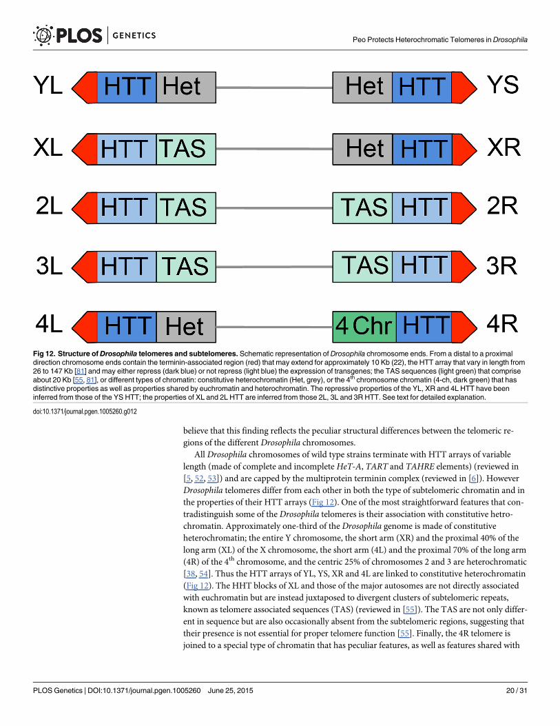
All Drosophila chromosomes of wild type strains terminate with HTT arrays of variablelength (made of complete and incomplete HeT-A, TART and TAHRE elements) (reviewed in[5, 52, 53]) and are capped by the multiprotein terminin complex (reviewed in [6]). HoweverDrosophila telomeres differ from each other in both the type of subtelomeric chromatin and inthe properties of their HTT arrays (Fig 12). One of the most straightforward features that con-tradistinguish some of the Drosophila telomeres is their association with constitutive hetro-chromatin. Approximately one-third of the Drosophila genome is made of constitutiveheterochromatin; the entire Y chromosome, the short arm (XR) and the proximal 40% of thelong arm (XL) of the X chromosome, the short arm (4L) and the proximal 70% of the long arm(4R) of the 4th chromosome, and the centric 25% of chromosomes 2 and 3 are heterochromatic[38, 54]. Thus the HTT arrays of YL, YS, XR and 4L are linked to constitutive heterochromatin(Fig 12). The HHT blocks of XL and those of the major autosomes are not directly associatedwith euchromatin but are instead juxtaposed to divergent clusters of subtelomeric repeats,known as telomere associated sequences (TAS) (reviewed in [55]). The TAS are not only differ-ent in sequence but are also occasionally absent from the subtelomeric regions, suggesting thattheir presence is not essential for proper telomere function [55]. Finally, the 4R telomere isjoined to a special type of chromatin that has peculiar features, as well as features shared with
Fig 12. Structure ofDrosophila telomeres and subtelomeres. Schematic representation of Drosophila chromosome ends. From a distal to a proximaldirection chromosome ends contain the terminin-associated region (red) that may extend for approximately 10 Kb (22), the HTT array that vary in length from26 to 147 Kb [81] and may either repress (dark blue) or not repress (light blue) the expression of transgenes; the TAS sequences (light green) that compriseabout 20 Kb [55, 81], or different types of chromatin: constitutive heterochromatin (Het, grey), or the 4th chromosome chromatin (4-ch, dark green) that hasdistinctive properties as well as properties shared by euchromatin and heterochromatin. The repressive properties of the YL, XR and 4L HTT have beeninferred from those of the YS HTT; the properties of XL and 2L HTT are inferred from those 2L, 3L and 3R HTT. See text for detailed explanation.
doi:10.1371/journal.pgen.1005260.g012
Peo Protects Heterochromatic Telomeres in Drosophila
PLOSGenetics | DOI:10.1371/journal.pgen.1005260 June 25, 2015 20 / 31

both euchromatin and heterochromatin; for example the 4R distal chromatin is enriched in the4th chromosome-specific Painting of four (Pof) protein and the heterochromatic markersHP1a and histone 3 methylated at lysine 9 (H3K9) (reviewed in [56]) (Fig 12).
Additional features that differentiate Drosophila telomeres are the silencing properties oftheir HTT arrays (Fig 12). Several studies have shown that white+ transgenes inserted within ornext to the TAS are partially silenced leading to a variegated eye phenotype (a phenomenonknown as telomere position effect or TPE) (reviewed in [5]). However, white+ transgenes in-serted into the HTT arrays behave differently depending on the insertion site. Insertions intothe HTTs of telomeres not directly joined to heterochromatin such as those of the 2L, 3L and3R arms do not appear to be subject to TPE [57]. In contrast white+ transgenes inserted withinthe 4R (only one tested) and YS (6 tested) HTTs lead to a variegated eye phenotype [57, 58].Furthermore, the white+ transgenes inserted into the HTT array of YS and those embeddedinto or near the TAS respond differently to genetic modifiers. For example, mutations in Su(var)205 (HP1a), which suppress variegation of genes relocated next to constitutive hetero-chromatin (position effect variegation or PEV; reviewed in [59, 60]) do not affect the TAS-as-sociated TPE [5, 55] but strongly suppress the TPE of transgenes inserted into the HTT of YS(58). These results indicate that the chromatin of the YS HTT shares some features with consti-tutive heterochromatin and has different properties from the chromatin that includes the auto-somal HTT arrays and the TAS.
These results suggest that the HTT arrays of XR, YL, and 4th chromosome telomeres haveproperties similar to those of YS, namely they are subject to a “heterochromatinization” processrelated to their particular location. Numerous studies on PEV have shown that proteins thatare typically enriched in the heterochromatin can spread into neighboring euchromatic geneschanging their chromatin composition and packaging and downregulating their expression[59, 60]. Thus, although the properties of the HTT arrays of YL, XR and 4th chromosome havenot been tested, it is quite likely that they are similar to those of YS. This would suggest that ina peomutant background the preferential involvement of these telomeres in TF is a conse-quence of their “heterochromatinization”. This in turn implies that the heterochromatic mark-ers of the HTT regions of the YL, XR and 4h chromosome telomeres extend to the terminin-coated chromosome ends. However, “heterochromatinization” does not extend to the HTT ar-rays at the end of the left arm end of the BSw+y+Y chromosome, because in peomutants thismarked Y forms much fewer Y rings than a normal Y. Our findings on ring Y formation in peomutants also suggest that the different “heterochromatic” telomeres respond differently to Peodepletion. The higher than expected frequency of ring Ys observed in peomutants could be aconsequence of the particularly high fusigenic properties of the Y telomeres.
We would like to note that the “heterochromatinization” of the YL, YS, XR and 4th chromo-some termini is the natural condition of these telomeres and that we only know that this condi-tion makes them more fusigenic than the other Drosophila telomeres. However, we have noinformation on the actual properties of these telomeres. Namely, we do not know the proper-ties of their terminin-associated regions. For example, we do not know whether the terminin-coated chromosome ends of YL, YS, XR and 4L have different silencing properties and differentresponses to PEV and TPE modifiers compared to the terminin-bound regions of the otherchromosome ends.
The mechanism underlying TF formation in peomutantsWe have shown that mutations in peo genetically interact with mutation in genes that encodethe terminin subunits. Consistent with these results, we have also shown that Peo directlybinds terminin and mapped the Peo-HOAP interacting domains. Thus, although Peo does not
Peo Protects Heterochromatic Telomeres in Drosophila
PLOSGenetics | DOI:10.1371/journal.pgen.1005260 June 25, 2015 21 / 31

share the properties of the terminin proteins, it is clearly a component of the Drosophila telo-mere capping machinery. What is then the role of peo in telomere protection? The finding thatmutations in peo do not affect HOAP, Moi and Ver localization at telomeres strongly suggestthat loss of peo function does not cause TFs by affecting terminin recruitment at telomeres.Similarly, the normal accumulation of HP1a at the telomeres of peomutants suggests a telo-mere fusion mechanism independent of HP1a. More in general, the fact that peo, but not theother TF genes (eff, Su(var)205, cav,mre11, rad50, nbs, woc,moi and ver), preferentially affect“heterochromatic” telomeres suggests that peomight either act upstream to these genes orfunction in a telomere protection pathway that does not involve them.
The findings that mutations in peo impair DNA replication and preferentially affect latereplicating “heterochromatic” telomeres raise the possibility that defective telomere duplicationmight be fusigenic in Drosophila. However, there are several reasons that lead us to excludethis possibility. Should it be correct, one would expect that impairment of some of the manyfactors that mediate DNA replication would results in TFs. However, in addition to the aphidi-colin treatment and Blmmutations described here, several additional DNA replication factorshave been described whose loss fails to induce TFs. Hydroxyurea (HU), which blocks DNAreplication by inducing a deoxyribonucleotide triphosphate (dNTP) pool depletion, does notinduce TFs in brain cells [48]. In addition, a number of mutations (or RNAi treatments) dis-rupting different aspect of DNA replication do not results in TFs in Drosophila brain cells or S2tissue culture cells, although they cause more or less extensive chromosome breakage. These in-clude lesions in the genes/RNAs encoding the origin recognition (ORC) and the minichromo-some maintenance (MCM) prereplication complexes, DNA primase, the Cul4 replicationlicensing factor, the replisome components DNA polymerase alpha, Rpa70, Tim2 and PCNA,as well as the chromatin assembly factor Caf1 that assists in loading the histone tetramer afterDNA replication [21, 48, 61–64]. We thus hypothesize that the peo-dependent TFs are generat-ed by a peculiar defect in telomeric DNA replication that creates specific fusigenic lesions. Het-erochromatin replication is likely to be different from that of euchromatin, as it requires notonly DNA duplication but also reinstallment of specific epigenetic markers such as heterochro-matin-associated proteins and histone modifications (reviewed in [65]). It is thus possible thatin the presence of weak peomutations replication of “heterochromatic” telomeres is preferen-tially affected leading to specific Peo-dependent fusigenic lesions concentrated in the XR, the Yand the 4th chromosome ends. In strong peomutants, these fusigenic lesions would be generat-ed also in “euchromatic” telomeres resulting into a more general involvement of telomeres infusion events. However, we cannot exclude the possibility that peo plays a dual function beingindependently required for DNA replication and telomere capping.
In conclusion, we propose that Peo is a Drosophila telomere-capping protein that preferen-tially protects chromosome ends associated with heterochromatic markers. Our results also in-dicate that Peo is required for general DNA replication and most likely also for telomerereplication. However, while it is possible that loss of Peo generates specific fusigenic lesionsduring telomere replication, it is unlikely that a general impairment of Drosophila telomere du-plication leads to telomere fusion. In this respect Drosophila telomeres are similar to humantelomeres, which fail to fuse following defective replication [8, 46, 47, 66].
Materials and Methods
Drosophila strainsThe pendolino1 (peo1)mutation was isolated by a cytological screen of 120 late lethal mutantsmapping to the second chromosome, recovered after I element mobilization by I-R dysgeniccrosses [67]. The late lethal mutations l(2)1527, l(2)2723, the insertion lines p112, p221 and
Peo Protects Heterochromatic Telomeres in Drosophila
PLOSGenetics | DOI:10.1371/journal.pgen.1005260 June 25, 2015 22 / 31

p520 were described previously [30] and were kindly provided by P. Taghert (Washington Uni-versity, MO). The peoh allele was isolated from a cytological screen of a collection of 193 late le-thal mutants that arose in the Zucker’s collection of heavily mutagenized viable lines [68]. TheDf(2R)X3, Df(2R)B5, Df(2R)X1 and Df(2R)BSC298 deletions, the insertion line cbx05704, wereobtained from the Bloomington Stock Center and are described in FlyBase. All peo alleles andthe deficiencies of 2R were balanced over CyO Tb balancer [69]. The eff, Su(var)205, cav,mre11, rad50, nbs, woc,moi and vermutations were previously described [10, 12, 15, 16, 21, 23,24]. The DNA sequences flanking the peop112 insertion were obtained by inverse PCR usingstandard procedures.
To obtain the rescue constructs, the LD08052 full lenght peo cDNA was cloned into pCAS-PER4 transformation vectors. Germline transformation was carried out by the BestGene Com-pany. A pCasper4 [w+, peo+] insertion (with aHsp70 promoter) on the X chromosome wasused to establish the [w+, peo+]; peo1/CyO Tb stock. Animals from this stock were heat-shockedfor 1h at 37°C every day starting from the embryonic stages; we then examined the brains ofnon-Tb third instar larvae for the presence and frequency of TFs and the adult flies for thepresence of w+, peo+; peo1/peo1 individuals. For the rescue experiments we also employed apCASPER4 [w+, peo+-HA] insertion (with a Tubulin promoter) on the third chromosomes. Weestablished [w+, peo+-HA]/TM6C; peo1/CyO Tb and [w+, peo+-HA]/TM6C; peoh/CyO Tb stocksand examined them for TFs in non-Tb larvae. The [w+, peo+-HA]/TM6C; peo1/CyO Tb stockwas also examined for the presence of peo1/peo1 non Cy flies. Information on the genetic mark-ers and balancers used in this study is available at FlyBase (http://flybase.bio.indiana.edu/).Stocks were maintained and crosses were made on standard Drosophilamedium at 25°C.
Anti-Peo antibody production and purificationTo generate anti-Peo polyclonal antibodies, rabbits were immunized with bacterially expressed6His-Peo. Immunization and production of anti-Peo antibodies were carried out by the Agro-Bio Company (France).
To purify the anti-Peo antibody we used a bacterially expressed GST-Peo protein. About1 mg of this tagged protein was run on a polyacrylamide gel and then blotted onto a nitrocellu-lose membrane. The membrane strip containing GST-Peo was cut out, washed in 100 mM gly-cine/HCl pH 2.5 for 5 min, washed for 5 min in TBS, blocked by incubation with 3% BSA for1 hour, and washed again for 4 min in TBS. The membrane was then cut into small pieces andincubated overnight with rotation at 4°C in 2 ml of serum diluted 1:5 in TBS. After centrifuga-tion and removal of supernatant the membrane pieces were washed for 15 min in 50 mMTris/HCl pH 7.5, 500 mMNaCl, for 5 min in 50 mM Tris/HCl pH 7.5, 100 mMNaCl, and for10 min in TBS. The membrane was then incubated for 30 min in 1ml of 100 mM glycine/HClpH 2.5 to elute the antibody. The eluate was then mixed to a proper volume 1M Tris pH 8.8 tobring the final pH to 8.0, and then kept at 4°C before use.
Chromosome cytology and immunostainingDAPI-stained colchicine-treated larval brain chromosomes were prepared according to [10].Preparation and immunostaining of mitotic and polytene chromosomes were carried out as de-scribed previously [10, 20], with minor modifications.
For anti-PCNA immunostaining, dissected larval brains were incubated in PBS with 0.5%Triton X-100 for 3 min, and then fixed in 3.7% formaldehyde according to [70]. Before immu-nostaining, brain squash preparations were Triton-X extracted by incubating the slides in 0.1%Triton X-100 containing PBS (PBT), 2 times for 10 min.
Peo Protects Heterochromatic Telomeres in Drosophila
PLOSGenetics | DOI:10.1371/journal.pgen.1005260 June 25, 2015 23 / 31

The primary antibodies used for immunostaining were: the rabbit anti-Peo described abovediluted 1:10; rabbit anti-HOAP (1:100), mouse anti-HP1a (C1A9; 1:10), and mouse anti-PCNA (1:20; Abcam ab29). The anti-HP1a antibody C1A9 was obtained from the Develop-mental Studies Hybridoma Bank, created by the NICHD of the NIH and maintained at TheUniversity of Iowa, Department of Biology, Iowa City, IA 52242. After an overnight incubationat 4°C with the primary antibody, slides were washed twice in TBS-Tween 0.1% for 15 min andthen incubated for 2 h at room temperature with FITC-conjugated goat anti-mouse (1:20; Jack-son Laboratories), or AlexaFluor 555-conjugated donkey anti-rabbit (1:200; Invitrogen) anti-bodies. All slides were then mounted in Vectashield medium H-1200 with DAPI to stain DNA.in vivo detection and immunostaining of GFP-tagged proteins on polytene chromosomes werecarried out as previously described [24].Chromosome preparations were analyzed using a ZeissAxioplan epifluorescence microscope (CarlZeiss, Obezkochen, Germany), equipped with acooled CCD camera (CoolSnap HQ, Photometrics, Woburn, MA). Gray-scale digital imageswere collected separately, converted to Photoshop format, pseudocolored, and merged.
To quantify the polytene chromosome fluorescence intensity after Peo immunostaining, weused the ImageJ software (National Institute of Mental Health, Bethesda, Maryland, USA).Given that the distribution of fluorescent bands along the chromosomes was rather uniform,for each polytene nucleus we selected 3–4 different chromosome regions of similar length, andmeasured both their fluorescence and the fluorescence of a close chromosome-free region tocorrect for background fluorescence. For each genotype (wild type, +/Df, peoh/Df and peo1/Df)shown in Fig 8 we measured at least 30 polytene regions from at least 10 nuclei.
Yeast two-hybrid assayThe coding sequences of Peo, Hp1a and HOAP were PCR-amplified and cloned into pGBKT7or pGAD-T7 vectors (Clontech). The S. cerevisiae AH109 strain was transformed with the indi-cated combinations of plasmids and assayed for growth on SD/–His/–Trp/–Leu selection platessupplemented with 20 mM 3-amino-1,2,4-triazole (3-AT), according to the manufacturer’sinstructions.
Purification of recombinant proteinsTo obtain the GST-Moi, GST-Ver, GST-HOAP and GST-Peo fusion proteins, the correspond-ing full length cDNAs were cloned in either pGEX-6P1 or pGEX-3X, expressed in bacteria andpurified as described previously [24]. The GST-Peo1, GST-Peo2, GST-Peo3, GST-HOAP Δ3,GST-HOAP Δ2, 3, GST-HOAP Δ1–3 and GST-HOAP ΔN-TERM truncated proteins were ob-tained by cloning the corresponding PCR-generated sequences in pGEX-6P1; bacterially ex-pressed GST fusion proteins were then purified by incubating crude lysates with glutathionesepharose beads (QIAGEN) as recommended by the manufacturer. To generate 6His-Peo, theLD08052 peo full-length cDNA was cloned into the pQE32 expression vector (QIAGEN), andexpressed in bacteria; 6His-Peo was affinity purified with a Ni-NTA resin using standardprocedures.
Protein extract preparation, GST pulldown and western blotTo obtain extracts for Western Blot analysis, 50 dissected third instar larval brains were lysedin an ice-cold buffer containing 20 mMHepes KOH pH 7.9, 1.5 mMMgCl2, 10 mM KCl,420 mMNaCl, 30 mM NaF, 0.2 mMNa3VO4, 25 mM BGP, 0.5 M PMSF, 0.1% NP40, and 1Xprotease inhibitor cocktail (Roche). To obtain HOAP-FLAG, Ver-FLAG and Peo-FLAG ex-pressing S2 cells, cav, ver or peo cDNAs were cloned in the pAWF vector (DGRC) in framewith the FLAG-coding sequence. For the expression of Moi-HA, themoi full lenght cDNA was
Peo Protects Heterochromatic Telomeres in Drosophila
PLOSGenetics | DOI:10.1371/journal.pgen.1005260 June 25, 2015 24 / 31

fused in frame with the HA-coding sequence and then cloned into a pCASPER4 vector. All con-structs were transfected in S2 tissue culture cells using Cellfectin (Invitrogen), and cells wereharvested 72 h after transfection. Extracts were lysed in 20 mM Tris pH 8.0, 420 mMNaCl,1 mMMgCl2, 1 mM DTT, 0.1% NP40, and 1X protease inhibitor cocktail (Roche). For prepa-ration of human cell extracts, HeLa cells expressing Peo-FLAG cloned in the pCDNA vectorwere harvested after 72hr transfection and lysated in 20 mM Tris pH 8.0, 420 mMNaCl, 1 mMMgCl2, 1 mM DTT, 0.1% NP40 and 1X protease inhibitor cocktail (Roche).
For GST-pulldown assays, protein extracts were incubated with 2 μg of each GST fusionprotein bound to sepharose beads in a buffer containing 20 mMHepes KOH, 20 mMNaF and0.8% NP40 for 1h at 4°C. Sepharose-bound GST proteins were collected by centrifugation,washed several times with 20 mMHepes KOH, 20 mMNaF and 1.8% NP40, and resuspendedin Laemli buffer in a 30μl final volume for Western Blot analysis. For immunoblotting, proteinsamples were run into SDS polyacrilammide gels and electro-blotted on a nitrocellulose mem-brane (Bio-Rad) in a phosphate buffer (390 mMNaH2PO4 /610 mMNa2HPO4). For the detec-tion of HOAP-FLAG, Ver- FLAG, Peo- FLAG, and Moi-HA, membranes were probed withanti-FLAG HRP-conjugated (1:1000; Roche), and anti-HA HRP-conjugated (1:500; Roche) an-tibodies; Peo and His-Peo were detected with our rabbit anti-Peo (1:100), and Giotto with ouranti-Giotto (1:5000; [71]). Secondary antibodies were sheep anti-mouse IgG HRP-conjugated(1:5000), or donkey anti-rabbit IgG HRP-conjugated (1:5000) (both from Amersham Biosci-ences). The blots were developed using the ECL or ECL Plus method (Amersham Biosciences)and signals were detected with the ChemiDoc scanning system (BioRad). Band intensities werequantified using the image acquisition and analysis Image lab 4.0.1 software (Biorad).
EdU incorporation and stainingEdU labeling was performed as per the manufacturer's instructions (Invitrogen, ClickiT AlexaFluor 488 Imaging kit). Larval brains were cultured with 10μM EdU in 1 X PBS for 60 minprior to fixation and detection.
Aphidicolin treatmentBrains from third instar larvae were dissected in saline (0.7% NaCl), incubated in saline with110 μMAphidicolin (APH) for 1.5 hours, rinsed in saline, and then transferred into a 33 mmPetri dish containing 3 ml of Schneider’s medium (SIGMA) supplemented with 10% fetal bo-vine serum (FBS, Gibco BRL) for 1, 2 or 3h. Colchicine at a final concentration of 10-5M wasadded to the medium 1 hour before fixation according to [10]. Control wild type and peoh/peoh
brains were incubated in saline without APH for 1.5, and processed like the APH-treatedbrains; they were fixed after 2 hours incubation n the medium.
Bioinformatic analysis of the Peo structureAs a first step towards the construction of a three-dimensional model of Peo, we used its full-length sequence (Accession code: Q7K4V4) as a query to search the UniProtKB database(http://www.uniprot.org/) using CSI-BLAST [72], with an Expectation (E) value threshold of10–5. Iterative searches of the database yielded 59 unique sequences. The retrieved sequenceswere aligned with the CLUSTALW software [73] with default parameters. The multiple se-quence alignment (MSA) was next used as a seed to construct a Hidden Markov Model(HMM) of the family.
The HMM was employed to search the Pfam database (http://pfam.sanger.ac.uk/) via theHHpred server [74]. The highest scoring hit was the ubiquitin-conjugating enzyme (UBC)family also known as the E2 enzyme family [34] (probability to be a true positive more than to
Peo Protects Heterochromatic Telomeres in Drosophila
PLOSGenetics | DOI:10.1371/journal.pgen.1005260 June 25, 2015 25 / 31

99%, E-value equal to 1.2 x10-42). The third scoring hit was the ubiquitin E2 variant (UEV)family (Pfam ID: PF05743) that includes UBC homologs such as Tsg101, Mms2 and UEV1(probability to be a true positive equal to 96.44%, E-value equal to 1.3 x10⁻⁴). Peo belongs tothe UEV family because it contains an aspartic acid residue (at position 106, according to Swis-sProt numbering) in place of the E2 active site cysteine, and it is unable to catalyze ubiquitintransfer as it lacks the cysteine that forms a transient thioester bond with the C-terminus ofubiquitin (Ub).
Prediction of potentially disordered regions using the GeneSilico MetaDisorder server(http://iimcb.genesilico.pl/metadisorder/) revealed that at the C-terminus of Peo there is astretch of ~ 70 aa (from residue 177 to 244, according SwissProt numbering) that has the ten-dency to be intrinsically disordered (i.e. lack a unique three dimensional structure at least inthe absence of a binding partner), while the region including residues 16–176 shows propensityto form a folded globular domain with a well-defined pattern of secondary structures as re-vealed by the Quick2d web server analysis [75].
Because no homologous structure with sufficiently high sequence identity with Peo is avail-able, we performed the Peo modeling using the composite approach implemented in I-TASSERserver (Iterative Threading ASSEmbly Refinement) [76].The Peo sequence from residue 16 to176 (predicted to fold in a globular domain) was submitted to the server and the model withthe best confidence score (C-score = 0.5) returned by I-TASSER was selected. We added hydro-gen atoms in this model using HAAD software [77] and refined it close to the native structureusing FG-MD molecular dynamics based algorithm [78].
Our final refined model of Peo was evaluated as a potentially extremely good model (with apredicted LGscore of 2.50) by the PRO-Q model quality assessment program [79]. TheQMEAN score [80] was 0.6 (the variability range is 0–1, with 1 being a perfect model). Collec-tively these parameters indicate that the Peo three-dimensional model is sufficiently accuratefor making functional inferences.
Supporting InformationS1 Table. Patterns of telomere fusions (TFs) in different mutants defective in telomere pro-tection. FTs, fused telomeres (each TF involves 2 pairs FTs); A, major autosomes, namely 2L,2R, 3L and 3R arms; XL and XR, left and right arm of the X chromosome, respectively; 4th,fourth chromosomes, 4L and 4R arms; Y chromosome, YL and YS arms; Eu FTs, fused"euchromatic telomeres" (A and XL); Het FTs, fused "heterochromatic telomeres" (XR, 4th,and Y). The expected numbers of FTs have been calculated on the basis of the expected fre-quencies from a random involvement of telomeres in fusions events (Males, A, 50%; XL,6.25%; XR, 6.25%; 4th, 25%; Y, 12.5%. Females, A, 50%; XL, 12.5%; XR, 12.5%; 4th, 25%). Withthe exception of those highlighted in blue, all the differences between the observed and ex-pected numbers of FTs are statistically significant in the Chi-square test. The differences be-tween the global numbers of Eu FTs and Het FTs are all significant with p< 0.001. In peomutants, the numbers of all types of heterochromatic telomeres involved in fusion events aresignificantly higher than expected, while those of euchromatic telomeres are significantlylower. In ver, cav,moi, Su(var)205, tefu, woc, nbs,mre11, and effmutants there is an oppositepattern of telomere fusions, with a few exceptions. The numbers highlighted in blue are not sig-nificantly different; those highlighted in yellow are significantly different, but the observed dif-ferences are at odds with the general pattern of telomere fusions observed in these mutants.(DOCX)
S1 Fig. peo interacts with HOAP in the yeast two-hybrid assay. Protein interactions betweenPeo and HOAP or HP1a were tested by the yeast two-hybrid assay. The GAL4 DNA-binding
Peo Protects Heterochromatic Telomeres in Drosophila
PLOSGenetics | DOI:10.1371/journal.pgen.1005260 June 25, 2015 26 / 31

domain (BD) and GAL4 activation domain (AD) were fused to the indicated proteins. Cells ex-pressing the indicated combinations of bait (BD) and prey (AD) fusion proteins were plated onmedium lacking Leucine and Tryptophan (-LT). The presence of physical interactions is re-vealed by growth on plates lacking Histidine and supplemented with 3-AT (-LTH +20 mM3-AT). The interaction between HP1a and HOAP was used as a positive control.(TIF)
S2 Fig. The predicted Peo structure and its comparison with those hUBC13, hUEV2,hUEV1 and AKTIP. (A) Alignment of the amino acid sequence of Peo, hUEV1, hUEV2,hUBC13 and AKTIP. Secondary structure elements predicted for Peo are shown above thealignment. Red and blue arrowheads indicate the sites of the catalytic Cys (Asp in Peo) and theHPNmotif (HPH in Peo), respectively. The red broken line indicates the predicted intrinsicallydisordered portion of Peo. (B) A three-dimensional molecular model for Peo. The arrows pointto the N terminus and to the starting site of the disordered C-terminal region (not depicted);the variant Asp residue and His-Pro-His motif are represented as sticks and indicated by redand purple arrows.(TIF)
S3 Fig. The D.melanogaster telomere-capping proteins exhibit different degrees of evolu-tionary conservation. The graph shows the percentages of identity between the D.melanoga-ster proteins and the homologous proteins form 11 Drosophila species (D.mel, D.melanogaster; D. sim, D. simulans; D. sec, D. sechellia; D. yak, D. yakuba; D. ere, D. erecta; D.ana, D. ananassae, D. pse, D. pseudoobscura; D. per, D. persimilis; D. wil, D. willistoni; D.moj,D.mojavensis; D. vir, D. virilis; D. grim, D. grimshawi). The identity percentage is the percent-age of matches between two amino acid sequences, calculated using the pairwise alignmentEMBOSS Needle Software. Note that the terminin components HOAP, Moi, Ver and HipHopare poorly conserved, whereas Peo exhibits a high degree of conservation comparable to that ofnonterminin proteins such as Woc or HP1a.(TIF)
AcknowledgmentsWe thank P. Dimitri for providing us with the lethal mutant collection from which we isolatedpeo1, Carlo Santolamazza and Giorgio Belloni for their assistance during the early stages of thiswork. We also thank Claudia Berdini for help in the construction and expression of the GST-HOAP truncations. Further thanks are due to Dan L. Lindsley for the late lethal collection usedto isolate the peoh allele, to Paul Taghert for the peomutant alleles, and to Gianni Prosseda foradvice on peo translation initiation sites.
Author ContributionsConceived and designed the experiments: GC LC GDRMG. Performed the experiments: GCLCMMGDR PM. Analyzed the data: LC MMGDR PM RB IS. Wrote the paper: GC MG. Per-formed a bioinformatic analysis of the Peo structure: DR.
References1. PalmW, de Lange T. How shelterin protects mammalian telomeres. Annual Review of Genetics. 2008;
42:301–34. doi: 10.1146/annurev.genet.41.110306.130350 PMID: 18680434
2. O'Sullivan RJ, Karlseder J. Telomeres: protecting chromosomes against genome instability. Nature re-views Molecular Cell Biology. 2010; 11:171–81. doi: 10.1038/nrm2848 PMID: 20125188
Peo Protects Heterochromatic Telomeres in Drosophila
PLOSGenetics | DOI:10.1371/journal.pgen.1005260 June 25, 2015 27 / 31

3. Jain D, Cooper JP. Telomeric strategies: means to an end. Annual Review of Genetics. 2010; 44:243–69. doi: 10.1146/annurev-genet-102108-134841 PMID: 21047259
4. Stewart JA, Chaiken MF, Wang F, Price CM. Maintaining the end: roles of telomere proteins in end-pro-tection, telomere replication and length regulation. Mutation Research. 2012; 730:12–9. doi: 10.1016/j.mrfmmm.2011.08.011 PMID: 21945241
5. Mason JM, Frydrychova RC, Biessmann H. Drosophila telomeres: an exception providing new insights.BioEssays. 2008; 30:25–37. PMID: 18081009
6. Raffa GD, Cenci G, Ciapponi L, Gatti M. Organization and Evolution of Drosophila Terminin: Similaritiesand Differences between Drosophila and Human Telomeres. Frontiers in Oncology. 2013; 3:112. doi:10.3389/fonc.2013.00112 PMID: 23675571
7. Rong YS. Telomere capping in Drosophila: dealing with chromosome ends that most resemble DNAbreaks. Chromosoma. 2008; 117:235–42. doi: 10.1007/s00412-007-0144-2 PMID: 18193446
8. Leman AR, Dheekollu J, Deng Z, Lee SW, Das MM, Lieberman PM, et al. Timeless preserves telomerelength by promoting efficient DNA replication through human telomeres. Cell Cycle. 2012; 11:2337–47.doi: 10.4161/cc.20810 PMID: 22672906
9. Vannier JB, Pavicic-Kaltenbrunner V, Petalcorin MI, Ding H, Boulton SJ. RTEL1 dismantles T loopsand counteracts telomeric G4-DNA to maintain telomere integrity. Cell. 2012; 149:795–806. doi: 10.1016/j.cell.2012.03.030 PMID: 22579284
10. Cenci G, Rawson RB, Belloni G, Castrillon DH, Tudor M, Petrucci R, et al. UbcD1, a Drosophila ubiqui-tin-conjugating enzyme required for proper telomere behavior. Genes Dev. 1997; 11:863–75. PMID:9106658
11. Cipressa F, Romano S, Centonze S, Zur Lage PI, Verni F, Dimitri P, et al. Effete, a Drosophila Chroma-tin-Associated Ubiquitin-Conjugating Enzyme that Affects Telomeric and Heterochromatic Position Ef-fect Variegation. Genetics. 2013; 195:147–58 doi: 10.1534/genetics.113.153320 PMID: 23821599
12. Fanti L, Giovinazzo G, Berloco M, Pimpinelli S. The heterochromatin protein 1 prevents telomere fu-sions in Drosophila. Mol Cell. 1998; 2:527–38. PMID: 9844626
13. Bi X, Srikanta D, Fanti L, Pimpinelli S, Badugu R, Kellum R, et al. Drosophila ATM and ATR checkpointkinases control partially redundant pathways for telomere maintenance. Proc Natl Acad Sci U S A.2005; 102:15167–72. PMID: 16203987
14. Bi X, Wei SC, Rong YS. Telomere protection without a telomerase; the role of ATM and Mre11 in Dro-sophila telomere maintenance. Curr Biol. 2004; 14:1348–53. PMID: 15296751
15. Ciapponi L, Cenci G, Ducau J, Flores C, Johnson-Schlitz D, Gorski MM, et al. The Drosophila Mre11/Rad50 complex is required to prevent both telomeric fusion and chromosome breakage. Curr Biol.2004; 14:1360–6. PMID: 15296753
16. Ciapponi L, Cenci G, Gatti M. The Drosophila Nbs protein functions in multiple pathways for the mainte-nance of genome stability. Genetics. 2006. 173:1447–54 PMID: 16648644
17. Oikemus SR, McGinnis N, Queiroz-Machado J, Tukachinsky H, Takada S, Sunkel CE, et al. Drosophilaatm/telomere fusion is required for telomeric localization of HP1 and telomere position effect. GenesDev. 2004; 18:1850–61. PMID: 15256487
18. Oikemus SR, Queiroz-Machado J, Lai K, McGinnis N, Sunkel C, Brodsky MH. Epigenetic telomere pro-tection by Drosophila DNA damage response pathways. PLoS Genet. 2006; 2:e71. PMID: 16710445
19. Silva E, Tiong S, Pedersen M, Homola E, Royou A, Fasulo B, et al. ATM is required for telomere main-tenance and chromosome stability during Drosophila development. Curr Biol. 2004; 14:1341–7. PMID:15296750
20. Raffa GD, Cenci G, Siriaco G, Goldberg ML, Gatti M. The putative Drosophila transcription factor woc isrequired to prevent telomeric fusions. Mol Cell. 2005; 20:821–31. PMID: 16364909
21. Cenci G, Siriaco G, Raffa GD, Kellum R, Gatti M. The Drosophila HOAP protein is required for telomerecapping. Nat Cell Biol. 2003; 5:82–4. PMID: 12510197
22. Gao G, Walser JC, Beaucher ML, Morciano P, Wesolowska N, Chen J, et al. HipHop interacts withHOAP and HP1 to protect Drosophila telomeres in a sequence-independent manner. The EMBO Jour-nal. 2010; 29:819–29. doi: 10.1038/emboj.2009.394 PMID: 20057353
23. Raffa GD, Raimondo D, Sorino C, Cugusi S, Cenci G, Cacchione S, et al. Verrocchio, a Drosophila OBfold-containing protein, is a component of the terminin telomere-capping complex. Genes Dev. 2010;24:1596–601. doi: 10.1101/gad.574810 PMID: 20679394
24. Raffa GD, Siriaco G, Cugusi S, Ciapponi L, Cenci G, Wojcik E, et al. The Drosophila modigliani (moi)gene encodes a HOAP-interacting protein required for telomere protection. Proc Natl Acad Sci U S A.2009; 106:2271–6. doi: 10.1073/pnas.0812702106 PMID: 19181850
Peo Protects Heterochromatic Telomeres in Drosophila
PLOSGenetics | DOI:10.1371/journal.pgen.1005260 June 25, 2015 28 / 31

25. Raffa GD, Ciapponi L, Cenci G, Gatti M. Terminin: a protein complex that mediates epigenetic mainte-nance of Drosophila telomeres. Nucleus. 2011; 2:383–91. doi: http://dx.doi.org/10.4161/nucl.2.5.17873PMID: 21989238
26. Shareef MM, King C, Damaj M, Badagu R, Huang DW, Kellum R. Drosophila heterochromatin protein 1(HP1)/origin recognition complex (ORC) protein is associated with HP1 and ORC and functions in het-erochromatin-induced silencing. Mol Biol Cell. 2001; 12:1671–85. PMID: 11408576
27. Drosopoulos WC, Kosiyatrakul ST, Yan Z, Calderano SG, Schildkraut CL. Human telomeres replicateusing chromosome-specific, rather than universal, replication programs. The Journal of Cell Biology.2012; 197:253–66. doi: 10.1083/jcb.201112083 PMID: 22508510
28. Arnoult N, Schluth-Bolard C, Letessier A, Drascovic I, Bouarich-Bourimi R, Campisi J, et al. Replicationtiming of human telomeres is chromosome arm-specific, influenced by subtelomeric structures andconnected to nuclear localization. PLoS Genet. 2010; 6:e1000920. doi: 10.1371/journal.pgen.1000920PMID: 20421929
29. Novo C, Arnoult N, BordesWY, Castro-Vega L, Gibaud A, Dutrillaux B, et al. The heterochromatic chro-mosome caps in great apes impact telomere metabolism. Nucleic acids research. 2013; 41:4792–801.doi: 10.1093/nar/gkt169 PMID: 23519615
30. O'Brien MA, Roberts MS, Taghert PH. A genetic and molecular analysis of the 46C chromosomal re-gion surrounding the FMRFamide neuropeptide gene in Drosophila melanogaster. Genetics. 1994;137:121–37. PMID: 8056304
31. Levis R, Hazelrigg T, Rubin GM. Separable cis-acting control elements for expression of the white geneof Drosophila. The EMBO Journal. 1985; 4:3489–99. PMID: 3004962
32. Castrillon DH, Gonczy P, Alexander S, Rawson R, Eberhart CG, Viswanathan S, et al. Toward a molec-ular genetic analysis of spermatogenesis in Drosophila melanogaster: characterization of male-sterilemutants generated by single P element mutagenesis. Genetics. 1993; 135:489–505. PMID: 8244010
33. Fabrizio JJ, Hime G, Lemmon SK, Bazinet C. Genetic dissection of sperm individualization in Drosophi-la melanogaster. Development. 1998; 125:1833–43. PMID: 9550716
34. vanWijk SJ, Timmers HT. The family of ubiquitin-conjugating enzymes (E2s): deciding between lifeand death of proteins. FASEB Journal. 2010; 24:981–93. doi: 10.1096/fj.09-136259 PMID: 19940261
35. de Lange T. Protection of mammalian telomeres. Oncogene. 2002; 21:532–40. PMID: 11850778
36. Csink AK, Henikoff S. Large-scale chromosomal movements during interphase progression in Dro-sophila. The Journal of Cell Biology. 1998; 143:13–22. PMID: 9763417
37. Konishi A, de Lange T. Cell cycle control of telomere protection and NHEJ revealed by a ts mutation inthe DNA-binding domain of TRF2. Genes Dev. 2008; 22:1221–30. doi: 10.1101/gad.1634008 PMID:18451109
38. Gatti M, Pimpinelli S. Functional elements in Drosophila melanogaster heterochromatin. Annual Re-view of Genetics. 1992; 26:239–75. PMID: 1482113
39. Lindsley DL, ZimmGG. The Genome of Drosophila melanogaster. 1250 Sixth Avenue, San Diego, Cal-ifornia 92101–4311: Academic Press, Inc.; 1992.
40. Ye Y, Rape M. Building ubiquitin chains: E2 enzymes at work. Nature Reviews Molecular Cell biology.2009; 10:755–64. doi: 10.1038/nrm2780 PMID: 19851334
41. Cottee PA, Abs ELOYG, Nisbet AJ, Gasser RB. Ubiquitin-conjugating enzyme genes in Oesophagos-tomum dentatum. Parasitology Research. 2006; 99:119–25. PMID: 16518612
42. Fanti L, Pimpinelli S. HP1: a functionally multifaceted protein. Current opinion in genetics & develop-ment. 2008; 18:169–74.
43. Burla R, Carcuro M, Raffa GD, Galati A, Raimondo D, Rizzo A, et al. AKTIP/Ft1, a new shelterin-inter-acting factor required for telomere maintenance. PLoS Genet. 2015; 11:e1005167. doi: 10.1371/journal.pgen.1005167
44. Chagin VO, Stear JH, Cardoso MC. Organization of DNA replication. Cold Spring Harbor perspectivesin biology. 2010; 2:a000737. doi: 10.1101/cshperspect.a000737 PMID: 20452942
45. Ghosh AK, Rossi ML, Singh DK, Dunn C, Ramamoorthy M, Croteau DL, et al. RECQL4, the protein mu-tated in Rothmund-Thomson syndrome, functions in telomere maintenance. The Journal of BiologicalChemistry. 2012; 287:196–209. doi: 10.1074/jbc.M111.295063 PMID: 22039056
46. Barefield C, Karlseder J. The BLM helicase contributes to telomere maintenance through processing oflate-replicating intermediate structures. Nucleic Acids Research. 2012; 40:7358–67. doi: 10.1093/nar/gks407 PMID: 22576367
47. Sfeir A, Kosiyatrakul ST, Hockemeyer D, MacRae SL, Karlseder J, Schildkraut CL, et al. Mammaliantelomeres resemble fragile sites and require TRF1 for efficient replication. Cell. 2009; 138:90–103. doi:10.1016/j.cell.2009.06.021 PMID: 19596237
Peo Protects Heterochromatic Telomeres in Drosophila
PLOSGenetics | DOI:10.1371/journal.pgen.1005260 June 25, 2015 29 / 31

48. Benna C, Bonaccorsi S, Wulbeck C, Helfrich-Forster C, Gatti M, Kyriacou CP, et al. Drosophila time-less2 is required for chromosome stability and circadian photoreception. Curr Biol. 2010; 20:346–52.doi: 10.1016/j.cub.2009.12.048 PMID: 20153199
49. Gotter AL, Suppa C, Emanuel BS. Mammalian TIMELESS and Tipin are evolutionarily conserved repli-cation fork-associated factors. Journal of Molecular Biology. 2007; 366:36–52. PMID: 17141802
50. Kondratov RV, Antoch MP. Circadian proteins in the regulation of cell cycle and genotoxic stress re-sponses. Trends in Cell Biology. 2007; 17:311–7. PMID: 17644383
51. Kusano K, Johnson-Schlitz DM, Engels WR. Sterility of Drosophila with mutations in the Bloom syn-drome gene—complementation by Ku70. Science. 2001; 291:2600–2. PMID: 11283371
52. Pardue ML, Debaryshe P. Adapting to life at the end of the line: How Drosophila telomeric retrotranspo-sons cope with their job. Mobile Genetic Elements. 2011; 1:128–34. PMID: 22016861
53. Zhang L, Rong YS. Retrotransposons at Drosophila telomeres: host domestication of a selfish elementfor the maintenance of genome integrity. Biochimica et Biophysica Acta. 2012; 1819:771–5. doi: 10.1016/j.bbagrm.2012.01.018 PMID: 22342531
54. Hoskins RA, Carlson JW, Kennedy C, Acevedo D, Evans-Holm M, Frise E, et al. Sequence finishingand mapping of Drosophila melanogaster heterochromatin. Science. 2007; 316:1625–8. PMID:17569867
55. Mason J, Villasante A. Subtelomeres in Drosophila and Other Diptera. In: Louis EJ, Becker MM, editors.Subtelomeres: Springer Berlin Heidelberg; 2014. p. 211–25.
56. Riddle NC, Shaffer CD, Elgin SC. A lot about a little dot—lessons learned from Drosophila melanoga-ster chromosome 4. Biochemistry and Cell Biology. 2009; 87:229–41. doi: 10.1139/O08-119 PMID:19234537
57. Biessmann H, Prasad S, Semeshin VF, Andreyeva EN, Nguyen Q, Walter MF, et al. Two distinct do-mains in Drosophila melanogaster telomeres. Genetics. 2005; 171:1767–77. PMID: 16143601
58. Wang SH, Nan R, Accardo MC, Sentmanat M, Dimitri P, Elgin SC. A distinct type of heterochromatin atthe telomeric region of the Drosophila melanogaster Y chromosome. PLoS One. 2014; 9:e86451. doi:10.1371/journal.pone.0086451 PMID: 24475122
59. Weiler KS, Wakimoto BT. Heterochromatin and gene expression in Drosophila. Annual Review of Ge-netics. 1995; 29:577–605. PMID: 8825487
60. Elgin SC, Reuter G. Position-effect variegation, heterochromatin formation, and gene silencing in Dro-sophila. Cold Spring Harbor Perspectives in Biology. 2013; 5:a017780. doi: 10.1101/cshperspect.a017780 PMID: 23906716
61. Gatti M, Baker BS. Genes controlling essential cell-cycle functions in Drosophila melanogaster. GenesDev. 1989; 3:438–53. PMID: 2498166
62. Loupart ML, Krause SA, Heck MS. Aberrant replication timing induces defective chromosome conden-sation in Drosophila ORC2mutants. Curr Biol. 2000; 10:1547–56. PMID: 11137005
63. SommaMP, Ceprani F, Bucciarelli E, Naim V, De Arcangelis V, Piergentili R, et al. Identification of Dro-sophila mitotic genes by combining co-expression analysis and RNA interference. PLoS Genet. 2008;4:e1000126. doi: 10.1371/journal.pgen.1000126 PMID: 18797514
64. Balasov M, Huijbregts RP, Chesnokov I. Functional analysis of an Orc6 mutant in Drosophila. Proc NatlAcad Sci U S A. 2009; 106:10672–7. doi: 10.1073/pnas.0902670106 PMID: 19541634
65. Wallace JA, Orr-Weaver TL. Replication of heterochromatin: insights into mechanisms of epigenetic in-heritance. Chromosoma. 2005; 114:389–402. PMID: 16220346
66. Martinez P, Thanasoula M, Munoz P, Liao C, Tejera A, McNees C, et al. Increased telomere fragilityand fusions resulting from TRF1 deficiency lead to degenerative pathologies and increased cancer inmice. Genes Dev. 2009; 23:2060–75. doi: 10.1101/gad.543509 PMID: 19679647
67. Dimitri P, Arca B, Berghella L, Mei E. High genetic instability of heterochromatin after transposition ofthe LINE-like I factor in Drosophila melanogaster. Proc Natl Acad Sci U S A. 1997; 94:8052–7. PMID:9223313
68. Koundakjian EJ, Cowan DM, Hardy RW, Becker AH. The Zuker collection: a resource for the analysisof autosomal gene function in Drosophila melanogaster. Genetics. 2004; 167:203–6. PMID: 15166147
69. Lattao R, Bonaccorsi S, Guan X, Wasserman SA, Gatti M. Tubby-tagged balancers for the DrosophilaX and second chromosomes. Fly. 2011; 5:369–70. doi: 10.4161/fly.5.4.17283 PMID: 21785267
70. Bonaccorsi S, Giansanti MG, Gatti M. Spindle assembly in Drosophila neuroblasts and ganglion mothercells. Nat Cell Biol. 2000; 2:54–6. PMID: 10620808
71. Giansanti MG, Bonaccorsi S, Kurek R, Farkas RM, Dimitri P, Fuller MT, et al. The class I PITP giotto isrequired for Drosophila cytokinesis. Curr Biol. 2006; 16:195–201. PMID: 16431372
Peo Protects Heterochromatic Telomeres in Drosophila
PLOSGenetics | DOI:10.1371/journal.pgen.1005260 June 25, 2015 30 / 31

72. Biegert A, Soding J. Sequence context-specific profiles for homology searching. Proc Natl Acad Sci US A. 2009; 106:3770–5. doi: 10.1073/pnas.0810767106 PMID: 19234132
73. Chenna R, Sugawara H, Koike T, Lopez R, Gibson TJ, Higgins DG, et al. Multiple sequence alignmentwith the Clustal series of programs. Nucleic Acids Research. 2003; 31:3497–500. PMID: 12824352
74. Soding J, Biegert A, Lupas AN. The HHpred interactive server for protein homology detection and struc-ture prediction. Nucleic Acids Research. 2005; 33 (Web Server issue):W244–8. PMID: 15980461
75. Biegert A, Mayer C, Remmert M, Soding J, Lupas AN. The MPI Bioinformatics Toolkit for protein se-quence analysis. Nucleic Acids Research. 2006; 34 (Web Server issue):W335–9. PMID: 16845021
76. Roy A, Kucukural A, Zhang Y. I-TASSER: a unified platform for automated protein structure and func-tion prediction. Nature Protocols. 2010; 5:725–38. doi: 10.1038/nprot.2010.5 PMID: 20360767
77. Li Y, Roy A, Zhang Y. HAAD: A quick algorithm for accurate prediction of hydrogen atoms in proteinstructures. PloS One. 2009; 4:e6701. doi: 10.1371/journal.pone.0006701 PMID: 19693270
78. Zhang J, Liang Y, Zhang Y. Atomic-level protein structure refinement using fragment-guided moleculardynamics conformation sampling. Structure. 2011; 19:1784–95. doi: 10.1016/j.str.2011.09.022 PMID:22153501
79. Wallner B, Elofsson A. Identification of correct regions in protein models using structural, alignment,and consensus information. Protein Science. 2006; 15:900–13. PMID: 16522791
80. Benkert P, Biasini M, Schwede T. Toward the estimation of the absolute quality of individual proteinstructure models. Bioinformatics. 2011; 27:343–50. doi: 10.1093/bioinformatics/btq662 PMID:21134891
81. Abad JP, De Pablos B, Osoegawa K, De Jong PJ, Martin-Gallardo A, Villasante A. Genomic analysis ofDrosophila melanogaster telomeres: full-length copies of HeT-A and TART elements at telomeres. Mo-lecular Biology and Evolution. 2004; 21:1613–9. PMID: 15163766
Peo Protects Heterochromatic Telomeres in Drosophila
PLOSGenetics | DOI:10.1371/journal.pgen.1005260 June 25, 2015 31 / 31


![[Regulation of telomeres length: making the telomeres accessible?]](https://static.fdokumen.com/doc/165x107/633f1028d121719806096682/regulation-of-telomeres-length-making-the-telomeres-accessible.jpg)