Calcitonin gene-related peptide enhances release of native brain-derived neurotrophic factor from...
13
Calcitonin gene-related peptide enhances release of native brain-derived neurotrophic factor from trigeminal ganglion neurons Ilya Buldyrev,* , , à ,1 Nathan M. Tanner,* ,1 Hui-ya Hsieh,* Emily G. Dodd,* Loi T. Nguyen* and Agnieszka Balkowiec* , à *Department of Integrative Biosciences, Neurological Sciences Institute and àNeuroscience Graduate Program, Oregon Health and Science University, Portland, Oregon, USA Abstract Activity-dependent plasticity in nociceptive pathways has been implicated in pathomechanisms of chronic pain syn- dromes. Calcitonin gene-related peptide (CGRP), which is expressed by trigeminal nociceptors, has recently been identified as a key player in the mechanism of migraine headaches. Here we show that CGRP is coexpressed with brain-derived neurotrophic factor (BDNF) in a large subset of adult rat trigeminal ganglion neurons in vivo. Using ELISA in situ, we show that CGRP (1–1000 nM) potently enhances BDNF release from cultured trigeminal neurons. The effect of CGRP is dose-dependent and abolished by pretreatment with CGRP receptor antagonist, CGRP(8–37). Intriguingly, CGRP- mediated BDNF release, unlike BDNF release evoked by physiological patterns of electrical stimulation, is independent of extracellular calcium. Depletion of intracellular calcium stores with thapsigargin blocks the CGRP-mediated BDNF release. Using transmission electron microscopy, our study also shows that BDNF-immunoreactivity is present in dense core vesicles of unmyelinated axons and axon terminals in the subnucleus caudalis of the spinal trigeminal nucleus, the pri- mary central target of trigeminal nociceptors. Together, these results reveal a previously unknown role for CGRP in regu- lating BDNF availability, and point to BDNF as a candidate mediator of trigeminal nociceptive plasticity. Keywords: brain-derived neurotrophic factor, calcitonin gene-related peptide, migraine, pain, synaptic plasticity, trigeminal. J. Neurochem. (2006) 99, 1338–1350. Trigeminal sensory pathways, with first-order neurons located in the trigeminal ganglion (TG), are the major carrier of sensory information from the head, and constitute the cranial analog of spinal sensory pathways (Shankland 2000). Previous studies examining cellular mechanisms of synaptic transmission and plasticity in sensory pathways focused on the spinal system, with first-order neurons in dorsal root ganglia, as a model (Moore et al. 2000; Woolf and Salter 2000; Scholz and Woolf 2002). Consequently, cellular mechanisms that underlie synaptic plasticity in trigeminal pathways are poorly understood (Ren and Dubner 1999; Sessle 2000; Pietrobon and Striessnig 2003). TG neurons express and release calcitonin gene-related peptide (CGRP), which has been implicated in modulation of trigeminal sensory information (Samsam et al. 2000; Sam- sam et al. 2001). CGRP is present in 35–50% of all TG neurons, and over 90% of TG cells with diameters below 15 lm (Alvarez et al. 1991; Tajti et al. 1999; Lazarov 2002). Moreover, recent studies have identified several potential mechanisms of chronic craniofacial pain syndromes, inclu- ding migraines, with CGRP as a key mediator. For example, CGRP released from peripheral terminals of trigeminal nociceptors leads to sensitization of nerve endings subse- quent to an inflammatory response (Bolay et al. 2002; Pietrobon and Striessnig 2003). In support, other studies Received April 11, 2006; revised manuscript received June 6, 2006; accepted June 20, 2006. Correspondence should be addressed to Dr Agnieszka Balkowiec, Department of Integrative Biosciences, Oregon Health and Science University School of Dentistry, 611 SW Campus Drive, Portland, OR 97239, USA. E-mail: [email protected] 1 These authors contributed equally to this work and are listed alpha- betically. Abbreviations used: BDNF, brain-derived neurotrophic factor; CGRP, calcitonin gene-related peptide; DRG, dorsal root ganglion; NF, neuro- filament; P, postnatal day; TG, trigeminal ganglion; TTX, tetrodotoxin. Journal of Neurochemistry , 2006, 99, 1338–1350 doi:10.1111/j.1471-4159.2006.04161.x 1338 Journal Compilation Ó 2006 International Society for Neurochemistry, J. Neurochem. (2006) 99, 1338–1350 Ó 2006 The Authors
-
Upload
independent -
Category
Documents
-
view
6 -
download
0
Transcript of Calcitonin gene-related peptide enhances release of native brain-derived neurotrophic factor from...

Calcitonin gene-related peptide enhances release of nativebrain-derived neurotrophic factor from trigeminalganglion neurons
Ilya Buldyrev,*,�,�,1 Nathan M. Tanner,*,1 Hui-ya Hsieh,* Emily G. Dodd,* Loi T. Nguyen* andAgnieszka Balkowiec*,�
*Department of Integrative Biosciences, �Neurological Sciences Institute and �Neuroscience Graduate Program, Oregon Health and
Science University, Portland, Oregon, USA
Abstract
Activity-dependent plasticity in nociceptive pathways has
been implicated in pathomechanisms of chronic pain syn-
dromes. Calcitonin gene-related peptide (CGRP), which is
expressed by trigeminal nociceptors, has recently been
identified as a key player in the mechanism of migraine
headaches. Here we show that CGRP is coexpressed with
brain-derived neurotrophic factor (BDNF) in a large subset of
adult rat trigeminal ganglion neurons in vivo. Using ELISA
in situ, we show that CGRP (1–1000 nM) potently enhances
BDNF release from cultured trigeminal neurons. The effect of
CGRP is dose-dependent and abolished by pretreatment with
CGRP receptor antagonist, CGRP(8–37). Intriguingly, CGRP-
mediated BDNF release, unlike BDNF release evoked by
physiological patterns of electrical stimulation, is independent
of extracellular calcium. Depletion of intracellular calcium
stores with thapsigargin blocks the CGRP-mediated BDNF
release. Using transmission electron microscopy, our study
also shows that BDNF-immunoreactivity is present in dense
core vesicles of unmyelinated axons and axon terminals in the
subnucleus caudalis of the spinal trigeminal nucleus, the pri-
mary central target of trigeminal nociceptors. Together, these
results reveal a previously unknown role for CGRP in regu-
lating BDNF availability, and point to BDNF as a candidate
mediator of trigeminal nociceptive plasticity.
Keywords: brain-derived neurotrophic factor, calcitonin
gene-related peptide, migraine, pain, synaptic plasticity,
trigeminal.
J. Neurochem. (2006) 99, 1338–1350.
Trigeminal sensory pathways, with first-order neuronslocated in the trigeminal ganglion (TG), are the major carrierof sensory information from the head, and constitute thecranial analog of spinal sensory pathways (Shankland 2000).Previous studies examining cellular mechanisms of synaptictransmission and plasticity in sensory pathways focused onthe spinal system, with first-order neurons in dorsal rootganglia, as a model (Moore et al. 2000; Woolf and Salter2000; Scholz and Woolf 2002). Consequently, cellularmechanisms that underlie synaptic plasticity in trigeminalpathways are poorly understood (Ren and Dubner 1999;Sessle 2000; Pietrobon and Striessnig 2003).
TG neurons express and release calcitonin gene-relatedpeptide (CGRP), which has been implicated in modulation oftrigeminal sensory information (Samsam et al. 2000; Sam-sam et al. 2001). CGRP is present in 35–50% of all TGneurons, and over 90% of TG cells with diameters below15 lm (Alvarez et al. 1991; Tajti et al. 1999; Lazarov 2002).
Moreover, recent studies have identified several potentialmechanisms of chronic craniofacial pain syndromes, inclu-ding migraines, with CGRP as a key mediator. For example,CGRP released from peripheral terminals of trigeminalnociceptors leads to sensitization of nerve endings subse-quent to an inflammatory response (Bolay et al. 2002;Pietrobon and Striessnig 2003). In support, other studies
Received April 11, 2006; revised manuscript received June 6, 2006;accepted June 20, 2006.Correspondence should be addressed to Dr Agnieszka Balkowiec,
Department of Integrative Biosciences, Oregon Health and ScienceUniversity School of Dentistry, 611 SW Campus Drive, Portland, OR97239, USA. E-mail: [email protected] authors contributed equally to this work and are listed alpha-betically.Abbreviations used: BDNF, brain-derived neurotrophic factor; CGRP,
calcitonin gene-related peptide; DRG, dorsal root ganglion; NF, neuro-filament; P, postnatal day; TG, trigeminal ganglion; TTX, tetrodotoxin.
Journal of Neurochemistry, 2006, 99, 1338–1350 doi:10.1111/j.1471-4159.2006.04161.x
1338 Journal Compilation � 2006 International Society for Neurochemistry, J. Neurochem. (2006) 99, 1338–1350� 2006 The Authors

have detected increased levels of CGRP in the cerebral andextracerebral circulation during migraine attacks (Goadsbyet al. 1990; Sarchielli et al. 2000). There is also evidence foran activity-dependent release of CGRP from central termi-nals of TG neurons in the brainstem (Samsam et al. 2000;Samsam et al. 2001). However, the role of CGRP at first-order synapses in trigeminal sensory pathways is unknown.
In spinal pathways, activity-dependent plastic changes insynaptic transmission are thought to be mediated by brain-derived neurotrophic factor (BDNF) or other neurotrophins(Mannion et al. 1999; Mendell et al. 1999; Shu and Mendell1999; Shu et al. 1999; Thompson et al. 1999; Moore et al.2000; Lucas et al. 2003; Malcangio and Lessmann 2003).This is based on evidence that BDNF is transported in centralprojections of DRG neurons (Zhou and Rush 1996; Tonra1999), localized to dense-core vesicles within DRG centralaxon terminals (Michael et al. 1997; Luo et al. 2001), andreleased from DRG nociceptors by high intensity stimuli(Lever et al. 2001). In addition, BDNF enhances NMDA(Kerr et al. 1999) and GABA (Pezet et al. 2002) receptor-mediated responses of dorsal horn neurons. Despite theevidence that BDNF mRNA and protein are present in adulttrigeminal ganglia in vivo (Wetmore and Olson 1995; Quartuet al. 1997; Pan et al. 2000), the role of BDNF in activity-dependent modifications of trigeminal sensory transmissionhas never been addressed before.
The present study was designed to test the hypothesis thatBDNF is expressed by CGRP-containing TG neuronsin vivo, and can be released from cultured TG neurons byphysiologically relevant patterns of electrical stimulation andby CGRP.
Portions of this work have previously been published inabstract form (Buldyrev et al. 2004; Nguyen et al. 2004;Tanner et al. 2004).
Materials and methods
Animals
Postnatal day (P) 1, P7 and adult Sprague–Dawley rats (Charles
River Laboratories, Wilmington, MA, USA) were used for this
study. All procedures were approved by the Institutional Animal
Care and Use Committee of the Oregon Health and Science
University, and conformed to the Policies on the Use of Animals andHumans in Neuroscience Research approved by the Society for
Neuroscience.
Preparation of TG cultures
P1 (24–48 h old) and P7 rat pups were deeply anesthetized by
intraperitoneal injection of Euthasol (0.1 mg/kg) and decapitated.
TGs were rapidly and aseptically dissected from each animal (Eckert
et al. 1997) in ice-cold Ca2+/Mg2+-free Dulbecco’s phosphate-
buffered salt solution (Mediatech, Herndon, VA, USA). The ganglia
were next digested in 0.1% crystallized trypsin-3X (Worthington
Biochemical Corp., Lakewood, NJ, USA) for 30 min, followed by a
30-min incubation in 0.2% collagenase (Sigma, St Louis, MO,
USA). Both enzymes were dissolved in Ca2+/Mg2+-free Hanks’
balanced salt solution (Mediatech), and the incubations performed at
37�C in a humidified atmosphere of 5% CO2 and 95% air
(Balkowiec and Katz 2000, 2002). Following the enzymatic
treatment, TGs were rinsed twice: first in 0.1% soybean trypsin
inhibitor (Worthington) dissolved in Ca2+/Mg2+-containing Dul-
becco’s phosphate-buffered salt solution (Mediatech), and next in
plating medium. The tissue was next transferred to the plating
medium and triturated through fire-polished Pasteur pipettes.
Dissociated TG cells were plated in UV-sterilized, 96-well, flat
bottom ELISA plates (MaxiSorp�, Nalge Nunc Int., Naperville, IL,
USA) precoated with anti-BDNF capture antibody (BDNF Emax�ImmunoAssay System, Promega, Madison, WI, USA; Balkowiec
and Katz 2000, 2002), and/or on poly-D-lysine and laminin-coated
glass coverslips (for immunocytochemistry). TG cultures were
grown for 3 days in Neurobasal-A medium supplemented with B-27
serum-free supplement (Life Technologies, Gaithersburg, MD,
USA), 0.5 mM L-glutamine (Life Technologies), 2.5% fetal bovine
serum (HyClone, Logan, UT, USA; except for the studies of the role
of BDNF in survival of TG neurons), 1% Penicillin-Streptomycin-
Neomycin antibiotic mixture (Life Technologies), and, in some
initial experiments, 2.5% Nystatin. The cultures were not supple-
mented with growth factors because other studies from our
laboratory show that TG neurons lose their growth factor depend-
ence for survival within the first 24 h of postnatal development
(Nguyen et al. 2004).In experiments designed to rule out a contribution of non-
neuronal cells to mechanisms of BDNF release, dissociated TG cells
(in 0.5 mL of plating medium) were layered on top of a two-layer
gradient of Percoll (Sigma; 35%/60%) and centrifuged for 10 min at
800 g at 4�C. The neuron–enriched interface of the two Percoll
layers was collected and rinsed twice with culture medium before
plating.
Measurement of BDNF release
BDNF protein was measured with a modified sandwich ELISA,
termed ELISA in situ, as previously described (Balkowiec and Katz
2000, 2002). Briefly, 96-well ELISA plates were UV-sterilized for
20 min and coated with anti-BDNF monoclonal antibody (BDNF
Emax� ImmunoAssay System, Promega) at 4�C for 12–18 h. Next,
plates were washed and blocked, followed by two at least 1- h
incubations with culture medium to remove any residue of the
ELISA washing solution. Then, TG cultures were prepared as
described above, plated in anti-BDNF-coated wells, and grown for
three days (see Results). The BDNF Emax� ImmunoAssay System
(Promega) was used according to the protocol of the manufacturer,
except that the concentration of the anti-BDNF monoclonal and
anti-human BDNF polyclonal antibody was 5 lg/mL and 2 lg/mL,
respectively, and the dilution of the anti-IgY-HRP antibody was
1 : 100. All reagents used prior to cell plating were sterilized with
0.2 lm syringe filters (Acrodisc�., Pall, Ann Arbor, MI, USA or
Millex� GP, Millipore, Carrigtwohill, Ireland). BDNF samples used
to generate the standard curves were incubated in the same plate as
the cells. At the end of the culture period, plates were extensively
washed to remove all cells and cell debris, and the anti-human
BDNF polyclonal antibody was applied, followed by subsequent
steps according to the manufacturer’s protocol. Absorbance values
CGRP-induced BDNF release from trigeminal neurons 1339
� 2006 The AuthorsJournal Compilation � 2006 International Society for Neurochemistry, J. Neurochem. (2006) 99, 1338–1350
were read at 450 nm in a plate reader (Vmax�, Molecular Devices,
Sunnyvale, CA, USA).
Electrical field stimulation of TG neurons
Following an initial 3-day incubation, the neurons were stimulated
in 96-well plates fitted with paired Ag/AgCl electrodes (one pair per
well), connected in parallel (four wells per set) to one of four
independent outputs of the stimulator (MultiStim System; Digitimer,
Welwyn Garden City, UK). The stimulation pattern delivered by
each of the outputs was controlled by the 8-channel programmable
pulse generator (Master-8-cp, AMPI, Jerusalem, Israel). Four
additional wells were also fitted with pairs of electrodes, but were
not connected to the stimulator and served as controls. The plate was
put back to the incubator, and the neurons were stimulated with
biphasic rectangular pulses of 0.5 ms delivered at various patterns
(see Results).
Drugs used
All drugs were obtained from Sigma. Calcitonin gene-related
peptide (CGRP); CGRP (8–37) fragment, x-Conotoxin GVIA,
x-Agatoxin IVA, and tetrodotoxin were dissolved in distilled water,
and used at final concentrations of 0.001–1.0 lM, 3 lM, 1 lM,
0.4 lM and 1.5 lM, respectively. Nimodipine, dantrolene and
thapsigargin were dissolved in ethyl alcohol and used at final
concentrations of 2 lM, 50 lM, and 10 lM, respectively.
Preparation of TGs and TG cultures for BDNF, CGRP and
Neurofilament immunostaining
Adult rats were euthanized and perfused transcardially with
phosphate-buffered saline (PBS), followed by 2% paraformaldehyde
and 0.2% parabenzoquinone in 0.07 M phosphate buffer. TGs were
dissected and postfixed with 2% paraformaldehyde and 0.2%
parabenzoquinone for 30 min, followed by rinsing in PBS,
cryoprotection in 30% sucrose, and embedding in O.C.T. Tissue-
Tek compound (Sakura Finetek USA, Inc., Torrance, CA, USA).
Blocks of tissue were sectioned on a cryostat at 8 lm, and sections
of TGs collected onto glass slides.
Cultures for immunocytochemical staining were fixed with 2%
paraformaldehyde and 0.2% parabenzoquinone in 0.07 M sodium
phosphate buffer, pH 7.4, for 30 min at room temperature. BDNF,
CGRP and Neurofilament immunostaining was performed at room
temperature, using chicken polyclonal anti-BDNF (1 : 100; Pro-
mega), rabbit polyclonal anti-CGRP (1 : 4000; Calbiochem), and
mouse monoclonal anti-neurofilament 68 and 160 (1 : 100; Sigma),
applied for 2 h, followed by biotinylated donkey anti-chicken
(Vector Laboratories) or donkey anti-chicken Cy3 (Jackson
Immunoresearch), goat anti-rabbit Alexa 488 (Invitrogen), and goat
anti-mouse Cy3 (Jackson Immunoresearch), applied for 2 h at
1 : 200 dilutions. To minimize a non-specific staining, the anti-
BDNF was precleared by overnight incubation with vibratome slices
of a 4% paraformaldehyde-fixed adult cerebellum. Control cultures,
in which primary antibody was omitted, were completely devoid of
staining.
Preparation of brainstems for BDNF immunohistochemistry
and transmission electron microscopy
Processing of brainstem tissue was performed as previously
described (Aicher et al. 2003). Adult rats were euthanized and
perfused through the ascending aorta with heparinized saline (1000
units/mL), followed by 3.8% acrolein in 2% paraformaldehyde, and
2% paraformaldehyde in 0.1 M phosphate buffer. The brain was
removed and postfixed with 2% paraformaldehyde for 30 min,
followed by rinsing in 0.1 M phosphate buffer. Blocks of tissue
containing the spinal trigeminal nucleus were sectioned on a
vibrating microtome at 40 lm, and collected into 0.1 M phosphate
buffer. Prior to immunohistochemical processing, the sections were
incubated for 30 min in 1% sodium borohydride solution (to
increase the antigenicity of acrolein-perfused tissues), and 0.5%
bovine serum albumin (to reduce non-specific binding). Next,
sections were cryoprotected (25% sucrose, 3% glycerol in 0.05 M
phosphate buffer for 15 min), followed by brief immersions in
Freon and liquid nitrogen to increase penetration of antibodies into
the surface of the tissue. The tissue sections were incubated in
chicken anti-BDNF polyclonal antibody (1 : 100; Promega) for 40 h
at 4�C. BDNF was visualized using biotinylated donkey anti-
chicken IgG (1 : 200, Vector Laboratories) and the avidin-biotin
detection method (Vectastain, Vector Laboratories). Following the
immunohistochemical procedure, tissue sections were fixed for 1 h
in 2% osmium tetroxide in 0.1 M phosphate buffer, washed for
10 min in 0.1 M phosphate buffer, dehydrated through a graded
series of ethanol, followed by propylene oxide, and propylene
oxide:EMBed (1 : 1) solution overnight. Next, sections were
incubated in EMBed for 2 h, embedded between two sheets of
Aclar plastic, and placed in an oven for 24–48 h at 60�C.Sections containing the spinal trigeminal nucleus were glued to
plastic blocks formed in Beem capsules, and ultrathin sections
(75 nm) through the outer surface of the tissue were collected onto
copper grids. The ultrathin sections were counterstained with uranyl
acetate and Reynolds lead citrate, and examined using an FEI Tecnai
12 electron microscope (FEI Company). Images were captured
using a digital camera (Advanced Microscopy Techniques Corpora-
tion).
Calculations and statistical analysis
BDNF levels were calculated from the standard curve prepared for
each plate, using SOFTmax PRO� vs. 4.3 software (Molecular
Devices; Balkowiec and Katz, 2000). The standard curves were
linear within the range used (0–500 pg/mL) and the quantities of
BDNF in experimental samples were always within the linear range
of the standard curve. Data are expressed as mean ± SE. Samples
were compared using ANOVA followed by Duncan’s multiple
comparison procedure, and p < 0.05 was considered significant.
Results
BDNF protein is expressed by a large subset of TG
neurons, including CGRP-positive cells, in vivo
In dorsal root ganglia, 55% of BDNF-immunoreactiveneurons also show CGRP immunoreactivity, and mostBDNF immunoreactive nerve terminals in the dorsal horncontain CGRP (Luo et al. 2001). To test the hypothesis thatBDNF is coexpressed with CGRP in TG neurons, sections ofTGs from four adult rats (two males and two females; the
1340 I. Buldyrev et al.
Journal Compilation � 2006 International Society for Neurochemistry, J. Neurochem. (2006) 99, 1338–1350� 2006 The Authors

right-side ganglion) were processed for dual BDNF/CGRPimmunohistochemistry (Fig. 1). Two-hundred BDNF-immu-noreactive profiles were counted in each of four distinctregions of the ganglion (the total of 800 profiles in eachanimal). Among all BDNF-positive cells, 34.75 ± 1.56%(n ¼ 4 ganglia) also showed CGRP immunoreactivity.Moreover, there were no significant differences in the extentof BDNF/CGRP colocalization among the four regions ofthe ganglion, associated with each of the three divisionsof the trigeminal nerve that innervate different areas of thehead.
Endogenously expressed BDNF does not support survival
of postnatal TG neurons in vitro
BDNF expressed by sensory neurons can act in an autocrineor paracrine fashion to support their survival (Acheson et al.1995; Robinson et al. 1996). This brings up the possibilitythat BDNF expressed by postnatal TG neurons supportssurvival of these neurons. Dual immunostaining of newbornTG dissociate cultures with anti-BDNF and anti-Neurofila-ment 68 and 160 (NF), a pan-neuronal marker, shows that alarge subset of NF-positive cells is also BDNF positive(Fig. 2a). Therefore, to address the issue of survivaldependence of postnatal TG neurons on endogenous BDNF,we used an in vitro model of P1 and P7 TG neurons grownfor 3 days in the presence or absence of TrkB-Fc, a fusionprotein with BDNF scavenging properties (Shelton et al.1995; Brady et al. 1999). One-half of each, i.e. TrkB-Fc-treated and untreated, cultures were exposed to depolarizingconcentrations of KCl (50 mM), a treatment that can facilitaterelease of endogenous BDNF from neurons (Ghosh et al.1994; Griesbeck et al. 1999). When applied to BDNF-expressing embryonic day (E) 16.5 petrosal ganglionneurons, which depend on BDNF for survival, KCl depolar-
ization is as effective as exogenous BDNF in supportingsurvival of these cells (Brady et al. 1999). Moreover, theKCl-supported survival of E16.5 petrosal ganglion neuronscan be significantly inhibited by treatment with 5 lg/mLTrkB-Fc (Brady et al. 1999). The survival of postnatal TGneurons was not affected by the presence of TrkB-Fc (5 lg/mL) either under normal or depolarizing conditions(Fig. 2b). In addition, neither P1 nor P7 TG neuron survivalwas affected by exogenous BDNF (1–100 ng/mL, data notshown). These data strongly suggest that BDNF expressed bypostnatal TG neurons serves functions other than supportingsurvival of these cells.
BDNF immunoreactivity is present in dense core vesicles
of unmyelinated axons and terminals in the subnucleus
caudalis of the spinal trigeminal nucleus
BDNF can be anterogradely transported in central axons ofsensory neurons (Zhou and Rush 1996). Therefore, BDNFexpressed in cell bodies of TG neurons could be transportedto their central terminals. To begin addressing this possibility,we have examined at the electron microscopy level thedistribution of BDNF immunoreactivity in central targets ofTG neurons. TG neurons project to second-order neuronsin trigeminal nuclei that extend from the rostral midbrain toupper segments of the cervical spinal cord. We focused ouranalysis on the subnucleus caudalis of the spinal trigeminalnucleus, which plays the most significant role in processingof trigeminal nociceptive information (Sessle 2000; Kyrka-nides et al. 2002). A total of 208 structures displaying BDNFimmunoreactivity were examined in the outer lamini of thesubnucleus caudalis derived from 3 male and 3 female adultrats. The vast majority of BDNF immunoreactivity wasfound in unmyelinated axons and nerve terminals (Fig. 3a).Unmyelinated axons and terminals together constituted a
Fig. 1 BDNF protein is expressed by a large subset of adult rat
trigeminal ganglion (TG) neurons, including CGRP-immunoreactive
cells. Micrographs of a single 8-lm section of an adult TG double-
immunostained for BDNF (aBDNF) and CGRP (aCGRP). A large
number of BDNF-immunoreactive cells can be seen throughout the
section. An overlay image (aBDNF/aCGRP) shows that many CGRP-
positive cells also express BDNF immunoreactivity (arrows). Scale bar
100 lm.
CGRP-induced BDNF release from trigeminal neurons 1341
� 2006 The AuthorsJournal Compilation � 2006 International Society for Neurochemistry, J. Neurochem. (2006) 99, 1338–1350

significant 73% of all BDNF-positive profiles. Moreover,16% of the BDNF-immunoreactivity in this group waslocalized to organelles identified as dense core vesicles basedon their dimensions (i.e. 80–200 nm), whereas the rest of thepresynaptic staining could not be assigned to specificintracellular structures. The remaining 27% of BDNF-immunoreactive profiles were postsynaptic and non-neuronal(Fig. 3b).
Endogenous BDNF is released from cultured newborn
TG neurons by physiologically relevant modes of
stimulation in a pattern-dependent manner
The predominant presynaptic localization of BDNF immu-noreactivity in the spinal trigeminal nucleus suggests thatBDNF is present in, and can be released from, centralterminals of TG neurons. Moreover, the presence of BDNFin dense core vesicle-like structures supports the possibilitythat BDNF release from TG neurons can be regulated byneuronal activity, as for other peptide neuromodulators. Ourprevious studies, using a sensitive ELISA in situ technique,
demonstrated that the magnitude of endogenous BDNFrelease from visceral sensory (Balkowiec and Katz 2000) andhippocampal (Balkowiec and Katz 2002) neurons, was notonly dependent on the pattern of stimulation, but the patterndependence was cell type-specific. Our recent studiesindicate that the cellular mechanisms of activity-dependentBDNF release are also cell type-specific (Balkowiec andKatz 2002; Robertson, Hsieh and Balkowiec, unpublishedobservations). Therefore, we next sought to determinewhether endogenous BDNF can be released from TGneurons by activity, using patterned electrical field stimula-tion of cultured newborn TG neurons as a model (Balkowiecand Katz 2000, 2002).
We examined the effects of seven 1-h stimulation proto-cols on BDNF release from newborn TG neurons, includingcontinuous stimulation at 1, 5, 10 and 30 Hz, and threebursting patterns. As the magnitude of peptide releasedepends on the overall number of pulses (Whim and Lloyd1994), the three bursting patterns were designed in such waythat the total number of delivered pulses and, consequently,
(a)
(b)
Fig. 2 Endogenously expressed BDNF
does not support survival of postnatal TG
neurons in vitro. (a) A representative
example of a neuron-enriched dissociate
culture of newborn rat TG, double-immu-
nostained for BDNF (aBDNF) and Neuro-
filament 68 and 160, a pan neuronal marker
(aNF). Control cultures, in which the pri-
mary antibodies were omitted, were com-
pletely devoid of staining. Scale bar 50 lm
(b) Total number of NF-positive cells per
culture in control conditions (Control), in the
presence of TrkB-Fc fusion protein, an
immunological agent that inhibits TrkB
receptor activation by binding BDNF (TrkB-
Fc; 5 lg/mL), in depolarizing concentra-
tions of KCl (KCl; 40 mM), and in the pres-
ence of 40 mM KCl combined with TrkB-Fc
(KCl + TrkB-Fc), in postnatal day 1 and
postnatal day 6–7 TG neuron cultures (n ¼9). The differences between different con-
ditions are not statistically significant for
either age (ANOVA, p ¼ 0.263).
1342 I. Buldyrev et al.
Journal Compilation � 2006 International Society for Neurochemistry, J. Neurochem. (2006) 99, 1338–1350� 2006 The Authors

the average frequency (5 Hz), remained the same for each ofthe protocols: (1) bursts of 2 pulses at 10 Hz (100-msinterpulse interval), delivered once every 400 ms; (2) burst of4 pulses at 10 Hz, delivered once every 800 ms; (3) burstsof 6 pulses at 10 Hz, delivered once every 1200 ms.
Each stimulation protocol evoked a significant release ofBDNF, with continuous stimulation frequencies of 5 Hz orhigher being significantly more effective than 1 Hz stimu-lation (Fig. 4 a). Moreover, 4-pulse-burst stimulation resultedin BDNF release of a significantly higher magnitudecompared to other bursting stimulation protocols (Fig. 4b).
Electrical stimulation-evoked release of native BDNF
from TG neurons depends on TTX-sensitive sodium
currents and calcium influx through voltage-activated
calcium channels
Our previous studies indicate that cellular mechanisms ofBDNF release are independent of pattern of stimulation(Balkowiec and Katz 2002). Therefore, here we used the4-pulse-burst stimulation, which has proven to be moreeffective at releasing BDNF than any other protocol tested(Fig. 4b).
Stimulated BDNF release was abolished by treatment ofcultures with 1.5 lM tetrodotoxin (TTX), an inhibitorof voltage-gated sodium channels, 30 min prior to stimula-tion (3.2 ± 2.40 pg/mL with TTX vs. 35.2 ± 1.28 pg/mL
without TTX, n ¼ 3, p < 0.001), indicating that actionpotentials are required for the release.
We next examined dependence of BDNF release from TGneurons on calcium influx. Similar to hippocampal (Balk-owiec and Katz 2002) and visceral sensory neurons(Robertson, Hsieh and Balkowiec, unpublished observa-tions), the electrical stimulation-evoked BDNF release fromTG neurons was abolished in the absence of extracellularcalcium (Fig. 5), indicating that calcium influx is required forthe release.
Activity-dependent BDNF release from hippocampalneurons requires calcium influx through N-type, but notL-type, voltage-activated channels (Balkowiec and Katz2002). On the other hand, activity-dependent BDNF releasefrom visceral sensory neurons requires calcium influxthrough N- and L-type channels (Robertson, Hsieh andBalkowiec, unpublished observations). Previous studies haveshown that P/Q-type channels carry 40% of the calciumcurrent in dissociated TG neurons (Borgland et al. 2001),and play a major role in control of peptide release from TGnociceptors (Hong et al. 1999; Asakura et al. 2000; Aker-man et al. 2003). Therefore, we used antagonists of L-, N-and P/Q-type calcium channels to determine the requirementfor specific voltage-activated calcium channels in BDNFrelease from TG neurons. Pretreatment of TG cultures with1 lM x-Conotoxin GVIA, an N-type calcium channel
(a)
(b)
Fig. 3 BDNF immunoreactivity is present in
axons and terminals in the adult rat sub-
nucleus caudalis of the spinal trigeminal
nucleus. (a) Electron micrographs of the
outer lamini of the subnucleus caudalis of
the spinal trigeminal nucleus, the principal
target of trigeminal nociceptive afferents,
showing representative examples of BDNF
immunoreactivity found in a dense core
vesicle in an unmyelinated axon (left panel;
black arrow) and dense core vesicles in an
axon terminal (right panel; black arrows).
White arrows in the right-panel image point
to small clear vesicles. Scale bar 0.5 lm (b)
Diagram showing a percentage distribution
of BDNF-immunoreactivity among various
profiles. A total of 208 BDNF-immunoreac-
tive structures were surveyed.
CGRP-induced BDNF release from trigeminal neurons 1343
� 2006 The AuthorsJournal Compilation � 2006 International Society for Neurochemistry, J. Neurochem. (2006) 99, 1338–1350

antagonist, inhibited BDNF release by 47% (Fig. 5). Pre-treatment with an L-type channel antagonist, Nimodipine(2 lM), inhibited BDNF release by 32%, whereas pretreat-ment with x-Agatoxin IVA (0.4 lM), a P/Q-type channelantagonist, resulted in a 37% inhibition (Fig. 5). In a separateset of cultures, we examined the effect of a combinedapplication of all three antagonists (1 lM x-ConotoxinGVIA, 2 lM Nimodipine and 0.4 lM x-Agatoxin IVA).Consistent with the effects of these antagonists administeredindividually, pretreatment with the cocktail resulted in anabolition of the electrical stimulation-mediated BDNF release(46.7 ± 3.26 pg/mL in control vs. 4.9 ± 1.39 pg/mL with thecocktail, n ¼ 4, p < 0.001). Together, these data indicate that
activity-dependent release of native BDNF from TG neuronsis controlled by calcium entry through L-, N- and P/Q-typecalcium channels, with N-type channels playing the mostsignificant role.
CGRP enhances BDNF release from newborn TG
neurons in vitro
CGRP is expressed by a large proportion of TG neuronsin vivo, including BDNF-positive cells (Fig. 1). This raisesthe possibility that CGRP is involved in regulation of BDNFsecretion from TG neurons. To approach this issue, weexamined the effects of CGRP application on endogenousBDNF release from newborn TG neurons grown under theELISA in situ conditions. One-hour application of 100 nMCGRP caused a marked increase in BDNF release (21.4 ±1.65 pg/mL above control), the effect that was significantlystrengthened by extending the time of CGRP exposure to 2 h(29.3 ± 2.84 pg/mL above control). Moreover, two-hourapplication of various concentrations of CGRP (1, 10, 100and 1000 nM) resulted in dose-dependent increases in BDNFrelease (Fig. 6a). Pretreatment of TG cultures with acompetitive CGRP antagonist, CGRP(8–37) (3 lM), signifi-cantly inhibited BDNF release evoked by a two-hourapplication of 100 nM CGRP (Fig. 6a). Together, these datastrongly suggest that the CGRP effect on BDNF release from
Fig. 4 Endogenous BDNF is released from newborn TG neurons
in vitro in a pattern-dependent manner. (a) Mean levels of BDNF re-
leased from cultures of TG neurons in unstimulated controls (Control)
and during 60-min electrical field stimulation at various frequencies
(1 Hz, 5 Hz, 10 Hz and 30 Hz). A total of eight independent experi-
ments, each including four cultures per stimulation frequency, were
performed. (b) Mean levels of BDNF released in sister cultures of TG
neurons during 60 min of control conditions (no stimulation; Control),
or electrical field stimulation delivered continuously at 5 Hz (1 p) or as
10-Hz bursts of 2 (2 p), 4 (4 p) or 6 (6 p) pulses with interburst inter-
vals, respectively, of 400, 800 and 1200 ms, at the average frequency
of 5 Hz. A total of four independent experiments, each with four cul-
tures per stimulation pattern, were performed; *p < 0.05; **p < 0.01;
***p < 0.001; n ¼ 16.
Fig. 5 Release of native BDNF evoked by patterned electrical sti-
mulation of newborn TG neurons requires calcium influx through
voltage-activated channels. Mean above control levels of BDNF re-
leased in sister cultures of TG neurons during 60 min of electrical field
stimulation with biphasic rectangular pulses of 0.5 ms, delivered as
10-Hz bursts of 4 pulses with the interburst interval of 800 ms in the
presence (Stim/Control) or absence (Stim/Ca2+-free) of extracellular
calcium, or in the presence of voltage-activated calcium channel
antagonists: 1 lM x-Conotoxin GVIA, an N-type channel antagonist
(Stim + Ctx), 2 lM Nimodipine, an L-type channel antagonist (Stim +
Nim), or 0.4 lM x-Agatoxin IVA, an P/Q-type channel antagonist (Stim
+ Agtx). A total of three independent experiments, each with four
cultures per stimulation pattern, were performed; **p < 0.01; ***p
< 0.001; n ¼ 12.
1344 I. Buldyrev et al.
Journal Compilation � 2006 International Society for Neurochemistry, J. Neurochem. (2006) 99, 1338–1350� 2006 The Authors

TG neurons is mediated through a CGRP receptor-dependentmechanism.
To rule out the possibility that CGRP leads to BDNFrelease from TG neurons through an indirect mechanism thatinvolves non-neuronal cells present in our cultures, weexamined CGRP effects on BDNF release in neuron-enrichedTG cultures. In these cultures, 80–90% of non-neuronal cellswere removed by selective plating of the neuron-rich fractionof a Percoll density gradient (Fig. 2a). Two-hour applicationof CGRP (1–1000 ng/mL) to the neuron-enriched TGcultures resulted in dose-dependent increases in BDNFrelease (Fig. 6b) that were similar in magnitude to thoseobtained in mixed cultures (Fig. 6a). Although there was atrend toward higher levels of BDNF released by 1–100 nMCGRP under the new, neuron-enriched conditions, thedifferences were not statistically significant. Interestingly,1000 nM CGRP applied to the neuron-enriched culturesresulted in BDNF release that was similar in magnitude to therelease by 100 nM CGRP (Fig. 6b). This result suggests thatthe BDNF release evoked by higher concentrations ofCGRP involves an additional mechanism, dependent onnon-neuronal cells. Pretreatment with the CGRP antagonist,CGRP(8–37) (3 lM), abolished BDNF release in response to2-h application of 100 nM CGRP (Fig. 6b). The effect of theantagonist on the neuron-enriched cultures was strongercompared to regular TG cultures, in which BDNF releasewas significantly inhibited, but not abolished (Fig. 6a).Together, the data obtained in neuron-enriched TG culturesspeak against the role of non-neuronal cells in CGRP-mediated BDNF release, and strongly suggest that CGRPacts directly on TG neurons to regulate BDNF secretion.
CGRP-evoked release of native BDNF from TG neurons
is independent of extracellular calcium, but requires
calcium released from intracellular stores
BDNF release evoked by patterned electrical field stimula-tion of cultured TG (Fig. 5) and hippocampal (Balkowiecand Katz 2002) neurons requires calcium influx throughvoltage-activated calcium channels. However, previous stud-ies in hippocampal neurons showed that BDNF secretion inresponse to KCl-depolarization, glutamate or Trk receptoractivation is independent of extracellular calcium, andrequires calcium release from intracellular stores (Canossaet al. 2001). Therefore, we next examined the role ofextracellular calcium and calcium release from intracellularstores in CGRP-mediated BDNF release from newborn TGneurons. For these studies, we used neuron-enriched TGcultures exposed for two hours to 100 nM CGRP inthe presence or absence of extracellular calcium, voltage-activated calcium channel antagonists, and pharmacologicalmodulators of intracellular calcium.
In sharp contrast to BDNF release in response to electricalstimulation, CGRP-mediated BDNF release was unaffectedby removal of extracellular calcium (Fig. 7). Consistent with
this result, pretreatment with a cocktail of N-, L-, and P/Q-type calcium channel antagonists, 1 lM x-Conotoxin GVIA,2 lM Nimodipine, and 0.4 lM x-Agatoxin IVA, respectively,had no effect on CGRP-mediated BDNF release (Fig. 7).
To establish whether CGRP-evoked BDNF release fromTG neurons requires calcium release from intracellularstores, we examined the effect of depleting calcium storeswith thapsigargin, an inhibitor of endoplasmic reticulumCa2+-ATP-ase, applied under calcium-free conditions. Inaddition, dantrolene, a ryanodine receptor antagonist, wasapplied to prevent activation of caffeine/ryanodine-sensitivestores (Balkowiec and Katz 2002).
Pretreatment with either 10 lM thapsigargin, or 10 lM
thapsigargin combined with 50 lM dantrolene, the treatments
Fig. 6 CGRP enhances BDNF release from newborn TG neurons
in vitro. Mean levels of BDNF released in standard (a) and neuron-
enriched (b) TG cultures during 2-h applications of various concen-
trations of CGRP (1–1000 nM; black bars). The values represent the
amounts of BDNF released above 2-h vehicle-treated control cultures.
The effect of 100 nM CGRP was blocked by pretreatment and simul-
taneous application of 3 lM CGRP (8–37), the competitive CGRP
receptor antagonist (grey bar). A total of four independent experi-
ments, each with four cultures per stimulation pattern, were per-
formed; *p < 0.05; ***p < 0.001; n ¼ 16.
CGRP-induced BDNF release from trigeminal neurons 1345
� 2006 The AuthorsJournal Compilation � 2006 International Society for Neurochemistry, J. Neurochem. (2006) 99, 1338–1350
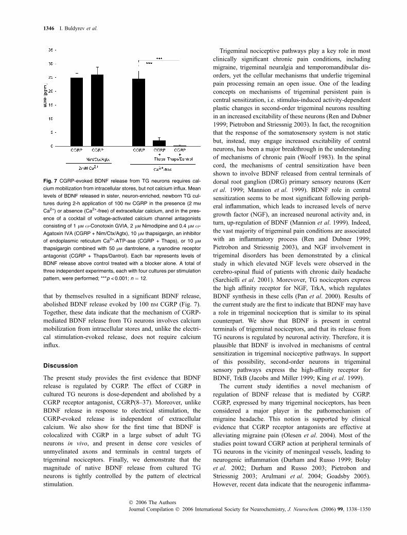
that by themselves resulted in a significant BDNF release,abolished BDNF release evoked by 100 nM CGRP (Fig. 7).Together, these data indicate that the mechanism of CGRP-mediated BDNF release from TG neurons involves calciummobilization from intracellular stores and, unlike the electri-cal stimulation-evoked release, does not require calciuminflux.
Discussion
The present study provides the first evidence that BDNFrelease is regulated by CGRP. The effect of CGRP incultured TG neurons is dose-dependent and abolished by aCGRP receptor antagonist, CGRP(8–37). Moreover, unlikeBDNF release in response to electrical stimulation, theCGRP-evoked release is independent of extracellularcalcium. We also show for the first time that BDNF iscolocalized with CGRP in a large subset of adult TGneurons in vivo, and present in dense core vesicles ofunmyelinated axons and terminals in central targets oftrigeminal nociceptors. Finally, we demonstrate that themagnitude of native BDNF release from cultured TGneurons is tightly controlled by the pattern of electricalstimulation.
Trigeminal nociceptive pathways play a key role in mostclinically significant chronic pain conditions, includingmigraine, trigeminal neuralgia and temporomandibular dis-orders, yet the cellular mechanisms that underlie trigeminalpain processing remain an open issue. One of the leadingconcepts on mechanisms of trigeminal persistent pain iscentral sensitization, i.e. stimulus-induced activity-dependentplastic changes in second-order trigeminal neurons resultingin an increased excitability of these neurons (Ren and Dubner1999; Pietrobon and Striessnig 2003). In fact, the recognitionthat the response of the somatosensory system is not staticbut, instead, may engage increased excitability of centralneurons, has been a major breakthrough in the understandingof mechanisms of chronic pain (Woolf 1983). In the spinalcord, the mechanisms of central sensitization have beenshown to involve BDNF released from central terminals ofdorsal root ganglion (DRG) primary sensory neurons (Kerret al. 1999; Mannion et al. 1999). BDNF role in centralsensitization seems to be most significant following periph-eral inflammation, which leads to increased levels of nervegrowth factor (NGF), an increased neuronal activity and, inturn, up-regulation of BDNF (Mannion et al. 1999). Indeed,the vast majority of trigeminal pain conditions are associatedwith an inflammatory process (Ren and Dubner 1999;Pietrobon and Striessnig 2003), and NGF involvement intrigeminal disorders has been demonstrated by a clinicalstudy in which elevated NGF levels were observed in thecerebro-spinal fluid of patients with chronic daily headache(Sarchielli et al. 2001). Morevover, TG nociceptors expressthe high affinity receptor for NGF, TrkA, which regulatesBDNF synthesis in these cells (Pan et al. 2000). Results ofthe current study are the first to indicate that BDNF may havea role in trigeminal nociception that is similar to its spinalcounterpart. We show that BDNF is present in centralterminals of trigeminal nociceptors, and that its release fromTG neurons is regulated by neuronal activity. Therefore, it isplausible that BDNF is involved in mechanisms of centralsensitization in trigeminal nociceptive pathways. In supportof this possibility, second-order neurons in trigeminalsensory pathways express the high-affinity receptor forBDNF, TrkB (Jacobs and Miller 1999; King et al. 1999).
The current study identifies a novel mechanism ofregulation of BDNF release that is mediated by CGRP.CGRP, expressed by many trigeminal nociceptors, has beenconsidered a major player in the pathomechanism ofmigraine headache. This notion is supported by clinicalevidence that CGRP receptor antagonists are effective atalleviating migraine pain (Olesen et al. 2004). Most of thestudies point toward CGRP action at peripheral terminals ofTG neurons in the vicinity of meningeal vessels, leading toneurogenic inflammation (Durham and Russo 1999; Bolayet al. 2002; Durham and Russo 2003; Pietrobon andStriessnig 2003; Arulmani et al. 2004; Goadsby 2005).However, recent data indicate that the neurogenic inflamma-
Fig. 7 CGRP-evoked BDNF release from TG neurons requires cal-
cium mobilization from intracellular stores, but not calcium influx. Mean
levels of BDNF released in sister, neuron-enriched, newborn TG cul-
tures during 2-h application of 100 nM CGRP in the presence (2 mM
Ca2+) or absence (Ca2+-free) of extracellular calcium, and in the pres-
ence of a cocktail of voltage-activated calcium channel antagonists
consisting of 1 lM x-Conotoxin GVIA, 2 lM Nimodipine and 0.4 lM x-
Agatoxin IVA (CGRP + Nim/Ctx/Agtx), 10 lM thapsigargin, an inhibitor
of endoplasmic reticulum Ca2+-ATP-ase (CGRP + Thaps), or 10 lM
thapsigargin combined with 50 lM dantrolene, a ryanodine receptor
antagonist (CGRP + Thaps/Dantrol). Each bar represents levels of
BDNF release above control treated with a blocker alone. A total of
three independent experiments, each with four cultures per stimulation
pattern, were performed; ***p < 0.001; n ¼ 12.
1346 I. Buldyrev et al.
Journal Compilation � 2006 International Society for Neurochemistry, J. Neurochem. (2006) 99, 1338–1350� 2006 The Authors
tion might not be sufficient to produce migraine pain(Pietrobon and Striessnig 2003). Sensitization of centraltrigeminovascular neurons may be required for the develop-ment of allodynia and intense pain in migraines (Levy et al.2004). In the spinal cord, CGRP released from centralterminals of DRG neurons leads to increased nociceptivesensitivity, i.e. central sensitization (Sun et al. 2004). Thereis evidence that CGRP can also be released from centralterminals of trigeminal afferents (Samsam et al. 2000;Samsam et al. 2001; Jenkins et al. 2004), yet its role in thespinal trigeminal nucleus has been elusive. The central site ofCGRP action in the trigeminal system has been suggested bya recent study of the effects of the non-peptide CGRPreceptor antagonist BIBN4096BS on activity of second-orderneurons in the spinal trigeminal nucleus (Fischer et al. 2005).Our current data indicate that centrally released CGRP couldact at presynaptic terminals of trigeminal nociceptors toenhance BDNF release. In support of this possibility, CGRPreceptor mRNA is present in human trigeminal gangliain vivo (Edvinsson et al. 1997). It has also been demonstra-ted that CGRP potentiates the dorsal root stimulation-evokedrelease of glutamate and aspartate from the spinal dorsalhorn, suggesting the presence of CGRP receptors onpresynaptic terminals of primary afferents (Kangrga andRandic 1990). In fact, CGRP receptor has been localized toboth pre- and postsynaptic sites in the spinal cord (Ye et al.1999).
Previous studies have demonstrated that BDNF and othergrowth factors can regulate expression and release of CGRPfrom sensory neurons (Malcangio et al. 1997; Malcangio2003; Price et al. 2005). Thus, BDNF and CGRP interac-tions at trigeminal synapses could in fact be reciprocal. Thisbecomes particularly significant for mechanisms of inflam-mation-induced hyperalgesia and other forms of sensoryplasticity, since both BDNF and CGRP have been shown tobe up-regulated by inflammatory conditions (Donaldsonet al. 1992; Donnerer et al. 1992; Mapp et al. 1993;Galeazza et al. 1995; Wheeler et al. 1998; Mannion et al.1999). Our data indicate that approximately one-third of allBDNF-immunoreactive trigeminal neurons also expressCGRP. This suggests that CGRP regulates BDNF releaseonly in a subpopulation of trigeminal nociceptors. Thedegree of BDNF/CGRP coexpression is similar among fourdistinct regions of the trigeminal ganglion that, based on thesomatotopic organization of the ganglion (O’Connor and vander Kooy 1986; Borsook et al. 2003), innervate differentareas of the head. These data rule out the possibility that theBDNF–CGRP interaction is limited to a selective subpopu-lation of neurons innervating a specific target/region. Instead,it is likely that the BDNF/CGRP-coexpressing population isresponsible for a particular form of nociception, such asinflammatory pain, or other sensory modality.
To examine regulation of BDNF release from TG neuronsin this study, we used dissociate cultures of newborn TG
neurons grown under a highly sensitive ELISA in situconditions that enable detection of relatively small quantitiesof released endogenous BDNF (Balkowiec and Katz 2000,2002). This model, however, does not allow to determinethe subcellular localization of BDNF release. Consequently,we cannot rule out the possibility that BDNF is releasedfrom the soma and/or peripheral terminals of TG neurons,and that the interaction between BDNF and CGRP occurs atthose alternative sites (Wheeler et al. 1998; Ulrich-Lai et al.2001).
Our data indicate that CGRP regulates BDNF releaseacting through a CGRP receptor-dependent mechanism. TheCGRP receptor is a G protein-coupled receptor, and cansignal to pertussis toxin sensitive G proteins (Main et al.1998; Steiner et al. 2002). Therefore, our data are consistentwith previous observations that the release of a majorfraction of BDNF in the secretory pathway in vitro andin vivo is regulated by the activity of pertussis toxin-sensitiveG proteins (Gunther et al. 2000). Studies in hippocampalneurons indicate that neurotrophin release mediated bymetabotropic glutamate group I (mGluRI) receptor, whichis a G protein-coupled receptor, occurs through activation ofphospholipase C, and subsequent mobilization of calciumfrom intracellular stores (Canossa et al. 2001). Althoughelucidation of the signal transduction pathway that results inBDNF secretion by CGRP requires more detailed futureanalysis, a mechanism similar to that for mGluRI-mediatedrelease could explain the insensitivity of CGRP-mediatedBDNF release to extracellular calcium removal and depend-ence on calcium mobilization from intracellular stores.
Consistent with previous studies of electrical stimulation-evoked BDNF release in the dorsal horn (Lever et al. 2001),and dissociate cultures of visceral sensory (Balkowiec andKatz 2000) and hippocampal (Balkowiec and Katz 2002)neurons, the magnitude of BDNF release from TG neuronswas precisely controlled by the pattern of stimulationdelivered at the same average frequency. This suggests thattrigeminal neurons could employ activity-dependent BDNFrelease along side conventional neurotransmitters to code fordynamic properties of a stimulus.
Electrical stimulation-evoked release of BDNF in thepresent study was abolished by pretreatment with tetrodo-toxin (TTX). It has previously been demonstrated that TTX-resistant sodium channels are significant contributors tomechanisms of action potential generation in nociceptiveneurons (Akopian et al. 1996; Sangameswaran et al. 1996;Akopian et al. 1999; Dib-Hajj et al. 1999; McCleskey andGold 1999; Strassman and Raymond 1999; Waxman et al.1999; Amaya et al. 2000). Our data, on the other hand,suggest that only TTX-sensitive sodium channels areinvolved in activity-dependent BDNF release from newbornTG neurons in vitro. One potential explanation of this resultis a possibility that, in fact, sodium influx specificallythrough TTX-sensitive channels is required for BDNF
CGRP-induced BDNF release from trigeminal neurons 1347
� 2006 The AuthorsJournal Compilation � 2006 International Society for Neurochemistry, J. Neurochem. (2006) 99, 1338–1350

release. This possibility is supported by the fact that bothTTX-resistant and TTX-sensitive sodium currents contributeto action potential generation in TG neurons (Kim andChung 1999).
In conclusion, the present study identifies BDNF as alikely mediator of activity-dependent changes at first-ordersynapses in trigeminal nociceptive pathways, and providessupport for a novel role for CGRP in regulating BDNFrelease. These data add a new dimension to our understand-ing of cellular mechanisms of sensory plasticity. Onceconfirmed in vivo, our findings will indicate the need torevisit current views on critical sites and mechanisms ofCGRP action in chronic pain syndromes, such as migraines.
Acknowledgements
The authors express their gratitude to Dr Sue A. Aicher and Marc B.
Silverman of Neurological Sciences Institute, Beaverton, OR, USA
for help with the electron microscopy portion of this study,
Dr William Cameron for making his laboratory equipment available
for our studies, and Carolyn Robertson for the expert technical
assistance. This work was supported by the Medical Research
Foundation of Oregon (AB), National Institutes of Health (NIH)
grant HL076113 (AB), and OHSU School of Dentistry new faculty
start-up funds (AB). IB was supported in part by a predoctoral
fellowship from NIH training grant 5T32NS007466, and NMT by a
research fellowship from the American Association for Dental
Research.
References
Acheson A., Conover J. C., Fandl J. P., DeChiara T. M., Russell M.,Thadani A., Squinto S. P., Yancopoulos G. D. and Lindsay R. M.(1995) A BDNF autocrine loop in adult sensory neurons preventscell death. Nature 374, 450–453.
Aicher S. A., Mitchell J. L., Swanson K. C. and Zadina J. E. (2003)Endomorphin-2 axon terminals contact mu-opioid receptor-con-taining dendrites in trigeminal dorsal horn.Brain Res. 977, 190–198.
Akerman S., Williamson D. J. and Goadsby P. J. (2003) Voltage-dependent calcium channels are involved in neurogenic duralvasodilatation via a presynaptic transmitter release mechanism.Br. J. Pharmacol. 140, 558–566.
Akopian A. N., Sivilotti L. and Wood J. N. (1996) A tetrodotoxin-resistant voltage-gated sodium channel expressed by sensoryneurons. Nature 379, 257–262.
Akopian A. N., Souslova V., England S. et al. (1999) The tetrodotoxin-resistant sodium channel SNS has a specialized function in painpathways. Nature Neurosci. 2, 541–548.
Alvarez F. J., Morris H. R. and Priestley J. V. (1991) Sub-populations ofsmaller diameter trigeminal primary afferent neurons defined byexpression of calcitonin gene-related peptide and the cell sur-face oligosaccharide recognized by monoclonal antibody LA4.J. Neurocytol. 20, 716–731.
Amaya F., Decosterd I., Samad T. A., Plumpton C., Tate S., MannionR. J., Costigan M. and Woolf C. J. (2000) Diversity of expressionof the sensory neuron-specific TTX-resistant voltage-gated so-dium ion channels SNS and SNS2. Mol. Cell. Neurosci. 15, 331–342.
Arulmani U., Maassen VanDenBrink A., Villalon C. M. and Saxena P. R.(2004) Calcitonin gene-related peptide and its role in migrainepathophysiology. Eur. J. Pharmacol. 500, 315–330.
Asakura K., Kanemasa T., Minagawa K., Kagawa K., Yagami T.,Nakajima M. and Ninomiya M. (2000) Alpha-eudesmol, a P/Q-type Ca (2+) channel blocker, inhibits neurogenic vasodilation andextravasation following electrical stimulation of trigeminal gan-glion. Brain Res. 873, 94–101.
Balkowiec A. and Katz D. M. (2000) Activity-dependent release ofendogenous brain-derived neurotrophic factor from primary sen-sory neurons detected by ELISA in situ. J. Neurosci. 20, 7417–7423.
Balkowiec A. and Katz D. M. (2002) Cellular mechanisms regulatingactivity-dependent release of native brain-derived neurotrophicfactor from hippocampal neurons. J. Neurosci. 22, 10 399–10 407.
Bolay H., Reuter U., Dunn A. K., Huang Z., Boas D. A. and MoskowitzM. A. (2002) Intrinsic brain activity triggers trigeminal meningealafferents in a migraine model. Nature Med. 8, 136–142.
Borgland S. L., Connor M. and Christie M. J. (2001) Nociceptin inhibitscalcium channel currents in a subpopulation of small nociceptivetrigeminal ganglion neurons in mouse. J. Physiol. (Lond.) 536, 35–47.
Borsook D., DaSilva A. F. M., Ploghaus A. and Becerra L. (2003)Specific and somatotopic functional magnetic resonance imagingactivation in the trigeminal ganglion by brush and noxious heat.J. Neurosci. 23, 7897–7903.
Brady R., Zaidi S. I. A., Mayer C. and Katz D. M. (1999) BDNF is atarget-derived survival factor for arterial baroreceptor and chemo-afferent primary sensory neurons. J. Neurosci. 19, 2131–2142.
Buldyrev I., Silverman M. B., Aicher S. A. and Balkowiec A. (2004)CGRP regulates BDNF release from trigeminal ganglion neurons.Soc. Neurosci. Abstract. 726.10.
Canossa M., Gartner A., Campana G., Inagaki N. and Thoenen H. (2001)Regulated secretion of neurotrophins by metabotropic glutamategroup I (mGluRI) and Trk receptor activation is mediated viaphospholipase C signalling pathways. EMBO J. 20, 1640–1650.
Dib-Hajj S. D., Tyrrell L., Cummins T. R., Black J. A., Wood P. M. andWaxman S. G. (1999) Two tetrodotoxin-resistant sodium channelsin human dorsal root ganglion neurons. FEBS Lett. 462, 117–120.
Donaldson L. F., Harmar A. J., McQueen D. S. and Seckl J. R. (1992)Increased expression of preprotachykinin, calcitonin gene- relatedpeptide, but not vasoactive intestinal peptide messenger RNA indorsal root ganglia during the development of adjuvant mono-arthritis in the rat. Brain Res. Mol. Brain Res. 16, 143–149.
Donnerer J., Schuligoi R. and Stein C. (1992) Increased content andtransport of substance P and calcitonin gene-related peptide insensory nerves innervating inflamed tissue: evidence for a regula-tory function of nerve growth factor in vivo. Neuroscience 49, 693–698.
Durham P. L. and Russo A. F. (1999) Regulation of calcitonin gene-related peptide secretion by a serotonergic antimigraine drug.J. Neurosci. 19, 3423–3429.
Durham P. L. and Russo A. F. (2003) Stimulation of the calcitonin gene-related peptide enhancer by mitogen-activated protein kinases andrepression by an antimigraine drug in trigeminal ganglia neurons.J. Neurosci. 23, 807–815.
Eckert S. P., Taddese A. and McCleskey E. W. (1997) Isolation andculture of rat sensory neurons having distinct sensory modalities.J. Neurosci. Meth. 77, 183–190.
Edvinsson L., Cantera L., Jansen-Olesen I. and Uddman R. (1997)Expression of calcitonin gene-related peptide1 receptor mRNA inhuman trigeminal ganglia and cerebral arteries. Neurosci. Lett. 229,209–211.
1348 I. Buldyrev et al.
Journal Compilation � 2006 International Society for Neurochemistry, J. Neurochem. (2006) 99, 1338–1350� 2006 The Authors

Fischer M. J. M., Koulchitsky S. and Messlinger K. (2005) The non-peptide calcitonin gene-related peptide receptor antagonistBIBN4096BS lowers the activity of neurons with meningeal inputin the rat spinal trigeminal nucleus. J. Neurosci. 25, 5877–5883.
Galeazza M. T., Garry M. G., Yost H. J., Strait K. A., Hargreaves K. M.and Seybold V. S. (1995) Plasticity in the synthesis and storage ofsubstance P and calcitonin gene-related peptide in primary afferentneurons during peripheral inflammation. Neuroscience 66, 443–458.
Ghosh A., Carnahan J. and Greenberg M. E. (1994) Requirement forBDNF in activity-dependent survival of cortical neurons. Science263, 1618–1623.
Goadsby P. J. (2005) New targets in the acute treatment of headache.Curr. Opin. Neurol. 18, 283–288.
Goadsby P. J., Edvinsson L. and Ekman R. (1990) Vasoactive peptiderelease in the extracerebral circulation of humans during migraineheadache. Ann. Neurol. 28, 183–187.
Griesbeck O., Canossa M., Campana G., Gartner A., Hoener M. C.,Nawa H., Kolbeck R. and Thoenen H. (1999) Are there dif-ferences between the secretion characteristics of NGF andBDNF? Implications for the modulatory role of neurotrophins inactivity-dependent neuronal plasticity. Microsc. Res. Techn 45,262–275.
Gunther E. C., von Bartheld C., Goodman L. J., Johnson J. E. andBothwell M. (2000) The G-protein inhibitor, pertussis toxin,inhibits the secretion of brain-derived neurotrophic factor. Neuro-science 100, 569–579.
Hong K. W., Kim C. D., Rhim B. Y. and Lee W. S. (1999) Effect ofomega-conotoxin GVIA and omega-agatoxin IVA on the capsai-cin-sensitive calcitonin gene-related peptide release and autoregu-latory vasodilation in rat pial arteries. J. Cereb. Blood FlowMetabol. 19, 53–60.
Jacobs J. S. and Miller M. W. (1999) Expression of nerve growth factor,p75, and the high affinity neurotrophin receptors in the adult rattrigeminal system: evidence for multiple trophic support systems.J. Neurocytol. 28, 571–595.
Jenkins D. W., Langmead C. J., Parsons A. A. and Strijbos P. J. (2004)Regulation of calcitonin gene-related peptide release from rattrigeminal nucleus caudalis slices in vitro. Neurosci. Lett. 366,241–244.
Kangrga I. and Randic M. (1990) Tachykinins and calcitonin gene-related peptide enhance release of endogenous glutamate andaspartate from the rat spinal dorsal horn slice. J. Neurosci. 10,2026–2038.
Kerr B. J., Bradbury E. J., Bennett D. L., Trivedi P. M., Dassan P.,French J., Shelton D. B., McMahon S. B. and Thompson S. W.(1999) Brain-derived neurotrophic factor modulates nociceptivesensory inputs and NMDA-evoked responses in the rat spinal cord.J. Neurosci. 19, 5138–5148.
Kim H. C. and Chung M. K. (1999) Voltage-dependent sodium andcalcium currents in acutely isolated adult rat trigeminal root gan-glion neurons. J. Neurophysiol. 81, 1123–1134.
King V. R., Michael G. J., Joshi R. K. and Priestley J. V. (1999) TrkA,trkB, and trkC messenger RNA expression by bulbospinal cells ofthe rat. Neuroscience 92, 935–944.
Kyrkanides S., Tallents R. H., Macher D. J., Olschowka J. A. andStevens S. Y. (2002) Temporomandibular joint nociception: effectsof capsaicin on substance P-like immunoreactivity in the rabbitbrain stem. J. Orofac. Pain 16, 229–236.
Lazarov N. E. (2002) Comparative analysis of the chemical neuroanat-omy of the mammalian trigeminal ganglion and mesencephalictrigeminal nucleus. Prog. Neurobiol. 66, 19–59.
Lever I. J., Bradbury E. J., Cunningham J. R., Adelson D. W., JonesM. G., McMahon S. B., Marvizon J. C. and Malcangio M. (2001)
Brain-derived neurotrophic factor is released in the dorsal horn bydistinctive patterns of afferent fiber stimulation. J. Neurosci. 21,4469–4477.
Levy D., Jakubowski M. and Burstein R. (2004) Disruption of com-munication between peripheral and central trigeminovascularneurons mediates the antimigraine action of 5HT 1B/1D receptoragonists. Proc. Natl Acad. Sci. USA 101, 4274–4279.
Lucas G., Hendolin P., Harkany T. et al. (2003) Neurotrophin-4 medi-ated TrkB activation reinforces morphine-induced analgesia.Nature Neurosci. 6, 221–222.
Luo X., Rush R. A. and Zhou X. (2001) Ultrastructural localization ofbrain-derived neurotrophic factor in rat primary sensory neurons.Neurosci. Res. 39, 377–384.
Main M. J., Brown J., Brown S., Fraser N. J. and Foord S. M. (1998)The CGRP receptor can couple via pertussis toxin sensitive andinsensitive G proteins. FEBS Lett. 441, 6–10.
Malcangio M. (2003) GDNF and somatostatin in sensory neurones.Curr. Opin. Pharmacol. 3, 41–45.
Malcangio M., Garrett N. E. and Tomlinson D. R. (1997) Nerve growthfactor treatment increases stimulus-evoked release of sensoryneuropeptides in the rat spinal cord. Eur. J. Neurosci. 9, 1101–1104.
Malcangio M. and Lessmann V. (2003) A common thread for pain andmemory synapses? Brain-derived neurotrophic factor and trkBreceptors. Trends Pharmacol. Sci. 24, 116–121.
Mannion R. J., Costigan M., Decosterd I. et al. (1999) Neurotrophins:peripherally and centrally acting modulators of tactile stimulus-induced inflammatory pain hypersensitivity. Proc. Natl Acad. Sci.USA 96, 9385–9390.
Mapp P. I., Terenghi G., Walsh D. A., Chen S. T., Cruwys S. C., GarrettN., Kidd B. L., Polak J. M. and Blake D. R. (1993) Monoarthritisin the rat knee induces bilateral and time-dependent changes insubstance P and calcitonin gene-related peptide immunoreactivityin the spinal cord. Neuroscience 57, 1091–1096.
McCleskey E. W. and Gold M. S. (1999) Ion channels of nociception.Annu. Rev. Physiol. 61, 835–856.
Mendell L. M., Albers K. M. and Davis B. M. (1999) Neurotrophins,nociceptors, and pain. Microsc. Res. Techn 45, 252–261.
Michael G. J., Averill S., Nitkunan A., Rattray M., Bennett D. L., Yan Q.and Priestley J. V. (1997) Nerve growth factor treatment increasesbrain-derived neurotrophic factor selectively in TrkA-expressingdorsal root ganglion cells and in their central terminations withinthe spinal cord. J. Neurosci. 17, 8476–8490.
Moore K. A., Baba H. and Woolf C. J. (2000) Synaptic transmission andplasticity in the superficial dorsal horn.Prog. Brain Res. 129, 63–80.
Nguyen L. T., Robertson C. L., Palmer E. A. and Balkowiec A. (2004)Growth factor dependence of postnatal trigeminal neurons. Soc.Neurosci. Abstract. 836.11.
O’Connor T. P. and van der Kooy D. (1986) Pattern of intracranial andextracranial projections of trigeminal ganglion cells. J. Neurosci. 6,2200–2207.
Olesen J., Diener H. C., Husstedt I. W., Goadsby P. J., Hall D., Meier U.,Pollentier S. and Lesko L. M. (2004) Calcitonin gene-relatedpeptide receptor antagonist BIBN 4096 BS for the acute treatmentof migraine. New Eng. J. Med. 350, 1104–1110.
Pan M., Naftel J. P. and Wheeler E. F. (2000) Effects of depriva-tion of neonatal nerve growth factor on the expression ofneurotrophin receptors and brain-derived neurotrophic factor bydental pulp afferents of the adult rat. Arch. Oral Biol. 45, 387–399.
Pezet S., Cunningham J., Patel J., Grist J., Gavazzi I., Lever I. J. andMalcangio M. (2002) BDNF modulates sensory neuron synapticactivity by a facilitation of GABA transmission in the dorsal horn.Mol. Cell. Neurosci. 21, 51–62.
CGRP-induced BDNF release from trigeminal neurons 1349
� 2006 The AuthorsJournal Compilation � 2006 International Society for Neurochemistry, J. Neurochem. (2006) 99, 1338–1350

Pietrobon D. and Striessnig J. (2003) Neurobiology of migraine. NatureRev. Neurosci. 4, 386–398.
Price T. J., Louria M. D., Candelario-Soto D. et al. (2005) Treatment oftrigeminal ganglion neurons in vitro with NGF, GDNF or BDNF:effects on neuronal survival, neurochemical properties andTRPV1-mediated neuropeptide secretion. BMC Neurosci. 6, 4.
Quartu M., Geic M. and Del Fiacco M. (1997) Neurotrophin-like im-munoreactivity in the human trigeminal ganglion. Neuroreport 8,3611–3617.
Ren K. and Dubner R. (1999) Central nervous system plasticity andpersistent pain. J. Orofac. Pain 13, 155–163.
Robinson M., Buj-Bello A. and Davies A. M. (1996) Paracrine inter-actions of BDNF involving NGF-dependent embryonic sensoryneurons. Mol. Cell. Neurosci. 7, 143–151.
Samsam M., Covenas R., Ahangari R., Yajeya J., Narvaez J. A. andTramu G. (2000) Simultaneous depletion of neurokinin A, sub-stance P and calcitonin gene-related peptide from the caudaltrigeminal nucleus of the rat during electrical stimulation of thetrigeminal ganglion. Pain 84, 389–395.
Samsam M., Covenas R., Csillik B., Ahangari R., Yajeya J., RiquelmeR., Narvaez J. A. and Tramu G. (2001) Depletion of substance P,neurokinin A and calcitonin gene-related peptide from the con-tralateral and ipsilateral caudal trigeminal nucleus following uni-lateral electrical stimulation of the trigeminal ganglion; a possibleneurophysiological and neuroanatomical link to generalized headpain. J. Chem. Neuroanat. 21, 161–169.
Sangameswaran L., Delgado S. G., Fish L. M. et al. (1996) Structureand function of a novel voltage-gated, tetrodotoxin-resistant so-dium channel specific to sensory neurons. J. Biol. Chem. 271,5953–5956.
Sarchielli P., Alberti A., Codini M., Floridi A. and Gallai V. (2000) Nitricoxide metabolites, prostaglandins and trigeminal vasoactive pep-tides in internal jugular vein blood during spontaneous migraineattacks. Cephalalgia 20, 907–918.
Sarchielli P., Alberti A., Floridi A. and Gallai V. (2001) Levels of nervegrowth factor in cerebrospinal fluid of chronic daily headachepatients. Neurology 57, 132–134.
Scholz J. and Woolf C. J. (2002) Can we conquer pain? Nature Neurosci.5, 1062–1067.
Sessle B. J. (2000) Acute and chronic craniofacial pain: brainstemmechanisms of nociceptive transmission and neuroplasticity, andtheir clinical correlates. Crit. Rev. Oral Biol. Med. 11, 57–91.
Shankland W. E. (2000) The trigeminal nerve. Part I: An over-view.Cranio 18, 238–248.
Shelton D. L., Sutherland J., Gripp J., Camerato T., Armanini M. P.,Phillips H. S., Carroll K., Spencer S. D. and Levinson A. D. (1995)Human trks: molecular cloning, tissue distribution, and expressionof extracellular domain immunoadhesins. J. Neurosci. 15, 477–491.
Shu X. Q., Llinas A. and Mendell L. M. (1999) Effects of trkB and trkCneurotrophin receptor agonists on thermal nociception: a behavi-oral and electrophysiological study. Pain 80, 463–470.
Shu X. Q. and Mendell L. M. (1999) Neurotrophins and hyperalgesia.Proc. Natl Acad. Sci. USA 96, 7693–7696.
Steiner S., Muff R., Gujer R., Fischer J. A. and Born W. (2002) Thetransmembrane domain of receptor-activity-modifying protein 1 is
essential for the functional expression of a calcitonin gene-relatedpeptide receptor. Biochemistry 41, 11398–11404.
Strassman A. M. and Raymond S. A. (1999) Electrophysiologicalevidence for tetrodotoxin-resistant sodium channels inslowly conducting dural sensory fibers. J. Neurophysiol. 81,413–424.
Sun R. Q., Lawand N. B., Lin Q. and Willis W. D. (2004) Role ofcalcitonin gene-related peptide in the sensitization of dorsal hornneurons to mechanical stimulation after intradermal injection ofcapsaicin. J. Neurophysiol. 92, 320–326.
Tajti J., Uddman R., Moller S., Sundler F. and Edvinsson L. (1999)Messenger molecules and receptor mRNA in the human trigeminalganglion. J. Auton. Nerv. Syst. 76, 176–183.
Tanner N. M., Robertson C. L. and Balkowiec A. (2004) Identificationof BDNF in trigeminal ganglion neurons: Role of patterns oftrigeminal activity in BDNF release. Soc. Neurosci. Abstract.726.11.
Thompson S. W., Bennett D. L., Kerr B. J., Bradbury E. J. andMcMahon S. B. (1999) Brain-derived neurotrophic factor is anendogenous modulator of nociceptive responses in the spinal cord.Proc. Natl Acad. Sci. USA 96, 7714–7718.
Tonra J. R. (1999) Classical and novel directions in neurotrophintransport and research: anterograde transport of brain-derivedneurotrophic factor by sensory neurons. Mic. Res. Techn 45, 225–232.
Ulrich-Lai Y. M., Flores C. M., Harding-Rose C. A., Goodis H. E. andHargreaves K. M. (2001) Capsaicin-evoked release of immuno-reactive calcitonin gene-related peptide from rat trigeminal gan-glion: evidence for intraganglionic neurotransmission. Pain 91,219–226.
Waxman S. G., Cummins T. R., Dib-Hajj S., Fjell J. and Black J. A.(1999) Sodium channels, excitability of primary sensory neurons,and the molecular basis of pain. Muscle Nerve 22, 1177–1187.
Wetmore C. and Olson L. (1995) Neuronal and nonneuronal expressionof neurotrophins and their receptors in sensory and sympatheticganglia suggest new intercellular trophic interactions. J. Comp.Neurol. 353, 143–159.
Wheeler E. F., Naftel J. P., Pan M., von Bartheld C. and Byers M. R.(1998) Neurotrophin receptor expression is induced in a subpop-ulation of trigeminal neurons that label by retrograde transport ofNGF or fluoro-gold following tooth injury. Brain Res. Mol. BrainRes. 61, 23–38.
Whim M. D. and Lloyd P. E. (1994) Differential regulation of the releaseof the same peptide transmitters from individual identified motorneurons in culture. J. Neurosci. 14, 4244–4251.
Woolf C. J. (1983) Evidence for a central component of post-injury painhypersensitivity. Nature 306, 686–688.
Woolf C. J. and Salter M. W. (2000) Neuronal plasticity: increasing thegain in pain. Science 288, 1765–1769.
Ye Z., Wimalawansa S. J. and Westlund K. N. (1999) Receptor forcalcitonin gene-related peptide: localization in the dorsal andventral spinal cord. Neuroscience 92, 1389–1397.
Zhou X. F. and Rush R. A. (1996) Endogenous brain-derived neuro-trophic factor is anterogradely transported in primary sensoryneurons. Neuroscience 74, 945–953.
1350 I. Buldyrev et al.
Journal Compilation � 2006 International Society for Neurochemistry, J. Neurochem. (2006) 99, 1338–1350� 2006 The Authors