Responses induced by acetylcholine and ATP in the rabbit petrosal ganglion
8
Respiratory Physiology & Neurobiology 172 (2010) 114–121 Contents lists available at ScienceDirect Respiratory Physiology & Neurobiology journal homepage: www.elsevier.com/locate/resphysiol Responses induced by acetylcholine and ATP in the rabbit petrosal ganglion Carolina R. Soto 1 , Fernando C. Ortiz 2 , Romina V. Vargas, Jorge Arroyo, Julio Alcayaga ∗ Laboratorio de Fisiología Celular, Departamento de Biología, Facultad de Ciencias, Universidad de Chile, Casilla 653, Santiago, Chile article info Article history: Accepted 2 May 2010 Keywords: Petrosal ganglion Carotid sinus nerve Afferent activity Acetylcholine receptors ATP receptors Carotid body abstract Acetylcholine and ATP appear to mediate excitatory transmission between receptor (glomus) cells and the petrosal ganglion (PG) neuron terminals in the carotid body. In most species these putative transmit- ters are excitatory, while inhibitory effects had been reported in the rabbit. We studied the effects of the application of acetylcholine and ATP to the PG on the carotid nerve activity in vitro. Acetylcholine and ATP applied to the PG increased the carotid nerve activity in a dose-dependent manner. Acetylcholine-induced responses were mimicked by nicotine, antagonized by hexamethonium, and enhanced by atropine. Bethanechol had no effect on basal activity, but reduced acetylcholine-induced responses. Suramin antag- onized ATP-induced responses, and AMP had little effect on the carotid nerve activity. Our results suggest that rabbit PG neurons projecting through the carotid nerve are endowed with nicotinic acetylcholine and purinergic P2 receptors that increase the carotid nerve activity, while simultaneous activation of muscarinic cholinergic receptors reduce the maximal response evoked by nicotinic cholinergic receptor activation. © 2010 Elsevier B.V. All rights reserved. 1. Introduction The carotid body (CB) is the principal arterial chemoreceptor organ, constituted by specific parenchymal cells: the receptor (glo- mus, type-I) cells and the glial-like (sustentacular, type-II) cells. The glomus cells are innervated, through the carotid (sinus) nerve, by sensory neurons whose perikarya are located in the petrosal ganglion (PG). The most accepted paradigm of CB chemoreception states that, as a result of the transduction mechanism, the glomus cells release one or more transmitters that, acting on postsynaptic receptors located on the nerve terminals of PG neurons, gener- ate or maintain the afferent activity (Eyzaguirre and Zapata, 1984; González et al., 1994; Prabhakar, 2000). There are several trans- mitter molecules present in the CB, but their exact participation in the generation of the chemo-afferent activity is still controversial. Part of the controversy arises from conflicting results obtained from different species and preparations. Acetylcholine (ACh) was one of the first molecules postulated as a transmitter in the CB (see Heymans, 1955). ACh and its metabolic synthesis and degradation pathways are present in the CB of cats ∗ Corresponding author. Tel.: +56 2 978 7366; fax: +56 2 271 2983. E-mail addresses: c soto [email protected] (C.R. Soto), ortiz 9266 [email protected] (F.C. Ortiz), [email protected] (J. Alcayaga). 1 Present address: Programa de Doctorado en Ciencias, mención Neurociencia, Universidad de Valparaiso, Gran Breta ˜ na 1111 Playa Ancha, Valparaiso, Chile. 2 Present address: Programa de Doctorado en Ciencias Biológicas, mención Cien- cias Fisiologicas, P. Universidad Católica de Chile, Facultad de Ciencias Biológicas, Portugal 49, Casilla 114-D, Santiago, Chile. (Fidone et al., 1976; Wang et al., 1989), rabbits (Wang et al., 1989; Kim et al., 2004) and in the rat CB-PG reconstituted system in vitro (Nurse and Zhang, 1999). ACh is released from the cat CB during electrical (Eyzaguirre and Zapata, 1968) and hypoxic stimulation (Fitzgerald et al., 1999; Fitzgerald, 2000), but ACh release from the rabbit CB is reduced during hypoxic stimulation (Kim et al., 2004). The exogenous application of ACh to the cat CB increases the carotid nerve frequency of discharge (Eyzaguirre and Zapata, 1968), while its effect is mainly inhibitory in the rabbit CB (Docherty and McQueen, 1979; Monti-Bloch and Eyzaguirre, 1980). However, application of nicotine to the CB increases the carotid nerve fre- quency of discharge both in the cat (Eyzaguirre and Zapata, 1968; Reyes et al., 2007) and in the rabbit (Monti-Bloch and Eyzaguirre, 1980; Jonsson et al., 2004), effects blocked by nicotinic ACh recep- tor antagonists (Eyzaguirre and Zapata, 1968; Jonsson et al., 2004; Reyes et al., 2007). On the other hand, muscarinic ACh receptor sub- types M1 and M2 have been described in the cat CB (Shirahata et al., 2004). However, muscarinic antagonists have no effect on the cat chemosensory activity but depressed the carotid nerve frequency of discharge on the rabbit (Monti-Bloch and Eyzaguirre, 1980). In the rabbit CB, atropine antagonizes the inhibitory effects of mus- carinic ACh receptor agonists on the carotid nerve frequency of discharge (Monti-Bloch and Eyzaguirre, 1980), increases basal ACh release and revert the reduction of ACh release induced by hypoxia to an increased release (Kim et al., 2004). Thus, the available data indicate that ACh appear to be excitatory in the cat CB, but may have both excitatory and inhibitory actions in the rabbit, mediated by nicotinic and muscarinic ACh receptors, respectively. However, the exogenous application of ACh to the CB may involve activation 1569-9048/$ – see front matter © 2010 Elsevier B.V. All rights reserved. doi:10.1016/j.resp.2010.05.003
-
Upload
independent -
Category
Documents
-
view
0 -
download
0
Transcript of Responses induced by acetylcholine and ATP in the rabbit petrosal ganglion

R
CL
a
AA
KPCAAAC
1
omTbgscraGmtPd
as
o
U
cP
1d
Respiratory Physiology & Neurobiology 172 (2010) 114–121
Contents lists available at ScienceDirect
Respiratory Physiology & Neurobiology
journa l homepage: www.e lsev ier .com/ locate / resphys io l
esponses induced by acetylcholine and ATP in the rabbit petrosal ganglion
arolina R. Soto1, Fernando C. Ortiz2, Romina V. Vargas, Jorge Arroyo, Julio Alcayaga ∗
aboratorio de Fisiología Celular, Departamento de Biología, Facultad de Ciencias, Universidad de Chile, Casilla 653, Santiago, Chile
r t i c l e i n f o
rticle history:ccepted 2 May 2010
eywords:etrosal ganglionarotid sinus nerve
a b s t r a c t
Acetylcholine and ATP appear to mediate excitatory transmission between receptor (glomus) cells andthe petrosal ganglion (PG) neuron terminals in the carotid body. In most species these putative transmit-ters are excitatory, while inhibitory effects had been reported in the rabbit. We studied the effects of theapplication of acetylcholine and ATP to the PG on the carotid nerve activity in vitro. Acetylcholine and ATPapplied to the PG increased the carotid nerve activity in a dose-dependent manner. Acetylcholine-inducedresponses were mimicked by nicotine, antagonized by hexamethonium, and enhanced by atropine.
fferent activitycetylcholine receptorsTP receptorsarotid body
Bethanechol had no effect on basal activity, but reduced acetylcholine-induced responses. Suramin antag-onized ATP-induced responses, and AMP had little effect on the carotid nerve activity. Our results suggestthat rabbit PG neurons projecting through the carotid nerve are endowed with nicotinic acetylcholineand purinergic P2 receptors that increase the carotid nerve activity, while simultaneous activation ofmuscarinic cholinergic receptors reduce the maximal response evoked by nicotinic cholinergic receptor
activation.. Introduction
The carotid body (CB) is the principal arterial chemoreceptorrgan, constituted by specific parenchymal cells: the receptor (glo-us, type-I) cells and the glial-like (sustentacular, type-II) cells.
he glomus cells are innervated, through the carotid (sinus) nerve,y sensory neurons whose perikarya are located in the petrosalanglion (PG). The most accepted paradigm of CB chemoreceptiontates that, as a result of the transduction mechanism, the glomusells release one or more transmitters that, acting on postsynapticeceptors located on the nerve terminals of PG neurons, gener-te or maintain the afferent activity (Eyzaguirre and Zapata, 1984;onzález et al., 1994; Prabhakar, 2000). There are several trans-itter molecules present in the CB, but their exact participation in
he generation of the chemo-afferent activity is still controversial.art of the controversy arises from conflicting results obtained from
ifferent species and preparations.Acetylcholine (ACh) was one of the first molecules postulated astransmitter in the CB (see Heymans, 1955). ACh and its metabolic
ynthesis and degradation pathways are present in the CB of cats
∗ Corresponding author. Tel.: +56 2 978 7366; fax: +56 2 271 2983.E-mail addresses: c soto [email protected] (C.R. Soto),
rtiz 9266 [email protected] (F.C. Ortiz), [email protected] (J. Alcayaga).1 Present address: Programa de Doctorado en Ciencias, mención Neurociencia,niversidad de Valparaiso, Gran Bretana 1111 Playa Ancha, Valparaiso, Chile.2 Present address: Programa de Doctorado en Ciencias Biológicas, mención Cien-
ias Fisiologicas, P. Universidad Católica de Chile, Facultad de Ciencias Biológicas,ortugal 49, Casilla 114-D, Santiago, Chile.
569-9048/$ – see front matter © 2010 Elsevier B.V. All rights reserved.oi:10.1016/j.resp.2010.05.003
© 2010 Elsevier B.V. All rights reserved.
(Fidone et al., 1976; Wang et al., 1989), rabbits (Wang et al., 1989;Kim et al., 2004) and in the rat CB-PG reconstituted system in vitro(Nurse and Zhang, 1999). ACh is released from the cat CB duringelectrical (Eyzaguirre and Zapata, 1968) and hypoxic stimulation(Fitzgerald et al., 1999; Fitzgerald, 2000), but ACh release fromthe rabbit CB is reduced during hypoxic stimulation (Kim et al.,2004). The exogenous application of ACh to the cat CB increasesthe carotid nerve frequency of discharge (Eyzaguirre and Zapata,1968), while its effect is mainly inhibitory in the rabbit CB (Dochertyand McQueen, 1979; Monti-Bloch and Eyzaguirre, 1980). However,application of nicotine to the CB increases the carotid nerve fre-quency of discharge both in the cat (Eyzaguirre and Zapata, 1968;Reyes et al., 2007) and in the rabbit (Monti-Bloch and Eyzaguirre,1980; Jonsson et al., 2004), effects blocked by nicotinic ACh recep-tor antagonists (Eyzaguirre and Zapata, 1968; Jonsson et al., 2004;Reyes et al., 2007). On the other hand, muscarinic ACh receptor sub-types M1 and M2 have been described in the cat CB (Shirahata et al.,2004). However, muscarinic antagonists have no effect on the catchemosensory activity but depressed the carotid nerve frequencyof discharge on the rabbit (Monti-Bloch and Eyzaguirre, 1980). Inthe rabbit CB, atropine antagonizes the inhibitory effects of mus-carinic ACh receptor agonists on the carotid nerve frequency ofdischarge (Monti-Bloch and Eyzaguirre, 1980), increases basal AChrelease and revert the reduction of ACh release induced by hypoxia
to an increased release (Kim et al., 2004). Thus, the available dataindicate that ACh appear to be excitatory in the cat CB, but mayhave both excitatory and inhibitory actions in the rabbit, mediatedby nicotinic and muscarinic ACh receptors, respectively. However,the exogenous application of ACh to the CB may involve activation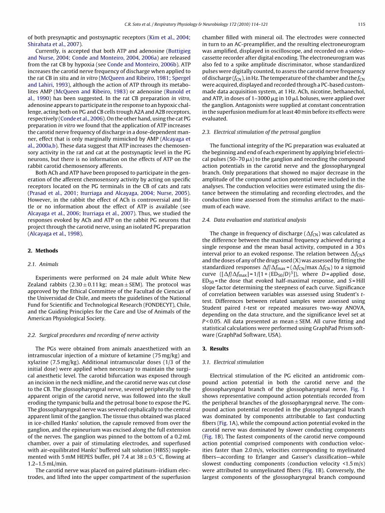
ogy &
oS
afitalaalrptnasnr
er(HtArp(
2
2
ZatFaA
2
ixicataeTaigocwm1
t
C.R. Soto et al. / Respiratory Physiol
f both presynaptic and postsynaptic receptors (Kim et al., 2004;hirahata et al., 2007).
Currently, is accepted that both ATP and adenosine (Buttigiegnd Nurse, 2004; Conde and Monteiro, 2004, 2006a) are releasedrom the rat CB by hypoxia (see Conde and Monteiro, 2006b). ATPncreases the carotid nerve frequency of discharge when applied tohe rat CB in situ and in vitro (McQueen and Ribeiro, 1981; Spergelnd Lahiri, 1993), although the action of ATP through its metabo-ites AMP (McQueen and Ribeiro, 1983) or adenosine (Runold etl., 1990) has been suggested. In the rat CB preparation in vitro,denosine appears to participate in the response to an hypoxic chal-enge, acting both on PG and CB cells trough A2A and A2B receptors,espectively (Conde et al., 2006). On the other hand, using the cat PGreparation in vitro we found that the application of ATP increaseshe carotid nerve frequency of discharge in a dose-dependent man-er, effect that is only marginally mimicked by AMP (Alcayaga etl., 2000a,b). These data suggest that ATP increases the chemosen-ory activity in the rat and cat at the postsynaptic level in the PGeurons, but there is no information on the effects of ATP on theabbit carotid chemosensory afferents.
Both ACh and ATP have been proposed to participate in the gen-ration of the afferent chemosensory activity by acting on specificeceptors located on the PG terminals in the CB of cats and ratsPrasad et al., 2001; Iturriaga and Alcayaga, 2004; Nurse, 2005).owever, in the rabbit the effect of ACh is controversial and lit-
le or no information about the effect of ATP is available (seelcayaga et al., 2006; Iturriaga et al., 2007). Thus, we studied theesponses evoked by ACh and ATP on the rabbit PG neurons thatroject through the carotid nerve, using an isolated PG preparationAlcayaga et al., 1998).
. Methods
.1. Animals
Experiments were performed on 24 male adult White Newealand rabbits (2.30 ± 0.11 kg; mean ± SEM). The protocol waspproved by the Ethical Committee of the Facultad de Ciencias ofhe Universidad de Chile, and meets the guidelines of the Nationalund for Scientific and Technological Research (FONDECYT), Chile,nd the Guiding Principles for the Care and Use of Animals of themerican Physiological Society.
.2. Surgical procedures and recording of nerve activity
The PGs were obtained from animals anaesthetized with anntramuscular injection of a mixture of ketamine (75 mg/kg) andylazine (7.5 mg/kg). Additional intramuscular doses (1/3 of thenitial dose) were applied when necessary to maintain the surgi-al anesthetic level. The carotid bifurcation was exposed throughn incision in the neck midline, and the carotid nerve was cut closeo the CB. The glossopharyngeal nerve, severed peripherally to thepparent origin of the carotid nerve, was followed into the skullroding the tympanic bulla and the petrosal bone to expose the PG.he glossopharyngeal nerve was severed cephalically to the centralpparent limit of the ganglion. The tissue thus obtained was placedn ice-chilled Hanks’ solution, the capsule removed from over theanglion, and the epineurium was excised along the full extensionf the nerves. The ganglion was pinned to the bottom of a 0.2 mLhamber, over a pair of stimulating electrodes, and superfused
ith air-equilibrated Hanks’ buffered salt solution (HBSS) supple-ented with 5 mM HEPES buffer, pH 7.4 at 38 ± 0.5 ◦C, flowing at.2–1.5 mL/min.The carotid nerve was placed on paired platinum–iridium elec-
rodes, and lifted into the upper compartment of the superfusion
Neurobiology 172 (2010) 114–121 115
chamber filled with mineral oil. The electrodes were connectedin turn to an AC-preamplifier, and the resulting electroneurogramwas amplified, displayed in oscilloscope, and recorded on a video-cassette recorder after digital encoding. The electroneurogram wasalso fed to a spike amplitude discriminator, whose standardizedpulses were digitally counted, to assess the carotid nerve frequencyof discharge (fCN), in Hz. The temperature of the chamber and the fCNwere acquired, displayed and recorded through a PC-based custom-made data acquisition system, at 1 Hz. ACh, nicotine, bethanechol,and ATP, in doses of 1–3000 �g in 10 �L boluses, were applied overthe ganglion. Antagonists were supplied at constant concentrationin the superfusion medium for at least 40 min before its effects wereevaluated.
2.3. Electrical stimulation of the petrosal ganglion
The functional integrity of the PG preparation was evaluated atthe beginning and end of each experiment by applying brief electri-cal pulses (50–70 �s) to the ganglion and recording the compoundaction potentials in the carotid nerve and the glossopharyngealbranch. Only preparations that showed no major decrease in theamplitude of the compound action potential were included in theanalyses. The conduction velocities were estimated using the dis-tance between the stimulating and recording electrodes, and theconduction time assessed from the stimulus artifact to the maxi-mum of each wave.
2.4. Data evaluation and statistical analysis
The change in frequency of discharge (�fCN) was calculated asthe difference between the maximal frequency achieved during asingle response and the mean basal activity, computed in a 30 sinterval prior to an evoked response. The relation between �fCNsand the doses of any of the drugs used (X) was assessed by fitting thestandardized responses �f/�fmax = (�fCN/max �fCN) to a sigmoidcurve ([�f/�fmax] = 1/[1 + {ED50/D}S]), where D = applied dose,ED50 = the dose that evoked half-maximal response, and S = Hillslope factor determining the steepness of each curve. Significanceof correlation between variables was assessed using Student’s t-test. Differences between related samples were assessed usingStudent paired t-test or repeated measures two-way ANOVA,depending on the data structure, and the significance level set atP < 0.05. All data presented as mean ± SEM. All curve fitting andstatistical calculations were performed using GraphPad Prism soft-ware (GraphPad Software, USA).
3. Results
3.1. Electrical stimulation
Electrical stimulation of the PG elicited an antidromic com-pound action potential in both the carotid nerve and theglossopharyngeal branch of the glossopharyngeal nerve. Fig. 1shows representative compound action potentials recorded fromthe peripheral branches of the glossopharyngeal nerve. The com-pound action potential recorded in the glossopharyngeal branchwas dominated by components attributable to fast conductingfibers (Fig. 1A), while the compound action potential evoked in thecarotid nerve was dominated by slower conducting components(Fig. 1B). The fastest components of the carotid nerve compoundaction potential comprised components with conduction veloc-
ities faster than 2.0 m/s, velocities corresponding to myelinatedfibers—according to Erlanger and Gasser’s classification—whileslowest conducting components (conduction velocity <1.5 m/s)were attributed to unmyelinated fibers (Fig. 1B). Conversely, thelargest components of the glossopharyngeal branch compound
116 C.R. Soto et al. / Respiratory Physiology & Neurobiology 172 (2010) 114–121
Fig. 1. Compound action potentials recorded from the branches of the rabbit glos-sopharyngeal nerve. Average of 12 consecutive responses elicited by 70 �s squareetat
a(
3
fdatoi5riiddesc8mcmf
r(dame
Fig. 2. Effect of ACh on the carotid nerve activity in a single preparation. (A) Increas-ing doses of ACh (arrowheads) increases carotid nerve frequency of discharge (f ).
lectrical pulses delivered every second to the petrosal ganglion. (A) Recording fromhe glossopharyngeal branch. (B) Recording from the carotid nerve. Numbers andrrows indicate the estimated conduction velocity, in m/s, for each component ofhe compound action potential. Arrowheads indicate stimulus artifact.
ction potential presented conduction velocities faster than 5 m/sFig. 1A)
.2. Acetylcholine
The application of ACh to the PG produced a fast increase ofCN, which amplitude and duration increased with the dose. Fig. 2epicts the increases in fCN induced by increasing doses of ACh,pplied every 5 min, in a single preparation. In this preparation,he increases in fCN (�fCN) induced by ACh presented a thresholdf about 1 �g, the amplitude of the response increased with increas-ng doses, and attained a plateau of maximal activity for doses over0 �g (Fig. 2A and B). Similarly, the duration of the ACh-inducedesponse, measured as the period where fCN remained significantlyncreased over the confidence limit (99%) of the mean basal fCN,ncreased in a dose-dependent manner, attaining a plateau foroses larger than 50 �g (Fig. 2C). The �fCN evoked by each AChose were standardized to the maximal response evoked by ACh onach experiment. The standardized mean dose-�fCN relationshiphows that the variables were highly (r = 0.99; n = 13) and signifi-antly (P < 0.001; Student’s t-test) correlated, with a mean ED50 of.15 ± 1.21 �g, and a slope of 0.67 ± 0.06 (Fig. 3A). Similarly, theean dose–duration relationship showed a high and significant
orrelation (r = 0.99; P < 0.001; Student’s t-test; n = 7), with a maxi-al duration of 24.29 ± 0.92 s, an ED50 of 7.33 ± 1.25 �g and a slope
actor of 0.92 ± 0.12 (Fig. 3B).The responses induced by ACh presented little or no tempo-
al desensitization. Fig. 4A shows that 4 maximal doses of ACh
1000 �g) induced almost the same increases in fCN, although theoses were delivered at 1 min intervals. Bethanechol, a muscarinicgonist not degraded by acetylcholinesterase, was ineffective inodifying fCN even at doses as high as 1000 �g (Fig. 4A). How-ver, the responses induced by the same maximal dose of ACh were
CN
(B) Relationship between ACh dose and the increase in carotid nerve frequency ofdischarge (�fCN). (C) Dose–duration relationship.
reduced in amplitude during a 4–5 min interval after the appli-cation of bethanecol (Fig. 4A). Fig. 4B depicts the mean responseof four consecutive responses induced by ACh (1000 �g) recordedprior to (control) and after the application of bethanechol (500 �g),showing a significant reduction (P = 0.017; Student’s paired test)of the responses induced by ACh after the activation of mus-carinic receptors. The inclusion of 10 �M atropine, a muscarinicreceptor blocker, in the superfusion medium in three prepara-tions produced a significant increase in the magnitude of theACh-induced responses (P < 0.001; two-way ANOVA), without asignificant modification the sensitivity (ED50; control = 2.00 ± 0.30vs. atropine = 2.49 ± 0.33) or the slope (S; control = 2.16 ± 0.62 vs.atropine = 1.84 ± 0.36) of the dose-amplitude relationship (P > 0.05;two-way ANOVA) (Fig. 5A). Similarly, in the same preparations theduration of the ACh-induced responses were significantly increased(P < 0.001; two-way ANOVA) during atropine superfusion, without
major modification of the sensitivity (ED50; control = 2.97 ± 0.69vs. atropine = 2.53 ± 0.31) or the slope (S; control = 1.10 ± 0.24 vs.atropine = 1.31 ± 0.17) of the dose–duration relationship (P > 0.05;two-way ANOVA) (Fig. 5B).
C.R. Soto et al. / Respiratory Physiology & Neurobiology 172 (2010) 114–121 117
Fig. 3. Mean effect of ACh on the rabbit carotid nerve activity. (A) Relationshipbetween ACh dose and the standardized increases in carotid nerve frequency ofdischarge (�f /�f max). (B) Dose–duration relationship.
Fig. 4. Effect of bethanechol on the ACh-induced responses in the carotid nerve. (A)Applications of ACh (filled arrowheads) every 60 s increased the carotid nerve fre-quency of discharge (fCN). Bethanechol (filled diamond; 1000 �g) had no apparenteffect on fCN, but reduced the amplitude of ACh-induced responses (empty arrow-heads) during the following 4 min. (B) The mean standardized increases in carotidnerve frequency of discharge (�f/�fmax) evoked by 4 successive ACh applicationsin control conditions were significantly reduced (P < 0.05; paired Student t-test) bybethanechol (1000 �g).
Fig. 5. Effect of atropine on the responses evoked by ACh in the carotid nerve. (A)The mean standardized increases in carotid nerve frequency of discharge (�f/�fmax)evoked in control conditions (filled circles) were significantly increased in amplitude(P < 0.05; two-way ANOVA) during atropine superfusion (empty circles), withoutsignificant modification of the sensitivity (ED50) or the slope (S) of the dose–responserelationship (P > 0.05; two-way ANOVA). (B) The mean duration of the responses
evoked in control conditions (filled circles) were significantly increased (P < 0.05;two-way ANOVA) during atropine superfusion (empty circles), without signifi-cant modification of the sensitivity or the slope of the dose–response relationship(P > 0.05; two-way ANOVA).Application of nicotine (0.1–1000 �g) increased fCN in a dose-dependent manner, but the evoked responses presented a highdegree of temporal desensitization (not shown). On the other hand,the responses induced by ACh were blocked when the prepara-tion was superfused with HBSS containing 10 �M hexamethonium.Fig. 6A shows the ACh-induced responses in a single preparation,its reduction by near 66% during the superfusion with hexametho-nium (10 �M), and the complete recovery of the responses after theexclusion of hexamethonium from the superfusion medium. Themean effect of hexamethonium on the ACh-induced responses onthree preparations is showed in Fig. 6B. Hexamethonium produceda significant reduction of the ACh-induced responses (P < 0.001;two-way ANOVA), without significant modification the sensitivity(ED50; control = 7.17 ± 1.17 vs. hexamethonium = 12.44 ± 3.62) orthe slope (S; control = 1.81 ± 0.57 vs. hexamethonium = 1.75 ± 0.74)of the dose-amplitude relationship (P > 0.05; two-way ANOVA)(Fig. 6B).
3.3. Adenosine 5′-triphosphate
ATP applied to the PG produced fast increase of fCN, whichamplitude and duration increased in a dose-dependent manner,without any noticeable temporal desensitization of the responses.
Fig. 7A shows the responses induced by increasing ATP doses,applied every 5 min, in a single preparation. The �fCNs inducedby ATP were detectable for doses of about 2 �g, the amplitude ofthe response continued increasing with increasing doses, attain-ing the maximal response for the largest dose tested. Similarly, the
118 C.R. Soto et al. / Respiratory Physiology & Neurobiology 172 (2010) 114–121
Fig. 6. Effect of hexamethonium on the responses evoked by ACh in the carotidnerve. (A) Increases in carotid nerve frequency of discharge (�fCN) evoked by AChin control conditions (filled circles), during hexamethonium (filled diamonds), andafter (empty circles) blocker washout in a single preparation. (B) The mean standard-ized increases in carotid nerve frequency of discharge (�f/�fmax) evoked in controlconditions (filled circles) were significantly reduced (P < 0.05; two-way ANOVA)inr
doian(at0IfAap
ostawossenvtr
Fig. 7. Effect of ATP on the rabbit carotid nerve activity. (A) Increasing doses of
n amplitude during hexamethonium superfusion (filled diamonds), without sig-ificant modification of the sensitivity (ED50) and slope (S) of the dose–responseelationship.
uration of the response presented a similar threshold, increasingver the whole dose-range. The mean dose–response standard-zed �fCN relationship, showed that the variables were highlynd significantly correlated (r = 0.99; P < 0.001; Student’s t-test;= 5), with an ED50 of 42.26 ± 6.03 �g, and a slope of 0.69 ± 0.06
Fig. 7B). Similarly, the mean dose–duration relationship showedhigh and significant correlation (r = 0.99; P < 0.001; Student’s t-
est; n = 5), with an ED50 of 54.88 ± 12.95 �g, a slope factor of.59 ± 0.05, and a maximal duration of 32.07 ± 1.56 s (Fig. 7C).t is noteworthy that AMP was highly ineffective in modifyingCN. Fig. 7B shows the standardized mean increases induced byMP in two preparations, where the responses induced by AMPttained at the most only 20% of those induced by ATP in the samereparations.
The responses evoked by ATP were antagonized by the additionf suramin (50 �M) to the superfusion medium. Fig. 8A and Bhows the effects of suramin on the responses evoked by ATP onwo different experiments. During control conditions ATP induced
dose-dependent increase in fCN, while during the superfusionith suramin the responses were completely abolished (Fig. 8A)
r highly reduced in sensitivity (Fig. 8B). The mean dose–responsetandardized �fCN relationship for five preparations (Fig. 8C)hows that suramin produced a significant reduction in the
voked responses (P < 0.001; two-way ANOVA) without a sig-ificant reduction of sensitivity (ED50; control = 49.59 ± 16.91s. suramin = 145.80 ± 140.60) or the slope (S; con-rol = 0.73 ± 0.12 vs. suramin = 0.56 ± 0.15) of the dose-amplitudeelationship.ATP (arrowheads) increases carotid nerve frequency of discharge (fCN) in a singlepreparation. (B) Mean relationship between ATP (filled circles) and AMP (emptycircles) doses and the standardized increases in carotid nerve frequency of discharge(�f/�fmax). (C) Mean dose–duration relationship for ATP doses.
4. Discussion
4.1. General
One main finding of this study is that application of ACh tothe rabbit PG increases carotid nerve frequency of discharge,effect blocked by hexamethonium (10 �M) and mimicked bynicotine (0.1–1000 �g) but not by bethanechol (0.1–1000 �g).Acetylcholine-induced responses were transiently reduced by priorapplication of bethanechol (500–1000 �g), but were enhanced dur-ing atropine (10 �M) treatment. Similarly, ATP increases carotidnerve frequency of discharge, responses that were reversiblyblocked by suramin (50 �M), while AMP was less effective on
increasing the carotid nerve activity. Thus, present results suggestthat rabbit PG neurons projecting through the carotid nerve areendowed with nicotinic acetylcholine and purinergic P2 receptors,which increase the carotid nerve frequency of discharge. On theother hand, activation of muscarinic acetylcholine receptors has no
C.R. Soto et al. / Respiratory Physiology &
Fig. 8. Effect of suramin on the responses evoked by ATP in the carotid nerve. (A)Increases in carotid nerve frequency of discharge (�fCN) evoked by ATP in controlconditions (filled circles) and during (empty circles) suramin superfusion in a singlepreparation. (B) Responses evoked in control conditions (filled circles) and duringsuramin superfusion (empty circles) in a single preparation. (C) The mean standard-ica
ata
4
cttimin(
zed increases in carotid nerve frequency of discharge (�f/�fmax) evoked in controlonditions (filled circles) were significantly reduced (P < 0.05; two-way ANOVA) inmplitude during suramin superfusion (empty circles).
pparent effect on the basal ongoing activity but appear to reducehe maximal response evoked by nicotinic acetylcholine receptorctivation.
.2. Carotid nerve responses to electrical stimulation
The compound action potentials recorded from the rabbitarotid nerve and the glossopharyngeal branch, induced by elec-rical stimulation of the PG, indicate that the fibers projecting tohe carotid bifurcation are mainly slow conducting fibers. Record-
ngs of the chemosensory activity in the rat (Donnelly, 1999), theouse (Donnelly and Rigual, 2000), and the cat (Sato et al., 1968)ndicate a preponderance of slow conducting fibers in the carotiderve, while morphological studies of the fiber content in the ratMcDonald, 1983) and the cat (Eyzaguirre and Uchizono, 1961)
Neurobiology 172 (2010) 114–121 119
indicate that small diameter fibers are common in the carotid nerve,although with large differences in their relative numbers on dif-ferent species (Eyzaguirre and Uchizono, 1961; McDonald, 1983;Donnelly, 1999).
4.3. Acetylcholine-induced responses
The present data indicate that rabbit PG neurons projectingthrough the carotid nerve increase their frequency of dischargein response to applications of ACh to their soma. This responseincreased in a dose-dependent manner, both in amplitude andin duration. Similar responses to ACh have been described in thecat PG in vitro (Alcayaga et al., 1998), in cat PG neurons in tis-sue culture (Varas et al., 2000; Shirahata et al., 2000; Alcayaga etal., 2003; Varas et al., 2006), and in rat jugular-petrosal ganglionneurons alone or co-cultured with CB tissue (Zhong and Nurse,1997; Zhong et al., 1997; Nurse and Zhang, 1999). The responsesevoked in the rabbit were mimicked by nicotine and largely andreversibly blocked by hexamethonium (10 �M), suggesting thatnicotinic cholinergic receptors mediate most of these responses.Similar pharmacological properties have been described for thereceptors involved in the ACh-induced increase of frequency of dis-charge in the cat carotid nerve in vivo (Reyes et al., 2007), PG in vitro(Alcayaga et al., 1998) and in the responses evoked by ACh in iso-lated neurons of the cat (Alcayaga et al., 2003; Varas et al., 2006)and rat jugular-petrosal ganglion (Zhong and Nurse, 1997; Kogaand Bradley, 2000).
In the cat PG the ACh-induced responses presented a highdegree of temporal desensitization (Alcayaga et al., 1998). How-ever, the responses evoked by ACh in the rabbit presented littleor no temporal desensitization, and the duration of the responseswere dose-dependent. These differences suggest that the nicotinicreceptors expressed by PG neurons (Varas et al., 2006) may differbetween species. The application of a muscarinic cholinergic ago-nist to the rabbit PG in vitro had no effect on the basal ongoingactivity, but reduced the responses evoked by further applicationsof ACh to the ganglion. Moreover, both the amplitude and theduration of the responses induced by ACh were enhanced in thepresence of atropine, a muscarinic cholinergic receptor blocker. Ithas been described that ACh has an inhibitory effect on the rab-bit afferent carotid chemosensory activity when applied to theCB (Docherty and McQueen, 1979; Monti-Bloch and Eyzaguirre,1980). In cat PG neurons in culture voltage clamp recordings showthat ACh induces a nicotinic receptor-mediated inward current(Alcayaga et al., 2007; Varas et al., 2006), but an outward currenthas also been reported (Shirahata et al., 2000). Thus, existence ofionotropic and metabotropic ACh receptors in PG neuron terminalsmay explain this opposite effects.
Muscarinic ACh receptors have been reported to be present inrabbit CB parenchymal cells, but absent in the PG nerve terminals(Dinger et al., 1986, 1991), while nicotinic ACh receptors are presentin PG terminals (Shirahata et al., 1998) and in cultured glomuscells of cats (Higashi et al., 2003). Our results suggest that rab-bit PG neurons appear to be endowed with both muscarinic andnicotinic cholinergic receptors that are simultaneously activated bythe exogenous application of ACh. Nicotinic cholinergic receptorsmediate the increase in frequency of discharge, while muscariniccholinergic receptors appear to partially inhibit the excitatoryresponse mediated by cholinergic receptors. A similar expression ofnicotinic and muscarinic receptors with excitatory and inhibitoryactions, respectively, has been reported in rat dorsal root ganglion
neurons (Genzen et al., 2001; Dubé et al., 2005; Hayashida et al.,2006). Nevertheless, because the recorded responses arise froma large population of neurons, we cannot rule out the possibilitythat a small population of PG neurons express muscarinic receptorswhich activation leads to neuronal excitation.
1 ogy &
4
e2bisadia2ranasrrfiaracaPppneatupC
MsnRcctg
4
itontfia(hacp2cbat
20 C.R. Soto et al. / Respiratory Physiol
.4. ATP-induced responses
In this study we confirm and extend previous reports on ATPffects on rabbit PG neurons (Alcayaga et al., 2006; Iturriaga et al.,007). Indeed, we found that the application of ATP to the rab-it PG increased the frequency of discharge in the carotid nerve
n a dose-dependent manner. The duration of the response pre-ented a similar dose dependency. Our previous work indicated thatpplication of ATP to the cat PG in vitro increases the frequency ofischarge from the carotid nerve (Alcayaga et al., 2000a,b). Sim-
larly, cat PG neurons (Alcayaga et al., 2003; Varas et al., 2003)nd neurons from the rat jugular-petrosal complex (Zhang et al.,000) are depolarized by ATP, that induces inwardly directed cur-ents at resting membrane potential (Zhang et al., 2000; Alcayaga etl., 2003, 2007). Intracellular recordings from rat jugular-petrosaleurons in co-culture with CB tissue show that increased neuronalctivity evoked by chemosensory stimuli are partially blocked byuramin (Zhang et al., 2000). Similarly, recordings from cat PG neu-ons in an acutely isolated preparation, in which the CB and PGemain functionally connected, indicate that responses to acidi-cation and stop-flow are partially blocked by suramin (Varas etl., 2003). Those same identified cat petrosal chemosensory neu-ons responded to somatic application of ATP with depolarizationnd increased firing rate (Varas et al., 2003), indicating that PGhemosensory neurons express ATP receptors both at their somand terminals. Immunohistological evidence shows that P2X2 and2X3 receptor subunits are expressed in the rat CB and jugular-etrosal neurons (Prasad et al., 2001). Similarly, both mRNA androtein of P2X2 and P2X3 receptor subunits are present in cat PGeurons (Bairam et al., 2007). Moreover, selective deletion of genesncoding for P2X2 and P2X3 receptor subunits in mice indicate thatfferent chemosensory responses to hypoxia are largely reduced inhe absence of P2X2 subunit, but are further reduced if P2X3 sub-nit is deleted (Rong et al., 2003). Our data provides evidence for theossible involvement of P2X receptors in the generation of rabbitB chemosensory activity, as in most of the species studied.
Both ATP (Buttigieg and Nurse, 2004) and adenosine (Conde andonteiro, 2004) are released from CB during hypoxia. It has been
uggested that adenosine may mediate some of the effects of exoge-ously applied ATP in the rat CB (McQueen and Ribeiro, 1981, 1983;unold et al., 1990; Conde et al., 2006). However, our results indi-ate that rabbit PG neurons are largely insensitive to AMP, as theat PG neurons (Alcayaga et al., 2000a). Thus, our data provides fur-her evidence for a postsynaptic involvement of P2 receptors in theeneration of chemosensory activity in the CB.
.5. Source of ACh and ATP evoked activity
In our preparation the application of ACh and ATP to the PGncreases the frequency of discharge in the carotid nerve throughhe activation of receptors that must be located in the perikaryonf the sensory neurons. It has been shown that �7 subunits of theicotinic ACh receptor are present both in the cat PG perikarya andheir terminals in the CB (Shirahata et al., 1998), and that identi-ed carotid chemosensory PG neurons respond with depolarizationnd increased discharge to applications of ACh to their perikaryonVaras et al., 2003). Similarly, nucleotide P2X receptor subunitsave been demonstrated in the perikaryon of rat petrosal neuronsnd their terminals in the CB (Prasad et al., 2001), and identified cathemosensory PG neurons are depolarized and discharge actionotentials when ATP is applied to their perikaryon (Varas et al.,
003). Moreover, cat PG neurons in tissue culture respond to appli-ations of ACh and ATP with inward currents that are completelylocked by suramin and hexamethonium, respectively (Alcayaga etl., 2007). Thus, PG neurons perikarya and their terminals appearo share similar properties with respect to the receptors expressed.Neurobiology 172 (2010) 114–121
To the best of our knowledge there is no available informationon the presence or possible functional role of nucleotide or AChreceptors in the terminals of PG neurons in the nucleus of the soli-tary tract. Moreover, ACh or ATP had no effect on the dischargewhen the drugs were applied over the glossopharyngeal nerve orits branches and when the glossopharyngeal nerve was crushed atits apparent origin in the ganglion. Thus, the responses recorded inthe carotid nerve in response to applications of ACh and ATP to thePG neurons must arise from the activation of receptors present onthe perikaryon, and that those responses must resemble the effectsof the transmitters in the peripheral terminals.
The PG provides sensory innervation to the carotid sinus and CBthrough the carotid nerve. Because our recordings were obtainedfrom the whole population of axons of the carotid nerve, the activityevoked by ACh and ATP could be conveyed both by barosensory andchemosensory units. However, cat carotid barosensory activity isinsensitive to ACh (McQueen, 1980), and intracellular recordings ofidentified cat carotid barosensory neurons in the PG show that theyare unresponsive to ACh applied to the soma (Varas et al., 2003).On the other hand, most cat chemosensory neurons respond to ATPapplications to the soma, and the vast majority of these cells alsorespond to ACh (Varas et al., 2003). Thus, although we cannot ruleout the participation of barosensory neurons in the generation ofthe recorded activity, we suggest that the activation of chemosen-sory neurons in the PG is the main source of the activity elicited byACh and ATP.
Acknowledgement
This work was supported by grants 1040638 and 1090157 fromthe National Fund for Scientific and Technological Development(FONDECYT) of Chile.
References
Alcayaga, J., Iturriaga, R., Varas, R., Arroyo, J., Zapata, P., 1998. Selective activationof carotid nerve fibers by acetylcholine applied to the cat petrosal ganglion invitro. Brain Res. 786, 47–54.
Alcayaga, J., Cerpa, V., Retamal, M., Arroyo, J., Iturriaga, R., Zapata, P., 2000a. Adeno-sine triphosphate-induced peripheral nerve discharges generated from the catpetrosal ganglion in vitro. Neurosci. Lett. 282, 185–188.
Alcayaga, J., Varas, R., Arroyo, J., Iturriaga, R., Zapata, P., 2000b. Responses of pet-rosal ganglion neurons in vitro to hypoxic stimuli and putative transmitters.In: Lahiri, S., Prabhakar, N.R., Forster II, R.E. (Eds.), Oxygen Sensing: Molecule toMan. Plenum Press, London, pp. 389–396.
Alcayaga, J., Varas, R., Iturriaga, R., 2003. Petrosal ganglion responses in vitro: fromentire ganglion to single cell. In: Lahiri, S., Semenza, G.L., Prabhakar, N.R. (Eds.),Oxygen Sensing: Responses and Adaptation to Hypoxia. Kluwer Press, London,pp. 671–683.
Alcayaga, J., Soto, C.R., Vargas, R.V., Ortiz, F.C., Arroyo, J., Iturriaga, R., 2006. Carotidbody transmitters actions on rabbit petrosal ganglion in vitro. Adv. Exp. Med.Biol. 580, 331–337.
Alcayaga, C., Varas, R., Valdés, V., Cerpa, V., Arroyo, J., Iturriaga, R., Alcayaga, J., 2007.ATP- and ACh-induced responses in isolated cat petrosal ganglion neurons. BrainRes. 1131, 60–67.
Bairam, A., Joseph, V., Lajeunesse, Y., Kinkead, R., 2007. Developmental profile ofcholinergic and purinergic traits and receptors in peripheral chemoreflex path-way in cats. Neuroscience 146, 1841–1853.
Buttigieg, J., Nurse, C.A., 2004. Detection of hypoxia-evoked ATP release fromchemoreceptors cells of the rat carotid body. Biochem. Biophys. Res. Comm.322, 82–87.
Conde, S.V., Monteiro, E.C., 2004. Hypoxia induces adenosine release from the ratcarotid body. J. Neurochem. 89, 1148–1156.
Conde, S.V., Monteiro, E.C., 2006a. Activation of nicotinic ACh receptors with alpha4subunits induces adenosine release at the rat carotid body. Br. J. Pharmacol. 147,783–789.
Conde, S.V., Monteiro, E.C., 2006b. Profiles for ATP and adenosine release at thecarotid body in response to O2 concentrations. Adv. Exp. Med. Biol. 580, 179–184.
Conde, S.V., Obeso, A., Vicario, I., Rigual, R., Rocher, R., González, C., 2006. Caffeineinhibition of rat carotid body chemoreceptors is mediated by A2A and A2Badenosine receptors. J. Neurochem. 98, 616–628.
Dinger, B.G., Almaraz, L., Hirano, T., Yoshizaki, K., González, C., Gomez-Nino, A.,Fidone, S.J., 1991. Muscarinic receptor localization and function in rabbit carotidbody. Brain Res. 562, 190–198.

ogy &
D
D
D
D
D
E
E
E
F
F
F
G
G
H
H
H
I
I
J
K
K
M
M
M
M
143–158.
C.R. Soto et al. / Respiratory Physiol
inger, B.G., Hirano, T., Fidone, S.J., 1986. Autoradiographic localization of mus-carinic receptors in rabbit carotid body. Brain Res. 367, 328–331.
ocherty, R.J., McQueen, D.S., 1979. The effects of acetylcholine and dopamine oncarotid chemosensory activity in the rabbit. J. Physiol. 288, 411–423.
onnelly, D.F., 1999. Developmental changes in membrane properties of chemore-ceptor afferent neurons of the rat petrosal ganglia. J. Neurophysiol. 82, 209–215.
onnelly, D.F., Rigual, R., 2000. Single-unit recordings of arterial chemoreceptorsfrom mouse petrosal ganglia in vitro. J. Appl. Physiol. 88, 1489–1495.
ubé, G.R., Kohlhaas, K.L., Rueter, L.E., Surowy, C.S., Meyer, M.D., Briggs, C.A., 2005.Loss of functional neuronal nicotinic receptors in dorsal root ganglion neuronsin a rat model of neuropathic pain. Neurosci. Lett. 376, 29–34.
yzaguirre, C., Uchizono, K., 1961. Observations on the fibre content of nerves reach-ing the carotid body of the cat. J. Physiol. 159, 268–281.
yzaguirre, C., Zapata, P., 1968. The release of acetylcholine from carotid body tissue.Further study on the effects of acetylcholine and cholinergic blocking agents onthe chemosensory discharge. J. Physiol. 195, 589–607.
yzaguirre, C., Zapata, P., 1984. Perspectives in carotid body research. J. Appl. Physiol.57, 931–957.
idone, S.J., Weintraub, S., Stavinoha, W.B., 1976. Acetylcholine content of normaland denervated cat carotid bodies measured by pyrolysis gas chromatogra-phy/mass fragmentometry. J. Neurochem. 26, 1047–1049.
itzgerald, R.S., 2000. Oxygen and carotid body chemotransduction: the cholinergichypothesis—a brief history and new evaluation. Respir. Physiol. 120, 89–104.
itzgerald, R.S., Shirahata, M., Wang, H.Y., 1999. Acetylcholine release from catcarotid bodies. Brain Res. 841, 53–61.
enzen, J.R., Van Cleve, W., McGehee, D.S., 2001. Dorsal root ganglion neuronsexpress multiple nicotinic acetylcholine receptor subtypes. J. Neurophysiol. 86,1773–1782.
onzález, C., Almaraz, L., Obeso, A., Rigual, R., 1994. Carotid body chemoreceptors:from natural stimuli to sensory discharges. Physiol. Rev. 74, 829–898.
ayashida, K.I., Bynum, T., Vincler, M., Eisenach, J.C., 2006. Inhibitory M2 muscarinicreceptors are upregulated in both axotomized and intact small diameter dorsalroot ganglion cells after peripheral nerve injury. Neuroscience 140, 259–268.
eymans, C., 1955. Action of drugs on carotid body and sinus. Pharmacol. Rev. 7,119–142.
igashi, T., McIntosh, J.M., Shirahata, M., 2003. Characterization of nicotinic acetyl-choline receptors in cultured arterial chemoreceptor cells of the cat. Brain Res.974, 167–175.
turriaga, R., Alcayaga, J., 2004. Neurotransmission in the carotid body: transmittersand modulators between glomus cells and petrosal ganglion nerve terminals.Brain Res. Rev. 47, 46–53.
turriaga, R., Varas, R., Alcayaga, J., 2007. Electrical and pharmacological propertiesof petrosal ganglion neurons that innervate the carotid body. Respir. Physiol.Neurobiol. 157, 130–139.
onsson, M., Wyon, N., Lindahl, S.G.E., Fredholm, B.B., Eriksson, L.I., 2004. Neuro-muscular blocking agents block carotid body neuronal nicotinic acetylcholinereceptors. Eur. J. Pharmacol. 497, 173–180.
im, D.K., Prabhakar, N.R., Kumar, G.K., 2004. Acetylchioline release from the carotidbody by hypoxia: evidence for the involvement of autoinhibitory receptors. J.Appl. Physiol. 96, 376–383.
oga, T., Bradley, R.M., 2000. Biophysical properties and responses to transmittersof petrosal and geniculate ganglion neurons innervating the tongue. J. Neuro-physiol. 84, 1404–1413.
cDonald, D.M., 1983. Morphology of the rat carotid sinus nerve. II. Number andsize of axons. J. Neurocytol. 12, 373–392.
cQueen, D.S., 1980. Effects of acetylcholine and sodium cyanide on cat carotid
baroreceptors. Br. J. Pharmacol. 69, 433–440.cQueen, D.S., Ribeiro, J.A., 1981. Effect of adenosine on carotid chemoreceptoractivity in the cat. Br. J. Pharmacol. 74, 129–136.
cQueen, D.S., Ribeiro, J.A., 1983. On the specificity and type of receptor involved incarotid body chemoreceptor activation by adenosine in the cat. Br. J. Pharmacol.80, 347–354.
Neurobiology 172 (2010) 114–121 121
Monti-Bloch, L., Eyzaguirre, C., 1980. A comparative physiological and pharmaco-logical study of cat and rabbit carotid body chemoreceptors. Brain Res. 193,449–470.
Nurse, C.A., 2005. Neurotransmission and neuromodulation in the chemosensorycarotid body. Auton. Neurosci. 120, 1–9.
Nurse, C.A., Zhang, M., 1999. Acetylcholine contributes to hypoxic chemotransmis-sion in co-cultures of rat type 1 cells and petrosal neurons. Respir. Physiol. 115,189–199.
Prabhakar, N.R., 2000. Oxygen sensing by the carotid body chemoreceptors. J. Appl.Physiol. 88, 2287–2295.
Prasad, M., Fearon, I.M., Zhang, M., Laing, M., Vollmer, C., Nurse, C.A., 2001. Expressionof P2X2 and P2X3 receptor subunits in rat carotid body afferent neurones: rolein chemosensory signalling. J. Physiol. 537, 667–677.
Reyes, E.P., Fernández, R., Larraín, C., Zapata, P., 2007. Effects of combinedcholinergic–purinergic block upon cat carotid body chemoreceptors in vitro.Respir. Physiol. Neurobiol. 156, 17–22.
Rong, W., Gourine, A.V., Cockayne, D.A., Xiang, Z., Ford, A.P.D.W., Spyer, K.M.,Burnstock, G., 2003. Pivotal role of nucleotide P2X2 receptor subunit of the ATP-gated ion channel mediating ventilatory responses to hypoxia. J. Neurosci. 23,11315–11321.
Runold, M., Cherniack, N.S., Prabhakar, N.R., 1990. Effect of adenosine on isolatedand superfused cat carotid body activity. Neurosci. Lett. 113, 111–114.
Sato, A., Fidone, S., Eyzaguirre, C., 1968. Presence of chemoreceptor and baroreceptorC-fibers in the carotid nerve of the cat. Brain Res. 11, 459–463.
Shirahata, M., Ishizawa, Y., Rudisill, M., Schofield, B., Fitzgerald, R.S., 1998. Presenceof nicotinic acetylcholine receptors in cat carotid body afferent system. BrainRes. 814, 213–217.
Shirahata, M., Ishizawa, Y., Rudisill, M., Sham, J.S., Schofield, B., Fitzgerald, R.S., 2000.Acetylcholine sensitivity of cat petrosal ganglion neurons. In: Lahiri, S., Prab-hakar, N.R., Forster III, R.E. (Eds.), Oxygen Sensing: Molecule to Man. PlenumPress, London, pp. 377–387.
Shirahata, M., Hirasawa, S., Okumura, M., Mendoza, J.A., Okumura, A., Balbir, A.,Fitzgerald, R.S., 2004. Identification of M1 and M2 muscarinic acetylcholinereceptors in the cat carotid body chemosensory system. Neuroscience 128,635–644.
Shirahata, M., Balbir, A., Otsubo, T., Fitzgerald, R.S., 2007. Role of acetylcholine inneurotransmission of the carotid body. Respir. Physiol. Neurobiol. 157, 93–105.
Spergel, D., Lahiri, S., 1993. Differential modulation by extracellular ATP of carotidchemosensory responses. J. Appl. Physiol. 74, 3052–3056.
Varas, R., Alcayaga, J., Zapata, P., 2000. Acetylcholine sensitivity in primary sen-sory neurons dissociated from the cat petrosal ganglion. Brain Res. 882,201–205.
Varas, R., Alcayaga, J., Iturriaga, R., 2003. ACh and ATP mediate excitatory transmis-sion in identified cat carotid body chemoreceptor units in vitro. Brain Res. 988,154–163.
Varas, R., Valdes, V., Iturriaga, P., Cassels, B., Iturriaga, R., Alcayaga, J., 2006. Elec-trophysiological characterization of nicotinic acetylcholine receptors in the catpetrosal ganglion neurons in culture. Effects of cytisine and its bromo deriva-tives. Brain Res. 1072, 72–78.
Wang, Z.Z., Stensaas, L.J., Dinger, B., Fidone, S.J., 1989. Immunocytochemical local-ization of choline acetyltransferase in the carotid body of the cat and rabbit.Brain Res. 498, 131–134.
Zhang, M., Zhong, H., Vollmer, C., Nurse, C.A., 2000. Co-release of ATP and AChmediates hypoxic signalling at rat carotid body chemoreceptors. J. Physiol. 525,
Zhong, H., Nurse, C.A., 1997. Nicotinic acetylcholine sensitivity of rat petrosal sensoryneurons in dissociated cell culture. Brain Res. 766, 153–161.
Zhong, H., Zhang, M., Nurse, C.A., 1997. Synapse formation and hypoxic signalling inco-cultures of rat petrosal neurons and carotid body type 1 cells. J. Physiol. 503,599–612.