Balance between synaptic versus extrasynaptic NMDA receptor activity influences inclusions and...
18
Balance between synaptic versus extrasynaptic NMDA receptor activity influences inclusions and neurotoxicity of mutant huntingtin Shu-ichi Okamoto 1,5 , Mahmoud A. Pouladi 2,5 , Maria Talantova 1,5 , Dongdong Yao 1,5 , Peng Xia 1 , Dagmar E. Ehrnhoefer 2 , Rameez Zaidi 1 , Arjay Clemente 1 , Marcus Kaul 1 , Rona K. Graham 2 , Dongxian Zhang 1 , H.-S. Vincent Chen 1,3 , Gary Tong 1,4 , Michael R. Hayden 2 , and Stuart A. Lipton 1,4 1 Center for Neuroscience, Aging, and Stem Cell Research, Burnham Institute for Medical Research, La Jolla, CA 92037, USA 2 Centre for Molecular Medicine and Therapeutics, University of British Columbia, Vancouver, B.C. V5Z 4H4, Canada 3 Division of Cardiology, University of California at San Diego, La Jolla, CA 92093, USA 4 Department of Neurosciences, University of California at San Diego, La Jolla, CA 92093, USA Abstract The neurodegenerative disorder Huntington disease (HD) is caused by an expanded CAG repeat in the huntingtin gene, resulting in loss of striatal and cortical neurons. Although, the gene product is widely expressed, it remains unclear why neurons are selectively targeted. Here, we demonstrate the relationship between synaptic and extrasynaptic activity, inclusion formation of mutant huntingtin protein (mtHtt), and neuronal survival. Synaptic NMDA receptor (NMDAR) activity induces mtHtt inclusions via a TCP1 ring complex (TRiC)-dependent mechanism, rendering neurons more resistant to mtHtt-mediated cell death. In contrast, stimulation of extrasynaptic NMDARs increases vulnerability of mtHtt-neurons to cell death by impairing a neuroprotective CREB—PGC-1α cascade and increasing the small guanine nucleotide-binding protein Rhes, which is known to sumoylate and disaggregate mtHtt. Treatment of transgenic YAC128 HD mice with low-dose memantine blocks extrasynaptic (but not synaptic) NMDARs and ameliorates neuropathological and behavioral manifestations. By contrast, high-dose memantine also blocks synaptic NMDAR activity, decreases neuronal inclusions, and worsens these outcomes. Our findings offer a rational therapeutic approach for protecting susceptible neurons in HD. Users may view, print, copy, download and text and data- mine the content in such documents, for the purposes of academic research, subject always to the full Conditions of use: http://www.nature.com/authors/editorial_policies/license.html#terms Correspondence should be addressed to S.A.L. ([email protected]) or M.R.H. ([email protected]).. 5 These authors contributed equally to this work. AUTHOR CONTRIBUTIONS S.-i.O. and D.Y. designed and performed the in vitro experiments. R.Z. and A.C. assisted with the in vitro experiments. M.K. offered important advice and helped analyze the in vitro experiments on mtHtt inclusions and cell death. M.A.P., D.E.E., and R.K.G. designed and conducted the mouse studies. M.R.H. conceptualized and supervised the mouse studies. M.T. and P.X. performed the electrophysiology experiments. D.Z., H.-S.V.C., G.T., and S.A.L. supervised the electrophysiological experiments and gave crucial advice. S.-i.O., M.A.P., M.T., D.Y., M.R.H. wrote the first draft of the manuscript. S.-i.O. and S.A.L. formulated the hypothesis, conceptualized the whole study, and finalized the manuscript. COMPETING INTERESTS STATEMENT The authors declare competing financial interests: details accompany the full-text HTML version of the paper at http:// www.nature.com/naturemedicine/. NIH Public Access Author Manuscript Nat Med. Author manuscript; available in PMC 2010 June 1. Published in final edited form as: Nat Med. 2009 December ; 15(12): 1407–1413. doi:10.1038/nm.2056. NIH-PA Author Manuscript NIH-PA Author Manuscript NIH-PA Author Manuscript
-
Upload
independent -
Category
Documents
-
view
0 -
download
0
Transcript of Balance between synaptic versus extrasynaptic NMDA receptor activity influences inclusions and...

Balance between synaptic versus extrasynaptic NMDA receptoractivity influences inclusions and neurotoxicity of mutanthuntingtin
Shu-ichi Okamoto1,5, Mahmoud A. Pouladi2,5, Maria Talantova1,5, Dongdong Yao1,5, PengXia1, Dagmar E. Ehrnhoefer2, Rameez Zaidi1, Arjay Clemente1, Marcus Kaul1, Rona K.Graham2, Dongxian Zhang1, H.-S. Vincent Chen1,3, Gary Tong1,4, Michael R. Hayden2, andStuart A. Lipton1,4
1Center for Neuroscience, Aging, and Stem Cell Research, Burnham Institute for MedicalResearch, La Jolla, CA 92037, USA2Centre for Molecular Medicine and Therapeutics, University of British Columbia, Vancouver, B.C.V5Z 4H4, Canada3Division of Cardiology, University of California at San Diego, La Jolla, CA 92093, USA4Department of Neurosciences, University of California at San Diego, La Jolla, CA 92093, USA
AbstractThe neurodegenerative disorder Huntington disease (HD) is caused by an expanded CAG repeat inthe huntingtin gene, resulting in loss of striatal and cortical neurons. Although, the gene product iswidely expressed, it remains unclear why neurons are selectively targeted. Here, we demonstratethe relationship between synaptic and extrasynaptic activity, inclusion formation of mutanthuntingtin protein (mtHtt), and neuronal survival. Synaptic NMDA receptor (NMDAR) activityinduces mtHtt inclusions via a TCP1 ring complex (TRiC)-dependent mechanism, renderingneurons more resistant to mtHtt-mediated cell death. In contrast, stimulation of extrasynapticNMDARs increases vulnerability of mtHtt-neurons to cell death by impairing a neuroprotectiveCREB—PGC-1α cascade and increasing the small guanine nucleotide-binding protein Rhes,which is known to sumoylate and disaggregate mtHtt. Treatment of transgenic YAC128 HD micewith low-dose memantine blocks extrasynaptic (but not synaptic) NMDARs and amelioratesneuropathological and behavioral manifestations. By contrast, high-dose memantine also blockssynaptic NMDAR activity, decreases neuronal inclusions, and worsens these outcomes. Ourfindings offer a rational therapeutic approach for protecting susceptible neurons in HD.
Users may view, print, copy, download and text and data- mine the content in such documents, for the purposes of academic research,subject always to the full Conditions of use: http://www.nature.com/authors/editorial_policies/license.html#terms
Correspondence should be addressed to S.A.L. ([email protected]) or M.R.H. ([email protected])..5These authors contributed equally to this work.AUTHOR CONTRIBUTIONSS.-i.O. and D.Y. designed and performed the in vitro experiments. R.Z. and A.C. assisted with the in vitro experiments. M.K. offeredimportant advice and helped analyze the in vitro experiments on mtHtt inclusions and cell death. M.A.P., D.E.E., and R.K.G. designedand conducted the mouse studies. M.R.H. conceptualized and supervised the mouse studies. M.T. and P.X. performed theelectrophysiology experiments. D.Z., H.-S.V.C., G.T., and S.A.L. supervised the electrophysiological experiments and gave crucialadvice. S.-i.O., M.A.P., M.T., D.Y., M.R.H. wrote the first draft of the manuscript. S.-i.O. and S.A.L. formulated the hypothesis,conceptualized the whole study, and finalized the manuscript.
COMPETING INTERESTS STATEMENTThe authors declare competing financial interests: details accompany the full-text HTML version of the paper at http://www.nature.com/naturemedicine/.
NIH Public AccessAuthor ManuscriptNat Med. Author manuscript; available in PMC 2010 June 1.
Published in final edited form as:Nat Med. 2009 December ; 15(12): 1407–1413. doi:10.1038/nm.2056.
NIH
-PA Author Manuscript
NIH
-PA Author Manuscript
NIH
-PA Author Manuscript
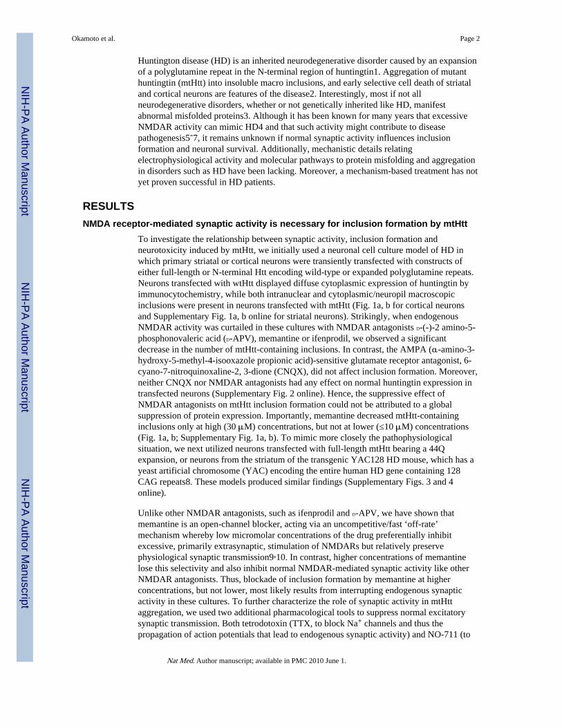
Huntington disease (HD) is an inherited neurodegenerative disorder caused by an expansionof a polyglutamine repeat in the N-terminal region of huntingtin1. Aggregation of mutanthuntingtin (mtHtt) into insoluble macro inclusions, and early selective cell death of striataland cortical neurons are features of the disease2. Interestingly, most if not allneurodegenerative disorders, whether or not genetically inherited like HD, manifestabnormal misfolded proteins3. Although it has been known for many years that excessiveNMDAR activity can mimic HD4 and that such activity might contribute to diseasepathogenesis5-7, it remains unknown if normal synaptic activity influences inclusionformation and neuronal survival. Additionally, mechanistic details relatingelectrophysiological activity and molecular pathways to protein misfolding and aggregationin disorders such as HD have been lacking. Moreover, a mechanism-based treatment has notyet proven successful in HD patients.
RESULTSNMDA receptor-mediated synaptic activity is necessary for inclusion formation by mtHtt
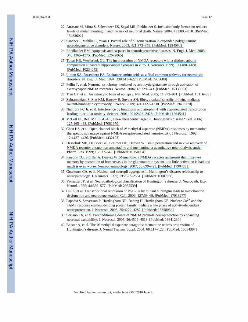
To investigate the relationship between synaptic activity, inclusion formation andneurotoxicity induced by mtHtt, we initially used a neuronal cell culture model of HD inwhich primary striatal or cortical neurons were transiently transfected with constructs ofeither full-length or N-terminal Htt encoding wild-type or expanded polyglutamine repeats.Neurons transfected with wtHtt displayed diffuse cytoplasmic expression of huntingtin byimmunocytochemistry, while both intranuclear and cytoplasmic/neuropil macroscopicinclusions were present in neurons transfected with mtHtt (Fig. 1a, b for cortical neuronsand Supplementary Fig. 1a, b online for striatal neurons). Strikingly, when endogenousNMDAR activity was curtailed in these cultures with NMDAR antagonists D-(-)-2 amino-5-phosphonovaleric acid (D-APV), memantine or ifenprodil, we observed a significantdecrease in the number of mtHtt-containing inclusions. In contrast, the AMPA (α-amino-3-hydroxy-5-methyl-4-isooxazole propionic acid)-sensitive glutamate receptor antagonist, 6-cyano-7-nitroquinoxaline-2, 3-dione (CNQX), did not affect inclusion formation. Moreover,neither CNQX nor NMDAR antagonists had any effect on normal huntingtin expression intransfected neurons (Supplementary Fig. 2 online). Hence, the suppressive effect ofNMDAR antagonists on mtHtt inclusion formation could not be attributed to a globalsuppression of protein expression. Importantly, memantine decreased mtHtt-containinginclusions only at high (30 μM) concentrations, but not at lower (≤10 μM) concentrations(Fig. 1a, b; Supplementary Fig. 1a, b). To mimic more closely the pathophysiologicalsituation, we next utilized neurons transfected with full-length mtHtt bearing a 44Qexpansion, or neurons from the striatum of the transgenic YAC128 HD mouse, which has ayeast artificial chromosome (YAC) encoding the entire human HD gene containing 128CAG repeats8. These models produced similar findings (Supplementary Figs. 3 and 4online).
Unlike other NMDAR antagonists, such as ifenprodil and D-APV, we have shown thatmemantine is an open-channel blocker, acting via an uncompetitive/fast ‘off-rate’mechanism whereby low micromolar concentrations of the drug preferentially inhibitexcessive, primarily extrasynaptic, stimulation of NMDARs but relatively preservephysiological synaptic transmission9,10. In contrast, higher concentrations of memantinelose this selectivity and also inhibit normal NMDAR-mediated synaptic activity like otherNMDAR antagonists. Thus, blockade of inclusion formation by memantine at higherconcentrations, but not lower, most likely results from interrupting endogenous synapticactivity in these cultures. To further characterize the role of synaptic activity in mtHttaggregation, we used two additional pharmacological tools to suppress normal excitatorysynaptic transmission. Both tetrodotoxin (TTX, to block Na+ channels and thus thepropagation of action potentials that lead to endogenous synaptic activity) and NO-711 (to
Okamoto et al. Page 2
Nat Med. Author manuscript; available in PMC 2010 June 1.
NIH
-PA Author Manuscript
NIH
-PA Author Manuscript
NIH
-PA Author Manuscript
block GABA uptake and hence augment inhibitory neurotransmission) decreased mtHtt-containing inclusions (Fig. 1c). Taken together, our data suggest that interruption of normalexcitatory synaptic activity (mediated by NMDARs) ameliorates macro inclusion formation.
Electrophysiological validation of pharmacological modifiers of synaptic activityTo confirm the notion that our pharmacological manipulations modified synaptic activity,we used whole-cell recording with patch electrodes to study spontaneous excitatorypostsynaptic currents (sEPSCs). These experiments were designed to analyze the effect ofshort-term additions of drugs to determine their mechanism of action. The same drugs wereused to prevent sEPSCs from triggering the intracellular signaling cascades involved ininclusion formation or neuronal cell death (Fig. 1; Supplementary Fig. 1). We found thattotal charge transfer during the NMDAR-mediated component of sEPSCs was significantlyreduced by TTX or NO-711, in both wtHtt and mtHtt transfected cortical neurons (Fig. 2a,b). Similarly, high (30 μM) memantine significantly inhibited the NMDAR-mediatedcomponent of sEPSCs, while low (1-10 μM) memantine relatively spared this synapticactivity (Fig. 2c). In contrast, ifenprodil (10 μM) blocked this synaptic activity (Fig. 2d).However, in the presence of 10 μM CNQX, NMDAR-mediated synaptic activity was notsignificantly attenuated (Fig. 2e).
Additionally, both memantine (even at low concentrations), and ifenprodil, attenuatedextrasynaptic NMDAR-mediated currents (Fig. 2f). The effect of low-dose memantine onextrasynaptic NMDAR-mediated currents was further confirmed by isolating extrasynapticNMDAR activity using the previously published MK-801 protocol11 (Supplementary Fig. 5online). These electrophysiological findings validate the specificity of our pharmacologicaltools for NMDARs when studying mtHtt aggregation and neuronal cell death. Next, weexamined whether NMDAR-sEPSCs were affected by mtHtt under our conditions. Wefound that total charge transfer during sEPSCs was not significantly different in mtHtt- vs.wtHtt-transfected neurons (Fig. 2g). Similar findings were observed in striatal slicerecordings from the YAC72 mouse model of HD during low-frequency cortical afferentstimulation12, simulating the spontaneous activity level in our cultures. At higher levels ofstimulation, mtHtt itself is known to enhance NMDAR activity6.
Chaperonin TRiC facilitates synaptic activity-dependent inclusion formationNext, we investigated the molecular mechanism of mtHtt-induced inclusion formation.Recently, three groups reported in yeast and cell lines that the chaperonin TRiC (TCP1 ringcomplex, also known as CCT) detoxifies mtHtt, at least in part by forming multiple mtHttinclusions, while decreasing toxic soluble microaggregates of mtHtt and thereby decreasingtoxicity13. Augmentation of TRiC activity increased the appearance of multiple, smallmtHtt-containing inclusions (Fig. 3c and 5c in Tam et al.14), reminiscent of our findings inneurons with preserved synaptic activity (our Fig. 1a; Supplementary Figs. 1a and 3). Hence,we examined if synaptic activity regulates the expression of TCP1, the subunit of TRiC thatpreferentially interacts with mtHtt14. We added TTX to our neuronal cultures to blocksynaptic activity and found that it significantly reduced the expression of TCP1 (Fig. 3a).While low-dose memantine (10 μM) did not affect the TCP1 expression, both high-dosememantine (30 μM) and ifenprodil also decreased TCP1 levels (Supplementary Fig. 6online), supporting the notion that synaptic NMDAR activity is necessary for TCP1expression.
To test if TCP1 is involved in synaptic activity-induced inclusion formation, we generatedtwo small hairpin RNA interference constructs (shRNAs designated TCP1 RNAi-1 andRNAi-2) to knockdown TCP1 expression. TCP1 RNAi-1 and RNAi-2 reduced TCP1 levelsto 70% and 60%, respectively (Supplementary Fig. 7 online). Both small hairpin TCP1
Okamoto et al. Page 3
Nat Med. Author manuscript; available in PMC 2010 June 1.
NIH
-PA Author Manuscript
NIH
-PA Author Manuscript
NIH
-PA Author Manuscript
RNAi vectors significantly decreased inclusion formation from mtHtt (Fig. 3b).Additionally, knockdown of TCP1 by RNAi occluded the inhibitory effect on inclusionformation exerted by suppression of synaptic activity with high-dose (30 μM) memantine(Supplementary Fig. 8 online). Taken together, our results indicate that synaptic activityinduces expression of TCP1, which in turn mediates inclusion formation in neurons.Moreover, TRiC has been reported to cooperate with the Hsp70 chaperone system15-17;Hsp70 attaches to mtHtt inclusions to protect cells from death17,18. Indeed, here weobserved co-localization of inclusions and Hsp70 (Supplementary Fig. 9 online), suggestingthat the TRiC-Hsp70 system might ameliorate mtHtt toxicity.
mtHtt renders neurons more vulnerable to excitotoxic insult via extrasynaptic NMDAreceptors
We have thus shown that physiological synaptic NMDAR activity is necessary for macroinclusion formation in mtHtt-expressing neurons. However, the relationship of mtHtt-induced inclusion formation and neurotoxicity in HD pathogenesis has remainedcontentious19,20. Large aggregates of abnormally folded proteins have been hypothesizedto contribute to synaptic damage in neurodegenerative disorders21, but a recent reportsuggests that inclusion formation protects neurons from cell death22, possibly by decreasingthe level of toxic soluble forms of mtHtt23. Neurodegeneration in HD has also beenproposed to represent excitotoxic-mediated apoptotic cell death triggered by excessiveactivation of NMDARs24. Increased sensitivity to NMDAR-mediated excitotoxicity hasbeen observed in the YAC128 mouse model of HD6. On the other hand, recent studies haveindicated that physiological levels of synaptic NMDAR activity (predominantly composedof NR2A subunits in mature neurons) promote survival in the face of various forms ofstress11. The exact role of normal neuronal electrical activity in the pathogenesis of HD hasnot previously been studied.
Our observation that endogenous synaptic NMDAR activity promotes formation of mtHtt-containing inclusions prompted our investigation of whether this normal activity couldprotect neurons. Conversely, we hypothesized that excessive activation of extrasynapticNMDARs may increase neuronal vulnerability induced by mtHtt. To test this notion, weinitially applied a relatively low concentration of exogenous glutamate (50 μM), which byitself was not excitotoxic to untransfected or wtHtt-transfected neurons under theseconditions, to stimulate both extrasynaptic and synaptic receptors. While transfection ofmtHtt by itself did not result in neuronal death, as shown previously6, we found that mtHttneurons became more vulnerable in the presence of 50 μM glutamate (Fig. 4a-c). WhenmtHtt neurons were treated with memantine or ifenprodil, neuronal cell death induced byexogenous glutamate was significantly reduced. Of note, we found that low concentrationsof memantine (1-10 μM), which selectively block excessive extrasynaptic NMDAR activitywhile sparing physiological synaptic NMDARs (Fig. 2; Supplementary Figs. 5 and 10online)9,10, were sufficient to protect mtHtt-transfected neurons from glutamate challenge.Additionally, extrasynaptic NMDARs preferentially contain NR2B subunits25, andifenprodil, a relatively selective inhibitor of NR2B, blocked this neurotoxicity6,11. Takentogether, these results imply that mtHtt renders neurons more vulnerable to acute excitotoxicinsults and that blockade of extrasynaptic NMDARs efficiently ameliorates this excitotoxiccell death. In this regard, we observed similar results for both truncated (N-terminalfragment) (Fig. 4b) and full-length Htt constructs (Fig. 4c).
Physiological synaptic activity protects mtHtt expressing-neurons from cell deathNext, we examined the effect of normal synaptic activity on survival of neurons expressingmtHtt. We found that TTX, which suppresses normal excitatory synaptic transmission, orNO-711, which enhances inhibitory neurotransmission, triggered significant cell death in
Okamoto et al. Page 4
Nat Med. Author manuscript; available in PMC 2010 June 1.
NIH
-PA Author Manuscript
NIH
-PA Author Manuscript
NIH
-PA Author Manuscript
mtHtt- but not wtHtt-expressing neurons (Fig. 4d; Supplementary Fig. 11 online). We thenasked if extrasynaptic NMDAR activity causes cell death in mtHtt neurons when protective(physiological) synaptic NMDAR activity is inhibited. Accordingly, we investigated if anNMDAR antagonist that preferentially blocks extrasynaptic activity could protect mtHttneurons from toxicity induced by TTX or NO-711. Indeed, we found that treating mtHttcultures with low concentrations (~5 μM) of memantine significantly reduced neuronaldeath induced by TTX or NO-711 (Fig. 4d). The question arises where the extrasynapticglutamate originates in our culture system in the presence of neuronal activity blockade withTTX because synaptic release is inhibited. Since the intracellular pool of glutamate, used formetabolic purposes, may be significant, up to 10 mM per cell26, mtHtt-insulted neuronsmay potentially leak glutamate from nonsynaptic sites. Additionally, similar to the intactbrain, our culture system contains nonneuronal cells, predominantly astrocytes. Even in theabsence of synaptic activity, glutamate released from astrocytes can activate extrasynapticNMDARs in neurodegenerative conditions27,28.
Effect of electrical activity on the molecular pathways involved in inclusion formation andcell survival in mtHtt-expressing neurons
We next wanted to investigate the relationship of macro inclusion formation, electricalactivity, and neuronal survival pathways. Since we had demonstrated earlier that TCP1mediates inclusion formation (Fig. 3b), we examined if TCP1 is involved in the survival ofneurons expressing mtHtt. We found that knockdown of TCP1 not only decreased inclusionformation but also significantly increased neuronal cell death in mtHtt-expressing neurons(Fig. 4e). These RNAi experiments imply causality between TCP1 levels, inclusionformation, and neuronal cell death. Importantly, this form of cell death was significantlyameliorated by blockade of extrasynaptic NMDARs using low-dose memantine (Fig. 4e).Taken together, our results suggest that synaptic activity induces expression of TCP1, whichin turn contributes to protective inclusion formation and decreased neurotoxicity.Conversely, blockade of synaptic activity reduces TCP1 levels, which in turn decreasesmtHtt inclusion formation, and contributes to neuronal cell death in conjunction withexcessive extrasynaptic NMDARs activity.
Additionally, using low-dose memantine, we investigated if the level of Rhes, a smallguanine nucleotide-binding protein recently reported to mediate mtHtt sumoylation,disaggregation and cytotoxicity29, is affected by blockade of extrasynaptic NMDARs. Wefound that low-dose memantine (5-10 μM) reduced Rhes levels (Supplementary Fig. 12online), consistent with the notion that extrasynaptic NMDAR activity controls Rhesexpression. Thus, the detrimental effects of excessive extrasynaptic NMDAR activity in thecontext of mtHtt may be ascribed, at least in part, to a relative increase in Rhes.
NMDAR activity modulates the neuroprotective CREB—PGC-1α pathway in neuronsexpressing mtHtt
CREB/CBP transcriptional activity, which triggers the neuroprotective PGC-1α pathway,has been shown to be decreased by extrasynaptic NMDAR activity11 as well as by bindingto soluble mtHtt30. We therefore hypothesized that impairment of the CREB—PGC-1αcascade may contribute to neuronal cell death in HD31. Hence, we determined if CREBfunction was compromised in mtHtt-expressing neurons when synaptic activity was blocked.In mtHtt- but not wtHtt-transfected neurons we found a significant decrease in CREBactivity in the presence of TTX, which suppresses excitatory synaptic activity (Fig. 4f).Additionally, blockade of extrasynaptic NMDARs with low concentrations of memantinerestored CREB function, suggesting that mtHtt and activation of extrasynaptic NMDARs arenecessary for CREB inactivation.
Okamoto et al. Page 5
Nat Med. Author manuscript; available in PMC 2010 June 1.
NIH
-PA Author Manuscript
NIH
-PA Author Manuscript
NIH
-PA Author Manuscript
We next determined the level of PGC-1α under these same conditions. We observed asignificant decrease in PGC-1α levels in mtHtt-transfected neurons in the presence of TTX,but preservation of PGC-1α levels in the presence of low concentrations of memantine (Fig.4g; Supplementary Fig. 13 online). Additionally, we found that RNAi knockdown of TCP1decreased CREB activity and PGC-1α levels in mtHtt-expressing neurons (SupplementaryFig. 14 online). These findings support the notion that mtHtt in the absence of TCP1, inconjunction with excessive extrasynaptic NMDAR activity, interferes with theneuroprotective CREB—PGC-1α cascade. Importantly, decreased levels of PGC-1α likelycontribute to cell death in mtHtt-transfected neurons exposed to TTX (and thus in neuronswith blocked synaptic activity and decreased mtHtt inclusions). As would be predicted, celldeath under these conditions was diminished by co-transfection with PGC-1α (Fig. 4d).
NMDAR activity modulates toxicity and behavior in HD transgenic miceThese findings raise the possibility that long-term blockade of extrasynaptic activity wouldbe advantageous while simultaneous blockade of synaptic activity might prove deleterious invivo, for example, in the transgenic YAC128 HD mouse8 (Supplementary Fig. 4b). Wehypothesized that the balance between synaptic and extrasynaptic activity in the face ofmtHtt would determine neuronal survival. Therefore, while short-term blockade of bothsynaptic and extrasynaptic activity might cause transient improvement, eventually blockadeof synaptic activity would lead to neuronal loss. In contrast, maintenance of synaptic activitywith abrogation of excessive extrasynaptic activity would be most beneficial to combat thedeleterious effects of mtHtt. To test this hypothesis in vivo, YAC128 mice were treatedusing drinking water with low-dose memantine (1 mg kg-1) to block extrasynaptic NMDARactivity or high-dose memantine (30 mg kg-1) to additionally block synaptic NMDARactivity since we knew that these doses would produce low and high concentrations ofmemantine approaching those used in the culture experiments9,10,32-34. Treatmentcommenced at 2 months of age and continued for 10 months until 12 months of age. Wefirst analyzed mtHtt inclusions using immunostaining with antibody EM48, whichrecognizes N-terminal huntingtin and is highly specific for aggregates35. Treatment withlow-dose memantine increased inclusion formation (Fig. 5a, b), as confirmed using a filtertrap assay (Supplementary Fig. 15 online), while high-dose memantine significantlydecreased inclusion formation in 12-month-old YAC128 mice (Fig. 5a, b). Accordingly,high-dose memantine also increased soluble mtHtt (Supplementary Fig. 16 online). Theseeffects of memantine treatment on inclusion formation were not due to alterations in mtHttprotein expression since low and high doses of memantine did not affect mtHtt levels(Supplementary Fig. 17 online). On the other hand, TCP1 levels increased with low-dosememantine and decreased with high-dose memantine (Supplementary Fig. 18 online),supporting the notion that normal synaptic NMDAR activity increases TCP1 and promotesinclusion formation.
Next, we measured loss in striatal volume in YAC128 mice, a cardinal neuropathologicalfeature of HD36. Low-dose memantine improved striatal volume, while high-dosememantine worsened this parameter (Fig. 5c). These findings support our hypothesis thatmaintenance of synaptic activity, while inhibiting excessive extrasynaptic activity, isprotective, and that long-term blockade of synaptic activity is detrimental. We alsomonitored motor function by rotatrod testing at 12 months of age. YAC128 HD mice treatedwith low-dose memantine demonstrated improvements on these motor tests, while high-dose-treated mice showed no improvement (Fig. 5d). Taken together, these results supportthe premise that maintenance of synaptic activity with abrogation of extrasynaptic activity isbeneficial in YAC128 HD mice.
Okamoto et al. Page 6
Nat Med. Author manuscript; available in PMC 2010 June 1.
NIH
-PA Author Manuscript
NIH
-PA Author Manuscript
NIH
-PA Author Manuscript
DISCUSSIONOur results provide a mechanistic framework to help us understand the selectivevulnerability of striatal and cortical neurons in HD. We show here that synaptic activitycontrols expression of the chaperonin TRiC, which in turn modulates inclusion formationand toxicity of mtHtt. Moreover, the electrical properties of these neurons coupled with thepredominant effect of the small guanine nucleotide-binding protein Rhes and the CREB—PGC-1α pathway in this cell type29,37 renders them particularly sensitive to the toxiceffects of mtHtt. Specifically, excessive extrasynaptic activity increases the relative Rheslevel and, in conjunction with mtHtt, decreases CREB—PGC-1α activity to promoteneuronal cell death. Coupled with a decrease in synaptic transmission, which alsocontributes to a decrease in CREB activity in the presence of mtHtt, neurons becomeincreasingly vulnerable to injury and death (Fig. 6). We speculate that these findings implythat therapies for HD that minimize excessive extrasynaptic activity while maintaining orenhancing normal synaptic activity will yield considerable benefit. Notably, it has beencontentious whether mtHtt inclusions are cytotoxic. The presence of inclusions may reflecteither successful sequestration of toxic soluble oligomers of mtHtt and thus contribute toneuroprotection, or insufficient sequestration of toxic soluble mtHtt, which can no longer beaccommodated by the inclusion formation process, and is thus toxic. Here, we report thatsynaptic activity-driven inclusions are neuroprotective, supporting the former premise.
Nonetheless, we also demonstrate that cultured neurons containing mtHtt inclusions becomeincreasingly vulnerable to excitotoxic insults, consistent with the notion that over time,accumulation of excitotoxic challenges may eventually contribute to the death of mtHtt-expressing neurons in HD6. We show not only that mtHtt increases the vulnerability ofneurons to relatively low concentrations of exogenous glutamate, but also that suppressingspontaneous excitatory synaptic activity can mimic this phenomenon via the excitotoxiceffect of endogenous glutamate apparently acting on extrasynaptic receptors.
Our data using primary neurons manifesting synaptic activity may also explain why othershad reported direct mtHtt toxicity in the absence of excitotoxic insult in yeast, various celllines, and synaptically-immature neurons. That is, the presence of normal synaptic activityin our mature neuronal cultures leads to the formation of inclusions, thereby avoiding toxicoligomers of mtHtt, whereas these other cell types lack synaptic activity and therefore areexposed to soluble mtHtt oligomers.
Our findings further suggest that the balance between synaptic and extrasynaptic NMDARactivity may be critical in determining neuronal cell survival in HD. We show that lowconcentrations of the NMDAR antagonist, memantine, afford the advantage of restoringexcitatory balance by maintaining physiological synaptic activity while blocking excessiveextrasynaptic NMDAR stimulation9,10. Inhibiting excessive extrasynaptic NMDAR activitydecreased Rhes levels, thus preserving mtHtt inclusions and lessening mtHtt cytotoxicity29.To the contrary, blocking synaptic activity with high-dose memantine decreased inclusionformation and increased soluble mtHtt. Soluble mtHtt interferes with the neuroprotectiveCREB—PGC-1α pathway, thus accelerating neuronal cell death. Indeed, prolongedadministration of high-dose memantine may contribute to toxicity for several reasons,including interference with other neuroprotective pathways that are dependent on synapticactivity, such as the phosphatidylinositol 3 kinase (PI3K)-Akt cascade38,39. Thus, we positthat restoring excitatory balance, which is disrupted by mtHtt protein, can affect proteinmisfolding and protect neurons in HD, as also suggested in a small, open-label humanclinical trial of memantine in HD patients40. This novel concept of balancing synaptic andextrasynaptic neuronal NMDAR activity may also lead to strategies to combat cell injuryand death associated with other neurodegenerative disorders.
Okamoto et al. Page 7
Nat Med. Author manuscript; available in PMC 2010 June 1.
NIH
-PA Author Manuscript
NIH
-PA Author Manuscript
NIH
-PA Author Manuscript
Supplementary MaterialRefer to Web version on PubMed Central for supplementary material.
AcknowledgmentsWe thank C. A. Ross (Johns Hopkins University School of Medicine) and L. M. Ellerby (Buck Institute for AgeResearch) for N-terminal and full-length constructs of huntingtin, T. F. Newmeyer and H. Fang for providingprimary neuronal cultures, X.-J. Li (Emory University School of Medicine) for providing the EM48 antibody, andE. Bossy-Wetzel, Y.S. Choo, W. Zago, and T. Nakamura for assistance or discussion. This work was supported inpart by NIH grants P01 HD29587, R01 EY09024, R01 EY05477 and R01 NS41207, and a Senior Scholar Award inAging Research from the Ellison Medical Foundation (S.A.L.). Additional support was provided by the NIHBlueprint Grant for La Jolla Interdisciplinary Neuroscience Center Cores P30 NS057096. M.A.P. was supported bythe Canadian Institute of Health Research and the Michael Smith Foundation for Health Research. M.R.H. wassupported by grants from the Canadian Institutes of Health Research, the Huntington Society of Canada, theHereditary Disease Foundation, the Huntington’s Disease Society of America, CHDI Inc., and the HighQFoundation.
Appendix
ONLINE METHODSCell culture and transfection
Primary cerebrocortical or striatal cultures, containing both neurons and glia, were madefrom E16 rat pups and grown on 12-mm glass coverslips, as we have described41. Cellswere transfected via LipofectAMINE 2000 (Invitrogen) on the 15-20th DIV with either N-terminal fragments of Htt consisting of exon 1 (Myc-wtHtt-N63-18Q or Myc-mtHtt-N63-148Q), or with full-length Htt (wtHtt with 15Q or mtHtt with 138Q). In a number ofexperiments, cells were co-transfected with enhanced green fluorescence protein (EGFP) tofacilitate identification of transduced cells. Labeled cells could be subsequently stained withanti-NeuN or anti-MAP-2 antibody, verifying their neuronal identity. In a series ofexperiments, a PGC-1α expression vector (ATCC) was also co-transfected. Transfectionefficiency was 10-15% for cortical neurons, and 5-10% for striatal neurons. In experimentsmanipulating synaptic activity, drugs were added to cultures five hours post transfection.Extracellular Mg2+ was set at ≤ 0.4 mM to produce spontaneous NMDAR activity in theabsence of excessive AMPAR stimulation, as assessed by patch-clamp recordings42. CREBtranscriptional activity was analyzed using a CRE luciferase reporter (Stratagene) and renillaluciferase internal control vector (Promega), as previously described41. siSTRIKE plasmidsexpressing shRNAs targeting TCP1 and a non-targeted sequence were constructed accordingto the manufacturer’s protocol (Promega). TCP1 shRNA targeting the sequences weredesigned using Promega’s siRNA Target Designer.
Assessment of inclusion formation and neuronal cell deathFor assessment of inclusions, Htt-transfected cortical or striatal neurons were stained withanti-myc (for truncated N-terminal Htt constructs, Santa Cruz), or anti-huntingtin (for full-length Htt constructs, Chemicon) to visualize huntingtin expression under deconvolutionmicroscopy. The expression level of transfected full-length Htt was sufficiently abovebaseline to allow us to easily distinguish transfected from endogenous Htt (SupplementaryFig. 3). Images were captured with a Zeiss Axiovert 100M microscope equipped withdeconvolution software (see Supplementary Methods).
Inclusion formation was assessed 15-17 h after drug application. Immunocytochemistry forMAP2 (Sigma) and NeuN (Chemicon) were used to identify neurons. The total number oftransfected neurons with Htt-positive inclusions and the total number of transfected neuronson each coverslip were counted. In a series of experiments combining inclusion and
Okamoto et al. Page 8
Nat Med. Author manuscript; available in PMC 2010 June 1.
NIH
-PA Author Manuscript
NIH
-PA Author Manuscript
NIH
-PA Author Manuscript
neuronal cell death analysis, wtHtt or mtHtt was co-transfected with EGFP, the latter tofacilitate identification of transduced cells; in this manner, we could normalize the numberof viable, dead, or inclusion-containing neurons to the total number of transfected neuronsfor each treatment paradigm. In the cell death experiments, wtHtt- or mtHtt-expressingneurons were incubated in media with or without 50 μM glutamate for 40 h. In anotherseries of experiments, cultures were incubated for 20 h with TTX or NO-711. In someexperiments, PGC-1α was also expressed. Cells were then stained with anti-MAP2/anti-NeuN, anti-myc antibodies, anti-PGC-1α antibody (Santa Cruz), anti-TCP1 antibody (SantaCruz), and Hoechst dye 33342. Neuronal cell death was assessed on the basis of condensednuclear morphology (with Hoechst dye) and shrunken neurites. Neurons were scored asviable if they possessed normal nuclei and neuritic processes30. In the assessment of bothinclusion formation and neuronal cell death, for each experimental condition, ≥100 neuronswere counted on triplicate coverslips, and the experiments were repeated at least four times;thus, n ≥ 1200 in each case. Data are expressed as mean ± s.e.m., and statistical significancewas determined using an ANOVA followed by Dunnett’s post-hoc test.
Electrophysiological recordingsWhole-cell recordings were performed with an Axopatch 200B amplifier (AxonInstruments) from EGFP-positive neurons 48 to 96 h after transfection42. The extracellularsolution contained (in mM): NaCl 135, KCl 2.5, CaCl2 2, MgCl2 0-0.01, NaHCO3 1,Na2HPO4 0.34, KH2PO4 0.44, glucose 20, Hepes 10, pH 7.4. Patch electrodes with a finaltip resistance of 4-7 MΩ, were filled with a solution containing (in mM): CsCl 120,tetraethylammonium chloride (TEA-Cl) 20, Hepes 10, EGTA 2.25, CaCl2 1, MgCl2 2, andphalloidin 0.001, pH 7.4. To examine NMDAR-mediated sEPSCs, we used “chargetransfer.” Details of this analysis are given in the Analysis of NMDAR-mediated sEPSCssection in the Supplementary Methods and in Supplementary Fig. 10. pCLAMP 8-9.2software (Axon Instruments) was used for data acquisition and analysis. All recordings weremade at room temperature with a holding potential of -60 mV. Currents were digitallysampled at 10-20 kHz and filtered at 2-5 kHz. Data are expressed as mean ± s.e.m., andstatistical significance was determined using a Student’s t-test for pairwise comparisons.
Transgenic YAC128 HD miceMale and female YAC128 mice expressing expanded human huntingtin with 128 CAGrepeats and WT littermates maintained on the FVB/N strain (Charles River) were used forthese experiments8,43. Mice were housed singly or in pairs in duplex cages with littermatesof mixed genotype and maintained under a 12 L:12 D light cycle (lights on at 2300) in aclean facility and given free access to food and water. Experimenters were blind to thegenotype of the mice. All experiments were performed with the approval of the animal carecommittee at the University of British Columbia.
Drug treatmentFor administration of drugs in the drinking water, water consumption of individual cageswas monitored on a biweekly-basis along with animal body weights. The concentrations ofdrug solutions for each cage were then adjusted accordingly. The drug solutions werereplaced twice/week and provided ad libitum. Animals were treated starting at 2 months ofage with 1 mg kg-1 day-1 to 30 mg kg-1 day-1 of memantine for 10 months, and theirneuropathology and motor function was assessed at 12 months of age. The apparenteffective concentration of memantine at the NMDAR-associated channel in vivo under theseconditions after administration of low-dose memantine was estimated to be in the range of5-10 μM based on our current observations (see Supplementary Methods).
Okamoto et al. Page 9
Nat Med. Author manuscript; available in PMC 2010 June 1.
NIH
-PA Author Manuscript
NIH
-PA Author Manuscript
NIH
-PA Author Manuscript
Assessment of motor functionTraining and baseline testing for motor function tasks were carried out prior to memantinetreatment initiation at 2 months of age during the dark cycle. Assessment of the effect oflong-term (10-month) treatment with memantine on motor function was carried out bytesting at 12 months of age. Motor coordination and balance were assessed usingaccelerating and fixed-speed rotarod tasks (UGO Basile, Comerio, Italy). In the acceleratingtask, the rotarod sped up from 5 revolutions per minute (RPM) to 40 RPM over 4 minutes.In the fixed task, the rotarod revolved at 24 RPM. Performance in the rotarod tasks wasassessed by the amount of time that a mouse could remain running on the rotarod. Duringtraining, mice were given three trials per day for three consecutive days.
Neuropathological analysisA series of 25 μm-thick coronal sections spaced 200 μm apart spanning the striatum wasstained with NeuN antibody (Chemicon) overnight at room temperature, followed byincubation with biotinylated anti-mouse antibody (Vector Laboratories, Burlingame). Thesignal was amplified with an ABC Elite kit (Vector) and detected with diaminobenzidine(DAB; Pierce). Striatal volumes were determined from a series of mounted sections usingStereoInvestigator software (Microbrightfield) by tracing the perimeter of the striatum inserial sections spanning the striatum.
Assessment of huntingtin inclusions in vivoPerfused brain sections of 25 μm thickness from WT, untreated YAC128, 1 mg kg-1- and 30mg kg-1-treated YAC128 animals were immunoassayed with the antibody EM48 to assessthe presence of mtHtt inclusions, as described previously35. Polyclonal EM48 antibody wasused at 1:500 and DAB was used as the chromogen (Vector). Htt inclusions were defined asEM48-positive staining at the light microscope level. Photographs of mounted sections weretaken on a light microscope (Zeiss) under a 100X objective using a MetaMorph ImagingSystem (Molecular Devices), and the intensity of the staining of striatal neurons wasmeasured.
Statistical analysisData are expressed as mean ± s.e.m. When suitable, results were interpreted using a one-wayANOVA with a Student-Newman-Keuls (SNK) post-hoc test. Pairwise comparisonsbetween genotypes/treatments were assessed with a Student’s t-test. Differences wereconsidered statistically significant at P < 0.05.
Additional methodsSupporting methodology is described in the Supplementary Methods.
References
41. Okamoto, S.-i., et al. Dominant-interfering forms of MEF2 generated by caspase cleavagecontribute to NMDA-induced neuronal apoptosis. Proc. Natl. Acad. Sci. USA. 2002; 99:3974–3979. [PubMed: 11904443]
42. Lei SZ, et al. Effect of nitric oxide production on the redox modulatory site of the NMDAreceptor-channel complex. Neuron. 1992; 8:1087–1099. [PubMed: 1376999]
43. Van Raamsdonk JM, et al. Cognitive dysfunction precedes neuropathology and motorabnormalities in the YAC128 mouse model of Huntington’s disease. J. Neurosci. 2005; 25:4169–4180. [PubMed: 15843620]
Okamoto et al. Page 10
Nat Med. Author manuscript; available in PMC 2010 June 1.
NIH
-PA Author Manuscript
NIH
-PA Author Manuscript
NIH
-PA Author Manuscript
References1. The Huntington’s Disease Collaborative Research Group. A novel gene containing a trinucleotide
repeat that is expanded and unstable on Huntington’s disease chromosomes. Cell. 1993; 72:971–983. [PubMed: 8458085]
2. DiFiglia M, et al. Aggregation of huntingtin in neuronal intranuclear inclusions and dystrophicneurites in brain. Science. 1997; 277:1990–3. [PubMed: 9302293]
3. Ciechanover A, Brundin P. The ubiquitin proteasome system in neurodegenerative diseases:sometimes the chicken, sometimes the egg. Neuron. 2003; 40:427–446. [PubMed: 14556719]
4. Beal MF, et al. Replication of the neurochemical characteristics of Huntington’s disease byquinolinic acid. Nature. 1986; 321:168–171. [PubMed: 2422561]
5. Ferrante RJ, et al. Therapeutic effects of coenzyme Q10 and remacemide in transgenic mousemodels of Huntington’s disease. J. Neurosci. 2002; 22:1592–1599. [PubMed: 11880489]
6. Zeron MM, et al. Increased sensitivity to N-methyl-d-aspartate receptor-mediated excitotoxicity in amouse model of Huntington’s disease. Neuron. 2002; 33:849–860. [PubMed: 11906693]
7. Heng MY, Detloff PJ, Wang PL, Tsien JZ, Albin RL. In vivo evidence for NMDA receptor-mediated excitotoxicity in a murine genetic model of Huntington disease. J. Neurosci. 2009;29:3200–3205. [PubMed: 19279257]
8. Slow EJ, et al. Selective striatal neuronal loss in a YAC128 mouse model of Huntington disease.Human Molec. Genet. 2003; 12:1555–1567. [PubMed: 12812983]
9. Lipton SA. Paradigm shift in neuroprotection by NMDA receptor blockade: Memantine and beyond.Nat. Rev. Drug Disc. 2006; 5:160–170.
10. Lipton SA. Pathologically activated therapeutics for neuroprotection. Nat. Rev. Neurosci. 2007;8:803–808. [PubMed: 17882256]
11. Hardingham GE, Fukunaga Y, Bading H. Extrasynaptic NMDARs oppose synaptic NMDARs bytriggering CREB shut-off and cell death pathways. Nat. Neurosci. 2002; 5:405–414. [PubMed:11953750]
12. Li L, Murphy TH, Hayden MR, Raymond LA. Enhanced striatal NR2B-containing N-methyl-d-aspartate receptor-mediated synaptic currents in a mouse model of Huntington disease. J.Neurophysiol. 2004; 92:2738–2746. [PubMed: 15240759]
13. Pickett J. Folding away the bad guys. Nat. Rev. Neurosci. 2006; 7:832–833.
14. Tam S, Geller R, Spiess C, Frydman J. The chaperonin TRiC controls polyglutamine aggregationand toxicity through subunit-specific interactions. Nat. Cell Biol. 2006; 8:1155–1162. [PubMed:16980959]
15. Melville MW, McClellan AJ, Meyer AS, Darveau A, Frydman J. The Hsp70 and TRiC/CCTchaperone systems cooperate in vivo to assemble the von Hippel-Lindau tumor suppressorcomplex. Mol. Cell. Biol. 2003; 23:3141–3151. [PubMed: 12697815]
16. Behrends C, et al. TRiC promotes the assembly of polyQ expansion proteins into nontoxicoligomers. Mol. Cell. 2006; 23:887–897. [PubMed: 16973440]
17. Kitamura A, et al. Cytosolic chaperonin prevents polyglutamine toxicity with altering theaggregation state. Nat. Cell Biol. 2006; 8:1163–1170. [PubMed: 16980958]
18. King MA, et al. Cytoplasmic inclusions of Htt exon1 containing an expanded polyglutamine tractsuppress execution of apoptosis in sympathetic neurons. J. Neurosci. 2008; 31:14401–14415.[PubMed: 19118173]
19. Davies SW, et al. Formation of neuronal intranuclear inclusions underlies the neurologicaldysfunction in mice transgenic for the HD mutation. Cell. 1997; 90:537–548. [PubMed: 9267033]
20. Saudou F, Finkbeiner S, Devys D, Greenberg ME. Huntingtin acts in the nucleus to induceapoptosis but death does not correlate with the formation of intranuclear inclusions. Cell. 1998;95:55–66. [PubMed: 9778247]
21. Tsai J, Grutzendler J, Duff K, Gan WB. Fibrillar amyloid deposition leads to local synapticabnormalities and breakage of neuronal branches. Nat. Neurosci. 2004; 7:1181–1183. [PubMed:15475950]
Okamoto et al. Page 11
Nat Med. Author manuscript; available in PMC 2010 June 1.
NIH
-PA Author Manuscript
NIH
-PA Author Manuscript
NIH
-PA Author Manuscript
22. Arrasate M, Mitra S, Schweitzer ES, Segal MR, Finkbeiner S. Inclusion body formation reduceslevels of mutant huntingtin and the risk of neuronal death. Nature. 2004; 431:805–810. [PubMed:15483602]
23. Sanchez I, Mahlke C, Yuan J. Pivotal role of oligomerization in expanded polyglutamineneurodegenerative disorders. Nature. 2003; 421:373–379. [PubMed: 12540902]
24. Friedlander RM. Apoptosis and caspases in neurodegenerative diseases. N. Engl. J. Med. 2003;348:1365–1375. [PubMed: 12672865]
25. Tovar KR, Westbrook GL. The incorporation of NMDA receptors with a distinct subunitcomposition at nascent hippocampal synapses in vitro. J. Neurosci. 1999; 19:4180–4188.[PubMed: 10234045]
26. Lipton SA, Rosenberg PA. Excitatory amino acids as a final common pathway for neurologicdisorders. N. Engl. J. Med. 1994; 330:613–622. [PubMed: 7905600]
27. Fellin T, et al. Neuronal synchrony mediated by astrocytic glutamate through activation ofextrasynaptic NMDA receptors. Neuron. 2004; 43:729–743. [PubMed: 15339653]
28. Tian GF, et al. An astrocytic basis of epilepsy. Nat. Med. 2005; 11:973–981. [PubMed: 16116433]
29. Subramaniam S, Sixt KM, Barrow R, Snyder SH. Rhes, a striatal specific protein, mediatesmutant-huntingtin cytotoxicity. Science. 2009; 324:1327–1330. [PubMed: 19498170]
30. Nucifora FC Jr. et al. Interference by huntingtin and atrophin-1 with cbp-mediated transcriptionleading to cellular toxicity. Science. 2001; 291:2423–2428. [PubMed: 11264541]
31. McGill JK, Beal MF. PGC-1α, a new therapeutic target in Huntington’s disease? Cell. 2006;127:465–468. [PubMed: 17081970]
32. Chen HS, et al. Open-channel block of N-methyl-d-aspartate (NMDA) responses by memantine:therapeutic advantage against NMDA receptor-mediated neurotoxicity. J Neurosci. 1992;12:4427–4436. [PubMed: 1432103]
33. Hesselink MB, De Boer BG, Breimer DD, Danysz W. Brain penetration and in vivo recovery ofNMDA receptor antagonists amantadine and memantine: a quantitative microdialysis study.Pharm. Res. 1999; 16:637–642. [PubMed: 10350004]
34. Parsons CG, Stöffler A, Danysz W. Memantine: a NMDA receptor antagonist that improvesmemory by restoration of homeostasis in the glutamatergic system--too little activation is bad, toomuch is even worse. Neuropharmacology. 2007; 53:699–723. [PubMed: 17904591]
35. Gutekunst CA, et al. Nuclear and neuropil aggregates in Huntington’s disease: relationship toneuropathology. J. Neurosci. 1999; 19:2522–2534. [PubMed: 10087066]
36. Vonsattel JP, et al. Neuropathological classification of Huntington’s disease. J. Neuropath. Exp.Neurol. 1985; 44:559–577. [PubMed: 2932539]
37. Cui L, et al. Transcriptional repression of PGC-1α by mutant huntingtin leads to mitochondrialdysfunction and neurodegeneration. Cell. 2006; 127:59–69. [PubMed: 17018277]
38. Papadia S, Stevenson P, Hardingham NR, Bading H, Hardingham GE. Nuclear Ca2+ and thecAMP response element-binding protein family mediate a late phase of activity-dependentneuroprotection. J. Neurosci. 2005; 25:4279–4287. [PubMed: 15858054]
39. Soriano FX, et al. Preconditioning doses of NMDA promote neuroprotection by enhancingneuronal excitability. J. Neurosci. 2006; 26:4509–4518. [PubMed: 16641230]
40. Beister A, et al. The N-methyl-d-aspartate antagonist memantine retards progression ofHuntington’s disease. J. Neural Transm. Suppl. 2004; 68:117–122. [PubMed: 15354397]
Okamoto et al. Page 12
Nat Med. Author manuscript; available in PMC 2010 June 1.
NIH
-PA Author Manuscript
NIH
-PA Author Manuscript
NIH
-PA Author Manuscript
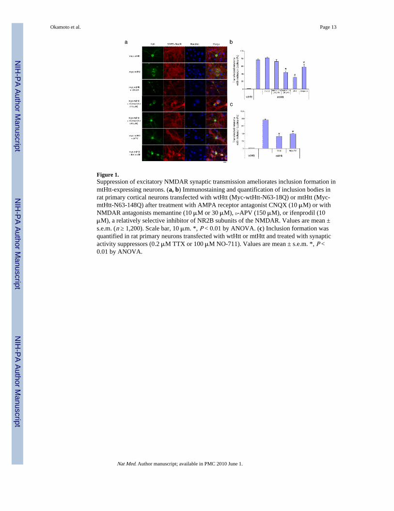
Figure 1.Suppression of excitatory NMDAR synaptic transmission ameliorates inclusion formation inmtHtt-expressing neurons. (a, b) Immunostaining and quantification of inclusion bodies inrat primary cortical neurons transfected with wtHtt (Myc-wtHtt-N63-18Q) or mtHtt (Myc-mtHtt-N63-148Q) after treatment with AMPA receptor antagonist CNQX (10 μM) or withNMDAR antagonists memantine (10 μM or 30 μM), D-APV (150 μM), or ifenprodil (10μM), a relatively selective inhibitor of NR2B subunits of the NMDAR. Values are mean ±s.e.m. (n ≥ 1,200). Scale bar, 10 μm. *, P < 0.01 by ANOVA. (c) Inclusion formation wasquantified in rat primary neurons transfected with wtHtt or mtHtt and treated with synapticactivity suppressors (0.2 μM TTX or 100 μM NO-711). Values are mean ± s.e.m. *, P <0.01 by ANOVA.
Okamoto et al. Page 13
Nat Med. Author manuscript; available in PMC 2010 June 1.
NIH
-PA Author Manuscript
NIH
-PA Author Manuscript
NIH
-PA Author Manuscript
Figure 2.Pharmacology of NMDAR-mediated sEPSCs and whole-cell currents recorded from wtHtt-and mtHtt-transfected neurons. (a-e) sEPSCs (left-hand panels) and charge transfer (right-hand panels), normalized for each cell (see Supplementary Methods). Both wtHtt- andmtHtt-transfected neurons were treated with TTX (a), NO-711 (b), memantine (c), orifenprodil (d). Recording solutions in (a-e) contained 20 μM glycine; for (c. d), CNQX andbicuculline (10 μM each) were also added. Values are mean ± s.e.m. (n ≥ 4 cells in eachcase); *, P < 0.03 (paired t-test on raw data). (f) Effects of memantine and ifenprodil onextrasynaptic NMDAR-mediated currents. Incomplete reversal was observed because ofslow washout in this system. Recordings were performed in the presence of 0.1-1 μM TTX.NMDA currents were evoked by co-application of 100 μM NMDA and 20 μM glycine. (g)Charge transfer (fC/pF) for NMDAR-sEPSCs in mtHtt- and wtHtt-transfected neurons.
Okamoto et al. Page 14
Nat Med. Author manuscript; available in PMC 2010 June 1.
NIH
-PA Author Manuscript
NIH
-PA Author Manuscript
NIH
-PA Author Manuscript

Figure 3.Involvement of the chaperonin TRiC TCP1 subunit in synaptic activity-mediated inclusionformation. (a) Immunoblotting demonstrating the level of TCP1 protein expression inneurons exposed to TTX (0.2 μM) or control conditions for 24 h. Actin served as a loadingcontrol. (b) Inclusion formation was quantified in neurons transfected with mtHtt plus twodifferent small hairpin vectors for TCP1 or control vector. Values are mean ± s.e.m. for n ≥300. *, P ≤ 0.001 by ANOVA.
Okamoto et al. Page 15
Nat Med. Author manuscript; available in PMC 2010 June 1.
NIH
-PA Author Manuscript
NIH
-PA Author Manuscript
NIH
-PA Author Manuscript

Figure 4.Excitatory synaptic versus extrasynaptic activity in HD-related neuronal cell death. (a)Excessive glutamate insult (50 μM for 40 h) led to neuronal cell death in mtHtt-transfectedcortical neurons in the presence of inclusion formation (scale bar, 10 μm). Neuronstransfected with mtHtt contained inclusions and displayed healthy nuclei. (b, c) Cell deathanalysis after transfection with the N-terminal fragment of mtHtt (b) and full-length mtHtt(c). Values represent mean ± s.e.m. for n ≥ 1,200. *, P < 0.01 by ANOVA. (d) Memantineattenuated neuronal cell death induced by blockade of physiological excitatory synapticactivity with NO-711 or TTX in mtHtt-transfected neurons. Co-transfection of PGC-1αmitigated neuronal cell death induced by blockade of synaptic activity with TTX. Valuesrepresent mean ± s.e.m. for n ≥ 1,200. *, P < 0.05; **, P < 0.01 by ANOVA. (e) Cell deathwas quantified in neurons transfected with wtHtt or mtHtt plus two different small hairpinvectors for TCP1 or a control vector. Values are mean ± s.e.m. for n ≥ 300. *, P ≤ 0.01 byANOVA. (f) Low concentration (5 μM) memantine abrogated the decrease in CREBactivity induced by blockade of physiological excitatory synaptic activity with TTX inmtHtt-transfected neurons. Neurons were transfected with a CRE-luciferase reporterconstruct and wtHtt or mtHtt. Ordinate axis represents relative luciferase activity. Valuesrepresent mean ± s.e.m. *, P < 0.01 by ANOVA. (g) Low concentration memantineameliorated the decrease in PGC-1α levels induced by blockade of synaptic activity withTTX in mtHtt-transfected neurons. PGC-1α levels were quantified by immunofluorescenceunder deconvolution microscopy in wtHtt- or mtHtt-transfected neurons. Values representmean ± s.e.m. for n ≥ 300. *, P < 0.01 by ANOVA.
Okamoto et al. Page 16
Nat Med. Author manuscript; available in PMC 2010 June 1.
NIH
-PA Author Manuscript
NIH
-PA Author Manuscript
NIH
-PA Author Manuscript
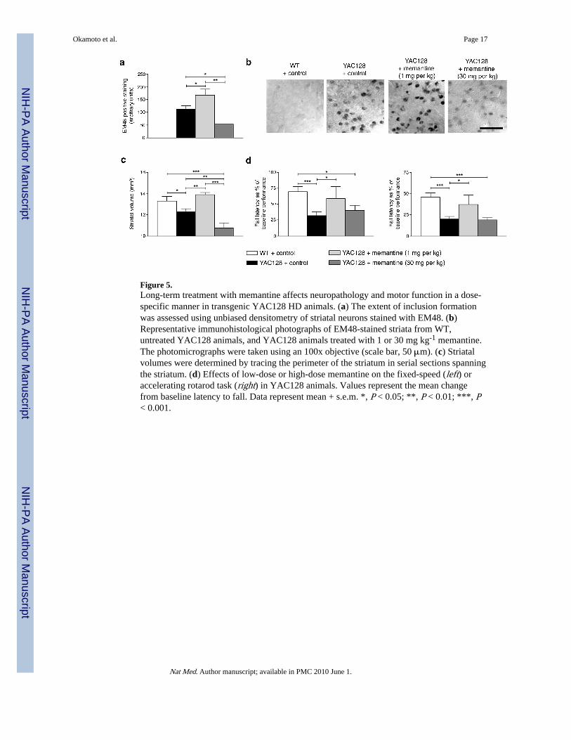
Figure 5.Long-term treatment with memantine affects neuropathology and motor function in a dose-specific manner in transgenic YAC128 HD animals. (a) The extent of inclusion formationwas assessed using unbiased densitometry of striatal neurons stained with EM48. (b)Representative immunohistological photographs of EM48-stained striata from WT,untreated YAC128 animals, and YAC128 animals treated with 1 or 30 mg kg-1 memantine.The photomicrographs were taken using an 100x objective (scale bar, 50 μm). (c) Striatalvolumes were determined by tracing the perimeter of the striatum in serial sections spanningthe striatum. (d) Effects of low-dose or high-dose memantine on the fixed-speed (left) oraccelerating rotarod task (right) in YAC128 animals. Values represent the mean changefrom baseline latency to fall. Data represent mean + s.e.m. *, P < 0.05; **, P < 0.01; ***, P< 0.001.
Okamoto et al. Page 17
Nat Med. Author manuscript; available in PMC 2010 June 1.
NIH
-PA Author Manuscript
NIH
-PA Author Manuscript
NIH
-PA Author Manuscript
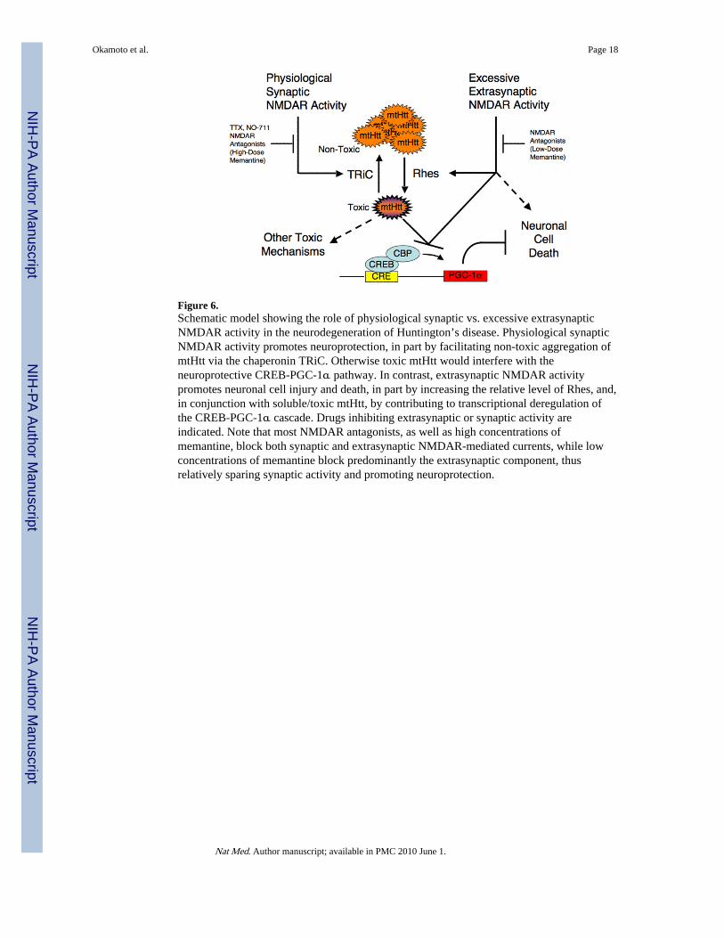
Figure 6.Schematic model showing the role of physiological synaptic vs. excessive extrasynapticNMDAR activity in the neurodegeneration of Huntington’s disease. Physiological synapticNMDAR activity promotes neuroprotection, in part by facilitating non-toxic aggregation ofmtHtt via the chaperonin TRiC. Otherwise toxic mtHtt would interfere with theneuroprotective CREB-PGC-1α pathway. In contrast, extrasynaptic NMDAR activitypromotes neuronal cell injury and death, in part by increasing the relative level of Rhes, and,in conjunction with soluble/toxic mtHtt, by contributing to transcriptional deregulation ofthe CREB-PGC-1α cascade. Drugs inhibiting extrasynaptic or synaptic activity areindicated. Note that most NMDAR antagonists, as well as high concentrations ofmemantine, block both synaptic and extrasynaptic NMDAR-mediated currents, while lowconcentrations of memantine block predominantly the extrasynaptic component, thusrelatively sparing synaptic activity and promoting neuroprotection.
Okamoto et al. Page 18
Nat Med. Author manuscript; available in PMC 2010 June 1.
NIH
-PA Author Manuscript
NIH
-PA Author Manuscript
NIH
-PA Author Manuscript