Atherosclerosis of the descending aorta predicts cardiovascular events: a transesophageal...
7
BioMed Central Page 1 of 7 (page number not for citation purposes) Cardiovascular Ultrasound Open Access Research Atherosclerosis of the descending aorta predicts cardiovascular events: a transesophageal echocardiography study Albert Varga*, Noemi Gruber, Tamás Forster, Györgyi Piros, Kálmán Havasi, Éva Jebelovszki and Miklos Csanády Address: 2nd Department of Medicine and Cardiology Center, University of Sciences, Szeged, Hungary Email: Albert Varga* - [email protected]; Noemi Gruber - [email protected]; Tamás Forster - [email protected]; Györgyi Piros - [email protected]; Kálmán Havasi - [email protected] szeged.hu; Éva Jebelovszki - [email protected]; Miklos Csanády - [email protected] * Corresponding author Abstract Purpose: Previous studies have shown that atherosclerosis of the descending aorta detected by transesophageal echocardiography (TEE) is a good marker of coexisting coronary artery disease. The aim of our study was to evaluate whether the presence of atherosclerosis on the descending aorta during TEE has any prognostic impact in predicting cardiovascular events. Material and Methods: The study group consisted of 238 consecutive in-hospital patients referred for TEE testing (135 males, 103 females, mean age 58 +/- 11 years) with a follow up of 24 months. The atherosclerotic lesions of the descending aorta were scored from 0 (no atherosclerosis) to 3 (plaque >5 mm and/or "complex" plaque with ulcerated or mobile parts). Results: Atherosclerosis was observed in 102 patients, (grade 3 in 16, and grade 2 in 86 patients) whereas 136 patients only had an intimal thickening or normal intimal surface. There were 57 cardiovascular events in the follow-up period. The number of events was higher in the 102 patients with (n = 34) than in the 136 patients without atherosclerosis (n = 23, p < 0.01). The frequency of events was in close correlation with the severity of the atherosclerosis of the descending aorta. Fifty percent of the patients with grade 3 experienced cardiovascular events. Excluding patients with subsequent revascularization, the multivariate analysis only left ventricular function with EF < 40% (HR 3.0, CI 1.3–7.1) and TEE atherosclerotic plaque >=2 (HR 2.4, CI 1.0–5.5) predicted hard cardiovascular events. Conclusion: Atherosclerosis of the descending aorta observed during transesophageal echocardiography is a useful predictor of cardiovascular events. Introduction During the past decades many methods and factors have been proposed as good prognostic tools or markers for cardiovascular risk stratification. However, even with the most sophisticated stress testing procedures the prediction capability still remains imperfect [1]. Previous studies have shown that atherosclerosis of the descending aorta detected by transesophageal echocardiography (TEE) is a good marker of coexisting coronary artery disease [2-9]. Cohen et al. have demonstrated, that in patients with Published: 22 October 2004 Cardiovascular Ultrasound 2004, 2:21 doi:10.1186/1476-7120-2-21 Received: 23 February 2004 Accepted: 22 October 2004 This article is available from: http://www.cardiovascularultrasound.com/content/2/1/21 © 2004 Varga et al; licensee BioMed Central Ltd. This is an open-access article distributed under the terms of the Creative Commons Attribution License (http://creativecommons.org/licenses/by/2.0 ), which permits unrestricted use, distribution, and reproduction in any medium, provided the original work is properly cited.
-
Upload
independent -
Category
Documents
-
view
3 -
download
0
Transcript of Atherosclerosis of the descending aorta predicts cardiovascular events: a transesophageal...

BioMed CentralCardiovascular Ultrasound
ss
Open AcceResearchAtherosclerosis of the descending aorta predicts cardiovascular events: a transesophageal echocardiography studyAlbert Varga*, Noemi Gruber, Tamás Forster, Györgyi Piros, Kálmán Havasi, Éva Jebelovszki and Miklos CsanádyAddress: 2nd Department of Medicine and Cardiology Center, University of Sciences, Szeged, Hungary
Email: Albert Varga* - [email protected]; Noemi Gruber - [email protected]; Tamás Forster - [email protected]; Györgyi Piros - [email protected]; Kálmán Havasi - [email protected]; Éva Jebelovszki - [email protected]; Miklos Csanády - [email protected]
* Corresponding author
AbstractPurpose: Previous studies have shown that atherosclerosis of the descending aorta detected bytransesophageal echocardiography (TEE) is a good marker of coexisting coronary artery disease.The aim of our study was to evaluate whether the presence of atherosclerosis on the descendingaorta during TEE has any prognostic impact in predicting cardiovascular events.
Material and Methods: The study group consisted of 238 consecutive in-hospital patientsreferred for TEE testing (135 males, 103 females, mean age 58 +/- 11 years) with a follow up of 24months. The atherosclerotic lesions of the descending aorta were scored from 0 (noatherosclerosis) to 3 (plaque >5 mm and/or "complex" plaque with ulcerated or mobile parts).
Results: Atherosclerosis was observed in 102 patients, (grade 3 in 16, and grade 2 in 86 patients)whereas 136 patients only had an intimal thickening or normal intimal surface. There were 57cardiovascular events in the follow-up period. The number of events was higher in the 102 patientswith (n = 34) than in the 136 patients without atherosclerosis (n = 23, p < 0.01). The frequency ofevents was in close correlation with the severity of the atherosclerosis of the descending aorta.Fifty percent of the patients with grade 3 experienced cardiovascular events. Excluding patientswith subsequent revascularization, the multivariate analysis only left ventricular function with EF <40% (HR 3.0, CI 1.3–7.1) and TEE atherosclerotic plaque >=2 (HR 2.4, CI 1.0–5.5) predicted hardcardiovascular events.
Conclusion: Atherosclerosis of the descending aorta observed during transesophagealechocardiography is a useful predictor of cardiovascular events.
IntroductionDuring the past decades many methods and factors havebeen proposed as good prognostic tools or markers forcardiovascular risk stratification. However, even with themost sophisticated stress testing procedures the prediction
capability still remains imperfect [1]. Previous studieshave shown that atherosclerosis of the descending aortadetected by transesophageal echocardiography (TEE) is agood marker of coexisting coronary artery disease [2-9].Cohen et al. have demonstrated, that in patients with
Published: 22 October 2004
Cardiovascular Ultrasound 2004, 2:21 doi:10.1186/1476-7120-2-21
Received: 23 February 2004Accepted: 22 October 2004
This article is available from: http://www.cardiovascularultrasound.com/content/2/1/21
© 2004 Varga et al; licensee BioMed Central Ltd. This is an open-access article distributed under the terms of the Creative Commons Attribution License (http://creativecommons.org/licenses/by/2.0), which permits unrestricted use, distribution, and reproduction in any medium, provided the original work is properly cited.
Page 1 of 7(page number not for citation purposes)

Cardiovascular Ultrasound 2004, 2:21 http://www.cardiovascularultrasound.com/content/2/1/21
brain infarction, "complex" plaques (with ulcerated sur-face, mobile parts and thrombi) are powerful predictors offuture cardiovascular events [10]. The aim of this studywas to evaluate whether the presence of atherosclerosis onthe descending aorta observed by routine scanning duringTEE has any prognostic impact in predicting cardiovascu-lar events such as cardiac death, myocardial infarction orfatal stroke. Therefore, we conducted a prospective study;collecting and analyzing the data of 238 consecutivepatients referred to the echo lab for transesophagealechocardiography.
MethodsPatient selectionDuring the year 1998, 238 consecutive patients (135males, 103 females, and mean age 58 ± 11 years) werestudied with transesophageal echocardiography at the 2nd
Department of Medicine and Cardiology Center, Univer-sity of Sciences, Szeged, Hungary. The patients underwentTEE examination for the following reasons: TIA or sus-pected cerebral embolism (n = 100), coronary flowreserve evaluation (n = 71), evaluation of the native orartificial mitral valve (n = 23), suspected endocarditis (n =15), evaluation of the aortic valve (n = 13), suspected aor-tic dissection (n = 6), atrial septal defect or patent foramenovale (n = 6), others (n = 4). Thirty patients had suffereda previous myocardial infarction. The patients were fol-lowed-up to a period of at least 2 years, median 31 ± 9months.
Transthoracic and transesophageal echocardiographyAll patients had a transthoracic echocardiographic exami-nation to assess the global and regional left ventricularfunction. The ejection fraction was calculated using thearea-length single plane method [11]. Left ventricularfunction was considered to be depressed in case of ejec-tion fraction ≤40%.
The TEE examination was carried out according to recom-mendations of the Mayo clinic procedure [12]. Two-dimensional echocardiograms were obtained by usingcommercially available imaging system (ATL-HDI) with abiplane transducer. Echocardiographic images wererecorded on videotape for subsequent playback and anal-ysis. The atherosclerotic lesions of the descending aortawere graded according to the modified scoring systemoriginally proposed by Fazio et al [2]:
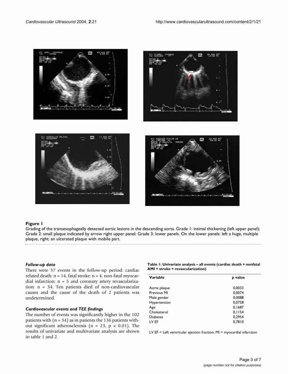
Grade 0 – no sign of atherosclerosis
Grade 1 – intimal thickening
Grade 2 – plaque < 5 mm
Grade 3 – plaque > 5 mm and/or "complex" plaque withulcerated or mobile parts.
Significant atherosclerosis was considered in case of grade2 or 3 (fig 1 and additional files 1, 2, 3, 4).
Follow up dataFollow-up data were obtained from at least one of foursources: 1) review of the patient's hospital record; 2) per-sonal communication with the patient's physician andreview of the patient's chart; 3) a telephone interview withthe patient conducted by trained personnel; or 4) a staffphysician visiting the patients at regular intervals in theoutpatient clinic. Follow-up data were obtained in allpatients. The outcome events were: cardiac related death,fatal stroke, non-fatal myocardial infarction and revascu-larization either by percutaneous transluminal coronaryangioplasty (PTCA) or by coronary artery by-pass grafting(CABG). Myocardial infarction was documented by a con-sistent history, EKG changes and cardiac enzyme level ele-vations and confirmed by hospital chart or hospitaldischarge letter review. In the next step, patients withrevascularization were censored and the remainingpatients were considered for the analysis of hard events(cardiac death, myocardial infarction, and fatal stroke).All-cause mortality (cardiovascular death plus death ofother causes) was also analyzed.
Statistical analysisValues are expressed as mean ± Standard Deviation. Con-tinuous variables have been compared by the means ofStudent's t test (two-tailed). Statistical analysis of discretevariables has been performed with chi-square test; aFisher's exact test has been used when appropriate. Theindividual effect of certain variables on infarction-free sur-vival has been evaluated with the use of the Cox propor-tional hazard model with univariate and multivariateanalysis and with the Kaplan-Meier method. The patientswere stratified into two subgroups: patients with andwithout cardiac events. The examined variables were age,sex, risk factors (arterial hypertension, diabetes, hypercho-lesterolemia), previous myocardial infarction, ejectionfraction, and significant atherosclerosis of the descendingaorta. A p value <0.05 was considered statisticallysignificant.
ResultsTransesophageal echocardiographyThe examination was successful and complete in allpatients, without any side effect. Significant atherosclero-sis of the descending aorta was observed in 102 patients,(grade 3 in 16, and grade 2 in 86 patients) whereas 136patients had only mild intimal thickening (n = 46) or nor-mal endocardial surface (n = 90).
Page 2 of 7(page number not for citation purposes)
Cardiovascular Ultrasound 2004, 2:21 http://www.cardiovascularultrasound.com/content/2/1/21
Follow-up dataThere were 57 events in the follow-up period: cardiacrelated death: n = 14, fatal stroke: n = 4, non-fatal myocar-dial infarction: n = 5 and coronary artery revasculariza-tion: n = 34. Ten patients died of non-cardiovascularcauses and the cause of the death of 2 patients wasundetermined.
Cardiovascular events and TEE findingsThe number of events was significantly higher in the 102patients with (n = 34) as in patients the 136 patients with-out significant atherosclerosis (n = 23, p < 0.01). Theresults of univariate and multivariate analysis are shownin table 1 and 2.
Grading of the transesophageally detected aortic lesions in the descending aortaFigure 1Grading of the transesophageally detected aortic lesions in the descending aorta. Grade 1: intimal thickening (left upper panel); Grade 2: small plaque indicated by arrow right upper panel; Grade 3: lower panels. On the lower panels: left a huge, multiple plaque, right: an ulcerated plaque with mobile part.
Table 1: Univariate analysis – all events (cardiac death + nonfatal AMI + stroke + revascularization)
Variable p value
Aorta plaque 0,0033Previous MI 0,0074Male gender 0,0088Hypertension 0,0758Age 0,1687Cholesterol 0,1154Diabetes 0,2954LV EF 0,7810
LV EF = Left ventricular ejection fraction; MI = myocardial infarction
Page 3 of 7(page number not for citation purposes)

Cardiovascular Ultrasound 2004, 2:21 http://www.cardiovascularultrasound.com/content/2/1/21
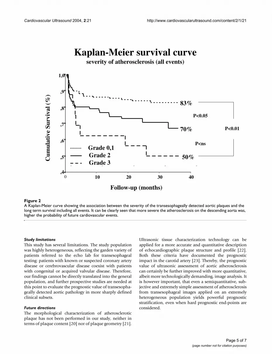
The frequency of events was in close correlation with theseverity of the atherosclerosis of the descending aorta (fig2). Fifty percent of the patients with grade 3 experiencedcardiovascular events. There was no significant differencewhen all causes of death were considered between sub-jects with aortic lesions or free of atherosclerosis of thedescending aorta (5% vs 1%, p = ns).
Spontaneous cardiovascular events and TEE findingsPatients with early (<3 months, n = 29) or late (>3months, n = 5) revascularization have been censored.
Nine events have occurred in the 122 patients with TEEscore ≤1 and 14 in the 82 patients with TEE score ≥2 (7%vs 17%, p < 0.05) (fig 3). The results of univariate analysisare shown in table 3. Impaired left ventricular function(EF ≤ 40%), significant atherosclerosis of the descendingaorta, and age were predictive for future cardiovascularevents. By multivariate analysis, only left ventricular func-tion with EF ≤ 40% (HR 3.0, 95% CI 1.3–7.1) and TEEatherosclerotic plaque ≥2 (HR 2.4, 95% CI 1.0–5.5) pre-dicted cardiovascular events (table 4). Similarly to theentire group, in patients with no revascularization, themore severe the atherosclerosis on the descending aortawas, higher the probability of future cardiovascularevents. There was no significant difference when all causesof death were considered between subjects with aorticlesions or free of atherosclerosis of the descending aorta(5% vs 1%, p = ns).
DiscussionAtherosclerosis of the descending aorta observed duringtransesophageal echocardiography is a useful predictor offuture cardiovascular events.
Comparison with previous studiesOur data are in keeping up with previous findings, show-ing that patients with atherosclerosis of the thoracic aortahave higher probability of coexisting coronary artery dis-ease [2-8]. In those series the positive predictive value ofTEE varied between 64% and 95%, whereas the negativepredictive value was consistently high (between 82 and
99%.), indicating that in the absence of echocardiograph-ically assessed atherosclerotic plaque in the thoracic aortathe probability of coronary artery disease is unlikely. Fur-thermore, Khoury et al have demonstrated, that athero-sclerotic plaques in patients with coronary artery diseasewere found predominantly in the descending aorta (in93%) and in the aortic arch (in 80%), whereas the ascend-ing aorta was the least involved (in 37%) [9]. Atheroscle-rosis is a complex polygenic, multifactorial vasculardisorder associated with many differing and changingmetabolic, anatomic and clinical manifestations [13]. Thepresence of atherosclerotic plaque in the thoracic aorta, asshown by chest x-ray, has been shown in previous studiesto be correlated with an increased risk of cardiovasculardeath [14,15]. However, several studies have also demon-strated that the generation of acute coronary syndromes isnot necessarily related to plaque severity rather to its mor-phology and complexity. From histopathologic and vas-cular biologic studies [13,16] plaque composition andvulnerability (type of lesion) rather than degree of steno-sis (size of lesion) have emerged as crucial factors leadingto sudden rupture of the plaque surface, usually withthrombosis superimposed, which underlies the greatmajority of infarctions. Angiographic studies also suggestthat the most frequent situation giving rise to infarction isthe occlusion of previously noncritical stenoses [17],which are more prevalent than the possibly moredangerous severe stenoses [18]. Taken together, thesestudies suggest that in two of three infarctions the culpritlesions had only mild to moderate stenosis on initial eval-uation in a substantial number of patients. This is againconsistent with our finding that significant coronaryartery disease is in close relationship with the atheroscle-rosis of the aorta but more severe the atherosclerosis is,higher the probability of spontaneous cardiovascularevents.
Clinical implicationsOne of the most important first steps in stratifying riskamong patients with proven or suspected coronary arterydisease is the identification of patients at high risk for cor-onary or vascular events during the course of the next fewmonths or years. To date, left ventricular dysfunction, thenumber of diseased vessels, and the severity of myocardialischemia have emerged as important determinants ofsurvival [19]. Our data suggest that atherosclerosis of thedescending aorta observed by a simple, routine trans-esophageal echocardiographic examination can be anadditional prognostic marker in identifying patients forhigher risk for cardiovascular events. When patients arereferred to transesophageal testing for whatever reason, asemiquantitative description of atherosclerotic burden ofthe descending aorta should be always included in theprognostic stratification.
Table 2: Multivariate analysis – all events (cardiac death + nonfatal AMI + stroke + revascularization)
95% CI
Variable p value HR Lower Upper
Aorta plaque <0,01 2,1 1,2 3,5Male gender <0,05 0,49 0,3 0,9
HR = Hazard Ratio; CI = Confidence Interval
Page 4 of 7(page number not for citation purposes)
Cardiovascular Ultrasound 2004, 2:21 http://www.cardiovascularultrasound.com/content/2/1/21
Study limitationsThis study has several limitations. The study populationwas highly heterogeneous, reflecting the garden variety ofpatients referred to the echo lab for transesophagealtesting: patients with known or suspected coronary arterydisease or cerebrovascular disease coexist with patientswith congenital or acquired valvular disease. Therefore,our findings cannot be directly translated into the generalpopulation, and further prospective studies are needed atthis point to evaluate the prognostic value of transesopha-geally detected aortic pathology in more sharply definedclinical subsets.
Future directionsThe morphological characterization of atheroscleroticplaque has not been performed in our study, neither interms of plaque content [20] nor of plaque geometry [21].
Ultrasonic tissue characterization technology can beapplied for a more accurate and quantitative descriptionof echocardiographic plaque structure and profile [22].Both these criteria have documented the prognosticimpact in the carotid artery [23]. Thereby, the prognosticvalue of ultrasonic assessment of aortic atherosclerosiscan certainly be further improved with more quantitative,albeit more technologically demanding, image analysis. Itis however important, that even a semiquantitative, sub-jective and extremely simple assessment of atherosclerosisfrom transesophageal images applied on an extremelyheterogeneous population yields powerful prognosticstratification, even when hard prognostic end-points areconsidered.
A Kaplan-Meier curve showing the association between the severity of the transesophageally detected aortic plaques and the long term survival including all eventsFigure 2A Kaplan-Meier curve showing the association between the severity of the transesophageally detected aortic plaques and the long term survival including all events. It can be clearly seen that more severe the atherosclerosis on the descending aorta was, higher the probability of future cardiovascular events.
Kaplan-Meier survival curveseverity of atherosclerosis (all events)
Follow-up (months)
Cu
mu
lati
ve
Su
rviv
al
(%)
403020100
1,0
,9
,8
,7
,6
,5
,4
83%
70%
50%
Grade 0,1
Grade 2
Grade 3
P<0.01
P<0.05
P<ns
Page 5 of 7(page number not for citation purposes)

Cardiovascular Ultrasound 2004, 2:21 http://www.cardiovascularultrasound.com/content/2/1/21
A Kaplan-Meier curve survival showing a better outcome of patients without transesophageally detected aortic plaquesFigure 3A Kaplan-Meier curve survival showing a better outcome of patients without transesophageally detected aortic plaques.
Follow-up (months)
403020100
100
90
80
Cu
mu
lati
ve
Su
rviv
al
(%)
Grade 2,3
Grade 0,1
P<0.05
83%
93%
Kaplan-Meier survival curve(hard events)
Table 3: Univariate analysis – Hard events (cardiac death + nonfatal AMI + stroke)
Variable p value
LV EF 0,0069Aorta plaque 0,0283Age 0,0440Cholesterol 0,5642Diabetes 0,9161Hypertension 0,3212Previous MI 0,6053Male gender 0,9880
LV EF = Left ventricular ejection fraction; MI = myocardial infarction
Table 4: Multivariate analysis – Hard events (cardiac death + nonfatal AMI + stroke)
95% CI
Variable p value HR Lower Upper
LV EF <0,01 3,0 1,3 7,1Aorta plaque =0,04 2,4 1,0 5,5
HR = Hazard Ratio; CI = Confidence Interval
Page 6 of 7(page number not for citation purposes)
Cardiovascular Ultrasound 2004, 2:21 http://www.cardiovascularultrasound.com/content/2/1/21
Publish with BioMed Central and every scientist can read your work free of charge
"BioMed Central will be the most significant development for disseminating the results of biomedical research in our lifetime."
Sir Paul Nurse, Cancer Research UK
Your research papers will be:
available free of charge to the entire biomedical community
peer reviewed and published immediately upon acceptance
cited in PubMed and archived on PubMed Central
yours — you keep the copyright
Submit your manuscript here:http://www.biomedcentral.com/info/publishing_adv.asp
BioMedcentral
Additional material
References1. Ambrose JA, Fuster V: Can we predict future acute coronary
events in patients with stable coronary artery disease? JAMA1997, 277:343-4.
2. Fazio GP, Redberg R, Winslow T, Schiller NB: Transesophagealechocardiography detected atherosclerotic aortic plaque ismarker for coronary artery disease. J Am Coll Cardiol 1993,21:144-50.
3. Tribouilloy C, Shen WF, Peltier M, Lesbre JP: Noninvasive predic-tion of coronary artery disease by transesophageal echocar-diographic detection of thoracic aortic plaque in valvularheart disease. Am J Cardiol 1994, 74:258-60.
4. Parthenakis F, Skalidis E, Simantirakis E, Kounali D, Vardas P, Nihoy-annopoulos P: Absence of atherosclerotic lesions in the tho-racic aorta indicates absence of significant coronary arterydisease. Am J Cardiol 1996, 77:1118-21.
5. Matsumura Y, Takata J, Yabe T, Furuno T, Chikamori T, Doi YL:Atherosclerotic aortic plaque detected by transesophagealechocardiography: its significance and limitation as a markerfor coronary artery disease in the elderly. Chest 1997, 112:81-6.
6. Tribouilloy C, Peltier M, Colas L, Rida Z, Rey JL, Lesbre JP: Multi-plane transesophageal echocardiographic absence of tho-racic aortic plaque is a powerful predictor for absence ofsignificant coronary artery disease in valvular patients, evenin elderly. A large prospective study. Eur Heart J 1997,18:1478-83.
7. Tribouilloy C, Peltier M, Senni M, Colas L, Rey JL, Lesbre JP: Multi-plane transesophageal echocardiographic detection of tho-racic aortic plaque is a marker for coronary artery disease inwomen. Int J Cardiol 1997, 61:269-75.
8. Tribouilloy C, Peltier M, Rey JL, Ruiz V, Lesbre JP: Use of trans-esophageal echocardiography to predict significant coronaryartery disease in aortic stenosis. Chest 1998, 113:671-675.
9. Khoury Z, Gottlieb S, Stern S, Keren A: Frequency and distribu-tion of atherosclerotic plaques in the thoracic aorta as deter-mined by transesophageal echocardiography in patients withcoronary artery disease. Am J Cardiol 1997, 79:23-27.
10. Cohen A, Tzourio C, Bertrand B, Chauvel C, Bousser MG, AmarencoP: Aortic plaque morphology and vascular events: a follow-up
study in patients with ischemic stroke. FAPS Investigators.French Study of Aortic Plaques in Stroke. Circulation 1997,96:3838-41.
11. Kan G, Visser CA, Lie KI, Durrer D: Left ventricular volumes andejection fraction by single plane two-dimensional apexechocardiography. Eur Heart J 1981, 2:339-343.
12. Seward JB, Khandheria BK, Edwards WD, Oh JK, Freeman WK, TajikAJ: Biplanar transesophageal echocardiography: anatomiccorrelations, image orientation, and clinical applications.Mayo Clin Proc 1990, 65:1193-213.
13. Fuster V, Badimon L, Badimon JJ, Chesebro JH: The pathogenesisof coronary artery disease and the acute coronary syn-dromes (2). N Engl J Med 1992, 326:310-18.
14. Hyman JB, Epstein FH: A study of correlation between roentge-nographic and postmortem calcifications of the aorta. AmHeart J 1954, 47:540-543.
15. Witteman JC, Kannel WB, Wolf PA, Grobbee DE, Hofman A, D'Ago-stino RB, Cobb JC: Aortic calcified plaques and cardiovasculardisease (the Framingham study). Am J Cardiol 1990,66:1060-1064.
16. Falk E: Why do plaques rupture? Circulation 1992, 86 Suppl (6III):III 30-42.
17. Little WC, Constantinescu M, Applegate RJ, Kutcher MA, BurrowsMT, Kahl FR, Santamore WP: Can coronary angiography predictthe site of a subsequent myocardial infarction in patientswith mild-to-moderate coronary artery disease? Circulation1988, 78:1157-66.
18. Haft JI, Al-Zarka AM: Comparison of the natural history ofirregular and smooth coronary lesions: insights into thepathogenesis, progression, and prognosis of coronaryatherosclerosis. Am Heart J 1993, 126:551-61.
19. Califf RM, Armstrong PW, Carver JR, D'Agostino RB, Strauss WE:27th Bethesda Conference: matching the intensity of riskfactor management with the hazard for coronary diseaseevents. Task Force 5. Stratification of patients into high,medium and low risk subgroups for purposes of risk factormanagement. J Am Coll Cardiol 1996, 27:1007-19.
20. Picano E, Landini L, Distante A, Benassi A, Sarnelli R, L'Abbate A:Fibrosis, lipids and calcium in human atherosclerotic plaque:in vitro differentiation from normal aortic walls by ultrasonicattenuation. Circ Res 1985, 56:556-62.
21. Urbani MP, Picano E, Parenti G, Mazzarisi A, Fiori L, Paterni M, PelosiG, Landini L: In vivo radiofrequency-based ultrasonic tissuecharacterization of the atherosclerotic plaque. Stroke 1993,24:1507-12.
22. Mazzone AM, Urbani MP, Picano E, Paterni M, Borgatti E, De FabritiisA, Landini L: In vivo ultrasonic parametric imaging of carotidatherosclerotic plaque by videodensitometric technique.Angiology 1995, 46:663-72.
23. Mathiesen EB, Bonaa KH, Joakimsen O: Echolucent plaques areassociated with high risk of ischemic cerebrovascular eventsin carotid stenosis: the tromso study. Circulation 2001,103:2171-5.
Additional File 1Grade I. atherosclerosis of the descending aorta. Intimal thickening.Click here for file[http://www.biomedcentral.com/content/supplementary/1476-7120-2-21-S1.avi]
Additional File 2Small plaque on the descending aorta, corresponding to Grade 2. atherosclerosis.Click here for file[http://www.biomedcentral.com/content/supplementary/1476-7120-2-21-S2.avi]
Additional File 3Grade 3. atherosclerosis, with large, multiple plaques.Click here for file[http://www.biomedcentral.com/content/supplementary/1476-7120-2-21-S3.avi]
Additional File 4Grade 3. atherosclerosis, plaque with mobile parts.Click here for file[http://www.biomedcentral.com/content/supplementary/1476-7120-2-21-S4.avi]
Page 7 of 7(page number not for citation purposes)
http://www.ncbi.nlm.nih.gov/entrez/query.fcgi?cmd=Retrieve&db=PubMed&dopt=Abstract&list_uids=9002499
http://www.ncbi.nlm.nih.gov/entrez/query.fcgi?cmd=Retrieve&db=PubMed&dopt=Abstract&list_uids=9002499
http://www.ncbi.nlm.nih.gov/entrez/query.fcgi?cmd=Retrieve&db=PubMed&dopt=Abstract&list_uids=8417055
http://www.ncbi.nlm.nih.gov/entrez/query.fcgi?cmd=Retrieve&db=PubMed&dopt=Abstract&list_uids=8417055
http://www.ncbi.nlm.nih.gov/entrez/query.fcgi?cmd=Retrieve&db=PubMed&dopt=Abstract&list_uids=8417055
http://www.ncbi.nlm.nih.gov/entrez/query.fcgi?cmd=Retrieve&db=PubMed&dopt=Abstract&list_uids=8037131
http://www.ncbi.nlm.nih.gov/entrez/query.fcgi?cmd=Retrieve&db=PubMed&dopt=Abstract&list_uids=8037131
http://www.ncbi.nlm.nih.gov/entrez/query.fcgi?cmd=Retrieve&db=PubMed&dopt=Abstract&list_uids=8037131
http://www.ncbi.nlm.nih.gov/entrez/query.fcgi?cmd=Retrieve&db=PubMed&dopt=Abstract&list_uids=8644671
http://www.ncbi.nlm.nih.gov/entrez/query.fcgi?cmd=Retrieve&db=PubMed&dopt=Abstract&list_uids=8644671
http://www.ncbi.nlm.nih.gov/entrez/query.fcgi?cmd=Retrieve&db=PubMed&dopt=Abstract&list_uids=8644671
http://www.ncbi.nlm.nih.gov/entrez/query.fcgi?cmd=Retrieve&db=PubMed&dopt=Abstract&list_uids=9228361
http://www.ncbi.nlm.nih.gov/entrez/query.fcgi?cmd=Retrieve&db=PubMed&dopt=Abstract&list_uids=9228361
http://www.ncbi.nlm.nih.gov/entrez/query.fcgi?cmd=Retrieve&db=PubMed&dopt=Abstract&list_uids=9228361
http://www.ncbi.nlm.nih.gov/entrez/query.fcgi?cmd=Retrieve&db=PubMed&dopt=Abstract&list_uids=9458455
http://www.ncbi.nlm.nih.gov/entrez/query.fcgi?cmd=Retrieve&db=PubMed&dopt=Abstract&list_uids=9458455
http://www.ncbi.nlm.nih.gov/entrez/query.fcgi?cmd=Retrieve&db=PubMed&dopt=Abstract&list_uids=9458455
http://www.ncbi.nlm.nih.gov/entrez/query.fcgi?cmd=Retrieve&db=PubMed&dopt=Abstract&list_uids=9363743
http://www.ncbi.nlm.nih.gov/entrez/query.fcgi?cmd=Retrieve&db=PubMed&dopt=Abstract&list_uids=9363743
http://www.ncbi.nlm.nih.gov/entrez/query.fcgi?cmd=Retrieve&db=PubMed&dopt=Abstract&list_uids=9363743
http://www.ncbi.nlm.nih.gov/entrez/query.fcgi?cmd=Retrieve&db=PubMed&dopt=Abstract&list_uids=9515841
http://www.ncbi.nlm.nih.gov/entrez/query.fcgi?cmd=Retrieve&db=PubMed&dopt=Abstract&list_uids=9515841
http://www.ncbi.nlm.nih.gov/entrez/query.fcgi?cmd=Retrieve&db=PubMed&dopt=Abstract&list_uids=9515841
http://www.ncbi.nlm.nih.gov/entrez/query.fcgi?cmd=Retrieve&db=PubMed&dopt=Abstract&list_uids=9024730
http://www.ncbi.nlm.nih.gov/entrez/query.fcgi?cmd=Retrieve&db=PubMed&dopt=Abstract&list_uids=9024730
http://www.ncbi.nlm.nih.gov/entrez/query.fcgi?cmd=Retrieve&db=PubMed&dopt=Abstract&list_uids=9024730
http://www.ncbi.nlm.nih.gov/entrez/query.fcgi?cmd=Retrieve&db=PubMed&dopt=Abstract&list_uids=9403605
http://www.ncbi.nlm.nih.gov/entrez/query.fcgi?cmd=Retrieve&db=PubMed&dopt=Abstract&list_uids=9403605
http://www.ncbi.nlm.nih.gov/entrez/query.fcgi?cmd=Retrieve&db=PubMed&dopt=Abstract&list_uids=9403605
http://www.ncbi.nlm.nih.gov/entrez/query.fcgi?cmd=Retrieve&db=PubMed&dopt=Abstract&list_uids=7297574
http://www.ncbi.nlm.nih.gov/entrez/query.fcgi?cmd=Retrieve&db=PubMed&dopt=Abstract&list_uids=7297574
http://www.ncbi.nlm.nih.gov/entrez/query.fcgi?cmd=Retrieve&db=PubMed&dopt=Abstract&list_uids=7297574
http://www.ncbi.nlm.nih.gov/entrez/query.fcgi?cmd=Retrieve&db=PubMed&dopt=Abstract&list_uids=2205761
http://www.ncbi.nlm.nih.gov/entrez/query.fcgi?cmd=Retrieve&db=PubMed&dopt=Abstract&list_uids=2205761
http://www.ncbi.nlm.nih.gov/entrez/query.fcgi?cmd=Retrieve&db=PubMed&dopt=Abstract&list_uids=1728735
http://www.ncbi.nlm.nih.gov/entrez/query.fcgi?cmd=Retrieve&db=PubMed&dopt=Abstract&list_uids=1728735
http://www.ncbi.nlm.nih.gov/entrez/query.fcgi?cmd=Retrieve&db=PubMed&dopt=Abstract&list_uids=1728735
http://www.ncbi.nlm.nih.gov/entrez/query.fcgi?cmd=Retrieve&db=PubMed&dopt=Abstract&list_uids=2220632
http://www.ncbi.nlm.nih.gov/entrez/query.fcgi?cmd=Retrieve&db=PubMed&dopt=Abstract&list_uids=2220632
http://www.ncbi.nlm.nih.gov/entrez/query.fcgi?cmd=Retrieve&db=PubMed&dopt=Abstract&list_uids=3180375
http://www.ncbi.nlm.nih.gov/entrez/query.fcgi?cmd=Retrieve&db=PubMed&dopt=Abstract&list_uids=3180375
http://www.ncbi.nlm.nih.gov/entrez/query.fcgi?cmd=Retrieve&db=PubMed&dopt=Abstract&list_uids=3180375
http://www.ncbi.nlm.nih.gov/entrez/query.fcgi?cmd=Retrieve&db=PubMed&dopt=Abstract&list_uids=8362708
http://www.ncbi.nlm.nih.gov/entrez/query.fcgi?cmd=Retrieve&db=PubMed&dopt=Abstract&list_uids=8362708
http://www.ncbi.nlm.nih.gov/entrez/query.fcgi?cmd=Retrieve&db=PubMed&dopt=Abstract&list_uids=8362708
http://www.ncbi.nlm.nih.gov/entrez/query.fcgi?cmd=Retrieve&db=PubMed&dopt=Abstract&list_uids=8609316
http://www.ncbi.nlm.nih.gov/entrez/query.fcgi?cmd=Retrieve&db=PubMed&dopt=Abstract&list_uids=8609316
http://www.ncbi.nlm.nih.gov/entrez/query.fcgi?cmd=Retrieve&db=PubMed&dopt=Abstract&list_uids=8609316
http://www.ncbi.nlm.nih.gov/entrez/query.fcgi?cmd=Retrieve&db=PubMed&dopt=Abstract&list_uids=3884177
http://www.ncbi.nlm.nih.gov/entrez/query.fcgi?cmd=Retrieve&db=PubMed&dopt=Abstract&list_uids=3884177
http://www.ncbi.nlm.nih.gov/entrez/query.fcgi?cmd=Retrieve&db=PubMed&dopt=Abstract&list_uids=3884177
http://www.ncbi.nlm.nih.gov/entrez/query.fcgi?cmd=Retrieve&db=PubMed&dopt=Abstract&list_uids=8378954
http://www.ncbi.nlm.nih.gov/entrez/query.fcgi?cmd=Retrieve&db=PubMed&dopt=Abstract&list_uids=8378954
http://www.ncbi.nlm.nih.gov/entrez/query.fcgi?cmd=Retrieve&db=PubMed&dopt=Abstract&list_uids=7639412