Fear shapes information acquisition in decisions ... - MPG.PuRe
Upload
saintpetersCategory
view
1download
0
SIAM J. APPLIED DYNAMICAL SYSTEMS c© 2014 Society for Industrial and Applied MathematicsVol. 13, No. 3, pp. 1239–1269
A Modeling Exploration of How Synaptic Feedback to Descending ProjectionNeurons Shapes the Activity of an Oscillatory Network∗
Nickolas Kintos† and Farzan Nadim‡
Abstract. Rhythmic activity which underlies motor output is often initiated and controlled by descendingmodulatory projection pathways onto central pattern generator (CPG) networks. In turn, thesedescending pathways receive synaptic feedback from their target CPG network, which can influencethe CPG output. However, the mechanisms underlying such bidirectional synaptic interactions aremostly unexplored. We develop a reduced mathematical model, including both feed-forward andfeedback circuitry, to examine how the synaptic interactions involving two projection neurons, MCN1and CPN2, can produce and shape the activity of the gastric mill CPG in the crab stomatogastricnervous system. We use simplifying assumptions that are based on the behavior of the biologicalsystem to reduce this model down to 2 dimensions, which allows for phase plane analysis of the modeloutput. The model shows a distinct activity for the gastric mill rhythm that is elicited when MCN1and CPN2 are coactive compared to the rhythm elicited by MCN1 activity alone. Furthermore, thepresence of feedback to the projection neuron CPN2 provides a distinct locus of pattern generationin the model which does not require reciprocally inhibitory interactions between the gastric millCPG neurons, but is instead based on a half-center oscillator that occurs through a trisynapticpathway that includes CPN2. Our modeling results show that feedback to projection pathways mayprovide additional mechanisms for the generation of motor activity. These mechanisms can havedistinct dependence on network parameters and may therefore provide additional flexibility for therhythmic motor output.
Key words. limit cycle, forced oscillation, phase plane, neuromodulation, stomatogastric, neural oscillation
AMS subject classifications. 92C20, 34A26, 37C27
DOI. 10.1137/130943881
1. Introduction. Projection neuron pathways play an important role in generating andshaping the activity patterns of neural networks. In mammals, for example, descending pro-jection pathways from the cerebellum and brainstem influence the rhythmic networks thatgenerate locomotor activity in the spinal cord [30, 44, 12]. The influence of projection path-ways on neural networks is typically studied assuming a feed-forward architecture, in whichdescending projection neuron pathways initiate, terminate, or modify the activity of the targetnetwork [7, 21, 39, 15]. However, neural circuitry in biological systems is more complex, inthat projection neurons receive feedback from their target networks [8, 38, 34, 45]. In rhyth-mically active oscillatory networks, such feedback can influence the pattern of descendinginputs from projection neurons, and, in some cases, rhythmically patterned projection neuron
∗Received by the editors November 4, 2013; accepted for publication (in revised form) by B. Ermentrout May 6,2014; published electronically August 12, 2014. This research was supported by NIH MH060605 (FN) and the KennyFund Fellowship (NK).
http://www.siam.org/journals/siads/13-3/94388.html†Department of Mathematics, Saint Peter’s University, Jersey City, NJ 07306 ([email protected]).‡Corresponding author. Department of Biological Sciences and Department of Mathematical Sciences, New
Jersey Institute of Technology, Newark, NJ 07102 ([email protected]).
1239

1240 NICKOLAS KINTOS AND FARZAN NADIM
input influences the motor pattern of the target network [42]. However, in most systems, theconsequences of patterned projection neuron input are generally unknown for the activity ofthe target networks.
We investigate the role of feedback to projection neurons in a mathematical model of thecrab gastric mill network [36]. The gastric mill network is located within the stomatogastricganglion (STG), one of the four main ganglia of the stomatogastric nervous system (STNS).The neural circuitry within the STG is innervated by descending inputs from ∼20 pairs ofdescending projection neurons whose cell bodies are located within the anterior ganglia ofthe STNS [36]. Within the STG gastric mill neural circuit, interneuron 1 (Int1) and thelateral gastric (LG) neuron reciprocally inhibit each other and form an asymmetric, half-center oscillator, which underlies the activity of the gastric mill rhythm [14, 32]. The gastricmill rhythm is generally not spontaneously active [3, 5]. However, tonic stimulation of theidentified projection neuron modulatory commissural neuron 1 (MCN1) readily elicits a gastricmill rhythm (frequency ∼0.1 Hz) in vitro. Moreover, within the STG, modulatory synapticinput from MCN1 axon terminals and presynaptic inhibition of those terminals by the LGneuron are necessary for MCN1 to activate the gastric mill rhythm [14]. The gastric millnetwork receives synaptic input from the faster (∼1 Hz) pyloric rhythmic network in the formof synaptic inhibition from the pyloric pacemaker AB neuron to Int1. This fast rhythmicinput has a strong influence on the cycle frequency of the MCN1-elicited gastric mill rhythm[3, 32].
The MCN1 soma is located in the commissural ganglia (CoGs; a pair of anterior STNSganglia located several centimeters away from the STG) and is often coactive with a secondCoG projection neuron, the commissural projection neuron 2 (CPN2), to elicit a version ofthe gastric mill rhythm that is distinct from the MCN1-elicited rhythm [5]. CPN2 axonterminals excite the LG neuron within the STG [34], and this excitation may be partly dueto an electrical coupling [35]. Both projection neurons, MCN1 and CPN2, receive rhythmicfeedback from the STG neurons in the biological system [10, 34, 43]. In particular, CPN2 isinhibited by Int1 via a feedback synapse in the CoGs [34].
We previously used modeling to explore the mechanisms underlying the MCN1-elicitedgastric mill rhythm [26, 2, 32]. We now develop a mathematical model to investigate howthe distinct gastric mill rhythm which is elicited by coactivation of MCN1 and CPN2 (theMCN1+CPN2-elicited gastric mill rhythm) is influenced by the interplay of synaptic con-nections between CPN2 and the gastric mill CPG neurons. Physiologically, our model ismotivated by the gastric mill rhythm that is elicited by MCN1 and CPN2 when they arecoactivated by the ventral cardiac neurons (VCNs) [5]. However, in this study we focusonly on the influence of gastric mill rhythmic feedback to CPN2, and MCN1 is assumed tobe tonically active in our model. This enables us to directly compare our results for theMCN1+CPN2-elicited gastric mill rhythm with the gastric mill motor pattern of the MCN1-elicited rhythm from previous experiments [14, 3] and modeling [32, 26]. The general propertiesof the MCN1+CPN2-elicited gastric mill rhythm in our model remain similar to those of theVCN-elicited rhythm in the biological system.
Our model builds upon our previous mathematical model of the MCN1-elicited gastricmill rhythm [26]. In the current model, we ignore the intrinsic properties of CPN2 and simplyfocus on the question of how the synaptic interactions of this projection neuron influence the
A MODEL OF FEEDBACK TO PROJECTION NEURONS 1241
LG
MCN1 MCN1+CPN2
PD
20 mV
2 s
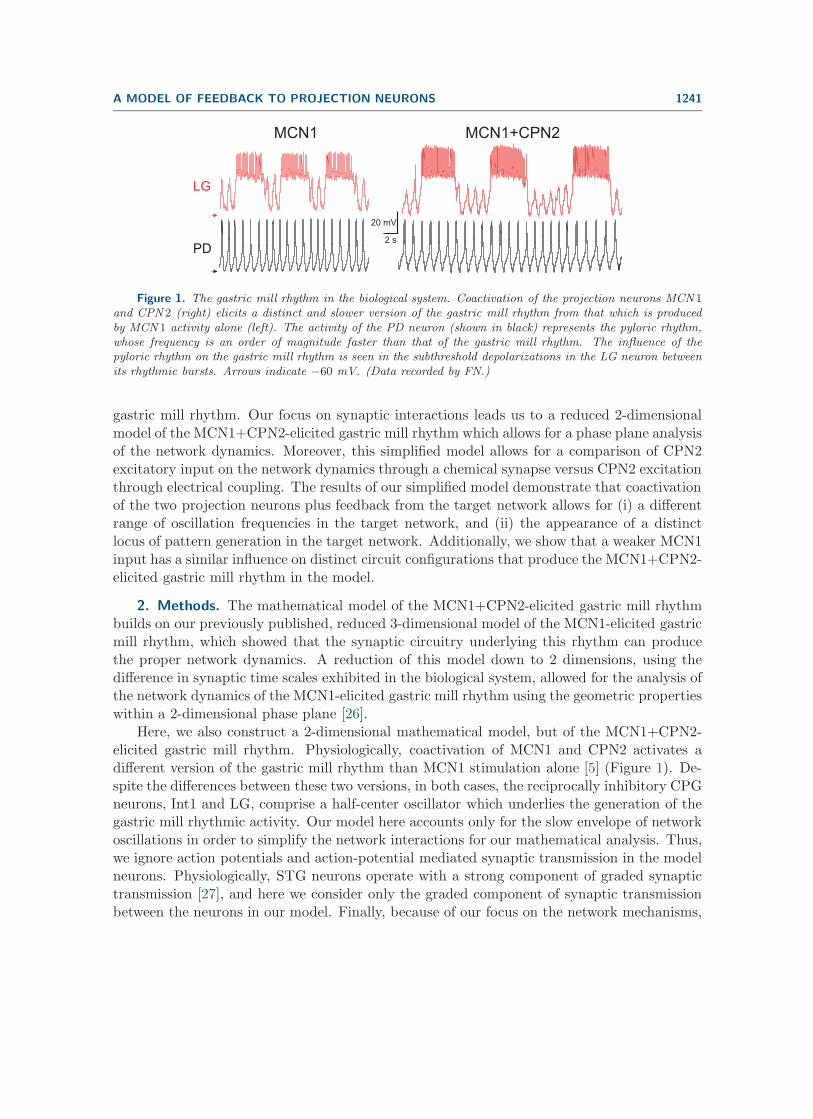
Figure 1. The gastric mill rhythm in the biological system. Coactivation of the projection neurons MCN 1and CPN 2 (right) elicits a distinct and slower version of the gastric mill rhythm from that which is producedby MCN 1 activity alone (left). The activity of the PD neuron (shown in black) represents the pyloric rhythm,whose frequency is an order of magnitude faster than that of the gastric mill rhythm. The influence of thepyloric rhythm on the gastric mill rhythm is seen in the subthreshold depolarizations in the LG neuron betweenits rhythmic bursts. Arrows indicate −60 mV. (Data recorded by FN.)
gastric mill rhythm. Our focus on synaptic interactions leads us to a reduced 2-dimensionalmodel of the MCN1+CPN2-elicited gastric mill rhythm which allows for a phase plane analysisof the network dynamics. Moreover, this simplified model allows for a comparison of CPN2excitatory input on the network dynamics through a chemical synapse versus CPN2 excitationthrough electrical coupling. The results of our simplified model demonstrate that coactivationof the two projection neurons plus feedback from the target network allows for (i) a differentrange of oscillation frequencies in the target network, and (ii) the appearance of a distinctlocus of pattern generation in the target network. Additionally, we show that a weaker MCN1input has a similar influence on distinct circuit configurations that produce the MCN1+CPN2-elicited gastric mill rhythm in the model.
2. Methods. The mathematical model of the MCN1+CPN2-elicited gastric mill rhythmbuilds on our previously published, reduced 3-dimensional model of the MCN1-elicited gastricmill rhythm, which showed that the synaptic circuitry underlying this rhythm can producethe proper network dynamics. A reduction of this model down to 2 dimensions, using thedifference in synaptic time scales exhibited in the biological system, allowed for the analysis ofthe network dynamics of the MCN1-elicited gastric mill rhythm using the geometric propertieswithin a 2-dimensional phase plane [26].
Here, we also construct a 2-dimensional mathematical model, but of the MCN1+CPN2-elicited gastric mill rhythm. Physiologically, coactivation of MCN1 and CPN2 activates adifferent version of the gastric mill rhythm than MCN1 stimulation alone [5] (Figure 1). De-spite the differences between these two versions, in both cases, the reciprocally inhibitory CPGneurons, Int1 and LG, comprise a half-center oscillator which underlies the generation of thegastric mill rhythmic activity. Our model here accounts only for the slow envelope of networkoscillations in order to simplify the network interactions for our mathematical analysis. Thus,we ignore action potentials and action-potential mediated synaptic transmission in the modelneurons. Physiologically, STG neurons operate with a strong component of graded synaptictransmission [27], and here we consider only the graded component of synaptic transmissionbetween the neurons in our model. Finally, because of our focus on the network mechanisms,

1242 NICKOLAS KINTOS AND FARZAN NADIM
we will ignore voltage-gated ionic currents and treat Int1 and LG as passive neurons [28].
2.1. A 4-dimensional model of the MCN1+CPN2-elicited gastric mill rhythm. Ourmathematical model of the MCN1+CPN2-elicited gastric mill rhythm begins with a systemof 4 nonlinear differential equations, given by
CIdVI
dt=− gLeak,I (VI −ELeak,I)︸ ︷︷ ︸
ILeak,I
− gL→ImL→I (VL) (VI − EL→I)︸ ︷︷ ︸IL→I
− gPP (t, VL) (VI − EP )︸ ︷︷ ︸IP
,
(1)
CLdVL
dt=− gLeak,L (VL − ELeak,L)︸ ︷︷ ︸
ILeak,L
− gI→LmI→L (VI) (VL − EI→L)︸ ︷︷ ︸II→L
− gss (VL − Es)︸ ︷︷ ︸Is
(2)
− gC→LmC→L (VC) (VL − EC→L)︸ ︷︷ ︸IC→L
− gelec (VL − VC)︸ ︷︷ ︸Ielec
,
CCdVC
dt=− gLeak,C (VC − ELeak,C)︸ ︷︷ ︸
ILeak,C
− gI→CmI→C (VI) (VC − EI→C)︸ ︷︷ ︸II→C
,
(3)
ds
dt=
⎧⎪⎨⎪⎩
1− s
τLO, VL ≤ vthresh,
−s
τHI, VL > vthresh.
(4)
Equations (1)–(4) describe the evolution of the four state variables in the model: the mem-brane potentials of Int1 (VI), the LG neuron (VL), and CPN2 (VC), and the slow excitatoryinput (s) from MCN1 to LG (Figure 2(A)). On the left-hand side of (1)–(3), the parametersCI , CL, and CC represent the membrane capacitance of Int1, the LG neuron, and CPN2,respectively. We set these to 1 for simplicity.
On the right-hand side of (1)–(2), the terms ILeak,I and ILeak,L represent the leak currentin Int1 and the LG neuron, respectively, with gLeak,I and ELeak,I (gLeak,L and ELeak,L) rep-resenting the leak conductance and reversal potential of Int1 (LG). In the biological system,the excitatory synaptic input s from MCN1 is necessary to elicit a gastric mill rhythm [14].Without the input from MCN1, Int1 exhibits a high membrane potential and remains active,while the LG neuron exhibits a low membrane potential and remains inactive [3]. As in pre-vious work [26], we produce this asymmetry between Int1 and the LG neuron by assigning ahigh value to ELeak,I and a low value to ELeak,L in our model (see Table 1).
The term ILeak,C on the right-hand side of (3) models the leak current in CPN2, withgLeak,C and ELeak,C representing the leak conductance and reversal potential, respectively.We assign a high value to ELeak,C , indicating that CPN2 remains active whenever it is notinhibited by Int1.
The terms IL→I and II→L in (1)–(2) describe the reciprocally inhibitory synapses betweenInt1 and the LG neuron. Similarly II→C on the right-hand side of (3) models the inhibitory

A MODEL OF FEEDBACK TO PROJECTION NEURONS 1243
20 mV
5 sec
0.31
VL
VC
s
VI
(A)
LG
MCN1 CPN2
CoG
STG
Int1
s
AB
(B.1)
LG
MCN1 CPN2
CoG
STG
Int1
s
AB
(B.2)
LG
MCN1 CPN2
CoG
STG
Int1
s
AB
(B.3)
Inhibition
Excitation
Figure 2. Output of the mathematical model of the gastric mill CPG. (A) The time course of all statevariables in the 4-dimensional model (eqs. (1)–(4)). (B.1) Circuit diagram of the 4-dimensional model. ABand MCN 1 are not modeled explicitly but only through their postsynaptic influences. The projection neuronsMCN 1 and CPN 2 are in the CoG and spatially separated from the gastric mill network (in the STG). TheLG neuron and Int1 reciprocally inhibit each other, and Int1 is inhibited by a pyloric-timed synaptic inputfrom AB, the pacemaker of the pyloric circuit. The LG neuron receives excitatory synaptic input from bothprojection neurons (MCN 1 and CPN 2), and it presynaptically inhibits MCN 1 within the STG. In the CoG,CPN 2 is inhibited by a feedback synapse from Int1. (B.2) Circuit diagram of the reduced 3-dimensional model(eqs. (11)–(13)). Due to the difference in time scales, VI can be expressed in terms of VL (the effects of Int1and AB are absorbed into the VL equation). The activity of VL, VC , and s remains practically identical tothat of the 4-dimensional model shown in (A). (B.3) Circuit diagram of the reduced 2-dimensional model (eqs.(16)–(17)). VC can be expressed in terms of VL (the effect of CPN 2 is absorbed into the VL equation). Theactivity of VL and s remains practically identical to the traces in (A).
Table 1Parameter values for the reduced 2-dimensional model of the MCN 1+CPN 2-elicited gastric mill rhythm.
Int1 AB (P ) CPN2 LG MCN1 (s)
gLeak,I = 0.75 mS/cm2 gP = 0.85 mS/cm2 gLeak,C = 1 mS/cm2 gLeak,L = 1 mS/cm2 gs = 7.5 mS/cm2
ELeak,I = 10 mV EP = −60 mV ELeak,C = 10 mV ELeak,L = −60 mV Es = 50 mV
gL→I = 2 mS/cm2 per = 1 sec gI→C = 17 mS/cm2 gI→L = 12 mS/cm2 vthresh = −27 mV
EL→I = −80 mV dur = 0.5 sec EI→C = −80 mV EI→L = −80 mV τLO = 14 sec
vL→I = −30 mV vq = −35 mV vI→C = −40 mV vI→L = −30 mV τHI = 5 sec
kL→I = 5 mV kq = 3 mV kI→C = 3 mV kI→L = 5 mV
CI = 1 μF/cm2 CC = 1 μF/cm2 gC→L = 0.5 mS/cm2
τI = 0.00133 sec τC = 0.001 sec EC→L = 30 mV
vC→L = −25 mV
kC→L = 8 mV
gelec = 0.7 mS/cm2
CL = 1 μF/cm2
τL = 0.001 sec

1244 NICKOLAS KINTOS AND FARZAN NADIM
feedback synapse from Int1 onto the CPN2 soma in the CoG (Figure 2(B.1)). The parametersgpre→post and Epre→post represent the synaptic maximal conductance and reversal potentialof these synapses. The synapses are gated by functions mpre→post(Vpre) which depend on themembrane potential of the presynaptic neuron and are modeled as a sigmoid:
(5) mpre→post (Vpre) =1
1 + exp ((vpre→post − Vpre)/kpre→post ).
The parameters vpre→post and kpre→post in (5) describe the half-activation voltage and steep-ness of the sigmoid.
The term IP on the right-hand side of (1) models the inhibitory synapse from the pyloricpacemaker AB neuron onto Int1 within the STG (Figure 2(B.1)), with gP and EP representingits maximal conductance and reversal potential, respectively. This synapse is gated by thefunction P (t, VL), and, to show the separate time- and voltage-dependent components, we canrewrite the gating function as
(6) P (t, VL) = P (t) q (VL) .
This synaptic input essentially acts as a periodic forcing input to the gastric mill network and,here, the time-dependent component is modeled as a periodic, half-sine function
(7) P (t) = sin
(π mod (t, per)
dur
)H (dur − mod (t, per)) ,
where mod() and H() designate the modulo and Heaviside function, respectively. The para-meters per and dur represent the period and duty cycle, respectively, of this half-sine function.As mentioned above, the pyloric rhythm cycle frequency (∼1 Hz) is an order of magnitudefaster than the gastric mill rhythm (∼0.1 Hz) [3]. We account for this by assigning appropriatevalues to the parameters per and dur in our model (see Table 1).
The voltage-dependent component of the gating function in (6) is given by the decreasingsigmoid
(8) q (VL) =1
1 + exp ((VL − vq)/kq ),
where vq and kq describe its half-activation voltage and steepness, respectively. We haveincluded this voltage-dependent component in the gating function in order to account for thefact that the AB to Int1 synapse is gated out during the active phase of the LG neuron in thegastric mill cycle [3]. This gating is incorporated into our model by (8), which turns off thepyloric-timed forcing function in (6) during the active phase of the LG neuron.
The term Is on the right-hand side of (2) models the slow excitatory synapse from MCN1to the LG neuron. Physiologically, this synapse occurs within the STG (Figure 2(B.1)). Theparameters gs and Es represent its maximal conductance and reversal potential, respectively,and the state variable s is governed by (4). The slow dynamics of s are gated by VL due toLG presynaptic inhibition of MCN1 in the STG (Figure 2(B.1)): s approaches 1 with timeconstant τLO when VL is below the voltage threshold vthresh for presynaptic inhibition, anddecays to 0 with time constant τHI when VL is greater than vthresh.

A MODEL OF FEEDBACK TO PROJECTION NEURONS 1245
The buildup and decay of MCN1 input to the LG neuron is a determinant of the frequencyof the gastric mill rhythm [14, 32], and the MCN1 to LG synapse operates on a much slowertime scale than all other synapses in the network [14]. We account for this in our model byassigning large values to the time constants τLO and τHI in (4); see Table 1.
On the right-hand side of (2), the term IC→L represents the chemical synaptic excitationfrom CPN2 to LG, whereas Ielec represents the electrical coupling between the CPN2 axonterminals and LG. Both terms model the excitation of LG by CPN2 within the STG ofthe biological system. In our results section, we will assume that only one of these terms isnonzero. The gating function of IC→L is given by (5), and the parameters gC→L and EC→L
represent its maximal conductance and reversal potential, respectively. The conductance ofthe Ielec term is given by the parameter gelec.
Figure 2(A) shows the evolution over time of the state variables VI , VL, VC and s in the4-dimensional model of (1)–(4). Notice that the state variable VI oscillates in antiphase withVL, while the state variable VC oscillates in phase with VL in our model.
2.2. Reduction to a 3-dimensional model of the MCN1+CPN2-elicited gastric millrhythm. To reduce our model down to three dimensions, we absorb VI into the LG neuronmembrane potential VL. To do so, we note that, unlike the LG neuron, Int1 is not influencedby the slow synaptic input s but only by synaptic inputs that occur on a fast time scale.
Mathematically, we divide through (1) by gLeak,I to get
(9) τIdVI
dt= − (VI − ELeak,I)− gL→I
gLeak,ImL→I (VL) (VI − EL→I)− gP
gLeak,IP (t, VL) (VI − EP ) ,
where τI = CIgLeak,I
is the time constant of Int1. We note that τI � τLO, τHI (see Table 1).
Thus, we assume that VI adjusts instantaneously to its steady state by setting τI = 0, whichmakes the left-hand side of (9) equal to zero. This allows for the explicit expression of VI interms of the state variable VL and the periodic forcing function P (t, VL):
(10) VI = vI (VL;P (t, VL)) =gLeak,IELeak,I + gL→ImL→I (VL)EL→I + gPP (t, VL)EP
gLeak,I + gL→ImL→I (VL) + gPP (t, VL).
Substituting the expression for VI given by (10) into the gating functions mI→L(VI) of (2)and mI→C(VI) of (3) gives a 3-dimensional model of the MCN1+CPN2-elicited gastric millrhythm:
CLdVL
dt=− gLeak,L (VL − ELeak,L)︸ ︷︷ ︸
ILeak,L
− gI→LmI→L (vI (VL;P (t, VL))) (VL −EI→L)︸ ︷︷ ︸II→L
(11)
− gss (VL − Es)︸ ︷︷ ︸Is
− gC→LmC→L (VC) (VL − EC→L)︸ ︷︷ ︸IC→L
− gelec (VL − VC)︸ ︷︷ ︸Ielec
,
CCdVC
dt=− gLeak,C (VC − ELeak,C)︸ ︷︷ ︸
ILeak,C
− gI→CmI→C (vI (VL;P (t, VL))) (VC −EI→C)︸ ︷︷ ︸II→C
,(12)

1246 NICKOLAS KINTOS AND FARZAN NADIM
ds
dt=
⎧⎪⎨⎪⎩
1− s
τLO, VL ≤ vthresh,
−s
τHI, VL > vthresh.
(13)
By reducing our model to 3 dimensions, we have expressed the dynamics of VI in terms ofthe state variable VL in our equations. In addition, the effects of the fast inhibitory synapsesonto Int1 (which are LG inhibition of Int1 and the pyloric-timed AB inhibition of Int1; seeFigure 2(B.2)) are now included into the synapses that emanate from Int1. Mathematically,the synaptic inputs onto Int1 are now included in mI→L (vI (VL;P (t, VL))) for the Int1 to LGsynapse in (11) and mI→C (vI (VL;P (t, VL))) for the Int1 to CPN2 feedback synapse in (12).Note that the evolution of the state variables VL, VC , and s is practically identical to that ofthe 4-dimensional model in Figure 2(A).
2.3. Reduction to a 2-dimensional model of the MCN1+CPN2-elicited gastric millrhythm. We now further reduce our model to 2 dimensions by mathematically absorbing VC ,the membrane potential of CPN2, into the membrane potential of the LG neuron. CPN2 isonly influenced by the inhibitory feedback synapse from Int1 (Figure 2(B.3)), and this synapseacts with a much faster time scale than that of s, the slow MCN1 input to LG. Thus, we canagain use the difference in synaptic time scales to further reduce the dimension of our model.
To carry out this reduction, we divide through (12) by gLeak,C to get
(14) τCdVC
dt= − (VC − ELeak,C)− gI→C
gLeak,CmI→C (vI (VL;P (t, VL))) (VC − EI→C) ,
where τC = CCgLeak,C
is the time constant of CPN2. As in the previous reduction, τC � τLO, τHI
(see Table 1). Therefore, we make the simplifying assumption that VC also adjusts instanta-neously to its steady state by setting τC = 0 to make the left-hand side of (14) equal to zero.This allows for an explicit solution of VC in terms of the state variable VL and the periodicforcing function P (t, VL):
(15) VC = vC (vI (VL;P (t, VL))) =gLeak,CELeak,C + gI→CmI→C (vI (VL;P (t, VL)))EI→C
gLeak,C + gI→CmI→C (vI (VL;P (t, VL))).
Substituting the expression for VC on the right-hand side of (15) into the terms IC→L andIelec of (11) gives a 2-dimensional model of the MCN1+CPN2-elicited gastric mill rhythm:
dVL
dt=− gLeak,L (VL − ELeak,L)︸ ︷︷ ︸
ILeak,L
− gI→LmI→L (vI (VL;P (t, VL))) (VL − EI→L)︸ ︷︷ ︸II→L
− gss (VL − Es)︸ ︷︷ ︸Is
(16)
− gC→LmC→L (vC (vI (VL;P (t, VL)))) (VL − EC→L)︸ ︷︷ ︸IC→L
− gelec (VL − vC (vI (VL;P (t, VL))))︸ ︷︷ ︸Ielec
,
(17)ds
dt=
⎧⎪⎨⎪⎩
1− s
τLO, VL ≤ vthresh,
−s
τHI, VL > vthresh.

A MODEL OF FEEDBACK TO PROJECTION NEURONS 1247
To simplify the notation, we will describe this system as
(18)
dVL
dt= −ILeak,L − II→L(p)− gss (VL − Es)− IC→L − Ielec,
ds
dt= ε (H(vthresh − VL)− s) /τs(VL).
Here p is the periodic forcing function P (t, VL), ε = 1/τHI , and
τs(VL) =
{τLO/τHI , VL ≤ vthresh,
1, VL > vthresh.
In this 2-dimensional model, the effect of the fast inhibitory feedback synapse from Int1to CPN2 (Figure 2(B.3)) has been absorbed into the excitatory inputs from the CPN2 axonterminal to the LG neuron in (16). We did not express the CPN2 excitatory inputs, IC→L andIelec, in terms of p in (18) because the pyloric-timed forcing has little to no influence on themin the model. The output of the 2-dimensional model in (18) is again practically identical tothe VL and s traces of Figure 2(A) and is not replotted.
2.4. Phase plane geometry of the 2-dimensional model. The reduced 2-dimensionalmodel in (18) can be used to analyze the network dynamics of the MCN1+CPN2-elicitedgastric mill rhythm. We first compute the nullclines corresponding to the two state variables.To compute the VL-nullcline, we note that the model is nonautonomous due to the presenceof the periodic forcing p in (18). As a result, the VL-nullcline will move in the VL − s phaseplane due to the influence of the forcing function. We describe a one-parameter family ofVL-nullclines which are indexed by the values of p in the interval [0, 1]. Our expression forthe family of VL-nullclines is given by
(19) s (VL; p) = −ILeak,L + II→L(p) + IC→L + Ielecgs (VL − Es)
.
The expression in (19) describes a cubic VL-nullcline for each p, where p = 0 corresponds tothe unforced system (no pyloric input) and p = 1 corresponds to the maximally forced system(peak of the pyloric input; see Figure 3(A)). The periodic forcing shifts the VL-nullcline backand forth between the unforced and maximally forced cubics as the value of p varies in theinterval [0, 1]. The pyloric-timed forcing function only influences the LG neuron during itsinactive phase [3, 26]; see the discussion of (6). Correspondingly, in our model, the rightbranch of the VL-nullcline remains stationary in the VL−s phase plane during the active stateof the LG neuron (Figure 3(A)).
For the slow state variable (s) in our model, the s-nullcline is given by the step function
(20) s = H (vthresh − VL) .
Recall that vthresh in (20) corresponds to the synaptic threshold voltage for LG presynapticinhibition of MCN1. The value of vthresh, chosen in the 4-dimensional model to produceoscillations, lies in the middle branch of the cubic VL-nullcline (Figure 3(A)). If vthresh ischosen so that the s-nullcline intersects one of the outer branches of the cubic VL-nullcline,

1248 NICKOLAS KINTOS AND FARZAN NADIM
s-Nullcline
V -NullclineL
p = 0
p = 1
VL
s
(A)
0
150
I Leak,L
0
150
I Leak,L
CHEMICAL ELECTRICAL
-90 -50 0VL
s
50
0
1
V -s Phase PlaneL
-90 VL
s
50-50 0
0
1
V -s Phase PlaneL
(B)
0
550
I I �L
0
550
I I �L
0
20
-20
I C�
L
0
40
I ele
c
-20
Figure 3. Phase plane geometry of the 2-dimensional model. (A) The VL- and s-nullclines intersect alongthe middle branch of the cubic VL-nullcline, thus allowing for a stable limit cycle. The pyloric-timed forcingparameter p (eq. (19)) results in a family of cubic VL-nullclines with the highest cubic (p = 0) correspondingto the unforced system and the lowest cubic (p = 1) corresponding to the maximally forced system. As p variesbetween 0 and 1 (double-headed arrow), the VL-nullcline shifts between the two. Consistent with the biologicalsystem, the pyloric-timed input is inactive when LG is active, and thus the right branch of the cubic is notaffected by p. The flow in the horizontal direction is shown by the double arrows. (B) The cubic shape ofthe VL-nullcline arises from the properties of the synaptic interactions among the network neurons. The I-Vrelationships of the terms in the numerator of (19) illustrate how the shape of the VL-nullcline is producedfor the two alternative cases of the CPN 2-to-LG synapse: chemical (left panels: IC→L) and electrical (rightpanels: Ielec). The terms ILeak,L and II→L (which depends on p) occur in both cases. The IC→L and Ielecterms influence the VL-nullcline differently: IC→L only has an influence for high values of VL (LG neuronactive), while Ielec has an influence for all values of VL. However, the overall shape of the VL-nullcline isqualitatively similar for both cases of the CPN 2-to-LG synapse (bottom panels).

A MODEL OF FEEDBACK TO PROJECTION NEURONS 1249
this would give rise to a globally stable fixed point and no oscillations. Regardless of the valueof vthresh, the flow in the horizontal direction of the VL−s phase plane is such that dVL/dt > 0above the VL-nullcline, while dVL/dt < 0 below the VL-nullcline (Figure 3(A)). On the otherhand, the flow in the vertical direction is such that ds/dt > 0 below the s-nullcline, whileds/dt < 0 above the s-nullcline.
The cubic shape of the VL-nullcline arises from the properties of the synaptic circuitrybetween the network neurons and is not attributed to the intrinsic properties of any of theseneurons. We now describe how this cubic nullcline is produced for the two different typesof CPN2-to-LG excitation, chemical and electrical. Both cases share the terms ILeak,L andII→L (Figure 3(B) and (19)). For the case of the chemical CPN2-to-LG excitation, onlythe IC→L term contributes to the shape of the VL-nullcline (Figure 3(B), left; Ielec is setto zero). On the other hand, for the electrical CPN2-to-LG excitation, only the Ielec termcontributes to the shape of the VL-nullcline (Figure 3(B), right; IC→L is set to zero). Thechemical CPN2-to-LG excitation (IC→L) contributes to the shape of the VL-nullcline only forhigher values of VL, which corresponds to the active phase of the LG neuron (Figure 3(B),left). In contrast, the electrical CPN2-to-LG excitation (Ielec) contributes to the shape ofthe VL-nullcline for all values of VL (Figure 3(B), right). The overall cubic shape of the VL-nullcline is obtained by combining all the terms, which are present for the relevant case ofthe CPN2-to-LG excitation, as shown in (19). Notice that the shape of the cubic VL-nullclineis qualitatively similar (and almost identical) for both cases, chemical and electrical, of theCPN2-to-LG excitation (Figure 3(B), bottom panels).
Because ε is small, the 2-dimensional model of (18) exists in a relaxation regime. Inparticular, the flow in the horizontal (VL) direction is fast within the VL − s phase plane,while the flow in the vertical (s) direction is much slower. In the limit where ε = 0 and themodel becomes singularly perturbed, a general trajectory in the VL−s phase plane consists ofdistinct fast and slow portions. In particular, the slow portions track the stable outer branchesof the cubic VL-nullcline to which they are strongly attracted by the fast, horizontal flow. Onthe other hand, the fast portions occur as transitions between the stable outer branches ofthe cubic. Previous theoretical results have established that trajectories of a relaxation (fast-slow) system for the case in which 0 < ε � 1 (as occurs in (18) of our model) are closelyapproximated by the limiting, singularly perturbed trajectory that occurs when ε = 0 in thesystem [31]. The basic properties of the flow in the VL − s phase plane remain the same inthe presence of the pyloric-timed forcing p.
All simulations for the 2-dimensional model were performed using the software packageXPPAUT [18]. The graphs of the nullcline components in Figure 3(B) were produced usingMATLAB. Parameter values for the intact MCN1+CPN2-elicited gastric mill rhythm arelisted in Table 1. Changes in parameter values (to model the effect of changes to the circuitryof the intact system) are discussed in section 3.
3. Results. We investigate the network dynamics of the MCN1+CPN2-elicited gastricmill rhythm using a phase plane analysis of the fully reduced, 2-dimensional model in (18).The two state variables in this fully reduced model are the membrane potential of the LGneuron (VL) and the slow, synaptic input (s) from MCN1 onto the LG neuron. As explainedin section 2, we will assume that the CPN2-to-LG input in (18) is either through a chemical

1250 NICKOLAS KINTOS AND FARZAN NADIM
excitation (IC→L) or through an electrical coupling (Ielec), and we set the other term to zero.In this section, we will focus on the case of the chemical excitation and will only discuss theelectrical coupling in cases where the output is qualitatively different.
Within the VL − s phase plane, the VL-nullcline has a cubic shape, while the s-nullcline isgiven by a step function (see section 2). The left branch of the cubic, VL-nullcline correspondsto the inactive state of the LG neuron. Physiologically, during the LG inactive state (retractionphase) of the gastric mill rhythm, the LG neuron (i) is inhibited by Int1, and (ii) receives aslow, modulatory excitation (s) from MCN1. Conversely, the right branch of the cubic, VL-nullcline corresponds to the active state of the LG neuron (protraction phase of the gastric millrhythm). Physiologically, during this state, the LG neuron (i) inhibits Int1, (ii) presynapticallyinhibits MCN1, and (iii) is excited by CPN2.
Due to the presence of the periodic forcing function p in (18), the 2-dimensional modelis nonautonomous. As a result, a family of cubic VL-nullclines (indexed by p) exists in theVL−s phase plane (Figure 3(A)). Physiologically, pmodels the influence of the local, inhibitorysynapse from the AB neuron (the pacemaker of the pyloric network) to Int1 (see section 2).
3.1. Network dynamics without the pyloric input. We first explore the network dynamicsof the MCN1+CPN2-elicited gastric mill rhythm when the local inhibitory synapse from theAB neuron to Int1 is absent (p ≡ 0; see schematic of Figure 4(A)). In this case, (18) becomesautonomous and only a single cubic VL-nullcline exists in the VL−s phase plane (Figure 4(A)).Because of the fast-slow nature of the equation, the resulting trajectory of the unforced systemconsists of two slow and two fast portions in the phase plane. The slow portions track thestable outer branches of the VL-nullcline, while the fast portions occur as horizontal jumpsbetween the outer branches (Figure 4(A)). This trajectory captures the dynamics of theMCN1+CPN2-elicited gastric mill rhythm in the absence of the AB-to-Int1 synapse.
We begin on the left branch of the cubic, VL-nullcline (Figure 4(A)). Physiologically, theleft branch of the cubic corresponds to when the LG neuron is in its inactive state. Here, theslow, excitatory input s slowly builds up and, as a result, the trajectory in the VL − s phaseplane slowly climbs up the left branch of the cubic (from point 1 to point 2 in Figure 4(A)).The slow buildup of s causes VL to slowly rise (Figure 4(B)). When the trajectory reachesthe left knee of the cubic at point 2, it jumps to the right branch at point 3, because theflow obeys dVL/dt > 0 above the VL-nullcline (Figure 3(A)). Physiologically, the LG neuronbegins to inhibit Int1 as s slowly increases up the left branch of the cubic.
The jump to the right branch of the VL-nullcline corresponds to when the slow buildupof s is sufficient to allow the LG neuron to escape from Int1 inhibition. As a result, the LGneuron transitions to its active state, where it inhibits Int1. LG inhibition of Int1 releasesCPN2 from Int1 inhibition; as a result, CPN2 transitions to its active state, where it excitesthe LG neuron (red dotted trace in Figure 4(B)). Moreover, while the trajectory is on theright branch of the cubic, the LG neuron presynaptically inhibits MCN1, which causes thestate variable s to slowly decay (Figure 4(B)). As a result, the trajectory slowly moves downthe right branch of the cubic from point 3 to point 4 (Figure 4(A)), and VL slowly falls ass decays (Figure 4(B)). When the trajectory reaches the right knee of the cubic at point4, it jumps back to the left branch at point 1 (Figure 4(A)), because dVL/dt < 0 below theVL-nullcline. Physiologically, LG inhibition of Int1 decreases with VL and s as the trajectory

A MODEL OF FEEDBACK TO PROJECTION NEURONS 1251
MCN1+CPN2
MCN1
1
23
4
VL
s
(A)
(B)
LG
MCN1 CPN2
CoG
STG
Int1
s
AB
VL
VC
s
5 sec
20 mV
1
4
3
2
1
MCN1+CPN2
MCN1
MCN1+CPN2
MCN1
MCN1+CPN2
MCN1
0.70
Figure 4. The MCN 1+CPN 2- and MCN 1-elicited gastric mill rhythms in the absence of the pyloric-timedinput p. Inset shows the circuit diagram. (A) Phase plane diagram of the 2-dimensional model with p set to0. The VL- and s-nullclines (gray) are as in Figure 3. The red curve shows the oscillation trajectory of theMCN 1+CPN 2-elicited rhythm. Points 1–4 are transition points in the trajectory (see text). The VL-nullclinefor the MCN 1-elicited rhythm has a distinct right branch: the blue curve shows the distinct trajectory of theMCN 1-elicited gastric mill rhythm on the right knee. (B) Activity traces of VL and s for the two rhythmsshown in (A). For the MCN 1+CPN 2 model, the activity of VC (calculated from VL) is also shown. Points1–4 correspond to the points in panel A. Dashed horizontal line in the s trace indicates that in both rhythms sreaches the same maximum value at the transition from point 2 to point 3.

1252 NICKOLAS KINTOS AND FARZAN NADIM
slowly falls down the right branch of the cubic.The jump back to the left branch of the VL-nullcline corresponds to when the fall in VL is
sufficient to release Int1. As a result, Int1 inhibition pushes the LG neuron (and CPN2) backdown to its inactive state. In turn, LG presynaptic inhibition of MCN1 is removed, s beginsto slowly build up, and the cycle repeats.
3.1.1. Comparison of the MCN1+CPN2- and MCN1-elicited gastric mill rhythms. Weuse Figure 4 to compare how the MCN1+CPN2-elicited gastric mill rhythm (red traces) in theabsence of pyloric-timed input differs from the MCN1-elicited rhythm (blue traces) in whichCPN2 is inactive. The MCN1-elicited rhythm is obtained by setting ELeak,C (the restingpotential of CPN2) to −80 mV to reproduce the fact that CPN2 is quiescent for this versionof the gastric mill rhythm in the biological system. As seen in the figure, the primary differencebetween the two oscillations, in this case, is that the cycle frequency of the MCN1+CPN2-elicited gastric mill rhythm is slower than that of the MCN1-elicited rhythm, whose activitywe have previously described in a modeling study [26].
When the trajectory jumps to the right branch of the cubic (from point 2 to point 3),the LG neuron enters its active state, and CPN2 is released from Int1 inhibition. However,VC jumps to a high value only for the MCN1+CPN2-elicited rhythm (and only in this caseCPN2 excites LG). Thus, the jump to the right branch of the cubic enables CPN2 to excitethe LG neuron and prolong the duration of its active phase (compare the two VL traces inFigure 4(B)). Consequently, s must decay to a lower minimum value for the MCN1+CPN2-elicited gastric mill rhythm (compare the two s traces in Figure 4(B)) before the LG neuron cantransition back to its inactive state (from point 4 to point 1). As a result, the state variableVL exhibits a slightly lower minimum value for the MCN1+CPN2-elicited rhythm (Figure4(B)). Consequently, the lower minimum value of s prolongs the network oscillations (for thecase when CPN2 is activated) and causes the MCN1+CPN2-elicited gastric mill rhythm toexhibit a slower cycle frequency than the MCN1-elicited rhythm (Figure 4(B)). Notice thats attains the same maximum value for both versions of the gastric mill rhythm (horizontalblack dashed line in Figure 4(B)) because the jump to the right branch in Figure 4(A) occursfor the same value of s in both cases.
3.2. Network dynamics of the intact MCN1+CPN2-elicited gastric mill rhythm. Wenow explore the MCN1+CPN2-elicited gastric mill rhythm in the presence of the pyloric-timedinput p (see schematic of Figure 5(A)), which makes the model nonautonomous (in (18)). Notethat the unforced cubic in Figure 5(A) is the same as the VL-nullcline in Figure 4(A).
As before, we begin our description of the MCN1+CPN2-elicited gastric mill rhythm on theleft branch of the VL -nullcline, when the LG neuron is in its inactive state. Physiologically, theLG neuron and CPN2 are both inhibited by Int1 during this phase of the rhythm. Moreover,s slowly builds up in the LG neuron, and the trajectory slowly climbs up the left branch of theVL-nullcline. However, the trajectory is shifted back and forth between the unforced (higher)and maximally forced (lower) cubic nullclines due to the effect of the pyloric-timed input p(Figure 5(A)). In particular, each peak of the input p shifts the unforced cubic down to themaximally forced cubic. Physiologically, the fast, pyloric-timed transitions between the leftbranches of the VL-nullcline correspond to the small-amplitude depolarizations exhibited byVL (Figure 5(B); see also Figure 1, right). When the trajectory reaches the level of the lower

A MODEL OF FEEDBACK TO PROJECTION NEURONS 1253
(A)
MCN1+CPN2
VL
s
1
2 3
4
MCN1
(B) MCN1+CPN2
MCN1+CPN2
MCN1+CPN2
MCN1
MCN1
MCN1
VL
VC
s
P
4
3
2
1 1
5 sec
20 mV
1
0
0.31
LG
MCN1 CPN2
CoG
STG
Int1
s
AB
1
0
Figure 5. The MCN 1+CPN 2- and MCN 1-elicited gastric mill rhythms in the presence of the pyloric-timedinput p. Inset shows the circuit diagram. (A) The phase plane showing the cubic VL-nullcline without pyloricinput (p = 0; upper) and with maximal pyloric input (p = 1; lower). The red curve shows the oscillationtrajectory of the MCN 1+CPN 2-elicited rhythm. The trajectory shifts back and forth between the two cubicnullclines due to the pyloric-timed input. Points 1–4 are transition points in the trajectory (see text). The VL-nullcline for the MCN 1-elicited rhythm has a distinct right branch: the blue curve shows the distinct trajectoryof the MCN 1-elicited gastric mill rhythm on the right knee. (B) Activity traces of VL and s for the two rhythmsshown in (A). For the MCN 1+CPN 2 model, the activity of VC (calculated from VL) is also shown. Points 1–4are as in panel (A). Note that CPN 2 is inactive when the trajectory is on the left branch of the VL-nullcline.As a result, the left knees of the VL-nullcline are the same and s rises to (approximately) the same maximumvalue (horizontal dashed line in (B)).

1254 NICKOLAS KINTOS AND FARZAN NADIM
left knee in the phase plane, the next input peak from p shifts the VL-nullcline below thephase point and initiates the jump to the right branch from point 2 to point 3 (Figure 5(A)).
Physiologically, the pyloric-timed shifts between cubics in the phase plane correspond tothe fact that AB inhibition of Int1 in turn disrupts the Int1-to-LG inhibitory synapse inthe network (see schematic of Figure 5(A)). As a result, the LG neuron is disinhibited byeach forcing peak, and it exhibits the small pyloric-timed depolarizations during its inactivestate (Figures 5(B) and 1). Note that the jump to the right branch of the VL-nullcline canonly occur after the trajectory reaches the level of the lower (maximally forced) left knee.Physiologically, this means that a sufficient buildup of MCN1 excitation s must first occurbefore the LG neuron can jump to its active state. Therefore, the trajectory cannot jumpto the right branch during the first few pyloric-timed shifts. Additionally, the transition ofthe LG neuron to its active state coincides with a pyloric-timed forcing peak (vertical blackdotted line in Figure 5(B)). Physiologically, this means that the onset of the LG active phaseof the rhythm is initiated by the AB-to-Int1 inhibitory synapse. A similar effect has beenestablished for the MCN1-elicited rhythm in previous experiments [3] and modeling [32].
After the trajectory jumps to the right branch of the VL-nullcline, where the LG neu-ron enters its active state, the network dynamics are similar to that of the unforced systemin Figure 4. Figure 5 also illustrates that, similar to the case of the unforced system, theMCN1+CPN2-elicited gastric mill rhythm exhibits a slower cycle frequency than the MCN1-elicited rhythm when the pyloric-timed input p is present (Figure 5(B)). This is again dueto the influence of the CPN2 excitation of LG. In particular, s must decay to a lower mini-mum value, which prolongs the cycle period of the MCN1+CPN2-elicited gastric mill rhythmcompared to that of the MCN1-elicited rhythm. Thus, the CPN2 synaptic interactions havea similar influence on the cycle frequency of the MCN1+CPN2-elicited rhythm for both theunforced (Figure 4) and intact (Figure 5) systems.
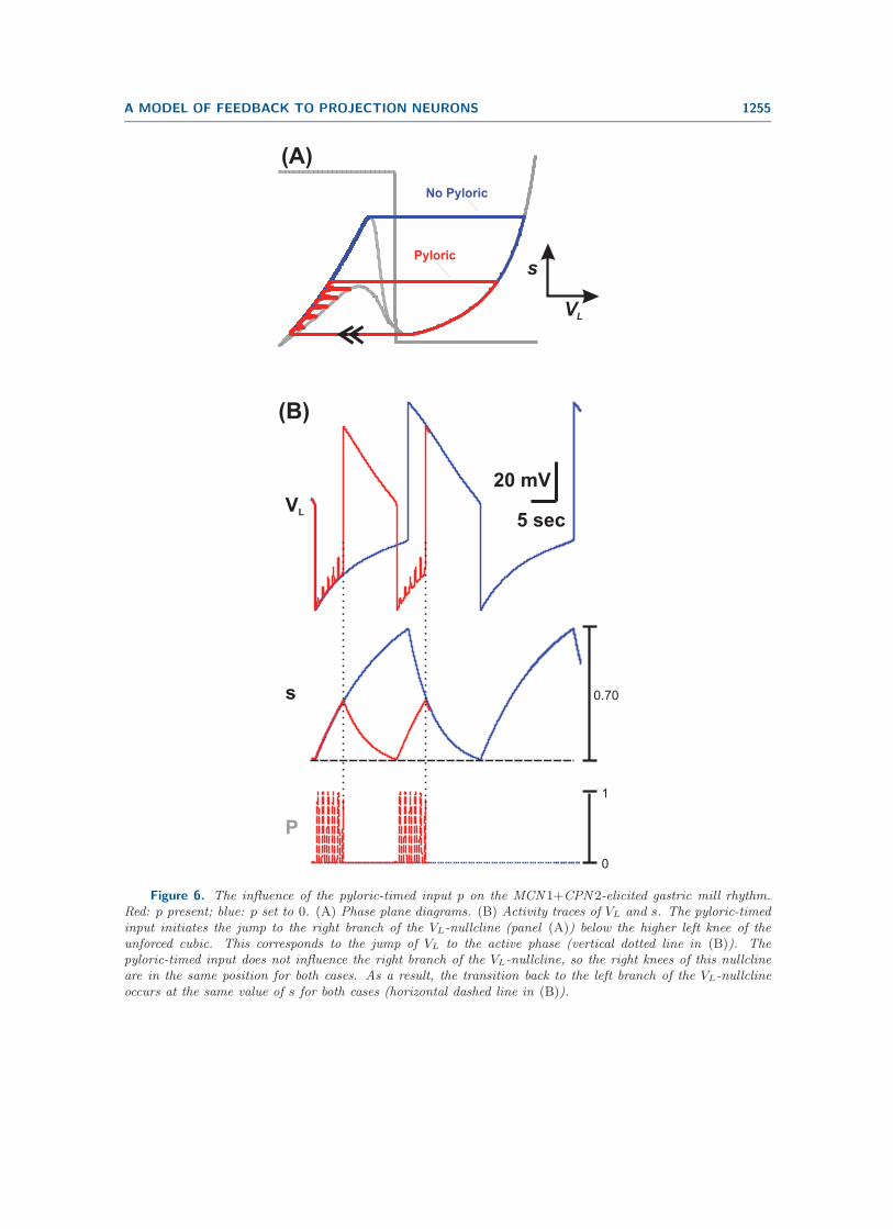
Figure 6 summarizes how the AB-to-Int1 synapse influences the MCN1+CPN2-elicitedgastric mill rhythm. First, as mentioned above, this synapse initiates the onset of the LGactive phase (see vertical dotted line in Figure 6(B)). This is also illustrated in the phaseplane by the fact that the jump to the right branch of the VL -nullcline is initiated by thepyloric-timed input p (red trajectory in Figure 6(A)). A second effect of the AB-to-Int1synapse is that it increases the cycle frequency of the MCN1+CPN2-elicited gastric millrhythm (compare red and blue in Figure 6(B)). This occurs because, in the presence of theinput p, the jump to the right branch of the VL-nullcline occurs below the higher left knee ofthe unforced cubic. In contrast, this jump to the right branch occurs at the higher left kneewhen the pyloric-timed input p is removed from the system. As a result, the duration of thetrajectory on the left branch of the VL-nullcline is reduced when the input p is present in thesystem. In turn, the duration of the trajectory on the right branch is also reduced because itbegins at a lower point (on the right branch) in the presence of the input p (compare red andblue in Figure 6(A)). Because the right branch of the VL-nullcline is not influenced by p, theright knee of the VL-nullcline is the same for both cases. As a result, VL and s decay to thesame minimum values in both the unforced and intact systems (red and blue, respectively, inFigure 6(B)). We also note that previous experiments [3] and modeling [32] have shown thatthe AB-to-Int1 synapse has a similar influence on the MCN1-elicited rhythm.
A MODEL OF FEEDBACK TO PROJECTION NEURONS 1255
(A)
VL
sPyloric
No Pyloric
VL
s
P
(B)
5 sec
20 mV
1
0
0.70
Figure 6. The influence of the pyloric-timed input p on the MCN 1+CPN 2-elicited gastric mill rhythm.Red: p present; blue: p set to 0. (A) Phase plane diagrams. (B) Activity traces of VL and s. The pyloric-timedinput initiates the jump to the right branch of the VL-nullcline (panel (A)) below the higher left knee of theunforced cubic. This corresponds to the jump of VL to the active phase (vertical dotted line in (B)). Thepyloric-timed input does not influence the right branch of the VL-nullcline, so the right knees of this nullclineare in the same position for both cases. As a result, the transition back to the left branch of the VL-nullclineoccurs at the same value of s for both cases (horizontal dashed line in (B)).

1256 NICKOLAS KINTOS AND FARZAN NADIM
5 sec
20 mV
ELECTRICAL(A.1)
stable fixed point
VL
s
stable fixed point
CHEMICAL(B.1)
LG
MCN1 CPN2
CoG
STG
Int1
s
AB
1
0LG
MCN1 CPN2
CoG
STG
Int1
s
AB
1
0
(A.2)
0.34
0
DEFAULT0 200% 400%
VL
s
DEFAULT
0.5
0
0 200% 400% 800%
(B.2)
VL
s
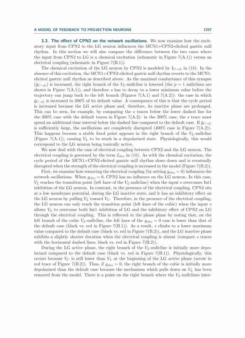
Figure 7. Effect of strengthening the CPN 2-to-LG synaptic input on the MCN 1+CPN 2-elicited gastricmill rhythm. For simplicity, only the p = 1 (maximally forced) nullcline is shown in the phase planes. (A.1)–(A.2) The CPN 2-to-LG input modeled as an excitatory chemical synapse IC→L. Default VL-nullcline is inred; zero synaptic strength in black; purple and green are 200% and 400% of default, respectively. IncreasingIC→L primarily influences the network for higher values of VL by pulling down the right knee of the cubic (in(A.1)) so that s decays to a lower minimum value (bottom dashed line in (A.2)). Subsequently, the cycle periodof the gastric mill rhythm is prolonged (A.2). IC→L has no influence on the left knee of the VL-nullcline,so the maximum value of s remains similar in all cases where oscillations occur (top dashed line in (A.2)).(B.1)–(B.2) The CPN 2-to-LG input modeled by an electrical coupling Ielec. Orange is the case where Ielec isat 800% of default; the other colors are as in panel (A). Ielec influences the network for all values of VL; thus,both knees of the VL-nullcline move as its strength is varied. Ielec has an inhibitory effect when VL is low, andtherefore strengthening Ielec moves up the left knee of the VL-nullcline. This gives a higher maximum valuefor s (top dashed line in (B.2)), which prolongs the LG inactive phase. When the trajectory first reaches theright branch, VC (black arrow in (B.2)) is initially lower than VL; therefore, the electrical coupling still has aninhibitory effect and a stronger Ielec pushes up the VL-nullcline. All VL-nullclines intersect at VL = VC (in(B.1)). Below this intersection point, VC > VL and Ielec is excitatory: a stronger Ielec moves down the rightknee of the VL-nullcline.
A MODEL OF FEEDBACK TO PROJECTION NEURONS 1257
3.3. The effect of CPN2 on the network oscillations. We now examine how the excit-atory input from CPN2 to the LG neuron influences the MCN1+CPN2-elicited gastric millrhythm. In this section we will also compare the difference between the two cases wherethe input from CPN2 to LG is a chemical excitation (schematic in Figure 7(A.1)) versus anelectrical coupling (schematic in Figure 7(B.1)).
The chemical excitation of the LG neuron by CPN2 is modeled by IC→L in (18). In theabsence of this excitation, the MCN1+CPN2-elicited gastric mill rhythm reverts to the MCN1-elicited gastric mill rhythm as described above. As the maximal conductance of this synapse(gC→L) is increased, the right branch of the VL-nullcline is lowered (the p = 1 nullclines areshown in Figure 7(A.1)), and therefore s has to decay to a lower minimum value before thetrajectory can jump back to the left branch (Figures 7(A.1) and 7(A.2)): the case in whichgC→L is increased to 200% of its default value. A consequence of this is that the cycle periodis increased because the LG active phase and, therefore, its inactive phase are prolonged.This can be seen, for example, by comparing the s traces below the lower dashed line forthe 200% case with the default traces in Figure 7(A.2): in the 200% case, the s trace mustspend an additional time interval below the dashed line compared to the default case. If gC→L
is sufficiently large, the oscillations are completely disrupted (400% case in Figure 7(A.2)).This happens because a stable fixed point appears in the right branch of the VL-nullcline(Figure 7(A.1)), causing VL to be stuck in a depolarized state. Physiologically, this wouldcorrespond to the LG neuron being tonically active.
We now deal with the case of electrical coupling between CPN2 and the LG neuron. Theelectrical coupling is governed by the term Ielec in (18). As with the chemical excitation, thecycle period of the MCN1+CPN2-elicited gastric mill rhythm slows down and is eventuallydisrupted when the strength of the electrical coupling is increased in the model (Figure 7(B.2)).
First, we examine how removing the electrical coupling (by setting gelec = 0) influences thenetwork oscillations. When gelec = 0, CPN2 has no influence on the LG neuron. In this case,VL reaches the transition point (left knee of the VL-nullcline) when the input s overcomes Int1inhibition of the LG neuron. In contrast, in the presence of the electrical coupling, CPN2 sitsat a low membrane potential, during the LG inactive state, and it has an inhibitory effect onthe LG neuron by pulling VL toward VC . Therefore, in the presence of the electrical coupling,the LG neuron can only reach the transition point (left knee of the cubic) when the input sallows VL to overcome both Int1 inhibition of LG and the inhibitory effect of CPN2 on LGthrough the electrical coupling. This is reflected in the phase plane by noting that, on theleft branch of the cubic VL-nullcline, the left knee of the gelec = 0 case is lower than that ofthe default case (black vs. red in Figure 7(B.1)). As a result, s climbs to a lower maximumvalue compared to the default case (black vs. red in Figure 7(B.2)), and the LG inactive phaseexhibits a slightly shorter duration when the electrical coupling is absent (compare s traceswith the horizontal dashed lines, black vs. red in Figure 7(B.2)).
During the LG active phase, the right branch of the VL-nullcline is initially more depo-larized compared to the default case (black vs. red in Figure 7(B.1)). Physiologically, thisoccurs because VC is still lower than VL at the beginning of the LG active phase (arrow inred trace of Figure 7(B.2)). Thus, if gelec = 0, the right branch of the cubic is initially moredepolarized than the default case because the mechanism which pulls down on VL has beenremoved from the model. There is a point on the right branch where the VL-nullclines inter-

1258 NICKOLAS KINTOS AND FARZAN NADIM
sect (Figure 7(B.1)). This point corresponds to VL = VC . Below this point, VC > VL, andthe electrical coupling has an excitatory influence on VL. Thus, when gelec = 0, the rightknee of the VL-nullcline is higher than the default case (black vs. red in Figure 7(B.1)). Thisis because the decay of s (in the gelec = 0 case) does not have to overcome the excitatoryinfluence from the CPN2 electrical coupling before reaching the transition point (right kneeof the cubic) to jump back to the left branch. The overall influence of the electrical couplingon the right branch is similar to that of the chemical excitation described in Figure 7(A.1).Similarly, as with the strong chemical excitation, if gelec is sufficiently strong, a stable fixedpoint appears on the right branch and oscillations are disrupted. In our model, the disruptionoccurs at a higher level of increase in gelec compared with the increase in gC→L (Figure 7(B.2)vs. 7(B.1)).
Note that the chemical excitation only influences the active phase of the LG neuron,whereas the electrical coupling influences both phases. This difference is, for example, seenin comparing the s traces in the two cases in Figures 7(A.2) and 7(B.2) (compare with thedashed lines).
3.4. Two distinct loci of oscillations in the network. One distinguishing factor in thecircuit of the MCN1+CPN2-elicited gastric mill rhythm as compared to the MCN1-elicitedrhythm is the presence of the Int1-to-CPN2 feedback synapse. This synapse inhibits CPN2,which prevents it from exciting the LG neuron. Yet, Int1 also has a direct inhibitory synapseonto the LG neuron. This brings up the question as to what these two seemingly redundantfunctionally inhibitory pathways accomplish. To address this question, we removed eachpathway from the network and examined the behavior of the model.
First, we examine how the removal of the inhibitory feedback synapse from Int1 to CPN2influences the MCN1+CPN2-elicited gastric mill rhythm. In our model, this feedback synapseappears in (3) as II→C with maximal conductance gI→C . In the reduced model, this synapseis mathematically absorbed into the IC→L and Ielec terms of (18).
Network oscillations persist when the Int1-to-CPN2 feedback synapse is removed fromthe network circuitry (Figure 8). This suggests that costimulation of MCN1 and CPN2 canproduce a gastric mill rhythm in the absence of the Int1-to-CPN2 feedback synapse. Notethat, in our model, when the Int1-to-CPN2 feedback synapse is removed from the network,the membrane potential of CPN2 remains fixed at its high resting potential (Figure 8(B)).As a result, CPN2 has an excitatory effect on VL in both phases of the gastric mill rhythm.Moreover, during the inactive state of the LG neuron, the position of the left knee of theVL-nullcline is lower than that of the case where the network circuitry is intact (Figure 8(A)).This is because, unlike the intact system, the excitation from CPN2 works together with theexcitatory input s during the LG inactive phase of the rhythm. As a result, less buildup of sis needed before VL can jump to its active state. However, as in the intact system, the jumpto the right branch of the VL-nullcline is still initiated by the pyloric-timed input p (verticaldotted line in Figure 8(B)).
The active state of the LG neuron remains mostly unchanged in the absence of the Int1-to-CPN2 feedback synapse (Figure 8(A)). In summary, there is no qualitative effect producedby removing the Int1-to-CPN2 feedback synapse on the MCN1+CPN2-elicited gastric millrhythm.
Next, we examine how removal of the Int1-to-LG synapse influences the MCN1+CPN2-

A MODEL OF FEEDBACK TO PROJECTION NEURONS 1259
FeedbackRemoved
FeedbackRemoved
Default
Default
(A)
VL
s
5 sec
20 mV
VL
VC
P
Default
FeedbackRemoved
FeedbackRemoved
(B)
0
1
LG
MCN1 CPN2
CoG
STG
Int1
s
AB
1
0
Figure 8. Removing the Int1-to-CPN 2 feedback synapse does not qualitatively change the MCN 1+CPN 2-elicited gastric mill rhythm. Inset shows the circuit diagram. (A) Phase plane diagram showing the VL-nullclineof the intact network (default: light blue) and when the Int1-to-CPN 2 feedback synapse is removed (red). Forsimplicity only the p = 1 VL-nullcline is shown. Without the feedback synapse, VC remains in an active stateand continually excites LG. Therefore, only the left branch of the VL-nullcline is affected by the Int1-to-CPN 2synapse. (B) The overall effect of removing the Int1-to-CPN 2 feedback synapse is a slightly shorter cycle period.Note, however, that, similar to the intact system, the pyloric-timed input still determines the onset of the LGactive phase when the Int1-to-CPN 2 feedback synapse is removed (vertical dotted line).

1260 NICKOLAS KINTOS AND FARZAN NADIM
elicited gastric mill rhythm in the model. When this synapse is removed, the VL-nullclineretains its cubic shape (Figure 9(A.1)), and the VL- and s-nullclines still intersect along themiddle branch of the cubic, which allows for a stable limit cycle (Figure 9(A.1)) and networkoscillations persist (Figure 9(A.2)). Note that the effect of removing the Int1-to-LG synapseon the network dynamics is removal of direct inhibition of the LG neuron (schematic ofFigure 9(A.1)). As a result, although the VL-nullcline retains its cubic shape, the left knee ofthe VL-nullcline is at a more depolarized value because of the absence of direct inhibition onthe LG neuron (compare red and blue cubics in Figure 9(A.1)).
While the trajectory is on the left branch of the VL-nullcline (with the Int1-to-LG synapseremoved from the network), Int1 inhibits CPN2, which prevents the latter from exciting LG.Also, the MCN1 input s slowly builds up in the LG neuron (Figure 9(B.2)) as the trajectoryslowly climbs up the left branch of the cubic. When the trajectory reaches the left knee ofthe cubic, it jumps to the LG active state on the stable right branch. Now, the LG neuroninhibits Int1 and presynaptically inhibits MCN1. The former allows CPN2 to escape fromInt1 inhibition and excite the LG neuron. The latter causes s to slowly decay down the rightbranch of the cubic (Figure 9(A.1)). When the trajectory reaches the right knee of the cubic,it jumps back to the stable left branch, and the cycle repeats.
Note that the right branch of the cubic is practically the same with or without the Int1-to-LG synapse because this synapse has little influence when the value of VL is large. Inthe absence of this synapse, the left branch of the cubic, however, is only influenced by onefactor: the LG neuron leak current ILeak,L. Figures 9(B.1) and 9(B.2) show how an increasein gLeak,L affects the shape of the nullcline and the network oscillations in the absence ofthe Int1-to-LG synapse. Note that the excitation strength for the CPN2-to-LG synapse wasincreased in these figures to compensate for the increase in gLeak,L. In the case of a largervalue of gLeak,L, the left knee of the VL-nullcline is shifted up (green cubic in Figure 9(B.1)),resulting in an increase in the inactive duration of the LG neuron oscillations (green vs. redin Figure 9(B.2)). Note also that, even with this increased gLeak,L, oscillations persist in theintact network in the presence of the Int1-to-LG synapse (not shown).
We have shown that oscillations can persist after removing either the Int1-to-CPN2 feed-back synapse or the Int1-to-LG synapse. However, removing both synapses changes the cubicVL-nullcline to a monotonic curve, and only a single stable fixed point remains in the phaseplane (Figure 9(A.1)). As a result, oscillations cannot persist without both of these synapses(Figure 9(A.2)).
In contrast with the MCN1+CPN2-elicited gastric mill rhythm, the MCN1-elicited rhythmcannot persist in the absence of the Int1-to-LG synapse. Removing this synapse disrupts thepattern-generating mechanism by changing the VL-nullcline to a monotonic curve (not shown).
3.5. The role of the MCN1 slow input s. As with the MCN1-elicited gastric mill rhythm[26], the slow oscillations in the MCN1+CPN2-elicited gastric mill rhythm are primarily de-termined by the slow rise and decay of s. The time constant of this state variable is the slowtime constant in the model. In the previous section, we showed that the network circuitryunderlying the MCN1+CPN2-elicited gastric mill rhythm involves two distinct loci of oscilla-tions. Here we examine the influence of the slow input s on the gastric mill oscillations for theintact network in comparison with the oscillations that occur in the absence of the Int1-to-LG

A MODEL OF FEEDBACK TO PROJECTION NEURONS 1261
No Int1 to LGDefault Leak
Large Leak
No Int1to LG
No Int1 to LGNo Int1 to CPN2
No Int1 to LGNo Int1 to CPN2
Default
Default
(A.2)
stable fixed point
VL
LG
MCN1 CPN2
CoG
STG
Int1
s
AB
1
0
5 sec
20 mV
VL
s
LG
MCN1 CPN2
CoG
STG
Int1
s
AB
1
0
(B.2)
(B.1)(A.1)
Large LeakDefault Leak
VL
s 0.51
0.03
Figure 9. The MCN 1+CPN 2-elicited gastric mill rhythm persists, but is qualitatively different, when theInt1-to-LG synapse is removed from the network. Inset shows the circuit diagram. (A.1)–(A.2) Phase planediagram showing the VL-nullcline of the intact network (default: light blue), when the Int1-to-LG synapse isremoved (red), and when both the Int1-to-LG and Int1-to-CPN 2 synapses are removed (brown). For simplicity,the p = 0 (unforced) VL-nullcline is shown. Due to the much lower left knee of the red cubic in the phase plane,the cycle period of the rhythm in the absence of the Int1-to-LG synapse is much smaller than the default case(red vs. blue trace in (A.2)). Also, notice that the pyloric-timed input does not influence the rhythm that occurswithout the Int1-to-LG synapse. Oscillations cannot occur when both synapses are removed (brown). (B.1)–(B.2) The LG leak current influences the cycle period in the absence of the Int1-to-LG synapse. Red shows theVL-nullcline from panel (A.1), while the green VL-nullcline corresponds to a 10-fold increase in gLeak,L. Thestronger leak conductance shifts up the left knee of the cubic in the phase plane (green vs. red in (B.1)), whichin turn prolongs the cycle period (green vs. red in (B.2)). Even with higher gLeak,L, the pyloric-timed inputdoes not influence the oscillations.

1262 NICKOLAS KINTOS AND FARZAN NADIM
(C)
VL
sweaker
syn
ap
se
(A) DEFAULT
weaker
syn
ap
se
default
10%0%
stable fixed point
50%
25%
default
50%
(B) NO Int1 to LG SYNAPSE
25%
100% 90% 75% 50% 25% 10% 0%
0
2
4
6
8
10
12
14
Du
rati
on
(s
ec
)
MCN1 to LG synapse
LG Inactive (Default)LG Active (Default)LG Inactive (No Int1 to LG)LG Active (No Int1 to LG)
Figure 10. Weakening the MCN 1-to-LG synaptic input prolongs the duration of the LG inactive phasein the presence or absence of the Int1-to-LG synapse. (A) Phase plane diagram for the default case. Forsimplicity, only the p = 1 (maximally forced) VL-nullcline is shown. Reducing gs shifts up the left knee of thecubic VL-nullcline. Thus, the trajectory must climb to a higher value of s before it can jump to the right branch.When gs is weakened to 25% of its default value, the nullclines intersect at a stable fixed point (arrow). Theright knee of the VL-nullcline is not as strongly influenced by gs. (B) Phase plane diagram for the case wherethe Int1-to-LG synapse is removed. As in (A), the left knee of the VL-nullcline moves upward as gs is reduced.When gs = 0 (0%), the VL-nullcline loses its cubic shape, and a stable fixed point appears (arrow). (C) Theeffect of reducing gs on the durations of both phases of the rhythm. Both for the default case and when theInt1-to-LG synapse is removed, the LG inactive phase duration is prolonged when gs is reduced. However, theLG active phase duration remains relatively unchanged.
synapse. The influence of s on the network oscillations in the absence of the Int1-to-CPN2feedback synapse is similar to its effects in the default case (intact network) because of thesimilar phase plane structure and is not separately discussed.
In the default case, the maximal conductance gs of the MCN1-to-LG excitatory synapseinfluences the cycle period of the MCN1+CPN2-elicited gastric mill rhythm primarily bychanging the shape of the VL-nullcline on the left branch. The primary effect is that, with asmaller gs, the left knee of the VL-nullcline is shifted up (Figure 10(A)). As a result, s has

A MODEL OF FEEDBACK TO PROJECTION NEURONS 1263
to rise to a larger value before reaching this knee, thus increasing the inactive phase durationof the LG neuron. However, the duration of the active phase remains relatively unchanged.The effect of gs on the inactive and active phase durations of the LG neuron is shown inFigure 10(C).
In the absence of the Int1-to-LG synapse, the role of s in producing the slow oscillationsremains central, but the sensitivity of the oscillations to gs is different. This is due to the factthat the VL-nullcline is less sensitive to changes in gs because of the lower position of the leftbranch of this nullcline compared to the intact system (Figure 10(B)). As seen in Figure 10(C),the inactive phase duration of the LG oscillation remains insensitive to changes in gs up to thepoint that gs is very weak (less than 25% of the default value; see Figure 10(C)). Additionally,oscillations are disrupted at a much smaller value of gs compared to the default case.
When gs is set to zero, the VL-nullcline loses its cubic shape in the phase plane. As aresult, the VL- and s-nullclines intersect at a stable fixed point (Figure 10(B)), and networkoscillations are disrupted. Physiologically, this implies that the MCN1 input is necessary toactivate the gastric mill rhythm, and CPN2 alone cannot activate the gastric mill rhythm,consistent with previously reported experiments [34].
4. Discussion. Activity in neural networks underlying motor output is often initiatedand controlled by input from descending modulatory projection pathways [13, 6, 20, 37]. Insome cases, network activity that underlies similar behaviors can result from the activity ofdistinct sets of projection pathways [42, 16, 40]. Additionally, descending projection pathwaysreceive feedback from their target motor networks, which can, in turn, influence the activityof the network [19, 11, 33, 43]. We are interested in the network principles that distinguishthe actions of distinct projection pathways. In particular, we address the question of howfeedback to the descending projection neurons can influence network activity in an oscillatorynetwork.
We have developed a reduced mathematical model to examine how the synaptic actionsof two descending projection neurons, MCN1 and CPN2, on STG neurons can produce andshape the gastric mill oscillatory motor pattern. We specifically focused on a simplified ver-sion of the gastric mill motor pattern generated by the ventral cardiac neurons in which theMCN1 input is tonically active [5]. The model included both feed-forward and feedback cir-cuitry that exists between these projection neurons and the gastric mill CPG neurons. Wespecifically investigated if the synaptic circuitry alone can distinguish the two different ver-sions of the gastric mill rhythm from each other: the version elicited by MCN1 stimulationalone [14] and the distinct version where MCN1 and CPN2 are coactive [5]. Physiologically,CPN2 is quiescent during the MCN1-elicited rhythm, so its synaptic interactions with thegastric mill CPG neurons (CPN2 excitation of LG and Int1 feedback inhibition of CPN2) in-fluence rhythm generation in only the MCN1+CPN2-elicited gastric mill rhythm. To simplifyour mathematical analysis, we reduced our model down to 2 dimensions by (i) ignoring theaction potential activity and modeling only the envelopes of the bursting activity patterns,(ii) treating all neurons in the model as passive, and (iii) exploiting the difference in synaptictime scales which are present in the biological system. This reduction of dimension allowed usto capture the fast-slow network dynamics of the model in the VL− s phase plane and readilycompare both versions of the gastric mill rhythm.

1264 NICKOLAS KINTOS AND FARZAN NADIM
4.1. The network circuitry can explain the different oscillation frequencies of the dis-tinct gastric mill rhythms. Our modeling revealed that the synaptic circuitry between theprojection neurons and gastric mill CPG neurons is sufficient to explain the different oscil-lation frequencies exhibited in the two distinct versions of the gastric mill rhythm, and thatthe intrinsic properties of individual neurons do not play a central role. In our model, theMCN1+CPN2-elicited gastric mill rhythm exhibited a longer cycle period than the MCN1-elicited rhythm. This difference in cycle period occurred in both the absence (Figure 4) andpresence (Figure 5) of the pyloric-timed input from the AB neuron to Int1. In both cases,CPN2 excitation of the LG neuron, which occurs during the protraction (LG active) phase ofthe MCN1+CPN2-elicited rhythm, lowers the right knee of the VL-nullcline in the phase plane(Figures 4(A) and 5(A)). Mechanistically, the excitation from CPN2 opposes the slow decayof s, the MCN1 input to the LG neuron. As a result, s must fall to a lower minimum value (forthe MCN1+CPN2-elicited rhythm) before the trajectory can jump back to the left branch ofthe VL-nullcline. This in turn prolongs the cycle period of the MCN1+CPN2-elicited gastricmill rhythm compared to the MCN1-elicited rhythm (where CPN2 is quiescent). Previousexperiments have established the difference in cycle periods between the two distinct versionsof the gastric mill rhythm in the presence of the pyloric-timed input from AB to Int1 [9]. Itremains to be seen if a similar difference in cycle periods, as our model predicts, occurs in theabsence of the AB-to-Int1 synapse in the biological system.
Our model also showed that the presence of the pyloric-timed input produces the samenetwork effects in the MCN1+CPN2-elicited gastric mill rhythm, as was previously reportedfor the MCN1-elicited rhythm [3, 32]. In particular, the pyloric-timed input determines theonset of the LG active phase and increases the oscillatory frequency of the MCN1+CPN2-elicited gastric mill rhythm in our model (Figure 6). Thus, the gastro-pyloric coordination thatexists for the MCN1-elicited rhythm is preserved when the synaptic circuitry between CPN2and the gastric mill CPG neurons is added to the network. The effect of the pyloric-timedinput on the cycle frequency of the MCN1+CPN2-elicited rhythm in our model is consistentwith that observed for the VCN-elicited rhythm in the biological system [42]. However, itremains to be seen if our model prediction that the pyloric-timed input determines the onsetof the LG active phase (for the MCN1+CPN2-elicited rhythm) also occurs in the biologicalsystem.
A previous experimental study has indicated that the CPN2-to-LG connection is a chem-ical synapse [34]. However, there is also evidence that this connection may persist in a lowcalcium solution, indicating that at least some component of the connection is due to anelectrical coupling, i.e., a gap junction between CPN2 axon terminals and the LG neuron inthe STG [35]. Our model was able to reproduce the general behavior of the MCN1+CPN2-elicited gastric mill rhythm using either a chemical synapse or an electrical coupling, and thebehavior of the model was mostly independent of which connection type was used. In general,the two different cases of the CPN2-to-LG synapse had a similar influence on the networkoscillations. The one instance in which qualitatively different network effects were observedwas in exploring the connection strengths in the model (Figure 7). Strengthening the chemicalCPN2-to-LG synapse lowered only the right branch of the VL-nullcline in the phase plane.In contrast, the electrical CPN2-to-LG coupling influenced the full VL-nullcline. As a result,the chemical synapse has a more direct effect on the LG neuron active phase, whereas the

A MODEL OF FEEDBACK TO PROJECTION NEURONS 1265
electrical coupling has a more symmetric effect on both the active and inactive phases of theLG neuron.
Previous experimental results have suggested that CPN2 exhibits postinhibitory reboundproperties [34]. However, these intrinsic properties of CPN2 were not necessary in our modelto reproduce the general behaviors of the MCN1+CPN2- and MCN1-elicited gastric millrhythms exhibited in the biological system. It is possible to include the postinhibitory reboundproperties of CPN2 in the model, but this would require a 4-dimensional model to analyzethe network dynamics and would not allow for using phase plane analysis. Nevertheless, theoverall behavior of network oscillations in the 4-dimensional model is similar to that of ourreduced 2-dimensional model in this work [25].
An alternative possibility is that CPN2 has intrinsic bursting properties. If CPN2 is anintrinsic burster, then its influence on the MCN1+CPN2-elicited gastric mill rhythm can bequite different. As a bursting neuron, CPN2 activity can potentially dominate the gastric millrhythm through its excitatory synapse onto the LG neuron. Thus, even in the absence of theInt1-to-CPN2 feedback synapse, the rhythm would remain mostly unchanged. However, theonset of the LG neuron active phase may not remain coordinated with the pyloric-timed input.In contrast, in our current model, removing the Int1-to-CPN2 feedback synapse diminishesthe influence of CPN2 on the gastric mill rhythm, thereby resulting in a rhythm which issimilar to that elicited by MCN1 alone. In addition, if CPN2 is an intrinsic burster, the roleof the Int1-LG reciprocal inhibition could be greatly diminished in generating the gastric millrhythm. In fact, CPN2 may be able to elicit a gastric mill rhythm even if all synapses fromInt1 are deactivated within the network circuitry.
Previous experiments and modeling [16] have accounted for a modulator-activated, voltage-gated inward current (IMI) in the LG neuron during the MCN1-elicited gastric mill rhythm.The presence of this current in a model makes the resulting MCN1-elicited rhythm quanti-tatively closer to that of the biological system. However, as we observed previously for themodel of the MCN1-elicited rhythm [24], we found that the presence of IMI does not producea qualitatively different rhythm in our current model.
4.2. The presence of multiple loci of oscillations in the network. Reciprocal inhibi-tion, leading to half-center oscillations, has long been established as a fundamental buildingblock of network circuitry [29, 17, 41]. The MCN1-elicited gastric mill rhythm relies on asingle reciprocally inhibitory circuit between Int1 and the LG neuron, which is necessary forproducing the rhythmic activity pattern [3]. Removing either one of the Int1-LG reciprocallyinhibitory synapses will disassemble the half-center and disrupt the MCN1-elicited rhythm. Inour model of the MCN1+CPN2-elicited gastric mill rhythm, the Int1-LG reciprocal inhibitionstill plays an important role in establishing the cubic shape of the VL-nullcline, which allowsfor the existence of stable oscillations. However, our model reveals an additional locus for thegeneration of oscillations that is independent of the Int1-LG reciprocal inhibition. As seenin Figure 9, gastric mill oscillations can persist even if the Int1-LG reciprocal inhibition isdisrupted by blocking the Int1-to-LG synapse. In this case, oscillations are generated througha distinct half-center circuit which consists of three synaptic interactions: Int1 inhibition ofCPN2, CPN2 excitation of LG, and LG inhibition of Int1. The first of these synaptic in-teractions influences the left branch of the VL-nullcline, whereas the latter two influence the

1266 NICKOLAS KINTOS AND FARZAN NADIM
right branch of the nullcline. Even in the absence of the Int1-to-LG inhibitory synapse, theseinteractions can result in a cubic VL-nullcline and allow for stable oscillations.
Note that the oscillatory activity (in the case where the Int1-to-LG synapse is removed)is also dependent, primarily, on the slow dynamics of s, the MCN1 excitatory input to theLG neuron (Figure 9(B.2)). When CPN2 is inhibited by Int1 (and therefore LG is inactive),s slowly builds up in the LG neuron, allowing it to depolarize and eventually start inhibitingInt1. LG inhibition of Int1 results in the removal of the Int1-to-CPN2 feedback inhibition,which permits CPN2 to excite the LG neuron, thus causing a rapid transition of LG to itsactive state. During the LG active state, s decays due to LG presynaptic inhibition of MCN1,and the LG membrane potential decays to the point that it eventually stops inhibiting Int1,allowing the latter to escape to its active state, and the cycle repeats.
The network oscillations that occur in the absence of the Int1-to-LG synapse are distinctfrom the oscillations of the intact circuit. First, the cycle period is substantially shorter be-cause LG does not need to overcome direct Int1 inhibition in order to transition to its activestate. Second, the pyloric-timed input no longer influences the MCN1+CPN2-elicited gastricmill rhythm, and the gastro-pyloric intercircuit regulation is lost. Thus, this reduced networkcircuitry (without the Int1-to-LG synapse) does not necessarily produce a normal motor be-havior as the full intact network does. Yet, there is evidence that the MCN1+CPN2-elicitedgastric mill rhythm can persist in the absence of the Int1-to-LG synapse in the biologicalsystem [1]. This implies a functional role for the feedback synapse from the gastric mill CPGto the descending projection neurons, where the presence of this synapse results in a newlocus of rhythm generation which can be potentially targeted by neuromodulators to regu-late the network output. In general, feed-forward plus feedback circuit interactions betweenprojection neurons and their target networks are ubiquitous in nervous systems. Therefore,it would be interesting to check if these circuit interactions can produce multiple distinct locifor the generation of oscillations in other networks as well.
4.3. Implications for sensory input to the STG. The gastropyloric muscle stretch re-ceptors (GPRs) are sensory neurons that are activated by the stretching of the gastric millprotractor muscles in the stomach [22, 23]. Previous experiments and modeling have estab-lished that synaptic input from the GPR neurons slows the cycle frequency of the MCN1-elicited gastric mill rhythm by presynaptically inhibiting MCN1 axon terminals during thesilent (interburst) phase of the LG neuron [4]. GPR presynaptic inhibition of MCN1 prolongsthe duration of the LG inactive phase and leaves the duration of the LG active phase relativelyunchanged, which together slows the overall cycle frequency of the MCN1-elicited gastric millrhythm [4]. However, the influence of GPR in the biological system is phasic in that it occursonly during the LG inactive phase of the MCN1-elicited rhythm.
Our modeling results have two implications for the influence of GPR input on the gastricmill rhythm. First, when the excitatory input from MCN1 to LG was weakened in the modelduring both phases of the gastric mill rhythm, the result was similar to that of the phasicGPR input in the biological system: a longer cycle period in which the LG inactive phase wasprolonged but the LG active phase duration remained relatively unchanged (Figure 10(C)).This suggests that a nonphasic, continuous, GPR-like input would have a qualitatively similarinfluence on the gastric mill rhythm. In the biological system, the excitatory MCN1 input

A MODEL OF FEEDBACK TO PROJECTION NEURONS 1267
to the STG is already inhibited during the LG active phase of the gastric mill rhythm dueto LG presynaptic inhibition of MCN1. Therefore, a further weakening of the MCN1 inputshould not qualitatively change the duration of the LG active phase, as confirmed by ourmodel. Mathematically, as the MCN1 input to the LG neuron is weakened in our model, thejump to the right branch of the cubic VL-nullcline occurs for a higher value of s. Thus, the(exponential) decay from higher values of s becomes more rapid along the right branch of thecubic as the conductance gs is weakened in the model, which leads to a similar LG activephase duration for all cases in Figure 10(C). We note that a similar influence occurs for theMCN1-elicited rhythm due to the similar phase plane structure.
The second implication is that the influence of the GPR-like input is independent of theInt1-LG reciprocal inhibition. In particular, removing the Int1-to-LG synapse dismantles theInt1-LG reciprocally inhibitory half-center in the network; yet, the effect of weakening theMCN1-to-LG synaptic input remains qualitatively similar to that of the intact network in themodel (Figure 10(C)).
4.4. Conclusions. Feedback from CPG networks to descending projection neurons is aubiquitous but seldom understood feature of motor activity. Our simple mathematical modelof the gastric mill CPG provides some clues for the potential importance of such feedbackpathways. In particular, this simple model shows that the patterned activity of the feedbacksynapse may provide additional mechanisms for the generation of oscillatory activity that aredistinct from those which are present in the isolated CPG. These additional mechanisms canhave distinct dependence on network parameters and may therefore be differentially controlledby sensory or modulatory inputs. Experimental data that involve manipulation of thesefeedback pathways, for example, in the networks of the spinal cord, can elucidate the extentto which such mechanisms are utilized in the control of rhythmic motor activity.
REFERENCES
[1] T. Akay, D. Wood, and M. P. Nusbaum, Reciprocal Inhibition is not necessary for generation of allgastric mill rhythms, in Proceedings of the Society for Neuroscience 34th Annual Meeting, San Diego,CA, 2004.
[2] C. Ambrosio-Mouser, F. Nadim, and A. Bose, The effects of varying the timing of inputs on a neuraloscillator, SIAM J. Appl. Dyn. Syst., 5 (2006), pp. 108–139.
[3] M. Bartos, Y. Manor, F. Nadim, E. Marder, and M. P. Nusbaum, Coordination of fast and slowrhythmic neuronal circuits, J. Neurosci., 19 (1999), pp. 6650–6660.
[4] M. P. Beenhakker, N. D. Delong, S. R. Saideman, F. Nadim, and M. P. Nusbaum, Proprioceptorregulation of motor circuit activity by presynaptic inhibition of a modulatory projection neuron, J.Neurosci., 25 (2005), pp. 8794–8806.
[5] M. P. Beenhakker and M. P. Nusbaum, Mechanosensory activation of a motor circuit by coactivationof two projection neurons, J. Neurosci., 24 (2004), pp. 6741–6750.
[6] M. C. Bellingham, Driving respiration: The respiratory central pattern generator, Clin. Exp. Pharmacol.Physiol., 25 (1998), pp. 847–856.
[7] D. M. Blitz, M. P. Beenhakker, and M. P. Nusbaum, Different sensory systems share projectionneurons but elicit distinct motor patterns, J. Neurosci., 24 (2004), pp. 11381–11390.
[8] D. M. Blitz and M. P. Nusbaum, Modulation of circuit feedback specifies motor circuit output, J.Neurosci., 32 (2012), pp. 9182–9193.
[9] D. M. Blitz and M. P. Nusbaum, Motor pattern selection via inhibition of parallel pathways, J. Neu-rosci., 17 (1997), pp. 4965–4975.

1268 NICKOLAS KINTOS AND FARZAN NADIM
[10] D. M. Blitz, R. S. White, S. R. Saideman, A. Cook, A. E. Christie, F. Nadim, and M. P.
Nusbaum, A newly identified extrinsic input triggers a distinct gastric mill rhythm via activation ofmodulatory projection neurons, J. Exp. Biol., 211 (2008), pp. 1000–1011.
[11] J. T. Buchanan and J. F. Einum, The spinobulbar system in lamprey, Brain Res. Rev., 57 (2008), pp.37–45.
[12] R. E. Burke, The central pattern generator for locomotion in mammals, Adv. Neurol., 87 (2001), pp.11–24.
[13] A. Buschges, T. Akay, J. P. Gabriel, and J. Schmidt, Organizing network action for locomotion:Insights from studying insect walking, Brain Res. Rev., 57 (2008), pp. 162–171.
[14] M. J. Coleman, P. Meyrand, and M. P. Nusbaum, A switch between two modes of synaptic trans-mission mediated by presynaptic inhibition, Nature, 378 (1995), pp. 502–505.
[15] T. G. Deliagina, P. V. Zelenin, P. Fagerstedt, S. Grillner, and G. N. Orlovsky, Activity ofreticulospinal neurons during locomotion in the freely behaving lamprey, J. Neurophysiol., 83 (2000),pp. 853–863.
[16] N. D. Delong, M. S. Kirby, D. M. Blitz, and M. P. Nusbaum, Parallel regulation of a modulator-activated current via distinct dynamics underlies comodulation of motor circuit output, J. Neurosci.,29 (2009), pp. 12355–12367.
[17] A. Doloc-Mihu and R. L. Calabrese, A database of computational models of a half-center oscillatorfor analyzing how neuronal parameters influence network activity, J. Biol. Phys., 37 (2011), pp. 263–283.
[18] B. Ermentrout, Simulating, Analyzing, and Animating Dynamical Systems: A Guide to XPPAUT forResearchers and Students, SIAM, Philadelphia, 2002.
[19] W. N. Frost and P. S. Katz, Single neuron control over a complex motor program, Proc. Natl. Acad.Sci. USA, 93 (1996), pp. 422–426.
[20] L. M. Jordan, J. Liu, P. B. Hedlund, T. Akay, and K. G. Pearson, Descending command systemsfor the initiation of locomotion in mammals, Brain Res. Rev., 57 (2008), pp. 183–191.
[21] S. Kasicki and S. Grillner, Muller cells and other reticulospinal neurones are phasically active duringfictive locomotion in the isolated nervous system of the lamprey, Neurosci. Lett., 69 (1986), pp. 239–243.
[22] P. S. Katz, M. H. Eigg, and R. M. Harris-Warrick, Serotonergic/cholinergic muscle receptor cellsin the crab stomatogastric nervous system. I. Identification and characterization of the gastropyloricreceptor cells, J. Neurophysiol., 62 (1989), pp. 558–570.
[23] P. S. Katz and R. M. Harris-Warrick, Serotonergic/cholinergic muscle receptor cells in the crabstomatogastric nervous system. II. Rapid nicotinic and prolonged modulatory effects on neurons inthe stomatogastric ganglion, J. Neurophysiol., 62 (1989), pp. 571–581.
[24] N. Kintos, D. F. Hsu, A. Bose, M. P. Nusbaum, and F. Nadim, Modeling the activation of a voltage-gated ionic current by a modulatory projection neuron, Soc. Neurosci. Abstr., Chicago, IL, 2009.
[25] N. Kintos, M. P. Nusbaum, and F. Nadim, Feedback to descending projection neurons can overridethe mechanisms underlying rhythmic pattern generation in the target network: A modeling study, Soc.Neurosci. Abstr., 33 (2007), 925.1.
[26] N. Kintos, M. P. Nusbaum, and F. Nadim, A modeling comparison of projection neuron- andneuromodulator-elicited oscillations in a central pattern generating network, J. Comput. Neurosci.,24 (2008), pp. 374–397.
[27] Y. Manor, F. Nadim, L. F. Abbott, and E. Marder, Temporal dynamics of graded synaptic trans-mission in the lobster stomatogastric ganglion, J. Neurosci., 17 (1997), pp. 5610–5621.
[28] Y. Manor, F. Nadim, S. Epstein, J. Ritt, E. Marder, and N. Kopell, Network oscillations gener-ated by balancing graded asymmetric reciprocal inhibition in passive neurons, J. Neurosci., 19 (1999),pp. 2765–2779.
[29] E. Marder and R. L. Calabrese, Principles of rhythmic motor pattern generation, Physiol. Rev., 76(1996), pp. 687–717.
[30] K. Matsuyama, F. Mori, K. Nakajima, T. Drew, M. Aoki, and S. Mori, Locomotor role of thecorticoreticular-reticulospinal-spinal interneuronal system, Prog. Brain Res., 143 (2004), pp. 239–249.
[31] E. F. Mishchenko and N. K. Rozov, Differential Equations with Small Parameters and RelaxationOscillators, Plenum Press, New York, 1980.

A MODEL OF FEEDBACK TO PROJECTION NEURONS 1269
[32] F. Nadim, Y. Manor, M. P. Nusbaum, and E. Marder, Frequency regulation of a slow rhythm by afast periodic input, J. Neurosci., 18 (1998), pp. 5053–5067.
[33] B. J. Norris, M. J. Coleman, and M. P. Nusbaum, Pyloric motor pattern modification by a newlyidentified projection neuron in the crab stomatogastric nervous system, J. Neurophysiol., 75 (1996),pp. 97–108.
[34] B. J. Norris, M. J. Coleman, and M. P. Nusbaum, Recruitment of a projection neuron determinesgastric mill motor pattern selection in the stomatogastric nervous system of the crab, Cancer borealis,J. Neurophysiol., 72 (1994), pp. 1451–1463.
[35] M. P. Nusbaum, Private communication, 2014.[36] M. P. Nusbaum and M. P. Beenhakker, A small-systems approach to motor pattern generation,
Nature, 417 (2002), pp. 343–350.[37] M. P. Nusbaum, D. M. Blitz, A. M. Swensen, D. Wood, and E. Marder, The roles of co-
transmission in neural network modulation, Trends Neurosci., 24 (2001), pp. 146–154.[38] M. C. Perreault, T. Drew, and S. Rossignol, Activity of medullary reticulospinal neurons during
fictive locomotion, J. Neurophysiol., 69 (1993), pp. 2232–2247.[39] S. Rossignol, R. Dubuc, and J. P. Gossard, Dynamic sensorimotor interactions in locomotion,
Physiol. Rev., 86 (2006), pp. 89–154.[40] W. Stein, N. D. Delong, D. E. Wood, and M. P. Nusbaum, Divergent cotransmitter actions underlie
motor pattern activation by a modulatory projection neuron, Eur. J. Neurosci., 26 (2007), pp. 1148–1165.
[41] A. E. Tobin and R. L. Calabrese, Endogenous and half-center bursting in morphologically inspiredmodels of leech heart interneurons, J. Neurophysiol., 96 (2006), pp. 2089–2106.
[42] R. S. White and M. P. Nusbaum, The same core rhythm generator underlies different rhythmic motorpatterns, J. Neurosci., 31 (2011), pp. 11484–11494.
[43] D. E. Wood, Y. Manor, F. Nadim, and M. P. Nusbaum, Intercircuit control via rhythmic regulationof projection neuron activity, J. Neurosci., 24 (2004), pp. 7455–7463.
[44] T. Yamaguchi, The central pattern generator for forelimb locomotion in the cat, Prog. Brain Res., 143(2004), pp. 115–122.
[45] P. V. Zelenin, Activity of individual reticulospinal neurons during different forms of locomotion in thelamprey, Eur. J. Neurosci., 22 (2005), pp. 2271–2282.
Copyright © 2022 FDOKUMEN