A Rapid, Validated RP-HPLC Method for the Determination of ...
Upload
independentCategory
view
0download
0
nature protocols | VOL.4 NO.12 | 2009 | 1709
p
uor
G g
n ih si l
bu
P eru ta
N 900 2©
nat
ure
pro
toco
ls/
moc. e r
ut an .
ww
w / /:pt t
h
protocol
IntroDuctIonPyrogenic (pyros is Greek for fire) contaminations pose a major safety concern for patients because their introduction into the human body, e.g., via parenterals, cell therapies or medical devices may cause severe reactions ranging from fever to shock and death. Known pyrogens are of microbial origin, but their presence does not correlate with colony-forming units, i.e., number of viable microorganisms, or with the presence of microbial genetic material. Examples of known pyrogens include lipopolysaccharides (LPS), i.e., well-described, highly potent endotoxins of Gram-negative bacteria; lipoteichoic acid (LTA)1, lipopeptides and peptidoglycan2 from Gram-positive bacteria; exotoxins3; enterotoxins4,5; viruses6; and fungal components7,8.
Development of the protocolIn mammals the exposition of inner tissues to pyrogens leads to the secretion of the so-called endogenous pyrogens by immune cells, i.e., blood monocytes and tissue macrophages9, which when injected into mammals induce fever reactions. The most potent endogenous pyrogen is the proinflammatory cytokine interleukin (IL)-1β (refs. 10,11), which binds to receptors on the blood side of the organum vasculosum laminae terminalis, one of the circum-ventricular organs of the brain, and initiates the expression of the enzyme cyclooxygenase-2 (COX-2)12,13, which converts arachidonic acid to prostaglandin (PG)E
2. PGE
2 then mediates an increase in
temperature14. Mice deficient in COX-2 do not develop fever in response to injection of IL-1β or LPS15–17.
Our basic idea that led to the development of the in vitro pyrogen test (IPT) was to use human immune cells to detect exogenous pyro-gens by measuring the release of an endogenous pyrogen18. Diluted fresh or cryopreserved blood is incubated for 10–24 h at 37 °C with the test samples, spiked test samples, endotoxin controls and negative controls. The blood monocytes produce proinflamma-tory cytokines in response to any pyrogen present in a concentra-tion-dependent manner and the IL-1β produced is measured by enzyme-linked immunosorbent assay (ELISA).
Human whole blood containing monocytes was chosen as an accessible source of primary human immune cells. The advantage
of using whole blood instead of isolated cells, such as blood mono-cytes, is that all the immune cell types and serum components are present in their natural composition, and sources of contamination or preactivation of the cells are reduced as no isolation procedures are required.
Interleukin-1β was chosen as a readout parameter as it is consist-ently released in a concentration-dependent manner by blood from different donors19 upon exposure to the same minimal pyrogenic stimulation and can be readily measured by an ELISA. The sand-wich ELISA is a highly specific and sensitive immunological assay that can detect antigens in the low pg ml − 1 range. The use of two antibodies, usually one monoclonal and one polyclonal antibody preparation, ensures high specificity, while linking the secondary antibody, directly or via biotin–streptavidin binding, to the enzyme horseradish peroxidase that metabolizes tetramethylbenzidine (TMB) ensures a high level of sensitivity.
The IPT thus poses a physiologically relevant human test system for the assessment of pyrogenic contaminations. It has successfully undergone an international validation process20,21 for both fresh and cryopreserved blood, and will be included in the European Pharmacopoeia in 2010 as announced by the European Directorate for the Quality of Medicines & Health Care (www.edqm.eu/medias/fichiers/133rd_Session_of_the_Eu.pdf). A similar statement by the United States Food and Drug Administration is pending. The IPT test will help to significantly reduce the use of animals for research and safety assessments in cosmetic and pharmaceutical industry. The methodology of IPT using either fresh or cryopreserved blood as well as its adaptations described herein are protected for com-mercial use by international patents22–24.
Applications of the methodWe have adapted the basic protocol of IPT, originally developed for the detection of pyrogens in aqueous liquid samples such as parenteral medications using fresh human whole blood, to a variety of different specific requirements. These adaptations include the development of cryopreserved human whole blood as an alternative to fresh human whole blood25, the assessment of the pyrogenicity
Assessment of pyrogenic contaminations with validated human whole-blood assayMardas Daneshian1, Sonja von Aulock1, 2 & Thomas Hartung1, 3
1Biochemical Pharmacology, University of Konstanz, Konstanz, Germany. 2Zukunftskolleg, University of Konstanz, Konstanz, Germany. 3Doerenkamp–Zbinden-Chair for Evidence-based Toxicology, Bloomberg School of Public Health, Johns Hopkins University, Baltimore, Maryland, USA. Correspondence should be addressed to T.H. ([email protected]).
Published online 5 November 2009; doi:10.1038/nprot.2009.159
We present an internationally validated protocol for the evaluation of pyrogenic contaminations using human whole blood. In the in vitro pyrogen test (Ipt) the sample is incubated with fresh or cryopreserved human whole blood, and the proinflammatory cytokine interleukin-1 (Il-1) is detected by enzyme-linked immunosorbent assay (elIsa). In addition to detecting pyrogenic contaminations in aqueous samples, e.g., parenteral drugs; adaptations allow the assessment of lipidic, toxic or immunomodulatory substances; detection of low-grade contaminations in large-volume parenterals, e.g., dialysis water and fluids; pyrogenicity assessment of solid materials, e.g., medical devices; and evaluation of airborne pyrogenic burden. In contrast to the rabbit pyrogen test and the limulus amoebocyte lysate (lal) test, it requires no components of animal origin. In comparison with the lal, it also detects nonlipopolysaccharide pyrogens. In comparison with other monocyte activation tests it requires no cell preparation steps or cell culture facilities. the procedure takes 21–35 h to complete.

1710 | VOL.4 NO.12 | 2009 | nature protocols
p
uor
G g
n ih si l
bu
P eru ta
N 900 2©
nat
ure
pro
toco
ls/
moc. e r
ut an .
ww
w / /:pt t
h
protocol
of solid materials, such as medical devices26,27, and the evaluation of airborne pyrogenic burden28, all of which are detailed herein. The assay opens up new avenues for the control of cell therapies (from blood infusions to stem cells), medical devices and topical applications of substances with specific concerns (intrathecal injections, eye drops and baby food). It allows the assessment of laboratory reagents (e.g., recombinant proteins) and equipment (e.g., cell culture materials) for pyrogenic contaminations.
Other applications for more specialized requirements are described elsewhere. They include a specific protocol for testing lipidic substances and lipid-enclosed drugs that may interfere with pyrogen detection29. Some drugs may be cytotoxic or immunomodu-latory and thus interfere with the whole-blood test independent of their containing possible pyrogenic contaminations. We have overcome this problem by collecting pyrogens from such samples onto bead-immobilized human serum albumin (HSA-beads)30,31. This allows the separation of pyrogens from the interfering drug by washing the beads and subjecting them to IPT. Similarly, the HSA-beads allow the accumulation of pyrogens from water samples or samples of dialysis fluids, where even very low-grade concentra-tions of pyrogens may be relevant, as for example a dialysis patient is exposed to many liters of dialysis fluids per session.
Comparison with other methodsUntil now, pyrogenic contamination of parenterals has been assessed by animal testing, i.e., the rabbit pyrogen test32,33, or by employing the limulus amoebocyte lysate (LAL) test. Both tests carry essen-tial limitations: in the rabbit test, next to ethical concerns, animal handling easily gives false-positive or negative results34, humans dif-fer in their sensitivity to pyrogens from rabbits, and rabbits cannot be employed to test a range of modern pharmaceutical products, especially biologicals, cancer drugs and immunomodulatory drugs, owing to interference effects35. It should be noted that the rabbit assay, which was adopted in 1940, has never been formally validated, and no conclusive data for the detection of pyrogens other than LPS are available. The LAL, which employs the blood of horseshoe crab Limulus polyphemus, drawn from living crabs, detects only LPS and does not reflect the different potencies of LPS from different bacterial species observed in humans36,37. It also gives false-positive reactions in the presence of glucans and a number of herbal preparations35.
Further in vitro pyrogen tests using other cell populations, i.e., peripheral blood mononuclear cells or the cell line Monomac6 were internationally validated in parallel with the test described here20,21,38. In contrast to these, the whole-blood assay requires no cell preparation procedures that are more time consuming and are prone to preactivation of cells, and no cell culture, which requires special facilities and is available as a ready-to-use, quality- controlled kit, thereby minimizing the time to set up and validate the assay.
Experimental designSample types. Sterile, pyrogen-free materials must be used for all steps excluding the ELISA procedure. It is recommended that the whole-blood incubation steps be carried out under a laminar flow bench to avoid contaminations. An overview of the different pro-tocol options developed for specific applications and to suit users’ requirements is given in Figure 1. The type of sample, whether liquid, solid or air determines the whole-blood incubation format. With regard to liquid samples and some solid samples, depending on their dimensions, the whole-blood incubation may be carried out in 1.5 ml reaction vials or in microtiter plates. Microtiter plates are recommended where the sample volume is limiting, however, they must be placed in an incubator. In contrast, 1.5 ml reaction vials can alternatively be inserted into a thermoblock if an incuba-tor is not accessible. For solid samples that are too large for plates or vials, a pyrogen-free incubation vessel must be identified in which the sample can be completely submerged for whole-blood incu-bation, e.g., 50 ml falcons or depyrogenized glassware or metal vessels may be suitable. Glassware can be depyrogenized by incuba-tion with 0.5 M NaOH for 30 min, followed by extensive washing with purified water (e.g., Milli-Q); the pyrogenic activity of water should be tested before use. Alternatively, heat-stable glassware or metal vessels may be covered with aluminum foil and submitted to dry sterilization at 250 °C for 3 h, followed by extensive wash-ing with purified water (e.g., Milli-Q). Determination of the pyro-genic burden in air requires the collection of airborne particles on a pyrogen-free, biocompatible membrane to allow interaction of the blood with the particles (see Box 1 for details). A resealable air monitor cassette containing a polytetrafluoroethylene (PTFE) membrane with a pore diameter of 5 µm has proven useful for this. Sealed air monitor cassettes may be stored dry at 4 °C for up to 7 d after sampling.
Aqueous liquid samples may need to be employed in differ-ent dilutions (e.g., a 1:10 dilution series) to exclude interference (see below) or to allow a semi-quantitative readout (see below). A semi-quantitative assessment of the pyrogenic burden of solid samples is only possible if the sample can be employed in different quantities, e.g., fibers and beads. Otherwise only a rough estimation is possible (see below). For the assessment of pyrogenic activity in air samples, it is recommended to pump a broad range of air volumes through different air monitor cassettes (e.g., 1, 3, 10, 30, 100 and 300 liters) to allow a semi-quantitative assessment (see below) of the pyrogenic burden.
Use of fresh blood or cryopreserved blood. Fresh human whole blood should be obtained from a healthy and nonallergic donor who has not taken any medication for at least 14 d. The blood is drawn by venipuncture into sterile, heparinized vials. Blood may only be drawn by a trained physician or nurse in accordance
Liquid sample
in reactionvials
in microtiterplates
in otherreceptacles
Incubation with
IL-1β detection with
Data analysis
IPT kit ELISA components
In air monitorcassettes
Solid sample Air sampleor or
or or
or
or
fresh human blood cryopreserved blood
Figure 1 | Overview of protocol options.

nature protocols | VOL.4 NO.12 | 2009 | 1711
p
uor
G g
n ih si l
bu
P eru ta
N 900 2©
nat
ure
pro
toco
ls/
moc. e r
ut an .
ww
w / /:pt t
h
protocol
with the legal requirements of the country and guidelines of the institution. Determination of a normal white blood cell count, e.g., by blood smear and microscopic evaluation or by using either an electronic cell counter or image analysis instrument, is recom-mended to exclude acute infection. The blood must be used for incubation within 4 h of being drawn, as the sensitivity of the blood to pyrogens decreases after this time. During this time it may be stored at room temperature (15 – 30 °C).
The choice between fresh blood and cryopreserved blood is deter-mined by a laboratory’s accessibility to fresh human blood within 4 h of starting the whole-blood incubation step and the possibil-ity to store cryopreserved blood at − 70 °C or in liquid nitrogen. Cryopreserved blood can be produced25 or obtained in large lots from a pool of donations by different healthy donors, and the batch can be pretested for HIV or Hepatitis B antibodies as an added safety precaution. Cryopreserved blood must be incubated in a 5% CO
2
atmosphere to ensure cytokine release on pyrogenic stimulation.The total volume of blood required is determined by the number
of samples and controls, and the volume of incubations, e.g., 20 µl per well for microtiter plates, 100 µl per reaction vial or 300 µl per air monitor cassette for airborne samples. Fresh blood is diluted in clinical-grade saline (150 mM) to a final dilution of 1:12. Cryopreserved blood is already diluted 1:2 by the cryopreservation procedure and is further diluted 1:6 in RPMI 1640 to a final dilu-tion of 1:12. The dilution with RPMI 1640 must occur immediately after thawing for 15 min at 37 °C to avoid toxic effects of the cryop-reservative DMSO. The dilution of 1:12 results in a high release of cytokines using a minimal amount of blood. A recent report dem-onstrated that the cytokine inhibitor α-1-antitrypsin is no longer active at this dilution, which explains why the cytokine release is higher and thus more robustly detectable in diluted samples39.
Detection of IL-1 release. The levels of IL-1β released in the course of the whole-blood incubation are determined by ELISA. For diagnostic, clinical or commercial purposes the quality-assured IPT kit, which contains all reagents required for the whole-blood incubation apart from the blood itself, is recommended. For research purposes, the cheaper option of an IL-1β ELISA kit or respective antibody pairs may be chosen instead. An IL-1β ELISA kit generally contains all ELISA components including the antibodies, recombinant IL-1β standard (not required for IPT), microtiter plate, buffers and other reagents as well as a detailed protocol, which also gives the sensitivity and specificity of the assay. Antibody pairs are pretested for compatibility in the respec-
tive ELISA, and a recommendation for the concentration to be used is usually given in the data sheet by the manufacturer. The other ELISA components can be bought in bulk, and the buffers can be mixed using standard laboratory chemicals. The ELISA must then be set up, optimized and validated. The sensitivity is determined using recombinant IL-1β cytokine and must reach down to the low pg ml − 1 range to allow IPT testing. Setting up an ELISA in this way requires some experience in immunological techniques but reduces the cost per ELISA substantially in com-parison with the IPT kit or the ELISA kit if the assay is carried out regularly. New batches of components must always be validated against the former batches to ensure consistency of results.
The ELISA plate is coated with the primary antibody. The IL-1β in the sample is then sandwiched between the primary coat anti-body and a secondary horseradish peroxidase-labeled detection antibody or a biotinylated detection antibody. In the latter case, a streptavidin-linked horseradish peroxidase is added after washing. The unbound material is removed by washing. The peroxidase metabolizes the substrate 3,3′,5,5′-TMB. The reaction is stopped with hydrochloric or sulfuric acid, and the optical density (OD) is measured at 450 nm against a reference wavelength of 600 to 690 nm. No IL-1β reference material is required to quantify IL-1β protein as the ELISA OD readout is compared with that of the endotoxin controls. It is to be noted that various other cytokines (e.g., IL-6, TNF and IL-8) and PGE
2 and assays on protein or mRNA
level have been shown to give similar results as the IL-1β ELISA but require in-case validation against the standard protocol18.
Design of an incubation plan. The design of an incubation plan is essential before the initiation of the experiment. Each incuba-tion setup should be composed of endotoxin controls (0.5 EU ml − 1 is necessary for qualitative assessment; a range of endotoxin controls, e.g., 0.25, 0.5, 1.0, 2.5 and 5.0 EU ml − 1 is required for semi-quantitative assessment), a negative control (saline for fresh blood or RPMI 1640 for cryopreserved blood), samples and respective spiked samples (spiked with 0.5 EU ml − 1 of the endo-toxin control). Spiked samples are only required for interference testing in new samples. Data should be collected in quadrupli-cate. Ideally, the incubation plan should already correspond to the given requirements of the respective ELISA reader software to allow easy transfer of samples after the whole-blood assay onto the ELISA plates. Table 1 presents a recommended incubation plan. The number of samples and spiked samples will vary depending on the experiment.
Box 1 | MeAsureMent of AirBorne pyrogens (i) Collect airborne pyrogens by drawing different amounts of air through the air monitor cassettes at a flow rate of 1 liter min − 1. Pump a broad range of air volumes through different air monitor cassettes (e.g., 1, 3, 10, 30, 100 and 300 liters). pause poInt Monitors may be stored dry at 4 °C for 4–7 d.(ii) Set up the whole blood or cryopreserved blood incubations as described in step 4 (A) and (B), respectively, with a final volume of 3,600 µl. See tables 2 and 3.(iii) Seal the monitors with the pins provided and mix by gentle inversion.(iv) Incubate the samples as described in Step 5.(v) Transfer >300 µl of samples to 1.5 ml tubes or for storage transfer 150 µl aliquots to round bottom microtiter plates; seal with a plastic cover. pause poInt Samples can be stored at − 20 °C for no longer than 4 weeks or at − 80 °C until required.(vi) Carry out the ELISA in Steps 8–17 using 50 µl of samples.(vii) Analyze the data as outlined in Steps 18–21.

1712 | VOL.4 NO.12 | 2009 | nature protocols
p
uor
G g
n ih si l
bu
P eru ta
N 900 2©
nat
ure
pro
toco
ls/
moc. e r
ut an .
ww
w / /:pt t
h
protocol
Endotoxin controls. The endotoxin controls are set up with the international World Health Organization (WHO) reference stand-ard from Escherichia coli O113:H10 (ref. 40) or another LPS that has been calibrated against this WHO reference standard by LAL assay, e.g., control standard endotoxin (CSE) or the LPS from E. coli O111:B4 included in the IPT kit. As LPS from different species may differ in potency and preparations from the same species may vary in potency between batches, the calibration against the WHO standard ensures that the same stimulus potency is used in every assay, thus ensuring comparability of results. One concentration of the endotoxin controls, i.e., 0.5 EU ml − 1, is also employed to pre-pare spiked samples for an interference test, which is required for any new sample types or drug formulations (see below). The spike is allowed to interact with the sample for 2 h at room temperature, i.e., 15–30 °C before incubation with human whole blood.
Statistical analysis. All samples are assayed in quadruplicates. This allows outlier analysis using the methods described by Dixon or Grubbs41,42.
Validity of the assay. Two criteria must be fulfilled for the assay to be valid. First, the mean OD of the negative controls must lie at or below 100 mean OD. Second, the mean OD of 0.5 EU ml − 1 control must be greater than 1.6 times the mean OD of the negative controls. The endotoxin control at a concentration of 0.5 EU ml − 1 corresponds to 50 pg ml − 1 of the WHO international reference standard from E. coli O113:H10 and is the threshold endotoxin concentration that causes fever in 50% of animals of the most sensitive rabbit strains. This threshold was confirmed by a study carried out at the Paul–Ehrlich Institute in 2005, which analyzed 171 rabbits43.
Interference test. To exclude putative interferences of test sub-stances with the activity of blood cells, e.g., toxicity or immu-nomodulation, new sample types or drug formulations should be spiked with an endotoxin control (0.5 EU ml − 1) in parallel to the incubation with the unspiked sample. The mean OD of the spiked sample replicates must lie within a 50–200% range of the 0.5 EU ml − 1 concentration of the endotoxin control. If this is not the case, then the sample is interfering with the assay and the
experiment must be repeated using a diluted sample and spiked once again until the interference is excluded. However, the maxi-mum valid dilution (MVD) or minimal valid concentration (MVC) may not be overstepped. These are calculated figures that indicate the degree to which a product may be diluted to overcome interference before the effect of dilution exceeds the ability of the test method to detect pyrogens in the original preparation. The MVD is usually applied to preparations already in liquid form, wherein the dose is administered per ml and the endotoxin limit is expressed as EU ml − 1. The MVC is applied to those prepara-tions wherein the endotoxin limit is expressed as EU mg − 1 and the dose is expressed as mg kg − 1 body weight. If interference cannot be excluded before MVD or MVC is reached, other assay variants may be helpful30,31. Interference must only be excluded for each new sample, i.e., it does not have to be repeated for every batch of similar preparation.
Estimation of the sample pyrogenic activity. Samples that induce the release of values of IL-1β higher than the positive control sam-ple containing 0.5 EU ml − 1 are considered positive for pyrogenic contamination (qualitative assessment). The IPT also detects the activity of pyrogens other than LPS, e.g., LTA, exotoxins and fungal components, without identifying the pyrogen type or pyrogen mix-ture composition. The concentration response curves of different pyrogens do not necessarily run in parallel. Therefore, the readout of IPT is generally qualitative, i.e., the sample is positive or negative in comparison with 0.5 EU ml − 1 endotoxin control.
For a semi-quantitative assessment of the pyrogenic activity using liquid samples, a limit dilution may be carried out, i.e., samples are employed in different dilutions (e.g., a series of 1:10 dilutions) and the dilution that results in IL-1β release that is comparable with 0.5 EU ml − 1 endotoxin control is used to calculate the endotoxin contamination in the undiluted sample. This estimation is given in endotoxin-equivalent units (EEUs) in consideration of the fact that non-LPS pyrogens may be contributing to the pyrogenic activity. Similarly, a semi-quantitative assessment of the pyrogenic burden in air samples may be given in EEU by comparison of the IL-1β induc-tion in the monitors sampled with different volumes of air. Again, the sample inducing IL-1β release comparable with 0.5 EU ml − 1 endotoxin control should be used to calculate the total EEU in the
table 1 | Example of an incubation plan.
1 2 3 4 5 6 7 8 9 10 11 12
A EC 2.5 EC 0.5 EC 0.12 P01 P02 P03 P04 P05 P06 P07 P08 P09
B EC 2.5 EC 0.5 EC 0.12 P01 P02 P03 P04 P05 P06 P07 P08 P09
C EC 2.5 EC 0.5 EC 0.12 P01 P02 P03 P04 P05 P06 P07 P08 P09
D EC 2.5 EC 0.5 EC 0.12 P01 P02 P03 P04 P05 P06 P07 P08 P09
E EC 1.0 EC 0.25 EC 0.0 P01′ P02′ P03′ P04′ P05′ P06′ P07′ P08′ P09′
F EC 1.0 EC 0.25 EC 0.0 P01′ P02′ P03′ P04′ P05′ P06′ P07′ P08′ P09′
G EC 1.0 EC 0.25 EC 0.0 P01′ P02′ P03′ P04′ P05′ P06′ P07′ P08′ P09′
H EC 1.0 EC 0.25 EC 0.0 P01′ P02′ P03′ P04′ P05′ P06′ P07′ P08′ P09′EC, endotoxin control given in EU ml − 1. P01–P09, samples. P01′–P09′, samples spiked with 0.5 EU ml − 1.

nature protocols | VOL.4 NO.12 | 2009 | 1713
p
uor
G g
n ih si l
bu
P eru ta
N 900 2©
nat
ure
pro
toco
ls/
moc. e r
ut an .
ww
w / /:pt t
h
protocol
respective volume of sampled air. For solid samples a similar semi-quantitative assessment is possible if the samples can be employed in different quantities, e.g., fibers and beads. Otherwise only a rough estimation can be made by finding comparable IL-1β release in response with the sample and one of the endotoxin controls in the linear part of the concentration response curve. This rough esti-mation would not consider the different shapes of concentration response curves stemming from different types of pyrogens.
Timing. It is most efficient to set up the whole-blood incubation during one working day, leave it to incubate overnight and carry out the ELISA on the following day. Although some ELISA kits contain plates already coated with the primary antibody, other kits as well as ELISA’s setup with antibody pairs will require the primary antibody to first be coated to the ELISA plate. This may be set up on the first day, leaving the coating process to occur overnight at 4 °C parallel to the whole blood incubation.
MaterIalsREAGENTSIPT using fresh human whole blood
Human whole blood from healthy volunteers (stored for up to 4 h at room temperature) ! cautIon Human blood may transmit infectious diseases such as Hepatitis B or HIV. Use protective gloves, dispose of contaminated sharp waste in safety containers at the point of use, dispose of contaminated waste after autoclaving. ! cautIon Experiments with human subjects must comply with institutional and national guidelines.
IPT using cryopreserved human whole blood Cryopreserved blood prepared according to21 or obtained from commercial sources, e.g., Qualis Laboratorium GmbH. Blood may be stored at − 70 °C or in liquid nitrogen for at least 6 months ! cautIon Human blood may transmit infectious diseases such as Hepatitis B or HIV. Use protective gloves, dispose of the contaminated sharp waste in safety containers at the point of use, dispose of contaminated waste after autoclaving.Pyrogen-free RPMI 1640 (included in the IPT kit) (e.g., BioWhittaker Europe, Lonza, cat. no. BE12-702G/U1).
IPT for diagnostic, clinical or commercial purposes using the IPT kit
Endosafe IPT kit (Charles River Laboratories International, cat. no. IPT100)
IPT for research purposes using ELISA components Clinical grade saline (e.g., B. Braun, cat. no. 2350556)Endotoxin control: the international WHO reference standard from E. coli O113:H10 (National Institute for Biological Standards and Control, cat. no. 94/580), or an LPS that has been calibrated against the WHO reference standard (e.g., CSE, Charles River Laboratories International, cat. no. E110)Antihuman IL-1β antibody pair, e.g., consisting of a coating antibody and a biotin-labeled detection antibody (e.g., MAB601 and BAF201, R&D Systems)Streptavidin peroxidase (e.g., Streptavidin, horseradish peroxidase conjugate, Biosource, cat. no. SNN1004)NaHCO
3 (e.g., Sigma, cat. no. S5761)
NaCl (e.g., Sigma, cat. no. S3014)KH
2PO
4 (e.g., Sigma, cat. no. P9791)
Na2HPO
4 (e.g., Sigma, cat. no. S3264)
KCl (e.g., Sigma, cat. no. P8041)Bovine serum albumin (e.g., PAA, cat. no. K45-001)Tween20 (e.g., Sigma, cat. no. P1379)3,3’,5,5’-TMB substrate (e.g., Sigma, cat. no. T8665) ! cautIon Flammable. Toxic by inhalation, in contact with skin and if swallowed. Do not inhale the vapor. Avoid contact with eyes, skin and clothing. Avoid prolonged or repeated exposure. Do not use if skin is cut or scratched. Wash thoroughly after handling. Use in a fume hood.1M H
2SO
4 (e.g., Merck, cat. no. 100716) ! cautIon Causes severe burns.
Do not inhale the vapor. Avoid contact with eyes, skin and clothing. Avoid prolonged or repeated exposure. Use in a fume hood.
EQUIPMENTPyrogen-free reservoirs (e.g., 50 ml falcon, Greiner Bio-one, cat. no. 210261)Sterile and pyrogen-free pipette tips (e.g., Biosphere Tips blue, Sarstedt, cat. nos. 70.760.202 and 70.762.200)
•
•
•
•
••
•
•
••••••••
•
••
Vortex mixerRound-bottomed microplates (e.g., PS Microplate, 96 well, U-shape; Greiner Bio-one, cat. no. 650101) with sealing tape (e.g., Nunc, cat. no. 232701)Pyrogen-free borosilicate test tubes (e.g., KairoSafe, cat. no. 1959000)Optional: Microplate mixer (e.g., MX4, Finepcr)Optional: Microplate washerMicroplate reader capable of reading at 450 nm (reference filter in the range of 600–690 nm) (e.g., Sunrise,TECAN)Optional: Software package for data analysis and graphic representation (e.g., Prism 4.0, GraphPad )
IPT using fresh human whole blood Sterile, endotoxin-free, heparinized tubes for blood sampling (e.g., Sarstedt S-Monovette 7.5 ml, LH, Sarstedt, cat. no. 01.1608.001)Sterile needle set (Multifly Set 21G tubing 8′′, Sarstedt, cat. no. 85.1638.035)Antiseptic agent for dermal use (Freka-derm farblos, Fresenius Kabi, cat. no. 4928211)Incubator or thermoblock that can be adjusted to 37 °C ± 1 °C
IPT using cryopreserved blood Incubator that can be adjusted to 37 °C ± 1 °C with 5% CO
2
IPT in reaction vials 1.5 ml sealable, pyrogen-free reaction tubes (Eppendorf, cat. no. 0030 102.002)
IPT on microtiter plates 96-well flat-bottom tissue culture plates (TC-plate, Greiner Bio-one, cat. no. 655 180)
IPT using ELISA components Multisorp ELISA plates (F96 Maxisorp Nunc-Immunoplate, Nunc, cat. no. 442404)
IPT for airborne samples Air monitor cassettes with 5.0 µm PTFE membranes (37 mm air monitor, 5.0 µm PTFE membrane, PALL Life Sciences, cat. no. 4269).Air sampling pump for low flows (1 liter min−1) (e.g., GSM, cat. no. SG400ex)
REAGENT SETUPEndotoxin control Prepare a 2,000 EU ml − 1 stock solution using clinical grade saline. Aliquots (e.g., 50 µl) can be stored at − 20 °C for up to 6 months. Vortex stock solutions and all dilutions made from these for at least 60 s directly before use.Coating buffer Prepare a solution of 0.1 M NaHCO
3 in deionized water;
can be stored at − 20 °C for 6 months.Blocking and dilution buffer Prepare a solution of 14 mM NaCl, 1.5 mM KH
2PO
4, 6.5 mM Na
2HPO
4, 2.7 mM KCl in deionized water; adjust the pH
to 7.0. Add 30 g l−1 bovine serum albumin and stir until dissolved. Can be stored at − 20 °C for 6 months. crItIcal Add bovine serum albumin after pH adjustment.Washing buffer Prepare a solution of 14 mM NaCl, 1.5 mM KH
2PO
4,
6.5 mM Na2HPO
4, 2.7 mM KCl in deionized water; adjust the pH to 7.0. Add
0.05% Tween 20 (vol/vol). Can be stored at 4 °C for 1 week. crItIcal Add Tween20 after pH adjustment.
••
••••
•
•
•
•
•
•
•
•
•
•
•

1714 | VOL.4 NO.12 | 2009 | nature protocols
p
uor
G g
n ih si l
bu
P eru ta
N 900 2©
nat
ure
pro
toco
ls/
moc. e r
ut an .
ww
w / /:pt t
h
protocol
proceDureIncubation plan design ● tIMInG 15 min1| Calculate the number of samples to be assayed. Include endotoxin controls (e.g., 0.25, 0.5, 1.0, 2.5 and 5.0 EU ml − 1), a negative saline control (for fresh blood) or negative RPMI 1640 control (for cryopreserved blood), spiked samples (0.5 EU ml − 1) if required and samples. Prepare each sample in quadruplicate. An example of an incubation plan is given in table 1. crItIcal step Only samples for which interference with the assay has not been previously excluded must also be included as spiked samples.
prepare endotoxin controls ● tIMInG 15 min2| Dilute the endotoxin stock solution (2,000 EU ml − 1) to the required endotoxin concentrations, e.g., 0.25, 0.5, 1.0, 2.5 and 5.0 EU ml − 1, in clinical grade saline. crItIcal step The endotoxin concentration 0.5 EU ml − 1 must be included to allow qualitative assessment of pyrogenic contamination in the sample.
sample preparation ● tIMInG 30 min3| Pyrogenic activity can be measured in option A liquid samples or option B solid materials. For measurement of pyrogenic activity in air samples see box 1.(a) liquid samples (i) Prepare the required liquid samples (e.g., dilutions if necessary) according to the incubation plan (see table 1 for
example). (ii) The whole blood incubation may be carried out in 1.5 ml sealable, pyrogen-free reaction tubes in a total volume of
1,200 µl or 96-well flat-bottom tissue culture plates in a total volume of 240 µl.(b) solid materials (i) Place solid samples in sterile and pyrogen-free vials or receptacles in which they can be fully submerged with fluid.
Whole-blood incubation ● tIMInG 13–27 h4| Pyrogenic activity can be measured using option A fresh human whole blood or option B cryopreserved human whole blood. ! cautIon All experiments using human blood must comply with the legal requirements of the country and guidelines of the institution. Human blood may transmit infectious diseases such as Hepatitis B or HIV. Use protective gloves, dispose of contaminated sharp waste in safety containers at the point of use, dispose of contaminated waste after autoclaving.(a) Fresh human whole blood (i) Determine the final volume of the assay by e.g., considering for liquid samples whether vials or a microtiter
plate is being used (in which case, the final volume should be 1,200 µl and 240 µl, respectively) or whether solid samples are being used (in which case, the final volume should be adapted according to the requirements) (see table 2).
(ii) Add nine parts clinical grade saline to the receptacles for the spiked samples and ten parts clinical grade saline to each other receptacle.
(iii) Add one part of the appropriate endotoxin control solution to the receptacles for endotoxin controls and spiked samples.
(iv) Add one part saline to the receptacles for negative controls. (v) Add one part sample to the receptacles for samples and spiked samples. (vi) Incubate at room temperature for 2 h. Omit this step if the incubation does not include any spiked samples. (vii) Resuspend the fresh blood (drawn less than 4 h before use) by gently inverting the closed heparinized vials. (viii) Add one part fresh human whole blood to each receptacle to reach the required total volume.(b) cryopreserved human whole blood (i) Determine the final volume of the assay by e.g., considering for liquid samples whether vials or a microtiter
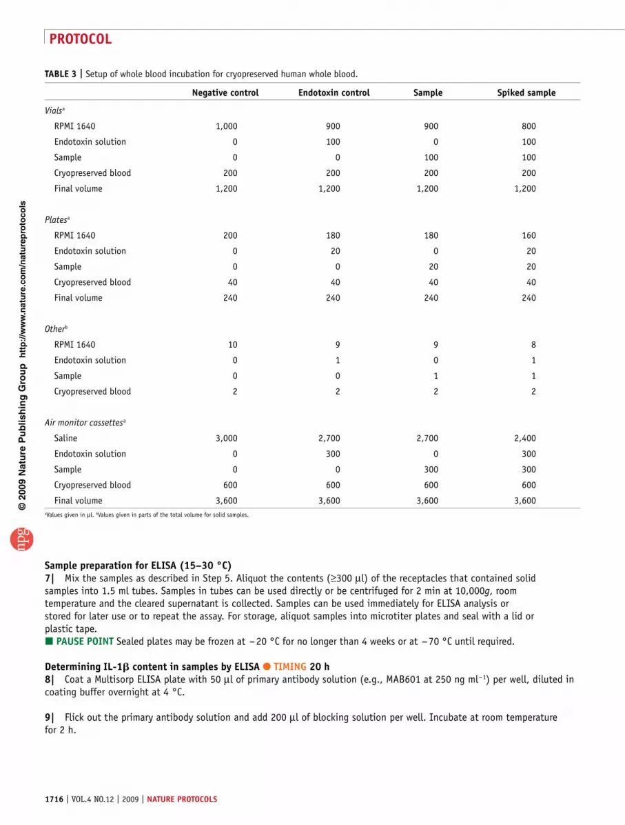
plate is being used (in which case, the final volume should be 1,200 µl and 240 µl, respectively) or whether solid samples are being used (in which case, the final volume should be adapted to your requirements) (see table 3).
(ii) Add eight parts RPMI 1640 to the receptacles for spiked samples and nine parts clinical grade saline to each other receptacle.

nature protocols | VOL.4 NO.12 | 2009 | 1715
p
uor
G g
n ih si l
bu
P eru ta
N 900 2©
nat
ure
pro
toco
ls/
moc. e r
ut an .
ww
w / /:pt t
h
protocol
(iii) Add one part of the appropriate endotoxin control solution to the receptacles for endotoxin controls and spiked samples.
(iv) Add one part saline to the receptacles for negative controls. (v) Add one part sample to the receptacles for samples or spiked samples. (vi) Incubate at room temperature for 2 h. Omit this step if incubation does not include any spiked samples. (vii) Thaw the cryopreserved blood at 37 °C for a maximum period of 15 min to continue immediately with
Step viii. crItIcal step Use the thawed cryopreserved blood within 15 min to prevent the high concentration of DMSO from becoming toxic to the cells.
(viii) Resuspend the blood by gently inverting the closed vials. Combine blood from different vials into one pyrogen-free reservoir. Add two parts of cryopreserved human whole blood to each receptacle to reach the total incubation volume.
5| For samples assayed in vials mix the samples by closing the reaction vials and gently inverting the tubes a few times. For samples prepared in microtiter plates, mix by aspiring and dispensing five times. Change tips between each sample to avoid cross contamination. Cover the microtiter plate with a lid. For solid samples, mix the contents by gently inverting the receptacles by swinging them manually.
6| Incubate samples prepared with fresh blood in an incubator or a heating block for 10–24 h at 37 °C ± 1 °C. Incubate samples prepared with cryopreserved blood in an incubator for 10–24 h at 37 °C ± 1 °C and 5% CO2.
table 2 | Setup of whole blood incubation for fresh human whole blood.
negative control endotoxin control sample spiked sample
Vialsa
Saline 11,00 1,000 1,000 900 Endotoxin solution 0 100 0 100 Sample 0 0 100 100 Fresh blood 100 100 100 100 Final volume 1,200 1,200 1,200 1,200
Platesa
Saline 220 200 200 180 Endotoxin solution 0 20 0 20 Sample 0 0 20 20 Fresh blood 20 20 20 20 Final volume 240 240 240 240
Otherb
Saline 11 10 10 9 Endotoxin solution 0 1 0 1 Sample 0 0 1 1 Fresh blood 1 1 1 1
Air monitor cassettesa
Saline 3,300 3,000 3,000 2,700 Endotoxin solution 0 300 0 300 Sample 0 0 300 300 Fresh blood 300 300 300 300 Final volume 3,600 3,600 3,600 3,600aValues given in µl. bValues given in parts of the total volume for solid samples.
1716 | VOL.4 NO.12 | 2009 | nature protocols
p
uor
G g
n ih si l
bu
P eru ta
N 900 2©
nat
ure
pro
toco
ls/
moc. e r
ut an .
ww
w / /:pt t
h
protocol
sample preparation for elIsa (15–30 °c)7| Mix the samples as described in Step 5. Aliquot the contents (≥300 µl) of the receptacles that contained solid samples into 1.5 ml tubes. Samples in tubes can be used directly or be centrifuged for 2 min at 10,000g, room temperature and the cleared supernatant is collected. Samples can be used immediately for ELISA analysis or stored for later use or to repeat the assay. For storage, aliquot samples into microtiter plates and seal with a lid or plastic tape. pause poInt Sealed plates may be frozen at − 20 °C for no longer than 4 weeks or at − 70 °C until required.
Determining Il-1 content in samples by elIsa ● tIMInG 20 h8| Coat a Multisorp ELISA plate with 50 µl of primary antibody solution (e.g., MAB601 at 250 ng ml − 1) per well, diluted in coating buffer overnight at 4 °C.
9| Flick out the primary antibody solution and add 200 µl of blocking solution per well. Incubate at room temperature for 2 h.
table 3 | Setup of whole blood incubation for cryopreserved human whole blood.
negative control endotoxin control sample spiked sample
Vialsa
RPMI 1640 1,000 900 900 800
Endotoxin solution 0 100 0 100
Sample 0 0 100 100
Cryopreserved blood 200 200 200 200
Final volume 1,200 1,200 1,200 1,200
Platesa
RPMI 1640 200 180 180 160
Endotoxin solution 0 20 0 20
Sample 0 0 20 20
Cryopreserved blood 40 40 40 40
Final volume 240 240 240 240
Otherb
RPMI 1640 10 9 9 8
Endotoxin solution 0 1 0 1
Sample 0 0 1 1
Cryopreserved blood 2 2 2 2
Air monitor cassettesa
Saline 3,000 2,700 2,700 2,400
Endotoxin solution 0 300 0 300
Sample 0 0 300 300
Cryopreserved blood 600 600 600 600
Final volume 3,600 3,600 3,600 3,600aValues given in µl. bValues given in parts of the total volume for solid samples.

nature protocols | VOL.4 NO.12 | 2009 | 1717
p
uor
G g
n ih si l
bu
P eru ta
N 900 2©
nat
ure
pro
toco
ls/
moc. e r
ut an .
ww
w / /:pt t
h
protocol
10| Wash all the wells three times with 250 µl of washing buffer per well.
11| Add 50 µl of secondary antibody per well (e.g., BAF201 at 60 ng ml − 1) diluted in blocking solution and add 50 µl of samples per well from whole-blood incubation (Step 4 A) or 50 µl of samples per well from the cryopreserved blood incubation (Step 4B). Incubate at room temperature for 2 h. crItIcal step Change pipette tips to avoid cross contamination.
12| Wash all the wells four times with 250 µl washing buffer per well.
13| Add 100 µl of 50 ng ml − 1 streptavidin peroxidase per well. Incubate at room temperature for 30 min.
14| Wash all the wells eight times with 250 µl of washing buffer per well.
15| Add 100 µl of TMB substrate per well. ! cautIon TMB is flammable. Toxic by inhalation, in contact with skin and if swallowed. Do not inhale the vapor. Avoid contact with eyes, skin and clothing. Avoid prolonged or repeated exposure. Do not use if skin is cut or scratched. Wash thoroughly after handling. Use in a fume hood.
16| Allow substrate metabolism in the dark for 5–20 min at 15–30 °C. Stop the reaction with 50 µl of 1 M H2SO4 per well. ! cautIon H2SO4 causes severe burns. Do not inhale the vapor. Avoid contact with eyes, skin and clothes. Avoid prolonged or repeated exposure. Use in a fume hood.
17| Cover with a plastic tape and measure the OD at 450 nm against a reference wavelength of 600–690 nm.? troublesHootInG
Data analysis ● tIMInG 30 min18| Calculate the mean and s.d. of the OD of the replicate values of all controls and samples. Use the nonparametric Dixon’s test or Grubb’s test for normally distributed samples to detect and exclude possible outliers among the replicates.
19| Compare the mean OD of the negative control to 0.1 OD.? troublesHootInG
20| Divide the mean OD for 0.5 EU ml − 1 endotoxin control by the mean OD of negative control.? troublesHootInG
21| Compare the mean OD of the spiked samples with the mean OD of 0.5 EU ml − 1 endotoxin control.? troublesHootInG
● tIMInGStep 1, Incubation plan design: 15 minStep 2, Preparation of endotoxin controls: 15 minStep 3, Sample preparation: 30 minSteps 4–7, Whole blood incubation: 13–27 hSteps 8–17, ELISA: 20 hSteps 18–21, Data analysis: 30 min
? troublesHootInGTroubleshooting advice can be found in table 4.

1718 | VOL.4 NO.12 | 2009 | nature protocols
p
uor
G g
n ih si l
bu
P eru ta
N 900 2©
nat
ure
pro
toco
ls/
moc. e r
ut an .
ww
w / /:pt t
h
protocol
table 4 | Troubleshooting table.
step problem possible reason possible solution
17 All wells turn equally yellow after addition of acid
Plate was coated with detection antibody instead of capture antibody
Repeat Steps 8–17 with samples from Step 7
Endotoxin control wells remain blank
Endotoxin control stock was too old or thawed and refrozen too often
Repeat entire procedure with new endotoxin control stock
Systematically increasing signals from left to right on the ELISA plate
Contamination of samples with IL-1β from other samples, e.g., by not changing pipette tips appropriately or defective ELISA plate
Repeat Steps 8–17 with samples from Step 7, changing the pipette tips appropriately or test batch of ELISA plates by applying the same sample throughout
All wells remain blank Detection antibody was not added or destroyed by freezing and thawing
Repeat Steps 8–17 with samples from Step 7 and a new aliquot of detection antibody
19 Mean OD of the negative control replicates > 0.1 OD
The blood was prestimulated (e.g., infection, allergy) or the saline or RPMI 1640 was contaminated
Repeat whole blood incubation with blood from a different donor and a new tube of saline or RPMI 1640
20 Ratio of mean OD of the 0.5 EU ml − 1 endotoxin control over mean OD of the negative control is less than 1.6
The endotoxin control was destroyed by repeated freezing and thawing, is too old or was set up incorrectly
Repeat assay with a new aliquot of endotoxin control
21 Interference test is positive Samples may be lipidic, toxic or immunomodulatory
Repeat assay with higher dilution of sample. Other adaptations of the assay may be applicable21,30,31
ELISA, enzyme-linked immunosorbent assay; OD, optical density.
antIcIpateD resultsFigure 2 shows an ELISA plate of an IPT carried out with fresh human whole-blood incubated for 20 h after stopping the TMB metabolism with sulfuric acid according to the incubation plan given in table 1.
The minimum assay suitability requirements are as follows:• The mean OD of control samples is 0.1 OD or below.• The ratio of mean OD of 0.5 EU ml − 1 endotoxin control over the mean OD of control samples is greater than 1.6.• Interference of the samples is excluded. If an unspiked sample is nonpyrogenic, i.e., the OD lies below the mean OD of
0.5 EU endotoxin control, and the mean OD of the respective spiked sample lies within a 50–200% range of the mean OD (0.5 EU ml − 1), this sample is free of interference.
If the minimum assay suitability requirements are fulfilled, the prediction model can be applied: Rabbits are likely to develop fever if tested with 10 ml kg − 1 of the sample if:mean OD (sample) > mean OD (0.5 EU ml − 1).
This is a qualitative evaluation for which the IPT has been validated, the readout being either “pyrogenic” or “nonpyro-genic”. For a semi-quantitative assessment of the pyrogenic activity in liquid samples, the dilution which results in an OD that is comparable to 0.5 EU ml − 1 endotoxin control can be used to calculate the endotoxin contamination in the undiluted sample. This estimation should be given in EEU because non-LPS pyrogens may also contribute to the pyrogenic activity. Similarly, the pyrogenic burden in air samples may be given in EEU by comparison of the IL-1β induction in the air monitor cassettes with that of 0.5 EU ml − 1 control. For solid samples a similar semi-quantitative assessment is possible if the samples can be employed in different quantities, e.g., fibers and beads. Otherwise only a rough estimation can be made by finding comparable IL-1β
Figure 2 | Illustration of an enzyme-linked immunosorbent assay (ELISA) plate after the completion of an in vitro pyrogen test (IPT). ELISA plate of an IPT carried out with fresh human whole-blood incubated for 20 h after stopping the tetramethylbenzidine (TMB) metabolism with sulfuric acid. The plate setup conforms to the incubation plan given in table 1. All experiments using human blood must comply with the legal requirements of your country and guidelines of your institution.

nature protocols | VOL.4 NO.12 | 2009 | 1719
p
uor
G g
n ih si l
bu
P eru ta
N 900 2©
nat
ure
pro
toco
ls/
moc. e r
ut an .
ww
w / /:pt t
h
protocol
release in response to the sample and one of the endotoxin controls. This rough estimation would not consider the dif-ferent shapes of concentration response curves from different types of pyrogens.
An example for the detection of pyrogenic activity in a liquid sample is given in Figure 3a. Here a cooling lubricant was tested for pyrogenic activity. Samples were employed at different dilutions and spiked with 0.5 EU ml − 1 LPS at each of these dilutions. Horizontal lines show the OD value of the endotoxin control (0.5 EU ml − 1) and the range of 50–200% of this value. At dilutions below 1:100 interference is observed, as the spiked sample does not reach an OD greater than 50% of the endotoxin control. At dilutions starting from 1:100 the OD of the spiked sample is within this range and interference is excluded. A vitality test (AlamarBlue metabolism) carried out with the diluted blood after the whole-blood incubation (Fig. 3b) demonstrated that the interference at dilutions below 1:100 was caused by toxic effects to the blood cells.
Figure 4 shows an example of assessment of the pyrogenic burden in air samples from different environments. As would be expected, the pyrogenic burden in a hog house was far greater than that in an office environment.
In Figure 5 surgical steel beads were tested as an example of solid samples used in the IPT. The spiked sample results in an OD in the 50–200% range of the 0.5 EU ml − 1 endotoxin control, thus interference is excluded. The samples were negative for pyrogenic activity.
Different donors release different absolute amounts of IL-1β in response to pyrogenic stimulation. However, their sensitiv-ity to pyrogens is highly comparable19. This is the reason why the endotoxin controls must always be run in parallel with the
0.3
200%a
b
Non-spikedSpiked
Non-spikedSpiked
50%
0.5 EU
IL-1
β (O
D 4
50 n
m)
0.2
0.1
Dilution factor
Dilution factor
0.0
12,00011,000
Cou
nts
10,0009,0008,0007,0006,0005,0004,0003,0002,0001,000
0
1:0
1:3
1:10
1:30
1:10
01:
300
1:1,
000
1:3,
000
1:10
,000
1:30
,000
1:10
0,00
0
1:30
0,00
0
1:0
1:3
1:10
1:30
1:10
01:
300
1:1,
000
1:3,
000
1:10
,000
1:30
,000
1:10
0,00
0
1:30
0,00
0Dea
dVita
l
Figure 3 | Analysis of pyrogenic activity of a cooling lubricant sample. (a) A cooling lubricant sample was employed in the in vitro pyrogen test (IPT) using fresh human whole blood in a series of dilutions (white bars). Spiked samples were prepared with 0.5 EU ml − 1 lipopolysaccharides (LPS) (black bars). After 20 h incubation with whole blood the IL-1β enzyme-linked immunosorbent assay (ELISA) was carried out and the absorbance at 450 nm recorded. Horizontal lines show the optical density (OD) of the 0.5 EU ml − 1 endotoxin control as well as the range of 50–200% of this value. Error bars are the s.d. of quadruplicate samples. (b) After incubation the blood was subjected to a vitality assay (AlamarBlue metabolism)44. For comparison, fresh untreated human blood (vital) and ethanol (10% vol/vol for 10 min)-treated human blood (dead) were employed in the assay. Error bars are the s.d. of quadruplicate samples. All experiments using human blood must comply with the legal requirements of the country and guidelines of the institution.
100
Hog houseCattle stableLPS (EU/ml)
Waste sorting plantOfficeAmbient air
10
0.1
0.01
1
0.10.01
IL-1
β (O
D 4
50 n
m)
1 10
Volumes air pumped through filter (liter)
100 1,000
Figure 4 | Air quality assessment with the in vitro pyrogen test (IPT). Pyrogenic activity of particles from air sampled using air monitor cassettes was evaluated by IPT. Various volumes of air were sampled at different locations. The whole-blood incubation was carried out in air monitor cassettes with negative controls, endotoxin controls () (given in EU ml − 1) and samples. After 20 h incubation with whole blood the IL-1β ELISA was carried out and the absorbance at 450 nm recorded. Error bars are the s.d. of quadruplicate samples. All experiments using human blood must comply with the legal requirements of the country and guidelines of the institution.
0.6
0.5
0.4
200%
50%
Endotoxin controls(EU ml–1)
Sample
Spiked
sam
ple
Negat
ive co
ntro
l0.
250.
50 1.00
2.00
0.3
IL-1
β (O
D 4
50 n
m)
0.2
0.1
0.0
Figure 5 | Assessment of pyrogenic activity or interference of a steel sample by the in vitro pyrogen test (IPT). Samples of surgical steel beads were subjected to the IPT in 1.5 ml reaction vials using fresh human whole blood. Parallel samples were spiked with 0.5 EU ml − 1 lipopolysaccharides (LPS) for interference testing. After 20 h incubation with whole blood, the IL-1β enzyme-linked immunosorbent assay (ELISA) was carried out and the absorbance at 450 nm recorded. Horizontal bands show the range of 50–200% of the 0.5 EU ml − 1 endotoxin control. All experiments using human blood must comply with the legal requirements of the country and guidelines of the institution.

1720 | VOL.4 NO.12 | 2009 | nature protocols
p
uor
G g
n ih si l
bu
P eru ta
N 900 2©
nat
ure
pro
toco
ls/
moc. e r
ut an .
ww
w / /:pt t
h
protocol
sample assessment. Similarly, although the cryopreserved blood generally results in a somewhat higher absolute release of IL-1β, the sensitivity of fresh blood and cryopreserved blood to pyrogens is highly comparable, see e.g., Figure 6.
acknoWleDGMents The authors acknowledge the contributions of countless scientists, technicians, students, authorities and funding institutions to the development and validation of these protocols.
autHor contrIbutIons All authors contributed to the development of the method and the writing of the manuscript.
Published online at http://www.natureprotocols.com. Reprints and permissions information is available online at http://npg.nature.com/reprintsandpermissions.
1. Morath, S., Geyer, A. & Hartung, T. Structure-function relationship of cytokine induction by lipoteichoic acid from Staphylococcus aureus. J. Exp. Med. 193, 393–397 (2001).
2. Tsuchida, K. et al. Detection of peptidoglycan and endotoxin in dialysate, using silkworm larvae plasma and limulus amebocyte lysate methods. Nephron 75, 438–443 (1997).
3. Watson, D.W. Host-parasite factors in group A streptococcal infections. Pyrogenic and other effects of immunologic distinct exotoxins related to scarlet fever toxins. J. Exp. Med. 111, 255–284 (1960).
4. Bodel, P.T. & Atkins, E. Studies in staphylococcal fever. V. Staphlococcal filtrate pyrogen. Yale J. Biol. Med. 38, 282–298 (1965).
5. Brunson, K.W. & Watson, D.W. Pyrogenic specificity of streptococcal exotoxins, staphylococcal enterotoxin, and gram-negative endotoxin. Infect. Immun. 10, 347–351 (1974).
6. Atkins, E. & Huang, W.C. Studies on the pathogenesis of fever with influenzal viruses. I. The appearance of an endogenous pyrogen in the blood following intravenous injection of virus. J. Exp. Med. 107, 383–401 (1958).
7. Braude, A.I., Mc, C.J. & Douglas, H. Fever from pathogenic fungi. J. Clin. Invest. 39, 1266–1276 (1960).
8. Kobayashi, G.S. & Friedman, L. Characterization of the pyrogenicity of Candida albicans, Saccharomyces cerevisiae, and Cryptococcus neoformans. J. Bacteriol. 88, 660–666 (1964).
9. Beeson, P.B. Temperature-elevating effect of a substance obtained from polymorphonuclear leucocytes. J. Clin. Invest. 27, 524 (1948).
10. Dinarello, C.A. et al. Tumor necrosis factor (cachectin) is an endogenous pyrogen and induces production of interleukin 1. J. Exp. Med. 163, 1433–1450 (1986).
11. Dinarello, C.A. et al. Interleukin-6 as an endogenous pyrogen: induction of prostaglandin E2 in brain but not in peripheral blood mononuclear cells. Brain Res. 562, 199–206 (1991).
12. Dinarello, C.A., Marnoy, S.O. & Rosenwasser, L.J. Role of arachidonate metabolism in the immunoregulatory function of human leukocytic pyrogen/lymphocyte-activating factor/interleukin 1. J. Immunol. 130, 890–895 (1983).
13. Vellucci, S.V. & Parrott, R.F. Expression of mRNAs for vasopressin, oxytocin and corticotrophin releasing hormone in the hypothalamus, and of cyclooxygenases-1 and -2 in the cerebral vasculature, of endotoxin-challenged pigs. Neuropeptides 32, 439–446 (1998).
14. Rotondo, D., Abul, H.T., Milton, A.S. & Davidson, J. Pyrogenic immunomodulators increase the level of prostaglandin E2 in the blood simultaneously with the onset of fever. Eur. J. Pharmacol. 154, 145–152 (1988).
15. Li, S. et al. The febrile response to lipopolysaccharide is blocked in cyclooxygenase-2(-/-), but not in cyclooxygenase-1(-/-) mice. Brain Res. 825, 86–94 (1999).
16. Li, S., Ballou, L.R., Morham, S.G. & Blatteis, C.M. Cyclooxygenase-2 mediates the febrile response of mice to interleukin-1beta. Brain Res. 910, 163–173 (2001).
17. Li, S., Goorha, S., Ballou, L.R. & Blatteis, C.M. Intracerebroventricular interleukin-6, macrophage inflammatory protein-1 beta and IL-18: pyrogenic and PGE(2)-mediated? Brain Res. 992, 76–84 (2003).
18. Hartung, T. & Wendel, A. Detection of pyrogens using human whole blood. Altex 12, 70–75 (1995).
19. Wiegandt, M. Dissertation. Der Human Vollblut-Pyrogentest – Optimierung, Validierung und Vergleich mit den Arzneibuchmethoden. Heidelberg (2002).
20. Hoffmann, S. et al. International validation of novel pyrogen tests based on human monocytoid cells. J. Immunol. Methods 298, 161–173 (2005).
21. Schindler, S. et al. International validation of pyrogen tests based on cryopreserved human primary blood cells. J. Immunol. Methods 316, 42–51 (2006).
22. Wendel, A. & Hartung, T. Test for determining pyrogenic effect of a material (DPC Biermann GmbH (Bad Nauheim, DE) Wendel, Albrecht (Tubingen, DE), Hartung, Thomas (Konstanz, DE), US Patent 5891728, 1999).
23. Wendel, A. & Hartung, T. Use of frozen blood in a biological test method (DPC Biermann GmbH (Bad Nauheim, DE) Wendel, Albrecht (Tubingen, DE), Hartung, Thomas (Konstanz, DE), European Patent Ep0851231, 1997).
24. Hartung, T. Method for assaying flowing media for microbial toxins (Hartung, Thomas (Konstanz, DE) European Patent EP1377835, 2002).
25. Schindler, S. et al. Cryopreservation of human whole blood for pyrogenicity testing. J. Immunol. Methods 294, 89–100 (2004).
26. Mazzotti, F. et al. In vitro pyrogen test–a new test method for solid medical devices. J. Biomed. Mater Res. A 80, 276–282 (2007).
27. Hasiwa, M., Kullmann, K., von Aulock, S., Klein, C. & Hartung, T. An in vitro pyrogen safety test for immune-stimulating components on surfaces. Biomaterials 28, 1367–1375 (2007).
28. Kindinger, I. et al. A new method to measure air-borne pyrogens based on human whole blood cytokine response. J. Immunol. Methods 298, 143–153 (2005).
29. Schindler, S. et al. Pyrogen testing of lipidic parenterals with a novel in vitro test–application of the IPT based on cryopreserved human whole blood. Pharmeur. Sci. Notes 2006, 1–7 (2006).
30. Daneshian, M., Wendel, A., Hartung, T. & von Aulock, S. High sensitivity pyrogen testing in water and dialysis solutions. J. Immunol. Methods 336, 64–70 (2008).
31. Daneshian, M., Guenther, A., Wendel, A., Hartung, T. & von Aulock, S. In vitro pyrogen test for toxic or immunomodulatory drugs. J. Immunol. Methods 313, 169–175 (2006).
32. Welch, H., Calvery, H.O., McClosky, W.T. & Price, W.T. Method of preparation and test for bacterial pyrogen. J. Am. Pharm. Assoc. 3, 65–69 (1943).
33. McClosky, W.T., Price, W.T., van Winkle, W.J., Welch, H. & Calvery, H.O. Results of the first USP collaborative study of pyrogens. J. Am. Pharm. Assoc. 32, 69–73 (1943).
34. Grant, R. Emotional hypothermia in rabbits. Am. J. Physiol. 160, 285–290 (1950).
35. Hartung, T. et al. Novel pyrogen tests based on the human fever reaction. The report and recommendations of ECVAM Workshop 43. European Centre for the Validation of Alternative Methods. European Centre for the Validation of Alternative Methods. Altern. Lab. Anim. 29, 99–123 (2001).
36. Fennrich, S. et al. Detection of endotoxins and other pyrogens using human whole blood. Dev. Biol. Stand. 101, 131–139 (1999).
37. Dehus, O., Hartung, T. & Hermann, C. Endotoxin evaluation of eleven lipopolysaccharides by whole blood assay does not always correlate
1.75
1.50
1.25
IL-1
β (O
D 4
50 n
m)
0.75
0.50
0.25
0.000.00 0.12 0.25 0.50
Endotoxin controls (EU ml–1)
1.00 2.00
1.00
Fresh bloodCryopreserved blood
Figure 6 | Comparison of the response of fresh and cryopreserved human whole blood to different concentrations of lipopolysaccharides (LPS). Fresh and cryopreserved human whole blood from the same five donors was stimulated with the given concentrations of LPS. IL-1β was measured by enzyme–linked immunosorbent assay (ELISA). Error bars are the s.e. of the mean of the five donors’ responses. All experiments using human blood must comply with the legal requirements of the country and guidelines of the institution.

nature protocols | VOL.4 NO.12 | 2009 | 1721
p
uor
G g
n ih si l
bu
P eru ta
N 900 2©
nat
ure
pro
toco
ls/
moc. e r
ut an .
ww
w / /:pt t
h
protocol
with limulus amebocyte lysate assay. J. Endotoxin Res. 12, 171–180 (2006).
38. Hartung, T. Statement on the validity of the in-vitro pyrogen test (European Centre for the Validation of Alternative Methods, Ispra, 2006).
39. Pott, G.B., Chan, E.D., Dinarello, C.A. & Shapiro, L. Alpha-1-antitrypsin is an endogenous inhibitor of proinflammatory cytokine production in whole blood. J. Leukoc. Biol. 85, 886–895 (2009).
40. Poole, E.J., Dawson, P. & Gaines Das, R.E. Second International standard for endotoxin: calibration in an international collaborative study. J. Endotoxin Res. 4, 221–231 (1997).
41. Dixon, W.J. Processing data for outliers. Biometrica 9, 74–89 (1953).42. Grubbs, F.E. Sample criteria for testing outlying observations. Ann. Math.
Stat. 21, 27–58 (1950).43. Hoffmann, S., Luderitz-Puchel, U., Montag, T. & Hartung, T.
Optimisation of pyrogen testing in parenterals according to different pharmacopoeias by probabilistic modelling. J. Endotoxin Res. 11, 25–31 (2005).
44. Nakayama, G.R., Caton, M.C., Nova, M.P. & Parandoosh, Z. Assessment of the Alamar Blue assay for cellular growth and viability in vitro. J. Immunol. Meth. 204, 205–208 (1997).
Copyright © 2022 FDOKUMEN