A Review: Validated Analytical Methods Developed on ...
12
JPSBR: Volume 5, Issue 3: 2015 (254-265) ISSN NO. 2271-3681 Desai D. et al 254 ABSTRACT: Rifampicin is a first line medication used as an anti-tubercular agent. Rifampicin acts by binding and inhibiting DNA dependent RNA polymerase. It is active against gram positive and negative both types of bacteria . The clinical and pharmaceutical analysis of this drug requires effective analytical procedures for quality control and pharmacodynamic and pharmacokinetic studies as well as stability study. An extensive survey of the literature published in various analytical and pharmaceutical chemistry related journals has been conducted and the instrumental analytical methods which were developed and used for determination of Rifampicin as single or combination with other drugs in bulk drugs, formulations and biological fluids have been reviewed. This review covers the time period from 1997 to 2014 during which 33 analytical methods including spectrophotometric methods like UV and derivative; visible which is based on formation of metal complexation, spectrofluorometric methods and chromatographic method including HPLC, HPTLC, and miscellaneous method like HPLC-MS were reported. The application of these methods for the determination of Rifampicin in pharmaceutical dosage form and biological samples has also been discussed. KEY WORDS: Rifampicin, UV spectroscopy, HPLC, HPLC-MS, HPTLC Cobalt complexes A Review: Validated Analytical Methods Developed on Antitubercular Drug, Rifampicin Article history: Received 11 Feb 2015 Accepted 18 March 2015 Available online 01 May 2015 Citation: Desai D. A Review: Validated Analytical Methods Developed on Antitubercular Drug, Rifampicin. J Pharm Sci Bioscientific Res. 2015 5(3):254-265 For Correspondence: Mr. Desai Drashti Rofel, Shri G.M. Bilakhia, College Of Pharmacy, Vapi, Gujatat, India Email: [email protected] (www.jpsbr.org) INTRODUCTION: Mycobacteria are intrinsically resistant to most antibiotics. Because they grow slowly compared with other bacteria, antibiotics that are most active against growing cells are relatively ineffective. Mycobacterial cells can also be dormant and thus completely resistant to many drugs or get killed very slowly. The lipid-rich mycobacterial cell wall is impermeable to many agents. Mycobacterial species are intracellular pathogens, and organisms residing within macrophages are inaccessible to drugs that penetrate these cells poorly. Finally, mycobacteria are notorious for their ability to develop resistance. Combinations of two or more drugs are required to overcome these obstacles and to prevent emergence of resistance during the course of therapy. The response of mycobacterial infections to chemotherapy is slow, and treatment must be administered for months to years, depending on which drugs are used. 1 Tuberculosis is the most important communicable disease. The one-third of the world’s population is infected by Mycobacterium tuberculosis according to the World Health organization(WHO) estimation. HIV-infected persons, immigrants from countries with high rates of tuberculosis, the homeless, health care professionals, intravenous drug users, persons taking immunosuppressive agents, and those in institutional settings, such as nursing homes and correctional facilities groups are at high risks for tuberculosis infection . There is a progressive increase in multidrugresistant (MDR) tuberculosis. 2 Drashti Desai*, Megha Shah Rofel, Shri G.M. Bilakhia, College Of Pharmacy, Vapi, Gujatat, India
-
Upload
khangminh22 -
Category
Documents
-
view
3 -
download
0
Transcript of A Review: Validated Analytical Methods Developed on ...

JPSBR: Volume 5, Issue 3: 2015 (254-265) ISSN NO. 2271-3681
Desai D. et al 254
ABSTRACT:
Rifampicin is a first line medication used as an anti-tubercular agent. Rifampicin acts by binding and inhibiting DNA dependent
RNA polymerase. It is active against gram positive and negative both types of bacteria . The clinical and pharmaceutical analysis of
this drug requires effective analytical procedures for quality control and pharmacodynamic and pharmacokinetic studies as well as
stability study. An extensive survey of the literature published in various analytical and pharmaceutical chemistry related journals
has been conducted and the instrumental analytical methods which were developed and used for determination of Rifampicin as
single or combination with other drugs in bulk drugs, formulations and biological fluids have been reviewed. This review covers
the time period from 1997 to 2014 during which 33 analytical methods including spectrophotometric methods like UV and
derivative; visible which is based on formation of metal complexation, spectrofluorometric methods and chromatographic
method including HPLC, HPTLC, and miscellaneous method like HPLC-MS were reported. The application of these methods for the
determination of Rifampicin in pharmaceutical dosage form and biological samples has also been discussed.
KEY WORDS: Rifampicin, UV spectroscopy, HPLC, HPLC-MS, HPTLC Cobalt complexes
A Review: Validated Analytical Methods Developed on Antitubercular Drug,
Rifampicin
Article history: Received 11 Feb 2015 Accepted 18 March 2015 Available online 01 May 2015 Citation: Desai D. A Review: Validated Analytical Methods Developed on Antitubercular Drug, Rifampicin. J Pharm Sci Bioscientific Res. 2015 5(3):254-265
For Correspondence:
Mr. Desai Drashti
Rofel, Shri G.M. Bilakhia, College Of
Pharmacy, Vapi, Gujatat, India
Email: [email protected]
(www.jpsbr.org)
INTRODUCTION:
Mycobacteria are intrinsically resistant to most antibiotics. Because they
grow slowly compared with other bacteria, antibiotics that are most active against
growing cells are relatively ineffective. Mycobacterial cells can also be dormant and
thus completely resistant to many drugs or get killed very slowly. The lipid-rich
mycobacterial cell wall is impermeable to many agents. Mycobacterial species are
intracellular pathogens, and organisms residing within macrophages are inaccessible
to drugs that penetrate these cells poorly. Finally, mycobacteria are notorious for
their ability to develop resistance. Combinations of two or more drugs are required
to overcome these obstacles and to prevent emergence of resistance during the
course of therapy. The response of mycobacterial infections to chemotherapy is
slow, and treatment must be administered for months to years, depending on which
drugs are used.1
Tuberculosis is the most important communicable disease. The one-third of
the world’s population is infected by Mycobacterium tuberculosis according to the
World Health organization(WHO) estimation. HIV-infected persons, immigrants from
countries with high rates of tuberculosis, the homeless, health care professionals,
intravenous drug users, persons taking immunosuppressive agents, and those in
institutional settings, such as nursing homes and correctional facilities groups are at
high risks for tuberculosis infection . There is a progressive increase in
multidrugresistant (MDR) tuberculosis.2
Drashti Desai*, Megha Shah
Rofel, Shri G.M. Bilakhia, College Of Pharmacy, Vapi, Gujatat, India
JPSBR: Volume 5, Issue 3: 2015 (254-265) ISSN NO. 2271-3681
Desai D. et al 255
Classification of tuberculosis:
I. First line drugs- rifampicin, isoniazid, pyrazinamide,
ethambutol and streptomycin (superior in efficacy and
possess an acceptable degree of toxicity)
II. Second line drugs- cycloserine,ethionamide,
aminosalicylic acid, rifabutin, quinolones, capreomycin,
viomycin, and thiacetazone (more toxic and less
effective, and they are indicated only when the M.
tuberculosis organisms are resistant to the first-line
agents)2
To decrease the possibility of the emergence of resistant
organisms, compound drug therapy is employed, involving the
following:
a first initial phase of about 2
months consisting of three
drugs used concomitantly: isoniazid , rifampicin,
pyrazinamide (plus ethambutol if the organism is
suspected to be resistant)
a second, continuation phase, of 4
months, consisting of
two drugs: isoniazid and rifampicin; longer-term
treatment is needed for patients with meningitis,
bone/joint involvement or drug-resistant infection. 3
Table -1 Antimicrobials Used in the Treatment of Tuberculosis.1
Drug Typical Adult Dosage1
First-line agents (in approximate order of preference)
Isoniazid 300 mg/d
Rifampin 600 mg/d
Pyrazinamide 25 mg/kg/d
Ethambutol 15–25 mg/kg/d
Streptomycin 15 mg/kg/d
Second-line agents
Amikacin 15 mg/kg/d
Aminosalicylic acid 8–12 g/d
Capreomycin 15 mg/kg/d
Ciprofloxacin 1500 mg/d, divided
Clofazimine 200 mg/d
Cycloserine 500–1000 mg/d, divided
Ethionamide 500–750 mg/d
Levofloxacin 500 mg/d
Rifabutin 300 mg/d2
Rifapentine 600 mg once or twice weekly
1Assuming normal renal function.
2150 mg/d if used concurrently with a protease inhibitor.
Table –2 Recommended Duration of Therapy for
Tuberculosis.1
Regimen (in Approximate Order of
Preference)
Duration in
Months
Isoniazid, rifampin, pyrazinamide 6
Isoniazid, rifampin 9
Rifampin, ethambutol, pyrazinamide 6
Rifampin, ethambutol 12
Isoniazid, ethambutol 18
All others 24
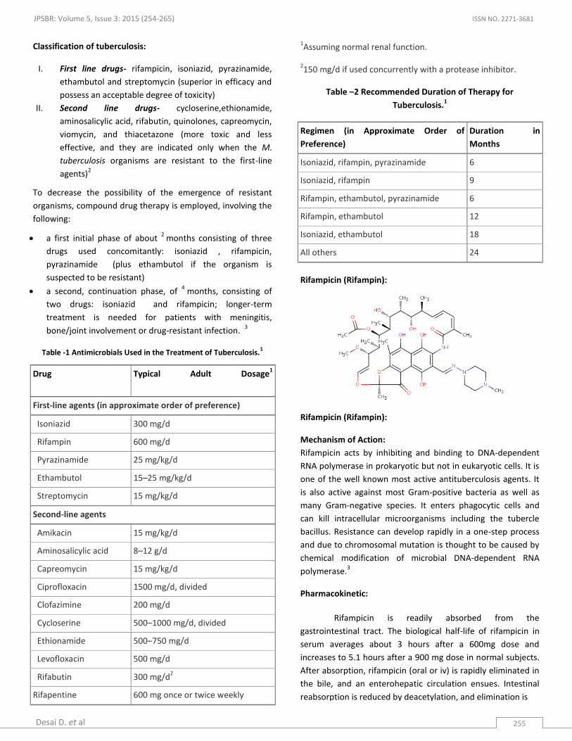
Rifampicin (Rifampin):
Rifampicin (Rifampin):
Mechanism of Action:
Rifampicin acts by inhibiting and binding to DNA-dependent
RNA polymerase in prokaryotic but not in eukaryotic cells. It is
one of the well known most active antituberculosis agents. It
is also active against most Gram-positive bacteria as well as
many Gram-negative species. It enters phagocytic cells and
can kill intracellular microorganisms including the tubercle
bacillus. Resistance can develop rapidly in a one-step process
and due to chromosomal mutation is thought to be caused by
chemical modification of microbial DNA-dependent RNA
polymerase.3
Pharmacokinetic:
Rifampicin is readily absorbed from the
gastrointestinal tract. The biological half-life of rifampicin in
serum averages about 3 hours after a 600mg dose and
increases to 5.1 hours after a 900 mg dose in normal subjects.
After absorption, rifampicin (oral or iv) is rapidly eliminated in
the bile, and an enterohepatic circulation ensues. Intestinal
reabsorption is reduced by deacetylation, and elimination is

JPSBR: Volume 5, Issue 3: 2015 (254-265) ISSN NO. 2271-3681
Desai D. et al 256
facilitated. With about half of this being unchanged rifampicin,
up to 30% of a dose is excreted in the urine,.
On food ingestion, absorption of rifampicin is reduced.
Rifampicin is widely distributed throughout the body. It is also
present in many organs and body fluids, and also in
cerebrospinal fluid. Rifampicin is about 80% protein bound.
Most of the unbound fraction is not ionized and therefore is
diffused freely in tissues.5
Pharmacodynamics:
Rifampicin has high activity against many organisms,
Mycobacterium tuberculosis and M.leprae, including
Staphylococcus aureus, coagulase-negative staphylocci,
Listeria monocytogenes, Neisseria meningitidis, Haemophilus
influenzae, Legionella spp., Brucella, some strains of E. coli,
Proteus mirabilis, anaerobic cocci, Clostridium spp., and
Bacteroides. It is also reported that rifampicin exhibits an
immunosuppressive effect which has been seen in some
animal experiments, but this may not be clinically significant in
humans. Depending on the concentration of drug attained at
site of infection, rifampicin may be bacteriostatic or
bactericidal. The bactericidal actions are secondary to
interfering with the synthesis of nucleic acids by inhibiting
bacterial DNAdependent RNA polymers at the B-subunit thus
preventing initiation of RNA transcription. 6
Indications:
Clinical Uses 2
Rifampicin, a first-line antitubercular drug is used in the
treatment of all forms of pulmonary and extrapulmonary
tuberculosis. It is an alternative to isoniazid in the treatment
of latent tuberculosis infection. Rifampicin may also be
combined with an antileprosy agent for the treatment of
leprosy and to protect those in close contact with patients
having H. influenza type B and N. meningitidis infection; and is
also used innmethicillin-resistant staphylococcal infections,
such as osteomyelitis and prosthetic valve endocarditis.
Adverse Reactions 2
The most commonly observed side effects are GI disturbances
and nervous system symptoms, such as nausea, vomiting,
headache, dizziness, and fatigue. A major adverse effect is
Hepatitis, and the risk is highest in patients with underlying
liver diseases and in slow isoniazid acetylators; if isoniazid and
rifampin are combined, the rate of hepatotoxicity is increased.
Rifabutin is commonly substituted for rifampin in the
treatment of tuberculosis in HIV-infected patients.
Hypersensitivity reactions, such as pruritus, cutaneous
vasculitis, and thrombocytopenia, are seen in some patients,
and an immune-mediated systemic flulike syndrome with
thrombocytopenia also has been described.
Rifampicin imparts a harmless red-orange color to urine, feces,
saliva, sweat, tears, and contact lenses. Patients should be
advised of such discoloration of body fluids.
Different analytical methods for Rifampicin and its
combination:
Development of new methods, provide determinations with
maximum accuracy, which are responsible to increase the
interest in analytical methods as such. They should enable to
simultaneously determine the individual components in
multicomponent preparations and in Dosage form as well as
biological material. These developed validated Methods
confirms them the appropriate quality of the product and of
the analytical method used. These are different parameters
that validate reliability of the results and enable comparing
efficiency of the methods used. Validation parameters were
done according to the ICH guideline.
Several techniques like HPLC [10, 12, 13, 14, 15, 16,
17, 18, 19, 20, 21, 22, 23, 24, 25, 26, 27, 28, 29, 30, 31, 33],
HPTLC [11,32], Spectroscopic methods [1, 2, 4, 5, 6, 7, 8, 9 ],.
Colorimetric method [3], have been used for the
determination of Rifampicin. Chromatographic methods have
been extensively used and recommended. Since, these
methods require complex and expensive equipment, provision
for use and disposal of solvents, labour-intensive sample
preparation procedures and personal skills in such techniques.
There was no review published covering all different analytical
method used for the determination of Rifampicin. The high
importance of Rifampicin in Tuberculosis prompted us to
review the most important recent analytical methods for their
analysis in pure forms, in different pharmaceutical dosage
forms and in biological fluids reported so far in the literature.
The present review comprises references covering the period
from 1997 to 2014. The official method of determination of
rifampicin was HPLC method in Indian Pharmacopoeia and
spectroscopy and HPLC in British Pharmacopoeia.

JPSBR: Volume 5, Issue 3: 2015 (254-265) ISSN NO. 2271-3681
Desai D. et al 257
Different analytical methods for Rifampicin and its combination:
SR NO.
DRUG METHOD DESCRIPTION DETECTION AT SPECIFICATION REF NO.
1. Rifapentine UV Chemito UV 2600 spectrophotometer with 1 cm matched quartz cells Y = 0.004x+ 0.024
478nm (0.1N HCl)
R2- 0.999
Linearity range-5-50µg/ml LOD-3.28 µg/ml(2.15) LOQ-9.96 µg/ml(6.25) %RSD-0.003985
7.
2. Rifampicin & Piperine
2nd
dvt. No intereference from the capsule excipients
341nm for rifampicin (0 cross point for piperine) 241nm for piperine (0 cross point for rifampicin)
R2-0.999
Linearity range- 10-60 µg/ml for rifampicin & 2-20 µg/ml for piperine LOD= ( µg/ml)- RIFA-1.65 PIPE- 0.57 LOQ=( µg/ml) RIFA-5.03 PIPE-1.75 Repeatability(%RSD)= RIFA-0.31 PIPE-0.37
8.
3. Rifampicin
Visible shimadzu double beam spectrophotometer UV-140 A=0.0009+0.0119C Buffer solution (pH=7.0)
400-650nm (max at 510nm)
R2-0.9999
Linearity range- Conc range:5-50µg/ml Molar absorptivity(l/mole cm)- 9.83x10
3
Sandell’s sensitivity( μg / cm2 / 0.001 absorbance unit)-0.084 Optimum photometric range:7.9-39.1µg/ml %RSD=1.90
9.
4. Rifampicin and Isoniazid
Simultaneous equation method)
Non interference of the excipients Solvent-ethanol
RIF- 337nm INH- 263nm
R2- RIF- 0.9991
INH- 0.9998 Linearity range- RIF- 5-35 µg/ml INH- 5-25 µg/ml Molar absorptivity(L/molcm) RIF-25349 INH- 9341 LOD= RIF-1.653µg/ml INH-0.585 µg/ml LOQ= RIF-5.007 µg/ml INH-1.772 µg/ml Precision(%RSD)= RIF-0.578 INH-0.673
10.
5. Rifampicin and Piperine
Q-Absorbance ratio
A shimadzu model 1700 double beam UV/Visible spectral width - 2 nm, wavelength accuracy- 0.5 nm & pair of 10 mm matched
In methanol RIF- 337nm PIPE- 337nm RIFA+PIPE- 387nm
R2-0.999
Linearity range- RIF- 5-40 µg/ml PIPE-2-20 µg/ml LOD= RIF-1.51 µg/ml PIPE-0.28 µg/ml
11.

JPSBR: Volume 5, Issue 3: 2015 (254-265) ISSN NO. 2271-3681
Desai D. et al 258
quartz cell .
RIF+PIPE-0.80 and 0.32 LOQ= RIF- 4.6 µg/ml PIPE- 0.86 µg/ml RIFA+PIPE- 2.45 and 0.98 µg/ml %RSD= RIF-0.54 PIPE-0.091 RIF+PIPE-0.68 Precision: RIFA-0.14-0.71 PIPE-0.11-1.50 RIFA and PIPE-0.17-1.31
6. Rifampicin and Isoniazid
Visible & 1st
derivative
Buffer solution pH 7.4 as solvent
257nm(1st
derivative, HCl 0.012M)
Recovery- 99.03% for rifampicin & 100.01% for isoniazid.
12.
7. Isoniazid, rifampicin and Piperine
UV & RPLC-PDA Methods
2 chemometric methods applied- ILS and CLS Reversed phase: phenomenex Luna C18 column- gradient elution with mobile phase 0.05M Na2HPO4 buffer pH 7 and acetonitrile Total run time: 12min
220-360nm with interval of 10nm
Linearity range- INH- 30-270 µg/ml RIF- 20-180 µg/ml PIPE- 1-9 µg/ml
13.
8. Rifampicin, isoniazid, pyrazinamide
Simultaneo
us
spectropho
tometric by
1st
derivative
UV
spectropho
tometry
--- RIF at ZCP for INH and PYZ( 262.2 and 268.8) PYZ at ZCP for RIF and PYZ (254 and 268.8nm) PYZ at ZCP for INH and RIF (262.2 and 254 nm)
--- 14.
9. Isoniazid, rifampicin and Piperine
UV, RP-
HPLC
Methanol and distilled water was used as diluents 2
nd RP-HPLC- acetonitrile
as diluents Flow rate of 0.9mL/min
INH-262nm PIPE- 338nm RIFA-477nm
Correlation coefficient-0.995 Linearity range- INH- 12-34.5 µg/ml RIFA- 8-23 µg/ml PIPE-0.4-1.15 µg/ml
15.
10. Rifampicin
HPLC SP-C18 monolithic MP-Methanol: CAN :mono potassium phosphate (0.075 M):citric acid (1.0 M) (28:30:38:4, v/v) flow rate 2 mL/min
with UV detection at 254 nm
Linearity range- RQ- 1.5-60 µg/ml SV- 1-40 µg/ml RNO-1-40 µg/ml 3-FR-1-40 µg/ml RIF- 5-200 µg/ml LOD=0.2 µg/ml LOQ= 1 µg/ml %RSD= 2.5%
16.
11. Rifampicin HPTLC SP- Al backed silica gel 254nm R2-INH- 0.994 17.

JPSBR: Volume 5, Issue 3: 2015 (254-265) ISSN NO. 2271-3681
Desai D. et al 259
and Isoniazid
60 F254
plates
MP- n-hexane:2-propanol:acetone:am-monia:formic acid=3:3.8:2.8:0.3:0.1 (v/v) Resolved INH and RIF with R
F values of 0.59 ±
0.02 and 0.73 ± 0.04, respectively
RIF- 0.997 Linearity range- 100-700 ng per spot LOD= INH- 20 ± 0.51ng RIF- 25 ± 0.63ng LOQ=INH- 60 ± 1.05 ng RIF- 75 ± 1.12 ng %RSD= INH=Conc (ng per spot) 200- 0.17 300-0.26 600-0.19 RIF= 200- 0.37 300- 0.44 600- 0.38
12. Rifampicin & Hydrochlorothiazide
HPLC SP- 150 X 4.6 mm i.d, 5 μm Phenomenex ODS 2 C18 column. MP - 40:60 % v/v acetonitrile and 10mM KH2PO4 (pH 3.2), flow rate of 1.0 ml/min Rt for RIF and HCTZ were 6.80 and 2.56 min, respectively.
337nm Correlation coefficient-0.9971 Linearity range- 0.31 – 25.48 μg/ml LOD=100ng/ml LOQ=1μg/ml
18.
13. Rifampicin HPLC SP- An Ultrabase-C18 column MP- water (pH 2.27): acetonitrile= (40:60 v/v), flow-rate - 1 ml/min Rt- 4 min.
333nm Linearity range- 0.1–1 and 1–50_g/ml for plasma 0.6–40_g/g for liver LOD= 0.025µg/ml for plasma 0.06 µg/g for liver LOQ= 0.05 µg/ml for plasma 0.25µg/g for liver
19.
14. Rifampicin in complex (isoniazide and serum)
HPLC Stationary phase- a 5 µm C18 Mobile phase- water-methanol at a flow rate of 1 ml/min. Rt- 12.8 min
333.6 R2- >0.98
Linearity range- 0.1- 0.3 mg/ ml for drug content in Rifamazid and from 1 - 3 µg/ml for serum %RSD= 1)RIF in rifamazid capsules- 0.0024 2) RIF in serum- 0.037
20.
15. Serum rifampicin
HPLC SP- Phenomenex Prodigy ODS3 150mm x 4.6mm, 5μm, 100Å MP- 70% 0.1mmol/L phosphate buffer pH 4.8, 30% methanol Flow: 1.0mL/min
335nm Linearity range- 2-20µmol/L
21.
16. pyrazinamide ,isoniazid rifampicin ,and
RP-HPLC Stationary phase- pre-column derivatization with phenethyl isocyanate
210nm R2- 0.9998
Linearity range- PYP : 16.0–160 μg/ml INH : 4.8–48.0 μg/ml
22.

JPSBR: Volume 5, Issue 3: 2015 (254-265) ISSN NO. 2271-3681
Desai D. et al 260
Ethambutol 2HCl
(PEIC) Mobile phase- gradient consisting of acetonitrile: phosphate buffer (8 mM, pH 6.8) 10:90v/v at 0min(gradient) 60:40v/v for 18min (linear gradient) at a flow rate of 1.0 ml/min
RIF : 4.8–48.0 μg/ml EMB : 10.1–101.0 μg/ml LOD= PYR- 0.13 µg/ml INH- 0.08 µg/ml RIF- 0.20 µg/ml EMB- 0.10 µg/ml LOQ= PYR- 0.40 µg/ml INH- 0.24 µg/ml RIF- 0.60 µg/ml EMB- 0.30 µg/ml Reproducibility (%RSD) PYR-0.83 INH-0.86 RIF-0.66 EMB-0.95
17. Rifampicin & papaverine HCl
HPLC SP- Kromasil C18 column MP- ammonium acetate (20mM, pH 4) & ACN
334nm R2- 0.2
Linearity range- 0.5-20µg/ml LOQ= 0.5 µg/ml in plasma 1.5 µg/ml in blood
23.
18.
Rifampicin RP-HPLC SP-C18 column MP-phosphate buffer pH 7.4: methanol(75:25 v/v) Rt - 2.54min
475nm Linearity range- 0.05-20L1/4gm/mL for plasma Good reproducibility (both interday and intraday)
24.
19. Rifampicin RP-HPLC SP-C18 column
MP-
Acetonitrile:monobasic
potassium phosphate
buffer solution 0.05 M
(38:62 v/v)
The Rt of RFP and RFP-
QN were 7.81 and 12.26
minutes, resp.
at 335 nm in which standard rifampicin quinone (RFP-QN) was used.
Linearity range- 0.5-250µg/mL Good accuracy, precision, specific, sensitive, selective
25.
20. Isoniazid (INH), rifampicin (RIF), and pyrazinamide (PZA), deacetylrifampicin(DRIF)
HPLC SP- a C8 reversed phase
column
MP- RIF and DRIF=80%
acetonitrile/0.1%
trifluoroacetic acid (TFA)
INH and PZA =3%
acetonitrile/0.6% TFA
Correlation coefficient- 0.9995
26.
21. Rifampicin and Isoniazid
HPLC SP-isocratic conditions with a octadecylsilane column MP-Methanol (75%) :0.02M Disodium Hydrogen Orthophosphate(25%) with pH4.5 adjusted with o-phosphoric acid.
254nm Precise 27.
22. Rifampicin RP-HPLC SP-a C18 column 254nm Correlation coefficient- 28.

JPSBR: Volume 5, Issue 3: 2015 (254-265) ISSN NO. 2271-3681
Desai D. et al 261
(RMP) and desacetyl rifampicin(DRMP) Rifapentine(RPN)
MP-0.05 M phosphate buffer: acetonitrile (55:45 v/v) Rt= DRMP-2.9min RMP-4.8min RPN-10.5min
Plasma- 0.9996 urine- 0.9943 linearity range- 0.25-15µg/ml for plasma 2.5-80µg/ml for urine LOD- 0.1-0.25µg/ml LOQ- 1-2.5µg/ml Accuracy and precision good for both intra day and inter day
23. Rifampicin HPLC-MS MP-Acetonitrile :water linearity range- 100-12800ng/mL
29.
24. Rifampicin RP-HPLC SP- an ODS C18(4.6x150mm, 3.5µm) analytical column MP- potassium dihydrogen phosphate buffer( pH3 adjusted with o-phosphoric acid) and acetonitrile in the ratio of 50:50(v/v) The flow rate was 1ml/min.
238nm using PDA detector
Correlation coefficient- 0.9999 linearity range- 10-50 ppm LOD= 0.026µg/ml LOQ=0.087 µg/ml Theoretical plates- 4092.567 Tailing factor- 1.46
30.
25. Rifampicin and piperine
RP-HPLC SP-a Hypersil BDS C18 (25cm x 4.6mm, 5 μm) column,temp 25°C MP-Methanol: Acetonitrile(Buffer and Acetonitrile in the proportion of 55 : 45 (v/v) with apparent pH adjusted to 6.8) Flowrate-1.5ml/min Rt (min)- RIF= 3.5min PIPR= 7min Tailing factor- RIF= 1.1±0.3% PIPE=1±0.6%
341nm (200-400 nm)
Correlation coeffient- RIF=1 (Y= 23308x- 1898.2) PIPE=0.999 (y= 105066x-372.4) linearity range- RIF- 8-24µm/ml PIPE- 0.4-1.2 µg/ml Resolution= 8.93±0.8% RIF= LOD- 0.498 µg/ml LOQ- 1.51 µg/ml PIPE= LOD-0.081 µg/ml LOQ- 0.246 µg/ml Theoretical plates- RIF= 5631±0.7% PIPE= 13218±0.8%
31.
26. Isoniazid, Rifampincin
Isocratic RP-HPLC
SP- A Inertsil ODS (250*4.6*5μ) column Column temp. was 30°C MP-Water pH 4.5 adjusted with Sodium di hydrogen phosphate: Acetonitrile in the ratio of (40: 60, v/v) The flow rate was 1.0mL/min Rt= INH-2.953min RIF-3.382min
274nm Correlation coefficient- 0.99 for both linearity range- Y= 16646x for Isoniazid and y = 19288x for Rifampicin LOD= INH-2.359 RIF-2.896 LOQ= INH-7.864 RIF-9.6541 Theoritical plates= INH-6642 RIF-10333
32.

JPSBR: Volume 5, Issue 3: 2015 (254-265) ISSN NO. 2271-3681
Desai D. et al 262
27. Rifampicin and a flavonoid glycoside(CC-I)
RP-HPLC SP- RP-18 column MP- acetonitrile: phosphate buffer, 50 mM, pH 5.0 in a ratio of 60:40 v/v; oven temperature, 40 0C; flow rate 0.8 ml min total run time-15min Rt- RIF-4.779 CC-I-3.072
340nm Correlation coefficient- >0.999 linearity range- 0.1-10 μg/mL for RIF 0.05-10 μg/ mL for CC-I in combination Precision values: RIF-1.08-2.77% CC-I-1.14-2.98% LOQ= RIF-0.10µg/mL CC-I-0.05µg/mL
33.
28.
Rifampicin, isonoazid
HPLC and wall-jet/ thin layer electrochemical detection
SP-Reversed phase C18 column (150mmx4.6mm, 5µm) MP- gradient elution Flow rate- 1mL/min
268nm
linearity range- 0.01-100µm for INH and RIF LOD= INH-0.3nM RIF-0.5nM (S/N=3)
34.
29. Rifampicin, isonoazid, pyrazinamide
RP-HPLC SP- A Hypersil C18, 5 mm, 250 mm x 4.6 mm internal diameter column was maintained at 40°C MP- isocratic elution with potassium phosphate buffer (pH 6.0; 0.05 M) for 10 min, followed by linear gradient to potassium phosphate buffer (pH 6.0; 0.05 M)-methanol (40:60, v/v) in 5 min, isocratic elution at the same composition for a further 15 min and then linear gradient back to potassium phosphate buffer (pH 6.0; 0.05 M) in 5 min. The flow-rate was 1 ml/min
254nm Analysis time is 35 minutes. 35.
30. Rifampicin, isoniazid
Isocratic HPLC
SP-A shimadzu liq-cromatographic unit equipped with a C18 GraceVydac analytical column(250mmx4.5 mm, 5µm particle size) MP-0.05M sodium dihydrogen phosphate sol(pH 3.1) and acetonitrile (20:80) flow-rate set at 0.6 ml/min.
254nm Correlation coefficient- 0.999 linearity range- good Flow rate set at 0.6ml/min Good accuracy, precision
36.
31. Rifampicin, isonoazid,
HPLC SP- Micro-bondpack C18, 4.6x250mm column
Retention time were measured on different
37.

JPSBR: Volume 5, Issue 3: 2015 (254-265) ISSN NO. 2271-3681
Desai D. et al 263
pyrazinamide
Optimized using an artificial neural network(ANN) for data modelling. MP-Acetonitrile as solvent and tetrabutylammonium hydroxide(tBAH)- 42.5:57.5v/v), used to adjust pH 3.10
experimental condition(solvent, buffer type and pH)
32. Rifampicin, 3-Formyl rifamycin SV (3-FRSV) and isoniazid
HPTLC MP-chloroform:methanol:water (80:20:2.5 v/v)
RIF-475nm and 507nm 3-FRSV- 457nm and 492nm
Linearity range- 3-FRSV was 2-10 µg/ml and 50-250 ng/spot for DW spectrophotometric method and HPTLC method, respectively, and 5-50 µg/ml for RIF using DW spectrophotometric method. The rate of degradation of RIF in presence of INH was almost two times more than that of RIF alone. Specific, accurate and reproducible RIF degrades by 12.4% to form 3-FRSV (RIF formulations) while in presence of INH the degradation is catalyzed to about 21.5% (RIF+INH formulations), in 45 min.
38.
33. Rifampicin
HPLC SP-C18 (150x4.6mm Phenomenex Gemini) MP-1.49gm of monosodium phosphate monohydrate, 0.31gm of disodium phosphate hetahydrate,400ml of acetonitrile, using 85% phosphoric acid to pH 5.87 Flow rate- 1.2mL/min
254nm Correlation coefficient-0.995 Linearity range- 10-200mcg/ml Peak tailing-1.183 Theoretical plates- 4769.02 %RSD=0.18 Specificity(USP)=4.32
39.

JPSBR: Volume 5, Issue 3: 2015 (254-265) ISSN NO. 2271-3681
Desai D. et al 264
DISCUSSION:
The presented review gives detail on various analytical
methods published on Rifampicin and combination with other
drug with different validation parameters. Various analytical
methods like spectrophotometry, chromatography and in
combinations are presented in under Table 2 . Developed
spectroscopic methods mentioned in the above texts are rapid
and far more economical than chromatographic methods but
their destructive nature and lack of sensitivity is huge
disadvantage for the estimation in biological fluids and
impurities estimationn which is possible by chromatography
method. In this way various analytical methods for the
estimation of Rifampicin in bulk or in various matrixes like
blood, serum, plasma, alone or in combination with other
drugs is discussed. The presented information is useful for the
researchers especially those involved in the development of
different dosage forms and for Quality control of rifampicin
and combination with other drug.
REFERENCES:
[1] Katzung B G., Masters S B., Trevor A J. Basic & Clinical
Pharmacology, Mc Graw Hill Publicatin, North America,10th
Edition (2007): 771-772.
[2] Craig C R. and Stitzel R E. Modern Pharmacology with
Clinical Application. Little, brown and company Publication,
Bostan, MA, 5th
edition(1997):557.
[3] Rang H.P., Dale M.M., Ritter J.M., Flower R.J., Henderson G.
Pharmacology. Elsevier publication, India,Edition-5th
( 2006):
649-652.
[4] www.drugbank.ca/drugs/DB01045. Accessed 13 june 2005.
[5] Sanofi-aventis new zealand limited 56 Cawley St Ellerslie,
Auckland, New Zealand; Data Sheet- Rifadin, 27 May 2010.
[6] Firdaus R: Rifampicin- an overview; International journal of
research in pharmacy and chemistry; (2013):3(1).
[7] Jain Priyanshu and Pathak Vinay Madhav. Development
and validation of UV-visible spectrophotometric method for
estimation of rifapentine in bulk and dosage form; Scholars
Research Library, Der Pharma Chemica, 2013; 5(2):251-255
(http://derpharmachemica.com/archive.html)
[8] Khamar J C., Patel S A. Second Derivative
Spectrophotometric Method For Estimation of Rifampicin and
Piperine In their combined dosage form, International
Research Journal Of Pharmacy, 2012; 3(4).
[9] Tella E D, Sunitha S., Garikipati. D K., T. Benjamin, N.
Prasad, Tand Ch. Shechinah Felice. Assay of Rifampicin in Bulk
and its Dosage Forms by Visible Spectrophotometry using
Chloranilic Acid; International Journal of Chemical,
Environmental and Pharmaceutical Research, January-April,
2012; 3(1): 64-67.
[10] Arifa Begum. SK, Basava R D and Rao R N. Simultaneous
estimation of rifampicin and isoniazid in combined dosage
form by a simple UV spectrophotometric method, Scolar
research library, 2013; 5(3):419-426.
[11] Khamar J C. and Patel S A., Q-Absorbance Ratio
Spectrophotometric Method for the Simultaneous Estimation
of Rifampicin and Piperine in their Combined Capsule Dosage,
Journal of Applied Pharmaceutical Science 02 (04), 2012: 137-
141.
[12] Benetton S.A, Kedor-Hackmann E.R.M, Santoro
M.I.R.M, Borges V.M. Visible spectrophotometric and first-
derivative UV spectrophotometric determination of rifampicin
and isoniazid in pharmaceutical preparations, Talanta,
November 1998; 47(3): 639–643.
[13] Mansuri S R, Pathak A, Rajput S J *. Development and
validation of chemometric assisted UV spectrophotometer and
RPLC-PDA methods for the simultaneous in-vitro analysis of
isoniazid, rifampicin and piperine in their pharmaceutical
formulation; Indo American Journal Of pharmaceutical
research(IAJPR. ), 2014; 4(1) : 540-553.
[14] Rote A R and Sharma A K, Simultaneous
Spectrophotometric Determination Of Rifampicin, Isoniazid
And Pyrazinamide By First – Derivative UV
Spectrophotometry In Combined Pharmaceutical Dosage
Forms, Indian journal of Pharmaceutical Sciences, 1997;
59(3): 119-123.
[15] Shah U H, Shah A H. UV spectrophotometric and RP-HPLC
methods for simultaneous estimation of isoniazid, rifampicin
and piperine in pharmaceutical dosage form.
[16]. Jianfang Liua, b, Jin Suna, Wei Zhanga, Kun Gaoa,
Zhonggui Hea, , HPLC determination of rifampicin and related
compounds in pharmaceuticals using monolithic column,
Journal of Pharmaceutical and Biomedical Analysis, 22 January
2008; 46(2): 405–409.
[17] Ali J., Ali N., Sultana Y., Baboota S., and Faiyaz S.
Development and validation of a stability- indicating HPTLC
method for analysis of antitubercular drugs, ACTA
chromatographica, no.18, 2007: pg 168.
[18] Sriram S T. , Prasanthi B., Tata S, Ratna V J. *.
Development and validation of high performance liquid
chromatographic method for the determination of Rifampicin
in human plasma, International Journal of Pharmacy and
Pharmaceutical Sciences, ISSN- 0975-1491, 2012; 4(5).
[19] Ignacio Calleja, Mar´ıa J. Blanco-Pr´ıeto, Noelia Ruz,
Mar´ıa Jesús Renedo,Mar´ıa Carmen Dios-Viéitez. High-
performance liquid–chromatographic determination of
rifampicin in plasma and tissues, Journal of Chromatography
A, 1031 (2004); 289–294.

JPSBR: Volume 5, Issue 3: 2015 (254-265) ISSN NO. 2271-3681
Desai D. et al 265
[20] Tatarczak Ma£gorzata, Flieger Jolanta and Szumi£o
Halina. High performance liquid chromatographic
determination of rifampicin in complex pharmaceutical
preparation and in serum mycobacterium tuberculosis
infected patients, Acta Poloniae Pharmaceutica ñ Drug
Research,2005; 62(4): 251- 256.
[21] Woollard G, Madhavaram H, Chiu. W. Measurement of
serum Rifampicin by high performance liquid chromatography
with ultra violet detection, sample preparation and stability
considerations, Department of Chemical Pathology, LabPlus,
Auckland City Hospital, Auckland, New Zealand.
[22] Wang H, Cai C, Chu C, Liu J, Kong Y, Zhu M, Zhang T. A
simple and rapid HPLC/UV method for simultaneous
quantification of four constituents in anti-tuberculosis 4-FDC
tablets by pre-column derivatization, Asian Journal of
Pharmaceutical Sciences, 2012; 7(4): 303-309.
[23] Allanson, A.L. and Cotton, M.M. and Tettey,
J.N.A. and Boyter, A.C. Determination of rifampicin in human
plasma and blood spots by high performance liquid
chromatography with UV detection: a potential method for
therapeutic drug monitoring, Journal of Pharmaceutical and
Biomedical Analysis, 2007; 44(4):963-969. ISSN 0731-7085
[24] Sabitha, P.; Ratna, J. Vijaya; Reddy, K. Ravindra.
Development and validation of new RP-HPLC method with
UV detection for the determination of rifampicin in plasma,
Journal of Pharmacy Research, Oct2009; 2(10): 1561.
[25] Moreno-Exebio L1, Grande-Ortiz M
1. Validation of a
liquid chromatography method for rifampicin determination
in human plasma, Rev Peru Med Exp Salud Publica, 2014;
31(1): 56-61.
[26] Smith PJ1, van Dyk J, Fredericks A. Determination of
rifampicin, isoniazid and pyrazinamide by high performance
liquid chromatography after their simultaneous extraction
from plasma, Int J Tuberc Lung Dis. 1999 Nov; 3(11 suppl 3):
S325-8, discussion ¬S351-2.
[27] Shah Y, Khanna S., Jindal K.C., and Dighe V.S.
Determination of rifampicin and isoniazid in pharmaceutical
formulations by HPLC, 1992; 18(14):1589-1596
(doi:10.3109/03639049209040861)
[28] Kumar AK , Chandra I, Geetha R, Chelvi KS, Lalitha V,
Prema G. A validated high-performance liquid
chromatography method for the determination of rifampicin
and desacetyl rifampicin in plasma and urine, Indian J
Pharmacol, 2004; 36(4):231-233.
[29] Hartkoorn, Ruben C. , Khoo, Saye , Back, David J. , Tjia,
John F. , Waitt, Catriona J. , Chaponda, Masautso , Davies,
Geraint, Ardrey, Alison , Ashleigh, Samantha and Ward,
Stephen A. (2007) A rapid and sensitive HPLC-MS method for
the detection of plasma and cellular rifampicin, Journal of
Chromatography B, 857 (1): 76-82. ISSN 1570-0232 [30] Kumar S. A, Debnath M, Rao J. V. L. N. Seshagiri, B. Chaitanya, Pavani D, G. P. Ramya, M. Ujjwala, P. R. N. Govinda Raj, P. Ganesh Babu. Rapid and Sensetive RP-HPLC analytical method development and validation for estimation of rifampicin in bulk as well as in pharmaceutical formulation by using PDA detector, IAJPR.2014; 4(3): 1561-1572. [31] Kapuriya K G., Parmar P M., Topiya H R., Faldu S D. Method development and validation of rifampicine and piperine in their combined dosage form, International Bulletin of Drug Research., 1(2): 71-80. [32] Thahaseen A, Reddy R Y. Development and validation of liquid chromatographic method for the simultaneous estimation of isoniazid and rifampicin in combined dosade form, Int. Res J Pharm. App Sci., 2014; 4(1):40-46. [33] Bhusari S S, Bhat V, Koul M, Sharma S C, Tikoo M, Tikoo A K, Satti N K , Suri K A and Johri R K. Development and Validation of a RP-HPLC Method for the Simultaneous Determination of Rifampicin and a Flavonoid Glycoside - A Novel Bioavailability Enhancer of Rifampicin, Tropical Journal of Pharmaceutical Research, December 2009; 8 (6): 531-537. [34] Hongling Yan, Yaping Zhou, Qingji Xie, Yi Zhang, Pei Zhang, Hualing Xiao, Wen Wang and Shuozhuo Yao. Simultaneous analysis of isoniazid and rifampicin by high-performance liquid chromatography with gradient elution and wall-jet/thin-layer electrochemical detection, Anal. Methods, 2014;6, 1530-1537. [35] Thoithi GN, Kibwage IO, King’ondu O, Hoogmartens J. Liquid Chromatographic Separation of Isoniazid, Pyrazinamide and Rifampicin on a Reversed-Phase Silica Column, East and central African journal of pharmaceutical sciences,2002;5(1). [36] Vamshi, Krishna T and Reddy, Sreenivasa M (2014). Isocratic high performance liquid chromatographic (HPLC) determination of rifampicin in presence of isoniazid, Research Journal of Pharmacy and Technology; 7 (3): 328-331. ISSN 0974-3618 [37] Glass, B.D., Agatonovic-Kustrin, S., Chen, Y-J., and Wisch, M.H. (2007). Optimization of a stability-indicating HPLC method for the simultaneous determination of rifampicin, isoniazid, and pyrazinamide in a fixed-dose combination using artificial neural networks, Journal of Chromatographic Science; 45(1): 38-44. [38] Shishoo CJ, Shah SA, Rathod IS, Savale SS, Kotecha JS,
Shah PB. Stability of rifampicin in dissolution medium in
presence of isoniazid, International Journal of Pharmaceutics,
10 november 1999;190(1):109–123
[39] Bridget Sorenson, BS, CAPM Paul Whaley. Stability of
rifampicin in SyrSpend SF, International Journal of
Pharmaceutical Compounding, 2013-march/april; 17(2): 162-
164.