Antiviral mode of action of bovine dialyzable leukocyte extract against human immunodeficiency virus...
9
RESEARCH ARTICLE Open Access Antiviral mode of action of bovine dialyzable leukocyte extract against human immunodeficiency virus type 1 infection Humberto H Lara * , Liliana Ixtepan-Turrent, Elsa N Garza-Treviño, Jose I Badillo-Almaraz and Cristina Rodriguez-Padilla Abstract Background: Bovine dialyzable leukocyte extract (bDLE) is derived from immune leukocytes obtained from bovine spleen. DLE has demonstrated to reduce transcription of Human Immunodeficiency Virus Type 1 (HIV-1) and inactivate the nuclear factor kappa-light-chain-enhancer of activated B cells (NF-B) signaling pathway. Therefore, we decided to clarify the mode of antiviral action of bDLE on the inhibition of HIV-1 infection through a panel of antiviral assays. Results: The cytotoxicity, HIV-1 inhibition activity, residual infectivity of bDLE in HIV-1, time of addition experiments, fusion inhibition of bDLE for fusogenic cells and the duration of cell protection even after the removal of bDLE were all assessed in order to discover more about the mode of the antiviral action. HIV-1 infectivity was inhibited by bDLE at doses that were not cytotoxic for HeLa-CD4-LTR-b-gal cells. Pretreatment of HIV-1 with bDLE did not decrease the infectivity of these viral particles. Cell-based fusion assays helped to determine if bDLE could inhibit fusion of Env cells against CD4 cells by membrane fusion and this cell-based fusion was inhibited only when CD4 cells were treated with bDLE. Infection was inhibited in 80% compared with the positive (without EDL) at all viral life cycle stages in the time of addition experiments when bDLE was added at different time points. Finally, a cell-protection assay against HIV-1 infection by bDLE was performed after treating host cells with bDLE for 30 minutes and then removing them from treatment. From 0 to 7 hours after the bDLE was completely removed from the extracellular compartment, HIV-1 was then added to the host cells. The bDLE was found to protect the cells from HIV-1 infection, an effect that was retained for several hours. Conclusions: bDLE acted as an antiviral compound and prevented host cell infection by HIV-1 at all viral life cycle stages. These cell protection effects lingered for hours after the bDLE was removed. Interestingly, bDLE inhibited fusion of fusogenic cells by acting only on CD4 cells. bDLE had no virucidal effect, but could retain its antiviral effect on target cells after it was removed from the extracellular compartment, protecting the cells from infection for hours. bDLE, which has no reported side effects or toxicity in clinical trials, should therefore be further studied to determine its potential use as a therapeutic agent in HIV-1 infection therapy, in combination with known antiretrovirals. Background The pandemic of Human Immunodeficiency Virus Type 1 (HIV-1) infection, the cause of Acquired Immunodefi- ciency Syndrome (AIDS), is a grave public health issue and ranks among the greatest infectious disease scourges in history [1]. There were more than 33.3 million people worldwide with HIV-1 infection or AIDS, according to the latest estimates by the Joint United Nations Program on HIV/AIDS (UNAIDS) [2]. The use of highly active antiretroviral therapies has dramatically reduced morbidity and mortality among patients infected with HIV-1 [3,4]. However, the success of antiretroviral treatment is frequently restricted by the emergence of HIV-1 drug resistance [5]. Therefore, the search for new drugs to inhibit viral replication [6] or to restore the immune system in HIV-1 patients continues. Newly discovered naturally derived or chemically synthe- sized substances are continuously being evaluated as * Correspondence: [email protected] Laboratorio de Inmunología y Virología, Departamento de Microbiología e Inmunología, Universidad Autonoma de Nuevo Leon, Nuevo Leon, Mexico Lara et al. BMC Research Notes 2011, 4:474 http://www.biomedcentral.com/1756-0500/4/474 © 2011 Lara et al; licensee BioMed Central Ltd. This is an open access article distributed under the terms of the Creative Commons Attribution License (http://creativecommons.org/licenses/by/2.0), which permits unrestricted use, distribution, and reproduction in any medium, provided the original work is properly cited.
Transcript of Antiviral mode of action of bovine dialyzable leukocyte extract against human immunodeficiency virus...

RESEARCH ARTICLE Open Access
Antiviral mode of action of bovine dialyzableleukocyte extract against humanimmunodeficiency virus type 1 infectionHumberto H Lara*, Liliana Ixtepan-Turrent, Elsa N Garza-Treviño, Jose I Badillo-Almaraz andCristina Rodriguez-Padilla
Abstract
Background: Bovine dialyzable leukocyte extract (bDLE) is derived from immune leukocytes obtained from bovinespleen. DLE has demonstrated to reduce transcription of Human Immunodeficiency Virus Type 1 (HIV-1) and inactivatethe nuclear factor kappa-light-chain-enhancer of activated B cells (NF-�B) signaling pathway. Therefore, we decided toclarify the mode of antiviral action of bDLE on the inhibition of HIV-1 infection through a panel of antiviral assays.
Results: The cytotoxicity, HIV-1 inhibition activity, residual infectivity of bDLE in HIV-1, time of addition experiments,fusion inhibition of bDLE for fusogenic cells and the duration of cell protection even after the removal of bDLEwere all assessed in order to discover more about the mode of the antiviral action.HIV-1 infectivity was inhibited by bDLE at doses that were not cytotoxic for HeLa-CD4-LTR-b-gal cells. Pretreatmentof HIV-1 with bDLE did not decrease the infectivity of these viral particles. Cell-based fusion assays helped todetermine if bDLE could inhibit fusion of Env cells against CD4 cells by membrane fusion and this cell-based fusionwas inhibited only when CD4 cells were treated with bDLE. Infection was inhibited in 80% compared with thepositive (without EDL) at all viral life cycle stages in the time of addition experiments when bDLE was added atdifferent time points. Finally, a cell-protection assay against HIV-1 infection by bDLE was performed after treatinghost cells with bDLE for 30 minutes and then removing them from treatment. From 0 to 7 hours after the bDLEwas completely removed from the extracellular compartment, HIV-1 was then added to the host cells. The bDLEwas found to protect the cells from HIV-1 infection, an effect that was retained for several hours.
Conclusions: bDLE acted as an antiviral compound and prevented host cell infection by HIV-1 at all viral life cyclestages. These cell protection effects lingered for hours after the bDLE was removed. Interestingly, bDLE inhibited fusionof fusogenic cells by acting only on CD4 cells. bDLE had no virucidal effect, but could retain its antiviral effect on targetcells after it was removed from the extracellular compartment, protecting the cells from infection for hours.bDLE, which has no reported side effects or toxicity in clinical trials, should therefore be further studied to determine itspotential use as a therapeutic agent in HIV-1 infection therapy, in combination with known antiretrovirals.
BackgroundThe pandemic of Human Immunodeficiency Virus Type 1(HIV-1) infection, the cause of Acquired Immunodefi-ciency Syndrome (AIDS), is a grave public health issueand ranks among the greatest infectious disease scourgesin history [1]. There were more than 33.3 million peopleworldwide with HIV-1 infection or AIDS, according to the
latest estimates by the Joint United Nations Program onHIV/AIDS (UNAIDS) [2].The use of highly active antiretroviral therapies has
dramatically reduced morbidity and mortality amongpatients infected with HIV-1 [3,4]. However, the successof antiretroviral treatment is frequently restricted by theemergence of HIV-1 drug resistance [5]. Therefore, thesearch for new drugs to inhibit viral replication [6] or torestore the immune system in HIV-1 patients continues.Newly discovered naturally derived or chemically synthe-sized substances are continuously being evaluated as
* Correspondence: [email protected] de Inmunología y Virología, Departamento de Microbiología eInmunología, Universidad Autonoma de Nuevo Leon, Nuevo Leon, Mexico
Lara et al. BMC Research Notes 2011, 4:474http://www.biomedcentral.com/1756-0500/4/474
© 2011 Lara et al; licensee BioMed Central Ltd. This is an open access article distributed under the terms of the Creative CommonsAttribution License (http://creativecommons.org/licenses/by/2.0), which permits unrestricted use, distribution, and reproduction inany medium, provided the original work is properly cited.
therapeutic drug candidates with antiviral activity. Thesepotential drugs are eagerly awaited and may prove benefi-cial for the growing number of HIV-infected individualswho have developed resistance to the currently availableantiretrovirals [7].Dialyzable Leukocyte Extract (DLE) is derived from
immune leukocytes and contains low molecular weightproteins (< 10, 000 Da) [8]. DLE possess three chromato-graphic fractions (Fa, Fb and Fc), Fraction Fb inhibitsviral production more than 80%. Therefore, fractions Faand Fc did not show inhibitory effect for any viral doseused [9].This preparation is a modulator of the immune response
that is able to transmit the ability to express delayed-typehypersensitivity (DTH) and cell mediated immunity (CMI)from sensitized donors to immune deficient recipients[10]. DLE also mediates effects on immune system func-tions, further influencing its response. These effectsinclude cytokine modulation [11,12], the activation ofmonocyte and macrophage chemotaxis [13] and naturalkiller activity enhancement [14]. The therapeutic and pro-phylactic applications have been the most important andinteresting aspects of DLE [15], principally because therehas been no reported side effects or toxicity in humans[16].DLE has demonstrated to be effective in those diseases
in which Cell-Mediated Immunity (CMI) plays a relevantrole in protection against and control of the disease, suchas viral infections ((herpes zoster [17], hepatitis B [18],intracellular bacterial diseases like tuberculosis [19] andleprosy [20], parasite infections, such as leishmaniasis [21]or cryptosporidiosis [22], and fungal infections (mucous-cutaneous candidiasis [23]), as well as in primary immuno-deficiencies (Wiskott Aldrich syndrome [24], Behçet’s syn-drome [25]), bronchial asthma [26], otitis media [27],uveitis [28]) and some types of cancer [29,30].Previously we reported that bovine DLE (bDLE) was
useful as an adjuvant in breast cancer patients under-going chemotherapy, demonstrating protective effectsagainst myelosuppression secondary to antitumoral drugsby improving cellular and humoral immunity, as well asin regulating the production of different cytokinesinvolved in cellular proliferation [16,29,30]. Furthermore,in vitro assays demonstrated that bDLE affected the regu-lation of the expression of p53, bab-1, c-myc, bax, bcl-2and bad mRNA [31,32]. Nowadays, the majority of thestudies on DLE are limited to diseases that occur withchronic inflammation [33], like HIV-1 infection.A main feature of HIV infection is the expression of
several proinflammatory cytokines expressed as solublefactors or membrane-bound molecules that regulate bothHIV replication and T cell apoptosis. Proinflammatorycytokines have key roles in the HIV lifecycle, especially atthe level of transcription, by enhancing the ability of HIV
to establish latent reservoirs on HIV infected patients. Inaddition, several HIV proteins, such as Nef, Tat, and Vprhijack proinflammatory cytokine signaling, further under-lining the potential importance of inflammation in HIVpathogenesis. Moreover, an in vivo chronic inflammatorystate has been correlated to increased levels of viremiaand accelerated disease progression [34]. DLE has beenused to treat HIV-1 infected patients, either asympto-matic or at the AIDS phase, resulting in a partial immunereconstitution [35,36], a lower incidence of opportunisticinfections [37], and clinically relevant improvement[16,38].Viruses have evolved to modulate the NF-kB pathway
to enhance viral replication, improve host cell survival,and evade the immune response [39]. With HIV, viraland cellular membrane fusion activates NF-�B, a processthat requires CD4+ T cells. HIV-1 contains regulatoryregions in its long terminal repeat (LTR) implicated inthe control of viral gene expression that contain threeSp1 core promoter binding sites and two NF-�B coreenhancer motifs [40] that are recognized by endogenoushost cell transcription factors. These are important regu-latory elements in the LTR that control expression of thepromoter along with Tat, a viral transactivator proteinnecessary for HIV-1 replication [41].Previous studies have reported DLE in vitro reduced
HIV-1 transcription [42] by regulating activation of NF-�Band Sp1 transcription factors [42-44]. Other studiesreported DLE induced the production of leukocytes andreduced TNF-a [44] and TFG-b1 [45] secretion, which arecytokines that play a pivotal role in HIV-1 pathogenesis byup-regulating the transcription of HIV-1 and increasingthe expression of HIV co-receptor CXCR4, respectively.Additionally, envelope glycoprotein gp120 can signal NF-�B by engaging the CD receptor in a pathway that involvesp56 and activates NF-�B and HIV-1 LTR transcription[46]. HIV-1 gene expression and transcription is an essen-tial step in the viral life cycle and is considered to be apossible target for the inhibition of HIV-1 replication [43].The exact mechanism of action of bDLE is still unclear,however we focused on the bDLE mode of action againstHIV-1 infection.
ResultsCytotoxic effectThe half cytotoxic concentration (CC50) of bDLE whenexposed to HeLa-CD4-LTR-b-gal cells was 3.41 ± 0.1 IU(P < 0.0001) (Figure 1A).
Range of antiviral activitybDLE was tested against an HIV-1IIIB isolate using indi-cator cells in which infection was quantified by a lucifer-ase-based assay. The concentration of bDLE at whichHIV-1IIIB infectivity was inhibited by 50% (IC50) was
Lara et al. BMC Research Notes 2011, 4:474http://www.biomedcentral.com/1756-0500/4/474
Page 2 of 9

found to be 1.53 ± 0.1 IU (P < 0.0001) (Figure 1B). Inaddition, bDLE inhibited HIV-1IIIB infectivity at dosesthat were not cytotoxic for HeLa-CD4-LTR-b-gal cells.The therapeutic index (TI = CC50/IC50) for bDLE inthese cells was then calculated to be 2.23. The therapeu-tic index reflects a compound’s overall efficacy by relatingcytotoxicity (CC50) with effectiveness, measured as theability to inhibit infection (IC50), under the same assayconditions.
Virucidal activityTo determine if the bDLE might have effects on thevirus itself, HIV-1IIIB isolates were treated with differentconcentrations of bDLE. After removal of bDLE, theresidual infectivity of the cell-free viruses was quantifiedby a luciferase-based assay. As shown in Figure 2, bDLEpretreatment of HIV-1IIIB did not decrease the infectiv-ity of the viral particles in a dose dependent manner.
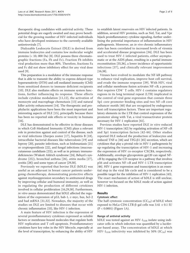
Inhibition of Env/CD4-mediated membrane fusionA cell-based fusion assay was used to mimic the gp120-CD4-mediated fusion process of HIV-1 with bDLE.When bDLE was exposed first to the CD4 cells-Env cellsmixture, fusion between both cells was blocked in adose-dependent manner. Cell-based fusion was alsoinhibited when bDLE was applied only to CD4 cells for30 minutes, followed by bDLE removal. However, whenEnv cells were first exposed to bDLE for 30 minutes,then removed and added to CD4 cells, fusion betweenboth cells was not inhibited (Figure 3A). Known
antiretroviral drugs, such as UC781 (NNRTI), were usedas controls in this cell based fusion assay and did notinhibit cell fusion (Figure 3B). T-20 (Fusion Inhibitor),did inhibit cell fusion in all assays, however, except whenexposed to CD4 cells for only 30 minutes and thenremoved, after which the cells were mixed (Figure 3C).
Figure 1 Cytotoxicity assessment of bDLE and HIV-1 inhibition activity. A) HeLa-CD4-LTR-b-gal cells (5 × 10⁴ cells/well) and B) HIV-1IIIB cell-free viruses (MOI 0.2-0.5) were challenged with two-fold serial dilutions of bDLE. Cell viability and b-gal activity were measured with a luciferase-based assay 24 h after nanosilver exposure. Percentage values are relative to the positive control (no compound treatment). The data representthe means ± standard deviations from three separate experiments, each of which was carried out in duplicate.
Figure 2 Residual activity in HIV-1 strains. HIV-1IIIB cell-freeviruses were exposed to serial dilutions of bDLE for 5 minutes. Theviruses were then ultracentrifuged, washed twice and added toHeLa-CD4-LTR-b-gal cells. After 24 hours, b-gal activity wasmeasured. Percentage values are relative to the positive control(infected cells without bDLE treatment). The data represent themeans ± standard deviations from three separate experiments, eachof which was carried out in duplicate.
Lara et al. BMC Research Notes 2011, 4:474http://www.biomedcentral.com/1756-0500/4/474
Page 3 of 9
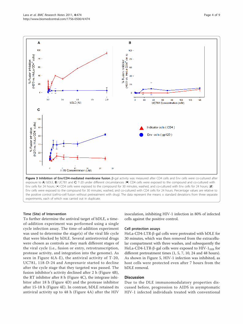
Time (Site) of InterventionTo further determine the antiviral target of bDLE, a time-of-addition experiment was performed using a singlecycle infection assay. The time-of-addition experimentwas used to determine the stage(s) of the viral life cyclethat were blocked by bDLE. Several antiretroviral drugswere chosen as controls as they mark different stages ofthe viral cycle (i.e., fusion or entry, retrotranscription,protease activity, and integration into the genome). Asseen in Figure 4(A-E), the antiviral activity of T-20,UC781, 118-D-24 and Amprenavir started to declineafter the cycle stage that they targeted was passed. Thefusion inhibitor’s activity declined after 2 h (Figure 4B),the RT inhibitor after 8 h (Figure 4C), the integrase inhi-bitor after 18 h (Figure 4D) and the protease inhibitorafter 15-18 h (Figure 4E). In contrast, bDLE retained itsantiviral activity up to 48 h (Figure 4A) after the HIV
inoculation, inhibiting HIV-1 infection in 80% of infectedcells against the positive control.
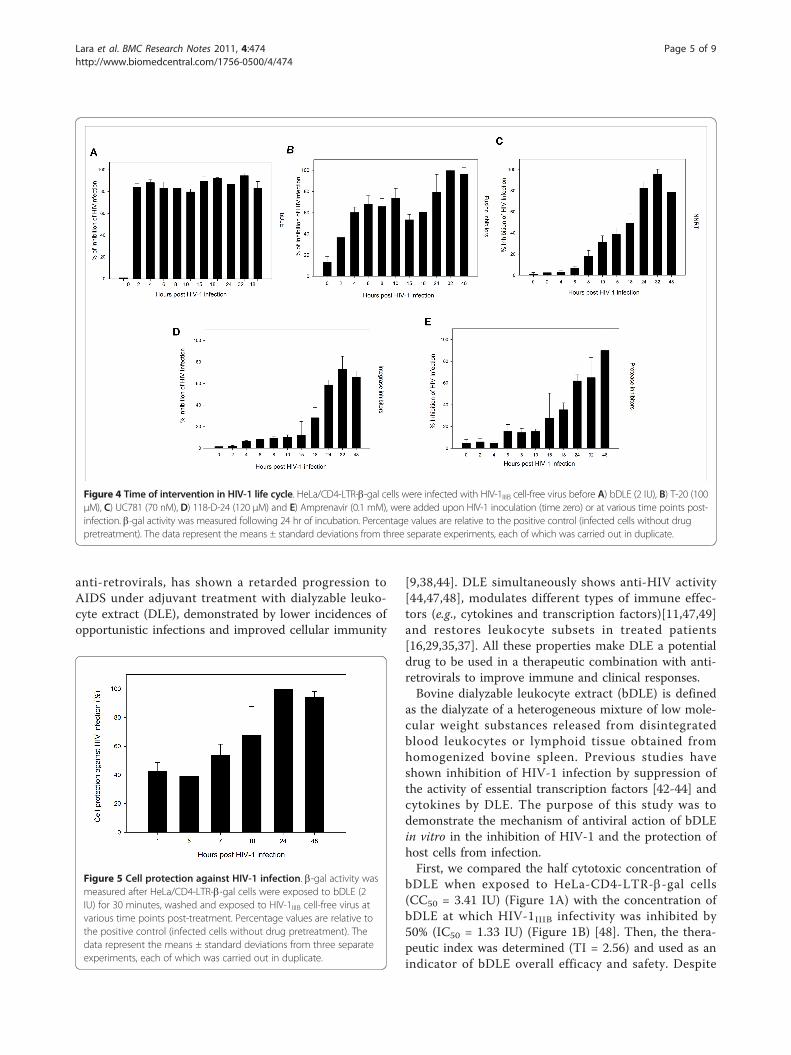
Cell protection assaysHeLa-CD4-LTR-b-gal cells were pretreated with bDLE for30 minutes, which was then removed from the extracellu-lar compartment with three washes, and subsequently theHeLa-CD4-LTR-b-gal cells were exposed to HIV-1IIIB fordifferent pretreatment times (1, 5, 7, 10, 24 and 48 hours).As shown in Figure 5, HIV-1 infection was inhibited, ashost cells were protected even after 7 hours from thebDLE removal.
DiscussionDue to the DLE immunomodulatory properties dis-cussed before, progression to AIDS in asymptomaticHIV-1 infected individuals treated with conventional
Figure 3 Inhibition of Env/CD4-mediated membrane fusion. b-gal activity was measured after CD4 cells and Env cells were co-cultured afterexposure to A) bDLE, B) UC781 and C) T-20 under different circumstances: (■) CD4 cells were exposed to the compound and co-cultured withEnv cells for 24 hours; (▲) CD4 cells were exposed to the compound for 30 minutes, washed, and co-cultured with Env cells for 24 hours; (✘)Env cells were exposed to the compound for 30 minutes, washed, and co-cultured with CD4 cells for 24 hours. Percentage values are relative tothe positive control (cell-to-cell fusion without pretreatment with drug). The data represent the means ± standard deviations from three separateexperiments, each of which was carried out in duplicate.
Lara et al. BMC Research Notes 2011, 4:474http://www.biomedcentral.com/1756-0500/4/474
Page 4 of 9
anti-retrovirals, has shown a retarded progression toAIDS under adjuvant treatment with dialyzable leuko-cyte extract (DLE), demonstrated by lower incidences ofopportunistic infections and improved cellular immunity
[9,38,44]. DLE simultaneously shows anti-HIV activity[44,47,48], modulates different types of immune effec-tors (e.g., cytokines and transcription factors)[11,47,49]and restores leukocyte subsets in treated patients[16,29,35,37]. All these properties make DLE a potentialdrug to be used in a therapeutic combination with anti-retrovirals to improve immune and clinical responses.Bovine dialyzable leukocyte extract (bDLE) is defined
as the dialyzate of a heterogeneous mixture of low mole-cular weight substances released from disintegratedblood leukocytes or lymphoid tissue obtained fromhomogenized bovine spleen. Previous studies haveshown inhibition of HIV-1 infection by suppression ofthe activity of essential transcription factors [42-44] andcytokines by DLE. The purpose of this study was todemonstrate the mechanism of antiviral action of bDLEin vitro in the inhibition of HIV-1 and the protection ofhost cells from infection.First, we compared the half cytotoxic concentration of
bDLE when exposed to HeLa-CD4-LTR-b-gal cells(CC50 = 3.41 IU) (Figure 1A) with the concentration ofbDLE at which HIV-1IIIB infectivity was inhibited by50% (IC50 = 1.33 IU) (Figure 1B) [48]. Then, the thera-peutic index was determined (TI = 2.56) and used as anindicator of bDLE overall efficacy and safety. Despite
Figure 4 Time of intervention in HIV-1 life cycle. HeLa/CD4-LTR-b-gal cells were infected with HIV-1IIIB cell-free virus before A) bDLE (2 IU), B) T-20 (100μM), C) UC781 (70 nM), D) 118-D-24 (120 μM) and E) Amprenavir (0.1 mM), were added upon HIV-1 inoculation (time zero) or at various time points post-infection. b-gal activity was measured following 24 hr of incubation. Percentage values are relative to the positive control (infected cells without drugpretreatment). The data represent the means ± standard deviations from three separate experiments, each of which was carried out in duplicate.
Figure 5 Cell protection against HIV-1 infection. b-gal activity wasmeasured after HeLa/CD4-LTR-b-gal cells were exposed to bDLE (2IU) for 30 minutes, washed and exposed to HIV-1IIIB cell-free virus atvarious time points post-treatment. Percentage values are relative tothe positive control (infected cells without drug pretreatment). Thedata represent the means ± standard deviations from three separateexperiments, each of which was carried out in duplicate.
Lara et al. BMC Research Notes 2011, 4:474http://www.biomedcentral.com/1756-0500/4/474
Page 5 of 9

the fact that TI was lower than expected (< 10), bDLE isa compound that has been used in clinical assays formore than fifty years without adverse reactions [10].Better understanding of its inhibition mechanism cancontribute to the development of new and improvedanti-HIV-1 agents, which could be more efficient andhave lower cytotoxicity. Furthermore, the residual infec-tivity [50,51] of cell-free viruses after bDLE treatmentshowed no inhibitory activity (Figure 2), which suggeststhat bDLE does not act as a virucide on the viral mem-brane to inhibit infection. bDLE showed inhibition offusogenic cell-cell interactions in a dose-dependentmanner when bDLE was exposed to CD4 cells, bDLEwas then removed and the CD4 cells were mixed withEnv cells. However, after 30 minutes pretreatment ofEnv cells with bDLE and then transfer to CD4 cells,there was no inhibition of Env-CD4 cell fusion. Thisobservation further supported our previous results thatbDLE acted on CD4 expressing cells and not on the Envof the HIV-1 virus. Furthermore, when exposed to CD4cells for only 30 minutes, which is the time required forconformational changes in gp120 after CD4 binding[52], bDLE again showed inhibition in a dose-dependentmanner (Figure 3A). These data suggested that bDLEacted on CD4 cells to inhibit HIV-1 infection. Antiviralresults reported by Fernandez-Ortega et al. [53,54] havealso contributed to the knowledge of the molecularmechanisms responsible for the effectiveness of bDLEagainst HIV infection.To further determine the antiviral target of bDLE, a
time-of-addition experiment was used to define the stage(s) of the viral life cycle that are blocked by these com-pounds. These results were compared with several antire-troviral drugs as controls that marked different stages ofthe viral cycle [55-61] (Figure 4B-E). Our findings sug-gested that bDLE highly inhibited HIV-1 infection at allstages (Figure 4A), possibly due to viral Tat proteindown-regulation (Tat activates b-galactosidase indicatorgene expression in HeLa-CD4-LTR-b-gal cells). Inhibi-tion of HIV-1 Tat activity correlates with down-regula-tion of bcl-2 [62], but the action of bDLE on bcl-2 hasnot yet been determined in HIV-1 studies. Previously,bcl-2 was found to be reduced in breast cancer cell lineswhen treated with bDLE [32]. Furthermore, DLE inacti-vated the NF-�B signaling pathway by reducing thesecretion of cytokines, such as IL-1 and TNF-a, whichare effective inducers of NF-�B activity [63]. In HIV-infected T cells, NF-�B-dependent transactivation isessential for HIV-LTR induction. Interestingly, even thefunction of HIV Tat in resting CD4 T lymphocytesdepends on �B responsive elements in the LTR [46].Based on these interesting findings, it will be necessary tofocus specifically on the transcriptional factors (NF-�B
and SP1) and pro-apoptotic genes (bcl-2) in futureresearch on bDLE as an antiviral against HIV-1 infection.Lastly, bDLE was capable of rendering CD4 expressing
cells resistant against HIV-1 infection by residual activevirus for several hours [64]. Previous results indicated that,although pretreatment of cells (MT-4) with DLE for 3hours had no effect, inhibition of HIV-1 production wasobserved when cells were pre-treated for a longer periodof time (from 1 to 7 days), an effect that was characterizedby the decline in TNFa and TGFb1 gene expression andinhibition of transcriptional factors [9]. In our assays, afterpretreatment of the HeLa-CD4-LTR-b-gal cells and bDLEremoval prior to viral challenge, protection against infec-tion lasted 7 hours after bDLE was removed from theextracellular compartment (Figure 5). These results indi-cated that bDLE could induce long-term viral inhibitionthrough cell protection, as well as modulate cell suscept-ibility to viral infection in vitro, in agreement with pre-viously reported data on DLE obtained from humandonors by molecular methods [9].
ConclusionThe data presented here were novel in that they provedthat bDLE acted by inhibiting HIV-1 infection throughprotection of the host target CD4 cells at noncytotoxiclevels. This effect was found to be modulated throughtranscriptional factors (NF-�B and SP1) necessary forHIV-1 replication. In addition, bDLE was discovered toact through all viral cycle stages to protect cells from HIV-1 infection for hours without affecting the HIV membrane.Based on our results obtained above, bDLE should befurther studied to determine its potential use as a thera-peutic agent in HIV-1 infection, especially due to its long-lasting cell protection against HIV-1 infection and lack ofnegative side effects.
MethodsReagents, cells and HIV-1 isolatesThe following reagents were obtained through the AIDSResearch and Reference Reagent Program (NIH): HeLa-CD4-LTR-b-gal cells from Dr. Michael Emerman; HL2/3cells from Dr. Barbara K. Felber and Dr. George N. Pavla-kis; HIV-1IIIB, fusion inhibitor T-20, integrase inhibitor118-D-24 and protease inhibitor Amprenavir from Dr.Suzanne Gartner, Dr. Mikulas Popovic and Dr. RobertGallo. UC781, a no nucleoside reverse-transcriptase inhi-bitor (NNRTI), was kindly donated by Dr. Gadi Borkow.The bDLE used in our study was produced by theLaboratory of Immunology and Virology at the Universi-dad Autonoma de Nuevo Leon, Mexico, following a mod-ified process described by Lawrence et al. [9]. The bDLEwas lyophilized, tested for endogenous pyrogens usingthe Limulus amebocyte lysate assay (MP Biomedicals,
Lara et al. BMC Research Notes 2011, 4:474http://www.biomedcentral.com/1756-0500/4/474
Page 6 of 9

Inc.), and determined to be free of bacterial contamina-tion by culturing in media and in vivo mice inoculations.The bDLE obtained from 15 × 108 leukocytes wasdefined as one unit (1 Unit)[37].
Cytotoxicity AssaysA stock solution of bDLE was diluted two-fold diluted ingrowth medium and subsequently added into wells con-taining 5 × 10⁴ HeLa-CD4-LTR-b-gal cells. Microtiterplates were incubated at 37°C in a 5% CO2 air humidifiedatmosphere for 24 hours. Assessments of cell viabilitywere carried out using a CellTiter-Glo® Luminescent CellViability Assay (Promega). Cytotoxicity was evaluatedbased on the percentage cell survival relative to the resultobtained in the absence of any compound.
HIV-1 Infection Inhibition AssaysSerial two-fold dilutions of bDLE were mixed with 105
TCID50 of HIV-1IIIB and added to the wells containing 5 ×10⁴ HeLa-CD4-LTR-b-gal cells with a multiplicity of infec-tion (MOI) of 0.2 - 0.5. HIV-1 infection was assessed after24 hours of incubation by quantifying the activity of the b-galactosidase produced after infection with the Beta-GloAssay System (Promega). The 50% inhibitory concentra-tion (IC50) was defined according to the percentage ofinfection inhibited relative to the positive control.
Virucidal Activity AssaySerial two-fold dilutions of bDLE were added to HIV-1IIIB(T tropic virus) and HIV-1Ba-L (M Tropic) cell-free virus.After incubation for 5 min at room temperature, the mix-tures were centrifuged three times at 10, 000 rpm, thesupernatant fluids removed, and the pellets washed threetimes. The final pellets were resuspended in Dulbecco’sModified Eagle Medium (DMEM) and placed into 96-wellplates with HeLa-CD4-LTR-b-gal cells. The cells wereincubated in a 5% CO2 humidified incubator at 37°C for24 h. Assessment of HIV-1 infection was made with theBeta-Glo Assay System. The percentage of residual infec-tivity after bDLE treatment was then calculated withrespect to the positive control of untreated virus.
Cell-based Fusion AssayHeLa-derived HL2/3 cells (Env cells), which express theHIV-1HXB2 Env, Tat, Gag, Rev, and Nef proteins, wereco-cultured with HeLa-CD4-LTR-b-gal cells (CD4 cells)at a 1:1 cell density ratio (5 × 10⁴ cells/well each) for 24 hin the absence or presence of two-fold dilutions of bDLE,UC781, and T-20 in order to examine whether the com-pounds interfered with the binding process of HIV-1 Envand the CD4 receptor. Also, both HeLa-CD4-LTR-b-galand HL2/3 cells were exposed to the aforementionedcompounds for only 30 minutes and then washed twiceto eliminate residual compound before co-cultivating
with the other cell line. Upon fusion of both cell lines,the Tat protein from HL2/3 cells activated b-galactosi-dase (b-gal) indicator gene expression in HeLa-CD4-LTR-b-gal cells [41]. b-gal activity was quantified withthe Beta-Glo Assay System (Promega). The percentage ofinhibition of HL2/3-HeLa CD4 cell fusion was calculatedwith respect to the positive control of untreated cells.
Time of Addition ExperimentsHeLa-CD4-LTR-b-gal cells were infected with 105 TCID50
of HIV-1IIIB cell-free virus with a 0.2-0.5 MOI. bDLE (2IU), T-20 (100 μM), UC781 (70 nM), 118-D-24 (120 μM)and Amprenavir (0.1 mM) were then added upon HIV-1inoculation (time zero) or at various time points post-inoculation. The reference compounds were added at aconcentration several times their EC50 for infectivity ofHIV-1IIIB. Infection inhibition was quantified after 24 h bymeasuring b-gal activity with the Beta-Glo Assay System.
Cell Protection AssaysHeLa-CD4-LTR-b-gal cells were incubated with bDLE(2 Units) for 30 minutes and subsequently washed withPBS three times. Then, the cells were exposed to 105
TCID50 of HIV-1IIIB cell-free virus with a 0.2-0.5 MOIfor different times (1, 5, 7, 10, 24 and 48 h). Infectioninhibition was quantified after 24 h by measuring b-galactivity with the Beta-Glo Assay System.
Statistical analysisGraphs were done with SigmaPlot 10.0 software and thevalues shown are means ± standard deviations fromthree separate experiments, each of which was carriedout in duplicate. Cytotoxicity and inhibition assessmentgraphs are linear regression curves done with SigmaPlot10.0 software.
AcknowledgementsThe following funding sources supported our experiments: the Programa deApoyo a la Investigacion en Ciencia y Tecnologia (PAICyT) of the UniversidadAutonoma de Nuevo Leon, Mexico, and the Consejo Nacional de Ciencia yTecnologia (CONACyT) of Mexico.
Authors’ contributionsAll authors read and approved the final manuscript. HHL participated in theconception and experimental design of the in vitro HIV-1 manipulation andinfection assays, in the analysis and interpretation of the data, and in thewriting and revision of this report. LI-T participated in the analysis andinterpretation the results. HHL and LI-T made equal contributions to thisstudy. EN-GT participated in the analysis and interpretation of the data andin writing and revising this report. SM-FT participated in the analysis andwriting of the report. JIB participated in revising this report. CR-Pparticipated in the experimental design of this research.
Competing interestsThe authors declare that they have no competing interests.
Received: 24 July 2011 Accepted: 1 November 2011Published: 1 November 2011
Lara et al. BMC Research Notes 2011, 4:474http://www.biomedcentral.com/1756-0500/4/474
Page 7 of 9
References1. Fauci AS: The AIDS epidemic–considerations for the 21st century. N Engl
J Med 1999, 341:1046-1050.2. Ganguly N: State of the Globe: The Immunological Quest for an HIV/AIDS
Vaccine Continues. J Glob Infect Dis 2011, 3:209-210.3. Palella FJ Jr, Delaney KM, Moorman AC, Loveless MO, Fuhrer J, Satten GA,
et al: Declining morbidity and mortality among patients with advancedhuman immunodeficiency virus infection. HIV Outpatient StudyInvestigators. N Engl J Med 1998, 338:853-860.
4. Sterne JA, May M, Costagliola D, de WF, Phillips AN, Harris R, et al: Timingof initiation of antiretroviral therapy in AIDS-free HIV-1-infected patients:a collaborative analysis of 18 HIV cohort studies. Lancet 2009,373:1352-1363.
5. Johnson VA, Brun-Vezinet F, Clotet B, Gunthard HF, Kuritzkes DR, Pillay D,et al: Update of the drug resistance mutations in HIV-1: December 2010.Top HIV Med 2010, 18:156-163.
6. Lara HH, Garza-Trevino EN, Ixtepan-Turrent L, Singh DK: Silver nanoparticlesare broad-spectrum bactericidal and virucidal compounds. JNanobiotechnology 2011, 9:30.
7. Caffrey M: HIV envelope: challenges and opportunities for developmentof entry inhibitors. Trends Microbiol 2011, 19:191-197.
8. Kirkpatrick CH: Transfer factors: identification of conserved sequences intransfer factor molecules. Mol Med 2000, 6:332-341.
9. Fernandez-Ortega C, Dubed M, Ruibal O, Vilarrubia OL, Menendez de SanPedro JC, Navea L, et al: Inhibition of in vitro HIV infection by dialysableleucocyte extracts. Biotherapy 1996, 9:33-40.
10. Lawrence HS, Borkowsky W: Transfer factor–current status and futureprospects. Biotherapy 1996, 9:1-5.
11. Franco-Molina MA, Mendoza-Gamboa E, Castillo-Leon L, Tamez-Guerra RS,Rodriguez-Padilla C: Bovine dialyzable leukocyte extract modulates thenitric oxide and pro-inflammatory cytokine production inlipopolysaccharide-stimulated murine peritoneal macrophages in vitro. JMed Food 2005, 8:20-26.
12. Franco-Molina MA, Mendoza-Gamboa E, Castillo-Tello P, Isaza-Brando CE,Garcia ME, Castillo-Leon L, et al: Bovine dialyzable leukocyte extractmodulates cytokines and nitric oxide production in lipopolysaccharide-stimulated human blood cells. Cytotherapy 2007, 9:379-385.
13. Kirkpatrick CH, Rich RR, Smith TK: Effect of transfer factor on lymphocytefunction in anergic patients. J Clin Invest 1972, 51:2948-2958.
14. Lang I, Nekam K, Gergely P, Petranyi G: Effect in vivo and in vitrotreatment with dialyzable leukocyte extracts on human natural killer cellactivity. Clin Immunol Immunopathol 1982, 25:139-144.
15. Fudenberg HH, Pizza G: Transfer factor 1993: new frontiers. Prog Drug Res1994, 42:309-400.
16. Lara HH, Ixtepan Turrent L, Garza Treviño EN, Tamez-Guerra RS, Rodriguez-Padilla C, (Eds): Clinical and Immunological assessment in breast cancerpatients receiving anticancer therapy and bovine dialyzable extract asan adjuvant. Experimental and Therapeutic Medicine 2010, 1:425-431.
17. Estrada-Parra S, Nagaya A, Serrano E, Rodriguez O, Santamaria V, Ondarza R,et al: Comparative study of transfer factor and acyclovir in the treatmentof herpes zoster. Int J Immunopharmacol 1998, 20:521-535.
18. Mazzella G, Ronchi M, Villanova N, Mohamed AA, Pizza G, De VC, et al:Treatment of chronic B virus hepatitis with specific transfer factor.Journal Exp Pathol 1987, 1:421-423.
19. Fabre RA, Perez TM, Aguilar LD, Rangel MJ, Estrada-Garcia I, Hernandez-Pando R, et al: Transfer factors as immunotherapy and supplement ofchemotherapy in experimental pulmonary tuberculosis. Clin Exp Immunol2004, 136:215-223.
20. Hastings RC, Morales MJ, Shannon EJ, Jacobson RR: Preliminary results onthe safety and efficacy of transfer factor in leprosy. Int J Lepr OtherMycobact Dis 1976, 44:275.
21. Delgado O, Romano EL, Belfort E, Pifano F, Scorza JV, Rojas Z: Dialyzableleukocyte extract therapy in immunodepressed patients with cutaneousleishmaniasis. Clin Immunol Immunopathol 1981, 19:351-359.
22. Louie E, Borkowsky W, Klesius PH, Haynes TB, Gordon S, Bonk S, et al:Treatment of cryptosporidiosis with oral bovine transfer factor. ClinImmunol Immunopathol 1987, 44:329-334.
23. Masi M, De VC, Baricordi OR: Transfer factor in chronic mucocutaneouscandidiasis. Biotherapy 1996, 9:97-103.
24. Levin AS, Spitler LE, Stites DP, Fudenberg HH: Wiskott-Aldrich syndrome, agenetically determined cellular immunologic deficiency: clinical and
laboratory responses to therapy with transfer factor. Proc Natl Acad SciUSA 1970, 67:821-828.
25. Wolf RE, Fudenberg HH, Welch TM, Spitler LE, Ziff M: Treatment ofBechcet’s syndrome with transfer factor. JAMA 1977, 238:869-871.
26. Valdes Sanchez AF, Martin Rodriguez OL, Lastra AG: [Treatment of extrinsicbronchial asthma with transfer factor]. Rev Alerg Mex 1993, 40:124-131.
27. Kaminkova J, Lange CF: Transfer factor and repeated otitis media. CellImmunol 1984, 89:259-264.
28. Abramson A, Khan A, Tate GW Jr, Martin RG, Hill NO: Immunocompetenceand transfer factor therapy in uveitis. Br J Ophthalmol 1980, 64:332-338.
29. Franco-Molina MA, Mendoza-Gamboa E, Zapata-Benavides P, Vera-Garcia ME, Castillo-Tello P, Garcia dlF, et al: IMMUNEPOTENT CRP (bovinedialyzable leukocyte extract) adjuvant immunotherapy: a phase I studyin non-small cell lung cancer patients. Cytotherapy 2008, 10:490-496.
30. Whyte RI, Schork MA, Sloan H, Orringer MB, Kirsh MM: Adjuvant treatmentusing transfer factor for bronchogenic carcinoma: long-term follow-up.Ann Thorac Surg 1992, 53:391-396.
31. Franco-Molina MA, Mendoza-Gamboa E, Miranda-Hernandez D, Zapata-Benavides P, Castillo-Leon L, Isaza-Brando C, et al: In vitro effects of bovinedialyzable leukocyte extract (bDLE) in cancer cells. Cytotherapy 2006,8:408-414.
32. Mendoza-Gamboa E, Franco-Molina MA, Zapata-Benavides P, Castillo-Tello P,Vera-Garcia ME, Tamez-Guerra RS, et al: Bovine dialyzable leukocyteextract modulates AP-1 DNA-binding activity and nuclear transcriptionfactor expression in MCF-7 breast cancer cells. Cytotherapy 2008,10:212-219.
33. Franco-Molina MA, Mendoza-Gamboa E, Castillo-Leon L, Tamez-Guerra RS,Rodriguez-Padilla C: Bovine dialyzable leukocyte extract protects againstLPS-induced, murine endotoxic shock. Int Immunopharmacol 2004,4:1577-1586.
34. Roberts L, Passmore JA, Williamson C, Little F, Bebell LM, Mlisana K, et al:Plasma cytokine levels during acute HIV-1 infection predict HIV diseaseprogression. AIDS 2010, 24:819-831.
35. Gottlieb AA, Sizemore RC, Gottlieb MS, Kern CH: Rationale and clinicalresults of using leucocyte-derived immunosupportive therapies in HIVdisease. Biotherapy 1996, 9:27-31.
36. Raise E, Guerra L, Viza D, Pizza G, De VC, Schiattone ML, et al: Preliminaryresults in HIV-1-infected patients treated with transfer factor (TF) andzidovudine (ZDV). Biotherapy 1996, 9:49-54.
37. McMeeking A, Borkowsky W, Klesius PH, Bonk S, Holzman RS, Lawrence HS:A controlled trial of bovine dialyzable leukocyte extract forcryptosporidiosis in patients with AIDS. J Infect Dis 1990, 161:108-112.
38. Pizza G, Chiodo F, Colangeli V, Gritti F, Raise E, Fudenberg HH, et al:Preliminary observations using HIV-specific transfer factor in AIDS.Biotherapy 1996, 9:41-47.
39. Li JC, Yim HC, Lau AS: Role of HIV-1 Tat in AIDS pathogenesis: its effectson cytokine dysregulation and contributions to the pathogenesis ofopportunistic infection. AIDS 2010, 24:1609-1623.
40. Lim SP, Garzino-Demo A: The human immunodeficiency virus type 1 Tatprotein up-regulates the promoter activity of the beta-chemokinemonocyte chemoattractant protein 1 in the human astrocytoma cell lineU-87 MG: role of SP-1, AP-1, and NF-kappaB consensus sites. J Virol 2000,74:1632-1640.
41. Lalonde MS, Lobritz MA, Ratcliff A, Chamanian M, Athanassiou Z, Tyagi M,et al: Inhibition of both HIV-1 reverse transcription and gene expressionby a cyclic peptide that binds the Tat-transactivating response element(TAR) RNA. PLoS Pathog 2011, 7:e1002038.
42. Fernandez-Ortega C, Dubed M, Ramos Y, Navea L, Alvarez G, Lobaina L,et al: Non-induced leukocyte extract reduces HIV replication and TNFsecretion. Biochem Biophys Res Commun 2004, 325:1075-1081.
43. Ojeda MO, Fernandez-Ortega C, Rosainz MJ: Dialyzable leukocyte extractsuppresses the activity of essential transcription factors for HIV-1 geneexpression in unstimulated MT-4 cells. Biochem Biophys Res Commun2000, 273:1099-1103.
44. Ojeda MO, van’t Veer C, Fernandez Ortega CB, Arana Rosainz MJ,Buurman WA: Dialyzable leukocyte extract differentially regulates theproduction of TNFalpha, IL-6, and IL-8 in bacterial component-activatedleukocytes and endothelial cells. Inflamm Res 2005, 54:74-81.
45. Elrefaei M, Burke CM, Baker CA, Jones NG, Bousheri S, Bangsberg DR, et al:HIV-specific TGF-beta-positive CD4+ T cells do not express regulatory
Lara et al. BMC Research Notes 2011, 4:474http://www.biomedcentral.com/1756-0500/4/474
Page 8 of 9
surface markers and are regulated by CTLA-4. AIDS Res Hum Retroviruses2010, 26:329-337.
46. Hiscott J, Kwon H, Genin P: Hostile takeovers: viral appropriation of theNF-kappaB pathway. J Clin Invest 2001, 107:143-151.
47. Carey JT, Lederman MM: Treatment of AIDS with transfer factor. JAMA1987, 258:3515-3516.
48. Flory E, Weber CK, Chen P, Hoffmeyer A, Jassoy C, Rapp UR: Plasmamembrane-targeted Raf kinase activates NF-kappaB and humanimmunodeficiency virus type 1 replication in T lymphocytes. J Virol 1998,72:2788-2794.
49. Alvarez-Thull L, Kirkpatrick CH: Profiles of cytokine production inrecipients of transfer factors. Biotherapy 1996, 9:55-59.
50. Lara HH, Ayala-Nunez NV, Ixtepan-Turrent L, Rodriguez-Padilla C: Mode ofantiviral action of silver nanoparticles against HIV-1. J Nanobiotechnology2010, 8:1.
51. Yang QE, Stephen AG, Adelsberger JW, Roberts PE, Zhu W, Currens MJ,et al: Discovery of small-molecule human immunodeficiency virus type 1entry inhibitors that target the gp120-binding domain of CD4. J Virol2005, 79:6122-6133.
52. Jones PL, Korte T, Blumenthal R: Conformational changes in cell surfaceHIV-1 envelope glycoproteins are triggered by cooperation between cellsurface CD4 and co-receptors. J Biol Chem 1998, 273:404-409.
53. Demarchi F, d’Adda di FF, Falaschi A, Giacca M: Activation of transcriptionfactor NF-kappaB by the Tat protein of human immunodeficiency virustype 1. J Virol 1996, 70:4427-4437.
54. Gaynor R: Cellular transcription factors involved in the regulation of HIV-1 gene expression. AIDS 1992, 6:347-363.
55. Auwerx J, Stevens M, Van Rompay AR, Bird LE, Ren J, De CE, et al: Thephenylmethylthiazolylthiourea nonnucleoside reverse transcriptase (RT)inhibitor MSK-076 selects for a resistance mutation in the active site ofhuman immunodeficiency virus type 2 RT. J Virol 2004, 78:7427-7437.
56. Hombrouck A, Van RB, Michiels M, Noppe W, Christ F, Eneroth A, et al:Preclinical evaluation of 1H-benzylindole derivatives as novel humanimmunodeficiency virus integrase strand transfer inhibitors. AntimicrobAgents Chemother 2008, 52:2861-2869.
57. Stevens M, Pannecouque C, De CE, Balzarini J: Novel humanimmunodeficiency virus (HIV) inhibitors that have a dual mode of anti-HIV action. Antimicrob Agents Chemother 2003, 47:3109-3116.
58. Svarovskaia ES, Barr R, Zhang X, Pais GC, Marchand C, Pommier Y, et al:Azido-containing diketo acid derivatives inhibit humanimmunodeficiency virus type 1 integrase in vivo and influence thefrequency of deletions at two-long-terminal-repeat-circle junctions. JVirol 2004, 78:3210-3222.
59. Witvrouw M, Balzarini J, Pannecouque C, Jhaumeer-Laulloo S, Este JA,Schols D, et al: SRR-SB3, a disulfide-containing macrolide that inhibits alate stage of the replicative cycle of human immunodeficiency virus.Antimicrob Agents Chemother 1997, 41:262-268.
60. Witvrouw M, Fikkert V, Pluymers W, Matthews B, Mardel K, Schols D, et al:Polyanionic (i.e., polysulfonate) dendrimers can inhibit the replication ofhuman immunodeficiency virus by interfering with both virusadsorption and later steps (reverse transcriptase/integrase) in the virusreplicative cycle. Mol Pharmacol 2000, 58:1100-1108.
61. Zhang X, Pais GC, Svarovskaia ES, Marchand C, Johnson AA, Karki RG, et al:Azido-containing aryl beta-diketo acid HIV-1 integrase inhibitors. BioorgMed Chem Lett 2003, 13:1215-1219.
62. Corallini A, Sampaolesi R, Possati L, Merlin M, Bagnarelli P, Piola C, et al:Inhibition of HIV-1 Tat activity correlates with down-regulation of bcl-2and results in reduction of angiogenesis and oncogenicity. Virology 2002,299:1-7.
63. Duh EJ, Maury WJ, Folks TM, Fauci AS, Rabson AB: Tumor necrosis factoralpha activates human immunodeficiency virus type 1 throughinduction of nuclear factor binding to the NF-kappa B sites in the longterminal repeat. Proc Natl Acad Sci USA 1989, 86:5974-5978.
64. Zussman A, Lara L, Lara HH, Bentwich Z, Borkow G: Blocking of cell-freeand cell-associated HIV-1 transmission through human cervix organculture with UC781. AIDS 2003, 17:653-661.
doi:10.1186/1756-0500-4-474Cite this article as: Lara et al.: Antiviral mode of action of bovinedialyzable leukocyte extract against human immunodeficiency virustype 1 infection. BMC Research Notes 2011 4:474.
Submit your next manuscript to BioMed Centraland take full advantage of:
• Convenient online submission
• Thorough peer review
• No space constraints or color figure charges
• Immediate publication on acceptance
• Inclusion in PubMed, CAS, Scopus and Google Scholar
• Research which is freely available for redistribution
Submit your manuscript at www.biomedcentral.com/submit
Lara et al. BMC Research Notes 2011, 4:474http://www.biomedcentral.com/1756-0500/4/474
Page 9 of 9