
Dynamics of in silico leukocyte rolling, activation, and adhesion
25
BioMed Central Page 1 of 25 (page number not for citation purposes) BMC Systems Biology Open Access Research article Dynamics of in silico leukocyte rolling, activation, and adhesion Jonathan Tang 1 , Klaus F Ley 2 and C Anthony Hunt* 1,3 Address: 1 The UCSF/UCB Joint Graduate Group in Bioengineering, University of California, Berkeley, CA, USA, 2 Robert M. Berne Cardiovascular Research Center and Departments of Biomedical Engineering, Molecular Physiology and Biological Physics, University of Virginia, Charlottesville, VA, USA and 3 The Department of Biopharmaceutical Sciences, Biosystems Group, University of California, San Francisco, CA, USA Email: Jonathan Tang - [email protected]; Klaus F Ley - [email protected]; C Anthony Hunt* - [email protected] * Corresponding author Abstract Background: We present a multilevel, agent based, in silico model that represents the dynamics of rolling, activation, and adhesion of individual leukocytes in vitro. Object-oriented software components were designed, verified, plugged together, and then operated in ways that represent the molecular and cellular mechanisms believed responsible for leukocyte rolling and adhesion. The result is an in silico analogue of an experimental in vitro system. The experimentally measured, phenotypic attributes of the analogue were compared and contrasted to those of leukocytes in vitro from three different experimental conditions. Results: The individual in silico dynamics of "rolling" on simulated P-selectin, and separately on simulated VCAM-1, were an acceptable match to individual in vitro distance-time and velocity-time measurements. The analogues are also able to represent the transition from rolling to adhesion on P-selectin and VCAM-1 in the presence of GRO-α chemokine. The individual in silico and in vitro behavioral similarities translated successfully to population level measures. These behavioral similarities were enabled in part by subdividing the functionality of the analogue's surface into 600 independent, "cell"-controlled, equally capable modules of comparable functionality. Conclusion: The overlap in phenotypic attributes of our analogue with those of leukocytes in vitro confirm the considerable potential of our model for studying the key events that determine the behavioral outcome of individual leukocytes during rolling, activation, and adhesion. Our results provide an important foundation and framework for future in silico research into plausible causal links between well-documented, subcellular molecular level events and the variety of systemic phenotypic attributes that distinguish normal leukocyte adhesion from abnormal disease-associated adhesion. Background What molecular-level events determine the behavioral outcome of individual leukocytes during rolling, activa- tion, and adhesion to venular surfaces? These processes are necessary steps for the proper recruitment of leuko- cytes from circulating blood to sites of inflammation. Once at the target site, leukocytes help destroy pathogens and decompose damaged tissue. However, inflammatory mechanisms are also associated with diseases such as asthma, rheumatoid arthritis, multiple sclerosis, and atherosclerosis. Such diseases can be characterized by Published: 19 February 2007 BMC Systems Biology 2007, 1:14 doi:10.1186/1752-0509-1-14 Received: 4 October 2006 Accepted: 19 February 2007 This article is available from: http://www.biomedcentral.com/1752-0509/1/14 © 2007 Tang et al; licensee BioMed Central Ltd. This is an Open Access article distributed under the terms of the Creative Commons Attribution License (http://creativecommons.org/licenses/by/2.0 ), which permits unrestricted use, distribution, and reproduction in any medium, provided the original work is properly cited.
Transcript of Dynamics of in silico leukocyte rolling, activation, and adhesion
BioMed CentralBMC Systems Biology
ss
Open AcceResearch articleDynamics of in silico leukocyte rolling, activation, and adhesionJonathan Tang1, Klaus F Ley2 and C Anthony Hunt*1,3Address: 1The UCSF/UCB Joint Graduate Group in Bioengineering, University of California, Berkeley, CA, USA, 2Robert M. Berne Cardiovascular Research Center and Departments of Biomedical Engineering, Molecular Physiology and Biological Physics, University of Virginia, Charlottesville, VA, USA and 3The Department of Biopharmaceutical Sciences, Biosystems Group, University of California, San Francisco, CA, USA
Email: Jonathan Tang - [email protected]; Klaus F Ley - [email protected]; C Anthony Hunt* - [email protected]
* Corresponding author
AbstractBackground: We present a multilevel, agent based, in silico model that represents the dynamicsof rolling, activation, and adhesion of individual leukocytes in vitro. Object-oriented softwarecomponents were designed, verified, plugged together, and then operated in ways that representthe molecular and cellular mechanisms believed responsible for leukocyte rolling and adhesion. Theresult is an in silico analogue of an experimental in vitro system. The experimentally measured,phenotypic attributes of the analogue were compared and contrasted to those of leukocytes invitro from three different experimental conditions.
Results: The individual in silico dynamics of "rolling" on simulated P-selectin, and separately onsimulated VCAM-1, were an acceptable match to individual in vitro distance-time and velocity-timemeasurements. The analogues are also able to represent the transition from rolling to adhesion onP-selectin and VCAM-1 in the presence of GRO-α chemokine. The individual in silico and in vitrobehavioral similarities translated successfully to population level measures. These behavioralsimilarities were enabled in part by subdividing the functionality of the analogue's surface into 600independent, "cell"-controlled, equally capable modules of comparable functionality.
Conclusion: The overlap in phenotypic attributes of our analogue with those of leukocytes invitro confirm the considerable potential of our model for studying the key events that determinethe behavioral outcome of individual leukocytes during rolling, activation, and adhesion. Our resultsprovide an important foundation and framework for future in silico research into plausible causallinks between well-documented, subcellular molecular level events and the variety of systemicphenotypic attributes that distinguish normal leukocyte adhesion from abnormal disease-associatedadhesion.
BackgroundWhat molecular-level events determine the behavioraloutcome of individual leukocytes during rolling, activa-tion, and adhesion to venular surfaces? These processesare necessary steps for the proper recruitment of leuko-cytes from circulating blood to sites of inflammation.
Once at the target site, leukocytes help destroy pathogensand decompose damaged tissue. However, inflammatorymechanisms are also associated with diseases such asasthma, rheumatoid arthritis, multiple sclerosis, andatherosclerosis. Such diseases can be characterized by
Published: 19 February 2007
BMC Systems Biology 2007, 1:14 doi:10.1186/1752-0509-1-14
Received: 4 October 2006Accepted: 19 February 2007
This article is available from: http://www.biomedcentral.com/1752-0509/1/14
© 2007 Tang et al; licensee BioMed Central Ltd. This is an Open Access article distributed under the terms of the Creative Commons Attribution License (http://creativecommons.org/licenses/by/2.0), which permits unrestricted use, distribution, and reproduction in any medium, provided the original work is properly cited.
Page 1 of 25(page number not for citation purposes)
BMC Systems Biology 2007, 1:14 http://www.biomedcentral.com/1752-0509/1/14
inappropriate leukocyte recruitment and the misdirectedactions of leukocytes towards healthy host-tissue [1].
Rolling and adhesion following attachment are two of theleast complicated of many individual leukocyte behaviorsthat have been studied in vitro using flow chamber assays.Those behaviors are by no means deterministic. A strikingfeature of such studies is that cell behavior is heterogene-ous: individual cell behaviors under identical conditionscan be quite different. No two cells behave the same, yetcollective behaviors are robust and fall reliably within nar-row ranges. Rolling, for example, exhibits an irregular,jerky stop-and-go pattern along with highly fluctuatingrolling velocities [2]. Additionally, in the presence ofchemokine, only a fraction of a leukocyte population willadhere firmly [3].
A goal in systems biology research is to understand link-ages from molecular level events to system phenotype:link genotype to phenotype. That task requires havingplausible, adequately detailed design plans for how com-ponents (single and composite) at various system levelsare thought to fit and function together. Ideas about suchplans can be induced from the results of experiments.Experimentation is then used to reconcile different designplan hypotheses. More is needed, however, to actuallydemonstrate that a design plan is functionally plausible,which is very different from demonstrating that it is con-sistent with measured behaviors. The former requires thatone assemble individual components according to adesign, and then show that the constructed device, an ana-logue – on its own – exhibits behaviors that match thoseobserved in the original experiment.
Building such analogues in silico is now feasible. To makeit practicable, we need multilevel modeling and simula-tion methods that make it easy to test, reject, and refinemany candidate design plans. In this report, we describediscovering, building, and testing aspects of a simple yetplausible design plan. That achievement is an essentialstep toward the long-term biological goal for this project:develop scientifically useful, validated simulations of leu-kocyte recruitment under physiological and pathophysio-logical conditions. We use the synthetic modelingapproach. Object-oriented software components weredesigned, verified, plugged together, and then operated inways that represent the mechanisms and processesbelieved responsible for leukocyte rolling and adhesion.The result is an analogue of an in vitro experimental sys-tem. An analogue refinement method is used in whichexperimentally measured phenotypic attributes are itera-tively compared and contrasted to those of leukocytes invitro.
During leukocyte rolling, the initial interactions betweenleukocyte and surface are primarily mediated by the selec-tin family of receptors and their respective carbohydrateligands found on the membranes of both the leukocytesand endothelial cells. Transient selectin-ligand interac-tions enable rolling. Leukocyte arrest is mediated by theintegrin receptor family. Integrins exist largely in nonad-hesive states to prevent leukocytes from sticking non-spe-cifically to blood vessels. As leukocytes roll along thevenular surface, they use their chemokine receptors todetect immobilized chemokines on activated endothelialcells. Upon detection, intracellular signaling events triggerintegrin conformational changes, increasing integrin-lig-and affinity. High affinity integrins enable the leukocytesto firmly adhere to the vessel wall or flow chamber surface[4]. Signaling through chemokine receptors can alsoinduce lateral movement and clustering of integrinswithin the membrane to promote leukocyte adhesion[5,6]. In addition, the engagement of some selectins andintegrins with their ligands can initiate various activationsignals to enhance adhesion [7,8].
With their Adhesive Dynamics simulations, Hammer andco-workers have elegantly demonstrated how challengingit can be to generate similar rolling characteristics in silicowhen using physical and mechanical representationscombined with traditional modeling formalisms [9-11].In their models, leukocytes are represented as solidspheres decorated with rod-like "microvilli" containingreceptors at their tips. Using a Monte Carlo algorithm forthe determination of receptor-ligand interactions, theyhave successfully produced a jerky stop-and-go patternsimilar to that observed for rolling leukocytes. Such mod-els can be fragile to context. When they target specific phe-nomena and constrain their use to account for specific setsof data, it becomes difficult to extend the model to differ-ent experimental circumstances or to help explain differ-ent phenomena or particular examples of individualbehavior. It becomes difficult to explore potential mecha-nistic differences between individual cell behaviors, forexample. Are there alternative modeling and simulationapproaches that are less susceptible to such problems? Themultilevel approach described here was selected in part tohelp circumvent those problems.
A measure of a particular behavior is a phenotypicattribute. Table 1 lists several targetable attributes. Thegreater the similarity between the measured behaviors ofour in silico white blood cells (ISWBCs) and the in vitroattributes of interest, the more useful that in silico systemwill become as a research tool and as an expression of thecoalesced, relevant leukocyte knowledge. An early goalhas therefore been to produce increasing overlap betweenISWBC behaviors, properties, and characteristics, and thein vitro properties listed in Table 1. Once that is achieved,
Page 2 of 25(page number not for citation purposes)
BMC Systems Biology 2007, 1:14 http://www.biomedcentral.com/1752-0509/1/14
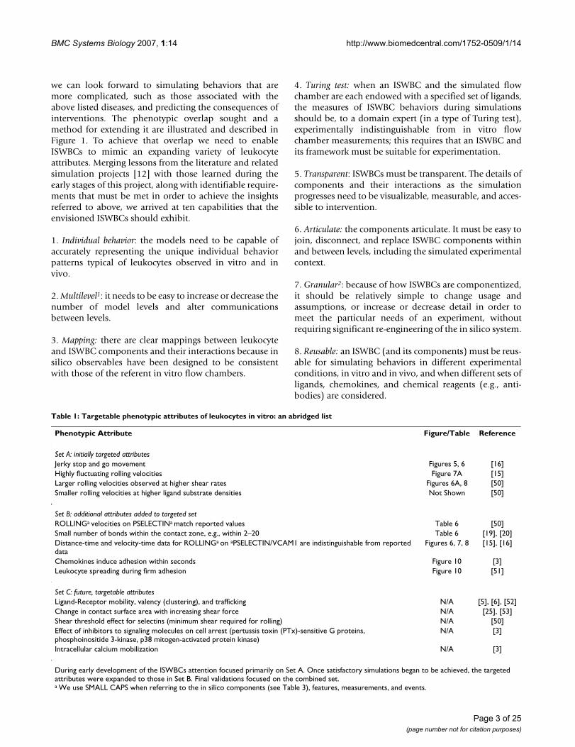
we can look forward to simulating behaviors that aremore complicated, such as those associated with theabove listed diseases, and predicting the consequences ofinterventions. The phenotypic overlap sought and amethod for extending it are illustrated and described inFigure 1. To achieve that overlap we need to enableISWBCs to mimic an expanding variety of leukocyteattributes. Merging lessons from the literature and relatedsimulation projects [12] with those learned during theearly stages of this project, along with identifiable require-ments that must be met in order to achieve the insightsreferred to above, we arrived at ten capabilities that theenvisioned ISWBCs should exhibit.
1. Individual behavior: the models need to be capable ofaccurately representing the unique individual behaviorpatterns typical of leukocytes observed in vitro and invivo.
2. Multilevel1: it needs to be easy to increase or decrease thenumber of model levels and alter communicationsbetween levels.
3. Mapping: there are clear mappings between leukocyteand ISWBC components and their interactions because insilico observables have been designed to be consistentwith those of the referent in vitro flow chambers.
4. Turing test: when an ISWBC and the simulated flowchamber are each endowed with a specified set of ligands,the measures of ISWBC behaviors during simulationsshould be, to a domain expert (in a type of Turing test),experimentally indistinguishable from in vitro flowchamber measurements; this requires that an ISWBC andits framework must be suitable for experimentation.
5. Transparent: ISWBCs must be transparent. The details ofcomponents and their interactions as the simulationprogresses need to be visualizable, measurable, and acces-sible to intervention.
6. Articulate: the components articulate. It must be easy tojoin, disconnect, and replace ISWBC components withinand between levels, including the simulated experimentalcontext.
7. Granular2: because of how ISWBCs are componentized,it should be relatively simple to change usage andassumptions, or increase or decrease detail in order tomeet the particular needs of an experiment, withoutrequiring significant re-engineering of the in silico system.
8. Reusable: an ISWBC (and its components) must be reus-able for simulating behaviors in different experimentalconditions, in vitro and in vivo, and when different sets ofligands, chemokines, and chemical reagents (e.g., anti-bodies) are considered.
Table 1: Targetable phenotypic attributes of leukocytes in vitro: an abridged list
Phenotypic Attribute Figure/Table Reference
Set A: initially targeted attributesJerky stop and go movement Figures 5, 6 [16]Highly fluctuating rolling velocities Figure 7A [15]Larger rolling velocities observed at higher shear rates Figures 6A, 8 [50]Smaller rolling velocities at higher ligand substrate densities Not Shown [50]
Set B: additional attributes added to targeted setROLLINGa velocities on PSELECTINa match reported values Table 6 [50]Small number of bonds within the contact zone, e.g., within 2–20 Table 6 [19], [20]Distance-time and velocity-time data for ROLLINGa on aPSELECTIN/VCAM1 are indistinguishable from reported data
Figures 6, 7, 8 [15], [16]
Chemokines induce adhesion within seconds Figure 10 [3]Leukocyte spreading during firm adhesion Figure 10 [51]
Set C: future, targetable attributesLigand-Receptor mobility, valency (clustering), and trafficking N/A [5], [6], [52]Change in contact surface area with increasing shear force N/A [25], [53]Shear threshold effect for selectins (minimum shear required for rolling) N/A [50]Effect of inhibitors to signaling molecules on cell arrest (pertussis toxin (PTx)-sensitive G proteins, phosphoinositide 3-kinase, p38 mitogen-activated protein kinase)
N/A [3]
Intracellular calcium mobilization N/A [3]
During early development of the ISWBCs attention focused primarily on Set A. Once satisfactory simulations began to be achieved, the targeted attributes were expanded to those in Set B. Final validations focused on the combined set.a We use SMALL CAPS when referring to the in silico components (see Table 3), features, measurements, and events.
Page 3 of 25(page number not for citation purposes)
BMC Systems Biology 2007, 1:14 http://www.biomedcentral.com/1752-0509/1/14
9. Embeddable: ISWBCs must be constructed so that theycan function as components of larger, whole organism ortissue models, and eventually represent the full range oftrafficking attributes.
10. Discrete interactions: to enable the above capabilities,an ISWBC and its framework must use discrete interac-
tions that explicitly show the relation between compo-nents.
In this report, we describe significant early progress inbuilding and experimenting with ISWBCs designed toexhibit these capabilities. To achieve them, the ISWBCneeded to be distinct from cellular automata (CA) mod-els, including lattice-gas, Ising, and Potts models, in itscombination of spatial and non-spatial interactionsbetween components. Some components (agents, asdescribed in Methods) interact via a regular grid as in aCA. However, they also interact directly or through otherdata structures like lists.
We initially focused on the attributes in Set A of Table 1.We then iteratively revised the ISWBC to mimic the com-bined attributes in Sets A and B. Each ISWBC was autono-mous. Because the preceding capabilities are orientedtoward the evaluation of plausible mechanisms, lessemphasis is placed on condition-dependent, precise pre-diction. Behaviors were observed over a range of condi-tions without modeler intervention. Simulation resultswere heterogeneous and non-deterministic. Importantly,when simulating populations of leukocytes under differ-ent experimental conditions (combinations of substratemolecules), the in silico system generated quantitativepopulation-level data that were similar to data from invitro experiments. The results have provided an importantfoundation and framework for future modeling and sim-ulation studies capable of exploring plausible causal linksbetween well-documented, subcellular molecular levelevents and the variety of system level phenotypicattributes that accurately reflect leukocyte rolling andarrest under a wide range of conditions.
ResultsTo avoid confusion hereafter and clearly distinguish in sil-ico components, features, measurements, and events fromtheir in vitro counterparts, such as leukocyte, adhesion,bond, etc., we use SMALL CAPS when referring to the insilico components. Examples are provided in Table 2.
The system described below is the product of iterativerefinement of predecessor analogues that focused initiallyon Set A in Table 1 and later on Sets A and B. Refinementfollowed the process illustrated in Figure 1B. The in vitroexperimental results that we chose to designate as targetedattributes focused on leukocyte ligands PSGL-1, VLA-4integrin, and CXCR-2 chemokine receptor in addition totheir respective substrate ligands: P-selectin, VCAM-1, andGRO-α chemokine. This group represents a minimal set ofligand-binding pairs sufficient for allowing leukocytes toroll, become activated, and firmly adhere in parallel plateflow chambers [3,15-18]. Table 3 lists the leukocyte recep-
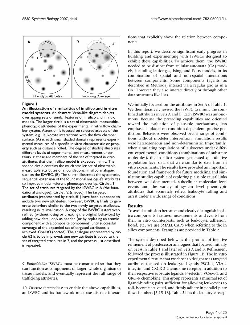
An illustration of similarities of in silico and in vitro model systemsFigure 1An illustration of similarities of in silico and in vitro model systems. An abstract, Venn-like diagram depicts overlapping sets of similar features of in silico and in vitro models. The larger circle is a set of observable, measurable, phenotypic attributes of the experimental in vitro flow cham-ber system. Attention is focused on selected aspects of the system, e.g., leukocyte interactions with the flow chamber surface. (A) a: each small shaded domain represents experi-mental measures of a specific in vitro characteristic or prop-erty such as distance rolled. The degree of shading illustrates different levels of experimental and measurement uncer-tainty. t: these are members of the set of targeted in vitro attributes that the in silico model is expected mimic. The shaded circle contains the much smaller set of observable, measurable attributes of a foundational in silico analogue, such as the ISWBC. (B) The sketch illustrates the systematic, sequential extension of the foundational analogue's attributes to improve model-referent phenotype overlap. Circle #1: The set of attributes targeted by the ISWBC in A (the foun-dational analogue). Circle #2 (shaded): The targeted attributes (represented by circle #1) have been expanded to include two new attributes; however, ISWBC #1 fails to gen-erate behaviors similar to the two newly targeted attributes, resulting in its invalidation. A copy of the ISWBC is iteratively refined (without losing or breaking the original behaviors) by adding new detail only as needed (or by replacing an atomic component with a composite component) until successful coverage of the expanded set of targeted attributes is achieved. Oval #3 (dotted): The analogue represented by cir-cle #2 is to be improved: one new attribute is added to the set of targeted attributes in 2, and the process just described is repeated.
Page 4 of 25(page number not for citation purposes)
BMC Systems Biology 2007, 1:14 http://www.biomedcentral.com/1752-0509/1/14
tors represented, their corresponding ligands, and thebehaviors they support.
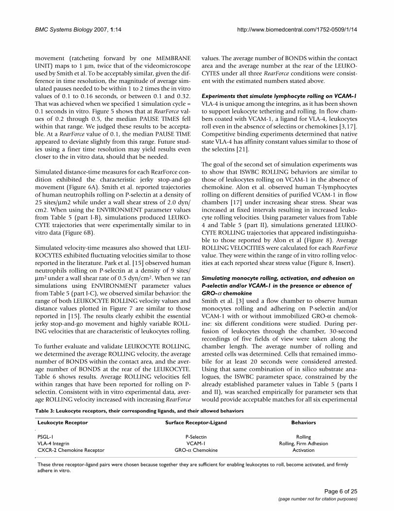
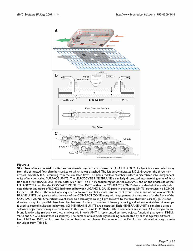
The system illustrated in Figure 2A has been designed torepresent an in vitro flow chamber (Figure 2B). The exper-iments using it are analogous to those performed using anin vitro system (four examples are listed below). The SUR-FACE of the simulated chamber is "coated" with a uni-form density of the LIGANDS of interest. A LIGAND is acomponent that represents a number of ligands foundwithin a discrete portion (see MEMBRANE UNIT below)of a leukocyte membrane or the flow chamber surface.While in the FLOW CHAMBER, LEUKOCYTES use theirLIGANDS to interact and form BONDS with the SUB-STRATE-COATED SURFACE. Each BOND is an in silicoanalogue of a single ligand-ligand bond. Those interac-tions are recorded and measured. Under the influence ofsimulated flow, BONDS at the rear of the CONTACTZONE experience a simulated cumulative tensile forcedue to shear; it is controlled by the parameter RearForce.We learned from earlier, simpler analogues that adynamic decisional process of the type diagrammed inFigure 3, was needed at two levels: LEUKOCYTE/MEM-BRANE and MEMBRANE UNIT. As described in Methods,the SURFACE, LEUKOCYTE, and MEMBRANE are sepa-rate objects. A MEMBRANE UNIT is an analogue of a func-tional unit of leukocyte membrane within the CONTACTZONE. There is a corresponding SURFACE UNIT. Thenumber of UNITS is a discretization decision. How fine orcourse does the discretization (granularity) need to be inorder to avoid discretization artifacts and achieve the tar-geted behaviors? As shown below, a level of discretizationis reached at which an acceptable match to targeted behav-iors is achieved; further increasing granularity does notimprove the quality of the match.
We investigated the ISWBC's ability to represent in vitromeasures of rolling and adhesion by comparing simula-tion results with data from three different flow chamberexperiments:
(1) Smith et al. [16] and Park et al. [15] observed humanneutrophils rolling on various densities of P-selectin andunder varying wall shear rates.
(2) Alon et al. [17] recorded human lymphocyte-rollingtrajectories on VCAM-1 in the presence of increasing wallshear rates at fixed time intervals.
(3) Monocyte rolling and adhesion on P-selectin and/orVCAM-1 in the presence or absence of GRO-α chemokinewas studied by Smith et al. [3].
Simulating neutrophil rolling on P-selectinThe in vitro data suggests that leukocyte rolling is medi-ated by small numbers of receptor-ligand bonds formedwithin the contact zone. Mathematical model estimates ofthis number range between two and twenty [19,20]. Sin-gle bond events are believed to cause rolling behaviorssimilar to what one might expect of a stochastic process.There are two easily recognized manifestations: jerky stop-and-go patterns and highly fluctuating rolling velocities[2]. Given the importance of single bond events, eachISWBC BOND maps to a single ligand-ligand bond. How-ever, a LIGAND can map to more than one ligand mole-cule.
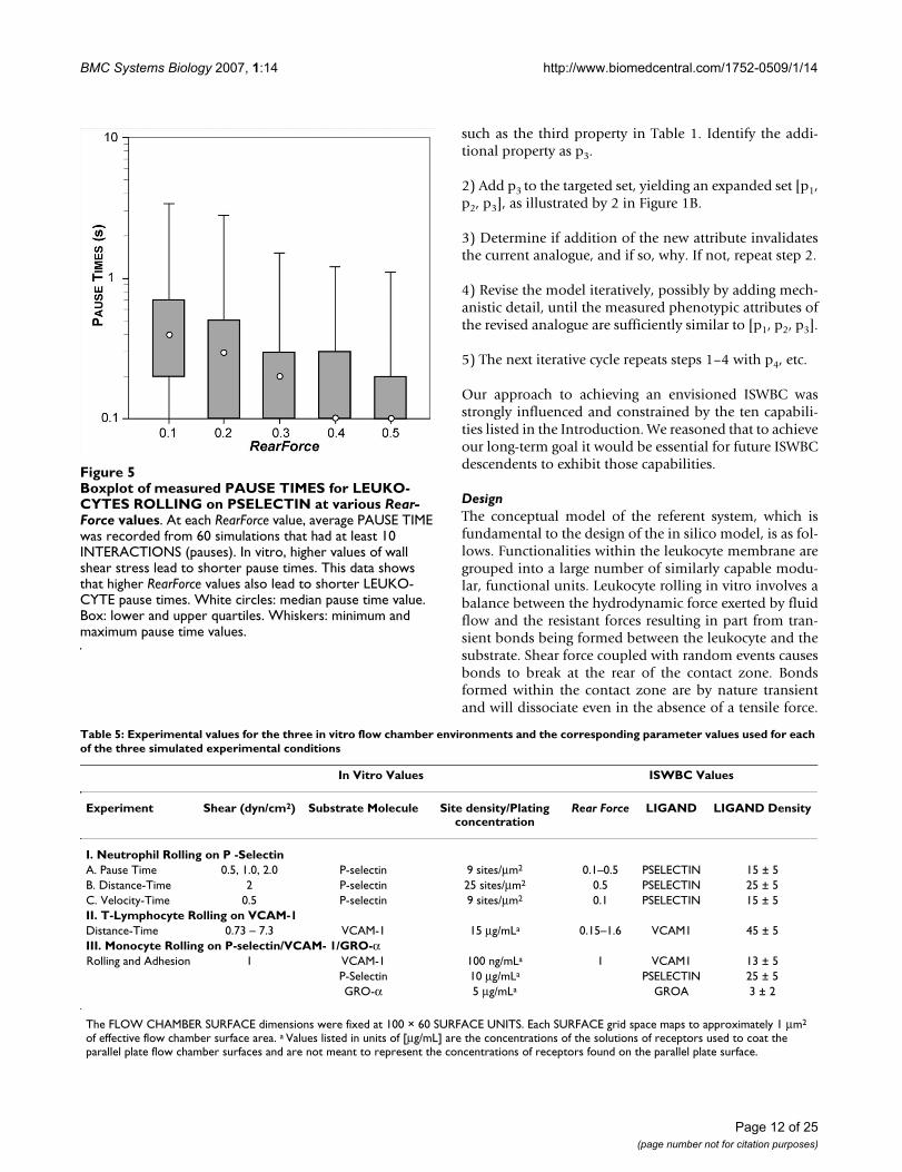
Two observations considered together, pause times andforward movement during rolling, required that the upperlimit for the mapping of simulation cycle to in vitro timebe about 1 simulation cycle = 0.1 seconds. Pause timemeasurements, the first targeted attribute, were made overa small range of shear values as reported by Smith et al.[16] for human neutrophils rolling on a P-selectin-coatedsurface. Using high-speed videomicroscopy with a small-est resolvable step size of 0.5 μm, average pause timeswere in the range of 0.1 to 0.16 seconds for shear stressvalues of 0.5 to 2.0 dyn/cm2. Higher wall shear stress val-ues caused shorter average pause times.
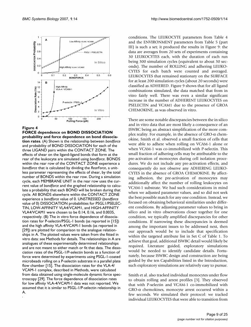
We defined LEUKOCYTE ROLLING as a sequence of atleast 10 simulation cycles during which a LEUKOCYTEremained non-stationary on the SURFACE. During ROLL-ING, ISWBCs used the relationship between LIGAND dis-association probability and bondforce graphed in Figure4A. Bondforce is an in silico analogue of the force experi-enced by each BOND within the contact zone. Whenparameterized according to Table 4, using the values listedin Table 5 (part I-A), the simulation's smallest forward
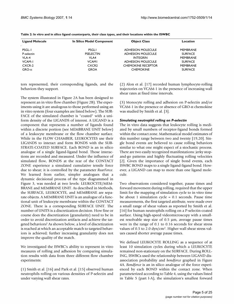
Table 2: In vitro and in silico ligand counterparts, their class types, and their locations within the ISWBC
Ligand Molecule In Silico Model Component Object Class Location
PSGL-1 PSGL1 ADHESION MOLECULE MEMBRANEP-selectin PSELECTIN ADHESION MOLECULE SURFACEVLA-4 VLA4 INTEGRIN MEMBRANEVCAM-1 VCAM1 ADHESION MOLECULE SURFACECXCR-2 CXCR2 CHEMOKINE RECEPTOR MEMBRANEGRO-α GROA CHEMOKINE SURFACE
Page 5 of 25(page number not for citation purposes)
BMC Systems Biology 2007, 1:14 http://www.biomedcentral.com/1752-0509/1/14
movement (ratcheting forward by one MEMBRANEUNIT) maps to 1 μm, twice that of the videomicroscopeused by Smith et al. To be acceptably similar, given the dif-ference in time resolution, the magnitude of average sim-ulated pauses needed to be within 1 to 2 times the in vitrovalues of 0.1 to 0.16 seconds, or between 0.1 and 0.32.That was achieved when we specified 1 simulation cycle =0.1 seconds in vitro. Figure 5 shows that at RearForce val-ues of 0.2 through 0.5, the median PAUSE TIMES fellwithin that range. We judged these results to be accepta-ble. At a RearForce value of 0.1, the median PAUSE TIMEappeared to deviate slightly from this range. Future stud-ies using a finer time resolution may yield results evencloser to the in vitro data, should that be needed.
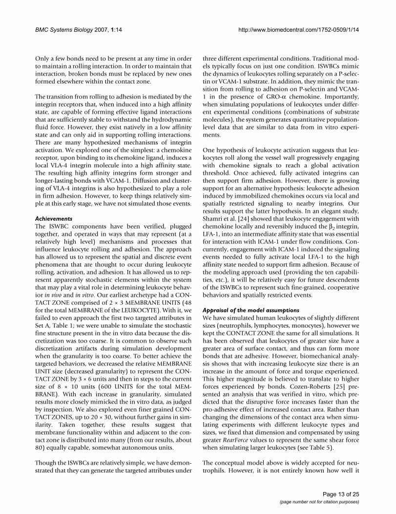
Simulated distance-time measures for each RearForce con-dition exhibited the characteristic jerky stop-and-gomovement (Figure 6A). Smith et al. reported trajectoriesof human neutrophils rolling on P-selectin at a density of25 sites/μm2 while under a wall shear stress of 2.0 dyn/cm2. When using the ENVIRONMENT parameter valuesfrom Table 5 (part I-B), simulations produced LEUKO-CYTE trajectories that were experimentally similar to invitro data (Figure 6B).
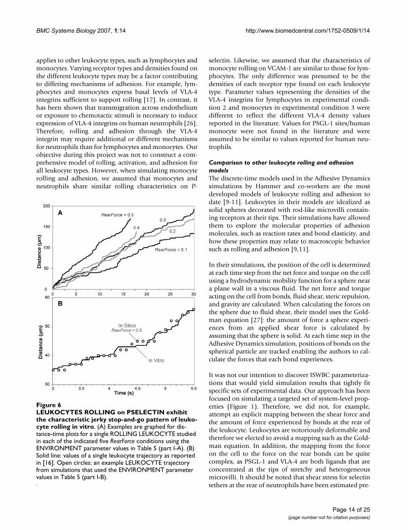
Simulated velocity-time measures also showed that LEU-KOCYTES exhibited fluctuating velocities similar to thosereported in the literature. Park et al. [15] observed humanneutrophils rolling on P-selectin at a density of 9 sites/μm2 under a wall shear rate of 0.5 dyn/cm2. When we ransimulations using ENVIRONMENT parameter valuesfrom Table 5 (part I-C), we observed similar behavior: therange of both LEUKOCYTE ROLLING velocity values anddistance values plotted in Figure 7 are similar to thosereported in [15]. The results clearly exhibit the essentialjerky stop-and-go movement and highly variable ROLL-ING velocities that are characteristic of leukocytes rolling.
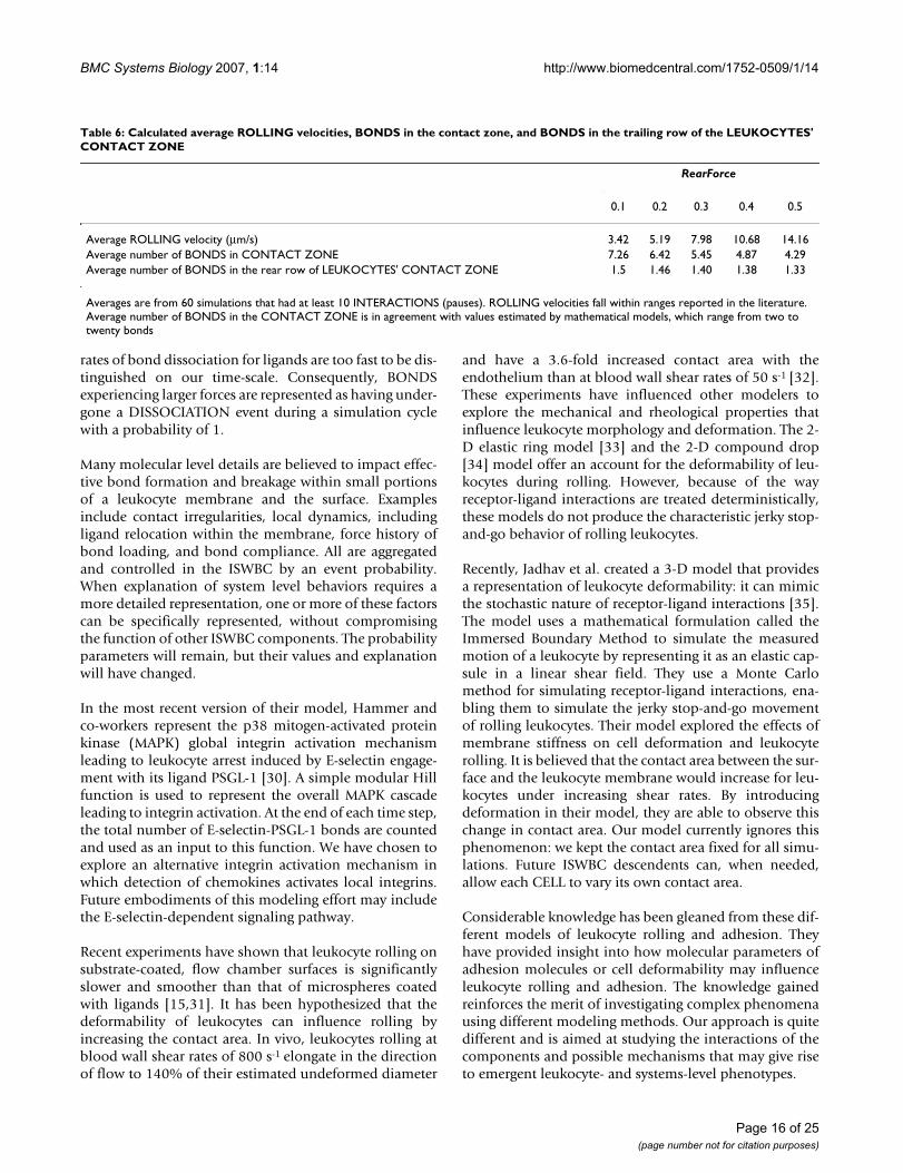
To further evaluate and validate LEUKOCYTE ROLLING,we determined the average ROLLING velocity, the averagenumber of BONDS within the contact area, and the aver-age number of BONDS at the rear of the LEUKOCYTE.Table 6 shows results. Average ROLLING velocities fellwithin ranges that have been reported for rolling on P-selectin. Consistent with in vitro experimental data, aver-age ROLLING velocity increased with increasing RearForce
values. The average number of BONDS within the contactarea and the average number at the rear of the LEUKO-CYTES under all three RearForce conditions were consist-ent with the estimated numbers stated above.
Experiments that simulate lymphocyte rolling on VCAM-1VLA-4 is unique among the integrins, as it has been shownto support leukocyte tethering and rolling. In flow cham-bers coated with VCAM-1, a ligand for VLA-4, leukocytesroll even in the absence of selectins or chemokines [3,17].Competitive binding experiments determined that nativestate VLA-4 has affinity constant values similar to those ofthe selectins [21].
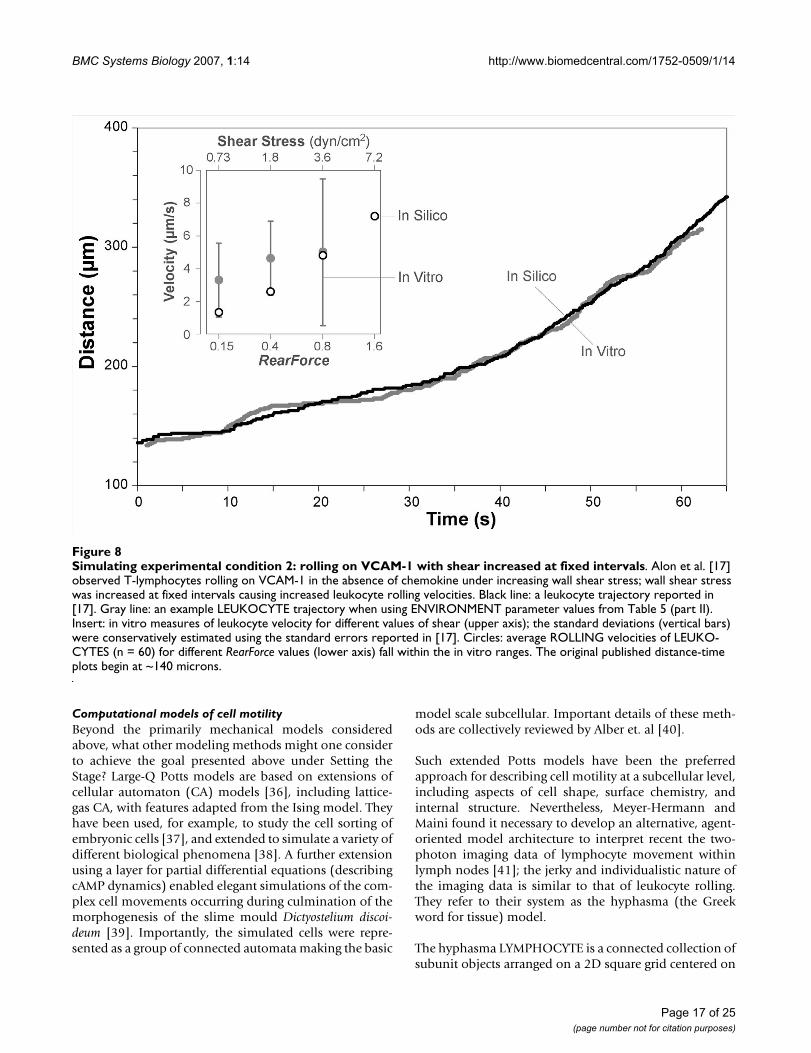
The goal of the second set of simulation experiments wasto show that ISWBC ROLLING behaviors are similar tothose of leukocytes rolling on VCAM-1 in the absence ofchemokine. Alon et al. observed human T-lymphocytesrolling on different densities of purified VCAM-1 in flowchambers [17] under increasing shear stress. Shear wasincreased at fixed intervals resulting in increased leuko-cyte rolling velocities. Using parameter values from Table4 and Table 5 (part II), simulations generated LEUKO-CYTE ROLLING trajectories that appeared indistinguisha-ble to those reported by Alon et al (Figure 8). AverageROLLING VELOCITIES were calculated for each RearForcevalue. They were within the range of in vitro rolling veloc-ities at each reported shear stress value (Figure 8, Insert).
Simulating monocyte rolling, activation, and adhesion on P-selectin and/or VCAM-1 in the presence or absence of GRO-α chemokineSmith et al. [3] used a flow chamber to observe humanmonocytes rolling and adhering on P-selectin and/orVCAM-1 with or without immobilized GRO-α chemok-ine: six different conditions were studied. During per-fusion of leukocytes through the chamber, 30-secondrecordings of five fields of view were taken along thechamber length. The average number of rolling andarrested cells was determined. Cells that remained immo-bile for at least 20 seconds were considered arrested.Using that same combination of in silico substrate ana-logues, the ISWBC parameter space, constrained by thealready established parameter values in Table 5 (parts Iand II), was searched empirically for parameter sets thatwould provide acceptable matches for all six experimental
Table 3: Leukocyte receptors, their corresponding ligands, and their allowed behaviors
Leukocyte Receptor Surface Receptor-Ligand Behaviors
PSGL-1 P-Selectin RollingVLA-4 Integrin VCAM-1 Rolling, Firm AdhesionCXCR-2 Chemokine Receptor GRO-α Chemokine Activation
These three receptor-ligand pairs were chosen because together they are sufficient for enabling leukocytes to roll, become activated, and firmly adhere in vitro.
Page 6 of 25(page number not for citation purposes)
BMC Systems Biology 2007, 1:14 http://www.biomedcentral.com/1752-0509/1/14
Page 7 of 25(page number not for citation purposes)
Sketches of in vitro and in silico experimental system componentsFigure 2Sketches of in vitro and in silico experimental system components. (A) A LEUKOCYTE object is shown pulled away from the simulated flow chamber surface to which it was attached. The left arrow indicates ROLL direction; the three right arrows indicate SHEAR resulting from the simulated flow. The simulated flow chamber surface is discretized into independent units of function called SURFACE UNITS. The LEUKOCYTE'S MEMBRANE is similarly discretized into matching units of func-tion called MEMBRANE UNITS: 600 total (20 × 30). The 8 × 10-shaded region on the SURFACE and on the underside of the LEUKOCYTE identifies the CONTACT ZONE. The UNITS within the CONTACT ZONES that are shaded differently indi-cate different numbers of BONDS had formed between LIGAND-LIGAND pairs in overlapping UNITS; otherwise, no BONDS formed. ROLLING is the result of a sequence of forward ratchet events. One ratchet event is the result of one row of MEM-BRANE UNITS being released at the rear of the CONTACT ZONE along with engagement of a new row of at the front of the CONTACT ZONE. One ratchet event maps to a leukocyte rolling 1 μm (relative to the flow chamber surface). (B) A shop drawing of a typical parallel plate flow chamber used for in vitro studies of leukocyte rolling and adhesion. A video microscope is used to record leukocyte behaviors. (C) MEMBRANE UNITS are illustrated. Each MEMBRANE UNIT is simulated using a software object functioning as a container. In this sketch, nine MEMBRANE UNIT containers are shown. All leukocyte mem-brane functionality (relevant to these studies) within each UNIT is represented by three objects functioning as agents: PSGL1, VLA4 and CXCR2 (illustrated as spheres). The number of leukocyte ligands being represented by each is typically different from UNIT to UNIT, as illustrated by the numbers on the spheres. That number is specified for each simulation using parame-ter values from Table 3.
BMC Systems Biology 2007, 1:14 http://www.biomedcentral.com/1752-0509/1/14
Page 8 of 25(page number not for citation purposes)
The decisional process for the LEUKOCYTE MEMBRANE and each MEMBRANE UNIT during a simulation cycleFigure 3The decisional process for the LEUKOCYTE MEMBRANE and each MEMBRANE UNIT during a simulation cycle. (A) The LEUKOCYTE steps through its decisional process only once during a simulation cycle. At the start of the cycle, the MEMBRANE instructs all MEMBRANE UNITS within the CONTACT ZONE to follow the decisional process in B. Once that process is complete, the MEMBRANE completes its process by selecting and following the one applicable action option. (B) A MEMBRANE UNIT is described and illustrated in Figure 2. The state of each depends on the properties of the three LIG-AND objects contained within. During each simulation cycle, each MEMBRANE UNIT, selected at random, uses this decisional process to update its status relative to the SURFACE UNIT over which it is positioned. (C) The hierarchical organization of the ISWBC system is illustrated. There are six levels. An Experiment Agent exists within the system, but separate from the FLOW CHAMBER and LEUKOCYTE. It represents a researcher conducting wet-lab experiments: it measures and records events and behaviors during each simulation.
BMC Systems Biology 2007, 1:14 http://www.biomedcentral.com/1752-0509/1/14
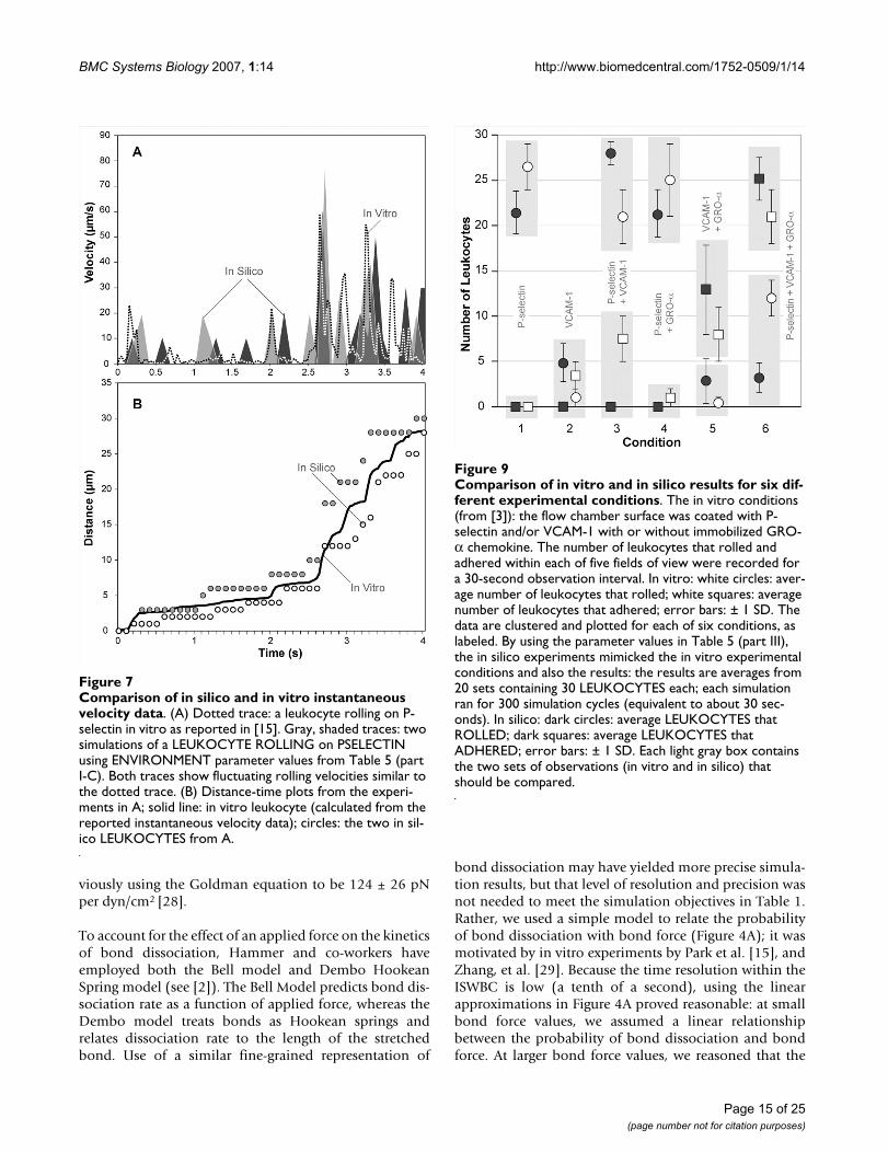
conditions. The LEUKOCYTE parameters from Table 4and the ENVIRONMENT parameters from Table 5 (partIII) is such a set; it produced the results in Figure 9: thedata are averages from 20 sets of experiments containing30 LEUKOCYTES each, with the duration of each runbeing 300 simulation cycles (equivalent to about 30 sec-onds). The number of ROLLING and adhering LEUKO-CYTES for each batch were counted and averaged.LEUKOCYTES that remained stationary on the SURFACEfor at least 200 simulation cycles (about 20 seconds) wereclassified as ADHERED. Figure 9 shows that for all ligandcombinations simulated, the data matched that from invitro fairly well. There was even a similar significantincrease in the number of ADHERENT LEUKOCYTES onPSELECTIN and VCAM1 due to the presence of GROACHEMOKINE, as was observed in vitro.
There are some notable discrepancies between the in silicoand in vitro data that are most likely a consequence of anISWBC being an abstract simplification of the more com-plex reality. For example, in the absence of GRO-α chem-okine, Smith et al. observed a small fraction of cells thatwere able to adhere when rolling on VCAM-1 alone orwhen VCAM-1 was co-immobilized with P-selectin. Thissmall fraction of adhering cells may be attributable to thepre-activation of monocytes during cell isolation proce-dures. We do not include any pre-activation effects, andconsequently do not observe any ADHERING LEUKO-CYTES in the absence of GROA CHEMOKINE. By affect-ing adhesion, the pre-activation of monocytes mayadditionally affect the number of rolling leukocytes onVCAM-1 substrate. We had such considerations in mindwhen we adjusted parameter values, and so did not seekthe best possible match for any one condition. Instead, wefocused on obtaining behavioral similarities under differ-ent conditions. By adjusting parameter values to bring insilico and in vitro observations closer together for onecondition, we typically amplified discrepancies for otherconditions. If narrowing these discrepancies is deemedamong the important issues to be addressed next, thenour approach would be to include that specificationwithin the targeted attribute list in Set C of Table 1. Toachieve that goal, additional ISWBC detail would likely berequired. Literature guided, exploratory simulationswould be needed to identify candidate details. Fortu-nately, because ISWBC design and construction are beingguided by the ten Capabilities listed in the Introduction,such exploratory simulations are relatively easy to pursue.
Smith et al. also tracked individual monocytes under flowto obtain rolling and arrest profiles [3]. They observedthat with P-selectin and VCAM-1 co-immobilized withGRO-α chemokines, monocyte arrest occurred within afew seconds. We simulated their protocol: we trackedindividual LEUKOCYTES that were able to transition from
FORCE dependence on BOND DISSOCIATION probability and force dependence on bond dissociation ratesFigure 4FORCE dependence on BOND DISSOCIATION probability and force dependence on bond dissocia-tion rates. (A) Shown is the relationship between bondforce and probability of BOND DISSOCIATION for each of the three LIGAND pairs within the CONTACT ZONE. The effects of shear on the ligand-ligand bonds that form at the rear of the leukocyte are simulated using bondforce. BONDS within the rear row of the CONTACT ZONE experience a bondforce that is calculated by dividing the RearForce, a unit-less parameter representing the effects of shear, by the total number of BONDS within the rear row. During a simulation cycle, each MEMBRANE UNIT in the rear row uses the cur-rent value of bondforce and the graphed relationship to calcu-late a probability that each BOND will be broken during that cycle. All BONDS elsewhere within the CONTACT ZONE experience a bondforce value of 0. UNSTRESSED (bondforce value of 0) DISSOCIATION probabilities for PSGL1/PSELEC-TIN, LOW-AFFINITY VLA4/VCAM1, and HIGH-AFFINITY VLA4/VCAM1 were chosen to be 0.14, 0.16, and 0.0035, respectively. (B) The in vitro force dependence of dissocia-tion rates for P-selectin/PSGL-1 bonds (as reported in [15]) and the high affinity VLA-4/VCAM-1 bonds (as reported in [29]) are plotted for comparison to the analogue relation-ships in A. The plotted values were taken from the fitted in vitro data: see Methods for details. The relationships in A are analogues of these experimentally determined relationships and are not meant to either match or fit that data. The disso-ciation rates of the PSGL-1/P-selectin bonds as a function of force were determined by experiments using PSGL-1-coated microbeads rolling on a P-selectin substrate in a parallel plate flow chamber [15]. The dissociation rates for the VLA-4/VCAM-1 complex, described in Methods, were calculated from data obtained using single-molecule dynamic force spec-troscopy [29]. The force dependence of dissociation rates for low affinity VLA-4/VCAM-1 data was not reported. We assumed that it is similar to PSGL-1/P-selectin relationship in A.
Page 9 of 25(page number not for citation purposes)
BMC
Sys
tem
s Bi
olog
y 20
07, 1
:14
http
://w
ww
.bio
med
cent
ral.c
om/1
752-
0509
/1/1
4
Page
10
of 2
5(p
age
num
ber n
ot fo
r cita
tion
purp
oses
)
Table 4: Model parameter values for the LEUKOCYTE MEMBRANE and LIGANDS along with corresponding in vitro values
Parameter Name Description Model Parameter Value Experimental Value Reference
LeukTotalWidth LEUKOCYTE MEMBRANE width (in the y [east-west] dimension) 20 MEMBRANE UNITS Avg. Human Leukocyte Diameters (μm):
Lymphocyte: 6.2; Neutrophil: 7; Monocyte: 7.5
[48]
LeukTotalLength LEUKOCYTE MEMBRANE length (in the x [north-south] dimension) 30 MEMBRANE UNITS
LeukExposedWidth CONTACT ZONE width (in the y dimension) 8 MEMBRANE UNITS NA NA
LeukExposedLength CONTACT ZONE length (in the x dimension) 10 MEMBRANE UNITS NA NA
PSGL1DensityMean ± PSGL1DensitySTDev
Mean number of PSGL-1 molecules (± SD) represented by each PSGL1 agent
Experiment 1: Neutrophils 150 ± 5a molecules ~18,000/Human Neutrophil [54]
Experiment 3: Monocytes 150 ± 5b molecules N/A N/A
VLA4DensityMean ± VLA4DensitySTDev
Mean number of VLA-4 molecules (± SD) represented by each VLA4 agent
Experiment 2: T-lymphocytes 45 ± 5 molecules 3,000/Human T-Lymphocyte [55]
Experiment 3: Monocytes 60 ± 5 molecules 6,000/Human Monocyte
CXCR2DensityMean ± CXCR2DensitySTDev
Number of CXCR-2 molecules (± standard deviation) represented by each CXCR2 agent 1 ± 0 molecules NA NA
VLA4MaxPercHighAff Maximum percent of VLA-4 integrins on the leukocyte membrane that can be induced into a high affinity statec
12.5% 10% [49]
Pon (PSGL1-PSELECTIN1)
Probability of forming a PSGL1-PSELECTIN1 BOND 0.001 NAd [56]
Pon (LOW AFFINITY VLA4-VCAM1)
Probability of forming a LOW AFFINITY VLA4-VCAM1 BOND 0.001 NAd [55]
Pon (HIGH AFFINITY VLA4-VCAM1)
Probability of forming a HIGH AFFINITY VLA4-VCAM1 BOND 0.005 NAd [55]
Pon (CXCR2-GROA) Probability of forming a CXCR2-GROA BOND 1.0c NA NA
Poff (CXCR2-GROA) Probability of breaking a CXCR2-GROA BOND 1.0c NA NA
a PSGL1DensityMean ± PSGL1DensitySTDev parameter values used for simulating Experiment 1, neutrophil rolling, were originally 25 ± 5. They were changed to the values currently listed to more closely reflect the experimental values of PSGL-1 density reported in the literature. No significant differences were observed when using these new parameter values.b Values for PSGL-1 sites/human monocyte were not found in the literature and were assumed to be similar to values reported for human neutrophils.c See Methods.d Pon and Kon are intended to map to aspects of the same in vitro phenomena. However, there is no direct mapping between these parameters because the parent models belong to fundamentally different classes [57].
BMC Systems Biology 2007, 1:14 http://www.biomedcentral.com/1752-0509/1/14
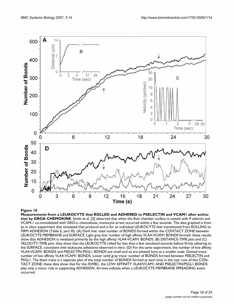
ROLLING to FIRM ADHESION. We also observed thatFIRM ADHESION on PSELECTIN and VCAM1 in the pres-ence of GROA CHEMOKINE was primarily mediated byhigh affinity VLA4 and that it occurred within SECONDS.Figure 10 shows an example LEUKOCYTE that is able toADHERE after ROLLING.
Discussions and conclusionSetting the stageA long-term biological goal for this project is to developscientifically useful, validated simulations of leukocyterecruitment under physiological and pathophysiologicalconditions. The envisioned LEUKOCYTES, functioningwithin an analogue of the wet-lab context, will have manybehaviors, properties, and characteristics that mimic thoseof referent in vitro and in vivo systems. Stated differently,there will be similarities between leukocyte phenotypeand the "phenotype" of simulated leukocytes, as illus-trated in Figure 1. The expectation is that increasing phe-notype similarity will require, and can be achieved in partthrough, similarities in design plan and in generativemechanisms. Once they are achieved, the model will havebecome a scientifically useful analogue.
Our approach to designing and building the envisionedanalogues is motivated by aspect-oriented software devel-opment and the middle-out approach first suggested byBrenner [22] and later detailed by Noble [23] for buildingmodels of complex biological systems. We adapted andmerged these ideas in arriving at our working definition ofmiddle-out modeling. First, specify the biological featuresunder study (e.g., cell rolling and attachment to a surface).Each feature becomes an aspect of a software device – anarchetype model. That archetype is iteratively transformedinto a functioning analogue of the referent in vitro biolog-ical system. It is then validated and improved iteratively.
Following that approach, our first task was to pick a set ofproperties, p1, p2, ... pj (within a hyperspace of mechanis-tic properties) – around leukocyte rolling and adhesion invitro. We specified Set A, Table 1. We next asked what soft-ware device could we implement to realize p1? How canwe realize p2, etc.? We then asked, how can we realize p1-pj, all at the same time, using the same software device?Getting answers to those questions required exploratorymodeling and simulation, occasionally using differentmodeling and simulation support packages. The in silicoproperties and the device we created to realize p1-pjformed a foundational analogue, the above archetypemodel, from which we intended to expand outward (up,down, and even sideways) to establish a reductive hierar-chy of components that could be used to identify impor-tant systems biology principles.
Creating an analogue phenotypeThe ISWBC is an abstract model of a leukocyte, and whenplaced in an appropriate, simulated environment, it iscapable of mimicking a set of targeted characteristics. TheISWBC exists within a system that is a model of an in vitroflow chamber system (cells, media, the device in Figure2B, atmosphere, etc.). The ISWBC is a multilevel compos-ite model. The components are independent models oftheir referent leukocyte or environment counterpart. Forexample, the in silico component representing the MEM-BRANE UNITS in a portion of the trailing edge of a ROLL-ING LEUKOCYTE is itself a model of the correspondingleukocyte feature.
The envisioned relationship between measured LEUKO-CYTE behaviors and corresponding in vitro measures isillustrated in Figure 1. Pictured are overlapping (but notintersecting) sets. One set (Figure 1, large circle) containsthe results of experiments that measured specific pheno-typic attributes, properties, or characteristics of leukocytesin vitro (e.g., all three sets in Table 1). The smaller setscontain the results of simulation experiments that meas-ure corresponding in silico phenotypic attributes, proper-ties, or characteristics (such as Set A, Table 1). Ourmotivating hypothesis has been that, when attention isfocused on the same features, and the measured behaviorsof the two sets are similar, then there may also be usefulsimilarities in the generative mechanisms of the two sys-tems. Those similarities can be explored by iterative test-ing and refinement of the analogue coupled with relatedwet-lab experiments. An example: distance traveled by aLEUKOCYTE in silico (or a leukocyte in vitro) under vari-ous simulated (or actual) shear stresses. We cannot expectthese measurements to overlap completely or even havethe same units. Observations on behavior similarity canbe used simultaneously to eliminate invalid model fea-tures and identify gaps in our knowledge of the experi-mental in vitro system.
The first step in transitioning from an archetype to animproved LEUKOCYTE was to thoroughly document thein silico properties and characteristics of the former. Bydoing so, we improved insight into its strengths and weak-nesses. The next step was to obtain increasing overlap withmeasures of in vitro attributes. That was done systemati-cally by iteratively revising together the hypothesis andthe analogue by following the five steps below. An exam-ple hypothesis: the current analogue can acceptably simu-late the first two attributes [p1, p2,] in Set A, Table 1.Figures 7 and 8 are examples of successful simulations. Webegin the next iterative cycle with step 1.
1) Select an additional in vitro property or characteristicthat is related to those already in the target set, and forwhich wet-lab experimental observations are available,
Page 11 of 25(page number not for citation purposes)
BMC Systems Biology 2007, 1:14 http://www.biomedcentral.com/1752-0509/1/14
such as the third property in Table 1. Identify the addi-tional property as p3.
2) Add p3 to the targeted set, yielding an expanded set [p1,p2, p3], as illustrated by 2 in Figure 1B.
3) Determine if addition of the new attribute invalidatesthe current analogue, and if so, why. If not, repeat step 2.
4) Revise the model iteratively, possibly by adding mech-anistic detail, until the measured phenotypic attributes ofthe revised analogue are sufficiently similar to [p1, p2, p3].
5) The next iterative cycle repeats steps 1–4 with p4, etc.
Our approach to achieving an envisioned ISWBC wasstrongly influenced and constrained by the ten capabili-ties listed in the Introduction. We reasoned that to achieveour long-term goal it would be essential for future ISWBCdescendents to exhibit those capabilities.
DesignThe conceptual model of the referent system, which isfundamental to the design of the in silico model, is as fol-lows. Functionalities within the leukocyte membrane aregrouped into a large number of similarly capable modu-lar, functional units. Leukocyte rolling in vitro involves abalance between the hydrodynamic force exerted by fluidflow and the resistant forces resulting in part from tran-sient bonds being formed between the leukocyte and thesubstrate. Shear force coupled with random events causesbonds to break at the rear of the contact zone. Bondsformed within the contact zone are by nature transientand will dissociate even in the absence of a tensile force.
Boxplot of measured PAUSE TIMES for LEUKOCYTES ROLLING on PSELECTIN at various RearForce valuesFigure 5Boxplot of measured PAUSE TIMES for LEUKO-CYTES ROLLING on PSELECTIN at various Rear-Force values. At each RearForce value, average PAUSE TIME was recorded from 60 simulations that had at least 10 INTERACTIONS (pauses). In vitro, higher values of wall shear stress lead to shorter pause times. This data shows that higher RearForce values also lead to shorter LEUKO-CYTE pause times. White circles: median pause time value. Box: lower and upper quartiles. Whiskers: minimum and maximum pause time values.
Table 5: Experimental values for the three in vitro flow chamber environments and the corresponding parameter values used for each of the three simulated experimental conditions
In Vitro Values ISWBC Values
Experiment Shear (dyn/cm2) Substrate Molecule Site density/Plating concentration
Rear Force LIGAND LIGAND Density
I. Neutrophil Rolling on P -SelectinA. Pause Time 0.5, 1.0, 2.0 P-selectin 9 sites/μm2 0.1–0.5 PSELECTIN 15 ± 5B. Distance-Time 2 P-selectin 25 sites/μm2 0.5 PSELECTIN 25 ± 5C. Velocity-Time 0.5 P-selectin 9 sites/μm2 0.1 PSELECTIN 15 ± 5II. T-Lymphocyte Rolling on VCAM-1Distance-Time 0.73 – 7.3 VCAM-1 15 μg/mLa 0.15–1.6 VCAM1 45 ± 5III. Monocyte Rolling on P-selectin/VCAM- 1/GRO-αRolling and Adhesion 1 VCAM-1 100 ng/mLa 1 VCAM1 13 ± 5
P-Selectin 10 μg/mLa PSELECTIN 25 ± 5GRO-α 5 μg/mLa GROA 3 ± 2
The FLOW CHAMBER SURFACE dimensions were fixed at 100 × 60 SURFACE UNITS. Each SURFACE grid space maps to approximately 1 μm2
of effective flow chamber surface area. a Values listed in units of [μg/mL] are the concentrations of the solutions of receptors used to coat the parallel plate flow chamber surfaces and are not meant to represent the concentrations of receptors found on the parallel plate surface.
Page 12 of 25(page number not for citation purposes)
BMC Systems Biology 2007, 1:14 http://www.biomedcentral.com/1752-0509/1/14
Only a few bonds need to be present at any time in orderto maintain a rolling interaction. In order to maintain thatinteraction, broken bonds must be replaced by new onesformed elsewhere within the contact zone.
The transition from rolling to adhesion is mediated by theintegrin receptors that, when induced into a high affinitystate, are capable of forming effective ligand interactionsthat are sufficiently stable to withstand the hydrodynamicfluid force. However, they exist natively in a low affinitystate and can only aid in supporting rolling interactions.There are many hypothesized mechanisms of integrinactivation. We explored one of the simplest: a chemokinereceptor, upon binding to its chemokine ligand, induces alocal VLA-4 integrin molecule into a high affinity state.The resulting high affinity integrins form stronger andlonger-lasting bonds with VCAM-1. Diffusion and cluster-ing of VLA-4 integrins is also hypothesized to play a rolein firm adhesion. However, to keep things relatively sim-ple at this early stage, we have not simulated those events.
AchievementsThe ISWBC components have been verified, pluggedtogether, and operated in ways that may represent (at arelatively high level) mechanisms and processes thatinfluence leukocyte rolling and adhesion. The approachhas allowed us to represent the spatial and discrete eventphenomena that are thought to occur during leukocyterolling, activation, and adhesion. It has allowed us to rep-resent apparently stochastic elements within the systemthat may play a vital role in determining leukocyte behav-ior in vivo and in vitro. Our earliest archetype had a CON-TACT ZONE comprised of 2 × 3 MEMBRANE UNITS (48for the total MEMBRANE of the LEUKOCYTE). With it, wefailed to even approach the first two targeted attributes inSet A, Table 1; we were unable to simulate the stochasticfine structure present in the in vitro data because the dis-cretization was too coarse. It is common to observe suchdiscretization artifacts during simulation developmentwhen the granularity is too coarse. To better achieve thetargeted behaviors, we decreased the relative MEMBRANEUNIT size (decreased granularity) to represent the CON-TACT ZONE by 3 × 6 units and then in steps to the currentsize of 8 × 10 units (600 UNITS for the total MEM-BRANE). With each increase in granularity, simulatedresults more closely mimicked the in vitro data, as judgedby inspection. We also explored even finer grained CON-TACT ZONES, up to 20 × 30, without further gains in sim-ilarity. Taken together, these results suggest thatmembrane functionality within and adjacent to the con-tact zone is distributed into many (from our results, about80) equally capable, somewhat autonomous units.
Though the ISWBCs are relatively simple, we have demon-strated that they can generate the targeted attributes under
three different experimental conditions. Traditional mod-els typically focus on just one condition. ISWBCs mimicthe dynamics of leukocytes rolling separately on a P-selec-tin or VCAM-1 substrate. In addition, they mimic the tran-sition from rolling to adhesion on P-selectin and VCAM-1 in the presence of GRO-α chemokine. Importantly,when simulating populations of leukocytes under differ-ent experimental conditions (combinations of substratemolecules), the system generates quantitative population-level data that are similar to data from in vitro experi-ments.
One hypothesis of leukocyte activation suggests that leu-kocytes roll along the vessel wall progressively engagingwith chemokine signals to reach a global activationthreshold. Once achieved, fully activated integrins canthen support firm adhesion. However, there is growingsupport for an alternative hypothesis: leukocyte adhesioninduced by immobilized chemokines occurs via local andspatially restricted signaling to nearby integrins. Ourresults support the latter hypothesis. In an elegant study,Shamri et al. [24] showed that leukocyte engagement withchemokine locally and reversibly induced the β2 integrin,LFA-1, into an intermediate affinity state that was essentialfor interaction with ICAM-1 under flow conditions. Con-currently, engagement with ICAM-1 induced the signalingevents needed to fully activate local LFA-1 to the highaffinity state needed to support firm adhesion. Because ofthe modeling approach used (providing the ten capabili-ties, etc.), it will be relatively easy for future descendentsof the ISWBCs to represent such fine-grained, cooperativebehaviors and spatially restricted events.
Appraisal of the model assumptionsWe have simulated human leukocytes of slightly differentsizes (neutrophils, lymphocytes, monocytes), however wekept the CONTACT ZONE the same for all simulations. Ithas been observed that leukocytes of greater size have agreater area of surface contact, and thus can form morebonds that are adhesive. However, biomechanical analy-sis shows that with increasing leukocyte size there is anincrease in the amount of force and torque experienced.This higher magnitude is believed to translate to higherforces experienced by bonds. Cozen-Roberts [25] pre-sented an analysis that was verified in vitro, which pre-dicted that the disruptive force increases faster than thepro-adhesive effect of increased contact area. Rather thanchanging the dimensions of the contact area when simu-lating experiments with different leukocyte types andsizes, we fixed that dimension and compensated by usinggreater RearForce values to represent the same shear forcewhen simulating larger leukocytes (see Table 5).
The conceptual model above is widely accepted for neu-trophils. However, it is not entirely known how well it
Page 13 of 25(page number not for citation purposes)
BMC Systems Biology 2007, 1:14 http://www.biomedcentral.com/1752-0509/1/14
applies to other leukocyte types, such as lymphocytes andmonocytes. Varying receptor types and densities found onthe different leukocyte types may be a factor contributingto differing mechanisms of adhesion. For example, lym-phocytes and monocytes express basal levels of VLA-4integrins sufficient to support rolling [17]. In contrast, ithas been shown that transmigration across endotheliumor exposure to chemotactic stimuli is necessary to induceexpression of VLA-4 integrins on human neutrophils [26].Therefore, rolling and adhesion through the VLA-4integrin may require additional or different mechanismsfor neutrophils than for lymphocytes and monocytes. Ourobjective during this project was not to construct a com-prehensive model of rolling, activation, and adhesion forall leukocyte types. However, when simulating monocyterolling and adhesion, we assumed that monocytes andneutrophils share similar rolling characteristics on P-
selectin. Likewise, we assumed that the characteristics ofmonocyte rolling on VCAM-1 are similar to those for lym-phocytes. The only difference was presumed to be thedensities of each receptor type found on each leukocytetype. Parameter values representing the densities of theVLA-4 integrins for lymphocytes in experimental condi-tion 2 and monocytes in experimental condition 3 weredifferent to reflect the different VLA-4 density valuesreported in the literature. Values for PSGL-1 sites/humanmonocyte were not found in the literature and wereassumed to be similar to values reported for human neu-trophils.
Comparison to other leukocyte rolling and adhesion modelsThe discrete-time models used in the Adhesive Dynamicssimulations by Hammer and co-workers are the mostdeveloped models of leukocyte rolling and adhesion todate [9-11]. Leukocytes in their models are idealized assolid spheres decorated with rod-like microvilli contain-ing receptors at their tips. Their simulations have allowedthem to explore the molecular properties of adhesionmolecules, such as reaction rates and bond elasticity, andhow these properties may relate to macroscopic behaviorsuch as rolling and adhesion [9,11].
In their simulations, the position of the cell is determinedat each time step from the net force and torque on the cellusing a hydrodynamic mobility function for a sphere neara plane wall in a viscous fluid. The net force and torqueacting on the cell from bonds, fluid shear, steric repulsion,and gravity are calculated. When calculating the forces onthe sphere due to fluid shear, their model uses the Gold-man equation [27]: the amount of force a sphere experi-ences from an applied shear force is calculated byassuming that the sphere is solid. At each time step in theAdhesive Dynamics simulation, positions of bonds on thespherical particle are tracked enabling the authors to cal-culate the forces that each bond experiences.
It was not our intention to discover ISWBC parameteriza-tions that would yield simulation results that tightly fitspecific sets of experimental data. Our approach has beenfocused on simulating a targeted set of system-level prop-erties (Figure 1). Therefore, we did not, for example,attempt an explicit mapping between the shear force andthe amount of force experienced by bonds at the rear ofthe leukocyte. Leukocytes are notoriously deformable andtherefore we elected to avoid a mapping such as the Gold-man equation. In addition, the mapping from the forceon the cell to the force on the rear bonds can be quitecomplex, as PSGL-1 and VLA-4 are both ligands that areconcentrated at the tips of stretchy and heterogeneousmicrovilli. It should be noted that shear stress for selectintethers at the rear of neutrophils have been estimated pre-
LEUKOCYTES ROLLING on PSELECTIN exhibit the charac-teristic jerky stop-and-go pattern of leukocyte rolling in vitroFigure 6LEUKOCYTES ROLLING on PSELECTIN exhibit the characteristic jerky stop-and-go pattern of leuko-cyte rolling in vitro. (A) Examples are graphed for dis-tance-time plots for a single ROLLING LEUKOCYTE studied in each of the indicated five RearForce conditions using the ENVIRONMENT parameter values in Table 5 (part I-A). (B) Solid line: values of a single leukocyte trajectory as reported in [16]. Open circles: an example LEUKOCYTE trajectory from simulations that used the ENVIRONMENT parameter values in Table 5 (part I-B).
Page 14 of 25(page number not for citation purposes)
BMC Systems Biology 2007, 1:14 http://www.biomedcentral.com/1752-0509/1/14
viously using the Goldman equation to be 124 ± 26 pNper dyn/cm2 [28].
To account for the effect of an applied force on the kineticsof bond dissociation, Hammer and co-workers haveemployed both the Bell model and Dembo HookeanSpring model (see [2]). The Bell Model predicts bond dis-sociation rate as a function of applied force, whereas theDembo model treats bonds as Hookean springs andrelates dissociation rate to the length of the stretchedbond. Use of a similar fine-grained representation of
bond dissociation may have yielded more precise simula-tion results, but that level of resolution and precision wasnot needed to meet the simulation objectives in Table 1.Rather, we used a simple model to relate the probabilityof bond dissociation with bond force (Figure 4A); it wasmotivated by in vitro experiments by Park et al. [15], andZhang, et al. [29]. Because the time resolution within theISWBC is low (a tenth of a second), using the linearapproximations in Figure 4A proved reasonable: at smallbond force values, we assumed a linear relationshipbetween the probability of bond dissociation and bondforce. At larger bond force values, we reasoned that the
Comparison of in silico and in vitro instantaneous velocity dataFigure 7Comparison of in silico and in vitro instantaneous velocity data. (A) Dotted trace: a leukocyte rolling on P-selectin in vitro as reported in [15]. Gray, shaded traces: two simulations of a LEUKOCYTE ROLLING on PSELECTIN using ENVIRONMENT parameter values from Table 5 (part I-C). Both traces show fluctuating rolling velocities similar to the dotted trace. (B) Distance-time plots from the experi-ments in A; solid line: in vitro leukocyte (calculated from the reported instantaneous velocity data); circles: the two in sil-ico LEUKOCYTES from A.
Comparison of in vitro and in silico results for six different experimental conditionsFigure 9Comparison of in vitro and in silico results for six dif-ferent experimental conditions. The in vitro conditions (from [3]): the flow chamber surface was coated with P-selectin and/or VCAM-1 with or without immobilized GRO-α chemokine. The number of leukocytes that rolled and adhered within each of five fields of view were recorded for a 30-second observation interval. In vitro: white circles: aver-age number of leukocytes that rolled; white squares: average number of leukocytes that adhered; error bars: ± 1 SD. The data are clustered and plotted for each of six conditions, as labeled. By using the parameter values in Table 5 (part III), the in silico experiments mimicked the in vitro experimental conditions and also the results: the results are averages from 20 sets containing 30 LEUKOCYTES each; each simulation ran for 300 simulation cycles (equivalent to about 30 sec-onds). In silico: dark circles: average LEUKOCYTES that ROLLED; dark squares: average LEUKOCYTES that ADHERED; error bars: ± 1 SD. Each light gray box contains the two sets of observations (in vitro and in silico) that should be compared.
Page 15 of 25(page number not for citation purposes)
BMC Systems Biology 2007, 1:14 http://www.biomedcentral.com/1752-0509/1/14
rates of bond dissociation for ligands are too fast to be dis-tinguished on our time-scale. Consequently, BONDSexperiencing larger forces are represented as having under-gone a DISSOCIATION event during a simulation cyclewith a probability of 1.
Many molecular level details are believed to impact effec-tive bond formation and breakage within small portionsof a leukocyte membrane and the surface. Examplesinclude contact irregularities, local dynamics, includingligand relocation within the membrane, force history ofbond loading, and bond compliance. All are aggregatedand controlled in the ISWBC by an event probability.When explanation of system level behaviors requires amore detailed representation, one or more of these factorscan be specifically represented, without compromisingthe function of other ISWBC components. The probabilityparameters will remain, but their values and explanationwill have changed.
In the most recent version of their model, Hammer andco-workers represent the p38 mitogen-activated proteinkinase (MAPK) global integrin activation mechanismleading to leukocyte arrest induced by E-selectin engage-ment with its ligand PSGL-1 [30]. A simple modular Hillfunction is used to represent the overall MAPK cascadeleading to integrin activation. At the end of each time step,the total number of E-selectin-PSGL-1 bonds are countedand used as an input to this function. We have chosen toexplore an alternative integrin activation mechanism inwhich detection of chemokines activates local integrins.Future embodiments of this modeling effort may includethe E-selectin-dependent signaling pathway.
Recent experiments have shown that leukocyte rolling onsubstrate-coated, flow chamber surfaces is significantlyslower and smoother than that of microspheres coatedwith ligands [15,31]. It has been hypothesized that thedeformability of leukocytes can influence rolling byincreasing the contact area. In vivo, leukocytes rolling atblood wall shear rates of 800 s-1 elongate in the directionof flow to 140% of their estimated undeformed diameter
and have a 3.6-fold increased contact area with theendothelium than at blood wall shear rates of 50 s-1 [32].These experiments have influenced other modelers toexplore the mechanical and rheological properties thatinfluence leukocyte morphology and deformation. The 2-D elastic ring model [33] and the 2-D compound drop[34] model offer an account for the deformability of leu-kocytes during rolling. However, because of the wayreceptor-ligand interactions are treated deterministically,these models do not produce the characteristic jerky stop-and-go behavior of rolling leukocytes.
Recently, Jadhav et al. created a 3-D model that providesa representation of leukocyte deformability: it can mimicthe stochastic nature of receptor-ligand interactions [35].The model uses a mathematical formulation called theImmersed Boundary Method to simulate the measuredmotion of a leukocyte by representing it as an elastic cap-sule in a linear shear field. They use a Monte Carlomethod for simulating receptor-ligand interactions, ena-bling them to simulate the jerky stop-and-go movementof rolling leukocytes. Their model explored the effects ofmembrane stiffness on cell deformation and leukocyterolling. It is believed that the contact area between the sur-face and the leukocyte membrane would increase for leu-kocytes under increasing shear rates. By introducingdeformation in their model, they are able to observe thischange in contact area. Our model currently ignores thisphenomenon: we kept the contact area fixed for all simu-lations. Future ISWBC descendents can, when needed,allow each CELL to vary its own contact area.
Considerable knowledge has been gleaned from these dif-ferent models of leukocyte rolling and adhesion. Theyhave provided insight into how molecular parameters ofadhesion molecules or cell deformability may influenceleukocyte rolling and adhesion. The knowledge gainedreinforces the merit of investigating complex phenomenausing different modeling methods. Our approach is quitedifferent and is aimed at studying the interactions of thecomponents and possible mechanisms that may give riseto emergent leukocyte- and systems-level phenotypes.
Table 6: Calculated average ROLLING velocities, BONDS in the contact zone, and BONDS in the trailing row of the LEUKOCYTES' CONTACT ZONE
RearForce
0.1 0.2 0.3 0.4 0.5
Average ROLLING velocity (μm/s) 3.42 5.19 7.98 10.68 14.16Average number of BONDS in CONTACT ZONE 7.26 6.42 5.45 4.87 4.29Average number of BONDS in the rear row of LEUKOCYTES' CONTACT ZONE 1.5 1.46 1.40 1.38 1.33
Averages are from 60 simulations that had at least 10 INTERACTIONS (pauses). ROLLING velocities fall within ranges reported in the literature. Average number of BONDS in the CONTACT ZONE is in agreement with values estimated by mathematical models, which range from two to twenty bonds
Page 16 of 25(page number not for citation purposes)
BMC Systems Biology 2007, 1:14 http://www.biomedcentral.com/1752-0509/1/14
Computational models of cell motilityBeyond the primarily mechanical models consideredabove, what other modeling methods might one considerto achieve the goal presented above under Setting theStage? Large-Q Potts models are based on extensions ofcellular automaton (CA) models [36], including lattice-gas CA, with features adapted from the Ising model. Theyhave been used, for example, to study the cell sorting ofembryonic cells [37], and extended to simulate a variety ofdifferent biological phenomena [38]. A further extensionusing a layer for partial differential equations (describingcAMP dynamics) enabled elegant simulations of the com-plex cell movements occurring during culmination of themorphogenesis of the slime mould Dictyostelium discoi-deum [39]. Importantly, the simulated cells were repre-sented as a group of connected automata making the basic
model scale subcellular. Important details of these meth-ods are collectively reviewed by Alber et. al [40].
Such extended Potts models have been the preferredapproach for describing cell motility at a subcellular level,including aspects of cell shape, surface chemistry, andinternal structure. Nevertheless, Meyer-Hermann andMaini found it necessary to develop an alternative, agent-oriented model architecture to interpret recent the two-photon imaging data of lymphocyte movement withinlymph nodes [41]; the jerky and individualistic nature ofthe imaging data is similar to that of leukocyte rolling.They refer to their system as the hyphasma (the Greekword for tissue) model.
The hyphasma LYMPHOCYTE is a connected collection ofsubunit objects arranged on a 2D square grid centered on
Simulating experimental condition 2: rolling on VCAM-1 with shear increased at fixed intervalsFigure 8Simulating experimental condition 2: rolling on VCAM-1 with shear increased at fixed intervals. Alon et al. [17] observed T-lymphocytes rolling on VCAM-1 in the absence of chemokine under increasing wall shear stress; wall shear stress was increased at fixed intervals causing increased leukocyte rolling velocities. Black line: a leukocyte trajectory reported in [17]. Gray line: an example LEUKOCYTE trajectory when using ENVIRONMENT parameter values from Table 5 (part II). Insert: in vitro measures of leukocyte velocity for different values of shear (upper axis); the standard deviations (vertical bars) were conservatively estimated using the standard errors reported in [17]. Circles: average ROLLING velocities of LEUKO-CYTES (n = 60) for different RearForce values (lower axis) fall within the in vitro ranges. The original published distance-time plots begin at ~140 microns.
Page 17 of 25(page number not for citation purposes)
BMC Systems Biology 2007, 1:14 http://www.biomedcentral.com/1752-0509/1/14
Page 18 of 25(page number not for citation purposes)
Measurements from a LEUKOCYTE that ROLLED and ADHERED to PSELECTIN and VCAM1 after activation by GROA CHEMOKINEFigure 10Measurements from a LEUKOCYTE that ROLLED and ADHERED to PSELECTIN and VCAM1 after activa-tion by GROA CHEMOKINE. Smith et al. [3] observed that when the flow chamber surface is coated with P-selectin and VCAM-1 co-immobilized with GRO-α chemokines, monocyte arrest occurred within a few seconds. The data graphed is from an in silico experiment that simulated that protocol and is for an individual LEUKOCYTE that transitioned from ROLLING to FIRM ADHESION (Table 5, part III). (A) Dark line: total number of BONDS formed within the CONTACT ZONE between LEUKOCYTE MEMBRANE and SURFACE. Light gray line: number of high affinity VLA4-VCAM1 BONDS formed: these results show that ADHESION is mediated primarily by the high affinity VLA4-VCAM1 BONDS. (B) DISTANCE-TIME plot and (C) VELOCITY-TIME plot: they show that the LEUKOCYTE rolled for less than a few simulated seconds before firmly adhering to the SURFACE, consistent with leukocyte adhesions observed in vitro. (D) For the same experiment, the number of low affinity VLA4-VCAM1 BONDS and PSELECTIN-PSGL1 BONDS are small and so are plotted here at a smaller scale. Dotted trace: number of low affinity VLA4-VCAM1 BONDS. Lower solid gray trace: number of BONDS formed between PSELECTIN and PSGL1. The black trace is a separate plot of the total number of BONDS formed at each time in the rear row of this CON-TACT ZONE; these data show that for the ISWBC, the LOW AFFINITY VLA4/VCAM1 AND PSELECTIN/PSGL1 BONDS play only a minor role in supporting ADHESION. Arrows indicate when a LEUKOCYTE MEMBRANE SPREADING event occurred.
BMC Systems Biology 2007, 1:14 http://www.biomedcentral.com/1752-0509/1/14
a virtual barycenter. Each one has a polarity vector, a listof its constituent subunits, internal velocity states, and aCELL volume that determines the number of subunits.During movement, the barycenter is shifted in the direc-tion of the polarity vector and border subunits are ran-domly moved to lattice points near the newly establishedbarycenter. The simulated results exhibit Capability 4(Turing Test) and support the hypothesis that lymphocytemovement within secondary lymphoid tissue may not bea consequence of chemotaxis or haptotaxis, but a simplerandom walk. It seems likely that the hyphasma modelcould be used to simulate data like that in Figures 5, 6, 7,8. However, extending the model so that it delivers Capa-bility 3 (Mapping) seems problematic. Relative to theabstract nature of the subunits, the authors state: "a forcebalance equation for the cell subunits is not necessarily acorrect description of active processes of deformation andreshaping of cells. Thus, the interpretation of cell subunitvelocities in terms of forces has to be considered as anapproximation to more complex internal processes withinthe cell." Simulating data like that in Figures 9 and 10 isbeyond hyphasma's current capabilities, but then themodel was not developed with the intent that it be adapt-able and extensible to represent such data (Capabilities 5and 8). As stated in Introduction, when developing theISWBC we had in mind objectives beyond simply simulat-ing the in vitro data presented. We sought and presenteda method for assembling individual components (thatmap to identifiable biological counterparts) according toa design, and then showed that the constructed ISWBC canexhibit behaviors that match those observed.
Future directionsThe ISWBC described here is an important first steptowards our long-term goal of building in silico devicesthat can simulate leukocyte behaviors in a variety of invitro experimental systems even in the face of considera-ble uncertainty. They are useful objects for experimenta-tion, as are the referent in vitro models. The currentISWBC is capable of mimicking only a few (Table 1) of along list of desired phenotypic attributes of leukocytesobserved in vivo and in vitro. The expectation is thatISWBCs can be iteratively refined to become increasinglyrealistic in terms of both components and behaviors.Because we strove to deliver the ten capabilities listed inthe Introduction, any of the above, abstract, low resolu-tion components can be replaced with validated, morerealistic, higher resolution composite objects that map tomore detailed biological counterparts, without having toreengineer the whole system, and without having to com-promise already validated features and behaviors.
MethodsAn in silico deviceOne of the best ways to understand a complex system is tobuild a device, an analogue, which exhibits some of thesame behaviors. We cannot build scientifically useful cell-like devices from inanimate organic components, butbecause of advances in software engineering, we can buildcell-like devices in software. The in silico system describedbelow is such a device. It is a discrete event, discrete space,and discrete time analogue of the entire in vitro parallelplate flow chamber system sketched in Figure 2B. We usedthe synthetic modeling method [42-45]. Object-orientedsoftware components were designed, verified, and thenplugged together in ways that were intended to representthe modules, components, mechanisms, and processesthat are believed to influence leukocyte adhesion (or lackthereof) and rolling in vitro. We have tried to keep the gapbetween in silico mechanism and measured phenomenaas small as practicable. In so doing, we avoided getting toocomplicated too soon. We have used the Recursive PorousAgent Simulation Toolkit (RePAST) as our modeling andsimulation framework. It is a java-based software toolkitdeveloped at the University of Chicago for creating andexercising agent-based models. The libraries provided areused to create, run, display, and collect data. From withinthe RePAST framework, the system described below canoperate in ways that represent the hypothesized mecha-nisms and processes at the level of detail and resolutionneeded simulate the targeted attributes (Sets A and B) inTable 1.
In silico system componentsThe reliance on grids (Figures 2A and 11) in the detailsthat follow, where different locations can have differentproperties, is reminiscent of CA. However, the ISWBC isnot a CA, nor is it similar to extensions of CA, such ashybrid CA, lattice-gas CA, and Potts models. A CA [36]consists of an infinite, regular grid of cells, each in one ofa finite number of states. The grid can be in any finitenumber of dimensions. Time is discrete. The state of a cellat time t is a function of the states of a finite number ofcells (its neighborhood) at time t-1. The neighborhooddoes not change. Every cell has the same rule for updating,based on the values in this neighborhood. Each time therules are applied to the whole grid a new generation is cre-ated.
A CA is not a grid with several automata in it. The wholecollection, cells, logic, and data, constitute a single autom-aton. This differs from the ISWBC, in which objects resideand interact, and in which each agent can act autono-mously (both within and between spaces) and, e.g., settheir own schedules and determine the other agents withwhom they will interact. A CA takes place on a regular gridand has a fixed mediation space that is globally defined
Page 19 of 25(page number not for citation purposes)
BMC Systems Biology 2007, 1:14 http://www.biomedcentral.com/1752-0509/1/14
and does not change. Extended CA, such as a hybrid CA,might have several grids (or irregular grids, like a Voronoigrid) that may come into play at different times.
Rather than attempt several extensions of the CA concept,we draw on concepts used in object-orientation softwaredesign and are built into agent-based modeling methods.Object-orientation design (irrelevant of programming) isthe most natural technology in which to render an in-depth analogous modeling relation. To achieve all tencapabilities we need a multi-scale, hierarchical, discrete-event, discrete-time, object-oriented, agent-based mode-ling method that relies on message passing and partiallyordered sets of events for object interaction. By using theminimal formalism of partially ordered sets, such a modelcan maximize its expressibility and minimize artifacts thatmay be caused by formalisms that are more restrictive,such as CA and its extensions.
Each LEUKOCYTE is represented by an object (Figure 2A).Models of leukocyte adhesion often assume that themolecular interactions within the area of contact betweenleukocytes and the substrate-coated surface are the onlyrelevant interactions during rolling and adhesion. We dothe same. We assume that, because of the required, locallyfast reaction times, all required intracellular processesoccur quickly, or are localized close to the membrane. Weassume that during rolling and adhesion, intracellularprocesses work together to enable behaviors. Conse-quently, for the attributes targeted, we treat intracellularprocesses as being constant and MEMBRANE UNIT proc-esses (defined below) as being autonomous.
We refer to that portion of a leukocyte membrane in con-tact with the surface as the contact zone. Leukocyte mem-brane functionality within the contact zone isheterogeneously distributed. Some receptor types are con-fined to microvilli tips, whereas others are found in thespaces between. We discretized the leukocyte membraneby subdividing the contact zone into arbitrary units offunctionality (hereafter, membrane unit). All of the func-tionality within a membrane unit (signaling to and fromthe cell interior, endocytosis, secretion, etc.) is aggregated,and represented by the behavior of a MEMBRANE UNIT.That behavior is an emergent property of the objects (oragents) contained within (see Figure 2C). The granularityof that subdivision need only be fine enough to representthe targeted behaviors (Table 1). Because of having speci-fied the articulate and granular capabilities (5 and 6: Intro-duction), it was easy to increase or decrease subdivisiongranularity.
Because the targeted attributes involve only the contactzone, only events that specifically influence those behav-iors are represented. Other aspects of membrane dynam-
ics and molecular biology are not ignored. They aresimply not among the system aspects on which we havefocused in this project. Any of them can be brought intofocus when needed by adding one or more new compo-nents to each MEMBRANE UNIT, and that can be donewithout having to completely reengineer the ISWBC.There are currently only three objects within each MEM-BRANE UNIT: one to represent the functionality of each ofthe three different leukocyte membrane ligands: PSGL-1ligand, VLA-4 integrin, and CXCR-2 chemokine receptor.Each of these objects is an agent3.
For convenience and simplicity, we represent the contactzone using an arrangement of MEMBRANE UNITS in auniform grid (Figure 2A), and refer to it as the CONTACTZONE. A MEMBRANE UNIT within the CONTACT ZONEmaps to an arbitrary portion of membrane that is in con-tact with the surface, and may include the tethers that areknown to form between rolling leukocytes and surfaces[46]. Leukocytes have stretchy microvilli and are highlyirregular in shape [46-48]. Consequently, the precise areaof membrane-surface contact during rolling is uncertain.We reflect that uncertainty by specifying that MEMBRANEUNITS are changeable, and by not specifying a mappingbetween a MEMBRANE UNIT and a precise amount ofmembrane area. However, the mapping between MEM-BRANE UNIT and distance rolled is important: that is dis-cussed below under Behavior. The balance of theLEUKOCYTE'S surface outside the CONTACT ZONE isassumed a reservoir of MEMBRANE UNITS. For program-ming convenience, we represented that reservoir as anextension of the CONTACT ZONE, as illustrated in Figure2A, that has grid dimensions 5–10 times that of the size ofCONTACT ZONE. Hereafter, we refer to the entire grid asthe MEMBRANE. To accommodate MEMBRANE mobilityrelative to the SURFACE, the MEMBRANE at the softwarelevel is a 2D toroidal lattice.
The number of MEMBRANE UNITS within the CONTACTZONE can be easily increased or decreased. The observedratio of length to width of rolling rat leukocytes in vivo isapproximately 1.25-1.5 [47] over a range of shear values.We assumed that rat and human leukocytes deform simi-larly, and therefore represented the contact zone using arectangular grid with width-to-length dimensions havinga similar ratio. All simulations in this report represent theCONTACT ZONE using an 8 × 10 arrangement of MEM-BRANE UNITS. That resolution (level of granularity) wasselected after experimenting with coarser and finer repre-sentations. Larger granularities failed to exhibit the tar-geted behaviors4 (within a factor of two or better). Smallergranularities failed to improve simulation results. For tworeasons, we did not optimize CONTACT ZONE size. First,we have not yet developed a means of determining pre-cisely which of two similar behaving but differently
Page 20 of 25(page number not for citation purposes)
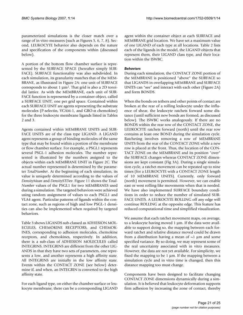
BMC Systems Biology 2007, 1:14 http://www.biomedcentral.com/1752-0509/1/14
parameterized simulations is the closer match over arange of in vitro measures (such as Figures 5, 6, 7, 8). Sec-ond, LEUKOCYTE behavior also depends on the natureand specification of the components within (discussedbelow).
A portion of the bottom flow chamber surface is repre-sented by the SURFACE SPACE (hereafter simply SUR-FACE). SURFACE functionality was also subdivided. Ineach simulation, its granularity matches that of the MEM-BRANE, as illustrated in Figure 2A: one unit of SURFACEcorresponds to about 1 μm2. That grid is also a 2D toroi-dal lattice. As with the MEMBRANE, each unit of SUR-FACE function is represented by a container object, calleda SURFACE UNIT, one per grid space. Contained withineach SURFACE UNIT are agents representing the substratemolecules (P-selectin, VCAM-1, and GRO-α chemokine)for the three leukocyte membrane ligands listed in Tables2 and 3.
Agents contained within MEMBRANE UNITS and SUR-FACE UNITS are of the class type LIGAND. A LIGANDagent represents a group of binding molecules of the sametype that may be found within a portion of the membraneor flow chamber surface. For example, a PSGL1 representsseveral PSGL-1 adhesion molecules. The number repre-sented is illustrated by the numbers assigned to theobjects within each MEMBRANE UNIT in Figure 2C. Theactual number represented is determined by the parame-ter TotalNumber. At the beginning of each simulation, itsvalue is uniquely determined according to the values ofDensityMean and DensitySTDev. Figure 11 shows the Total-Number values of the PSGL1 for two MEMBRANES usedduring a simulation. The targeted behaviors were achievedusing random assignment of values to each PSGL1 andVLA4 agent. Particular patterns of ligands within the con-tact zone, such as regions of high and low PSGL-1 densi-ties can also be implemented when required by targetedbehaviors.
Table 3 shows LIGANDS sub-classed as ADHESION MOL-ECULES, CHEMOKINE RECEPTORS, and CHEMOK-INES, corresponding to adhesion molecules, chemokinereceptors, and chemokines, respectively. In addition,there is a sub-class of ADHESION MOLECULES calledINTEGRINS. INTEGRINS are different from the other LIG-ANDS in that they have two sets of parameters, one repre-sents a low, and another represents a high affinity state.All INTEGRINS are initially in the low affinity state.Events within the CONTACT ZONE (see below) deter-mine if, and when, an INTEGRIN is converted to the highaffinity state.
For each ligand type, on either the chamber surface or leu-kocyte membrane, there can be a corresponding LIGAND
agent within the container object at each SURFACE andMEMBRANE grid location. We have set a maximum valueof one LIGAND of each type at all locations. Table 2 listseach of the ligands in the model, the LIGAND objects thatrepresent them, their LIGAND class type, and their loca-tion within the ISWBC.
BehaviorsDuring each simulation, the CONTACT ZONE portion ofthe MEMBRANE is positioned "above" the SURFACE sothat LIGANDS in overlapping MEMBRANE and SURFACEUNITS can "see" and interact with each other (Figure 2A)and form BONDS.
When the bonds on tethers and other points of contact arebroken at the rear of a rolling leukocyte under the influ-ence of shear, the leukocyte ratchets forward some dis-tance (until sufficient new bonds are formed, as discussedbelow). The ISWBC works analogously. If there are noBONDS within the rear row of the CONTACT ZONE, theLEUKOCYTE ratchets forward (north) until the rear rowcontains at least one BOND during the simulation cycle.Ratcheting involves removing a row of MEMBRANEUNITS from the rear of the CONTACT ZONE while a newrow is placed at the front. Thus, the location of the CON-TACT ZONE on the MEMBRANE and its position "over"the SURFACE changes whereas CONTACT ZONE dimen-sions are kept constant (Fig 3A). During a single simula-tion cycle, a ratchet movement can be repeated up to ninetimes (for a LEUKOCYTE with a CONTACT ZONE lengthof 10 MEMBRANE UNITS). Currently, only forward(north) movement is permitted. However, we can enableeast or west rolling-like movements when that is needed.We have also implemented SURFACE boundary condi-tions in order to reduce the number of simulated SUR-FACE UNITS. A LEUKOCYTE ROLLING off any edge willcontinue ROLLING at the opposite edge. This feature hasreduced computational time and simplified visualization.
We assume that each ratchet movement maps, on average,to a leukocyte having moved 1 μm. If the data were avail-able to support doing so, the mapping between each for-ward ratchet and relative distance moved could be drawnfrom a distribution having a mean of ~1 μm and somespecified variance. By so doing, we may represent some ofthe real uncertainty associated with in vitro measures.However, the data are not yet available. For simplicity, wefixed the mapping to be 1 μm. If the mapping between asimulation cycle and in vitro time is changed, then thisdistance mapping too must change.
Components have been designed to facilitate changingCONTACT ZONE dimensions dynamically during a sim-ulation. It is believed that leukocyte deformation supportsfirm adhesion by increasing the zone of contact, thereby
Page 21 of 25(page number not for citation purposes)
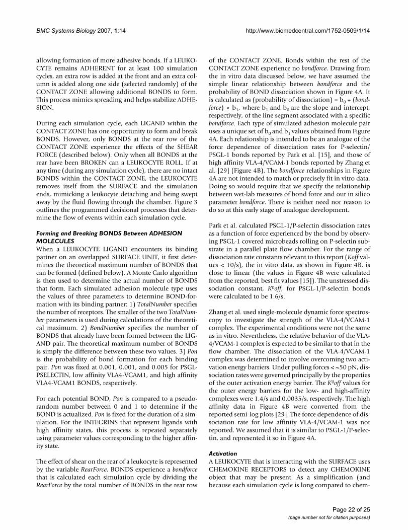
BMC Systems Biology 2007, 1:14 http://www.biomedcentral.com/1752-0509/1/14
allowing formation of more adhesive bonds. If a LEUKO-CYTE remains ADHERENT for at least 100 simulationcycles, an extra row is added at the front and an extra col-umn is added along one side (selected randomly) of theCONTACT ZONE allowing additional BONDS to form.This process mimics spreading and helps stabilize ADHE-SION.
During each simulation cycle, each LIGAND within theCONTACT ZONE has one opportunity to form and breakBONDS. However, only BONDS at the rear row of theCONTACT ZONE experience the effects of the SHEARFORCE (described below). Only when all BONDS at therear have been BROKEN can a LEUKOCYTE ROLL. If atany time (during any simulation cycle), there are no intactBONDS within the CONTACT ZONE, the LEUKOCYTEremoves itself from the SURFACE and the simulationends, mimicking a leukocyte detaching and being sweptaway by the fluid flowing through the chamber. Figure 3outlines the programmed decisional processes that deter-mine the flow of events within each simulation cycle.
Forming and Breaking BONDS Between ADHESION MOLECULESWhen a LEUKOCYTE LIGAND encounters its bindingpartner on an overlapped SURFACE UNIT, it first deter-mines the theoretical maximum number of BONDS thatcan be formed (defined below). A Monte Carlo algorithmis then used to determine the actual number of BONDSthat form. Each simulated adhesion molecule type usesthe values of three parameters to determine BOND-for-mation with its binding partner: 1) TotalNumber specifiesthe number of receptors. The smaller of the two TotalNum-ber parameters is used during calculations of the theoreti-cal maximum. 2) BondNumber specifies the number ofBONDS that already have been formed between the LIG-AND pair. The theoretical maximum number of BONDSis simply the difference between these two values. 3) Ponis the probability of bond formation for each bindingpair. Pon was fixed at 0.001, 0.001, and 0.005 for PSGL-PSELECTIN, low affinity VLA4-VCAM1, and high affinityVLA4-VCAM1 BONDS, respectively.
For each potential BOND, Pon is compared to a pseudo-random number between 0 and 1 to determine if theBOND is actualized. Pon is fixed for the duration of a sim-ulation. For the INTEGRINS that represent ligands withhigh affinity states, this process is repeated separatelyusing parameter values corresponding to the higher affin-ity state.
The effect of shear on the rear of a leukocyte is representedby the variable RearForce. BONDS experience a bondforcethat is calculated each simulation cycle by dividing theRearForce by the total number of BONDS in the rear row
of the CONTACT ZONE. Bonds within the rest of theCONTACT ZONE experience no bondforce. Drawing fromthe in vitro data discussed below, we have assumed thesimple linear relationship between bondforce and theprobability of BOND dissociation shown in Figure 4A. Itis calculated as (probability of dissociation) = b0 + (bond-force) × b1, where b1 and b0 are the slope and intercept,respectively, of the line segment associated with a specificbondforce. Each type of simulated adhesion molecule pairuses a unique set of b0 and b1 values obtained from Figure4A. Each relationship is intended to be an analogue of theforce dependence of dissociation rates for P-selectin/PSGL-1 bonds reported by Park et al. [15], and those ofhigh affinity VLA-4/VCAM-1 bonds reported by Zhang etal. [29] (Figure 4B). The bondforce relationships in Figure4A are not intended to match or precisely fit in vitro data.Doing so would require that we specify the relationshipbetween wet-lab measures of bond force and our in silicoparameter bondforce. There is neither need nor reason todo so at this early stage of analogue development.
Park et al. calculated PSGL-1/P-selectin dissociation ratesas a function of force experienced by the bond by observ-ing PSGL-1 covered microbeads rolling on P-selectin sub-strate in a parallel plate flow chamber. For the range ofdissociation rate constants relevant to this report (Koff val-ues < 10/s), the in vitro data, as shown in Figure 4B, isclose to linear (the values in Figure 4B were calculatedfrom the reported, best fit values [15]). The unstressed dis-sociation constant, K0off, for PSGL-1/P-selectin bondswere calculated to be 1.6/s.
Zhang et al. used single-molecule dynamic force spectros-copy to investigate the strength of the VLA-4/VCAM-1complex. The experimental conditions were not the sameas in vitro. Nevertheless, the relative behavior of the VLA-4/VCAM-1 complex is expected to be similar to that in theflow chamber. The dissociation of the VLA-4/VCAM-1complex was determined to involve overcoming two acti-vation energy barriers. Under pulling forces < ~50 pN, dis-sociation rates were governed principally by the propertiesof the outer activation energy barrier. The K0off values forthe outer energy barriers for the low- and high-affinitycomplexes were 1.4/s and 0.0035/s, respectively. The highaffinity data in Figure 4B were converted from thereported semi-log plots [29]. The force dependence of dis-sociation rate for low affinity VLA-4/VCAM-1 was notreported. We assumed that it is similar to PSGL-1/P-selec-tin, and represented it so in Figure 4A.
ActivationA LEUKOCYTE that is interacting with the SURFACE usesCHEMOKINE RECEPTORS to detect any CHEMOKINEobject that may be present. As a simplification (andbecause each simulation cycle is long compared to chem-
Page 22 of 25(page number not for citation purposes)
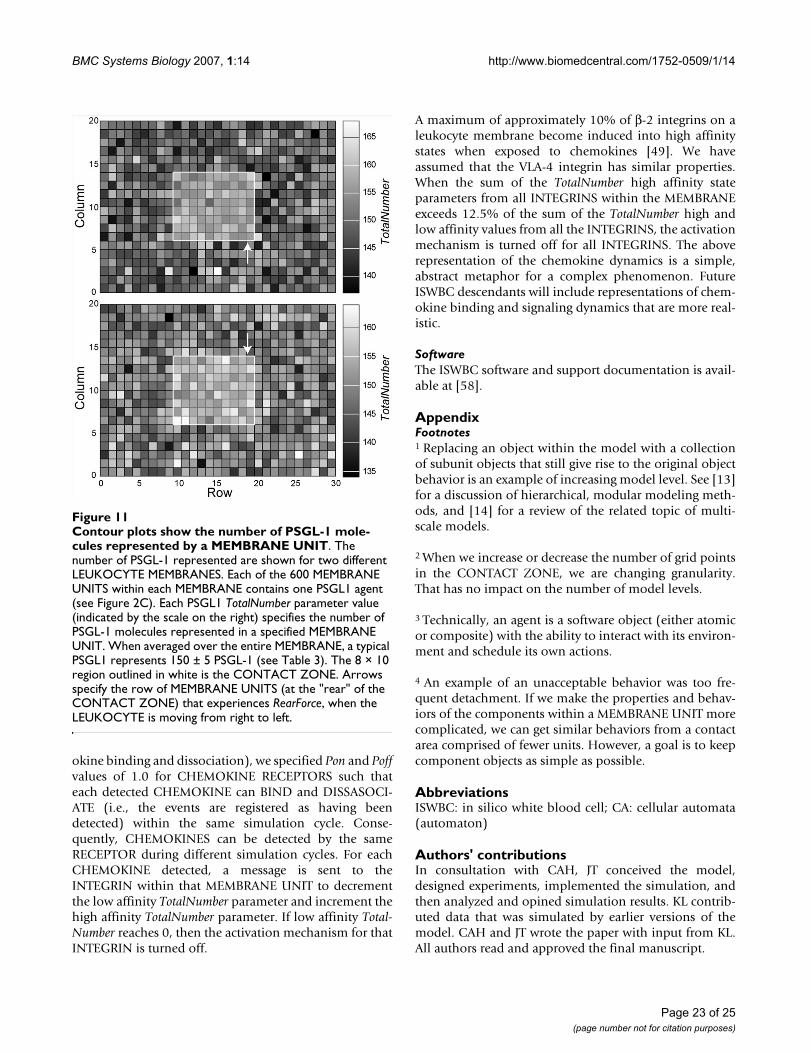
BMC Systems Biology 2007, 1:14 http://www.biomedcentral.com/1752-0509/1/14
okine binding and dissociation), we specified Pon and Poffvalues of 1.0 for CHEMOKINE RECEPTORS such thateach detected CHEMOKINE can BIND and DISSASOCI-ATE (i.e., the events are registered as having beendetected) within the same simulation cycle. Conse-quently, CHEMOKINES can be detected by the sameRECEPTOR during different simulation cycles. For eachCHEMOKINE detected, a message is sent to theINTEGRIN within that MEMBRANE UNIT to decrementthe low affinity TotalNumber parameter and increment thehigh affinity TotalNumber parameter. If low affinity Total-Number reaches 0, then the activation mechanism for thatINTEGRIN is turned off.
A maximum of approximately 10% of β-2 integrins on aleukocyte membrane become induced into high affinitystates when exposed to chemokines [49]. We haveassumed that the VLA-4 integrin has similar properties.When the sum of the TotalNumber high affinity stateparameters from all INTEGRINS within the MEMBRANEexceeds 12.5% of the sum of the TotalNumber high andlow affinity values from all the INTEGRINS, the activationmechanism is turned off for all INTEGRINS. The aboverepresentation of the chemokine dynamics is a simple,abstract metaphor for a complex phenomenon. FutureISWBC descendants will include representations of chem-okine binding and signaling dynamics that are more real-istic.
SoftwareThe ISWBC software and support documentation is avail-able at [58].
AppendixFootnotes1 Replacing an object within the model with a collectionof subunit objects that still give rise to the original objectbehavior is an example of increasing model level. See [13]for a discussion of hierarchical, modular modeling meth-ods, and [14] for a review of the related topic of multi-scale models.
2 When we increase or decrease the number of grid pointsin the CONTACT ZONE, we are changing granularity.That has no impact on the number of model levels.
3 Technically, an agent is a software object (either atomicor composite) with the ability to interact with its environ-ment and schedule its own actions.
4 An example of an unacceptable behavior was too fre-quent detachment. If we make the properties and behav-iors of the components within a MEMBRANE UNIT morecomplicated, we can get similar behaviors from a contactarea comprised of fewer units. However, a goal is to keepcomponent objects as simple as possible.
AbbreviationsISWBC: in silico white blood cell; CA: cellular automata(automaton)
Authors' contributionsIn consultation with CAH, JT conceived the model,designed experiments, implemented the simulation, andthen analyzed and opined simulation results. KL contrib-uted data that was simulated by earlier versions of themodel. CAH and JT wrote the paper with input from KL.All authors read and approved the final manuscript.
Contour plots show the number of PSGL-1 molecules repre-sented by a MEMBRANE UNITFigure 11Contour plots show the number of PSGL-1 mole-cules represented by a MEMBRANE UNIT. The number of PSGL-1 represented are shown for two different LEUKOCYTE MEMBRANES. Each of the 600 MEMBRANE UNITS within each MEMBRANE contains one PSGL1 agent (see Figure 2C). Each PSGL1 TotalNumber parameter value (indicated by the scale on the right) specifies the number of PSGL-1 molecules represented in a specified MEMBRANE UNIT. When averaged over the entire MEMBRANE, a typical PSGL1 represents 150 ± 5 PSGL-1 (see Table 3). The 8 × 10 region outlined in white is the CONTACT ZONE. Arrows specify the row of MEMBRANE UNITS (at the "rear" of the CONTACT ZONE) that experiences RearForce, when the LEUKOCYTE is moving from right to left.
Page 23 of 25(page number not for citation purposes)
BMC Systems Biology 2007, 1:14 http://www.biomedcentral.com/1752-0509/1/14
AcknowledgementsThis research was funded in part by the CDH Research Foundation (R21-CDH-00101). Our dialogues with Michael Smith and David Smith (at the University of Virginia) as well as with Glen Ropella covering many technical, biological, and theoretical issues have been especially helpful. For their help-ful advice and commentary during growth of this project, along with helpful discussions and suggestions, we thank Glen Ropella, Jesse Engelberg, Sean Kim, and other members of the BioSystems Group. Posters presenting ear-lier versions of the ISWBCs were presented at the 2006 Computational Systems Bioinformatics Conference (CSB2006), August 14–18, Stanford, CA, and the 2006 International Conference on Computational Methods in Systems Biology (CSMB'06), October 18–19, Trento Italy.
References1. Ulbrich H, Eriksson EE, Lindbom L: Leukocyte and endothelial
cell adhesion molecules as targets for therapeutic interven-tions in inflammatory disease. Trends Pharmacol Sci 2003,24:640-647.
2. Zhu C, Bao G, Wang N: Cell mechanics: mechanical response,cell adhesion, and molecular deformation. Annu Rev Biomed Eng2000, 2:189-226.
3. Smith DF, Galkina E, Ley K, Huo Y: GRO family chemokines arespecialized for monocyte arrest from flow. Am J Physiol HeartCirc Physiol 2005, 289:H1976-1984.
4. Simon SI, Green CE: Molecular mechanics and dynamics of leu-kocyte recruitment during inflammation. Annu Rev Biomed Eng2005, 7:151-185.
5. Hamann A, Engelhardt B: Leukocyte trafficking: molecular mechanisms,therapeutic targets, and methods Weinheim: Wiley-VCH Verlag GmbH& Co. KGaA; 2005.
6. Constantin G, Laudanna C: Mechanisms of leukocyte integrinactivation. In Leukocyte trafficking: molecular mechanisms, therapeutictargets, and methods Edited by: Hamann A, Engelhardt B. Weinheim:Wiley-VCH Verlag GmbH & Co. KGaA; 2005:68-81.
7. Laudanna C: Integrin activation under flow: a local affair. NatImmunol 2005, 6:429-430.
8. Ley K: Integration of inflammatory signals by rolling neu-trophils. Immunol Rev 2002, 186:8-18.
9. Chang KC, Tees DF, Hammer DA: The state diagram for celladhesion under flow: leukocyte rolling and firm adhesion.Proc Natl Acad Sci U S A 2000, 97:11262-11267.
10. Hammer DA, Apte SM: Simulation of cell rolling and adhesionon surfaces in shear flow: general results and analysis ofselectin-mediated neutrophil adhesion. Biophys J 1992,63:35-57.
11. Bhatia SK, King MR, Hammer DA: The state diagram for celladhesion mediated by two receptors. Biophys J 2003,84:2671-2690.
12. Grant MR, Mostov KE, Tlsty TD, Hunt CA: Simulating propertiesof in vitro epithelial cell morphogenesis. PLoS Comput Biol 2006,2:e129. DOI: 10.1371/journal. pcbi.0020129
13. Zeigler BP: Object-oriented simulation with hierarchical, modular modelsAcademic Press; 1990:89-200.
14. E W, Engquist B: Multiscale Modeling and Computation. Noticesof the AMS 2003, 50:1062-1070.
15. Park EY, Smith MJ, Stropp ES, Snapp KR, DiVietro JA, Walker WF,Schmidtke DW, Diamond SL, Lawrence MB: Comparison ofPSGL-1 microbead and neutrophil rolling: microvillus elon-gation stabilizes P-selectin bond clusters. Biophys J 2002,82:1835-47.
16. Smith MJ, Berg EL, Lawrence MB: A direct comparison of selec-tin-mediated transient, adhesive events using high temporalresolution. Biophys J 1999, 77:3371-3383.
17. Alon R, Kassner PD, Carr MW, Finger EB, Hemler ME, Springer TA:The integrin VLA-4 supports tethering and rolling in flow onVCAM-1. J Cell Biol 1995, 128:1243-1253.
18. Huo Y, Weber C, Forlow SB, Sperandio M, Thatte J, Mack M, Jung S,Littman DR, Ley K: The chemokine KC, but not monocyte che-moattractant protein-1 triggers monocyte arrest on earlyatherosclerotic endothelium. J Clin Invest 2001, 108:1307-1314.
19. Caputo KE, Hammer DA: Effect of microvillus deformability onleukocyte adhesion explored using Adhesive Dynamics sim-ulations. Biophys J 2005, 89:187-200.
20. Chen S, Springer TA: An automatic braking system that stabi-lizes leukocyte rolling by an increase in selectin bondnumber with shear. J Cell Biol 1999, 144:185-200.
21. Zwartz G, Chigaev A, Foutz T, Larson RS, Posner R, Sklar LA: Rela-tionship between molecular and cellular dissociation ratesfor VLA-4/VCAM-1 interactions in the absence of shearstress. Biophys J 2004, 86:1243-1252.
22. Brenner S: Biological computation. In The limits of reductionism inbiology Edited by: Bock G, Goode JA. Chichester (United Kingdom):Wiley; 1998:106-116.
23. Noble D: The future: putting Humpty-Dumpty togetheragain. Biochem Soc Trans 2003, 31:156-158.
24. Shamri R, Grabovsky V, Gauguet JM, Feigelson S, Manevich E, KolanusW, Robinson MK, Staunton DE, von Andrian UH, Alon R: Lym-phocyte arrest requires instantaneous induction of anextended LFA-1 conformation mediated by endothelium-bound chemokines. Nat Immunol 2005, 6:497-506.
25. Cozen-Roberts C, Quinn JA, Lauffenburger DA: Receptor-medi-ated adhesion phenomena: model studies with the radialflow detachment assay. Biophys J 1990, 58:107-125.
26. Kubes P, Niu XF, Smith CW, Kehrli MEJ, Reinhardt PH, WoodmanRC: A novel beta 1-dependent adhesion pathway on neu-trophils: a mechanism invoked by dihydrocytochalasin B orendothelial transmigration. FASEB J 1995, 9:1103-1111.
27. Goldman AJ, Cox RG, Brenner H: Slow viscous motion of asphere parallel to a plane wall. II. Chem Eng Sci 1967,22:635-660.
28. Alon R, Chen S, Puri KD, Finger EB, Springer TA: The kinetics of L-selectin tethers and the mechanics of selectin-mediated roll-ing. J Cell Biol 1997, 138:1169-1180.
29. Zhang X, Craig SE, Kirby H, Humphries MJ, Moy VT: Molecularbasis for the dynamic strength of the integrin a4b1/VCAM-1interaction. Biophys J 2004, 87:3470-3478.
30. Krasik EF, Yee KL, Hammer DA: Adhesive Dynamics simulationof neutrophil arrest with deterministic activation. Biophys J2006, 91:1145-1155.
31. Yago T, Leppanen A, Qiu H, Marcus WD, Nollert MU, Zhu C, Cum-mings RD, McEver RP: Distinct molecular and cellular contribu-tions to stabilizing selectin-mediated rolling under flow. J CellBiol 2002, 158:787-799.
32. Firrell JC, Lipowsky HH: Leukocyte margination and deforma-tion in mesenteric venules of rat. Am J Physiol 1989,256:H1667-1674.
33. Dong C, Cao J, Struble EJ, Lipowsky HH: Mechanics of leukocytedeformation and adhesion to endothelium in shear flow. AnnBiomed Eng 1999, 27:298-312.
34. N'Dri NA, Shyy W, Tran-Son-Tay R: Computational modeling ofcell adhesion and movement using a continuum-kineticsapproach. Biophys J 2003, 85:2273-2286.
35. Jadhav S, Eggleton CD, Konstantopoulos K: A 3-D computationalmodel predicts that cell deformation affects selectin-medi-ated leukocyte rolling. Biophys J 2005, 88:96-104.
36. Wolfram S: Cellular automata as models of complexity. Nature1984, 311:419-424.
37. Graner F, Glazier JA: Simulation of biological cell sorting usinga two-dimensional etended potts model. Phys Rev Lett 1992,69:2013-2016.
38. Glazier JA, Graner F: Simulation of differential adhesion drivenarrangement of biological cells. Phys Rev E Stat Phys Plasmas FluidsRelat Interdiscip Topics 1993, 47:2128-2154.
39. Marée AF, Hogeweg P: Modelling Dictyostelium discoideummorphogenesis: the culmination. Bull Math Biol 2002,64:327-353.
40. Alber M, Kiskowski M, Glazier J, Jiang Y: On Cellular AutomatonApproaches to Modeling Biological Cells. In IMA 134; Mathe-matical systems theory in biology, communication, and finance Edited by:Arnold DN, Santosa F. Springer-Verlag, New York; 2002.
41. Meyer-Hermann ME, Maini PK: Interpreting two-photon imagingdata of lymphocyte motility. Phys Rev E Stat Nonlin Soft MatterPhys 2005, 71:061912.
42. Steels L: The artifical life route to artificial intelligence Edited by: Steels L,Brooks R. New Jersey: Larence Earlbaum Associates, Inc;1995:83-121.
43. Czarnecki K, Eisenecker U: Generative programming: methods, tools, andapplications Volume 10. New York: Addison-Wesley; 2000:251-254.
Page 24 of 25(page number not for citation purposes)
http://www.ncbi.nlm.nih.gov/entrez/query.fcgi?cmd=Retrieve&db=PubMed&dopt=Abstract&list_uids=1384734
http://www.ncbi.nlm.nih.gov/entrez/query.fcgi?cmd=Retrieve&db=PubMed&dopt=Abstract&list_uids=1384734
http://www.ncbi.nlm.nih.gov/entrez/query.fcgi?cmd=Retrieve&db=PubMed&dopt=Abstract&list_uids=1384734
http://www.ncbi.nlm.nih.gov/entrez/query.fcgi?cmd=Retrieve&db=PubMed&dopt=Abstract&list_uids=7534768
http://www.ncbi.nlm.nih.gov/entrez/query.fcgi?cmd=Retrieve&db=PubMed&dopt=Abstract&list_uids=7534768
http://www.ncbi.nlm.nih.gov/entrez/query.fcgi?cmd=Retrieve&db=PubMed&dopt=Abstract&list_uids=7534768
http://www.ncbi.nlm.nih.gov/entrez/query.fcgi?cmd=Retrieve&db=PubMed&dopt=Abstract&list_uids=9885254
http://www.ncbi.nlm.nih.gov/entrez/query.fcgi?cmd=Retrieve&db=PubMed&dopt=Abstract&list_uids=9885254
http://www.ncbi.nlm.nih.gov/entrez/query.fcgi?cmd=Retrieve&db=PubMed&dopt=Abstract&list_uids=9885254
http://www.ncbi.nlm.nih.gov/entrez/query.fcgi?cmd=Retrieve&db=PubMed&dopt=Abstract&list_uids=2166596
http://www.ncbi.nlm.nih.gov/entrez/query.fcgi?cmd=Retrieve&db=PubMed&dopt=Abstract&list_uids=2166596
http://www.ncbi.nlm.nih.gov/entrez/query.fcgi?cmd=Retrieve&db=PubMed&dopt=Abstract&list_uids=2166596
http://www.ncbi.nlm.nih.gov/entrez/query.fcgi?cmd=Retrieve&db=PubMed&dopt=Abstract&list_uids=7544310
http://www.ncbi.nlm.nih.gov/entrez/query.fcgi?cmd=Retrieve&db=PubMed&dopt=Abstract&list_uids=7544310
http://www.ncbi.nlm.nih.gov/entrez/query.fcgi?cmd=Retrieve&db=PubMed&dopt=Abstract&list_uids=7544310
http://www.ncbi.nlm.nih.gov/entrez/query.fcgi?cmd=Retrieve&db=PubMed&dopt=Abstract&list_uids=9281593
http://www.ncbi.nlm.nih.gov/entrez/query.fcgi?cmd=Retrieve&db=PubMed&dopt=Abstract&list_uids=9281593
http://www.ncbi.nlm.nih.gov/entrez/query.fcgi?cmd=Retrieve&db=PubMed&dopt=Abstract&list_uids=9281593
http://www.ncbi.nlm.nih.gov/entrez/query.fcgi?cmd=Retrieve&db=PubMed&dopt=Abstract&list_uids=2735435
http://www.ncbi.nlm.nih.gov/entrez/query.fcgi?cmd=Retrieve&db=PubMed&dopt=Abstract&list_uids=2735435
http://www.ncbi.nlm.nih.gov/entrez/query.fcgi?cmd=Retrieve&db=PubMed&dopt=Abstract&list_uids=9960234
BMC Systems Biology 2007, 1:14 http://www.biomedcentral.com/1752-0509/1/14
Publish with BioMed Central and every scientist can read your work free of charge
"BioMed Central will be the most significant development for disseminating the results of biomedical research in our lifetime."
Sir Paul Nurse, Cancer Research UK
Your research papers will be:
available free of charge to the entire biomedical community
peer reviewed and published immediately upon acceptance
cited in PubMed and archived on PubMed Central
yours — you keep the copyright
Submit your manuscript here:http://www.biomedcentral.com/info/publishing_adv.asp
BioMedcentral
44. Liu Y, Hunt CA: Mechanistic study of the cellular interplay oftransport and metabolism. Pharm Res 2006, 23:493-505.
45. Liu Y, Hunt CA: Studies of intestinal drug transport using an insilico epithelio-mimetic device. Biosystems 2005, 82:154-167.
46. Ramachandran V, Williams M, Yago T, Schmidtke DW, McEver RP:Dynamic alterations of membrane tethers stabilize leuko-cyte rolling on P-selectin. Proc Natl Acad Sci USA 2004,101:13519-13524.
47. Tozeren A, Ley K: How do selectins mediate leukocyte rollingin venules. Biophys J 1992, 63:700-709.
48. Schmid-Schoenbein GW, Shih YY, Chien S: Morphometry ofhuman leukocytes. Blood 1980, 56:866-875.
49. Diamond MS, Springer TA: A subpopulation of Mac-1 (CD11b/CD18) molecules mediates neutrophil adhesion to ICAM-1and fibrinogen. J Cell Biol 1993, 120:545-556.
50. Lawrence MB, Kansas GS, Kunkel EJ, Ley K: Threshold levels offluid shear promote leukocyte adhesion through selectins(CD62L P, E). J Cell Biol 1997, 136:717-727.
51. Granger DN, Kubes P: The microcirculation and inflammation:modulation of leukocyte-endothelial cell adhesion. J LeukocBiol 1994, 55:662-675.
52. Grabovsky V, Feigelson S, Chen C, Bleijs DA, Peled A, Cinamon G,Baleux F, Arenzana-Seisdedos F, Lapidot T, van Kooyk Y, et al.: Sub-second induction of alpha4 integrin clustering by immobi-lized chemokines stimulates leukocyte tethering and rollingon endothelial vascular cell adhesion molecule 1 under flowconditions. J Exp Med 2000, 192:495-506.
53. Dong C, Lei XX: Biomechanics of cell rolling: shear flow, cell-surface adhesion, and cell deformability. J Biomech 2000,33:35-43.
54. Norman KE, Katopodis AG, Thoma G, Kolbinger F, Hicks AE, CotterMJ, Pockley AG, Hellewell PG: P-selectin glycoprotein ligand-1supports rolling on E- and P-selectin in vivo. Blood 2000,96:3585-91.
55. Chigaev A, Blenc AM, Braaten JV, Kumaraswamy N, Kepley CL,Andrews RP, Oliver JM, Edwards BS, Prossnitz ER, Larson RS, SklarLA: Real time analysis of the affinity regulation of alpha 4-integrin. The physiologically activated receptor is intermedi-ate in affinity between resting and Mn(2+) or antibody acti-vation. J Biol Chem 2001, 276:48670-78.
56. Mehta P, Cummings RD, McEver RP: Affinity and kinetic analysisof P-selectin binding to P-selectin glycoprotein ligand-1. J BiolChem 1998, 273:32506-32513.
57. Long M, Lu S, Sun G: Kinetics of receptor-ligand interactions inimmune responses. Cell Mol Immunol 2006, 3:79-86.
58. In Silico White Blood Cell (ISWBC) Model [http://systemsbiology.ucsf.edu:8080/iswbc/]
Page 25 of 25(page number not for citation purposes)
http://www.ncbi.nlm.nih.gov/entrez/query.fcgi?cmd=Retrieve&db=PubMed&dopt=Abstract&list_uids=1420908
http://www.ncbi.nlm.nih.gov/entrez/query.fcgi?cmd=Retrieve&db=PubMed&dopt=Abstract&list_uids=1420908
http://www.ncbi.nlm.nih.gov/entrez/query.fcgi?cmd=Retrieve&db=PubMed&dopt=Abstract&list_uids=6775712
http://www.ncbi.nlm.nih.gov/entrez/query.fcgi?cmd=Retrieve&db=PubMed&dopt=Abstract&list_uids=6775712
http://www.ncbi.nlm.nih.gov/entrez/query.fcgi?cmd=Retrieve&db=PubMed&dopt=Abstract&list_uids=7678422
http://www.ncbi.nlm.nih.gov/entrez/query.fcgi?cmd=Retrieve&db=PubMed&dopt=Abstract&list_uids=7678422
http://www.ncbi.nlm.nih.gov/entrez/query.fcgi?cmd=Retrieve&db=PubMed&dopt=Abstract&list_uids=7678422
http://www.ncbi.nlm.nih.gov/entrez/query.fcgi?cmd=Retrieve&db=PubMed&dopt=Abstract&list_uids=9024700
http://www.ncbi.nlm.nih.gov/entrez/query.fcgi?cmd=Retrieve&db=PubMed&dopt=Abstract&list_uids=9024700
http://www.ncbi.nlm.nih.gov/entrez/query.fcgi?cmd=Retrieve&db=PubMed&dopt=Abstract&list_uids=9024700
http://www.ncbi.nlm.nih.gov/entrez/query.fcgi?cmd=Retrieve&db=PubMed&dopt=Abstract&list_uids=8182345
http://www.ncbi.nlm.nih.gov/entrez/query.fcgi?cmd=Retrieve&db=PubMed&dopt=Abstract&list_uids=8182345
http://www.ncbi.nlm.nih.gov/entrez/query.fcgi?cmd=Retrieve&db=PubMed&dopt=Abstract&list_uids=9829984
![in silico (BFA thesis), 2002 [español]](https://static.fdokumen.com/doc/165x107/631f4913dbf756400702aca8/in-silico-bfa-thesis-2002-espanol.jpg)