Supportive care after active periodontal treatment. A review
Eo
RWa
b
c
d
a
ARRAA
KMLALI
1
gpamAtfiit
0d
Chemico-Biological Interactions 179 (2009) 344–350
Contents lists available at ScienceDirect
Chemico-Biological Interactions
journa l homepage: www.e lsev ier .com/ locate /chembio int
ffect of mangiferin on the development of periodontal disease: Involvementf lipoxin A4, anti-chemotaxic action in leukocyte rolling
oney Rick Carvalhoa, Claudia Helena Pellizzonb, Luis Justulin Jr. b, Sergio Luis Felisbinob,agner Vilegasc, Fernanda Brunid, Mônica Lopes-Ferreirad, Clélia Akiko Hiruma-Limaa,∗
Department of Physiology, Biosciences Institute, São Paulo State University (UNESP), Rubiao Junior cp. 510, 18618-000 Botucatu, São Paulo, BrazilDepartment of Morphology, Biosciences Institute, São Paulo State University (UNESP), Botucatu, São Paulo, BrazilInstitute of Chemistry, São Paulo State University (UNESP), Araraquara, São Paulo, BrazilSpecial Laboratory of Applied Toxinology (CAT/CEPID), São Paulo, Brazil
r t i c l e i n f o
rticle history:eceived 22 August 2008eceived in revised form 23 October 2008ccepted 24 October 2008vailable online 5 November 2008
eywords:angiferin
ipoxinlveolar bone losseukocytesnflammation
a b s t r a c t
Mangiferin is a polyphenol compound obtained from mango and has been reported to possess antioxidantand anti-inflammatory properties.Aim: We propose to evaluate the influence of mangiferin in preventing and treating experimental peri-odontitis induced in Wistar rats.Main methods: Periodontitis was induced in rats by applying a ligature around the lower right first molar.After ligature, groups of animals were submitted orally to the following treatments: saline 10 mL/kg,piroxicam 20 mg/kg or mangiferin 100 mg/kg. On days 1, 4 or 7 after ligature application the alveolarbone loss (ABL) was determined. We evaluated the effect of mangiferin on ABL by histological techniques(alveolar bone loss and cellularity), enzyme immunoassay (lipoxin A4), intravital microscopy (rollingleukocytes and endothelial–leukocyte adhesion), zymographic analyses (metalloproteinases, MMPs 2 and9), immunohistochemistry (PCNA, COX-2 and CXCR4) and toxicology.
Key findings: Oral administration of mangiferin significantly reduced ABL. We also observed the reductionof cellularity in mangiferin-treated rats. Treatment with mangiferin inhibited COX-2 expression and therolling and adhesion of leukocytes, while maintaining normal lipoxin A4 levels. The mangiferin did notinterfere in the activity of MMP-2 or -9. The mangiferin-treated rats presented an earlier peak of cellproliferation and augmented angiogenesis in the injured region.Significance: Our results have demonstrated promising therapeutic potential of mangiferin both in theof pe
rtimp
itt
prevention and treatment
. Introduction
Periodontitis is a chronic inflammatory disease whose patho-enesis is related to the formation of colonies of microorganismsresent in subgingival plaque, including some gram-negativenaerobic rod bacteria, spirochetes, and viruses [1]. The diseaseay also be due to alterations in the immune response [2].mong the gram-negative microorganism inducers of periodonti-
is, the most important are Porphyromonas gingivalis and Bacteriodes
orsythus, which have virulence factors that increase their infectiv-ty and provide conditions for their multiplication and persistencen periodontium [3]. The presence of these microorganisms leadso the formation of a biofilm that initiates a dental inflammatory∗ Corresponding author. Tel.: +55 15143811 6077; fax: +55 15143815 3744.E-mail address: [email protected] (C.A. Hiruma-Lima).
o[tvptae
009-2797/$ – see front matter. Published by Elsevier Ireland Ltdoi:10.1016/j.cbi.2008.10.041
riodontitis.Published by Elsevier Ireland Ltd
esponse in the individual. This inflammatory response results inhe formation of edema, infiltration of leukocytes and release ofnflammatory mediators. The final result of this process is the for-
ation of an inflammatory periodontal pocket, destruction of theeriodontal ligament, alveolar bone resorption and tooth loss [4].
Leukocytic infiltration is a process of many steps, character-zed by the migration (rolling) of leukocytes which enables themo undergo the action of chemotaxins, endothelial adhesion andransmigration through the endothelium. The interruption of anyf these steps is a potential target for anti-inflammatory drugs5,6]. The various chemical mediators involved in the inflamma-ory process include prostaglandins, whose main activities are
asodilatation and increased capillary permeability, which in turnromotes leukocytic infiltration and consequent development ofhe inflammatory process. Prostaglandins are formed from therachidonic acid pathway by the action of two cyclooxygenasenzymes (COX-1 and COX-2). The enzyme COX-1 is constitutive
ogical
wdosaiTbtpppsatInatha(ooaadbdi
LpuMxd[aBwcamspmmcr
2
2
oabsrapkw
2
oBrmibS
2
CmtogppotCP
2
ffisddtd
2
wm(mcVetve
2
dcAc
2
R.R. Carvalho et al. / Chemico-Biol
hile COX-2 is inducible and found abundantly in periodontaliseases [7–9]. Besides prostaglandins, arachidonic acid providesther chemical mediators such as leukotriene B4 (LTB4) respon-ible for leukocyte chemotaxis, and lipoxin A4 (LXA4) whichcts as an endogenous anti-inflammatory mediator by present-ng anti-chemotaxic action to inhibit the rolling of leukocytes.he relation of lipoxins to periodontitis has been demonstratedy various authors, including their action in blocking inflamma-ion by reducing the recruitment of leukocytes, and in targetingotential new strategies for treatment [10,11]. Concomitant witheriodontitis, the metalloproteinases (MMPs) use various com-onents of the extracellular matrix and basement membrane asubstrates. These enzymes are essential to the growth, repairnd remodeling of tissues, but are also involved in the destruc-ion of tissues in pathological cases including periodontal disease.n particular, MMP-2 and MMP-9 belong to the group of gelati-ases identified as prevalent in both rat and human periodontitis,nd are notable for their association with bone resorption inhe inflammatory periodontal procedure [12]. In contrast to thearmful effects of these inflammatory process, other events suchs the formation of new blood vessels in the injured regionangiogenesis) and cell proliferation assist in the regenerationf injured periodontal tissues [6,13]. Against periodontitis, vari-us drugs, mainly non-steroidal anti-inflammatory drugs (NSAIDs)re used as an adjunctive therapy option to reverse the dam-ge caused by periodontal disease, but have proven unsatisfactoryue to various side effects such as gastrointestinal tract injuries,leeding, perforations, obstructions, ulcerations and symptomaticiverticular disease, while increasing the risk of myocardial
nfarction [14,15].Mangiferin is the major component (10%) of Mangifera indica
. (mango) which belongs to the family Anacardiaceae. Thislant, which grows in tropical and subtropical regions, is widelysed in folk medicine for various therapeutic indications [16].angiferin is a glycosylated xanthone (1,3,6,7-tetra-hydroxy-
anthone-C2-�-d-glucose) of natural origin [17], that has beenescribed as presenting the following effects: immunomodulation18,19], anti-inflammatory inhibition of COX-2 expression [20,21],s well as LTB4 [22], NF-� B (nuclear transcription factor-kappa), TNF-� (tumor necrosis factor-alpha) and IL-1 (interleukin-1),hich are very important cytokines in the inflammatory pro-
ess and in bone resorption [19,21,23,24]. Its bone anti-resorptionctivity in lumbar vertebrae was demonstrated in the experi-ental model using a dose of 100 mg/(kg day) [25]. This plant
pecies also shows antibacterial activity in vivo against specificeriodontal pathogens including P. gingivalis and Prevotella inter-edia [26]. Based on all these activities already described forangiferin, the present study aims to evaluate its pharmacologi-
al effects on alveolar bone loss in experimental periodontitis inats.
. Materials and methods
.1. Drugs
Mangiferin (Sigma–Aldrich, USA) was administered at the dosef 100 mg/(kg day). This dose was selected based on its bonenti-resorption activity in the lumbar vertebra model describedy Li et al. [25]. Piroxicam (Feldene®, Sao Paulo, Brazil) waselected at 20 mg/(kg day) on the basis of the alveolar bone anti-
esorption activity of meloxicam [27], which is a non-steroidalnti-inflammatory drug with effects similar to piroxicam. Theiroxicam dose was selected on the basis of effective doses mar-eted for the two drugs. As the vehicle, we used saline (distilledater + 0.9% NaCl) at 10 mL/(kg day).rtc
Interactions 179 (2009) 344–350 345
.2. Animals
Adult male rats (n = 88, Rattus norvegicus albinus, Wistar, 180 g),btained from the UNESP Breeding Center (Botucatu, São Paulo,razil), were fed food and water ad libitum. The rats were dividedandomly into groups that orally received saline, piroxicam orangiferin. After treatment, the rats were anesthetized with
ntraperitoneal injection of 10 mg/kg of ketamine (Dopalen®, Vet-rands, SP, Brazil) and 10 mg/kg of xylazine (Anasedan®, Vetbrands,P, Brazil).
.3. Induction of periodontal disease
After anesthesia, the animals received a cotton ligature (Coatsorrente Ltda., SP, Brazil) around the lower right first molar in a sub-arginal position to induce experimental periodontitis, according
o the method described by Johnson [28]. The lower first molarsn the left side did not receive the ligature, and served as the unli-ated control. The ligature was maintained for 1, 4 or 7 days after itslacement, and then the rats were killed by decapitation. Treatmentrior to induction of periodontitis (1 day) provides an indicationf preventive action at the onset of the disease. This experimen-al protocol was submitted to the Animal Experimentation Ethicsommittee at the Biosciences Institute of State University of Sãoaulo (CEEA/IB/UNESP) and was approved as no. 43/06.
.4. Obtainment of the histological slide
The mandible of each animal was removed and fixed in bufferedormalin (pH 7.2) 10% for 48 h. Then, the mandibles were decalci-ed in a solution of 50 mL of 50% formic acid and 50 mL of 20%odium citrate [29] which was replaced three times a week. Afterecalcification, the mandibles were washed in running water, dehy-rated in increasing alcohol series and diaphanized in xylol. Thenhe mandibles were enclosed in paraffin, in order to obtain stan-ardized serial 5-�m thick histological section.
.5. Measurement of alveolar bone loss
The slides were stained with hematoxylin/eosin (HE) afterhich alveolar bone loss (ABL) was determined by measuring inicrometers (�m) the distance from the cemento-enamel junction
CEJ) to the alveolar bone crest on the distal face of the first lowerolars, with the aid of a Leica DM microscope (5× magnification)
oupled with the image-capturing software Leica QWin Standardersion 3.1.0. The ABLs were obtained by comparing the differ-nt values on the side with ligature (first lower right molar) andhe unligated side (first lower left molar), respecting the individualariation of each animal, from which we obtained three images ofach slide that were counted manually.
.6. Total cell count
Through the images captured by the program Leica QWin Stan-ard Version 3.1 of the histological slides stained with HE, theellularity of the injured region was determined by the programVSoft BioView/Seeker 4, utilizing an area of 5 cm2 (40× magnifi-ation) from the distal region of the first lower molars.
.7. Immunohistochemical analysis
A representative slide from each treatment was deparaffinized,ehydrated and designed by the immunohistochemical methodo reveal peroxidase (PCNA, Nova Castra; COX-2, Cayman Chemi-al and CXCR4, Nova Castra). The blockade of nonspecific reaction
3 ogical
wmit(Kdise
2
tamtitctcrtet[cZao
2
mmsWscwksotp[
2
mo(nwtsfgC(t
be(o
2
satloas[
2
tcuaTao
2
T(c
3
trvnmmatt
tspaomtdgMt4
46 R.R. Carvalho et al. / Chemico-Biol
as accomplished with skimmed milk and whey from nor-al sheep. After the recovery, the antigen and samples were
ncubated overnight, with specific antibodies, in blocking solu-ion. Subsequently, the sample was washed in phosphate buffer0.01 mol/L, pH 7.4) and incubated in secondary antibody (Vectorit ABC) and revealed with Avidin-biotin associated with 3-3′-iaminobenzidine tetrahydrochloride (DAB, Sigma), and examined
n a Leica DM microscope coupled with Leica QWin Standard Ver-ion 3.1.0. image-capturing software, obtaining three images ofach slide that were counted manually [30–32].
.8. Intravital microscopy
Mice (Swiss, male, 18–22 g) provided by the Butantan Insti-ute (Brazil) were divided into two groups of five animals eachnd maintained fasting for 2 h. After that, one group receivedangiferin (100 mg/kg) orally and 30 min later all mice were anes-
hetized with sodium pentobarbital (Hypnol®, Cristália; 50 mg/kg,ntraperitoneally) and underwent surgery to expose the cremas-er muscle [33]. The animals were kept on a platform thermallyontrolled at 37 ◦C. An initial leukocyte count was performed, andhen 1 �g/20 �L LPS was applied topically to the cremaster mus-le. The number of rolling leukocytes in post-capillary venules wasecorded every 5 min for a period of 30 min to surpass a prede-ermined fixed point. The number of leukocytes adhering to thendothelial wall for a period of 1 min or more at a pre-set dis-ance of 100 �m was assessed every 5 min for a period of 30 min34,35]. This microvascular study by transillumination of tissue wasompleted with the aid of an optical microscope (Imager A1, Carl-eiss, Oberkochen, DE) coupled to a camera (AxionCam, ICc1) usingn objective opening/longitudinal numerical distance × 10/0.3 andptovar 1.6.
.9. Determination of lipoxin A4 concentration in gingival tissues
Seven days after the induction of periodontitis in rats, the ani-als were killed and the gingival tissues surrounding the lowerolars were removed. A homogenate was obtained from these tis-
ues using a tissue macerator (Ultra-turbax® T 25 basic IKA® –erke) and added along with 1 mL of PBS (phosphate buffered
aline), pH 7.4, to each sample, maintained at 37 ◦C for 20 min, thenentrifuged at 9000 × g for 25 min, after which the supernatantsere extracted and diluted 10 times with a phosphate buffer (ELISA
it BioAssayTM, USBiological). The lipoxin A4 concentration of theseamples, expressed in picograms (pg) of lipoxin per microgram (�g)f protein, was determined using the kit, following the manufac-urer’s recommendations supplied in the manual. The amount ofrotein present in the sample was determined by the BCA method36].
.10. Determination of the activity of MMPs
Zymographic analysis was used to evaluate the effect ofangiferin on gelatinolytic activity of MMPs 2 and 9. Samples
f recombinant MMPs (4 ng of MMP-2 and 40 pg of MMP-9)Calbiochem, CA, USA) were subjected to electrophoresis, underon-reducing conditions, in 8% polyacrylamide gel, co-polymerizedith 0.1% gelatin. After electrophoresis, the gels were washed
wice (15 min) in a solution of 2.5% Triton X-100 to remove theodium dodecyl sulfate (Bio-Rad, CA, USA), followed by two washes
or 5 min in buffer solution of 50 mM Tris–HCl, pH 8.0. Then theels were incubated overnight in Tris–HCl buffer, containing 5 mMaCl2 and 1 �M ZnCl2 and increasing concentrations of mangiferin10 �g/mL, 100 �g/mL, 500 �g/mL and 1 mg/mL), at 37 ◦C. Finally,he gels were stained with Coomassie Brilliant Blue R-250 [37]. Thei(t
A
Interactions 179 (2009) 344–350
ands were analyzed in an ImageQuant 300 gel image-capturingquipment employing ImageQuant 300 image-analysis softwareGE Healthcare, NJ, USA). The values are expressed as integratedptical density.
.11. Development of body weight and vital organs
The rats submitted to periodontal disease and treated withaline, piroxicam or mangiferin were weighed daily for 7 days tossess the possible body weight alterations, a potential indicator ofoxicity in these animals. After the sacrifice of the rats, the heart,ungs, liver, kidney and spleen were removed and weighed (in g)n a precision analytical balance. The ratios between organ weightsnd body weights were converted into arcsine for statistical analy-is, since this parameter may indicate drug toxicity in these organs38].
.12. Biochemical evaluation of serum
Blood samples were obtained from all animals submitted toreatment with saline, piroxicam or mangiferin, which were thenentrifuged for serum removal to determine the concentrations ofrea, creatinine, �-GT (gamma glutamyltransferase), ALT (alanineminotransferase), AST (aspartate aminotransferase) and glucose.hese serum biochemical parameters were quantified by theutomatic biochemical analyzer Cobas Mira S® (Roche) with col-rimetric and kinetic kits (CELM®, Brazil).
.13. Statistical analysis
The data were presented as the mean ± standard error (S.E.).he means obtained were submitted to the analysis of varianceANOVA), followed by Tukey or Dunnett’s test. Differences wereonsidered significant at P < 0.05.
. Results and discussion
Periodontitis is a chronic inflammatory disease that leads tohe loss of alveolar bone [4], and can be induced in rats in a formeproducible in humans [39]. Previous studies under the lumbarertebra model have demonstrated that mangiferin, a xanthone ofatural origin, presents anti-resorption activity in bone [25]. Theangiferin dose–response curve obtained in the same study deter-ined the dose of 100 mg/kg to be significant [25]. The latter work
imed to evaluate the effect of mangiferin on ABL in the experimen-al periodontitis model and also proposed the action mechanism ofhis polyphenol compound.
Table 1 shows the result 1 day after the induction of periodon-itis. Only the group of rats treated with mangiferin was able toignificantly reduce (P < 0.05) ABL (105.5 ± 33.1 �m) when com-ared to the vehicle-treated group (217.6 ± 18.9 �m). Piroxicamt the dose of 20 mg/kg was unable to prevent the appearancef periodontitis 24 h after its induction. We could observe thatangiferin administered prior to the induction of periodonti-
is induction was already displaying a preventive action at theisease onset. Our results also showed that mangiferin has a pro-ressive healing effect on periodontal disease after the onset.angiferin significantly reduced ABL 4 and 7 days after induc-
ion when compared to saline-treated animals (Table 1). Afterdays of daily treatment, both the groups treated with pirox-
cam and mangiferin significantly reduced (P < 0.001) the ABL167.2 ± 13.4 �m vs. 162.2 ± 11 �m, respectively) when comparedo saline-treated animals (290.4 ± 20 �m).
We concluded that the mangiferin treatment was able to reduceBL in rats submitted to experimental periodontitis after 1, 4 or 7

R.R. Carvalho et al. / Chemico-Biological Interactions 179 (2009) 344–350 347
Table 1Alveolar bone loss (�m) in rats submitted to experimental periodontitis and treated with vehicle (saline), piroxicam or mangiferin.
Groups (dose) Treatment periods
1 day 4 days 7 days
Saline 217.67 ± 18.95 290.47 ± 20.00 620.98 ± 103.37Piroxicam (20 mg/kg) 138.14 ± 18.68 167.29 ± 13.48*** 298.53 ± 30.99**
Mangiferin (100 mg/kg) 105.56 ± 33.12* 162.26 ± 11.03*** 220.32 ± 15.79***
D
dmp
iavssrua((aihgtttttaciro1p
trcp2st
TT
G
USPM
D
Table 3Immunohistochemical analysis of COX-2 in rats with experimental periodontitis.
Groups (dose) Treatment periods
1 day 4 days 7 days
Unligated control 0 0 0Saline 48 84 58Piroxicam (20 mg/kg) 0 3 7M
Ds
tatCi(mi[
mavmaeste(ously administered to the mice inhibited the rolling of leukocytesinduced by topical application of LPS at all experimental moments
ata are means ± S.E. (n = 7). ANOVA, followed by Tukey’s test.* P < 0.05 vs. saline group within the same treatment period.
** P < 0.01 vs. saline group within the same treatment period.*** P < 0.001 vs. saline group within the same treatment period.
ays of treatment. These results point out the promising effect ofangiferin in the treatment of ABL. Therefore, we investigated the
ossible action mechanisms of this natural xanthone.ABL is caused by an inflammatory process, whose main features
nclude the recruitment of defense cells, a process which results inlarge number of cells at the inflamed site [35]. Based on this obser-ation, we chose to quantify the cells present in injured region, ashown in Table 2. One day after periodontitis induction there wasignificant increase (P < 0.001) in cellularity of the injured region inats treated with vehicle (221.8 ± 4.59 cells) when compared to thenligated control group (155.17 ± 3.64 cells). Both the piroxicam-nd mangiferin-treated groups presented significantly diminishedP < 0.01 and P < 0.001, respectively) cellularity in the injured region196 ± 5.12 cells vs. 187.6 ± 1.88 cells, respectively) in relation tonimals treated only with saline. Four days after periodontitisnduction, the cellularity in rats treated with saline (221 ± 4.14 cells)ad risen significantly (P < 0.001) compared to the unligated controlroup (155.6 ± 6.35 cells), but had dropped significantly in animalsreated with piroxicam or mangiferin (P < 0.05 and P < 0.001, respec-ively, 191.6 ± 4.63 cells vs. 173.8 ± 7.87 cells) when compared tohe rats treated with vehicle. The mangiferin-treated group showedhe lowest reduction of cellularity 4 days after periodontitis induc-ion. After 7 days of periodontitis both treated groups (piroxicamnd mangiferin) also displayed significantly reduced (P < 0.001)ellularity (204.25 ± 3.09 cells vs. 181.5 ± 2.78 cells, respectively)n comparison to vehicle-treated rats (249.4 ± 9.04 cells). Theseesults demonstrate that the least recruitment of defense cellsccurred in mangiferin-treated rats. We also observed that, after, 4 or 7 days of mangiferin treatment, all the periodontitis carriersresented lower cellularity in the injured region.
The literature reported that cell recruitment is directly relatedo vasodilatation and increased capillary permeability, effects thatesult largely from the action of prostaglandins synthesized fromyclooxygenases (COXs) 1 and 2 [7,8]. COX-2 is found abundantly in
eriodontitis [9], and our results showed greater inhibition of COX-expression in mangiferin-treated rats than in those treated withaline (Table 3). The immunohistochemical analysis showed thathe saline-treated group showed diminished COX-2 expression in
able 2otal cell counts in rats with experimental periodontitis.
roups (dose) Treatment periods
1 day 4 days 7 days
nligated control 155.17 ± 3.64 155.6 ± 6.35 153 ± 3.5aline 221.8 ± 4.59*** 221 ± 4.14*** 249.4 ± 9.04***
iroxicam (20 mg/kg) 196 ± 5.12++ 191.6 ± 4.63+ 204.25 ± 3.09+++
angiferin (100 mg/kg) 187.6 ± 1.88+++ 173.8 ± 7.87+++ 181.5 ± 2.78+++
ata are means ± S.E. (n = 4–6). ANOVA, followed by Tukey’s test.*** P < 0.001 vs. unligated control group within the same treatment period.
+ P < 0.05 vs. saline group within the same treatment period.++ P < 0.01 vs. saline group within the same treatment period.
+++ P < 0.001 vs. saline group within the same treatment period.
ep
Fm(p
angiferin (100 mg/kg) 33 2 9
ata represent the sum of cells stained in three images obtained from a slide repre-entative of each group.
he injured region on days 1 (48 cells), 4 (84 cells) and 7 (58 cells)fter periodontitis induction when compared to the unligated con-rol group (0 cells). Yet mangiferin-treated rats showed inhibitedOX-2 expression in 33 cells 1 day after treatment. But the drop
n COX-2 expression was more evident after 4 (2 cells) and 7 days9 cells) of mangiferin treatment than it was under vehicle treat-
ent. These data are consistent with the literature in which COX-2nhibition by mangiferin was proven in other experimental models19,21].
Leukocytes are among the defense cells recruited in the inflam-atory process of periodontitis. These cells, when attracted by the
ction of chemotaxins, increase their capacities for rolling throughessels and for adhering to the endothelium in order to cross it andigrate toward the inflammation site [5,40]. Therefore, the rolling
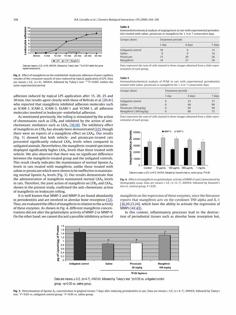
nd endothelial–leukocyte adhesions are the important param-ters for evaluating the action mechanism of anti-inflammatoryubstances such as mangiferin. Our results showed that mangiferin-reated animals presented significant inhibition of the rolling andndothelial–leukocyte adhesion compared with the control groupFigs. 1 and 2, respectively). As shown in Fig. 1, mangiferin previ-
valuated. According to Fig. 2, the mangiferin-treated mice alsoresented significant inhibition of the endothelial–leukocyte
ig. 1. Effect of mangiferin on leukocyte rolling in post-capillary venules of the cre-aster muscle of mice induced by topical application of LPS. Data are means ± S.E.
n = 6). ANOVA, followed by Tukey’s test. ***P < 0.001 within the same experimentaleriod.
348 R.R. Carvalho et al. / Chemico-Biological Interactions 179 (2009) 344–350
Fvas
a3wam
ocot(pudvbTlsitiso
iTotO
Table 4Immunohistochemical analysis of angiogenesis in rats with experimental periodon-titis treated with saline, piroxicam or mangiferin for 1, 4 or 7 consecutive days.
Groups (dose) Treatment periods
1 day 4 days 7 days
Unligated control 10 6 15Saline 8 9 14Piroxicam 13 29 26Mangiferin 14 27 38
Data represent the sum of cells stained in three images obtained from a slide repre-sentative of each group.
Table 5Immunohistochemical analysis of PCNA in rats with experimental periodontitistreated with saline, piroxicam or mangiferin for 1, 4 or 7 consecutive days.
Groups (dose) Treatment periods
1 day 4 days 7 days
Unligated control 0 23 37Saline 0 48 98Piroxicam (20 mg/kg) 0 69 90Mangiferin (100 mg/kg) 0 88 72
Data represent the sum of cells stained in three images obtained from a slide repre-sentative of each group.
FZt
mreports that mangiferin acts on the cytokines TNF-alpha and IL-1
Ft
ig. 2. Effect of mangiferin on the endothelial–leukocyte adhesion of post-capillaryenules of the cremaster muscle of mice induced by topical application of LPS. Datare means ± S.E. (n = 6). ANOVA, followed by Tukey’s test. ***P < 0.001 within theame experimental period.
dhesion induced by topical LPS application after 15, 20, 25 and0 min. Our results agree closely with those of Beltrán et al. [20,41]ho reported that mangiferin inhibited adhesion molecules such
s ICAM-1, ICAM-2, ICAM-3, ELAM-1 and VCAM-1, all adhesionolecules involved in leukocyte–endothelial adhesion.As mentioned previously, the rolling is stimulated by the action
f chemotaxins such as LTB4 and inhibited by the action of anti-hemotaxic mediators such as LXA4 [10,16]. The inhibitory effectf mangiferin on LTB4 has already been demonstrated [22], thoughhere were no reports of a mangiferin effect on LXA4. Our resultsFig. 3) showed that both vehicle- and piroxicam-treated ratsresented significantly reduced LXA4 levels when compared tonligated animals. Nevertheless, the mangiferin-treated specimensisplayed significantly higher LXA4 levels than those treated withehicle. We also observed that there was no significant differenceetween the mangiferin-treated group and the unligated controls.his result clearly indicates the maintenance of normal lipoxin A4evels in rats treated with mangiferin, unlike those treated withaline or piroxicam which were shown to be ineffective in maintain-ng normal lipoxin A4 levels (Fig. 3). Our results demonstrate thathe administration of mangiferin maintained normal LXA4 levelsn rats. Therefore, the joint action of mangiferin on LTB4 and LXA4,hown in the present study, confirmed the anti-chemotaxic actionf mangiferin on leukocyte rolling.
It is well known that MMP-2 and MMP-9 are found abundantlyn periodontitis and are involved in alveolar bone resorption [12].
hus, we evaluated the effect of mangiferin in relation to the activityf these enzymes. As shown in Fig. 4, different mangiferin concen-rations did not alter the gelatinolytic activity of MMP-2 or MMP-9.n the other hand, we cannot discard a possible inhibitory action of[M
t
ig. 3. Determination of lipoxin A4 concentration in gingival tissues 7 days after inducingest. *P < 0.05 vs. unligated control group. +P < 0.05 vs. saline group.
ig. 4. Effects of mangiferin on gelatinolytic activity of MMPs 9 and 2 determined byymography assay. Data are means ± S.E. (n = 6–7). ANOVA, followed by Dunnett’sest vs. control group. P > 0.05.
angiferin on the expression of these enzymes, since the literature
16,20,23,24], which have the ability to activate the expression ofMPs [42,43].In this context, inflammatory processes lead to the destruc-
ion of periodontal tissues such as alveolar bone resorption but,
periodontitis in rats. Data are means ± S.E. (n = 4–7). ANOVA, followed by Tukey’s
R.R. Carvalho et al. / Chemico-Biological Interactions 179 (2009) 344–350 349
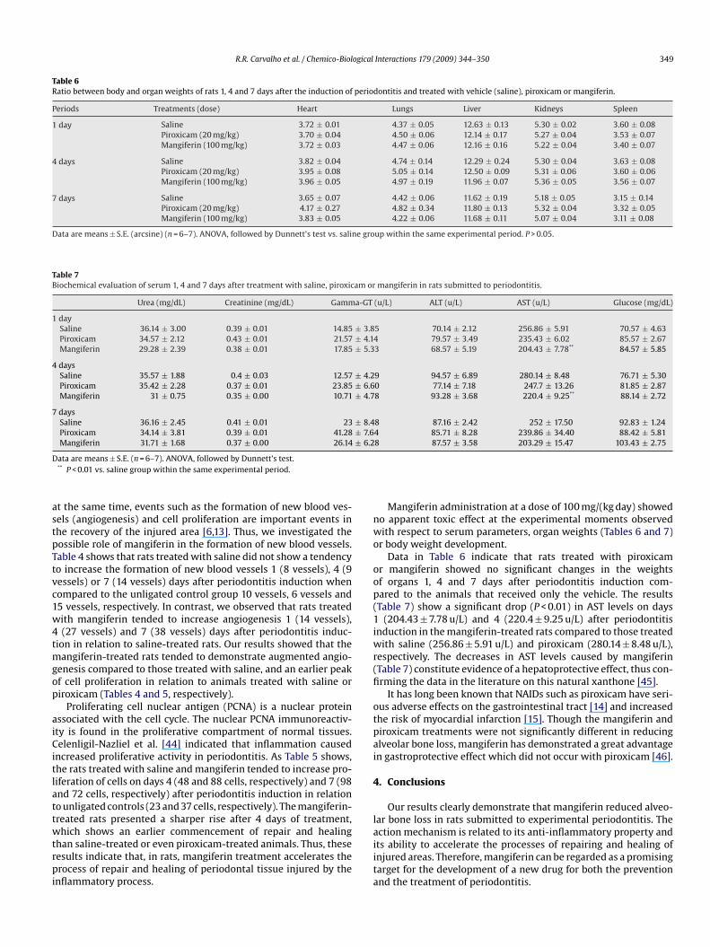
Table 6Ratio between body and organ weights of rats 1, 4 and 7 days after the induction of periodontitis and treated with vehicle (saline), piroxicam or mangiferin.
Periods Treatments (dose) Heart Lungs Liver Kidneys Spleen
1 day Saline 3.72 ± 0.01 4.37 ± 0.05 12.63 ± 0.13 5.30 ± 0.02 3.60 ± 0.08Piroxicam (20 mg/kg) 3.70 ± 0.04 4.50 ± 0.06 12.14 ± 0.17 5.27 ± 0.04 3.53 ± 0.07Mangiferin (100 mg/kg) 3.72 ± 0.03 4.47 ± 0.06 12.16 ± 0.16 5.22 ± 0.04 3.40 ± 0.07
4 days Saline 3.82 ± 0.04 4.74 ± 0.14 12.29 ± 0.24 5.30 ± 0.04 3.63 ± 0.08Piroxicam (20 mg/kg) 3.95 ± 0.08 5.05 ± 0.14 12.50 ± 0.09 5.31 ± 0.06 3.60 ± 0.06Mangiferin (100 mg/kg) 3.96 ± 0.05 4.97 ± 0.19 11.96 ± 0.07 5.36 ± 0.05 3.56 ± 0.07
7 days Saline 3.65 ± 0.07 4.42 ± 0.06 11.62 ± 0.19 5.18 ± 0.05 3.15 ± 0.14Piroxicam (20 mg/kg) 4.17 ± 0.27 4.82 ± 0.34 11.80 ± 0.13 5.32 ± 0.04 3.32 ± 0.05Mangiferin (100 mg/kg) 3.83 ± 0.05 4.22 ± 0.06 11.68 ± 0.11 5.07 ± 0.04 3.11 ± 0.08
Data are means ± S.E. (arcsine) (n = 6–7). ANOVA, followed by Dunnett’s test vs. saline group within the same experimental period. P > 0.05.
Table 7Biochemical evaluation of serum 1, 4 and 7 days after treatment with saline, piroxicam or mangiferin in rats submitted to periodontitis.
Urea (mg/dL) Creatinine (mg/dL) Gamma-GT (u/L) ALT (u/L) AST (u/L) Glucose (mg/dL)
1 daySaline 36.14 ± 3.00 0.39 ± 0.01 14.85 ± 3.85 70.14 ± 2.12 256.86 ± 5.91 70.57 ± 4.63Piroxicam 34.57 ± 2.12 0.43 ± 0.01 21.57 ± 4.14 79.57 ± 3.49 235.43 ± 6.02 85.57 ± 2.67Mangiferin 29.28 ± 2.39 0.38 ± 0.01 17.85 ± 5.33 68.57 ± 5.19 204.43 ± 7.78** 84.57 ± 5.85
4 daysSaline 35.57 ± 1.88 0.4 ± 0.03 12.57 ± 4.29 94.57 ± 6.89 280.14 ± 8.48 76.71 ± 5.30Piroxicam 35.42 ± 2.28 0.37 ± 0.01 23.85 ± 6.60 77.14 ± 7.18 247.7 ± 13.26 81.85 ± 2.87Mangiferin 31 ± 0.75 0.35 ± 0.00 10.71 ± 4.78 93.28 ± 3.68 220.4 ± 9.25** 88.14 ± 2.72
7 daysSaline 36.16 ± 2.45 0.41 ± 0.01 23 ± 8.48 87.16 ± 2.42 252 ± 17.50 92.83 ± 1.24Piroxicam 34.14 ± 3.81 0.39 ± 0.01 41.28 ± 7.64 85.71 ± 8.28 239.86 ± 34.40 88.42 ± 5.81Mangiferin 31.71 ± 1.68 0.37 ± 0.00 26.14 ± 6.28 87.57 ± 3.58 203.29 ± 15.47 103.43 ± 2.75
D
astpTtvc1w4tmgop
aiCitlattwtrpi
nwo
oop(1iwr(fi
otpai
4
l
ata are means ± S.E. (n = 6–7). ANOVA, followed by Dunnett’s test.** P < 0.01 vs. saline group within the same experimental period.
t the same time, events such as the formation of new blood ves-els (angiogenesis) and cell proliferation are important events inhe recovery of the injured area [6,13]. Thus, we investigated theossible role of mangiferin in the formation of new blood vessels.able 4 shows that rats treated with saline did not show a tendencyo increase the formation of new blood vessels 1 (8 vessels), 4 (9essels) or 7 (14 vessels) days after periodontitis induction whenompared to the unligated control group 10 vessels, 6 vessels and5 vessels, respectively. In contrast, we observed that rats treatedith mangiferin tended to increase angiogenesis 1 (14 vessels),(27 vessels) and 7 (38 vessels) days after periodontitis induc-
ion in relation to saline-treated rats. Our results showed that theangiferin-treated rats tended to demonstrate augmented angio-
enesis compared to those treated with saline, and an earlier peakf cell proliferation in relation to animals treated with saline oriroxicam (Tables 4 and 5, respectively).
Proliferating cell nuclear antigen (PCNA) is a nuclear proteinssociated with the cell cycle. The nuclear PCNA immunoreactiv-ty is found in the proliferative compartment of normal tissues.elenligil-Nazliel et al. [44] indicated that inflammation caused
ncreased proliferative activity in periodontitis. As Table 5 shows,he rats treated with saline and mangiferin tended to increase pro-iferation of cells on days 4 (48 and 88 cells, respectively) and 7 (98nd 72 cells, respectively) after periodontitis induction in relationo unligated controls (23 and 37 cells, respectively). The mangiferin-reated rats presented a sharper rise after 4 days of treatment,
hich shows an earlier commencement of repair and healinghan saline-treated or even piroxicam-treated animals. Thus, theseesults indicate that, in rats, mangiferin treatment accelerates therocess of repair and healing of periodontal tissue injured by the
nflammatory process.
aiita
Mangiferin administration at a dose of 100 mg/(kg day) showedo apparent toxic effect at the experimental moments observedith respect to serum parameters, organ weights (Tables 6 and 7)
r body weight development.Data in Table 6 indicate that rats treated with piroxicam
r mangiferin showed no significant changes in the weightsf organs 1, 4 and 7 days after periodontitis induction com-ared to the animals that received only the vehicle. The resultsTable 7) show a significant drop (P < 0.01) in AST levels on days
(204.43 ± 7.78 u/L) and 4 (220.4 ± 9.25 u/L) after periodontitisnduction in the mangiferin-treated rats compared to those treated
ith saline (256.86 ± 5.91 u/L) and piroxicam (280.14 ± 8.48 u/L),espectively. The decreases in AST levels caused by mangiferinTable 7) constitute evidence of a hepatoprotective effect, thus con-rming the data in the literature on this natural xanthone [45].
It has long been known that NAIDs such as piroxicam have seri-us adverse effects on the gastrointestinal tract [14] and increasedhe risk of myocardial infarction [15]. Though the mangiferin andiroxicam treatments were not significantly different in reducinglveolar bone loss, mangiferin has demonstrated a great advantagen gastroprotective effect which did not occur with piroxicam [46].
. Conclusions
Our results clearly demonstrate that mangiferin reduced alveo-ar bone loss in rats submitted to experimental periodontitis. The
ction mechanism is related to its anti-inflammatory property andts ability to accelerate the processes of repairing and healing ofnjured areas. Therefore, mangiferin can be regarded as a promisingarget for the development of a new drug for both the preventionnd the treatment of periodontitis.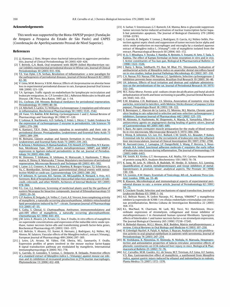
3 ogical
A
d(
R
[
[
[
[
[
[
[
[
[
[
[
[
[
[
[
[
[
[
[
[
[
[
[
[
[
[
[
[
[
[
[
[
[
[
[
[
50 R.R. Carvalho et al. / Chemico-Biol
cknowledgements
This work was supported by the Biota-FAPESP project (Fundacãoe Amparo a Pesquisa do Estado de São Paulo) and CAPESCoordenacão de Aperfeicoamento Pessoal de Nível Superior).
eferences
[1] J.J. Kamma, J. Slots, Herpes virus–bacterial interaction in aggressive periodon-titis, Journal of Clinical Periodontology 30 (2003) 420–426.
[2] T. Breivik, G.A. Rook, Oral treatment with SR299 (killed Mycobacterium vac-cae) inhibits experimental periodontal disease in Wistar rats, Journal of ClinicalPeriodontology 30 (2003) 931–936.
[3] T.E. Van Dyke, C.N. Serhan, Resolution of Inflammation: a new paradigm forthe pathogenesis of periodontal diseases, Journal of Dental Research 82 (2003)82–90.
[4] V. Lima, M.M. Bezerra, V.B.M. Alencar, Effects of clorpromazine on alveolar boneloss in experimental periodontal disease in rats, European Journal Oral Science108 (2000) 123–129.
[5] T.A. Springer, Traffic signals on endothelium for lymphocyte recirculation andleukocyte emigration, in: C.P. Leendert (Ed.), Adhesion Molecules in Health andDisease, CRC Press, Boca Raton, 1997, pp. 1–54.
[6] D.L. Cochran, J.M. Wosney, Biological mediators for periodontal regeneration,Periodontology 19 (1999) 40–58.
[7] J.A. Mitchell, S. Larkin, T.J. Williams, Cyclooxygenase-2 regulation and relevancein inflammation, Biochemical Pharmacology 50 (1995) 1535–1542.
[8] J.R. Vane, Y.S. Bakhle, R.M. Botting, Cyclooxygenases 1 and 2, Annual Review ofPharmacology and Toxicology 38 (1998) 97–120.
[9] Z. Lohinai, R. Stachlewitz, A.D. Székely, E. Fehér, L. Dézsi, C. Szabó, Evidence forthe expression of cyclooxygenase-2 enzyme in periodontitis, Life Sciences 70(2001) 279–290.
10] A. Kantarci, T.E.V. Dyke, Lipoxin signaling in neutrophils and their role inperiodontal disease, Prostaglandins, Leukotrienes and Essential Fatty Acids 73(2005) 289–299.
11] M. Romano, Lipoxin analogs and lipoxin formation in vivo, Prostaglandins,Leukotrienes and Essential Fatty Acids 73 (2005) 239–243.
12] R. Achong, I. Nishimura, H. Ramachandran, T.H. Howell, J.P. Fiorellini, N.Y. Karim-bux, Membrane Type, (MT)1-matrix metalloproteinase (MMP) and MMP-2expression in ligature-induced periodontitis in the rat, Journal of Periodon-tology 74 (2003) 494–500.
13] M. Shimono, T. Ishikawa, H. Ishikawa, H. Matsuzaki, S. Hashimoto, T. Mura-matsu, K. Shima, K. Matsuzaka, T. Inoue, Regulatory mechanisms of periodontalregeneration, Microscopy Research and Technique 60 (2003) 491–502.
14] L. Laine, L.G. Connors, A. Reicin, C.J. Hawkey, R. Burgos-Vargas, T.J. Schnitzer, Q.Yu, C. Bombardier, Serious lower gastrointestinal clinical events with nonse-lective NSAID or coxib use, Gastroenterology 124 (2003) 288–292.
15] S.P. Johnsen, H. Larsson, R.E. Tarone, J.K. McLaughlin, B. Norgard, S. Friis, H.T.Sorensen, Risk of hospitalization for myocardial infarction among users of rofe-coxib, celecoxib, and other NSAIDs, Archieves of Internal Medicine 165 (2005)978–984.
16] F.G. Coe, G.J. Anderson, Screening of medicinal plants used by the garifuna ofeastern Nicaragua for bioactive compounds, Journal of Ethnopharmacology 53(2005) 29–50.
17] G.P. Andreu, R. Delgado, J.A. Velho, C. Curti, A.E. Vercesi, Iron complexing activityof mangiferin, a naturally occurring glucosylxanthone, inhibitis mitochondriallipid peroxidation induced by Fe2+–citrate, European Journal of Pharmacology513 (2005) 47–55.
18] S. Guha, S. Ghosal, U. Chattopadhyay, Antitumor, immunomodulatory andanti-HIV effect of mangiferin, a naturally occurring glucosylxanthone,Chemotherapy 42 (1996) 443–451.
19] J.M. Leiro, E. Alvarez, J.A. Arranz, I.G. Siso, F. Orallo, In vitro effects of mangiferinon superoxide concentrations and expression of the inducible nitric oxide syn-thase, tumor necrosis factor-alfa and transforming growth factor-beta genes,Biochemical Pharmacology 65 (2003) 1361–1371.
20] A.E. Beltrán, Y. Alvarez, F.E. Xavier, R. Hernanz, J. Rodriguez, A.J. Núnez, M.J.Alonso, M. Salaices, Vascular effects of the Mangifera indica L. extract (Vimang),European Journal of Pharmacology 499 (2004) 297–305.
21] J. Leiro, J.A. Arranz, M. Yánez, F.M. Ubeira, M.L. Sanmartín, F. Orallo,Expression profiles of genes involved in the mouse nuclear factor-kappa
B signal transduction pathway are modulated by mangiferin, InternationalImmunopharmacology 4 (2004) 763–778.22] G. Garrido, D. González, Y. Lemus, C. Delporte, R. Delgado, Protective effectsof a standard extract of Mangifera indica L. (Vimangs) against mouse ear ede-mas and its inhibition of eicosanoid production in J774 murine macrophages,Phytomedicine 13 (2006) 412–418.
[
Interactions 179 (2009) 344–350
23] A. Sarkar, Y. Sreenivasan, G.T. Ramesh, S.K. Manna, Beta-d-glucoside suppressestumor necrosis factor-induced activation of nuclear transcription factor kapa-b but potentiates apoptosis, The Journal of Biological Chemistry 279 (2004)33768–33781.
24] G. Garrido, R. Delgado, Y. Lemus, J. Rodríguez, D. García, A.J. Núnez-Sellés, Pro-tection against septic shock and suppression of tumor necrosis factor alpha andnitric oxide production on macrophages and microglia by a standard aqueousextract of Mangifera indica L. (Vimang®) role of mangiferin isolated from theextract, Pharmacological Research 50 (2004) 165–172.
25] H. Li, T. Miyahara, Y. Tezuka, T. Namba, N. Remoto, S. Tonami, H. Seto, T. Tada, S.Kadota, The effect of Kampo formulae on bone resorption in vitro and in vivo.I. Active constituents of Tsu-kan-gan, Biological & Pharmaceutical Bulletin 21(1998) 1322–1326.
26] I. Bairy, S. Reeja, Siddharth, P.S. Rao, M. Bhat, P.G. Shivananda, Evaluation ofantibacterial activity of Mangifera indica on anaerobic dental microflora basedon in vivo studies, Indian Journal Pathology Microbiology 45 (2002) 307–310.
27] C.A. Nassar, P.O. Nassar, P.M. Nassar, L.C. Spolidorio, Selective cyclooxygenase-2inhibition prevents bone resorption, Brazilian Oral Research 19 (2005) 36–40.
28] I.H. Johnson, Effects of local irritation and dextran and sulphate administra-tion on the periodontium of the rat, Journal of Periodontal Research 10 (1975)332–345.
29] M.S. Anna-Morse, Formic acid–sodium citrate decalcification and butyl alcoholdehydratation of teeth and bone sectioning in paraffin, Journal Dental Research24 (1945) 143–153.
30] E.W. Kitajima, C.H. Rodrigues, J.S. Silveira, Association of isometric virus likeparticles, restricted to laticifers, with Meleira (Sticky disease) of papaya (Caricapapaya), Fitopatologia Brasil 18 (1993) 118–122.
31] B. Berenguer, C. Alarcon de La Lastra, F.J. Moreno, M.J. Martin, Chronic gastriculcer healing in rats subjected to selective and non-selective cyclooxygenase-2inhibitors, European Journal of Pharmacology 442 (2002) 125–135.
32] M. Akimoto, H. Hashimoto, M. Shigemoto, A. Maeda, K. Yamashita, Effects ofantisecretory agents on angiogenesis during healing of gastric ulcers, Journalof Gastroenterology 40 (2005) 685–689.
33] S. Baez, An open cremaster muscle preparation for the study of blood vesselsby in vivo microscopy, Microvascular Research 5 (1973) 384–394.
34] J. Wong, B. Johnston, S.S. Lee, D.C. Bullard, C.W. Smith, A.L. Beaudet, P. Kubes,A minimal role for selectins in the recruitment of leukocytes into the inflamedliver microvasculature, Journal of Clinical Investigation 99 (1997) 2782–2790.
35] M. Aurrand-Lions, C. Lamagna, J.P. Dangerfield, S. Wang, P. Herrera, S. Nour-shargh, B.A. Imhof, Junctional adhesion molecule-C regulates the early influxof leukocytes into tissues during inflammation, The Journal of Immunology 174(2005) 6406–6415.
36] P.K. Smith, R.I. Krohn, G.T. Hermanson, A.K. Mallia, F.H. Gartner, Measurementof protein using BCA, Analyses Biochemistry 150 (1985) 76–78.
37] K. Jung, M. Lein, N. Ulbrich, B. Rudolph, W. Henke, D. Schnorr, A.S. Loening,Quantification of matrix metalloproteinases and tissue inhibitors of metal-loproteinases in prostatic tissue: analytical aspects, The Prostate 34 (1998)130–136.
38] T.A. Loomis, A.W. Hayes, Essentials of Toxicology, 4th ed., Academic Press Lim-ited, London, 1996, pp. 33–46.
39] B. Klausen, Microbiological and immunological aspects of experimental peri-odontal disease in rats: a review article, Journal of Periodontology 62 (1991)59–73.
40] E. Crockett-Torabi, Selectins and mechanisms of signal transduction, Journal ofLeukocyte Biology 63 (1998) 1–14.
41] A.E. Beltran Nunez, N. Ledon Naranjo, P.C. Romay, VIMANG® y mangiferinainhiben la expresión de ICAM-1 en células endoteliales estimuladas con citoci-nas proinflamatorias, Revista Cubana de Investigacion Biomédica 22 (2003)164–172.
42] K.L. MacNaul, N. Chartrain, M. Lark, M.J. Tocci, N.I. Hutchinson, Disco-ordinate expression of stromelysin, collagenase and tissue inhibitor ofmetalloproteinases-1 in rheumatoid human synovial fibroblasts. Synergisticeffects of Interleukin-1 and tumor necrosis factor-� on stromelysin expression,The Journal Biological Chemistry 265 (1990) 17238–17245.
43] H. Birkedal-Hansen, W.G.I. Moore, M.K. Bodden, Matrix metalloproteinases: areview, Critical Reviews in Oral Biology and Medicine 4 (1993) 197–250.
44] H. Celenligil-Nazliel, A. Palali, A. Ayhan, S. Ruacan, Analysis of in situ prolifera-tive activity in oral gingival epithelium in patients with xerostomia, Journal ofPeriodontology 74 (2003) 247–254.
45] M. Yoshikawa, K. Ninomiya, H. Shimoda, N. Nishida, H. Matsuda, Hepatopro-tective and antioxidative properties of Salacia reticulata: preventive effects of
phenolic constituents on CCl4-induced liver injury in mice, Biological & Phar-maceutical Bulletin 25 (2002) 72–76.46] A.C. Carvalho, M.M. Guedes, A.L. de Souza, M.T. Trevisan, A.F. Lima, A.F. Santos,V.S. Rau, Gastroprotective effect of mangiferin, a xanthonoid from Mangiferaindica, against gastric injury induced by ethanol and indomethacin in rodents,Planta Medica 73 (2007) 1372–1376.
Copyright © 2022 FDOKUMEN