Anticomplementary activity of equine whole IgG antivenoms: comparison of three fractionation...
6
Short communication Anticomplementary activity of equine whole IgG antivenoms: comparison of three fractionation protocols Guillermo Leo ´n, Bruno Lomonte, Jose ´ Marı ´a Gutie ´rrez * Instituto Clodomiro Picado, Facultad de Microbiologı ´a, Universidad de Costa Rica, San Jose ´, Costa Rica Received 5 May 2004; accepted 26 July 2004 Abstract Early adverse reactions occur in a number of patients treated with heterologous antivenoms and have been associated with anticomplementary activity (ACA). In order to reduce the ACA of equine whole IgG antivenoms produced by caprylic acid fractionation, three different fractionation protocols were compared: (a) routine caprylic acid fractionation; (b) caprylic acid fractionation followed by b-propiolactone treatment; and (c) caprylic acid fractionation followed by ion-exchange chromatography using a quaternary ammonium membrane. The three protocols yielded products with similar physicochemical characteristics and anti-Bothrops asper venom antibody titers, except that ion-exchange purified antivenom had a lower protein concentration. Antivenoms fractionated by using b-propiolactone or filtration through quaternary ammonium membrane had a significantly reduced in vitro ACA. A preparation of caprylic acid-fractionated antivenom was heated in order to induce the formation of protein aggregates; however, its ACA was similar to non-heated antivenom. None of the antivenoms affected the hemolytic activity of serum complement in rabbits after a bolus intravenous administration. It is concluded that (a) b-propiolactone and quaternary ammonium membranes significantly reduce in vitro ACA of caprylic acid-fractionated equine antivenom, and (b) the validity of in vitro ACA as a predictor of EAR needs to be reexamined in clinical and experimental studies, since it may not adequately predict in vivo complement activation by antivenoms. q 2004 Elsevier Ltd. All rights reserved. Keywords: Antivenom; Anticomplementary activity; Early adverse reactions; IgG; CH 50 Parenteral administration of horse or sheep-derived antivenoms constitutes the only scientifically validated treatment for snakebite envenomation worldwide (Theakston et al., 2003; Lalloo and Theakston, 2003). Despite their well demonstrated clinical efficacy, adminis- tration of antivenoms is often associated with the appear- ance of early adverse reactions (EARs), with a highly variable incidence reported for different products (Cardoso et al., 1993; Chippaux et al., 1998; Dart and McNally, 2001; Lalloo and Theakston, 2003; Gawarammana et al., 2004). The severity of EARs ranges from mild manifestations, i.e. urticaria, to more complicated reactions involving bronch- ospasm and hypotension (Lalloo and Theakston, 2003). The mechanisms involved in EAR are not completely clear at present. It has been proposed that de novo complement activation, or anticomplementary activity (ACA), is an important mechanism associated with EARs (Sutherland, 1977; Lalloo and Theakston, 2003), and this mechanism has been also implicated in some adverse reactions occurring after administration of human immunoglobulin preparations (Barandun et al., 1962). On this basis, it has been assumed that pepsin digestion of horse antivenom IgG, with the consequent removal of Fc fragment, would reduce the ACA of antivenoms (see for example Grandgeorge et al., 1996). Nevertheless, F(ab 0 ) 2 antivenoms still present ACA (Sutherland, 1977; Morais et al., 1994; Leo ´n et al., 2001). Moreover, clinical studies carried out with many antive- noms have demonstrated a conspicuous variability in terms of incidence of EAR; in some cases, pepsin-digested F(ab 0 ) 2 0041-0101/$ - see front matter q 2004 Elsevier Ltd. All rights reserved. doi:10.1016/j.toxicon.2004.07.025 Toxicon 45 (2005) 123–128 www.elsevier.com/locate/toxicon * Corresponding author.
-
Upload
independent -
Category
Documents
-
view
1 -
download
0
Transcript of Anticomplementary activity of equine whole IgG antivenoms: comparison of three fractionation...
Short communication
Anticomplementary activity of equine whole IgG antivenoms:
comparison of three fractionation protocols
Guillermo Leon, Bruno Lomonte, Jose Marıa Gutierrez*
Instituto Clodomiro Picado, Facultad de Microbiologıa, Universidad de Costa Rica, San Jose, Costa Rica
Received 5 May 2004; accepted 26 July 2004
www.elsevier.com/locate/toxicon
Abstract
Early adverse reactions occur in a number of patients treated with heterologous antivenoms and have been associated with
anticomplementary activity (ACA). In order to reduce the ACA of equine whole IgG antivenoms produced by caprylic acid
fractionation, three different fractionation protocols were compared: (a) routine caprylic acid fractionation; (b) caprylic acid
fractionation followed by b-propiolactone treatment; and (c) caprylic acid fractionation followed by ion-exchange
chromatography using a quaternary ammonium membrane. The three protocols yielded products with similar physicochemical
characteristics and anti-Bothrops asper venom antibody titers, except that ion-exchange purified antivenom had a lower protein
concentration. Antivenoms fractionated by using b-propiolactone or filtration through quaternary ammonium membrane had a
significantly reduced in vitro ACA. A preparation of caprylic acid-fractionated antivenom was heated in order to induce the
formation of protein aggregates; however, its ACA was similar to non-heated antivenom. None of the antivenoms affected
the hemolytic activity of serum complement in rabbits after a bolus intravenous administration. It is concluded that
(a) b-propiolactone and quaternary ammonium membranes significantly reduce in vitro ACA of caprylic acid-fractionated
equine antivenom, and (b) the validity of in vitro ACA as a predictor of EAR needs to be reexamined in clinical and
experimental studies, since it may not adequately predict in vivo complement activation by antivenoms.
q 2004 Elsevier Ltd. All rights reserved.
Keywords: Antivenom; Anticomplementary activity; Early adverse reactions; IgG; CH50
Parenteral administration of horse or sheep-derived
antivenoms constitutes the only scientifically validated
treatment for snakebite envenomation worldwide
(Theakston et al., 2003; Lalloo and Theakston, 2003).
Despite their well demonstrated clinical efficacy, adminis-
tration of antivenoms is often associated with the appear-
ance of early adverse reactions (EARs), with a highly
variable incidence reported for different products (Cardoso
et al., 1993; Chippaux et al., 1998; Dart and McNally, 2001;
Lalloo and Theakston, 2003; Gawarammana et al., 2004).
The severity of EARs ranges from mild manifestations, i.e.
urticaria, to more complicated reactions involving bronch-
ospasm and hypotension (Lalloo and Theakston, 2003).
0041-0101/$ - see front matter q 2004 Elsevier Ltd. All rights reserved.
doi:10.1016/j.toxicon.2004.07.025
* Corresponding author.
The mechanisms involved in EAR are not completely clear
at present. It has been proposed that de novo complement
activation, or anticomplementary activity (ACA), is an
important mechanism associated with EARs (Sutherland,
1977; Lalloo and Theakston, 2003), and this mechanism has
been also implicated in some adverse reactions occurring
after administration of human immunoglobulin preparations
(Barandun et al., 1962). On this basis, it has been assumed
that pepsin digestion of horse antivenom IgG, with the
consequent removal of Fc fragment, would reduce the
ACA of antivenoms (see for example Grandgeorge et al.,
1996). Nevertheless, F(ab 0)2 antivenoms still present ACA
(Sutherland, 1977; Morais et al., 1994; Leon et al., 2001).
Moreover, clinical studies carried out with many antive-
noms have demonstrated a conspicuous variability in terms
of incidence of EAR; in some cases, pepsin-digested F(ab 0)2
Toxicon 45 (2005) 123–128

G. Leon et al. / Toxicon 45 (2005) 123–128124
antivenoms have induced EAR in more than 75% of patients
(Moran et al., 1998; Gawarammana et al., 2004), whereas
other F(ab 0)2 products induce EAR in less than 10% of
patients (Chippaux et al., 1998). Moreover, whole IgG
antivenoms prepared by caprylic acid fractionation of horse
plasma have been shown to induce a low incidence of EAR
(Otero-Patino et al., 1998; Otero et al., 1999). Therefore, it
seems that the physicochemical characteristics of antive-
noms, such as presence of non-IgG proteins and aggregates,
may play a more relevant role in the incidence of EAR than
the presence of Fc fragment in intact IgGs. Protein
aggregates and non-IgG proteins may thus be involved in
the ACA of antivenoms.
Fractionation of horse plasma with caprylic acid is a
simple and convenient method for antivenom production
which has been adapted for industrial production (Rojas
et al., 1994). The clinical performance of various whole IgG
antivenoms obtained with this methodology has been highly
satisfactory in terms of efficacy and safety (Otero-Patino
et al., 1998; Otero et al., 1999). However, such antivenoms
still present ACA when tested on human complement in
vitro (Otero et al., 1999; Leon et al., 2001). Owing to the
potential relevance of ACA in the development of EAR
associated with antivenom administration, in this work we
compared three different fractionation protocols, all of them
involving caprylic acid precipitation of non-IgG proteins, in
order to find novel ways to reduce ACA.
The same batch of hyperimmune horse plasma was used
throughout this study. It was obtained from a group of 30
horses immunized with a mixture of equal parts of the
venoms of Bothrops asper, Crotalus durissus durissus and
Lachesis stenophrys (Angulo et al., 1997). Plasma was
fractionated by three different protocols: (1) Caprylic acid
precipitation of non-IgG proteins, as routinely performed at
the Production Division of Instituto Clodomiro Picado
(Rojas et al., 1994). Briefly, caprylic acid was added to horse
plasma, at a ratio of 6 ml acid per 100 ml plasma, and the
mixture was stirred for 1 h at 20–22 8C. The mixture was
then filtered through a 8 mm paper filter, and desalted and
concentrated by diafiltration. Afterwards, the preparation
was formulated in order to have 0.15 M NaCl, 2.5 g/l phenol
and 0.05 g/l thimerosal, pH 7.0, Finally, the antivenom was
sterilized by filtration through 0.22 mm membrane and
bottled in 10 ml glass flasks. (2) Five millilitres of a solution
of 2 g/l b-propiolactone were added to 95 ml of antivenom
fractionated as in protocol (1). The mixture was stirred for
2 h at 20–22 8C and then allowed to stand overnight at 4 8C.
Afterwards, the preparation was extensively dialyzed
against 0.15 M NaCl, 2.5 g/l phenol and 0.05 g/l thimerosal,
pH 7.0 and the preparation sterilized as described.
(3) Aliquots of 10 ml of antivenom purified by protocol
(1) were filtered through a strongly basic quaternary
ammonium cellulose microporous membrane (Sartobind
Q-15 membrane, Sartorius, Germany). After filtration of
each 10 ml aliquot, the membrane was regenerated by
passing 10 ml of 1N NaOH and 10 ml of sterile 0.15 M
NaCl solution. A total of 100 ml of antivenom were filtered.
At the end, the preparation was sterilized as described.
Antivenoms resultant from the three fractionation protocols
were formulated to have 0.15 M NaCl, 2.5 g/l phenol and
0.05 g/l thimerosal, with a pH of 7.0. In addition, a sample
with an increased content of protein aggregates was
prepared by heating 200 ml of antivenom fractionated as
in protocol (1) during 90 min at 60 8C in a water bath.
b-propiolactone and ion-exchange steps were selected
because these procedures have been proposed to improve
purity and safety of human IgG preparations (Stephan,
1975) and equine antivenoms (Guidolin et al., 1997;
Grandgeorge et al., 1996; Jones and Landon, 2003).
The following parameters were comparatively analyzed:
(1) Total protein concentration was determined by the
Biuret method (Schosinsky et al., 1983). (2) Turbidity was
evaluated by determining the absorbance of undiluted
antivenoms at 590 nm (Rojas et al., 1994). (3) Aggregate
content was analyzed by analytical gel filtration FPLC in a
Superdex 200 column (HR 10/30) using phosphate-
buffered saline solution (PBS) as eluent (Garcıa et al.,
2002). (4) Electrophoresis was carried out on cellulose
polyacetate membranes (Sephrapore III, Gelman Instru-
ments, Michigan) in barbital buffer, pH 8.6, at 180 V.
Strips were stained with Amidoblack and destained with
0.9N acetic acid. (5) Antibody titers against B. asper
venom were determined by ELISA. Ten micrograms
venom, dissolved in 100 ml PBS were adsorbed onto
Immulon-2 microplates (Dynatech, Virginia). After wash-
ing, the wells were blocked with PBS-2% bovine serum
albumin, and various dilutions of the antivenoms were
added and incubated for 1 h. After washing, a peroxidase-
conjugated anti-equine IgG was added. After a final
washing step, substrate (o-phenylenediamine and H2O2)
were added and the reaction stopped with 2 M HCl, before
recording the absorbances at 492 nm in a Dynatech
microplate reader. In all these determinations, experiments
were run in triplicates.
In vitro ACA on human complement was assessed as
follows: Briefly, 2 ml of various dilutions of antivenoms
were added to 0.5 ml of fresh human serum previously
diluted 1:20 with barbital buffer, pH 7.6. After 1 h of
incubation at 37 8C, 100 ml of sheep erythrocytes, pre-
viously sensitized with rabbit anti-sheep erythrocyte
antibodies, were added, and the mixture was incubated 1 h
at 37 8C. Then, 2 ml of barbital buffer were added, the
samples centrifuged and the absorbances of the supernatants
were recorded at 540 nm. ACA activity was expressed as the
inverse of the dilution that reduced complement activity by
50%, taking as 100% the complement activity of serum
samples that were incubated with barbital buffer instead of
antivenom. In vivo ACA was assessed in groups of 10
rabbits (2.5–3.0 kg) injected intravenously (i.v.) with a
bolus of 10 ml antivenom. One hour after injection, rabbits
were bled and the hemolytic activity of complement (CH50)
was determined (Montero et al., 1989). This time interval

Table 1
Protein concentration, content of aggregates, turbidity, and antibody titer of the four antivenoms compared in this studya
Antivenom Protein (g/dl) Aggregates (%)b Turbidity (A590)c Antibody titer (A492)d
Caprylic 4.18G0.13 2.8G0.4 0.045G0.002a 1.408G0.043
CaprylicC b-propiolactone 3.88G0.12 3.2G0.5 0.021G0.003b 1.343G0.058
CaprylicCion-exchange 3.70G0.11* 2.2G0.3 0.020G0.001b 1.580G0.104
Caprylic heated 4.13G0.16 6.8G0.6** 0.072G0.006c 1.502G0.195
*Significantly lower (P!0.05) than protein concentration in antivenoms fractionated with caprylic acid only, both heated and non-heated.
**Significantly higher (P!0.05) than values of the rest of antivenoms.a Results are presented as meanGSD (nZ3).b Percentage of antivenom protein corresponding to high molecular mass aggregates, as judged by FPLC gel filtration.c Turbidity was assessed by determining the absorbance at 590 nm of undiluted antivenom samples. Values with different superscripts (a,b,c)
are significantly different between them.d Tested against B. asper venom. Results correspond to the absorbance at 492 nm in samples in which antivenom was diluted 1:9000.
G. Leon et al. / Toxicon 45 (2005) 123–128 125
was selected on the basis of results obtained in a previous
study (Montero et al., 1989). For each rabbit, the CH50 was
determined in parallel in a sample collected immediately
before antivenom infusion. Complement hemolytic activity
was expressed as the percentage change of CH50 of samples
collected after antivenom administration, as compared with
preinjection samples. All the results are presented as
meanGSD, and the significance of the differences between
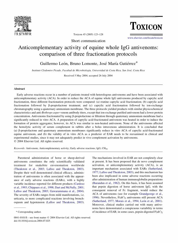
Fig. 1. Anticomplementary activity (ACA) of antivenoms in vitro (A) and
human serum and incubated. Then, sheep erythrocytes sensitized with rab
and centrifuged, and the extent of hemolysis assessed by recording the abso
samples incubated with buffer instead of antivenom represent 100% compl
that reduced hemolytic activity of complement by 50%. Assays were
intravenously into groups of 10 rabbits (2.5–3.0 kg). One hour later, rabbit
vivo complement hemolytic activity. For each rabbit, CH50 was also deter
Hemolytic activity of serum was expressed as percentage, taking as 100%
before antivenom infusion. Results are presented as meanGSD. Identificat
antivenom fractionated with caprylic acid followed by b-propiolactone tr
filtration in an ion-exchange membrane of quaternary ammonium; and (4) a
when compared with groups (1) and (4).
mean values of experimental groups was determined by
ANOVA, followed by Tukey–Kramer test.
As shown in Table 1, the four antivenoms had a similar
protein concentration, although antivenom passed through
the ion-exchange membrane had a significantly reduced
protein content when compared with that of caprylic acid
heated and non-heated antivenoms. Heated antivenom
presented a larger content of aggregates, and showed
in vivo (B). (A) Various dilutions of each antivenom were added to
bit anti-erythrocyte antibodies were added. Samples were incubated
rbances of the supernatants at 540 nm. Values obtained from serum
ement activity. ACA was expressed as the reciprocal of the dilution
performed in triplicate. (B) Antivenoms (10 ml) were injected
s were bled and the CH50 of serum was determined as an index of in
mined in serum samples collected before antivenom administration.
the value of CH50 of samples of each individual rabbit collected
ion of antivenoms: (1) antivenom fractionated with caprylic acid; (2)
eatment; (3) antivenom fractionated with caprylic acid followed by
ntivenom fractionated with caprylic acid and then heated. * P!0.05

G. Leon et al. / Toxicon 45 (2005) 123–128126
a significantly higher turbidity than the other antivenoms.
There were no significant differences between antivenoms
in the antibody titer against the venom of B. asper (Table 1).
No overt differences were observed in the electrophoretic
patterns of antivenoms in polyacetate membranes, charac-
terized by a conspicuous band in the gamma- and beta-
glogulin regions (results not shown). The four antivenoms
showed ACA on human complement in vitro (Fig. 1A).
b-Propiolactone and ion-exchange-fractionated antivenoms
had a significantly lower ACA activity than antivenoms
fractionated by caprylic acid alone (Fig. 1A); b-propiolac-
tone-treated antivenom presented the lowest ACA. In
contrast, when antivenoms were administered i.v. to rabbits,
the hemolytic activity of complement was not significantly
reduced, even in the case of heated antivenom (Fig. 1B).
Our results demonstrate that addition of b-propiolactone
or purification through an ion-exchange membrane signifi-
cantly reduces the ACA of equine whole IgG antivenom
which was initially fractionated by caprylic acid precipi-
tation of non-IgG proteins. b-propiolactone has been
previously shown to reduce ACA of human IgG prep-
arations (Stephan, 1975) and anaphylaxia induced by equine
antivenoms and antitoxins in guinea pigs (Guidolin et al.,
1997), as well as to inactivate virus in the manufacture of
human immunoglobulin preparations for intravenous use
(Burnouf and Radosevich, 2000). On the other hand, ion-
exchange chromatography on anion exchangers has been
widely used for human IgG purification (Jungi et al., 1986;
Tanaka et al., 2000) and in antivenom production, as
a purification step after initial antibody fractionation
(Grandgeorge et al., 1996; Saetang et al., 1997; Jones and
Landon, 2003). Thus, it is suggested that these methods
could be included in antivenom fractionation protocols after
IgG purification by caprylic acid precipitation to further
purify whole IgG antivenoms and to reduce their ACA. The
characteristics of the antivenoms resulting from the three
protocols tested are highly similar in terms of aggregate
content, electrophoretic pattern and antibody titer, although
the use of ion-exchange membrane filtration caused a small
reduction in protein concentration, probably due to the
removal of traces of non-IgG serum proteins. A somehow
surprising finding was that heated antivenom, which
presented a higher content of aggregates, showed a similar
ACA in vitro that antivenom fractionated with caprylic acid
only. It is likely that, in order to increase ACA, a higher
content of aggregates is required. A stronger heat treatment
is probably required to achieve such higher aggregate
antivenom.
Despite the observation of ACA of antivenoms in vitro,
no such activity was detected in rabbits in vivo after a bolus
i.v. administration. These observations raise doubts on the
clinical significance of the in vitro ACA test as a predictor of
ACA in vivo. Various workers have described ACA activity
by antivenoms in vitro (Sutherland, 1977; Montero et al.,
1989; Otero et al., 1999; Leon et al., 2001), but evidence of
ACA in vivo in snakebitten patients receiving antivenoms is
weak. One of the most detailed studies performed on this
subject did not demonstrate complement activation in vivo
in a number of patients receiving various antivenoms,
despite the observation of ACA activity of these products in
vitro (Malasit et al., 1986). In our case, the dose
administered in rabbits (3–4 ml/kg) is higher than the dose
usually administered in humans, which roughly corresponds
to 1–2 ml/kg (Gutierrez, 1995). It may be that the assay used
to assess ACA in vitro does not predict complement
activation in vivo and, therefore, may not be a good
laboratory tool to evaluate the safety of antivenoms.
Alternatively, the test used to evaluate complement
activation in vivo, the CH50 assay, may not be of high
enough sensitivity to detect a minor activation of comp-
lement which may be pharmacologically significant for the
induction of EARs. For instance, high-dose intravenous
treatment of human immunoglobulin preparations induces
complement activation, demonstrated by increments in
serum C3bc, Bb and C5a, although no changes in
complement hemolytic activity, i.e. CH50, occurred
(Mollnes et al., 1998). Nevertheless, Montero et al. (1989)
demonstrated a reduction in CH50 in rabbits receiving a
whole IgG antivenom obtained by ammonium sulfate
precipitation, a procedure that yields a product with higher
content of aggregates and non-IgG proteins (Otero et al.,
1999). Additional studies aimed at detecting complement
activation in vivo, in both clinical and experimental settings,
are required to ascertain the value of in vitro ACA of
antivenoms in the prediction of EARs.
In conclusion, introduction of additional steps in the
production of whole IgG antivenoms, especially the
treatment with b-propiolactone after the purification of
horse IgG by caprylic acid precipitation, significantly
reduces in vitro ACA activity. If this test is indeed useful
in the assessment of the safety of these immunobiologicals
and in predicting EARs in patients, an antivenom produced
by caprylic acid precipitation and b-propiolactone treatment
would induce less EARs than an antivenom produced only
by caprylic acid precipitation. This hypothesis is currently
being tested in a clinical trial.
Acknowledgements
The authors thank Javier Nunez and the staff of
Production Division, Instituto Clodomiro Picado, for their
collaboration, and to G.H. Steinvorth for the donation of the
ion-exchange membrane. This study was supported by
Vicerrectorıa de Investigacion, Universidad de Costa Rica
(project 741-A1-027) and by UNESCO (grant 883.701-3).
This work was performed in partial fulfillment of
the doctoral degree of G. Leon at the University of Costa
Rica.

G. Leon et al. / Toxicon 45 (2005) 123–128 127
References
Angulo, Y., Estrada, R., Gutierrez, J.M., 1997. Clinical and
laboratory alterations in horses during immunization with
snake venoms for the production of polyvalent (Crotalinae)
antivenom. Toxicon 35, 81–90.
Barandun, S., Kistler, P., Jeunet, F., Isliker, H., 1962. Intravenous
administration of human g-globulin. Vox Sanguinis 7, 157–174.
Burnouf, T., Radosevich, M., 2000. Reducing the risk of infection
from plasma products: specific preventive strategies. Blood
Reviews 14, 94–110.
Cardoso, J.L.C., Fan, H.W., Franca, F.O.S., Jorge, M.T., Leite, R.P.,
Nishioka, S.A., Avila, A., Sano-Martins, I.S., Tomy, S.C.,
Santoro, M.L., Chudzinski, A.M., Castro, S.C.B.,
Kamiguti, A.S., Kelen, E.M.A., Hirata, M.H.,
Mirandola, R.M.S., Theakston, R.D.G., Warrell, D.A., 1993.
Randomized comparative trial of three antivenoms in the
treatment of envenoming by lance-headed vipers (Bothrops
jararaca) in Sao Paulo, Brazil. Quarterly Journal of Medicine
86, 315–325.
Chippaux, J.P., Lang, J., Amadi Eddine, S., Fagot, P., Rage, V.,
Peyrieux, J.C., Le Mener, V., 1998. Clinical safety of a
polyvalent F(ab0)2 equine antivenom in 223 African snake
envenomations: a field trial in Cameroon. Transactions of the
Royal Society of Tropical Medicine and Hygiene 92, 657–662.
Dart, R.C., McNally, J., 2001. Efficacy, safety and use of snake
antivenoms in the United States. Annals of Emergency
Medicine 37, 181–188.
Garcıa, M., Monge, M., Leon, G., Lizano, S., Segura, E., Solano, G.,
Rojas, G., Gutierrez, J.M., 2002. Effect of preservatives on IgG
aggregation, complement-activating effect and hypotensive
activity of horse polyvalent antivenom used in snakebite
envenomation. Biologicals 30, 143–151.
Gawarammana, I.B., Kularatne, S.A.M., Dissanayake, W.P.,
Kumarasiri, R.P.V., Senanayake, N., Ariyasena, H., 2004.
Parallel infusion of hydrocortisone G chlorpheniramine bolus
injection to prevent acute adverse reactions to antivenom for
snakebites. A randomized, double-blind, placebo-controlled
study. Medical Journal of Australia 180, 20–23.
Grandgeorge, M., Veron, J.L., Lutsch, C., Makula, M.F., Riffard, P.,
Pepin, S., Scherrman, J.M., 1996. Preparation of improved
F(ab 0)2 antivenoms. An example: new polyvalent European
viper antivenom (equine), in: Bon, C., Goyffon, M. (Eds.),
Envenomings and Their Treatments, Fondation Marcel Mer-
ieux, Lyon, pp. 161–172.
Guidolin, R., Morais, J.F., Stephano, M.A., Marcelino, J.R.,
Yamaguchi, I.K., Higashi, H.G., 1997. Effect of b-propiolactone
treatment on the complement activation mediated by equine
antisera. Revista Instituto de Medicina Tropical de Sao Paulo
39, 119–122.
Gutierrez, J.M., 1995. Clinical toxicology of snakebite in Central
America, in: Meier, J., White, J. (Eds.), Handbook of Clinical
Toxicology of Animal Venoms and Poisons. CRC Press,
Florida, pp. 645–665.
Jones, R.G.A., Landon, J., 2003. A protocol for ‘enhanced pepsin
digestion’: a step by step method for obtaining pure antibody
fragments in high yield from serum. Journal of Immunological
Methods 275, 239–250.
Jungi, T.W., Santer, M., Lerch, P.G., Barandun, S., 1986. Effect of
various treatments of gamma-globulin (IgG) for achieving
intravenous tolerance on the capacity to interact with human
monocyte Fc receptors. Vox Sanguinis 51, 18–26.
Lalloo, D.G., Theakston, R.D.G., 2003. Snake antivenoms. Journal
of Toxicology Clinical Toxicology 41, 277–290.
Leon, G., Monge, M., Rojas, E., Lomonte, B., Gutierrez, J.M., 2001.
Comparison between IgG and F(ab 0)2 polyvalent antivenoms:
neutralization of systemic effects induced by Bothrops asper
venom in mice, extravasation to muscle tissue, and potential for
induction of adverse reactions. Toxicon 39, 793–801.
Malasit, P., Warrell, D.A., Chanthavanich, P., Viravan, C.,
Mongkolsapaya, J., Singhthong, B., Supich, C., 1986. Predic-
tion, prevention, and mechanism of early (anaphylactic)
antivenom reactions in victims of snake bites. British Medical
Journal 292, 17–20.
Mollnes, T.E., Hogasen, K., de Carolis, C., Vaquero, E.,
Nielsen, E.W., Fontana, L., Perricone, R., 1998. High-dose
intravenous immunoglobulin treatment activates complement in
vivo. Scandinavian Journal of Immunology 48, 312–317.
Montero, J., Trejos, M., Lomonte, B., 1989. Efecto del suero
antiofıdico sobre la actividad hemolıtica del complemento
humano (in vitro) y de conejo (in vitro e in vivo). Revista
Costarricense de Ciencias Medicas 10, 1–9.
Morais, J.F., de Freitas, M.C.W., Yamaguchi, I.K., dos
Santos, M.C., Dias da Silva, W., 1994. Snake antivenoms
from hyperimmunized horses: comparison of the antivenom
activity and biological properties of their whole IgG and F(ab 0)2
fragments. Toxicon 32, 725–734.
Moran, N.F., Newman, W.J., Theakston, R.D.G., Warrell, D.A.,
Wilkinson, D., 1998. High incidence of early anaphylactoid
reaction to SAIMR polyvalent snake antivenom. Transactions of
the Royal Society of Tropical Medicine and Hygiene 92, 69–70.
Otero, R., Gutierrez, J.M., Rojas, G., Nunez, V., Dıaz, A.,
Miranda, E., Uribe, A.F., Silva, J.F., Ospina, J.G., Medina, Y.,
Toro, M.F., Garcıa, M.E., Leon, G., Garcıa, M., Lizano, S., De
La Torre, J., Marquez, J., Mena, Y., Gonzalez, N., Arenas, L.C.,
Puzon, A., Blanco, N., Sierra, A., Espinal, M.E., Arboleda, M.,
Jimenez, J.C., Ramırtez, P., Dıaz, M., Guzman, M.C., Barros, J.,
Henao, S., Ramırez, A., Macea, U., Lozano, R., 1999. A
randomized blinded clinical trial of two antivenoms, prepared
by caprylic acid or ammonium sulphate fractionation of IgG, in
Bothrops and Porthidium snake bites in Colombia: correlation
between safety and biochemical characteristics of antivenoms.
Toxicon 37, 895–908.
Otero-Patino, R., Cardoso, J.L.C., Higashi, H.G., Nunez, V.,
Sierra, A., Dıaz, A., Toro, M.F., Garcıa, M.E., Moreno, A.M.,
Medina, M.C., Castaneda, N., Silva-Dıaz, J.F., Murcia, M.,
Cardenas, S.Y., Dias da Silva, W.D., 1998. A randomized,
blinded, comparative trial of one-pepsin digested and two whole
IgG antivenoms for Bothrops snake bites in Uraba, Colombia.
American Journal of Tropical Medicine and Hygiene 58, 183–
189.
Rojas, G., Jimenez, J.M., Gutierrez, J.M., 1994. Caprylic acid
fractionation of hyperimmuine horse plasma: description of a
rapid procedure for antivenom production. Toxicon 32, 351–363.
Saetang, T., Treamwattana, N., Suttijitpaisal, P.,
Ratanabanangkoon, K., 1997. Quantitative comparison on the
refinement of horse antivenom by salt fractionation and ion-
exchange chromatography. Journal of Chromatography 700,
233–239.
Schosinsky, K., Vargas, M., Vinocour, G., Gonzalez, O.M.,
Brilla, E., Gutierrez, A., 1983. Manual de Tecnicas de

G. Leon et al. / Toxicon 45 (2005) 123–128128
Laboratorio. Quımica Clınica. Universidad de Costa Rica, San
Jose p. 224.
Stephan, W., 1975. Undegraded human immunoglobulin for
intravenous use. Vox Sanguinis 28, 422–437.
Sutherland, S.K., 1977. Serum reactions. An analysis of commercial
antivenoms and the possible role of anticomplementary activity
in de-novo reactions to antivenoms and antitoxins. Medical
Journal of Australia 1, 613–615.
Tanaka, K., Sawatani, E., Dias, G.A., Shigeoka, E.M.,
Campos, T.C.X.B., Nakao, H.C., Arashiro, F., 2000. High
quality human immunoglobuin G purified from Cohn fractions
by liquid chromatography. Brazilian Journal of Medical and
Biological Research 33, 27–30.
Theakston, R.D.G., Warrell, D.A., Griffiths, E., 2003. Report of a
WHO workshop on the standardization and control of
antivenoms. Toxicon 41, 541–557.