ANNUAL - OSTI.GOV
114
FR0005095 ANNUAL ILL s& FOR SCIENCE
-
Upload
khangminh22 -
Category
Documents
-
view
0 -
download
0
Transcript of ANNUAL - OSTI.GOV

FR0005095
ANNUAL
ILL
s&
FOR SCIENCE

L ... KA ~.^,
The Institut Max von Laue - Paul Langevin
The institut Laue-Langevin (ILL) is an international research centre using neutrons to probe
the microscopic structure and dynamics of a broad range of materials. The combination of
the world's most powerful neutron source with dedicated instrumentation enables the study
of a wide variety of scientific questions. Problems in materials science, chemistry, biology,
solid-state physics and nuclear physics are investigated. For these diverse studies, the
Institute offers its experimental facilities (some 30 instruments) to scientists world-wide via
a peer-review system, and confidential industrial experiments are also welcomed.
NEUTRONSFOR SCIENCE

The MENI consortium (Austria and Czech Republic)signs the membership contract during a ceremony
for all scientific members,held at the ILL on the 3«< of May 1999.
The Czech Republic became a memberat the beginning of 1999.
The Prefect Alain Rondepierre (centre)visits the ILL on the 24th of November 1999;on the left Ekkehard Bauer, head of the reactor division.
David Schildt (left), head of the British associatesdelegation and chairman of the steering committee,
and Dirk Dubbers (centre), the ILL director,welcome the mayor of Grenoble, Michel Destot,
during the steering committee meetingon 29 November 1999.
Vittorio De Giorgio (left), vice-president ofINFM, University of Pavia, and Manuela Arata(centre), general director of INFM, INFMGenova, inaugurate the OGG (OperativeGroup in Grenoble) of the Italian scientificcommunity INFM on the ESRF/ILL site,here together with Yves Petroff, the ESRFdirector.This group co-ordinates the activitiesof INFM scientists and technicians working atthe ESRF and ILL and gives scientific, technicaland logistic support to Italian users.

REPORT
HIGHLIGHTS"
BIOLOGY
SOFT MATTER
LIQUIDS S. GLASSES
CHEMISTRY & STRUCTURE
MATERIALS SCIENCE
THIN FILMS
MAGNETISM
. a
13
3O
36
44
43
FUNDAMENTAL PHYSICS 53
QUANTUM SYSTEMS 6 4
2
3
4
6 9
WDEVELOPMENTS
PROGRAMME . .
AND FIGURES
6 •PUBUCATOKIS
. 37
1O1

EMRECT'OR'S
1999 started on a high point for ILL. The long-awaiteddelivery of reactor fuel coincided with the new year,ensuring supplies for the ILL neutron source well into
' the new Millennium. At the same time, 1999 saw a posi-tive improvement in inputs to funding, due largely to the coming intoforce on I January 1999 of new contracts with our six "ScientificMembers". Scientific Member countries include our latest partnerthe Czech Republic, together with Austria, Italy, Russia, Spain andSwitzerland. ILL's associate members are France, Germany and theUnited Kingdom. The formal signing of the new contracts was cele-brated at ILL in early May 1999, with representatives of political andscientific bodies.
New funds gave the ILL sufficient means to meet some of the fluctua-ting costs being encountered in 1999, such as the fuel cycle, but alsoto come to grips with projects launched in the past but recently becal-med due to lack of funding. Most significantly, however, it was in 1999that ILL was able, for the first time in fifteen years, to give seriousattention to the systematic renovation of its instrument suite - a pointwhich allows me to move from the necessary question of "inputs" tothe main topic of this report, namely ILL's essential "output".The ILL's output is the operation of the highest quality instrumenta-tion for the benefit of its numerous researchers, to produce ground-breaking scientific results. In the pages that follow you will find across-section of recent results -1 hope you enjoy reading it.Preparatory work on the Millennium development programme was apart of ILL's scientific output in 1999. Early in the year, a 200-pageprogramme document was produced, which gave details of morethan thirty proposals for the renewal of the instrument suite.
The first five of these projects will be launched early in 2000. Thismodernisation is particularly urgent, if we are to carry into the futurethe Institute's capacity to optimise returns from the sums with whichit is entrusted. Whilst it is true that the Millennium Programme hasa relatively slim financial base and is prey to a number of uncertain-ties, given the imagination, determination and confidence whichexists at ILL, a great deal of progress will certainly be achieved.
1999 also saw the departure of Alan Leadbetter and PhilippeLeconte, our former British and French directors. We take thisopportunity to thank them again most sincerely for the depth andquality of the services rendered during their five years at ILL. Theirwork has been taken up with great enthusiasm by their successors,Colin Carlile and Christian Vettier.
There were other noteworthy events in 1999. The beginning of theyear 2000 brings the new French 35-hour week into force at ILL.Negotiations between management and staff ended in a timely agree-ment on the framework to be adopted. ILL's industrial R&D unit wasdeveloped and the nucleus of a scientific communication unit wasestablished. The ILL's organisation of the 1999 Winter Atomiades atLes Gets in Haute-Savoie was a great success, with 250 participantsfrom 5 different countries competing in various skiing disciplines.
Finally, I must report the breakdown of ILL's hot neutron source at
the end of the year which affects four instruments. However, this
only serves to underline the real need to renew ILL's infrastructure.
A new hot source will be available in 2001.
All in all, 1999 was a good year for ILL. May the new Millennium
bring many more!
L'annee 1999 a tres bien debutee avec un evenement capi-tal pour FILL : la livraison de combustible tant attendue,. qui permettra egalement d'assurer l'approvisionnement
de la source neutronique de 1'ILL au cours du prochain millenaire,est arrivee juste a temps pour la Nouvelle Annee. La situation finan-ciere de 1'ILL s'est egalement amelioree en 1999, en particulier gracea Fentree en vigueur des nouveaux contrats avec nos six partenairesscientifiques: 1'Autriche, l'Espagne, l'ltalie, la Russie, la Suisse, et,depuis peu, la Republique tcheque. Les pays membres sontl'Allemagne, la France et le Royaume-Uni. Debut mai 1999, la signa-ture des nouveaux contrats a ete dument celebree a 1'ILL en presencedes representants politiques et scientifiques des differents pays.Les ressources supplementaires ont permis a 1'ILL de faire face a['augmentation de certains couts, dans le cycle du combustible parexemple, mais aussi de mener a bien certains projets entrepris il y a
plusieurs annees mais restes inacheves par manque de moyens.Avant tout, elles ont permis a 1'ILL d'envisager, pour la premiere foisdepuis quinze ans, un renouvellement systematique de son pare ins-trumental.
Ainsi, apres avoir parle des ressources necessaires au fonctionne-ment de l'lnstitut, j'en viens maintenant a la partie essentielle de cerapport, e'est-a-dire la production de FILL. Celle-ci emane de l'ex-ploitation des dispositifs experimentaux par de nombreux scienti-fiques des laboratoires et des universites des pays membres et par leschercheurs de l'lnstitut. Les pages suivantes de ce rapport vous don-nent, comme d'habitude, une vue d'ensemble des travaux effectuesau cours de l'annee ecoulee, en mettant 1'accent sur une serie deresultats marquants et parfois innovateurs. J'espere que, cette anneeencore, leur lecture vous procurera un certain plaisir.
L'elaboration du programme de renouvellement des instruments,

le Programme Millenium, fait également partie des réalisationsscientifiques de l'année 1999. Plus de 30 propositions détaillées pourle renouvellement du parc instrumental de FILL ont été présentéesau printemps dans un document de 200 pages. Les cinq premièrespropositions doivent être mises en œuvre dès le début de l'an 2000.Cette modernisation de FILL est d'une importance primordiale sil'on veut rentabiliser dans les années à venir le capital investi dansl'Institut. La base financière du Programme Millenium est certesétroite et de nombreuses incertitudes persistent, cependant, grâce àl'imagination, la confiance et la ténacité déjà à l'œuvre à I'LL, desprogrès importants seront réalisés.
Au cours de l'année 1999, les Directeurs britannique et français,Alan Leadbetter et Philippe Leconte, ont quitté l'Institut à l'issue deleur mandat de cinq ans. Nous voudrions une fois de plus les remer-cier pour les immenses services qu'ils ont rendus à FILL au cours decette période. Leur travail est poursuivi avec enthousiasme par leurssuccesseurs, Colin Carlile et Christian Vettier.
D'autres événements ont également marqué cette année 1999. Audébut 2000, la semaine de 35 heures sera introduite en France. Lepersonnel et la Direction de FILL sont parvenus, dans les délaisfixés, à trouver un accord sur les modalités d'application à l'Institutde la réduction du temps de travail. D'autre part, l'ILL a renforcé seséquipes à la fois au sein du groupe chargé des relations avec l'in-dustrie, et dans le domaine de la communication. Enfin, FAtomiaded'hiver 1999 organisée aux Gets par l'ILL a regroupé 250 partici-pants de 5 pays différents et a remporté un franc succès.
Je dois malheureusement terminer ce bilan sur une note d'amertumeet annoncer la défaillance de la source chaude de l'ILL. Quatre ins-truments seront affectés par cet événement survenu à la fin de l'année.Cette défaillance souligne la nécessité de renouveler les infrastruc-tures de l'ILL. Une nouvelle source chaude sera disponible en 2001.
Dans l'ensemble, 1999 aura été une bonne année pour l'ILL. Que denombreuses années tout aussi fructueuses lui succèdent au cours duprochain millénaire !
Das Jahr 1999 hatte für das ILL einen guten Start:Pünktlich zum Neuen Jahr erreichte uns die langerwar-tete Lieferung Brennstoff, die die Versorgung der
Neutronenquelle des ILL auch im neuen Millenniumsicherstellen wird. Auch die Versorgung des ILL mit den notwendi-gen Geldmitteln hat sich 1999 etwas erholt. Dies lag zu einem gutenTeil daran, dass am 1. Januar 1999 die neuen Verträge mit unserensechs sog. "wissenschaftlichen Mitgliedsländern" in Kraft traten.Die Mitgliedsländer sind Italien, Österreich, Russland, Schweiz,Spanien, sowie die neu hinzugekommene Tschechische Republik;die Stammländer des ILL sind Deutschland, Frankreich undGrossbritannien. Anfang Mai 1999 wurde die Unterzeichnung derneuen Verträge mit Delegierten aus Politik und Wissenschaft am ILLgebührend gefeiert.
Die neuen Geldmittel erlaubten es dem ILL in 1999, nicht nur einigeTeuerungen zum Beispiel im Brennstoffkreislauf aufzufangen, son-dern auch vor Jahren begonnene, aber durch Geldknappheit insStocken geratene Vorhaben abzuschliessen. Vor allem aber wurde esdem ILL erstmals seit 11/2 Jahrzehnten überhaupt wieder möglich,über eine systematische Erneuerung seines Instrumentenparks nach-zudenken. Und hiermit komme ich, nach der Diskussion des not-wendigen "Inputs", zum wesentlichen Teil dieses Berichtes - dem"Output" des ILL.
Unser Produkt ist die Nutzung der wissenschaftlichen Instrumentedurch unsere vielen auswärtigen Gäste und durch die Mitarbeiter desILL. Auf den folgenden Seiten der vorliegenden Broschüre wird, wiegewohnt, das Produkt dieser Nutzung, nämlich die oft hervorragen-den, manchmal bahnbrechenden wissenschaftlichen Arbeiten amILL aus jüngster Zeit dargestellt, und ich wünsche wieder vielVergnügen bei der Lektüre.
Zur wissenschaftlichen Produktion des Jahres 1999 gehört auch dieAusarbeitung des Millennium-Erneuerungsprogramms. In dem im
Frühjahr vorgelegten Dokument werden auf 200 Seiten mehr als 30detaillierte Vorschläge zur instrumenteilen Erneuerung des ILL vor-gestellt. Die ersten fünf Erneuerungsvorschläge sollen ab Beginn desJahres 2000 in die Tat umgesetzt werden. Diese Erneuerung des ILList von grosser Dringlichkeit, um die hohe Rentabilität der im Institutinvestierten Mittel auch in Zukunft zu gewährleisten. Zwar ist diefinanzielle Basis des Millennium-Programms schmal und durchmanche Unwägbarkeiten gefährdet, aber mit Phantasie, Zuversichtund Hartnäckigkeit wird sich viel erreichen lassen.
Im Laufe des Jahres 1999 verliessen uns die bisherigen britischenund französischen Direktoren, Alan Leadbetter und PhilippeLeconte, die in ihrer jeweils fünfjährigen Amtszeit dem ILLimmense Dienste geleistet haben, für die wir Ihnen hier nochmalsherzlich danken. Ihre Arbeit wird mit grossem Enthusiasmus vonihren Nachfolgern Colin Carlile und Christian Vettier weitergeführt.
An weiteren Ereignissen des Jahres 1999 sind zu vermerken: Mitdem Jahr 2000 wird in Frankreich die 35-Stundenwoche eingeführt,auf deren Modalitäten sich Belegschaft und Institutsleitung auf demVerhandlungswege rechtzeitig zum Jahresende geeinigt haben. DieGrappe zur industriellen Nutzung des ILL wurde ausgebaut, und eswurde der Kern einer Gruppe zur professionellen Öffentlichkeitsar-beit ins Leben gerufen. Die Wintersport-Atomiade 1999 wurde inLes Gets für 250 Teilnehmer aus 5 Ländern mit grossem Erfolg vomILL ausgerichtet.
Schliesslich muss, als Wermutstropfen in diesem Bericht, der Ausfallder heissen Neutronenquelle des ILL gemeldet werden. Von diesemgegen Ende des Jahres aufgetretenen Ereignis sind vier Instrumentebetroffen. Eine neue Heisse Quelle wird 2001 zur Verfügung stehen.
Alles in allem war 1999 ein gutes Jahr für das ILL, mögen viele wei-tere im neuen Millennium folgen.
Dirk Dubbers

Dirk Dubbers (centre) sees off his colleagues,Alan Leadbetter (left, white shirt), the British directorand head of science, and Philippe Leconte(right, white shirt), the French director and head ofprojects & techniques.
Alan Leadbetter welcomes his successor ChristianVettier.the new French director coming from ESRF.
Colin Carlile, the new British director and head ofthe projects & techniques division, enjoying his firstILL Steering Committee meeting; on the leftMichel Destot, the Mayor of Grenoble.
The ILL finishes champion of the medal tableat the Atomiade in Les Gets; from left
Bob Pratt, Barbara Standke, Ariel Brun andHeinz Rhein, FZ Julich.The 7 * Atomiade,
organised by the ILL works committee on13-20 March 1999, gathered about 250 peoplefrom European research centres to compete
in a variety of skiing disciplines.

: • , . : • • • - : - : • , • • • • • : • • : • • . . . : • • • • : • . - • • . . • • • ' . • • • • . : . : • • • • . : • . • • - . - • . ^ . ^ : o - : » w W : ¥ :
SGIEHIGHLIGHTS
Our goal at the Institut Laue Langevin (ILL) is to achieve scientific excellence. Thisvolume of the Annual Report for the year 1999 provides the best illustration of thisattempt: 30 highlights are presented covering a remarkable range of science and technique.As with last year's report, a wide spectrum of neutron science is embraced from biology,through to studies in chemistry, materials science and magnetism, to particle physics.To maintain this excellence, important efforts have been made in instrumentation; theyare described in the technical section together with the Millennium Programme. Thisreport is an appropriate illustration of the unique value of neutron methods for the studyof a wide range of materials proving the usefulness of large-scale facilities such as the ILL.
In 1999, the reactor operated for 208 days and more than 700 experiments were carriedout in over 4400 days of scheduled beam time. Unfortunately, there was a failure of thehot source in December 1999 leading to a loss of about 20 days of beam time.Therefore,in 2000 the reactor will operate without the hot source. However, this will only affect 10%of ILL's instruments. AS in previous years, a large number of high-quality experiments wasproposed and performed to tackle a broad range of scientific questions. For example inbiology, the results presented here demonstrate that the contrast-variation method com-bined with small-angle scattering and neutron reflectivity techniques is a remarkable andunique tool for the investigation of biological materials. In the field of soft matter andliquids, it is known that confined geometry substantially modifies the properties of sys-tems as diverse as simple water, polymers and quantum fluids. Neutron scattering experi-ments, presented here, have revealed the static and dynamic characteristics of thesematerials under conditions of confinement. In materials sciences, the penetration andcontrast properties of neutrons are exploited, particularly in the case of small-angle scat-tering applied to alloys. Finally, the exploration of magnetism with neutrons continues tobe very productive: simply to mention the discovery of a new class of phase transitionsinvolving magnetic chirality and the observation of super high-spin magnetic clusters.
We thank all our collaborators who have submitted highlight reports for this AnnualReport, but we regret that we had to make a selection. For the year 2000 report, we inviteour users again to submit highlights. Note that this version of the I 999 Annual Report willbe followed by a version dedicated to the general public. Herma Biittner who has been theeditor of the ILL Annual Report for the last 4 years, has announced her departure fromILL to start a new career. The staff and the management of ILL would like to reiteratetheir grateful thanks to Herma for her dedication to Scientific Coordination andCommunication at ILL.
<r.? / ; &

BIOLOGY
D . P I G N O L , J . H E R M O S O A N D J . C . F o N T E c I L L A - C A M p s ( L C C P , C E A - C R N S G R E N O B L E ) ,
L . A Y V A Z I A N , B . K E R F E L E C , I . C R E N O N A N D C . C H A P U S ( C N R S M A R S E I L L E ) ,
P . T I M M I N S ( I L L ) .
The digestive process is carried out by secretions from several organs: the salivary glands, the stomach, the gall bladder and the pan-
creas. In the small intestine, pancreatic lipase (PL) hydrolyses insoluble triglyceride by binding to the oil-droplets in the presence of its
protein cofactor colipase (PC) and bile-salt micelles. Combined x-ray and neutron diffraction experiments have previously revealed
the crystal structure of an activated lipase-colipase non- physiological micelle complex. This structure suggested that the adsorption
of the enzyme to the emulsified oil-droplets is mediated by this ternary complex.We decided to carry out small-angle neutron scat-
tering experiments with D2O/H2O contrast variation in order to characterise the physiological ternary PC/PL/bile salt micelle com-
plex in solution. The experimental radii of gyration and match point of the protein/micelle solutions correspond to those expected
for a ternary complex, similar to the one observed in the crystal structure. This preformed ternary complex would be the entity that
adsorbs to the water-insoluble substrate.
Triglycerides (TG) comprise more than 95% of the dietaryfats in the western diet. TG are mainly cleaved in the smallintestine by pancreatic lipase (PL) into triglycerol and fatty-acids. TG, which are insoluble in water, form oil-droplets. Asa consequence, TG hydrolysis implies the adsorption of PL tothe oil-water interface II!. In the intestine, PL alone wouldnot be able to react against the emulsified oil-droplets: itsactive site is covered by a loop that prevents the access of thesubstrate. Two other physiological components are requiredfor PL activation (the unmasking of the active site and the
1-10-3 2 - 1 0 3 3-10-3 4-10-3
adsorption of the enzyme to the substrate interface): 1, coli-pase (PC), a small protein also produced by the pancreas thatforms a complex with PL and 2, bile-salts, that solubilise fatty-acids (the product of the hydrolysis) into mixed micelles thatare later discharged into lymph or blood. The aim of our stu-dies is to understand, at a molecular level, the interactions bet-ween the different components of the system (PL, PC, bile-saltsmicelles, substrate oil-droplets). We initially solved the crys-tal structure of a PL-PC-micelle complex using x-rays j 2 i. Inthis complex, the detergent micelles were non-physiological.
1-103 2-1O3
Q2 Q2
Figure I: Guinier plots ofPLIPC complex solutions at different D2O concentrations: a) in the absence of bile-salt micelles,b) in the presence of bile-salt micelles.

The structure solved at 2.8 A resolution, only revealed thestructural organisation of the protein components of the sys-tem (PC/PL). Subsequently, we decided to carry out a neu-tron-diffraction analysis. Neutron diffraction data collected to15 A resolution allowed us to obtain the structure of themicellar partner of the system in the crystal [31.
In the resulting ternary complex, both PC and the micellehelp stabilising the PL activated conformation. The aim ofthe work reported here was to characterise the ternaryPC/PL/bile salt micelles in solution. We carried out neutronsmall-angle scattering experiments on D22, combined withthe D2O/H2O contrast variation method because this proce-dure is very well adapted to obtain structural information onsystems composed of two components of different scatteringdensity (such as lipid-protein associations).
Initial experiments were carried out on PC/PL complex solu-tions, in the absence of bile-salt micelles. The Guinier plotsat different D2O concentrations are presented in Fig. la. Theresulting radius of gyration at infinite contrast is 26 A andthe match point of the solution is 41.6% D2O. These valuesare in good agreement with those calculated for the PC-PLcomplex using the x-ray structure coordinates (Rg = 25 A,match point = 42% D2O).
We applied the same procedure to spectra obtained from thePL/PC complex solutions in the presence of bile-saltmicelles (sodium taurodeoxycholate micelles). The Guinierplots at different D2O concentrations are presented inFig. lb. The resulting radius of gyration is 56 A, and thematch point of the solution is 38.5% D2O. Since both valuesdiffer significantly from the ones obtained in the absence ofmicelles, different protein-micelle associations weremodelled.
The experimental values are very close to those calculatedfor a model built with a stoichiometry of 2 lipase/colipasemolecules per micelle (Rg = 55 A Match point = 39.2%D2O), which corresponds to that observed in the neutronlow-resolution crystal structure (Fig. 2). All the other modelswe built did not fit the experimental results.
These results clearly demonstrate for the first time, in solu-tion, the formation of a ternary PC/PL/bile-salt micelle com-plex. This complex is similar to the one observed previouslyin crystals with a non-physiological detergent. Our resultsstrongly suggest that the association of PC and bile-saltmicelle stabilises the enzyme in its active conformation. Thepreformed complex behaves as an entity that adsorbs to thesubstrate oil-droplets 4|.
Rg = 55 Amatch point 39 %
Figure 2: Calculated model built with a stoichiometry of 2 lipaselcolipase molecules per micelle corresponding to observed low-resolution crystal structure.
B . B O R G S T R O M E T A L . , E U R . J . B I O C H E M . 3 7 ( 1 9 7 3 ) 6 0 • [ 2 ] J . H E R M O S O E T A L . , J . B I O L . C H E M . 2 7 1 ( 1 9 9 6 ) 1 8 0 0 7
J . H E R M O S O E T A L . , E M B O J . 1 6 ( I 1 9 7 ) 5 5 3 1 • [ 4 ] D . P I G N O L E T A L . , J . B I O L . C H E M . 2 7 5 ( 2 0 0 0 ) 4 2 2 0 .
V

H . C H A N Z Y ( C E R M A V - C N R S , G R E N O B L E ) ,
Y . N I S H I Y A M A ( U N I V . T O K Y O ) ,
P . L A N G A N ( L O S A L A M O S N A T I O N A L L A B O R A T O R Y ) .
Neutron fibre diffraction experiments carried out at the ILL on instrument DI9 have provided detailed new information on the posi-
tion of hydrogen atoms in cellulose II and on the hydrogen bonding network that holds the cellulose structure together. DI9 is cur-
rently the only instrument in the world that is capable of this type of fibre diffraction study; this work on cellulose illustrates its
potential for the investigation of other biological and non-biological polymer systems.
Cellulose is often said to be the most abundant polymer onearth. It is certainly one of the most important structural ele-ments in plants and other living systems. In nature it is syn-thesised as slender rod-like crystalline microfibrils. One ofthe key features of cellulose is that each of its monomersbears three hydroxyl groups. It is these hydroxyl groups andtheir hydrogen bonding ability that not only play a major rolein directing crystalline packing but also in governing impor-tant physical properties of cellulose materials.
Cellulose can occur in a number of different forms. Nativecellulose is known as cellulose I, but other forms such as cel-lulose II, cellulose III and cellulose IV have been described.We are involved in a long-term study, using the neutron andsynchrotron x-ray facilities in Grenoble, to unravel the finedetails of these allomorphs. In this report, the focus is on cel-lulose II fibres that result from the swelling of fibres inconcentrated sodium hydroxide. By using deuterated sol-vents, crystalline fibres can also be prepared in which all theOH groups in the crystalline unit cell are replaced by ODs.Neutron fibre diffraction data recorded from these samplescan then be used to locate these groups using Fourier ima-ging methods.
Neutron fibre diffraction patterns of cellulose II, obtained atD19 (wavelength 1.529 A) are shown in Fig. 1 (the left partof the diagram shows data recorded from the hydrogenatedsample, and the right part shows the data recorded from thedeuterated sample). Both data sets show diffraction exten-ding to 1.2 A resolution. The data have been indexed with aP2L unit cell having lattice parameters a = 8.01 A, b = 9.04 A,c (chain axis) = 10.36 A, y= 117.1°. Despite strong similari-ties between the left and right parts of the diagram, there arenevertheless substantial differences in the distribution ofintensities, particularly on the equator and on the 4th layerline where for instance the reflection 004 is very strong in thedeuterated pattern, but almost absent in the hydrogenatedone. These differences can be used to image the OH groupsin the cellulose unit cell.
Using the CCP13 program suite 111, 97 independent intensi-ties (I) were measured for each part of the diagram in Fig. 1;41 with I > 2 a and 56 with I < 2 a. These intensities and
OH OD
4 - 4*
Figure I: Neutron fibre diffraction patterns collected from two cellulosesamples, one treated with HaOHIHiO (left hand side) and the other treatedin NaODID2O (right hand side).The fibre axis is vertical and the two patternshave been displayed together by joining equivalent halves along the meridian.
their differences were tested against two different modelsexisting already in literature, which we refer to here as model 1\231 and model 2 [4,5!. Both models agree on several points,namely that the structure of cellulose II is based on a twochain unit cell where the chains are antiparallel and that thechains are located on the 2\ axes of the monoclinic cell.However, the two models differ significantly in the descrip-tion of their chain conformations. We could not differentiatebetween these two models on the basis of their agreementwith the x-ray data alone.

Figure 2: Final 2Fd-Fc map (red density) of cellulose II, showing views of the planes containing a) the centre chains b) the origin chains and c) origin and centrechains. Cellulose chains are represented by a skeletal model in which carbon atoms are yellow, oxygen atoms are red and exchangeable hydrogen atoms are pink.The arrows in a) indicate density peaks which could not be accounted for by exchangeable hydrogen atom positions. The potential hydrogen bonds are represen-ted by broken lines.
origin
OS 05
D2
03
05
%
C
OS
02
6
02
origin
05
03
05
06
02
06IS
Un
origin
05
03
OS 06
06
origin
03
05
D3
05
02
06
M
03
02
06
03
centre
05
031
1OS
j
16
m
02
origin
03
05
03
05 06 03
centre
05
05
06
03
06
0
centre
s
0! 05 ,
05
03
S
02
D6
03
06
03
D6
02
centre
2 05
05
OS
03
Figure 3: A schematic representation of the hydrogen bonding in cellulose II.
The H/D atom positions of both models were then refinedagainst our D19 neutron diffraction data. Model 2 was insignificantly better agreement with the data than model 1 andwe were able to reject model 1. A final 2Fd-Fc differenceFourier map is shown in Fig. 2. The identified hydrogen bon-ding system, shown schematically in Fig. 3, reveals some
interesting three and four centred hydrogen bonding arrange-ments \(i}.This study, which has now been followed by similar evalua-tions for cellulose I and III, is the first three-dimensional des-cription of the hydrogen bonding system in a fibrous poiy-saccharide.
• [ : ] F I B R E D I F F R A C T I O N R E V I E W 7 ( 1 9 9 8 ) 1 2 • [ 1 ] F . J . K O L P A K A N D j . B L A C K W E L L , M A C R O M O L E C U L E S 9 ( 1 9 7 6 ) 2 7 3
• [ 3 " : A . S T I P A N O V I C A N D A . S A R K O , M A C R O M O L E C U L E S 9 ( 1 9 7 6 ) 8 5 1 I [ - ' j K . G E S S L E R , N . K R A U S S , T . S T E I N E R , C . B E T Z E L . C .
S A N D M A N A N D W . S A E N G E R , S C I E N C E 2 6 6 ( 1 9 9 4 ) 1 0 2 7 • [ 5 ] S . R A Y M O N D , A . H E Y R A U D , D . T R A N Q U I , A . K Y I C K A N D
H . C H A N Z Y , M A C R O M O L E C U L E S 2 8 ( 1 9 9 5 ) 2 0 9 6 • ; 6 j P . L A N G A N , Y . N I S H I Y A M A A N D H . C H A N Z Y , J . A M C H E M . S O C . 1 2 1 ( 1 9 9 9 ) 9 9 4 0 .

5 I 3^ "^
i t
\ J L ^ J lir"
C . C Z I H A K ( I L L , U N I V . W I E N A N D H M 1 B E R L I N ) ,
G . V O G L ( U N I V . W I E N A N D H M I B E R L I N ) ,
M . M O L L E R ( E S R F ) ,H . S C H O B E R ( I L L ) ,Y . N i S H i Y A M A ( U N I V . T O K Y O ) .
Cellulose is the most abundant structural biopolymer. Despite considerable research efforts the structural details of native cellulose,
in particular on the morphological level, are still controversial. Inelastic neutron scattering provides important clues to understand
the problem. Using the hydroxyl groups of cellulose as a microscopic probe, it is possible to investigate the dynamic response (spec-
trum of vibrational modes) of regions accessible to water. For these disordered regions the dynamic response is distinctly different
from that obtained for the crystal cores and therefore constitutes a signature of the disordered parts. This signature turns out iden-
tical when comparing cellulose types of different crystallinity and origin indicating its universal character.
Cellulose is a complex composite material which structurallycomprises three hierarchical levels: (i) The molecular levelof the single molecule; (ii) the supermolecular level concer-ning the packing and aggregation of the molecules in crystalscalled micwfibrils; and (iii) the morphological level, i.e., thearrangement of microfibrils and interstitial voids in relationto the cell wall (see Fig. 1). On the molecular level, celluloseis composed of linear chains of glucose units. These chainsform whisker-like crystals which are assembled in a super-structure. Details about the arrangement of the chains bothinside the crystals and in the interface between the crystalshave been discussed extensively over the last years; M.
molecule (< 10 A)
microfibrils(20-200 A)
H
plant cell walls
The structural organisation at all levels influences the macro-scopic properties of the material and is equally of importancefor the chemical reactions taking place during industrial pro-cessing. In addition, from structural properties conclusionscan be drawn for the cellulose synthesis in plants.The "classical" two-phase model assuming a compositearrangement of distinct crystalline and extended amorphousregions 12 i to describe the superstructure of cellulose appa-rently has to be revised. Concepts like crystallinity and amor-phicity are well adapted to describe homogeneous states ofmatter. They are, however, rather ill-defined when it comesto treat dense composite materials like cellulose given that
c: fibre axis
monoclinicunit cell (10 A)
Crystalline packing withdisordered interface layer
Figure I: Hierarchical organisation of cellulose.The polymer (top left) aggregates to cellulose nano<rystallites, which are interconnected by disordered interfacelayers (bottom rightf.These crystallites (20 - 200 A) are organised in bundles of single fibres (bottom left).

intermolecular correlations do not build up or die off abrupt-ly at some fictitious interfaces. This equally makes the inter-pretation of diffraction experiments problematic.
A by far better defined quantity than crystallinity or amor-phicity is the accessibility of cellulose to various guest mole-cules, in particular water |3 j. Inelastic neutron scattering inconjunction with deuteration becomes a very powerful toolof investigation. Using the hydroxyl groups of the celluloseas a microscopic probe inelastic neutron scattering allows toinvestigate the dynamic response of regions accessible towater. The low-energy dynamics of the accessible OHgroups in natural cellulose was carried out on the cold neu-tron time-of-flight spectrometer IN6 and is shown in Fig. 2for cellulose material of different origin. Even without quan-titative analysis it is evident that the native samples with nopreferential orientation, namely cotton and sugar beet pulp,show spectra identical to a completely disordered referencesample - amorphous cellulose, all within the experimentaluncertainties.
The restoring forces which are locally encountered by OHgroups are averaged over the accessible parts of cellulose andtherefore do not depend on the cellulose origin.In addition, the dynamic OH response is indistinguishablefrom that obtained on artificial amorphous cellulose and,therefore, it is of universal nature. As amorphous cellulose isfully accessible to water it constitutes the ideal reference towhich the accessible regions of the native samples should becompared. In conclusion the accessible regions seem to cor-respond to disordered regions, which are all similar to eachother.
Albeit universal, the spectral response is not isotropic as canbe seen in the spectrum from oriented flax fibres. Thisdemonstrates that the disordered regions retain a high degreeof directionality. The latter can be linked to the morphologyby identifying disordered regions with surface and singleinterface layers. There is no evidence for the presence of lar-gely extended amorphous regions [4|.
200 250100 150
Time-of-flight channels
figure 2: The dynamic response of disordered regions of cellulose samples which are of different origin." Flax parallel "stands for flax fibres that are aligned inthe scattering plane, so does "flax perp", but perpendicularly aligned.
I H ] A . C . O ' S U L L I V A N , C E L L U L O S E 4 ( 1 9 9 7 ) 1 7 3 • [ 2 ] H . K R A S S I G , C E L L U L O S E : S T R U C T U R E , A C C E S S I B I L I T Y A N D R E A C T I V I T Y ;
P O L Y M E R M O N O G R A P H S I I , G O R D O N A N D B R E A C H S C I E N C E P U B L . : Y V E R D O N 1 9 9 3 I [ 3 ] M . I O E L O V I T C H A N D M . G O R D E E V , A C T A P O L Y M E R
4 5 ( 1 9 9 4 ) I 2 3 I ; < : ] M . M U L L E R , C . C Z I H A K , H . S C H O B E R , Y . N I S H I Y A M A . A N D G . V O G L , M A C R O M O L E C U L E S 3 3 ( 2 0 0 0 ) 1 8 3 4 - 1 8 4 0 .

G . F R A G N E T O , E . B E L L E T - A M A L R I C ( I L L ) ,: : F . G R A N E R , L . P E R I N O - G A L L I C E ( U J F , G R E N O B L E ) ,
T . C H A R I T A T ( I C S , S T R A S B O U R G ) .
Neutron reflection has been applied to the study of a new physical model of cell membranes consisting of a well controlled phospho-
lipid bilayer (free bilayer) floating in water.This system offers several advantages with respect to classical models of biological mem-
branes and it has great potential for studies of membrane/protein interactions and bilayer fluctuations. Measurements were
carried out at several temperatures ranging from 25.4°C, where lipids are in the gel phase, to 64.1 °C, where lipids are in the liquid-
crystalline phase, the transition occuring at 52°C. Around the phase transition large fluctuations inducing giant swelling were observed.
In the liquid-crystalline phase the system has a similar structure as in the gel phase and it is stable over several hours.
The structure and composition of cell membranes is wellknown and described in classical books. Less known are thenature and mechanisms of interaction of membranes withother biological entities like peptides, proteins, DNA, etc.The complicated structure of cell membranes and the varietyof events happening around them makes it difficult to studysuch mechanisms at the molecular level with real cells. Forthat reason biophysicists have searched for decades forsimple and easily-controlled physical models of membranesand several systems are now available including lipid-stacked multibilayers, vesicles, lamellae, monolayers on thewater surface, bilayers on solid surfaces.
We have recently succeeded in preparing double phospho-lipid bilayers assembled at the silicon/water interface |' I j .This is a new system, promising for membrane/protein inter-action studies and ideal for the study of bilayer fluctuations.The first bilayer is adsorbed on the flat solid substrate whilethe second one, named the free bilayer, floats at 15 to 30 Aabove the first and, by interacting less strongly with the solidsubstrate, it is a better model than the single bilayer. The freebilayer has many remarkable properties: it exists in bulkwater, unlike stacked bilayers or monolayers; it fluctuateslike real membranes; it is stable in the biologically relevantfluid phase (as it will be shown below); it can be investigatedalone (that is without averaging the effect of several layers asin lamellae, stacked bilayers and vesicles), with techniquesable to reveal details at the Angstrom scale like neutron orx-ray reflectivity; finally it is a useful model for determininghow fluctuations influence the interactions of peptides ortransmembrane proteins with the cell membrane.
Phospholipid bilayers exhibit a main phase transition from agel phase, where lipid chains are rigid and well ordered, to aliquid crystalline phase (La), where chains are disorderedand fluid-like. Here, we present the results of a neutron spe-cular reflectivity study of the structural modifications indu-ced on double phosphocholine bilayers by changing the tem-perature and going from the gel to the liquid-crystalline
lipid-phases. Preliminary data were collected on the reflecto-meter ADAM while a more complete study was done recent-ly on the rebuilt diffractometer D16 specially adapted forreflectivity measurements. Samples were double bilayers ofhydrogenated DSPC (di-stearylphosphatidil-choline) in D2Oand deuterated DSPC in H2O and SMW (silicon-matchedwater, that is water with the same scattering-length density ofsilicon). Phosphocholines were used since they are the maincomponents of cell membranes. The double bilayers wereassembled on very smooth and highly hydrophilic siliconsingle crystals with the Langmuir-Blodgett and Langmuir-Schaeffer techniques. For full details on the deposition andmeasurement procedures see 11,2].
For hydrogenated DSPC in D2O the phase transition occursat 52°C (3;. As expected deuterated lipids show a transitiontemperature different from that of the hydrogenated ones.Data analysis is still in progress and only the analysis for thehydrogenated lipids will be discussed here. Figure 1 shows
0.2
Figure I: Measured reflectivity profiles and fitted curves for DSPC doublebilayers in D2O at (C) 2SA°Q(%) 5I.5°Q(®) J5.4°C;(@) 64.IX.For details on model used see text and Fig. 2.

the reflectivity curves recorded at room temperature(25.4°C), in the pre-transitional phase (51.5°C), just above(53.4°C) and well above (64.1°C) the transition for fullyhydrogenated DSPC in D2O. Data were analysed by modelfitting using the optical matrix method !4i: the interface isdivided in several boxes, each characterised by a scattering-length density, a thickness and the interfacial roughness bet-ween two consecutive boxes. These parameters are varieduntil the optimum fit to the data is found. Between 25.4°Cand 42°C, curves were very similar and data were interpretedwith the same model. Figure 2a shows a cartoon of such amodel. Lipid headgroups and chains could be distinguishedand their thickness determined with 1 A precision. No mea-surement is available for the hydrogenated lipids between42°C and 51.5°C but at 51.5T and 52.5°C the curves differdramatically from those at lower temperature indicatingmajor changes in the bilayer structure (cf. Fig. 1). Data ana-lysis indicates that the water layer between the bilayersbecomes much thicker (60 to 90 A) and the second bilayermuch rougher (~ 30 A). With such high value of the rough-ness the box-model used to fit the data is not very efficientanymore. Figure 2b is a cartoon of a speculative interpreta-tion of the model. Additional work is needed for a more pre-cise interpretation. Plans for future work include off-specularreflectivity and atomic force microscopy measurements for
extracting information in the membrane plane. Moreover, astudy of the theoretical explanation of the experimentalobservation is in progress [2 j.
A further increase of temperature brings the system nearlyback to the initial state in terms of total thickness (immedia-tely noticeable from the positions of the first minimum inFig. 1). The water layer thickness is 21 ± 1 A. The secondbilayer chain thickness decreases from 3 7 ± l A t o 3 1 ± l A(cf. Fig. 2c). The highest temperature measured was 64.1°C.Surprisingly, the double bilayer is still at the surface althoughthe chain thickness of both bilayers decreases to 27 ± 1 Aand the roughness of the free bilayer is of 10 ± 2 A (cf.Fig. 2d).
Finally, this work has confirmed the good stability ofdouble bilayer systems over several days and at all tempe-ratures investigated and therefore of their suitability asmodel biological membranes. Results are reproducible andpre-transitional effects on the structure are remarkable. Thegiant increase of both the thickness and the roughness at thetransition (i.e. where the bilayer is extremely flexible)might be the first direct observation of the fluctuation-indu-ced repulsion predicted by Helfrich [5] using entropy argu-ments and considered a key ingredient of bilayer-bilayerinteractions.
figure 2: Cartoons of a possible interpretation of models found to fit the data measured at a) 25.4°C, b) 5I.5°C, c) 55A°C and d) 64. TC. For darity,in b), c)and d) only the thickness parameters that change with respect to a) are indicated. Vertical dimensions are drawn to scale.
Rsfenences
I [ ! ) T . C H A R I T A T , E . B E L L E T - A L M A L R I C , G . F R A G N E T O A N D F . G R A N E R , E U R . P H Y S . j . B . 8 ( 1 9 9 9 ) 5 8 3 I [ X ] G . F R A G N E T O ,
T . C H A R I T A T , F . G R A N E R , K . M E C K E , L . P E R I N O - G A L U C E A N D E . B E L L E T - A M A L R I C , S U B M I T T E D T O E U R O P H Y S I C S L E T T E R S I [ 3 ] N I S T
O N L I N E R E F E R E N C E D A T A B A S E S ( L I P I D A T ) I [ 4 ] M . B O R N A N D E . W O L F . P R I N C I P L E S O F O P T I C S ( 1 9 8 9 ) P E R G A M O N P R E S S , O X F O R D
• [ 5 ] W . H E L F R I C H , Z . N A T U R F O R S C H U N G 3 3 A ( 1 9 7 8 ) 3 0 5 .

'fit
C . M O N S T E R , T . S A L D I T T ( U N I V . M U N C H E N ) ,
B . B E C H I N G E R ( M P I B I O C H E M I E , M A R T I N S R I E D ) .
R . S l E B R E C H T , V . L E I N E R ( I L L A N D U N I V . B O C H U M )
We report a neutron-scattering study of multilamellar membranes supported on solid silicon substrates. In contrast to previous
work, the high degree of orientational alignment allows for a clear distinction between specular and nonspecular reflectivity contri-
butions. In particular, we demonstrate that by using the specific advantages of neutron optics the nonspecular diffuse scattering can
be mapped over a wide range of reciprocal space, which is usually not accessible by x-rays. In the neutron case, as is often argued, a
lack of intensity severely limits the practicability of neutron diffuse studies, which are therefore rather scarce. Contrarily, we demons-
trate in this study that several orders of magnitude in scattering signal and parallel momentum transfer can easily be recorded in
multilamellar stacks of lipid membranes.This opens up the possibility to study fluctuations and lateral structure parameters of mem-
branes on length scales between a few A up to several urn.
Lipid bilayers are the basic building blocks of biologicalmembranes [ 11. A quantitative understanding of the elasticproperties, the thermal fluctuations, and the interactionsgoverning membrane self-assembly has significant impacton various research areas, including the biophysics of biolo-gical membranes, synthetic biomaterial engineering, andpharmaceutical applications. In this context, the interactionbetween lipid bilayers and antimicrobial peptides, such asmagainin 2, is of particular interest, since the peptides exhi-bit a broad antibacterial, fungicidal and antitumor activities• ?;. However, the structural motifs of the interaction are pre-sently poorly understood. Therefore, we study fundamentalaspects of structure, elasticity and fluctuations in model sys-tems composed of neutral and charged phospholipids withand without membrane-active peptides such as magainin.
Neutron reflectivity offers unique possibilities to study thevertical density profile of lipid membranes on solid sub-strates with molecular resolution and is widely used for this
Figure I: The scattering experiment in reciprocal space.The schematics of
the incident, the scattered beam and the scattering vector are sketched rela-
tive to the sample surface.The Laue region, which is not accessible in x-ray
experiments, is shaded in gray.
purpose complementing x-ray techniques by means ofcontrast variation. Apart from contrast variation, a uniqueadvantage of neutron diffuse scattering over x-rays resultsfrom the simple fact that most standard substrates are essen-tially transparent, such that the diffuse scattering can be mea-sured continuously for positive and negative angles of inci-dence and scattering, see Fig. 1. Consequently, a much widerrange of the parallel momentum transfer Qn is accessible.Important information on the lateral membrane structure can
0.135
g %&mm*>$i
O0.125
0.12
0.115
-0.007 -0.006 -0.005 -0.004 -0.003 -0.002 -0.001 0
0.00001
Figure 2: Top: a one-dimensional cross-section of the diffuse scattering as a
function of parallel momentum-transfer. Bottom: Reciprocal space mapping
around the first Bragg peak of a stack of multilamellar phospholipid mem-
branes (DMPC).

be revealed, yielding new and previously inaccessible infor-mation on the membrane fluctuation spectrum, i.e. theheight-height self- and cross-correlations.
For the experiments it is a prerequisite to prepare samples ofseveral hundred bilayers oriented perfectly on a silicon sub-strate. The degree of membrane mosaicity achieved is typi-cally better than 0.02°, compared to 1-5° in conventionalmeasurements. These samples show a tremendous amount ofdiffuse intensity reflecting the softness of the system. For theinvestigation the samples were either partially hydrated(water layer of KH5 A in between membranes) and kept atconstant temperature in a humidity cell or completelyimmersed under water in a chamber of full hydration, withthe neutron beam impinging from the side of the silicon sub-strate ;3j.
The contour plot of a reciprocal space mapping around thefirst multilamellar Bragg peak is shown in Fig. 2 (lowerpart). The so-called diffuse Bragg sheet arises from layerfluctuations which are correlated over several adjacent layers(conformal fluctuations) such that the diffuse scattering isconcentrated along ridges of constant Qz corresponding tothe specular Bragg peak positions. From the Qz value, amean distance d of the DMPC membranes of d = 49.8 A canbe inferred, corresponding to a water layer of about 14 A inbetween adjacent membranes. Note that the mapping com-bines data taken in reflection and transmission geometry. Thecollapse of the intensity along the Laue sphere (black line)marks the loci in reciprocal space where the incident beamimpinges at an angle of 0°, in the vicinity of which evanes-cent waves are excited. A one-dimensional transverse scan isalso shown for comparison (Fig. 2, upper part). A lateral cor-relation length of £[] = 1000 A is inferred from the crossoverpoint around 0.006 A"1 between the two characteristicslopes. Note that the nonspecular scattering can be measuredover an unprecedented range of up to four orders of magni-tude in intensity and four orders of magnitude in parallel
momentum-transfer. From this data, the membrane fluctua-tion spectrum can be determined and directly compared totheoretical models |4j.Upon the insertion of antibacterial peptides such asmagainin 2, drastic changes in the specular and nonspecularreflectivity are observed at increasing peptide/lipid ratio(P/L). The first and second order peaks are shown in Fig. 3(right) as a function of Qz for increasing molar peptideconcentration (P/L). Note that the data is clean enough to per-form a line shape analysis of the Bragg peaks. Futhermore,the scattering-length density-profile can be modelled fromthe data and provides information on the configuration of thepeptides with respect to the lipid bilayer 151.In summary, the static structure-factor of solid supportedmembranes was measured as a function of the perpendicularand lateral momentum transfer, making use of the specialadvantages of neutron optics. From the data the fluctuationspectrum of the membrane system was determined [45. Theresults may be put in perspective to the relevant microscopicinteractions and also be compared to current theoretic predic-tions of membrane distortions by inclusions (peptides, pro-teins). In future, the method presented here to map out thereciprocal space over a wide range and with high resolutionmay be applied to many interesting lipid-membrane basedmaterials, including lipid/peptide, lipid/protein or lipid/DNAsystems. The technique can reveal lateral structures onlength scales between a few A up to several um at in situconditions.
0.24
figure 3: Left Sketch of a peptide (green) adsorbed on the lipid bilayer (blue). Right Details of the reflectivity curves (Ist and 2nd Bragg peaks ofmultilamellar DMPC)at increasing peptide concentration (from top to bottom:pure DMPC (PIL-O), PIL-0.025,0.05,0.1). The peptides affect the multilamellar order.
I [ ! j R . L I P O W S K Y , E . S A C K M A N N , S T R U C T U R E A N D D Y N A M I C S O F M E M B R A N E S , H A N D B O O K O F B I O L O G I C A L P H Y S I C S , V O L I . A M S T E R D A M ,
N O R T H - H O L L A N D ( 1 9 9 5 ) I [ 2 ] B . B E C H I N G E R , J . M E M B R A N E B I O L . 1 5 6 ( 1 9 9 7 ) 1 9 7 A N D R E F E R E N C E S T H E R E I N I [ 3 ] M . V O G E L ,
C . M U N S T E R , W . F E N Z L , T . S A L D I T T , P H Y S . R E V . L E T T . 8 4 ( 2 0 0 0 ) 3 9 0 1 H ] C . M U N S T E R , T . S A L D I T T , M . V O G E L , j . P E I S L , E U R O P H Y S .
L E T T . 4 6 ( 1 9 9 9 ) 4 8 6 • \ i \ C . M U N S T E R , j . L u , S . S C H I N Z E L , B . B E C H I N G E R , T . S A L D I T T , E U R O . B I O P H Y S . j . 2 8 ( 2 0 0 0 ) 0 6 8 3 .

SOFT MM
P . M U L L E R - B U S C H B A U M ( T U M U N C H E N ) ,
J . S . G U T M A N N , 0 . W U N N I C K E , M . W O L K E N H A U E R ( M P I
R . C U B I T T ( I L L ) ,
M . S T A M M ( I P F D R E S D E N ) ,
W . P E T R Y ( T U M U N C H E N ) .
P O L Y M E R F O R S C H U N G M A I N Z ) ,
Surface morphology of thin films is another probe of the method of grazing-incidence small-angle neutron scattering (GISANS).
In case of homopolymer samples, GISANS yields the surface topology. In addition for blend samples the chemical composition is
determined with GISANS. Compared to common transmission experiments GISANS enables the determination of very small sample
volumes.
Polymer films are extensively used in many applications.With the ongoing miniaturisation of any type of application areduction of the polymer-film thickness is required. In thelimit of ultra-thin films the thickness is in the order of thepolymer dimension. This induces a different conformation,morphology and kinetics. To determine these confinementeffects, the film thickness of the polymer sample has to besmall compared to the unperturbed radius of gyration of thepolymer molecule (e.g. only 30 A). In this film-thicknessregime, a free-standing film is unstable. Thus, a preparationon top of a solid support is required. In a common transmis-sion set-up the scattering volume of the confined polymer
film is very small as compared to the one of the substrate.The scattering signal contains only a small contribution ofthe polymer film.
Therefore, a transmission geometry is extremely disadvanta-geous. One way to overcome this problem is the use of areflection set-up as demonstrated previously with x-ray scat-tering techniques. With grazing-incidence small-angle scat-tering, a small scattering volume located near the sample sur-face is probed. In contrast to x-ray experiments this geometryis only rarely applied in neutron experiments. This mayresult from the limited fluxes available with neutrons compa-red to x-ray experiments.
2D detector
Horizontal slice
Vertical slice
b)
Sample surface
Figure /; a) Schematic drawing of the reflection set up performing QSANS atD22. b) Example of the two-dimensional intensity distribution measured with GISANS.In the contour plot a logarithmic contour line spacing with an alternating colour scheme is used.

10°
d/Ra
Figure 2: Comparison of surface topographies determined with atomic forcemicroscopy (+) and with GISANS (O). The mean droplet distance A and theoriginally prepared film thickness d are normalised by the radius of gyration R?
The resolution of the scattering experiment is shown with the dashed line.Thesolid line shows a fit to the data based on a spinodal dewetting model.
However, in the case of x-rays, the contrast, even for significant-ly different polymers is not large, whereas with neutrons strongcontrast between two components can be generated by deutera-tion. This is extremely useful for investigations on blends ofweakly incompatible polymers and is commonly applied inneutron reflectometry. The first successful experimentsapplying GISANS have been performed at the D22 instrument.
In general, the D22 instrument is used in transmission geo-metry. The scattering signal of interest is located near thebeam stop which limits the largest resolvable length-scale.We applied a reflection geometry for GISANS. Thus, thesignal of interest is reflected out-of-plane of incidence andgeometrical constraints due to a beam stop are not present.This enables the detection of in-plane length-scales A up to3.3 urn. It is a significant increase compared to the highestresolvable length-scale in transmission geometry under com-parable conditions.
A schematic drawing of the experimental set-up is shown inFig. la. At one fixed angle of incidence a; the scatteredintensity is detected at exit angles ctf and out-of-plane angles \\l.The two-dimensional intensity distribution consists of seve-ral vertical and horizontal slices. Figure lb shows a typicaltwo-dimensional scattering distribution. In the centre of thepicture the specular peak is located. Horizontal slices containinformation parallel to the sample surface. Vertical slices
basically carry information perpendicular to the sample sur-face.In the case of a dewetted confined homopolymer film thelength A can be attributed to the mean distance between thedroplets. During the dewetting of a originally homogeneousfilm isolated monodisperse droplets are created. Thus, theresulting sample is no longer continuous. With increasingfilm thickness, d, the mean droplet diameter and distanceincreases as well. In the past, the investigation of these smallsurface structures were only possible with atomic forcemicroscopy. Thus, only locally very limited information hasbeen available. Due to the progress in the GISANS techniqueit is now possible to determine heterogenieties on surfaces byscattering. In a scattering experiment, the mean droplet dis-tance is determined on basis of a much larger surface areaand therefore with a greater statistical significance. Asdemonstrated previously with atomic force microscopy andgrazing incidence small-angle scattering, using x-rays aswell as neutrons yields identical information in the case ofdewetted confined homopolymer films 11].In more complex systems such as confined blend samples, inaddition to the dewetting, a phase separation is possible. InFig. 2 the mean droplet distances as determined withGISANS (circles) and with atomic force microscopy(crosses) are similar for the complete range of confined films(Rg/3 < d < Rg). Thus the local technique atomic force micro-scopy and the averaging technique GISANS deliver the sametopological information. The observed droplets are explainedby a spinodal dewetting model (solid line in Fig. 2).
An internal contrast within single droplets is available withneutrons whereas with atomic force microscopy and x-rayscattering only the surface topology is detected. UsingGISANS in addition to the topological signal, the internalchemical composition has been determined [2|. A periodicvariation of different polymer phases can be induced by care-ful sample preparation and this internal structure has beenobserved with the GISANS technique [ 3],
Of course, the GISANS technique is not limited to polymersamples. The topology and chemical composition of any typeof sample surface can be probed.In addition, GISANS enables the investigation of non-homo-geneous samples consisting of isolated structures. In-planedistances up to several micrometres are accessible. Due theneed for a very high neutron flux, GISANS experiments arelimited to a few neutron instruments such as D22.
References
1 [ i j P . M U L L E R - B U S C H B A U M , j . S . G U T M A N N , M . S T A M M , P H Y S . C H E M . C H E M . P H Y S . I ( 1 9 9 9 ) 3 8 5 7 I [ 2 ] P . M U L L E R - B l l S C H B A U M ,
J . S . G U T M A N N , M . S T A M M , R . C U B I T T , M A C R O M O L . S Y M P . , I N P R E S S . I [ 3 ] P . M C l L L E R - B U S C H B A U M , J . S . G U T M A N N , R . C U B I T T , M . S T A M M ,
C O L L O I D P O L Y M . S C I . 2 7 7 ( 1 9 9 9 ) I 1 9 3 .

S . B A R E , A . R . R E N N I E ( K I N G S C O L L E G E L O N D O N ) ,
E . B E L L E T - A M A L R I C , G . F R A G N E T O ( I L L ) .
Shear has a profound influence on the behaviour of complex fluids due to their non-newtonian behaviour and neutron scattering is an
important method for the characterisation of shear induced transformations. A new rotating disc shear-cell has been built and mea-
surements on the wide-angle d iff racto meter D16 on concentrated surfactant solutions under shear have allowed to determine orien-
tations not visible with the geometries used in the past on similar systems. A complete study will be useful for many applications
where it is important to know how shear may effect phase behaviour such as micelle to lamellar phase transitions or induce forma-
tion of vesicles.
Amphiphilic molecules such as surfactants are used in manyapplications that range from emulsion stabilisers, detergents,cosmetics, food and mineral processing. Surfactants at highconcentrations in water often form lamellar phases withbilayers packed together to give a regular interlayer spacing(Fig. 1). In some circumstances other concentrated phases(cubic, hexagonal, etc.) may form. The properties of concen-trated phases of surfactants can be of importance in manyareas: such as the formulation of concentrated detergents,properties of foods and behaviour of cosmetic preparations.Flow of soft materials has attracted considerable interest andthe Theological properties of surfactants are no exception.
C1 2EO4
Figure I: Schematic diagram of lamellar phase structure of surfactantsshowing the inter-lamellar spacing used to determine the orientation.
For some years small-angle neutron scattering (SANS) hasbeen used to produce detailed pictures of structures underflow i .• ,:.•]. In the presence of shear field two different orien-tations of the lamellar layers have been observed, respec-tively with the normal to the layers parallel to the velocitygradient direction (orientation c) and perpendicular to it(orientation a). Orientational transitions have been observeddepending on the shear rate and the system studied. In parti-cular, Penfold et al.:; • observed a transition from orientationa at low shear rate to orientation c at high shear rate for theCifcEOg (hexaethylene glycol monohexadecyl ether)/watersystem. In a recent work Richtering at al. {'•!) observed thesame transition for a system of SDS/decanol/water.Additionally, they observed two more orientational transi-tions at very low and intermediate shear rates and the forma-tion of vesicles. Both the transitions and the formation ofvesicles were influenced by the content of decanol. Also inblock copolymer melts, a transition from the a to the c align-ment was observed, but there is no evidence of vesicle for-mation. Recently, neutron diffraction has been used to deter-mine orientational order in dispersions of crystalline,anisotropic colloidal particles pi . Measurement of the distri-bution of intensity of a particular Bragg reflection directlydetermines the orientation distribution of the plate-like par-ticles. In many respects lamellar surfactant phases resemblethe structure of dispersions of plate-like particles. The distri-bution of scattered intensity arising from the lamellar spa-cing can be used to determine the orientation distribution.The flexibility and dynamic equilibrium of lamellae can giverise to new and interesting phenomena.
The principal directions in a shear flow field are shown inFig. 2 and a full description of the alignment will requiredetermination of the full three-dimensional structure, or dis-tribution of orientation, in relation to these axes. In order toachieve a better understanding of the flow behaviour oflamellae under shear, a rotating disc shear-cell has been built.This has been used on the diffractometer D16 to investigatethe orientational alignment, using a sample geometry that isnot readily accessible with a Couette shear cell and conven-

([J\ 1Compressional
Grad ien t " ' » , _
ExtensionalBow
Shear cell
Incident bean
Figure 2: Shear<ell orientation on the diffractometer.The principal axes of
the shear field can be aligned with respect to the scattering vector to allow
full determination of the orientation distribution. It is useful to identify not just
the axes of flow, shear gradient and vortichy but also to distinguish in mea-
surements between the extensional and compressional quadrants associated
with the shear flow.
tional SANS measurements. Data were collected for the non-ionic surfactant tetraoxyethylene glycol monododecyl ether(C12EO4) in D2O, which has a wide range of lamellar phasesat room temperature. The cell geometry allowed scans todetermine the orientation distribution of the lamellar phasesin the plane of shear velocity and shear gradient. With thecell normal to the incident beam the measured intensity cor-responds to scattering from the lamellae with normals almostin the flow direction. As co increases to 90° this angleapproaches the condition of lamellae normal to gradientdirection. Two concentrations corresponding to 40 and60%wt surfactant were studied.
First results from these studies are shown in Fig. 3. Datawere collected over a range of angles and corrected for detec-tor efficiency, sample thickness and transmission. Spectrawere acquired varying the angle co from -75° to +75° (seeFig. 2) at different values of § and % (ranging between 0° and24° for (j) and 0° and 20° for %). All experiments were repea-ted at different shear rates. A strong Bragg reflection atQ = 0.1 A-' for the 60% sample and at Q = 0.07 A-" for the40% sample was observed. This corresponds to a d-spacingof 63 A and 90 A respectively corresponding to the distancebetween adjacent lamellar planes. Two less intense peakswere observed in the diffraction pattern of the 60% sample,at Q « 0.075 A-' (d = 82 A) and at Q = 0.2 A-'. Peaks havebeen fitted with a Gaussian and their intensities have beennormalised making allowance for the variation of the scatte-ring volume, which depends on the cell orientation. The plotof the intensity of the Bragg reflection at Q ~ 0.1 A-' versusthe rotation angle co is shown in Fig. 3b. A maximum isobserved for co « 50°. A similar pattern is observed at highershear rates. The experimental arrangement allowed to inves-tigate only small a range of (j) and %; small variations are
observed in the peak intensities for experiment recorded afterrotating the cell through <j) and %. For the less concentratedsample the plot of the intensity of the Bragg reflection atQ ~ 0.07 A-1 versus the rotation angle co showed a differentpattern with a maximum corresponding to 0 < co < 20 depen-ding on the shear rate (Fig. 3a). These preliminary data clearlyshow alignment that changes with concentration and indirections that have not been reported previously.Suprisingly for the sample at 60%wt fraction, the maximumis seen well away from either the flow or gradient directions.The positive co direction corresponds to the extensional qua-drant that lies between the flow and gradient directions (seeFig. 2). The normals to the lamellae are seen to lie in thisquadrant.
Under the flow conditions studied, orientation is observed ina rather different manner to that reported in previous work onconcentrated surfactants. The lamellae are oriented with thenormal to the plane in the flow gradient-plane and formingan angle with the vorticity-flow plane. The tilt depends bothon the concentration of the surfactant and the applied shearrate. The orientation of the particles is only observable whenscanning a large part of reciprocal space. The observedorientation of the layers with the peak in the compressionaldirection of the flow field shows certain similarities to thebehaviour of plate-like colloidal particles I •-• j. Further work isrequired to determine the exact orientation of the layers as afunction of surfactant concentration and shear rate.
-60 -40 -20 -0 20 40 60
CO
b)
Figure 3: Plots of the normalised intensity versus the angle 0) for a) CjjEf at
40 wt% and b) 60 wt% at two different shear rates (aO.5r> and $30 r ' j .
For the 60% sample the maximum is atw = 50 for both shear rates, while
for the 40% sample the maximum is at a - 0 for 30 s~! and at
I0<a<20for0.5s-i.
I [ i ] J . P E N F O L D , E . S T A P L E S , A . K L A N L O D H I A N D G . j . T . T I D D Y , j . P H Y S . C H E M B 1 0 1 ( 1 9 9 7 ) 6 6 • U ] j . Z I P F E L , J . B E R G H A U S E N ,
P . L I N D N E R A N D W . R I C H T E R I N G , j . P H Y S . C H E M B 1 0 3 ( 1 9 9 9 ) 2 8 4 1 I [ 3 j S . M . C L A R K E , A . R . R E N N I E A N D P . C O N V E R T E U R O P H Y S I C S
L E T T E R S 3 5 ( 1 9 9 6 ) 2 3 3 • ! / • ] A . B . D . B R O W N ' O R D E R I N C O N C E N T R A T E D C O L L O I D A L D I S P E R S I O N S O F A N I S O T R O P I C P A R T I C L E S U N D E R
S H E A R ' P H . D . T H E S I S , U N I V E R S I T Y O F C A M B R I D G E ( 1 9 9 8 ) . A . B . D . B R O W N A N D A . R . R E N N I E , S U B M I T T E D T O P H Y S . R E V . E .

LIQUIDS
-&•
W . S C H M I D T , U . B U C H E N A U ( F Z J U L I C H ) .
Can a quantitative connection be established between the low temperature anomalies of glasses and the fast picosecond motion in
the melt? Very recent inelastic neutron scattering experiments on IN 12 seem to show such a connection.
A reasonably successful description of the low-temperatureglass anomalies is the soft potential model [1], an extensionof the tunnelling model, which postulates a co-existence oflocalised tunnelling, relaxational and vibrational localisedmodes with the sound waves at low frequencies, below theboson peak at 2 meV. This peak may arise from scattering ofthe phonon modes of the whole Brillouin-zone due to disor-der effects in these systems. The localised modes are suppo-sed to interact bilinearly with elastic strains, in order toexplain the strong sound absorption. In terms of that model,an extended strain field is expected around each of theselocalised modes, fluctuating with the mode amplitude.
A calculation of the coherent inelastic scattering from such afluctuating strain field yields a weakly Q-dependent inelasticsignal at small momentum transfer Q, as long as Q remainslarger than the Brillouin scattering wave-vector at the givenfrequency [2]. If these localised modes exist not only at lowtemperatures, but also in the melt, one should be able to seethis small-Q inelastic signal.
The measurements were carried out on the CRG-instrumentIN 12, a three-axis spectrometer for cold neutrons, with asample of amorphous deuterated 1,4-polybutadiene at 288 K(the glass transition temperature is 186 K). For comparison, aprotonated polybutadiene sample was also examined underthe same conditions. Measurements on the empty samplecontainer were used for background corrections.
The instrument was optimised for lowest background at lowmomentum transfer. In addition to 30'-collimation, we used alarge vacuum tank (1 m in diameter) around the cryostat tosuppress the air scattering of the direct beam which producesa strong inelastic signal at small angles. Its efficiency for thepresent problem is demonstrated in Fig.l which comparesthe scans before and after evacuation of the tank. In general,this set-up can be considered as a competitive alternative totime-of-flight instruments for this kind of measurements.
42
o
Figure J: Inelastic scattering on deuterated polybutadiene at 288 K and anenergy transfer of 0.5 meV. The green curve shows the signal with surroun-ding air while the red curve demonstrates the drastic background reductionat low Q after evacuating the tank.
The soft-potential model postulates modes with a stabilisingfourth-order term in the potential, but with varying first andsecond order terms, eventually even negative ones, toaccount for the asymmetric double-well potentials of the tun-nelling states. These modes are assumed to couple to theuniaxial and shear strains of the long-wavelength longitudi-nal and transverse sound-waves, respectively. So far, this isthe only model capable to explain consistently the low-tem-perature glassy anomalies in the heat capacity, the thermalconductivity and the acoustic absorption [3].
If the soft-potential model is correct, there should be fluctua-ting strain fields around each of these localised modes. Thesimplest theoretical description of the strain field around alocalised mode is to assume an elastic dipole in the centre ofthe mode, proportional to the mode displacement. The cohe-rent inelastic small-angle scattering from such an elasticdipole, fluctuating or oscillating in an elastically isotropicmedium, has been calculated recently [2|. This calculationassumes a homogeneous deformation within an inner sphereof radius rs, continuity at the sphere boundary, and a decay

according to the elastic dipole field outside. Besides theradius rs the model requires two further parameters: Thesquared ratio of the transversal and longitudinal sound velo-city called f, and the dipole strength which is expressed interms of a value Qo which also can be calculated from thesoft potential model in the glass phase.
The main difficulty of the measurements is the multiple-scat-tering intensity from the strong inelastic scattering athigher Q. These processes produce a sizeable linear back-ground at low Q of the same order of magnitude as thecontribution we are looking for. Therefore the multiple-scat-tering calculations must be reliable. With this aim a protona-ted sample with the same scattering power was measured,too. For an incoherent scatterer the low Q inelastic signalshould be absent, it should start with a Q2 dependence.Indeed, the offset at low Q confirms the calculations (Fig. 2).
The data of the deuterated sample are shown in Fig. 3. Forthe fit we took into account that the data consist of a mixtureof localised modes and sound waves. Both contributionsmust be considered. For the latter the structure factor is takenas Q2 Sel(Q) where Sei(Q) is obtained from the elastic data.The fit gives about 50% for that contribution. Further, the fit
provides the three relevant parameters for the model of thelocalised modes which have been described above. Theresulting values are Qo = 0.48 ± 0.04 A-i, rs = 8.3 + 0.8 Aand f = 0.25 ± 0.03. From soft potential data J3],Qo = 0.46 A"1 has been calculated. Also the radius rs of thedistorted sphere is a reasonable value in the order of the dis-tance of neighbouring polymer chains. Further, the ratio ofthe sound velocities f is consistent with the value calculatedfrom the sound velocities in the glass phase (f = 0.21).Figure 3 shows the data with the corresponding fit. The lowQ contribution is indicated separately.Inelastic neutron scattering data from deuterated amorphouspolybutadiene show a clearly discernible inelastic coherentcontribution at small momentum transfer well above theglass transition. The result is consistent with the existence ofelastic dipoles in the polymer melt, fluctuating on a pico-second scale. Within experimental error the dipole strength(in terms of Qo) from the high-temperature data equals theone expected from the soft-potential model parameters forthe low-temperature anomalies. For the first time this pro-vides a quantitative connection between the low temperatureglass anomalies and the properties of the melt.
Q (A-l
Figure 2: Inelastic scattering signal ofprotonated polybutadiene after back-
ground correction at 0.5 meV. In contrast to the deuterated sample the data
at low Q show the expected Q2-exp(-a Q2) dependence plus a constant mul-
tiple scattering contribution.The solid line shows the corresponding fit
Q (A-l
Figure 3: Background corrected scattering signal of deuterated polybuta-
diene at 288 K with an energy transfer of 0.5 meV. The solid line shows the
fit for the inelastic structure factor while the dashed line indicates the contri-
bution of the localised modes as calculated in the model, plus again the
constant multiple-scattering contribution.
• [ I ] D . A . P A R S H I N , P H Y S . S O L I D S T A T E 3 6 ( 1 9 9 4 ) 9 9 1 I [ 2 ] U . B U C H E N A U . A . W I S C H N E W S K I , M . M O N K E N B U S C H A N D W . S C H M I D T ,
P H I L . M A G . B 7 9 ( 1 9 9 9 ) 2 0 2 1 I [ 3 j M . A . R A M O S A N D U . B U C H E N A U , P H Y S . R E V . B 5 5 ( 1 9 9 7 ) 5 7 4 9 .

a M . J I M E N E Z - R U I Z , A . C R I A D O , F . J . B E R M E J O , C . C A B R I L L O ( C S I C , M A D R I D ) ,
iiS G . J . C U E L L O ( I L L ) ,;':! F . R . T R O U W , R . F E R N A N D E Z - P E R E A ( A R G O N N E N A T I O N A L L A B O R A T O R Y ) ,
:3 H . L O W E N ( U N I V . D U S S E L D O R F ) ,
a H . E . F I S C H E R ( L U R E , O R S A Y ) .
The dynamics of the freezing transition of the rotator phase crystal of ethanol into its orientational glass phase is monitored by mea-
surements of molecular rotational components using quasielastic neutron scattering. The phenomena observed at microscopic scales
are mapped onto those shown by a model of infinitely thin hard needles rotating around body-centred-cubic lattice positions.
The basic signatures across the orientational glass transition are similar in the needle model and in the neutron scattering data for
ethanol. As the model's glass transition is of purely dynamical origin, our findings support the idea that the glass transition is purely
dynamical and not associated with any thermodynamic phase-transition [1].
Despite recent progress in understanding the nature of theliquid-glass transition, a clearer view is still needed. It can beviewed either as a purely dynamical phenomenon withoutany associated changes in static quantities or as a remnant ofan underlying thermodynamic phase-change that is partiallyobscured due to kinetics reasons. Arguments in favour of bothviews are partially supported by experiment. The difficultiesin evaluating the merits of both approaches concern the widerange of complex phenomena involved within the glass-tran-sition of a real material, which tend to hide some of the cha-racteristic signatures of glassy behaviour.Here we concentrate our efforts in studying the rota-tional freezing transition of ethanol, Rotator Phase(RP) -> Orientational Glass (OG), which takes place in thesame range of temperatures that the Supercooled Liquid(SCL) -> Glass (G) transition (see Fig. 1). The measuredmacroscopic phenomena associated with the RP -» OGtransition (jumps in specific heat and in the thermal expan-sion coefficient) are tantalisingly similar to those exhibitedby the SCL -> G transformation. Both RP and OG crystalshave the same lattice structure (the molecular centres of masssit in both phases at the nodes of a bcc lattice), which impliesthat the transition is purely rotational. The quantitative simi-liarity of the two transitions was revealed by dielectric spec-troscopy [2], where a and subTg relaxations appear to bevery close in frequency. This suggests that if there is eviden-ce that the RP -» OG transition is purely dynamical, thesame conclusion should then be pertinent for the canonicalglass-transition (SCL -> G).
We compare neutron-scattering data for ethanol across thetransition RP -> OG with result of a simple, albeit non-trivial,model [3] consisting of a set of hard-needles reorienting on abcc lattice. This simple model exhibits a purely dynamicalcrossover from free needle rotation down to a "glassy" phasedepending upon a sole control parameter: the ratio I - L/a ofneedle length to lattice parameter (see Fig. 2). The transition ismonitored by following the relaxational behaviour of $i(t), acorrelation function associated with single-needle reorienta-
b
OrientationalGlass (OG)
' " nil—
Glass
-si1111111_111111I
\
— • .
RotatorPhase(RP)
—
Crystal
SupercooledLiquid (SCL)
i —-~ii
ii|
\ Liquid
\95 125
Temperature |159
Figure /: Schematic phase diagram of ethanol where the transition tem-peratures of the condensed phases are shown. Depending upon temperaturesand cooling rates, ethanol presents four different solid-state phases at ambientpressure: the stable monodinic crystal, the structural glass obtained by quen-ching the liquid, the rotator phase RP (bcc crystal with rotational disorder,obtained after an annealing of the Supercooled liquid), and finally the orien-tational glass (OG) obtained by cooling the RP.
tions. For t ~ 3.4 the autocorrelation is almost blocked on thetimescale explored by the simulation. For i = 4.5 the orientatio-nal autocorrelation is almost equal to unity, i.e., the orientationsbecome frozen within a very narrow solid angle.Two sets of neutron-scattering experiments were earned out.Exploration of the microscopic dynamics within a scale ofabout 1 meV was pursued using the inverted-geometry time-of-flight spectrometer QENS at the intense pulsed neutronsource at Argonne, whereas that taking place at peV scaleswas monitored using the IN 16 backscattering spectrometer atILL. The spectra show a strong elastic (resolution-limited)component plus a quasielastic intensity and the concomitantdecrease of the elastic peak, as shown in Fig. 3. There it isshown that the transition is nicely monitored by following

Figure 2: Unit cell of the bcc lattice together with the needles of length Lwhose centre-of-mass positions are fixed to lattice sites.
the transfer of spectral intensity from elastic to quasi- (orinelastic) scattering and this shows very similar characteris-tics for both frequency windows. The time scale of the need-le model can be mapped directly into the physical spaceusing the time x - (mL2/24kBT)1/2. If we identify themoment of inertia of the needles, J = mL2/12, with that ofone ethanol molecule (J = 0.741 x 1(H5 kg-m2), we obtainthat % is of the order of 1 ps at T = 100 K.
a)
b)0,5
_ 0,4
- § - 0,3
0,2
0,1
10
- 1 ii—
01
vo1
AcoR= 1 jieV
ao
56 60
T*
i c 1 r ° a
1001 1
eo -
64 68
a
° 1 1
110 TOO
-
-
a -
•
AcoR= 1 peV
i i
60 80 90 100 110
Consequently, we can translate experimental frequenciesinto simulation data and vice versa. Finally, in the athermalneedle model only I enters, whereas temperature is the cru-cial parameter for our measurements. Hence, the translationof temperatures in the experiment into effective ratios I ofour model is given by I = £0 exp (Tg/T*).
The comparison between experiment and model results iscarried out by transforming Ot(t) into the frequency domainand then convolving with the instrumental resolution func-tions in order to mimic the measurements. The resulting func-tion <E>(co) was split into elastic and quasielastic parts. Thewidth and the amplitude of the quasielastic part were deter-mined afterwards. The results for the widths and the intensityfractions are given in Fig. 3. One clearly sees a kink in thequasielastic intensity both in the experimental data and in thetransformed needle model data at temperatures of about 97 Kand 75 K, respectively, which is a clear-cut fingetprint of theorientational glass-transition.
The elastic intensity exhibits a similar kink. The effectivewidths of the quasielastic spectra of both model and experi-ment (Fig. 3) also exhibit kinks at the same temperatures asdo the intensities. This gives compelling evidence that theessential signatures of the orientational glass-transition canbe understood from a purely dynamical model. The implica-tions of such an analogy in dynamical behaviour can, in thelight of previous data, to a large extent be applied to theglass/liquid transition inasmuch as the latter must carry alarge rotational component.
c) 95
90 100 110 TOO
4 0 -
20
0
1 1 1 i
^ " o
8
a
o e *
1 1 1 !
'a •
A»R
f •
la 8
= l(ieV
i H
6
5
100
- | 40
20
0
60 70
Figure 3: a) Temperature dependence of the quasielastic linewidth as measured on IN 16. b) Variation of the width of <P(0)) as calculated from results for the sys-tem of hard needles.The inset shows data in the peV scale on a semi-logarithmic plot c) Temperature dependence of the elastic (green) and quasielastic (red sym-bols) intensities across the RP-OG transition measured on /N/6. d) Contributions from &(co) to the elastic (green) and quasielastic (red symbols) frequency win-dows versus the equivalent temperature T for the resolution width 4O)R = / yeV.
M . J I M E N E Z - R U I Z E T A L . , P H Y S . R E V . L E T T . 8 3 ( 1 9 9 9 ) 2 7 5 7
C . R E N N E R E T A L . , P H Y S . R E V . E 5 2 ( 1 9 9 5 ) 5 0 9 1 .
M . J I M E N E Z - R U I Z E T A L . , P H Y S . R E V . B 5 9 ( 1 9 9 9 ) 9 1 5 5
o

9 C . P E T R I L L O , F . S A C C H E T T I ( I N F M , P E R U G I A ) ,
« B . D O R N E R ( I L L ) ,S J . B . S U C K ( T U C H E M N I T Z ) .
The dynamics of water has been investigated previously by Brillouin scattering with neutrons and x-rays. Our new neutron data can
explain the complexity of the inelastic signals by a simple model of two interacting modes, one acoustic-like and one optic-like.
The different structure-factors allow for a detailed distinction of mode intensities.
Water is probably the most studied liquid and, nonetheless,there remain open questions due to its intrinsic complexity.The investigation of the dynamics of water is of basic interestbecause of its role for the existence and the behaviour ofliving systems. The dynamics of water has also attractedmuch attention because it is the prototype of a hydrogen-bonded network, where many of the complex features areconnected to the presence of the medium-strength hydrogen-bonds. As the local structure of water is very close to that ofits solid phase under normal conditions, i.e. hexagonal ice, itis quite reasonable to expect the dynamics of the liquid todevelop in a way similar to that of the solid. In contrast tothis anticipation, the sound velocity in water is found to betwice smaller than in ice.
A first investigation of the collective dynamics of (heavy)water by means of inelastic neutron scattering [1] has revea-led an acoustic-like collective mode with an associated velo-city of about 3000 m/s, to be compared with 1360 m/s, themeasured sound velocity in heavy water. This rather strikingeffect is known as positive dispersion. The recently develo-ped ultra-high resolution x-ray inelastic scattering technique[2-4] made it possible to perform Brillouin scattering inwater over rather extended energy and momentum regions.The good energy-resolution achieved in x-ray inelastic scat-tering experiments (up to 1.5 meV), although limited by thequasi-Lorentzian shape of the resolution function, has confir-med the existence of the fast sound and, in addition, revealeddispersion-less signals around 5 meV [4].The inelastic scattering function probed by x-rays originatesfrom the electron density which, in water, is concentrated onthe oxygen site. Therefore, the information obtained is limi-ted to the motion of the oxygen atom. In contrast, in inelasticneutron scattering from heavy water the deuterium contri-butes equally well as oxygen and this favourable property ofneutrons was the reason why we carried out experiments onthe three-axis spectrometer INI.
The experiment was performed with a rather high (constantanalyser) energy of 120 meV. To push the resolution as muchas possible, we used vertically focusing Cu(331) andCu(400) as monochromator and analyser respectively.The collimation was squeezed to 25', 20', 20', 20' fromreactor to detector. This set-up resulted in a resolution of2.6 meV full-width half-maximum (FWHM), a value which
is the top performance presently achievable on a three-axisspectrometer at such a high incoming neutron energy.Moreover, such a resolution is comparable with the bestultra-high resolution x-ray measurements which attain aFWHM of ~ 1.5 meV. However, it is not only the FWHM,which counts in resolution but also the line shape.Particularly, if one wants to distinguish signals at small energy-transfers, a Gaussian shape, as in the present case, has enor-mous advantages over a Lorentzian shape as in the x-ray case.
o.oo-20
Figure /; Experimental dynamic structure-factor of heavy water versus energytransfer and at the wavevector transfer values of the measurements.Tbe expe-rimental data (O) are also shown on a scale expanded five times( # ) to emphasise the inelastic structures.The full lines are the curves calcu-lated according to the fitted model described in the text

At 293 K, constant Q-scans were carried out at five wavevec-tor transfers Q. Figure 1 shows the coherent scattering-func-tion S(Q,co) as a function of energy and wavevector transfer.Details on the data reduction are given in Ref. |5L Q-valuessmaller than 0.3 A-1 were not investigated because of kine-matical limitations in Q and CO transfers.To interpret the present experimental data, we tried to deve-lop a model requiring a limited number of parameters andalso accounting for the observations in Refs. 11-4], i.e. thelow macroscopic sound velocity at very small Qs, the highsound velocity at large Qs and the low energy signals around5.5 meV at Q greater than 0.3 A"1. We assumed two interac-ting modes, one acoustic-like and one optic-like. Due to theinteraction, the resulting dispersion curves do not cross, seeFig. 2. In the fitting procedure, the low Q sound velocity,which was out of the range of our data, was kept fixed toCo = 1360 m/s as taken from the literature. Then, the essen-tial parameters of the model were co0 (5.5 ± 0.3 meV), thefrequency of the optic-like mode, cM (3200 ± 320 m/s), thesound velocity at high Qs, and a factor to account for the fre-quency-proportional broadening of the excitation. The valuesobtained by the fit are given in parenthesis. Note that, bykeeping CQ fixed, there was no additional free parameter todescribe the mode coupling. It is important to remind that alow frequency optic mode has been observed in a single crys-tal of hexagonal ice [6|. The dispersion curves in Fig. 2 havebeen obtained by using all present neutron data of the diffe-rent constant Q-scans for the fit. The open symbols in Fig. 2indicate the frequencies determined by the fit. They coincidenecessarily with the curves. We were very satisfied when wecompared our result to the x-ray data [ 2-4-1 which are alsoshown in Fig. 2. The model fitted to the neutron data repro-duces the x-ray data very well.
For the investigation of interacting (mixing) modes, the ana-lysis of intensities is of high interest because the eigenvectorsof the modes mix to produce two composite (still orthogonal)eigenvectors. We derived experimental values for structurefactors, which are quantities independent of Q and o> butwhich reflect the eigenvectors. With increasing Q, the struc-ture factor of the upper dispersion curve should vary fromoptic-like (vanishing value for Q towards zero) to acoustic-like, while the opposite should be the case for the lower dis-persion curve varying from acoustic-like to optic-like. Theoptic structure-factor is expected to be smaller than theacoustic one. The results are shown in Fig. 3. At Q ~ 0.35 A-1
both structure factors have the same value indicating a maxi-mum (50%—50%) of mixing of eigenvectors. Finally, the sumof both structure factors is constant within the error bars, as itshould be. The sum must be independent of the interaction.We analysed also one scan from the x-ray data using ourmodel with our parameters. We observed that the structurefactor of the optic mode relative to the acoustic mode wasweaker in the x-ray case than in the neutron case. We takethis as a hint that deuterium (hydrogen) amplitudes dominatethe optic eigenvector.
Such a model for mode mixing is taken from solid-state phy-sics, where modes and their interaction are well defined. Theapplication to a liquid is an approximation which works wellin the present case. The interpretation of the two sound velo-cities leads us to call cTO = 3200 ± 320 m/s the unperturbedsound velocity which, in fact, is close to the sound velocity inthe solid with about 4000 m/s. The macroscopic sound velo-city c0 = 1360 m/s is, following the model, perturbed due tothe coupling to a low frequency optic mode, which is suppo-sed to be similar to the one observed in ice.
20-
E
0.0Q ( A
Figure 2: Dispersion relation as deduced from the present experiment (full
line) in comparison with the results of the x-ray scattering experiments repor-
ted in Refs. 12-4} (%). The open symbols (O) indicate the results from neutron
scattering which lie on the calculated curves (see text). The dashed lines are
reference linear dispersion curves corresponding to the velocities c« and CQ.
Refer
tor•be
20-
15-
10-
•
5-
o-
1
X
f 1t .
1 1 1 1—
•
•
—i0.0 0.2 0.4 0.6
Q(A-i)
Figure 3: Squared structure-factors provided by the present model versus
wavevector transfer. Lower branch: 9. Upper branch: A. The sum of the two
squared structure-factors is also shown by open circles (O).
I [ I ] J . T E I X E I R A E T A L . , P H Y S . R E V . L E T T . 5 4 ( 1 9 8 5 ) 2 6 8 1 • [ 2 ] F . S E T T E E T A L . , P H Y S . R E V . L E T T . 7 5 ( 1 9 9 5 ) 8 5 0 • [ 3 ] G . R u o c c o
E T A L . , N A T U R E 3 7 9 ( 1 9 9 6 ) 5 2 1 • [ 4 ] F . S E T T E E T A L . , P H Y S . R E V . L E T T . 7 7 ( 1 9 9 6 ) 8 3 I [ 5 ] C . P E T R I L L O , F . S A C C H E T T I , B . D O R N E R
A N D j . B . S U C K , U N P U B L I S H E D I [ 6 ] B . R E N K E R , L A T T I C E D Y N A M I C S O F H E X A G O N A L I C E . ( I N P H Y S I C S A N D C H E M I S T R Y O F I C E : E D I T E D B Y
E . W H A L L E Y E T A L . , O T T A W A , C A N A D A ( 1 9 7 2 ) 8 2 .

H J . D O R E ( U N I V . O F K E N T , C A N T E R B U R Y ) ,
i=i C . H A G G E N M U L L E R ( U N I V . M U N C H E N ) ,
iis P . B E H R E N S ( U N I V . H A N N O V E R ) ,
Ei H . F I S C H E R ( L U R E , O R S A Y ) .
Structural studies of water in ordered mesoporous MCM silicas have shown that the liquid phase remains stable to temperatures of
- 4S°C, below the normal phase transition to hexagonal ice.This deeply super-cooled liquid is characterised by a well developed
hydrogen-bond connectivity and eventually undergoes a reversible phase transition to cubic, not hexagonal, ice.
Characteristics of bulk water
Water has a number of distinctive properties which distinguishit from other molecular liquids. Earlier neutron diffractionmeasurements have shown that the hydrogen-bonding inter-action has a direct influence on the structural characteristics.At low temperatures, particularly in the super-cooled region,the orientational correlations are enhanced [1] and the systemseems to be evolving towards the continuous random networkwhich characterises the structure of low-density amorphousice [2], made by vapour deposition onto a cold substrate plateat < 100 K. At ambient pressures there appears to be a limit,the homogenous nucleation temperature, to the stability of thesuper-cooled phase which is -45°C for H2O and -40°C forD2O. Special techniques using water droplets in emulsionshave been developed to study deeply super-cooled D2O waterover a temperature regime extending down to -30°C. Themain diffraction peak, Qo(T), changes systematically to lowervalues [31 as shown in Fig. 1; the corresponding density chan-ge is given in the inset. The nature of the eventual transition tothe ice phase remains controversial and several different theo-ries have been advanced. The situation is puzzling becausethe glass-transition temperature and the upper limit to the sta-bility of bulk amorphous ice are at much lower temperatures,in the region of 140 K.
Water/ice in mesoporous sol-gel silicas
The properties of water are substantially modified when theliquid is contained in the voids of a mesoporous solid.Neutron diffraction studies [4] of water in sol-gel silicas haveshown that the water readily super-cools between -15 and-25 °C (depending on the pore size) and subsequentlynucleates to produce cubic ice rather than the hexagonal iceformed in the bulk phase. The super-cooled liquid phaseshows enhanced hydrogen-bonding over bulk water at thesame temperature indicating that the confinement has adirect effect on the hydrogen-bond structure and the nuclea-tion process. Unfortunately, sol-gel derived silicas haverather broad pore-sized distributions and a poorly definedpore topology.
Figure I: The diffraction patterns for DjO water in MCM silica showing thedeep super<ooling and transition to cubic ice.
Water and ice nucleation in M41Ssilicas
Only recently, a new form of ordered mesoporous silica hasbecome available. It has a well defined geometrical structurebased on a regular array of cylindrical pores with narrowpore-size distributions and diameters typically in the range25-35 A. The MCM silicas [5] have different geometriescategorised as M41S (hexagonal) and M48S (cubic) and areshown schematically in Fig. 2. These new silicas provide asuperb means of extending the investigation of water inconfined geometries and have produced some remarkableand unexpected results.
Neutron diffraction measurements [6] indicated that thewater confined in MCM silicas super-cooled to even lowertemperatures, down to - 45°C, before nucleation, as shownin the variable temperature plot of Fig. 1. Furthermore, theliquid phase could be re-gained by re-heating the sample tojust above the nucleation point and showed no hysteresis.This behaviour is totally different to that observed in the pre-vious studies and represents a reversible phase transition bet-ween two states that are both metastable with respect to thebulk phase! The MCM silicas therefore provide a suitablesystem for the production of liquid water at temperatures

hexagonal MCM-41
|^|| | | i |L'-K:r
cubic MCM-48
Figure 2: The geometry of mesoporous MCM silicas; a) hexagonal,b) cubic.
well below those normally available and open up new possi-bilities for studying liquid water under extreme low tempera-ture conditions.
Characteristics of confined water
The reasons for this unusual behaviour are thought to dependon the interaction of the water molecules with the surroun-
ding pore walls which have a low density of surface silanolgroups. The largely hydrophobic surface causes an effectiveisolation of the water volume from the surrounding solidmatrix. The hydrogen-bond connectivity is expected to bewell developed with a disordered tetrahedral geometry suchthat the structure could be more accurately described as a gelnetwork than a molecular liquid, as shown schematically inFig. 3.
Clearly, it will be interesting to study other characteristics,such as the dynamics, of this 'strange' form of liquid water.This investigation also has implications for the properties ofwater at other more complex interfaces, particularly in abiosciences context.
Water has a relatively simple molecular conformation invol-ving just three atoms but the complexities of its collectivebehaviour remain a major challenge for current scientificunderstanding.
Figure 3: A schematic model of low-temperature water based on the struc-ture of low-density amorphous ice.
I ( i J D . C . S T E Y T L E R , J . C . D O K E A N D C . J . W R I G H T , M O L . P H Y S . 4 8 ( 1 9 8 3 ) 1 0 3 1 I [ 2 ] M . R . C H O W D H U R Y , J . C . D O R E A N D J . T . W E N Z E L ,
j . N O N . C R Y S T . S O L I D S 5 3 ( 1 9 8 4 ) 2 4 7 , J . C . D O R E A N D D . M . B L A K E Y , j . M O L . L I Q . 6 5 ( 1 9 9 5 ) 8 5 • [ 3 ] M . C . B E L L I S S E N T - F U N E L ,
j . T E I X E I R A , L . B O S I O , j . C . D O R E , A N D P . C H I E U X , E U R O P H Y S L E T T S . 2 ( 1 9 8 6 ) 2 4 1 I [ 4 ] J . M . B A K E R , J . C . D O R E A N D P . B E H R E N S ,
j . P H Y S . C H E M . B I O I ( 1 9 9 7 ) 6 2 2 6 • [ 5 ] P . B E H R E N S A N D G . D . S T U C K Y , A N G E W . C H E M . I N T . E D . E N G L . 3 2 ( 1 9 9 3 ) 6 9 6
• [ 6 ] J . C . D O R E , P . B E H R E N S , C . H A G G E N M U L L E R A N D H . E . F I S C H E R , S U B M I T T E D T O P H Y S . C H E M . C H E M . P H Y S .
o

' i
M . M . K O Z A , A . T O L L E , F . F U J A R A ( U N I V . D O R T M U N D ) ,
H . S C H O 8 E R , T . H A N S E N ( I L L ) .
Water possesses a large number of crystalline phases.The latest phase which has been discovered is ice XII. Originally produced
within a narrow band of pressure and temperature ice XII turns out to be a rather prolific form of water. It competes successfully
with other crystalline and amorphous phases along production routes at low temperature and high pressure.
Although, water has been the object of extensive experimen-tal and theoretical investigation it still rewards us with newand unexpected properties. This has been demonstratedrecently by the discovery of polyamorphism [ I i, i.e. the exis-tence of two different amorphous phases of water, and by theidentification of a twelfth crystalline ice-phase (ice XII) [2].Having been observed in different regions of water's phasediagram the two phenomena were originally thought to bedisconnected.
Ice XII is produced at about 0.55 GPa and 260 K. It providesthe first example of a four-connected network which is builtup by seven- and eight-membered rings 131 These rings arearranged such that they lead to the densest crystalline water-phase known so far which does not show hydrogen bondinterpenetration.
Polyamorphism is experimentally confined to much lowertemperatures. At T below 150 K common crystalline hexago-nal ice Ij, can be compressed into a high-density amorphousstate (HDA) by the application of pressure exceeding 1 GPa.HDA can be recovered at ambient pressure below 80 K.Upon heating HDA transforms into a low-density amorphousphase (LDA). While the microscopic structure of HDA isstill a matter of speculation there are strong experimentalindications that LDA consists of a random network of fullyconnected water tetrahedra. LDA can be maintained atambient pressure up to T = 140 K before it crystallises into acubic phase Ic of nearly equal density.
In the past, HDA has often been found to be contaminated bycrystalline phases. As these phases could not be properlyindexed in a diffraction experiment, they were given only littleattention. Our recent study, performed on the instruments
a
E
liquid
>
II!Ml
(
Ih / !
/ IX
XI
<ll/IVi
[/Av
ill \
: II \
; 200
150
100
/
/
/
VI
;
-
Ih-
- 1
0.1
VII
—X
VIII
\
- V1....
1
X
\ iV1
III/1X
> XII/HDA
.0 10
0.1 1.0 10
Pressure (GPa)
100
Figure I: The phase diagram of water. • represents the region in which
ice XII has been observed by Lobban etal.(T = 260K,p = 0.55 GPa); .••.'.
Please note that this region is fully surrounded by the stability range of ice V.
The inset sketches the pressure induced transition line of lh (red line) as
studied by 0. Mishima '':. The green area indicates the region in which
ice XII is successfully formed. Horizontal arrows indicate that //, transforms
by compression below 150 K. to ice XII or high-density amorphous state
(HDA) and above 150 K to ice IIIIIX. The orange area displays the 150 K
boundary studied by O. Mishima for HDA which is equally observed for
ice XII.
30

D20 and D2B at ILL, reveals that all contaminations corres-pond to the recently detected ice XII modification [4.51. Thisimplies that ice XII forms under identical thermodynamicconditions as HDA, i.e. at temperatures T below 150 K and atpressures p above 1 GPa. By properly choosing the compres-sion rate it is possible to favour the production of either iceXII or HDA. Under rapid compression the formation ofice XII dominates, whereby compression rates exceeding1 GPa/min lead to pure bulk ice XII samples.When increasing the temperature beyond 77 K, the forma-tion of ice XII does not only compete with HDA but alsowith other crystalline phases. Above 150 K no ice XII forma-tion has been observed in our experiments [5 ]. Ice XII, there-fore, is rather prolific in water's phase diagram existing in atleast two topologically unconnected regions. The study ofthe formation of ice Ih to ice XII contributes valuable clues to
the understanding of water amorphisation under pressure.Crystallisation implies a reorganisation of water's hydrogenbond network and not merely its deformation. The latter issufficient for amorphisation. A molecular reorganisationrequires a high molecular mobility as provided by a thermo-dynamic mechanical instability or a melting process. Theinstability or melting may be followed either by recrystallisa-tion or amorphisation. Moreover, since the structure of iceXII has been established as the first example of a non-self-clathrating network built up by seven- and eight-memberedrings of water molecules it may help us to deduce somestructural characteristics for HDA. It may even be possible tomodel the structure of HDA by putting disorder into themolecular network of ice XII. This process has been success-fully performed on the Ih network leading to a model struc-ture for LDA.
Figure 2: Transition of ice XII towards hexago-nal ice upon heating. The upper left photographa) shows ice XII as recovered from the pressurecell at low temperature. In contrast to high-den-sity amorphous ice it has a milky appearance.The following photographs show the Poking ofice XII as ft transforms to the lower density formscubic ice and hexagonal ice (b-d).
Mefi re rices
; i ; O . M I S H I M A , N A T U R E 3 8 4 ( I 9 9 6 ) 5 4 6 • [ 2 ] C . L O B B A N , J . L . F I N N E Y A N D W . F . K U H S , N A T U R E 3 9 I ( I 9 9 8 ) 2 6 8
[ 3 ] M . O ' K E E F F E , N A T U R E 3 9 2 ( I 9 9 8 ) 8 7 9 I M ] M . K O Z A , H . S C H O B E R , A . T O L L E , F . F U J A R A , A N D T . H A N S E N , N A T U R E 3 9 7 ( I 9 9 9 ) 6 6 0
[ S i M . K O Z A , H . S C H O B E R , T . H A N S E N , A . T O L I E A N D F . F U J A R A , P R L , I N P R I N T .
31

; 5 P H . O L E I N E K , K . - H . H U L L E R , L . S C H U L T Z ( I N S T I T U T F U R M E T A L L I S C H E W E R K S T O F F E , I F W D R E S D E N ) ,
s i O . I S N A R D ( L A B . C R I S T A L L O G R A P H I E C N R S , G R E N O B L E ) ,
S P . C O N V E R T ( I L L ) ,K : M . L O E W E N H A U P T ( I N S T . F . A N G E W A N D T E P H Y S I K , T U D R E S D E N ) .
Recently discovered new hard magnetic materials are made of rare-earth element and transition-metal intermetallics whose proper-
ties are optimised by insertion of light element within the crystal structure (H, C, N).These elements are inserted in the interstices
of the crystal structure.The understanding of the gas-solid reaction process is crucial for achieving new hard magnetic material with
optimum magnetic properties. High intensity kinetic neutron diffraction, such as that available by the D20 diffractometer, has been
used to investigate in situ the reaction of the NdFei [Ti with nitrogen gas.The location of N atoms is determined unambiguously, the
gas-solid reaction characterised and the stability domain of the new NdFenTiN type phase has been revealed.
The study of the physical properties of the rare-earth (R)transition-metal interraetallic (T) compounds is a major fieldof research in solid-state physics. Some of these compoundsare industrially used such as SmCos or the Nd2Fe]4B phaseswhich are produced for permanent magnet purposes. Therequirements for such applications are good intrinsic magne-tic properties, i.e. high Curie temperature, magnetisation andanisotropy field combined with a suitable microstructuregenerating a high coercivity. To fulfil these requirements,research has turned towards alloys composed mainly of theferromagnetic 3d elements, iron or cobalt, thus leading tohigh magnetisation and Curie temperature: their associationwith rare-earth elements may induce strong uniaxial aniso-tropy. Since the discovery of Nd2Fe14B in 1983, many stu-dies have been devoted to iron-containing materials leadingto new series of intermetallic compounds such as R(FeT)i2
which crystallise in the ThMn^ structure type (space groupI4/mmm).
More recently, it was reported that RaFe^ compounds can beinterstitially modified with N atoms by means of aheat treatment in ammonia [ 1,21. Gas-solid reac-tion has become a new method for producinginterstitially modified R-T compounds with C orN atoms. New high-performance hard-magneticmaterials have been obtained such as Sm2Fe]7N3and Srri2Fei7C3 which are the most promising interms of possible permanent magnet applications[3j. This technique was transferred to R(Fe,T)i2
compounds soon after [4|. Among the R(Fe,T)12
compounds, R = Nd and T = Ti, V or Mo showedthe best magnetic properties after absorption of
Figure /.• Neutron thermodiffractogram of the reaction ofNdFe 11 Ti with nitrogen gas at ambient pressure. The final phaseis a-Fe plus an amorphous phase containing Nd, N and prob-ably some Fe.
N atoms [5,6]. Significant improvement of the magneticfeatures of the NdFejjTi phases can be achieved by insertionof nitrogen atoms. An increase of the saturation magnetisa-tion is observed as well as a spectacular increase of the Curietemperature which goes from 547 K in NdFe^Ti to 740 Kin NdFenTiN. The saturation magnetisation increases from16.8 \lB per formula unit in NdFenTi to 19.2 LIB inNdFeHTiN.
The Nd sublattice contribution to the magnetocrystalline ani-sotropy changes of sign upon N insertion in the RFe12_xMx
structure. Finally, whereas the magnetisation of NdFe^Ti isnot aligned along the c-axis of the structure, the NdFe] ]TiNphase exhibits a strong uniaxial anisotropy field of about 8 T.The control of the gas-solid reaction process is crucial inorder to obtain materials with optimum magnetic properties.
The in situ neutron diffraction was carried out on the high-flux two-axis diffractometer D20, using a position-sensitive3He microstrip gas-detector covering an angular range
32

of 160°. The N2-gas pressure was kept constant at ambientpressure during the measurements so that the pressure atthe sample was constant throughout the whole measurement.The nitrogenation and nitrogen disproportionation behaviourof NdFe! ]Ti was investigated by in situ neutron powder diffrac-tion at temperatures between 100°C and 800°C. The neutrondiffraction diagram recorded at each step is given in Fig. 1.
The nitrogen content and lattice parameters were measuredas a function of temperature and time. The amount ofinterstitial nitrogen atoms is estimated to be 0.7 atoms performula unit. Anisotropic cell expansion occurs upon Ninsertion. The lattice parameters a and c increase by 0.8%and 2% respectively, leading to a relative cell-volume increaseAV/V of about 3.7%.
The interstitial modification starts at a temperature of 360°Cand is complete at 440°C. A decrease of the Bragg intensitiesand an increase of the background occurs simultaneously tothe peak shift, which means that interstitial modification anddecomposition reactions overlap. The experiment demons-trates that in NdFe! {Ti the shift of the Bragg peaks due to the
insertion of N atoms continues until a temperature of 440°Cis reached. The nitrogen atoms are located in the rare earthneighbourhood in a pseudo-octahedral site 2b drawn inFig. 2.
These sites are surrounded by two rare-earth and two ironatoms. After 5 hours at 450°C the decomposition reaction isfinished. Figures 1 and 3 clearly evidence that the product ofthe nitrogen disproportionation is a mixture of a-Fe anamorphous phase, which is stable up to temperatures of800°C.
Incomplete interstitial modification may result in an inhomo-geneous distribution of the N atoms within the crystal lattice,causing inhomogeneous magnetic properties. Excessivenitrogenation for too long times or at too high temperaturesmay lead to the destruction of the crystal structure and,in consequence, of the hard magnetic properties 17]. Wefound that the N atoms fills only the 2b sites in the R(Fe,T)i2structure and no other interstitial sites are significantly occu-pied. This means that the decomposition reaction is notdependent on the occupation of other interstitial sites.
R © 2 a
F e © 8 f O 8 i
N €> 2b
Figure 2: Crystal structure of the NdFeiJiN phase.TheTi atoms ore partly
filling the Fe 8i site.
(301)t t(002)
- '.' -
(400X32 l) t t
Fe alpha 1(101)
A
t(2O2)1 S(400)(411)tt t(222)
Figure 3: Details of the neutron diffraction pattern evidencing the peak shift
induced by the insertion ofN atoms wkhin the crystal structure and the appea-
rance ofa-iron Bragg reflection due to the decomposition of the NcfFe/ /T7N
phase.
References
• [ I ] J . M . D . C O E Y A N D H O N G S U N , J . M A G N . M A T E R 8 7 ( 1 9 9 0 ) L 2 5 1 • [ 2 ] K . H . J . B U S C H O W E T A L . , J . M A G N . M A G N . M A T E R . 9 2
( 1 9 9 0 ) L 3 5 • [ 4 ] O . I S N A R D , S . M I R A G L I A , M . G U I L I O T A N D D . F R U C H A R T , J . A P P L . P H T S . 7 5 ( 1 9 9 4 ) 5 9 8 8 • [ 3 ] Y . Z . W A N G A N D
G . C . H A D J I P A N A Y I S , J . A P P L . P H Y S . 7 0 ( 1 9 9 1 ) 6 0 0 9 I [ 5 1 Y . Z . W A N G E T A L . , J . M A G N . M A G N . M A T E R . 1 0 4 - 1 0 7 ( 1 9 9 2 ) 1 1 3 2
I [ 6 ] J . M . D . C O E Y , H O N G S U N A N D D . P . F . H U R L E Y , j . M A G N . M A G N . M A T E R . 1 0 1 ( 1 9 9 1 ) 3 0 1 I [ 7 ] P H . O L E I N E K E T A L . , M A T . L E T T . 3 5
( 1 9 9 8 ) 1 2 6 .
&
33
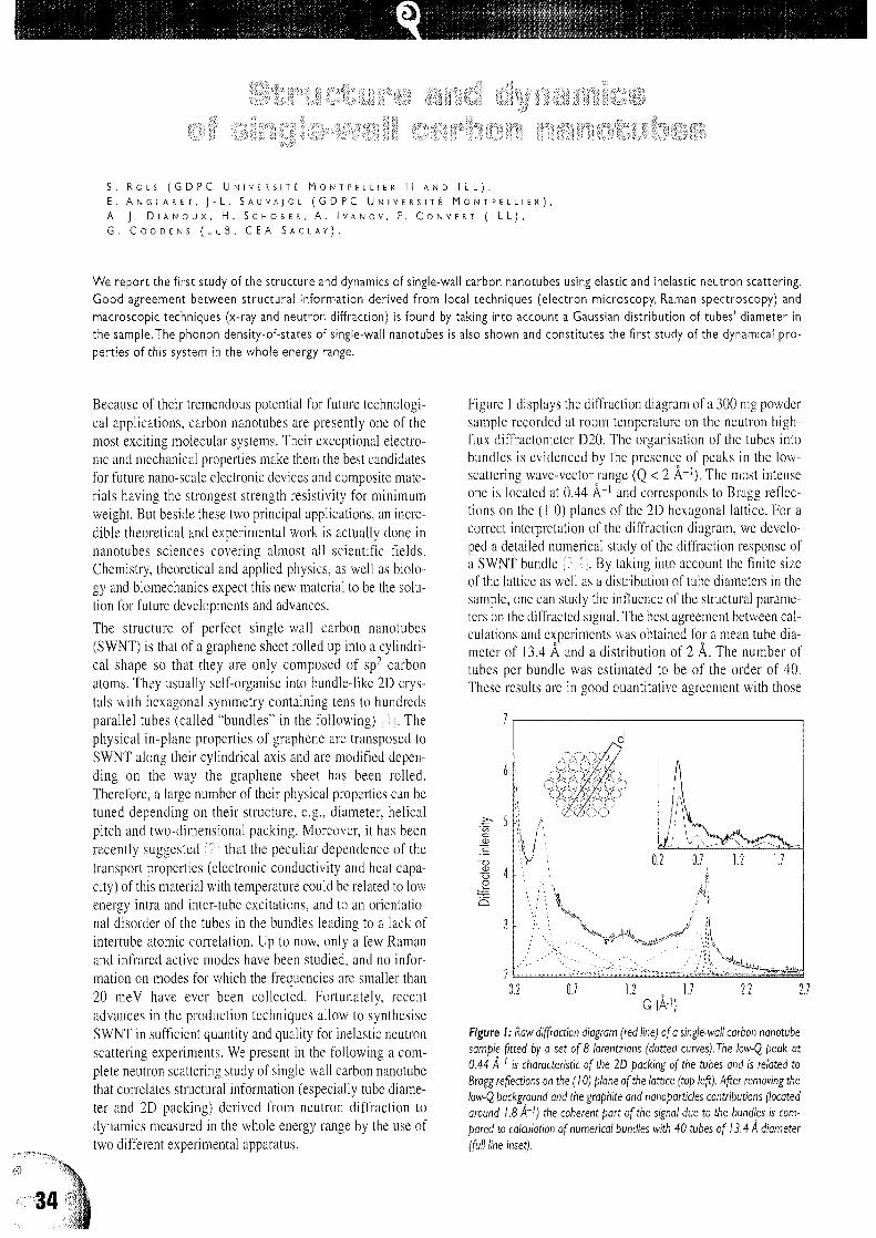
S . R O L S ( G D P C U N I V E R S I T E M O N T P E I U E R I I A N D I L L ) ,
E . A N G L A R E T , J - L . S A U V A J O L ( G D P C U N I V E R S I T E M O N T P E L L I E R ) ,
A . J . D i A N O U X , H . S C H O B E R , A . I V A N O V , P . C O N V E R T ( I L L ) ,G . C O D D E N S ( L L B , C E A S A C L A Y ) .
We report the first study of the structure and dynamics of single-wall carbon nanotubes using elastic and inelastic neutron scattering.
Good agreement between structural information derived from local techniques (electron microscopy, Raman spectroscopy) and
macroscopic techniques (x-ray and neutron diffraction) is found by taking into account a Gaussian distribution of tubes' diameter in
the sample.The phonon density-of-states of single-wall nanotubes is also shown and constitutes the first study of the dynamical pro-
perties of this system in the whole energy range.
Because of their tremendous potential for future technologi-cal applications, carbon nanotubes are presently one of themost exciting molecular systems. Their exceptional electro-nic and mechanical properties make them the best candidatesfor future nano-scale electronic devices and composite mate-rials having the strongest strength resistivity for minimumweight. But beside these two principal applications, an incre-dible theoretical and experimental work is actually done innanotubes sciences covering almost all scientific fields.Chemistry, theoretical and applied physics, as well as biolo-gy and biomechanics expect this new material to be the solu-tion for future developments and advances.
The structure of perfect single-wall carbon nanotubes(SWNT) is that of a graphene sheet rolled up into a cylindri-cal shape so that they are only composed of sp2 carbonatoms. They usually self-organise into bundle-like 2D crys-tals with hexagonal symmetry containing tens to hundredsparallel tubes (called "bundles" in the following) ; i L Thephysical in-plane properties of graphene are transposed toSWNT along their cylindrical axis and are modified depen-ding on the way the graphene sheet has been rolled.Therefore, a large number of their physical properties can betuned depending on their structure, e.g., diameter, helicalpitch and two-dimensional packing. Moreover, it has beenrecently suggested '2\ that the peculiar dependence of thetransport properties (electronic conductivity and heat capa-city) of this material with temperature could be related to lowenergy intra and inter-tube excitations, and to an orientatio-nal disorder of the tubes in the bundles leading to a lack ofintertube atomic correlation. Up to now, only a few Ramanand infrared active modes have been studied, and no infor-mation on modes for which the frequencies are smaller than20 meV have ever been collected. Fortunately, recentadvances in the production techniques allow to synthesiseSWNT in sufficient quantity and quality for inelastic neutronscattering experiments. We present in the following a com-plete neutron scattering study of single-wall carbon nanotubethat correlates structural information (especially tube diame-ter and 2D packing) derived from neutron diffraction todynamics measured in the whole energy range by the use oftwo different experimental apparatus.
Figure 1 displays the diffraction diagram of a 300 mg powdersample recorded at room temperature on the neutron high-flux diffractometer D20. The organisation of the tubes intobundles is evidenced by the presence of peaks in the low-scattering wave-vector range (Q < 2 A"1). The most intenseone is located at 0.44 A"1 and corresponds to Bragg reflec-tions on the (1 0) planes of the 2D hexagonal lattice. For acorrect interpretation of the diffraction diagram, we develo-ped a detailed numerical study of the diffraction response ofa SWNT bundle j-'.4j. By taking into account the finite sizeof the lattice as well as a distribution of tube diameters in thesample, one can study the influence of the structural parame-ters on the diffracted signal. The best agreement between cal-culations and experiments was obtained for a mean tube dia-meter of 13.4 A and a distribution of 2 A. The number oftubes per bundle was estimated to be of the order of 40.These results are in good quantitative agreement with those
r
Figure I: Raw diffraction diagram (red line) of a single-wall carbon nanotube
sample fitted by a set of 8 lorentzians (dotted curves). The low-Q peak at
0.44 A~' is characteristic of the 2D packing of the tubes and is related to
Bragg reflections on the (10) plane of the lattice (top left). After removing the
low-Q background and the graphite and nanopartides contributions (located
around 1.8 A'1) the coherent part of the signal due to the bundles is com-
pared to calculation of numerical bundles with 40 tubes of 13.4 A diameter
(full line inset).
34

deduced from local characterisation techniques. We derivedthe generalised phonon density-of-states (GDOS) from theone phonon coherent cross-section measured at 300 K on thetime-of-flight spectrometer IN6 (full symbols) and at 5 K onthe INI Be-Filter analyser spectrometer (open symbols). Thesingularities in the GDOS spectrum are characteristic ofmodes for which the dispersion curves present flat behaviour.
We compare in Fig. 2 the GDOS of the same SWNT sampleused for diffraction experiments (Fig. 2b) with that of a stan-dard graphite sample (Fig. 2a). Both graphite and SWNTGDOS spectra display five bands located around 58, 80,100,180 and 200 meV, while two additional bands in the nanotubespectrum are observed at 36 and 22 meV (Fig. 3a). Thesedifferences and analogies can be explained as follows: ingraphite, normal modes involving in-plane and out-of-planemotions are decoupled. This is no more the case in SWNT forwhich out-of-plane modes with motions in the radial directioninvolve a mixing of out-of-plane bond bending and in-planebond bending and bond stretching. It is thus expected that themain differences in the GDOS between graphite and SWNToccur at low energy where modes with a dominant out-of-plane radial component dominate the dynamics. According tothis, the 22 and 36 meV bands are attributed to radial modesfor which the frequencies are calculated to be inversely pro-portional to the tubes diameter. As an example, the Alg radialbreathing mode is Raman active and provides a powerful toolto probe the diameter of the tube in the sample under study. InFig. 3a, we compare the profile of the 22 meV band with theRaman signature of this mode (dotted line) where each welldefined peak corresponds to a fixed tube diameter. From the
Jl
Eo
Energy (meV)
Figure 2: Generalised phonon density-of-states of a) graphite and b) single-wall nanotubes derived from time-of-flight measurements (filled symbols)at 300 K and from three axis Be-Filter analyser experiments (open symbols)atSK.
envelope of these contributions and from the width of the cor-responding band in the GDOS spectrum we derived a meandiameter and a diameter distribution that were found to be ingood agreement with those deduced from diffraction experi-ments and calculations. Finally, the detailed analysis of the0-12 meV range (Fig. 3b) reveals, in addition to the obviouscontribution of other low energy excitations, a linear beha-viour of the GDOS with energy from 0 to 3 meV. This rangecorresponds to acoustic modes for which the contribution tothe density of states is sensitive to the dimensionality of thesystem. This linear behaviour can be tentatively assigned tothe dominant contributions of intertube modes in the 2D pac-king of the bundle. A possible reason for this enhancementcould be related to an orientational disorder of the tubes in the2D lattice of the bundle. Preliminary studies of the temperatu-re dependence of the GDOS shows changes in the low fre-quency profile at low temperature that can be assigned to free-zing of orientational fluctuation.
Finally, neutron diffraction was used to derive a statisticalpicture of the purity and structure of our sample.Consequently, inelastic neutron scattering experiments couldbe performed and were shown to be efficient to derive theGDOS of SWNT where additional information about vibra-tional modes are clearly observed with regards to Ramandata. A real effort in the synthesis (monodisperse samples)and in modelisation is now required in order to interpretunambiguously these data. A first step consist also to separa-te the low energy intra and inter-tube excitations. For that, aspecial sample containing mostly isolated tubes is actuallyprepared, and further time-of-flight experiments are planned.
phon
o
gcz
$
0.000
a)
I1
!*
!:
':'•"' ; i ; t - i . i t !
/ ,j ^ v,
-V
I'-''"
if
CD
10 20 30 40 0Energy (meV)
10
Figure 3: Low energy parts of the generalised phonon density-of-states ofgraphite (red) and single-wall nanotubes (green) .The energy of the radial brea-thing modes as measured in Raman spectroscopy (dashed blue line) is in goodagreement with that of the main low-frequency peak observed at 22 meVpeak in the generalised phonon density-of-states of single-wall nanotubes.
References
I [ i ] R . S A I T O , G . D R E S S E L H A U S S A N D M . S . D R E S S E L H A U S S , P H Y S I C A L P R O P E R T I E S O F C A R B O N N A N O T U B E S ( I M P E R I A L C O L L E G E P R E S S ,
L O N D O N ( 1 9 9 8 ) I [ 2 ] A . M I Z E L E T A L . , P H Y S . R E V . B 6 0 ( 1 9 9 9 ) 3 2 6 4 ; C . L . K A N E E T A L . , E U R O P H Y S . L E T T . 4 1 ( 1 9 9 8 ) 6 8 3 ; P . P E T I T E T
A L , P H Y S . R E V . B 5 6 ( 1 9 9 7 ) 9 2 7 5 I [ 3 ] S . R O L S E T A L . , E U R . P H Y S . J . B 1 0 ( 1 9 9 9 ) 2 6 3 I [ 4 ] E . A N G L A R E T E T A L . , P H Y S . R E V . L E T T .
8 1 ( 1 9 9 8 ) 4 7 8 0 .
0
35

MATERIALS SUlENCE
cr
A . P . R A D L I N S K I ( A U S T R A L I A N G E O L O G I C A L S U R V E Y O R G A N I S A T I O N , C A N B E R R A )
E . Z . R A D L I N S K A ( T H E A U S T R A L I A N N A T I O N A L U N I V E R S I T Y , C A N B E R R A ) ,
M . A G A M A L I A N , G . D . W I G N A L L ( O A K R I D G E N A T I O N A L L A B O R A T O R Y ) ,
P . L I N D N E R , O . G . R A N D L ( I L L ) .
Neutron scattering has been used to study the fractal microstructure of sedimentary rocks.The results have proven to be useful in
prospecting for oil. "Immature" shales, which have the right geochemical characteristics to produce oil but have not yet generated,
typically have a high fractal dimension, like the rocks in this study. By contrast, in "mature" oil-bearing rock, the fractal dimension for
large pores drops to nearly 2, reflecting a smooth interface between the grain-surface-covering mobile bitumen and the pore space.
Sedimentary rocks are formed from a mixture of organic andinorganic debris deposited in an aqueous environment,buried and compacted at elevated temperatures over geologi-cal periods of time. The fractal structure of these systems isof great interest to oil industry because it could indicate arock's oil-producing potential.
The small-angle scattering technique (SANS), using coldneutrons, has been applied to a hydrocarbon source rockU116, originating from the Urapunga 4 well in theMacArthur Basin, Northern Territory, Australia [1].A series of SANS experiments have been performed usingthe instrument Dl 1 at ILL at the wavelength of 14 A, as wellas with the ORNL facility at 4.75 A. Recent progress in neu-tron scattering instrumentation at ORNL allowed the micro-structure of rocks to be studied well beyond the conventionallimit and into the range of ultra small angle scattering(USANS). In particular, the ORNL ultra-small angle neutronscattering (USANS) facility [2] can probe Q-ranges down toQ ~ 2-10~5 A"1 (momentum transfer Q = A%XA sinG, scatte-ring angle = 20).
Figure 1 shows a combination of SANS data taken on theILL and ORNL SANS instruments, along with USANS data[3|. It may be seen that all three data sets overlap smoothlywith no adjustable parameters and the differential scatteringcross section in the log-log representation can be approxima-ted by a straight line above Q = 2-l(H A"1. Detailed analysisof the data in Fig. 1 shows that the pore-rock fabric interfaceis a surface fractal with dimension D§ = 2.82 over threeorders of magnitude of the length scale and ten orders ofmagnitude in cross section. To our knowledge, these datarepresent the largest range to date over which fractal beha-viour has been observed in a natural system. Such an extentof fractal microstructure in a rock is remarkable, when com-
pared with the limited size range over which the fractalproperties are usually observed (typically one order ofmagnitude) [4|.
This study extends the widest fractal length range previouslyobserved in sedimentary rocks, which covered 2 decades inlength scale and 7.5 decades in intensity [5], and shows thatsedimentary rocks are in fact one of the most extensive frac-tal systems found in nature.
a
8to
10-2
Theory: Ds=2.82, C,= 1.2 fim
LLD11 SANS
10-5 10-2 10-1
Scattering vector Q (A~
Ftgure I: SANS and USANS data from sedimentary rock showing that thepore-rock interface is a surface fractal (Ds = 2.82) over three orders of magni-tude in length scale and ten orders of magnitude in cross section (intensity).

The fractal character of the scattering cross-section eventual-ly breaks down at scales larger than several micrometres.This is reflected in a flattening out of the scattering curve inFig. 1 for smallest Q-values. In real space, the fractal dimen-sion can be quantified by counting the frequency of structu-ral features of given size on scanning electron microscope(SEM) images of the rock. Figure 2 illustrates statistical datafor SEM images of rock Ul 16, with the breakdown of fractalproperties at the scale of 4 micrometres clearly observed as asharp change of slope.
In conclusion, a "mature" (oil bearing) sample would haveits fractal dimension decreased compared to the immaturesample. Also, due to the decrease of contrast (bitumen-filledpores as opposed to water filled pores), the scattering intensi-ty at a given Q-value is smaller for mature samples that have
Feature size, 10-* (m)
Figure 2: Variation of the average number of SEM features per unit length(NIL) with feature size obtained from SEM images of the cleaved surface ofsedimentary rock Ul 16. Note the breakdown offractalhy (Ds = 2.8 to 2.9)for length scales larger than 4 micrometres.
generated hydrocarbons than for immature samples.Therefore, the technique has the potential to distinguish thesource rocks that have produced hydrocarbons from thosethat have not.
As observed in Physical Review Focus |6I, "The constancyof the fractal dimension over so many scales is astounding,considering what a messy, heterogeneous material sedimen-tary rock appears to be... This study will enhance the ideathat you can describe rock with simple concepts... There willbe certain bonafide uses of fractal concepts, and one of themwill be in the geological sciences".
Peter Lindner (centre) and his collaborators aligning a sample onDI I.
I [ I ] A . P . R A D L I N S K I , C . J . B O R E H A M , G . D . W I G N A L L A N D j . S . L I N , P H Y S . R E V . B 5 3 ( 1 9 9 6 ) 1 4 1 5 2 I [ 2 j M . M . A G A M A L I A N ,
G . D . W I G N A L L A N D R . T R I O L O , J . A P P L . C R Y S T . 3 0 ( 1 9 9 7 ) 3 4 5 I [ 3 ] A . P . R A D U N S K I , E . Z . R A D L I N S K A , M . A G A M A L I A N ,
G . D . W I G N A L L , P . L I N D N E R , 0 . G . R A N D L , P H Y S . R E V . L E T T . 8 2 ( 1 9 9 9 ) 3 0 7 8 I [ 4 ] D . A V N I R , 0 . B I H A M , D . L I D A R , 0 . M A L C A I , S C I E N C E
2 7 9 ( 1 9 9 7 ) 3 9 I [ 5 ] H . D . B A L E , P . W . S C H M I D T , P H Y S . R E V . L E T T E R S 5 3 ( 1 9 8 4 ) 5 9 6 • : 6 ] P H Y S I C A L R E V I E W F o c u s , 2 0 / 0 4 / 9 9 .

' \.
G . K O S T O R Z , J . H E C H T , M . K O M P A T S C H E R , j . - M . S C H N E I D E R A N D B . S C H O N F E L D ( E T H Z U R I C H )
B . D E M f ( I L L ) ,
J . K O H L B R E C H E R A N D W . W A G N E R ( P S I V l L L I G E N ) .
Nickel-rich alloys are technologically useful, especially as high-temperature structural materials.They often contain submicroscopic,
finely dispersed precipitates.The properties of this two-phase microstructure, especially the (meta-)stability and evolution of the pre-
cipitate population, are of considerable interest. Small-angle neutron scattering experiments were performed on Ni-alloy single crys-
tals containing aluminium and molybdenum or titanium to follow closely their phase separation paths. The analysis of the anisotropic
scattering patterns of Ni-AI-Mo single crystals yields a detailed description of the average precipitate morphology. Thus, a separation
of the influence of interfacial energy and elastic strain energy of the precipitates during decomposition becomes feasible.The complex
decomposition process of binary Ni-Ti alloys involves a sequence of metastable states which can be distinguished by small-angle neu-
tron scattering.The transformation from the metastable state to the stable hexagonal phase was also followed in-situ.
Ni-base alloys are widely used in structural applicationsowing to their good mechanical properties and their resistan-ce to corrosion. Especially, Ni superalloys exhibit highmechanical strength at temperatures exceeding 1300 Kowing to the presence of an internally ordered phase of hard,structurally coherent precipitates embedded in the Ni-richmatrix. Although the technological development of superal-loys is quite advanced, there is still a general need to betterunderstand the basic physical processes controlling micro-structural changes and long-term stabilisation of decomposedstates with favourable properties. In the present experiments,small-angle neutron scattering (SANS) has been employed tocharacterise the microstructure of Ni-AI-Mo and to investi-gate the decomposition sequence in Ni-Ti.
The formation of coherent ordered precipitates from supersa-turated Ni-rich solid solutions in Ni-AI-Mo is stronglyinfluenced by temperature and overall composition as thesolid solubility of both alloying elements varies sharply withtemperature. Additions of Mo modify the lattice parametermismatch between matrix and coherent precipitates [1-3].Strain fields are built up during precipitate growth and mayslow down the decomposition kinetics. The strain fields, cou-pled with the anisotropic elastic properties of the two phases,lead to non-spherical precipitate shapes and a preferredarrangement of precipitates along <100> crystallographicdirections.
Experiments on Mo single crystals with Ni-12al%, Al-2aL% andNi-10at.% Al-5at.% (during ageing, for up to 10 h at 973 Kand 1073 K) were performed on the SANS instrument Dllwith neutrons of a wavelength of 6 A. With advancingdecomposition, the two-dimensional scattering patternsrecorded on the Dl 1 detector show an increasing anisotropy.For large scattering vectors (Q > 0.05 A"1 whereQ = An sinG/A,, 0 is half the scattering angle, and X the wave-length of incident neutrons), the scattering intensity mainlyrepresents the tail of the scattering function of the averageprecipitate. From transmission electron microscopy it is
known that the precipitates are cuboidal in shape. The small-angle scattering of such objects can be described by a one-parameter analytical expression able to pass continuouslyfrom a sphere to a cube. Comparison with experimental datashows that, as decomposition proceeds, cuboidal precipitatesprogressively approach the shape of a perfect cube. Figure 1illustrates the results obtained for one of the crystals. Theaverage particle shape is found to correspond to the particleoutlined in Fig. lb [4]. The evolution of the particle shape
Figure I: a) Measured SANS pattern of a Ni-IOat%AI-5at% Mo singlecrystal (face<entred cubic) after ageing at 1073 K for 3 k The incident beamwas parallel to the <l IO> surface normal of the sample. The dotted linesrepresent lines of equal intensity as calculated for a cuboidal precipitate(Fig. Ib). b) Best-fitting cuboidal shape.

a) Ni-12.0ol.%Ti b ) Ni-l 1.7 ot.% Ti
. ' • : • • ' • !
c) Ni-11.2ot.%Ti
- " ?
Intensitycm~' s r
,03 r-
102 ,
101
>}r 100
tf;',1
" * . ; • •>
10-1 S
10-2m
0.01 A"1 0.01 A"1
Figure 2: SANS patterns ofNi-rich Ni-Ti single crystals (face<entred cubic) homogenised at 1440 K and annealed in-situ for a) 46 min at 1240 K,b) 10 min at1220 K, c) 43 min at 1200 K.The patterns were symmetrised to highlight the dominating features. The incident beam was parallel tothe<IIO> surface normalof the sample, the horizontal axis is along <IOO>.
and size gives information on the competition between inter-facial energy and elastic strain energy. This allows macrosco-pic properties to be interpreted on the basis of microstructu-ral parameters.
The decomposition sequence in Ni-rich Ni-Ti is more com-plex than in Ni-Al-Mo, as intermediate (metastable) statesappear during phase separation, whereas the stable orderedhexagonal phase ih^Ti is observed only after extendedageing above about 1200 K. Previous SANS studies hadindicated that the metastable precipitates may pass throughtwo successive regimes with Ti concentrations of about 18and 22 at.% 15,6 ]. The coexistence of these metastable statesand the subsequent formation of the stable hexagonal phaseare still to be explored in more detail.
In situ SANS investigations were performed to follow thedecomposition progressively within individual samples.Polycrystals were investigated at SINQ/PSI (Villigen,Switzerland) with a heatable (up to 1020 K) sample holder,while single crystals were measured at Dl 1 with a specialfurnace (up to 1470 K) for SANS, built in collaboration withthe ILL. With this furnace, rapid temperature changes and insitu solid-solution treatments at high temperatures becomefeasible.
Polycrystalline Ni-Ti quenched in water after homogenisa-tion treatment at 1440 K develops a SANS interference peakupon ageing at 900 K; this peak reflects the alignment of the
coherent metastable precipitates along <100>. After an initialrapid increase of the integrated SANS intensity (the integra-ted intensity yields the total amount of Ti in precipitates), atemporary slowing down is observed between 1 and 5 h forNi-l 1.3at.% Ti. Plateaus in the temporal evolution of theintegrated intensity of such alloys confirm the succession oftwo states J7.8L
For single crystals at temperatures near and slightly abovethe suggested coherent miscibility gap, an unexpected resultwas obtained, as shown in Fig. 2; the metastable states stillprevail, as indicated by the strong SANS intensity along<100>, and are still present after 1 h. After some time a newfeature progressively dominates the scattering pattern. Sharpintensity streaks start growing along the <111> directions.They are due to the formation of platelets of the stable hexa-gonal phase. The direct formation of the hexagonal phasewithout the preceding appearance of metastable states wasobserved just below the incoherent solvus line.
These SANS investigations are accompanied by high-tempe-rature, high-resolution x-ray diffraction close to fundamentaland superstructure Bragg peaks as well as by transmissionelectron microscopy. The interplay between local atomicordering and decomposition in the presence of coherencystresses will thus be clarified, and the results may be used inthe numerical modelling of high-temperature stability andstrength of superalloys.
I [ i ] H . A . C A L D E R O N A N D G . K O S T O R Z , I N ' M O R R I S E . F I N E S Y M P O S I U M ' , E D I T E D B Y P . K . L I A W , J . R . W E E R T M A N , H . L . M A R C U S , A N D
j . S . S A N T N E R , T H E M I N E R A L S , M E T A L S & M A T E R I A L S S O C I E T Y ( 1 9 9 1 ) I I I ; 2 ) M . F A H R M A N N , P . F R A T Z L , 0 . P A R I S , E . F A H R M A N N , A N D
W . C . J O H N S O N , A C T A M E T A L L . M A T E R . 4 3 ( 1 9 9 5 ) 1 0 0 7 • [ 3 ] A . D . S E Q U E I R A , H . A . C A L D E R O N , G . K O S T O R Z , A N D J . S . P E D E R S E N , A C T A
M E T A L L . M A T E R . 4 3 ( 1 9 9 5 ) 3 4 2 7 I [ 4 ] J . - M . S C H N E I D E R , B . S C H O N F E L D , B . D E M E , A N D G . K O S T O R Z , J . A P P L . C R Y S T . 3 3 ( 2 0 0 0 )
• [ 5 1 A . C E R R I , B . S C H O N F E L D , A N D G . K O S T O R Z , P H Y S . R E V . B 4 2 ( 1 9 9 0 ) 9 5 8 I [ 6 ] P . V Y S K O C I L , J . S . P E D E R S E N , G . K O S T O R Z , A N D
B . S C H O N F E L D , A C T A M A T E R . 4 5 ( 1 9 9 7 ) 3 3 1 I • [ 7 ] G . K O S T O R Z , M . K O M P A T S C H E R , A N D B . S C H O N F E L D , ' C O H E R E N T P R E C I P I T A T I O N I N
N I - R I C H S I N G L E C R Y S T A L S ' , S O L I D - S O L I D P H A S E T R A N S F O R M A T I O N ' 9 9 , E D I T E D B Y M . K O I W A , K . O T S U K A , A N D T . M I Y A Z A K I , K Y O T O ,
J A P A N ( 1 9 9 9 ) 3 0 5 I [ 8 ] M . K O M P A T S C H E R , B . S C H O N F E I D , H . H E I N R I C H , A N D G . K O S T O R Z , J . A P P L . C R Y S T . 3 3 ( 2 0 0 0 ) .
39

* 1
I t I
a R . C O P P O L A ( E N E A , C A S A C C I A ) ,
3 G . C I C O G N A N I , R . P. M A Y ( I L L ) ,B M . M A G N A N I ( E N E A , B O L O G N A ) ,
H A . M O E S L A N G ( F Z K A R L S R U H E Y ) .
Small-angle neutron scattering has been employed to study the growth of He bubbles in a martensitic steel developed for fusion reac-
tor technology.The He-bubble volume distribution functions obtained from our small-angle neutron scattering data show that a uni-
form distribution of small bubbles (about 15 A) evolves during high temperature annealing within a few hours into a bimodal one with
an additional population of larger bubbles (approximately 100 A).Transmission electron microscopy can give insight into the mor-
phology and size of He bubbles in the range from 20 to 50 A. However, quantitative information cannot be derived, because only a
small fraction of bubbles are visible by transmission electron microscopy.
One of the problems to be solved in the design of a futurefusion reactor is the choice of the most adequate structuralmaterials. Dramatic changes in both mechanical propertiesand microstructure are expected especially in the first wall(the element that is closest to the plasma), due to the veryhigh neutron irradiation and the thermal stresses expected11 ]. Fast neutrons and a particles produced by the fusionreaction induce radiation damage in the materials and lead tohelium accumulation in the first wall. Helium is not solublein solids and it precipitates by forming bubbles, whose growthcauses the swelling and consequent embrittlement of thecomponents. The growth of He bubbles under high-energyneutron irradiation is therefore one of the key factors deter-mining the lifetime and the reliability of structural materialsfor fusion reactors steels. For a better understanding of thegrowth mechanism of He bubbles, and the influence of tem-perature on their formation, evolution, and size distributionshould be determined.
To simulate the effect of the radiation from the fusion reac-tion on the first-wall components, and in particular helium-bubble formation, one can irradiate steels with accelerateda-particles. As shown in Fig. 1, the experimental observationof the bubbles using transmission electron microscopy (TEM)is difficult because of their tiny sizes (typically 10 A), andbecause the electron beam is defocused by the martensiticmatrix. Small-angle neutron scattering (SANS), on the otherhand, is a particularly appropriate technique for this study, forseveral reasons. Neutrons have a wavelength that is of theorder of the size of these particles, they penetrate easily relati-vely thick samples (up to several millimetres). Finally, becau-se they have a spin, they sense the magnetisation of the steel;this property can be used to enhance the contrast of the (non-magnetic) He bubbles with respect to the steel matrix.
We investigated [2-4] the martensitic steel F82H1. Thesamples were homogeneously implanted at 250°C with 400atomic parts per million He, using 104 MeV a-particles atthe cyclotron facility at the FZ Karlsruhe. They were then
Figure / H-i,, • • .,Wes in F82H after implantation of400 appm He atTin = 250°C(fromRef.[7J).
heat treated (at 525°C, 825°C and 975°C) for two hours inhigh vacuum. These implantation conditions are representati-ve of the effect of helium in fusion reactors. Of course, thereare other microstructural effects, such as cavities, loops,segregation, etc., produced by neutron irradiation (that wemay study in future).
One non-implanted sample for each temperature served as areference to isolate the SANS signal due to the He bubblesfrom that of the structural changes induced by the thermaltreatment. A further non implanted sample was heated at250°C for 5 days to simulate the heat effects of the implanta-tion process.
The SANS measurements were carried out on the instrumentDl 1 at the ILL with a sample-to-detector distance of 1.46 mand a wavelength A, of 6 A. A horizontal magnetic field was
1 composition: 8.0 Cr, 0.10 C, 0.16 Mn, 0.16 V, 2.0 W, 0.02 Ta wt%

applied perpendicular to the incoming neutron beam in orderto fully align the magnetic moments in the sample. Since theHe bubbles are non-magnetic inhomogeneities imbedded in amagnetised matrix, we observe the scattering from twophases of different scattering density. In this study, we main-ly use the stronger signal in the direction perpendicular to themagnetic field, where the magnetic scattering adds to thenuclear contribution.
The contribution of He bubbles of different sizes leads torelatively featureless scattering curves; therefore it is diffi-cult to visually extract quantitative information from them.Using the method described in reference [5], the data weretransformed into volume distribution functions D(r) definedas D(r) = N(r) r3, where N(r) is the number per unit volumeof bubbles (assumed to be spherical) of radius r.
The distribution functions for the three different annealingtemperatures are shown in Fig. 2. The large error bands(dashed area in Fig. 2) in the volume distribution functionarise both from the uncertainty in background subtraction [3]and from the limited Q range investigated. However, suchuncertainties do not affect the relative values we found. TheHe bubble volume fractions V, determined by integration ofthe D(r)s, are reported in Tab. 1 together with the averagebubble radii Rb.
At lower temperatures (Fig. 2a and b) we observe basicallyone well defined peak centred around 15 A corresponding toa dense population of bubbles of small size. This value vali-dates the estimation of 16 A obtained by TEM [6,7],
Table I: He bubble volume fraction}/, and average radius, Rj, (A), from SANS
analysis (* with a secondary bubble population around 100 A).
At 975°C (Fig. 2c), a second distribution of bubbles, oneorder of magnitude larger, appears. The density of bothbubble populations is almost one order of magnitude largerthan at 825°C.
The bubbles of large size present at 975 °C contribute a highscattering intensity at smaller Q-values than those explored.This accounts for the larger error band found for the distribu-tion associated to this temperature. There is an increase of afactor 5 in the relative value of the bubble volume fractionfrom 250°C to 825°C and of about 50% from 825°C to975°C(seeTab.l).
We conclude that the thermal treatment is responsible for thegrowth of larger bubbles at the expense of the smallest onesand by coalescence of the He atoms initially implanted in thematrix. The latter are "invisible", because individual atomscannot be resolved by SANS; for this reason the integratedintensity in Fig. 2 increases with temperature.
Nevertheless, SANS turned out to be an essential tool for thequantitative characterisation of the bubble-growth mechan-ism in these steels.
R (A) R (A) R (A)
Figure 2: He-bubble volume distribution functions (arbitrary units proportional to bubble volume per unit volume vs. bubble size in A) obtained from the SANS
data:a) 250°C, b) 825°C and c) 975°C.The dashed areas represent the 80% confidence band.
References s
I [ ! ] A . H I S H I N U M A , A . K O H Y A M A , R . L . K L U E H , D . S . G E L L E S , W . D I E T Z , K . E H R L I C H , j . N u c . M A T . 2 5 8 - 2 6 3 ( 1 9 9 8 ) 1 9 3 • [ 2 ] I L L
E X P . R E P . 7 - 0 1 - 3 1 ( 1 9 9 8 ) I [ 3 ] R . C O P P O L A , M . M A G N A N I , R . P . M A Y , A . M O E S L A N G , ) . A P P L . C R Y S T , I N P R E S S 1 [ 4 ] R . C O P P O L A ,
M . M A G N A N I , R . P . M A Y , A . M O E S L A N G , M . V A L L I , j . N u c . M A T . , S U B M I T T E D • [ 5 ] M . M A G N A N I , P. P U L I T I , M . S T E F A N O N , N . I N S T . &
M E T H . A 2 7 1 ( 1 9 8 8 ) 6 1 I I [ 6 ] A . M O E S L A N G , S . C I E R . J A . C K S A N D R . L I N D A U , P R O C E E D I N G S O F 1 2 T H I N T E R N . C O N F . O N C Y C L O T R O N S
A N D T H E I R A P P L I C A T I O N S B E R L I N 1 9 8 9 , E D S . B . M A R T I N A N D K . Z l E G L E R , W O R L D S C I E N T I F I C , L O N D O N ( 1 9 9 1 ) 5 4 5 I [ 7 ] ) . B E R T S C H ,
FZK REPORT 5984 (1997), FZ KARLSRUHE.

R . S l E B R E C H T ( I L L A N D R U H R - U N I V . B O C H U M ) ,
C . M O N D E L L I ( I L L ) ,
G . B A T T A G U N , E . C A T T A R U Z Z A A N D F . G O N E L L A ( I N F M V E N I C E ) ,
F . D ' A C A P I T O ( I N F M G R E N O B L E A N D E S R F ) ,P . M A Z Z O L D I ( I N F M P A D U A ) .
Ion implantation is a useful technique to obtain composite materials such as metal nanocluster composite glasses (MNCGs), formed
by nanometer-size metallic clusters embedded in silicate glasses. Due to quantum and dielectric confinement effects, these compo-
sites exhibit several effects such as an enhanced optical Kerr susceptibility, which can be important for application in all-optical swit-
ching devices. Moreover, MNCGs obtained by ion implantation of transition elements are important for their magnetic properties
itself. Neutron reflectometry was used as a non-destructive method to determine the in-depth metal cluster density distribution and
to roughly estimate the cluster size.The data analysis under a multilayer sample approximation gives realistic results: this suggests the
possibility to use neutron beams to study very diluted systems such as MNCGs.
Ion implantation of metal elements in glass substrates canlead to the formation of nanometer-radius colloidal particlesin a thin surface layer. The formation of nanoclustersdepends on the chemical reactivity of the pair "implantedatom-dielectric host". Metal nanocluster composite glasses(MNCGs) exhibit an enhanced intensity-dependent refracti-ve index due to the optical Kerr effect: this feature could beexploited in all-optical switching device technology, with theaim to process light signals without conversion to electronicform. Thus allowing to operate such a device in fast timeregimes (tenths of pico seconds). Moreover, MNCGs obtai-ned by ion implantation of transition elements are importantfor their magnetic properties: when the size of magnetic par-ticles is in the nanometer range inside a non-magneticmatrix, surface effects are dominant. These affect the magne-tic properties significantly, in terms of both oxidation and
anisotropy effects. For this range of magnetic particle size,the composite material offers new technological possibilities,for example in the field of magnetic recording substrates forhigh-density information storage. The knowledge of thecluster size distribution, as well as the spatial correlationfunction, is important in order to relate the morphologicalcharacteristics to the physical properties.
The investigated samples are amorphous fused silica-glassslides implanted sequentially with Ni+ ions at two differentenergies and (nominal) fluxes, namely, 180 keV (1.7-1017
ions/cm2) and 70 keV (6-1016 ions/cm2). Only glass slideswithout voids were chosen to serve as substrates for implan-tation. The double implantation leads to a flatter depth distri-bution of the nickel atoms, as suggested by simulations [1].The average in-plane cluster distribution is constant over thewhole sample area, which is known from transmission
depth profile RBS1.0 0.8 0.6 0.4 0.2 0.0
depth profile Reflectivity 28x10;
0
500
<T IOOO
N
J~ 1500-
2000
1
I1:
surface2.0 2.5 3.0 3.5
* » • * . * -«^- « », *• A
Figure l:The experimental in-depth nickel distribution measured with Rutherford backscattering spearoscopy (RBS), left, and reflectivity, right.

electron microscopy measurements during former experi-ments. The experimental in-depth nickel distribution, asmeasured by Rutherford backscattering spectrometry isreported in Fig. 1 on the left side. The retained implanteddose was about 2.51017 ions/cm2. Grazing incidence x-raydiffraction on these samples gives evidence for fee nickelnanoclusters. To learn about the sample's magnetic proper-ties a non-destructive method, sensitive to the depth-depen-dent magnetism is needed. In our case polarised neutronreflectivity with spin analysis comprises all the demandedqualities, making it to the method of choice. The analysis ofspin dependent reflectivity data enables one to reveal quanti-tatively the nuclear and magnetic depth profile at the sametime. Nevertheless, due to the relatively low total amount ofmagnetic material in the sample the scattered magneticsignal may be difficult to sort out from the data. Thus, in afirst step one has to confirm that it is possible to reconstructthe nuclear non-magnetic depth profile for such a samplewith unpolarised neutron reflectivity.
We performed neutron reflectivity measurements on the dif-fractometer ADAM [?.]. The data were taken at the instrumen-tal wavelength of X - 4.41 A and over a Q-range of 0.1 A.
Figure 2 shows the reflectivity curve (open circles). The fit tothe data is given by the solid line. The basic hypothesis forthe fit was to take into account the lateral homogeneity of thepotential. The depth variation of the potential in the z-direc-tion, roughly proportional to the local metallic element den-sity, was approximated by potential slabs of different scatte-ring-length density and thickness. On the right side of Fig. 1the potential obtained from the fit is depicted: two maximaare clearly visible, in agreement with the simulation results.The peaks are both shifted of about 350 A towards the surfacewith respect to the positions in the simulation, because of thesurface erosion effect of the incident ion beam during thesample preparation. This result is in agreement with theRutherford backscattering spectrometry measurementsshown on the left side of Fig. 1.
Figure 2: Reflectivity measurement (open circles) taken on ADAM(X - 4.41 A), solid line fit to the data.
Furthermore, the roughness of the slabs in the fit can be rela-ted to the mean size of the clusters which is sketched inFig. 1. In fact, due to the high local nickel concentration, Niclusters are expected to be very close one to each other.Under this assumption, we obtain reasonable values of thecluster size for all the implanted regions, except for the dee-pest part, where a very large value was found for the rough-ness. Considering the low nickel concentration in the tail ofthe in-depth distribution, we interpret this result as due to alarge average distance among the metallic clusters.
This unpolarised reflectivity experiment is the first step to seta more general approach to MNCGs using neutron-basedtechniques, to study their magnetic properties. We clearlydemonstrated that with this technique it is possible to revealthe non-magnetic nuclear depth profile. This result is inagreement with precedent Rutherford backscattering spec-trometry measurements. The first neutron reflectivity experi-ments with polarised neutrons and spin analysis on similarsamples with magnetic implantations were performedrecently. The data obtained looks promising and data treat-ment is currently in progress.
• [ I ] j . P . B l E R S A C K A N D L . G . H A G G M A R K , N U C L . I N T R . A N D M E T H . 1 7 4 ( 1 9 8 0 ) 2 5 7
H . Z A B E L , P H Y S I C A B 2 4 1 ( 1 9 9 8 ) 1 6 9 .
R . S l E B R E C H T , A . S C H R E Y E R , U . P l E T S C H ,

THIN FILMS
H V . L E I N E R A N D R . S I E B R E C H T ( I L L ) ,
i s l D . L A B E R G E R I E , C H . S U T T E R , A N D H . Z A B E L ( U N I V . B O C H U M ) .
We report on magnetic ordering in an epitaxial Ho single film with a thickness of 46 A. Using the ADAM diffractometer at ILL, we
were able to investigate the evolution of the first magnetic satellite of the Ho magnetic helix in the temperature region between 10 K
andTN.As compared to bulk Ho, the Neel temperature T N is reduced from 132 K to 105 K. Simultaneously, an increase of the turn
angle a for the magnetic moments from one layer to the next is observed, implying a shorter period for the spin helix.
Due to new advances in thin-film deposition techniques it ispossible today to grow ultra-thin epitaxial magnetic filmswith very high structural quality. This allows to raise thequestion of how the bulk magnetic properties are modified inthin films either due to dimensionality effects or due toproximity with other material layers. Information on nanofa-bricated magnetic films is of high importance for the designand functionality of magnetic nanostructures integrated innew electronic devices. The most complete information onsuch ultra-thin structures comes from neutron scattering. Forrevealing antiferromagnetic or incommensurate spin struc-tures, neutron scattering is unique. However, known as a bulkprobe, its applicability to thin films has to be proven. In thiscontext, we have studied the magnetic phase-transition insingle ultra-thin Holmium films. The strong magneticmoment of Ho and its long-range magnetic order yield inten-se magnetic peaks at small Q-values, thus, making Ho a per-fect model system for scattering experiments in the ultra-thinregime.
In the bulk, Holmium exhibits an incommensurate spin helixbelow the Neel temperature, TN, of ~ 132 K. Between 20 Kand 132 K the magnetic moments order ferromagnetically inthe hexagonal basal plane. From one plane to the next, theaverage orientation of the magnetic moments encloses a turnangle a, setting up a magnetic spiral along the direction nor-mal to the planes. The turn angle is about 30° at 20 K andincreases continuously to 50° with rising temperature up tothe Neel temperature. Below 20 K the magnetic momentslock into a commensurate magnetic cone structure with aturn angle of 30° in the basal plane and a ferromagnetic com-
ponent normal to the planes. In Fig. 1, the phase diagram isschematically reproduced, which is the result of magneticneutron scattering [1-3] and resonant magnetic x-rayscattering experiments at the Ho - L{1 and Lm absorptionedge [4-6].
In magnetic scattering experiments the helical magneticstructure gives rise to magnetic satellite reflections at distance± x* around the allowed Bragg peak along the c* direction.T* in units of c* is related to the turn angle a via a = 180°-T *.
Using resonant magnetic x-ray scattering at the Ho - Lmabsorption edge, the minimum thickness which can be studiedis around 200 A. Below this value the charge background fromthe Laue-oscillations of the nearby Bragg-reflection becomestoo strong and buries the magnetic (00.2 ± t*) reflections [?]•Using neutron scattering, we have now succeeded to study themagnetic phase-transition in a single Ho(00.1) film which hasa thickness of only 46 A. The epitaxial Ho film was grown on
30° commensuratemagnetic cone structure
incommensuratespin helix
npara magphase
•
figure /: Schematic phase diagram of bulk Holmium.

a sapphire substrate with the use of a Nb and Y buffer layer.The Ho film was capped with an Y and Nb layer to prevent itfrom oxidation. The embedding of Ho between Y layersensures symmetric structural and magnetic boundary condi-tions. The film thickness was determined from x-ray reflecti-vity measurements; the out-of-plane roughness was found tobe ~ 10 A. For the magnetic scattering experiments we usethe ADAM reflectometer at the ILL because of its high flux,low background and high Q-resolution. These features areessential for studying weak satellite reflections close to the(00.0) Bragg peak. Throughout the experiments we used awavelength of 4.41 A. Figure 2 shows the magnetic(00.0+T *) peak from the Ho film with 46 A thickness. Withincreasing temperature the peak shifts to higher Q values dueto an increasing turn angle. At the same time the intensitydrops due to a decreasing order parameter. For this thin filmwe measure a Neel temperature of only 105 K which is
considerably lower than in the bulk. The inset shows theorder parameter as a function of temperature. Below 20 K,we could not observe a transition to a commensurate magne-tic structure. The magnetic order in the ultra-thin Ho filmremains incommensurate, in agreement with results obtainedpreviously from Ho/Y superlattices 18]. Looking at the lengthof the magnetic helix at ~ 20 K, we find T* ~ 0.251c*, i.e. anincrease of the turn angle to 46° (as compared to 30° foundfor bulk Ho).
We have demonstrated for the first time that it is possible tostudy the magnetism in such ultra-thin films by investigatingthe magnetic ordering in Ho films as thin as 46 A using ahigh-resolution neutron reflectometer. The neutron resultsindicate that even thinner samples can be investigated withreasonable counting statistics and underline impressively theimportance of neutron scattering for the investigation ofnanostructured magnetic systems.
0.20 0.25 0 . 3 0 0.35 0.40
3 0 0 0 -
2500 -
2000 -
1500 -
1000 -
500 -
1
•
®
•
0
©
©
•
-
0 *1
1 '
10 K /
25 K V40 K F
a l55 K f J70 K / /85 K J190 K / / /os if mLi /
100K • / / • /
IlL / m/
1 ' '_ 11A#... 1 IU
S/--"€1L 80f \ \ - 70
V& 60
^ \ U 50
V k 30
• ° o
i i i i i
1 ' i
o
o
oQ
\
\
\
-
-
-
40 60 80 100
Temperature T (K)
-
• i® • i i0.20 0.25 0.30 0.35 0.40 0.45
QjA1
Figure 2: Radial neutron scans of the (00.0+T*) magnetic satellite peak for the 46 A thick single epitaxial HofOO. I) film for different temperatures. The peak shifts
to higher scattering vertors due to an increasing turn angle of the Ho spin helix with increasing temperature. SimultoneousI/ the intensity decreases due to a loss
of long-range magnetic-order with increasing temperature. In trie inset the magnetic-order parameter is plotted as a function of temperature.
References
I [ I ] W . C . K O E H L E R , E T A L . , R E V . 1 5 8 ( 1 9 6 7 ) 4 5 0 I [ 2 ] G . P . F E L C H E R , E T A L . , S P E D D I N G , P H Y S . R E V . 8 1 3 ( 1 9 7 6 ) 3 0 3 4
I [ 3 ] M . J . P E C H A N , C . S T A S S I S , J . A P P L . P H Y S . 5 5 ( 1 9 8 4 ) 1 9 0 0 I [ 4 ] D . G I B B S , E T A L . , P H Y S . R E V . L E T T . 5 5 ( 1 9 8 5 ) 2 3 4
• [ 5 ] J . B O H R , E T A L . , P H Y S I C A I 4 0 A ( 1 9 8 6 ) 3 4 9 1 [ 6 ] D . G I B B S , E T A L . , P H Y S . R E V . B 4 3 ( 1 9 9 1 ) 5 6 6 3 I [ 7 : C . S U T T E R , E . W E S C H K E ,
R . M E I E R , C . S C H U S S L E R - L A N G E H E I N E , G . G R U B E L , D . A B E R N A T H Y , T O B E P U B L I S H E D I [ 8 ] D . A . J E H A N , E T A L . , P H Y S . R E V . B 4 8 ( 1 9 9 3 ) 5 5 9 4 .

V . L A U T E R - P A S Y U K ( U N I V . M O N C H E N A N D J I N R D U B N A ) ,
B . T O P E R V E R G ( I L L A N D P N P I S T . P E T E R S B U R G ) ,
H . J . L A U T E R ( I L L ) ,
0 . N I K O N O V ( I L L A N D J I N R D U B N A ) ,
E . K R A V T S O V , M . A . M I L Y A E V , L . R O M A S H E V A N D V . U S T I N O V ( I M P E K A T E R I N B U R G ) .
Complete polarisation analysis of neutron reflection and simultaneous measurements of polarised neutron off-specular scattering
have been employed to verify atomic-spin arrangements in Fe/Cr multilayers, a typical representative of systems showing the giant
magnetoresistance (GMR) effect.The quantitative theoretical data analysis shows that in the GMR region small column-like antiferro-
magnetic domains exist, arranged perpendicularly to the film surface through the whole multilayer depth.The domains may provide an
efficient spin-flip mechanism for electron scattering, a crucial ingredient for the GMR effect.
The understanding of a wide range of exciting properties ofartificially superstructured magnetic materials including giantmagnetoresistance (GMR) [1], oscillating interlayer exchangecoupling [2], and non-collinear magnetic ordering [3,4]requires unambiguous microscopic information on the atomic-spin arrangement in those materials. In a set of experiments onmagnetic Fe/Cr multilayers we demonstrate that such informa-tion can only be obtained if two conditions are fulfilled. Firstly,the interface quality of the sample must be so high, that off-spe-cular scattering originating from magnetic and non-magneticroughness at the interfaces is negligible. Secondly, the data col-lection in specular reflection mode (with complete polarisationanalysis) must be combined with polarised neutron off-specularscattering measured in a broad range of incident and outgoingangles, so that a model can be compared over a necessary broadoff-specular region. This wide map over the specular and off-specular region should be obtained for both, spin-flip (SF) andnon spin-flip (NSF) scattering in order to separate unambi-guously the true specular signal from the SF off-specular signal,which may appear also along the specular line. These twoconditions together with the numerical analysis allowed us to
0.07
0.06
0.05
I a04
0.03
0.02
0.01
i ••
, - _ • * •• • . - " -
10.5
l o
0 0.01 0.02 0.03 0.04 0.05 0.06 0.07
°w r a d
Figure J; Intensity map of specular (R+) and off-specular scattered neutrons
from the FelCr multilayer atH = 5.48 kG as a function ofOjn and «jjno|, the
incident and outgoing scattering angle, respectively. The colours reflect the
scattered intensity on a log-scale. The horizontal colour lines crossing the pic-
tures are background effects. (Wavelength used: 4.4 A)
establish a detailed description of the microscopic spin-arrange-ment. The central issue is that the off-specular SF-scatteringarises from small column-like antiferromagnetic domainsoriented perpendicular to the film surface. So, our interpretationdoes not confirm the picture of laterally uniformly magnetisedFe-layers with non-collinear or canted angle structure publishedbefore e.g. in [3,4]. Our finding is important due to the fact thatthe presence of domains cause spin-dependent electron scatte-ring which accounts for the GMR-effect.The spin-resolved polarised neutron reflectrometry (PNR)experiment from a superlattice [Cr(9 A) / 57 Fe(68 A)] x 12grown by molecular beam epitaxy on an Al2O3-substrate wasperformed on ADAM with its new multidetector. The magne-tic field H was applied parallel to the sample surface.The very good quality of the sample is visible in Fig. 1 whichshows the scattered intensity from this multilayer for H = 5.48 kGparallel to the sample surface in the saturation regime. Thereflectivity was measured beyond the second structural Bragg-peak. No off-specular scattering is observed within theexperimental accuracy around these Bragg-peaks atctj = Of = 0.029 rad and 0.058 rad. This picture represents thesample with ferromagnetic magnetisation in the Fe-layers,having interfaces free from non-magnetic roughness or magne-tic interface disorder within the accuracy of the measurement.
0.07-
0.06-
0.05
•O Q.Wr
dso.o3|
0.021
0.01
fss:-
%g
:. ..ML - . i |§B§§ii&;;"."'.'i""-'."
ysS35,-at'v3:: :
1 :
o
0 0.01 0.02 0.03 0.04 0.05 0.06 0.07
"f inal ' r a d
Figure 2: Intensity map as in Fig. I but for the field H = 0.428 kG forR* = R++ + R+-.

This last statement is very important, because samples withthis quality are unique and allow us to separate the effects ofinterface disorder from other magnetic effects.In Fig. 2, for the lower field of H = 0.428 kG, additional inten-sity is observed with respect to Fig. 1 going through the twohalf-order positions at a = 0.0145 rad and a = 0.0435 rad. Atthis field the sample shows the GMR-effect. This additionalintensity is off-specular scattering distributed along lines per-pendicular to the reflectivity curve forming a superstructureBragg-sheet. However, in contrast to the usual case when theBragg sheet crosses the specular line at the Bragg peak positionno appreciable enhancement of the intensity at the superstruc-ture position was observed. Thus, the objects giving rise to off-specular scattering must be smaller than the lateral coherence-length. Furthermore, the off-specular scattering is asymmetric,where it approaches the axes near Ojn = 0 and Og^ = 0.The intensity along the first superstructure Bragg-sheet is cutbefore reaching oqn = 0 or a ^ j = 0. On one side the intensity iscut at (Xfina! nearly zero and on the other side at <Xjn ~ 0.005 rad.These angles are related to the critical angle of the Fe-layerswhich depends on the polarisation direction of the neutronswith respect to the magnetisation direction of Fe; this shows,without spin analysis, that off-specular scattering consists of SFneutrons. With this unusual, but effective method, SF neutronscan be determined. R in the figure caption relates to reflectivitywith the polarisation of the neutrons parallel (+) or antiparallel(-) to the external field.The spin-flip effect is also shown in Fig. 3 which was obtainedunder the same experimental conditions as Fig. 2 but with ananalyser placed between sample and detector and centred at theposition of the specular beam. Only NSF neutrons are transmit-ted by the analyser. The footprint of the analyser covers only acertain angular band of neutrons in transmission scattered fromthe sample, the width of which is marked in Fig. 3 by the
dashed lines.This band is already sufficient for a polarisationanalysis. The scattering along the half-order Bragg-sheetscrosses the reflectivity curve and consists purely of SF-scatte-ring as can be deduced by comparing figs. 2 and 3.In our model, the disorder leading to off-specular scattering hasa lateral con-elation length smaller than the lateral neutroncoherence length, which is estimated to be in the order of a fewtens of microns. Furthermore, the perturbation must have anti-ferromagnetic character due to the location at half-order Bragg-sheets. Therefore, antiferromagnetic domains are assumed. Thedomains extend in the perpendicular direction throughout thefilm thickness as evidenced by the width of these off-specularBragg-sheets. The off-specular SF-scattering together with theNSF-specular scattering is calculated with the supermatrix for-malism 16 j and presented in Fig. 4.This antiferromagnetic domain model can describe correctly theoff-specular scattering appearing as Bragg-sheets through thehalf order magnetic Bragg-positions reproducing the details likethe asymmetry in the lower Q Bragg-sheet and with all thedetails at the border of the picture. In addition, our model cor-rectly reproduces the relative intensities between first andsecond Bragg-sheet as well as the off-specular scattering inten-sities between the half order Bragg-sheets. The average size ofthe antiferromagnetic regions is around -3000 A.The high quality of the sample and the complete mapping ofthe off-specular scattering has allowed identifying unambi-guously the origin of the spin-flip off-specular scattering. Thismodel of antiferromagnetic domains is quite in contrast to theusually used model of homogeneously magnetised layers stac-ked into a sequence with magnetisation direction varying bet-ween alternative Fe-layers, [e.g. 3.41. The new interpretationmay lead to reconsidering electron scattering in these type ofsamples for a more exact description of the GMR effect.
0.07 0.07
0' 0.01 0.02 0.03 0.04 0.05 0.06 0.07
"final' r a d
Figure 3: Intensity map as in Fig. 2 but with analyser. The arrows indicatethe specular line in one spin state (transmission; R~) and the specular linein the opposite spin state (reflected;R~+);the region between the dashed linesshows the working area of the analyser in transmission.
0.01 0.02 0.03 0.04 0.05 0.06 0.07
° W r a d
Figure 4: Intensity map of off-specular spin-flipped and specular non-spinflipped neutrons from the Fe/Cr multilayer calculated with a model of anti-ferromagnetic domains to be compared to Fig. 2.
efences
• [ I ] M . N . B A I B I C H , E T A L . , P H Y S . R E V . L E T T . 61 ( I 9 8 8 ) 2 4 7 2 I [ 2 ] S.S.P. P A R K I N , N . M O R E , A N D K.P. R O C H E , P H Y S . R E V . L E T T . 6 4 ( I 9 9 0 ) 2 3 0 4
• [ 3 ] A . S C H R E Y E R , E T A L , E U R O P H Y S . L E T T . 3 2 ( I 9 9 5 ) 5 9 5 I [ 4 ] A . S C H R E Y E R , E T A L . , P H Y S . R E V . B 5 2 ( I 9 9 5 ) I 6 0 6 6 • [ S ] V. L A U T E R - P A S Y U K ,
E T A L . , S X N S 6 ( 1 9 9 9 ) , A C C E P T E D P H Y S I C A B I [ 6 ] B.P. T O P E R V E R G , A . R O H M , W . D O N N E R , H . D O S C H , P H Y S I C A B 2 6 7 - 2 6 8 ( 1 9 9 9 ) 1 9 8 .
«« r*<

I . M I R E B E A U , M . H E N N I O N ( L L B ) ,
H . C A S A L T A ( I L L ) ,
H . A N D R E S , H . U . G U D E L ( U N I V . B E R N ) ,
V . I R O D O V A ( I N S T . K U R C H A T O V M O S C O W ) ,
A . C A N E S C H I ( U N I V . F I R E N Z E ) .
Mn^-acetate is a ferrimagnetic spin cluster with total spin S = 10, providing the first and most well known example of macroscopic
quantum tunnelling of magnetisation. By inelastic neutron scattering we separated the energy levels corresponding to the splitting of
the ground state.We could measure accurately, and for the first time in zero field, a very small energy term, responsible for the quan-
tum tunnelling process.
The study of molecular nanomagnets, besides its great theoreti-cal interest, could help to determine the size limit for informationstorage. The most attractive clusters consist of a few (typically10-20) paramagnetic ions coupled by exchange interactions atthe borderline between quantum and classical behaviour. Amongthem, Mn12-acetate (Mn12-ac) is the best-studied spin cluster sofar, providing the first experimental evidence of macroscopicquantum tunnelling .: j. The cluster has a plate-like shape, with atotal spin S - 10, and a uniaxial anisotropy. Its magnetisationrelaxes between two equivalent sublevels of the ground state(M = ± 10) separated by an energy barrier. Above 10 K, it relaxesby thermal activation like a superparamagnet. Below 2 K, themagnetic reversal is governed by a quantum process yielding aspectacular effect, regular steps in the magnetisation curve .' |.The quantum tunnelling which occurs within the S = 10 groundstate is governed by extremely small high-order terms of the ani-sotropy energy. To understand it better, we have studied the ener-gy sublevels of the ground state by inelastic neutron scattering.The S = 10 ground state arises from a ferrimagnetic coupling bet-ween eight Mn'" ions (S = 2) and four MnIV ions (S = 3/2). It issplit by crystal fields in 21 sublevels (- 10 < M < 10). Usinghigh-energy resolution, we separated all the sublevels of theS ground-state. We could show the presence of high-order spininteractions unambiguously. The main result is the accuratedetermination in zero field, of a very small non-diagonal termin the spin Hamiltonian (hereafter called transverse term). Itspresence has been searched for by many theoreticians and expe-rimentalists to explain the tunnelling effects.A powder sample:
[Mn 12(CD3COO) 16(D2O)4012] • 2CD3COOD • 4D2Owas synthesised using deuterated solvents. The crystal structurewas checked between 10 and 290 K by neutron diffraction on the
3.0
2.0
10 T = 1.5K
-1.4 -1 .0 -0.6 -0.2 0.2 0.6 1.0 1.4
2.0
1.0T=14.1K
-0.6 -0.2 0.2 0.6 1.0 1.4
1.0
0.5
-1.4 -1.0 -0.6 -0.2 0.2 0.6 1.0 1.4
kio (meVj
Figure I: Energy spectra at three temperatures (raw data, averaged in
Q-space).The intensity units are the same but the scales differ for all spectra.
In the inset, the low energy range for a < 0.

50
20
10
±1
±3±4±5
±6
±1
±8
±9
+ 1010
+ B 44 ( 1 O W
20
Figure 2; Energy subkvek of the S = 10 ground state versus the transversecoefficient i>\.
diffractometer G6.1 at LLB. The first inelastic measurements upto 13 meV, were performed on the cold three-axis spectrometer4F1 at LLB [ 3 ]. The present ones with a higher energy resolutionwere made on the time-of-fiight spectrometer IN5 at ILL. Theexperimental spectra are shown in Fig. 1 for three temperatures.At T = 1.5 K, a well defined excitation is observed at 1.24 meV onthe sample energy gain side (co > 0). It is readily attributed to anexcitation from the lowest energy-level (M = ± 10) to the firstexcited level (M = ± 9). As the temperature increases, higherenergy levels are populated so that new excitations appear onboth sides of the spectrum. At 23.8 K, we distinctly observe 7well separated peaks on each side of the spectrum, together withmuch smaller ones at about ± 0.2 meV (see inset of Fig. 1).The magnetic neutron cross-section was analysed quantitati-vely by using the spin Hamiltonian already proposed to inter-pret electron paramagnetic resonance |4|, magnetisation andoptical infrared experiments. Due to the neutron selectionrules, only the transitions with AM = 0 or ± 1 are allowed.When only the diagonal terms are considered, the S = 10 mani-fold splits into 11 sublevels corresponding to M = + 10, + 9,0.In this case, the + M degeneracy of these levels is not lifted andwhen all possible states are populated we expect nine transi-tions (from M = ± 10 to ±9 and so). Seven of these transitionsare clearly observed on the spectrum at 23.8 K. The non-equivalent energy-spacing between them arises from the 4th
order diagonal term. The transverse term induces a mixing ofenergy states with different M that becomes more and moreeffective on approaching the top of the barrier. It is shown inFigure 2 where the energy levels E; are plotted versus the trans-verse coefficient B4. The energies of the highest excited statesstrongly depend on B4 and the degeneracy of some levels islifted. Figure 3 (top) shows the calculated intensities at 23.8 Kfor three B4 values. The position, shape and intensity of thelow energy peaks are strongly modified, whereas the other
peaks remain unaffected. Only B4 values in a very narrow rangecould explain the low energy features found experimentally.We find that B4 = ± 3.0(5)-10"5 cn r ' gives the best agree-ment with the data. To compare with experiment (Fig. 3 bot-tom) we added a small quasielastic background, probably dueto hydrogen. The agreement with the calculation is excellent.The main result of the experiment is the precise determinationof the coefficient B4 of the transverse term. This term is verysmall, but important since only components which do notcommute with the longitudinal magnetisation could be respon-sible for the tunnelling. The fourth order coefficient foundhere is the lowest order term allowed by the tetragonal sym-metry. It allows tunnelling between sublevels with AM = + 4,as observed in the magnetisation. This could explain the domi-nant tunnelling process. Other tunnelling transitions betweensublevels AM = ± 1 are also observed by magnetisation, andcannot be understood in the expression of the anisotropy withtetragonal symmetry. It suggests that other origins than aniso-tropy also contribute to the tunnelling, like dipolar coupling,hyperfme interaction or spin-phonon mechanism.
0.4
0.6 1.0 1.4-0.2 0.2
ftco (meV)
Figure 3: Top: intensity calculations for three values ofS>\, focusing on thelow energy peaks. Bottom:Energy spectrum. The corrected experimental dataare shown by red dots. The intensity calculation (purple line), hydrogen back-ground (green dashed line), elastic intensity (blue dashed line) and the sumof all components (red line) are also shown.
The above picture mostly relies on the assumption a "singlespin" ground-state. This simple picture which neglects theinner features of the cluster, describes the low-energy excita-tions within the ground state quite well. Very recently a micro-scopic description of the Mn^-ac was proposed [5J, whichattempts to describe both low and high-energy excitationsobserved by neutron scattering. Our new experimental results[6 j combined with previous results at higher energies 13 j , couldbe a crucial checkpoint for further theoretical work.
ei icss
I [ I ] R . S E S S O L I E T A L . , N A T U R E 3 6 5 ( 1 9 9 3 ) 1 4 1 I [ 2 ] L . T H O M A S E T A L . , N A T U R E 3 8 3 ( 1 9 9 6 ) 1 4 5 I [ 3 ] M . H E N N I O N E T A L . ,
P H Y S . R E V . B 5 6 ( 1 9 9 7 ) 8 8 1 9 1 M ] A . B A R R A E T A L . , P H Y S . R E V . B 5 6 ( 1 9 9 7 ) 8 1 9 2 I [ 5 ] M . I . K A T S N E L S O N E T A L . , P H Y S . R E V . B 5 9
( 1 9 9 9 ) 6 9 1 9 • [ 6 ] I . M I R E B E A U E T A L . , P H Y S . R E V . L E T T . 8 3 ( 1 9 9 9 ) 6 2 8 .

-iiriJjrs
J\ Tti " *'t *
V . P . P L A K H T Y , E . M O S K V I N ( P N P I G A T C H I N A S T . P E T E R S B U R G ) ,
J . K U L D A ( I L L ) ,D . V I S S E R ( U N I V . W A R W I C K C O V E N T R Y ) ,
J . W O S N I T Z A ( P H Y S I K . I N S T . U N I V . K A R L S R U H E ) ,
R . K . K R E M E R ( M P I S T U T T G A R T ) .
Using polarised neutrons we have observed spin chirality in the frustrated triangular-lattice antiferromagnet CsMnB^.We were able
to determine both the critical exponent pc = 0.43(2) describing the average chirality in the ordered phase below the Neel tempera-
ture TN = 8.37 K and the crossover exponent ^ = 1.27(9) corresponding to the dynamical chirality above T^.These results provide
the first experimental proof of the new chiral universality class of phase transitions predicted by theory.
An important feature of phase transitions is their universality,which means that the critical behaviour of physically differ-ent systems depends only on the number of the order-para-meter components and on the spatial dimensionality of thesystem. Each universality class has a particular set of expo-nents which characterises its critical behaviour. The orderparameter of frustrated triangular-lattice antiferromagnets, aswell as of helimagnets includes, in addition to the ordinaryspin variables, SR, the spin chirality, C = [S^, x SR2\, whichdescribes whether the spin structure is a right- or left-handedone. According to Kawamura h i, the chirality results in newuniversality classes. The conventional critical exponents aremodified and, moreover, two new exponents, Pc ~ 0.45 forthe average chirality below the Neel temperature (TN) andYc ~ 0.77 for the chiral susceptibility above TN are expectedto appear.
Since the observed values of the conventional critical expo-nents fit different scenarios ; !./• i Kawamura's conjecture isunder considerable discussion. However, the chiral expo-
CsMnBr3 (domain A)
nents, were never determined experimentally, in part becauseof the absence of a suitable technique to produce a single-domain crystal. The original motivation of the present expe-riment was the proposition by Maleyev , suggesting thatabove the Neel temperature the dynamical chirality, i.e., theprojection of the chiral field on the sample magnetisation,results in an inelastic polarisation-dependent cross sectionthat is odd in energy transfer. Its temperature dependencewould be described by the crossover chiral exponent,<i>c = P c + Yc-A convenient representant of planar (XY) frustrated triangu-lar-lattice antiferromagnets is CsMnBr3 whose structure isdisplayed in Fig. 1. The magnetic moments of the mangane-se ions lie in the hexagonal plane. The chirality is characteri-sed by the sense of their rotation when passing from a man-ganese atom to the next one along one of the crystallographicaxes in the basal plane. Figure 1 gives an example ofdomains with spin rotations in the clockwise (A) and coun-ter-clockwise (B) sense.
CsMnBr3 (domain B)
1
Figure l:The clockwise (A) and counter-clockwise (B) chiral domains in the magnetic structure ofCsMnBrj.

The dynamical chirality has been investigated atQ = (1/3,1/3,1) using the cold neutron three-axis spectrome-ter IN 14. A crystal of about 3 cm3 has been mounted in a4 Tesla horizontal cryomagnet with field parallel to the hexa-gonal axis. The difference between the scattering intensitiesof neutrons with incident polarisation parallel and antiparal-lel to the applied field, AI(co) = I (co)—iJ'(co), has then beenmeasured. A typical spectrum exhibiting the antisymmetricvariation with the energy transfer is shown in the inset ofFig. 2.
An empirical function, a squared Lorentzian multiplied bythe energy transfer,
AI(co) = ScoP/[P+(0)-co0)2]2 (1)
convoluted with the resolution function has been used to fitthese data. According to Ref. [5] the value of the chiral cross-section characterised by S should depend on:
T = I ( T - T N ) / T N I as S = T-<S>C (2)
A logarithmic plot of S versus % is shown in Fig. 2. From theslope at t > 0.1 one obtains the value of the chiral crossoverexponent <j)c = 1.29(7) which is in good agreement withKawamura's calculated value 1.22(6) for the XY model 11 ].At lower x, the slope decreases, apparently due to the inversecorrelation length becoming smaller than the resolutionwidth.
H II cH IIQ
MV= 0.98(6)
-0.5E(meV|
tDc= 1.29(7)
0.01 0.1 1
Figure 2: Critical behaviour of the dynamical chirality (above TN) in magne-tic field applied along the scattering vector and along the hexagonal axis. Insetthe polarisation-dependent component of the energy spectra.
In the course of the experiment it turned out that in the orde-red state, below T N = 8.37 K, different populat ions of left-and right-handed domains can be frozen in. Their proportiondepends, in a random manner, on the cooling history. In thecase of unequal popula t ions again a difference in the(1/3,1/3,1) Bragg-peak intensity is observed between the twoincident neutron polarisations, parallel and antiparallel to theguide field (about 10 Gauss) along the scattering vector. Thispart of the study was carried out using polarised thermal neu-trons on the IN20 spectrometer (k = 2.662 A"1 , Si 111 analy-ser and P G filter to remove any 2 n d order contamina t ion) .The sum of the scattered intensities for the two polarisationsis proportional to X2P with the expected exponent 2 p ~ 0 . 4 2 .The difference signal, displayed in Fig. 3 , provides the criti-cal exponent f5c ~ 0.43(2), being in excellent agreement withKawamura 's theory | i .
Combining the two measured critical exponents, p\. and § c ,the experimental value for the chiral susceptibility exponent,y c ~ 0.86(9), can be derived. All these results provide sup-port to Kawamura ' s predictions of a new universality class ofchiral phase transitions.
10 i i 11111
AI
Figure 3: Critical behaviour of the average chirality belowTN. Insetthe dif-ference in (113, 113, I) Bragg peak intensities I*, h, due to unequal chiraldomain population.
• [ I ] H . K A W A M U R A , j . P H Y S . : C O N D E N S . M A T T E R 1 0 ( 1 9 9 8 ) 4 7 0 7 • [ 2 ] P . A Z A R I A , B . D E L A M O T T E , T . J O U C O E R , P H Y S . R E V . L E T T . 6 4
( 1 9 9 0 ) 3 1 7 5 I [ 3 ] S . A . A N T O N E N K O A N D A . I . S O K O L O V , P H Y S . R E V . B 4 9 ( 1 9 9 4 ) 1 5 9 0 1 • [ 4 ] S . V . M A L E Y E V , P H Y S . R E V . L E T T . 7 5
( 1 9 9 5 ) 4 6 8 2 • : 5 J V . P . P L A K H T Y , S . V . M A L E Y E V , j . K U L D A , J . W O S N I T Z A , D . V I S S E R , E . M O S K V I N , E U R O P H Y S . L E T T . 4 8 ( 1 9 9 9 ) 2 1 .

t l
M
Pl l } t\ i M
P . J . B R O W N ( L O U G H B O R O U G H U N I V E R S I T Y A N D I L L ) ,
A . H l L L I E R , H . H U M B L O T , E . L E L I E V R E - B E R N A A N D F . T A S S E T ( I L L ) ,j . D R E Y E R ( S P S M S , D R F M C , C E A G R E N O B L E ) ,J . B . F O R S Y T H ( C L A R E N D O N L A B O R A T O R Y . O X F O R D ) .
Cryopad II in conjunction with a 3He neutron-spin filter has enabled us to measure the form-factor of the Cr3+ ion in C^C^ out to
sinO/X = 0.75 A-'.These new measurements supplement those obtained in a previous experiment in which the limit in sin9/A was
restricted by use of IN20 to a maximum of 0.5 A-'.These measurements provide the first high-precision determination of an antiferro-
magnetic form-factor with high enough resolution to draw conclusions about the asphericity in the magnetisation due to 3d electrons.
The form-factors of magnetic ions in ferromagnetic andparamagnetic materials have been extensively studied usingclassical polarised-neutron flipping-ratio measurements andin favourable cases these measurements can be very precisei i i. Such form-factors are much more difficult to measure inantiferromagnetic structures because in antiferromagnets theneutron scattering cross-section is not often polarisationdependent; the classical method is then not applicable. As aconsequence, very few measurements of antiferromagneticform-factors have been made. In the few cases where suchmeasurements have been undertaken they have given veryinteresting results j".'. •••;:. The antiferromagnetic form-factorsare more sensitive than ferromagnetic or paramagnetic onesto the effects of covalency. This is because the overlap ofpositive and negative transferred spin on the ligand ions leadsto an actual loss of moment rather than to its redistribution.Up to now no precise measurements of the form factor havebeen made in antiferromagnetic structures in which the per-iodicicty of the magnetic and nuclear structures are the sameand in which magnetic atoms of opposite spin are related bya centre of symmetry. In such structures, the magnetic andnuclear scattering are superimposed, making their separationdifficult. Additionally, the magnetic and nuclear structurefactors are in phase quadrature so that there is no interferen-ce between them to give a polarisation-dependent cross-sec-tion. The newly developed technique of spherical neutronpolarimetry ;.:i allows a precise measurement of the magne-tic scattering in such structures, and this can be exploited todetermine the antiferromagnetic form-factor.
The technique of neutron polarimetry which we have develo-ped using Cryopad consists in choosing a direction for theincident neutron polarisation and determining the directionof polarisation of the beam scattered by the sample with achosen momentum and energy transfer. In the present experi-ment we are looking at elastic Bragg scattering so that theenergy transfer is zero. It is convenient to choose three ortho-gonal directions (x,y,z) for the incident polarisation: z per-pendicular to the scattering plane, x parallel to the momentumtransfer vector, and y completing the right-handed Cartesian
set. The components of the scattered polarisation parallel tothe same three directions can be designated Pux, PIIV, Pir
where the subscript u indicates the direction of incident pola-risation (x, y or z).
Q2O3 provides a well known example of an antiferromagnetfor which magnetic and nuclear scattering appear in the sameBragg reflections and are in phase quadrature. The compo-nents of scattered polarisation in this case can be written ;:
P,, = p />„ = <? Pxz = c,
Pyx = 0 Pyy=] Pv: = 0
Where j8 = (1 -f)/( 1+72) and \ = 2ny//( 1+y2)
y is the ratio between the magnetic and nuclear structure-fac-tors, and mv the projection of the magnetic-moment directionon the plane perpendicular to the momentum transfer. Thereare two possible 180° domains for which mv is of equalmagnitude but opposite in sign. If the volumes of crystal
Ea
sine/A (A-')
Figure I: The experimental values of the magnetic form-factor measured
at the hO.f Bragg reflections ofQjO^Jhe smooth curve is the spin-only free
ion form-factor for Cfi* normalised to 2.5 Ug.

belonging to the two domains are v+ andv-, the domain ratio is defined as:
r\ = (v+- v-)/(v+- v-)and the non-zero off-diagonal terms in thepolarisation matrix become:
Pxz = tl£ and Pzx = -TitWe have already measured the ratios y ofall reflections of the form hO.-d withsinG/A, < 0.5 A-1 using Cryopad II onIN20 [A]. These measurements, whichwere made on two crystals of differentsizes in three different degrees of domainimbalance, gave amazingly consistentresults which enabled the lower angle partof the Cr3+ form factor shown in Fig. 1 tobe determined with high precision. It isnot, however, possible to extend thesemeasurements to higher sinGM, usingIN20 since the highest useable incidentwave-vector is 4.1 A-'.
We have recently, however, been able toextend our measurements to highermomentum-transfer by installing Cryopadon the hot-source polarised-neutron dif-fractometer D3 and using the newly avai-lable 3He neutron-spin filter to allow polarisation analysis ofthe diffracted beam. The experimental arrangement is shownin Fig.2.
At the higher sin9/A. values the magnetic scattering becomesvery weak and it is impractical to carry out the full polarisa-tion analysis. However, if the domain ratio is high and isdetermined using the lower angle reflections, then it is suffi-cient to measure just the Pxz and Pu components of scatteredpolarisation. These are linearly rather than quadraticallydependent on y and may be corrected for deficiencies in thetransmitted polarisation using the Pyy component. Using thismethod we were able to deduce the magnetic structure-factors
of 14 WJ reflections with sinG/A, between 0.5 and 0.75 A"1.
Figure 2: Cryopad installed on D3 showing the •'He spin filter in the magnetically isolating enclosure"Cryopol".
Their contributions to the Cr3+ form-factor are shown in Fig. 1and are magnified in the inset.
We were able to fit the low angle data to the Cr3+ free atomform-factor by assuming a chromium moment of 2.5 jig. Therather low value suggests that here is significant covalenttransfer to the oxygen ligands.
These lower angle data fit rather well onto a smooth curve.The higher angle data are scattered above and below thecurve corresponding to a spherical distribution of moment,this scatter contains information about the deviationsfrom spherical symmetry and can be used to determine thedistribution of unpaired electrons amongst the different 3dorbitals.
• [ I j H . A . M O O K , P H Y S . R E V . 1 4 8 ( 1 9 6 6 ) 4 9 5 I [ } . ) H . A . A L P E R I N , J . P H Y S . S O C J A P A N S U P L . B - l l l ( 1 9 6 1 ) 1 7 • [ 3 ] j . W . L Y N N ,
G . S H I R A N E A N D M . B L U M E , P H Y S . R E V . L E T T E R S 3 7 ( 1 9 7 6 ) 1 5 4 I ( 4 j X . L . W A N G , E T A L . , j . A P P I . P H Y S . 6 9 ( 1 9 9 1 ) 4 8 6 0
1 [ 5 ] P . j . B R O W N , J . B . F O R S Y T H A N D F . T A S S E T , P R O C . R . S O C . L O N D O N A 4 4 2 ( 1 9 9 3 ) 1 4 7 • [ 6 1 P . J . B R O W N , j . B . F O R S Y T H A N D
F . T A S S E T , P H Y S I C A B 2 6 7 - 2 6 8 ( 1 9 9 9 ) 2 1 5 .

P . M A N U E L A N O R . C Y W I N S K I ( U N I V . S T . A N D R E W S ) ,
R . I . B E W L E Y ( I S I S , R U T H E R F O R D A P P I E T O N L A B . , C H I L T O N ) ,
P . S C H L E G E R , B . F A R A G O ( I L L ) .
Whilst the nature of the glass transition is one of the outstanding problems in physics, the spin-glass transition introduces an additio-
nal degree of complexity through the orientational disorder of the magnetic spins. It is believed that the clue to the nature of the glass
transition in spin glasses lies in the spin dynamics above and close to the glass temperature, yet for the last twenty-five years there has
been considerable debate and controversy surrounding the nature of the time dependent spin correlation function in such disordered
magnetic systems. Using the amorphous random anisotropy magnets cc-Er7Fe3 and oe-Er7Ni3 as model systems, we have used the neu-
tron spin-echo technique to map the evolution of the dynamical spin correlations as a function of temperature. Interestingly, the func-
tional form of the time dependent spin correlations closely follows the stretched exponential form first recognised by Kohlrausch 150
years ago, and points to distribution of relaxation rates which broadens considerably as the glass temperature is approached from
above.
In 1847, Kohlrausch [I j discovered that the decay of the resi-
dual charge on a glass Leyden jar followed a stretched expo-
nential law defined by Q(t) = exp(-(cot)P). Since then, this
functional form of relaxation has been found to describe
a remarkably wide range of physical processes, including
phenomena as diverse as the frequency dependence of the
complex dielectric constant and the intermediate light
scattering function in microgel solutions. The Kohlrausch
form, which is often associated with a broad distribution of
relaxation times, X, is also believed by many authors to be
characteristic of the time-dependent paramagnetic spin-cor-
relation function, <S(Q,t)S(Q,0)>, in highly disordered
magnets such as spin glasses and random anisotropy sys-
tems. Extensive numerical simulations of such random
magnets, particularly those by Ogielski [•'.], have suggested
that not only x but also the exponent fi is temperature depen-
dent, taking a value of unity at high temperatures (4TO) and
slowly decreasing to 1/3 as the spin freezing or glass tempe-
rature, Ta is approached. However, several theoretical
models predict that the spin correlation function should ins-
tead follow a power-law time dependence. Experimentally it
has proved extremely difficult to distinguish between these
two models, and the controversy regarding the functional
form of the spin dynamics of disordered magnets remains
largely unresolved.
Neutron spin-echo is a particularly powerful tool with which
to investigate such dynamics. Indeed, it offers the possibility
of measuring the spin correlation function directly in the
time domain, rather than in the frequency domain as offered
by more conventional inelastic neutron scattering techniques.
Moreover, the time window covered by the spin-echo tech-
nique is of the order of K H - K H 2 s. This window not only
effectively bridges the gap between AC susceptibility
methods (KH-ICH5 s) and conventional neutron scattering
techniques ( ] (h l 0 - l (H ? s), but is also of precisely the same
order as predicted for relaxation times in spin glasses and
random anisotropy systems close to Ta.
We have performed a series of neutron spin-echo measure-
ments on two amorphous random anisotropy magnets,
a-E]'7Fe3 and ct-E^NN,. Neither the iron nor the nickel car-
ries a magnetic moment in these amorphous alloys, whilst
the inherent topological disorder gives rise to crystalline
electric fields which are almost random from erbium site to
erbium site. Consequently, single ion anisotropy, rather than
exchange dominates, and the magnetic ground state is highly
disordered \:•;. In the present context these random anisotropy
alloys have the particular advantage that, although displaying
spin-glass-like properties, they have a much higher concen-
tration of magnetic spins than the canonical dilute spin
glasses such as CuMn. This feature, together with the relati-
vely large erbium magnetic moment, ensures a sizeable
magnetic cross section.
The samples were prepared at the University of St. Andrews,
Scotland, using a melt-spinning technique in which molten
alloy is ejected onto a rapidly rotating copper wheel, achie-
ving cooling rates of the order of 106 K • s-'. The resulting
Time (ns)
Figure I: The normalised spin correlation function Pnse = S(Q,t) I S(Q,0)
at Q = 0.12 / H for a-ErjFej measured by neutron spin-echo for several
temperatures. The solid lines represent fits of the Kohlrausch stretched
exponential function, Q(t) = exp(-(tlz)P to the data. It should be noted that
the data are inconsistent with a simple power law dependence upon time.

amorphous ribbons have a typical width of 2 mm and thick-ness of 20 Jim, and are up to several meters in length. Thespin-echo measurements were performed using the IN 11spectrometer with a wavelength of 4.64 A, allowing relaxa-tion times from 5.75 • 1CH2 s to 3.7 -10-9 s to be sampled. Theinstrumental resolution was determined using spectra collec-ted from the respective samples at 1.5 K, at which temperatu-re all magnetic spins can be assumed to be static.
Figure 1 shows the measured time-dependent spin-correlationfunction at Q = 0.12 A-' for the a-Er7Fe3 sample at severaltemperatures up to 2Tg (For this sample Tg = 32 K as deter-mined by AC susceptibility). The neutron spin-echo signalswere found to be Q-independent within experimental error.
In most paramagnetic metals the spin-spin correlation func-tion is generally of a simple exponential form, and characte-rised by a single relaxation time, x. On a plot such as that ofFig. 1, such a relaxation process would appear as a simplerapidly depolarising exponential. As the relaxation rate, x,changes with temperature the spectra would conserve thesame shape but be displaced laterally with respect to oneanother. However, in the case of the spectra obtained fromthe a-Er7Fe3 sample we observe instead a slow depolarisa-tion associated with a very broad distribution of relaxationtimes. Moreover, we find that the functional form of thespectra is well described by the Kohlrausch stretched expo-nential form rather than a simple power law. Fitting theKohlrausch form to the spin-echo spectra obtained froma-Er7Fe3 allows the exponent (3 and relaxation time x to beextracted. The temperature dependence of these two parame-ters is shown in Fig. 2 and 3.
Our neutron-spin echo measurements of the evolution of thespin correlations in the a-Er7Fe3 random anisotropy systemare in broad agreement with the theoretical predictions ofOgielski 12 i. Not only is the time dependence of the correla-
tions well modelled by the Kohlrausch stretched exponentialform Q(t) = exp(-(t/x)P), but the exponent (3 also decreaseswith decreasing temperature, approaching 1/3 at Tg as pre-dicted. The temperature dependence of the characteristicrelaxation time, x, shown as a modified Arrhenius plot inFig. 3, indicates that the spin relaxation arises from a simpleactivation process. However, two activation regimes, corres-ponding to two different activation energies, can be distin-guished. The crossover point between the regimes coincideswith the glass temperature Ta = 32 K. It is likely that whilstthe high-temperature regime is dominated by the single ionrandom anisotropy of the rare earth ions, below Tg exchangeinteractions begin to play an important, but still not domi-nant, role. It may be possible in future experiments to inves-tigate this crossover in greater detail by judicious substitu-tion of the rare earth ion to vary the relative balance ofexchange to anisotropy.
Neutron spin-echo has therefore provided a unique andinvaluable insight into the nature of spin dynamics indisordered spin glass-like magnets as Tg is approached, andit has thereby helped to resolve a long-standing controversy.
We have shown convincingly that it is the 150 year-oldKohlrausch model, rather than a simple power law depend-ence, that provides an appropriate description of the dynamicspin correlations, at least in the limit of strong randomanisotropy considered here. A picture emerges of a broadlydistributed relaxation process in which the width of thedistribution of relaxation times broadens considerably (i.e. (3decreases) as the temperature is lowered. Correspondingly,the mean relaxation time, x, increases as the glass temperatureis approached. However there is also evidence that bothP and x continue to evolve below Tg, although here it mightbe expected that x is associated with quite different activationprocesses to those evident above Tg.
oX<D
CD
0.52
0 . 4 8 -
0 . 4 4 -
0 . 3 6 -
0.32
T
/ c
s
20 25 55 60 6535 40 45 50Temperature (K)
Figure 2: The temperature dependence of the exponent jiasa function oftemperature. The dashed line is a guide to the eye.
0.36 :
0.32 k20 25 35 40 45 50
Temperature (Kj60 65
Figure 3: The temperature dependence of the logarithm of the mean relaxa-tion time T. Note the change in slope of the plot, indicative of a change inthe associated activation energy, atTg = 32K.
• [ I ] R . K O H L R A U S C H , A N N . P H Y S . 1 2 ( 1 8 4 7 ) 3 9 2
R . C Y W I N S K I P H Y S R E V B . 5 4 ( 1 9 9 6 ) 1 5 2 5 1 .
[ 2 ] A . T . O G I E L S K I , P H Y S . R E V . B . 3 2 ( 1 9 8 5 ) 7 3 8 4 •' 3 : R . I . B E W L E Y A N D

T i
J . A . P A I X A O ( U N I V . O F C O I M B R A ) .
P . j . B R O W N ( I L L ) ,B . L E B E C H ( R i s e N A T . L A B . ) ,
G . H . L A N D E R ( I N S T . T R A N S U R A N I U M E L E M E N T S , K A R L S R U H E ) .
Intermetallics between transition (3d) and rare-earth (4f) elements are vital for many technologies, including those of permanent
magnets. However, understanding these interactions at the fundamental level is difficult. We report here a study of stoichiometric
MFe4Alg (M = rare-earth and uranium) single crystals. Conflicting explanations of the magnetic interactions of these material have
been reported in the past. By using polarised neutrons we have characterised the Fe and M metal magnetism, and their temperature
dependence. A further key to their understanding is provided by resonant magnetic scattering of photons (at the ESRF) which char-
acterises the conduction-electron states.
Interactions between elements with d and f magnetism arecrucial to many technologies. The richness of their beha-viour, as well as the difficulty in establishing detailedmodels, can be traced partly to the lack of a clear hierarchyof interactions. In addition to the magnetic 3d and 4f states,there are the conduction electrons, which are important indefining the nature of the long-range magnetic order. The
compounds, about which there are many conflic-ting reports in the literature, including the suggestion thatthese materials are spin glasses, provide an interesting testcase. Using a combination of neutron and synchrotron tech-niques, including the use in both cases of polarisation analy-sis, we have established not only magnetic structure as afunction of temperatures, but also how the different sublat-tices interact with one other. This then provides a benchmarkfor theories addressing these important problems; indeed,one theoretical paper has already appeared.Since the early work on these systems some 20 years ago \ U,it has been recognised that the strongest magnetic interac-tions are between the Fe atoms and that, at the stoichiometryMFe4Alg, it is principally antiferromagnetic in nature. (Withfurther replacement of Fe for Al, up to ivlFe 10Ab the mate-rials are high-temperature ferromagnets and have potentialapplications). The tetragonal unit cell is shown in Fig. 1.The M atom is surrounded by 8 Fe atoms. If the latter have anperfect AF configuration, then the molecular field at the Msite is zero.
Bulk measurements show the rare-earth materials to be pure-ly antiferromagnetic, and Fe Mossbauer studies > '> \ establishthat the Fe ordering is between 150 and 180 K for all com-pounds. The use of D10, together with single crystals, allowedus to identify the initial ordering only of the Fe sublattice (at170 K in the Dy material) as incommensurate and probablycycloidal in nature. This was confirmed by using the neutronpolarimeter (IN20-cryopad), which showed that the envelopeof the Fe modulation is circular. The wavevector isImag= [0-13, 0.13, 0] and has little temperature dependence.As the temperature is lowered, there is a long-range orderingof the rare-earth component :2:. This starts in DyFe4Al8 atTDv ~ 50 K. At the lowest temperature about 60% of the rare-earth moment is ordered; the configuration at ~ 15 K is
A . . , •>"«••j> ,
Fe
Figure / ; One unit cell of the crystallography structure of the MFe^AIg series.
Shown also is the unusual magnetic configuration of the UFe^fAIg compound.
Weak ferromognetism due to a moment of 0.47(3) JJR on the U atom is com-
plemented by an almost antiferromagnetic configuration of the Fe sublattice
with moments of 1.08(2) fjg. The Fe moments ore canted by 16° in zero field
by their interaction with the U sublattice. This canting angle increases with a
magnetic field applied in the basal plane. Both sublattices order at~ ISO K.
shown in Fig. 2. A careful examination of this cycloid withthe neutron polarimeter (IN20-cryopad) shows that the Feand Dy cycloids are turning in opposite directions, but withthe same q wavevector. At lower temperatures the ellipticityof the Dy sublattice changes sign and higher-order harmo-nics (up to 7tl1 order) from the rare-earth moments are seen;however, the envelope of the Fe cycloid remains circular at alltemperatures. The higher-order harmonics distort the envel-ope of the rare-earth cycloid and may be viewed as establi-shing ferromagnetic interactions between the rare-earthatoms. At the same time, the disordered component ofthe rare-earth moments develop short-range ferromagnetic

Dy
figure 2: The complex cycbidal magnetic configuration found in the rare-earth compounds - here shown for Dyfe^AIg atl5K Four unit cells are shown. The magnetic moments areahvays confined to the ab plane, with their propagation wavevector qms - [0.13,0.13,0]. Notice that the Fe and Dy qdoids turn in opposite directions. At lower temperature additionalhigher-order harmonics in the diffraction pattern are observed and signify a further distortion of the envelope of tie Dy c/doidlhe Fe moment i s - I ju& whereas the Dy moment is~ 6 fjg. The remaining Dy moment (to make up the fke-bn moment of? p^ contributes to ferromagnetic diffuse scattering vwfhin the ab plane.
correlations. This inherent instability of the rare-earth sublat-tice means that it can be easily modified in a field, an appliedfield of 0.5 T is sufficient to induce a large moment within therare-earth sublattice. However, the interaction with the Fesublattice, which remains antiferromagnetic to very highfield, confines the directions of the rare-earth moments to thebasal plane. It is this unusual behaviour that led previousauthors to claim the materials were spin glasses.
The development with temperature of the ordered rare-earthmoment in both the Dy and Ho compounds is shown in Fig. 3.The 110+ satellite reflection arises from both the Fe andrare-earth sublattices so that it is sensitive to the coherentinterference between the two. The different temperaturedependencies may be explained by a temperature-indepen-dent phase- factor between the modulations of the rare-earthand Fe sublattices.
The final piece to the puzzle is the behaviour of the conduc-tion-electron states. This has been examined by performingresonant x-ray magnetic scattering experiments on theDy L edges at the ESRFID20 beamline using the same crystalas for the neutron studies. These x-ray experiments focus onthe Dy 5d electrons that belong to the conduction band. Wehave found that they initially polarise at TN (170 K); i.e. theirinteraction with the Fe 3d states is sufficiently strong that theyare immediately polarised at TN rather than TDy ~ 50 K.Important changes in their polarisation occur at TDy, which arerelated to changes in the band splitting of the 5d states 13].
In comparison with the rare earth compounds, the interac-tions in UFe4Alg are quite different. Previous reports on thismaterial were also confusing, but again single-crystal experi-ments at Ris0 and D3 led [4j to a clear-understanding of themagnetic configuration, which is shown in Fig. 1. Here theantiferromagnetic modulation vector is [000] so that no newpeaks occur; the unit cell is thus easier to represent, but theinteractions are more complex than in the RFe4Al8 com-pounds.
Sandratskii and Kiibler [5] have considered this configura-tion and shown that many of its features can be explained bytaking account of the larger (than in the rare earths) spin-orbit coupling and hybridisation between the Fe 3d and U 5felectrons. The local symmetry at the Fe site, which definesthe directions for the Fe moments, gives rise to their canting.UFe4Alg is more anisotropic than the RFe4Alg systems, sho-wing interesting hysteresis behaviour when a field is appliedwithin the basal plane [fi]. These effects, and the fact thatboth sublattices order at the same temperature, are a conse-quence of hybridisation between them 151.
The present measurements illustrate the power of single-crystal neutron and x-ray techniques (in both cases usingpolarisation analysis) to unravel complex sublattice magneticinteractions in compounds containing d and f electrons. Theyalso demonstrate the profound differences in behaviour bet-ween the rare-earths and actinides in an isostructural series.

4UU
300
Inte
nsity
100
0
. a )
-
• 8
\
1 i ' i
ff
J;121 +
i i
1 I
-
\ n o + -
\
i V0 50
Tl150
TFigure 3: The temperature dependencies of the 110* and 111* magnetic satellites.The former arise from both sublattices, whereas the latter arise from only the
rare-earth sublattice. The changes atT^ ~ 50 Kin panel a) andT^0 ~80K in panel b) are due to the coherent interference between the two sublattices and indi-
cates the appearance of long-range component at the rare-earth site. This is not observed readily at the 121* position as the amplitude squared is too small. The
dashed lines represent the extension of the intensity from the Fe sublattice only, and the solid lines are drawn taking into account a constant (with temperature) phase
factor between the Fe and rare-earth magnetic sublattices. These phase factors are 42(6)° for the Dy compound and 150(8)° for the Ho compound.Thus, in panel
a) we observe almost constructive interference, whereas in b) the interference is mostly destructive. The drop in intensity in panel a) for DyFe^Ng below ~ 12 Kis
associated with the appearance of higher-harmonics, which are not included in our simple model.
i K . H . j . B U S C H O W A N D A . M . V A N D E R K R A A N , J . P H Y S . F 8 ( 1 9 7 8 ) 9 2 1 • ; : ; J . A . P A I X A O E T A L . , P H Y S . R E V . B , I N P R E S S
.: ; S . L A N G R I D G E E T A L . , P H Y S . R E V . L E T T E R S 8 2 ( 1 9 9 9 ) 2 1 8 7 • -i j . A . P A I X A O E T A L . . P H Y S . R E V . B 5 5 ( 1 9 9 7 ) 1 4 3 7 0
L . M . S A N D R A T S K I I A N D j . K U B L E R , P H Y S . R E V . B 6 0 ( 1 9 9 9 ) R 6 9 6 I • G . B O N F A I T E T A L . . P H Y S . R E V . B 5 3 ( 1 9 9 6 ) R 4 8 0 .

PHYSICS
o
J n L I '- W r ' t | ^ r - l ' l F i :
u" u O ffrvV^'r V-l
:;!; P . J E S I N G E R , A . K O T Z L E , F . G O N N E N W E I N ( P H Y S I K . I N S T I T U T U N I V . , T U B I N G E N ) ,
• G . P E T R O V , A . G A G A R S K I , V . P E T R O V A ( P N P 1 G A T C H I N A , S T . P E T E R S B U R G ) ,
; M . M U T T E R E R , J . V O N K A L B E N , S . R . N E U M A I E R ( I N S T . K E R N P H Y S I K , D A R M S T A D T ) ,
:.:; G . D A N I L Y A N , V . P A V L O V , V . C H V A T C H K I N ( I T E P , M O S C O W ) ,
'T; W . H . T R Z A S K A ( D E P . P H Y S I C S , J Y V A S K Y L A ) ,
:'• V . N E S V I S H E V S K Y , 0 . Z I M M E R ( I L L ) .
The correlations between neutron spin and the momenta of the outgoing fission fragments and ternary charged particles have been
investigated in ternary fission of actinide nuclei induced by cold polarised neutrons. These correlations challenge the validity of fun-
damental conservation laws like time reversal and space parity. While probing time reversal invariance an unexpectedly large effect
has been discovered whose mere size appears to rule out violation of time reversal invariance as being responsible for the observa-
tion. On the other hand parity non-conservation for ternary fission has been clearly established.This has interesting implications for
the process at hand. Furthermore, surprising features of the left-right asymmetries in the emission of fission fragments relative to the
incoming polarised beam are catching the eye with the sign of the asymmetry depending on the direction of emission of the ternary
particles with respect to the reaction plane.These new findings have attracted interest from theoreticians but a generally accepted
theory is still missing.
Time Reversal Invariance
In an ILL report of 1988, it was suggested byK. Schreckenbach [ I ] that ternary fission might be a nuclearreaction well suited to test Time Reversal Invariance (TRI).In ternary fission three charged particles show up in the finalstate of a scissioning nucleus. Besides the two main fissionfragments (FF) a third charged particle, usually an a-particleor a triton, is emitted. In most cases the ternary particle (TP)is ejected roughly at right angles relative to the fission axisdefined by the two FFs receding in opposite directions. Thesuggestion was that, in analogy to tests of TRI in the decay offree polarised neutrons, also in ternary fission induced bypolarised neutrons a triple correlation between neutron spina and the momenta of both, the momenta of FFsp and the
A
momenta of T?sp could be investigated. The observableproposed to be studied is:
= a.[PlA
(1)
with all vectors being normalised to unit length and with/?being the momentum of the light fragment by convention.
B runs from - 1 to + 1. The correlation observable B is oddunder time reversal and even under the parity operation. Anynon-zero expectation value <B> for the correlation couldindicate a violation of TRI.
So far two fission reactions have been analysed on the polar-ised beam facility PF1: 233U (n, f) and 235U (n, f). Twoindependent reaction chambers were mounted sharing theneutron beam. The measuring principle is different in the twochambers. One of the chambers was developed in Gatchina.It relies on fast ionisation chambers to detect any charged
PIN-diodesternary particles
n-beam
fission fragmentsMWPC
MWPCission fragments
N,,iodesternary particles
Figure I: The experimental setup used on PFI.

particles. It could be demonstrated that the novel technique
works satisfactorily and first results were obtained. The
second chamber was built in Tubingen. In the following,
mainly results from this latter chamber are reported.
A sketch of the experimental setup is given in Fig. 1. The
longitudinally polarised neutron beam hits the fissile target
and both, the fission fragments and the ternary particles are
observed perpendicularly to the neutron beam. The frag-
ment and ternary particle detectors are likewise positioned
at right angles relative to each other. This geometry was
chosen to give the search for a non-vanishing expectation
value <B> maximum sensitivity. The detectors for the two
fragments to be registered in coincidence are multiwire pro-
portional counters (MWPC). The ternary particles are iden-
tified by PIN diodes of rectangular shape ( 3 x 3 cm2). With
these pn-junctions a-particles are distinguished from tritons
by inspecting the risetimes of the current signals in the
diodes.
A total of up to 40 diodes are grouped into two arrays of
20 diodes each and mounted above and below the reaction
plane defined by the neutron beam and the fission axis
(Fig. 1). To get rid of instrumental asymmetries the spin flip
technique is employed. A current sheet placed in the polari-
sed neutron beam with the current being switched at a rate of
1 Hz serves as a very efficient spin flipper. The technique
allows to determine from the count rate differences for the
two spin helicities directly a quantity D, called the correla-
tion coefficient, which is linked to the expectation value <B>
to good approximation by D = 3><B>.
Since from theory the absolute size for a non-vanishing cor-
relation <B> due to the violation of TRI is expected to be in
the 10~7 range, the experiment had to aim at large count rates
and long measuring times to reach statistically significant
results. With a polarised beam flux of 6-108 n/cm2-s and
5 mg of highly enriched 233U or 235U isotopes, count rates of
106 coincident fission fragments per sec. and 3 • 102 ternary
events per sec. are reached. Beam times allocated so far to
these experiments were 2 cycles.
In the following all results are quoted in terms of the correla-
tion coefficient D calculated from the raw data. Any correc-
tions are only to increase the modulus of D. For the two reac-
tions under study the values found for D are
D = - (1.81 ± 0.10) • 10-3 for the 2-«u target and
D = + (0.52 ± 0.06) • 10-3 for the 2-«U target.
A first surprise in these results comes from the sizes of the
coefficient D and, hence, also the expectation values of the
observable B which are huge, at least in comparison to expec-
tations. This raises doubts whether it is really a violation of
TRI which is becoming manifest in the present experiments.
A further surprise is the change in sign for D (and <B>) for
the two reactions studied. Probably, this points to a change in
sign of the polarisation of the two compound nuclei which
depends on the relative orientations of target and neutron spin
in the capture state. Physically it is the spin of the compound
nucleus and not the neutron spin which steers the correlation.
Energy (MeV)
Figure 2: Dependence of the correlation on energy of the ternary particlefor WU and 23SU.
Offhand it is argued that some initial or final state interac-
tions between the partners of the reaction might be respon-
sible for the observations. However, so far it is not clear
which intricate mechanism could be at work and bring about
the above correlations.
To gain more detailed information on the characteristics of
the triple correlation, the dependence of the coefficient D has
been evaluated as a function of both, fragment masses and/or
energies and ternary particle energies. As shown in Fig. 2. a
very strong variation of the correlation is disclosed as a func-
tion of ternary particle energy. For both reactions studied the
modulus of the correlation coefficient D increases with the
energy of the ternary particle. The pronounced effect appears
to be difficult to reconcile with initial state interactions.
More details on the experiments are given in . ] and \ .
Parity Non-Conservationand Left-Right Asymmetries
It is a remarkable feature of the target and detector assembly
sketched in Fig. 1 that, upon turning around the spin o of the
incoming neutron into a plane at right angles to the beam
direction, experiments with completely different physics can
be performed. Two classes of experiments may be distingui-
shed for neutron beams polarised perpendicularly: either
probing parity non-conservation (PNC) or measuring left-
right asymmetries (LRA). For the neutron spin pointing
towards the fragment detectors the pseudoscalar observable
. A A -
APNC = < 7 % (2)
and its expectation value <APNC> can be analysed. The ter-
nary particle is only required in this case to be in coincidence
with the FFs and, hence, to identify ternary fission. A non-
zero expectation value <ApNC> is an unambiguous signature
for PNC. As with the triple correlation experiments, from the
difference in count rates for the two spin orientations from

the flip technique a PNC coefficient (XPNC is derived fromwhich the quantity of physical interest <APN^> is calculatedthrough the relation ccp c = 3 • <APNC>. The result obtainedfor the 233U (n, f) reaction is
ccPNC = (0.32 ±0.08) -10-3.
Thus, similar to binary fission, where PNC was discoveredmore than twenty years ago Kj, also in ternary fission parityis non-conserved. But more striking is the fact that the PNCcoefficient for ternary fission established here has the samesize within experimental uncertainty as the one known sincelong for binary fission. This property of ternary fission wasalready disclosed in former work at ILL by a group fromITEP, Moscow 15 j. This has interesting implications for theternary process which are discussed elsewhere j 6j.
In the second class of experiments the neutron spin is requi-red to face the TP detectors. For this choice of the polarisa-tion the left-right asymmetry of fragment emission may beinvestigated with the observable:
K*K = O-[Pn4LF} (3)and the corresponding expectation values <ALRA>. In eq. (3)p is the unit vector of neutron momentum. The LRA is to be
n
traced to an interference effect between incoming s- andp-waves of the neutron. Again from the count rates of thespinflip technique an LRA coefficient CXLRA is deducedwhich is related to the expectation value <ALRA> throughaLRA = 3 -<ALRA>- It should be stressed that similar to thePNC studies also here it has only to be made sure that a TPhas been emitted in coincidence with two fragments.In the LRA experiment another strange observation wasmade. Referring to Fig. 1 the sign of the LRA coefficient forfragments from ternary fission is found to depend on whetherthe TP is ejected upwards or downwards, i.e. parallel or anti-parallel to neutron spin, while the modulus of the asymmetry
is virtually the same for the two directions of TP emission.The modulus is determined for the 233U (n, f) reaction to be:
IOCLRAN (0.56 ±0.17). 10-3
Yet a different type of PNC and LRA experiments is perfor-med by inspecting the ternary particles instead of the fissionfragments. Formally, this is simply achieved by replacing ineqs.(2) and (3) the FF momentump by the TP momentump . In contrast to the fragments, the PNC and LRA coeffi-cients measured for the ternary particles are all compatiblewith nil. This demonstrates that the main fission fragmentsand the light ternary particles are not on an equal footing.
Conclusion
Apart from the PNC effects, all of the other results quotedare new. They are intriguing not only because they were notanticipated, but also because even after their discovery noconcept for a possible mechanism behind the triple correla-tion and the curious left-right asymmetries has been proposedso far. It is not clear whether the characteristics of theternary disintegration process of a fissioning nucleus or viola-tions of conservation laws are responsible for these results.
Finally, it should be mentioned that, beyond the panoply ofcorrelation studies described, in the course of experimentssome 109 ternary and multiple particle events were collectedand registered on tape, and this huge data base enables thedetailed investigation of yields and energetic properties ofquaternary fission. In quaternary fission two light chargedparticles in addition to the two main fission fragments areformed in the decay of a heavy nucleus. However, quaternaryfission should be the topic of a separate report.
Acknowledgements
This work was supported by grants from BMBF, Bonn andRFBR, Moscow.
• f .• J K . S C H R E C K E N B A C H , I N T E R N A L I L L R E P O R T 8 8 S C O C T , 1 9 8 8 • [ ? ] P . J E S I N G E R E T A L . N U C L . I N S T R . A N D M E T H . A 4 4 0 ( 2 0 0 0 ) 6 1 8
• [ ' • ) A . G A G A R S K I E T A L . " I N T . S E M . O N I N T E R A C T I O N S O F N E U T R O N S W I T H N U C L E I " , E D . W . F U R M A N , D U B N A 1 9 9 9 , 2 7 9 •
H ] G . D A N I L Y A N E T A L . , J E T P L E T T 2 6 ( 1 9 7 7 ) 1 8 6 • [ S j A . V . B E L O Z E R O V E T A L . , J E P T L E T T 5 4 ( 1 9 9 1 ) 1 3 3 I | 6 j F . G O N N E N W E I N
E T A L . , N U C L . P H Y S . A 5 6 7 ( 1 9 9 4 ) 3 0 3 .

'mm
G . V A N D E R Z O U W A N D A . Z E I L I N G E R ( U N I V . W I E N ) ,
P . H 0 G H 0 J , R . G A H L E R , P . G E L T E N B O R T , J . B U T T E R W O R T H ( I L L ) .
In classical physics the gravitational and the inertial mass of a body are the same up to a proportionality constant.This forms the basis
of the General Theory of Relativity.The observation of gravity-induced phase shifts was one of the earliest successes in the field of
neutron interferometry and demonstrates, in the quantum limit, the influence of the gravitational potential on systems described by
quantum mechanics. However, after corrections, these experiments did not confirm the equivalence of the neutron's gravitational and
inertial mass. Because all earlier experiments were carried out with one type of neutron interferometer only, we have repeated them
with our interferometer for very-cold-neutrons at ILL. We can now confirm the equivalence of the neutron's gravitational and
inertial mass, excluding at the same time earlier experimental results on a I-a level.
By dropping masses off the tower of Pisa, Galileo Galilei forthe first time demonstrated that all bodies subject only to thegravitational force of the Earth fall the same way, when star-ting from the same position with the same velocity. InNewton's theory of gravity this is explained phenomenologi-cally by the fact that the gravitational mass and the inertialmass of a body are proportional to each other and thereforecancel in the equation of motion. Einstein's General Theoryof Relativity promoted this phenomenon to a ruling prin-ciple, called the weak equivalence principle, stating that anaccelerating system is indistinguishable from a system in agravitational field.
It is interesting to ask what happens when the equivalenceprinciple is applied to a system, such as the neutron, which isclearly governed by the laws of quantum mechanics. Thepresent consensus is that the principle of proportionality bet-ween gravitational and inertial mass also holds for quantummechanical systems, but not the classically equivalent prin-ciple of equal paths 11 ]. In practice this means that the quan-tum system can be described by a Schrodinger equation thatcontains the gravitational potential.
Until 1975 there had been no experiment that demonstrateddirectly the effect of this gravitational potential in the quan-tum limit, i.e. in the limit where Planck's constant is differentfrom zero. In that year Collela, Overhauser and Werner
(COW) [2 j demonstrated what has come to be known as gra-vity-induced quantum, interference. In their experiment aneutron interferometer made out of a silicon single-crystalwas rotated about its optical axis, resulting in a difference inheight and thus in gravitational potential energy between thetwo paths the neutron can take (see Fig. 1).
The potential difference causes a phase shift given by
Ag Asin a .h2
In this equation m, and mg are the inertial mass and gravita-tional mass of the neutron, respectively, h is Planck'sconstant, A is the area enclosed by the interferometer, g is thelocal gravitational acceleration, as measured by a classicaldevice, X is the neutron de Broglie wavelength and a is thetilt angle. By comparing the measured value of m/ng with, forinstance, m? known from other experiments, one can test theproportionality of gravitational and inertial mass. The origi-nal COW experiments and subsequent, more sophisticatedversions of it, showed beautiful agreement with theoreticalprediction within the error of 1%.
However, years later it was pointed out by Bonse andWroblewski [3] and by Home |4|, independently, that a cor-rection for dynamical diffraction effects inside the siliconslabs, ignored in the original calculations, leads to a discre-pancy between theory and experiment of the order - 4%.
grating 3 grating 2 grating 1
3
9// = gsin a
Figure I: When a neutron interferometer (here seen from above) is rotated around its optical axis by an angle a the neutron wave in one path travels at agreater height than in the other, causing a phase shift between the two waves due to the difference in gravitational potential energy. This phase difference is measured byinterferometry. At the same time the overall envelope of the interference pattern, as shown in the "magnification", shifts by the same amount as the fringes,representing the bending of the classical trajectories.

•3 -2 • ] 0 1 2
Position of 1s t grating (|juin)
Figure 2: Two interfemgrams at tik angles +2 mrad and -2 mrad showingthe count-rate as function of the first grating position. The displacement ofthe sinusoidal pattern between the top and the bottom graph correspondsto a gravitational phase difference of about 19.6 rod (modulo 2%).
This discovery was followed by a series of new experiments,of which the recent experiment by Littrell et al. [51 is themost advanced. It nevertheless still shows a -1.0 ± 0.1%deviation from theory.
It is important to note that all these experiment were carriedout using essentially the same neutron interferometer. Recentexperiments by Peters et al. [6] using an atomic fountaininterferometer show no significant discrepancies on a levelof 7 parts in 109, so the neutron results are most likely due toan unknown systematic effect. To verify this hypothesis, tobe sure that no fundamental property of the neutron wasoverlooked, we carried out a series of COW experimentsusing a different type of instrument, namely our interfero-meter for very cold neutrons (VCN) at ILL [7 j .This interferometer is based on the principle that for VCN,we can use micro-fabricated quartz glass refractive gratingsas beam splitting elements, instead slabs of silicon crystal.Because of the large neutron wavelength of about 100 A andits large length (1.016 m) our interferometer is also about 50times more sensitive to gravity than the COW one.Up to this year, however, we had not been able to measure allparameters in our experiment with high enough accuracy toclearly distinguish between theoretical prediction and earlierexperiments. Especially, the uncertainty in the spectrum ofour neutron beam contributed to the experimental error ofabout 1% 181. This is too large to distinguish between theoryand earlier results.
This year we started a new series of measurements, butinstead of the full available neutron spectrum, we usedonly a narrow band that is well-defined by a multi-layerbased filter (AX/X ~ 0.1) made at ILL. This not onlydecreases the error from the measurement of the spectrum,
but, also makes it possible to measure over a larger range of tiltangles a, which also results in a better accuracy. It, of course,reduces the already low count-rate further to about 0.1 n/s.The experiment itself consisted of two independent series ofmeasurements. In the first series we measured the gravitatio-nal phase shift itself by taking interferograms at different tiltangles. An interferometer (as shown in Fig. 2) measures theintensity as a function of a gravity-independent phase shift:the position of one of the gratings. A change in tilt anglecauses this sinusoidal pattern to shift, which directly givesthe difference in gravitational phase. The second series ofmeasurements consisted of an accurate time-of-flight measu-rement of the spectrum detected by the same detector at thesame position as during the first series.
2 4Act (mrad)
Figure 3: (Top) Measured gravitationally induced phase shift plotted as func-tion of the difference in tilt angle. Each point represents one set of interfero-grams as in fig. 2.The line is the theoretical prediction. (Bottom) Measuredphase shift minus theoretical prediction. One typical errorbar is indicated.
In Fig. 3, we show our final results. The graph shows the dif-ferences in gravitational phase as a function of the differencein tilt angle. Comparing with the theoretical prediction, cal-culated from the spectrum measurement, we find no discre-pancy on the level of accuracy of about 0.4%. In other words,within the measurement error we have experimentallydemonstrated that the gravitational and the inertial mass ofthe neutron are the same in the quantum limit. At the sametime this result excludes all earlier results from single-crystalinterferometers on the 1-a level. We thus could clarify along-standing issue in the field of neutron interferometry.
Acknowledgements
We thank Th. Brenner, C. Gomez and W. Graf for their tech-nical assistance at ILL.This work was supported by the Austrian ScienceFoundation FWF, Project No. S65-03. GvdZ gratefully ack-nowledges support from the TMR European Network No.ERB-FMRX-CT96-0057.
I [ I ] J . A N A N D A N , P H Y S . R E V . D 1 5 ( 1 9 7 6 ) 1 4 4 8 • [ 2 ] R . C O L E U A , E T A L . , P H Y S . R E V . L E T . 3 4 ( 1 9 7 5 ) 1 4 7 2 • [ < ] U . B O N S E A N D
T . W R O B L E W S K I , P H Y S . R E V . D 3 0 ( 1 9 8 4 ) 1 2 1 4 • [ 4 ] M . A . H O R N E , P H Y S I C A B 1 3 7 ( 1 9 8 6 ) 2 6 0 . [ 5 j K . C . L I T T R E L L , E T A L . , P H Y S . R E V .
A 5 6 ( 1 9 9 7 ) 1 7 6 7 I [ 6 1 A . P E T E R S , E T A L . . N A T U R E 4 0 0 ( 1 9 9 9 ) 8 4 9 I [ 7 ] M . G R U B E R , E T A L . , P H Y S . L E T T . A 1 4 0 ( 1 9 8 9 ) 3 6 3
I [ 8 ; G . V A N D E R Z O U W , E T A l . , N U C L . i N S T R . A N D M E T H . A 4 4 0 ( 2 0 0 0 ) 5 6 8 .
oG

QUANTUM SH«iPEMS
B . F A K , 0 . P L A N T E V I N ( C E A G R E N O B L E ) ,
H . R . G I Y D E . N . M U L D E R S ( U N I V . O F D E L A W A R E )
G . C O D D E N S ( L L B S A C L A Y ) ,
H . S C H O B E R ( I L L ) .
The elementary excitations of superfluid 4He, characterised by the phonon-roton dispersion curve, have been studied in great detail
over the last forty years since the first observation of the roton in a pioneering neutron-scattering experiment in Stockholm in 1957 [I ].
The interest today is turning towards the effect of disorder and confinement on these excitations, when e.g. helium is immersed in
porous media.The key word is here "dirty bosons". We have studied the microscopic dynamics of superfluid 4He in porous media
using the IN 12 and IN6 spectrometers at the ILL.These measurements suggest that the modifications of the macroscopic properties
(specific heat, superfluid fraction) are related to the existence of two-dimensional (2D) excitations rather than to modifications of the
three-dimensional (3D) bulk excitations, such as the roton.
The porous media used in this investigation are aerogel andVycor. Aerogel is a highly tenuous structure of irregularlyconnected silica (SiO?) strands with a large distribution ofpore sizes and porosities ranging from 85 to 99.5%. Vycorcan be considered as a network of worm-like channels in sili-ca with a rather well defined diameter of about 70 A and aporosity of about 30%. When superfluid helium is immersedin these media, the macroscopic properties are modified dueto confinement and/or disorder [2 \. In aerogel, the superfluidtransition temperature Ts is lowered from that of the bulk(Tjv = 2.17 K) by only a few mK, while the critical exponent£ in the expression for the temperature dependence of thesuperfluid fraction ps(T)/p = (1-T/TS)C is modified, indica-ting a possible change in the universality class. In Vycor onthe other hand, Ts is lowered to 1.95 K, while £ is the same asin the bulk.
Our measurements on 4He in aerogel show that the 3D bulk-like excitations are the same as in bulk superfluid helium.In particular, the temperature dependence of the roton energy(Fig. 1) and the roton width is the same in aerogel as in bulk.There are no observable deviations from the bulk behaviourthat could explain the differences in the macroscopicproperties.
However, additional intensity is observed slightly below theroton energy, as illustrated in Fig. 2 for different filling frac-tions. The intensity of this low-energy mode saturates quite
I
2.5
Figure I: Temperature dependence of the roton energy measured for
different aerogels on IN 12 (red and green) and IN6 (blue). No deviations are
found from bulk 4He (line).
rapidly with filling fraction, in contrast to the bulk modewhose intensity increases linearly with filling. This suggeststhat the low-energy mode is propagating in the first liquidlayers near the substrate (the two first layers are solid).Similar excitations have been observed in thin films ofhelium on graphite surfaces i 3.4!, but the present result is thefirst observation of layer modes in aerogel. The dispersion ofthe layer mode is similar to that of the bulk roton, as shownin the inset of Fig. 2, but with a lower energy.

3
aco 2
090
"§"'•'•'' J . J
1070
—•— Roton (3D)- —••- -Layer mode /
70%
1.8 1.9 . 2.0 2.1
Q ^ 60%50% | ®.
• 45% ! tf ! . •*;A 1 * i 1 i *
^!. .! t
j i
i \f • i
100%i .
' f i •' '
ijI f . '
. - 1 !i !
1 f . ••• ! ! ' •
('• i . :( \
0.7 0.8 0.7 0.8 0.7 0.8 0.7 0.8
E n e r g y (meV)
0.7 0.8
Figure 2: Dynamical structure-factor for superfluid 4He in 87% porous aero-
gel atT= 0.5 K for different percentage fillings measured on IN I l.Jhe thick
blue line shows the 2D layer mode. Trie inset shows the energy of the bulk
3D roton and the ID layer mode.
Layer modes are also observed in our measurements onhelium in fully filled Vycor. The energies of these modes aresmaller than that of the bulk roton, and consistent with theobserved specific heat and ps(T)/p at low temperatures. The3D phonon-roton excitations are very similar to bulk helium,as shown in Fig. 3, with the same energy and width, and thesame temperature dependence. With increasing temperature,the intensity of the main (roton) peak decreases. In bulkliquid 4He, the intensity of the roton scales approximatelywith the superfluid fraction ps(T)/p and disappears atT^ = 2.17 K [5], where ps/p = 0. Above T , only a very broadcontribution is seen. We find that the roton intensity ofhelium in Vycor does not scale with ps/p in Vycor.Specifically, there is a clear peak at T = 1.99 K, which isabove the superfluid transition temperature Ts = 1.95 K inVycor. The peak has disappeared by T = 2.3 K. Theoreticalwork [6] suggests that the roton intensity should scale withthe Bose condensate fraction no(T) rather than ps(T).The results in Vycor suggest that if the intensity scales withno(T), then the Bose-Einstein condensation temperature
(BEC) TBEc must lie above the superfluid transition tem-perature Ts in Vycor. In other words, Ts and TBEC may beseparated in Vycor.There are sound physical reasons to believe that such aseparation due to disorder is possible. Neutron scatteringmeasurements of the condense fraction of helium in Vycorare needed to clarify this intriguing possibility.
3
GO
^He in Vycor
Q=1.70A-i
T=l.73 K
T-1.99K
T=2.31 K~*rww9-*-9-it
U 0.8 1.0 1.2
Energy (meV)
Figure 3: Comparison of the dynamical structure factor of superfluid 4He
in Vycor (filled circles) with bulk helium (lines) at different temperatures meas-
ured on IN6 for a wave-vector slightly smaller than the roton wave-vector.
Refer
• [ f ] H . P A L E V S K Y E T A L . , P H Y S . R E V . 1 0 8 ( 1 9 5 7 ) 1 3 4 6 1 [ 2 ] J . D . R E P P Y . J . L o w T E M P . P H Y S . 8 7 ( 1 9 9 2 ) 2 0 5 • [ 3 ] W . T H O M L I N S O N
E T A L . , P H Y S . R E V . L E T T . 4 4 ( 1 9 8 0 ) 2 6 6 1 [ 4 ] H . J . L A U T E R E T A L . , P H Y S . R E V . L E T T . 6 8 ( 1 9 9 2 ) 2 4 8 4 ; j . L o w T E M P . P H Y S . 8 7 ( 1 9 9 2 ) 4 2 5
I | 5 ] A . D . B . W O O D S A N D E . C . S V E N S S O N , P H Y S . R E V . L E T T . 4 1 ( 1 9 7 8 ) 9 7 4 I [ 6 ] H . R . G L Y D E A N D A . G R I F F I N , P H Y S . R E V . L E T T . 6 5
( 1 9 9 0 ) 1 4 5 4 .

A . I . K O L E S N I K O V , V . E . A N T O N O V , V . K . F E D O T O V ( I S S P R A S C H E R N O G O L O V K A } ,
A . j . D I A N O U X , 8 . D O R N E R , A . H E W A T , A . S . I V A N O V ( I L L ) ,
G . G R O S S E , F . E . W A G N E R ( T U M U N C H E N ) .
A neutron diffraction investigation of a-MnHooz showed that hydrogen atoms randomly occupy interstitial positions which form
dumb-bells, 0.68 A long.Together with the high-energy optical bands, pronounced peaks at 6.3 and 1.6 meV were observed in the
inelastic neutron-scattering spectra of a-MnHoo7 and a-MnDgos- Our data show that these peaks originate from the splitting of the
vibrational ground-state of hydrogen and deuterium atoms due to their tunnelling between the adjacent sites forming a dumb-bell.
The intensity and the energy of the tunnelling peak are anomalously high compared to those in metal-hydrogen systems studied ear-
lier. Hydrogen tunnelling in a-MnHx is one of the very few quantum effects observed at temperatures as high as 100 K.
Apart from their technological importance, metal-hydrogensystems have repeatedly been found to exhibit unique pheno-mena not observed in other materials. The study of thesephenomena helps in understanding fundamental aspects ofquantum and solid-state physics. The special geometry of thehydrogen positions in the complex structure a-phase of man-ganese gives rise to an unusually pronounced tunnelling effect.
A recent high-pressure study showed that the solubility ofhydrogen in a-Mn can be increased up to a few atomic per-cent j j j. The crystal structure of a powder sample of MnH007synthesised under high hydrogen-pressure was studied byneutron diffraction, using the DIB diffractometer at ILL andthe high-resolution Fourier diffractometer HRFD at JINR(Dubna) W. The cubic unit-cell of the a-Mn structure, spacegroup 143m, contains 58 atoms located on the four crystallo-graphically inequivalent sites 2a, 8c, 24gi, and 24g?. A profi-le analysis of the diffraction patterns of MnH0 07 showed thathydrogen randomly occupies interstitial positions 12e(0,0,0.538) inside distorted octahedra of manganese atomson 24g] and 24g2 sites, as schematically shown by the dra-wing in the upper right corner of Fig. 1. These sites formdumb-bells positioned rather far apart, at the centres of theedges and faces of the unit cell of a-Mn with a = 8.9403 A,see the upper left drawing in Fig. 1. Because of the small dis-tance of It - 0.68 A between the 12e sites in a dumb-bell;each dumb-bell can accommodate only one hydrogen atom.An occupancy of half of the 12e sites corresponds to anH/Mn atomic ratio of x = 6/58 = 0.103. In the studiedMnHo.o7 sample hydrogen therefore filled about 70% of theaccessible interstices.
An inelastic neutron-scattering study i'2- of a-MnH007 at90 K with the KDSOG-M spectrometer at JINR revealed aband of optical hydrogen vibrations split into three peaks, inaccordance with the low site symmetry of the hydrogen posi-tions, and also a strong peak at 6.4 meV which was tentative-ly attributed to the splitting of the vibrational ground-state ofhydrogen due to tunnelling between the adjacent 12e sites.Further results of inelastic neutron scattering studies strongly
corroborated the assumption of the tunnelling origin of the6.4 meV peak. These results are partly published in Ref. \Mand include the temperature and the neutron momentum-transfer dependences of the spectra of a-MnHo.o7 at 5-200 Kmeasured with the TFXA at ISIS, MARI at ISIS and IN6 atILL and the spectra of a-MnDo.o5 at 1.7-180 K measuredwith the IN6 and INlBeF ILL-spectrometers.
A representative inelastic neutron-scattering spectrum mea-sured with TFXA is shown in Fig. 1 at the bottom. The peaksof the fundamental H optical modes in a-MnHo.n? are obser-ved at 74, 107 and 130 meV, in reasonable agreement withthe KDSOG-M data. The intensity of the peak at 6.2 meVdecreases with increasing temperature and at 200 K the peakexhibits relaxation behaviour.
Figure 2 shows the difference between the inelastic neutron-scattering spectra in energy gain of an a-MnDo.o5 sampleand a sample of pure a-Mn which were measured with IN6
3
o
150fus> (meV)
Figure I: The CC-MnHggj solid solution. On the right two adjacent positions
lie partly occupied by hydrogen (open circles (j 9 and 10) and their nea-
rest environment of two tetrahedra formed by manganese atoms on positions
24gt (blue circles w / to 4) and on positions 24g2 (black cirdesQ 5 to 8);
the Mn atoms 1-4,5, and 6 form a distorted octahedron around the H atom
on position 9. On the left the arrangement of dumb-bells of I2e positions in
the unit cell ofaMn. Bottom: The inelastic neutron-scattering spectrum mea-
sured at 23 K with the TFXA spectrometer at ISIS.
66

3
oCO
•6 -4 -2 0kw (meV)
Figure 2: The differences between the inefostic neutron-scattering spectra ofcc-MnDo.05H0.005 onc' a ' ^ n measured in energy gain with the IN6 spectro-meter at ILLThe curves are shifted along the y-axis.The a-MnDggs samplewas contaminated with about O.SatX H which manifested itself by the peakat 6.3 meV. The peak at 1.6 meV is due to 5at% D.
under the same conditions. The positions of these H and Dpeaks agree with roughly estimated values, A^= 5 meV andA^= 1.5 meV, for the splitting of the hydrogen and deuteriumvibrational ground-states due to tunnelling. These values fol-low from the relation [4j:
Ao ~ (1/2) ftcooexp (- mcoo€2/%)
if one substitutes the measured energies h(i^ - 73 meV andftco^ = 51 meV of H and D local vibrations along the line21 = 0.68 A which connects the 12e sites in a dumb-bell; m isthe mass of the H or D atom.
The intensity of the 6.3 meV peak of hydrogen in a-Mn mea-sured with MARI as a function of momentum transfer, Q,can also be described fairly well by the Q-dependence cha-racteristic of tunnelling peaks [51:
S(Q,(Otun) -[1/2 - sin(2€Q)/(4€Q)] exp(- QV).The value of u2, the effective mean-square displacement ofH atoms, obtained by fitting was 0.162 A2.The temperature dependences of the integrated intensities ofthe H and D tunnelling peaks are shown in Fig. 3 and can bewell explained by a Boltzmann thermal population of thecorresponding ground-states (solid curves in Fig. 3).
The most remarkable features of the hydrogen tunnellingpeak in the inelastic neutron-scattering spectrum ofa-MnHo.07 are its occurrence in a very large temperatureinterval up to 100 K, its anomalously large integrated intensi-ty compared to that of the optical hydrogen band and its ano-malously high energy of 6.3 meV. This energy is about30 times higher than that of tunnelling splittings found for
hydrogen in other metals [51. Hydrogen tunnelling in metalswas observed earlier only at temperatures below 10 K, andthis is the first time that deuterium tunnelling in metals wasmeasured using neutron spectroscopy.
0.5
- - 0 . 2 .
T(K)
Figure 3: The temperature dependences of the tunnelling peak intensitiesfor hydrogen (blue symbols) and deuterium (red symbols) in a-Mn obtainedfrom the INS spectra collected in the regime of neutron energy gain a) andloss b). The spectra were measured w'lth the spectrometers IN6.ILL (circles),MARI, ISIS (triangles) andTFXA, ISIS (squares).The solid lines represent theBoltzmann thermal population factors, exp(~ AQII<^T)/[I + exp(- Ao/k^T)]for the upper ground-state (Fig. 3a) and I / [ / + exp(-Aolk&T)] for the lowerground-state (Fig. 3b). The dashed line in Fig. 3b is the dependence:Il[exp(Aolk%T) - / J +/ valid forphonons and harmonic oscillators.
References
I f i ] V . E . A N T O N O V E T A L . , S C R I P T A M A T E R I A L I A 3 4 ( 1 9 9 6 ) 1 3 3 1 • [ 2 ] V . K . F E D O T O V E T A L . J . P H Y S . : C O N D E N S . M A T T E R 1 0 ( 1 9 9 8 ) 5 2 5 5
• [ 3 ; A . I . K O L E S N I K O V E T A L . , P H Y S I C A B 2 6 3 - 2 6 4 ( 1 9 9 9 ) 4 2 1 I [ 4 ] S . I . D R E C H S L E R E T A L . , J . P H Y S . F 1 4 ( 1 9 8 4 ) L 2 4 3
I [ 5 ] A . M A G E R L E T A L . , P H Y S . R E V . L E T . 5 6 ( 1 9 8 6 ) 1 5 9 .

Emmanuel Farhi (right) shows the 'virtual' ILL to visitorsduring the French science week "La Science en fete".
The ILL thesis students (from left) Darius Sullivan,Charlotte Anderson, Jorg Holzinger,Alexandre Sicardenjoy the break during the clip session.
"A hit, a very palpable hit"*,Alan Leadbetter in his lecture about glass.
*Shakespeare, Hamlet, act 5 (1601)
Recollection of the old days: Pierre Chieux,jean-Louis Soubeyroux, CNRS Grenoble,and Jacques Bouillot,Annecy.

WORKSHOPS
Workshops and Meetingsin which ILL played a major role in the organisation
Frontiers in SAXS and SANS organised by B. Deme (ILL),O. Diat.Th. Naraynan (ESRF),Th. Zemb (CEA Saclay)
ESRF 12-13 Feb. 99
Powder-diffractioninstruments review
organised by A. Hewat (ILL) ILL 22-23 Mar. 99and C-H. De Novion (LLB Saclay)
Informal workshop on'Inelastic Scattering with theNeutron Resonant Spin Echo(NRSE) Technique'
'Clip session* of thesisstudents
organised by R. Currat (ILL)
organised by G. Brotons
5th EU Residual Stress organised byT. PirlingStandards Steering CommitteeMeeting (RESTAND)
ILL 23 Jul. 99
ILL 23 Nov. 99
ILL 9-10 Dec. 99

Small-angle scattering is a well established tech-nique to characterise structures ranging from nano-scopic (10 A) to microscopic (a few microns) scales.The combination of small-angle x-ray scattering(SAXS) and small-angle neutron scattering (SANS)provides complementary (contrast) structural infor-mation in numerous systems belonging to hard,soft, as well as biological matter. On the 12-13February 1999, the workshop frontiers in SAXS andSANS gathered 120 scientists at the ILL for a com-prehensive overview of the state-of-the-art instru-ments existing at ESRF and ILL.The first session was dedicated to the current statusof the instruments, whereas the other sessions dis-cussed the present limits in spatial and time resolu-tions presenting recent developments in optics,detectors and sample environments, and emphasis-ing the need for further improvements. The closingsession demonstrated the power of small-anglescattering as applied to two different situations involving thetriple isomorphous substitution method in SANS of biologi-cal samples and the use of SAXS in polymer processing.
Exciting discussion during the poster session of the small-angle workshop; from left Marie
Foret, Montpellier, Olivier Dhez and Olivier Diat, both ESRF.
Furthermore, the workshop stressed the usefulness of bring-ing both x-ray and neutron communities together for jointendeavours.
On 22-23 March 1999, the scientific council's reviewof powder and liquids/amorphous materials diffraction(www.ill.fr/dif/powder-review) hosted 50 participants. Therewere sessions dedicated to individual instruments, D2B, D20,D4, and the two CRGs: Dl A and DIB, and sessions on 'conti-nuous versus TOF neutron sources', 'synchrotrons versus neu-trons', and 'data-acquisition and software'.The final report congratulated the ILL on its world-class ins-truments, with D2B, D20 and D4 being the best in their cate-gories. Nevertheless, it was found that the total capacity forpowder diffraction at ILL is smaller than it should be, takinginto account the size of the community and the scientificresults. However, at present, the situation is aggravated by theshut-down of D20. The report concluded that the first prioritywas, of course, to repair D20, but that high priority should begiven to improving the efficiency, especially of D2B and D4.D1A, at present scheduled half time, should remain availablefor powder diffraction at least until the definitive repair of D20.A new powder diffractometer would be desirable, but the cha-racteristics of such a machine requires further discussion.
The D4C upgrade, providing 10 times more flux, should befinished by the end of this year. It was recommended thatthe upgrade of D2B, presented within the ILL 'MillenniumProgramme', should become the first priority to allow theroutine use of D2B in its highest resolution mode. Theneed for a dedicated full-time strain scanner was emphasi-sed, totally independent of D1A, with more space, forexample behind DIB.
The genera] discussion concluded that x-rays are better forstructure determination, but neutrons are preferable for therefinement to obtain quantitative and precise data, e.g. atomicpositions of oxygen in oxides. This is specially true for ins-truments on continuous sources due to the great stability oftheir neutron flux, e.g. D4. Finally, data acquisition and refi-nement software is increasingly important, with the need forneutron laboratories to develop and maintain software such asFullProf and CCP14 in a collaborative network.
"When shall we three meet again. In thunder, lightning, or in rain?"*; from
left David Martin, ISIS, John Loveday, Edinburgh, and Adrian Barnes, Bristol.
*SJiatepeare, Macbeth, aa I (1606)
70

I 1 1 '
On 23 July an informal workshop on Inelastic Scattering withthe Neutron Resonant Spin Echo (NRSE) Technique was heldat the ILL attended by 20 participants interested in the NRSEtechnique and its potential applications for inelastic scatte-ring. The specific objectives of the meeting were as follows:
- to assess the scientific case for developing a spin echo set-upoptimised for inelastic work (study of excitation linewidths,anharmonic frequency shifts, and others),
- to define the optimum characteristics of such a set-up, interms of energy and momentum transfer range, energy andmomentum resolution, luminosity, polarisation characteristics),
- to review the principles and capabilities of the NRSEmethod and its technical requirements in the context ofinelastic scattering in the light of the instrumentation effortsin this area currently underway at HMI, FRM-II and LLB,- to discuss the opportunity of developing a NRSE set-up ona polarised-beam three-axis spectrometer at ILL (first on thetest instrument IN3, later to be transported as an option ontoIN20, IN 14, IN22) and to explore possible collaborations inthat direction.
After various presentations the workshop participantsdiscussed the feasibility of developing a spin-echo set-up
Discuss/on of the NRSE set-up on IN3; from left: Roland Gahkr, PhilippeMalbert, Roland Currat, Bruno Dorner andArno Hiess.
dedicated to inelastic scattering on the test instrument IN3.It was generally felt that:- the resonant technique appeared better suited in order totake advantage of the increase in luminosity associated withthe use of focusing geometries, both the resonant and preces-sion techniques should address, at some stage, the question ofhow to properly correct for second-order dispersion effectsresulting from the local curvature of the dispersion surface.- a NRSE set-up on IN3 will serve a useful purpose if itbecomes operational in 2001 at the latest.
On 23 November, ILL's thesis students invited toa clip and poster session. Since the students workin various disciplines this gathering gave theopportunity to exchange ideas and to help to knoweach other better. During the session "Physique enClips" in December 1999, Guillaume Brotons wasawarded the prize for the best short presentationby the Grenoble section of the French PhysicalSociety (SFP).
Guillaume Brotons (right) discusseshis results with Heinrich Stuhrmann,IBS Grenoble.
f >
On 9-10 December the ILL hosted the 5th EU Residual StressStandards (RESTAND) Steering Committee meeting, chairedby Professor A.G. Youtsos (Petten). Engineers and scientistsfrom various institutions and companies participated: HMI,ILL, ISIS, Petten, Ris0, Studsvik, the Universities ofCambridge, London (Imperial College), Salford, and AEATechnology, British Aerospace Airbus, Rolls Royce, SchunkKohlenstofftechnik, Sintec Keramik, Volkswagen. They haveembarked on an EU contract to establish European standardsfor the measurement of residual stress using neutron diffrac-tion. This is done by comparing measurements on standardsamples provided by industry to the neutron laboratories. Inaddition, developing strategies and procedures for measure-ments and analysis of data are discussed.
Since many large European companies are participating inthis effort, we expect that the new standards will encouragegreater interest in neutron diffraction by industry. Moreover,this will help neutron centres to better understand the needs ofcommercial applications. The RESTAND contract is a followup of the international VAMAS-TWA20 working group topromote the use of neutron strain scanning; ILL hosted theinaugural meeting of VAMAS in 1995.These initiatives are particularly timely in view of the propo-sal (by a consortium of UK Universities to EPSRC) toconstruct a dedicated neutron strain scanner at ILL, buildingon the present instrument on D1A (see also AR97, p. 87).This is one of the ILL's Millennium programme projects, onwhich work will start in 2000.
71

Working for IN 13's upgrade: Frederic Marchal(left) and Jerome Locatelli.
"And Jose Dianoux said,'Let there be light'...and there was light".*
Giovanna Cicognani, Luigi Cristofolini, Univ.Parmajerdme Combetjose Dianoux(from left) setting up a light scattering
experiment on IN 10.
* Genesis cb. /, v, I
"Life is just one damn thing after another."*Paul Martin (left) sorting out furnace problems on DIAfor Maria Jesus Martinez Lope, ICM Madrid,TeresaFernandez-Diaz and Clemens Ritter (from right).
*£. Hubbari •Philistine', Dec. (909 P.32
Pierre Palieau adjusts the crystal analysers(pyrolithic graphite) on INI .

DEVELOPMENTS
With the advent of the new millennium, the ILL has prepared a programme of instru-
ment renewal. Proposals for new methods and instrument upgrades were made by ILL
users and ILL scientists during the last year. Feasibility studies have been undertaken,
as described in the following. After discussions through ad-hoc committees
(Instrument Subcommittee and Scientific Council), 5 projects were approved by the
last Steering Committee. These projects include: the construction of the thermal
LADI instrument and the implementation of a strain scanner in collaboration with the
British associate; the development of a fast detector for SANS experiments, the
rebuild of the D3 diffractometer and the upgrade of the IN20 neutron optics. This
phase of the ILL Millennium Programme, which should start in 2000, is a first step to
bring the ILL instruments suite up to users expectations.The Millennium Programme
will later be extended to other instruments and general infrastructure of the ILL.
Other instrumentation projects are well under way, such as the new IN8 spectrome-
ter, the time-of-fiight option on IN 15 and the upgrade of the IN 13 CRG instrument.
New methods are also tested and exploited.The development of instrumentation and
techniques is an essential activity at a facility such as the ILL; the outstanding results
achieved last year have been made possible by the dedication of the whole staff from
technicians and engineers to scientists.

| | |
DEVELOP M ifiTS
Review of developments
Some notable developments accomplished in 1999, but which are not mentioned in the articleson the following pages, are reviewed below.
The new neutron guides H17 and H18 which supply the new diffractometers D16 and D17B, respecti-vely, were commissioned in November 1998. Flux measurements show that the flux on H17 is 5 to 12%higher than on the identical H16 guide, and the flux on H18 is 27 to 45% higher than on the new H17 guide(note the supermirror coating is only on the horizontal surfaces of the guide).
The H17 and HI8 guides have the same geometry as the H14 to H16 guides, section of 200 x 30 mm2, cur-vature radius of 2700 m, length 25 m from the rear plate in the swimming pool. The natural nickel coatedH17 guide is followed by the 10 m long straight H171 guide with a section of 150 x 30 mm2. The HI8guide has a natural nickel coating on the vertical faces and a m=2 nickel-titanium supermirror coating ontop and bottom. The last 3 m have m=3 supermirror coating on top and bottom and are converging to150x30 mm2.
The 'upgrade of PF1 on H113' started in 1998. The HI 13 guide is of a non-conventional design withcross section varying over its length such as to improve its transmission properties. The guide in the out-of-pile has a geometry diverging from 200 x 60 mm2 to 200 x 90 mm2, curvature radius of 4000 m, leng-th of 72 m from the rear plate in the swimming pool and m = 2 nickel-titanium supermirror coating on thetotal surface. The housings in the neutron-guide hall are ready to receive the glass elements. The deliveryand mounting of the supermirror-coated glass-elements by the University of Heidelberg is scheduled forthe next winter shutdown. The project is co-fmanced by the Universities of Heidelbergand Munich.
As compared to the old PF1 station on H53,on HI 13 the gain in neutron flux can bedoubled due to the supermirror coating.Further gains can be achieved by installing afocusing device (which is not yet financed)and by dedicating the beam to PF1 withouthaving further instruments upstream, so thatan overall gain of more than ten times can beexpected. The missing parts will be installedduring the long shut-down end of 1999, andthe new beam-position will be properlyequipped in the course of the year 2000. Asmost neutron-particle physics experimentsare statistics limited, this new installationshould give some additional impetus to thei- i , Multiple reflections looking into the new supermirror guide HI 13 (during installation
in August 1999).

The new detectors of the liquids diffrac-tometer D4C were assembled during 1999and first tests are foreseen in spring 2000.
D16, the upgraded 2-axis diffractometerfor medium-resolution work on biologicalmaterials, was fully commissioned during1999. First results of experiments carriedout on D16 are presented in the scientifichighlights.
D17B, the new reflectometer, was par-tially commissioned during 1999. The ins-tallation of the monochromator mode,including polarised neutrons, is undercompletion during the winter shutdown(T> 1QQO/T OfinrA Commissioning D / 78. From left to right Lo'ic Pajou, Bob Cubitt, Eric Beloeuvre, EMBL,
' Andre Gabriel, EMBL, Frederic Descamps.
The position-sensitive detector (PSD) ofD20 is currently under repair. After the breakdown of D20's detector, the microstrip-gas-chamber detec-tor-plates were investigated by Auger electron spectrometry. This showed that the anodes transformedfrom chromium to chromium oxide while working at high voltage. The fact that this oxide appears onlyon the anodes but not on the cathodes suggests an electrolytic reaction in the glass or on its surface. Dueto the reduction of the width of chromium on several anode strips, discharges appeared which led to theprogressive destruction of the anodes. This electrochemical effect is now controlled by an enforced clea-ning of the glass surface and/or the replacement of chromium strips by strips of gold. This will allow thereconstruction of the detector with a longer lifetime. A prototype of the new microstrip-gas-chamber detec-tor-plates has been tested and all plates are delivered. After mounting these plates into the PSD housing,followed by a heat treatment under vacuum, the PSD will be filled with the detection gas and brought intothe reactor hall after the first reactor cycle. Before adjusting and calibrating the PSD's electronics the pro-tection has to be mounted. D20's re-operation is foreseen for June 2000.The time-of-flight spectrometer IN4C saw its first user experiments during the last cycle of '99 usingthe low-energy range. During the first half of 1999, the optimisation of the operational parameters, align-ment, focussing and chopper speeds has been carried out over the full available wavelength range. Mostrecent efforts are concentrated on the minimisation of the background, eliminating parasitic scattering andsearch for the best resolution conditions. Unfortunately, the performance of the instrument is severely limitedby the safety restrictions concerning the background choppers. The loss of flux due to the limited trans-mission of the Al-shield is most severe at longer wavelengths (> 2.5 A), while the imposed speed limit of10,000 rpm requires to operate with a 1/2 speed ratio (background choppers/Fermi chopper) at short wave-lengths (< 2 A), with a corresponding loss of intensity on sample, as well as degraded background conditions.The maximum flux on sample was found to be of the order of 1.4 -105 n/cm2/s (at X = 1.1 A).
The replacement of the actual background choppers with discs - that should allow the suppression of thesafety restrictions - is in progress and the installation of the new choppers is foreseen for July 2000.Meanwhile, we have planned to install a secondary diaphragm before the monochromator position in orderto try to improve the energy resolution that seems to be degraded due to the broadening of the incidentdivergence caused by the 9 cm thick Al-shield.
A circular 3He detector for forward scattering with a central hole for the direct beam is close to completion.It will cover an angular range of approximately 3 to 9°. After beam tests and checks of the electronics itwill be mounted on the instrument at the beginning of 2000.
The rebuild of the primary spectrometer IN5B, the time-of-flight instrument, is going ahead as foreseen.The choppers are under design, the magnetic bearings and the supermirror guide are ordered.

f
iV
i l r ••:.
- :k ^ I <
C . W I L K I N S O N ( K I N G S C O L L E G E L O N D O N ) ,
G . J . M C I N T Y R E , J . A . C O W A N ( I L L ) ,
D . A . A . M Y L E S ( E M B L ) ,
M . D E B O I S S I E U ( L T P C M / E N S E E G G R E N O B L E ) ,
0 . N I M Z ( F R E I E U N I V E R S I T A T B E R L I N ) ,
P . S C H O B I N G E R - P A P A M A N T E L L O S ( E T H Z U R I C H ) .
Test experiments on LADI have shown that a Laue d iff racto meter adapted to difficult sample environments wouldbe a powerful tool for physics and chemistry, offering rapid collection of complete diffraction data from new mate-rials, from weakly scattering samples, and through phase transitions.
A detector employing neutron-sensitive image-plates is com-paratively cheap, offers high spatial resolution, has goodhomogeneity, a large dynamical range and extended linear-ity, and can be constructed to subtend very large angles at thespecimen. When used with the Laue technique to exploit thefull flux of a white neutron beam, such a detector allows col-lection of single-crystal diffraction data 10 to 100 times fas-ter than on a conventional monochromatic diffractometer,with only a modest loss in precision. The image-plate LAueDiffractometer (LADI) was constructed by the ILL andEMBL to exploit these advantages [1 ]. It is now in schedu-led operation on a cold-neutron beam giving good structuralinformation for macromolecular crystallography [2].An image-plate Laue diffractometer on a thermal-neutronbeam would also have wide application in the small unit-cellproblems of physics, chemistry and mineralogy. The possi-bility to observe complete volumes of reciprocal space rapid-ly will allow, for example, structural studies of small orweakly scattering crystals in an acceptable time, or comple-te data collections as a function of temperature or pressure ofstructural or magnetic phase transitions, which often result incomplex incommensurable structures.LADI has occasionally been moved to the thermal beam H22for test experiments on problems in physics and chemistry.Some recent highlights are given below.
Quasi-crystalsThe structure of a quasi-crystal can be described in a six-dimensional (6D) space where translational periodicity isrecovered. The 6D periodic lattice is divided into two 3Dspaces: physical space, also called parallel space, and thecomplementary perpendicular space. The 6D lattice is deco-rated by a set of 3D atomic objects lying in perpendicularspace. The reciprocal lattice observed by diffraction can alsobe described by a 6D lattice, defined by QparaJiei and Qperpand is consequently infinitely densely packed in the observa-tional 3D physical space. Fortunately the intensity of reflec-tions falls off as IQIpe ,, which allows reasonable resolutionof quasi-crystal reflections, sufficiently so that the firstknown quasi-crystal structures could be solved by x-raypowder diffraction using isomorphous replacement [3].Further detail comes from single-crystal diffraction, espe-cially by exploiting the contrast of the different relative scat-
tering powers of the constituent elements by neutrons vis-a-vis x-rays to detect chemical order in the structure.Despite the projection of the diffraction pattern into 2D inthe Laue technique, the high flux, large dynamic range, largedetector surface, and uniform resolution of LADI shouldprove to be a useful tool for study of quasi-crystals. Figure 1shows the full image of one Laue diffraction pattern of thenew magnetic icosahedral phase ZngM^Y. The five-fold axisis readily identified, and the enlarged region in inset hints atthe detail that we will be able to extract routinely from suchpatterns. The unique area of the pattern is outlined in bold,and is repeated several times in the pattern, but with differentwavelengths contributing to the different reflections.
V
" 1 1,
1 Mr
;
•
•
< i
••p.
' r
r it• • • -
•
LZZ1
•
Figure I: Laue diffraction pattern from icosahedrai Zn^M^Y obtained intwo hours on LADI at the end of the thermal guide H22.The five-fold axisis indicated.
Hydrogen bonding in a large cyclodextrinSynchrotron x-ray data of the cycloamylose:
(C6H10O5)10 + 23.5H2O
showed that two of the ten glucose units are flipped by 180°around the glycosidic bond to give a distinctive boat-shapedstructure with narrow slit-like cavities. Hydrogen bondinginvolving the hydroxyl O atoms undoubtedly plays an impor-tant role in stabilising the orientation of the flipped glucoseunits, but the positions of the hydroxyl H atoms could notbe localised from the x-ray data, possibly due to dynamicdisorder.
Just two and a half days of data collection from a very small(for neutron diffraction) crystal of volume 0.2 mm3 at room

Figure 2: Part of the 3D Fourier map of the cydoamylose(QH/0O5J/5+ 2J.5H2O,showing the hydrogen atoms in the neighbourhoodof the flipped glucose unit (3).Yellow, white and red on the stick model denotecarbon, hydrogen and oxygen atoms repectively. The (red) negative isocon-tours correspond to hydrogen atoms.
temperature on LADI on H22 yielded some 2000 uniquereflections from which the positions of the hydroxyl H atomsand the direction of hydrogen bonding could be unambi-guously determined (Fig. 1). In addition, the H atoms in awater-mediated bridge between two cycloamylose mole-cules could be located. Neutrons are competitively comple-mentary to x-rays!
The modulated structure of La2Coi.7
LADI gives a complete survey of reciprocal space, so thatany additional reflections present, which are not predicted ina conventional monochromatic data collection strategy,become evident.
In the idealised atomic structure of L a ? ^ 7 the Co atomswould form chains along the c-axis; however the c latticeparameter (4.13 A) is too short to accommodate two Coatoms of radius 2.37 A in close contact. Previous studiessuggested that the Co atoms do form continuous chains withinter-atomic spacing of 2.37 A, but with only weak correla-tion between the chains, to give rise to planes of diffuse scat-
tering perpendicular to c |4|. Laue diffraction patterns fromLADI revealed that the planes of diffuse scattering are in factsatellites corresponding to a charge density wave with pro-pagation vector O.I 13a* + 0.203c*. Six domains are formedin the hexagonal crystal to produce a twelve-fold prism ofsatellite reflections around each fundamental reflection posi-tion, which is seen in projection in the Laue pattern (Fig. 3).Modelling the intensities of the satellite and fundamentalreflections shows that the charge-density wave does indeedarise from the lack of space for two Co atoms to be in closecontact along the c axis. The Co atoms lie at incommensura-te positions along c, and adjust to the long-range 3-D latticeby leaving inclined planes of vacancies perpendicular to0.113a* + 0.203c*.
An image-plate Laue diffractometer (Fig. 4) for use with dif-ficult sample-environments is one of the projects proposed inthe ILL's Millennium Programme. This instrument willcover studies such as magnetism, charge (nuclear) densitywaves, high-pressure studies and structural phase-transi-tions. When (even small) single crystals of a material areavailable, it will offer more detail than powder diffraction inthe preliminary investigation of new materials. It will provi-de a tool for development of new diffraction experiments andwill be complementary to existing ILL diffractometers.
Light baffle
Zirconium /, , windows
IP-Cylinderpositioning motor
IP-Cylinderencoder
Image-platescanning motor
Figure 3:Laue diffraction pattern oflaiCo\j at 15 K.
Figure 4: A schematic of the proposed thermal-beam Laue Diffractometer.The main difference compared with LADI is that the detector cylinder willbe vertical to allow free access for different sample holders such as cryo-stats, furnaces, magnets or pressure cells.
I [ I ] F . C I P R I A N I , J . - C . C A S T A G N A , C . W I L K I N S O N , P . O L I E N E K A N D M . S . L E H M A N N , j . N E U T R O N R E S E A R C H 4 ( 1 9 9 6 ) 7 9
• [ 2 ] N . N l l M U R A , Y . M l N E Z A K I , T . N O N A K A , J . - C . C A S T A G N A , F . C I P R I A N I , P . H 0 G H 0 J , M . S . L E H M A N N A N D C . W I L K I N S O N , N A T U R E
S T R U C T U R A L B I O L O G Y 4 ( 1 9 9 7 ) 9 0 9 I [ 3 ] M . B O U D A R D , M . D E B O I S S I E U , C . J A N O T , G . H E G E R , C . B E E L I , H . N I S S E N , H . V I N C E N T , R .
I B B E R S O N , M . A U D I E R A N D J . M . D U B O I S , j . P H Y S . C O N D . M A T T E R 4 ( 1 9 9 2 ) 1 0 1 4 9 • [ ' ' < } J . S C H W E I Z E R , K . A . S T R N A T A N D j . T s u i , 9 "
R A R E - E A R T H R E S . C O N F . B L A C K S B U R G , V I R G I N I A , U S A ( 1 9 7 1 ) .
77

A . H l E S S , R . C U R R A T ( I L L ) ,F . J . B E R M E J O ( C S I C M A D R I D ) .
ILL'S first fully optimised thermal-neutron three-axis spectrometer IN8C is soon to be installed on site and willbe operational in the year 2000. Here we report on the progress made during the design and prototyping phaseof the project.
The following objectives have motivated the current upgradeII | of the thermal three-axis spectrometer IN8:
(a) the monochromatic flux at the sample position is expec-ted to increase more than six times [21 as a result of usinglarge double-focusing monochromators [3j, short distancesand a wider neutron beamtube,
(b) the background level will be reduced by adopting a beamgeometry with a horizontal virtual source,
(c) more flexibility will be gained by enlarging the range ofaccessible scattering angles.
The IN8C project has been undertaken by the ILL in colla-boration with Spanish scientific partners, who subcontractedthe design study and the manufacturing of the primary pro-tection, monochromator mechanics and optical elements toSpanish industry. On the other hand, the reactor-related ele-ments, the installation on site as well as the monochromatorcrystals are under ILL's responsibility. The primary casema-te of IN8C, the new beamtube H10 and the new platform forthe diffractometer D15, which is situated above IN8 in thereactor hall, have already been manufactured and the instal-lation on site will start during the winter shutdown1999/2000. The manufacturing of the monochromator drummakes progress and its construction will continue into 2000.
The instrument is equipped with three double-focusingmonochromators (PG002, Cu200 and bent perfect S i l l l )mounted on a rotating exchanger. A new design concept forthe focusing mechanics has been explored by TEKNIKER,Eibar, Spain. Fig. 1 shows the proposed design. The first twofaces (one shown in light blue in Fig.l) consist in 9 x 11 ele-ments of (W x H) 25 mm x 17 mm which will build up anactive area of (W x H) 233 mm x 197 mm. The horizontaland vertical curvatures can be varied independently. TheCu200 crystals will have an anisotropic mosaicity of 30'horizontal and 10' vertical, realised by the "onion pealmethod" recently developed by ILL's Neutron OpticsLaboratory.
The crystals will be glued on a so-called transfer plate, whichwill clip onto the reference plate of the monochromatormechanics. The fixed horizontal curvature of the Sil 11 face(shown in red in Fig. 1) will be realised by bending a sand-wich of several perfect Si blades ;'4j of (W x H)260 mm x 17 mm. We use 11 "benders" to realise a variablevertical curvature. The use of perfect crystals together withthe large active area requires high mechanical precision.
Figure I: Proposed design of the three-face monochromator ofSN8C.
To gain experience with such a novel monochromatormechanics and to identify possible problems with the design,a prototype has been built recently. It consists in the outer-most 3 x 3 (out of 9 x 11) elements for the PG002 and theCu200 faces, and in 2 (out of 11) "benders" for the S i l l l .The overall precision for the 3 x 3 elements including zeroposition repeatability and absolute positioning precisionis < 0.03°, well within specifications. The repeatability in thepositioning of the Si face is also < 0.03°. Only the error inthe linearity of the positioning is higher (0.1°), but it ispossible to account for this effect in the instrument controlprogramme.
The precision on the Si horizontal curvature has been deter-mined mechanically. The deviation from a perfect circularbending appears to be 5 mm in a radius of 8000 mm i.e.smaller than 0.1%. The performance of the Sil 11 "bender"has been checked on the spectrometer IN3 at ILL. One singlebender has been installed as an analyser instead of the standard
78

IN3 PG002 analyser. We performed energy scans using avanadium cylinder at the sample position. We find that theelastic intensity with the Si bender reaches about 50% of theintensity with the PG002 analyser (normalised to identicalactive area), and the energy resolution is slightly better withSilll than with the PG002 analyser. This is consistent withthe performance of the other Si analysers in use on ILLthree-axis spectrometers instruments 141.A 2D multi-detector has been used to verify the focusingproperties of the analyser in monochromatic focusing mode.The image in the detector is sharper than with a PG002analyser.
The clip system to fix the crystals on the focusing mechanicsworks with high precision and appears to be reliable. Furthertests will be performed to check if it also allows compensa-ting for the misorientation of the crystals after gluing (by re-machining the back face of the transfer plates).
Given those promising results from the prototype, the manu-facturing is now under progress. The future of IN8C will bebright!
Acknowledgements
We thank the ILL technical services and our Spanish partnersfor their work, enthusiasm and help with the IN8C project.
References
[ i j A . H l E S S E T A L . , S U B M I T T E D T O P H Y S I C A B , E C N S 1 9 9 9 P R O C E E D I N G S I [ 2 j J . S A R O U N ; I L L R E P O R T 1 9 9 7
[ 3 1 L . P I N T S C H O V I U S ; N u c . I N S T . A N D M E T H . A 3 3 8 ( 1 9 9 4 ) 1 3 6 I [ 4 ] j . K U L D A , J . S A H O U N ; N U C . I N S T . A N D M E T H . A 3 7 9 ( 1 9 9 6 ) 1 5 5 .
H 'y OT t
•:: B . F A R A G O , G . E H L E R S ( I L L ) ,
:i:i G . K A L I ( H M I B E R L I N ) ,
; ; i A . W I S C H N E W S K I ( F Z J U L I C H ) .
IN 15 was operated for the first time in time-of-flight mode and has shown an excellent instrumental resolution.As well as proving the feasibility of the neutron spin-echo technique on a pulsed source, it offers even more flexi-bility: a larger than ever dynamical range in one single run and variable monochromatisation. It is especially wellsuited for the study of relaxations in the small-angle range.
IN15 is a joint venture between theJulich to push further the limits oftroscopy. Substantial progresshas been already achieved by rea-ching 350 nsec Fourier times (orthe equivalent sub neV energyresolution) [1|. This Novemberwe passed a new milestone, forthe first time IN 15 has been ope-rated in time-of-flight mode. Thismode of operation is not only afeasibility test for future pulsedsources, but can offer some realadvantages depending on thephysical problem studied.The main advantages of neutronspin-echo are that a) the energyresolution is decoupled from themonochromatisation of the inco-ming beam b) the intermediatescattering function S(Q,t) is thedirectly measured quantity c) themaximum Fourier time is propor-
ILL, HMI Berlin and FZ tional to the magnetic field integral and to the third power ofneutron spin-echo spec- the wavelength 121.
" 1 4 - |
" I • .
2 4Phase current (A)
Figure I: Echo group in all time channels as a function of the symmetry (phase current) between the two precession/ie/ds on the two sides of the spectrometer. Insert shows a cut at 9 and IS A. The periodicity gives the mean wavelength.

At a given wavelength with a now routinely applied configu-
ration (double echo) _••] three orders of magnitude in time
can be covered. This range can be further extended by chan-
ging the wavelength due to the strong t ~ P dependence.
While this possibility has been exploited already both on
INI 1 and IN15 i '*•-, in practice typically two or maximum
three wavelengths were used because for each wavelength
the instrument has to be tuned and calibrated, which is rather
time consuming.
1.0 -
0.8 -
0.6 -
0.4 -
0.2 -
Precessi
field nur
I}
i!onnber
HiUK
5
Resolution
r~- .
ili
—-H~' ;* : V ' -',: ":'!',10 ] 5
l - 1.0
n R- u.o
n k— u.o
- 0.4
- 0.2
00 Time channel
Figure 2: Resolution surface for all time channels and precession currentsFor clarity it was not plotted against Q and time (see Fig. 3).
In the TOF version of IN 15 the usual velocity selector is
replaced by four choppers. The first three (triple chopper) pro-
duce a neutron pulse with variable monochromatisation (2-
15% FWHM). The fourth chopper selects a wavelength band
and avoids frame overlap. In fact the time-of-flight enables
only the use of a wide range of wavelength, the energy
analysis is still made by the spin-echo technique. By the time
the neutrons reach the detector the neutron pulse is spread
and the detector is continuously counting. The wide wave-
length band with the t ~ X3 dependence covers an even larger
dynamical range in one single run.
To produce the echo group we have to ensure that the magne-tic field integral in the two arms of the spectrometer isidentical to 10~5 precision, and we have to operate the threeflippers (n/2 - n- nil) for all wavelength. The symmetrycondition once satisfied does not depend on the wavelengthas long as all currents contributing to the field integral areconstant. On the other hand the three flippers at any timehave different wavelength neutrons just crossing them, sotheir action must depend on time. This was achieved by fastpower supplies which have programmable current profiles,synchronised to a trigger from the choppers. The profileswere adjusted to produce the desired action n or n/2. Thissounds not too difficult, however, for a given precession fieldwe need to optimise at least 12 other correction currents.Thus it was not evident that a common set can be foundwhich will work for all wavelengths.In the test configuration the choppers were turning at a repe-tition rate of about 13 Hz and with a nominal wavelengthband of 6-19 A. Due to the cut-off of the neutron guide, inpractice only the 7-19 A band proved to be useable. The TOFelectronics was set up to 128 time channels allowing 128S(Q,t) curves to be measured.
As a first test the symmetry between the two sides was scan-ned (phase current) at a fixed precession field, to see if theecho group is found for all wavelengths. Figure 1 shows a 2Dimage of the scan with a horizontal cut at the time channelscorresponding to 9 and 15 A. As expected the centre of theecho group is at the same position for all wavelength.Encouraged by this success the instrumental resolution wasmeasured at 8.2 scattering angle on the usual standard gra-phite elastic scatterer. For better visibility, the resolution sur-face is shown in Fig. 2 at different precession currents andtime channels. As can be seen even at the longest wavelength(time channel = 128) the resolution drops only to about 0.5.Finally for the real test we measured a polymer melt sample(15% protonated PEO in the deuterated PEO matrix atT = 140°). Figure 3 shows the S(Q.t) map as measured at onesingle detector position. The colouring gives an idea aboutthe decay. There is a very favourable match between thewavelength dependence of Q and instrumental resolution.Indeed at a given scattering angle long wavelength yields
0.06
0.08
• < _ 0.10
00.12
0.14 "~-"r
• ' , ' • .'•••'. '•'. • • • •
' ''•£&&$
" - T" "
50 100 150Time (nsec)
i
200
Figure 3: S(Q,t) map in Q,t space as measured at one given detector angle on the PEO test sample.

small Q and long Fourier time and vice versa. In the case ofpolymer melt dynamics the relaxation time increases veryfast about III ~Q4 so the resolution is well matched to thisphysical problem.
A further advantage is that at low Q (long wavelength) thescattered intensity (S(Q)) is high, partially compensating thelower incoming flux. Figure 4 shows a comparison of oneS(Q,t) as measured on IN 15 in TOF mode and the samesample on the Julich NSE spectrometer at the same Q value.As a final demonstration we show the result of a fit ofexp(-t/t) at all Q values in Fig. 5. While the fitted function isnot really appropriate for the polymer dynamics, neverthe-less it illustrates the wide dynamical range which can beobtained in one single measurement.
1.0
5-0.6
0 . 2 -
0.0
-
'-
- 1
f' ' ' ' ' 'E O B S H ft m
i «
PEO T= 140 Cels. at Q = 0.1 A"1
9 Julich NSE instrument• IN 15 TOF channel = 31
, , • - , I i i , i • , , , 1 1 , 1
% 9 ,
,- i , , i- , 1
1 - .
i , •, , l • • , i
i i I , • _
« * i
Hi i-10 15 20 25 30 35
Time (nsec)
Figure 4:Comparison ofS(Q,t) measured in julich and on IN IS at the same
Q value.
Despite a promising start still a lot remains to be done.Instrument tuning, data treatment has to be refined takingadvantage of all related informations in the data set.Instrument stability and monitoring becomes even moreessential as acquisition times become longer. For the datastorage a new compressed format is under development asusing the multidetector of IN 15 each run takes up 136Mbyteof space without compression.
111 I I " |inTpn i-| i ii n 11111.
Tii:n:iiij.ijit n ih i niii 11hi MI i i n I i i i ii ±ut0.06 0.08 0.10
Q (A-')
0.12 0.14 0.16
Figure 5: Decay time (t) as a function ofQ from one measurement. It illus-
trates the dynamical range covered. In double-echo setup at least another
decade can be gained.
Acknowledgement
This successful start is evidently a result of a teamwork wherethe hard work of everybody has to be acknowledged. First ofall C. Lartigue and A. Kollmar who started the construction.Later P. Schleger effectively started up the instrument andmade the first real experiment [ i j . The ILL technical staff hel-ped with design, testing, making the choppers and electronicswork together; the Julich technical staff was responsible forthe time-dependent power supplies. There was continuoussupport by the "shareholders" F. Mezei from HMI Berlin,T. Springer and D. Richter from FZ Julich.
I [ I ] P . S C H L E G E R , G . E H L E R S , A . K O L L M A R , B . A L E F E L D , J . F . B A R T H E L E M Y , H . C A S A L T A , B . F A R A G O , P . G I R A U D , C . H A Y E S , C . L A R T I G U E ,
F . M E Z E I , D . R I C H T E R , - T H E S U B - N E V R E S O L U T I O N N S E S P E C T R O M E T E R I N I S A T T H E I N S T I T U T E L A U E - L A N G E V I N , P H Y S I C A B 2 6 6 ( 1 9 9 9 ) 4 9
• [ 2 ] N E U T R O N S P I N E C H O E D . F . M E Z E I L E C T U R E N O T E S I N P H Y S I C S 1 2 8 , S P R I N G E R - V E R L A G 1 9 7 9 I [ 3 ] B . F A R A G O A N N U A L R E P O R T
I L L ( 1 9 8 8 ) 1 0 3 • M ] H . C A S A L T A , P . S C H L E G E R , C . B E L L O U A R D , M . H E N N I O N , I . M I R E B E A U , G . E H L E R S , B . F A R A G O , j . L . D O R M A N N , M .
K E L S C H , M . L I N O E , F . P H I L L I P P , - D I R E C T M E A S U R E M E N T O F S U P E R P A R A M A G N E T I C F L U C T U A T I O N S I N M O N O D O M A I N F E P A R T I C L E S V I A N E U -
T R O N S P I N - E C H O S P E C T R O S C O P Y - P H Y S . R E V . L E T T . 8 2 ( 1 9 9 9 ) 1 3 0 1 .

C . P F I S T E R ( I B S A N D U N I V . J . F O U R I E R , G R E N O B L E ) ,
A . P A C I A R O N I ( I N F M , O P E R A T I V E G R O U P G R E N O B L E ) .
The backscattering spectrometer IN 13 at the ILL became a CRG instrument in July 1998 under a contract bet-ween the Universite J. Fourier Grenoble and the ILL. A major upgrade of the electronics and instrument controlprograms has been performed in August 1999. A new auto-adaptative temperature controller for the monochro-mator furnace allows now for an energy stabilisation of the neutron beam to ca. 0.1 ueV.The temperature of thesample, also regulated by an autoadaptative controller, is stable to better than 0.05 K.The previous instrumentcharacteristics are retained: energy resolution of about 10 ueV, Q-range of 0.3-5.5 A-', energy transfer up toca. 250 ueV.These characteristics are exceptionally well suited for the study of large molecular assemblies heldtogether by weak interactions.
The upgrade of the instrument
The instrument was previously controlled by CAMAC elec-tronics, and operated by a VMS computer. All these elementshave become obsolete and therefore, the instrument wascompletely modernised.
For the electronics, VME-organised controls for all parts ofthe instrument have been installed. The instrument controlprogram now runs on a PC under Visual Basic. The structu-re of the written program is clearer and faster in execution;data can be saved every 5 seconds, and transferred to the ILLarchive every minute; it presents a much more convivialinteraction with the user, not only for the writing of experi-ment programs but also for survey of the runs and for theavailability of the results "on line".
Because of the overall faster speed of the processes, wecould improve the measurement of the monochromator tem-perature: now, the temperatures of 5 thermocouples installedat different positions on the monochromator crystals aremeasured by a fast-reading Keithley, and the program takesthe mean of the obtained values, which is a much more rele-vant parameter.
The major limitation of the upgrade instrument remains itslow flux. Long accumulation times are necessary to reachgood statistics (typically, for 200 mg of a given biologicalsample at one fixed temperature, one needs at least 2 hoursof counting for data relative to elastic scattering, and3-4 days to record a quasielastic spectrum from -100 ueV to+ 100 ueV). The CRG is highly interested by the ILL projectof coating the neutron guide with supermirrors (ca. 4-foldgain in flux with little loss in energy resolution on IN 13).
Biophysical studies in the frame ofthe CRG
In the following we present the study of two examples ofinvestigations carried out in CRG-time: the dynamics ofbiological macromolecules and complex systems, as a func-tion of their environment (hydration, temperature, pressure,
Figure I :Hastically scattered intensity by ice IhatlOK (blue dots), 100 K (greentriangles), 180 K (blue squares) and 260 K (red triangles). Lines are fits by amodel in which protons are able to jump between two sites of different energies.(The experiments have been carried out by L 8ove, E Sacchetti and A Paciaroni,Perugia, Italy and INFM-OGG-Grenobk, France)
additives as salts, protectants, ligands) to find correlationswith their characteristics in stability and function. Thesestudies are related to hydrogen-bonded networks in biologi-cal matter and in ice.
The first example (Fig. 1, Bove et al.), shows results of ice Ihat different temperatures; this represents a simple prototypeserving as reference system for the behaviour of protons inmore complex hydrogen-bonded networks like hydrated pro-teins. The scattering appeared as totally elastic, even at thehighest temperature studied (260 K); its integrated intensity(in the energy range -20 ueV to +20 ueV), shown in theFigure, appeared as nonharmonic even at the lowest tempe-rature studied (20 K); this could be fitted by a model
82

in which the protons are able to jump between two sites ofdifferent energies, possibly along the oxygen-oxygen bondsin hexagonal loops present in the disordered ice structure.
For the second example (Fig. 2a, Di Bari et al), we show theelastically scattered intensity by amylose and amylopectine(main components of starch) at different hydration levels(water, the "plasticiser" as in spaghettis, and involved in"gelification" as in gelly, is obviously of major importance infood science). The good quality of the data and the large Q-range available allow to analyse the results in term of confor-mational transitions in a double-well potential (Fig. 2a). Thederived mean-square displacements (<u2>, Fig. 2b) show aharmonic behaviour in the dry sample up to 320 K, whereasin hydrated samples a "glass-like" kinetic transition (TG)appears at ca. 230 K. The observed transition temperaturesvary markedly with hydration, and they are systematicallyhigher than those observed in globular proteins (typically150-180 K).
15 20 25
Q2 (A-2)
Figure 2: a) Elastic scattered intensity vs. Q2 at different temperatures for
an amylose sample hydrated at 0.47 (g.D2)lg. dry saccharide). The solid
lines are fits to an asymetric double-well potential.
These observations will have major industrial impact: themodern "food polymer science" approach for successfulmoisture management of food systems involve kineticallymetastable, dynamically constrained glassy states rather thanequilibrium thermodynamics; the glass transition temperature(TG) is a relevant parameter for product properties, stabilityand safety.
0.10
0.08
' 0.06
0.04
; 0.02
0.00
0.10
.0.08
0.06
' 0.04
0.02
0.00 "
1 ' 1 r
• dry
^ 15%
• 47%
———— harmonic fil
i—'—i—'—r
I I I -H 1 1 H
100 150 200 250 300 351
Tl
Figure 2:b) Temperature dependence of the proton mean square displace-
ment <u2> (normalised to 20 K) for amylose (top) and amylopectine (bot-
tom). The hydration is expressed as g.OiOlg.dry polysaccharide.The conti-
nous line is a fit of the low temperature data to an Einstein model of
independent harmonic oscillators.
(The experiments have been carried out by M-T. Di Bari, G.AIIbanese, F.
Cavatorta and A. Deriu, Parma, Italy)
83

r , ,^ [::j,_._ ...
P . L I N D N E R , J . Z I P F E L ( I L L )
J . B E R G H A U S E N , F . N E T T E S H E I M , W . R I C H T E R I N G ( U N I V . F R E I B U R G ) .
A technological important extension of classical scattering techniques is to investigate soft-matter systems undernon-equilibrium conditions. Especially, mechanical deformation is known to have a strong influence on the mor-phology of complex fluids as e.g. colloidal dispersions, polymer and surfactant solutions or liquid crystals. In orderto be able to directly correlate flow properties of complex fluids with the microstructure of the material in themechanical field we installed a commercial Bohlin rheometer at DI I. This rheometer allows to perform many dif-ferent rheological tests as e.g. simple shear flow, creep as well as low or large amplitude oscillatory deformation.As an example, we demonstrate the influence of added, water-soluble polymer on the structure and flow beha-viour of a lyotropic lamellar phase.
The influence of shear flow on the structure of complexfluids (such as surfactants in solution or block copolymermelts) has attracted interest recently [1-3], Samples consis-ting of anisotropic mesophases of polymers and surfactantsof macroscopic size usually display a polydomain structureand a typical texture is observed in polarising microscopy.The texture strongly influences the rheological propertiesand often complex flow behaviour is found because the flowfield itself can alter the structure of the sample.Consequently, it is necessary to monitor the structure underflow in order to understand rheological properties of thesematerials. However, although many different systems havebeen studied during the last few years, the exact influence ofthe particular experimental parameters like shear rate, stress,strain of frequency remains to be investigated properly.
A further aspect that has to be considered is the type of flowthe material is subjected to. In studies on block copolymermelts, large amplitude oscillatory shear has been used. Buteven the "simple" shear flow can be performed under diffe-rent conditions, namely at constant stress or at constant shearrate. It has been reported that in some systems differentresults were obtained when the stress or the shear rate wasconstant [4-5].
It is obvious that the combination of the powerful small-angle neutron scattering (SANS) instrumentation at the ILLwith a state-of-the-art rheometer will provide a unique possi-bility to directly correlate rheological properties of complexfluids with the underlying microstruture. For this purpose weinstalled a commercial rheometer at D11, see Fig. 1, whichallows to perform many different rheological tests as e.g.simple shear flow either at constant shear stress or constantshear rate, creep and creep recovery as well as long and largeamplitude oscillatory shear deformation.
A Couette-type shear-cell with a 1 mm gap was developedthat fits into the rheometer fixtures such that two differentbeam-configurations can be achieved: one where the incidentbeam is aligned along the direction of the velocity gradient(often called "radial" beam configuration) and a second one
Figure I: Picture of the BOHUN Rheometer installed on the small-angleneutron scattering instrument D11.
where the beam is along the flow direction ("tangential"beam configuration). The latter beam configuration is extre-mely important in studies on samples with lamellar morpho-logy. Since high quality quartz cylinders are used for theshear cell, excellent scattering data can be obtain even in therange of very low Q available at Dll. The rheometer wasmounted on a translation table which enables a precise com-puter-controlled positioning of the shear cell with respect tothe neutron beam.

0.01 -
Figure 2 shows flow curves of thelyotropic lamellar phase withoutand with added, water solublepolymer. Obviously, the flowbehaviour of the sample wasextremely altered when 0.5 wt%of poly(N-isopropylacrylamide,PNIPAM) was added. Withoutpolymer, the sample was shearthinning only whereas shear thic-kening and shear thinning wasobserved when the polymer waspresent. The microstructure of thesamples under shear was investi-gated by SANS simultaneously tothe rheological measurement.
In Fig. 2, SANS spectra are shownthat were obtained beam-configu-ration with increasing shear stress.For the sample without polymer ashear induced alignment of thelamellae was found, which can becharacterised by comparing theorientation of the layer normalwith the direction of flow, thedirection of the velocity gradientand the vorticity (neutral) direc-tion, respectively. The real-spaceorientation is denoted parallelwhen the layer normal pointsalong the velocity gradient direction, and perpendicularwhen the layer normalpoints along the vorticity direction. Atlow shear stresses, the Bragg peak is observed along thevelocity gradient direction thus the lamellae are alignedparallel to the walls of the shear cell. With increasing shear,the layers flip to the perpendicular orientation which is cha-racterized by the scattering peak along the neutral (vorticity)direction. The data clearly show that the shear thinning iscorrelated with a reorientation process of the surfactantdouble layers.
A different behaviour was observed when a small amount ofthe water-soluble polymer was added to the aqueous surfac-tant solution. The SANS spectra obtained at the low and highshear stresses were similar to those obtained from the samplewithout polymer. At intermediate shear stressses, however,
• / without polymer• / 0.5 wr% polymer added
vorticitydirection
\
shear stress (Pa)
Figure 2: Flow curve of the lyotropic lamellar phase.Viscosity as a funaion of shear stress without polymer andwith O.S wt% polymer present, together with corresponding SANS spectra as measured at D / /.
the Bragg peak was observed along the entire azimuthal traceof the two dimensional multidetector in the tangential beamconfiguration as show in the upper part of Fig.2.From the isotropic Bragg scattering one can deduce the pre-sence of multilamellar vesicles because the radial symmetryof such "onions" gives rise to an isotropic distributions oflayer normals. Again, the simultaneous detection of rheolo-gical data and SANS spectra allows a direct correlation offlow properties with the (shear induced) microstructure.Obviously, the vesicles are destroyed at high shear and a per-pendicular orientation of planar layers was found.
This is a first example of the new opportunities to explorethe differences between rate and stress controlled sheardeformations by using the recently installed commercialrheometer.
renoes
• [ ] G . H . F R E D R I C K S O N , F . S . B A T E S , A N N . R E V . M A T E R . S C I . 2 6 ( 1 9 9 6 ) 5 0 1 I \ ~ . } O . D I A T , D . R o u x , F . N A L L E T , j . P H Y S . I I 3 ( 1 9 9 3 ) 1 4 2 7
• [ 3 j j . Z I P F E L . J . B E R G H A U S E N , P . L I N D N E R , W . R I C H T E R I N G . J . P H Y S . C H E M . B 1 0 3 ( 1 9 9 9 ) 2 8 4 1 • ; i ] j . B E R G E N H O L T Z , N . j . W A G N E R ,
L A N G M U I R 1 2 ( 1 9 9 6 ) 3 1 2 2 • [ 5 ] G . S C H M I D T , S . M U L L E R , C . S C H M I D T , W . R I C H T E R I N G , R H E O L . A C T A 3 8 V O L . 8 ( 1 9 9 9 ) 4 8 6 .

>r'\'
I . V . B O N D A R E N K O . A . I . F R A N K ( F L N P , D U B N A ) ,
S . V . M A S A L O V I C H , S . N . B A L A S H O V ( R R C " K U R C H A T O V I N S T I T U T E " , M O S C O W )
j . B l I T T E R W O R T H , P . G E L T E N B O R T P . H 0 G H 0 ] ( I L L ) ,
A . C I M M I N O , A . G . K L E I N ( S C H O O L O F P H Y S I C S , M E L B O U R N E U N I V E R S I T Y ) .
A gravity spectrometer for ultra-cold neutrons (UCN) using neutron interference filters has been installed andtested. Energy resolution of the order of 6.5 neV has been obtained, which is adequate for a number of proposedfundamental experiments in UCN optics.
The neutron interference filter is analogous to the opticalFabry-Perot interferometer for light. It was first proposed byA. Seregin [1] and manufactured and tested by A. Steyerl etal. [2,3]. The operation of the filter is based on the specificquantum properties of a one-dimensional potential structure.
The long wavelength neutron interaction with matter can bedescribed by the introduction of an effective potential, asso-
ciated with the medium U = , where m is the neu-
tron mass, p is the density of nuclei and b the coherentscattering length. In the simplest case, the filter is made of athree-layered structure of thin films deposited onto asubstrate transparent to UCN. Since the outer layers havea greater value of pb than the inner layer, the potentialstructure of a filter has a two-humped barrier with a well in
1.0
0.8
. 1 0.63
1 0.4D
0.2
fl n
i
50 100
u2
Us
us
/\\J\50 200 250 300 350
Energy jneV)
Figure I: Potential structure of the simplest interference filter and its trans-mission function.The narrow peak on the left corresponds to tunneling throughthe barriers.
between. For a sufficient thickness of the middle layer, thewidth of the gap is large enough for quasi-bound states to beformed. As a result, the transmission function of the filter hasan essentially resonant behaviour as shown in Fig. 1.
For UCN spectrometry, two horizontally oriented interferen-ce filters, each having only one resonance, are placed at dif-ferent heights, one above the other, inside a vertical neutronguide. UCNs, which pass through the first filter, are accelera-ted in the Earth's gravitational field.The second filter has a different value of the resonant energyso as to compensate for the change of energy caused by gra-vity. By changing the position of this analysing filter, it ispossible to scan the energy of the transmitted neutrons.Recently, a spectrometer based on this idea [4,5] wasconstructed and tested (see Fig. 2).
Motor
UCN
Filter-monochromator
Vacuumchamber,
Detector
Entrancechamber
Pre-monochromator
Neutron guide
Steppermotor
Figure 2: Gravity UCN spectrometer with interference filters.

UCNs enter the spectrometer from the source and after seve-ral reflections off the walls of the entrance chamber passthrough the cylindrical channel. The channel guides the neu-trons to the filter-monochromator. UCNs irradiate only theperipheral region of the filter.The fitter may be rotated by the motor which is necessary forcertain types of experiment (see details in j-i-. I).It is also possible to install an additional filter-premonochro-mator between the exit of the channel and the main mono-chromator. UCNs which are transmitted through the filterenter the hexagonal glass neutron guide with vertical walls.They have a relatively narrow energy spectrum, with a widthof the order of 4 neV. Inside this guide a second filter-analy-ser is located. A precise stepper motor driver can change theposition of the analyser. The neutrons transmitted through theanalyser filter are counted with a 3He proportional detector.
A set of interference filters of different types [5 weredesigned and manufactured by magnetron sputtering onsilicon wafers at the ILL's sputtering machine ;6L
The spectrometer was installed at the test beam of the PF2instrument. The obtained resolution was about 6.5 neV, themaximum count rate was 1.5-3 c/s and the background was0.2 c/s (see Fig. 3). These parameters make it possible tomeasure line shifts (arising from any physical effects) downto the order of 10-" eV. This allows us to perform a numberof experiments in UCN optics.
The first experiment, which was performed with this device,aimed to test the validity of the commonly accepted neutrondispersion law in matter for the case of very small neutronenergies.
Furthermore, the Gravity Spectrometer may be used for theinvestigation of a number quantum phenomena, namely:
a) controlled change of neutron energy caused by diffractionof neutrons on a moving grating, b) neutron time focusing,which may used in future for the creaton of new intense UCNsources at pulsed neutron sources, c) observation of neutronenergy changed due to transmission through an acceleratingplate etc.
1.6 -]
1.4-
1.2"
a 0 .5 "
1 0 .6 -o
U 0.4 :
0.2"
0.0 J
a« e
0
8 0
6.5 neV
9
10 15 20
Vertical distance between
s
* • • .
25 30
filters cm)35
2 . 4 -
2 . 0 -
1 . 6 -
| 1 . 2 -
0 . 4 -
o.o- l '0
0
0
0-0
e •
5 10
Vertical
e
1 ' 1 ' f
15 20 25
distance between
i
s
e
1 ' i ' 1
30 35 40
filters (cm)
Figure 3 : Scanning curve for two filters with resonance at 107 nev and 127 neV (left) and demonstration of the level splitting which appeared in a pair of
coupled resonators (right). In this last case a double-well five layer Ni(N)-Ti structure was used as the monochromator.
I H i A . A . S E R E G I N , J E T P 7 3 ( 1 9 7 7 ) 1 6 3 4 • \ ' i \ K . - A . S T E I N H A U S E R , A . S T E Y E R L , H . S C H E C K E N H O F E R , S . S . M A L I K , P H Y S . R E V . L E T T .
4 4 ( 1 9 8 0 ) 1 3 0 6 • : J ] A . S T E Y E R L , W . D R E X E L , S . S . M A L I K , E . G U T S M I E D E L , P H Y S I C A B 1 5 1 ( 1 9 8 8 ) 3 6 I K : I . V . B O N D A R E N K O E T A L . ,
J E T P L E T T E R S 6 7 ( 1 9 9 8 ) 7 8 6 • ; ; ] I . V . B O N D A R E N K O E T A L . , P H Y S I C S O F A T O M I C N U C L E I 6 2 ( 1 9 9 9 ) 7 2 1 I U I . A N D E R S O N ,
P . H 0 G H 0 J , I L L A N N U A L R E P O R T ( 1 9 9 6 ) 8 4 .
87

"The one that got away, it was this big."Giuseppe Zaccai, IBS/ILL
Michel Ferrand (right), CEA Grenoble,offering a quick shave to Peter Geltenbort.
"If I talk to it nicely it might work",Beatrice Nicola'i, LLB Saclay,
looking after her cryostat experiment.
Jean-RemyVillard, Jean-Louis Champonand Peter Suttling (from right to left)test the shutters on IN22.
Fa*

EXPERIMENTALPROGRAMME
I Instrument list
2 Reactor operation
3 Beam-time allocation
4 Instrument availability
5 Instrument performance

DlA(l/2)
D2B
D3
D4 (1/2 with INI)
D7
D9
D10
Dll
D16
D17
D19
D20
D22
INI (1/2 with D4)
IN4
IN5
IN6
IN8
IN1O
IN11
IN14
IN16
IN20
PF1
PF2
PN1
PN3
ILL INSTRUMENTS
powder diffractometer
powder diffractometer
single-crystal diffractometer
liquids diffractometer
diffuse-scattering spectrometer
single-crystal diffractometer
single-crystal diffractometer
small-angle scattering diffractometer
small momentum-transfer diffractometer
reflectometer
single-crystal diffractometer
powder diffractometer
small-angle scattering diffractometer
three-axis spectrometer
time-of-flight spectrometer
time-of-flight spectrometer
time-of-flight spectrometer
three-axis spectrometer
backscattering spectrometer
spin-echo spectrometer
three-axis spectrometer
backscattering spectrometer
three-axis spectrometer
neutron beam for particle physics
ultracold neutron source for particle physics
fission product mass-spectrometer
gamma-ray spectrometer
operational
operational
operational
operational
operational
operational
operational
operational
operational
commissioning
operational
under repair
operational
operational
commissioning
operational
operational
under reconstruction
operational
operational
operational
operational
operational
operational
operational
operational
operational
DB21
LADI
IN15
(1/2)
(1/2)
JOINTLY FUNDED
single-crystal diffractometer
LAUE diffractometer
spin-echo spectrometer
INSTRUMENTS
operational, with EMBL
operational, with EMBL
operational, with FZ Ju'lichand HMI Berlin
ADAM
BRISP
D1ADIBD15D23EVA
IN12
INI 3
IN22
S18S51
CRG INSTRUMENTS
reflectometer
Brillouin spectrometer
powder diffractometer
powder diffractometer
single-crystal diffractometer
single-crystal diffractometer
reflectometer
three-axis spectrometer
backscattering spectrometer
three-axis spectrometer
interferometer
Avogadro
CRG-B operational
CRG-B under construction
potential CRG
CRG-A operational
CRG-B operational
CRG-B operational
CRG-B operational
CRG-B operational
CRG-A operational
CRG-B operational
CRG-C operational
CRG-C operational
CT1,CT2
IN3S42T3T13A,C
T17testS10,S34,
TEST BEAMS
detector test-facility
three-axis test spectrometer
Laue crystal alignment facility
neutron optic test-facility
monochromator test-facility
cold neutron-beam test-facilityuninstrumented beam-positions

Guide hall 2 (ILL 22)
SIO SSI S34AVOGADRO
O Reactor core
™#~Hot neutrons
— T h e r m a l neutrons
-Hl-Cold neutrons
Reactor hallLevel C
ILL instruments filled in : operational
jointly funded instruments i open : commissioning or
CRG instruments [ under construction
A Three-axis group
D Diffraction group
^-"1 Large-scale structure group
X Time-of-flight/high-resolution group
O Nuclear and particle physics group
[j Test and other beam positions

"VlhW
cycle n°
117-2
118
119
120
121-1
start date
02/02/99
16/03/99
25/05/99
12/08/99
19/10/99
finish date
28/02/99
05/05/99
16/07/99
09/10/99
23/11/99
days scheduled
25
50
50
50
50
days of operation
24.5
50
50
50
33.5
unscheduledshutdowns
1
0
1
2
2
Table I: Reactor operation in 1999.
In 1999, the reactor provided scientists with 208 operatingdays. Apart from the loss of half a day in the first cycle, alltime lost due to unscheduled shutdowns during the first fourcycles was recovered and the operating schedule was respec-ted until cycle 121-1. This cycle was suspended due to diver-gent readings on certain temperature measurements on the hotsource. Investigations revealed that the hot source would haveto be dismantled before the reactor could be restarted.Therefore, the hot source was removed on 15 December 1999.Owing to administrative problems in connection with the trans-port container, delivery of the new fuel element for cycle 121-1was delayed. Nevertheless, the cycle started on schedule.An important project, completed end of 1999, was the installa-tion of the new neutron guide HI 13 (see also page 74), whichhad to pass through both of the containment walls of the reactorbuilding.
The simultaneous removal of three fuel elements is now pos-sible with the new transport container which was used for the
first time at the end of 1999. Three elements in storage at ILLand six at the Cadarache site were delivered to COGEMA forreprocessing in 1999.
Andre Rimet helps putting the dismantled hot source into storage.
1 •Hi'!)
Overall the subcommittees of the scientific council(meetings in Oct. 98 and Apr. 99) scrutinised almost 1000proposals, out of which 685 proposals received beam time,allocating about 4500 instrument days of beam time on thedifferent instruments; about 730 experiments were carriedout. Table 2 shows the request and allocation of beam timeper instrument.Note that D4 and INI share a beam and that the CRG instru-ments offer a reduced number of days for ILL users. For PF2several experiments share the beam taking neutrons alterna-tively, so the table contains the beam-days allocated but givesthe total number of experiments running simultaneously.Table 3 shows the distribution of beam-time request and allo-cation amongst the member and scientific-member countries.In 1999, the following countries were members: France,Germany, UK, Spain, Switzerland, Austria, Italy, and the
country
AUT+CZCHDEF
GBI
RUS
Total
requesteddays
; 198.6:; 332:8-^
V; 365.72039.01997 J; 365.8
: 5 8 4 . 0 ' • • • •
8134.1
requestedin%
2.4%4.1%
27.7%4.5%
25.1%24.6%
4.5%7.2%
100.0%
^Dlocafedi" day's- I
1143 ;
1349.7196.1
1143.01042.0
139.5: 264.3
4427.3
allocatedi n %
2.6%4.0%
30.5%4.4%
25.8%23.5%
3.2%6.0%
100.0%
Table 3: Beam-time request and allocation by country.

Czech Republic. In calculating the above statistics of beamtime per country, the attribution is based on the location ofthe laboratory of the proposers, not their individual nationa-lity. For a proposal involving laboratories from more thanone member country, the total number of days is dividedequally among the collaborating countries. When a proposalinvolves a collaboration with a non-member country, theallocated time is attributed entirely to the collaborating mem-ber country (or countries). When ILL scientists are proposersor co-proposers, the allocated 'ILL time' is attributed amongthe member countries according to their financial contribu-tions to ILL. Local contacts are not counted as proposers.The ILL welcomed about 1200 visitors in 1999. Three quar-
"Cheerfulness gives elasticity to the spirit. Spectres fly before it."*
Henna Buttner (left) and Brigitte Aubert, the organisers of the
subcommittee meetings, at the fmal lunch of the meeting.
"Self-Help (1859) ch.12
"There is no such thing as a free lunch."* Valeria Arrighi, Univ.Edinburgh, Caterina Petrillo, Univ. Perugia, Carmen Miangos, ICTP-CSIC Madrid, (from left to right) enjoy their lunch after the subcom-mittee meetings.
* Axiom in US economics in the 60s. R. Heinlein "The Moon is a Harsh Mistress"(1966)
ters came from the member countries including 285 fromFrance, 242 from Germany and 211 from the UK; many ofour visitors were received more than once. There were thusalmost 1800 visits carrying out about 730 experiments. Thedistribution of beam time for these experiments amongst thedifferent 'colleges' was as follows: 17% of the days wereallocated to nuclear and particle physics (college 3), 16% tostructural and magnetic excitations (college 4), 31 % to crys-tal and magnetic structures (college 5), 10% to structure anddynamics of liquids and glasses (college 6), 8% to materialsscience, surfaces and spectroscopy (college 7), 9% to biology(college 8) and 9% to structure and dynamics of soft-conden-sed matter (college 9).
instrument
ADAM*
D10DllD15*
D16D17D19D1ADIB*
D20D22D23*
D2BD3D4EVA*D7D9DB21
INIIN10IN 12*
INI 3*
IN14
IN15
IN16IN20IN22*
IN4IN5IN6IN8LADI
PF1PF2PN1PN3
Total
daysrequested
181289200174177115238204735126358
033926417146
362243118202163
4102335183302196
0149333324291183350
229,5343423
8147.5
daysallocated
5417514057
705017397
120
15630
1651458522
1731658771
141541417280
1651452656
165161162117225175162199
4427
number ofexperiments
1021606
1110162548
774
6617193
20245
121952
219
26153
1434401975
121414
732
Table 2: Beam-time request and allocation by instrument. CRG instruments
are marked with an asterisk.

The instrumental facilities at the ILL are shown in the sche-matic diagram on page 91. Besides the ILL instruments thereare CRG-instruments, which are operated by externalCollaborating Research Groups. There are currently threedifferent categories of CRG instruments. CRG-A in whichthe external group leases an instrument owned by ILL. Theyhave 50% of the beam time at their disposal and for the other50% they support ILL'S scientific user programme. TheCRG-B category owns their instrument and have 70% of thebeam time, supporting the ILL programme for the other30%. Finally, CRG-C instruments are used full time for spe-cific research programmes by the external group who hasexclusive use of the beam.
DB21, LADI and IN15 are a joint venture of ILL with otherlaboratories: in the case of DB21/LADI with EMBL and forIN15 with FZ Jiilich and HMI Berlin.
The list of instruments by type as at December 1999 issummarised below (CRG instruments are marked with anasterisk *):
• powder diffractometers: D1A, DIB*, D2B, (D20, detectorin repair)
• liquids diffractometer: D4
• single-crystal diffractometers: D3, D9, D10, D15*, D19,DB21,D23*,LADI
• small-angle scattering: Dl 1, D22
• small momentum-transfer diffractometer: D16
• reflectometers: ADAM*, (D17 in commissioning), EVA*
• diffuse-scattering spectrometer: D7• three-axis spectrometers: INI, (IN8 under reconstruction),
IN12*, IN14, IN20, IN22*• time-of-flight spectrometers: IN5, IN6, IN4
• backscattering and spin-echo spectrometers: IN 10, INI 1,IN13*, IN15, IN16
• nuclear-physics instruments: PN1, PN3• particle-physics instruments: PF1, PF2
Details of the instruments can be found on the web underhttp://www.ill.fr.
AVOGADRO* and SI8*, an interferometer, are CRG-C ins-truments and are not available as 'user' instruments.
"Today is the last day of an era past." * Nick Bernhoeft (left), CEA Grenoble, and GerryLander, EITU Karlsruhe, before INS was shut down for reconstruction.*B. Yeltsin, •Guardian'1 Sep. 1994(at a Berlin ceremony to end the Soviet military presence)

Table 4 below gives a summary of instrument performancefor 1999.
For each cycle a record is kept of any time lost from the totalavailable beam-time, and the reasons for the lost time areanalysed for all the instruments.
The table gives a global summary for the year: Overall about400 days of the total available beam-time was lost due tovarious malfunctions. However, most of this time was notlost to users because time for minor breakdowns, tests, cali-brations and scheduling difficulties is allowed for by initial-ly scheduling about 80% of the total available beam-time.Thus, the total number of days delivered to users was slight-ly higher than the number of days originally scheduled:3730 compared with 3716 originally scheduled. In previousyears, the ILL gave even 80 days more than originallyscheduled.
But unfortunately, in 1999 we experienced several unfore-seen shutdowns and therefore the overall loss of beam timewas a bit higher than usual. Please note that CRG instru-ments are not included, because they are not operated byILL.
Detailed comments on the larger beam-time losses (10% orgreater) are as follows:
• Unfortunately, D3's change over of the control computerdid not work as smoothly as expected and some time waslost. In addition, beam time could not be used efficiently dueto problems on the user side; it was impossible to findreplacement experiments.
• D1A faced user problems, too, and could not fill the gapwith allocated beam time.
• Also DB21 suffered time loss due to samples which couldnot be made available in time.
• Dl 1 had various problems mainly due to malfunctioning ofthe electronics and the selector. However, the selector isreplaced now and the electronics will be replaced beginningof 2000.
• IN6 experienced problems with the new temperature regu-lation and the electronics of the monochromator drive.
• IN 15 struggled with the mechanics and electronics of itschopper for the time-of-flight option. But finally, the instru-ment team succeeded to operate IN 15 successfully in time-of-flight mode as you can see on page 79.
• PNl's breakdown was due to failures in the high-voltagecomponents (400k V).
• In 1999's last cycle, PN3 had to be shutdown earlier becauseof a protection default at the sample waste container.
instruments
D1AD2BD3D4D7D9D10DllD19DB21D22LADIINIIN5IN6IN8IN10IN11IN14IN15IN16IN20PF1PF2PN1PN3
Total
days lost
14.08.5
32.07.9
20.015.510.133.98.0
29.814.99.06.0
13.022.015.58.0
12.79.2
41.012.810.52.05.0
32.521.0
414.7
% lost
11.3%4.0%
15.2%8.3%9.5%7.4%4.8%
16.2%3.8%
32.8%7.1%8.5%5.2%6.2%
10.5%7.4%3.8%6.0%4.4%
19.5%6.1%5.0%1.0%2.4%
15.5%10.0%
sched. days
110.0161.0138.089.0
162.0169.0177.0160.0171.087.0
149.0104.074.0
147.0184.0162.0138.0161.0160.0131.0146.0168.0210.0
*167.0191.0
3716.0
days used
101.0174.0126.090.3
145.0183.0181.5149.3184.0103.0145.5106.086.0
145.0177.0174.0132.0174.0168.0122.0147.0168.0208.0
*171.0170.0
3730.7
instruments
D1AD2BD3D4D7D9D10DllD19DB21D22LADIINIIN5IN6IN8IN10IN11IN14IN15IN16IN20PF1PF2PN1PN3
Table 4: Instrument performance. (*PF2 consists of several long-term experiments so comparison of days scheduled and used is not meaningful).

Resto Basler (left) and Hanspeter Aebersold, both Univ. Bern,change their sample on IN5.
The lady scientists of the large-scale structuregroup: Laurence Perino, Isabelle Grillo
and Giovanna Fragneto-Cusani (from left).
Francis Tasset (right) discusses dilution-fridgeset-ups with Serge Pujol.
Red shirt, white wine, white shirt, red wine:Ted Forgan (left), Univ. Birmingham,and Bob Cywinski, Univ. St. Andrews.

AND FIGURES
Sculpture by Ipousteguy "Man Accomplishing Unity"in front of the ILL main building.

ca
•DIRECTOR'S SERVICES/;
•SAFETYvMEDJCAL,HEALTHPHYSICS
^ENVIRONMENT;'/SAFETYENGINEER"
H. Schweitzer/v\
/ASSISTANT.//
J DIRECTORD.Dubbers
SCIENCE DIVISION
C.VettierAssistant Director
PROJECTS & TECHNIQUES N••• DIVISION i.: C.J.Carlile v
Assistant Director /•
; Assistant: A. Heidemann ;.
REACTORDIVISIONE. Bauer
Assistant; J.M.Astruc
ADMINISTRATIONDIVISIONS.Lettow
Assistant; M.Dom
THEORYP Nozieres
SENIOR ILL FELLOWSR GahlerF. Tasset
SCIENTIFICCOMPUTING
M Johnson
SCIENTIFIC SUPPORTH G Burner
SCIENTIFICCOORDINATION
JOINT ILL/ESRFLIBRARYV.Teissier
COLLEGE SECTORI - Instruments &Techniques1-Theory
3 - Nuclear & Particle Physics4 - Structural & Magnetic Excitations5 - Crystallography & Magnetic Structures6 - Strutture and Dynamics of Liquids and Glasses7 - Materials Science, Surfaces and Spectroscopy8 - Biology9 - Strutture and Dynamics of Soft-Condensed Matter
NUCLEAR& PARTICLE
PHYSICS GROUPHBorner
DIFFRACTIONGROUPAHewat
LARGE SCALESTRUCTURE GROUP
RTimmins
BIOLOGY/CHEMISTRYLABORATORIES
THREE-AXISSPECTROMETER GROUP
RXurrat
TIME-OF-FLIGHT,HIGH-RESOLUTION
GROUPA.J. Dianoux
: NEUTRON DISTRIBUTION,?; MECHANICS s
W. Kaiser is
INSTRUMENTCONTROLE Descamps
COMPUTINGA. Barthefemy
NEUTRON-OPTICSI. Anderson
DETECTORSB. Guerard
SAMPLEENVIRONMENT
S. Pujol
SHIFTTEAMS,SITE SECURITY
LBrayer
ELECTRICITY& ELECTRONICS
M. Kramer
MECHANICALSERVICEJ.Bazin
FINANCEPh.Guerin
PURCHASINGB, Donnelly
PERSONNEL& HUMAN RESOURCES
R.Mulot
BUILDING& MAINTENANCE
C Bouton
i MANAGEMENT; INFORMATION SYSTEMS
M.Grevaz
ILL organisation chart on 31/12/99

Name:Founded:Associates:
Institut Max von Laue - Paul Langevin (ILL)1967France: Commissariat a PEnergie Atomique (CEA)
Centre National de la Recherche Scientifique (CNRS)Federal Republic of Germany: Forschungszentrum MichUnited Kingdom: Engineering and Physical Sciences Research Council (EPSRC)
Countries with Scientific Membership:Spain: Oficina de Ciencia y Tecnologia (OCYT)Switzerland: Schweizer Bundesamt fur Bildung und Wissenschaft (SBBW)Italy: Istituto Nazionale per la Fisica della Materia (INFM)Russia: MINATOMMENI (Middle European Neutron Initiative) Consortium, composed of:
a) Austria: Osterreichische Akademie der Wissenschaftenb) Czech Republic: Charles University of Prague
Staff: 394 people including 48 experimentalists in the scientific sector254 French, 49 German, 53 British, 38 others
Budget: 373.6 MF (excluding taxes)
own income
incomefrom
associates
income .in MF304.6 81.53%50.5 13.52%18.5 4.95%
from associates (divided: F 37.87%; D 37.13%; UK 25%)from scientific members
own income
fuel cycie
investment staff costs
-sp *^ Jim
.expenditure in HF190.277.935.570.0
50.91%20.85%9.50%
18.74%
staff costsoperating costsinvestment costsfuel cycle
Germany13 50% 4 17%
ft. Ml
France65 32%
1
If «Jj&#
Purcha51.810.73.3
13.5
ses in IMF65.32%13.50%4.17%
17.02%
(total 68. i M¥)FranceGermanyUKOthers
Bodies: Steering Committee, meeting twice a yearScientific Council with 8 Subcommittees, meeting twice a yearManagement Board, meeting weekly
Reactor: 58 MW, running 4.5 reactor cycles per year (with cycles of 50 days)Experimental Programme:
about 730 experiments (allocated by subcommittees) on 25 ILL-fundedand 8 CRG instrumentsabout 1200 visitors coming from 30 countriesabout 1000 proposals submitted and 685 accepted
Experiment Selection by the Scientific Council via its 8 Subcommittees:nuclear and particle physics (college 3)structural and magnetic excitations (college 4)crystallographic structures (college 5a)magnetic structures (college 5b)structure and dynamics of liquids and glasses (college 6)materials science, surfaces and spectroscopy (college 7)biology (college 8)structure and dynamics of soft-condensed matter (college 9)
Scientific Life: based on 10 colleges, 8 of which correspond to the subcommittees plus:instruments and techniques (college 1), theory (college 2)

Dietmar Puschner replacing the paper clipon the He cryostat.
Kazu Kakurai (left), Univ.Tokyo,and Louis-Pierre Regnault, CEA Grenoble,
set up 1N22.
"Something must be done."* Project meeting of DI7B,Peter H0gh0j, Gills Pastrello, Miche! Bonnaud, Luc Oidier,Bob Cubitt (from left to right).
*Edwar<JVItl in Western Mail' I9HOV. 1936
Jacques Previtali, CEA Grenoble,
installs the new 4Tesla horizontal field cryomagnet on IN 12.

PUBUCA
In 1999, the ILL received notice of 437 publications by ILL staff and users of which 283were published as journal articles, and 154 as conference proceedings in journals, booksor reports. The distribution by subject is as follows: 48 in instruments and methods,7 in theory, 29 in particle and nuclear physics, 48 in structural and magnetic excitations,54 in crystallographic structures, 116 in magnetic structures, 35 in structure anddynamics of liquids and glasses, 57 in materials science, surfaces and spectroscopy, 14 inbiology and 54 in structure and dynamics of soft-condensed matter.

This list gives publications received during 1999 resulting from research carried out at the ILL. It was generated from thelibrary database LORIS-DORIS and can be consulted on the Web at http://www.ill.fr
PUBLISHED IN SCSENTIFSC PERIODICALS, BOOKS AND CONFERENCE PROCEEDINGS
ABAD E., PIQUE C, BLANCO J.A, ARTIGAS M.,BURRIEL R., FERNANDEZ-DIAZ M.T. Magneticstructures of RFe,.Mni2.v compounds {R = Tb and Y).Journal of Magnetism and Magnetic Materials 196-197,745-747(1999)
ALLMAN B.E., CIMMINO A., GRIFFIN S.L., KLEINA.G., NUGENT K.A., ANDERSON I.S., H0GH0J P.Novel optics for conditioning neutron beams II:Focusing neutrons with a "Lobster-Eye" optic.Neutron News 10,20-23 (1999)
ALLMAN B.E., CIMMINO A., GRIFFIN S.L., KLEINA.G., NUGENT K.A., ANDERSON I.S., H0GH0J P.,PEELE A.G. Focussing neutrons with a "Lobster-eye"optic.Proceedings SPIE 3449,165-174 (1998)
ALONSO J.A., GARCIA-MUNOZ J.L.,FERNANDEZ-DIAZ M.T., ARANDA M.A.G., MAR-TINEZ-LOPE M.J., CASAIS M.T. Charge dispropor-tionalion in ffiN'iO, perovskites: Simultaneous metal-insulator and structural transition in YN1O3.Physical Review Letters 82,3871-3874 (1999)
ALONSO J.A., MARTINEZ J.L, MARTINEZ-LOPEM.J., CASAIS M.T., FERNANDEZ-DIAZ M.T. Roomtemperature magnetoresistance and cluster-glass beha-vior in the Tl, jBijMriiOy (0 < x < 0.5) pyrochlore series.Physical Review Letters 82, 189-192 (1999)
ALONSO LA., MARTINEZ-LOPE M.J., CASAISM.T., ARANDA M.A.G., FERNANDEZ-DIAZ M.T.Metal-insulator transitions, structural and microstructu-ral evolution of RNiOj (R = Sm. Eu. Gd, Dy, Ho, Y)perovskites: Evidence for room-temperature charge dis-proportionation in monoclinic HoNiO3 and YNiOv
Journal of the American Chemical Society 121, 4754-4762 (1999)
AMORETTI G., CACIUFFO R., SANTINI P.,LANDER G.H., KULDA J., DU PLESSIS P. DE V.Polarized neutron scattering study of the magnetic res-ponse across 2*v in a single crystal of UOi.Journal of Applied Physics 85,4524-4526(1999)
ANDERSON C.R., ANDERSEN K.H., BOSSY J.,STIRLING W.G, DIMEO R.M., SOKOL P.E., COOKJ.C., BROWN D.W. High-resolution neutron-scatteringstudy of the roton in confined supcrfluid 4He.Physical Review B 59,13588-13591 (1999)
ANDRES H., CLEMENTE-JUAN J.M., AEBERSOLDM, GIJDEL H.U., CORONADO E., BUTTNER H.,KEARLY G., MELERO J., BURRIEL R. Magnetic-excitations in polyoxometalate clusters observed byinelastic neutron scattering: Evidence for anisotropicferromaanetic exchange interactions in the tetramericcobalt(tl) cluster [Coj(H2O)2(PW9O.!4)_,]ir»-.Comparison with the magnetic and specific heat pro-perties.Journal of the American Chemical Society 121, 10028-10034(1999)
ANGLARET E., ROLS S., SAUVAJOL J.L. Commenton ''Effect of the growth temperature on the diameterdistribution and chirality of single-wall carbon nano-tubes".Physical Review Letters 81,4780 (1998)
ANTONIAD1S A., BERRUYER J., FILHOL A.Estimation semi-paramelrique dans les families double-ment poissonniennes et application aux spectres de dif-fraction.Revue de Statistique Appliquee 47,57-80 (1999)
APRAHAMIAN A., DE HAAN R.C., LESHER S.R..DORING J., BRUCE A.M., BORNER H.G.,JENTSCHEL M., LEHMANN H. Collective K<~ = 0*vibraiional excitations in ' /SHf.Journal of Physics G: Nuclear and Particle Physics 25,685-689(1999)
ARBE A., COLMENERO J., GOMEZ D, RICHTERD., FARAGO B. Reply to "Comment on 'Merging ofthe a and p relaxations in polybutadiene: A neutronspin echo and dielectric study'".Physical Review E 60,1103-1105 (1999)
ARGYRIOU D.N., MITCHELL J.F., RADAELLI P.G.,BORDALLO H.N., COX D.B., MEDARDE M.,JORGENSEN J.D. Lattice effects and magnetic structu-re in the layered colossal magnetoresistance manganiteLa2i[Sr1+^Mn,O7..v = 0.3.Physical Review B 59,8695-8702 (1999)
ARZUMANOV S.S., BELYAEV S.T., BONDARENKOL.N., IVANOV S.M, KOROBKMA E.I., LYUBIMOVA.N., MOROZOV V.I., RYAZANOV A.I., PANIN Y.N..FOMIN A.I., CHERNYAVSKII S.M., GELTENBORTP., PENDLEBURY J., SCHRECKENBACH K. Clusterstructure of the material surface as the cause of theselective enhancement of ullracold-ncutron captureassociated with subbarricr reflection.Journal of Experimental and Theoretical Physics 88,72-78 (1999)
AYRES DE CAMPOS J., FERREIRA L.P., CRUZM.M.. GIL J.M., MENDES P.J., FERREIRA I.C.,BACMANN M., SOUBEYROUX J.L., FRUCHARTD., GODINHO M, AYRES DE CAMPOS N. Study ofRFep.jMo, (R = Y, Ho) compounds by neutron powderdiffraction, ac susceptibility and magnetization.Journal of Physics Condensed Matter 11, 687-701(1999)
BAFILE U., HOCHGESAND K., WINTER R.,BAROCCHIR, CONVERT P., HANSEN T, FISCHERH.E. Neutron diffraction on mercury: density depen-dence of the static structure factor.Journal of Non-Crystalline Solids 250-252, 35-39(1999)
BAKO I., PALINKAS G., DORE L, FISCHER H.Investigation of liquid 1,4-dioxane: An X-ray and neu-tron diffraction study.Molecular Physics 96,743-747 (1999)
BAKO I., PALINKAS G., DORE J.C., FISCHER H.E.Structural studies of a water/dioxane mixture by neu-tron diffraction with hydrogen/deuterium substitution.Chemical Physics Lette'rs 303,315-319 (1999)
BAO W., RAYMOND S., SHAPIRO S.M., MOTOYAK., FAK B., ERWIN R.W. Unconventional ferromagne-lic and spin-glass states of )he reentrant spin glassFe07Al0,.Physical Review Letters 82,4711-4714 (1999)
BASCH E , MUSAEV D.G., MOROKUMA K.,FRYZUK M.D., LOVE J.B., SEIDEL W.W., ALBINATTA., KOETZLET.F., KLOOSTER W.T., MASON S.A.,ECKERT J. Theoretical predictions and single-crystalneutron diffraction and inelastic neutron scattering stu-dies on the reaction of dihydrogen with the dinucleardinilrogen complex of zirconium[P,N2]Zr{/Hi2-N,)Zr(P,NV],P2N2 = PhP(CH2SiMe-,NS'iMe2CH2):,PPh.Journal of the American Chemical Society 121,523-528 (1999)
BECVAR F. Simulation of ycascades in complex nucleiwith emphasis on assessment of uncertainties of cascade-related quantities.Nuclear Instruments and Methods in Physics ResearchA 417,434-449 (1998)
BENITES G.M., AURELIO G., FERNANDEZGUILLERMET A., CUELLO G.J., BERMEJO F.J.Atomic ordering and systematic* of bonding lengths inthe Ti-V omega phase: A neutron diffraction study.Journal of Alloys and Compounds 284,251-255 (1999)
BENSCH W., NATHER C, HELMER O., RITTER C.The magnetic structures of TlvCr5SeB (x = 1 and 0.2) asdetermined by neutron scattering.Journal of Alloys and Compounds 290,41-51 (1999)
BERNHOEFT N. Geometrical effects in diffractionanalysis.Ada Crystallographica A 55,274-288 (1999)
BERNHOEFT N , ROESSLI B , SATO N, ASO N.,H1ESS A., LANDER G.H., ENDOH Y,KOMATSUBARA T. Magnetic fluctuations above andbelow Tc in the heavy fermion superconductorUPd2Al,.Physica'B 259-261,614-620 (1999)
BIEBER R., WAGEMANS C, GOEMINNE G..WAGEMANS J., DENECKE B., LOISELET M..GAELENS M., GELTENBORT P., OBERHUMMER H.Thermal neutron induced (n,p) and (n,oc) reactions on-wAr.Nuclear Physics A 647,3-11(1999)
BIENFAIT M., ZEPPENFELD P., RAMOS R.C. Jr.,GAY J.M., VILCHES O.E., CODDENS G. Isotopicordering in adsorbed hydrogen monolayers.Physical Review B 60,11773-11782 (1999)
BLANCO J.A., ESPESO J.I., GARCIA SOLDEVILLAJ., GOMEZ SAL J.C, IBARRA M.R., MARQUINAC, FISCHER H.E. Magnetic structure ol'GdCu throughthe martensitic structural transformation: A neutron-dif-fraction study.Physical Review B 59, 512-518 (1999)
BON C, LEHMANN M.S., WILKINSON C. Quasi-Lauc neutron-diffraction study of the water arrange-ment in crystals of triclinic hen egg-white lysozyme.Ada Crystallographica D 55,978^-987 (1999)
BONDARENKO I.V., KRASNOPEROV A.V., FRANKA.I., BALASHOV S.N., MASALOVICH S.V., NOSOVV.G., GELTENBORT P., H0GH0J P., KLEIN A.G.,CIMMINO A. Experimental check of the dispersionlaw for ultracokl neutrons.JETP Letters 67,786-792 (1998)
BONI P., LORENZO J.E., ROESSLI B., SHIRANE G.,WERNER S.A., WILDES A. Polarization analysis oflow-energy excitations in single-domain Cr.Physica B 267-268,255-258 (1999)
BORNER H.G., JENTSCHEL M., ZAMFIR N.V.,CASTEN R.F., KRTICKA M., ANDREJTSCHEFF W.Ultrahigji resolution study of collective modes in i5SGd.Physical Review C 59,2432-2439 (1999)
BOUCHERLE J.X., GIVORD R, SCHWEIZER J.,GUKASOV A., MIGNOT J.M., LELIEVRE-BERNAE., AOKIH., OCHIAI A. Polarized neutron investiga-tion in the mixed-valence compound Sm-,Te4.Physica B 267-268,37-40 (1999)
BOUCHERLE J.X., GIVORD R. SCHWEIZER J.,MIGNOT J.M., LELIEVRE-BERNA E., AOKI H,OCHIAI A. A polarized neutron investigation of char-ge-ordering in mixed-valence Sm.Bi?.Physica B 267-268,47-50 (1999)
BOULLAY P., GREBILLE D., HERVIEU M.,RAVEAU B., SUARD E. Incommensurate nuclear andmagnetic structure of the oxygen-deficient perovskites(Ba, 3vBi,v ,)(Fe,tBi, g O u , , ^ (0.43 < .v < 0.50).Journal of Solid State Chemistry 147,450-463 (1999)
BOURDAROT R, BOMBARDI A.. BURLET P.,CALEMCZUK R., LANDER G.H., LAPIERRE E,SANCHEZ J.P., MATTENBERGER K., VOGT O.Collapse of the magnetic ordering and .structural ano-malies in the L\Laj.rS system: Neutron diffraction andspecific heat measurements.European Physical Journal B 9,605-611 (1999)

BRION S. DE, CIORCAS R, CHOUTEAU G., LEJAYP., RADAELLI P., CHAILLOUT C. Magnetic and elec-tric properties of La, 5Mn03.Physical Review B 59,1304-1310 (1999)
BROWN A.B.D., CLARKE S.M., RENNIE A.R.Measurement of the flow alignment of clay dispersionsby neutron diffraction.In: "Dynamics of Complex Fluids", ADAMS M.J.,MASHELKAR R.A., PEARSON J.R.A., RENNIEA.R. (Eds) (Imperial College Press, 1998) pp.330-337
BROWN P.J., BARGAWI A.Y., CRANGLE I ,NEUMANN K.U., ZIEBECK K.R.A. Direct observa-tion of a band Jahn-Teller effect in the martensitic phasetransition of NijMnGa.Journal of Physics Condensed Matter 11, 4715-4722(1999)
BROWN P.J., BARGAWI A.Y., CRANGLE 1,NEUMANN K.U., ZIEBECK K.R.A. The inducedmoment distribution in atomically ordered and disorde-red Pt,V.Solid State Communications 109,329-332 (1999)
BROWN P.J., FORSYTH J.B., TASSET F. Precisiondetermination of antiferromagneiic form factors.PhysicaB 267-268,215-220 (1999)
BRUCKNER G., CZERMAK A., RAUCH H.,WEILHAMMER P. Position sensitive detection of ther-mal neutrons with solid state detectors (Gd Si planardetectors).Nuclear Instruments and Methods in Physics ResearchA 424,183-189(1999)
BRUNAUER G., BOYSEN H., FREY R, HANSEN T.Hochtemperatur-Strukturanalyse von 3:2-Mullit.Zeitschrift fiir Kristallographie Supplement 16, 100(1999)
BUCHER R., SCHONFELD B., KOSTORZ G.,ZOLLIKER M. Short-range order in Ni-rich Ni-Tistudied by diffuse neutron scattering.Physica Status Solidi (a) 175,527-536 (1999)
BUMAJDAD A., EASTOE I , GRIFFITHS P.,STEYTLER D.C., HEENAN R.K., LU J.R., TIMMINSP. Interfacial compositions and phase structures inmixed surfactant microemulsions.Langmuir 15,5271-5278 (1999)
BURIAN A., DORE J.C., FISCHER H.E., SLOAN J.Structural studies of mulliwall carbon nanotubes byneutron diffraction.Physical Review B 59,1665-1668 (1999)
BURIAN A., DORE J.C., FISCHER H.E., SLOAN J.,SZCZYGIELSKA A. Structural studies of carbon nano-tubes by wide-angle neutron scattering.Proceedings SPIE 3725,107-110 (1999)
BURLET P., HENRY J.Y., REGNAULT L.P. In-planemagnetic anisotropy in antiferromagnetic
6+(
Physica C 296,205-209 (1998)
BURLET P., REGNAULT L.P., VETTIER C. To theeditor and readers of Neutron News.Neutron News 10,2 (1999)
BYRNE J., GELTENBORT P. Particle physics withslow neutrons. Workshop held at Grenoble.Neutron News 10,5-6 (1999)
CACIUFFO R., AMORETTI G., MURANI A.,SESSOLI R., CANESCHI A., GATTESCHI D.Neutron spectroscopy for the magnetic anisotropy ofmolecular clusters.Physical Review Letters 81,4744-4747 (1998)
CACIUFFO R., AMORETTI G., SANTINI P.,LANDER G.H., KULDA J., DU PLESSIS P. DE V.Magnetic excitations and dynamical Jahn-Teller distor-tions in UO7.Physical Review B 59,13892-13900 (1999)
CACIUFFO R., MIRA J., RIVAS J., SENARIS-RODRIGUEZ M.A., RADAELLI P.G., CARSUGHIR,FIORANI D., GOODENOUGH J.B. Transition fromitinerant to polaronic conduction in Laj.vSrrCoO3perovskiles.Europhysics Letters 45, 399-405 (1999)
CAIGNAERT V., MILLANGE R, DOMENGES B.,RAVEAU B., SUARD E. A new ordered oxygen-defi-cient manganite perovskite: LaBaMn2O5 ^ Crystal andmagnetic structure.Chemistry of Materials 11,930-938 (1999)
CAMPBELL S.I., KEMALI M., ROSS D.K., BULLD.J., FERNANDEZ J.F., JOHNSON M.R. Quasi-elas-tic neutron scattering study of the hydrogen diffusion inthe CI5 laves structure, TiCr, S5.Journal of Alloys and Compounds 293-295, 351-355(1999)
CANTON P., FAGHERAZZI G., FRATTINI R.,RIELLO P. Stabilization of cubic Ma-modified ZrO2: Aneutron diffraction study.Journal of Applied Crystallography 32,475480 (1999)
CARLSSON P., ZORN R., ANDERSSON D.,FARAGO B., RICHTER D., TORELL L.M.,BORJESSON L, JACOBSSON P. The segmental dyna-mics of a polymer electrolyte investigated by neutronspin echo technique.In: "CP469, Slow Dynamics in Complex Systems:Eighth Tohwa University International Symposium",TOKUYAMA M., OPPENHEIM I. (Eds) (AmericanInstitute of Physics, 1999) pp. 607-614
CASALTA H., SCHLEGER P., BELLOUARD C,HENNION M., MIREBEAU I., EHLERS G., FARAGOB., DORMANN J.L., KELSCH M., LINDE M.,PHILLIPP F. Direct measurement of superparamagne-tic fluctuations in monodomain Fe particles via neutronspin-echo spectroscopy.Physical Review Letters 82,1301-1304 (1999)
CAVADINI N., HENGGELER W., FURRER A.,GUDEL H.U., KRAMER K., MUTKA H. Magneticexcitations in Ihe quantum spin system KCuCl3.European Physical Journal B 7,519-522 (1999)
CENDOYA I., ALEGRIA A., ALBERDI J.M.,COLMENERO J., GRIMM H., RICHTER D., FRICK B.Effect of blending on the PVME dynamics. A dielectric,NMR and QENS" investigation.Macromolecules 32,4065-4078 (1999)
CHAHID A., MCGREEVY R.L., WICKS J., MUTKAH. Critical narrowing of molten 7Li06,Na0 38 alloy - II.Approach to the low Q limiting behaviour.PhysicaB 266, 121-125(1999)
CHARITAT X, BELLET-AMALRIC E., FRAGNETOG., GRANER F. Adsorbed and free lipid bilayers at thesolid-liquid interface.European Physical Journal B 8,583-593 (1999)
CHARRIER B., SCHMITT D. Investigation of the localatomic order in i-REgMg^Zn^o quasicrystals (RE =rare earth).In: "Proceedings of the International Conference onAperiodic Crystals - Aperiodic'97", BOISSIEU M. DE,VBRGER-GAUGRY J.L., CURRAT R. (Eds) (WorldScientific, 1998) pp.733-737
CHATTERJIX, MCINTYRE G.J., SURYANARAYA-NAN R., DHALENNE G., REVCOLEVSCHI A.Spin correlations in the bilayer manganiteLa, 2Sr, 8Mn,O7.Solid State Communications 112,235-239 (1999)
CHATTERJI X, REGNAULT L.P., THALMEIER P.,SURYANARAYANAN R, DHALENNE G.,REVCOLEVSCHI A. Spin waves in the quasi-two-dimensional ferromagnetic bilayer manganiteLa] .,Srj gMibOj.Physical Review B 60, R6965-R6968 (1999)
CHATTERJI X, THALMEIER P., MCINTYRE G.J.,VAN DE KAMP R., SURYANARAYANAN R.,DHALENNE G., REVCOLEVSCHI A. Low-energymagnetic excitations in double-layered manganiteLa, 2Sr, sMn207.Europhysics Letters 46, 801-807 (1999)
CHAUMAT V., RESSOUCHE E., OULADDIAF B.,DESRE P., MORET F. Experimental study of phaseequilibria in the Nb-Ti-AI system.Scripta Materiala 40,905-911 (1999)
CHAZEAU L, CAVAILLE J.Y., TERECH P.Mechanical behaviour above Tr of a plasticised PVCreinforced with cellulose whiskers; a SANS structuralstudy.Polymer 40,5333-5344 (1999)
CHRISTENSEN A.N., FJELLVAG H. Neutron powderdiffraction study of the dehydration of zeolite N.Acta Chemica Scandinavica 53,85-89 (1999)
C1COGNANI G., DIANOUX A.J., FONTANA A.,ROSSI R, MONTAGNA M., SCOPIGNO X, PELOUSJ, TERKI R, PILLIEZ J.N., WOIGNIER T. Low fre-quency dynamics of silica xerogel porous system.Philosophical Magazine B 79,2091-2102 (1999)
CIMMINO A., ALLMAN B.E., BRUMBY S.P.,IRVING T.H.K., KLEIN A.G., NUGENT K.A.,ANDERSON I.S., H0GH0J P., PEELE A.G. Squaremicro-channel arrays for focussing neutrons and X-rays. Proceedings SPIE 3511,393-400 (1998)
CIMMINO A., ALLMAN B.E., KLEIN A.G..HAMILTON W.A., ANDERSON I.S., HAMELIN B.,H0GH0J P., BASTIE P. Micro-array collimators forX-rays and neutrons.Proceedings SPIE 3511,376-384 (1998)
CLEMENTE-JUAN J.M., ANDRES H., BORRAS-ALMENAR J.J., CORONADO E., GUDEL H.U.,AEBERSOLD M., KEARLY G., BUTTNER H.,ZOLLIKER M. Magnetic excitations in polyoxometa-late clusters observed by inelastic neutron scattering:Evidence for ferromagnetic exchange interactions andspin anisotropy in the tetrameric nickel(II) cluster[Nri4(H2O)2(P\V9O,4)2]il'- and comparison with themagnetic properties.Journal of the American Chemical Society 121, 10021-10027 (1999)
CLEMENTYEV E.S., BRADEN M., LAZUKOV V.N..ALEKSEEV P.A., MIGNOT J.M., SADIKOV I.P.,HIESS A., LAPERTOT G. Anomalous phonon softe-ning in intermediate-valence CeNi.Physica 6 259-261,4243 (1999)
CODDENS G., LYONNARD S., HENNION B ,CALVAYRAC Y. Phason dynamics in perfect icosahc-dral quasicrystals.In: "Proceedings of the International Conference onAperiodic Crystals - Aperiodic'97", BOISSIEU M. DE.VERGER-GAUGRY J.L., CURRAT R. (Eds) (WorldScientific, 1998) pp.629-638
COMBET J., MORELON N.D., FERRAND M., BEE M.Dynamical transition and molecular motions in a chan-nel-like inclusion compound under hydrostatic pressure.PhysicaB 266,56-59 (1999)
CORNELL K., WIPF E , COOK J.C., KEARLEY G.J.,NEUMAIER K. Local and long-range hydrogen diffu-sion in Nb(OH)001|.Journal of Alloys and Compounds 293-295, 275-278(1999)
COURTOIS P. Characterization of Heusler crystals forpolarized neutrons inonochromators.Physica B 267-268,363-366 (1999)
CREMILLIEUX Y, BERTHEZENE Y., HUMBLOTH., VIALLON M., CANET E., BOURGEOIS M.,ALBERT X, HEIL W, BRIGUET A. A combined 'Hperfusion/'He ventilation NMR study in rat lungs.Magnetic Resonance in Medicine 41,645-648 (1999)
CRISTOFOLINI L, CICOGNANI G., DIANOUXA.J., FACCI P., FONTANA M.P., RICCO M. Li diffu-sion and fullorene dynamics in lithium fulleride Li|iC,-,ofrom inelastic neutron scattering experiments.Philosophical Magazine B 79,2065-2071 (1999)

CYWINSKI R., KILCOYNE S.H., STEWART J.R.Diffuse magnetic scattering of polarised neutrons.Physica B 267-268, 106-1 ?4 (1999)
CZIHAK C, MULLER M., SCHOBER H., HEUX L,VOGL G. Dynamics of water adsorbed to cellulose.Physica B 266,87-91 (1999)
DAHLBORG U., SFN ADMINISTRATION BOARDThe trench neutron community meets at Albe.Neutron News 10,3-4(1999)
DALMAS DE REOTIER P.. YAOUANC A., VAN DERLAAN G., KERNAVANOIS N., SANCHEZ J.P..SMITH J.L., HIESS A., HUXLEY A.. ROGALEV A.Probing the magnetic 5/ density of states above theFermi level in metallic uranium compounds by X-raymagnetic circular dichroism.Physical Review B 60, 10606-10609 (1999)
DANZIG A., RUPP A. The -'He filter project at HMIBerlin.Physica B 267-268,344-347 (1999)
D'ASTUTO M., BOURGES P., CASALTA H.,IVANOV A., PETITGRAND D. Low-energy magneticdynamics of Nd-moments in Nd->CuO4.Physica B 259-261, 875-876 (1999)
DAVIDSON M.G., GOETA A.E., HOWARD J.A.K.,LEHMANN C.W., MC1NTYRE G.M., PRICE R.D.Low temperature neutron and X-ray diffraction study ofimino(triphenyl)phosphorane.Journal of Organometallic Chemistry 550, 449-452(1998)
DE BERNABE A., CAPITAN M.J., FISCHER H.E.,LEQUIEN S., MOMPEAN F.J., PRIETO C. QUIROSC, COLINO J.. LEFEBVRE S., BESSIERE M., SANZJ.M. Oxidation study ol'Co/Cu multilayers by resonantX-ray reflectivity.Vacuum 52,109-113(1999)
DE BERNABE A., CAPITAN M.J., FISCHER H.E.,QUIROS C, PRIETO C, COLINO J., MOMPEAN E,SANZ J.M. Combination of specular and off-specularlow-angle X-ray diffraction in the study of Co/Cu mul-tilayers: Mesoscopic structure and layer oxidation.Surface and Interface Analysis 27, 1-7 (1999)
DE COSTER C, DECROIX B.. HEYDE K., JOLIE J.,LEHMANN H., WOOD J.L. Particle-hole excitationsin the interacting boson model (IV). The U(5)-SU(3)coupling.NuclearPhysicsA651,31-55 (1999)
DEME B. SAXS and SAKS techniques discussed inGrenoble.Neutron News 10, 15(1999)
DERVENAGAS P., HIESS A., LANDER G.H.,WASTIN E, REBIZANT J. Neutron diffraction stud}1
of the magnetic structure of Uri]Np(,,.)RthSi>.Solid State Communications 109,35-39 (1999)
DERVENAGAS P., KACZOROWSKI D.,BOURDAROT E, BURLET P., CZOPNIK A., LAN-DER G.H. Neutron diffraction on a UGa; single crystal.Physica B 269,368-372 (1999)
DIMEO R.M., SOtCOL P.E., ANDERSON C.R.,STIRLING W.G., ADAMS M.A. Confinement effectson superfluid helium.Journal of Low-Temperature Physics 113, 369-374(1998)
DIMEO R.M., SOKOL P.E., ANDERSON C.R.,STIRLING W.G., ANDERSEN K.H., ADAMS M.A.Localized collective excitations in superfluid helium inVycor.Physical Review Letters 81, 5860-5863 (1998)
DJURADO D.. NICOLAU Y.F.. RANNOU P., LUZNYW., SAMUELSEN E.J., TERECH P., BEE M.,SAUVAJOL J.L. An overall view of the structure of anheterogeneous medium: The conducting polyanilme.Synthetic Metals 101,764-767 (1999)
DOLL C, BORNER H.G., JAAG S.. KAPPELER E,ANDREJTSCHEFFW. Lifetime measurement in !7('Luand its astrophysical consequences.Physical Review C 59,492-499 (1999)
DONNI A., FISCHER P., FAUTH E. CONVERT P.,AOKI Y, SUGAWARA H., SATO H. Antiferro-ma«netic ordering in the cubic superconductorYbPd,Sn.Physica B 259-261,705-706 (1999)
DONNI A., KELLER L, FISCHER P.. AOKI Y, SATOH., FAUTH E, ZOLLIKER M., KOMATSUBARA t ,ENDOH Y. Magnetic order and crystal-Held excitationsin Nd,Pd,nGc6.~Journal of Physics Condensed Matter 10, 7219-7229
DORNER B., PETITGRAND D., GUDEL H.U.Minute splitting of magnetic excitations due to dipolarinteractions in CsFcCl^ and RbFeCI- observed withpolarised neutrons.Physica B 267-268,263-265 (1999)
DORR M., GERLACH H., KALUS J.. KARL N.,MONKENBUSCH M., NATKANIEC I..SCHMELZER U., SCHMIDT W., STEZOWSKI J.J.,VORDERWISCH P., VOSS G., WARTH M. Structureand lattice dynamics of dipolarly disordered 2.3-dime-thylanthracene crystals.Journal of Phvsics Condensed Matter 10,10879-10899
DORR M., LOEWENHAUPT M.. HAHN W., BRUCKE., HAGMUSA I.H., KLAASSE J.C.P., ROTTER M.Specific heat of DyCih single crystals in lush magneticfields.Physica B 262,340-347 (1999)
DUBBERS D. Fundamental interactions (Experiments).Nuclear Physics A 654,297c-314c (1999)
DUBOIS M., DED1EU J.C., DEME B.,GUL1K-KRZYWICKI T, ZEMB T. Phase diagram ofpositively charged catanionic surfactant aggregates: Themyristicacid-cetylirimethylammonium hydroxide-watersystem.ACS Symposium Series 736, 86-101 (1999)
DUGAIN E, BOISSIEU M. DE, HRADIL K.,SHIBATA K., CURRAT R., KORTAN A.R.. TS AI A.P.,SUCK J.B., FREY E Inelastic neutron scattering studyof the dynamics of the decagonal Al.NiCo phase.In: "Proceedings of the International Conference onAperiodic Crystals - Aperiodic'97", BOISSIEU M. DE,VERGER-GAUGRY J.L., CURRAT R. (Eds) (WorldScientific. 1998) pp.651-655
DUGAIN E, BOISSIEU M. DE, SHIBATA K.,CURRAT R., SATO T.J.. KORTAN A.R., SUCK J.B.,HRADIL K., FREY E, TSAI A.P. Inelastic neutronscattering study of the dynamics of the AINiCo decago-nal phase.European Physical Journal B 7, 513-516 (1999)
DUVAL E., SAVIOT L, SUROVTSEV N..WIEDERSICH J., D1ANOUX A.J. Observation of pro-pagating acoustic vibratiomil modes in glasses byRaman scattering.Philosophical Magazine B 79,2051-2056 (1999)
EBEL C, FAOU P, FRANZETTI B.. KERNEL B..MADERN D., PASCU M., PFISTER C, RICHARD S.,ZACCAI G. Molecular interactions in extreme halo-philes - The solvation - Stabilization hypothesis forhalophilic proteins.
In: "Microbiology and Biogeochemistry of HypersalineEnvironments". OREN A. (Eds) (CRC Press, 1998)pp.227-237
EBEL C, FAOU P., ZACCAI G. Protein-solvent andweak protein-protein interactions in halophilic malatedehydrogenase.Journal of Crystal Growth 196,395-402 (1999)
EGELHAAF S.U.. SCHURTENBERGER P. Micelle-to-vesicle transition: A time-resolved structural study.Physical Review Letters 82,2804-2807 (1999)
EHLERS G.. RITTER C, KRUTJAKOW A..MIEKELEY W, STUSSER N., ZEISKE T.,MALETTA H. Anomalous transition from antiferroma-gnetic to ferromagnetic order inTbi..YvNiAl.Physical Review B 59, 8821-8827 (1999)
EILHARD J., ZIRKEL A., TSCHOP W.. HAHN O.,KREMER K., SCHARPF O., RICHTER D.,BUCHENAU U. Spatial correlations in polycarbonates:Neutron scattering and simulation.Journal of Chemical Physics 110,1819-1830 (1999)
ENGBERG D., WISCHNEWSKI A.. BUCHENAU U..BORJESSON L, D1ANOUX A.J., SOKOLOV A.P.,TORELL L.M. Origin of the boson peak in a networkalassB,O-,.Physical Review B 59,4053-4057 (1999)
FAK B., BOSSY J. Excitations and their temperaturedependence in superfluid 4He beyond the roton.Journal of Low-Temperature Physics 113, 531-536(1998)
FAK B., FARAGO B., VAN DUK N.H. Neutron spin-echo study of URU-.SKPhysica B 259-261^ 644-645 (1999)
FARAGO B. Recent neutron spin-echo developments atthe ILL UN 11 and 1X15).Physica B 267-268,270-276 (1999)
FEDOTOV V.K.. ANTONOV V.E.. ANTONOVA T.E..BOKHENKOV E.L., DORNER B , GROSSE G.,WAGNER F.E. Atomic ordering in the hep cobalthydrides and deuierides.Journal of Alloys and Compounds 291, 1-7 (1999)
FERNANDEZ-DIAZ M.T., MARTINEZ J.L..ALONSO J.M., HERRERO E. Structural, thermal,transport, and magnetic properties of the charge-orde-red La;,:Cai™MiiO; oxide.Physical Review B 59, 1277-1284 (1999)
FIORETTO D., BUCHENAU U.. COMEZ L,SOKOLOV A.. MASCIOVECCHIO C, MERMET A.,RUOCCO G., SETTE E, WILLNER L, FRICK B.,RICHTER D., VERDINI L. High-frequency dynamicsof glass-forming poly-butadiene.Physical Review E 59,4470-4475 (1999)
FISCHER H.E., BERMEJO F.J., CUELLO G.J.,FERNANDEZ-DIAZ M.T., DAWIDOWSKI L,GONZALEZ M.A., SCHOBER H., JIMENEZ-RUIZ M.Quantitative evaluation of anharmonic and disordereffects on glassy dynamics.Physical Review Letters 82,1193-1196 (1999)
FISCHER H.E., BERMEJO F.J., CUELLO G.J.,FERNANDEZ-DIAZ M.T., DAWIDOWSKI J.,JIMENEZ-RUIZ M., SCHOBER H. An experimentalseparation of anharmonic and disorder effects on glassydynamics.Europhysics Letters 46,643-648 (1999)
FISCHER P., FAUTH E, BOTTGER G., SKRIPOVA.V., KOZHANOV V.N. Neutron diffraction study ofthe location ol deuterium in the deuterium-stabilizedHtTi,D4 phase.Journal of Alloys and Compounds 282, 184-186 (1999)
FITTER J. The temperature dependence of internalmolecular motions in Imitated and dry a-amyiase: Therole of hydraiion water in the dynamical transition ofproteins.Biophysical Journal 76, 1034-1042 (1999)
FITTER J., LECHNER R.E., DENCHER N.A.Interactions ol hydraiion water and biological mem-branes studied by neutron scattering.Journal of Physical Chemistry B103,8036-8050 (1999)
FONG H.F., BOURGES P., SIDIS Y, REGNAULTL.P., BOSSY J., IVANOV A, MILIUS D.L., AKSAYI.A., KEIMER B. Effect of nonmagnetic impurities onthe magnetic resonance peak in YBaXu-,O:.Physical Review Letters 82, 1939-1942 (1999)
104

FONG H.F., BOURGES P., SIDIS Y.. REGNAULTL.P., IVANOV A., GU G.D., KOSHIZUKA N.,KEIMER B. Neutron scattering from magnetic excita-tions in BKSr^CaCibO^g.Nature 398, 588-591" (1999)
FORET M.. HEHLEN B„ COURTENS E.. VACHERR„ CASALTA H., DORNER B. Is there a ioííe-Regellimit for sound propagation in glasses?Physica B 263-264,264-267 (1999)
FORGAN E.M., KEALEY P.G., RISEMAN T.M., LEES.L.. PAUL D.MCK., AEGERTER CM., CUBITT R.,SCHLEGER P., PAUTRAT A., SIMON C, JOHNSONS.T. Fluxoids and neutron polarisation effects.Physica B 267-268,115-121(1999)
FÖRSTER S., HERMSDORF N., LEUBE W.,SCHNABLEGGER H., REGENBRECHT M.. AKARIS., LINDNER P., BÖTTCHER C. Fusion of chargedblock copolymer micelles into toroicl networks.Journal of Physical Chemistry B103,6657-6668 (1999)
FÖRSTER S., NEUBERT I., SCHLÜTER A.D.,LINDNER P. How dendrons stiffen polymer chains: ASANS study.Macromolecules 32,4043-4049 (1999)
FORTIN J.Y., BELLISSARD J„ GUSMAO M„ZIMAN T. De Haas-van Alphen oscillations andmagnetic breakdown: Semiclassical calculation of mul-tiband orbits.Physical Review B 57,1484-1497 (1998)
FREUDENBERGER J.. KREYSSIG A., RITTER C,NENKOV K.. DRECHSLER S.L., FUCHS G.,MÜLLER K.H., LOEWENHAUPT M., SCHULTZ L.Suppression of superconductivity by nonmagneticimpurities, structural properties and magnetic orderingin HoALa..vNi,B1C.Physica C 315,91-98 (1999)
FREY F., WEIDNER E., HRADIL K., BOISSIEU M.DE, CURRAT R. Über das diffuse Schichtliniensystemin dekagonakn AlCoNi Phasen.Zeitschrift für Kristallographie Supplement 16,140(1999)
FRICK B., ALBA-SIMIONESCO C. Comparison ofthe pressure and temperature dependence of the elasticincoherent scattering for the polymers polybutadieneand polyisobutylene.Physica B 266,'13-19 (1999)
GABRYS B.J. Applications of polarized neutrons tonon-magnetic materials.Physical 267-268,122-130(1999)
GAPINSKI J., STEFFEN W., PATKOWSKI A.,SOKOLOV A.P., KISLIUK A., BUCHENAU U.,RUSSINA M., MEZEI F., SCHOBER H. Spectrum offast dynamics in glass forming liquids: Does the "knee"exist?Journal of Chemical Physics 110,2312-2315 (1999)
GARCIA-HERNANDEZ M., MOMPEAN F.J..SCHÄRPF O., ANDERSEN K.H., FÂK B. Collectiveexcitations in liquid para-H2: A neutron polarization-analysis study.Physical Review B 59,958-964 (1999)
GARCIA-LANDA B„ RITTER C, IBARRA M.R.,BLASCO J., ALGARABEL P.A., MAHENDIRAN R.,GARCIA J. Magnetic and magnetotransport propertiesof the ordered pcrovskite SriFeMoOj.Solid State Communications' 110,435-438 (1999)
GARCIA-MUNOZ J.L., LLOBETA., FRONTERA C,FONTCUBERTA J., OBRADORS X., RITTER C.Charge localization and magnetic dynamics in manga-nites.Journal of Magnetism and Magnetic Materials 196-197,477-478(1999)
GARCIA-MUNOZ J.L., LLOBETA., FRONTERA C,RITTER C. Charge localization and magnetic dynamicsin ferromagnetic and charge-ordered manganites.Journal of Applied Physics 85,5639-564Í (1999)
GARCIA SOLDEVILLA J., ESPESO J.I..RODRIGUEZ FERNANDEZ J., GOMEZ SAL J.C..BLANCO J.A., FERNANDEZ-DIAZ M.T. The role ofmagnetic interactions on the stability of magnetic struc-tures in RCu compounds (R = Ce, Pr and Nd).Journal of Magnetism and Magnetic Materials 196-197,
GARRETT P.E., LEHMANN H., JOLIE J..MCGRATH CA., YEH M., YATES S.W. Quadrupole-ocuipole coupled states in ' '-Cd.Physical Review C 59,2455-2461 (1999)
GEISSLER E„ HORKAY F., HECHT A.M., ROCHASC Effect of solvent on chain unfolding in polymer net-works.The Wiley Polymer Networks Group Series 1,433-443(1998)
GELTENBORT P., NESVIZHEVSKY V.V..KARTASHOV D.G., LYCHAG1N E.V., MUZYCHKAA.Y.. NEKHAEV G.V., SHVETSOV V.N.,STRBLKOV A.V., KHARITONOV A.G., SEREBROVA.P., TAL'DAEV R.R., PENDLEBURY J.M. A newescape channel for ullracokl neutrons in traps.JETP Letters 70,170-175(1999)
GENET F., LORIDANT S., RITTER C, LUCAZEAU G.Phase transitions in BaCeO,: Neutron diffraction andRaman studies.Journal of Physics and Chemistry of Solids 60, 2009-2021 (1999)
GENEVEY J., IBRAHIM F., PINSTON J.A., FAUSTH„ FRIEDRICHS T., GROSS M„ OBERSTEDT S.Identification of /is isomers in the fission products of241Pu(nlh./).Physical Review C 59, 82-89 (1999)
GIANNINI E., BELLINGERI E., PASSERINI R.,FLÜKIGER R. hi-situ high temperature neutron dif-fraction study of Bi,Pb(2223) phase formation in Ag-sheathed monofilamentary tapes.International Journal of Modern Physics B 13, 1067-1072(1999)
GIBBS M.R., ANDERSEN K.H., STIRLING W.G.,SCHOBER H. The collective excitations of normal andsuperfluid 4He: The dependence on pressure andtemperature.Journal of Physics Condensed Matter 11, 603-628(1999)
GIVORD F., BOUCHERLE J.X., DREYER J..ISIKAWA Y, OULADDIAF B., PUJOL S.,SCHWEIZER J., TASSET F. A complex modulatedmagnetic structure in CeNi2Al5 persisting at very lowtemperatures.Journal of Physics Condensed Matter 11, 7327-7338(1999)
GLISS C, CASALTA H., BAYERL T.M. Surfactant-induced alterations of lecithin molecular dynamics inbi layers studied by quasielastic neutron scattering andsolid-state NMR.Journal of Physical Chemistry B103,8908-8914 (1999)
GÖBEL A., WANG D.T, CARDONA M„PINTSCHOVIUS L, REICHARDT W., KULDA J.,PYKA N.M., ITOH K., HALLER E.E. Effects of iso-tope disorder on energies and lifetimes of phonons ingermanium.Physical Review B 58, 10510-10522 (1998)
GONCHARENKO I.N., M1REBEAU I., IRODOVAA.V., SUARD E. Magnetic and hydrosen ordering inthe frustrated laves hydrides RMnfil5\R = Y. Gd/Tb,Dy. Ho): A neutron-diffraction study.Physical Review B 59,9324-9331 (1999)
GONZALEZ M.A., ENCISO E., BERMEJO F.J., BEEM. Ethanol force fields: A molecular dynamics study ofpolarization effects on different phases.Journal of Chemical Physics 110,8045-8059 (1999)
GOUTENOIRE R, RETOUX R., SUARD E.,LACORRE P. ,4ft inilio determination of the novelperovskitc-related structure of La7Mo7030 from powderdiffraction.Journal of Solid State Chemistry 142,228-235 (1999)
GRATZ E., GOREMYCHKIN E., LATROCHE M.,HILSCHER G.. ROTTER M., MÜLLER H.,LINDBAUM A., MICHOR H., PAUL-BONCOUR V.,FERNANDEZ-DIAZ T. New magnetic phenomena inTbNkJournal of Physics Condensed Matter 11, 7893-7905(1999)
GRENIER B., RENARD J.P.. VEILLET P..REGNAULT L.P., LORENZO J.E., PAULSEN C,DHALENNE G„ REVCOLEVSCHI A. Universalphase diagram of Si-, Zn-, Mg- and Ni-doped CuGeO-,:Spin-Peierls order and anliferromaanetism.Physica B 259-261,954-955 (1999")
GROSS M., HABS D.. KESTER O., OTT J., RAO R.,THIROLF P., VON EGIDY T., KIENLE P.. KÖSTERU., STEICHELE E. The status of ihe Munich FissionFragment Accelerator Project.In: "CP447 Nuclear Fission and Fission-ProductSpectroscopy: Second International Workshop", FIONIG.et al (Eds) (American Institute of Physics, 1998)pp. 126-134
GUKASOV A., PLAKHTY V.R. DORNER B„KOKOVIN S.Y., SYROMYATNIKOV V.N..SMIRNOV O.P., CHERNENKOV Y.P. Inelastic neu-tron scattering study of spin waves in the garnetMn^AUGe^O:-! with a triangular magnetic structure.Journal" of Physics Condensed Matter 11, 2869-2878(1999)
GÜNTHER R., DONNER W„ TOPERVERG B.P.,DOSCH H. Biréfringent Bragg diffraction of evanes-cent neutron states in magnetic films.Physical Review Letters 81, 116-119 (1998)
GUTT C. ASMUSSEN B , PRESS W., MERKL C.CASALTA H., GREINERT J., BOHRMANN G., TSEJ.S., HULLERA. Quantum rotations in natural meihane-clathrates from the Pacific sea-floor.Europhysics Letters 48,269-275 (1999)
GUZIK A., PIERRE J., KACZMARSKA K.,OULADDIAF B. Magnetic structures ofCe(AgvCu].,.)¡ gSb? Kondo compounds.Journal of Magnetism and Magnetic Materials 196-197,891-892(1999)
HAHN K„ JOBIC H., KÄRGER J. Investigating one-dimensional diffusion by quasielastic neutron scatte-ring: A theoretical approach.Physical Review E 59,6662-6671 (1999)
HALM T., NOMSSI NZALI J , HOYER W., MAY R.P.Neutron small-angle scattering on molten Ga-Tl alloys.Journal of Non-Crystalline Solids 250-252, 293-296(1999)
HECHT A.M., GEISSLER E., HORKAY F. Structure ofsilica-filled poly(dimelhyl siloxane) sels and solutions.Physical Review E 59,1976-1981 (Í999)
HEIL W„ DREYER J.. HOFMANN D., HUMBLOTH., LELIEVRE-BERNA E., TASSET F. 3He neutronspin-filter.Physica B 267-268, 328-335 (1999)
HEIL W., HUMBLOT H., HOFMANN D.,KRASNOSCHEKOVA I.. LELIEVRE-BERNA E.,PETOUKHOV A., PETROV G., SEREBROV A.,TASSET F., VALSKY G. Neutron optics P-violationeffects near p-wave resonance.Physica B 267-268,289-293 (1999)
HENGGELER W., ROESSLI B., FURRER A.,VORDERWISCH P., CHATTERJIT. Henggeler el al.Reply to comment on "Correlations of the Nd magneticmoments and their influence on the specific heat in
Physical" Review Letters 82,2218 (1999)
HERRMANNSDÖRFER T., DÖNNI A.. FISCHER P.,KELLER L , BÖTTGER G., GUTMANN M.,KITAZAWA H., TANG J. Successive magneticordering of the Tb sublattices in Tb^Pci2oSiivJournal of Physics Condensed Matter" 11, 2929-2936(1999)
105

HIESS A., COAD S., BUSCHINGER B.,TROVARELLI 0., BOUCHERLE J.X, GIVORD F,HANSEN T., LELIEVRE-BERNA E., SUARD E..GEIBELC, STEGLICH F. Magnetism in R-,T,X9 (R =Ce. Yb. U; T = Rh. Ir: X = Al."Ga) intermetal'lic com-pounds.Physica B 259-261,343-344 (1999)
H0GH0J P., ANDERSON I.. SIEBRECHT R., GRAFW., BEN-SAIDANE K. Neutron polarizing Fe/Si mir-rors ai ILL.Physica B 267-268,355-359 (1999)
HORKAY F., HECHT A.M., GEISSLER E. Fine struc-ture of polymer networks as revealed by solvent swel-ling.Macromolecules 31,8851-8856 (1998)
HUXLEY A., VAN DIJK N.H., PAUL D. MCK,CUBITT R., LEJAY P. A low field study of the flux linelattice in CeRu2.Physica B 259-261, 696-698 (1999)
IGA F., BOUVET A., REGNAULT L.P.,TAKABATAKE T., HIESS A., KASUYA T. Magneticexcitations in a single crystal of the Rondo semicon-ductor YbBp.Journal of Physics and Chemistry of Solids 60, 1193-1196(1999)
IRODOVA A.V., SUARD E. Evolution of hydrogensuperstructure with k = (1/2 1/2 1/2) in ZrV2D,+8.-0.8 < 5 < 0.2.Journal of Alloys and Compounds 291,184-189 (1999)
IVANOV A.S., BOURGES P, PETITGRAND D.In-plane copper spin wave sap in Pr~CuO¿.Physica B 259-261, 879-881 (1999) "
JANOT C. Clusters and properties in quasicrystals.In: "Proceedings of the International Conference onAperiodic Crystals - Aperiodic'97", BOISSIEU M. DE,VERGER-GAUGRY J.L., CURRAT R. (Eds) (WorldScientific, 1998) pp. 699-714
JANOT C, PATERA J. Simple physical generation ofaperiodic structures.Journal of Non-Crystalline Solids 232-234, 234-238(1998)
JIMENEZ-RUIZ M., CRIADO A., BERMEJO F.J..CUELLO G.J., TROUW F.R., FERNANDEZ-PEREAR., LÖWEN H„ CABRILLO C, FISCHER H.E.Purely dynamical signature of the orientalional glasstransition.Physical Review Letters 83,2757-2760 (1999)
JOBIC H., KÄRGER J., BEE M. Simultaneous measu-rement of self- and transport diffusivitics in zeolites.Physical Review Letters 82,4260-4263 (1999)
JOHNSON M.R., PRAGER M„ GRIMM H.,NEUMANN M.A., KEARLEY G.J., WILSON C.C.Methyl group dynamics in paracetamol and acetanilide:Probing the static properties of intermolecular hydrogenbonds formed by peptide groups.Chemical Physics 244,49-66 (1999)
JOHNSON S.T., FORGAN E.M., LLOYD S.H.,AEGERTER CM., LEE S.L., CUBITT R., KEALEYP.G., AGER C, TAJIMA S., RYKOV A., PAULD.MCK. Flux-line lattice structures in untwinnedYBa,Cu,O, ¡.Physical'Review Letters 82,2792-2795 (1999)
JOUBERT J.M., LATROCHE M., PERCHERON-GUEGAN A., BOUREE VIGNERON F.Thermodynamic and structural comparison betweentwo potential metal-hydride battery materials
Journal of Alloys and Compounds 275-277, 118-122(1998)
JOUMARD I., MARCUS J., KLEIN T., CUBITT R.Small angle neutron scattering and magnetization mea-surements in the cubic iK.BalBiO, superconductor.Physical Review Letters 82,4930-4933 (1999)
KAISER I.. ROTHENHÄUSLER C, BOYSEN H,FREY F., LERCH M., CONVERT P., HANSEN T.Anionen- und Leerslellenordnung in y-Zirkonoxinitrid(Zr,ON_?).Zeitschrift für Kristallographie Supplement 16, 113(1999)
KARLA L, PIERRE J , MURANI A.P., NEUMANN M.Crystalline electric field in RNiSb compounds investi-gated by inelastic neutron scattering.Physica B 271,294-303 (1999)
KATAOKA M., FERRAND M., GOUPIL-LAMY A.V.,KAMIKUBO H., YUNOKI J., OKA X, SMITH J.C.Dynamical and structural modifications of staphylococ-cal nuclease on C-terminal truncation.Physica B 266,20-26 (1999)
KAZIMIROV V.Y., SARIN V.A., RITTER C,SHUVALOV L.A. Neutron diffraction study of the ato-mic structure of the ferroelectric phase of(CH,,bNH.,AI(S04lr6tM) ferroelectric-ferroelaslic(DMAAS)."Crystallography Reports 44,56-61 (1999)
KEIMER B., BOURGES P., FONG H.F., SIDIS Y.,REGNAULT L.P., IVANOV A., MILIUS D.L.,AKSAY I.A., GU G.D., KOSHIZUKA N. Resonantspin excitations in YBa,Cu;O6+vand Bi2Sr1CaCu20Sig.Journal of Physics and Chemistry of Solids 60, 1007-1011(1999)
KELLER L , DÖNNI A., ZOLLIKER M.,KOMATSUBARA T. Crystalline electric field excita-tions in R;Pd,pGe6 (R = Ce, Pr. Nd).Physica B 259-261,336-337 (1999)
KESSLER E.G. Jr., DEWEY M.S., DESLATTES R.D.,HENINS A., BORNER H.G., JENTSCHEL M., DOLLC, LEHMANN H. The deuteron binding energy andthe neutron mass.Physics Letters A 255, 221-229 (1999)
KING S.M. Small-angle neutron scattering.In: "Modern Techniques for Polymer Characterisation",PETHRICK R.A.. DAWKINS J.V. (Eds) (John Wiley &Sons, Ltd, 1999) pp.171-232
KÖBLER U., HOSER A., GRAF H.A.. FERNANDEZ-DIAZ M.T., FISCHER K., BRÜCKELT. Investigationsof the sublattice magnetizations Msa[,(Ti in anliferro-magnets with fourth-order exchange interactions:Eu,Sr,.Jc.European Physical Journal B 8,217-224 (1999)
KÖBLER U., HOSER A., KAWAKAMI M.,CHATTERJI T., REBIZANT J. An unified view of thespin dynamics in two- and three-dimensional magneticsystems.Journal of Magnetism and Magnetic Materials 205,343-356(1999)
KOHLMANN H., FAUTH F., YVON K. Hydrogenorder in monoclinic ZrCr,HJ8.Journal of Alloys and Compounds 285,204-211 (1999)
KOHLMANN H., GINGL F., HANSEN T., YVON K.The first determination of Eu-H distances by neutrondiffraction on the novel hydrides EuMg2H6 andEuMgH,.Angewandte Chemie International Edition English 38,2029-2032(1999)
KOLESNIKOV A.I., ANTONOV V.E., BENNINGTONS.M., DORNER B., FEDOTOV V.K., GROSSE G., LIJ.C., PARKER S.F., WAGNER F.E. The vibrationalspectrum and giant tunnelling effect of hydrogen dis-solved in cc-Mn.Physica B 263-264,421-423 (1999)
KÖSTER U., FAUST E , FIONI G., FRIEDRICHS T.,GROSS M.. OBERSTEDT S. Ternary fission yields of241Pu(nth,f).Nuclear Physics A 652,371-387 (1999)
KOZA M., SCHOBER H., TOLLE A., FUJARA F.,HANSEN T. Formation of ice XII at different conditions.Nature 397,660-661 (1999)
KRÄMER K.W., GÜDEL H.U., ROESSLI B.,FISCHER P., DÖNNI A.. WADA N , FAUTH F.,FERNANDEZ-DIAZ M.T., HAUSS T. Noncoilineartwo-and three-dimensional magnetic ordering in thehoneycomb lattices of ErX, (X = C1, Br, I).Physical Review B 60, R3724-R3727 (1999)
KRAMP S., PYKA N.M., LOEWENHAUPT M.,ROTTER M. Temperature and field dependence of thespin wave gap in NdCih.Journal of Applied Physics 85,5645-5647 (1999)
KREYSSIG A., LOEWENHAUPT M., FREUDEN-BERGER J., MÜLLER K.H., RITTER C. Evidence oftetragonal to orthorhombic distortion of HoNiiBX inthe magnetically ordered slate.Journafof Applied Physics 85,6058-6060 (1999)
KRIMMEL A., LOIDL A., KLEMM M., HORN S.,SCHOBER H. Dramatic change of the magnetic res-ponse in LiV7O4: Possible heavy fermion to itinerant ¡I-metal transition.Physical Review Letters 82,2919-2922 (1999)
KRINITSINA T.P., KRAVTSOV E.A., LAUTER-PASSIOUK V.V., LAUTER HJ., POPOV V.V..ROMASHEV L.N., TSURIN VA.. BURKHANOVA.M., USTINOV V.V. Morphology of crystallites andmagnetic structure of non-collinear Fe/Cr multilayers.Journal of Magnetism and Magnetic Materials 203,181-183(1999)
KRÜGER E., NISTLER W., WE1RAUCH W.Determination of the fine-structure constant by a preci-se measurement of !i/ma: The final result.Metrología 35,203-209 (1998)
KUHS W.F., BAUER F.C., AHSBAHS H.,MCINTYRE G.J. Neutron single crystal diffraction onKDPat20Kandl.7GPa.The Review of High Pressure Science and Technology7,307-309 (1998)
KUHS W.F, CHAZALLON B., KLAPPROTH A„PAUER F. Filling-isotherms in clathrale-hydrates.The Review of High Pressure Science and Technology7, 1147-1149(1998)
KUHS W.F., LOBBAN C , FINNEY J.L. PartialH-ordering in high pressure ices 111 and V.The Review of High Pressure Science and Technology7,1141-1143(1998)
KULDA J.. HEIL W., HUMBLOT H.. TASSET F.,WILDES A., PLAKHTY V. MOSKVIN E.. BURLETR, DREYER J., FÁK B. Use of the -'He filter in searchof the in-chain spin correlations in YBaiCihO^,..Physica B 267-268,252-254 (1999)
LANGAN P. ACA hosts "New directions in neutronscattering instrumentation for structural biology''.Neutron News 10,8-10(1999)
LANGAN P., LEHMANN M., WILKINSON G, JOGLG„ KRATKY C. Neutron Lane diffraction studies ofcoen/.yme cob(II)alamin.Acta Crystallographica D 55,51-59 (1999)
LANGAN P., NISHIYAMA Y., CHANZY H. A revie-sed structure and hydrogen-bonding system in celluloseII from a neutron fiber diffraction analysis.Journal of the American Chemical Society 121, 9940-9946 (1999)
LANGRIDGE S., PAIXAO J.A., BERNHOEFT N.,VETTIER C, LANDER G.H., GIBBS D, S0RENSENS.A., STUNAULT A., WERME1LLE D., TALIK E.Changes in 5</ band polarization in rare-earth com-pounds.Physical Review Letters 82,2187-2190 (1999)
LANZARA A., SAINI N.L., BIANCONI A., DUC F.,BORDET P. Anomalous local atomic correlations inHgBa,CuO4+8.Physical Review B 59,3851-3854 (1999)
LAPPAS A., BROWN CM., KORDATOS K., SUARDE.,TANIGAKIK., PRASSIDES K. Neutron diffractionstudy of the polymeric structure of NaiRbC60.Journal of Physics Condensed Matter 11, 371-381(1999)
106

LATROCHB M., JOUBERT J.M., PERCHERON-GUEGAN A., NOTTEN P.H.L. Crystal structure ofnonstoichiometric copper-substituted La(Ni1..Cu,)I
compounds studied by neutron and synchrotron anoma-lous powder diffraction.Journal of Solid State Chemistry 146,313-321 (1999)
LATROCHEM.,PAUL-BONCOURV.,PERCHERON-GUEGAN A., BOUREE-VIGNERON F. Temperaturedependence study of YMn2D4 5 by means of neutronpowder diffraction.Journal of Alloys and Compounds 274,59-64 (1998)
LATROCHE ML, PERCHERON-GUEGAN A.,CHABRE Y. Etude par diffraction de neutrons in situ del'influence de la teneur en cobalt sur le comportementdes electrodes negatives des batteries nickel-hydrure detypeMMNi43.vMn03Al04Cor
In: "Proceedings des Joirnees d'Etudes SEE-ENSEEG"(1999)pp.45-54
LAUTER-PASYUK V., LAUTER H.J., LORENZ M.,AKSENOV V.L., LErDERER P. Magnetic flux distribu-tion inside an YBa-)Cu3O7 superconducting thin film inthe mixed state.Physica B 267-268,149-153 (1999)
LEFMANN K., NIELSEN K. McStas, a general soft-ware package for neutron ray-tracing simulations.Neutron News 10,20-23 (1999)
LEHMANN H., BORNER H.G., CASTEN R.F.,CORMINBOEUF F., DOLL C, JENTSCHEL M.,JOLIE J., ZAMFIR N.V. On the importance of theSU(3) description for the interpretation of the first exci-ted KK = 0+ band in deformed nuclei.Journal of Physics G: Nuclear and Particle Physics 25,827-830 (1999)
LEHMANN R, NORD A., DE ALMEIDA PINTOA.E., BECK 0., BESSERER J., VON BRENTANO P.,DRISSI S., ECKERT T, HERZBERG R.D., JAGERD, JOLIE J., KNEISSL U., MARGRAF J , MASERH., PIETRALLA N., PITZ H.H. Dipole excitations inthe vibrational nucleus 112Cd.Physical Review C 60,024308-1/024308-10 (1999)
LEHNERT R, BOYSEN H., FREY F., RADAELLI P.Ordrmngsparameterkopplungen und Debye-Waller-Faktoren in PbTiO,.Zeitschrift fiir Knstallographie Supplement 16, 113(1999)
LELIEVRE-BERNA E. French neutron communitymeets.Neutron News 10,8-9(1999)
LELIEVRE-BERNA E, TASSET F. The D3C project:Improvements and new fields of science.Physica B 267-268,21-26 (1999)
LERCH M., BOYSEN K, HANSEN T.Neutronenbeugungsuntersuchungen zur Sauerstoff-diffusion in reinem und dotiertem LaGaOv
Zeitschrift fur Kristallographie Supplement 16, 46(1999)
LEVELUT C , SCHEYER Y., PELOUS J.,PROCHAZKA F. Brillouin and neutron inelastic scatte-ring investigation of the relaxational processes in glass-and gel-forming polymers.Journal of Non-Crystalline Solids 23S-237, 375-382(1998)
LI Z.X., WELLER A., THOMAS R.K., RENNIE A.R.,WEBSTER J.R.P., PENFOLD J., HEENAN R.K.,CUB1TT R. Adsorption of the lamellar phase of aero-sol-OT at the solid/liquid and air/liquid interfaces.Journal of Physical Chemistry B 103, 10800-10806(1999)
LINDNER P. ILL for industry held in Grenoble.Neutron News 10,4 (1999)
LISS K.D., MAGERL A., HOCK R., WAIBEL B.,REMHOF A. The investigation of ultrasonic fields bylime resolved X-ray diffraction.Proceedings SHE 3451, 117-127(1998)
LLOBET A., GARCIA-MUNOZ J.L., FRONTERA C,RITTER C. Tetragonal to monoclinic transition in themetallic antiferromaanet Pr0 5Sro 5Mn03.Physical Review B 60, R9889-R9892 (1999)
LLOBET A., RITTER C , FRONTERA C,OBRADORS X., GARCIA-MUNOZ J.L., ALONSOJ.A. Structural instability vs. bandwidth-controlledcharge ordering in x = 1/2 manganites.Journal of Magnetism and Maanetic Materials 196-197,549-551(1999)
LOEWENHAUPT M., PYKA N.M. Design of thetriple-axis spectrometer PANDA at the high-flux reac-tor FRM-II of Garching.Physica B 267-268,336-340 (1999)
LORENZO J.E., REGNAULT L.P., BOUCHER J.P.,HENNION B., DHALENNE G., REVCOLEVSCHI A.Anisotropy of the spin interactions in the spin-Peierlscompound CuGeO3: A new magnetic excitation branch.Europhysics Letters 45,619-625 (1999)
LOSILLA E.R., SALVADO M.A., ARANDA M.A.G.,CABEZA A., PERTIERRA P., GARCIA-GRANDA S.,BRUQUE S. Layered acid arsenates a-M(HAsO4)2-H2O (M = Ti, Sn, Pb): Synthesis optimiza-tion and crystal structures.Journal of Molecular Structure 470,93-104 (1998)
LOVESEY S.W., BALCAR E. The scattering of polari-zed neutrons by a magnetic material.Physica B 267-268,221-226 (1999)
LUTTERBACH N., VERSMOLD R, REUS V.,BELLONI L, ZEMB T., LINDNER P. Charge-stabili-zed liquidlike ordered binary colloidal suspensions. 2.Partial structure factors determined by small-angle neu-tron scattering.Langmuir 15,345-352 (1999)
MA G., BARLOW D.J., LAWRENCE M.J., TIMMINSP. Small angle neutron scattering studies of non-ionicsurfactant vesicles.Journal of Pharmacy and Pharmacology 50, 148 (1998)
MADIH-AYADI K., RAKOTOZAFY S., CEVA T.,CROSET B., DUPONT-PAVLOVSKY N., CONVERTP., MIREBEAU I., RESSOUCHE E. Growth mode ofdichloromethane physisorbed on graphite. Thernio-dynamic and structural characterizations.Surface Science 436,99-106 (1999)
MADSEN G.K.H., WILSON C, NYMAND T.M.,MCINTYRE G.J., LARSEN F.K. The structure ofnitromalonamide: A combined neutron-diffraction andcomputational study of a very short hydrogen bond.Journal of Physical Chemistry A103,8684-8690 (1999)
MAGERL A., ZABEL H., FRICK B., LINDNER P.Flow dynamics of sheared liquids explored by inelasticneutron scattering.Applied Physics Letters 74,3474-3476 (1999)
MALAMAN B., VENTURINI G., WELTER R.,SANCHEZ J.P., VULLIET P., RESSOUCHE E.Magnetic properties of RMn$Sri6 (R = Gd-Er) com-pounds from neutron diffraction and Mossbauermeasurements.Journal of Magnetism and Magnetic Materials 202,519-534(1999)
MALEYEV S.V., PETITGRAND D., BOURGES P.,IVANOV A.S. Pseudodipolar interaction in noncolli-near antiferromagnets and spin waves in Pr,CuO4.Physica B 259-261, 870-874 (1999)
MALIK S.S., SARKISOV D., STEYERL A.,BRENNER T., BUTTERWORTH J.. GELTENBORTP., HINO M., OKUMURA K., UTSURO M.Unconventional trapping of ultracold neutrons.Physics Letters A 260,328-334 (1999)
MARGADONNA S., BROWN CM., LAPPAS A.,PRASSIDES K., TANIGAKI K., KNUDSEN K.D.,LE BIHAN T, MEZOUAR M. Pressure and temperatu-re evolution of the structure of the superconductingNa2CsC60 fulleride.Journal of Solid State Chemistry 145,471-478 (1999)
MARIN C, CHARVOLIN t , BRAITHWAITE D.,CALEMCZUK R. Properties of a largeLaj 9iSr00SCuO4+5 single crystal grown by the travel-ling-solvent fioatina-zone method.Physica C 320,197-205(1999)
MARMEGGI J.C., CURRAT R., BOUVET A.,LANDER G.H. Phonon softening in alpha-uraniumassociated with the CDW transition.Physica B 263-264,624-626 (1999)
MARTIN-MARTIN A., PEREIRA L.C.J., LANDERG.H., REBIZANT J., WASTIN E, SPIRLET J.C..DERVENAGAS P., BROWN P.J. Neutron-diffractionstudies of sinale crystals of LVAIn (J = Ni, Pd, Pt).Physical Review BS9,11818-1 f825 (1999)
MAZET T.. WELTER R., VENTURINI G..RESSOUCHE E., MALAMAN B. Neutron diffractionstudy of the ZrMn$Ge6, LuMn6Ge6 and ScMnf)Ge()
compounds.Solid State Communications 110,407-412 (1999)
MCGREEVY R.L. Nykopina welcomes QENS'98.Neutron News 10,10-11 (1999)
MEIJER G.I., ECCLESTON R.S.. MUTKA H.,ROSSEL C , KARPINSKI J., KAZAKOV S.,WACHTER P. Magnetic excitations in the quasi-one-dimensional magnet Sr^CuOi: Coexistence of thespin gap and long-range magnetic order.PhysFcal Review~B 60,9260-9263 (1999)
MEIJER G.I., HENGGELER W., BROWN J., BECKERO.S., BEDNORZ J.G., ROSSEL C, WACHTER P.Reduction of ordered moment in strongly correlatedLaTiO3+g upon band filling.Physical Review B 59,11832-11836 (1999)
MELINON P., KEGHELIAN P., PEREZ A.,CHAMPAGNON B., GUYOT Y., SAVIOT L, RENYE., CROS C , POUCHARD M., DIANOUX A.J.Phonon density of slates of silicon clalhrates:Characteristic width narrowing effect with respecl tothe diamond phase.Physical Review 59,10099-10104 (1999)
MENTRE O., DHAUSSY A.C., ABRAHAM F.,SUARD E., STEINFINK H. Crystal structure ofPb-lV3O!): Rietveld refinement and electron lone-pairlocalization. The magnetic susceptibility of Sr2+-substi-tuted phases.Chemistry of Materials 11,2408-2416 (1999)
METZ A.. PYKA N.M., LOEWENHAUPT ML,MENOVSKY A.A., VAN DE KAMP R. Temperaturedependence of the magnetic excitations in NdXuO4.Journal of Magnetism and Magnetic Materials 196-197,473474 (1999)
MEYER A., BUSCH R., SCHOBER R Time-tempera-ture superposition of structural relaxation in a viscousmetallic liquid.Physical Review Letters 83,5027-5029 (1999)
MILLANGE R, CAIGNAERT V, DOMENGES B.,RAVEAU B., SUARD E. Order-disorder phenomena innew LaBaMn,Oc,_r CMR perovskitcs. Crystal andmagnetic structure.Chemistry of Materials 10,1974-1983 (1998)
MILLANGE F., SUARD E., CAIGNAERT V,RAVEAU B. YBaMihO,: Crystal and magnetic structu-re reinvestigation.Materials Research Bulletin 34, 1-9 (1999)
MIREBEAU I., HENNION ML, CASALTA H.,ANDRES R , GUDEL H.U., IRODOVA A.V.,CANESCHI A. Low-energy magnetic excitations of theMn,,-acetate spin cluster observed by neutron scattering.Physical Review Letters 83,628-630 (1999)
MITCHELL J.F., MILLBURN J.E., MEDARDE M.,SHORT S., JORGENSEN J.D., FERNANDEZ-DIAZM.T. Sr3Mn207: Mn4+ parent compound of the n = 2layered CMR manganites.Journal of Solid State Chemistry 141,599-603 (1998)
MONDELLIC, ANDERSEN K., MUTKA H, PAYENC, FRICK B. Polarized neutron scattering study of thekagome antiferromaanet SrCrsGa4O|i).Physica B 267-268,139-141 (1999)

MONDELLI C, MUTKA H., FRICK B., PAYEN C.Spin freezing in Ihc kagomc system SrCrsGa40[,j - highresolution study of the elastic and low-energy dynamicresponses.PhysicaB 266,104-107(1999)
MORENO A.J., ALEGR1A A., COLMENERO LFRICK B. Isotope effect on (he rotational tunnelingtransitions of methyl aroups in glassy polymers.Physical Review B'59.5983-5986 (1999)'
MORFIN I, LINDNER P., BOUE F. Temperature andshear rate dependence of small angle neutron scatteringfrom semidilute polymer solutions.Macromolecules 32] 7208-7223 (1999)
MURANI A.P., BROWN P.J. Comment on "X-raymagnetic circular dichroism study on CeFc," byA. Delobbe et at.Europhysics Letters 48, 353-354 (1999)
MURANI A.P., OULADDIAF B., ECCLESTON R.S.Single-ion 41-spectral response from Ce-ions in thecompound CeFe%Physica B 259-261, 1167-1168 (1999)
MURANI A.P., SCHARPF 0., ANDERSEN K.H..RICHARD D., RAPHEL R. Atomic and magnetic cor-relations in a Cu-5 at ?f Mn spin-glass alloy.Physica B 267-268, 131-133 (1999)
MUTKA H. Influence of defects and impurities oncharge density wave systems.In: "Advances in the Crystallographic andMicrostructural Analysis of Charge Density WaveModulated Crystals"." BOSWELL F.W., BENNETTJ.C. (Eds) (Kluwer Academic Publishers. 1999) pp. 153-184
MUZYCHKA A.Y., POKOTILOVSKH Y.N..GELTENBORT P. Search for anomalous transmissionof ultracold neutrons through metal foils.JETP Letters 67,459-463 (1998)
MUZYCHKA A.Y., POKOTILOVSKI Y.N..GELTENBORT P. Search for low-energy upscatteringof ultracold neutrons from a beryllium surface.Journal of Experimental and Theoretical Physics 88,79-83 (1999)
NABEREZHNOV A.. VAKHRUSHEV S., DORNERB., STRAUCH D., MOUDDEN H. Inelastic neutronscattering study of the rclaxor ferroelectricPbMS|,,XK',O! at hich temperatures.European Physical Journal B 11, 13-20 (1999)
NAKAMURA H., KIM N.. SHIGA M., KM1EC R.,TOMALA K., RESSOUCHE E., SANCHEZ J.P.,MALAMAN B. The partially disordered stale of thefrustrated face-centred cubic antifenomagnei GdlnCuj.Journal of Physics Condensed Matter 11. 1095-1104(1999)
NEE I., BUSE K., HAVERMEYER R, RUPP R.A.,FALLY M., MAY R.P. Neutron diffraction from ther-mally fixed gratings in photorctractive lithium niobatecrystals.Physical Review B 60, R9896-R9899 (1999)
NEM1ROVSKY D., MOREH R., ANDERSEN K.H.,MAYERS J. Anomalous kinetic energies of adsorbed4He on active carbon fibre I'ACFj.Journal of Physics Condensed Matter 11, 6653-6660(1999)
NESVIZHEVSKY V.V., STRELKOV A.V.,GELTENBORT P., IAYDJIEV PS. Investigation of sto-rage of ultra-cold neutrons in traps.European Physical Journal Applied Physics 6, 151-154(1999)
NESVIZHEVSKY V.V., STRELKOV A.V..GELTENBORT P., IAYDJIEV PS. Observation of anew mechanism of ultracold-neulron losses in traps.Physics of Atomic Nuclei 62,776-786 (1999)
NEUMANN M.A.. JOHNSON M.R., RADAELLIP.G., TROMMSDORFF H.P., PARKER S.F. Rotionaidynamics of methyl groups in durcne: A crystallogra-phic. spectroscopic. and molecular mechanics investi-gation.Journal of Chemical Physics 110,516-527 (1999)
NOGALES A., EZQUERRA T.A., BATALLAN F,FRICK B., LOPEZ-CABARCOS E., BALTA CALLEJAFJ. Restricted dynamics in polylether ether kelone) asrevealed by incoherent quasielastic neutron scatteringand broad-band dielectric spectroscopy.Macromolecules 32,2301-2308 (1999)
NOTTEN P.H.L., LATROCHE M., PERCHERON-GUEGAN A. The influence of Mn on the crystallogra-phy and electrochemistry of nonstoichiometrie AB5-lypc hydride-forming compounds.Journal of the Electrochemical Society 146, 3181 -3189
NOVION C. DE., HEWAT A. ILL reviews powder andliquids/amorphous materials diffraction.Neutron News 10, 5(1999)
NOZIERES P., PISTOLESI F. From semiconductors tosuperconductors: A simple model for pseudogaps.European Physical Journal B 10,649-662 (1999)
OAKLEY G.S., POUGET S., HARRISON A.,FRUNZKE J., VISSER D. Neutron spin echo study ofmagnetic fluctuations in the kagomc amiferromagnet(DAtFe:!SO4MODj(,.Physica B 267-268,145-148 (1999)
OAKLEY G.S., VISSER D., FRUNZKE J.,ANDERSEN K.H., WILLS A.S., HARRISON A.A polarised neutron scattering study of the magneticcorrelations in the kagomc anlifcrromagnetAFe^SO^WODi^lA^DjO.K).Physica B 267-268, 142-144 (1999)
OBERSTEDT S. Retrospective radon dosimelry.II Nuovo Cimento C 22,341 -344 (1999)
OBERSTEDT S., HAMBSCH F.J., VIVES F. Fission-mode calculations for r*'V. a revision of the multi-modal random neck-rupture model.Nuclear Physics A 644,289-305 (1998)
ODENBACH S., GILLY H., LINDNER P. The use ofmagnetic small angle neutron scattering for the detec-tion of flow profiles in magnetic fluids.Journal of Magnetism and Magnetic Materials 201,353-356 (1999)
OSBORN R., ARONSON M.C., RAINFORD B.D.,MAPLE M.B.. CHAU R., ANDERSEN K.H. Quantumcritical scattering in uranium non-Fermi liquid com-pounds.Physica B 241-243, 859-861 (1998)
OSBORN R., GOREMYCHKIN E.A., SASHIN I.L.,MURANI A.P. Inelastic neutron scattering study of thespin dynamics of Ybu.Lu,Al3.Journal of Applied Physics 85,5344-5346 (1999)
PAIXAO J.A., ROBINSON R.A., LANDER G.H.,BROWN PJ. Magnetic site susceptibilities in UPdSn.Journal of Physics Condensed Matter 11, 2127-2138(1999)
PAOLASINI L, CACIUFFO R., ROESSLI B.,LANDER G.H., MYERS K., CANFIELD P. Iron spinwaves in YFci and UFe-i.Physical Review B 59,6867-6872 (1999)
PAPPAS C, KALI G., BONI P., KISCHNIK R..MERTENS L.A., GRANZ P., MEZEI F. Performanceof the multidetector NSE spectrometer SPAN atBBNSC.PhysicaB 267-268,285-288 (1999)
PARK J.G., ADROJA D.T., MCEWEN K.A., BI Y.J.,KULDA J. Crystal field excitation of single crystalCeNiSn.PhysicaB 259-261,288-289 (1999)
PAUL-BONCOUR V.. GUENEE L, LATROCHE M..PERCHERON-GUEGAN A., OULADDIAF B.,BOUREE-VIGNERON F. Elaboration, structures, andphase transitions for YFciDv compounds (x = 1.3. 1.75.1.9. 2.6) studied by neutron diffraction.Journal of Solid State Chemistry 142, 120-129 (1999)
PETRENKO O.A., PAUL D.MCK., RITTER C,ZEISKE T., YETHIRAJ M. Magnetic frustration andorder in gadolinium gallium garnet.PhysicaB 266,41-48(1999)'
PETR11., SALMON P.S., FISCHER H.E. Structure ofthe liquid semiconductor GeSe.Journal of Physics Condensed Matter 11, 7051-7060(1999)
PFLEIDERER I , WALDNER L, BERTAGNOLLI H.,TODHEIDE K., KIRCHNER B., HUBER H.,FISCHER H.E. The structure of fluid argon from high-pressure neutron diffraction and ab inilio moleculardynamics simulations.Journal of Chemical Physics 111. 2641-2646 (1999)
PHILIPPOT E., ARMAND P.. YOT P., CAMBON O..GO1FFON A., MCINTYRE G.J., BORDET P. Neutronand X-ray structure refinements between 15 and 1073 Kof piezoelectric gallium arsenate, GaAsO.t:Temperature and pressure behavior compared withother a-quart/ materials.Journal of Solid State Chemistry 146, 114-123 (1999)
PIMPINELLI A., JENSEN P., LARRALDE H.,PEYLA P. Sealing and crossovers in models for thinfilm growth.In: "Morphological Organization in Epitaxial Growthand Removal", ZHANG Z., LAGALLY M.G. (Eds)(World Scientific, 1998) pp.121-148
PINTSCHOVIUS L, BLASCHKO O., KREXNER G.,PYKA N. Bulk modulus of C61) studied by single-crys-tal neutron diffraction.Physical Review B 59, 11020-11026 (1999)
PLAKHTY V.P., MALEYEV S.V., BURLET P..GAVRILOV S.V., SMIRNOV O.P. Spin-Hop transitionin Pr^CuO^by neutron diffraction.Physics Letters A 250,201-204 (1998)
PLAKHTY V.P., MALEYEV S.V., WOSNITZA J.,KREMER B.K., VISSER D., KULDA J, SMIRNOVO.P., GOUKASSOV A.G., ZOBKALO LA,MOSKVIN E. Polarized neutron scattering study of thespin chiralilj.PhysicaB 267-268,259-262 (1999)
PONTILLON Y, AKITA T, GRAND A.. KOBAYASHIK., LEL1EVRE-BERNA E., PECAUT J.,RESSOUCHE E., SCHWEIZER J. Experimental andtheoretical spin density in a ferromagnetic molecularcomplex presenting interheteromoleeular hydrogenbonds.Journal of the American Chemical Society 121, 10126-10133(1999)
PONTILLON Y, CANESCHI A., GATTESCHI D..RESSOUCHE E., SCHWEIZER J., SESSOLI R. Spindensitv in a ferromagnetic nitronyl nitroxide free radical.Physica B 267-268,^51-55 (1999)
PONTILLON Y, CANESCHI A., GATTESCHI D.,SESSOLI R., RESSOUCHE E., SCHWEIZER J.,LBLIEVRE-BERNA E. Magnetization density in aniron(IIi) magnetic cluster. A polarized neutron investi-gation.Journal of the American Chemical Society 121, 5342-5343(1999)
POTSCHKE D., BALLAUFF M., LINDNER P..FISCHER M., VOGTLE F. Analysis of the structure ofdendrimers in solution by small-angle neutron scatte-ring including contrast variation.Macromolecules 32,4079-4087 (1999)
POUGET S., ALBA M. NSE investigation of the spindynamics in the nearly percolating frustrated insulatingcompound CdCr;,•, j.In^Sj i.v = 0.10).PhysicaB 267-268,304-307(1999)
POWELL A.V., COLGAN D.C., RITTER C. Magnetic-structure and spin reorienlation in the ternary sullidesXi.Cr, ,S, I.v = 1/4, 1/2.3/4).Journal'of Solid State Chemistry 143, 163-173 (1999)
108

POWELL A.V., R1TTER C, VAQUEIRO P. A powderneutron diffraction study of the magnetic structure ofFeV,S4.Journal of Solid State Chemistry 144,372-378 (1999)
PRAGER M, SCHIEBEL P., JOHNSON M., GRIMMH., HAGDORN H., IHRINGER J., PRANDL W.,LALOWICZZ. The isotope effecl and phase transitionsin aminonium hexachioropaOadate studied by neutrontunnelling spectroscopy.Journal of Physics Condensed Matter 11, 5483-5495(1999)
PTASIEWICZ-BAK H., OLOVSSON I., MCINTYREG.J. Charge density in NiCl,.4H,0 at 295 and 30 K.Ada Crystallographica B 55,"830-840 (1999)
PYKA N.M., LOEWENHAUPT M., METZ A.Dispersion of Nd spin correlations in heavy-fermionlike Ndj 85Ce015CUO4.Journal of Applied Physics 85,5347-5349 (1999)
PYKA N.M., METZ A., LOEWENHAUPT M.,SCHMIDT W., MENOVSKY A.A., VAN DE KAMP R.Magnetic field dependence of low-energy magnetic cor-relations in Nd-> rCejCuO4.Physica B 259-261, 877-878 (1999)
RADAELLI P.G., COX D.E., CAPOGNA L.,CHEONG S.W., MAREZIO M. Wigner-crystal and bi-stripe models for the magnetic and crystal lographicsuperstructures of Lag 3r,Ca0 ^MnOyPhysical Review B 59^14440-14450 (1999)
RADLINSKI A.P., RADLINSKA E.Z., AGAMALIANM., WIGNALL G.D., LINDNER P., RANDL O.G.Fractal geometry of rocks.Physical Review Letters 82,3078-3081 (1999)
RAUCH H., ZAWISKY M., STELLMACH C,GELTENBORT P. Giant absorption cross section ofultracold neutrons in eadolinium.Physical Review Letters 83,4955-4958 (1999)
RAYMOND S., RAOELISON D., KAMBE S.,REGNAULT L.P., FAK B., CALEMCZUK R..FLOUQUET J., HAEN P., LEJAY P. Magnetic instabi-lities in CeRu2Si2 compounds.Physica B 259-261,48-53 (1999)
RAYMOND S., YOKOO T., ZHELUDEV A..NAGLER S.E., WILDES A., AKIMITSU J. Polarized-neutron observation of longitudinal Haldane-gap excita-tions in NdiBaNiO5
Physical Review Letters 82,2382-2385 (1999)
REEHUIS M., GERDES M., JEITSCHKO W.,OULADDIAF B., STUSSER N. Crystal and magneticstructure of the ternary carbides HoiMcC, andEr,Mo,C3.Zeitschrift fur Kristallographie Supplement 16, 166(1999)
REEHUIS M., GERDES M., JEITSCHKO W.,OULADDIAF B., STUSSER N. Crystal and magneticstructures of the ternary carbides Ho?Mo2C3 andEr2Mo,C3.Journal of Magnetism and Magnetic Materials 195,657-666 (1999)
REGNAULT L.P., BOUCHER J.P., MOUDDEN H.,LORENZO J.E., HIESS A., AMMERAHL U.,DHALENNE G., REVCOLEVSCHI A. Spin dynamicsin the magnetic chain arrays of Sr14Cu24O4): A neutroninelastic scattering investigation.Physical Review B 59,1055-1059 (1999)
REGNAULT L.P., MOUDDEN A.H., BOUCHER J.P.,LORENZO E., HIESS A., VIETKIN A.,REVCOLEVSCHI A. Spin dynamics of the spin laddersystem Sr|4Cu?404|.Physica B 259-261,1038-1039 (1999)
REGNAULT L.P., TASSET R, LORENZO J.E.,ROBERTS T., DHALENNE G., REVCOLEVSCHI A.Polarized neutron inelastic scattering on the spin-Peierls system CuGeO^ : "three-directional" versus"'three-dimensional" polarization analysis.Physica B 267-268,227-235 (1999) '
REMHOF A., SONG G., SUTTER C, SCHREYER A.,SIEBRECHT R., ZABEL H., GUTHOFF R,W1NDGASSE J. Hydrogen and deuterium in epitaxialY(OOOl) films: Structural properties and isotopeexchange.Physical Review B 59,6689-6699 (1999)
RENKER B., SCHOBER H., BRADEN M. Alignmentof polymer chains in RbC60 by uniaxial pressure.Solid State Communications 109,423-426 (1999)
RENKER B., SCHOBER H., LEBEDKIN S.Microscopic dynamics of C60-polymers.Synthetic Metals 103,2443-2444 (1999)
REQUARDT H., NAD F.Y., MONCEAU P., CURRATR., LORENZO J.E., GRUBEL G., VETTIER C.Current conversion in the sliding charge density wavestate of NbSev
Synthetic Metals 103,2586-2588 (1999)
RICHARD O., SCHUDDINCK W., VAN TENDELOOG, MILLANGE F., HERVIEU M, CAIGNAERT V,RAVEAU B. Room-temperature and low-temperaturestructure of NduCa,MnO, (0.3 < x < 0.5).Ada Crystallographica A 55,704-718 (1999)
RICHTER D., MONKENBUSCH M., ALLGEIER J.,ARBE A., COLMENERO J., FARAGO B.,CHEOL BAE Y, FAUST R. From Rouse dynamics tolocal relaxation: A neutron spin echo study on polviso-butylene melts.Journal of Chemical Physics 111, 6107-6120 (1999)
RICHTER D., MONKENBUSCH M., ARBE A.,COLMENERO J., FARAGO B., FAUST R. Space timeobservation of the a-process in polymers by quasielas-tic neutron scattering.Journal of Physics Condensed Matter 11, A297-A306
RIGHI A., AYALA A.P., BOURSON P., OULADDIAFB., MOREIRA R.L. High temperature neutron diffrac-tion study of LiK1.JRbvSO4 crj'stals.Journal of Physics Condensed Matter 11, 6859-6866(1999)
RISEMAN T.M., KEALEY P.G., FORGAN E.M.,MACKENZIE A.P., GALVIN L.M.. TYLER A.W.,LEE S.L., AGER C. PAUL D.MCK.. AEGERTERCM., CUBITT R., MAO Z.Q., AKIMA T., MAENO Y.Observation of a square flux-line lattice in the uncon-ventional superconductor S^RuO^Nature 396,242-245 (1998)"
RITTER C, IBARRA M.R., MARQUINA C.The influence of doping on the stability of a newmagnetic ground state in RMtii-compounds.Journal of Magnetism and Magnetic Materials 196-197,721-722 (1999)
ROBERTS T.W., ZOCHOWSKI S.W., TASSET E,MCEWEN K.A. Magnetic moment directions in the1-q phase of neodymium from neutron polarimetry withCRYOPAD.Physica B 267-268,243-247 (1999)
ROCHAS C, HECHT A.M., GEISSLER E. Scatteringpropenies of agarose gels.Macromolecules Symposium 138,157-163 (1999)
ROEPKE M., HOLLAND-MORITZ E.. BUCHNERB., BERG H., LECHNER R.E., LONGEVILLE S,FITTER J., KAHN R., CODDENS G, FERRAND M.4/-spin dynamics in La, x vSrvNd,.CiiO4.Physical Review B 60,9793-9800 (1999)
ROLS S., ALMAIRAC R., HENRARD L ,ANGLARET E, SAUVAJOL J.L. Diffraction by finite-size crystalline bundles of single wall nanotubes.European Physical Journal B 10,263-270 (1999)
ROLS S., ALMAIRAC R., HENRARD L,ANGLARET E., SAUVAJOL J.L. Structure of single-wall carbon nanotubes: Neutron powder diffraction andsimulations.Synthetic Metals 103,2517-2518 (1999)
ROMERO DE PAZ J., FERNANDEZ-DIAZ M.T.,HERNANDEZ VELASCO J., SAEZ PUCHE R.,MARTINEZ J.L. Crystal and magnelic structure ofPrCaCrO4.Journal of Solid State Chemistry 142,29-32 (1999)
ROTHENHAUSLER C, LERCH M., RAHAUSER O.,HANSEN T. Uber Kristallstruktur undHochtemperalurvcrhaltcn von NbXO.Zeitschrift fur Kristallographie Supplement 16, 59(1999)
ROZENBERG M.J., GREMPEL D.R. The I sing spinglass in a transverse field and the Kondo model.Physica B 259-261,174-175 (1999)
RUOCCO G., SETTE F. The high-frequency dynamicsof liquid water.Journal of Physics Condensed Matter 11. R259-R293(1999)
SAUVAJOL J.L., ANGLARET E., ROLS S.,JOURNET C, GOZE C . BERNIER P., MASER W.K..MUNOZ E., BENITO A.M., MARTINEZ M.T.,CODDENS G., DIANOUX A.J. Structure and vibratio-nal properties of single wall carbon nanotubes.Synthetic Metals 103,2537-2539 (1999)
SAVIOT L, DUVAL E., SUROVTSEV R, JAL J.F.,DIANOUX A.J. Propagating to nonpropagating vibra-tional modes in amorphous polycarbonate.Physical Review B 60,18-21 (1999)
SAYETAT R, DEPORTES J.. OULADDIAF B., KESS-LER M.. SAOUDIM.. REEHUIS M. Fe magnetism inTi^Sc^Jxi 0.2)1.Journal of Magnetism and Magnetic Materials 192,100-104(1999)
SCHAD R., BELIEN P., VERBANCK G.. MOSH-CHALKOV V.V., BRUYNSERAEDE Y., FISCHERH.E., LEFEBVRE S., BESSIERE M. Giant magnetore-sistance dependence on the lateral correlation length ofthe interface roughness in magnetic superlattices.Physical Review B 59, 1242-1248 (1999)
SCHAD R.. BELIEN P.. VERBANCK G., TEMST K.,FISCHER H, LEFEBVRE S.. BESSIERE M., BAHRD, FALTA J., DEKOSTER J., LANGOUCHE G.,MOSHCHALKOV V.V., BRUYNSERAEDE Y. Giantmagnetoresistance in I-'e/Cr superlattices with andwithout bulk scattering.
Journal of Magnetism and Magnetic Materials 198-199,104-106 (1999)
SCHEYER Y., LEVELUTC, PELOUS J., DURAND D.Cross-link density influence on the relaxations in glass-and gel-forming polyurethanes by neutron and Brillouinscattering.PhysicafReview B 57,11212-11220 (1998)
SCHLEGER P., EHLERS G., KOLLMAR A.,ALEFELD B , BARTHELEMY J.F., CASALTA H.,FARAGO B., G1RAUD P., HAYES C, LARTIGUE C,MEZEIE, RICHTER D. The sub-neV resolution NSEspectrometer IN15 at the Institute Laue-Langevin.Physica B 266,49-55 (1999)
SCHMITT D.. OULADDIAF B., ROUTSI CD.,YAK1NTHOS J.K., GAMARI-SEALE H. Magneticpropenies of RPdGe-, (R = Y. Gd, Tb, Dy) compounds.Journal of Alloys and Compounds 292,21-26 (1999)
SCHOBER H., RENKER B. On how to do solid statechemistry with inelastic neutron scattering: Theexample of network formation in fullerenes.Neutron News 10,28-33 (1999)
SCHOBER H., RENKER B. Pressure dependence ofthe external mode spectrum of solid C60.Physical Review B 59,3287-3290 (1999)
SCHOBER H., RENKER B., HE1D R. Vibrationalbehavior of Na4Cw in the monomer and two-dimensio-nal polymer states.Physical Review B 60,998-1004 (1999)

SCHOBINGER-PAPAMANTELLOS P.. BUSCHOWK.H.J., DE GROOT C.H., DE BOER F.R., BOTTGERG., RITTER C. Magnetic ordering of Pr6Fe,,Si andNdjFe^Au studied by neutron diffraction.Journal of Physics Condensed Matter 11, 4469-4481(1999)
SCHOBINGER-PAPAMANTELLOS P., BUSCHOWK.H.J., HAGMUSA I.H., DE BOER F.R., RITTER C,FAUTH F. Magnetic ordering of TbFe4Als studied byneutron diffraction. I.Journal of Magnetism and Magnetic Materials 202.410-425 (1999)
SCHOBINGER-PAPAMANTELLOS P., BUSCHOWK.H.J., RITTER C. The (T, x) magnetic phase diagramof TbNLjGei (0.4 < x < 1) compounds. A neutron study.Journal of Alloys and Compounds 287,51-56 (1999)'
SCHOLPP T, WEIDNER E., FREY F. GILLE P.,HRADJJL K., MCINTYRE G., BOISSIEU M. DE,CURRAT R., GRUSHKO B., TSAI A.P. Ncutroncn undRontgenbeugungsuntersuchungen an fehlgeordnetendekagonalen Al-Ni-Co Phaseti.Zeitschrift fiir Kristallographie Supplement 16, 144(1999)
SCHOLTEN M., DRONSKOWSKI R., JACOBS H.InCrBr-,: A ternary indium bromide containing Jahn-Teller unstable Cr-+ and the magnetic structures ofIiiCrBr3 and InFeBr3.Inorganic Chemistry 38,2614-2620 (1999)
SCHREYER A., SIEBRECHT R., ENGLISCH U..PIETSCH U., ZABEL H. ADAM, the new reflectome-ter at the ILL.PhysicaB 248, 349-354 (1998)
SCHWEIZER J. Historical account of polarized neu-trons in Grenoble.PhysicaB 267-268,9-20 (1999)
SEMADENI F, BONI P., ENDOH Y., ROESSLI B.,SHIRANE G. Direct observation of spin-flip excita-tions in MnSi.PhysicaB 267-268,248-251 (1999)
SERRANO-GONZALEZ H.. BRAMWELL ST.,HARRIS K.D.M.. KARIUKI B.M., NIXON L ,PARKIN I.P., RITTER C. Structural and magnetic cha-racterization of the frustrated triangular-lattice anlifer-romagnets CsFe(SO4), and RbFe(SO,)2.Physical Review B 59" 14451-14460 (1999)
SFERRAZZA M., HEPPENSTALL-BUTLER M.,CUBITT R., BUCKNALL D., WEBSTER J , JONESR.A.L. Interfacial instability driven by dispersiveforces: The early stages of spinodal dewetting of a thinpolymer film on a polymer substrate.Physical Review Letters 81,5173-5176 (1998)
SIEBRECHT R., SCHREYER A., ENGLISCH U..PIETSCH U., ZABEL H. The new reflectometerADAM at the ILL.PhysicaB 241-243,169-171 (1998)
SIEBRECHT R., SCHREYER A., SCHMiTTE T,SCHMIDT W., ZABEL H. Investigation of magneticcoupling phenomena in Fe^rCrv/Cr-superlattices withspin-polarized neutrons.Physica B 267-268,207-210 (1999)
SIERKS C , LOEWENHAUPT M., TILS P.,FREUDENBERGER J., MULLER K.H., LOONGC.K., SCHOBER H. Magnetic excitations in theKondo-latticeYbNi,nB,C.Physica B 259-261,~592-593 (1999)
SOKOLOV A.P., BUCHENAU U., RICHTER D.,MASCIOVECCHIO C, SETTE F., MERMET A.,FIORETTO D., RUOCCO G, WILLNER L., FRICK B.Brillouin and Umklapp scattering in polybutadiene:Comparison of neutron and X-ray scattering.Physical Review E 60, R2464-R2467 (1999)
STAUB U., GUTMANN M., FAUTH R, KAGUNYAW. Difficulty of probing the superconducting gap withrelaxation measurements on 4f crystal-field transitionswith neutron scattering.Journal of Physics Condensed Matter 11, L59-L64(1999)
STELLBRINK J., WILLNER L , RICHTER D.,LINDNER P., FETTERS L.J., HUANG J.S.Self-assembling behavior of butadienyllithium head-groups in benzene via SANS measurements.Macromolecules 32,5321-5329 (1999)
STOCKERT O., LOHNEYSEN H.V, ROSCH A.,PYKA N., LOEWENHAUPT M. Spin dynamics at themagnetic instability in CeCufi lAuv.Physica B 259-261,376-377 (1999)
STOCKERT O., SCHRODER A., LOHNEYSEN H.V.,PYKA N.. GARCIA-MATRES E., KAMP R.V.D.,WELZEL S., LOEWENHAUPT M. Evolution of themagnetic order in CeCu6 vAur
PhysicaB 259-261,383-384 (1999)
STREMPFER J., BRUCKEL T., MCINTYRE G.J.,TASSET R, ZEISKE T, BURGER K., PRANDL W.A reinvestigation of the field-induced magnetic formfactor of chromium.PhysicaB 267-268,56-59 (1999)
STRITT N., JOLIE J., JENTSCHEL M., BORNERH.G., DOLL C. Investigation of the interatomic poten-tial using the crystal gamma-ray-induced Doppler-broa-denins method on oriented Ni single crystals.Physical Review B 59,6762-6773~(1999)
STROBEL P., LE CRAS R, SEGUIN L, ANNE M.,TARASCON J.M. Oxygen nonstoichiometry in Li-Mn-0spinel oxides: A powder neutron diffraction study.Journal of Solid State Chemistry 135, 132-139 (1998)
STUHR U., CORNELL K., WIPF H. Deuterium diffu-sion in niobium: The influence of coherency stresses.Physical Review Letters 82,2302-2305 (1999)
STUHR U., WIPF R, ANDERSEN K.H., HAHN H.Low-frequency modes in nanocrystalline Pd.Physical Review Letters 81,1449-1452 (1998)
STUHRMANN H.B. Neutron contrast variation anddynamic nuclear polarisation.PhysicaB 267-268,92-96 (1999)
STUNAULT A., BERGEVIN F. DE, WERMEILLE D.,VETTIER C, BRUCKEL 1 , BERNHOEFT R ,MCINTYRE G.J., HENRY J.Y. K-edge resonant X-raymagnetic scattering from RbMnF,.Physical Review B~60, 10170-10179 (1999)
SUNDARESAN A., CAIGNAERT V., MAIGNAN A.,RAVEAU B., SUARD E. Anomalous magnetic orde-ring of Ce and Kondo-like effect in the double-exchan-ge ferromagnet (Pr0 |Ce0 jSr^MnO-,.Physical Review B 60,533-537 (1999)'
TAKEDA T, SETO H.. KAWABATA Y., OKUHARAD., KRIST T, ZEYEN C.M.E., ANDERSON I.S.,H0GH0J P., NAGAO M., YOSHIZAWA H.,KOMURA S., EBISAWA T, TASAKI S., MONKEN-BUSCH M. Improvement of neutron spin echo spectro-meter at C2-2 of JRR3M.Journal of Physics and Chemistry of Solids 60, 1599-1601 (1999)
TASSET F, BROWN P.J., LELIEVRE-BERNA E.,ROBERTS T., PUJOL S., ALLIBON J.,BOURGEAT-LAMI E. Spherical neutron polarimetrywith Cryopad-Il.Physica'B 267-268,69-74 (1999)
TASSET R, LELIEVRE-BERNA E. ILL hostsPNCMI'98.Neutron News 10,2-3 (1999)
TAYLOR J.W., SMITH T.J., ANDERSEN K.H.,CAPELLMANN H., KREMER R.K., SIMON A.,SCHARPF O., NEUMANN K.U., ZIEBECK K.R.A.Spin-spin correlations in the insulating and metallicphases of the Mott system V1O3.European Physical Journal B '12 , 199-207 (1999)
TERECH P., COUTIN A. Structure of a transient net-work made up of entangled monomolecular organome-tallic wires in organic liquids. Effects of an endcappingmolecule.Langmuir 15,5513-5525 (1999)
TERECH P., TRZNADEL M., RANNOU P.,TRAVERS J.P.. NECHTSCHEIN M., LEGRAND J.F.,DJURADO D. Preliminary SANS studies of PANI-CSA films.Synthetic Metals 101, 839(1999)
TERECH P., WEISS R.G. Low-mass luminescentorganogels.In: "Surface Characterization Methods. Principles,Techniques, and Applications", MILLING A.J. (Eds)(Marcel Dekker, Inc., 1999) pp.285-344
TERKIE, PILLIEZ J.N., WOIGNIER T., PELOUS J..FONTANA A., ROSSI F, MONTAGNA M., FERRARIM., CICOGNANIG, DIANOUX A.J. Low-frenquencylight scattering in silica xerogels: Influence of the heattreatment.Philosophical Magazine B 79,2081-2089 (1999)
TIETZE-JAENSCH H., VAN DE KAMP R.,SCHMIDT W. Magnetic excitation mode splitting andfinite size effects in Rb;MnCl4.Physica B 241-243,566-569 (1998)
TOLLA B, DEMOURGUES A., POUCHARD M.,RABARDEL L, FOURNES L., WATTIAUX A.Oxygen exchange properties in the new pyrochloresolid solution CeiSniOi-Ce^SmOg.Comptes Rendus de 1'Academie des Sciences 2, 139-146(1999)
TOPERVERG B., VOROBYEV A.. GORDEYEV G..LAZEBNIK A., REKVELDT T., KRAAN W.Use of the optical theorem in polarized neutron smallanale scattering from ferrofluid.Physica B 267-268,203-206 (1999)
TOPERVERG B.P., RUHM A., DONNER W.,DOSCH H. Polarized neutron grazing angle birefrigentdiffraction from magnetic stratified media.PhysicaB 267-268,198-202(1999)
TSEKHANOVICH I., DENSCHLAG H.O., DAVI M..BUYUKMUMCU Z., WOSTHEINRICH M.,GONNENWEIN R, OBERSTEDT S., FAUST H.R.Mass and charge distributions in the very asymmetricthermal neutron induced fission of the odd-Z nucleus24-'"Am*.Nuclear Physics A 658,217-239 (1999)
TSUKADA I., SASAGO Y., UCHINOKURA K.. ZHE-LUDEV A., MASLOV S.. SHIRANE G., KAKURAIK., RESSOUCHE E. BaCu2Si207: A quasi-one-dimen-sional S = 1/2 antiferromagnetic chain system.Physical Review B 60,6601-6607 (1999)
TUINIER R., TEN GROTENHUIS E., HOLT C,TIMMINS P.A., DE KRUIF C.G. Depletion interactionof casein micelles and an exocellular polysaccharide.Physical Review E 60, 848-856 (1999)
TURNER S.F., CLARKE S.M., RENNIE A.R.,THIRTLE P.N., COOKE D.J., LI Z.X., THOMAS R.K.Adsorption of sodium dodecyl sulfate to a polystyrene/water interface studied by neutron reflection and atte-nuated total reflection infrared spectroscopy.Langmuir 15, 1017-1023 (1999)
UHLIG EL, SUCK J.B. Quasielastic neutron scatteringexperiments on icosahedral AI7|Pd|9Mn]0 at differenttemperatures.In: "Proceedings of the International Conference onAperiodic Crystals - Aperiodic'97", BOISSIEU M. DE,VERGER-GAUGRY J.L., CURRAT R. (Eds) (WorldScientific, 1998) pp.657-661
VAN DER GRINTEN M.G.D, PENDLEBURY J.M.,SHIERS D, BAKER C.A., GREEN K., HARRIS P.G,IAYDJIEV P.S., IVANOV S.N., GELTENBORT P.Characterization and development of diamond-like car-bon coatings for storing ullracold neutrons.Nuclear Instruments and Methods in Physics ResearchA 423,421-427 (1999)
VANNIER R.N, ABRAHAM P., NOWOGROCKI G.,MAIRESSE G. New structural and electrical data onBi-Mo mixed oxides with a structure based on[Bi];O]4L columns.Journal of Solid State Chemistry 142.294-304 (1999)

VANNIER R.N., THERY 0., K1N0WSKI C, HUVEM., VAN TENDELOO G., SUARD E., ABRAHAM F.Zr substituted bismuth uranate.Journal of Materials Chemistry 9,435-443 (1999)
VERDIERE M., M'SAAD M. Commande prédictive entempérature de cryostats.In: "JDA'99, Journées Doctorales d'Automatique"(1999) pp.149-152
VERSMOLD H., MUSA S., DUX C, LINDNER P.On the structure of shear-ordered colloidal dispersions:Brass-rod intensity distribution.Langmuir IS, 5065-5067 (1999)
VERT R., BOUOUDINA M., FRUCHART D.,GIGNOUX D., KALYCHAK Y, OULADDIAF B.,SKOLOZDRA R.V. Magneúsation and neutron diffrac-tion studies of HoFe12 .Ta^X, (0.5 < x < 0.7, X = H. C).Journal of Alloys and"Compounds 285,56-63 (1999)
VISSER D., CARLING S.G., WATTS I.D., DAY P.,ANDERSEN K.H. Neutron polarisation analysis of themagnetic ordering of the quasi-two-dimensional honey-comb network ¿20-(P(C<,D5)4)FeIIFeIII(GO4)3.Physica B 267-268,266-269 (1999)
WAGEMANS C, WAGEMANS L, GELTENBORT P.,ZIMMER 0 . Experimental determination of the2MU(%, f) cross section.Nuclear Science and Engineering 132,30S-311 (1999)
WAGNER J., JANSSEN S., RUPP R., MAY R., HEM-PELMANN R. Characterization of nanoscale titaniumnitride dispersions by small-angle neutron scattering.Journal of the American Ceramic Society 81, 3313-3317 (1998)*
WESTERMANN S., KREITSCHMANN M., PYCK-HOUT-HINTZEN W., RICHTER D., STRÄUBE E.,FARAGO B., GOERIGK G. Matrix chain deformationin reinforced networks: A SANS approach.Macromolecules 32,5793-5802 (1999)
WILDES A.R. The polarizer-analyzer correction pro-blem in neutron polarization analysis experiments.Review of Scientific Instruments 70,42414245 (1999)
WILKINS C.J.T., RAINFORD B.D., GOFF J.P.,WARD R.C.C., WELLS M.R., MCMORROW D.F.,MCINTYRE G.J. Magnetic characterisation of Tm/Yand Tm/Lu superlattices.Journal of Magnetism and Magnetic Materials 198-199,509-512(1999)
WILSON R.W., BAILEY L, CUBITT R., GON-SALVES M., GLIDLE A., PULLMAN A.R, VOS J.G,HOGAN C, WEBSTER J.R.P. A study of[Os(bipy)2(PVP)33(PS)67CI]+ polymer film modifiedelectrodes using neutron reflectivity.Physical Chemistry - Chemical Physics 1, 843-853(1999)
WILSON R.W., CUBITT R., GLIDLE A., HILL-MANN A.R., SAVILLE P.M., VOS J.G. A neutronreflectivity study of [Os(bipy)2(PVP),0Cl]+ polymerfilm modified electrodes: Effect of redox state andcounter ion.ElectrochimicaActa44,3533-3548 (1999)
WÖSTHEINRICH M., PFISTER R., GÖNNENWEINF., DENSCHLAG H.O., FAUST H., OBERSTEDT S.Yields of ternary particles from the reactions 229Th(niil,f). 2-13U(nth. f) and 23()Pu(nlh. f).Acta Physica Slovaca 49,117-124 (1999)
WÜRGER A. Tunneling systems with dipolar interac-tions.Physica B 263-264,253-257 (1999)
WUTTKE J. Improved sample holder for multidetectorneutron spectrometers.Physica B 266,112-114(1999)
Y MARERO D.M., ENGBERG D. OSIRIS: The pola-risation analysis spectrometer and diffractometer atISIS.Physica B 267-268,134-138(1999)
YOKOO T., RAYMOND S., ZHELUDEV A.,MASLOV S., RESSOUCHE E., ZALIZNYAK I.,ERWIN R., NAKAMURA M., AKIMITSU J. Magneticordering, spin waves, and Haldane gap excitations in(NdlYiJ1BaNiO5 linear-chain mixed-spin antiferro-magnets.Physical Review B 58,14424-14435 (1998)
ZAKHAROVA S.S., EGELHAAF S.U., BHUIYANL.B., OUTHWAITE C.W., BRATKO D., VAN DERMAAREL J.R.C. Multivalenl ion-DNA interaction:Neutron scattering estimates of polyamine distribution.Journal of Chemical Physics 111, 10706-10716 (1999)
ZEMB T., DUBOIS M., DÉME B., GULIK-KRZY-WICKI T. Self-assembly of flat nanodiscs in salt-freecatanionic surfactant solutions,Science 283,816-819 (1999)
ZEYEN C.M.E. Spin echo three-axes spectrometers forimproved energy and momentum resolution.Journal of Physics and Chemistry of Solids 60, 1573-1578 (1999)
ZHELUDEV A. Haldane spin chains in a staggeredfield: 2-1-1-5 rare earth nickelates.Neutron News 10,16-19(1999)
ZIMMER O. A method for precise neutron beam pola-risation analysis using an opaque spin filter.Physics Letters B 461, 307-314 (1999)
ZMMER O., MÜLLER T.M., HAUTLE P., HEIL W.,HUMBLOT H. High-precision neutron polarizationanalysis using opaque spin filters.Physics Letters B 455,62-68 (1999)
ZIPFEL J., BERGHAUSEN J„ LINDNER P., RICH-TERING W. Influence of shear on lyotropic lamellarphases with different membrane defects.Journal of Physical Chemistry B103,2841-2849 (1999)
ZIPFEL J., BERGHAUSEN J„ SCHMIDT G., LIND-NER P., ALEXANDRIDIS P., TSIANOU M., RICH-TERING W. Shear induced structures in lamellarphases of amphiphilic block copolymers.Physical Chemistry - Chemical Physics 1, 3905-3910(1999)
ZIPFEL J., LINDNER P., TSIANOU M., ALEXAN-DRIDIS P., RICHTERING W. Shear-induced formationof multilamellar vesicles ("onions") in block copoly-mers.Langmuir 15,2599-2602 (1999)
BOOKS PUBLISHED
BOISSIEU M. DE, VERGER-GAUGRY J.L., CUR-RAT R. Proceedings of the International Conference onAperiodic Crystals - Aperiodic'97.World Scientific, 1998
GIVORD F., REGNAULT L. P., RESSOUCHE E.Proceedings of the Second International Workshop onPolarised Neutrons for Condensed Matterlnvestigations-PNCMF98. (TASSET F., chairman).Physica B, 1999
JOHNSON M. R., KEARLEY G. J., BÜTTNER H. G.Neutrons and Numerical Methods-N2M.AJP Conference Proceedings 479,1999
THESES AND HABILITATIONS
JOHNSON M.R. De l'effet tunnel des protons à lamodélisation de la surface d'énergie potentielle.Institut Laue-Langevin, Grenoble, France.Mémoire présenté pour un diplôme d'habilitation àdiriger les recherches. Université Joseph Fourier.Grenoble I, France (1999)
KREISEL J. Etude d'hexaferriies de baryum substituésBaFep.2lA ,.CovO|9 (A = Ir, Ti) - Synthèse de mono-cristaux, structures cristallines et magnétiques, spectro-scopie Raman -Thèse présentée pour obtenir le titre de Docteur del'Institut National Polytechnique de Grenoble, France(1999)
MORELON N.D. Dynamique moléculaire du composéd'inclusion TANO-heptane. Une étude combinée :simulation numérique/diffusion quasiélastique incohé-rente des neutrons.Thèse présentée pour obtenir le titre de Docteur del'Université Joseph FOURIER-Grenoble 1, France(1999)
MORFIN I. Structures induites par l'écoulement dansune solution viscoélastique de polymère.Thèse présentée pour obtenir le grade de Docteur del'Université Joseph Fourier, Grenoble, France (1999)
NICOLAI B. Dynamique rotationnelle des groupe-ments méthyles et structure cristalline à basse tempéra-ture, étude par diffusion des neutrons et modélisation.Thèse présentée pour obtenir le grade de Docteur del'Université Paris 7-Denis Diderot, France (1998)
PICHLMAIER A.C. Messung der Lebensdauer freierNeutronen mittels Speicherung von ultrakaltenNeutronen.Vollständiger Abdruck der von der Fakultät für Physikder Technischen Universität München zur Erlangungdes akademischen Grades eines Doktors derNaturwissenschaften genehmigten Dissertation,München, Germany (1999)
PORTES DE ALBUQUERQUE M. Mesure optimiséede densités d'aimantation.Thèse présentée pour obtenir le titre de Docteur del'Institut National Polytechnique de Grenoble - INPG.France (1999)
STEGMANN R. Untersuchung des ChaperonsystemsGroE mit Neutronenkleinwinkelstreuung.Vollständiger Abdruck der von der Fakultät für Physikder Technischen Universität München zur Erlangungdes akademischen Grades eines Doktors derNaturwissenschaften (Dr. rer. nat.) genehmigtenDissertation, München, Germany (1998)
STELLMACH C. Anderson-Lokalisierung undHochfrequenzinduzierte Polarisation von ultrakaltenNeutronen.Inaugural - Dissertation zur Erlangung derDoktorwürde der Naturwissenschaftlich-Mathematischen Gesamtfakultät der Ruprecht-Karls-Universität Heidelberg, Germany (1998)
WILSON R.W. Neutron reflectivity studies of polymermodified electrodes.Thesis submitted for the degree of Doctor ofPhilosophy at the University of Leicester, Great Britain(1999)
WÖSTHEINRICH M. Emission von temaren Teilchenaus den Reaktionen 2MTh(n„..f). 233U(n,h,f) und^Pufiu.fDissertation zur Erlangung des Grades eines Doktorsder Naturwissenschaften der Fakultät für Physik derEberhard-Karls-Universität zu Tübingen, Germany(1999)
111

E D I T O R S : H E R M A G . B U T T N E R , C H R I S T I A N V E T T I E R
D E S I G N A N D T Y P E S E T T I N G : S O F T O F F I C E - w w w . s o f t o f f i c e . f r
P H O T O G R A P H Y B Y j . L . B A U D E T , H . G . B U T T N E R , P. C O N V E R T , S . C L A I S S E ( I L L ) ,
S T U D I O D E L A R E V I R E E
P R I N T I N G : F R A N C E Q U E R C Y
A P R I L 2 0 0 0
A C K N O W L E D G E M E N T
W E W O U L D L I K E T O T H A N K A L L T H E P E O P L E W H O H E L P E D M A K E T H I S R E P O R T .
F U R T H E R C O P I E S C A N B E O B T A I N E D F R O M :
I N S T 1 T U T L A U E - L A N G E V I N
S C I E N T I F I C C O O R D I N A T I O N O F F I C E ( S C O )
8 P 1 5 6 - F - 3 8 0 4 2 G R E N O B L E C E D E X 9 ( F R A N C E )
T E L : + 3 3 ( 0 ) 4 7 6 2 0 7 2 4 0 - F A X : + 3 3 ( 0 ) 4 7 6 4 8 3 9 0 6
e m a i l : k j e n k i n s @ i l l . f r o r s c o @ i l l . f rW E B : w w w . i l l . f r
NEUTRONSFOR SCIENCE


![3} tj- 7] - OSTI.GOV](https://static.fdokumen.com/doc/165x107/631c4c86b8a98572c10cd705/3-tj-7-ostigov.jpg)