Affinity Proteomics Reveals Elevated Muscle Proteins in Plasma of Children with Cerebral Malaria
12
Affinity Proteomics Reveals Elevated Muscle Proteins in Plasma of Children with Cerebral Malaria Julie Bachmann 1. , Florence Burte ´ 2. , Setia Pramana 3 , Ianina Conte 2 , Biobele J. Brown 4,5,6 , Adebola E. Orimadegun 4 , Wasiu A. Ajetunmobi 4 , Nathaniel K. Afolabi 4 , Francis Akinkunmi 4 , Samuel Omokhodion 4,6 , Felix O. Akinbami 4,6 , Wuraola A. Shokunbi 5,6 , Caroline Kampf 7 , Yudi Pawitan 3 , Mathias Uhle ´n 1 , Olugbemiro Sodeinde 2,4,5,6 , Jochen M. Schwenk 1 , Mats Wahlgren 8 * , Delmiro Fernandez-Reyes 2,4,5,6,9 * , Peter Nilsson 1 * 1 SciLifeLab Stockholm, School of Biotechnology, KTH-Royal Institute of Technology, Stockholm, Sweden, 2 Division of Parasitology, Medical Research Council National Institute for Medical Research, London, United Kingdom, 3 Department of Medical Epidemiology and Biostatistics, Karolinska Institutet, Stockholm, Sweden, 4 Department of Paediatrics, College of Medicine, University of Ibadan, University College Hospital, Ibadan, Nigeria, 5 Department of Haematology, College of Medicine, University of Ibadan, University College Hospital, Ibadan, Nigeria, 6 Childhood Malaria Research Group, University College Hospital, Ibadan, Nigeria, 7 Department of Immunology, Genetics and Pathology, Rudbeck Laboratory, Uppsala University, Uppsala, Sweden, 8 Department of Microbiology, Tumour and Cell Biology, Karolinska Institutet, Stockholm, Sweden, 9 Brighton & Sussex Medical School, Sussex University, Brighton, United Kingdom Abstract Systemic inflammation and sequestration of parasitized erythrocytes are central processes in the pathophysiology of severe Plasmodium falciparum childhood malaria. However, it is still not understood why some children are more at risks to develop malaria complications than others. To identify human proteins in plasma related to childhood malaria syndromes, multiplex antibody suspension bead arrays were employed. Out of the 1,015 proteins analyzed in plasma from more than 700 children, 41 differed between malaria infected children and community controls, whereas 13 discriminated uncomplicated malaria from severe malaria syndromes. Markers of oxidative stress were found related to severe malaria anemia while markers of endothelial activation, platelet adhesion and muscular damage were identified in relation to children with cerebral malaria. These findings suggest the presence of generalized vascular inflammation, vascular wall modulations, activation of endothelium and unbalanced glucose metabolism in severe malaria. The increased levels of specific muscle proteins in plasma implicate potential muscle damage and microvasculature lesions during the course of cerebral malaria. Citation: Bachmann J, Burte ´ F, Pramana S, Conte I, Brown BJ, et al. (2014) Affinity Proteomics Reveals Elevated Muscle Proteins in Plasma of Children with Cerebral Malaria. PLoS Pathog 10(4): e1004038. doi:10.1371/journal.ppat.1004038 Editor: Kami Kim, Albert Einstein College of Medicine, United States of America Received July 29, 2013; Accepted February 6, 2014; Published April 17, 2014 Copyright: ß 2014 Bachmann et al. This is an open-access article distributed under the terms of the Creative Commons Attribution License, which permits unrestricted use, distribution, and reproduction in any medium, provided the original author and source are credited. Funding: This study was supported by grants from SciLifeLab Stockholm, ProNova VINN Excellence Centre for Protein Technology (VINNOVA) and the Knut and Alice Wallenberg Foundation. It was also supported by the Medical Research Council UK (funding no: U117585869), the College of Medicine, University of Ibadan, Nigeria, the Evimalar EU Network of Excellence, the Swedish Research Council (VR, grant number VR/2012-2014/521-2011-3377), the Swedish Academy of Sciences (KVA, So ¨ derberg foundation), and Karolinska Institutet-DPA. The funders had no role in study design, data collection and analysis, decision to publish, or preparation of the manuscript. Competing Interests: The authors have declared that no competing interests exist. * E-mail: [email protected] (MW); [email protected] (DFR); [email protected] (PN) . These authors contributed equally to this work. " MW, DFR and PN are joint senior authors on this work. Introduction Human malaria is a life-threatening disease causing an estimated 655,000 deaths in 2010 [1]. Although the mortality rates have decreased during the last decade, deaths in Africa due to childhood malaria are still elevated with Plasmodium falciparum attributable to a third of the childhood deaths accounted in Nigeria [2]. Complications may develop abruptly and may be fatal. Although the most common severe syndromes, i.e. cerebral malaria, severe malaria anemia or respiratory distress, have been widely investigated, many aspects of their pathogenesis remain elusive. Furthermore, it is yet unknown what predetermines which children are at risk of developing complications. Parasitized red blood cells (pRBC) are specifically withdrawn from the peripheral circulation during severe malaria infection through binding to and activation of vascular endothelial cells, erythrocytes, leukocytes and platelets, which may obstruct the blood flow. It is known that increased micro-vascular congestion accompanies coma in cerebral malaria and the depth of coma is correlated to the extent of the sequestration of the pRBC [3]. Plasma proteins are involved in the adhesive events of pRBC [4– 6] and an electron-dense fibrillar material composed of immu- noglobulins, fibrinogen and albumin was found deposited on vessels at autopsy and also involved in mediating adhesion of pRBC [6,7]. In the case of severe malaria anemia, increased destruction of pRBC and non-pRBC, splenic sequestration of RBC and dyserythropoiesis contribute to anemia and free-heme- PLOS Pathogens | www.plospathogens.org 1 April 2014 | Volume 10 | Issue 4 | e1004038 " " "
Transcript of Affinity Proteomics Reveals Elevated Muscle Proteins in Plasma of Children with Cerebral Malaria

Affinity Proteomics Reveals Elevated Muscle Proteins inPlasma of Children with Cerebral MalariaJulie Bachmann1., Florence Burte2., Setia Pramana3, Ianina Conte2, Biobele J. Brown4,5,6,
Adebola E. Orimadegun4, Wasiu A. Ajetunmobi4, Nathaniel K. Afolabi4, Francis Akinkunmi4,
Samuel Omokhodion4,6, Felix O. Akinbami4,6, Wuraola A. Shokunbi5,6, Caroline Kampf7, Yudi Pawitan3,
Mathias Uhlen1, Olugbemiro Sodeinde2,4,5,6, Jochen M. Schwenk1, Mats Wahlgren8* ,
Delmiro Fernandez-Reyes2,4,5,6,9* , Peter Nilsson1*
1 SciLifeLab Stockholm, School of Biotechnology, KTH-Royal Institute of Technology, Stockholm, Sweden, 2 Division of Parasitology, Medical Research Council National
Institute for Medical Research, London, United Kingdom, 3 Department of Medical Epidemiology and Biostatistics, Karolinska Institutet, Stockholm, Sweden, 4 Department
of Paediatrics, College of Medicine, University of Ibadan, University College Hospital, Ibadan, Nigeria, 5 Department of Haematology, College of Medicine, University of
Ibadan, University College Hospital, Ibadan, Nigeria, 6 Childhood Malaria Research Group, University College Hospital, Ibadan, Nigeria, 7 Department of Immunology,
Genetics and Pathology, Rudbeck Laboratory, Uppsala University, Uppsala, Sweden, 8 Department of Microbiology, Tumour and Cell Biology, Karolinska Institutet,
Stockholm, Sweden, 9 Brighton & Sussex Medical School, Sussex University, Brighton, United Kingdom
Abstract
Systemic inflammation and sequestration of parasitized erythrocytes are central processes in the pathophysiology of severePlasmodium falciparum childhood malaria. However, it is still not understood why some children are more at risks todevelop malaria complications than others. To identify human proteins in plasma related to childhood malaria syndromes,multiplex antibody suspension bead arrays were employed. Out of the 1,015 proteins analyzed in plasma from more than700 children, 41 differed between malaria infected children and community controls, whereas 13 discriminateduncomplicated malaria from severe malaria syndromes. Markers of oxidative stress were found related to severe malariaanemia while markers of endothelial activation, platelet adhesion and muscular damage were identified in relation tochildren with cerebral malaria. These findings suggest the presence of generalized vascular inflammation, vascular wallmodulations, activation of endothelium and unbalanced glucose metabolism in severe malaria. The increased levels ofspecific muscle proteins in plasma implicate potential muscle damage and microvasculature lesions during the course ofcerebral malaria.
Citation: Bachmann J, Burte F, Pramana S, Conte I, Brown BJ, et al. (2014) Affinity Proteomics Reveals Elevated Muscle Proteins in Plasma of Children withCerebral Malaria. PLoS Pathog 10(4): e1004038. doi:10.1371/journal.ppat.1004038
Editor: Kami Kim, Albert Einstein College of Medicine, United States of America
Received July 29, 2013; Accepted February 6, 2014; Published April 17, 2014
Copyright: � 2014 Bachmann et al. This is an open-access article distributed under the terms of the Creative Commons Attribution License, which permitsunrestricted use, distribution, and reproduction in any medium, provided the original author and source are credited.
Funding: This study was supported by grants from SciLifeLab Stockholm, ProNova VINN Excellence Centre for Protein Technology (VINNOVA) and the Knut andAlice Wallenberg Foundation. It was also supported by the Medical Research Council UK (funding no: U117585869), the College of Medicine, University of Ibadan,Nigeria, the Evimalar EU Network of Excellence, the Swedish Research Council (VR, grant number VR/2012-2014/521-2011-3377), the Swedish Academy ofSciences (KVA, Soderberg foundation), and Karolinska Institutet-DPA. The funders had no role in study design, data collection and analysis, decision to publish, orpreparation of the manuscript.
Competing Interests: The authors have declared that no competing interests exist.
* E-mail: [email protected] (MW); [email protected] (DFR); [email protected] (PN)
. These authors contributed equally to this work.
" MW, DFR and PN are joint senior authors on this work.
Introduction
Human malaria is a life-threatening disease causing an
estimated 655,000 deaths in 2010 [1]. Although the mortality
rates have decreased during the last decade, deaths in Africa due
to childhood malaria are still elevated with Plasmodium falciparum
attributable to a third of the childhood deaths accounted in
Nigeria [2]. Complications may develop abruptly and may be
fatal. Although the most common severe syndromes, i.e. cerebral
malaria, severe malaria anemia or respiratory distress, have been
widely investigated, many aspects of their pathogenesis remain
elusive. Furthermore, it is yet unknown what predetermines which
children are at risk of developing complications.
Parasitized red blood cells (pRBC) are specifically withdrawn
from the peripheral circulation during severe malaria infection
through binding to and activation of vascular endothelial cells,
erythrocytes, leukocytes and platelets, which may obstruct the
blood flow. It is known that increased micro-vascular congestion
accompanies coma in cerebral malaria and the depth of coma is
correlated to the extent of the sequestration of the pRBC [3].
Plasma proteins are involved in the adhesive events of pRBC [4–
6] and an electron-dense fibrillar material composed of immu-
noglobulins, fibrinogen and albumin was found deposited on
vessels at autopsy and also involved in mediating adhesion of
pRBC [6,7]. In the case of severe malaria anemia, increased
destruction of pRBC and non-pRBC, splenic sequestration of
RBC and dyserythropoiesis contribute to anemia and free-heme-
PLOS Pathogens | www.plospathogens.org 1 April 2014 | Volume 10 | Issue 4 | e1004038
"
" "

induced oxidative stress [8]. In addition, there is compelling
evidence that prolonged pro-inflammatory response and an
inadequate anti-inflammatory response might contribute to
persistent anemia [8]. However, due to different observations in
different cohort studies in cerebral malaria [9,10], the role of
circulatory inflammatory cytokines in malaria physiopathology
remains elusive.
Despite the current plethora of new technologies available for
analyzing and profiling proteins in body fluids, the yield of
validated biomarker molecules remains low [11]. Previous studies
have tried to detect protein signatures specific to malaria disease
but the wide dynamic range of plasma proteins has been a limiting
factor [12–14]. The technical issues have not yet allowed for
comprehensive studies of circulating proteins since this proteome
has many members and their identification is laborious. Here, we
have overcome some of these challenges by applying a single-
antibody microsphere-based multiplex assay utilizing more than
1,000 antibodies from the Human Protein Atlas (HPA) project
[15]. For the generation of antibody suspension bead arrays, HPA
antibodies are coupled to color-coded magnetic microspheres and
combined to create a 384-plex-bead array. After combination with
biotinylated samples, bead identity and captured plasma proteins
are detected using a flow cytometric analyzer. Previously, we have
shown that limits of detection reach into lower ng/ml or higher
pg/ml ranges while consuming less than 1 ml of plasma sample
[16] for the profiling of 384 proteins [17]. The potential to screen
hundreds of analytes in hundreds of patient samples simulta-
neously in one parallel assay allows for an effective exploration of
potential candidates in a time-efficient manner.
Here, we have employed an affinity proteomics approach to
study 1,015 human proteins with 1,132 antibodies in the
peripheral blood of children from Nigeria with different
syndromes from severe to uncomplicated malaria as well as non-
diseased parasite-negative children from the same community.
Protein markers of inflammation, endothelial activation, platelet
adhesion, vaso-modulation, glucose metabolism, oxidative stress
and muscle-damage were found in relation to severe malaria. In
particular, three muscle derived proteins, creatine kinase, carbonic
anhydrase III and myoglobin, were detected in the plasma of
children with cerebral malaria suggesting deep lesions into their
micro-vasculature including the vascular smooth-muscle cell-layer
and extra-vascular striated muscles cells, concurrent with excessive
sequestration of pRBC throughout the human body. As a
consequence we have identified protein signatures that allowed
the distinction between the different presentations of the disease
with AUCs up to 0.90 in plasma samples from a verification
cohort. The data contributes to a deeper understanding of the
complex mechanisms that lead to severe disease and may serve as
a basis for the development of novel diagnostic strategies that
would enable the prediction of the severity of malaria.
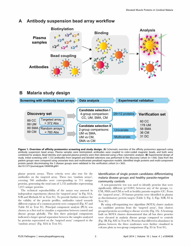
Results
In order to explore the potential of the human proteins in
plasma to predict disease status during malaria infections, we have
conducted an extensive exploratory profiling approach using
antibody suspension bead arrays (Fig. 1A). Using two different
approaches for target protein selection, with 304 unique proteins
in a targeted array with carefully selected proteins and 711 in a
random approach, a total of 1,015 human proteins have been
profiled in 719 blood samples from different patients aiming to
identify and verify protein signatures in plasma associated to the
severity of malaria infection (Fig. 1B).
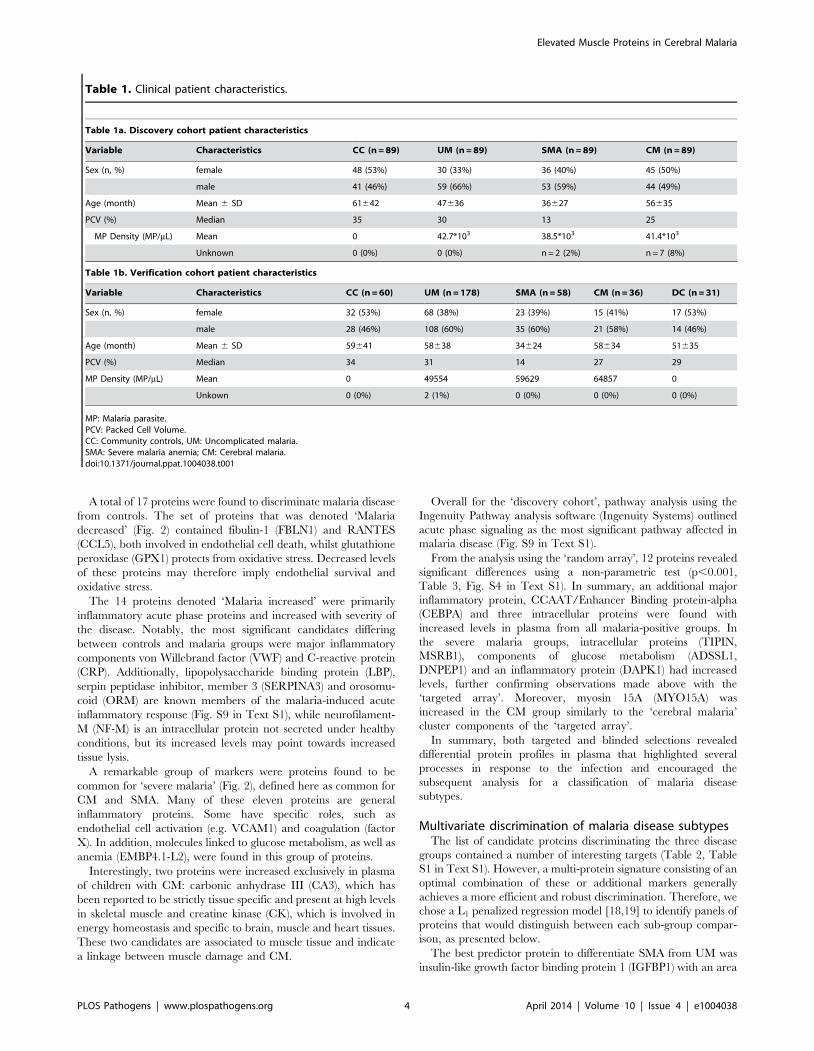
Study design and selection of participantsFirst, a set of patients was selected and carefully matched and
balanced to contain 356 childhood malaria patients and controls
recruited during 2006 to 2011 in Ibadan, Nigeria. This set,
denoted as ‘‘discovery cohort’’ in this study, included samples from
patients suffering from uncomplicated malaria (UM, n = 89),
severe malaria anemia (SMA, n = 89) and cerebral malaria (CM,
n = 89) and from parasite-negative community controls (CC,
n = 89; Table 1a). To confirm the validity of the protein signatures
identified, a second set of plasma samples was analyzed including
332 independent individuals recruited from the 2009 to 2012
period. This set consisted of 178 UM, 58 SMA, 36 CM, 60 CC
and 31 disease controls (DC) and was here denoted as ‘‘verification
cohort’’ (Table 1b). The discovery and verification cohorts had 24
patient samples in common to assess the technical quality of the
data in independent analyses (Methods S1 and Fig. S1C in Text
S1).
Antibody selection and array designAffinity proteomic arrays require that the analytes of interest are
defined prior to analysis. For the presented approach, an inclusive
and generous target selection strategy was employed by thorough
literature mining of major processes previously associated to
various aspects of malaria. Antibodies towards these targets were
obtained based upon availability within the Human Protein Atlas
(HPA). In total, a list of 372 antibodies targeting 304 different
human proteins was compiled and denoted as ‘targeted array’.
This list comprised primarily plasma proteins associated with acute
inflammation (Fig. S9 in Text S1), iron metabolism, oxidative
stress, endothelial activation, coagulation, complement activation,
angiogenesis, hematopoiesis and brain injury. Two additional sets
of 380 antibodies were used to profile all samples in the discovery
cohort and are denoted as ‘random arrays’.
The two additional sets of antibodies were randomly chosen
from the routine antibody production within the Human Protein
Atlas (HPA) where more than 40,000 antigen purified and protein
microarray validated antibodies have been generated. The
antibodies fulfill the quality criteria of having a concentration
that is higher than 50 mg/ml and that the specificity is validated on
Author Summary
Why do some malaria-infected children develop severeand lethal forms of the disease, while others only havemild forms? In order to try to find potential answers orclues to this question, we have here analyzed more than1,000 different human proteins in the blood of more than500 malaria-infected children from Ibadan in Nigeria, aholoendemic malaria region. We identified several proteinsthat were present at higher levels in the blood from thechildren that developed severe malaria in comparison tothose that did not. Some of the most interesting identifiedproteins were muscle specific proteins, which indicate thatdamaged muscles could be a discriminatory pathologicevent in cerebral malaria compared to other malaria cases.These findings will hopefully lead to an increasedunderstanding of the disease and may contribute to thedevelopment of clinical algorithms that could predictwhich children are more at risks to severe malaria. This inturn will be of high value in the management of thesechildren in already overloaded tertiary-care health facilitiesin urban large densely-populated sub-Saharan cities withholoendemic malaria such as in the case of Ibadan andLagos.
Elevated Muscle Proteins in Cerebral Malaria
PLOS Pathogens | www.plospathogens.org 2 April 2014 | Volume 10 | Issue 4 | e1004038
planar protein arrays. These criteria were also true for the
antibodies on the targeted array. These two ‘random arrays’,
covering 760 antibodies were corresponding to 711 unique
proteins, generating the total sum of 1,132 antibodies representing
1,015 unique proteins.
The technical reproducibility of the assays was assessed in
independent experiments (shown for ‘targeted array’ in Fig. S1A,
S1B and Methods S1 in Text S1). To provide further evidence for
the validity of the protein profiles, antibodies raised towards
different regions of a common protein were compared (Fig. S7 and
Table S3 in Text S1). Principal component analysis (PCA) was
chosen as a first tool to visualize a separation between control and
disease groups globally. The first three principal components
indicated a larger spread separation between the samples analyzed
by proteins represented on the ‘targeted array’ compared to the
‘random arrays’ (Fig. S2A in Text S1).
Identification of single protein candidates differentiatingmalaria disease groups and healthy parasite-negativecommunity controls
A non-parametric test was used to identify proteins that were
significantly different (p,0.001) between any of the groups, i.e.
UM, SMA and CM as well as healthy parasite-negative CC. From
the ‘targeted array’, 29 human proteins were identified in plasma
as discriminatory protein targets (Table 2, Fig. 2, Figs. S2B, S3 in
Text S1).
By using self-organizing tree algorithm (SOTA) cluster analysis
on candidate proteins from the ‘targeted array’, four clusters
grouped proteins according to disease severity (Fig. 2A). A heatmap
built on SOTA clusters demonstrated that all but three proteins
were elevated in malaria disease groups compared to controls
(Fig. 2B, Fig. S2C in Text S1). Proteins that differed in abundance in
plasma between the three syndromes were further visualized in
volcano plots as two-group comparisons (Fig. S5 in Text S1).
Figure 1. Overview of affinity proteomics screening and study design. (A) Schematic overview of the affinity proteomics approach usingantibody suspension bead arrays. Plasma samples were biotinylated, antibodies were coupled to color-coded magnetic beads, and both werecombined for analysis. Bead identity and captured plasma proteins were then detected using a flow cytometric analyzer. (B) Experimental design ofstudy. Initial screening with 1,132 antibodies from targeted and blinded selections was performed in the discovery cohort (n = 356). Data from thepatient groups were compared using univariate tests and multivariate penalized regression models. Identified single proteins and multi-componentprotein panels discriminating the 3 disease groups were validated in the verification cohort (n = 363).doi:10.1371/journal.ppat.1004038.g001
Elevated Muscle Proteins in Cerebral Malaria
PLOS Pathogens | www.plospathogens.org 3 April 2014 | Volume 10 | Issue 4 | e1004038
A total of 17 proteins were found to discriminate malaria disease
from controls. The set of proteins that was denoted ‘Malaria
decreased’ (Fig. 2) contained fibulin-1 (FBLN1) and RANTES
(CCL5), both involved in endothelial cell death, whilst glutathione
peroxidase (GPX1) protects from oxidative stress. Decreased levels
of these proteins may therefore imply endothelial survival and
oxidative stress.
The 14 proteins denoted ‘Malaria increased’ were primarily
inflammatory acute phase proteins and increased with severity of
the disease. Notably, the most significant candidates differing
between controls and malaria groups were major inflammatory
components von Willebrand factor (VWF) and C-reactive protein
(CRP). Additionally, lipopolysaccharide binding protein (LBP),
serpin peptidase inhibitor, member 3 (SERPINA3) and orosomu-
coid (ORM) are known members of the malaria-induced acute
inflammatory response (Fig. S9 in Text S1), while neurofilament-
M (NF-M) is an intracellular protein not secreted under healthy
conditions, but its increased levels may point towards increased
tissue lysis.
A remarkable group of markers were proteins found to be
common for ‘severe malaria’ (Fig. 2), defined here as common for
CM and SMA. Many of these eleven proteins are general
inflammatory proteins. Some have specific roles, such as
endothelial cell activation (e.g. VCAM1) and coagulation (factor
X). In addition, molecules linked to glucose metabolism, as well as
anemia (EMBP4.1-L2), were found in this group of proteins.
Interestingly, two proteins were increased exclusively in plasma
of children with CM: carbonic anhydrase III (CA3), which has
been reported to be strictly tissue specific and present at high levels
in skeletal muscle and creatine kinase (CK), which is involved in
energy homeostasis and specific to brain, muscle and heart tissues.
These two candidates are associated to muscle tissue and indicate
a linkage between muscle damage and CM.
Overall for the ‘discovery cohort’, pathway analysis using the
Ingenuity Pathway analysis software (Ingenuity Systems) outlined
acute phase signaling as the most significant pathway affected in
malaria disease (Fig. S9 in Text S1).
From the analysis using the ‘random array’, 12 proteins revealed
significant differences using a non-parametric test (p,0.001,
Table 3, Fig. S4 in Text S1). In summary, an additional major
inflammatory protein, CCAAT/Enhancer Binding protein-alpha
(CEBPA) and three intracellular proteins were found with
increased levels in plasma from all malaria-positive groups. In
the severe malaria groups, intracellular proteins (TIPIN,
MSRB1), components of glucose metabolism (ADSSL1,
DNPEP1) and an inflammatory protein (DAPK1) had increased
levels, further confirming observations made above with the
‘targeted array’. Moreover, myosin 15A (MYO15A) was
increased in the CM group similarly to the ‘cerebral malaria’
cluster components of the ‘targeted array’.
In summary, both targeted and blinded selections revealed
differential protein profiles in plasma that highlighted several
processes in response to the infection and encouraged the
subsequent analysis for a classification of malaria disease
subtypes.
Multivariate discrimination of malaria disease subtypesThe list of candidate proteins discriminating the three disease
groups contained a number of interesting targets (Table 2, Table
S1 in Text S1). However, a multi-protein signature consisting of an
optimal combination of these or additional markers generally
achieves a more efficient and robust discrimination. Therefore, we
chose a L1 penalized regression model [18,19] to identify panels of
proteins that would distinguish between each sub-group compar-
ison, as presented below.
The best predictor protein to differentiate SMA from UM was
insulin-like growth factor binding protein 1 (IGFBP1) with an area
Table 1. Clinical patient characteristics.
Table 1a. Discovery cohort patient characteristics
Variable Characteristics CC (n = 89) UM (n = 89) SMA (n = 89) CM (n = 89)
Sex (n, %) female 48 (53%) 30 (33%) 36 (40%) 45 (50%)
male 41 (46%) 59 (66%) 53 (59%) 44 (49%)
Age (month) Mean 6 SD 61642 47636 36627 56635
PCV (%) Median 35 30 13 25
MP Density (MP/mL) Mean 0 42.7*103 38.5*103 41.4*103
Unknown 0 (0%) 0 (0%) n = 2 (2%) n = 7 (8%)
Table 1b. Verification cohort patient characteristics
Variable Characteristics CC (n = 60) UM (n = 178) SMA (n = 58) CM (n = 36) DC (n = 31)
Sex (n, %) female 32 (53%) 68 (38%) 23 (39%) 15 (41%) 17 (53%)
male 28 (46%) 108 (60%) 35 (60%) 21 (58%) 14 (46%)
Age (month) Mean 6 SD 59641 58638 34624 58634 51635
PCV (%) Median 34 31 14 27 29
MP Density (MP/mL) Mean 0 49554 59629 64857 0
Unkown 0 (0%) 2 (1%) 0 (0%) 0 (0%) 0 (0%)
MP: Malaria parasite.PCV: Packed Cell Volume.CC: Community controls, UM: Uncomplicated malaria.SMA: Severe malaria anemia; CM: Cerebral malaria.doi:10.1371/journal.ppat.1004038.t001
Elevated Muscle Proteins in Cerebral Malaria
PLOS Pathogens | www.plospathogens.org 4 April 2014 | Volume 10 | Issue 4 | e1004038

under the ROC curve (AUC) of 0.84 alone (Fig. 3A, Table S2 in
Text S1). The best multi-protein combination was a 3-protein
signature consisting of IGFBP1 with von Willebrand factor (VWF)
and hemoglobin a-subunit (HBA2, HBA1), which resulted in a
slightly improved AUC of 0.87.
Next, Carbonic anhydrase III (CA3) alone discriminated CM
from UM with a high AUC of 0.90 (Table S2 in Text S1).
Variable selection refinement showed that a panel of 4 proteins,
including angiotensinogen (AGT), FBLN1 and 2,3-bisphosphogly-
cerate mutase (BPGM), resulted in a yet improved performance of
0.94 (Fig. 3B). The optimal signature identified for the discrim-
ination of CM and UM was a 23-protein signature with an AUC
of 0.98.
The two best classifier proteins for the comparison of the two
severe malaria groups, CM and SMA, were CA3 and CK. The
combination of the 2 proteins resulted in an AUC of 0.84 (Fig. 3C).
Moreover, the most optimal classifier combination comprised 9
proteins resulting in an AUC of 0.91.
In summary, protein targets in the multivariate signatures
overlapped largely with the targets found in the univariate
analysis in the two-group comparisons (Fig. S5 in Text S1). As
expected, ranking of the proteins differed, because multivariate
models aim at the identification of the best combination of
proteins to maximize the discriminative power. This resulted in
protein signatures that contained not only highly significant
proteins found with univariate analysis but proteins that carry
important discriminatory information when combined with
others.
Verification of multi-protein panels with independentpatient cohorts
Both the single protein classifiers and multi-protein panels were
validated with a new and independent set of 363 samples from the
same hospital. Using the classifiers determined in the multivariate
signatures containing the full list proteins, the AUC for the UM
versus SMA (3 proteins), UM versus CM (23 proteins) and SMA
Table 2. Overview of single protein candidates and SOTA clusters (targeted array).
Cluster Protein Uniprot Gene description Antibodies References
Malaria decreased CCL5 P13501 chemokine (C-C motif) ligand 5 HPA010552 [33,41]
FBLN1 P23142 fibulin 1 HPA001613 N/A
GPX1 P07203 glutathione peroxidase 1 HPA044758 [42,43]
Malaria increased CD14 P08571 CD14 molecule HPA002127 [44]
CRP P02741 C-reactive protein, pentraxin-related HPA027396 [27,45]
CSF1 P09603 colony stimulating factor 1 (macrophage) HPA022244 N/A
FURIN P09958 furin HPA005905 N/A
IL7 P13232 interleukin 7 HPA019590 N/A
LBP P18428 lipopolysaccharide binding protein HPA001508 [46]
MMP2 P08253 matrix metallopeptidase 2 HPA001939 N/A
NEFM P07197 neurofilament, medium polypeptide HPA022845 N/A
ORM P02763 orosomucoid 1 HPA047725 [4]
SERPINA3 P01011 serpin peptidase inhibitor, member 3 HPA000893 [13,47]
SLC12A3 P55017 solute carrier family 12, member 3 HPA028748 N/A
TNFRSF1B P20333 tumor necrosis factor receptorsuperfamily, member 1B
HPA004796 N/A
VWF P04275 von Willebrand factor HPA002082 [21,26,48]
Severe Malaria ADA P00813 adenosine deaminase HPA001399 N/A
ADM P35318 adrenomedullin HPA031806 N/A
AGT P01019 angiotensinogen HPA001557 [49]
BPGM P07738 2,3-bisphosphoglycerate mutase HPA016493 N/A
EPB41L2 O43491 erythrocyte membrane protein band4.1-like 2
HPA006642 [50,51]
F10 P00742 coagulation factor X HPA030629 [52]
FLT1 P17948 fms-related tyrosine kinase 1 HPA011740 [21,53]
HABP2 Q14520 hyaluronan binding protein 2 HPA019518 N/A
IGFBP1 P08833 insulin-like growth factor bindingprotein 1
HPA046972 N/A
LCP1 P13796 lymphocyte cytosolic protein 1 HPA019493 N/A
VCAM1 P19320 vascular cell adhesion molecule 1 HPA034796 [54]
Cerebral malaria CA3 P07451 carbonic anhydrase III, muscle specific HPA021775 N/A
CK P12277 creatine kinase HPA001254 [55,56]
N/A: not applicable, no previous known link with malaria.doi:10.1371/journal.ppat.1004038.t002
Elevated Muscle Proteins in Cerebral Malaria
PLOS Pathogens | www.plospathogens.org 5 April 2014 | Volume 10 | Issue 4 | e1004038

versus CM (8 proteins) comparisons were 0.72, 0.90 and, 0.86,
respectively (Fig. 3D).
To further verify the results technically, additional antibodies
were used targeting other regions of the same protein. For
example, von Willebrand factor (VWF) was repeated using the
same antibody (HPA00282) and an additional antibody
(HPA001815), with both showing comparable results in the
different disease groups in the verification cohort and the discovery
cohort (Fig. S7 in Text S1), demonstrating reproducibility of the
presented results.
Additionally, a small cohort of disease control (DC) samples
(Table 1b) from patients suffering from coma or meningitis was
also profiled with the same protein panel analyzed in the
verification cohort. Changes in levels due to inflammation, as
shown with CEBPA, CRP and CCL5 protein levels, were more
exacerbated in the CM than the DC samples (Fig. S10 in Text S1)
Figure 2. Identification of 29 human proteins discriminating community controls and malaria cases. (A) Applying a non-parametric test,29 human proteins were identified showing significant (adjusted p,0.001) differences between any of the four groups. Cluster analysis using self-organizing tree algorithm (SOTA) revealed four different clusters with distinct protein profiles designated as ‘malaria decreased’, ‘malaria increased’,‘severe malaria’ and ‘cerebral malaria’ (see also Fig. S3 in Text S1). (B) Heatmap visualizing protein profiles in individual patients. Samples wereorganized according to group affiliation and proteins were sorted following SOTA clusters. Displayed are scaled relative intensities of each protein ineach group (CC = grey, UM = green, SMA = red, CM = blue).doi:10.1371/journal.ppat.1004038.g002
Elevated Muscle Proteins in Cerebral Malaria
PLOS Pathogens | www.plospathogens.org 6 April 2014 | Volume 10 | Issue 4 | e1004038

suggesting a stronger inflammatory response in malaria-infected
patients than in other diseases.
Muscle proteins as indicators of cerebral malariaCA3 was identified as the top candidate to differentiate CM
from the other two malaria syndromes. In both the discovery and
the verification analysis, two antibodies with distinct specificities to
CA3 showed concordant performance (Fig. S7 in Text S1). An
additional antibody was acquired against the same target protein
and further confirmed the results. Immunohistochemical staining
of healthy human tissue confirmed the muscle specificity of both
antibodies (Fig. S8 in Text S1). Similarly, antibodies against
creatine kinase (CK) revealed increased levels in CM in the
discovery phase. The CK antibody used in the discovery phase
was raised against a region of the brain-specific form (CKB) that is
shared with the muscle-specific (CKM) isoform (Table S3 in Text
S1). To further evaluate which isoform is detected, an HPA
antibody directed towards the muscle-specific isoform (CKM) as
well as a commercially available antibody against CKM were
tested in the verification phase. The results from these CKM
antibodies were similar to the trends with the CKB/M antibody
used in the discovery phase (Fig. S7 in Text S1). Furthermore,
immunohistochemical staining showed that the CKB/M antibody
recognized skeletal and cardiac muscle as well as cerebral tissues
(Fig. S8 in Text S1). Finally, myoglobin (MB), a cardiac and
skeletal muscle protein, was also part of the discriminatory profile
between CM and UM (Fig. 3b) and also had higher plasma levels
in the CM group compared to all other groups in the verification
sample set.
Using the small additional DC cohort, CA3 and CK, previously
identified as related to CM syndrome, had levels slightly lower in
the DC group compared to CM but were not significantly different
(Fig. S10 in Text S1) suggesting that muscle damage might not be
specific to cerebral malaria but probably linked to coma. Further
studies with a larger cohort of DC comatose group of children with
pathologies other than malaria are required to verify these
findings.
Discussion
We have here investigated the levels of human proteins
circulating in plasma of children with different forms of
uncomplicated or severe malaria and compared the levels with
those of parasite-negative community controls. The study com-
prises a total of 709 plasma samples including 515 from malaria-
infected children. Amongst the 1,015 host proteins studied, 41
were identified as candidates discriminating between healthy
community controls and malaria patients. Protein markers of
oxidative stress were found elevated in anemic individuals while
markers of endothelial activation, platelet adhesion and muscle-
and tissue damage were found linked to cerebral malaria. Taken
together, this suggests the presence of a generalized vascular
inflammation, an unbalanced glucose metabolism and deep lesions
into the micro-vasculature.
The chosen bead array technology enabled the generation of
protein profiles in unfractionated and biotinylated plasma samples
by using combinations of large sets of antibodies as demonstrated
by the use of both carefully pre-selected and blindly chosen
antibodies.
Most previous studies have focused on markers of fatalities in
those already with severe malaria [20–22]. In one recent analysis
[14], discrimination of different malaria syndromes from each
other was suggested possible but only when using extensive protein
panels with up to 50 proteins (AUC of 0.7–0.8). In contrast, we
show herein that, employing small panels of proteins, it is possible
to build models that predict with an AUC higher than 0.90 which
children have severe malaria complications. We also demonstrate
a discriminatory signature that reaches superior accuracy for UM
vs. SMA with IGFBP1 alone and for UM vs. CM using only four
proteins.
Higher plasma levels of muscle-derived proteins were found in
children with cerebral malaria only including carbonic anhydrase
III and creatine kinase, suggesting that smooth muscle-cells of the
microvasculature may be injured. The excessive sequestration of
pRBC seen in cerebral vessels, the level of which has also been
found to correlate with coma [3] is probably one of the reasons for
the injury of the muscle cells. This is in concordance with previous
histo-pathological, studies where subjects who succumbed to
cerebral malaria showed vascular- and microvascular lesions
complicated by ring-hemorrhages [3]. The presence of myoglobin
in the plasma of the patients with CM, a marker of cardiac- and
striated muscles only, also indicates that the vasculature and the
muscles outside of the brain are severely affected by sequestration
as seen for example in muscle biopsies of Thai adults [23]. Further,
Table 3. Overview of single protein candidates with SOTA clusters (random array).
Cluster Protein Uniprot Gene description Antibodies
Malaria increased ADSSL1 Q8N142 adenylosuccinate synthase like 1 HPA052621
CCDC102A Q96A19 coiled-coil domain containing 102A HPA040598
EEF2 P13639 eukaryotic translation elongation factor 2 HPA040534
FAM71F2 Q6NXP2 family with sequence similarity 71, member F2 HPA052240
TIPIN Q9BVW5 TIMELESS interacting protein HPA039704
Severe Malaria CEBPA P49715 CCAAT/enhancer binding protein (C/EBP), alpha HPA052734
DAPK1 P53355 death-associated protein kinase 1 HPA040472
DNPEP Q9ULA0 aspartyl aminopeptidase HPA036398
HAP1 P54257 huntingtin-associated protein 1 HPA053019
MSRB1 Q9NZV6 methionine sulfoxide reductase B1 HPA052662
SEC24C P53992 SEC24 family HPA040196
Cerebral malaria MYO15A Q9UKN7 myosin XVA HPA039770
doi:10.1371/journal.ppat.1004038.t003
Elevated Muscle Proteins in Cerebral Malaria
PLOS Pathogens | www.plospathogens.org 7 April 2014 | Volume 10 | Issue 4 | e1004038

a recent study showed that blood flow obstruction might be
exacerbated by increased skeletal muscle oxygen consumption in
severe malaria [24], contributing to hypoxic and hyperlactemic
conditions in the microvasculature. Lack of oxygen in muscle cells
accompanied with hypoglycemia, lactate overproduction, oxida-
tive stress and inflammation are typical consequences of muscle
damage [25], which could further contribute to vascular injuries
and subsequent muscle cell death with the release of muscle-
specific proteins into the blood circulation. Whether creatine
kinase, myoglobin and carbonic anhydrase III release in the
plasma exacerbate these deleterious events is not known at this
stage and deserves further investigation. For example, increased
plasma carbonic anhydrase activity could contribute to the
impairment of the acid-base and excess myoglobin in blood
circulation could lead to kidney failure if filtrated by kidneys. An
additional small cohort of malaria-negative children, with other
illnesses involving coma, suggested that coma could either be a
cause or a consequence of muscle damage observed in cerebral
malaria, similarly to other comatose diseases. Further studies,
involving larger cohorts of non-malaria comatose children, will be
required to verify this hypothesis.
In our study, predicting which samples were from patients
diagnosed with cerebral malaria was very accurate in both cohorts
due to the discovery of the presence of the muscle-proteins only in
the plasma of the children with cerebral malaria. Previous studies
have successfully used endothelial cell activation markers to
predict severe malaria, notably using angiopoietin-1 and 2
[20,21,26], but their specificity to cerebral malaria, as compared
to other severe complications, remains unclear. Here we propose
that markers of muscle damage accompanied by markers of
endothelial cell activation/platelet adhesion in the plasma (Fig. 4,
orange bars) are specific to cerebral malaria pathogenesis and
distinct to severe malaria anemia. Our data therefore indicate that
children with uncomplicated malaria that develop cerebral
malaria are likely to have vascular lesions and muscle damage,
which can be readily monitored in plasma.
Most of the plasma proteins showing differences in between the
malaria patients and the community controls were components of
the inflammatory response (Fig. 4, blue bars). Further, multiple
novel inflammatory components were identified, including
MMP2, CSF1, and IL7 (table 2), demonstrating the presence of
a more generalized vascular inflammation in patients with malaria
infections. The reliability of the present study was furthermore
confirmed by the fact that a number of protein measured herein
Figure 3. Discrimination of the three malaria disease sub-typeswith multi-protein signatures. L1-penalized logistic regressionmodels were fitted for the three two-group comparisons. The plotsshow the resulting ROC curves when the model included all selectedproteins (black line) and only the top ones (coloured line). The areaunder the ROC curve (AUC) for the optimal number of proteins and thecombination with the smallest number of proteins after variableselection refinement is presented adjacent to the plots. (A) Forclassification of UM vs SMA a 3-protein signature provided an optimalresult (AUC = 0.87) (black line). (B) For classification of UM vs CM aprotein signature with 23 proteins showed the best result (black line).As comparison, the AUC of the top 4 proteins (blue line) and top 10proteins (grey line) after step-by-step removal of selected proteins isshown. CA3 = HPA021775, CA3* = HPA026700. (C) For classification ofSMA vs CM a protein signature with 9 proteins showed the best result(black line). As comparison, the AUC of the top 2 proteins after step-by-step removal of selected proteins is shown (green line). (D) ROC curvesfor the three subgroup comparisons using their respective best proteinsignatures in the verification cohort, UM vs SMA (red line), UM vs CM(blue line) and SMA vs CM (green line). SAA4 was excluded fromverification due to technical failure.doi:10.1371/journal.ppat.1004038.g003
Elevated Muscle Proteins in Cerebral Malaria
PLOS Pathogens | www.plospathogens.org 8 April 2014 | Volume 10 | Issue 4 | e1004038

Figure 4. Overview of panels of proteins and their physiological pathways predicting the different malaria clusters. Proteins withdifferential profiles between the malaria groups were classified into physiological pathways. This included proteins identified common to all malariagroups (blue bars), common to both defined severe malaria syndromes (purple bars), proteins with levels elevated in SMA (light green bars), proteinswith levels elevated in CM (light orange bars) and proteins specific to CM (dark orange bars). For each panel, columns were stacked by number ofproteins identified in each category. Grey dotted connectors represent either a regulation link or a common protein component between tophysiological pathways. UM: uncomplicated malaria; SMA: severe malaria anemia; CM: cerebral malaria. Proteins are represented by their gene names(Refer to Table 2, Fig. S3 and Table S2 in Text S1 for full names).doi:10.1371/journal.ppat.1004038.g004
Elevated Muscle Proteins in Cerebral Malaria
PLOS Pathogens | www.plospathogens.org 9 April 2014 | Volume 10 | Issue 4 | e1004038

include acute phase- and inflammatory proteins previously
documented to be present in the plasma of malaria infected
individuals [27,28] (Table 2). Proteins related to different aspects
of the glucose metabolism, including insulin-glucagon modulators
and glycolytic enzymes, were also elevated in both SMA and CM
patients compared to those with UM (Fig. 4, purple bars). Further
investigation of their potential role in the induction of hypogly-
cemia, a hallmark of severe malaria associated with fatality [29,30]
might refine knowledge on the human response mechanisms to
malaria infection.
Most proteins elevated in plasma from SMA compared to UM
were also elevated in CM but to a lower extent. Yet, IGFBP1 and
HBA were part of the protein signature for comparing SMA with
UM, and had highest levels in patients suffering with SMA.
IGFBP1 could be further expressed due to high levels of reactive
species [31], and free-hemoglobin release in blood circulation
could be a trigger of free-heme induced oxidative stress,
particularly if not properly scavenged by the haptoglobin-
hemopexin system [32]. It is noteworthy that predictive protein
signatures for SMA have in previous studies mainly included
inflammatory cytokines [22,33,34]. For example, we recently
showed in the same Nigerian population, that pro-inflammatory
cytokines were more pronounced in SMA than in CM [9], a
finding supported by the fact that pro-inflammatory TNF-alpha
has a role in anemia establishment [8]. Due to the sensitivity of the
assay, the levels of most of the cytokines tested in the present study
were too low to be detected. Consequently, we hypothesize that
including oxidative stress-related proteins as well as pro-inflam-
matory cytokines in future studies in the protein signature could
potentially assist to further improve SMA distinction from other
malaria complications.
In summary, a high-throughput antibody-based protein profil-
ing method and large-scale discovery and verification cohorts,
revealed muscle-specific proteins in plasma as potential indicators
of cerebral malaria. Our study could therefore provide key
elements towards the discovery of distinct mechanisms in the
human response to malaria infection between the two most fatal
syndromes of childhood malaria.
Materials and Methods
Ethics statementParents or guardians of study participants gave informed written
consent. This research was approved by the internationally
accredited joint ethics committee of the College of Medicine of
the University of Ibadan and the University College Hospital,
Ibadan.
Study designAll study participants with illness were recruited under the
auspices of the Childhood Malaria Research Group (CMRG) at
the University College Hospital (UCH) in the city of Ibadan,
Nigeria. Malaria-negative community control (CC) children were
recruited from local vaccination clinics as well as during school
visits across several Ibadan districts. This case-control study was
divided in a discovery cohort that contains those patients recruited
during 2006 to 2011 and a verification cohort made up of those
recruited in the 2009 to 2012 period.
Children were aged from 6 months to 13 years and were
screened for parasite detection by microscopy following Giemsa
staining of thick and thin blood films as performed routinely at
UCH. Clinical definitions used were as defined by the WHO
criteria for severe P. falciparum malaria [1]. Uncomplicated
malaria (UM) cases were defined as febrile children with P.
falciparum parasitaemia and PCV (Packed Cell Volume) greater
than 20% who did not require hospital admission. Severe
malarial anemia (SMA) cases were defined as conscious children
with PCVs less than 16% in the presence of P. falciparum
parasitaemia. Cerebral malaria (CM) cases were defined as
children in unrousable coma for at least one hour in the presence
of asexual P. falciparum parasitaemia with normal cerebrospinal
fluid and PCV greater than 20%. Community controls were
children that did not show any obvious symptoms of illness and
seemed healthy. They were screened for parasite presence and
were only included in the study if the Giemsa staining of both
thick and thin blood films were negative for Plasmodium parasites.
They were selected to match age and sex with malaria-infected
patients. The clinical data was compiled for each patient and
samples were collected as previously described [9,14] (see
Methods S1 in Text S1).
Antibody selection and array designAntibodies were selected and acquired from the huge antibody
collection within the Human Protein Atlas (HPA, www.
proteinatlas.org) consisting of more than 40,000 antigen purified
and protein microarray validated antibodies.
The selection of antibodies was carried out using two different
strategies. Using a ‘targeted’ approach, 380 antibodies were
selected against 304 protein targets according to a generous and
inclusive literature mining of malaria pathogenesis. The final set
was defined by availability and fulfillment of technical validations,
such as having a concentration that is higher than 50 mg/ml and
that the specificity is validated on planar protein arrays. The two
additional sets of 380 antibodies were randomly chosen from the
routine antibody production within HPA. These 760 antibodies
were directed to 711 unique proteins and fulfilled the same criteria
as for the antibodies on the targeted array.
Suspension bead array procedureFor the generation of antibodies suspension beads, antibodies
were diluted using a liquid handling system (EVO150, Tecan) and
coupled to carboxylated magnetic microspheres (MagPlex,
Luminex Corporation) as previously described [35]. Briefly,
carboxylated beads were activated with 1-ethyl-3-(3-dimethylami-
nopropyl) carbodiimide (EDC) and N-hydroxysulfosuccinimide
(Sulfo-NHS, Thermo Scientific) and incubated with 1.6 mg
antibody in a multi-well microtiter plate for 2 h. After the
coupling reaction, beads were stored at 4uC in a protein
containing buffer (Blocking Reagent for ELISA, Roche Applied
Science) supplemented with ProClin (Sigma-Aldrich). Before
incubation with samples, the different bead identities were
combined to create a 384-plex-bead array. Antibody coupling
was confirmed with R-phycoerythrin (PE)-conjugated donkey anti-
rabbit IgG antibody (Jackson ImmunoResearch).
The data from single antibody and direct sample labeling assays
was judged by technical replication of the experiment, profile
concordance of several antibodies raised towards a common target
protein, and biological replication of the analysis in new,
independent samples.
Biotinylation of plasma samples was performed as previously
described [36] (refer to Methods S1 in Text S1). Biotinylated
samples were then diluted in PBS containing 0.5% (w/v)
polyvinylalcohol, 0.8% (w/v) polyvinylpyrrolidone, 0.1% casein
(all from Sigma) supplemented with 0.5 mg/ml rabbit IgG (Bethyl)
using a liquid handler (SELMA, CyBio). Before incubation with
bead arrays, samples were heat-treated in a thermocycler for
30 min at 56uC for epitope retrieval. After incubation of samples
with beads for 14 h, beads were washed with PBS-T (pH 7.4,
Elevated Muscle Proteins in Cerebral Malaria
PLOS Pathogens | www.plospathogens.org 10 April 2014 | Volume 10 | Issue 4 | e1004038

0.05% Tween20) on a plate washer (EL406, Biotek) and incubated
with 0.4% paraformaldehyde for 10 min. Subsequently, beads
were incubated with 0.5 mg/ml R-Phycoerythrin labeled strepta-
vidin (Invitrogen) for 20 min and washed with PBS-T. Bead
identities and median fluorescence intensity of R-Phycoerythrin
were analyzed simultaneously using a FlexMAP 3D system
(Luminex Corp.).
Statistical analysisData analysis was performed using R language for statistical
computing [37,38]. The data from the ‘targeted array’ and the
‘random array’ was normalized using probabilistic quotient
normalization (PQN) as described before [39]. The non-paramet-
ric Kruskal-Wallis test was applied to identify proteins that are
different among the different malaria disease groups (CC, UM,
SMA and CM). For pairwise comparison of the different malaria
disease groups, a Wilcoxon rank sum test was applied (with
continuity correction). Bonferroni method was used to control the
family-wise error rate. For cluster analysis of the protein profiles
self-organizing tree algorithm (SOTA) was applied after the
medians per protein and subgroup were centered and scaled using
the R function scale [40] (Methods S1 in Text S1).
The data from the verification cohort was normalized using a
linear mixed model
yij~azbizb1Platezb2Indexzb3Plate|Indexzeij
where: y = log2(intensity), Plate: biotinylation plates, Index: order
of assay, and bi: random effect of the ith target.
For the identification of protein signatures a logistic regression
model was used. We used L1 penalization proposed by Tibshirani
[19], also known as Lasso, which performs parameter estimation
and variable selection at the same time. The penalization involves
a penalize parameter (l) which is chosen through a cross-
validation procedure. Thereby, the dataset was randomly divided
into subsets. The first subset (K) was designated as the test dataset,
while the model was fitted to the remaining training dataset (subset
K-1). This procedure was repeated k times for each subset (see
Methods S1 in Text S1 for details). For a first verification of the
identified multi-protein signatures, the parameter estimates from
the first dataset were used to obtain the prediction based on the
second replicate data of the discovery cohort (Fig. S6 in Text S1).
Supporting Information
Text S1 Supplementary Information. An extensive set of
supplementary material is available with detailed descriptions and
visualizations of the data and the identified proteins of interest with
ten figures, four tables and methods.
(PDF)
Acknowledgments
The authors thank all the children, guardians and parents who participated
in this study. We also thank all the consultants, registrars, nurses, and
administrative staff at the Department of Paediatrics, University College
Hospital, Ibadan, Nigeria, for all the support given for the present study.
We are very grateful to Dr. A. A. Holder at the National Institute for
Medical Research, London, UK for reagents, discussions and comments on
the manuscript. We also thank the whole group of Affinity Proteomics at
SciLifeLab Stockholm and the entire staff of the Human Protein Atlas for
their efforts.
Author Contributions
Conceived and designed the experiments: JB FB IC MU JMS MW DFR
PN. Performed the experiments: JB FB SP IC CK. Analyzed the data: JB
FB SP IC YP JMS PN. Contributed reagents/materials/analysis tools: BJB
AEO WAA NKA FA SO FOA WAS OS DFR. Wrote the paper: JB FB SP
IC CK YP MU JMS MW DFR PN.
References
1. World Health Organization (WHO). (2010). World Malaria Report 2010.
Available: http://www.who.int/malaria/world_malaria_report_2010/en/. Last
accessed 5th Mar 2014.
2. Amzat J (2011) Assessing the progress of malaria control in Nigeria. World
Health Popul 12: 42–51.
3. Ponsford MJ, Medana IM, Prapansilp P, Hien TT, Lee SJ, et al. (2012)
Sequestration and microvascular congestion are associated with coma in human
cerebral malaria. J Infect Dis 205: 663–671.
4. Friedman MJ (1983) Control of malaria virulence by alpha 1-acid glycoprotein
(orosomucoid), an acute-phase (inflammatory) reactant. Proc Natl Acad Sci U S A
80: 5421–5424.
5. Nussler A, Pied S, Pontet M, Miltgen F, Renia L, et al. (1991) Inflammatory
status and preerythrocytic stages of malaria: role of the C-reactive protein. Exp
Parasitol 72: 1–7.
6. Scholander C, Treutiger CJ, Hultenby K, Wahlgren M (1996) Novel fibrillar
structure confers adhesive property to malaria-infected erythrocytes. Nat Med 2:
204–208.
7. MacPherson GG, Warrell MJ, White NJ, Looareesuwan S, Warrell
DA (1985) Human cerebral malaria. A quantitative ultrastructural
analysis of parasitized erythrocyte sequestration. Am J Pathol 119: 385–
401.
8. Perkins DJ, Were T, Davenport GC, Kempaiah P, Hittner JB, et al. (2011)
Severe malarial anemia: innate immunity and pathogenesis. Int J Biol Sci 7:
1427–1442.
9. Burte F, Brown BJ, Orimadegun AE, Ajetunmobi WA, Afolabi NK, et al. (2013)
Circulatory hepcidin is associated with the anti-inflammatory response but not
with iron or anemic status in childhood malaria. Blood 121: 3016–22. doi:
10.1182/blood-2012-10-461418.
10. Grau GE, Taylor TE, Molyneux ME, Wirima JJ, Vassalli P, et al. (1989) Tumor
necrosis factor and disease severity in children with falciparum malaria.
N Engl J Med 320: 1586–1591.
11. Whiteaker JR, Lin C, Kennedy J, Hou L, Trute M, et al. (2011) A targeted
proteomics-based pipeline for verification of biomarkers in plasma. Nat
Biotechnol 29: 625–634.
12. Bahk YY, Na BK, Cho SH, Kim JY, Lim KJ, et al. (2010) Proteomic analysis of
haptoglobin and amyloid A protein levels in patients with vivax malaria.
Korean J Parasitol 48: 203–211.
13. Ray S, Renu D, Srivastava R, Gollapalli K, Taur S, et al. (2012) Proteomic
investigation of falciparum and vivax malaria for identification of surrogate
protein markers. PLoS One 7: e41751.
14. Burte F, Brown BJ, Orimadegun AE, Ajetunmobi WA, Battaglia F, et al. (2012)
Severe childhood malaria syndromes defined by plasma proteome profiles. PLoS
One 7: e49778.
15. Uhlen M, Oksvold P, Fagerberg L, Lundberg E, Jonasson K, et al. (2010)
Towards a knowledge-based Human Protein Atlas. Nat Biotechnol 28: 1248–
1250.
16. Schwenk JM, Igel U, Neiman M, Langen H, Becker C, et al. (2010) Toward
next generation plasma profiling via heat-induced epitope retrieval and array-
based assays. Mol Cell Proteomics 9: 2497–2507.
17. Haggmark A, Neiman M, Drobin K, Zwahlen M, Uhlen M, et al. (2012)
Classification of protein profiles from antibody microarrays using heat and
detergent treatment. N Biotechnol 29: 564–570.
18. Goeman JJ (2010) L-1 Penalized Estimation in the Cox Proportional Hazards
Model. Biometrical Journal 52: 70–84.
19. Tibshirani R (1996) Regression shrinkage and selection via the Lasso. Journal of
the Royal Statistical Society Series B-Methodological 58: 267–288.
20. Conroy AL, Glover SJ, Hawkes M, Erdman LK, Seydel KB, et al. (2012)
Angiopoietin-2 levels are associated with retinopathy and predict mortality in
Malawian children with cerebral malaria: A retrospective case-control study.
Critical Care Medicine 40: 952–959.
21. Erdman LK, Dhabangi A, Musoke C, Conroy AL, Hawkes M, et al. (2011)
Combinations of Host Biomarkers Predict Mortality among Ugandan Children
with Severe Malaria: A Retrospective Case-Control Study. PLoS One 6:
e17440. doi: 10.1371/journal.pone.0017440.
22. Wilson NO, Jain V, Roberts CE, Lucchi N, Joel PK, et al. (2011) CXCL4 and
CXCL10 predict risk of fatal cerebral malaria. Disease Markers 30: 39–49.
23. Davis TME, Pongponratan E, Supanaranond W, Pukrittayakamee S, Helliwell
T, et al. (1999) Skeletal muscle involvement in falciparum malaria: Biochemical
and ultrastructural study. Clinical Infectious Diseases 29: 831–835.
Elevated Muscle Proteins in Cerebral Malaria
PLOS Pathogens | www.plospathogens.org 11 April 2014 | Volume 10 | Issue 4 | e1004038

24. Yeo TW, Lampah DA, Kenangalem E, Tjitra E, Price RN, et al. (2013)
Impaired skeletal muscle microvascular function and increased skeletal muscleoxygen consumption in severe falciparum malaria. J Infect Dis 207: 528–536.
25. Brancaccio P, Lippi G, Maffulli N (2010) Biochemical markers of muscular
damage. Clin Chem Lab Med 48: 757–767.26. Conroy AL, Phiri H, Hawkes M, Glover S, Mallewa M, et al. (2010)
Endothelium-Based Biomarkers Are Associated with Cerebral Malaria inMalawian Children: A Retrospective Case-Control Study. PLoS One 5:
15291. doi: 10.1371/journal.pone.0015291.
27. O’donnell A, Fowkes FJI, Allen SJ, Imrie H, Alpers MP, et al. (2009) The acutephase response in children with mild and severe malaria in Papua New Guinea.
Transactions of the Royal Society of Tropical Medicine and Hygiene 103: 679–686.
28. Schofield L, Grau GE (2005) Immunological processes in malaria pathogenesis.Nat Rev Immunol 5: 722–735.
29. Jallow M, Casals-Pascual C, Ackerman H, Walther B, Walther M, et al. (2012)
Clinical Features of Severe Malaria Associated with Death: A 13-YearObservational Study in The Gambia. PLoS One 7: e45645. doi: 10.1371/
journal.pone.0045645..30. White NJ, Warrell DA, Chanthavanich P, Looareesuwan S, Warrell MJ, et al.
(1983) Severe Hypoglycemia and Hyperinsulinemia in Falciparum-Malaria.
New England Journal of Medicine 309: 61–66.31. Lang CH, Nystrom GJ, Frost RA (1999) Regulation of IGF binding
protein-1 in Hep G2 cells by cytokines and reactive oxygen species. AmericanJournal of Physiology-Gastrointestinal and Liver Physiology 276: G719–
G727.32. Quaye IK (2008) Haptoglobin, inflammation and disease. Trans R Soc Trop
Med Hyg 102: 735–742.
33. Ong’echa JM, Davenport GC, Vulule JM, Hittner JB, Perkins DJ (2011)Identification of Inflammatory Biomarkers for Pediatric Malarial Anemia
Severity Using Novel Statistical Methods. Infect Immun 79: 4674–4680.34. Thuma PE, van Dijk J, Bucala R, Debebe Z, Nekhai S, et al. (2011) Distinct
Clinical and Immunologic Profiles in Severe Malarial Anemia and Cerebral
Malaria in Zambia. Journal of Infectious Diseases 203: 211–219.35. Schwenk JM, Nilsson P (2011) Assessment of antibody specificity using
suspension bead arrays. Methods Mol Biol 785: 183–189.36. Neiman M, Hedberg JJ, Donnes PR, Schuppe-Koistinen I, Hanschke S, et al.
(2011) Plasma profiling reveals human fibulin-1 as candidate marker for renalimpairment. J Proteome Res 10: 4925–4934.
37. Ihaka R GR (1996) R: A Language for Data Analysis and Graphics. Journal of
Computational and Graphical Statistics 5: 299–314.38. (2012) R Development Core Team. R: A Language and Environment for
Statistical Computing. ISBN 3-900051-07-0. URL http://www.R-project.org/.R Foundation for Statistical Computing: Vienna, Austria.
39. Kato BS, Nicholson G, Neiman M, Rantalainen M, Holmes CC, et al. (2011)
Variance decomposition of protein profiles from antibody arrays using alongitudinal twin model. Proteome Sci 9: 73.
40. Becker RA, Chambers, J M. and Wilks, A R. (1988) The New S Language:Wadsworth & Brooks/Cole.
41. John CC, Opika-Opoka R, Byarugaba J, Idro R, Boivin MJ (2006) Low levels of
RANTES are associated with mortality in children with cerebral malaria.Journal of Infectious Diseases 194: 837–845.
42. Bilgin R, Yalcin MS, Yucebilgic G, Koltas IS, Yazar S (2012) Oxidative Stress in
Vivax Malaria. Korean Journal of Parasitology 50: 375–377.43. Mohan K, Ganguly NK, Dubey ML, Mahajan RC (1992) Oxidative Damage of
Erythrocytes Infected with Plasmodium-Falciparum - an Invitro Study. Annalsof Hematology 65: 131–134.
44. Wenisch C, Wenisch H, Parschalk B, Vanijanonta S, Burgamann H, et al. (1996)
Elevated levels of soluble CD14 in serum of patients with acute Plasmodiumfalciparum malaria. Clinical and Experimental Immunology 105: 74–78.
45. Naik P, Voller A (1984) Serum C-Reactive Protein-Levels and Falciparum-Malaria. Transactions of the Royal Society of Tropical Medicine and Hygiene
78: 812–813.46. Kassa FA, Shio MT, Bellemare MJ, Faye B, Ndao M, et al. (2011) New
Inflammation-Related Biomarkers during Malaria Infection. PLoS One 6:
e26495. doi: 10.1371/journal.pone.0026495.47. Ray S, Kamath KS, Srivastava R, Raghu D, Gollapalli K, et al. (2012) Serum
proteome analysis of vivax malaria: An insight into the disease pathogenesis andhost immune response. J Proteomics 75: 3063–3080.
48. Phiri HT, Bridges DJ, Glover SJ, van Mourik JA, de Laat B, et al. (2011)
Elevated plasma von Willebrand factor and propeptide levels in Malawianchildren with malaria. PLoS One 6: e25626.
49. Saraiva VB, Silva LD, Ferreira-DaSilva CT, da Silva JL, Teixeira-Ferreira A, etal. (2011) Impairment of the Plasmodium falciparum Erythrocytic Cycle
Induced by Angiotensin Peptides. PLoS One 6: e17174. doi: 10.1371/journal.pone.0017174.
50. Chishti A, Fisher D, Palek J, Liu SC (1994) Invasion and Growth of
Plasmodium-Falciparum into Elliptocytic Red-Blood-Cells with a CombinedDeficiency of Protein-4.1 Glycophorin-C, and P55. Blood 84: A115–A115.
51. Waller KL, Nunomura W, An XL, Cooke BM, Mohandas N, et al. (2003)Mature parasite-infected erythrocyte surface antigen (MESA) of Plasmodium
falciparum binds to the 30-kDa domain of protein 4.1 in malaria-infected red
blood cells. Blood 102: 1911–1914.52. Moxon CA, Heyderman RS, Wassmer SC (2009) Dysregulation of coagulation
in cerebral malaria. Molecular and Biochemical Parasitology 166: 99–108.53. Deininger MH, Winkler S, Kremsner PG, Meyermann R, Schluesener HJ
(2003) Angiogenic proteins in brains of patients who died with cerebral malaria.Journal of Neuroimmunology 142: 101–111.
54. Gamra MM, el-Sharkawy EM, Shinondo C (2001) Serum levels of some
cytokines and soluble adhesion molecules in normal and patients with malignantmalaria in Zambia. J Egypt Soc Parasitol 31: 905–914.
55. Ehrhardt S, Mockenhaupt FP, Anemana SD, Otchwemah RN, Wichmann D, etal. (2005) High levels of circulating cardiac proteins indicate cardiac impairment
in African children with severe Plasmodium falciparum malaria. Microbes Infect
7: 1204–1210.56. Herr J, Mehrfar P, Schmiedel S, Wichmann D, Brattig NW, et al. (2011)
Reduced cardiac output in imported Plasmodium falciparum malaria. Malar J10: 160.
Elevated Muscle Proteins in Cerebral Malaria
PLOS Pathogens | www.plospathogens.org 12 April 2014 | Volume 10 | Issue 4 | e1004038