Adult-onset hippocampal-specific neuropeptide Y overexpression confers mild anxiolytic effect in...
12
Adult-onset hippocampal-specific neuropeptide Y overexpression confers mild anxiolytic effect in mice En-Ju Deborah Lin a,b, ⁎ , Shu Lin a , Aygul Aljanova a , Matthew J. During b , Herbert Herzog a a Neurobiology Program, Garvan Institute of Medical Research, Sydney, Australia b Cancer Genetics and Neuroscience Program, Department of Molecular Virology, Immunology and Medical Genetics, and the Comprehensive Cancer Center, The Ohio State University, Columbus, Ohio, USA Received 1 April 2009; received in revised form 9 July 2009; accepted 18 August 2009 KEYWORDS Neuropeptide; Anxiety; rAAV; Gene transfer; Behavior Abstract The anticonvulsive properties of neuropeptide Y (NPY) are opening up opportunity for the development of NPY gene transfer as a therapy for epilepsy. In order to pursue the potential clinical translation of this approach, the effects of somatic NPY gene transfer on other hippocampal functions need to be assessed. The present study characterized the behavioral effects of recombinant adeno-associated viral vector (rAAV)-mediated hippocampal NPY overexpression in adult male mice and also Y1 receptor knockout mice. In wild-type mice, there were no obvious adverse effects on the general health, motor function and cognition following rAAV-NPY treatment. Moreover, hippocampal NPY overexpression induced a moderate anxiolytic effect in the open field test and elevated plus maze. Intriguingly, the treatment also increased depressive-like behavior in the tail suspension test. Elevated hippocampal NPY levels in the absence of Y1 signalling had no effects on anxiety or cognition and actually improved the depressive-like phenotype observed in the wild-type mice treated with rAAV-NPY. © 2009 Elsevier B.V. and ECNP. All rights reserved. 1. Introduction Neuropeptide Y is a 36 amino acid peptide abundantly expressed in the central nervous system with seizure- modulating effect (Vezzani et al., 1999). Seizures induce robust changes in the expression of NPY and its Y receptors in brain regions crucially involved in the initiation and propa- gation of seizures (Bellmann et al., 1991; Sperk et al., 1992). Intracerebral infusion of NPY suppresses epileptiform activity in various models of epilepsy (Smialowska et al., 1996; Woldbye, 1998; Woldbye et al., 1997), while agonists and antagonists of Y receptors also modulate susceptibility to experimentally-induced seizures (Gariboldi et al., 1998; Woldbye et al., 1997). Furthermore, transgenic rats over- expressing NPY showed reduced seizure susceptibility and epileptogenesis (Vezzani et al., 2002), whereas NPY knockout mice are more vulnerable to chemically- or electrically- ⁎ Corresponding author. Department of Molecular Virology, Immu- nology and Medical Genetics, Comprehensive Cancer Center, The Ohio State University, Suite 940, 460 West 12th Ave, Columbus, OH 43210, USA. Tel.: +1 614 247 4354; fax: +1 614 247 4905. E-mail address: [email protected] (E.-J.D. Lin). 0924-977X/$ - see front matter © 2009 Elsevier B.V. and ECNP. All rights reserved. doi:10.1016/j.euroneuro.2009.08.004 www.elsevier.com/locate/euroneuro European Neuropsychopharmacology (2010) 20, 164–175
Transcript of Adult-onset hippocampal-specific neuropeptide Y overexpression confers mild anxiolytic effect in...

www.e l sev i e r . com/ loca te /eu roneu ro
European Neuropsychopharmacology (2010) 20, 164–175
Adult-onset hippocampal-specific neuropeptide Yoverexpression confers mild anxiolytic effect in miceEn-Ju Deborah Lin a,b,⁎, Shu Lin a, Aygul Aljanova a,Matthew J. During b, Herbert Herzog a
a Neurobiology Program, Garvan Institute of Medical Research, Sydney, Australiab Cancer Genetics and Neuroscience Program, Department of Molecular Virology, Immunology and Medical Genetics,and the Comprehensive Cancer Center, The Ohio State University, Columbus, Ohio, USA
Received 1 April 2009; received in revised form 9 July 2009; accepted 18 August 2009
⁎ Corresponding author. Departmentnology and Medical Genetics, CompreOhio State University, Suite 940, 460 W43210, USA. Tel.: +1 614 247 4354; fax
E-mail address: [email protected]
0924-977X/$ - see front matter © 200doi:10.1016/j.euroneuro.2009.08.004
KEYWORDSNeuropeptide;Anxiety;rAAV;Gene transfer;Behavior
Abstract
The anticonvulsive properties of neuropeptide Y (NPY) are opening up opportunity for thedevelopment of NPY gene transfer as a therapy for epilepsy. In order to pursue the potentialclinical translation of this approach, the effects of somatic NPY gene transfer on otherhippocampal functions need to be assessed. The present study characterized the behavioraleffects of recombinant adeno-associated viral vector (rAAV)-mediated hippocampal NPY
overexpression in adult male mice and also Y1 receptor knockout mice. In wild-type mice,there were no obvious adverse effects on the general health, motor function and cognitionfollowing rAAV-NPY treatment. Moreover, hippocampal NPY overexpression induced a moderateanxiolytic effect in the open field test and elevated plus maze. Intriguingly, the treatment alsoincreased depressive-like behavior in the tail suspension test. Elevated hippocampal NPY levelsin the absence of Y1 signalling had no effects on anxiety or cognition and actually improved thedepressive-like phenotype observed in the wild-type mice treated with rAAV-NPY.© 2009 Elsevier B.V. and ECNP. All rights reserved.of Molecular Virology, Immu-hensive Cancer Center, Theest 12th Ave, Columbus, OH: +1 614 247 4905.du (E.-J.D. Lin).
9 Elsevier B.V. and ECNP. All r
1. Introduction
Neuropeptide Y is a 36 amino acid peptide abundantlyexpressed in the central nervous system with seizure-modulating effect (Vezzani et al., 1999). Seizures induce
igh
robust changes in the expression of NPY and its Y receptors inbrain regions crucially involved in the initiation and propa-gation of seizures (Bellmann et al., 1991; Sperk et al., 1992).Intracerebral infusion of NPY suppresses epileptiform activityin various models of epilepsy (Smialowska et al., 1996;Woldbye, 1998; Woldbye et al., 1997), while agonists andantagonists of Y receptors also modulate susceptibility toexperimentally-induced seizures (Gariboldi et al., 1998;Woldbye et al., 1997). Furthermore, transgenic rats over-expressing NPY showed reduced seizure susceptibility andepileptogenesis (Vezzani et al., 2002), whereas NPY knockoutmice are more vulnerable to chemically- or electrically-
ts reserved.

165Adult-onset hippocampal-specific NPY overexpression confers mild anxiolytic effect in mice
induced convulsions (Baraban et al., 1997; Shannon and Yang,2004). In hippocampal slices from epileptic patients, NPY hasa potent and long-lasting inhibitory action on perforant path-evoked excitatory responses from dentate granule cells(Patrylo et al., 1999). Based on the extensive evidence ofits anticonvulsant property, NPY has emerged as a promisingcandidate for epilepsy gene therapy. Using recombinantadeno-associated viral vectors (rAAV), we and others havepreviously shown that hippocampal NPY overexpressionconfers both anticonvulsant and antiepileptogenic effects(Foti et al., 2007; Lin et al., 2006; Richichi et al., 2004).Recently, the therapeutic efficacy using this approach wasdemonstrated in a rat model of temporal lobe epilepsy (TLE),where rAAV-NPY delivery in already epileptic rats leads to aremarkable decrease in the progression of seizures andspontaneous seizure frequency (Noe et al., 2008). The clinicalpotential of a rAAV-NPY based gene therapy for treatment ofTLE is intriguing.
NPY acts through the G-protein coupled Y receptors (Y1,Y2, Y4, Y5 and Y6), of which the Y1, Y2 and Y5 receptors havebeen implicated in its seizure modulatory effect (Vezzaniet al., 1999; Woldbye et al., 1997). In addition to its seizure-modulating action, NPY is also known to modulate a numberof other physiological functions including cardiovascularregulation, metabolism, nociception, bone homeostasis,cognition, anxiety, depression and stress sensitivity (Baldocket al., 2005; Kask et al., 2002; Lin et al., 2004). Of thesephysiological processes, the hippocampus is critically in-volved in themodulation of cognition, anxiety and depression(Bird and Burgess, 2008; Drevets et al., 2008; Santarelli et al.,2003). Indeed, transgenic rats with hippocampal NPY over-expression were shown to exhibit attenuated stress sensitiv-ity, absent fear suppression and impaired spatial memoryacquisition (Thorsell et al., 2000). For eventual clinicalapplication of hippocampal rAAV-NPY administration as atherapy for TLE, its potential effects on other hippocampal-mediated functions need to be addressed. To date, thebehavioral effects of vector-mediated adult-onset hippo-campal NPY overexpression has not been well characterized.Only one study reported a transient spatial learning deficit ina two-platform spatial discrimination water maze test(Sorensen et al., 2008a). This study therefore aimed toexamine the behavioral effects of rAAV-mediated hippocam-pal NPY overexpression in mice, with particular focus onaffective and cognitive parameters. Furthermore, since Y1receptor is downregulated in human epileptic patients(Furtinger et al., 2002), we also studied the effect of NPYoverexpression in Y1 receptor deficient mice.
2. Experimental methods
2.1. Animals
Generation of Y1 receptor knockout mice (Y1Δ) was describedpreviously (Howell et al., 2003). Both Y1Δ and wild-type mice weremaintained on a mixed C57BL/6-129/SvJ background. Mice weregroup-housed in 2–3 under a 12 h light/dark cycle (lights off at1900 h), with food and water provided ad libitum. All animal workwas conducted under approval of the “Garvan Institute/St. Vincent'sHospital Animal Experimentation Ethics Committee” and were inagreement with the “Australian Code of Practice for the Care andUse of Animals for Scientific Purpose”.
2.2. Recombinant AAV vector production
The construction of Human NPY cDNA was subcloned into an AAVexpression cassette consisting of the rat neuron-specific enolase(NSE) promoter, woodchuck post-transcriptional regulatory element(WPRE) and a bovine growth hormone polyA (bGHpA) signal flankedby AAV2 inverted terminal repeats (pAM/NSE-NPY-WPRE-bGHpA).The same expression cassette without the transgene (pAM/NSE-Empty-WPRE-bGHpA) was used as control.
High-titer chimeric AAV vectors expressing a mix of AAV serotype1 and serotype 2 capsid proteins were generated as describedpreviously (Richichi et al., 2004). Briefly, HEK 293 cells weretransfected with the AAV plasmid, together with the AAV helperplasmids pH21, pRV1 and pFΔ6 by calcium phosphate transfectionmethods. Forty-eight hours following transfection, cells wereharvested and the vector purified by heparin affinity columns asdescribed (During et al., 2003). Genomic titers were determinedusing the Perkin-Elmer-Applied Biosystem Prism 7700 sequencedetector system (Foster City, CA) as described previously (Clarket al., 1999) with primers against the WPRE sequence and vectortiter normalized to 1×10¹³genome copies/mL.
2.3. Vector administration
Male adult mice (25–35 g; 10–12 weeks old; n=10–12 per experimen-tal group) were anesthetized with a single dose of ketamine/xylazine(100 mg/kg and 20 mg/kg; i.p.) and placed on a Kopf stereotaxicframe. The injection coordinates for dorsal hippocampus were (frombregma): anterio-posterior, −1.7 mm; medio-lateral, ±0.8 mm;dorso-ventral, −2.2 mm; for ventral hippocampus: anterio-posterior,−2.7 mm, medio-lateral, ±3.0 mm; dorso-ventral, −3.0 mm (Franklinand Paxinos, 1997). 1 µL rAAV1/2 vector per injection site wasdelivered bilaterally into both dorsal and ventral hippocampus at arate of 0.1 µL/min using a 10 µL Hamilton syringe attached to Micro4Micro Syringe Pump Controller (World Precision Instruments Inc.,Sarasota, USA). Animals were monitored post-surgery until recoveryfrom anesthesia.
2.4. Behavioral characterization paradigm
Behavioral testing was conducted 4 weeks after the surgery whentransgene expression has reached a stable optimal level as shownpreviously (Xu et al., 2001). Mice were handled for 2 min per day for5 days prior to behavioral testing to reduce confounding handling-induced stress response. This was done by touching and picking upthe mice in their home cage and allowing the mice to sniff the hands(with glove) of the experimenter.
The behavioral tests were carried out at least two days apart inthe following order: (1) physical examination and reflexes, (2) motorfunction, (3) open field test, (4) hole-board test, (5) light–dark test,(6) elevated plus maze, (7) passive avoidance and (8) tail suspensiontest. At least 1 h prior to testing, mice were transported to thetesting room for habituation. Experiments were performed duringthe light phase between 1300 h and 1630 h with the exception of thehole-board test which was conducted between 1400 h and 1800 h.
2.5. Physical examination and reflexes
A series of simple tests on the general health and reflexes werecarried out prior to the behavioral test battery to ensure that theanimal did not have major health problems or any sensory or motordeficits that might affect its performance in complex tasks (Crawley,1999). Briefly, the following tests were performed. Empty cage —each mouse was individually placed in an empty cage for 3 min torecord abnormal spontaneous behaviors (such as wild-running,excessive grooming, freezing). Unknown object — an unfamiliarobject was placed in the mouse's home cage and any abnormal

166 E.-J.D. Lin et al.
behavior such as biting and attacking the object was recorded. Visualcliff test— for assessment of visual function, each mouse was placedonto the center of a small elevated platform (approximately20 cm×25 cm). The latency to reach the edge of the platform andthe frequency of dipping the head over the edgewasmeasured during120 s. In another version of this task animals were individually placedonto a beam, which was mounted on the edge of a platform installed10 cm above the bench. The time and side the mouse stepped downfrom the beam was recorded. Normally, the mouse would step downfrom the side onto the platform.
Balance reflex — each mouse was placed in an empty cage, whichwas rapidly moved from side to side and then up and down. Thenormal postural reflex is to extend all four legs in order to maintainan upright, balanced position. Righting reflex — the mouse wasturned on its back and observed whether it could right itself to anupright position. Eye blink reflex and ear twitch reflex — thesereflexes were examined by touching the eye of the mouse with acotton-tip swab and by slightly pinching the tip of the ear with atweezer.Whisker-orienting reflex — the whiskers of a freely movingmouse were touched lightly by a tweezer. The normal reflex is apause in the continual moving of the whiskers and turning towardsthe stimulus.
2.6. Motor functions
2.6.1. Wire-hang testThe neuromuscular strength of mice was examined by the wire-
hang test. The mouse was placed on a wire cage lid and the lid wasgently waved so that the mouse grips the wire. The lid was thenturned upside down approximately 50 cm above the surface of somesoft bedding materials. The latency to fall onto the bedding wasrecorded with a cut-off time of 60 s.
2.6.2. AccelerodMotor coordination, balance and ataxia were tested on an
accelerating rotarod (Ugo Basile, Comerio VA, Italy). The mice werefirst trained to walk on the rotating rod at a constant speed (12 rpm)for 2 consecutive days, 1 trial of 120 s per training session. Thelatency and frequency to fall off the rotarod within this time periodwas recorded. Mice were placed with their body axis perpendicularto the rotation axis and their head was directed against the directionof the rotation so that the animal had to progress forward tomaintain its balance. During the 120 s of the training trials, theanimals were instantly replaced on the rotarod if they fell to ensurethe amount of training received was consistent across all animals.Two hours after the second training session, mice were subjected toa single trial of accelerod testing. During this time, the rotationspeed was constantly increased in 4 rpm increments (30 s for eachrotation speed), i.e. from 4 rpm to 40 rpm over 4.5 min. The latencyto fall off the rod and the actual rotating speed level weremeasured. The maximum duration of this test was 5 min.
2.7. Open field test (OF)
Mice were tested in an automated infrared photobeam controlledopen field activity box of the dimension 43.2×43.2 cm (MEDAssociates Inc., St Albans, VT, USA) to analyse general motoractivity. Each mouse was placed into the right front corner of theopen field. The mouse was allowed 10 min in the arena during whichtime the various parameters including distance travelled, ambula-tory activity and resting behavior in the center and peripheral zoneswere measured and recorded by the automated infrared beam arraysystem (MED Associates Inc. software coordinates for central zone:3/3, 3/13, 13/3, 13/13). At the end of the trial, mouse was removedfrom the activity box, returned to its cage and the box was cleanedby 70% ethanol to remove any odor cues influencing the behavior ofthe subsequent mouse to be tested.
2.8. Light–dark test (LD)
In the LD test the travelled distance and time spent in a brightlyilluminated zone compared to a dark zone can be used to assessanxiety in rodents (Costall et al., 1989; Crawley, 1999). The sameactivity box used for OF was used for the LD test, with the addition ofa dark box insert that divides the activity box into two equal sizedlight and dark compartments. An opening located in the center ofthe partition connects the two compartments. The mouse wasplaced in the light compartment facing the entrance to the darkcompartment. The time spent in, entries into, and distancetravelled in the differentially illuminated compartments wererecorded for 10 min. The chamber was cleaned with 70% ethanolbetween trials.
2.9. Hole-board test (HB)
The HB test provides independent measures of locomotor activityand directed exploration. Furthermore, it can be used as a basic taskfor anxiety and basal screening for working memory (Karl et al.,2006; Ohl et al., 2003). The mouse was placed in the automatedopen field activity box, which is equipped with a hole-board floorinsert for mice (MED Associates, Inc.; 16 holes; diameter 1.6 cm).Distance travelled and the number of head-dipping into the holes ina 7 minute test session were measured by infrared beams. Both anincrease and a decrease in head-dipping has been associatedwith anxiety-like states (Ohl et al., 2003; Saitoh et al., 2006;Takeda et al., 1998) and can be reversed by treatment withanxiolytics (Do-Rego et al., 2006; Saitoh et al., 2006). Thus, this testis often used in combination with other anxiety-related tasks toprovide additional independent measures to establish the anxiety-related behavioral phenotype. The ratio of head-dipping (‘entries’)into novel holes (i.e. holes that had not been explored, asdetermined by head-dipping) to total hole entries was used as abasic assessment of working memory.
2.10. Elevated plus maze (EPM)
The EPM is an ethologically-based approach–avoidance conflict testtargeting the natural conflict between the tendency of mice toexplore a novel environment and the tendency to avoid a brightly litopen area (Montgomery, 1955). The elevated plus maze consists of 4arms in the shape of a “+” elevated 1 m above the floor. Twoalternate arms are dark and enclosed while two alternate arms areopen and lit. The open and enclosed arms of the plus maze generateexploratory behavior and the avoidance of elevated open arms is anindication of the intensity of anxiety. Each mouse was placed ontothe center field of the “+” facing an open arm and was allowed toexplore the maze for 5 min. The behavior and movement of eachmouse was recorded by a video camera and subsequently scored by ablinded experimenter. Anxiety was indicated by the time spent onopen arms as well as open arm entries. The number of total armentries was also recorded as a measure of general motor activity.After each test, mouse was returned to its home cage and the mazewas cleaned with 70% ethanol.
2.11. Passive avoidance test (PA)
In the training session, mouse was placed in the light chamber of thetwo-chamber apparatus (MED Associates Inc., St Albans, VT, USA)and the door to the dark chamber was opened. The latency to enterthe dark chamber was measured as a control for visual ability andpreference for the dark chamber. Immediately after the mouseentered the dark chamber, the door between the two chambers wasclosed and a 0.3 mA foot shock was delivered for 1 s. The animal wasleft in the dark chamber for a further 10 s to allow the formation ofan association between the dark chamber and the foot shock. After

Figure 1 Immunohistochemical staining of mouse braininjected with (a) rAAV-Empty or (b) rAAV-NPY. Arrows depict adense immunoreactive band in the inner molecular layer of therAAV-NPY injected hippocampus. h, dentate hilus; mol, molecularlayer. Scale bar: 1 mm.
167Adult-onset hippocampal-specific NPY overexpression confers mild anxiolytic effect in mice
10 s, the mouse was removed from the dark chamber and returned toits home cage. In the retention test session 24 h later, the mouse wasplaced in the light chamber and the latency to enter the darkchamber was measured. Upon entering the dark chamber, the mousewas removed from the chamber and returned to its home cage. Thecut-off time for each trial was 5 min, which was recorded as thelatency if the mouse failed to enter the dark chamber within the cut-off time. The test chamber was cleaned by 70% ethanol betweenanimals.
2.12. Tail suspension test (TST)
The apparatus consisted of a horizontal 25 cm metal wire elevatedapproximately 25 cm above the bench by two plastic poles at eachend of the wire. Mouse was suspended in the air by taping the distalend of the tail onto the wire with Scotch adhesive tape. The testsession was video-recorded and the length of time the mouseassumed an immobile posture during the 6 min testing period wasscored by a blinded experimenter.
2.13. Immunohistochemistry
To confirm NPY overexpression in the hippocampus, mice weresacrificed at the end of behavioral characterization by sodiumpentobarbitone overdose (15 µL Nembutal, i.p.) and perfusedtranscardially with 1× PBS followed by 4% PFA. Following cryopro-tection in 30% sucrose, coronal brain sections of 40 µm were cut forimmunohistochemistry. Briefly, sections were rinsed in PBS-Tritonbefore being incubated in 1% (v/v) H2O2 in 50% (v/v) methanol for30 min to remove endogenous peroxidase. Following 2×5 min rinsesin PBS-Triton, sections were incubated overnight at room temper-ature with a polyclonal NPY primary antibody (1:250 dilution;Auspep Pty Ltd., Victoria, Australia). Sections were then washedwith PBS-Triton and anti-rabbit biotinylated secondary antibody(1:250 dilution; Cell Signaling) was applied. Following a 3-hourincubation, sections were washed with PBS-Triton and treated withExtrAvidin Peroxidase (1:250 dilution; Sigma) for 2 h before a finalwash in PBS and stained with diaminobenzidine (DAB). Sections weremounted onto slides and left to dry overnight before beingdehydrated in ascending concentrations of ethanol, immersed inxylene and coverslipped. Immunostained brain sections werephotographed using a digital camera attached to a Zeiss Axiophotmicroscope, and images captured using Irfanview 4.23 Software.
2.14. Serum corticosterone assay
For serum corticosterone assay, mice were anesthetized byisoflurane and tail blood was collected from WT-YFP and WT-NPYmice (n=6 per group) 3 weeks after vector injection at 1000 h.Serum was isolated by centrifugation and corticosterone level wasdetermined using Enzyme Immunoassay Kit at 1:200 dilutionaccording to the manufacturer's instruction (Assay Designs, Inc.,Ann Arbor, MI, USA).
2.15. Statistical analysis
Statistical analysis was performed using JMP software (SAS InstituteInc., Cary, NC, USA). Two-way analysis of variance (ANOVA) was usedto assess the main effects ‘genotype’ and ‘vector’ and theirinteraction, followed by pair-wise comparison by Student's t testwhen genotype and/or vector effect reach statistical significance.Significant difference between rAAV-NPY treated group vs. rAAV-Empty group of the same genotype was denoted by *Pb0.05 or**Pb0.01. Significant difference between genotypes that receivedthe same vector (e.g. Y1Δ-Empty vs. WT-Empty) was denoted by#Pb0.05 or ##Pb0.01. Statistical significance was set at Pb0.05. Alldata are presented as means±standard error of the mean (S.E.M.).
3. Results
3.1. rAAV-mediatedhippocampalNPYoverexpression
Vector delivery and successful NPY overexpression was con-firmed by immunohistochemistry for NPY. As shown in Fig. 1,dramatically increased NPY immunostaining was observed in thehippocampus of rAAV-NPY injected mice as compared to therAAV-Empty injected controls. Particularly high NPY immunor-eactivity was observed in the dentate hilus, CA1 and CA3 sub-fields of rAAV-NPY injected mice.
3.2. Physical examination and reflexes
General health of all treated mice appeared normal. None ofthe different experimental groups exhibited aberrant behaviorsin the empty cage or toward an unknown object. All miceshowed normal neurological reflexes and sensory abilities(balance, righting, eye blink, ear twitch and whisker-orienta-tion reflexes). In the visual cliff beam test, there was nostatistical difference between the latencies mice stepped downfrom the beam (WT-Empty: 8.0±1.7; WT-NPY: 6.9±2.1; Y1Δ-Empty: 9.3±2.1; Y1Δ-NPY: 8.4±3.1). Similarly, in the alter-native version of the test, the latency to reach the edge of the

168 E.-J.D. Lin et al.
visual platform was comparable between the different groups(WT-Empty: 6.9±2.6; WT-NPY: 8.0±1.1; Y1Δ-Empty: 3.8±0.8;Y1Δ-NPY: 5.9±1.6). However, a significant genotype effect wasobserved for the number of head-dipping (genotype effect:F1,38=23.84, Pb0.001), with the Y1 knockout (Y1Δ) miceexhibiting significantly more head-dipping behaviors thanwild-type mice (Y1Δ-Empty: 19.8±2.6 vs. WT-Empty: 6.9±1.8,Pb0.001). There were no differences between the rAAV-NPYgroups (WT-NPY: 6.1 ± 2.0; Y1Δ-NPY: 15.7 ± 2.8) andtheir respective rAAV-Empty controls of the same genotype(geno-type×vector interaction: F1,38=0.51, P=0.480; vectoreffect: F1,38=1.14, P=0.292).
All mice exhibited normal muscular strength in the wire-hang test and were able to grasp the wire for the 2 min durationof the test. Although the NPY-overexpressing wild-type miceappeared to have a shorter latency to drop from the accelerodand at slower rotation speed (Fig. 2), this effect was notstatistically significant (genotype × vector interaction:F1,38=3.29, P=0.078; genotype effect: F1,38=0.07, P=0.787;vector effect: F1,38=2.21, P=0.145).
3.3. Open field test
Distances travelled in the OF, as a measure of locomotion andexploratory activity, were comparable between all groups(Fig. 3a).
In addition to its utility in evaluating the general motoractivity of animals, this test also mimics the natural conflict inmice between the tendency to explore a novel environment and
Figure 2 Mice performance on the accelerod. (a) Latency tofall from accelerod. (b) Maximum rotation speed achieved onthe accelerod. All data are presented as means±standard errorof the mean (S.E.M.).
Figure 3 Mice performance in the open field test. (a) Distancetravelled in the different regions of the open field and totaldistance travelled. (b) Time spent in the center of the openfield. (c) Number of entries into center of the open field.(d) Ratio of distance travelled in the central area of the openfield. Significant difference between rAAV-NPY treated groupvs. rAAV-Empty group of the same genotype was denoted by*Pb0.05 or **Pb0.01. Significant difference between WT andY1Δ mice that received the same treatment was denoted by##Pb0.01. All data are presented as means±standard error ofthe mean (S.E.M.).
to avoid an exposed open area (Crawley, 1985; Defries et al.,1966) and is now routinely used as a test to screen for changesin anxiety level (Belzung and Griebel, 2001; Holmes et al.,2002). NPY overexpression exerted a differential effect on theOF center time (genotype×vector interaction: F1,38=6.03,Pb0.05; Fig. 3b). While hippocampal NPY overexpression leadto a twofold increase in the center time in the wild-type mice

169Adult-onset hippocampal-specific NPY overexpression confers mild anxiolytic effect in mice
(Pb0.01), there was no significant difference between Y1Δ-Empty and Y1Δ-NPY groups. However, compared to wild-typecontrols, Y1Δ-Empty mice spent significantly more time in thecenter zone of the OF (Pb0.01). It is interesting to note that thetime spent in the center zone was also significantly higher in theY1Δ-NPY mice compared to wild-type control mice (Pb0.05).Similarly, NPY overexpression significantly increased ambula-tory time in the center zone in the wild-type mice but not inY1Δ mice (genotype×vector interaction effect: F1,38=5.19,Pb0.05; WT-Empty vs. WT-NPY, Pb0.05; Y1Δ-Empty vs. Y1Δ-NPY, P=0.244; Table 1), although ambulatory time was alreadylonger in the Y1Δ-Empty as compared to WT-Empty (Pb0.01).The differential effect was even more evident in the number ofentries mice made into the OF center zone (genotype×vectorinteraction effect: F1,38 =9.93, Pb0.01; genotype effect:F1,38=7.94, Pb0.01; Fig. 3c). Pair-wise comparison revealed asignificant difference between the genotypes, with Y1Δ-Emptymade significantly more center zone entries compared to WT-Empty (Pb0.01). Furthermore, while NPY overexpressionincreased center zone entries in the wild-type mice (Pb0.05),it had the opposite effect in Y1Δ mice (Pb0.05). Anothermeasure used to examine anxious behaviors in the OF is theproportion of distance that the mouse travelled in the centerzone (center/total distance), which allows for the adjustmentof difference in basal locomotion between groups. In thisparameter, there was a significant genotype effect (F1,38=5.02,Pb0.05), with Y1Δ mice exhibited higher center/total distancecompared to wild-type mice (Pb0.01; Fig. 3d). Pair-wisecomparison showed significant difference between the rAAV-
Table 1 Mice behavior in the OF, LD and HB tests.
WT-Empty
Open field testCenter zone ambulatory time (s) 15.1±3.7Center zone ambulatory episodes 131.5±35.1Center zone resting time (s) 11.1±4.9Peripheral zone ambulatory time (s) 61.7±5.7Peripheral zone ambulatory episodes 371.3±40.5Peripheral zone resting time (s) 412.4±15.1Total vertical count 26.9±4.9Total vertical time (s) 17.5±3.6
Light–dark testLight zone ambulatory time (s) 45.7±5.0Light zone ambulatory episodes 369.3±44.9Light zone resting time (s) 108.5±29.3Dark zone ambulatory time (s) 92.8±8.6Dark zone ambulatory episodes 672.8±73.7Dark zone resting time (s) 214.5±24.9
Hole-board testDistance travelled (cm) 798.8±78.9Ambulatory time (s) 78.7±7.6Ambulatory episodes 486.8±53.9Resting time (s) 261.6±12.4Latency to first hole (s) 45.9±18.4
Significant difference between rAAV-NPY treated group vs. rAAV-EmptySignificant difference between WT and Y1Δ mice that received the sampresented as means±standard error of the mean (S.E.M.).
Empty injected groups (Pb0.01) but the rAAV-NPY injectedgroups were comparable.
3.4. Light–dark test
In the LD test, there was a slight vector effect in the distancetravelled in the dark zone (vector effect: F1,38=5.13, Pb0.05),due to a reduction in the Y1Δ-NPY group compared to Y1Δ-Empty controls (Pb0.05; Fig. 4a). Vector treatment did notaffect the distance travelled in dark zone in wild-type mice.There was no difference between the treatment groups in termsof light zone and total distance travelled (Fig. 4a). Activities inthe light zone as measures for anxious behaviors, such as thetime and distance travelled in the light zone and light zone tototal distance ratio were unaffected by hippocampal NPYoverexpression (Fig. 4, Table 1). However, Y1Δ-Empty miceexhibited an increase in light zone entry compared to wild-typemice (genotype effect: F1,38=7.70, Pb0.01; Y1Δ-Empty vs. WT-Empty, Pb0.01; Fig. 4c).
Ambulatory activity in the dark zone was altered by NPYoverexpression in the Y1Δ mice (Table 1). Time and frequencyof ambulatory activities in the dark zone were reduced in Y1Δ-NPY mice compared to Y1Δ-Empty (vector effect: F1,38=6.09,Pb0.05 and F1,38=6.59, Pb0.05, respectively; pair-wise com-parison, Pb0.05 for both parameters). The reduction normal-ized a statistically non-significant increase in ambulatoryactivity by Y1 receptor deletion (Y1Δ-Empty vs. WT-Empty,dark zone ambulatory time, P=0.10; dark zone ambulatoryepisodes, P=0.06), to that of the wild-type mice level.
WT-NPY Y1Δ-Empty Y1Δ-NPY
23.6±2.5* 26.8±3.2## 21.7±1.7188.4±28.3 232.8±25.4 188.7±16.826.4±6.2 18.1±2.6 21.5±4.886.6±9.4 70.8±7.9 69.1±5.7
494.7±63.6 472.2±50.4 432.3±38.7361.2±8.8** 366.6±12.1# 375.3±12.634.5±7.4 36.1±3.9 36.7±4.525.8±5.4 26.9±5.2 24.9±2.9
53.5±5.7 55.3±3.6 52.6±7.5374.4±43.8 447.3±32.8 419.3±64.4119.8±12.3 118.9±9.4 95.3±13.083.6±6.3 108.3±5.4 84.3±5.0*
577.9±58.1 831.7±48.9 634.6±46.6*210.6±18.7 176.7±9.8 239.6±24.6
734.4±110.0 901.1±86.8 754.6±75.673.1±11.3 90.2±9.6 75.1±7.7
434.0±82.2 583.7±68.8 466.8±51.3259.2±12.7 246.2±9.8 258.1±10.065.4±29.5 37.5±9.3 36.3±13.1
group of the same genotype was denoted by *Pb0.05 or **Pb0.01.e treatment was denoted by #Pb0.05 or ##Pb0.01. All data are

Figure 4 Mice performance in the light–dark test. (a) Distancetravelled in the light, dark compartments and the total distancetravelled. (b) Time spent in the light compartment. (c) Numberof entries made into the light compartment. (d) Ratio of distancetravelled in the light compartment. Significant differencebetween rAAV-NPY treated group vs. rAAV-Empty group of thesame genotype was denoted by *Pb0.05. Significant differencebetweenWT and Y1Δmice that received the same treatment wasdenoted by ##Pb0.01. All data are presented asmeans±standarderror of the mean (S.E.M.).
Figure 5 Mice performance in the elevated plus test. (a) Numberof entries made into the open or closed arms of the EPM and totalarm entries made. (b) Time spent in the open arms of the EPM.(c) Ratio of entries made into the open arms of the EPM. Significantdifference between WT and Y1Δ mice that received the sametreatment was denoted by ##Pb0.01. All data are presented asmeans±standard error of the mean (S.E.M.).
170 E.-J.D. Lin et al.
3.5. Elevated plus maze
Y1 receptor knockout mice exhibited more than threefoldincrease in the number of open arm entries (genotype effect:F1,37=29.50, Pb0.001), 1.5-fold increase in closed arm entries(genotype effect: F1,37=17.35, Pb0.001) and twofold increasein the total arm entries (genotype effect: F1,37 =46.08,Pb0.001; no significant genotype×vector interaction for allthree parameters; Fig. 5a). rAAV-NPY groups performedcomparably to their corresponding rAAV-Empty groups of thesame genotype.
Similarly, Y1 receptor deletion had a significant effect onthe time the mouse spent in the open arms (genotype effect:F1,37=10.85, Pb0.01; Fig. 5b), with Y1Δ-Empty mice spentapproximately fourfold more time on the open arms than theWT-Empty mice (Pb0.01). Notably, WT-NPY mice spent muchlonger time in the open arms than the WT-Empty mice althoughthis difference did not reach statistical significance (WT-Empty:17.6±6.9 s vs. WT-NPY: 50.1±13.4 s, P=0.08; Fig. 5b).
As for the open field test, the ratio of open arm to total armentries can be used as a locomotor-independent measure ofanxiety-like behavior. A significant genotype effect wasobserved, with Y1Δ mice exhibiting higher numbers in openarm to total arm entries (genotype effect, F1,37=12.77,P=0.001; Fig. 5c). No significant vector effect was observedfor this parameter.
3.6. Hole-board test
In the HB test, there were no differences in parametersassessing activity or exploration (distance travelled, ambulatory
171Adult-onset hippocampal-specific NPY overexpression confers mild anxiolytic effect in mice
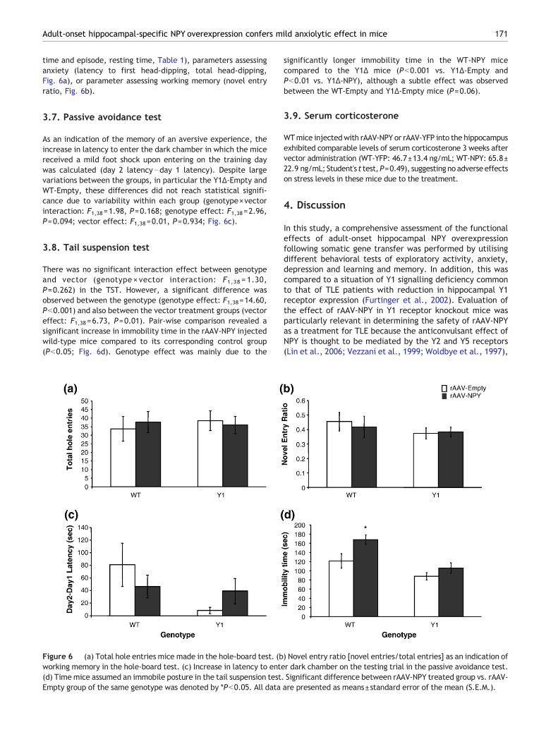
time and episode, resting time, Table 1), parameters assessinganxiety (latency to first head-dipping, total head-dipping,Fig. 6a), or parameter assessing working memory (novel entryratio, Fig. 6b).
3.7. Passive avoidance test
As an indication of the memory of an aversive experience, theincrease in latency to enter the dark chamber in which the micereceived a mild foot shock upon entering on the training daywas calculated (day 2 latency−day 1 latency). Despite largevariations between the groups, in particular the Y1Δ-Empty andWT-Empty, these differences did not reach statistical signifi-cance due to variability within each group (genotype×vectorinteraction: F1,38=1.98, P=0.168; genotype effect: F1,38=2.96,P=0.094; vector effect: F1,38=0.01, P=0.934; Fig. 6c).
3.8. Tail suspension test
There was no significant interaction effect between genotypeand vector (genotype × vector interaction: F1,38 = 1.30,P=0.262) in the TST. However, a significant difference wasobserved between the genotype (genotype effect: F1,38=14.60,Pb0.001) and also between the vector treatment groups (vectoreffect: F1,38=6.73, P=0.01). Pair-wise comparison revealed asignificant increase in immobility time in the rAAV-NPY injectedwild-type mice compared to its corresponding control group(Pb0.05; Fig. 6d). Genotype effect was mainly due to the
Figure 6 (a) Total hole entries mice made in the hole-board test. (bworking memory in the hole-board test. (c) Increase in latency to ente(d) Timemice assumed an immobile posture in the tail suspension test.Empty group of the same genotype was denoted by *Pb0.05. All data
significantly longer immobility time in the WT-NPY micecompared to the Y1Δ mice (Pb0.001 vs. Y1Δ-Empty andPb0.01 vs. Y1Δ-NPY), although a subtle effect was observedbetween the WT-Empty and Y1Δ-Empty mice (P=0.06).
3.9. Serum corticosterone
WTmice injectedwith rAAV-NPY or rAAV-YFP into the hippocampusexhibited comparable levels of serum corticosterone 3 weeks aftervector administration (WT-YFP: 46.7±13.4 ng/mL; WT-NPY: 65.8±22.9 ng/mL; Student's t test,P=0.49), suggesting no adverse effectson stress levels in these mice due to the treatment.
4. Discussion
In this study, a comprehensive assessment of the functionaleffects of adult-onset hippocampal NPY overexpressionfollowing somatic gene transfer was performed by utilisingdifferent behavioral tests of exploratory activity, anxiety,depression and learning and memory. In addition, this wascompared to a situation of Y1 signalling deficiency commonto that of TLE patients with reduction in hippocampal Y1receptor expression (Furtinger et al., 2002). Evaluation ofthe effect of rAAV-NPY in Y1 receptor knockout mice wasparticularly relevant in determining the safety of rAAV-NPYas a treatment for TLE because the anticonvulsant effect ofNPY is thought to be mediated by the Y2 and Y5 receptors(Lin et al., 2006; Vezzani et al., 1999; Woldbye et al., 1997),
) Novel entry ratio [novel entries/total entries] as an indication ofr dark chamber on the testing trial in the passive avoidance test.Significant difference between rAAV-NPY treated group vs. rAAV-are presented as means±standard error of the mean (S.E.M.).

172 E.-J.D. Lin et al.
which are the major receptors that were activated in the Y1receptor knockout mice by hippocampal rAAV-NPY treat-ment in this study.
Our results showed that increased expression of NPY inthe hippocampus did not cause any overt effects in thegeneral health, neurosensory reflexes and motor function ofthe experimental animals. Locomotor activity and explora-tion was not affected by hippocampal NPY overexpression asdemonstrated in the total distance travelled in the OF, LDand HB, and also the total arm entries in the EPM.
The observed increases in EPM arm entries and visual clifftest head-dipping behavior exhibited by the Y1 knockoutmice suggest enhanced locomotion and explorative behav-ior, consistent with a previous report on these mice (Karlet al., 2006). The mechanism underlying the increasedexploration is unclear and studies so far on the involvementof Y1 receptor in this behavioral domain reported conflictingdata (Karl et al., 2006; Karlsson et al., 2008; Pedrazziniet al., 1998). While the inconsistency in the data could bedue to genetic background difference of the differentmutant mice developed, another explanation may be thecomplexity of the involvement of Y1 receptor in thisbehavioral parameter, which has been shown to be highlycontext-specific and sensitive to stress (Karl et al., 2006).Notably, both NPY and Y1 receptors are expressed in theperipheral nervous system including the adrenal glands andNPY was shown to regulate catecholamine synthesis andsecretion via Y1 receptors. Both the adrenal and plasmalevels of catecholamines (norepinephrine and epinephrine)were shown to be higher in Y1 receptor knockout mice(Cavadas et al., 2006). Thus the altered adrenosympathetictone in the Y1 receptor knockout mice may play a role intheir observed change of motor activity and explorativebehaviors.
In the OF test, the NPY-overexpressing wild-type miceexhibited increased entries and time spent in the centerzone, parameters suggestive of an anxiolytic effect byhippocampal NPY. Although this effect was not apparent inthe LD and HB tests, the strong trend for increased open armtime in the WT-NPY group compared to the WT-Empty groupin the EPM is consistent with the anxiolytic action by NPYoverexpression observed in the OF test. Our finding is in linewith previous studies demonstrating the anxiolytic effect ofcentral NPY. Intracerebroventricular administration of NPYhas been shown to reduce anxiety in rodents in a wide rangeof paradigms (Kask et al., 2002), whereas mice deficient ofNPY exhibit an anxiogenic phenotype (Bannon et al., 2000;Karl et al., 2008; Palmiter et al., 1998). In addition,transgenic rats with hippocampal-specific NPY overexpres-sion were insensitive to the anxiogenic effect of restraintstress on the elevated plus maze and also showed absent fearsuppression in the punished drinking test, indicating a stress-protective role of hippocampal NPY (Thorsell et al., 2000).Interestingly, these NPY overexpressing rats showed compa-rable corticosterone levels to control rats both at baselineand after restraint stress (Thorsell et al., 2000), similar toour observation in the rAAV-NPY mice. Although serumcorticosterone is a stress hormone marker often used toindicate level of anxiety, various studies and paradigms haveshown that behavioral and endocrine stress responses can bedissociated (Benaroya-Milshtein et al., 2004; Koob et al.,1993; Moncek et al., 2004).
The anxiolytic effect of NPY is thought to be mediated byY1 receptors. Microinjection of NPY and Y1 receptor agonist,but not the Y2 receptor agonist, into the central nucleus ofthe amygdala reproduced the anxiolytic effect of ICV NPYwith high potency (Heilig et al., 1993). Moreover, theanxiolytic effect of intra-amygdaloid NPY was blocked byco-administration with Y1 receptor antagonist (Sajdyk et al.,1999) or intraventricular Y1 antisense administration (Heilig,1995), which alone is anxiogenic (Wahlestedt et al., 1993).Our data suggest that Y1 receptor may also play a role inmediating the anxiolytic effect of NPY in the hippocampussince hippocampal rAAV-NPY failed to confer anxiolyticaction in the Y1 receptor deficient mice. However, it shouldbe noted that the Y1 knockout mice already exhibited ananxiolytic-like phenotype, therefore it remains possible thata ‘ceiling’ effect exist in these mice in terms of anxiousbehavioral measures and that additional anxiolytic effectwas difficult to detect. While the reduced anxiety observedin Y1 receptor knockout mice may seem paradoxical to thesupposedly anxiolytic effect of Y1 receptors, this is likely dueto extra-hippocampal and extra-amygdaloid effects of Y1receptors as they are widely expressed both in the centraland peripheral nervous system (Balasubramaniam, 1997;Dumont et al., 1998). Future studies combining the rAAV-NPYtreatment with specific down-regulation of Y1 receptor inthe hippocampus either by conditional recombination meth-od or antisense oligonucleotide may circumvent the periph-eral effect of Y1 receptor deletion.
The mechanism underlying the effect of NPY on anxiety-like behaviors is still not entirely clear and is likely to becomplex considering the involvement of different Y recep-tors. Even in the hippocampus alone, it was shown that NPYmay mediate its anxiolytic effect by different Y receptordepending on the specific sub-regions and that a receptormay confer sub-region specific effect on anxiety (Smialowskaet al., 2007). One potential mechanism by which hippocam-pal Y1 receptors may regulate emotionality is its role in adulthippocampal neurogenesis. NPY stimulates neuroprolifera-tion in the hippocampus via Y1 receptors (Howell et al.,2005) and recently it was demonstrated that impairments inadult hippocampal neurogenesis lead to increased anxiety-related behaviors (Revest et al., 2009). This is supported bystudies showing manipulations that increase adult hippo-campal neurogenesis such as environmental enrichment,physical exercise and neurotrophin administration alsoreduce anxiety (Duman et al., 2008; Fox et al., 2006; Perezet al., 2009; Salam et al., 2009). Nevertheless, the presentstudy does not rule out involvement of other Y receptors andfurther studies are required to fully elucidate the effects andmechanisms by which the different Y receptors affectanxiety response in the hippocampus.
Surprisingly, we observed depressive-like phenotype inthe rAAV-NPY treated wild-type mice in the TST. Thisobservation was unexpected as numerous studies havesuggested NPY to be antidepressive. Animal models ofdepression and human postmortem study showed that NPYis significantly reduced in the brains of depressed subjects(Caberlotto and Hurd, 1999; Caberlotto et al., 1999; Heiligand Widerlov, 1990). Interventions with antidepressantefficacy, such as electroconvulsive shock stimulation (ECS)and antidepressant drugs, increase central NPY expression(Husum et al., 2000; Mikkelsen et al., 1994; Wahlestedt

173Adult-onset hippocampal-specific NPY overexpression confers mild anxiolytic effect in mice
et al., 1990). Furthermore, infusion of NPY into the cerebralventricles or hippocampus produces an antidepressant effect(Ishida et al., 2007; Redrobe et al., 2002; Stogner andHolmes, 2000). While the reason for the observed depres-sive-like behavior in the rAAV-NPY wild-type mice is unclear,it should be noted that NPY overexpression did not inducedepressive-like phenotype in the Y1 knockout mice, whichshowed comparable immobility scores to the wild-type mice.
Since the effect of rAAV-NPY gene transfer on spatiallearning and memory has already been evaluated (Sorensenet al., 2008a; Sorensen et al., 2008b), we used two differenttests to further assess the effect of NPY overexpression onother aspects of cognitive function. Using the novel to repeathole ratio in the hole-board test as an indication of workingmemory (Baiardi et al., 2007; Karl et al., 2006) and thepassive avoidance as a test for associative memory, we foundno evidence of adverse effect by NPY overexpression onworking memory and associative learning. This is consistentwith previous report on the behavioral phenotype of NPYdeficient mice, demonstrating the lack of effects by NPY incognitive parameters measured by the same paradigms(Karl et al., 2008). Our data are also consistent with thatreported by Ishida et al. (2007), showing no deficit inpassive avoidance test after intrahippocampal NPY infusion.Sorensen et al. (2008a) showed that rAAV-NPY treated ratsexhibited delayed learning in the two-platform spatialdiscrimination water maze test, but achieved comparablelearning to the control rats at the end of the 7 day testingperiod. The same study demonstrated that long-termpotentiation in the CA1 area was partially impaired bytransgene NPY acting via Y2 receptors, likely by an inhibitionof glutamate release onto pyramidal cells (Sorensen et al.,2008a). However, a follow up study showed that rAAV-mediated NPY overexpression did not affect short-termsynaptic plasticity nor did it further compromise LTP inkindled animals, a model for epilepsy (Sorensen et al., 2009).Together these data suggest that rAAV-NPY have limitedimpact on cognition, with spatial learning more sensitive toits effect. However, in epileptic subjects with alreadyimpaired LTP (Beck et al., 2000) and memory function(Elger et al., 2004; Helmstaedter et al., 2003), rAAV-NPYtreatment is unlikely to cause further impairment.
In summary, our data suggest that hippocampal NPYoverexpression confers a moderate anxiolytic effect, possiblyin part mediated by the Y1 receptors. Although hippocampalNPY overexpression induced a moderate increase in depres-sive-like behaviors, this effect is likely to be absent ordiminished in chronic epileptic subjects with reduced hippo-campal Y1 receptor expression. In the two cognitive testsused, hippocampal NPY overexpression did not alter cognitivefunctions. In conclusion, our study suggests that focal NPYoverexpression are unlikely to result in significant adverseeffects on mood regulation or learning and memory, and maybe a safe therapeutic alternative for the treatment of drug-resistant temporal lobe epilepsies. Nevertheless, the potentialsubtle effect on mood regulation should be monitored.
Role of the funding source
This project was supported by a fellowship to E-J.D. Lin from theNew Zealand Foundation for Research, Science and Technology
(Award #OHIO0601) and a fellowship to H. Herzog from the NationalHealth and Medical Research Council (NHMRC). NZFRST and NHMRChad no further role in the study design; in the collection, analysis andinterpretation of data; in the writing of the report; and in thedecision to submit the paper for publication.
Contributors
E-J.D.L. designed the study, conducted most of the experiments andwrote the manuscript. S.L. and A.A. conducted the histochemicalanalysis. M.J.D. provided the vectors and contributed to themanuscript. H.H. supervised the project, participated in the datainterpretation and edited the manuscript. All authors contributed toand have approved the final manuscript.
Conflict of interest
E-J.D.L. and M.J.D. have consulted for Neurologix Inc. All otherauthors declare that they have no conflicts of interest.
Acknowledgement
We thank Ms. Xiaobai Li for assistance in statistical analysis.
References
Baiardi, G., Ruiz, A.M., Beling, A., Borgonovo, J., Martinez, G.,Landa, A.I., Sosa, M.A., Gargiulo, P.A., 2007. Glutamatergicionotropic blockade within accumbens disrupts working memoryand might alter the endocytic machinery in rat accumbens andprefrontal cortex. J. Neural. Transm. 114, 1519–1528.
Balasubramaniam, A.A., 1997. Neuropeptide Y family of hormones:receptor subtypes and antagonists. Peptides 18, 445–457.
Baldock, P.A., Sainsbury, A., Allison, S., Lin, E.J., Couzens, M.,Boey, D., Enriquez, R., During, M., Herzog, H., Gardiner, E.M.,2005. Hypothalamic control of bone formation: distinct actions ofleptin and y2 receptor pathways. J. Bone Miner. Res. 20,1851–1857.
Bannon, A.W., Seda, J., Carmouche, M., Francis, J.M., Norman, M.H.,Karbon, B., McCaleb, M.L., 2000. Behavioral characterization ofneuropeptide Y knockout mice. Brain Res. 868, 79–87.
Baraban, S.C., Hollopeter, G., Erickson, J.C., Schwartzkroin, P.A.,Palmiter, R.D., 1997. Knock-out mice reveal a critical anti-epileptic role for neuropeptide Y. J. Neurosci. 17, 8927–8936.
Beck, H., Goussakov, I.V., Lie, A., Helmstaedter, C., Elger, C.E.,2000. Synaptic plasticity in the human dentate gyrus. J. Neurosci.20, 7080–7086.
Bellmann, R., Widmann, R., Olenik, C., Meyer, D.K., Maas, D.,Marksteiner, J., Sperk, G., 1991. Enhanced rate of expressionand biosynthesis of neuropeptide Y after kainic acid-inducedseizures. J. Neurochem. 56, 525–530.
Belzung, C., Griebel, G., 2001. Measuring normal and pathologicalanxiety-like behaviour in mice: a review. Behav. Brain Res. 125,141–149.
Benaroya-Milshtein, N., Hollander, N., Apter, A., Kukulansky, T.,Raz, N., Wilf, A., Yaniv, I., Pick, C.G., 2004. Environmentalenrichment in mice decreases anxiety, attenuates stressresponses and enhances natural killer cell activity. Eur. J.NeuroSci. 20, 1341–1347.
Bird, C.M., Burgess, N., 2008. The hippocampus and memory:insights from spatial processing. Nat. Rev. Neurosci. 9, 182–194.
Caberlotto, L., Hurd, Y.L., 1999. Reduced neuropeptide Y mRNAexpression in the prefrontal cortex of subjects with bipolardisorder. NeuroReport 10, 1747–1750.

174 E.-J.D. Lin et al.
Caberlotto, L., Jimenez, P., Overstreet, D.H., Hurd, Y.L.,Mathe, A.A.,Fuxe, K., 1999. Alterations in neuropeptide Y levels and Y1 bindingsites in the Flinders Sensitive Line rats, a genetic animal model ofdepression. Neurosci. Lett. 265, 191–194.
Cavadas, C., Cefai, D., Rosmaninho-Salgado, J., Vieira-Coelho, M.A.,Moura, E., Busso, N., Pedrazzini, T., Grand, D., Rotman, S.,Waeber, B., Aubert, J.F., Grouzmann, E., 2006. Deletion of theneuropeptide Y (NPY) Y1 receptor gene reveals a regulatory role ofNPY on catecholamine synthesis and secretion. Proc. Natl. Acad.Sci. U. S. A. 103, 10497–10502.
Clark, K.R., Liu, X., McGrath, J.P., Johnson, P.R., 1999. Highly purifiedrecombinant adeno-associated virus vectors are biologically activeand free of detectable helper and wild-type viruses. Hum. GeneTher. 10, 1031–1039.
Costall, B., Jones, B.J., Kelly, M.E., Naylor, R.J., Tomkins, D.M., 1989.Exploration of mice in a black and white test box: validation as amodel of anxiety. Pharmacol. Biochem. Behav. 32, 777–785.
Crawley, J.N., 1985. Exploratory behavior models of anxiety in mice.Neurosci. Biobehav. Rev. 9, 37–44.
Crawley, J.N., 1999. Behavioral phenotyping of transgenic andknockout mice: experimental design and evaluation of generalhealth, sensory functions, motor abilities, and specific behavioraltests. Brain Res. 835, 18–26.
DeFries, J.C., Hegmann, J.P., Weir, M.W., 1966. Open-field behaviorin mice: evidence for a major gene effect mediated by the visualsystem. Science 154, 1577–1579.
do-Rego, J.C., Viana, A.F., Le Maitre, E., Deniel, A., Rates, S.M.,Leroux-Nicollet, I., Costentin, J., 2006. Comparisons betweenanxiety tests for selection of anxious and non anxious mice.Behav. Brain Res. 169, 282–288.
Drevets, W.C., Price, J.L., Furey, M.L., 2008. Brain structural andfunctional abnormalities in mood disorders: implications forneurocircuitry models of depression. Brain Struct. Funct. 213,93–118.
Duman, C.H., Schlesinger, L., Russell, D.S., Duman, R.S., 2008.Voluntary exercise produces antidepressant and anxiolyticbehavioral effects in mice. Brain Res. 1199, 148–158.
Dumont, Y., Jacques, D., Bouchard, P., Quirion, R., 1998. Speciesdifferences in the expression and distribution of the neuropep-tide Y Y1, Y2, Y4, and Y5 receptors in rodents, guinea pig, andprimates brains. J. Comp. Neurol. 402, 372–384.
During, M.J., Young, D., Baer, K., Lawlor, P., Klugmann, M., 2003.Development and optimization of adeno-associated virus vectortransfer into the central nervous system. Methods Mol. Med. 76,221–236.
Elger, C.E., Helmstaedter, C., Kurthen, M., 2004. Chronic epilepsyand cognition. Lancet Neurol. 3, 663–672.
Foti, S., Haberman, R.P., Samulski, R.J., McCown, T.J., 2007.Adeno-associated virus-mediated expression and constitutivesecretion of NPY or NPY13–36 suppresses seizure activity in vivo.Gene Ther. 14, 1534–1536.
Fox, C., Merali, Z., Harrison, C., 2006. Therapeutic and protectiveeffect of environmental enrichment against psychogenic andneurogenic stress. Behav. Brain Res. 175, 1–8.
Franklin, K.B.J., Paxinos, G., 1997. The Mouse Brain in StereotaxicCoordinates. Academic Press, San Diego.
Furtinger, S., Pirker, S., Czech, T., Baumgartner, C., Sperk, G.,2002. Altered expression of neuropeptide Y, -Y(1), and -Y(2)receptors in the hippocampus of patients with mesial temporallobe epilepsy. Epilepsia 43 (Suppl 5), 152.
Gariboldi, M., Conti, M., Cavaleri, D., Samanin, R., Vezzani, A., 1998.Anticonvulsant properties of BIBP3226, a non-peptide selectiveantagonist at neuropeptide Y Y1 receptors. Eur. J. NeuroSci. 10,757–759.
Heilig, M., 1995. Antisense inhibition of neuropeptide Y (NPY)-Y1receptor expression blocks the anxiolytic-like action of NPY inamygdala and paradoxically increases feeding. Regul. Pept. 59,201–205.
Heilig, M., Widerlov, E., 1990. Neuropeptide Y: an overview of centraldistribution, functional aspects, and possible involvement inneuropsychiatric illnesses. Acta Psychiatr. Scand. 82, 95–114.
Heilig, M., McLeod, S., Brot, M., Heinrichs, S.C., Menzaghi, F., Koob,G.F., Britton, K.T., 1993. Anxiolytic-like action of neuropeptideY: mediation by Y1 receptors in amygdala, and dissociation fromfood intake effects. Neuropsychopharmacology 8, 357–363.
Helmstaedter, C., Kurthen, M., Lux, S., Reuber, M., Elger, C.E.,2003. Chronic epilepsy and cognition: a longitudinal study intemporal lobe epilepsy. Ann. Neurol. 54, 425–432.
Holmes, A., Wrenn, C.C., Harris, A.P., Thayer, K.E., Crawley, J.N.,2002. Behavioral profiles of inbred strains on novel olfactory,spatial and emotional tests for reference memory in mice. GenesBrain Behav. 1, 55–69.
Howell, O.W., Scharfman, H.E., Herzog, H., Sundstrom, L.E., Beck-Sickinger, A., Gray, W.P., 2003. Neuropeptide Y is neuroprolifera-tive for post-natal hippocampal precursor cells. J. Neurochem. 86,646–659.
Howell, O.W., Doyle, K., Goodman, J.H., Scharfman, H.E., Herzog,H., Pringle, A., Beck-Sickinger, A.G., Gray, W.P., 2005.Neuropeptide Y stimulates neuronal precursor proliferation inthe post-natal and adult dentate gyrus. J. Neurochem. 93,560–570.
Husum, H., Mikkelsen, J.D., Hogg, S., Mathe, A.A., Mork, A., 2000.Involvement of hippocampal neuropeptide Y in mediating thechronic actions of lithium, electroconvulsive stimulation andcitalopram. Neuropharmacology 39, 1463–1473.
Ishida, H., Shirayama, Y., Iwata, M., Katayama, S., Yamamoto, A.,Kawahara, R., Nakagome, K., 2007. Infusion of neuropeptide Yinto CA3 region of hippocampus produces antidepressant-likeeffect via Y1 receptor. Hippocampus 17, 271–280.
Karl, T., Burne, T.H., Herzog, H., 2006. Effect of Y1 receptordeficiency on motor activity, exploration, and anxiety. Behav.Brain Res. 167, 87–93.
Karl, T., Duffy, L., Herzog, H., 2008. Behavioural profile of a newmouse model for NPY deficiency. Eur. J. NeuroSci. 28, 173–180.
Karlsson, R.M., Choe, J.S., Cameron, H.A., Thorsell, A., Crawley, J.N.,Holmes, A., Heilig, M., 2008. The neuropeptide Y Y1 receptorsubtype is necessary for the anxiolytic-like effects of neuropeptideY, but not the antidepressant-like effects of fluoxetine, in mice.Psychopharmacology (Berl.) 195, 547–557.
Kask, A., Harro, J., von Horsten, S., Redrobe, J.P., Dumont, Y.,Quirion, R., 2002. The neurocircuitry and receptor subtypesmediating anxiolytic-like effects of neuropeptide Y. Neurosci.Biobehav. Rev. 26, 259–283.
Koob, G.F., Heinrichs, S.C., Pich, E.M., Menzaghi, F., Baldwin, H.,Miczek, K., Britton, K.T., 1993. The role of corticotropin-releasing factor in behavioural responses to stress. Ciba Found.Symp. 172, 277–289 discussion 290–275.
Lin, S., Boey, D., Herzog, H., 2004. NPY and Y receptors: lessonsfrom transgenic and knockout models. Neuropeptides 38,189–200.
Lin, E.J., Young, D., Baer, K., Herzog, H., During, M.J., 2006.Differential actions of NPY on seizure modulation via Y1 and Y2receptors: evidence from receptor knockout mice. Epilepsia 47,773–780.
Mikkelsen, J.D., Woldbye, D., Kragh, J., Larsen, P.J., Bolwig, T.G.,1994. Electroconvulsive shocks increase the expression ofneuropeptide Y (NPY) mRNA in the piriform cortex and thedentate gyrus. Brain Res. Mol. Brain Res. 23, 317–322.
Moncek, F., Duncko, R., Johansson, B.B., Jezova, D., 2004. Effect ofenvironmental enrichment on stress related systems in rats.J. Neuroendocrinol. 16, 423–431.
Montgomery, K.C., 1955. The relation between fear induced bynovel stimulation and exploratory behavior. J. Comp. Physiol.Psychol. 48, 254–260.
Noe, F., Pool, A.H., Nissinen, J., Gobbi, M., Bland, R., Rizzi, M.,Balducci, C., Ferraguti, F., Sperk, G., During, M.J., Pitkanen, A.,

175Adult-onset hippocampal-specific NPY overexpression confers mild anxiolytic effect in mice
Vezzani, A., 2008. Neuropeptide Y gene therapy decreaseschronic spontaneous seizures in a rat model of temporal lobeepilepsy. Brain 131, 1506–1515.
Ohl, F., Roedel, A., Binder, E., Holsboer, F., 2003. Impact of highand low anxiety on cognitive performance in a modified holeboard test in C57BL/6 and DBA/2 mice. Eur. J. NeuroSci. 17,128–136.
Palmiter, R.D., Erickson, J.C., Hollopeter, G., Baraban, S.C.,Schwartz, M.W., 1998. Life without neuropeptide Y. RecentProg. Horm. Res. 53, 163–199.
Patrylo, P.R., van den Pol, A.N., Spencer, D.D., Williamson, A.,1999. NPY inhibits glutamatergic excitation in the epileptichuman dentate gyrus. J. Neurophysiol. 82, 478–483.
Pedrazzini, T., Seydoux, J., Kunstner, P., Aubert, J.F., Grouzmann,E., Beermann, F., Brunner, H.R., 1998. Cardiovascular response,feeding behavior and locomotor activity in mice lacking the NPYY1 receptor. Nat. Med. 4, 722–726.
Perez, J.A., Clinton, S.M., Turner, C.A., Watson, S.J., Akil, H., 2009.A new role for FGF2 as an endogenous inhibitor of anxiety.J. Neurosci. 29, 6379–6387.
Redrobe, J.P., Dumont, Y., Fournier, A., Quirion, R., 2002. Theneuropeptide Y (NPY) Y1 receptor subtype mediates NPY-inducedantidepressant-like activity in the mouse forced swimming test.Neuropsychopharmacology 26, 615–624.
Revest, J.M., Dupret, D., Koehl, M., Funk-Reiter, C., Grosjean, N.,Piazza, P.V., Abrous, D.N., 2009. Adult hippocampal neurogenesisis involved in anxiety-related behaviors. Mol. Psychiatry 14,959–967.
Richichi, C., Lin, E.J., Stefanin, D., Colella,D., Ravizza, T., Grignaschi,G., Veglianese, P., Sperk, G., During, M.J., Vezzani, A., 2004.Anticonvulsant and antiepileptogenic effects mediated by adeno-associated virus vector neuropeptide Y expression in the rathippocampus. J. Neurosci. 24, 3051–3059.
Saitoh, A., Hirose, N., Yamada, M., Nozaki, C., Oka, T., Kamei, J.,2006. Changes in emotional behavior of mice in the hole-boardtest after olfactory bulbectomy. J. Pharmacol. Sci. 102, 377–386.
Sajdyk, T.J., Vandergriff, M.G., Gehlert, D.R., 1999. Amygdalarneuropeptide Y Y1 receptors mediate the anxiolytic-like actionsof neuropeptide Y in the social interaction test. Eur. J. Pharmacol.368, 143–147.
Salam, J.N., Fox, J.H., Detroy, E.M., Guignon, M.H., Wohl, D.F.,Falls, W.A., 2009. Voluntary exercise in C57 mice is anxiolyticacross several measures of anxiety. Behav. Brain Res. 197, 31–40.
Santarelli, L., Saxe, M., Gross, C., Surget, A., Battaglia, F., Dulawa,S., Weisstaub, N., Lee, J., Duman, R., Arancio, O., Belzung, C.,Hen, R., 2003. Requirement of hippocampal neurogenesis for thebehavioral effects of antidepressants. Science 301, 805–809.
Shannon, H.E., Yang, L., 2004. Seizure susceptibility of neuropeptide-Y null mutant mice in amygdala kindling and chemical-inducedseizure models. Epilepsy Res. 61, 49–62.
Smialowska, M., Bijak, M., Sopala, M., Tokarski, K., 1996. Inhibitoryeffect of NPY on the picrotoxin-induced activity in thehippocampus: a behavioural and electrophysiological study.Neuropeptides 30, 7–12.
Smialowska, M., Wieronska, J.M., Domin, H., Zieba, B., 2007. Theeffect of intrahippocampal injection of group II and III metobo-
tropic glutamate receptor agonists on anxiety; the role ofneuropeptide Y. Neuropsychopharmacology 32, 1242–1250.
Sorensen, A.T., Kanter-Schlifke, I., Carli, M., Balducci, C., Noe, F.,During, M.J., Vezzani, A., Kokaia, M., 2008a. NPY gene transferin the hippocampus attenuates synaptic plasticity and learning.Hippocampus 18, 564–574.
Sorensen, A.T., Kanter-Schlifke, I., Lin, E.J., During, M.J., Kokaia,M., 2008b. Activity-dependent volume transmission by transgeneNPY attenuates glutamate release and LTP in the subiculum. Mol.Cell. Neurosci. 39, 229–237.
Sorensen, A.T., Nikitidou, L., Ledri, M., Lin, E.J., During, M.J.,Kanter-Schlifke, I., Kokaia, M., 2009. Hippocampal NPY genetransfer attenuates seizures without affecting epilepsy-inducedimpairment of LTP. Exp. Neurol. 215, 328–333.
Sperk, G., Marksteiner, J., Gruber, B., Bellmann, R., Mahata, M.,Ortler, M., 1992. Functional changes in neuropeptide Y- andsomatostatin-containing neurons induced by limbic seizures inthe rat. Neuroscience 50, 831–846.
Stogner, K.A., Holmes, P.V., 2000. Neuropeptide-Y exerts antide-pressant-like effects in the forced swim test in rats. Eur. J.Pharmacol. 387, R9–R10.
Takeda, H., Tsuji, M., Matsumiya, T., 1998. Changes in head-dippingbehavior in the hole-board test reflect the anxiogenic and/oranxiolytic state in mice. Eur. J. Pharmacol. 350, 21–29.
Thorsell, A., Michalkiewicz, M., Dumont, Y., Quirion, R., Caberlotto,L., Rimondini, R., Mathe, A.A., Heilig, M., 2000. Behavioralinsensitivity to restraint stress, absent fear suppression ofbehavior and impaired spatial learning in transgenic rats withhippocampal neuropeptide Y overexpression. Proc. Natl. Acad.Sci. U. S. A. 97, 12852–12857.
Vezzani, A., Sperk, G., Colmers, W.F., 1999. Neuropeptide Y:emerging evidence for a functional role in seizure modulation.Trends Neurosci. 22, 25–30.
Vezzani, A., Michalkiewicz, M., Michalkiewicz, T., Moneta, D.,Ravizza, T., Richichi, C., Aliprandi, M., Mule, F., Pirona, L.,Gobbi, M., Schwarzer, C., Sperk, G., 2002. Seizure susceptibilityand epileptogenesis are decreased in transgenic rats over-expressing neuropeptide Y. Neuroscience 110, 237–243.
Wahlestedt, C., Blendy, J.A., Kellar, K.J., Heilig, M., Widerlov, E.,Ekman, R., 1990. Electroconvulsive shocks increase the concen-tration of neocortical and hippocampal neuropeptide Y (NPY)-like immunoreactivity in the rat. Brain Res. 507, 65–68.
Wahlestedt, C., Pich, E.M., Koob, G.F., Yee, F., Heilig, M., 1993.Modulation of anxiety and neuropeptide Y-Y1 receptors byantisense oligodeoxynucleotides. Science 259, 528–531.
Woldbye, D.P., 1998. Antiepileptic effects of NPY on pentylenete-trazole seizures. Regul. Pept. 75–76, 279–282.
Woldbye, D.P., Larsen, P.J., Mikkelsen, J.D., Klemp, K., Madsen, T.M.,Bolwig, T.G., 1997. Powerful inhibition of kainic acid seizures byneuropeptide Y via Y5-like receptors. Nat. Med. 3, 761–764.
Xu, R., Janson, C.G., Mastakov, M., Lawlor, P., Young, D., Mouravlev,A., Fitzsimons, H., Choi, K.L., Ma, H., Dragunow, M., Leone, P.,Chen, Q., Dicker, B., During, M.J., 2001. Quantitative comparisonof expression with adeno-associated virus (AAV-2) brain-specificgene cassettes. Gene Ther. 8, 1323–1332.