The Methyltransferase HemK Regulates the Virulence ... - MDPI

This journal is©The Royal Society of Chemistry 2014 Integr. Biol.
Cite this:DOI: 10.1039/c4ib00140k
C. pseudotuberculosis Phop confers virulence andmay be targeted by natural compounds†
Sandeep Tiwari,‡a Marcılia Pinheiro da Costa,‡b Sintia Almeida,a Syed Shah Hassan,a
Syed Babar Jamal,a Alberto Oliveira,a Edson Luiz Folador,a Flavia Rocha,c
Vinıcius Augusto Carvalho de Abreu,a Fernanda Dorella,a Rafael Hirata,d
Diana Magalhaes de Oliveira,b Maria Fatima da Silva Teixeira,b Artur Silva,e
Debmalya Barhf and Vasco Azevedo*ac
The bacterial two-component system (TCS) regulates genes that are crucial for virulence in several
pathogens. One of such TCS, the PhoPR system, consisting of a transmembrane sensory histidine kinase
protein (PhoR) and an intracellular response regulator protein (PhoP), has been reported to have a major
role in mycobacterial pathogenesis. We knocked out the phoP in C. pseudotuberculosis, the causal
organism of caseous lymphadenitis (CLA), and using a combination of in vitro and in vivo mouse system, we
showed for the first time, that the PhoP of C. pseudotuberculosis plays an important role in the virulence
and pathogenicity of this bacterium. Furthermore, we modeled the PhoP of C. pseudotuberculosis and our
docking results showed that several natural compounds including Rhein, an anthraquinone from Rheum
undulatum, and some drug-like molecules may target PhoP to inhibit the TCS of C. pseudotuberculosis, and
therefore may facilitate a remarkable attenuation of bacterial pathogenicity being the CLA. Experiments are
currently underway to validate these in silico docking results.
Insight, innovation, integrationHere, we report for the first time the importance of PhoP protein of the PhoPR system of C. pseudotuberculosis that plays a vital role in the virulence andpathogenicity of this bacterium. For this, we developed a mutant phoP gene of Corynebacterium pseudotuberculosis Cp1002 strain and subsequently evaluatedthe consequences of this mutation via in vitro and in vivo analyses. Furthermore, by extrapolating the results of our analyses, we modeled this PhoP protein ofC. pseudotuberculosis and computational analyses were performed. Our docking results showed that several natural compounds, including Rhein from Rheum
undulatum and some other drug-like compounds acquired from public drug database/s, targeted the N-terminal response regulator domain of the PhoP proteinto inhibit the TCS of C. pseudotuberculosis and might facilitate a remarkable attenuation of the pathogenicity of this important veterinary pathogen.
Introduction
The Gram-positive bacterium Corynebacterium pseudotuberculosisof the class Actinobacteria is the etiological agent of caseous
lymphadenitis (CLA), or cheesy gland disease, an illness thataffects small ruminants (sheep and goats) worldwide.1 CLAis mainly characterized by abscess formation in superficialand internal lymph nodes.2 In some cases, the visceral organs,
a PG Program in Bioinformatics, Laboratory of Cellular and Molecular Genetics (LGCM), Institute of Biological Sciences, Federal University of Minas Gerais, Belo Horizonte,
MG, Brazil. E-mail: [email protected], [email protected], [email protected], [email protected], [email protected],
[email protected], [email protected], [email protected], [email protected] Center for Genomics and Bioinformatics, State University of Ceara, Fortaleza, CE, Brazil. E-mail: [email protected], [email protected],
[email protected] Department of General Biology, PG Program in Genetics, Laboratory of Cellular and Molecular Genetics (LGCM), Institute of Biological Sciences,
Federal University of Minas Gerais, Belo Horizonte, MG, Brazil. E-mail: [email protected], [email protected]; Fax: +55-31 3409-2610; Tel: +55-31 3409-2610d Universidade do Estado do Rio de Janeiro, UERJ, Brazil. E-mail: [email protected] Laboratory of DNA polymorphism (LPDNA), Federal University of Para, Belem, PA, Brazil. E-mail: [email protected] Centre for Genomics and Applied Gene Technology, Institute of Integrative Omics and Applied Biotechnology (IIOAB), Nonakuri, Purba Medinipur, WB, India.
E-mail: [email protected]
† Electronic supplementary information (ESI) available. See DOI: 10.1039/c4ib00140k‡ These authors contributed equally to this work.
Received 18th June 2014,Accepted 22nd August 2014
DOI: 10.1039/c4ib00140k
www.rsc.org/ibiology
Integrative Biology
PAPER
Publ
ishe
d on
12
Sept
embe
r 20
14. D
ownl
oade
d by
Fed
eral
Uni
vers
ity o
f M
inas
Ger
ais
on 1
6/10
/201
4 21
:30:
07.
View Article OnlineView Journal
Integr. Biol. This journal is©The Royal Society of Chemistry 2014
such as the lungs, kidneys, liver and spleen, are also infected.3
The development of CLA compromises these animals andgenerates significant economic losses due to reduced wool,meat and milk yields, decreased reproductive efficiency andincreased condemnation of carcasses and skins in abattoirs.2,4
Once disseminated, CLA eradication is difficult due to theinefficacy of currently available drug therapies.2,3 The insufficiencyof available control procedures and the lack of knowledge regard-ing the molecular mechanisms of the virulence of this diseaserequire an aggressive search for efficient treatment strategies.5
Recently, novel virulence determinants of C. pseudotuberculosisCp1002 have been identified; however, little is known about thiscomplex pathogen. Phospholipase D and mycolic acids are themost important virulence factors identified in C. pseudotuberculosisCp1002.6
The role of two-component signal transduction systems(TCS) in bacteria and certain eukaryotes, such as protozoa, isto detect and respond to the milieu vicissitudes.7 Many studieshave reported the involvement of the versatile TCS systems inthe regulation of a variety of processes, including cell division,nutrient acquisition, regulation of osmolarity, redox potential,nitrogen fixation, phosphate uptake, sporulation, pilus formation,adhesion, drug resistance and expression of virulence factors.8–11
The PhoPR system consists of a transmembrane sensory histidinekinase protein (PhoR) that phosphorylates the receiver domainof the response regulator protein (PhoP) of this system.Phosphorylation induces a conformational change in the responseregulator, which activates the effectors domain and therebytriggering the cellular response.12
More than 4000 TCSs have been identified in bacterial genomes,demonstrating a broad range of interactions and environmentaladaptation of bacteria. Interestingly, the PhoPR system fromM. tuberculosis has shown a high degree of similarity to PhoPQfrom a diverse range of intracellular bacterial pathogens such asSalmonella sp., Shigella sp. and Yersinia sp.13 The development andapplication of specific inhibitors against TCS systems operatedifferently from conventional antibiotics. It is possible to transformsuch inhibitors into new drugs, which perform selectively againstvarious drug-resistant bacteria.14,15 Furthermore, inhibiting thefunction of TCSs that control the expression of virulence factorsmay attenuate the virulence of the pathogenic bacteria.16
PhoP gene is the response regulator of the PhoPR two-component systems, which plays a crucial role in the virulence.Progresses in the understanding of M. tuberculosis biology havepromoted the rational development of attenuated strains, and,currently, the phoP mutant is used as a main vaccinal strategy
against tuberculosis.17 Recent studies have demonstrated thatthis mutant has shown a variety of changes, including the loss ofexpression of lipids of the cell envelope, impaired intracellulargrowth in infected macrophages, which persists in in vitrocultured-macrophages and in mouse organs, lack of secretionof the major T-cell antigen ESAT-6, virulence attenuation andprotective efficacy against tuberculosis.18,19
In this work, we have attempted to study the possible role ofthe PhoP protein of the two-component signal transductionsystems in C. pseudotuberculosis Cp1002 through the generationof mutant strain by disrupting the phoP gene by simple homo-logous recombination. After the DphoP genotype analysis, testswere performed to confirm the role of PhoP in the virulence andpathogenicity of C. pseudotuberculosis. This mutant strain was alsoassessed for adhesion and viability in macrophages and forvirulence in mice to analyze the persistence of infection andmortality rates. After confirming the possible role of PhoP proteinin virulence, we followed an in silico approach with a focus on thePhoP protein of the TCS of animal pathogen C. pseudotuberculosisCp1002 for identifying putative therapeutic inhibitors.
Materials and methodsBacterial strains and culture conditions
All bacterial strains and plasmids used in this study are listed inTable 1. Escherichia coli DH5a was grown in Luria-Bertani broth(LB, Difco Laboratories, Detroit, USA) at 37 1C under agitation for18 h and on 1.5% (w/v) LB agar plates at 37 1C for 18 h. Plasmid-containing transformants were selected by adding ampicillin(Amp) (Invitrogen, San Diego, CA) and X-Gal (Invitrogen, SanDiego, CA) to the media. The supplement concentrations wereampicillin (100 mg ml�1) and X-Gal (40 mg ml�1), respectively.C. pseudotuberculosis biovar ovis strain 1002 was used as the wildtype parental. Bacteria were aerobically grown in brain heartinfusion broth (BHI, Acumedia Manufacturers, Inc., Baltimore,MD, USA) and on 1.5% (w/v) BHI agar plates at 37 1C for 72 h.20
C. pseudotuberculosis phoP mutant (DphoP) was aerobically grownin brain heart infusion broth (BHI) and on BHI agar plates at37 1C for 72 h. For the selection of recombinants, kanamycin(Km) 50 mg ml�1 was added to the media.
Genomic DNA extraction and the construction ofC. pseudotuberculosis DphoP strain
Genomic DNA extraction was performed according to a pre-viously described protocol.20 For the amplification of a segment
Table 1 Strains and plasmids used in this work
Strain or plasmid Description and relevant characteristics Source
Escherichia coli DH5a/[sup44DlacU169 (j 80 lacZDM15) hsdR17 recA1 endA1 gyrA96 thi-1 relA1] InvitrogenCorynebacterium pseudotuberculosisa 1002 strain (wild-type) UFBACorynebacterium pseudotuberculosis DphoP strain (phoP mutant) This studypCRs2.1-TOPOs Cloning vector/Amr-Kmr/pUC ORI InvitrogenpCRs2.1-TOPOs:phoP Cloning vector pCRs2.1-TOPOs containing the Cp 436 bp fragment from the ORF phoP This study
a Virulent C. pseudotuberculosis biovar ovis strain isolates from caprines; obtained from the Universidade Federal da Bahia, UFBA, Brazil.
Paper Integrative Biology
Publ
ishe
d on
12
Sept
embe
r 20
14. D
ownl
oade
d by
Fed
eral
Uni
vers
ity o
f M
inas
Ger
ais
on 1
6/10
/201
4 21
:30:
07.
View Article Online
This journal is©The Royal Society of Chemistry 2014 Integr. Biol.
of phoP ORF of 436 bp of C. pseudotuberculosis Cp1002 thefollowing primers were used: sense (50-GGATCCGATGGAAGGCGTGAACGAG-30) antisense (50- GCGTAAGATCGGCGTACACC-30).The PCR assays were carried out in a final volume of 50 ml,containing 50 ng of genomic DNA, 1 pmol ml�1 of each primer,0:25 mM dNTPs, 0.1 units of Taq DNA polymerase (Invitrogen),2 mM MgCl2, and 1� concentrated enzyme buffer (Invitrogen).Amplification was performed using the thermal cycler(PTC-100, MJ Research, Inc.) as follows: first denaturation at95 1C for 4 min, 30 cycles followed by denaturation at 95 1C for30 s, 58 1C annealing for 30 s, extension at 72 1C for 1.5 min;and final extension for 5 min at 72 1C. The DNA fragment waspurified from bands in 1.0% (w/v) agarose gels using theConsert TM Rapid Gel Extraction System kit (Gibco-BRL,Gaithersburg, MD, USA). The retrieved fragment of phoP wasthen ligated into the pCR2.1-TOPO vector, as described inthe manufacture’s protocol. The recombinant plasmid pCR2.1-TOPO/phoP was then introduced into the competentE. coli DH cella and single recombinant colonies were selected.The presence of insert DNA fragment was confirmed by colonyPCR of seven clones selected using the same primers describedabove. In this reaction, for final volume of 10 L, 1 pmol ml�1 ofeach primer was used; 0.25 mM dNTPs; 0.1 unit of Taq DNApolymerase (Invitrogen); 2 mM MgCl2 buffer and 1� enzymeconcentrate (Invitrogen). After the identification of E. coli clonein the genomic library that contained the phoP fragment clonedinto pCR2.1-TOPO (Invitrogen), plasmid DNA extraction wasperformed using the Wizard Plus Maxipreps DNA PurificationSystem (Promega). The extracted plasmid was directly trans-formed into C. pseudotuberculosis strain 1002 according toDorella et al.1 The selection of the phoP mutant clones wasperformed in BHI medium supplemented with 50 mg mL�1
kanamycin. For the construction of C. pseudotuberculosisDphoP by simple homologous recombination,21 a clone ofC. pseudotuberculosis genome library was used, which containeda fragment of the open reading frame (ORF) of the cloned phoPgene. To confirm phoP inactivation by the insertion of thesuicide vector, polymerase chain reaction (PCR) was performedusing primers aligned with phoP as well as m13 and km. Allother molecular biology techniques were performed accordingto Sambrook et al.51
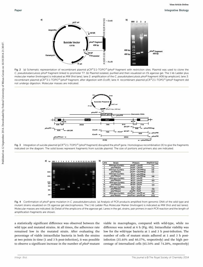
The constructed suicide plasmid was then introduced intocompetent C. pseudotuberculosis cells. This plasmid did notreplicate in C. pseudotuberculosis and it should integrate intothe genome by homologous recombination between the phoPgene on the chromosome and the cloned sequence on theplasmid (Fig. 3). The resistant strain of C. pseudotuberculosiswas confirmed by mPCR. The disruption of phoP gene of thisstrain was evaluated by PCR using pair primers listed inTable 2. Amplicons could also be observed in Table 3.Compared with the parent strain Cp1002, mutant (DphoP)strain was confirmed by the detection of phoP sequence inC. pseudotuberculosis. A 5.2 kb fragment was obtained inmutant phoP, indicating the presence of plasmid into genomicDNA (Fig. 4a, lane 15), while the wild type yielded a 0.8 kbproduct (Fig. 4a, lane 14).
Generation and characterization of phoP mutant ofC. pseudotuberculosis
High concentrated plasmid DNA was isolated from cells using theWizards Plus Maxipreps DNA Purification System kit (Promega,Madison, WI). The recombinant plasmid (pCRs2.1-TOPOs/phoPfragment) was then introduced into competent C. pseudotuberculosiscells.22 Plating recognized strains with homologous recombinationof recombinant plasmid at the phoP locus on medium containingkanamycin. Resistant colonies appeared after 72 h at 37 1C, andsingle recombinant colony was selected and characterized.
The chosen mutant strain of C. pseudotuberculosis Cp1002was evaluated by mPCR, as described by Pacheco et al., 2007.20
To compare the growth rate of C. pseudotuberculosis wild typeand phoP mutant, a growth curve was made in BHI T80 (0.05%Tween 80), and the OD600 and serial plate agar dilution weremeasured at 0, 30, 60, 120, 180, 240, 300 and 360 min. Thedifference between the bacterial size and colony morphologywere also observed between the wild type and mutant strains.
The proper integration of the plasmid was further determinedby PCR using the primers of Table 2. PCR primer pairs, meltingtemperature and amplicon lengths of each PCR reaction can beseen in Table 3.
In vitro assays of C. pseudotuberculosis strains in macrophagecells
The J774 murine macrophage cells were cultured in suspension inDulbecco’s Modified Eagle’s medium (DMEM, Sigma Chemical Co.,St. Louis, MO, USA) supplemented with gentamicin (50 mg ml�1),fungizone (2.5 mg ml�1) and 5% fetal calf serum (Gibco BRL,NY, USA) at 37 1C in an atmosphere of 5% (v/v) CO2. Confluentmonolayers were trypsinized at two day intervals withsaline containing 0.2% trypsin (w/v) and 0.02% EDTA (w/v).
Table 2 Primers used to confirm the deletion of phoP gene ofC. pseudotuberculosis strain
Primer designation Orientation Sequence (50- 30)
phoPF Forward GGATCCGATGGAAGGCGTGAACGAGphoPR Reverse AAGCTTTTACGTATTCCGAGGCTTACphoPRfrag Reverse GCGTAAGATCGGCGTACACCphoPFext Forward CTTTACTAAGAGATCGGGGphoPRext Reverse GCTCATCTACTACTTTCTGCKmF Forward ATGATTGAACAAGATGGATTGKmR Reverse TTAATAATTCAGAAGAACTCM13F Forward GTAAAACGACGGCCAGM13R Reverse CAGGAAACAGCTATGAC
Table 3 Initiator combinations and melting temperatures used in eachPCR reaction, and fragments produced by amplification
Primer pair Mt (1C) Amplicon (bp)
phoPF and phoPR 58 714phoPF and phoPRfrag 58 436M13F and phoPRfrag 58 548M13F and phoPR 58 825phoPF and M13R 58 525KmF and KmR 56 795phoPR and KmR 56 2532phoPFext and phoPRext 57 5152
Integrative Biology Paper
Publ
ishe
d on
12
Sept
embe
r 20
14. D
ownl
oade
d by
Fed
eral
Uni
vers
ity o
f M
inas
Ger
ais
on 1
6/10
/201
4 21
:30:
07.
View Article Online
Integr. Biol. This journal is©The Royal Society of Chemistry 2014
After 3 min of interaction, the culture medium was removedand the cells were incubated in DMEM, counted and diluted to1 � 106 cells per ml. For adherence assay, an average of 500 mLof cell suspension cultures (B5 � 105 cells per well) werecultured in 24-well plates. Then, the cells were incubated for48 h in a 5% CO2 incubator to allow the cells to grow to about95% confluency. C. pseudotuberculosis wild type and phoP mutantstrains were cultivated in BHI and incubated for 24 h at 37 1C.Microorganisms were washed three times with Dulbecco’sphosphate-buffered saline solution, resuspended in DMEM anddiluted to a concentration of 107 colony-forming units (CFU) ml�1.The suspension was diluted to 1:10 (B5 � 106 CFU ml�1) withDMEM (500 mL per well). After interaction times (1, 3 and 6 h), thesupernatants were removed, diluted and counted. Then, themonolayers were washed six times with Dulbecco’s phosphate-buffered saline solution and resuspended with 500 mL of lysisbuffer (0.1% Triton X-100 (Sigma) in Dulbecco’s phosphate-buffered saline solution). The lysates were diluted and cultivatedin BHI. Inoculated cells corresponded to the number of viablebacterial cells in supernatant plus the number of viable bacteria(intracellular plus extracellular) associated with cell monolayers.The total number of adhered cells was expressed as thepercentage of the inoculum recovered from monolayers after1, 3 and 6 h incubation. For the determination of intracellularviable bacteria after incubation times (1, 3 and 6 h), monolayerswere washed six times with PBS, and treated with 150 mg ml�1
gentamicin sulphate (Sigma) for 1 h. The number of intra-cellular bacteria was determined by viable counts in BHI afterthe lysis of monolayers with 0.5 ml of 0.1% Triton X-100(Sigma) in PBS. The percentage of intracellular bacteria wasdeduced from J774 cell-associated bacteria.
Immunization assay, challenge and easement of protectionlevel
The standardization of parameters such as animal model, calcu-lating the lethal dose (LD50) to be employed in the immunizationstudies, the volume of the cultures to be inoculated in animals,most appropriate route of inoculation, intervals between immu-nizations and challenges, and use of Glanvact 3 (P-fazer) ascontrol of immune response were already performed by Dorella(2009)52 and Moraes (2014).23 All procedures with animals were
carried out according to the regulations of the Ethics Committeefor Animal Experimentation of the Federal University of MinasGerais, Brazil. For experiments evaluating virulence throughexperimental infection, 6–8 weeks-old BALB/c mice were dividedinto four groups of 10 animals each. All groups of animals used inimmunization trials were submitted to blood sampling on days 0and 14 starting from the time of immunization. The blood wascollected from the retro-orbital vein plexus with the help ofPasteur pipettes. After coagulation, the blood was centrifuged at3000 rpm for 10 min. The serum was removed and stored at�20 1C until the determination of specific immunoglobulins byELISA. In pilot immunization experiments, mice were groupedinto four (Group 1, 2, 3 and 4) groups with each group consistingof ten animals. On day 0 (t = 0), the Group 1 mice were immunizedwith a commercial vaccine Glanvact 3 (P-fazer) subcutaneously ina volume of 300 mL. Four weeks later, on day 28 (t = 28), thesame group of mice received a second dose of the vaccine(re-vaccination) under the same conditions as the initial dose.Group 2 were vaccinated with 0.9% saline (negative control)intraperitoneally in a volume of 100 mL (t = 0). Group 3 wasimmunized with the parental strain C. pseudotuberculosisCp1002 intraperitoneally in a volume of 100 mL containing10 CFU per ml (t = 0). The fourth group was immunizedintraperitoneally with C. pseudotuberculosis mutant strain PhoPin a volume of 100 mL containing 10 CFU per ml (t = 0). Theanimals were challenged intraperitoneally 14 days after immuniza-tion with 10 CFU per ml of the virulent strain of C. pseudotuberculosisMIC-6 strain. The protection conferred by the immunization processwas evaluated by comparing the survival of immunized animals tothose inoculated with the commercial vaccine Glanvact and thewild strain of C. pseudotuberculosis. Mice were evaluated forfour weeks after the challenge, and the entire experiment wasperformed in triplicate (Fig. 1).
Detection of specific IgG, IgG1 and IgG2a antibodies
Serum samples were taken 14 days after immunization. Bloodsamples were collected through retro-orbital bleeding. Aftercoagulation, the blood was centrifuged at 3000 rpm for ten minutes.The serum samples were collected and stored at �20 1C. Thesesamples were analyzed using an enzyme-linked immuno-sorbentassay (ELISA) to measure the total levels of specific IgG, IgG1 and
Fig. 1 Vaccination plan. (a) Schematic diagram illustrating the timelines of the vaccination of Glanvact 3 group in two doses, infectious challenge andmonitoring time after challenge; (b) Illustration of the immunization of other groups, infectious challenge and monitoring time after challenge.
Paper Integrative Biology
Publ
ishe
d on
12
Sept
embe
r 20
14. D
ownl
oade
d by
Fed
eral
Uni
vers
ity o
f M
inas
Ger
ais
on 1
6/10
/201
4 21
:30:
07.
View Article Online
This journal is©The Royal Society of Chemistry 2014 Integr. Biol.
IgG2a antibodies. ELISA was performed according to a pre-viously describe protocol by Ribeiro et al.24
Statistical analysis
All data were expressed as means � standard deviation (S.D)and analyzed using GraphPad Prism (v4.03, GraphPad Software,San Diego, CA). Statistical differences among groups wereidentified using one-way ANOVA. A one-tailed Student’s t-testwas used to determine if there were any significant differencesbetween the experimental and control groups. A P value of 0.05or less was considered to be significant.
Homology modeling and protein model validation
The amino acid sequences of C. pseudotuberculosis Cp1002PhoP protein were retrieved from the UniProt (http://www.uniprot.org) that has 237 amino acids (Accession number:D8KPL7). The query sequence was searched for identity analysisusing the Basic Local Alignment Search Tool (BLAST)25 againstProtein Data Bank (PDB) for the corresponding template structure.The structure of PhoP protein from Mycobacterium Tuberculosis(PDB ID: 3R0J-A) was found to be the best template for the PhoPprotein with an identity of 64% and a sequence similarity of80%. PhoP protein was modeled in 2012 by Moraes et al.26
where the identity was 39% and the sequence similarity was60% with the selected template from T. maritima (PDB ID:1KGS); up to 2012, the crystal structure of PhoP protein fromMycobacterium Tuberculosis (PDB ID: 3R0J-A) was not present inthe PDB database. The computational 3D (three-dimensional)structure of C. pseudotuberculosis for PhoP was generated bycomparative homology modeling using 3R0J-A template struc-tures by through SWISS-MODEL.27 The protein structure wasvalidated using various bioinformatics tools such as Procheck28
and ANOLEA (Atomic Non-Local Environment Assessment).29
The best model structure was then compared with the templateprotein structure by superimposing both the structures via theChimera program.30
Ligand library preparation
A selection of active antimicrobial compounds isolated fromdifferent natural sources was carried out following a literaturereview. The structures of these compounds were retrieved fromPubChem (http://pubchem.ncbi.nlm.nih.gov/) and the DrugBank databases (http://www.drugbank.ca/). Calculation forvarious drugs properties, such as mutagenic, tumorigenic,irritant nature and an adverse effect of the compounds on thereproductive system, was performed using Molinspiration(http://www.molinspiration.com/cgi-bin/properties) and OsirisProperty Explorer (http://www.organic-chemistry.org/prog/peo/).The tools also give the drug-likeness and total drug-score of thecompounds based on the ‘‘Lipinski rule of five’’. The overalldrug score of a compound is the drug-likeness, i.e., c log P(logarithm of the partition coefficient between 1-octanol andwater, for the hydrophilicity of the compound, where low log Prefers to high absorption or permeation, value less than 5), log S(a unit stripped logarithm (base 10) for the aqueous solubilityof a compound in mol l�1), molecular weight and toxicity risk
were calculated by Osiris Property Explorer for each compound,thus having an overall potential to qualify for a drug. Furthermore,a second set of the ligand library was developed from the ZINCdatabase,31 by extracting 11 000 drug-like molecules with Tanimotocut-off level of 60%, and was subsequently screened against thePhop protein for the identification of putative inhibitors.
Active site residues identification and docking analysis
The 3D structure of the PhoP protein from C. pseudotuberculosisCp1002 was further checked for their active site amino acidresidues. For this, the published structural data related to thePhoP protein from C. pseudotuberculosis Cp1002 and the tem-plate structure of the target protein was also checked to confirma key role of these potential residues in substrate recognition.The docking analyses was performed using Molegro VirtualDocker.32
Result and discussionConstruction and characterization of phoP mutant
To study the importance of the phoP regulatory gene in the regu-lation of virulence and pathogenicity in C. pseudotuberculosis,a phoP mutant was generated. We investigated the consequencesof this mutation using a combination of in vitro and in vivo assays.The construction of C. pseudotuberculosis strain carrying chromo-somal deletion for the phoP gene was obtained by the insertion ofa plasmid containing a part of the phoP gene into the chromoso-mal copy of phoP by homologous recombination. To do this, wefirst amplified a fragment of the phoP gene of 436 bp by PCR usingprimers listed in Table 2. Subsequently, the gene sequence wascloned in the pCRs2.1-TOPOs plasmid (Fig. 2). The presenceor absence of the insert was confirmed by restriction enzymedigestion with EcoRI (Fig. 2).
The wild type and mutant strains observed under opticalmicroscopy showed no obvious difference in the shape and sizeof the bacteria (data not shown). No change was verified in themorphology of colony compared with both the strains (data notshown). No difference was observed in the in vitro growth curvesof the parent and mutant strains, implying that the deletionof the phoP gene had no significant influence on the growth ofC. pseudotuberculosis (Fig. 5).
Macrophages adhesion and intracellular viability ofC. pseudotuberculosis
Intracellular pathogens, such as C. pseudotuberculosis, have theability to adhere to host cells, internalize, survive and replicatewithin infected cells, thereby causing the illness. Thus, tounderstand the changes caused by mutation in phoP gene ofC. pseudotuberculosis, we studied the adhesion and invasion ofwild type and mutant strains in J774 murine macrophage cells.The bacterial adherence and the invasion rates of wild type andmutated strains are given in Table 4.
The results correlated with the percentage of bacteria thatadhered to macrophage cells are shown in Fig. 6a. In theadherence assay, in three times post-infection (1, 3 and 6 h),
Integrative Biology Paper
Publ
ishe
d on
12
Sept
embe
r 20
14. D
ownl
oade
d by
Fed
eral
Uni
vers
ity o
f M
inas
Ger
ais
on 1
6/10
/201
4 21
:30:
07.
View Article Online
Integr. Biol. This journal is©The Royal Society of Chemistry 2014
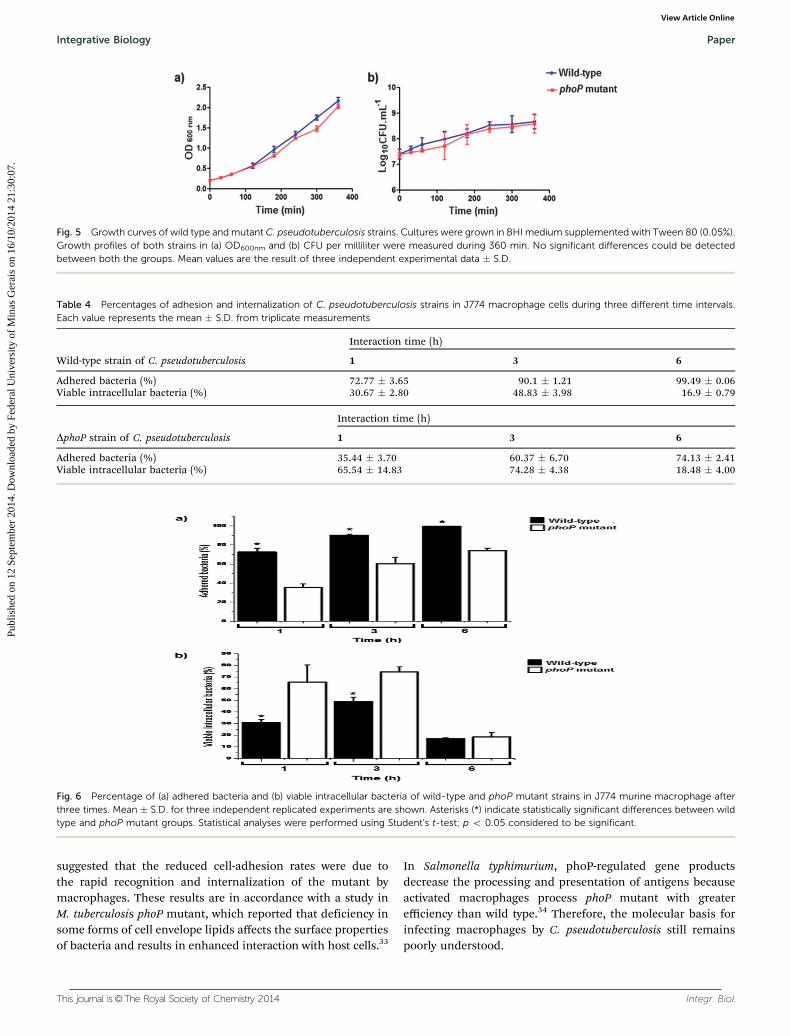
a statistically significant difference was observed between thewild type and mutated strains. At all times, the adherence rateremained low in the mutated strain. After evaluating thepercentage of viable intracellular bacteria in both the strainsat two points in time (1 and 3 h post-infection), it was possibleto observe a significant increase in the number of phoP mutant
viable in macrophages, compared with wild-type, while nodifference was noted at 6 h (Fig. 6b). Intracellular viability waslow for the wild-type bacteria at 1 and 3 h post-infection. Thenumber of cells of mutant strain adhered at 1 and 3 h post-infection (35.44% and 60.37%, respectively) and the high per-centage of internalized cells (65.54% and 74.28%, respectively)
Fig. 2 (a) Schematic representation of recombinant plasmid pCRs2.1-TOPOs/phoP fragment with restriction sites. Plasmid was used to clone theC. pseudotuberculosis phoP fragment linked to promoter T7. (b) Plasmid isolated, purified and then visualized on 1% agarose gel. The 1 kb Ladder plusmolecular marker (Invitrogen) is indicated as MW (first lane); lane 2: amplification of the C. pseudotuberculosis phoP fragment (436 bp amplicon); lane 3:recombinant plasmid pCRs2.1-TOPOs/phoP fragment, after digestion with EcoRI; lane 4: recombinant plasmid pCRs2.1-TOPOs/phoP fragment didnot undergo digestion. Molecular masses are indicated.
Fig. 3 Integration of suicide plasmid (pCRs2.1-TOPOs/phoP fragment) disrupted the phoP gene. Homologous recombination (X) to give the fragmentsindicated on the diagram. The solid boxes represent fragments from suicide plasmid. The size of portions and primers also are indicated.
Fig. 4 Confirmation of phoP gene mutation in C. pseudotuberculosis. (a) Analysis of PCR products amplified from genomic DNA of the wild type andmutant strains visualized on 1% agarose gel electrophoresis. The 1 kb Ladder Plus Molecular Marker (Invitrogen) is indicated as MW (first and last lanes).Molecular masses are indicated. (b) Detail of the amplicons of the agarose gel. Lanes in the gel, strains, pair primers in each PCR reaction and the length ofamplification fragments are shown.
Paper Integrative Biology
Publ
ishe
d on
12
Sept
embe
r 20
14. D
ownl
oade
d by
Fed
eral
Uni
vers
ity o
f M
inas
Ger
ais
on 1
6/10
/201
4 21
:30:
07.
View Article Online
This journal is©The Royal Society of Chemistry 2014 Integr. Biol.
suggested that the reduced cell-adhesion rates were due tothe rapid recognition and internalization of the mutant bymacrophages. These results are in accordance with a study inM. tuberculosis phoP mutant, which reported that deficiency insome forms of cell envelope lipids affects the surface propertiesof bacteria and results in enhanced interaction with host cells.33
In Salmonella typhimurium, phoP-regulated gene productsdecrease the processing and presentation of antigens becauseactivated macrophages process phoP mutant with greaterefficiency than wild type.34 Therefore, the molecular basis forinfecting macrophages by C. pseudotuberculosis still remainspoorly understood.
Fig. 5 Growth curves of wild type and mutant C. pseudotuberculosis strains. Cultures were grown in BHI medium supplemented with Tween 80 (0.05%).Growth profiles of both strains in (a) OD600nm and (b) CFU per milliliter were measured during 360 min. No significant differences could be detectedbetween both the groups. Mean values are the result of three independent experimental data � S.D.
Table 4 Percentages of adhesion and internalization of C. pseudotuberculosis strains in J774 macrophage cells during three different time intervals.Each value represents the mean � S.D. from triplicate measurements
Wild-type strain of C. pseudotuberculosis
Interaction time (h)
1 3 6
Adhered bacteria (%) 72.77 � 3.65 90.1 � 1.21 99.49 � 0.06Viable intracellular bacteria (%) 30.67 � 2.80 48.83 � 3.98 16.9 � 0.79
DphoP strain of C. pseudotuberculosis
Interaction time (h)
1 3 6
Adhered bacteria (%) 35.44 � 3.70 60.37 � 6.70 74.13 � 2.41Viable intracellular bacteria (%) 65.54 � 14.83 74.28 � 4.38 18.48 � 4.00
Fig. 6 Percentage of (a) adhered bacteria and (b) viable intracellular bacteria of wild-type and phoP mutant strains in J774 murine macrophage afterthree times. Mean � S.D. for three independent replicated experiments are shown. Asterisks (*) indicate statistically significant differences between wildtype and phoP mutant groups. Statistical analyses were performed using Student’s t-test; p o 0.05 considered to be significant.
Integrative Biology Paper
Publ
ishe
d on
12
Sept
embe
r 20
14. D
ownl
oade
d by
Fed
eral
Uni
vers
ity o
f M
inas
Ger
ais
on 1
6/10
/201
4 21
:30:
07.
View Article Online

Integr. Biol. This journal is©The Royal Society of Chemistry 2014
Immunization assay, challenge and easement of protectionlevel
To assess the protection against CLA in mice, immunizationassays were performed by intra-peritoneal inoculation withdoses of C. pseudotuberculosis wild-type 1002 and phoP mutantstrain, Glanvac, as well as a saline control. This was done by thedetermination of mice survival, after a challenge dose with thewild-type C. pseudotuberculosis MIC-6 strain.
All the mice vaccinated with the wild-type strain showedclinical signs of morbidity, and most mortality happened beforethe challenge. In this group, animals resistant to infection diedwithin 2 days after the challenge. Mice immunized with salinedied between 1 and 2 days after the challenge. Mice vaccinatedwith Glanvac presented a survival rate of 55% (Fig. 7). Thesurvival rate of mice given the DphoP strain was the highestamong the vaccine groups (60% survival) and was significantlydifferent than that of the saline group. A single immunizationwith phoP mutant strain as a live vaccine conferred a significantprotection against the bacterial infection in mice. These dataprovide the evidence that the deletion of phoP gene may havecaused an attenuation of virulence in mice.
Detection of specific IgG, IgG1 and IgG2a antibodies
To verify the production of specific IgG antibodies, the serumsamples from mice immunized with C. pseudotuberculosis DphoPwere compared with saline (control) and Glanvac immunizedmice by ELISA. IgG1 and IgG2a were investigated separatelybecause IgG1 is related to a Th2 cellular immune responsewhereas IgG2a is related to a Th1 response in the same species.The preliminary results from the immunological assays showedthat mice immunized with C. pseudotuberculosis DphoP straindeveloped high levels of IgG antibodies when compared tothe control group. Similar substantial levels were observed inanimals immunized with the vaccine Glanvact 3 (Fig. 8a).
Immunization with the phoP mutant strain also induced theproduction of IgG1 (Fig. 8b) in all the immunized mice.Analyzing the results shown in the graph indicate that IgG1production rates in the group vaccinated with Glanvact 3 wassignificantly higher relative to the control group. This increasewas also observed in the group inoculated with the phoPmutant showing a significant increase in the production of
this immunoglobulin; thus exhibiting a Th2 pattern immuneresponse. Regarding the levels of IgG2a induced after immuni-zation (Fig. 8c), inoculation with phoP mutant induced quitesignificant immunoglobulin response compared to the controlgroup. The production of IgG2a in mice is characteristic of aTh1 type of immune response, which is responsible for theelimination of intracellular pathogens C. pseudotuberculosis,35
suggesting that the protection type induced by mutant PhoPresponse is consistent with that expected in combating bacteria. Ourresults showed that the C. pseudotuberculosis phoP mutant conferredwith a single dose partial protection against CLA. 60% of micevaccinated with phoP mutant strain survived the lethal challengewith C. pseudotuberculosis MIC-6 strain. In contrast, other groups ofanimals (control, wild-type) were not protected against the samedose. These findings indicate an attenuated phenotype conferred bymutation in the phoP gene of C. pseudotuberculosis. Supporting thisargument, other studies have also shown virulence attenuation inpathogens such as M. tuberculosis and S. typhimurium.36 Vaccinationwith the phoP mutant induced significant production of mutant-specific IgG, IgG1 and IgG2, producing a mixed Th1/Th2, which isnecessary for the effective protection against C. pseudotuberculosis.
Fig. 7 Graphic: % survival or Number of survived animal vs. time afterchallenge (in days)
Fig. 8 Profile of total IgG antibody response after immunization in mice.(a) IgG antibody titer; (b) IgG1 iso-type titer; (c) IgG2a iso-type titer. Groups ofmice were immunized as follows: intra-peritoneal injection of saline 0.9%(control), phoP mutant strain and Glanvac. Data are expressed as mean� S.D.values. Results are representative of n = 5. Statistically significant differencesbetween phoP mutant and Glanvac groups and control mice are denotedwith an asterisk (p o 0.05).
Paper Integrative Biology
Publ
ishe
d on
12
Sept
embe
r 20
14. D
ownl
oade
d by
Fed
eral
Uni
vers
ity o
f M
inas
Ger
ais
on 1
6/10
/201
4 21
:30:
07.
View Article Online
This journal is©The Royal Society of Chemistry 2014 Integr. Biol.
The protection conferred by C. pseudotuberculosis DphoPstrain may be associated with changes conferred in phoP genemutation, as observed in M. tuberculosis.37 Absence of theexpression of important virulence genes may be the main factorjustifying partial protection observed after challenge with thevirulent strain of C. pseudotuberculosis. It has been reported inprevious studies that TCS systems are involved in the regulationof a variety of processes, including cell division, nutrient acquisi-tion, regulation of osmolarity, redox potential, nitrogen fixation,phosphate uptake, sporulation, pilus formation, adhesion, drugresistance and expression of virulence factors.38,39 Therefore, wefurther expanded our analyses to find out some inhibitors for thisprotein obtained from natural sources and ZINC database andperformed docking analysis.
PhoP protein homology modeling and structure validation
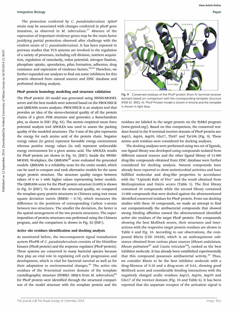
The PhoP protein 3D model was generated using SWISS-MODELserver and the best models were selected based on the PROCHECKand QMEAN6 scores analyses. PROCHECK is an analysis tool thatprovides an idea of the stereo-chemical quality of all the proteinchains of a given PDB structure and generates a Ramchandranplot, as shown in ESI† (Fig. S1). The atomic empirical mean forcepotential analysis tool ANOLEA was used to assess the packingquality of the modeled structures. The Y-axis of the plot representsthe energy for each amino acid of the protein chain. Negativeenergy values (in green) represent favorable energy environmentwhereas positive energy values (in red) represent unfavorableenergy environment for a given amino acid. The ANOLEA resultsfor PhoP protein are shown in Fig. S3, (ESI†). Inside the SWISS-MODEL Workplace, the QMEAN640 score evaluated the generatedmodels. QMEAN6 is a reliability score for the entire model, whichcan be used to compare and rank alternative models for the sametarget protein structure. The structure quality ranges betweenvalues of 0 to 1 with higher values representing better models.The QMEAN6 score for the PhoP protein structure (0.689) is shownin Fig. S3 (ESI†). To observe the structural quality, we comparedthe template-query protein structures in Chimera using root meansquare deviation metric (RMSD = 0.78), which measures thedifference in the positions of corresponding Carbon a-atomsbetween two structures. The smaller the deviation, the better isthe spatial arrangement of the two protein structures. The super-imposition of protein structures was performed using the Chimeraprogram, and the comparison is shown in Fig. S2 (ESI†).
Active site residues identification and docking analysis
As mentioned before, the two-component signal transductionsystem PhoPR of C. pseudotuberculosis consists of the Histidinekinases (PhoR protein) and the response regulator (PhoP protein).These systems are conserved in many bacterial species becausethey play an vital role in regulating cell cycle progression anddevelopment, which is vital for bacterial survival as well as fortheir adaptation to environmental changes.41 The active siteresidues of the N-terminal receiver domain of the templatecrystallographic structure (PDBID: 3R0J-A from M. tuberculosis)42
for PhoP protein were identified through the structural compari-son of the model structure with the template protein and the
residues are labeled in the target protein via the PyMol program(www.pymol.org/). Based on this comparison, the conserved resi-dues found in the N terminal receiver domain of PhoP protein are:Asp15, Asp16, Asp59, Glu17, Thr87 and Tyr106 (Fig. 9). Theseamino acid residues were considered for docking analyses.
The docking analyses were performed using two set of ligands,one ligand library was developed using compounds isolated fromdifferent natural sources and the other ligand library of 11 000drug-like compounds obtained from ZINC database were furtherconsidered for docking analysis. Natural compounds havealready been reported to show antimicrobial activities and havefulfilled molecular and drug-like properties in accordancewith the ‘‘Lipinski Rule of Five’’ and the result obtained fromMolinspiration and Osiris scores (Table 5). The first librarycontained 30 compounds while the second library contained11 000 compounds that were docked against the aforementionedidentified conserved residues for PhoP protein. From our dockingstudies with these 30 compounds, we made an attempt to findout computationally the antibacterial compounds that showedstrong binding affinities toward the aforementioned identifiedactive site residues of the target PhoP protein. The compoundsshowing the best MolDock scores, their structures and inter-actions with the respective target protein residues are shown inTable 6 and Fig. 10. According to our observations, the com-pound Rhein (CID 10168), which is an anthraquinone sub-stance obtained from various plant sources (Rheum undulatum,Rheum palmatum43 and Cassia reticulate44), ranked as the bestinhibitor molecule. It has already been established experimentallythat this compound possesses antibacterial activity.44 Thus,we consider Rhein to be the best inhibitor molecule with adrug-likeness of 0.18 and a drug-score of 0.61, showing goodMolDock score and considerable binding interactions with thenegatively charged acidic residues Asp15, Asp16, Asp59 andGlu17 of the receiver domain (Fig. 10 and Table 6). It has beenreported that the aspartate receptor of the activation signal is
Fig. 9 Conserved residues of the PhoP protein (from N-terminal receiverdomain) based on comparison with the corresponding template structure(PDB ID: 3R0J-A). PhoP Protein model is shown in limone and the templateis shown in light blue.
Integrative Biology Paper
Publ
ishe
d on
12
Sept
embe
r 20
14. D
ownl
oade
d by
Fed
eral
Uni
vers
ity o
f M
inas
Ger
ais
on 1
6/10
/201
4 21
:30:
07.
View Article Online

Integr. Biol. This journal is©The Royal Society of Chemistry 2014
the Asp59 residue (phosphorylation is absolutely conserved)26
that is located at the N-terminal receiver domain of the responseregulator of the PhoP protein, which along with Asp15 has animportant role in Mg+2 binding.42 In addition, after the virtualscreening of 11 000 compounds, the top ranked 200 compoundswere analyzed in Chimera for shape complementarity and hydro-gen bond interactions, leading to the selection of a final set of5 compounds (Table 7) for PhoP target protein, demonstratingthat these compounds are binding to some of the same residues
such as the natural antimicrobial Rhein compound. For example,the compound ZINC ID: 02127223 showed almost similar bind-ing affinity with the target protein residues as Rhein (Fig. 11). Theconsiderable binding affinities of Rhein compound isolated fromnatural sources and of other drug like compounds from the ZINCdatabase support the poly pharmacological idea of promiscuoustarget protein. Because Rhein showed good interactions with theconserved residues of the PhoP receiver domain, compoundsfrom the ZINC database might also act as putative drug moleculesin accordance with the aforementioned drug likeness properties,which may possibly hinder the normal phosphorylation processof the target protein and inhibit the normal functioning of thePhoPR signaling cascade. It is obvious from our in silico analysesthat the putative TCS inhibitors targeted the receiver domain ofthe response regulator protein and they might interfere with the
Table 5 Molinspiration and Osiris results of compounds selected for furtherdocking analyses
Compounds c log P SolubilityMol.weight
Drug-likeness
Drug-score
Liriodenin (CID 10144) 4 �6.25 275 �2.39 0.28Pinostrobin (CID 73201) 3 �3.24 270 2.05 0.8Axisonitrile-3 (CID 181226) 4.5 �3.47 231 �9.44 0.36Ileabethoxazole (CID 9549062) 5.47 �5.77 325 �1.3 0.26Rhein (CID 10168) 2.44 �4.15 284 0.18 0.61Pilocarpine (CID 5910) 0.4 �1.2 208 1.17 0.85Voacangine (CID 73255) 2.97 �3.86 368 1.8 0.7Texalin (CID 473253) 2.8 �4.9 266 0.56 0.58Araguspongine (CID 5276744) 5.98 �4.22 478 �1.7 0.24Jacarandic acid (CID 73645) 3.88 �5.36 488 0.19 0.37Leptophyllin B (CID 10447482) 4.06 �3.66 299 �8.03 0.37
Table 6 Docking results of the best-predicted natural compound (Rhein)that showed good interaction in our in silico analysis, with their MolDockScore and the interacting residues
Compoundname
MolDockscore
No. of H-bonds/residues-compoundinteractions
Rhein (CID 10168) �59.8635 3Asp15, Asp16, Asp59
Fig. 10 (a) 3D cartoon representation of the docking analyses for PhoPprotein structure with Rhein compound (CID 10168). (b) 3D surfacerepresentation of the docking analyses for the structures of Rhein com-pound with Phop receiver domains. (c) Two-dimensional representation ofRhein compound using PoseView45 interacting with the conserved resi-dues of the N-terminal receiver domain of the PhoP protein. The residuesare negatively charged acidic Asp15, Asp59, Phe111 and Lys109.
Table 7 ZINC codes, MolDock scores and predicted hydrogen bonds forthe five compounds selected among the top ranking 100 moleculesagainst PhoP protein
ZINC IDs MolDock score
No. of H-bonds/residues-compoundinteractions
ZINC02127223 �85.678 4Asp59, Asp16, Asp15, Lys109
ZINC00078435 �84.6183 3Asp15, Asp16, Met61
ZINC01423999 �97.227 4Asp16, Asp59, Ays109
ZINC00406666 �95.2886 2Asp15, Asp59
ZINC01408034 �85.216 3Asp16, Asp59, Lys109
Fig. 11 (a) 3D cartoon representation of the docking analyses for PhoPprotein structure with compound ZINC ID: 02127223. (b) 3D surface repre-sentation of the docking analyses for the structures of compound ZINC ID:02127223 with Phop receiver domains. (c) Two-dimensional representation ofcompound ZINC ID: 02127223 using PoseView interacting with the conservedresidues of the N-terminal receiver domain of the PhoP protein. The residuesare negatively charged acidic Asp15, Asp16, Asp59 and Lys109.
Paper Integrative Biology
Publ
ishe
d on
12
Sept
embe
r 20
14. D
ownl
oade
d by
Fed
eral
Uni
vers
ity o
f M
inas
Ger
ais
on 1
6/10
/201
4 21
:30:
07.
View Article Online
This journal is©The Royal Society of Chemistry 2014 Integr. Biol.
domain normal function. Here, after verifying a possible role ofthe PhoP protein in the virulence of C. pseudotuberculosis, anadditional computational approach has been taken to identifynew TCSs specific inhibitors with potential drug-like propertiesfor future experimental efforts (Fig. 10 and 11).
Conclusion
An effective prophylaxis for CLA requires the development ofnovel vaccine and drug candidates capable of generating anadequate immune response and protection against the disease.
Several virulence-attenuated mutants of intracellular pathogenshave been used with promising vaccine strategy, including mutantsof the gene phoP.46–49 Alternatively, due to the importance of theTCS system, the same PhoP protein could be an attractive target fordesigning rational and target specific molecular inhibitors. In thiscontext, we have initially described, in the present report, thedevelopment and use of a C. pseudotuberculosis Cp1002 vaccinestrain based on the disruption of phoP gene. At a later stage, the 3Dmodel of the PhoP protein has been subjected to detailed compu-tational analyses via in silico approaches as a therapeutic target forthe identification of novel inhibitors.
Our results confirm that the phoP mutated C. pseudotuberculosislacked certain properties or functions associated with PhoPRsystem, and were affected by the absence of the PhoP protein.Under normal conditions, no significant differences were detectedbetween the growth rate of C. pseudotuberculosis phoP mutant andwild-type strains. Similar result was found in M. tuberculosis phoPmutant during logarithmic and stationary phases of the growthcurve.13 Bacterial adhesion and invasion to the host cell areimportant steps in bacterial infection. Here, we reported theadhesion and invasion abilities of both C. pseudotuberculosisstrains (wild-type and phoP mutant) in macrophages. Possiblechanges in the lipid composition of the cell envelope may haveaffected the interaction of phoP mutant strain with macro-phages. Intracellular viability was related with the adhesiveability of strains. Our analyses suggest that the disruption ofphoP gene might be involved in the alteration of some surfacereceptors. The elucidation of this bacterial strategy for in vitroinfection is an important subject for understanding the dynamicsof gene regulation during host interactions and for the develop-ment of a novel attenuated vaccine. The protective immunity ofthe vaccine based on phoP mutant of C. pseudotuberculosis wasevaluated by determining the survival rates and the serum anti-body titers of the mice. Our data indicate that immunization withC. pseudotuberculosis phoP mutant promoted cellular immuneresponse and generated partial protection in mice possiblydue to reduction of the virulence; however, further studies arerequired to understand the mechanisms of attenuation.
The overall importance and a promiscuous role of the PhoPprotein brought us to follow some in silico approaches for thedevelopment of novel drugs. In this context, comparativestudies are very important to predict new target genes. Basedon this identification strategy, many bacterial genes havebeen revealed, and the genes of the PhoP regulatory locus,
initially studied in Enterobacteria, are among them. Based onour observations, we have proposed that targeting the PhoPprotein of the PhoPR system of the C. pseudotuberculosis, whichhave a similar role in regulating the expression of several genes tothe closely related M. tuberculosis,47,50 might help in attenuatingpathogen growth in disease condition. The relationship of virulence-associated two-component PhoPR system in phylogenetically distantbacteria also supports our hypothesis. However, further experi-mental studies are required to discover the relationship betweenC. pseudotuberculosis PhoPR system and a possible virulence effect.Furthermore, our docking results have revealed that Rhein fromRheum undulatum and Rheum palmatum might be good therapeuticmolecules for future wet lab studies. This might potentially inhibitthe response regulator domain of the two-component signaltransduction system, resulting in decreased pathogen virulencein in vitro experimentation. Because the PhoP protein is homo-logous and the residues are conserved across a broad range ofclosely related pathogenic bacteria (keeping in mind the non-pathogenic as well), the proposed drugs could target as manyTCSs in the pathogenic bacteria as possible. In this context thePhoP inhibitors could be regarded as the broad-spectruminhibitors and not only for C. pseudotuberculosis. The workpresented here is the first ever conducted on the PhoP proteinof C. pseudotuberculosis Cp1002 and optimistically the avail-ability of these in silico information might serve as a basis forfurther future investigation.
Competing interests
The authors declare no conflict of interest.
Financial disclosure
We would like to gratefully acknowledge the help of all the teammembers & the financing agencies. Sandeep Tiwari acknowl-edges the receipt of fellowship from ‘‘TWAS-CNPq PostgraduateFellowship Programme’’ for doctoral studies. This work waspartially executed by Rede Paraense de Genomica e Proteomicasupported by FAPESP (Fundaçao de Amparo a Pesquisa doEstado do Para), CNPq (Conselho Nacional de DesenvolvimentoCientıfico e Tecnologico, Brasil), CAPES (Coordenaçao deAperfeiçoamento de Pessoal de Nıvel Superior, Brasil) andFAPEMIG (Fundaçao de Amparo a Pesquisa do Estado de MinasGerais, Brasil).
References
1 F. A. Dorella, M. S. Fachin, A. Billault, E. Dias Neto, C. Soravito,S. C. Oliveira, R. Meyer, A. Miyoshi and V. Azevedo, GMR,Genet. Mol. Res., 2006, 5, 653–663.
2 L. H. Williamson, The Veterinary clinics of North America:Food animal practice, 2001, 17, 359–371.
3 R. G. Batey, Aust. Vet. J., 1986, 63, 269–272.4 F. A. Dorella, L. G. Pacheco, S. C. Oliveira, A. Miyoshi and
V. Azevedo, Vet. Res., 2006, 37, 201–218.
Integrative Biology Paper
Publ
ishe
d on
12
Sept
embe
r 20
14. D
ownl
oade
d by
Fed
eral
Uni
vers
ity o
f M
inas
Ger
ais
on 1
6/10
/201
4 21
:30:
07.
View Article Online
Integr. Biol. This journal is©The Royal Society of Chemistry 2014
5 G. J. Baird and M. C. Fontaine, J. Comp. Pathol., 2007, 137,179–210.
6 S. C. McKean, J. K. Davies and R. J. Moore, Microbiology,2007, 153, 2203–2211.
7 G. M. Pao and M. H. Saier Jr., J. Mol. Evol., 1995, 40, 136–154.8 A. G. Blanco, M. Sola, F. X. Gomis-Ruth and M. Coll,
Structure, 2002, 10, 701–713.9 J. A. Hoch, Curr. Opin. Microbiol., 2000, 3, 165–170.
10 I. Lopez-Goni, C. Guzman-Verri, L. Manterola, A. Sola-Landa,I. Moriyon and E. Moreno, Vet. Microbiol., 2002, 90, 329–339.
11 M. Matsushita and K. D. Janda, Bioorg. Med. Chem., 2002,10, 855–867.
12 A. M. Stock, V. L. Robinson and P. N. Goudreau, Annu. Rev.Biochem., 2000, 69, 183–215.
13 E. Perez, S. Samper, Y. Bordas, C. Guilhot, B. Gicquel andC. Martin, Mol. Microbiol., 2001, 41, 179–187.
14 L. E. Ulrich, E. V. Koonin and I. B. Zhulin, Trends Microbiol.,2005, 13, 52–56.
15 D. Beier and R. Gross, Curr. Opin. Microbiol., 2006, 9, 143–152.16 Y. Gotoh, Y. Eguchi, T. Watanabe, S. Okamoto, A. Doi and
R. Utsumi, Curr. Opin. Microbiol., 2010, 13, 232–239.17 P. J. Cardona, J. G. Asensio, A. Arbues, I. Otal, C. Lafoz,
O. Gil, N. Caceres, V. Ausina, B. Gicquel and C. Martin,Vaccine, 2009, 27, 2499–2505.
18 M. L. Chesne-Seck, N. Barilone, F. Boudou, J. Gonzalo Asensio,P. E. Kolattukudy, C. Martin, S. T. Cole, B. Gicquel, D. N. Gopauland M. Jackson, J. Bacteriol., 2008, 190, 1329–1334.
19 J. Gonzalo-Asensio, S. Mostowy, J. Harders-Westerveen,K. Huygen, R. Hernandez-Pando, J. Thole, M. Behr, B. Gicqueland C. Martin, PLoS One, 2008, 3, e3496.
20 L. G. Pacheco, R. R. Pena, T. L. Castro, F. A. Dorella, R. C.Bahia, R. Carminati, M. N. Frota, S. C. Oliveira, R. Meyer,F. S. Alves, A. Miyoshi and V. Azevedo, J. Med. Microbiol.,2007, 56, 480–486.
21 V. S. Kalogeraki and S. C. Winans, Gene, 1997, 188, 69–75.22 F. A. Dorella, E. M. Estevam, P. G. Cardoso, B. M. Savassi,
S. C. Oliveira, V. Azevedo and A. Miyoshi, Vet. Microbiol.,2006, 114, 298–303.
23 P. M. Moraes, N. Seyffert, W. M. Silva, T. L. Castro,R. F. Silva, D. D. Lima, R. Hirata Jr., A. Silva, A. Miyoshiand V. Azevedo, BioMed. Res. Int., 2014, 2014, 489782.
24 D. Ribeiro, S. Rocha Fde, K. M. Leite, C. Soares Sde, A. Silva,R. W. Portela, R. Meyer, A. Miyoshi, S. C. Oliveira,V. Azevedo and F. A. Dorella, Vet. Res., 2014, 45, 28.
25 S. F. Altschul, W. Gish, W. Miller, E. W. Myers andD. J. Lipman, J. Mol. Biol., 1990, 215, 403–410.
26 G. Moraes, V. Azevedo, M. Costa, A. Miyoshi, A. Silva,V. da Silva, D. de Oliveira, M. F. Teixeira, J. Lameira andC. N. Alves, J. Mol. Model., 2012, 18, 1219–1227.
27 K. Arnold, L. Bordoli, J. Kopp and T. Schwede, Bioinformatics,2006, 22, 195–201.
28 R. A. Laskowski, M. W. Macarthur, D. S. Moss and J. M.Thornton, J. Appl. Crystallogr., 1993, 26, 283–291.
29 F. Melo and E. Feytmans, J. Mol. Biol., 1998, 277, 1141–1152.
30 E. F. Pettersen, T. D. Goddard, C. C. Huang, G. S. Couch,D. M. Greenblatt, E. C. Meng and T. E. Ferrin, J. Comput.Chem., 2004, 25, 1605–1612.
31 J. J. Irwin, T. Sterling, M. M. Mysinger, E. S. Bolstad andR. G. Coleman, J. Chem. Inf. Model., 2012, 52, 1757–1768.
32 R. Thomsen and M. H. Christensen, J. Med. Chem., 2006, 49,3315–3321.
33 N. L. Ferrer, A. B. Gomez, O. Neyrolles, B. Gicquel andC. Martin, PLoS One, 2010, 5, e12978.
34 E. A. Groisman, J. Bacteriol., 2001, 183, 1835–1842.35 A. L. Hodgson, K. Carter, M. Tachedjian, J. Krywult, L. A.
Corner, M. McColl and A. Cameron, Vaccine, 1999, 17,802–808.
36 Y. Li, M. Jiang, W. Liu, L. Zhang, S. Zhang, X. Zhao, R. Xiangand Y. Liu, Mol. Cell. Probes, 2010, 24, 68–71.
37 S. Gupta, A. Sinha and D. Sarkar, FEBS Lett., 2006, 580,5328–5338.
38 J. F. Barrett, R. M. Goldschmidt, L. E. Lawrence, B. Foleno,R. Chen, J. P. Demers, S. Johnson, R. Kanojia, J. Fernandez,J. Bernstein, L. Licata, A. Donetz, S. Huang, D. J. Hlasta,M. J. Macielag, K. Ohemeng, R. Frechette, M. B. Frosco,D. H. Klaubert, J. M. Whiteley, L. Wang and J. A. Hoch,Proc. Natl. Acad. Sci. U. S. A., 1998, 95, 5317–5322.
39 R. Utsumi and M. Igarashi, Yakugaku zasshi, 2012, 132,51–58.
40 P. Benkert, M. Kunzli and T. Schwede, Nucleic Acids Res.,2009, 37, W510–W514.
41 E. J. Capra and M. T. Laub, Annu. Rev. Microbiol., 2012, 66,325–347.
42 S. Menon and S. Wang, Biochemistry, 2011, 50, 5948–5957.43 L. Hoerhammer, H. Wagner and I. Koehler, Arch. Pharm.
Ber. Dtsch. Pharm. Ges., 1959, 292/64, 591–601.44 M. Anchel, J. Biol. Chem., 1949, 177, 169–177.45 K. Stierand and M. Rarey, ACS Med. Chem. Lett., 2010, 1,
540–545.46 H. S. Garmory, K. A. Brown and R. W. Titball, FEMS
Microbiol. Rev., 2002, 26, 339–353.47 J. Gonzalo Asensio, C. Maia, N. L. Ferrer, N. Barilone,
F. Laval, C. Y. Soto, N. Winter, M. Daffe, B. Gicquel, C. Martinand M. Jackson, J. Biol. Chem., 2006, 281, 1313–1316.
48 C. Martin, A. Williams, R. Hernandez-Pando, P. J. Cardona,E. Gormley, Y. Bordat, C. Y. Soto, S. O. Clark, G. J. Hatch,D. Aguilar, V. Ausina and B. Gicquel, Vaccine, 2006, 24,3408–3419.
49 M. V. Mendes, S. Tunca, N. Anton, E. Recio, A. Sola-Landa,J. F. Aparicio and J. F. Martin, Metab. Eng., 2007, 9, 217–227.
50 S. B. Walters, E. Dubnau, I. Kolesnikova, F. Laval, M. Daffeand I. Smith, Mol. Microbiol., 2006, 60, 312–330.
51 J. Sambrook, E. F. Fritsch and T. Maniatis, MolecularCloning: A Laboratory Manual, Cold Spring Harbor Laboratory,Cold Spring Harbor, NY, USA, 2nd edn, 1989.
52 F. A. Dorella, L. G. Pacheco, N. Seyffert, R. W. Portela,R. Meyer, A. Miyoshi and V. Azevedo, Expert Rev. Vaccines,2009, 8, 205–213.
Paper Integrative Biology
Publ
ishe
d on
12
Sept
embe
r 20
14. D
ownl
oade
d by
Fed
eral
Uni
vers
ity o
f M
inas
Ger
ais
on 1
6/10
/201
4 21
:30:
07.
View Article Online
Copyright © 2022 FDOKUMEN