Survey of genome organization and gene content of Corynebacterium pseudotuberculosis
Upload
khangminh22Category
view
0download
0
Copyright © 2021, The Korean Society of Veterinary Service. All Rights Reserved. 61
한국가축위생학회지 제44권 제2호 (2021)Korean J Vet Serv, 2021, 44(2), 61-71ISSN 1225-6552, eISSN 2287-7630https://doi.org/10.7853/kjvs.2021.44.2.61 < Review Article >
Korean Journal ofVeterinary ServiceAvailable online at http://kjves.org
*Corresponding author: Yong-il Cho, E-mail. [email protected] https://orcid.org/0000-0001-7756-3416
Recent perspectives on caseous lymphadenitis caused by Corynebacterium pseudotuberculosis in goats–A review
Md. Aftabuzzaman, Yong-il Cho*
Department of Animal Science and Technology, Sunchon National University, Suncheon 57922, Korea
(Received 26 April 2021; revised 26 June 2021; accepted 26 June 2021)
Abstract
Caseous lymphadenitis, caused by Corynebacterium pseudotuberculosis has been a predominant disease among small ruminants on farms, causing significant losses for farm producer in the larger part of goat rearing nations across the world, for over a century. However, the control measures have not been very effective due to the chronic and usually subclinical nature of the infection. This has caused significant financial losses due to chronic ill-thrift, carcass condemnation, decreased meat yields and low repro-duction as well as animal welfare. The current status of caseous lymphadenitis, with updated reseach information to the etiology, pathogenesis, clinical signs, identification, prevalence, prevention and vacci-nation are delineated in the review.
Key words : Caseous lymphadenitis, Corynebacterium pseudotuberculosis, Goat
INTRODUCTION
Caseous lymphadenitis (CLA) is a chronic and sub-clinical disease of small ruminants with a high preva-lence in both isolated animals and herd. The causative pathogen, Corynebacterium (C.) pseudotuberculosis, af-fects goats and sheep, and can also infect cattle (Anderson et al, 1990; Shpigel et al, 1993; Yeruham et al, 1997), horses (Miers and Ley, 1980; Poonacha and Donahue, 1995) and rarely, humans (Goldberger et al, 1981; House et al, 1986; Shpigel et al, 1993). The pathogen has also been identified from other species including buffaloes, pigs, deer, llamas, camels and certain laboratory animals (Williamson, 2001; Dorella et al, 2006). CLA is widely distributed across the world and has been found in South and North America, Australia, Asia, Europe and Africa. It has been the cause of sig-nificant economic losses as a result of condemnation of skins and carcasses due to abscesses, decrease in re-
productive potential, milk and meat production. CLA has significant economic concern for goat production due to reduced meat and milk yield, loss of fertility, culling of affected animals, condemnation and down-grading of affected carcass at the time of slaughter and meat inspection (Wan-Choul et al, 2015; Kumaresan et al, 2018). This disease is characterized by the development of abscesses within the superficial and visceral lymph nodes. The abscesses in the peripheral lymph nodes are superficial, whereas those in the visceral lymph nodes can lead to systemic complications and chronic weight loss (Radostits et al, 2002). C. pseudotuberculosis is rapidly spread within the herd by normal farm manage-ment practices and environmental contamination (Brown and Olander, 1987). CLA is contagious enough to infect goat in a herd despite the apparent containment of the microorganism in the lesion. CLA infections in goats of-ten occur as superficial abscesses in the head and neck. However, mesenteric and mediastinal lymph nodes can also be affected (Smith and Sherman, 2009). The poor

62 Md. AftabuzzamanㆍYong-il Cho
Korean J Vet Serv, 2021, Vol. 44, No. 2
response to drug treatment, the long-term environemt survival of the organism and difficulties in identifying infected animals were challenges in eradicating CLA. The current research on CLA, encompassing etiology, clinical signs, diagnosis, distribution and prevalence, bacteriological culture and molecular identification, pre-vention and control, and treatment as well as farm man-agement were described in the review.
ETIOLOGY AND PATHOGENESIS
CLA is a chronic, contagious bacterial disease that af-fects goats and sheep (Stanford et al, 1998). The causa-tive agent is a guileful bacterium called C. pseudotu-berculosis, a non-motile, Gram-positive, facultative anae-robe coccobacillus (Brown and Olander, 1987). The bac-terium was known as the Preisz-Nocard bacillus, named after the researcher, Hugo von Preisz, who first identi-fied it from a goat renal abscess in 1894 (Brown and Olander, 1987). The causative organism was renamed C. ovis in 1923, and then again renamed, to the currently accepted nomenclature of C. pseudotuberculosis in 1948 (Brown and Olander, 1987). The organism does not du-plicate in the environment. However, it can survive for longer time after it is released from an abscess. Bacterial survival is enhanced when it is present in par-ticulate fomites like wood, straw, and feces (Augustine and Renshaw, 1986). It can survive on these fomites, for as long as 8 weeks and in soil samples for upto 8 months (Augustine and Renshaw, 1986; Brown and Olander, 1987; Rizvi et al, 1997). Survival is also en-hanced by shade, moist conditions and low temperatures (Augustine and Renshaw, 1986; Rizvi et al, 1997). C. pseudotuberculosis can be classified in two bio-vars, depend on their ability to modify nitrate to nitrite, the nitrate negative stains as biovar ovis and the nitrate positive ones as biovar equi (Almeida et al, 2017). In goats and sheep, CLA is considerably caused by biovar ovis strains, while buffalos and horses are manimly in-fected by biovar equi strains (Selim, 2001; Baird and Fontaine, 2007). Cattle, on the other hand, can be in-fected by the both biovars, which may have different tissues preference; biovar ovis infects mostly the mam-
mary gland (Yeruham et al, 1996), and the skin (Yeruham et al, 1997), and biovar equi causes ulcerative lympha-denitis, and coronet lesions (Steinman et al, 1999; de Sá Guimarães et al, 2011). All strains produce acid; how-ever, no gas is produced from carbon sources, including glucose, fructose, maltose, mannose, and sucrose. This bacterium is positive for catalase and phospholipase D production, negative for oxidase, and it is beta-hemolytic. The organism generally enters the body through skin break or mucous membranes, but it can not penetrate through intact skin. The organism can survive ingestion by phagocytic cells and lysosomal fusion and still repli-cate, destroying the phagocyte in the process (Brown and Olander, 1987; Smith and Sherman, 2009). The or-ganism spreads through the lymphatic system to auxil-iary locales, for example, regional lymph nodes and in-ternal organs such as the lungs, where it creates auxil-iary abscesses. The ideal incubation period from in-oculation to abscessation is 2 to 6 months (Smith and Sherman, 2009). External and pulmonary abscesses in-evitably rupture and contaminate the environment and infect the animal within the herd. Two significant virulence components improve the or-ganism’s ability to survive phagocytosis and to spread to secondary sites: (1) an external lipid coat and (2) a potent exotoxin called phospholipase D (Brown and Olander, 1987). The lipid coating gives mechanical and potentially biochemical protection from hydrolytic en-zymes in lysosomes, allowing the organism to survive as a facultative intracellular parasite. The phospholipase D (PLD) gene is considered the most potent virulence factor in C. pseudotuberculosis (Hodgson et al, 1999) and it encodes the PLD exotoxin that catalyzes the dis-sociation of sphingomyelin and the increase of vascular permeability. This results in the spread (Hodgson et al, 1994) and survival of C. pseudotuberculosis within the cells, and thereby the invasion of the body and transport by phagocytes to lymph nodes (Baird and Fontaine, 2007). Even though the PLD exotoxin has not been ob-served to be directly hemolytic, it has been seen to be, capable of producing synergistic hemolysis (Gyles et al, 2008). Iintegral membrane protein (FagA); iron enter-obactin transporter (FagB); cytoplasmic membrane pro-tein which binds ATP (FagC) and iron siderophore

Recent perspectives on caseous lymphadenitis caused by Corynebacterium pseudotuberculosis in goats–A review 63
Korean J Vet Serv, 2021, Vol. 44, No. 2
Fig. 1. A ruptured CLA abscess within the the pre-scapular lymph node; loss of hair (A). Discharge of pus from a ruptured abscess in a goat suffering from CLA (B).
Fig. 2. Abscesses induced by Corynebacterium pseudotubercu-losis localized in the parotid (A) and prefemoral (B) lymph node re-gions are characterized by thick yellow-green viscous pus with a toothpaste-like consistency. Severe abscessation of lung lymph nodes of the goat (C).
binding protein (FagD) are another important virulence factors. These are part of an operon included in iron up-take, which allows C. pseudotuberculosis infection to persist in goats (Sá et al, 2013; Li et al, 2018).
CLINICAL SIGNS
Superficial form of CLA is characterized by the in-fection of external lymph nodes (parotid, submandibular, pre-scapular, and subiliac), while the visceral form is presented by abscessing of different internal organs, for examples liver, lungs, uterus, kidneys, spleen, and also internal lymph nodes. These two forms can coexist (Piontkowski and Shivvers, 1998); however, other less basic sites can be involved, for example, the scrotum, mammary gland, the central nervous system, and joints (Williamson, 2001; Smith and Sherman, 2009). Internal
abscesses can be subclinical but are commonly asso-ciated with weight loss and ill thrift. Internal CLA is a leading cause of the "thin-doe syndrome" (Williamson, 2001; Solaiman, 2010; Pugh and Baird, 2012). External abscesses are characterized by slowly enlarging encapsu-lated masses in or near a peripheral lymph node. The abscess matures and ruptures through a fistula, draining infective purulent into the environment. CLA is asso-ciated with granulomatous, necrotizing inflammation of lymph nodes that causes chronic abscessation and en-largement of the nodes, loss of overlying hair, and pos-sible rupture of the abscesses and discharge of pus in goat (Fig. 1). The basic differences in the abscesses of goats are that the superficial form is more frequent among goats as opposed to the visceral form among sheep (Brown and Olander, 1987). Lymph node abscesses of the head and neck are more frequent in goats, while in sheep, ab-scesses are found in the subiliac and pre-scapular lymph nodes (Brown and Olander, 1987; Smith and Sherman, 2009). Differences in abscess content have also been found between goats and sheep. In the case of sheep, the contents have a laminar form when cut, like an on-ion, caused by the development of layers of fibrous tissue. In goats, on the other hand, the contents are made up of thick caseous material, while abscesses have a thin and pasty exudate (Fig. 2).
IDENTIFICATION AND DIAGNOSIS
Abscesses in goat are suggestive of CLA; however,

64 Md. AftabuzzamanㆍYong-il Cho
Korean J Vet Serv, 2021, Vol. 44, No. 2
Fig. 3. Characteristics of C. pseu-dotuberculosis isolates from goats during culture and staining. When grown on the blood agar medium, the colonies have a porcelain or chalky white, opaque, dry appear-ance with low adhesion to the me-dium plate (A). Palisade arrange-ment of the bacilli in a Gram stain (B).
bacterial isolation is important to distinguish the causative agent from other bacteria. For example, Arcanobacterium pyogenes, Staphylococcus aureus subsp. anaerobius, Pasteurella multocida, and Actinobacillus licheniformis can also be seen in abscesses (Shin et al, 2010; Pekelder, 2003). A thoracic X-ray can reveal masses within the pulmonary parenchyma and lymph nodes in animals with respiratory problems; which can also be confirmed by the culture of tracheal washes (Pugh and Baird, 2012). For cytological identification of the microorganism, Gram and Giemsa staining can be used. Since Gram staining is not primarily mention for staining tissues, the bluish color is taken on by C. pseudotuberculosis and a red-dish color appears in cellular and inflammatory material from aspirated lymph nodes (Radostits et al, 2002). It could help in detection of the infectious agents in the lesion. C. pseudotuberculosis is pleomorphic both club- shaped and coccoid forms are seen, and forms a pal-isade arrangement morphologically (Fig. 3). Bacterial growth in culture is sparse at first and is enhanced by the addition of whole blood to solid media (Dorella et al, 2006). C. pseudotuberculosis grew well in culture af-ter 18∼24 hours on incubation at 37°C at an optimum pH 7.2 (Dorella et al, 2006; Lopes Bastos B et al, 2012). C. pseudotuberculosis is identified through its mor-phology, staining characteristics, and various carbohy-drate fermentation tests in the laboratory after isolation (Jones, 1986). Direct and indirect tests to detect C. pseu-dotuberculosis have already been proposed, such as complement fixation test, synergistic hemolysis in-hibition test, microagglutination assay, PLD antigen-
based ELISA (Dercksen et al, 2000). From cultures and pus samples from CLA infected animals, the Multiplex PCR (mPCR) assay provides a reliable, effective, and rapid identification tool for C. pseudotuberculosis (Pacheco et al, 2007). A marker of cell-mediated immunity for determination of IFN-γ by ELISA has been used for di-agnosis of C. pseudotuberculosis infection, showing 91% sensitivity and 98% specificity, demonstrating its potential for use in CLA eradication programs (Menzies et al, 2004). The IFN-γ by ELISA test is more sensitive in identifying earlier infection in goats compared to the normal antibody ELISA, and it also does not appear to be influenced by vaccination (Menzies et al, 2004). Molecular typing methods have been used to decide the degree of relatedness between many numerous dis-tinctive corynebacterial species, including hybridization of nucleic acid, 16S rRNA gene sequence analysis and 16S rRNA gene restriction fragment length poly-morphism (RFLP) (Björkroth et al, 1999; Pavan et al, 2012) RFLP of chromosomal DNA (Costa et al, 1998), pulsed-field gel electrophoresis (PFGE) (Connor et al, 2000 & 2007) and randomly amplified DNA poly-morphisms (RAPD) (Foley et al, 2004; Stefańska et al, 2008). Pulsed field gel electrophoresis (PFGE) has been utilized to describe 50 strains of C. pseudotuberculosis isolated from small ruminants in the United Kingdom (Connor et al, 2000); six “pulsetypes” were found, which enabled the determination of the origin of the CLA outbreak. Be that as it may, in a report of 36 sheep samples and 6 goat samples from Australia, Canada, Holland and Northern Ireland, the same re-search team studied four different “pulsetypes” with the
Recent perspectives on caseous lymphadenitis caused by Corynebacterium pseudotuberculosis in goats–A review 65
Korean J Vet Serv, 2021, Vol. 44, No. 2
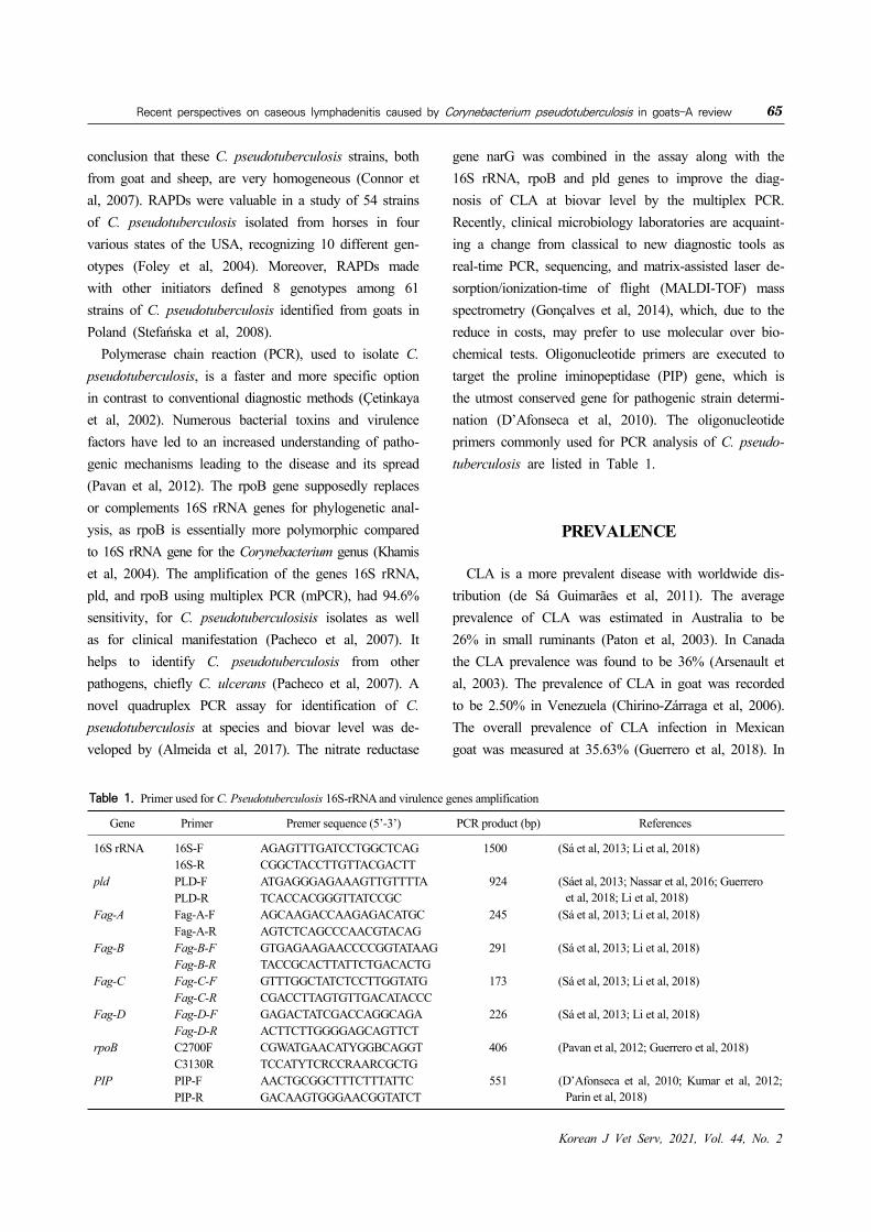
Table 1. Primer used for C. Pseudotuberculosis 16S-rRNA and virulence genes amplification
Gene Primer Premer sequence (5’-3’) PCR product (bp) References
16S rRNA 16S-F AGAGTTTGATCCTGGCTCAG 1500 (Sá et al, 2013; Li et al, 2018)16S-R CGGCTACCTTGTTACGACTT
pld PLD-F ATGAGGGAGAAAGTTGTTTTA 924 (Sáet al, 2013; Nassar et al, 2016; Guerrero et al, 2018; Li et al, 2018)PLD-R TCACCACGGGTTATCCGC
Fag-A Fag-A-F AGCAAGACCAAGAGACATGC 245 (Sá et al, 2013; Li et al, 2018)Fag-A-R AGTCTCAGCCCAACGTACAG
Fag-B Fag-B-F GTGAGAAGAACCCCGGTATAAG 291 (Sá et al, 2013; Li et al, 2018)Fag-B-R TACCGCACTTATTCTGACACTG
Fag-C Fag-C-F GTTTGGCTATCTCCTTGGTATG 173 (Sá et al, 2013; Li et al, 2018)Fag-C-R CGACCTTAGTGTTGACATACCC
Fag-D Fag-D-F GAGACTATCGACCAGGCAGA 226 (Sá et al, 2013; Li et al, 2018)Fag-D-R ACTTCTTGGGGAGCAGTTCT
rpoB C2700F CGWATGAACATYGGBCAGGT 406 (Pavan et al, 2012; Guerrero et al, 2018) C3130R TCCATYTCRCCRAARCGCTG
PIP PIP-F AACTGCGGCTTTCTTTATTC 551 (D’Afonseca et al, 2010; Kumar et al, 2012; Parin et al, 2018)PIP-R GACAAGTGGGAACGGTATCT
conclusion that these C. pseudotuberculosis strains, both from goat and sheep, are very homogeneous (Connor et al, 2007). RAPDs were valuable in a study of 54 strains of C. pseudotuberculosis isolated from horses in four various states of the USA, recognizing 10 different gen-otypes (Foley et al, 2004). Moreover, RAPDs made with other initiators defined 8 genotypes among 61 strains of C. pseudotuberculosis identified from goats in Poland (Stefańska et al, 2008). Polymerase chain reaction (PCR), used to isolate C. pseudotuberculosis, is a faster and more specific option in contrast to conventional diagnostic methods (Çetinkaya et al, 2002). Numerous bacterial toxins and virulence factors have led to an increased understanding of patho-genic mechanisms leading to the disease and its spread (Pavan et al, 2012). The rpoB gene supposedly replaces or complements 16S rRNA genes for phylogenetic anal-ysis, as rpoB is essentially more polymorphic compared to 16S rRNA gene for the Corynebacterium genus (Khamis et al, 2004). The amplification of the genes 16S rRNA, pld, and rpoB using multiplex PCR (mPCR), had 94.6% sensitivity, for C. pseudotuberculosisis isolates as well as for clinical manifestation (Pacheco et al, 2007). It helps to identify C. pseudotuberculosis from other pathogens, chiefly C. ulcerans (Pacheco et al, 2007). A novel quadruplex PCR assay for identification of C. pseudotuberculosis at species and biovar level was de-veloped by (Almeida et al, 2017). The nitrate reductase
gene narG was combined in the assay along with the 16S rRNA, rpoB and pld genes to improve the diag-nosis of CLA at biovar level by the multiplex PCR. Recently, clinical microbiology laboratories are acquaint-ing a change from classical to new diagnostic tools as real-time PCR, sequencing, and matrix-assisted laser de-sorption/ionization-time of flight (MALDI-TOF) mass spectrometry (Gonçalves et al, 2014), which, due to the reduce in costs, may prefer to use molecular over bio-chemical tests. Oligonucleotide primers are executed to target the proline iminopeptidase (PIP) gene, which is the utmost conserved gene for pathogenic strain determi-nation (D’Afonseca et al, 2010). The oligonucleotide primers commonly used for PCR analysis of C. pseudo-tuberculosis are listed in Table 1.
PREVALENCE
CLA is a more prevalent disease with worldwide dis-tribution (de Sá Guimarães et al, 2011). The average prevalence of CLA was estimated in Australia to be 26% in small ruminants (Paton et al, 2003). In Canada the CLA prevalence was found to be 36% (Arsenault et al, 2003). The prevalence of CLA in goat was recorded to be 2.50% in Venezuela (Chirino-Zárraga et al, 2006). The overall prevalence of CLA infection in Mexican goat was measured at 35.63% (Guerrero et al, 2018). In

66 Md. AftabuzzamanㆍYong-il Cho
Korean J Vet Serv, 2021, Vol. 44, No. 2
Brazil, the CLA injection rate in goat herd was ob-served at 31.4% (Seyffert et al, 2010) but in a recent study, the prevalence of CLA was calculated in Brazilian goat herds as 25.33% (da Costa Barnabé et al, 2020). In Ethiopia, the prevalence of CLA was examined as 10.68% in goat (Abebe and Sisay Tessema, 2015). However, a recent studies conducted in Ethiopia, indicate a preva-lence of CLA in goat was monitored as 18.8% (Yitagesu et al, 2020). In Algeria, the CLA prevalence at the goat and sheep was noticed at 1.6, and 8.9% respectively (Alloui et al, 2011). In Egypt the prevalence of CLA in goat and sheep was recorded to be 19.23% (Al-Gaabary et al, 2009). The prevalence of CLA in small ruminant was monitored at 45% in UK (Binns et al, 2002). A 37.9% CLA prevalence in goat was observed in Italy (Minozzi et al, 2017). In Spain, the CLA prevalence in small ruminants caused by C. pseudotuberculosis was detected at 26.3%. (de la Fuente et al, 2017). The fre-quency of CLA outbreak was recorded at 11.1% in Malaysia (Osman et al, 2012). In Thailand, the preva-lence of CLA in goat herds was found to be 5.06% (Thongkwow et al, 2019). In India, the overall preva-lence of CLA in goat was observed at 14.44% (Kumaresan et al, 2018). The prevalence of CLA caused by C. pseu-dotuberculosis in goat was found at 39.22% in China (Li et al, 2018). Substantial research on CLA in goats has been under-taken in various countries (Pulina et al, 2018), whereas information in Korea is limited, except in one case where C. pseudotuberculosis was identified from a Saanen dairy goat (Capra hircus aegagrus) (Shin et al, 2010) and in two cases from native Korean goats (Capra hir-cus coreanae) (Jung et al, 2015; Kong et al, 2019). The majority of small ruminant stock in Korea consists of Korean Native Goats (KNG), particularly the black vari-ety, accompanies more than 80% of the total goat pop-ulation (Min et al, 1999) and the population is about 271,000 heads bred on about 9,484 farms. In Korea, The prevalence of CLA in goat was estimated to be 7.3% (Jung et al, 2015). In recent decades, few studies on CLA have been conducted for protecting the Korean goat industry, even though C. pseudotuberculosis in-fection is widespread (Jung et al, 2015).
PREVENTION AND CONTROL
The drainage of abscesses is accompanied by cleans-ing and chemical cauterization, normally with 10% io-dine, or the removal of the infected superficial lymph nodes in affected animals (Nozaki et al, 2000). Even though it is an important control measure, this technique may not be effective as intended due to the internal ab-scesses in CLA. Drainage of the abscess need to be car-ried out to avoids environmental contamination, with proper disinfection of the surgical material during the procedure. Antibiotic therapy is another treatment option that is not very effective, despite the fact that C. pseu-dotuberculosis is sensitive to nearly all antibiotics tested in vitro. The intracellular location of C. pseudotubercu-losis, together with the formation of biofilm in natural infections making antimicrobials inefficient, decreases drug efficacy (Brown and Olander, 1987; Olson et al, 2002). Antibiotic therapy is an unviable solution for herd-level disease control due to its lack of effectiveness and high cost. An effective program for the control of CLA should be based on clinical assessment and intermittent serol-ogy of animals within the herd, which incorporates re-cently acquired animals and the others that return to the herd, separating the ones having clinical signs or sero-logically positive. An animal once infected barely elimi-nates the C. pseudotuberculosis due to their poor re-sponse to treatment, their ability to adapt in the environ-ment and the limitatons in detecting sub-clinically in-fected animals presence of internal abscesses in case of CLA (Williamson, 2001; Lopes Bastos et al, 2012). The main route of infection within a herd is the entry of in-fected animals, with or without clinical signs resulting in a high incidence of abscesses after 2 or 3 years (Al-Harbi, 2011). The infected animals contaminate the soil, water, pastures, feed and facilities with pus from draining abscesses, nasal secretions and feces of infected animal. This emphasizes the importance of implementing biosecurity procedures in the herd, particularly when new animal are introduced. Goats are infected mostly by contamination of superficial wounds, which may occur during castration and ear marking, or by injuries to the animal’s bodies caused by other traumatic events (Dorella
Recent perspectives on caseous lymphadenitis caused by Corynebacterium pseudotuberculosis in goats–A review 67
Korean J Vet Serv, 2021, Vol. 44, No. 2
et al, 2006). Measures should be taken to decrease the risk of infection, for example, the use of smooth wire fences, disinfection of surgical equipment and ear tag-ging instruments, the systematic use of individual dis-posable needles. All control measures of the disease ought to be based on a sanitary inspection of herd owners and technical personnel. The zoonotic potential of C. pseudotubercu-losis must be provided to the people who work directly or indirectly within the herd reinforcing their role for the success of the control program (Peel et al, 1997). Control measures alter with the prevalence of the disease. Affected animals should be isolated from the herd, kids must be kept away from their mothers, and equipment and installations to be well disinfected. Rigorous sanitary programs should be designed to com-bat a high incidence of the disease (Brown and Olander, 1987). The proper use of vaccine in the case of chronic illness is what determines the efficacy of a vaccination program.
VACCINATION
Immunization has been shown to minimize the spread of infection, and lead to a gradual decline in prevalence, but none of the currently licensed CLA vaccines provide full protection against C. pseudotuberculosis (Paton et al, 2003; Fontaine and Baird, 2008). Vaccinations should be applied once there is a high prevalence of infection (Paton et al, 2003). Generally, it is important to have a set vaccination schedule for the healthy herd. It is im-portant to mention that the protection taken by vacci-nation is only partial, both external and internal abecess development (Williamson, 2001). The foremost compo-nent of C. pseudotuberculosis used in the formulation of vaccines is PLD. The basis for its use is the good rate of protection obtained after immunization of goats with this toxin. Most of the commercial vaccines for C. pseu-dotuberculosis use inactivated PLD related to antigens of other pathogens, such as Clostridium tetani, Clostridium perfringens type D, Clostridium novyi, and Clostridium chauvoei. The Glanvac® 6 vaccine (Zoetis, Australia) is based on this formulation and is approved for use in
goats in several countries (de Pinho et al, 2021). The Biodectin® vaccine is also licensed in Brazil for use in goats. The vaccination of goats with Glanvac® resulted in protection against infection with C. pseudotubercu-losis, as seen by the reduction in the number of lesions (de Pinho et al, 2021). The other commercial vaccine that has been assessed, Caseous D-T® (Colorado Serum Co. Canada), has two formulations, one that contains clostridial toxoids and another that is a combination of C. pseudotuberculosis and clostridial toxoids (Piontkowski and Shivvers, 1998; Stanford et al, 1998; Paton et al, 2003). Preliminary results show that the second for-mulation provides better protection against infection than the first, decreasing the number of both internal and ex-ternal lesions (Piontkowski and Shivvers, 1998). The utilization of PLD toxoids for the immunization of adult goats can have a few side effects, including decrease in milk production, fever, ventral edema, and ataxia but no exposure in young stock. Hence, when prescribing the vaccine for goats, farm practitioners should be aware of the vaccine’s limitations and notify owners of the vac-cine’s potential side effects (Williamson, 2001).
SUMMARY
CLA caused by C. pseudotuberculosis is a contagious bacterial disease, has significant economic impact on goat industry due to the carcasses condemnation, ill thrift effect, culling of the infected animals, and loss of reproductive efficiency. C. pseudotuberculosis biovars ovis is common in goat and characterized by the devel-opment of abscesses within the superficial and visceral lymph nodes. Numerous bacterial toxins and virulence factors have contributed to the pathogenic mechanisms that cause disease and its spread. The PLD gene is though to be the most potent virulence factor in C. pseudotuberculosis. Bacterial culture and PCR assay from the lesion provides effective and rapid diagnosis of CLA. The entry of infected animals is the primary route of infection within a herd. Clinical assessment and serology survey of animals are an effective program for CLA control in the herd. Drainage of abscesses and dis-

68 Md. AftabuzzamanㆍYong-il Cho
Korean J Vet Serv, 2021, Vol. 44, No. 2
infection the lesion are the options in individual animal treatment. However, vaccine would be considered in high CLA prevalence herd. The effective control of CLA should be based on two main points. First, a clear understanding of pathogen is required based on its trans-mission route and clinical significance in herd. Second, separation of infected individual and proper sanitary farm management are necessary for disease prevention.
ACKNOWLEDGEMENTS
The study was supported by a grant from the Coopera-tive Research Program for Agriculture Science and Technology Development (PJ014960032021) funded by the Rural Development Administration, Republic of Korea.
CONFLICT OF INTEREST
No potential conflict of interest relevant to this article was reported.
ORCID
Md. Aftabuzzaman, https://orcid.org/0000-0002-9436-8345Yong-il Cho, https://orcid.org/0000-0001-7756-3416
REFERENCES
Abebe D, Sisay Tessema T. 2015. Determination of Corynebacterium pseudotuberculosis prevalence and antimicrobial suscept-ibility pattern of isolates from lymph nodes of sheep and goats at an organic export abattoir, Modjo, Ethiopia. Lett Appl Microbiol 61: 469-476.
Al-Gaabary MH, Osman SA, Oreiby AF. 2009. Caseous lympha-denitis in sheep and goats: clinical, epidemiological and preventive studies. Small Rumin Res. 87: 116-121.
Al-Harbi KB. 2011. Prevalence and etiology of abscess disease of sheep and goats at Qassim region, Saudi Arabia. Vet World. 4: 495.
Alloui MN, Kaba J, Alloui N. 2011. Prevalence and risk factors of caseous lymphadenitis in sheep and goats of Batna area (Algeria). Res Opin Anim Vet Sci 1: 162-164.
Almeida S, Dorneles EMS, Diniz C, Abreu V, Sousa C, Alves J, Carneiro A, Bagano P, Spier S, Barh D, Lage AP, Figueiredo H, Azevedo V. 2017. Quadruplex PCR assay for identification of Corynebacterium pseudotuberculosis differentiating biovar Ovis and Equi. BMC Vet Res. 13:290.
Al-Qudah KM, Al-Majali AM, Obaidat MM. 2008. A study on pathological and microbiological conditions in goats in slaughterhouses in Jordan. Asian J Anim Vet Adv. 3: 269-274.
Anderson ML, Lean IJ, Blanchard PC. 1990. Corynebacterium pseudotuberculosis associated skin disease of holstein cattle in the San Joaquin Valley, California. Bov Pract 73-75.
Arsenault J, Girard C, Dubreuil P, Daignault D, Galarneau J-R, Boisclair J, Simard C, Belanger D. 2003. Prevalence of and carcass condemnation from maedi–visna, para-tuberculosis and caseous lymphadenitis in culled sheep from Quebec, Canada. Prev Vet Med. 59: 67-81
Augustine JL, Renshaw HW. 1986. Survival of Corynebacterium pseudotuberculosis in axenic purulent exudate on com-mon barnyard fomites. Am J Vet Res. 47:713-715.
Baird GJ, Fontaine MC. 2007. Corynebacterium pseudotubercu-losis and its Role in Ovine Caseous Lymphadenitis. J Comp Pathol 137:179-210.
Binns SH, Green LE, Bailey M. 2002. Postal survey of ovine caseous lymphadenitis in the United Kingdom between 1990 and 1999. Vet Rec. 150: 263-268.
Björkroth J, Korkeala H, Funke G. 1999. rRNA gene RFLP as an identification tool for Corynebacterium species. Int J Syst Evol Microbiol 49:983-989.
Brown C, Olander HJ. 1987. Caseous lymphadenitis of goats and sheep:a review. Vet Bull. 57: 1-12.
Çetinkaya B, Karahan M, Atil E, Kalin R, de Baere T, Vaneechoutte M. 2002. Identification of Corynebacterium pseudotuberculosis isolates from sheep and goats by PCR. Vet Microbiol. 88:75-83.
Chirino-Zárraga C, Scaramelli A, Rey-Valeirón C. 2006. Bacterio-logical characterization of Corynebacterium pseudotu-berculosis in Venezuelan goat flocks. Small Rumin Res. 65:170-175.
Connor KM, Fontaine MC, Rudge K, Baird G, Donachie W. 2007. Molecular genotyping of multinational ovine and caprine Corynebacterium pseudotuberculosis isolates us-ing pulsed-field gel electrophoresis. Vet Res 38:613-623.
Connor KM, Quirie MM, Baird G, Donachie W. 2000. Characte-rization of United Kingdom Isolates ofCorynebacterium pseudotuberculosis Using Pulsed-Field Gel Electrophoresis. J Clin Microbiol 38:2633-2637.
Costa LRR, Spier SJ, Hirsh DC. 1998. Comparative molecular characterization of Corynebacterium pseudotuberculosis of different origin. Vet Microbiol 62:135-143.
D’Afonseca V, Prosdocimi F, Dorella FA, Pacheco LGC, Moraes PM, Izabela Pena I, Ortega JM, Teixeira S, Oliveira SC, Coser EM, Oliveira LM, de Oliveira GC, Meyer R,

Recent perspectives on caseous lymphadenitis caused by Corynebacterium pseudotuberculosis in goats–A review 69
Korean J Vet Serv, 2021, Vol. 44, No. 2
Miyoshi A, Vasco Azevedo V. 2010. Survey of genome organization and gene content of Corynebacterium pseudotuberculosis. Microbiol Res 165:312-320.
da Costa Barnabé NN, Alves JRA, de Farias AEM, Alves FSF, Faccioli-Martins PY, Pinheiro RR, de Azevedo SS, Alves CJ. 2020. Assessment of caseous lymphadenitis in goats in a slaughterhouse in the Brazilian semi-arid re-gion and estimates of economic losses due to carcass condemnation. Semin Ciências Agrárias. 41: 2655-2668.
de la Fuente R, de las Heras M, Torrijos C, de Tejada PD, Perez-Sancho M, Carrion FC, Order JA, Dominguez- Bernal G. 2017. Isolation frequency of bacteria causing lymphadenitis and abscesses in small ruminants in cen-tral Spain. Small Rumin Res. 154:5-8.
de Pinho RB, de Oliveira Silva MT, Bezerra FSB, Borsuk S. 2021. Vaccines for caseous lymphadenitis: up-to-date and forward-looking strategies. Appl Microbiol Biotechnol. 105: 2287-2296.
de Sá Guimarães A, do Carmo FB, Pauletti RB, Seyffert N, Ribeiro D, Lage AP, Heinemann MB, Miyoshi A, Azevedo V, Gouveira AMG. 2011. Caseous lymphadeni-tis: Epidemiology, diagnosis, and control. IIOAB J 2: 33-43.
Dercksen DP, Brinkhof JMA, Dekker-Nooren T, van Maanen K, Bode CF, Baird G, Kamp EM. 2000. A comparison of four serological tests for the diagnosis of caseous lym-phadenitis in sheep and goats. Vet Microbiol 75:167- 175.
Dorella FA, Pacheco LGC, Oliveira SC, Miyoshi A, Axevedo V. 2006. Corynebacterium pseudotuberculosis: microbiology, biochemical properties, pathogenesis and molecular stud-ies of virulence. Vet Res 37:201-218.
Foley JE, Spier SJ, Mihalyi J, Drazenovich N, Leutenegger CM. 2004. Molecular epidemiologic features of Corynebacterium pseudotuberculosis isolated from horses. Am J Vet Res 65:1734-1737.
Fontaine MC, Baird GJ. 2008. Caseous lymphadenitis. Small Rumin Res 76:42-48.
Goldberger AC, Lipsky BA, Plorde JJ. 1981. Suppurative Granulomatous Lymphadenitis Caused by Corynebacterlum ovis (Pseudotuberculosis). Am J Clin Pathol 76:486-490.
Gonçalves JL, Tomazi T, Barreiro JR, de Campos Braga PA, Ferreira CR, Junior JPA, Eberlin MN, dos Santod MV. 2014. Identification of Corynebacterium spp. isolated from bovine intramammary infections by matrix-assisted laser desorption ionization-time of flight mass spec-trometry. Vet Microbiol 173:147-151.
Guerrero J, Oca-Jiménez R, Dibarrat J, Leon F, Morales-Erasto V, Salazar H. 2018. Isolation and molecular character-ization of Corynebacterium pseudotuberculosis from sheep and goats in Mexico. Microb Pathog 117:304-309.
Gyles CL, Prescott JF, Songer JG, Thoen CO. 2008. Pathogenesis of bacterial infections in animals. John Wiley & Sons.
Hodgson AL, Tachedjian M, Corner LA, Radford AJ. 1994. Protection of sheep against caseous lymphadenitis by use
of a single oral dose of live recombinant Corynebacterium pseudotuberculosis. Infect Immun 62:5275-5280.
Hodgson ALM, Carter K, Tachedjian M, Krywult J, Corner LA, McColl M, Cameron A. 1999. Efficacy of an ovine case-ous lymphadenitis vaccine formulated using a genetically inactive form of the Corynebacterium pseudotuberculosis phospholipase D. Vaccine 17:802-808.
House RW, Schousboe M, Allen JP, Grant CC. 1986. Corynebacterium ovis (pseudo-tuberculosis) lymphadenitis in a sheep farmer: a new occupational disease in New Zealand. N Z Med J 99:659-662.
Jones D. 1986. Irregular, nonsporing Gram-positive rods. Bergey’s Man Syst Bacteriol 1261-1282.
Jung BY, Lee SH, Kim HY, Byun JW, Shin DH, Kim D, Kwak D. 2015. Serology and clinical relevance of Corynebacterium pseudotuberculosis in native Korean goats (Capra hircus coreanae). Trop Anim Health Prod 47:657-661.
Khamis A, Raoult D, La Scola B. 2004. rpoB Gene Sequencing for Identification of Corynebacterium Species. J Clin Microbiol 42:3925-3931.
Kong JY, Lee K, Jung JY, Kim JW, Yoon NS, So BJ, Choi EJ. 2019. Clinical case of internal caseous lymphadenitis in a native Korean goat (Capra hircus coreanae). J Prev Vet Med 43:58-61.
Kumar J, Singh F, Tripathi BN, Kumar R, Dixit SK, Sonawane GG. 2012. Epidemiological, bacteriological and molec-ular studies on caseous lymphadenitis in Sirohi goats of Rajasthan, India. Trop Anim Health Prod 44:1319-1322.
Kumaresan G, Singh DD, Pawaiya R, Andani D, Gangwar NK, Mishra AK, Chaturvedi V, Kumar A. 2018. Investigation of an outbreak of caseous lymphadenitis in goats. Indian J Small Ruminants 24: 95-100.
Li H, Yang H, Zhou Z, Li X, Yi W, Xu Y, Wang Z, Hu S. 2018. Isolation, antibiotic resistance, virulence traits and phylo-genetic analysis of Corynebacterium pseudotuberculosis from goats in southwestern China. Small Rumin Res 168:69-75.
Lopes Bastos B, Dias portela RW, Dorella FA, Rebeiro D, Seyeffert N, de Paula Castro TL, Miyoshi A, oliveira SC, Meyer R, Azevedo V. 2012. Corynebacterium pseu-dotuberculosis: Immunological Responses in Animal Models and Zoonotic Potential. J Clin Cell Immunol, S4.
Menzies P., Hwang YT, Prescott J. 2004. Comparison of an inter-feron-γ to a phospholipase D enzyme-linked immuno-sorbent assay for diagnosis of Corynebacterium pseudo-tuberculosis infection in experimentally infected goats. Vet Microbiol 100:129-137.
Middleton MJ, Epstein VM, Gregory GG. 1991. Caseous lympha-denitis on Flinders Island: prevalence and management surveys. Aust Vet J 68:311-312.
Miers KC, Ley WB. 1980. Corynebacterium pseudotuberculosis infection in the horse: study of 117 clinical cases and consideration of etiopathogenesis. J Am Vet Med Assoc 177:250-253.
Min TG, Kong KO, Song HB. 1999. The marketing of the goat

70 Md. AftabuzzamanㆍYong-il Cho
Korean J Vet Serv, 2021, Vol. 44, No. 2
in Korea. College of Natural Resources, Taegu University, South Korea.
Minozzi G, Mattiello S, Grosso L, Crepaldi P, Chessa S, Pagnacco G. 2017. First insights in the genetics of caseous lym-phadenitis in goats. Ital J Anim Sci 16: 31-38.
Nozaki CN, Faria MAR, Machado TMM.2000. Surgical ex-tirpation of abscesses of caseous lymphadenitis in goats. Arq Inst Biol 67:187-189.
Olson ME, Ceri H, Morck DW, Buret AG, Read RR. 2002. Biofilm bacteria: formation and comparative suscepti-bility to antibiotics. Can J Vet Res 66:86-92.
Osman AY, Abdullah F, Saharee AA. 2012. Sero-Prevalence of Caseous Lymphadenitis Evaluated by Agar Gel Precipi-tation Test among Small Ruminant Flocks in East Coast Economic Regions in Peninsular Malaysia. J Anim Vet Adv 11: 3474-3480.
Pacheco LGC, Pena RR, Castro TLP, Dorella FA, Bahia RC, Carminati R, Frota MNL, Oliveira SC, meyer R, Alves FSF, Miyoshi A, Azevedo V. 2007. Multiplex PCR as-say for identification of Corynebacterium pseudotubercu-losis from pure cultures and for rapid detection of this pathogen in clinical samples. J Med Microbiol 56:480- 486.
Paton MW, Walker SB, Rose IR, Watt GF. 2003. Prevalence of caseous lymphadenitis and usage of caseous lymphadeni-tis vaccines in sheep flocks. Aust Vet J 81:91-95.
Pavan ME, Robles C, Cairo FM, Marcellino R, Petttinari MJ. 2012. Identification of Corynebacterium pseudotubercu-losis from sheep by PCR-restriction analysis using the RNA polymerase β-subunit gene (rpoB). Res Vet Sci 92:202-206.
Peel MM, Palmer GG, Stacpoole AM, Kerr TG 1997. Human lymphadenitis due to Corynebacterium pseudotubercu-losis: report of ten cases from Australia and review. Clin Infect Dis 24:185-191.
Pekelder JJ. 2003. Caseous lymphadenitis. In: Martin, W.B.; Aitken, I.D. Diseases of Sheep. 3rd edn, Blackwell Science, Oxford.
Piontkowski MD, Shivvers DW. 1998. Evaluation of a commer-cially available vaccine against Corynebacterium pseudo-tuberculosis for use in sheep. J Am Vet Med Assoc 212: 1765-1768.
Poonacha KB, Donahue JM. 1995. Abortion in a mare associated with Coryrtebacterium pseudotuberculosis infection. J Vet Diagnostic Investig 7:563-564.
Pugh DG, Baird NN. 2012. Sheep & Goat Medicine-E-Book. Elsevier Health Sciences.
Pulina G, Milán MJ, Lavín MP, Theodoridis A, Morin E, Capote J, Thomas DL, Francesconi AHD, Caja G. 2018. Invited review: Current production trends, farm structures, and economics of the dairy sheep and goat sectors. J Dairy Sci. 101: 6715-6729.
Radostits OM, Gay CC, Blood DC, Hinchcliff, Kenneth W, McKenzie RA. 2002. Veterinary Clinic: a treatise on dis-eases of cattle, sheep pigs, goats and horses, 9th ed.
Guanabara Koogan, Rio de Janeiro.Rizvi S, Green LE, Glover MJ. 1997. Caseous lymphadenitis: an
increasing cause for concern. Vet Rec. 140: 586-7.Sá M da CA de, Gouveia GV, Krewer C da C, Veschi JLA,
Mattos-Guaraldi AL de, Costa MM da. 2013. Distribution of PLD and FagA, B, C and D genes in Corynebacterium pseudotuberculosis isolates from sheep and goats with caseus lymphadenitis. Genet Mol Biol. 36: 265-268.
Selim SA. 2001. Oedematous skin disease of buffalo in Egypt. J Vet Med Ser B. 48: 241-258.
Seyffert N, Guimarães AS, Pacheco LGC, Portela RW, Bastos BL, Dorella FA, Heinemann MB, Lage AP, Gouveia AMG, Meyer R, Miyoshi A, Azeveda V. 2010. High se-roprevalence of caseous lymphadenitis in Brazilian goat herds revealed by Corynebacterium pseudotuberculosis secreted proteins-based ELISA. Res Vet Sci 88: 50-55.
Shin DH, Song YK, Byun JW, Kim HY, Kim HS, Woo GH, Lee OS, Jung BY. 2010. Caseous lymphadenitis by Corynebacterium pseudotuberculosis in a Saanen dairy goat (Capra hircus aegagrus). Korean J Vet Res. 50:25-28.
Shpigel NY, Elad D, Yeruham I, Winkler M, Saran A. 1993. An outbreak of Corynebacterium pseudotuberculosis in-fection in an Israeli dairy herd. Vet Rec. 133:89-94.
Skalka B, Literrak I, Michalik I, Skrivanek M. 1998. Corynebac-terium pseudotuberculosis Infection in Goats in the Czech Republic. J Vet hied B 45: 31-35.
Smith MC, Sherman DM. 2009. Goat Medicine, 2nd Edition. Wiley-Black, Ames, Iowa.
Solaiman SG. 2010. Goat science and production, pp 221. John Wiley & Sons.
Stanford K, Brogden KA, McClelland LA, Kozub GC, Audibert F. 1998. The incidence of caseous lymphadenitis in Alberta sheep and assessment of impact by vaccination with commercial and experimental vaccines. Can J Vet Res. 62:38-43.
Stefańska I, Rzewuska M, Binek M. 2008. Evaluation of three methods for DNA fingerprinting of Corynebacterium pseudotuberculosis strains isolated from goats in Poland. Pol J Microbiol. 57:105-112.
Steinman A, Elad D, Shpigel NY. 1999. Ulcerative lymphangitis and coronet lesions in an Israeli dairy herd infected with Corynebacterium pseudotuberculosis. Vet. Rec. 145: 604- 606.
Thongkwow S, Poosiripinyo N, Pongkornkumpon N, Saengsakchai S, Klinkhiew N, Chalatan T, Kanistanon K, Lerk-u-suke S, Rerkyusuke S. 2019. Distribution and risk factors of clinical caseous lymphadenitis in small-holder goat herds in Northeastern Thailand. Thai J Vet Med 49: 343-351.
Tripathi BN, Kumar J, Sonawane GG, Kumar Rajiv, Dixit Sk. 2016. Microbiological and Molecular Investigation of Clinically Suspected Caseous Lymphadenitis Cases in Goats. Agric Res. 5:413-419.
Wan-Choul K, Hyoung-seok Y, Ko JA, Lee S, Lee DS, Son WG. 2015. Prevalence of Johne’s disease of Korean native cattle in Jeju Province, Korea. 38: 221-225.

Recent perspectives on caseous lymphadenitis caused by Corynebacterium pseudotuberculosis in goats–A review 71
Korean J Vet Serv, 2021, Vol. 44, No. 2
Williamson LH. 2001. Caseous Lymphadenitis in Small Ruminants. Vet Clin North Am Food Anim Pract. 17: 359-371.
Yeruham I, Braverman Y, Shpigel NY, Chizov-Ginzbury A, Saran A, Winkler M. 1996. Mastitis in dairy cattle caused by Corynebacterium pseudotuberculosis and the feasibility of transmission by houseflies I. Vet Q. 18: 87-89.
Yeruham I, Elad D, Van-Ham M, Shipigel NY Perl S. 1997. Corynebacterium pseudotuberculosis infection in Israeli
cattle: clinical and epidemiological studies. Vet Rec. 140: 423-427.
Yitagesu E, Alemnew E, Olani A, Asfaw T, Demis C. 2020. Survival Analysis of Clinical Cases of Caseous Lymphadenitis of Goats in North Shoa, Ethiopia. Vet Med Int 2020.
Zeru F, Kahsay AG. 2014. Caseous lymphadenitis in goats from Borena range Land South Ethiopia slaughtered at luna export abattoir. J Vet Med Anim Heal. 6: 168-173.
Copyright © 2022 FDOKUMEN