Treatment and Control of Chlamydial and Rickettsial Infections in Sheep and Goats
21
Treatment and Control of Chlamydial and Rickettsial Infections in Sheep and Goats Snorre Stuen, DVM, PhD, DrPhilos a, *, David Longbottom, PhD b Small ruminants are susceptible to several chlamydial and rickettsial infections. Some of them, such as Ehrlichia ruminantium, have a great impact on the sheep and goat industry while others, such as Coxiella burnetii, are important zoonotic agents. In this review the authors focus on measures of treatment and control for the following organisms: Chlamydophila abortus (formerly Chlamydia psittaci immunotype 1), Coxiella burnetii, Anaplasma ovis, Anaplasma phagocytophilum, and Ehrlichia ruminantium. ENZOOTIC ABORTION Etiological Agent Ovine enzootic abortion (OEA) or enzootic abortion of ewes (EAE) is caused by the obligate intracellular gram-negative organism Chlamydophila abortus. The organism belongs to the bacterial family Chlamydiaceae, which undergo a biphasic develop- mental cycle comprising 2 distinct developmental forms, the small (0.3 mm diameter) extracellular infectious elementary body (EB) and the larger (0.5–1.6 mm) intracellular noninfectious, metabolically active reticulate body (RB). 1 The organism is zoonotic, infecting humans and animals. Distribution C abortus is recognized as a major cause of reproductive loss in sheep and goats worldwide, although the disease does not appear to occur in Australia or The authors have nothing to disclose. a Department of Production Animal Clinical Sciences, Norwegian School of Veterinary Science, Kyrkjevegen 332/334, N-4325 Sandnes, Norway b Pentlands Science Park, Moredun Research Institute, International Research Centre, Bush Loan, Penicuik, Midlothian, Scotland, EH26 0PZ, UK * Corresponding author. E-mail address: [email protected] KEYWORDS Chlamydia Control of infection Diagnosis Rickettsia Treatment Vet Clin Food Anim 27 (2011) 213–233 doi:10.1016/j.cvfa.2010.10.017 vetfood.theclinics.com 0749-0720/11/$ – see front matter Ó 2011 Elsevier Inc. All rights reserved.
-
Upload
independent -
Category
Documents
-
view
0 -
download
0
Transcript of Treatment and Control of Chlamydial and Rickettsial Infections in Sheep and Goats
Treatment and Controlof Chlamydial andRickettsial Infectionsin Sheep and Goats
Snorre Stuen, DVM, PhD, DrPhilosa,*, David Longbottom, PhDb
KEYWORDS
� Chlamydia � Control of infection � Diagnosis � Rickettsia� Treatment
Small ruminants are susceptible to several chlamydial and rickettsial infections. Someof them, such as Ehrlichia ruminantium, have a great impact on the sheep and goatindustry while others, such as Coxiella burnetii, are important zoonotic agents. Inthis review the authors focus on measures of treatment and control for the followingorganisms: Chlamydophila abortus (formerly Chlamydia psittaci immunotype 1),Coxiella burnetii, Anaplasma ovis, Anaplasma phagocytophilum, and Ehrlichiaruminantium.
ENZOOTIC ABORTIONEtiological Agent
Ovine enzootic abortion (OEA) or enzootic abortion of ewes (EAE) is caused by theobligate intracellular gram-negative organism Chlamydophila abortus. The organismbelongs to the bacterial family Chlamydiaceae, which undergo a biphasic develop-mental cycle comprising 2 distinct developmental forms, the small (0.3 mm diameter)extracellular infectious elementary body (EB) and the larger (0.5–1.6 mm) intracellularnoninfectious, metabolically active reticulate body (RB).1 The organism is zoonotic,infecting humans and animals.
Distribution
C abortus is recognized as a major cause of reproductive loss in sheep andgoats worldwide, although the disease does not appear to occur in Australia or
The authors have nothing to disclose.a Department of Production Animal Clinical Sciences, Norwegian School of Veterinary Science,Kyrkjevegen 332/334, N-4325 Sandnes, Norwayb Pentlands Science Park, Moredun Research Institute, International Research Centre, BushLoan, Penicuik, Midlothian, Scotland, EH26 0PZ, UK* Corresponding author.E-mail address: [email protected]
Vet Clin Food Anim 27 (2011) 213–233doi:10.1016/j.cvfa.2010.10.017 vetfood.theclinics.com0749-0720/11/$ – see front matter � 2011 Elsevier Inc. All rights reserved.
Stuen & Longbottom214
New Zealand.2 In countries of Northern Europe, OEA is the most common infectiouscause of abortion in lowland flocks that are intensively managed during the lambingperiod. In the United Kingdom, OEA accounts for approximately 44% of all diagnosedinfectious cases of abortion. The organism can also infect cattle, pigs, horses, anddeer, although such infections are thought to be less common.
Clinical Expression
Infection in animals is asymptomatic, displaying no specific premonitory signs of theimpending abortion, although some behavioral changes or a vaginal discharge maybe observed in some animals up to a couple of days before.1,2 Usually the first signof a problem is the discovery of dead lambs 2 to 3 weeks before the expected lambing.Lambs aborted at this late stage of pregnancy appear well developed and normal, butsome may show a degree of edema giving rise to a “pot-bellied” appearance. Thefleece may be discolored or covered with a pinkish-brown material originating fromthe placental exudate. The placental membranes can present with a variable degreeof necrotic damage, although commonly the majority of the placenta have thickenedred intercotyledonary membranes and dark red cotyledons with a creamy-yellowcolored exudate on the surface.1,2 An infectious vaginal discharge may be observedfor several days following abortion, but otherwise the ewes are clinically normal andare considered immune to further disease. Occasionally the placenta can be retained,although this appears to occur more frequently in goats than sheep.3,4 Such retentioncan lead to the development of an associated metritis, which can result in a loss ofcondition and death due to secondary bacterial infections. As well as abortion,ewes may deliver stillborn or weakly lambs that fail to survive beyond 2 days of age.Also, it is not uncommon for infected ewes to deliver healthy lambs, with little necroticdamage evident in the placental membranes, as well as delivering one dead and oneweakly or healthy lamb.Although rare, the greatest threat of human infection is to pregnant women, for
whom the outcome of infection in the first trimester of pregnancy is likely spontaneousabortion; later, infection causes stillbirths or preterm labor.5 Several cases of abortion,puerperal sepsis and shock, including renal failure, hepatic dysfunction, and dissem-inated intravascular coagulation, as well as death have been reported.6,7 Most casesare associated with direct exposure to infected sheep or goats.8
Diagnostics
The choice of tests used to confirm diagnosis is often dictated by the sample received(blood, placental membranes, fetal tissues, swabs), organism viability, the presump-tive diagnosis and clinical history.9 A presumptive diagnosis of infection can bemade based on the abortion occurring in the last 2 to 3 weeks of gestation andfollowing examination of the placenta for gross pathology affecting both the intercoty-ledonary membranes and the cotyledons. The diagnosis is usually confirmed by theidentification of large numbers of EBs in smears prepared from the diseased placentalmembranes and cotyledons, following staining using a modified Ziehl-Neelsen,Giemsa, Gimenez, or Machiavello procedure.9,10 Other methods of antigen detectioninclude immunohistochemical staining of tissue sections using specific monoclonalantibodies to dominant chlamydial surface antigens, such as the major outermembrane protein (MOMP) and lipopolysaccharide (LPS), immunoassays, DNAamplification methods, microarray, and isolation in cell culture or embryonatedhens’ eggs.9,10 Although isolation in cell culture is still considered the gold standard,it is time consuming and expensive, and is restricted to specialist laboratories withthe relevant expertise. Instead, several specific polymerase chain reaction (PCR)
Chlamydial and Rickettsial Infections 215
and real-time PCR protocols, as well as an ArrayTube microarray test that detects anddifferentiates between Chlamydiaceae spp, have been recommended to be used asan alternative gold standard for the detection of chlamydiae in clinical samples.9
Serologic testing is generally performed by the complement fixation test11 on pairedblood samples taken at the time of abortion and then at least 3 weeks later, to detecta rising antibody titer.10 It should be noted that the test also cross-reacts with anothercommon chlamydial species that infects sheep and goats, Chlamydophila pecorum.1
A variety of other serologic tests have been developed, including the microimmuno-fluorescence test, tests based on peptides or recombinant chlamydial antigens,such as LPS, MOMP, and the polymorphic outer membrane proteins (POMPs), andtests based on monoclonal antibodies to MOMP, which vary considerably in theirsensitivity and specificity.9 Recent comparisons of commercial assays with “in-house”developed tests have suggested that tests based on the POMP antigens are moresensitive and specific, with those based on recombinant POMP90 antigen providingthe best specificity in terms of lack of cross-reaction with antibodies toCpecorum.12,13
None of the current serologic tests have been proved to be suitable for detectinginfection prior to abortion occurring, and are not able to differentiate vaccinatedanimals from those infected with wild-type strains. Regarding the latter point, recentlymolecular tools based on PCR-restriction fragment length polymorphism andsequence analysis of single-nucleotide polymorphisms in the 1B live-attenuatedvaccine strain, compared with its parent wild-type strain, have been developed andhave been shown to distinguish vaccinal from wild-type infections.14,15
Treatment
If OEA is suspected to be present in a flock/herd, the administration of a long-actingoxytetracycline preparation (20 mg/kg body weight [BW] intramuscularly) will reducethe severity of infection and losses resulting from abortion.1,2 It is important that treat-ment is given soon after the 95th to 100th day of gestation, the point at which patho-logic changes start to occur. Further doses can be subsequently given at 2-weekintervals until the time of lambing. Although such treatment reduces losses and limitsthe shedding of infectious organisms, it does not eliminate the infection nor reverseany pathologic damage already done to the placenta, thus abortions or the deliveryof stillborn or weakly lambs can still occur, and the shed organisms are a source ofinfection for other naıve animals.In humans, early therapeutic intervention is important. Severely ill patients require
supportive therapy, including fluids, oxygen, and measures to combat toxic shock.Tetracyclines, erythromycin, and clarithromycin are administered orally or parenter-ally, depending on clinical severity.1,16
Control
During an OEA outbreak the primary aim is to limit the spread of infection to other naıveanimals.1,2 The major sources of infection are the placental membranes, dead fetuses,coats of live lambs/kids born to infected mothers, and vaginal discharges. Thus,affected animals should be identified and isolated as quickly as possible, and alldead fetuses, placental membranes, and bedding should be carefully disposed of;lambing pens must be cleaned and disinfected.1 Pregnant women and immunocom-promised individuals are advised not to work with sheep, particularly during the lamb-ing period, and should avoid all contact with possible sources of infection, includingwork clothing.1,8 Basic hygiene procedures, including thorough washing of handsand the use of disposable gloves, are essential when handling potentially infectedmaterials.

Stuen & Longbottom216
Ewes that have aborted are considered immune to further disease, although thisimmunity is not sterile. Ewes may become persistently infected carriers and mightcontinue to excrete infectious organisms at next estrus,17,18 thus providing anopportunity for venereal transmission of infection via the ram, although morerecent evidence using quantitative real-time PCR suggests that the risks of thisare low.19
Although antibiotic treatment (see above) can be used in exceptional circumstancesto reduce abortion losses, it should not be used routinely to control infection. Instead itis better to use a combination of flock/herd management and vaccination.1 Flock/herdmanagement aims to keep animals “clean” by keeping the flock/herd “closed,”through breeding own replacement animals or by buying them in from OEA-freeaccredited sources, such as those that participate in the various United KingdomPremium Health Schemes.1,20 If there is any doubt regarding the status of replace-ments or for animals bought from nonaccredited sources, these should be vaccinatedbefore entering them into the flock/herd.In most of Europe, there is currently available an attenuated (“live”) vaccine based
on a temperature-sensitive mutant strain (C abortus strain 1B)21 that is availablefrom 2 commercial companies. The vaccines must be administered at least 4 weeksbefore mating and cannot be used in combination with antibiotic treatment. Inacti-vated vaccines can also be prepared from organisms grown in hens’ eggs or cellculture.22–25 These vaccines are safe for administration during pregnancy.Both types of vaccines confer good protection from abortion, but do not completely
eradicate the shedding of infectious organisms at parturition, and some vaccinatedanimals still abort as a result of wild-type infections. Recently the live vaccine hasbeen detected in the placentas of vaccinated animals that have aborted as a resultof OEA, suggesting a role for the vaccine in causing disease in some animals.15
However, this requires further investigations to determine the proportion of animalsaffected in an outbreak. Despite these findings, the importance of continuing thevaccinations is stressed, as this is still the most effective way to protect fromdisease.15
Vaccine development research to produce the next-generation OEA vaccinecontinues to progress. This treatment is likely to be a subunit vaccine, based onprotective recombinant antigens identified through comparative genomic and proteo-mic approaches, and which is capable of eliciting the required mucosal and systemiccellular and humoral responses.26
Q FEVEREtiological Agent
Q fever is caused by the aerobic intracellular organism Coxiella burnetii. AlthoughCoxiella was historically considered to be a Rickettsia, gene-sequence analysis nowclassifies the genus Coxiella in the order Legionellales, family Coxiellaceae, withRickettsiella and Aquicella.27
The organism exists in 2 different antigenic phases. In nature, C burnetii exists inphase I form, which is virulent. However, when cultivated in nonimmunocompetentcell cultures or hens’ eggs, the organism mutates irreversibly to the phase II form,which is less virulent.28 C burnetii has 2 different morphologic forms, a large anda small form. In addition, an endospore-like structure is observed in the largeform,29 which is highly resistant to environmental degradation, such as high tempera-tures, ultraviolet light, and osmotic shock.30 In the mammalian host, monocyte-macro-phages are the only known target cells of the bacteria.31
Chlamydial and Rickettsial Infections 217
Distribution
Q fever is a worldwide zoonosis that occurs in all geographic and climatic zones, withthe exception of Antarctica and possibly New Zealand.32 However, in many countriesQ fever is not a reportable disease, so it is difficult to know exactly where it occurs.
Clinical Expression
In animals, C burnetii infections are generally asymptomatic. Except for abortion, still-birth, and the delivery of weak offspring, clinical signs in ruminants are rare. However,C burnetii may induce pneumonia, conjunctivitis, and hepatitis.33 The abortion ratecan range from 3% to 80% of pregnant females.34–36 High abortion rates are rarelyobserved, although abortion storms in some herds have been described.34,37 Stress,resulting from overcrowding or poor nutrition, may play an important role in an infectedgoat aborting.38 In the majority of cases, abortion or stillbirth occurs at the end of thegestation period, without specific clinical signs, only when placental damage has beensevere. Aborted fetuses appear normal, but infected placentas exhibit intercotyledo-nary fibrous thickening and discolored exudates that may be mineralized.37,39
In humans, acute Q fever may not be promptly diagnosed, because of nonspecificinitial clinical signs such as fever, pneumonia, headache, and weakness, and the timebetween onset of clinical signs and therapy may be greater than 2 months. Chronicinfection may result in severe granulomatous hepatitis, osteomyelitis, and valvularendocarditis with high case fatality rates.40
Diagnostics
Current alternatives to diagnose C burnetii infection in ruminants include serologicanalysis, organism isolation by cell culture (eg, shell vial culture) or live animal inocu-lation, and immunohistochemical and PCR-based detection.41 For instance, a singletouchdown PCR could be used to detect C burnetii from genital swabs, milk, and fecalsamples.42 In the acute phase of the infection, C burnetii can be detected in lungs,spleen, liver, and blood.31
Placental smear or impression of placentas could be stained, for instance usinga modified Ziehl-Nielsen procedure. Coxiella is stained as acid-fast rod-like organ-isms, observed extra- and intracellularly.30 Because C burnetii can be shed heavilyat the time of normal lambing/kidding, isolation of the organisms as a sole procedureis not considered enough to confirm the diagnosis as the cause of abortion.43,44
Several serologic tests are available, such as complement fixation test, enzyme-linked immunosorbent assay (ELISA), and a fluorescent antibody test.45 However,carrier animals may also have an antibody titer increase in late pregnancy.46 In addi-tion, laboratory animal inoculation and isolation in embryonated eggs are otherpossible diagnostic techniques. For Q fever diagnostics, it has recently been recom-mended to use PCR and immunofluorescence tests of Coxiella on parturition productsand vaginal secretions at abortion.42,47
Treatment
If Q fever is suspected, aborting animals and other animals in late pregnancy shouldbe treated with tetracycline. The regime consists in 2 injections of oxytetracycline(20 mg/kg BW) during the last month of gestation, although this treatment does nottotally suppress abortions and shedding of C burnetii at lambing.48
Antibiotic treatment is mainly used to minimize shedding of the organisms in theplacenta and birth fluids rather than to eliminate it, but its efficacy has not beenevaluated.49 Placentas and aborted fetuses should be destroyed properly, and
Stuen & Longbottom218
aborted animals should be isolated. In addition, materials such as bedding and strawcontaminated with birth fluids and other secretions from affected animals should bedestroyed.
Control
C burnetii is widely spread and is able to infect many animal species, includingmammals, birds, and several arthropods. Practically all hematophagous arthropods,such as fleas, bugs, lice, and mites, may serve as a mechanical vector. However, ticksare the principal vector and reservoir of C burnetii, because they transmit the agenthorizontally and vertically, as well as excreting it in their feces. Every tick species para-sitizing a susceptible host in a known area of epidemics can be expected to harborand spread C burnetii.49 In addition, amoebae have also been found to be infectedwith C burnetii for several weeks. However, cattle, sheep, and goats are the primaryanimal reservoirs.31,35,44
The spread of C burnetii infection in domestic animals depends on many factors,such as population density of animals, the system of rearing, and management atparturition.49 Q-fever abortion is usually prevented by providing good nutrition andmanagement. Because the environment can remain infected for a long time andmany species can be carriers, test and cull strategies are not appropriate for infectedflocks/herds.46 However, during the recent outbreak of Q fever in humans in theNetherlands, the Dutch Government ordered the culling of more than 50,000 pregnantgoats to halt the worst outbreak of Q fever reported to date, where more than 3000human cases were recorded from 2007 to 2009. The reason for this strategy is thatparturient dairy goats are believed to be the main source of human infection.50,51
Serologic tests are not useful for determining which animals represent a current riskfor the transmission of C burnetii, because some animals shed the bacteria and posea risk of infection before they develop antibodies, and some infected animals neverseroconvert.36,52 The last situation has an important consequence for animal andpublic health. However, these shedders may be identified by PCR. A combination ofPCR and ELISA seems to be the optimum for diagnosis and tracking the sheddingof the organisms.53
The uterus and mammary gland of females are sites for persistent C burnetiiinfection.54 Reactivation of the bacterium in females during pregnancy results in shed-ding of a great amount of infectious agent into the environment during abortion or viabirth fluids, placenta, and fetal membranes.55 More than 109 bacteria per gram ofplacenta may be released at the time of delivery.54 In addition, infected animalsmay excrete the bacteria in the feces, vaginal discharge, and milk for several daysor months following parturition.56 Studies indicate that ewes shed the bacteriummostly in feces and vaginal mucus, with a much lower level in milk.57 In goats, shed-ding in milk seems to be the most frequent route.43,58 In addition, both goats and ewescan shed bacteria at subsequent pregnancies.59,60
Contaminated aerosols generated from desiccation of infected placentas, bodyfluids, or dust from contaminated manure are the main sources of both animal andhuman infection, and the control of fecal excretion and placental bacterial dischargeis essential.33,61,62 Grazing of contaminated pasture and tick bites are other modesof transmission. The organism is highly contagious, with an infective dose as low as1 to 10 bacteria.63 Because C burnetii is extremely resistant to desiccation and tophysical and chemical agents, it survives in the environment for long periods.49,54
The small endospore-like form survives in dust for 120 days, in tick feces for 568days, and in wool for 12 to 16 months at temperatures of 4�C to 6�C.30
Chlamydial and Rickettsial Infections 219
During a Q-fever outbreak, the contamination of animals and the environment canbe prevented or reduced by destroying placentas and fetuses to prevent their inges-tion by domestic and wild carnivores, which could disseminate the infection. Ifpossible, births should be confined to a specific location that is disinfected withoutinducing aerosols.58 C burnetii is resistant to standard disinfectants, but will, forinstance, be inactivated within 30 minutes in suspensions of 70% ethyl alcohol or5% chloroform.64 Manure should be treated with lime or 0.6% calcium cyanamidebefore spreading on fields; however, their efficacy has so far been tested only forthe treatment of slurry.58 Manure should not be spread in windy conditions, as thewind may propagate the bacteria over large distances.48,65,66 To prevent environ-mental contamination of C burnetii during the recent outbreak in the Netherlands,stringent hygiene protocols were implemented on sheep and goat farms. For instance,the farmers were not allowed to take out the manure from their stables for at least 1month after the lambing/kidding season, they were obliged to cover the manure duringstorage and transport, and had to plough it under immediately or after it had beencomposted for at least 3 months.50
Vaccine development is progressing. In animals, the most effective vaccines arethose composed of inactivated whole phase I bacteria. Bacterial shedding inplacentas andmilk was strongly reduced in experimental infection or in natural Q-feverinfection in ewes vaccinated by phase I vaccines.48,67–69
In one study, a commercial vaccine using inactivated phase I Coxiella protectedagainst abortion and excretion in milk, feces, and vaginal discharges, whereas aninactivated phase II vaccine did not.56,70 Because phase I vaccines are dangerousto produce, a subunit vaccine is being investigated.33
Vaccination does not eradicateC burnetii in animals naturally infected prior to vacci-nation, and C burnetii shedding persisted unchanged after vaccination.71 In theNetherlands, a compulsory vaccination campaign in the infected area with a phase Iinactivated vaccine in dairy sheep and goat flocks has been implemented.50
To prevent possible human infection, drinking raw milk or consumption of raw milkproducts should be restricted. For inactivation, pasteurization of milk at 62.8�C for 30minutes or at 71.7�C for 15 seconds is required.49 Q fever often occurs as an occupa-tional disease. People may be infected by handling contaminated wool, manure, orclothes, or indirectly via transport of animals, for instance through a valley.72 Personsat particular risk are livestock handlers, processors of animal products, abattoirworkers, those in contact with dairy products, veterinarians, and laboratory personnelworking withC burnetii–infected animals.31 In addition, it is necessary to inform vulner-able persons, such as immunodeficient patients or those suffering from cardiac valvul-opathy and pregnant women, that they must avoid contact with animals duringlambing/kidding.33
ANAPLASMOSISEtiological Agent
Anaplasmosis in sheep and goats is caused by Anaplasma ovis, belonging to thegenus Anaplasma (Box 1).73 In addition, a similar pathogen, A mesaeterum, mayalso cause anaplasmosis in small ruminants. Both are obligate pathogens oferythrocytes.74
Distribution
Anaplasmosis, caused by A ovis, is distributed particularly in tropical Africa, but hasalso been reported in Europe (mainly in the Mediterranean area), Asia, Russia, and
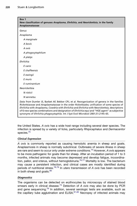
Box 1
New classification of genuses Anaplasma, Ehrlichia, and Neorickettsia, in the family
Anaplasmataceae
Genus
Anaplasma
A marginale
A bovis
A ovis
A phogocytophilum
A platys
Ehrlichia
E canis
E chaffeensis
E ewingii
E muris
E ruminantium
Neorickettsia
N risticii
N sennetsu
Data from Dumler JS, Barbet AF, Bekker CPJ, et al. Reorganization of genera in the familiesRickettsiaceae and Anaplasmataceae in the order Rickettsiales; unification of some species ofEhrlichiawith Anaplasma, Cowdriawith Ehrlichia and EhrlichiawithNeorickettsia, descriptionsof 6 new species combinations and designation of Ehrlichia equi and “HGE agent” as subjectivesynonyms of Ehrlichia phagocytophila. Int J Syst Evol Microbiol 2001;51:2145–65.
Stuen & Longbottom220
the United States. A ovis has a wide host range including several deer species. Theinfection is spread by a variety of ticks, particularly Rhipicephalus and Dermacentorspecies.75
Clinical Expression
A ovis is commonly reported as causing hemolytic anemia in sheep and goats.Anaplasmosis in sheep is normally subclinical. Outbreaks of severe illness in sheepare rare and seem to occur only under extreme conditions.75 However, A ovis appearsto be more pathogenic for goats than for sheep. After an incubation period of 1 to 3months, infected animals may become depressed and develop fatigue, incoordina-tion, pallor, and icterus, without hemoglobinuria.76,77 Mortality is low. The bacteriummay cause a persistent infection, and clinical cases are mostly identified duringperiods of nutritional stress.78,79 In utero transmission of A ovis has been recordedin both sheep and goats.80
Diagnostics
The organisms can be detected on erythrocytes by microscopy of stained bloodsmears early in clinical disease.73 Detection of A ovis may also be done by PCRand gene sequencing.79 In addition, several serologic tests are available, such asthe capillary tube agglutination and ELISA.81,82 Necropsy of infected animals may
Chlamydial and Rickettsial Infections 221
show watery blood, pallor, icteric tissues, and increased fluid in the body cavities. Inaddition, the liver may be enlarged.46
Treatment
Treatment is most efficient during the bacteremic phase of the infection and is directedat reducing the rate of erythrocyte infection, although treatment during the prepatentperiod does not prevent bacteremia. Stress should be avoided during handling andtreatment. Oxytetracycline or tetracycline hydrochloride has been used successfullyto treat clinically affected goats (10 mg/kg BW, once daily for up to 2 days). However,even a 5-day treatment course would not eliminate the carrier state.46
Control
Efforts should be focused on controlling tick infestation through regular dipping,spraying, or pour-on treatment. No specific vaccine for A ovis is currently available.In the case of an outbreak, prophylactic antibiotic administration might be used toprevent spread of the infection.46
TICK-BORNE FEVEREtiological Agent
Tick-borne fever (TBF) is caused by Anaplasma phagocytophilum (formerly Ehrlichiaphagocytophila), an obligate intracellular microbe in the genus Anaplasma (seeBox 1) that primarily infects phagocytes.73 Several genetic variants of the bacteriumhave been found with a variable degree of cross-protective immunity.83,84
Distribution
The bacterium is widespread in the northern hemisphere, especially in Europe.85 Aphagocytophilum has been detected in several animal species. The infection inhumans was first diagnosed in the United States.86
Clinical Expression
The most characteristic symptoms of TBF in domestic ruminants is high fever (up to42�C). Sheep exposed to infected ticks develop clinical signs within 14 days. The fevermay last for 1 to 2 weeks.87 However, the fever reaction may vary according to the ageof the animals, the variant of A phagocytophilum involved, the host species, and theimmunologic status of the host animal.88
Other clinical signs are often absent or mild. TBF is seldom fatal, unless complicatedby other infections. However, TBF causes immunosuppression and makes the sheepvulnerable to secondary infections, such as tick pyemia caused by Staphylococcusspp infections87 or septicemia caused by Mannheimia haemolytica.89,90 Complica-tions also include abortion,91 impaired spermatogenesis in rams,92 reduced weightgain in lambs, and a reduced milk yield in dairy animals.88,93
In humans, clinical manifestations range from a mild self-limiting febrile illness toa life-threatening and fatal infection. On average, patients develop a nonspecific influ-enza-like illness with fever, headache, myalgia, and malaise.94
Diagnosis
The clinical diagnosis is based on a sudden onset of very high fever associated withhematological changes and the presence of typical cytoplasmic inclusions in phago-cytes, especially in neutrophils. Microscopy of blood smears taken during the feverperiod is normally sufficient to confirm the diagnosis. Stained with May-GrunwaldGiemsa, the organisms appear as blue inclusions.87 Inoculation of infected blood

Stuen & Longbottom222
into susceptible animals was previously used to confirm the diagnosis.87 A PCRmethod is now commonly used to identify A phagocytophilum infection in bloodand tissue samples.95 Cultivation of A phagocytophilum in tissue cultures has alsobeen described.96
The presence of specific antibodies may support the diagnosis, the indirect immu-nofluorescent antibody (IFA) test being commonly used. However, it may be difficult touse the IFA test to diagnose an acute infection in lambs, because the IFA titers persistfor months after the primary A phagocytophilum infection.97
At postmortem examination, an enlarged (up to 4–5 times the normal size) spleencan be regarded as indicative of TBF in sheep.89,98 No other typical pathologicchanges have been described.99
Treatment
The safest way to prevent TBF is to avoid areas where ticks are abundant, such astemperate deciduous woodland or mixed forests. However, this is often not feasible.In endemic areas, regular dipping or pour-on treatment with pyrethroids may benecessary against ticks.100
In treatment, the drug of choice is tetracycline.94,101 However, a 5-day long treat-ment with oxytetracycline (10 mg/kg BW, daily) was not found adequate to clearA phagocytophilum from experimentally infected lambs.102 Data suggest that fluoro-quinolone antibiotics and rifampin may be alternative drugs for animals, whereallowed, and in patients with intolerance to tetracycline.103
Control
In the United Kingdom, it has been estimated that more than 300,000 lambs developtick pyemia each year100; up to 30% of TBF-infected lambs may develop cripplinglameness and paralysis following secondary infections. Most of these die or becomeof reduced economic value.104 TBF is also one of the main scourges in the sheepindustry in Norway, and it has been estimated that more than 300,000 lambs areinfected annually.105
The main disease problems associated with TBF in sheep are seen in lambs duringthe first grazing season and in sheep purchased from tick-free areas and placed ontick-infested pastures for the first time.106 Problems caused by TBF may differ signif-icantly between neighboring pastures, as several variants exist with differing degreesof virulence, and protective immunity is not necessarily transferred between thesevariants.107 In one study, 24 msp4 gene variants were found in one sheep flock.84
The infection causes persistence in sheep, and variants may therefore be carriedbetween geographic areas by purchasing infected animals. Ticks on the new pasturesmay become infected from these carriers and later transfer these variants to suscep-tible animals.Current control strategies are based on the reduction of tick infestation by applica-
tion of chemical acaricides at turnout onto a tick-infested pasture, mostly done bydipping or pour-on applications of pyrethroids.88,101 This treatment has to be repeatedseveral times during the tick season. In the United Kingdom, long-acting oxytetracy-cline is also used as a prophylactic measure, given before animals are moved froma tick-free environment onto a tick-infested pasture.104,108 However, there is a growingconcern about the environmental safety and human health because of antimicrobialresistance, increasing cost of chemical control, and the increasing resistance of ticksto pesticides.109
Another strategy to reduce the losses caused by TBF is to infect the lambs as earlyas possible. One study indicates that very young lambs (younger than 2 weeks of age)
Chlamydial and Rickettsial Infections 223
show milder symptoms than older lambs when experimentally infected withA phagocytophilum.110 However, this practice is only feasible if the lambs are infectedimmediately after birth, because 3- to 6-week-old lambs are very susceptible to theinfection.88 Another obstacle to this strategy is that the infection prevalence andA phagocytophilum variants in ticks may vary during the grazing season and fromyear to year.84
Pasture management and habitat modification may reduce the density of ticks andtherefore the occurrence of TBF. These changes may destroy the ticks’ microhabitator at least make the ticks vulnerable to macroclimatic conditions. Themethods includedrainage of marshy lands, controlled burning or herbicidal treatment of vegetation,mechanical clearing of bushes, removal of leaf litter and, in some cases, partialremoval of the forest canopy, all of which require substantial modification of the envi-ronment with possible chemical contamination.111 Alteration of the habitat may alsochange the ticks’ host availability. However, the tick abundance can only be reducedby these procedures for a short period, and several of these procedures have to berepeated periodically and are labor intensive. In addition, sufficient habitat modifica-tion is not always feasible and farm animals are always at risk from ticks brought infrom the surrounding areas, especially if other large animal species use the samepastures. The bacterium is widespread in the environment and may infect severalhosts.85 However, a recent investigation indicates that there may be natural enzooticcycles among different strains of A phagocytophilum.112
Biologic tick control is becoming an attractive approach to tick management. Bio-logic control of tick infestations has been difficult, because ticks have few naturalenemies. Most predators of ticks are generalists with a limited potential for tickmanagement. To date, studies have concentrated on examining the potential ofbacteria, entomopathogenic fungi, and nematodes to offer control.109 However, themain challenge is to create a sustainable biologic control of ticks in the natural habitat.A vaccine against A phagocytophilum is not yet available. Immunization of suscep-
tible sheepwith infected blood from carrier animals should no longer be recommendedor performed, due to a lack of infection control and possible spread of other infectiousagents. The challenge to producing an effective vaccine is to choose antigens that areconserved among all variants ofAphagocytophilum. Identification of epitopes involvedin protective immunity combined with the discovery of cytokines involved in patho-genesis is encouraging for future vaccine development.113 The whole genome ofa human variant of A phagocytophilum has recently been sequenced. However, otherstrains of the bacterium have to be sequenced to conduct comparative genomics anddevelop proper recombinant vaccine antigens for future cross-infection studies.114
Vaccines against ticks may be an option. So far, however, only a vaccine against theone-host cattle tick Rhipicephalus (Boophilus) microplus has been developed.115
Control of ticks by vaccination has the advantage of cost-effectiveness, reduced envi-ronmental contamination, and prevention of selecting for drug-resistant ticks that mayresult from repeated acaricide applications. The identification of tick-protective anti-gens remains the limiting factor in the development of an effective tick vaccine. Char-acterization of the ticks’ genomes will therefore have a great impact on the discoveryof new protective antigens. Development of vaccines against multiple tick speciesmay be possible using highly conserved tick-protective antigens or by antigensshowing immune cross-reaction in different tick species.116 An interesting conceptis the development of transmission-blocking vaccines by using tick immunomodula-tory molecules.117
The development of vaccines that target both ticks and pathogen transmission mayprovide a means of controlling tick-borne infections through immunization of the
Stuen & Longbottom224
human and animal population at risk or by immunization of the mammalian reservoir toreduce pathogen transmission.116
In the future, integrated tick control strategies should be implemented. These strat-egies must be based on host resistance to ticks and to the infections they transmit,strategic tick control on the actual pastures, cost/benefit analyses of the acaricidalapplications, and the availability of vaccines against ticks and tick-borne diseases.118
HEARTWATEREtiological Agent
Heartwater is caused by the rickettsia Ehrlichia ruminantium (formerly Cowdria rumi-nantium) in the genus Ehrlichia (see Box 1), which is transmitted by ticks of the genusAmblyomma.73 Various strains of E ruminantium exist, but only a variable degree ofcross-protective immunity occurs between the different strains.119–121 The bacteriamultiply in neutrophils, monocytes, and vascular endothelial cells.122
Distribution
Heartwater is an endemic disease in domestic and some wild ruminants throughoutsub-Saharan Africa, including islands in the near-eastern Africa but also in the Carib-bean following the introduction of Amblyomma variegatum ticks, probably in the 18thcentury.123 It has been estimated that more than 150 million animals are at risk in thesub-Saharan area.124
Clinical Expression
The incubation period after natural tick-transmitted infection is usually from about 2 to4 weeks. There are 4 clinical forms of heartwater: peracute, acute, subacute, andsubclinical; the most common is the acute form.46 The clinical symptoms dependon host susceptibility, virulence of the infective strains, and previous exposure. Thedisease is characterized by sudden onset of high fever (up to 42�C), nervous signs,and rapid and abdominal breathing. Auscultation may detect evidence of pulmonaryedema and hydropericardium. Recumbency, convulsions, and death may followwithin 24 hours. E ruminantium may cause high mortality in sheep, goats, and cattle,largely influenced by breed, age, animal species, and bacterial strains.125 Small rumi-nants are very susceptible hosts and greater than 90% and 50% mortality have beenobserved in nonindigenous goats and sheep, respectively.122 Subclinical and mildcases are common in young animals and in local indigenous breeds.An age-related resistance to disease occurs in young domestic ruminants, indepen-
dent of maternal transferred immunity. The period of resistance in lambs graduallywanes after approximately the first 3 weeks of life. The respective protective periodin kids is not well studied, but may be shorter.46
Diagnostics
Presumptive diagnosis is based on clinical history and the presence of Amblyommaticks in the region. At postmortem examination, massive transudates into the bodycavities, especially hydropericardium, hydrothorax, and edema of the lungs and brain,are recorded. In addition, splenomegaly and enlargement of the lymph nodes areseen. Histologically, heartwater is characterized by the presence of clusters of theorganisms in the vascular endothelium of virtually all tissues examined, especiallythe brain cortex. For rapid field diagnosis, E ruminantium can be detected in thevascular endothelial cells by squash preparations of the cerebrum.46 Confirmationof diagnosis was previously made by the intravenous inoculation of 5 to 10 mL ofwhole blood from suspected cases into susceptible sheep or goats, but is now
Chlamydial and Rickettsial Infections 225
done using molecular methods such as PCR.126 Diagnosis of heartwater based ondirect detection of the agent is recommended. Positive PCR should be accompaniedby isolation of the organisms in endothelial cell cultures, and corroborated by the pres-ence of infected Amblyomma ticks.125
Several serologic tests are available for detecting antibodies against E ruminantium,such as indirect fluorescent antibody test, Western blot assay, ELISA, and competitiveELISA. Because a large proportion of infected animals die following a primary infec-tion, antibody detection is not often an option. The duration of the antibody responseis also questionable. In addition, serology is constrained by cross-reactions with otherehrlichial agents. Serologic methods that have high enough sensitivity and specificityto diagnose heartwater during early infection or after recovery are still lacking.125
Treatment
Successful therapy depends on early antibiotic treatment. Animals treated during thefebrile stage of acute heartwater respond favorably to oxytetracycline at a dose rate of5 to 10 mg/kg BW, given intravenously or intramuscularly at the first sign of fever andrepeated once more 1 to 2 days later. The use of one dose of long-acting oxytetracy-cline at a dose rate of 20mg/kg BW is also effective. Therapy initiated after the onset ofneurologic signs is almost always inefficient.127 Therefore, treatment should not bedelayed until laboratory confirmation is established. Recovered animals can becarriers of the infection.122
Control
Although heartwater has been known for more than a century, it is still considered asa major obstacle against expansion and development of the livestock industry inAfrica.128 Imported breeds in particular are very susceptible, and severe infectionwith high mortality rates may occur.122 There is a considerable concern about thepossible spread of heartwater and its vector to tropical and subtropical regions ofNorth, South, and Central America, where other suitable tick vectors exist.46 E rumi-nantium can be transmitted by at least 13 species of Amblyomma ticks, with A varie-gatum (in West, East, and Central Africa) and A hebraeum (in South Africa) being the 2major vectors.129
The hosts for E ruminantium appear to be restricted to members of the family Bovi-dae, including both domestic and wild ruminants. The animals most at risk are thoseintroduced to endemic areas from heartwater-free areas. Prolonged survival of E rumi-nantium in ticks maintains the disease, as does the carrier state in ruminants.130
The current methods for heartwater control include the use of acaricides to controlthe tick vector, antibiotic prophylaxis, immunization by infection and treatment,farming with animal breeds resistant to the disease, and establishment of endemicstability.131–134 All of these methods have serious drawbacks. For instance, acaricidesare expensive, environmentally unfriendly, and may induce resistance in ticks. In addi-tion, treatment has to be repeated several times during the tick season. Another draw-back of using acaricides is that it hinders creation of endemic stability, as treatedanimals remain fully susceptible to infection.125 Other strategies against tick infesta-tion may be applied and have been discussed in the section on control of TBF.Attempts to control heartwater by controlling tick infestation have only partly beensuccessful. Eradication of the disease by vector control therefore seems unlikely.124
The best long-term cost-effective control method against heartwater isvaccination.124 At present, however, there is no safe, user-friendly, and reliablevaccine commercially available. Different vaccines with various efficacies have beenproduced against E ruminantium, including live organisms followed by antibiotic

Stuen & Longbottom226
(“infection and treatment method”), live-attenuated vaccines, whole-organism inacti-vated vaccines, and recombinant protein and DNA vaccines.135–140 Although many ofthe vaccines have conferred at least partial protection, a continuing problem is break-down infection on challenge with organisms heterologous to those included in thevaccines.114
There are some barriers to developing an effective vaccine. Attenuation of virulentorganisms is imprecise and there is little cross-protection between strains. Moreover,duration of immunity is variable; for example, in some goat breeds it may be as shortas 2 months.46 Vaccination should be timed to precede periods of peak tick feedingactivity in wet seasons.132
At the moment the only commercially available immunization method is the “infec-tion and treatment method,” using an attenuated strain of E ruminantium. At the firstsigns of fever, vaccinated animals are treated with oxytetracycline to diminish thesigns of disease. Successful vaccination depends on the proper timing of treatment.The disadvantages of such a procedure include: the need for chemotherapeutics afterimmunization; potential difficulties of distributing a deep-frozen vaccine, as thevaccine rapidly loses its infectivity and immunogenicity when thawed; and the require-ment for the vaccine to be administered by intravenous injection. However, a recentstudy indicates that an attenuated vaccine based on the Welgevonden strain, whenadministrated intramuscularly, seemed to provide good cross-protection in Merinosheep and Angora goats.141 In addition, attenuated (“live”) vaccines induce persistentinfection and hence provide a source of infection for ticks. These vaccines should notbe used in regions where the disease is absent and where potential vectors exist, suchas the United States and Central and South America. An inactivated vaccine may beused in these regions. However, a recombinant vaccine or a marker vaccine should bepreferred to differentiate vaccinated and truly infected animals.125
Control of heartwater through discovery and development of improved vaccinesremains the target for future research.124 The success of inactivated vaccines in pro-tecting against field challenge supports the concept that protective antigens can beidentified. The genome sequences of 2 strains of E ruminantium provide the opportu-nity for comparative genomics and the prediction and identification of conservedvaccine antigen candidates. To develop an improved heartwater recombinantvaccine, it is necessary to understand the heterologous immunity and cross-protectivemechanisms.114 Once protective antigens have been identified the main challenge willbe to design a recombinant vaccine, which induces cross-protective immuneresponses between strains.
SUMMARY
Chlamydial and rickettsial infections are a significant challenge to the small ruminantindustry in several geographic areas. This review focuses on the treatment and controlmethods that are available. Different strategies should be implemented to controlthese infections. It is hoped that development of new recombinant vaccines usingcomparative genomics and proteomics will continue to progress.
REFERENCES
1. Longbottom D, Coulter LJ. Animal chlamydioses and zoonotic implications.J Comp Pathol 2003;128:217–44.
2. Aitken ID, Longbottom D. Chlamydial abortion. In: Aitken ID, editor. Diseases ofsheep. 4th edition. Oxford (UK): Blackwell; 2007. p. 105–12.
Chlamydial and Rickettsial Infections 227
3. Rodolakis A, Boullet C, Souriau A. Chlamydia psittaci experimental abortion ingoats. Am J Vet Res 1984;45:2086–9.
4. Wittenbrink MM, Schoon HA, Bisping W, et al. Infection of the bovine femalegenital tract with Chlamydia psittaci as a possible cause of infertility. ReprodDomest Anim 1993;28:129–36.
5. Hyde SR, Benirschke K. Gestational psittacosis: case report and literaturereview. Mod Pathol 1997;10:602–7.
6. Buxton D. Potential danger to pregnant women of Chlamydia psittaci fromsheep. Vet Rec 1986;118:510–1.
7. Bloodworth DL, Howard AJ, Davies A, et al. Infection in pregnancy caused byChlamydia psittaci of ovine origin. Commun Dis Rep 1987;10:3–4.
8. Winter AC, Charnley JG. The sheep keeper’s veterinary handbook. Ramsbury(UK): Crowood Press; 1999. p. 208.
9. Sachse K, Vretou E, Livingstone M, et al. Recent developments in the laboratorydiagnosis of chlamydial infections. Vet Microbiol 2009;135:2–21.
10. Longbottom D. Enzootic abortion of ewes (ovine chlamydiosis). In: Manual ofdiagnostic tests and vaccines for terrestrial animals. 6th edition. Paris: WorldOrganisation for Animal Health (OIE); 2008. p. 1013–20.
11. Stamp JT, Watt JA, Cockburn RB. Enzootic abortion in ewes; complement fixa-tion test. J Comp Pathol 1952;62:93–101.
12. McCauley LME, Lancaster MJ, Young P, et al. Comparison of ELISA and CFTassays for Chlamydophila abortus antibodies in ovine sera. Aust Vet J 2007;85:325–8.
13. Wilson K, Livingstone M, Longbottom D. Comparative evaluation of eight sero-logical assays for diagnosing Chlamydophila abortus infection in sheep. VetMicrobiol 2009;135:38–45.
14. Laroucau K, Vorimore F, Sachse K, et al. Differential identification of Chlamydo-phila abortus live vaccine strain 1B by PCR-RFLP. Vaccine 2010;28:5653–8.
15. Wheelhouse N, Aitchison KD, Laroucau K, et al. Evidence of Chlamydophilaabortus vaccine strain 1B as a possible cause of ovine enzootic abortion.Vaccine 2010;28:5857–63.
16. Sillis M, Longbottom D. Chlamydiosis. In: Palmer S, Soulsby L, Torgerson P, et al,editors. Zoonoses. 2nd edition. Oxford (UK): Oxford University Press, in press.
17. Papp JR, Shewen PE, Gartley CJ. Abortion and subsequent excretion of chla-mydiae from the reproductive tract of sheep during estrus. Infect Immun1994;62:3786–92.
18. Papp JR, Shewen PE. Pregnancy failure following vaginal infection of sheep withChlamydia psittaci prior to breeding. Infect Immun 1996;64:1116–25.
19. Livingstone M, Wheelhouse N, Maley SW, et al. Molecular detection of Chlamy-dophila abortus in post-abortion sheep at oestrus and subsequent lambing. VetMicrobiol 2009;135:134–41.
20. Entrican G, Buxton D, Longbottom D. Chlamydial abortion: a brief overview. J RSoc Med 2001;94:273–7.
21. Rodolakis A. In vitro and in vivo properties of chemically induced temperature-sensitive mutants of Chlamydia psittaci var. ovis: screening in a murine model.Infect Immun 1983;42:525–30.
22. Foggie A. Preparation of vaccines against enzootic abortion of ewes. A review ofthe research work at the Moredun Institute. Vet Bull 1973;43:587–90.
23. Waldhalm DG, DeLong WJ, Hall RF. Efficacy of a bacterin prepared from Chla-mydia psittaci grown in cell culture for experimental immunization of ewes. VetMicrobiol 1982;7:493–8.
Stuen & Longbottom228
24. Anderson IE, Tan TW, Jones GE, et al. Efficacy against ovine enzootic abortionof an experimental vaccine containing purified elementary bodies of Chlamydiapsittaci. Vet Microbiol 1990;24:21–7.
25. Jones GE, Jones KA, Machell J, et al. Efficacy trials with tissue-culture grown,inactivated vaccines against chlamydial abortion in sheep. Vaccine 1995;13:715–23.
26. Longbottom D, Livingstone M. Vaccination against chlamydial infections of manand animals. Vet J 2006;171:263–75.
27. Seshadri R, Paulsen IT, Eisen JA, et al. Complete genome sequence of theQ-fever pathogen Coxiella burnetii. Proc Natl Acad Sci U S A 2003;100:5455–60.
28. Quevedo Diaz MA, Lukacova M. Immunological consequences of Coxiellaburnetii phase variant. Acta Virol 1998;42:181–5.
29. McCaul TF, Williams JC. Developmental cycle of Coxiella burnetii: structure andmorphogenesis of vegetative and sporogenic differentiations. J Bacteriol 1981;147:1063–76.
30. Mearns R. Other infectious causes of abortion. In: Aitken ID, editor. Diseases ofsheep. 4th edition. Oxford (UK): Blackwell; 2007. p. 126–36.
31. Maurin M, Raoult D. Q fever. Clin Microbiol Rev 1999;12:518–53.32. Hilbink F, Penrose M, Kovacova E, et al. Q fever is absent from New Zealand. Int
J Epidemiol 1993;22:945–9.33. Arricau-Bouvery N, Rodolakis A. Is Q fever an emerging or re-emerging
zoonosis? Vet Res 2005;36:327–49.34. Palmer NC, Kierstead M, Key DW, et al. Placentitis and abortion in goats and
sheep in Ontario caused by Coxiella burnetii. Can Vet J 1983;24:60–1.35. Zeman DH, Kirkbride CA, Leslie-Steen P, et al. Ovine abortion due to Coxiella
burnetii infection. J Vet Diagn Invest 1989;1:178–80.36. Berri M, Souriau S, Crosby M, et al. Relationships between the shedding of
Coxiella burnetii, clinical signs and serological responses of 34 sheep. VetRec 2001;148:502–5.
37. Sanford SE, Josephson GKA, MacDonald A. Coxiella burnetii (Q fever) abortionstorms in goat herds after attendance at an annual fair. Can Vet J 1994;35:376–8.
38. Crowther RW, Spicer AJ. Abortion in sheep and goats in Cyprus caused byCoxiella burnetii (sic). Vet Rec 1976;99:29–30.
39. Moore JD, Barr BC, Daft BM, et al. Pathology and diagnosis of Coxiella burnetiiinfection in a goat herd. Vet Pathol 1991;28:81–4.
40. Fournier PE, Marrie TJ, Raoult D. Diagnosis of Q fever. J Clin Microbiol 1998;36:1823–34.
41. Barlow J, Rauch B, Welcome F, et al. Association between Coxiella burnetiishedding in milk and subclinical mastitis in dairy cattle. Vet Res 2008;39:23.
42. Berri M, Laroucau K, Rodolakis A. The detection of Coxiella burnetii from ovinegenital swabs, milk and fecal samples by use of a single touchdown chain reac-tion. Vet Microbiol 2000;72:285–93.
43. Lang GH. Coxiellosis (Q fever) in animals. In: Marrie TJ, editor, Q fever: thedisease, vol. 1. Boca Raton (FL): CRC Press; 1990. p. 24–42.
44. Hatchette T, Hudson R, Schezch WF, et al. Goat-associated Q fever: a newdisease in Newfoundland. Emerg Infect Dis 2001;7:413–9.
45. Kovacova E, Kazar J, Spanelova D. Suitability of various Coxiella burnetiiantigen preparations for detection of serum antibodies by various tests. ActaVirol 1998;42:365–8.
Chlamydial and Rickettsial Infections 229
46. Smith MC, Sherman DM. Goat medicine. 2nd edition. Ames (IA): Wiley-Blackwell; 2009. p. 871.
47. Arricau Bouvery N, Souriau A, Lechopier P, et al. Experimental Coxiella burnetiiinfection in pregnant goats: excretion routes. Vet Res 2003;34:423–33.
48. Berri M, Crochet D, Santiago S, et al. Spread of Coxiella burnetii infection ina flock of sheep after an episode of Q fever. Vet Rec 2005;157:737–40.
49. Kazar J. Q fever - current concept. In: Raoult D, Brouqui P, editors. Rickettsiaeand rickettsial diseases at the turn of the third millennium. Paris: Elsevier; 1999.p. 304–19.
50. Van den Brom R, Vellema P. Q fever outbreaks in small ruminants and people inthe Netherlands. Small Rumin Res 2009;86:74–89.
51. Enserink M. Questions abound to Q-fever explosion in the Netherlands. Science2010;327:266–7.
52. Behymer D, Ruppaner R, Riemann HP. Observations on chemotherapy in cowschronically infected with Coxiella burnetii (Q fever). Folia Vet Lat 1977;7:64–70.
53. Rodolakis A. Q fever, state of art: epidemiology, diagnosis and prophylaxis. In:Proceedings of the 6th International Sheep Veterinary Congress. Hersonissos(Greece); 2005. p. 96–8.
54. Babudieri B. Q fever: a zoonosis. Adv Vet Sci 1959;5:181–2.55. Sawyer LA, Fishbein DB, McDade JE. Q fever: current concepts. Rev Infect Dis
1987;9:935–46.56. Arricau-Bouvry N, Souriau A, Bodir C, et al. Effect of vaccination with phase I and
phase IICoxiella burnetii vaccines in pregnant goats. Vaccine 2005;23:4392–402.57. Rodolakis A, Berri M, Hechard C, et al. Comparison of Coxiella burnetii shed-
ding in milk of dairy bovine, caprine and ovine herds. J. Dairy Sci 2007;90:5352–60.
58. Rodolakis A. Q fever in dairy animals. Ann N Y Acad Sci 2009;1166:90–3.59. Berri M, Souriau A, Crosby M, et al. Shedding of Coxiella burnetii in ewes in two
pregnancies following an episode of Coxiella abortion in a sheep flock. VetMicrobiol 2002;85:55–60.
60. Hatchette T, Campbell N, Hudson R, et al. Natural history of Q fever in goats.Vector Borne Zoonotic Dis 2003;3:11–5.
61. Welsh HH, Lennette EH, Abinanti FR, et al. Airborne transmission of Q fever: therole of parturition in generation of infective aerosol. Ann N Y Acad Sci 1957;70:528–40.
62. Tissot-Dupont H, Torres S, Nezri M, et al. Hyperendemic focus of Q fever relatedto sheep and wind. Am J Epidemiol 1999;150:67–74.
63. Tigertt WD, Benenson AS, Gochenour WS. Airborne Q fever. Bacteriol Rev 1961;25:285–93.
64. Scott GH, Williams JC. Susceptibility of Coxiella burnetii to chemical disinfec-tants. Rickettsiology: current issues and perspectives. Ann N Y Acad Sci1990;590:291–6.
65. Berri M, Rousset E, Champion JL, et al. Ovine manure used as a garden fertiliseris suspected to be a contamination source of two human Q fever cases. Vet Rec2003;153:269–70.
66. Tissot-Dupont H, Amadei M-A, Nezri M, et al. Wind in November, Q fever inDecember. Emerg Infect Dis 2004;10:1264–9.
67. Sadecky E, Brezina R. Vaccination of naturally infected ewes against Q-fever.Acta Virol 1977;21:89.
68. Brooks DL, Ermel RW, Franti CE, et al. Q fever vaccination of sheep: challengeand immunity in ewes. Am J Vet Res 1986;47:1235–8.

Stuen & Longbottom230
69. Sampere M, Font B, Font J, et al. Q fever in adults: review of 66 clinical cases.Eur J Clin Microbiol Infect Dis 2003;22:108–10.
70. Souriau A, Arricau-Bouvery N, Bodier C, et al. Comparison of the efficacy of Qfever vaccines against Coxiella burnetii experimental challenge in pregnantgoats. Ann N Y Acad Sci 2003;990:521–3.
71. Schmeer N, Muller P, Langel J, et al. Q fever vaccines for animals. ZentralblBakteriol Mikrobiol Hyg A 1987;267:79–88.
72. Dupuis G, Petite J, Petr O, et al. An important outbreak of human Q fever ina Swiss Alpine valley. Int J Epidemiol 1987;16:282–7.
73. Dumler JS, Barbet AF, Bekker CPJ, et al. Reorganization of genera in the familiesRickettsiaceae and Anaplasmataceae in the order Rickettsiales; unification ofsome species of Ehrlichia with Anaplasma, Cowdria with Ehrlichia and Ehrlichiawith Neorickettsia, descriptions of six new species combinations and designa-tion of Ehrlichia equi and “HGE agent” as subjective synonyms of Ehrlichiaphagocytophila. Int J Syst Evol Microbiol 2001;51:2145–65.
74. Uilenberg G, Van Vorstenbosch CJAH, Perie CJ. Blood parasites in sheep in theNetherlands. I. Anaplasma mesaeterum sp.n. (Rickettsiales, Anaplasmata-ceae). Vet Q 1979;1:14–22.
75. Friedhoff KT. Tick-borne diseases in sheep and goats caused by Babesia,Theileria or Anaplasma spp. Parasitologia 1997;39:99–109.
76. Splitter EJ, Anthony HD, Twiehaus MJ. Anaplasma ovis in the United States. AmJ Vet Res 1956;17:487–91.
77. Zwart D, Buys J. Studies on Anaplasma ovis infection. II. Pathogenicity of aNigerian goat strain for Dutch sheep and goats. Bull Epizoot Dis Afr 1968;16:73–80.
78. Ilemobade AA. Blood parasites of African goats. In: Proceedings of the 3rdInternational Conference on Goat Production and Disease. Tucson (Arizona);1982. p. 68–71.
79. Palmer GH, Abbott JR, French DM, et al. Persistence of Anaplasma ovis infec-tion and conservation of the msp-2 and msp-3 multigene families within thegenus Anaplasma. Infect Immun 1998;66:6035–9.
80. Barry DM, Van-Niekerk CH. Anaplasmosis in improved Boer goats in SouthAfrica artificially infected with Anaplasma ovis. Small Rumin Res 1990;3:191–7.
81. Mallick KP, Dwivedi SK, Malhotra MN. Anaplasmosis in goats: report of clinicalcases. Indian Vet J 1979;56:693–4.
82. Ndungu LW, Aguirre C, Rurangirwa FR, et al. Detection of Anaplasma ovis infec-tion in goats by major surface protein 5 competitive inhibition enzyme-linkedimmunosorbent assay. J Clin Microbiol 1995;33:675–9.
83. Stuen S, Moum T, Petrovec M, et al. Genetic variants of Anaplasma phagocyto-philum in Norway. Int J Med Microbiol 2006;296:164–6.
84. Ladbury GAF, Stuen S, Thomas R, et al. Dynamic transmission of numerousAnaplasma phagocytophilum genotypes among lambs in an infected sheepflock in an area of anaplasmosis endemicity. J Clin Microbiol 2008;46:1686–91.
85. Stuen S. Anaplasma phagocytophilum—the most widespread tick-borne infec-tion in animals in Europe. Vet Res Commun 2007;31(Suppl 1):79–84.
86. Bakken JS, Dumler JS, Chen S-M, et al. Human granulocytic ehrlichiosis in theupper Midwest United States. A new species emerging? J Am Med Assoc 1994;272:212–8.
87. Foggie A. Studies on the infectious agent of tick-borne fever in sheep. J PatholBacteriol 1951;63:1–15.
Chlamydial and Rickettsial Infections 231
88. Stuen S. In: Anaplasma phagocytophilum (formerly Ehrlichia phagocytophila)infection in sheep and wild ruminants in Norway. A study on clinical manifesta-tion, distribution and persistence [Dr Phil thesis]. University of Oslo; 2003.
89. Øveras J, Lund A, Ulvund MJ, et al. Tick-borne fever as a possible predisposingfactor in septicaemic pasteurellosis in lambs. Vet Rec 1993;133:398.
90. Stuen S. Tick-borne fever (TBF) and secondary infections in sheep. In: Kazar J,Toman R, editors. Rickettsiae and rickettsial diseases. Bratislava (Slovakia):Veda; 1996. p. 347–9.
91. Jamieson S. Tick-borne fever as a cause of abortion in sheep—part II. Vet Rec1950;62:468–70.
92. Watson WA. Infertility in the ram associated with tick-borne fever infection. VetRec 1964;76:1131–6.
93. Tuomi J. Studies in epidemiology of bovine of tick-borne fever in Finland anda clinical description of field cases. Ann Med Exp Biol Fenn 1966;44(Suppl 6):1–62.
94. Bakken JS, Dumler JS. Clinical diagnosis and treatment of human granulocyticanaplasmosis. Ann N Y Acad Sci 2006;1078:236–47.
95. Chen S-M, Dumler JS, Bakken JS, et al. Identification of a granulocytic Ehrlichiaspecies as the etiologic agent of human disease. J Clin Microbiol 1994;32:589–95.
96. Goodman JL, Nelson C, Vitale B, et al. Direct cultivation of the causative agentof human granulocytic ehrlichiosis. N Engl J Med 1996;334:209–15.
97. Paxton EA, Scott GR. Detection of antibodies to the agent of tick-borne fever byindirect immunofluorescence. Vet Microbiol 1989;21:133–8.
98. Gordon WS, Brownlee A, Wilson DR, et al. “Tick-borne fever” (A hitherto unde-scribed disease of sheep.). J Comp Pathol 1932;45:301–7.
99. Campbell RSF, Rowland AC, Scott GR. Sequential pathology of tick-borne fever.J Comp Pathol 1994;111:303–13.
100. Brodie TA, Holmes PH, Urquhart GM. Some aspects of tick-borne diseases ofBritish sheep. Vet Rec 1986;118:415–8.
101. Woldehiwet Z, Scott GR. Tick-borne (pasture) fever. In: Woldehiwet Z, Ristic M,editors. Rickettsial and chlamydial diseases of domestic animals. Oxford (UK):Pergamon Press; 1993. p. 233–54.
102. Stuen S, Bergstrom K. The effect of two different oxytetracycline treatments inexperimental Ehrlichia phagocytophila infected lambs. Acta Vet Scand 2001;42:339–46.
103. Horowitz HW, Hsieh T-C, Aguero-Rosenfeld ME, et al. Antimicrobial suscepti-bility of Ehrlichia phagocytophila. Antimicrob Agents Chemother 2001;45:786–8.
104. Woldehiwet Z. Tick-borne diseases. In: Aitken ID, editor. Diseases of sheep. 4thedition. Oxford (UK): Blackwell; 2007. p. 347–55.
105. Stuen S, Bergstrom K. Serological investigation of granulocytic Ehrlichia infec-tion in sheep in Norway. Acta Vet Scand 2001;42:331–8.
106. Scott GR. Tick-associated infections. In: Martin WB, Aitken ID, editors. Diseasesof sheep. 1st edition. Oxford (UK): Blackwell; 1983. p. 209–13.
107. Stuen S, Bergstrom K, Petrovec M, et al. Differences in clinical manifestationsand hematological and serological responses after experimental infection withgenetic variants of Anaplasma phagocytophilum in sheep. Clin Diagn LabImmunol 2003;10:692–5.
108. Brodie TA, Holmes PH, Urquhart GM. Prophylactic use of long-acting tetracy-cline against tick-borne fever (Cytoecetes phagocytophila) in sheep. Vet Rec1988;122:43–4.

Stuen & Longbottom232
109. Samish M, Ginsberg H, Glazer I. Biological control of ticks. Parasitology 2004;129:S389–403.
110. Stuen S, Hardeng F, Larsen HJ. Resistance to tick-borne fever in young lambs.Res Vet Sci 1992;52:211–6.
111. Sonenshine DE. Biology of ticks, vol. 2. New York: Oxford University Press;1993. p. 465.
112. Bown K, Lambin X, Ogden NH, et al. Delineating Anaplasma phagocytophilumecotypes in coexisting, discrete enzootic cycles. Emerg Infect Dis 2009;15:1948–54.
113. Zhi N, Ohashi N, Rikihisa Y. Multiple p44 genes encoding major outer membraneproteins are expressed in the human granulocytic ehrlichiosis agent. J BiolChem 1999;274:17828–36.
114. Barbet AF, Byrom B, Mahan SM. Diversity of Ehrlichia ruminantium major anti-genic protein 1-2 in field isolates and infected sheep. Infect Immun 2009;77:2304–10.
115. Willardsen P. Anti-tick vaccines. Parasitology 2004;129:S367–87.116. de la Fuente J, Kocan KM. Strategies for development of vaccines for control of
ixodid ticks species. Parasite Immunol 2006;28:275–83.117. Titus RG, Bishop JV, Mejla JS. The immunomodulatory factors of arthropod
saliva and the potential for these factors to serve as vaccine targets to preventpathogen transmission. Parasite Immunol 2006;28:131–41.
118. Jongejan F, Uilenberg G. The global importance of ticks. Parasitology 2004;129:S3–14.
119. Uilenberg G. Heartwater (Cowdria ruminantium infection): current status. AdvVet Sci Comp Med 1983;27:427–80.
120. Jongejan F, Uilenberg G, Franssen FF, et al. Antigenic differences betweenstocks of Cowdria ruminantium. Res Vet Sci 1988;44:186–9.
121. Allsopp MT, Van Strijp MF, Faber E, et al. Ehrlichia ruminantium variants whichdo not cause heartwater in South Africa. Vet Microbiol 2007;120:158–66.
122. Uilenberg G, Camus E. Heartwater (cowdriosis). In: Woldehiwet Z, Ristic M,editors. Rickettsial and chlamydial diseases of domestic animals. Oxford(UK): Pergamon Press; 1993. p. 293–332.
123. Maillard JC, Maillard N. Historique du peuplement bovin et de l’introduction dela tique Amblyomma variegatum dans les iles francaises des Antilles. Synthesebibliographique Ethnozootechnie 1998;1:19–26.
124. Allsopp BA. Trend in control of heartwater. Onderstepoort J Vet Res 2009;76:81–8.
125. Mahan SM. Diagnosis and control of heartwater, Ehrlichia ruminantium infection:an update. CAB Reviews: Perspectives in Agriculture, Veterinary Science, Nutri-tion and Natural Resources 2006;1(055):12.
126. Martinez D, Maillard JC, Coisne S, et al. Protection of goats against heartwateracquired by immunisation with inactivated elementary bodies of Cowdria rumi-nantium. Vet Immunol Immunopathol 1994;41:153–63.
127. Prozesky L, Du Plessis JL. The pathology of heartwater. II. A study of the lunglesions in sheep and goats infected with the Ball, strain of Cowdria ruminantium.Onderstepoort J Vet Res 1985;52:81–5.
128. Provost A, Bezuidenhout JD. The historical background and global importanceof heartwater. Onderstepoort J Vet Res 1987;54:165–9.
129. Walker JB, Olwage A. The tick vectors of Cowdria ruminantium (Ixodoidea, Ixo-didae, genus Amblyomma) and their distribution. Onderstepoort J Vet Res1987;54:353–79.
Chlamydial and Rickettsial Infections 233
130. Andrew HR, Norval RAI. The carrier status of sheep, cattle and African buffalorecovered from heartwater. Vet Parasitol 1989;34:295–308.
131. Peregrine AS. Chemotherapy and delivery systems: haemoparasites. Vet Para-sitol 1994;54:223–48.
132. Van der Merwe L. The infection and treatment method of vaccination againstheartwater. Onderstepoort J Vet Res 1987;54:489–91.
133. Camus E, Maillard JC, Ruff G, et al. Genetic resistance of Creole goats to cow-driosis in Guadeloupe. Status in 1995. Ann N Y Acad Sci 1996;791:46–53.
134. Tice GA, Bryson NR, Stewart CG, et al. The absence of clinical disease in cattlein communal grazing areas where farmers are changing from an intensivedipping programme to one of endemic stability to tick-borne diseases. Onder-stepoort J Vet Res 1998;65:169–75.
135. Nyika A, Mahan SM, Burridge MJ, et al. A DNA vaccine protects mice againstrickettsial agent Cowdria ruminantium. Parasite Immunol 1998;20:111–9.
136. Mahan SM, Allsopp B, Kocan KM, et al. Vaccine strategies for Cowdria ruminan-tium infections and their application to other ehrlichial infections. Parasitol Today1999;15:290–4.
137. Collins NE, Pretorius A, Van Kleef M, et al. Development of improved attenuatedand nucleic acid vaccines for heartwater. Dev Biol (Basel) 2003;1154:121–36.
138. Zweygarth E, Josemans AI, Van Strijp MF, et al. An attenuated Ehrlichia ruminan-tium (Welgevonden stock) vaccine protects small ruminants against virulentheartwater challenge. Vaccine 2005;23:1695–702.
139. Faburay B, Geysen D, Ceesay A, et al. Immunisation of sheep against heart-water in the Gambia using inactivated and attenuated Ehrlichia ruminantiumvaccines. Vaccine 2007;25:7939–47.
140. Shkap V, de Vos AJ, Zweygarth E, et al. Attenuated vaccines for tropical theiler-iosis, babesiosis and heartwater: the continuing necessary. Trends Parasitol2007;23:420–6.
141. Zweygarth E, Josemans AI, Steyn HC. Experimental use of the attenuatedEhrlichia ruminantium (Welgevonden) vaccine in Merino sheep and Angoragoats. Vaccine 2008;265:G34–9.