Chondrodysplasia of Texel Sheep
232
Copyright is owned by the Author of the thesis. Permission is given for a copy to be downloaded by an individual for the purpose of research and private study only. The thesis may not be reproduced elsewhere without the permission of the Author.
-
Upload
khangminh22 -
Category
Documents
-
view
4 -
download
0
Transcript of Chondrodysplasia of Texel Sheep

Copyright is owned by the Author of the thesis. Permission is given for a copy to be downloaded by an individual for the purpose of research and private study only. The thesis may not be reproduced elsewhere without the permission of the Author.
Chondrodysplasia of Texel Sheep
A thesis presented in partial fulfillment
of the requirements for the degree of
Doctor of Philosophy
at
Massey University,
Palmerston North,
New Zealand
Susan Amanda Piripi
2008
Abstract
Chondrodysplasia of Texel sheep is a newly described recessively inherited disorder
distinct from other chondrodysplasias described in sheep. Phenotypically normal
at birth, affected lambs develop microscopic lesions as early as 9 days of age, and
usually demonstrate gross deformities and markedly reduced rates of bone growth
by 2 to 3 weeks. Individual bone growth rates are most severely affected in the
proximal bones of the forelimbs. Chondrodysplastic lambs typically have short
stature, angular limb deformities, a barrel-shaped chest and a wide-based stance.
Gross lesions include tracheal narrowing and contortion, enlarged costochondral
junctions, and erosion of articular cartilage in major limb joints. Microscopic
lesions are confined to hyaline cartilage, and are characterised by degeneration of
the interterritorial matrix and dense perichondrocytic rings consisting
predominantly of type VI collagen. These lesions are identical in appearance to
those in achondrogenesis 1b and diastrophic dysplasia, two diseases caused by
defects of the diastrophic dysplasia sulphate transporter (DTDST) in human beings.
An investigation to measure the uptake of radiolabelled sulphate by dermal
fibroblasts in vitro did not provide evidence of a defect in the DTDST in
chondrodysplastic Texel sheep. A linkage disequilibrium study of ovine
chromosomes 1, 5, 6, 13 and 22 using microsatellite DNA markers was unable to
identify evidence of a mutation causing this form of chondrodysplasia. Capillary
electrophoresis of unsaturated chondroitin sulphate disaccharides demonstrated a
relative reduction in the ratio of chondroitin 4-sulphate to chondroitin 6-sulphate
in affected animals of all ages. This biochemical feature enables the potential
determination of the phenotype of newborn lambs prior to the emergence of gross
or microscopic lesions.
The pathology of the disease, combined with the findings of the genetic,
biochemical and in vitro studies, suggest that a mutation may be present in the
CHST11 gene. This gene is a good candidate for future studies aimed at discovering
the genetic defect in chondrodysplasia of Texel sheep and developing a test to
identify heterozygous animals.
ii

Acknowledgements
I would like to express my extreme gratitude to my chief supervisor, Keith
Thompson, for his enduring support, guidance and patience throughout my studies.
My co-supervisors, Hugh Blair and Elwyn Firth also assisted me greatly, and were
able to offer valuable new perspectives in many areas.
My studies were facilitated by financial support from Meat and Wool New Zealand,
Muriel Caddie scholarships in veterinary science, the Rose C. and George W.
Hopkins Memorial Fellowship for Veterinary Pathology Research, and the IVABS
Postgraduate Research Fund. For this assistance, I am most grateful.
I would like to thank my Mum, Joan Ellicott, for her sustained support and interest
in my studies, and I would also like to recognise Ian Muir for inspiring my passion
for biological science, all those years ago.
The efforts of Graeme Poole and Tim Byrne in the establishment and maintenance
of the experimental flock were most appreciated, along with the sheep-wrangling
skills of Richard Carter, Tim White, Chris Ellicott, Gillian Gibb, Steve Youngblood
and others. My thanks to Dianne Knight, Laryssa Howe, Jane Candy, John
Cockrem, Sarah Dorling and Trish McLenachan, for their advice, assistance, and
the use of their laboratories and equipment, and to Kathryn Stowell for her useful
suggestions. I am most grateful Mike Hogan for his helpfulness in the post-mortem
room, along with Pat Davey, Evelyn Lupton and Mary Gaddam for their skills in
specimen preparation for histology, Tony Poole for his expertise and assistance in
immunohistochemistry, and Aaron Hicks, Moira Brennan and Doug Hopcroft for
their technical work in electron microscopy. I would also like to express my
appreciation to James Koltes and Jim Reecy for so warmly welcoming me into their
department in Ames and for looking after me while I was away from home, to
Martin Williams for his enthusiasm and his technical and interpretative skills in
capillary electrophoresis, and to many others not listed here, for their support and
good wishes.
Thankyou so much to my wonderful husband, Morore Piripi, for his assistance in
the field, the lab and at home, for cooking countless dinners, providing endless cups
of tea, and for all his advice and encouragement.
iii

Table of Contents
List of Figures............................................................................................................ix
List of Tables..............................................................................................................xi
Glossary.....................................................................................................................xii
1 Introduction, literature review and study objectives................................................1
1.1 Introduction ......................................................................................................2
1.2 Composition and function of cartilage..............................................................3
1.2.1 Extracellular structure...............................................................................3
1.2.2 Sulphation of matrix proteoglycans...........................................................7
1.2.3 Endochondral ossification.........................................................................8
1.2.4 Structure of articular cartilage..................................................................9
1.2.5 Structure of physeal cartilage..................................................................10
1.2.6 Chondrodysplasias in human beings ......................................................13
1.2.7 Defects in extracellular structural proteins.............................................14 Defects of type II collagen......................................................................14 Defects of type IX collagen.....................................................................16 Defects of type X collagen......................................................................17 Defects of type XI collagen.....................................................................17 Defects of cartilage oligomeric matrix protein (COMP)........................17 Defects in other extracellular structural proteins.................................18
1.2.8 Defects in metabolic pathways................................................................18 Defects of the diastrophic dysplasia sulphate transporter (DTDST)....18 Defects in other metabolic pathways....................................................20
1.2.9 Defects in the folding and degradation of macromolecules....................21
1.2.10 Defects in hormones and signal transduction mechanisms.................22 Fibroblast growth factor receptor 3 (FGFR3).......................................22 Defects in the PTH/PTHrP receptor.....................................................23 Others.....................................................................................................24
1.2.11 Defects in nuclear proteins and transcription factors...........................24
1.2.12 Defects in RNA and DNA processing and metabolism.........................24
1.2.13 Defects in cytoskeletal elements............................................................25
1.2.14 Chondrodysplasias of unknown aetiology.............................................25 Chondrodysplasia punctata group........................................................26 Short rib polydactyly syndromes...........................................................26 Spondylometaphyseal dysplasias..........................................................26
1.3 Chondrodysplasias of animals.........................................................................27
1.3.1 Animals as models for human disease.....................................................27
iv
1.3.2 Sheep........................................................................................................28 "Spider lamb syndrome".......................................................................28 Ancon mutant........................................................................................29 Lethal dwarfism.....................................................................................30 Texel chondrodysplasia.........................................................................30 South Down dwarfism............................................................................31 Ectrodactyly............................................................................................31
1.3.3 Cattle........................................................................................................32 Brachycephalic "snorter".......................................................................32 Dexter bulldog.......................................................................................33 Complex vertebral malformation (CVM)..............................................34 Others.....................................................................................................34
1.3.4 Dogs..........................................................................................................35 Alaskan malamute chondrodysplasia....................................................35 Canine GM-1 gangliosidosis..................................................................35 Canine MPS I.........................................................................................36 Canine MPS VII.....................................................................................36 English pointer enchondrodystrophy...................................................36 Miniature poodle pseudoachondroplasia..............................................37 Oculoskeletal dysplasias........................................................................37 Norwegian Elkhound chondrodysplasia...............................................38 Great Pyrenees chondrodysplasia.........................................................39 Irish Setter chondrodysplasia...............................................................39 Multiple epiphyseal dysplasia of beagles..............................................39 Bull terrier osteochondrodysplasia.......................................................40 Others....................................................................................................40
1.3.5 Cats...........................................................................................................40 Feline MPS VI........................................................................................40 Feline MPS VII.......................................................................................41 Scottish Fold osteochondrodysplasia....................................................41 Others.....................................................................................................42
1.3.6 Pigs...........................................................................................................42 Dwarfism................................................................................................42 Danish Landrace chondrodysplasia......................................................42 Hereditary dwarfism.............................................................................42
1.3.7 Deer..........................................................................................................43
1.3.8 Goats........................................................................................................43
1.3.9 Mice..........................................................................................................43 Achondroplasia (cn)..............................................................................43 Brachymorphic mouse...........................................................................44 Cartilage matrix deficient (cmd) mice..................................................44 Others.....................................................................................................44
1.3.10 Rabbits...................................................................................................44
1.3.11 Birds........................................................................................................45
1.3.12 Non-human primates.............................................................................45
v
1.4 Nutritional chondrodysplasia.........................................................................45
1.4.1 Manganese deficiency..............................................................................45
1.4.2 Plant toxicity............................................................................................47
1.5 Conclusion and study objectives.....................................................................47
2 Pathology of chondrodysplasia in Texel sheep......................................................49
2.1 Introduction....................................................................................................50
2.2 Materials and methods....................................................................................51
2.2.1 Animals used and samples collected........................................................51
2.2.2 Tissue processing and analysis................................................................52
2.2.3 Immunohistochemistry...........................................................................53
2.2.4 Electron microscopy................................................................................53
2.3 Results.............................................................................................................54
2.3.1 Clinical signs............................................................................................54
2.3.2 Gross pathology.......................................................................................57
2.3.3 Histological findings...............................................................................60
2.3.4 Histomorphometry..................................................................................66
2.3.5 Immunohistochemistry...........................................................................68
2.3.6 Electron microscopy................................................................................69
2.4 Discussion........................................................................................................71
2.5 Summary.........................................................................................................78
3 Morphometric studies............................................................................................79
3.1 Introduction....................................................................................................80
3.2 Materials and methods....................................................................................81
3.2.1 Animals used and measurements taken..................................................81
3.2.2 Analysis....................................................................................................83
3.3 Results.............................................................................................................85
3.3.1 Overview...................................................................................................85
3.3.2 Whole body growth.................................................................................86
3.3.3 Thoracic growth......................................................................................88
3.3.4 Mandibular growth.................................................................................89
3.3.5 Forelimb bone growth..............................................................................91
3.3.6 Hindlimb bone growth............................................................................93
3.3.7 Allometry.................................................................................................96 Bone lengths in relation to crown-rump length....................................96 Relative limb bone lengths....................................................................97
3.4 Discussion.....................................................................................................100
vi
3.5 Summary.......................................................................................................103
4 Sulphate uptake by dermal fibroblasts.................................................................105
4.1 Introduction...................................................................................................106
4.2 Materials and methods..................................................................................107
4.2.1 Cell culture.............................................................................................107
4.2.2 Radiolabelled sulphate uptake..............................................................108
4.2.3 Protein determination...........................................................................108
4.2.4 Analysis..................................................................................................109
4.3 Results...........................................................................................................109
4.4 Discussion......................................................................................................110
4.5 Summary.........................................................................................................111
5 Genetics of Texel chondrodysplasia......................................................................113
5.1 Introduction....................................................................................................114
5.2 Materials and methods..................................................................................116
5.2.1 Animals used...........................................................................................116
5.2.2 DNA extraction.......................................................................................116
5.2.3 Microsatellite selection..........................................................................116
5.2.4 PCR protocol..........................................................................................118
5.2.5 Analysis...................................................................................................119
5.3 Results...........................................................................................................120
5.3.1 Pedigree information.............................................................................120
5.3.2 Microsatellite scoring............................................................................120
5.3.3 Chi-squared analysis..............................................................................121
5.3.4 Half-sib regression interval mapping....................................................122
5.4 Discussion......................................................................................................122
5.5 Summary........................................................................................................124
6 Sulphation of chondroitin disaccharides in cartilage...........................................125
6.1 Introduction...................................................................................................126
6.2 Materials and methods..................................................................................127
6.2.1 Animals used and samples collected......................................................127
6.2.2 Chondroitin disaccharide extraction.....................................................128
6.2.3 Capillary electrophoresis.......................................................................128
6.2.4 Analysis..................................................................................................129
6.3 Results...........................................................................................................129
6.4 Discussion......................................................................................................131
vii
6.5 Summary........................................................................................................134
7 General discussion................................................................................................135
7.1 Introduction...................................................................................................136
7.2 Development of lesions.................................................................................136
7.3 Possible aetiology..........................................................................................139
7.4 Importance of chondrodysplasia in Texel sheep..........................................143
7.4.1 Relevance to industry.............................................................................143
7.4.2 Biomedical research...............................................................................144
7.5 Limitations to this study................................................................................145
7.6 Future studies................................................................................................146
7.7 Conclusion.....................................................................................................148
Appendix 1 - Defects in extracellular structural proteins......................................150
Appendix 2 - Defects in metabolic pathways..........................................................153
Appendix 3 - Defects in the folding and degradation of macromolecules.............156
Appendix 4 - Defects in hormones and signal transduction mechanisms............158
Appendix 5 - Defects in nuclear proteins and transcription factors......................160
Appendix 6 - Defects in RNA and DNA processing and metabolism.....................161
Appendix 7 - Defects in cytoskeletal elements.......................................................162
Appendix 8 - Chondrodysplasias of unknown aetiology in humans.....................163
Appendix 9 - Chondrodysplasias of production animals.......................................166
Appendix 10 - Chondrodysplasias of companion animals.....................................170
Appendix 11 - Chondrodysplasias of laboratory animals.......................................175
Appendix 12 - Chondrodysplasias of birds.............................................................180
Appendix 13 - Microsatellite primer sequences.....................................................182
Appendix 14 - Pedigree information.......................................................................183
Appendix 15 - Microsatellite results.......................................................................184
References...............................................................................................................186
viii
List of FiguresFigure 1.1 Microarrangement of major collagens within hyaline cartilage matrix....5
Figure 1.2 Proteoglycan aggregate structure of hyaline cartilage..............................6
Figure 1.3 Sulphate transport and activation in chondrocytes..................................8
Figure 1.4 Structure of articular cartilage showing the arrangement of cells andmatrix components of the various zones.................................................10
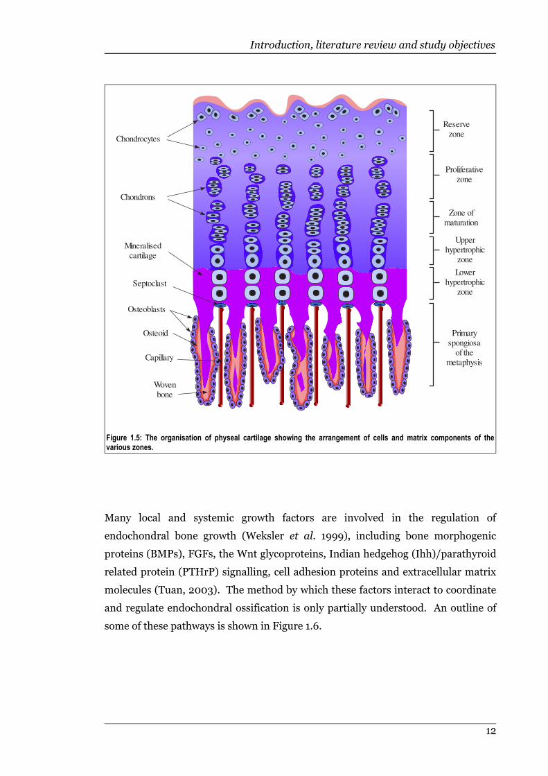
Figure 1.5 The organisation of physeal cartilage showing the arrangement of cellsand matrix components of the various zones..........................................12
Figure 1.6 Diagram outlining some of the pathways involved in the regulation ofendochondral ossification........................................................................13
Figure 1.7 Sulphation of cartilage proteoglycans in control and chondrodysplasticTexel sheep................................................................................................31
Figure 2.1 Chondrodysplastic Texel lambs...............................................................56
Figure 2.2 Chondrodysplasia in Texel sheep............................................................57
Figure 2.3 Trachea from a chondrodysplastic Texel lamb.......................................58
Figure 2.4 Thoracic cavities from 3-4-month-old control and chondrodysplasticlambs........................................................................................................59
Figure 2.5 Enlarged costochondral junctions in chondrodysplasia of Texel sheep 59
Figure 2.6 Joint lesions in chondrodysplasia of Texel sheep..................................60
Figure 2.7 Hydronephrosis in chondrodysplasia of Texel sheep.............................61
Figure 2.8 High-power view of control and chondrodysplastic hyaline cartilage. .62
Figure 2.9 Articular cartilage from the proximal humerus of a 30-day-oldchondrodysplastic lamb...........................................................................63
Figure 2.10 Costochondral junctions of normal and chondrodysplastic lambs 6-7weeks of age..............................................................................................64
Figure 2.11 Physes of control and month-old chondrodysplastic lambs.................65
Figure 2.12 Sagittal sections of 3rd-4th lumbar vertebral bodies and intervertebraldisc from 3-4-month-old control and chondrodysplastic Texel lambs. .66
Figure 2.13 Tracheal cartilage from normal and chondrodysplastic lambs............67
Figure 2.14 Immunohistochemical staining of types II and VI collagen in articular-epiphyseal cartilage from control and chondrodysplastic Texel sheep. .70
Figure 2.15 Articular cartilage chondrocytes and pericellular matrix from controland chondrodysplastic Texel lambs.........................................................71
Figure 2.16 High power views of pericellular and interterritorial matrix of cartilagefrom control and chondrodysplastic Texel lambs....................................71
Figure 3.1 Serial measurements taken from control and chondrodysplastic Texellambs........................................................................................................84
Figure 3.2 Crown-rump length and rate of growth in control andchondrodysplastic Texel lambs at different age intervals.......................87
ix
Figure 3.3 Shoulder-rump length and rate of growth in control andchondrodysplastic Texel lambs at different age intervals.......................88
Figure 3.4 Thoracic width and rate of increase in thoracic width in control andchondrodysplastic Texel lambs at different age intervals.......................89
Figure 3.5 Thoracic depth and rate of increase in thoracic depth in control andchondrodysplastic Texel lambs at different age intervals.......................90
Figure 3.6 Mandible length and growth rate of control and chondrodysplasticTexel lambs at different age intervals......................................................91
Figure 3.7 Humeral length and rate of growth in control and chondrodysplasticTexel lambs at different age intervals......................................................92
Figure 3.8 Ulnar length and rate of growth in control and chondrodysplastic Texellambs at different age intervals................................................................93
Figure 3.9 Metacarpal length and rate of growth in control and chondrodysplasticTexel lambs at different age intervals......................................................93
Figure 3.10 Femoral length and rate of growth in control and chondrodysplasticTexel lambs at different age intervals......................................................95
Figure 3.11 Tibial length and rate of growth in control and chondrodysplastic Texellambs at different age intervals................................................................95
Figure 3.12 Tarsal-metatarsal length and rate of growth in control andchondrodysplastic Texel lambs at different age intervals.......................96
Figure 3.13 Relative bone lengths in the forelimb of chondrodysplastic and controlTexel lambs at 21-30 days of age.............................................................99
Figure 3.14 Relative bone lengths in the hindlimb of chondrodysplastic and controlTexel lambs at 31-40 days of age.............................................................99
Figure 3.15 The change in relationship between tibial and tarsal-metatarsal lengthin chondrodysplastic and control Texel lambs......................................100
Figure 3.16 Comparison of lengths of equivalent bones in fore and hind limbs ofchondrodysplastic and control Texel lambs..........................................100
Figure 4.1 Fibroblast sulphate uptake in chondrodysplastic and control Texelsheep........................................................................................................111
Figure 5.1 Amplification scheme for fluorescently labelled universal primers.....120
Figure 5.2 Typical chromatogram showing amplified microsatellite sequence....122
Figure 5.3 Chi-squared test results for differences between control andchondrodysplastic Texel sheep in heterozygosity, allele frequency andgenotype of microsatellite markers........................................................123
Figure 6.1 Typical electropherogram of chondroitin disaccharides from ovinearticular cartilage....................................................................................131
Figure 6.2 Ratios of ∆di-mono4S to ∆di-mono6S in chondrodysplastic and controlTexel sheep of different ages..................................................................132
x
List of TablesTable 2.1 Chondrocyte density expressed as cells per 400X field in cartilage of
chondrodysplastic and control Texel lambs............................................68
Table 2.2 Cartilage thickness of chondrodysplastic and control Texel lambs........69
Table 3.1 ANOVA models fitted for the comparison of lengths of limb bonesbetween control and chondrodysplastic Texel lambs..............................85
Table 3.2 Influences other than chondrodysplasia on body length........................88
Table 3.3 Influences other than chondrodysplasia on thoracic dimensions...........90
Table 3.4 Influences other than chondrodysplasia on mandibular length..............91
Table 3.5 Influences other than chondrodysplasia on forelimb bone length..........94
Table 3.6 Influences other than chondrodysplasia on hindlimb bone length.........97
Table 3.7 Bone length and body dimensions influenced by sex, dam, rank,birthweight or status when crown-rump length is a covariate...............98
Table 5.1 List of microsatellite markers chosen for amplification, together withnearby genes related to cartilage or skeletal development....................118
Table 5.2 Optimal annealing temperatures for microsatellite primers..................119
Table 5.3 Half-sib regression interval mapping results for microsatellite data fromchondrodysplastic and control Texel sheep...........................................123
Table 6.1 Concentrations of chondroitin sulphate disaccharides fromchondrodysplastic and control Texel sheep of varying ages..................132
xi
Glossary
AB - alcian blue, histological stain
Abluminal - pertaining to the outer portion of a tubular structure
Acanthosisnigricans
- a form of skin hyperpigmentation
Achondroplasia - specific term for failure of cartilage growth, also commonly used torefer to a common form of dwarfism in humans
Acromesomelia - shortening of the middle and distal parts of the limbs
Alymphatic - lacking lymphatic vessels
Anauxetic - without growth
Aneural - lacking innervation
Anisospondyly - different abnormal shapes of vertebral bodies
Ankylosis - bony fusion of a joint
Anlage - an embryonic precursor to a structure
ANOVA - analysis of variance
Appositional growth - growth by the addition of external layers (c.f. interstitial growth)
APS - adenosine phosphosulphate
Arthropathy - joint disease
Articular-epiphysealcomplex
- the epiphyseal cartilage of young animals consisting of both anarticular surface and a zone of growth
ATP - adenosine triphosphate
Avascular - lacking blood vessels
Basophilic - a tissue that stains with a basic dye, such as haematoxylin
Blepharophimosis - abnormally narrow palpebral fissure (gap between eyelids)
BMP - bone morphogenic protein
Bossing - swelling
Brachycephaly - having a short (broad) head
Brachydactyly - short fingers
Brachygnathia - shortened mandible or jaw
Camptodactyly - flexural deformity of interphalangeal joints
Cancellous bone - has a latticed structure (c.f. compact bone)
CATSHL syndrome - a syndrome featuring camptodactyly, tall stature, scoliosis and hearingloss
CDMP-1 - cartilage derived morphogenic protein-1
CE - capillary electrophoresis
CHILD syndrome - congenital hemidysplasia with ichthyosiform erythroderma and limbdefects
CI - confidence interval
Chondroblasts - immature cartilage cells
Chondrocalcin - the C-propeptide of type II collagen
xii
Chondrocytes - mature cartilage cells
Chondrodysplasia - an abnormality of cartilage growth or development
Chondrogenic tissue - contains cells with the potential to differentiate into cartilage-formingcells
Chondron - the functional unit of cartilage
CHST3 - the gene encoding chondroitin 6-sulphotransferase, adding sulphateto the 6-position of chondroitin
CHST11 - the gene encoding chondroitin 4-sulphotransferase, adding sulphateto the 4-position of chondroitin
Cisternae - cavities or reservoirs
COMP - cartilage oligomeric matrix protein
Compact bone - has a dense, laminar structure (c.f. cancellous bone)
Coxa vara - a hip deformity where the angle between the ball and shaft of thefemur is reduced
cpm - counts per minute
Cyanosis - bluish colour of skin, mucous membranes, etc. due to lack ofoxygenated haemoglobin in the blood
Dentigerous cysts - follicular tooth-based cysts
Diaphysis - the main shaft of a long bone
Diarthrodial - a free-moving form of joint articulation
Distal - (as in part of a limb) far from the body
DJD - degenerative joint disease
DMC - Dyggve-Melchior-Clausen dysplasia
DMEM - Dulbecco's modified Eagle's medium
Dolichocephaly - having a long head
DTDST - diastrophic dysplasia sulphate transporter
Dysmorphism - abnormality of shape
Dysplasia - abnormality of growth or development
Dyspnoea - breathing difficulty
Ectrodactyly - absence of one or more digits
Elastic cartilage - has elastic fibres and lamellae within the matrix
Endochondral bones - bones that grow or develop within cartilage
Epiphysis - the end of a long bone separated from the main part of the bone bythe physis
Epitope - the part of a molecule to which an antibody may bind
Erythroderma - reddening of the skin
Erythrogenesis(erythropoiesis)
- the formation of red blood cells
Exophthalmus - abnormal protrusion of the eyeball
FAM - 6-carboxy-fluorescine
Fenestrated - containing one or more openings
FGF - fibroblast growth factor
FGF2 - fibroblast growth factor-2
FGFR3 - fibroblast growth factor receptor-3
xiii
Fibrillogenesis - formation of fibrils
Fibrocartilage - contains bundles of type I collagen within the matrix
Genu valgum - valgus deformity at the knees, "knock-knees"
Glaucoma - increased intraocular pressure
H&E - haematoxylin & eosin, common histological stain
Haploinsufficiency - occurs when a single functional gene is unable to produce enoughproduct to maintain a wild type (normal) phenotype
Hepato-splenomegaly
- enlarged liver and spleen
HEPES - N-2-hydroxyethylpiperazine-N'-2-ethanesulphonic acid
Homeobox genes - highly conserved genes that regulate bodily segmentation duringembryonic development
HPLC - high performance liquid chromatography
Hyaline cartilage - has a homogeneous, amorphous matrix
Hydrocephalus - dilatation of the cerebral ventricles
Hypertrophy - in chondrocytes is a stage late in maturation with increased cell size
Hypertelorism - widely-spaced eyes
Hypocellular - having decreased cell density
Hypoplasia - incomplete development of an organ or tissue
Hypotonia - decreased tone of skeletal muscles
IBD - identical by descent
Ichthyosiform - resembling scaly skin
Ihh - Indian hedgehog
Inclusion - abnormal aggregation of substance, e.g. within a cell
Interstitial growth - growth by internal expansion, e.g. division of cells already within thetissue (c.f. appositional growth)
Kyphoscoliosis - abnormal curvature of the spine both dorsoventrally and sagittally
Lacuna - the space surrounding chondrocytes caused by an artefact of fixation
Lamellar bone - mature bone with a lamellar arrangement of collagen fibres
LD - linkage disequilibrium
Lordosis - inward curvature of part of the spine
MATN3 - the gene encoding matrilin-3, a protein involved in the homeostasis ofcartilage and bone
Megalocephaly - abnormally enlarged head
Membranous bones - growing or developing as a result of direct differentiation ofosteoblasts from mesenchyme without a cartilaginous anlage
Mesenchyme - embryonic cells capable of developing into connective tissues orvasculature
Metachromasia - the staining of a tissue a different colour from that of the dye used
Metaphysis - the junction between the physis and diaphysis, containing abundanttrabecular bone and a relatively thin cortex
Microdontia - abnormally small teeth
Micrognathia - abnormally small jaw or mandible
Micromelia - abnormally small limbs
xiv
Microphthalmia - abnormally small eyes
MPS - mucopolysaccharidosis
Mydriasis - excessive dilation of the pupil of the eye
Myopathy - disease of muscle tissue
Myopia - short-sightedness
Myotonia - increased muscle irritability or spasming
Odontoid hypoplasia - underdevelopment of the odontoid process, leading to cervical spineinstability
OMIA - online mendelian inheritance in animals
OMIM - online mendelian inheritance in man
Organogenesis - the formation and development of bodily organs
OSMED - oto-spondylometa-epiphyseal dysplasia
Ossification - the process of bone formation
Osteoblasts - immature bone cells
Osteochondro-dysplasia
- abnormal growth or development of cartilage and bone
Osteocytes - mature bone cells
Osteopenia - deficiency of bone tissue
Osteophyte - a small abnormal bony growth, especially at joint margins
Osteoprogenitors - cell with the potential to differentiate into bone-forming cells
Osteosclerosis - abnormal hardening of bone
PAP - phosphoadenosine-phosphate
PAPS - phosphoadenosine-phosphosulphate
PAPSS - phosphoadenosine-phosphosulphate synthase
PAS - periodic acid-Schiff, a technique used in histology to identify glycogen
PBS - phosphate-buffered saline
PCR - polymerase chain reaction
Pectus carinatum - protrusion of the sternum causing "pigeon-breast"
Pectus excavatum - retrusion of the sternum causing a "caved-in" chest
Perichondrium - dense connective tissue surrounding non-articular cartilage containingan outer fibrous layer and an inner chondrogenic layer
Periosteum - dense connective tissue surrounding bone containing an outer fibrouslayer and an inner cambium layer containing osteoprogenitor cells
Peroxins - peroxisomal assembly proteins
Physis - the cartilaginous growth plate in an immature endochondral bone
Platyspondyly - having flattened vertebral bodies
Pleomorphic - having multiple forms
Polydactyly - the presence of more than five digits on hands or feet
Polymorphic - having many forms
Primary spongiosa - the initial trabecular network in the metaphysis immediately adjacentto the physis consisting of osteoid overlying calcified cartilage
Proximal - (as in part of a limb) close to the body
PTH - parathyroid hormone
PTHrP - parathyroid hormone-related protein
xv
QTL - quantitative trait linkage
Rarefaction - thinning, becoming less dense
rER - rough endoplasmic reticulum
Retinopathy - disease of the retina
Rhizomelia - abnormally short proximal limb-bones
RHT - ruthenium hexammine trichloride
RMRP - RNA component of mitochondrial RNA processing endoribonuclease
ROX - 6-carboxyl-X-rhodamine
SADDAN - severe achondroplasia with developmental delay and acanthosisnigricans
Sclerosis - hardening
Scoliosis - lateral curvature of the spine
SDS - sodium dodecyl sulphate
SDS-PAGE - sodium dodecyl sulphate polyacrylamide gel electrophoresis
Sedlin - endoplasmic reticulum protein with unknown function
SLC26A2 - the gene encoding the DTDST
SNPs - single nucleotide polymorphisms
Spondylolisthesis - displacement of vertebrae or the vertebral column in relation tovertebrae below
Secondaryspongiosa
- persisting trabeculae of the primary spongiosa that have beenremodelled to become lamellar bone
SHOX - short-stature homeobox
SLS - spider lamb syndrome
SMC - Smith-McCort dysplasia
Splanchnocranium - the part of the skull connected with the sense organs
Spondylo- - involving the spine
Spongiosa - cancellous bone consisting of a mesh of trabeculae
STAT - signal transducer and activator of transcription
Stenosis - abnormal narrowing of a tubular organ or structure
Synostosis - fusion of bone
Talipes equinovarus - flexural deformity causing "clubbed foot"
TB - toluidine blue, a histological stain
TD - thanatophoric dwarfism
TDT - transmission disequilibrium testing
TGF-β - transforming growth-factor-β
Tris - tris-(hydroxymethyl)-aminomethane
Valgus - abnormal lateral (outward) curvature of a bone or joint
Varus - abnormal medial (inward) curvature of a bone or joint
Woven bone - immature bone with a random arrangement of collagen fibres
∆di-mono4S - chondroitin 4-sulphate disaccharide
∆di-mono6S - chondroitin 6-sulphate disaccharide
xvi

1 Introduction, literature review and
study objectives
Introduction, literature review and study objectives
1.1 Introduction
Skeletal dysplasias are generalised disorders of bone formation (Mundlos & Olsen,
1997). The large number and variety of genetic and environmental diseases with
skeletal manifestations reflects the complexity of skeletal formation, growth and
homeostasis (Erlebacher et al., 1995). Skeletal dysplasias can be divided into
osteodysplasias, involving abnormal bone deposition or reduced bone mineral
density, or chondrodysplasias, which are characterised by disruption of the growth
and/or development of cartilage (Mundlos & Olsen, 1997). Because most bones in
the body develop by endochondral ossification, chondrodysplasias are often
characterised by severe skeletal abnormalities (Rimoin et al., 1976; van der Harten
et al., 1988; Ala-Kokko et al., 1990), although other tissues may also be affected
(Horton & Hecht, 2002b). These abnormalities frequently result in
disproportionate dwarfism, while proportionate forms of dwarfism are more
usually caused by hormonal disorders such as growth hormone deficiency (Sande &
Bingel, 1983). The estimated prevalence of chondrodysplasias in human beings is 1
in 4000 births, and over 100 different clinical phenotypes have been described
(Horton & Hecht, 2002b).
The growth and development of endochondral bones is regulated by numerous
systems, the characterisation of which is a rapidly expanding field. To this purpose,
valuable information has been gained from the study of naturally occurring
chondrodysplasias. Techniques in modern molecular biology have become more
refined so that some disorders have been characterised on a biochemical and
genetic level, along with gross, radiographic and histological levels (Superti-Furga
et al., 2001). Where possible, this becomes the basis of our understanding of the
pathogenic mechanisms involved. Unfortunately, there are still many diseases in
which the genetic defect, or the way in which a gene mutation results in identifiable
lesions, is unknown (Rimoin et al., 1998). In an effort to source enough tissue to
thoroughly characterise and investigate these rare disorders, animals models are
often used. Most typically, these are small transgenic rodents, which have the
advantages of relatively cheap maintenance and a short generation interval. In
skeletal disorders, however, larger animals with a more similar mass to human
beings have been recognised as better models due to their more similar physiology
and the comparable effects of weightbearing on the development of lesions
2

Introduction, literature review and study objectives
(Barsoum et al., 2000). This may be more important when investigating theraputic
options.
A recently identified recessively inherited chondrodysplasia of Texel sheep in New
Zealand shows similarities to a group of disorders in human beings (Thompson et
al., 2005). This thesis will present a detailed investigation of pathology,
biochemistry and genetics in order to explore whether chondrodysplasia in Texel
sheep could be a suitable model for some heritable diseases in human beings, and to
assist in the development of a genetic test for carrier animals to benefit breeders of
Texel sheep in New Zealand.
This literature review will provide an overview of the organisation and regulation of
cartilage development and endochondral ossification, describe the major forms of
chondrodysplasia in human beings and animals, and conclude with an outline of
the course this study will take.
1.2 Composition and function of cartilage
Cartilage is found in many parts of the body, and is involved in multiple functions,
such as providing support and flexibility to the airways, pinna and nose, acting as a
low-friction shock absorber for joint articulation and enabling elongation of
endochondral bones (Hamilton & Mossman, 1972). Three distinct types of cartilage
exist: fibrocartilage (forming joint menisci and fibrous joints), elastic cartilage
(found in the epiglottis, pinna and nose) and hyaline cartilage (in diarthrodial joints
and physes, or growth plates) (Martin et al., 1998). Hyaline cartilage is of most
interest in chondrodysplastic disease because of its importance in skeletal
development and articulation.
1.2.1 Extracellular structure
Adult hyaline cartilage is normally aneural, alymphatic and avascular (c.f. the
vascular canals found in fetal or newborn cartilage), consisting of chondrocytes
surrounded by an amorphous matrix of collagen and sulphated proteoglycans
(Hamilton & Mossman, 1972; Archer et al., 1996; Lefebvre et al., 1997; Martin et
al., 1998). Each chondrocyte is surrounded by a pericellular envelope consisting of
a thin layer of proteoglycans continuous with the cell membrane (Eggli et al., 1985;
Luyten et al., 1997), and in non-superficial layers is surrounded by a thin fibrous
capsule (Poole et al., 1987; Enomoto-Iwamoto et al., 1998). This capsular structure
3
Introduction, literature review and study objectives
and its contents are referred to as the chondron, which is considered to be the
functional unit of cartilage and protects the cell and its microenvironment during
physiological compression (Poole et al., 1987; Poole, 1997a; Naski et al., 1998).
Owing to its relatively high concentration of sulphated proteoglycans, matrix
adjacent to the chondron stains more densely with cationic dyes such as toluidine
blue (TB) or azure, compared with the matrix furthest from cells (Ross et al., 1995).
The latter areas are known as the territorial and interterritorial matrices,
respectively, and stain less intensely with cationic dyes especially as age increases,
due to their reduced proteoglycan content (Oohira & Nogami, 1980). Usually seen
in microscopic sections, the space between the chondrocyte and the adjacent
territorial matrix is an artifact of fixation, but has been referred to variously as the
lacuna, moat, halo, corona, or capsule. Fixation methods that preserve
proteoglycans, such as those using ruthenium hexammine trichloride, prevent
formation of this artifact (Eggli et al., 1985; Grimsrud et al., 1999).
Ultrastructurally, the chondron is surrounded by a “felt-like” fenestrated capsule of
fibres with a polar arrangement in articular and physeal cartilage, such that the
lateral parts and pole nearest the articular surface are denser than the pole nearest
the diaphysis (Poole et al., 1987; Healy et al., 1999). At the pole nearest the
diaphysis, chondrocytes have a primary cilium that interacts with matrix
components and is required for normal skeletal patterning and matrix synthesis
(McGlashan et al., 2007). In the physeal columnar zone, the territorial matrix
adjacent to the chondron has an outer layer containing a sheath of tightly packed,
longitudinally arranged fibrils, uniting chondrons within each column (Eggli et al.,
1985; St-Jacques et al., 1999). These layers are not distinguishable in transverse
septae. The remaining matrix consists of proteoglycans and randomly oriented
bundles of collagen fibrils, which are generally thicker in the interterritorial matrix
(Noonan et al., 1998; DeLise et al., 2000). Mineralisation, where it occurs, is
restricted to interterritorial matrix (Eggli et al., 1985).
Collagen accounts for approximately two thirds of the dry weight of cartilage and is
responsible for the material strength of the tissue (Ferguson et al., 2000; Eyre,
2002). The collagen is predominantly type II interacting with types IX and XI
(Kühn, 1987; van der Eerden et al., 2000; D'Angelo et al., 2001), although several
other collagen types also have vital functions in cartilage (Eyre & Wu, 1987; Olsen,
1996; Grimsrud et al., 2001; Asamura et al., 2005). The microarrangement of the
major types of collagen in hyaline cartilage matrix is illustrated in Figure 1.1. Type
4

Introduction, literature review and study objectives
II collagen exists in a continuous triple helix, typically of a smaller diameter and
more highly glycosylated than type I collagen, and without the tendency to form
parallel bundles (Kühn, 1987; Huang et al., 2001). The C-propeptide of type II
collagen is known as chondrocalcin (Alini et al., 1992). Type IX collagen is
covalently linked to the surface of type II collagen fibrils (Eyre, 2002; Tuan, 2003;
Yang et al., 2003) and is involved in the limitation of type II collagen fibril diameter
(Eyre & Wu, 1987; Akiyama et al., 2004; Ferguson et al., 2004; Seki & Hata, 2004;
Asamura et al., 2005) in a concentration-dependent manner. Type XI collagen is
intimately co-polymerised with type II collagen (Eyre, 2002). Type X collagen is
synthesised predominantly by hypertrophic chondrocytes at sites of endochondral
ossification and is more rapidly degraded by collagenase than type II collagen
(Schmid & Linsenmayer, 1987; Kobayashi et al., 2005). In hyaline cartilage, type I
collagen is expressed only by the most superficial articular chondrocytes (Sasano et
al., 1996; Minina et al., 2002). Type III collagen is a minor but consistent
component of hyaline cartilage, linked to type II collagen (Wu et al., 1996; Li et al.,
2003). Type VI collagen has a relatively short triple-helical domain, and is virtually
ubiquitous in extracellular matrices, including cartilage (Timpl & Engel, 1987).
Both type VI and IX collagens are concentrated in the pericellular matrix (Poole et
al., 1988b; Poole et al., 1992; Poole et al., 1997b; Hilton et al., 2005b). The
functions of collagen types XII and XIV, also found within cartilage, are currently
unknown (Eyre, 2002). The SOX9 gene is involved in the expression of collagen
types II, IX, XI and the core proteoglycan, aggrecan (Bi et al., 1999).
The proteoglycan component of hyaline cartilage matrix comprises sulphated
glycosaminoglycans (chondroitin 4-sulphate, chondroitin 6-sulphate, and keratan
sulphate) attached in a bottle-brush fashion to a central core protein, aggrecan,
which forms large macromolecular aggregates with hyaluronan, assisted by link
5
Figure 1.1: Microarrangement of major collagens within hyaline cartilage matrix.
Type II collagen
Type IX collagen
Type XI collagen
Introduction, literature review and study objectives
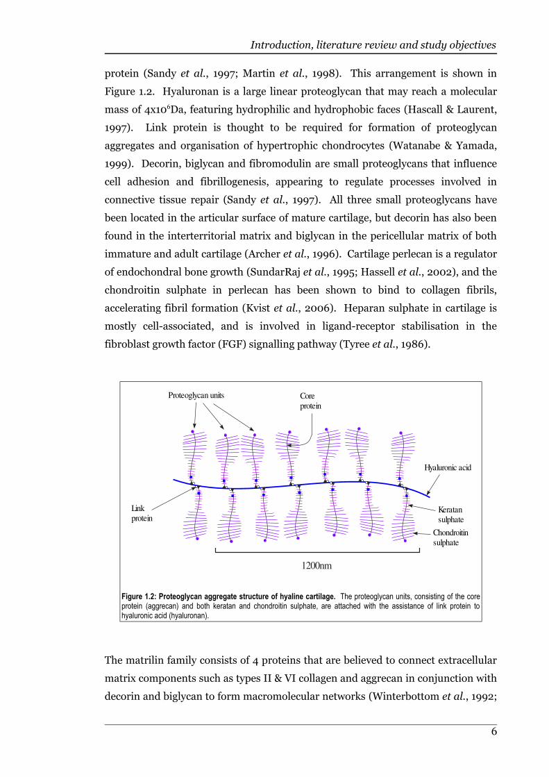
protein (Sandy et al., 1997; Martin et al., 1998). This arrangement is shown in
Figure 1.2. Hyaluronan is a large linear proteoglycan that may reach a molecular
mass of 4x10⁶Da, featuring hydrophilic and hydrophobic faces (Hascall & Laurent,
1997). Link protein is thought to be required for formation of proteoglycan
aggregates and organisation of hypertrophic chondrocytes (Watanabe & Yamada,
1999). Decorin, biglycan and fibromodulin are small proteoglycans that influence
cell adhesion and fibrillogenesis, appearing to regulate processes involved in
connective tissue repair (Sandy et al., 1997). All three small proteoglycans have
been located in the articular surface of mature cartilage, but decorin has also been
found in the interterritorial matrix and biglycan in the pericellular matrix of both
immature and adult cartilage (Archer et al., 1996). Cartilage perlecan is a regulator
of endochondral bone growth (SundarRaj et al., 1995; Hassell et al., 2002), and the
chondroitin sulphate in perlecan has been shown to bind to collagen fibrils,
accelerating fibril formation (Kvist et al., 2006). Heparan sulphate in cartilage is
mostly cell-associated, and is involved in ligand-receptor stabilisation in the
fibroblast growth factor (FGF) signalling pathway (Tyree et al., 1986).
The matrilin family consists of 4 proteins that are believed to connect extracellular
matrix components such as types II & VI collagen and aggrecan in conjunction with
decorin and biglycan to form macromolecular networks (Winterbottom et al., 1992;
6
Figure 1.2: Proteoglycan aggregate structure of hyaline cartilage. The proteoglycan units, consisting of the coreprotein (aggrecan) and both keratan and chondroitin sulphate, are attached with the assistance of link protein tohyaluronic acid (hyaluronan).
Proteoglycan units Coreprotein
Linkprotein
Hyaluronic acid
Keratansulphate
Chondroitinsulphate
1200nm
Introduction, literature review and study objectives
Hauser et al., 1996; Wiberg et al., 2003). Matrilin-1 (cartilage matrix protein) and
matrilin-3 are plentiful only in cartilage, while matrilin-2 and matrilin-4 can be
found in a wider range of tissues (Kleemann-Fischer et al., 2001). Matrilin-3
appears to be required for the appropriate timing of chondrocyte differentiation
(van der Weyden et al., 2006). Matrilin-3 is mainly expressed in the proliferative
and maturation zones in physeal cartilage (Zhang & Chen, 2000), and can form a
heterotetramer with matrilin-1 (Wu & Eyre, 1998).
1.2.2 Sulphation of matrix proteoglycans
The highly sulphated nature of cartilage matrix creates a strong negative charge
density, resulting in the accumulation of positive ions (mainly H+ and Na+) and the
retention of water within the matrix. The expansive action of the water acting
against type II collagen fibrils results in the useful mechanical characteristics of
hyaline cartilage, enabling reversible deformation and resilience to compressive
forces (Sandy et al., 1997). When cartilage loses this compressive stiffness, as
occurs normally with age, degenerative joint disease ranging from asymptomatic
lesions to severe crippling or even fusion of joints may occur (Olsen et al., 2000).
In fibroblasts and chondrocytes, sulphate ions are taken up by an anion exchange
mechanism called the diastrophic dysplasia sulphate transporter (DTDST), in
which intracellular chloride ions are exchanged for extracellular sulphate ions
(Vincourt et al., 2003). Chondrocytes are mostly dependent on extracellular
sulphate for proteoglycan sulphation (Ito et al., 1982).
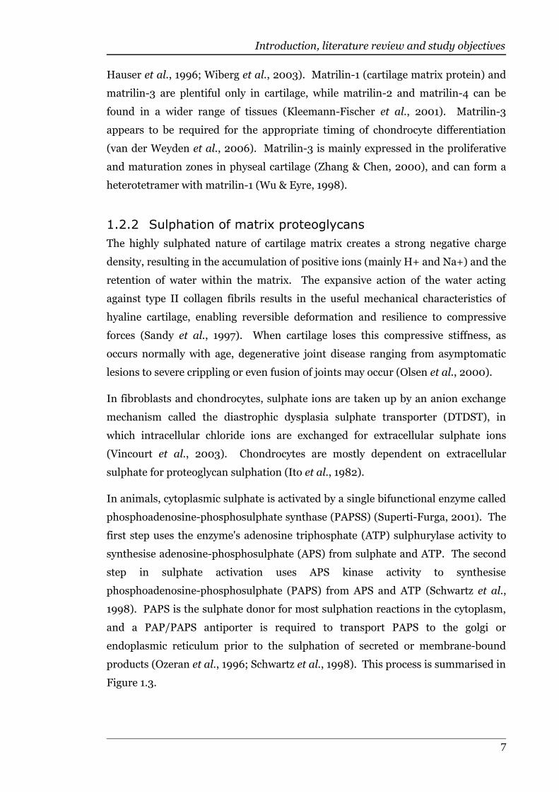
In animals, cytoplasmic sulphate is activated by a single bifunctional enzyme called
phosphoadenosine-phosphosulphate synthase (PAPSS) (Superti-Furga, 2001). The
first step uses the enzyme's adenosine triphosphate (ATP) sulphurylase activity to
synthesise adenosine-phosphosulphate (APS) from sulphate and ATP. The second
step in sulphate activation uses APS kinase activity to synthesise
phosphoadenosine-phosphosulphate (PAPS) from APS and ATP (Schwartz et al.,
1998). PAPS is the sulphate donor for most sulphation reactions in the cytoplasm,
and a PAP/PAPS antiporter is required to transport PAPS to the golgi or
endoplasmic reticulum prior to the sulphation of secreted or membrane-bound
products (Ozeran et al., 1996; Schwartz et al., 1998). This process is summarised in
Figure 1.3.
7
Introduction, literature review and study objectives
1.2.3 Endochondral ossification
Abnormalities of hyaline cartilage may affect the formation of endochondral bones
including limb bones, ribs, vertebrae and parts of the skull. These bones begin
development as crude cartilaginous anlagen in the embryo, as opposed to
membranous bones (such as the flat bones of the skull and pelvis) which form by
the direct differentiation of osteoblasts from condensations of embryonic
mesenchymal cells (Olsen et al., 2000). Membranous bone formation is usually
unaffected by abnormalities in cartilage development.
The process of endochondral ossification begins with the condensation of
embryonic mesenchymal cells and their differentiation into chondroblasts, which
form the cartilaginous anlage (Erlebacher et al., 1995). Pattern formation and
organogenesis during embryo development are regulated by homeobox genes,
which encode a large family of transcription factors (Gehring et al., 1994).
While the cartilage anlage increases in size due to both interstitial and appositional
growth, the diaphyseal chondrocytes differentiate into hypertrophic chondrocytes
8
Figure 1.3: Sulphate transport and activation in chondrocytes. Extracellular sulphate (SO42-) enters the cell through the
diastrophic dysplasia sulphate transporter (i) and is activated to phosphoadenosine-phosphosulphate (PAPS) via PAPSsynthases (ii). With the aid of the PAPS transporter (iii), PAPS then enters the golgi or endoplasmic reticulum where it isinstrumental in the sulphation of various products, including cartilage matrix proteoglycans.
Introduction, literature review and study objectives
with mineralisation of the surrounding matrix, and osteoprogenitor cells from the
vascular perichondrium growing over the diaphysis differentiate into osteoblasts
that produce a bony collar surrounded by periosteum (Olsen et al., 2000). The
perichondrium is continuous with the periosteum, with the exception of the
articular surfaces and cartilage-bone interfaces, and consists of an outer fibrous
layer and an inner chondrogenic layer. This inner layer blends with the
subperichondrial cartilage. A vascular bud from the periosteum penetrates both the
bony collar and the mineralising cartilage in the centre of what will become the
diaphysis, admitting septoclasts (chondroclasts) and osteoblasts. Septoclasts are
cathepsin-B-synthesising mononuclear cells associated with the growing portion of
invasive capillaries (Lee et al., 1995). The septoclasts break down the mineralised
cartilage matrix, causing lacunae to coalesce into primary areolae, which are rapidly
occupied by osteogenic tissue. The remaining spicules of mineralised cartilage
become lined by osteoblasts, which deposit osteoid. This is later mineralised to
form the primitive cancellous bone of the primary ossification centre, which is then
remodelled by the combined action of osteoclasts and osteoblasts (Hamilton &
Mossman, 1972). In a similar manner, secondary centres of ossification are
established in the epiphyses at the bone ends, leaving growth plates, or physes,
which allow continued longitudinal bone growth (Olsen et al., 2000). When bones
reach their mature length the physes are no longer required and “close” by
ossification. Remodelling of this synostosis eventually leads to continuity between
the epiphysis and the metaphysis (Martin et al., 1998).
The following sections describe the architecture of the remaining articular cartilage
and the physis prior to ossification.
1.2.4 Structure of articular cartilage
The cellular and fibrous arrangement of articular cartilage is highly organised, as
illustrated in Figure 1.4. Directly adjacent to the articular surface lies the acellular
lamina splendens, comprised of a superficial afibrillar layer and a deeper fibrillar
layer (Kumar et al., 2001). Beneath this lies a tangential zone with collagen fibrils
surrounding flattened chondrocytes with their long axes parallel to the articular
surface. Deeper to this region is a transitional zone where the chondrocytes are
rounded and seemingly randomly organised. Collagen fibrils in this zone are larger
and angled obliquely. Below this is a radial zone in which cells are aligned in short,
irregular columns with parallel collagen fibres, and a calcified zone rich in
9

Introduction, literature review and study objectives
hydoxyapatite (Mankin & Radin, 1997; Martin et al., 1998). The junction between
the calcified and non-calcified matrix is visible histologically in sections stained
with haematoxylin and eosin (H&E) as an undulating basophilic line, referred to as
the “tidemark”. This zone of calcified cartilage is firmly anchored to the
subchondral bone.
1.2.5 Structure of physeal cartilage
Physeal cartilage consists of several distinct zones (Ballock & O'Keefe, 2003), as
shown in Figure 1.5. Closest to the joint space and anchored to the epiphysis is a
resting zone consisting of moderately sized, slowly dividing chondrocytes scattered
in a random manner (Martin et al., 1998). In this zone there is a relatively high
ratio of extracellular matrix to cell volume (Ballock & O'Keefe, 2003). Adjacent is a
proliferative zone, in which chondrocytes divide rapidly to form columns of disk-
like cells (Martin et al., 1998). This columnar arrangement is more obvious in the
physes of fast growing bones, while slower growing bones tend to have cells
arranged in elongated clusters (Morris et al., 2002). Fibroblast growth factor-2
10
Figure 1.4: Structure of articular cartilage showing the arrangement of cells and matrix components of the variouszones.
Articularsurface
Chondrocytes
Lamina splendensTangential zone
Transitionalzone
Radial zone
Calcifiedzone
Territorialmatrix
Interterritorialmatrix
Tidemark
Subchondralbone
Collagenfibres
Introduction, literature review and study objectives
(FGF2) promotes chondrocyte proliferation and blocks further differentiation
(Weksler et al., 1999).
Following cell division in the proliferative zone, chondrocytic enlargement begins
(Breur et al., 1994), leading to the prehypertrophic (or maturational) stage, during
which chondrocytes actively secrete and become separated by matrix. This leads
into the hypertrophic zone where the chondrocytes are large and spherical with
increased rough endoplasmic reticulum (rER) and Golgi apparatus (Farquharson &
Jefferies, 2000). Associated with the hypertrophic phenotype are a range of
metabolic alterations, including increased activity of plasma membrane alkaline
phosphatase, synthesis of type X collagen, accumulation of glycogen (Martin et al.,
1998), down-regulation of type II collagen, secretion of osteonectin and
osteopontin, expression of vitamin D receptors (Farquharson & Jefferies, 2000),
and secretion of matrix vesicles that act as niduses for mineralisation (Ballock &
O'Keefe, 2003). Matrix vesicles are anchored to the collagenous matrix by the
collagen binding protein anchorin CII, which also binds collagen types II and X
(Kirsch & Pfäffle, 1992). The zone of provisional mineralisation consists of
degenerating hypertrophic chondrocytes surrounded by calcified matrix (Ballock &
O'Keefe, 2003). The hypertrophic chondrocytes dissolve some of their surrounding
matrix and the cartilage septum remaining at the bottom of each chondrocyte
column is resorbed by septoclasts (Martin et al., 1998). For any single chondrocyte,
this series of developmental events takes place in one site over time, but due to
bone growth these temporal events are represented spatially in the physis (Morris
et al., 2002).
Capillary loops from the metaphysis enter the cavities left by dead hypertrophic
chondrocytes. Most remaining transverse septae of mineralised matrix are actively
resorbed by septoclasts (Ballock & O'Keefe, 2003). Osteoblasts deposit a layer of
osteoid which is then mineralised to form bone on the remaining trabeculae,
creating the primary spongiosa (Martin et al., 1998). As the layer of bone tissue
becomes thicker, the trabculae of the primary spongiosa are gradually remodelled
and realigned, forming the secondary spongiosa (Ballock & O'Keefe, 2003).
Continued cell division at the circumference of the physis, the zone of Ranvier,
allows the physis to increase in diameter as the bone grows in length (Martin et al.,
1998).
11
Introduction, literature review and study objectives
Many local and systemic growth factors are involved in the regulation of
endochondral bone growth (Weksler et al. 1999), including bone morphogenic
proteins (BMPs), FGFs, the Wnt glycoproteins, Indian hedgehog (Ihh)/parathyroid
related protein (PTHrP) signalling, cell adhesion proteins and extracellular matrix
molecules (Tuan, 2003). The method by which these factors interact to coordinate
and regulate endochondral ossification is only partially understood. An outline of
some of these pathways is shown in Figure 1.6.
12
Figure 1.5: The organisation of physeal cartilage showing the arrangement of cells and matrix components of thevarious zones.
Reserve zone
Proliferative zone
Zone of maturation
Upper hypertrophic
zone
Lowerhypertrophic
zone
Primaryspongiosa
of the metaphysis
Chondrocytes
Chondrons
Mineralisedcartilage
Wovenbone
Osteoblasts
Osteoid
Capillary
Septoclast

Introduction, literature review and study objectives
1.2.6 Chondrodysplasias in human beings
The original classification of skeletal dysplasias in human beings consisted of two
groups – short trunked (“Morquio syndrome”) or short limbed (“achondroplasia”)
(Unger, 2002). As knowledge improved, disorders of the skeletal system came to be
classified primarily by their clinical and radiographic features. Because some
chondrodysplasias caused by mutations in different genes appeared to be the same
disease (Superti-Furga et al., 2001), many diseases are now known by more than
one name (Hall, 1978; Rimoin et al., 1998). As diagnostic tools have become more
advanced, especially with respect to the identification of causative gene mutations,
13
Figure 1.6: Diagram outlining some of the pathways involved in the regulation of endochondral ossification. Thickarrows indicate upregulation or activation, thin arrow indicate downregulation or inactivation, and dashed arrows have been usedwhere the method of regulation is not clear, but a link has been demonstrated. Acronyms used are: TGFβ1 (transforming growthfactor β-1), CDMP-1&2 (cartilage-derived morphogenic protein 1 and 2), BMP (bone morphogenic protein), Ihh (Indianhedgehog), PTH/PTHrP (parathyroid hormone/parathyroid hormone related protein), MMP-13 (matrix metalloprotease 13) Cbfa-1(core-binding factor α-1), FGF3 (fibroblast growth-factor 3), Wnt5a&7a genes (wingless-type MMTV integration site family 5a and7a), SOX9 gene (sex determining region Y-box 9) and Smad2&3 genes (mothers against decapentaplegic homologues 2 and 3).This information was collected from a variety of sources (Ballock et al., 1993; Chang et al., 1994; Vortkamp et al., 1996; Lefebvreet al., 1997; Luyten, 1997; Enomoto-Iwamoto et al., 1998; Naski et al., 1998; Grimsrud et al., 1999; Healy et al., 1999; St-Jacques et al., 1999; DeLise et al., 2000; Ferguson et al., 2000; van der Eerden et al., 2000; D'Angelo et al., 2001; Grimsrud etal., 2001; Huang et al., 2001; Minina et al., 2002; Li et al., 2003; Tuan, 2003; Yang et al., 2003; Akiyama et al., 2004; Ferguson etal., 2004; Seki & Hata, 2004; Hilton et al., 2005a).
Chondrocytedifferentiation
Expressed inhypertrophic cells
Limits chondrogenesisto central limb bud
Inhibits proliferation, hypertrophy
Expressed in medialproliferating zone
Expressed in prehypertrophic
cells, prehypertrophy
Expressed in prehypertrophic& hypertrophic cells, induceshypertrophy, proliferation
Involved in proliferation& hypertrophy
Potentiates TGF
Expressed inprehypertrophic cells
hypertrophy
differation from restingto proliferative & hypertrophy
Associated with matrix vesicles
Differentiation ofchondrocytes from
mesenchyme
Expressed in latehypertrophic cells& 1° spongiosa
Complex inhibitsproliferation, delays
hypertrophy, degradesSOX9
-catenin
Inhibits maturation& proliferation
TGF 1
Transforming Growth Factor
Superfamily
CDMP-1
CDMP-2
Wnt7a
FGF3
Ihh
Cbfa-1
Wnt5a
SOX9
PTH/PTHrP
MMP-13
Metalloproteases
Smad2&3
BMP-7
BMP-2
BMP-6
BMP-4Bone Morphogenic
Proteins
Introduction, literature review and study objectives
these older classification systems have been superceded. A recently developed
molecular-pathogenic classification system divides skeletal diseases into the
following groups: defects in extracellular structural proteins, defects in metabolic
pathways (including enzymes, ion channels and transporters), defects in the folding
and degradation of macromolecules, defects in hormones and signal transduction
mechanisms, defects in nuclear proteins and transcription factors, defects in
oncogenes and tumour suppressor genes, and defects in RNA and DNA processing
and metabolism (Superti-Furga et al., 2001). Since the publication of this
classification system, some chondrodysplasias of previously unknown aetiology
have been found to be caused by abnormalities in cytoskeletal elements (Krakow et
al., 2004; Beales et al., 2007). Consequently, defects in cytoskeletal elements will
be included as a separate group for the purpose of this review. With the exception
of defects in oncogenes and tumor suppressor genes, all of these groups contain at
least one chondrodysplastic disease.
The following sections of this review focus on chondrodysplasias of human beings
and animals. As chondrodysplasias affecting human beings tend to be better
described on a molecular-pathogenic level, these diseases will be organised
according to the classification system described by Superti-Furga et al., (2001).
Chondrodysplasias affecting animals have been organised by species.
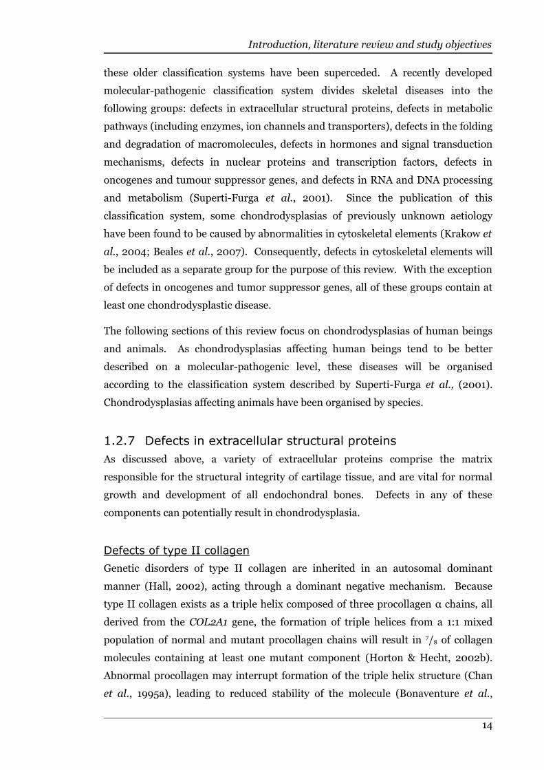
1.2.7 Defects in extracellular structural proteins
As discussed above, a variety of extracellular proteins comprise the matrix
responsible for the structural integrity of cartilage tissue, and are vital for normal
growth and development of all endochondral bones. Defects in any of these
components can potentially result in chondrodysplasia.
Defects of type II collagen
Genetic disorders of type II collagen are inherited in an autosomal dominant
manner (Hall, 2002), acting through a dominant negative mechanism. Because
type II collagen exists as a triple helix composed of three procollagen α chains, all
derived from the COL2A1 gene, the formation of triple helices from a 1:1 mixed
population of normal and mutant procollagen chains will result in 7/8 of collagen
molecules containing at least one mutant component (Horton & Hecht, 2002b).
Abnormal procollagen may interrupt formation of the triple helix structure (Chan
et al., 1995a), leading to reduced stability of the molecule (Bonaventure et al.,
14

Introduction, literature review and study objectives
1995). This may occur through delayed or atypical folding of the collagen molecule,
which can result in excessive post-translational modification, degradation, reduced
secretion, and/or accumulation of the product in the rER (Horton & Hecht, 2002a).
A reduced number of collagen fibrils would alter the mechanical properties of
cartilage, and the interaction of abnormal fibrils with the cell surface may
potentially interfere with the diffusion, sequestration and presentation of growth
factors to chondrocytes (Horton & Hecht, 2002a).
The genetic disorders of type II collagen in human beings include achondrogenesis
type II, Kniest dysplasia, Torrance type lethal platyspondylic skeletal dysplasia
(Nishimura et al., 2004; Zankl et al., 2005), spondyloepiphyseal dysplasia
congenita (Lee et al., 1989), spondyloepimetaphyseal dysplasia Strudwick type
(Tiller et al., 1995), Stickler syndrome type 1 (Williams et al., 1996) and
spondyloperipheral dysplasia (Zabel et al., 1996). Achondrogenesis type II and
Kniest dysplasia are described below; the remaining defects of type II collagen are
listed in Appendix 1. Along with skeletal deformities, type II collagenopathies are
often accompanied by ocular disturbances, such as myopia, abnormal vitreous
humor and retinal tearing or detachment (Meredith et al., 2007).
Achondrogenesis type II and hypochondrogenesis are caused by mutations in the
α1(II) chain of type II procollagen (Bonaventure et al., 1995) and are now
considered to be at different ends of a continuous disease spectrum, involving 1 in
40,000-60,000 births (Borochowitz et al., 1986).
Achondrogenesis type II is a lethal chondrodysplasia characterised by short, bowed,
long bones with flared and “cupped” metaphyses evident on radiographs, appearing
like capless “mushroom stems” (van der Harten et al., 1988). Other radiographic
lesions include variable ossification of the spine, axe-head-shaped iliac bones
accompanied by unossified ischial and pubic bones, and a hypoplastic, barrel- or
bell-shaped thorax (Sillence et al., 1979). Histologically, epiphyseal chondrocytes
are densely packed in a reduced extracellular matrix containing numerous cartilage
canals with perivascular fibrosis (van der Harten et al., 1988; Gruber et al., 1990).
The physis is generally disorganised and hypertrophic chondrocytes extend into the
primary and secondary spongiosa, which contain plump and disordered trabeculae.
Milder forms have a more organised physeal architecture. Cartilage in affected
individuals contains type I collagen rather than type II (Eyre et al., 1986).
15
Introduction, literature review and study objectives
Hypochondrogenesis histologically resembles achondrogenesis type II with slightly
improved organisation of the hypertrophic zone, but has radiographic features
more similar to those of spondylometaphyseal dysplasia (van der Harten et al.,
1988). Spondylometaphyseal dysplasia comprises a heterogeneous group of
heritable skeletal dysplasias characterised by modifications of the vertebral bodies
of the spine and metaphyses of tubular bones (Borochowitz et al., 1988a).
Kniest dysplasia is a relatively uncommon non-lethal disorder characterised by a
short trunk and limbs, progressive joint enlargement and a flattened face (Hicks et
al., 2001). Cleft palate, tracheal collapse, recurrent otitis media, and severe myopia
are variably present (Maumenee & Traboulsi, 1985). Radiographic abnormalities
include broad metaphyses, enlarged epiphyses, platyspondyly, progressive
kyphoscoliosis and lumbar lordosis (Hicks et al., 2001). Histologically,
intracytoplasmic inclusions are present within chondrocytes and there is
degeneration of cartilage matrix (Horton & Rimoin, 1979). Abnormally thin,
irregular collagen fibrils are visible ultrastructurally, and the epiphyseal
extracellular matrix lacks chondrocalcin (Poole et al., 1988a).
Defects of type IX collagen
Multiple epiphyseal dysplasia affects at least 1 in 10,000 people (McKeand et al.,
1996) and is caused by mutations in COL9A1 (Czarny-Ratajczak et al., 2001),
COMP (Maddox et al., 2000) or MATN3 (Mäkitie et al., 2004a). This disease has
an autosomal dominant mode of inheritance and is characterised by early onset
osteoarthritis, a waddling gait, variably short stature, mild distal shortening of the
ulnas, and irregular ossification of carpal and tarsal bones (Bönnemann et al.,
2000). Stippled calcification of epiphyses is identifiable radiologically at birth, and
may either occur in all endochondral bones or be limited to the tarsus (Rasmussen
& Reimann, 1973). Joint crepitation, knee joint laxity, scoliosis, subchondral
sclerosis, flattened femoral heads, valgus deformity, short femoral necks, flattened
and irregular epiphyses of the knees, and bipartate, chondromalacic patellae may
also be present (Czarny-Ratajczak et al., 2001).
Intracytoplasmic inclusions are present in chondrocytes, which are surrounded by a
slightly fibrillar matrix (Czarny-Ratajczak et al., 2001). Ultrastructurally, the
chondrocyte rER is markedly dilated, and contains linear arrays of alternating
electron-dense and electron-lucent material. Lesions are more severe in epiphyseal
than in physeal cartilage (Czarny-Ratajczak et al., 2001).
16
Introduction, literature review and study objectives
Mutations in type IX collagen have also been associated with intervertebral disk
disease (Annunen et al., 1999b).
Defects of type X collagen
Schmid metaphyseal chondrodysplasia, the most common type of metaphyseal
chondrodysplasia (Lachman et al., 1988), has an autosomal dominant mode of
inheritance (Ikegawa et al., 1997). This disease is caused by the synthesis of
abnormal procollagen X that cannot be incorporated into mature helical molecules
(Chan et al., 1995b), resulting in haploinsufficiency (Chan et al., 1998). The
resultant phenotype includes disproportionate dwarfism with bowed legs in early
childhood, coxa vara (Bateman et al., 2004), and lumbar lordosis with an anterior
pelvic tilt (Chan et al., 1998). Radiology reveals widened, sclerotic and irregular
metaphyses (Bateman et al., 2003). Histologically, there is an absence of
identifiable hypertrophic chondrocytes and disorganisation of physeal chondrocyte
columns (Chan et al., 1995b).
Defects of type XI collagen
Stickler syndrome type II, Marshall syndrome, and both dominant and recessive
forms of oto-spondylo-megaepiphyseal dysplasia (OSMED) are caused by
mutations in collagen type XI, and feature midface hypoplasia, cleft palate,
micrognathia, hearing loss, arthropathy, mild epiphyseal dysplasia, and wide
metaphyses (Spranger, 1998). The lesions are evident at birth, but alter with age.
See Appendix 1 for further details.
Defects of cartilage oligomeric matrix protein (COMP)
Pseudoachondroplasia and multiple epiphyseal dysplasia are two common
chondrodysplasias associated with mutations in COMP (Ikegawa et al., 1998). Such
mutations in COMP can reduce the calcium binding properties of the protein,
resulting in an altered conformation (Chen et al., 2000).
Pseudoachondroplasia is an autosomal dominant form of disproportionate
dwarfism with joint laxity and moderate brachydactyly, usually appearing at the
onset of walking, with a waddling gait and debilitating hip pain in childhood
(McKeand et al., 1996; Maddox et al., 2000). A mild form has also been recognised
(Manabe et al., 1998). Radiographically, there are small, irregular epiphyses,
17
Introduction, literature review and study objectives
delayed ossification, flared metaphyses and anterior beaking of vertebral bodies
(Wynne-Davies et al., 1986).
Histologically, prehypertrophic and hypertrophic zones of affected cartilage are
disorganised and hypocellular, with many dead cells throughout the growth plate
(Hecht et al., 2004). Ultrastructural investigation reveals chondrocytes with
dilated rER cisternae containing alternating electron dense and electron lucent
granular bands (Pedrini-Mille et al., 1984; Stanescu et al., 1984).
Immunoreactivity for COMP and type IX collagen is diminished in the cartilage
matrix, but increased within dilated rER cisternae (Hecht et al., 2004).
Defects in other extracellular structural proteins
Mutations in matrilin-3 (MATN3) are responsible for some forms of multiple
epiphyseal dysplasia (Mäkitie et al., 2004b) and spondyloepimetaphyseal dysplasia
(Borochowitz et al., 2004). Silverman-Handmaker dyssegmental dysplasia and
Schwartz-jampel syndrome are caused by mutations in the perlecan gene (Nicole et
al., 2000; Arikawa-Hirasawa et al., 2001b), which is considered to be involved with
the adhesion of chondrocytes to surrounding matrix (SundarRaj et al., 1995).
Marfan syndrome is caused by mutations in the gene for fibrillin (Tekin et al.,
2007), a component of extracellular microfibrils in many tissues (Sakai et al.,
1986). These disorders are listed in Appendix 1.
1.2.8 Defects in metabolic pathways
Defects of the diastrophic dysplasia sulphate transporter (DTDST)
Mutations in the DTDST gene, SLC26A2, are responsible for a group of disorders
with similar histological features but differing severity, including achondrogenesis
1b (Superti-Furga, 1994), atelosteogenesis type II (Hästbacka et al., 1996b),
diastrophic dysplasia (Hästbacka et al., 1994) and recessive multiple epiphyseal
dysplasia (Rimoin et al., 1998; Rossi & Superti-Furga, 2001). SLC26A2 mutations
identified in these disorders result in the variable reduction of sulphate uptake by
chondrocytes and fibroblasts (Hästbacka et al., 1994; Rossi & Superti-Furga, 2001;
Superti-Furga, 2001), causing undersulphation of cartilage extracellular matrix
(Superti-Furga et al., 1996a), and abnormal migration of type IX collagen on
sodium dodecyl sulphate polyacrylamide gel electrophoresis (SDS-PAGE) (Diab et
al., 1994). The severity of disease is determined to some degree by the level of
18
Introduction, literature review and study objectives
residual transporter function (Karniski, 2001), with the exception of the Finnish
diastrophic dysplasia mutation, which occurs in an untranslated exon and results in
severely reduced mRNA levels (Hästbacka et al., 1999). Achondrogenesis type 1b
and diastrophic dysplasia are described below; atelosteogenesis type II and
recessive multiple epiphyseal dysplasia are included in Appendix 2. A transgenic
mouse with an SLC26A2 mutation has been engineered to study this group of
disorders (Forlino et al., 2005).
Achondrogenesis type 1b (achondrogenesis Fraccaro type) is a rare autosomal
recessive, perinatally lethal chondrodysplasia characterised by short limb bones
and a hypoplastic thorax (van der Harten et al., 1988; Superti-Furga, 1994; Hall,
2002). Radiographic investigation of 23 cases by van der Harten et al (1988)
revealed minimal ossification of vertebral bodies and long bones, accompanied by
abundant metaphyseal flaring in the lower limbs. The pelvis was poorly formed,
and ribs were shortened with "cupped" metaphyses.
Histological studies exposed loosely arranged epiphyseal chondrocytes surrounded
by “collagen rings” and containing cytoplasmic vacuoles (van der Harten et al.,
1988; Superti-Furga, 1994). The physes were disorganised with rarefied cartilage
matrix containing coarse, “unmasked” collagen fibrils, and hypertrophic
chondrocytes extended into plump, disordered, metaphyseal trabeculae.
Originally considered to the be same disease, achondrogenesis type 1b and type 1a
can be distinguished histologically. In contrast with achondrogenesis type 1a, the
cartilage matrix in type 1b is abnormal, and only in type 1a do the reserve zone
chondrocytes contain variably sized periodic acid-Schiff (PAS)-negative
intracytoplasmic inclusions that stain green with Gomori’s trichrome procedure
(Yang et al., 1976; Superti-Furga, 1996).
Hyaline cartilage in achondrogenesis type 1b was found to be under-sulphated with
a reduced sulphate uptake by chondrocytes tested in vitro, and little or no residual
activity of the DTDST (Superti-Furga et al., 1996a). SLC26A2 mutations are
usually found in both alleles, and generally consist of premature stop codons
(Hästbacka et al., 1996b). Further experiments in vitro demonstrated that
sulphydryl compounds are able to contribute sulphate for proteoglycan sulphation
in both achondrogenesis 1b and diastrophic dysplasia chondrocytes, recovering to
some degree the level of sulphation lost as a result of the SLC26A2 mutation (Rossi
et al., 2003).
19

Introduction, literature review and study objectives
Diastrophic dysplasia is an autosomal recessively inherited disease caused by
mutations in SLC26A2 and is rare, other than its high incidence in the Finnish
population (carrier frequency of 1-2%) due to founder effect (Langer, 1965;
Hästbacka et al., 1996a; Rimoin et al., 1998; Hästbacka et al., 1999). Clinical
features include short limbed dwarfism, swollen ear lobes, severe talipes
equinovarus, deformed tarsal and metatarsal bones, and extremely shortened and
oval-shaped first metacarpal bones (Langer, 1965). Joint contractures affect the
hips, knees and elbows, and are accompanied by metacarpo-phalangeal and
interphalangeal joint rigidity, “hitch-hiker’s” thumb, and narrowed interpedicular
distance of the lower lumbar vertebrae (Shapiro, 1992). Young children affected
with diastrophic dysplasia may have double-layered manubrium sterni which fuse
to form a normal manubrium in later years (Currarino, 2000). The knees of
affected fetuses appear grossly normal initially, but deteriorate with age (Peltonen
et al., 1999).
Hyaline cartilage in diastrophic dysplasia contains microscopic foci of matrix
degeneration, with prominent fibrillar material traversing cystic areas (Shapiro,
1992), and accumulations of matrix around cells described as “large, multilayered
lacunae” (Stanescu et al., 1984). Transmission electron microscopy reveals the
fibrillar material to be short, randomly arranged, abnormally wide collagen fibrils
with a markedly irregular circumference on cross-section, interspersed with normal
collagen fibrils. Many cells have disorganised organelles and cystic regions
consistent with premature senescence and degeneration (Stanescu et al., 1984;
Shapiro, 1992).
Defects in other metabolic pathways
Loss of chondroitin-6-O-sulphotransferase-1 function results in a severe
chondrodysplasia with progressive spinal involvement (Thiele et al., 2004), while
mutations in phosphoadenosine-phosphosulphate synthase 2 (PAPSS2) are
responsible for Pakistani type spondlyoepimetaphyseal dysplasia (Ahmad et al.,
1998; ul Haque et al., 1998). Chondroitin biosynthesis is severely impaired in
schneckenbecken dysplasia, which is caused by mutations in the nucleotide sugar
transporter, SLC35D1 (Hiraoka et al., 2007).
Inborn errors of cholesterol biosynthesis manifest as X-linked chondrodysplasia
punctata types 1 and 2 (Franco et al., 1995; Braverman et al., 1999; Shotelersuk &
Tongkobpetch, 2005), and congenital hemidysplasia with ichthyosiform
20
Introduction, literature review and study objectives
erythroderma and limb defects (CHILD syndrome) (König et al., 2000).
Peroxisomal disorders featuring some degree of chondrodysplasia punctata include
rhizomelic chondrodysplasia punctata types 1, 2 and 3 (Braverman et al., 1997;
Ofman et al., 1998; de Vet et al., 1999), and Zellweger syndrome (Dodt et al., 1995;
Shimozawa et al., 2004). PEX genes encode peroxisomal assembly proteins
(peroxins), which import matrix proteins into peroxisomes (Distel et al., 1996). See
Appendix 2 for further details of these disorders.
1.2.9 Defects in the folding and degradation of macromolecules
This group of diseases consists of X-linked spondyloepiphyseal dysplasia (SED-XL),
caused by mutations in SEDL, encoding an endoplasmic reticulum protein of
unknown function, Sedlin (Gedeon et al., 1999), abnormalities in the activity of
cathepsin-K (Donnarumma et al., 2007), mutations in MMP13 (Kennedy et al.,
2005), and several types of mucopolysaccharidosis (MPS). The individual diseases
involved are described in Appendix 3, excluding the types of MPS that do not
feature obvious skeletal abnormalities.
The MPS disorders are a group of heritable diseases characterised by the
progressive intralysosomal storage of glycosaminoglycans (Leroy, 2002). Several
forms exist, each associated with inherited defects in different lysosomal enzymes
involved in mucopolysaccharide metabolism (Clarke et al., 1992; Yamagishi et al.,
1996; Bunge et al., 1997; Vervoort et al., 1997; Beesley et al., 2001; Litjens &
Hopwood, 2001; Leroy, 2002). Proteoglycan synthesis is affected in these diseases,
and indigestible substrates accumulate within the cytoplasm of affected cells
(Leroy, 2002).
MPS is typically associated with variable dwarfism, together with kyphosis, coarse
facial features, corneal opacity (except types IIA, IIB and III), cardiac
abnormalities, hepatosplenomegaly and excessive urinary excretion of
glycosaminoglycans (Arbisser et al., 1977; Schmidt et al., 1987; Wippermann et al.,
1995; Beesley et al., 2001; Leroy, 2002).
Cartilaginous lesions include enlarged chondrocytes containing abundant granular
or finely vacuolated cytoplasm. Large areas of loose connective tissue are scattered
throughout the reserve zone of physeal cartilage in MPS types I and IV, but the rest
of the growth plate remains well organised. MPS type I features wide trabeculae
with areas of continuous horizontal connection between longitudinally oriented
21

Introduction, literature review and study objectives
spicules. Ultrastructurally, chondrocytes contain numerous large intracytoplasmic
vacuoles filled with undegraded glycosaminoglycan (Rimoin et al., 1976).
1.2.10 Defects in hormones and signal transduction
mechanisms
While hormones and signal transduction mechanisms are crucial to the control and
regulation of endochondral ossification, many pathways are poorly understood.
The diseases in this group are listed in Appendix 4.
Fibroblast growth factor receptor 3 (FGFR3)
A family of autosomal dominant chondrodysplasias with variation in severity are
associated with mutations in FGFR3, which result in ligand-independent activation
of the receptor (Rimoin et al., 1998; Vajo et al., 2000; Ballock & O'Keefe, 2003).
FGFR3 normally plays an inhibitory role in the lengthening of endochondral bones
(McEwen et al., 1999; Aikawa et al., 2001; Rozenblatt-Rosen et al., 2002) (see
Figure 1.6). While a partial loss-of-function mutation of FGFR3 has been recorded
to result in camptodactyly, tall stature, scoliosis and hearing loss (CATSHL
syndrome) (Toydemir et al., 2006), most FGFR3 mutations in human beings are
characterised by dwarfism.
Achondroplasia is the most common form of dwarfism in humans, affecting 1 in
15,000-40,000 people (Park & Wallerstein, 2003). Although gonadal mosaicism in
normal parents with multiple affected children has been identified, more than 90%
of cases of achondroplasia are due to sporadic dominant mutations (Mettler &
Fraser, 2000). For this reason, nucleotide 1138 of FGFR3 is currently considered to
be the most mutable nucleotide of the human genome (Rousseau et al., 1994;
Shiang et al., 1994; Bellus et al., 1995a; Horton, 2006). Early literature on
achondroplasia included a variety of different chondrodysplasias, complicating the
perceived pathology (Rimoin et al., 1976). Even the name, “achondroplasia” is
misleading as it falsely implies a complete failure of cartilage growth. While
“achondroplasia” is an inaccurate descriptive term, it has become well established
by long usage (Langer et al., 1967).
Achondroplasia is characterised by a reduced rate of endochondral growth (Rimoin
et al., 1970), rhizomelia, megalocephaly, frontal bossing with midface hypoplasia,
and brachydactyly (Langer et al., 1967). Histologically, growth plates are relatively
well organised but cell columns are shortened, even though the number of cells per
22
Introduction, literature review and study objectives
column is not decreased (Rimoin et al., 1970). Large collagen fibrils are present in
the narrow hypertrophic zone, and the primary spongiosa contains coarse
longitudinal septae accompanied by horizontal bridging (Maynard et al., 1981).
Homozygosity is usually lethal in this disease due to complications of hydrocephaly
and pulmonary insufficiency resulting from thoracic hypoplasia (Pauli et al., 2005).
Hypochondroplasia is a form of short-limbed dwarfism similar to mild
achondroplasia, and is rarely diagnosed before 2 years of age (Bellus et al., 1995b;
Prinster et al., 1998; Ramaswami et al., 1998; Rimoin et al., 1998; Winterpacht et
al., 2000). It features short stature, megalocephaly with mild frontal bossing,
micromelia, and lumbar lordosis (Ramaswami et al., 1998).
Thanatophoric dysplasia (TD) is the most common neonatally lethal skeletal
dysplasia in humans, affecting 1 in 20,000-50,000 births (Orioli et al., 1986). The
FGFR3 mutations found in TD trigger premature apoptosis through activation of
the signal transducer and activator of transcription (STAT) signalling pathway
(Tavormina, 1997; Legeai-Mallet et al., 1998; Wilcox et al., 1998). TD is
characterised by rhizomelic dwarfism with megalocephaly, platyspondyly, and
thoracic hypoplasia. The disorder has been reclassified into type I (most common)
and type II based on femoral radiology and the underlying mutation. In TD type I
the femurs are curved, while in TD type II the femurs are straight and a clover-leaf
skull deformity is often present. Histologically, physeal chondrocyte columns are
disorganised, with short, thick primary trabeculae (Delezoide et al., 1997; Wilcox et
al., 1998).
Another chondrodysplasia resulting from mutations in FGFR3 is severe
achondroplasia with developmental delay and acanthosis nigricans (SADDAN)
(Bellus et al., 1999; Tavormina et al., 1999). For more details on these diseases,
refer to Appendix 4.
Defects in the PTH/PTHrP receptor
Mutations in PTHR1, which encodes the parathyroid hormone/parathyroid
hormone related protein (PTH/PTHrP) receptor, are reported in Jansen
metaphyseal chondrodysplasia (Schipani et al., 1995; 1997a; 1999; Bastepe et al.,
2004) and Blomstrand metaphyseal dysplasia (Karperien et al., 1999).
The mutations involved in Jansen metaphyseal chondrodysplasia result in
constitutive activation of the PTH/PTHrP receptor (David, 1998), causing
23
Introduction, literature review and study objectives
intracellular accumulation of adenosine 3’5’-monophosphate (Schipani et al.,
1997a). Asymptomatic hypercalcaemia and hypophosphataemia also occur
(Schipani et al., 1995), despite normal or low levels of PTH and PTHrP (Bastepe et
al., 2004).
Studies in transgenic mice with constitutive activation of PTH/PTHrP receptors
have revealed a decreased rate of chondrocyte maturation, persistence of
hypertrophic chondrocytes, along with delayed vascular invasion and
mineralisation of cartilage (Schipani et al., 1997b).
Others
Other defects in signal transduction mechnisms resulting in chondrodysplasia
include Grebe/Hunter-Thompson acromesomelic dysplasia and brachydactyly type
C, caused by a defect in cartilage-derived morphogenic protein-1 (CDMP1) (Stelzer
et al., 2003), a member of the TGF-β family that may influence the differentiation
of osteo-chondroprogenitor cells to chondrocytes (Luyten, 1997; Tsumaki et al.,
1999), and brachydactyly type A1 which is due to a defect in Indian hedgehog signal
molecule (Ihh) (Gao et al., 2001).
1.2.11 Defects in nuclear proteins and transcription factors
Mutations in SOX9 have been identified in campomelic dysplasia (Bi et al., 2001),
while short-stature homeobox (SHOX) mutations are responsible for Léri-Weill
dyschondrosteosis (Belin et al., 1998), and cleidocranial dysplasia is caused by
mutations in RUNX2 (CBFA1) (Mundlos, 1999). Ellis-van Creveld
chondroectodermal dysplasia is due to mutations in EVC2 (Galdzicka et al., 2002),
which is expressed in physeal prehypertrophic and hypertrophic chondrocytes
(Tsuji et al., 2004). These diseases are listed in Appendix 5.
1.2.12 Defects in RNA and DNA processing and metabolism
Defects in this category are listed in Appendix 6. The only forms of
chondrodsyplasia included in this category by Superti-Furga et al., (2001) are
autosomal recessive diseases caused by defects in the RNA component of
mitochondrial RNA processing endoribonuclease (RMRP). RMRP is required for
normal assembly of ribosomes, and is involved in cyclin-dependent cell cycle
regulation (Thiel et al., 2005). Cartilage-hair hypoplasia is a relatively common
disease in Finland, and is characterised by short limbs and hypoplastic hair, along
24
Introduction, literature review and study objectives
with abnormal cellular immunity and deficient erythrogenesis (Sulisalo et al., 1993;
Harada et al., 2005). Anauxetic dysplasia is a disorder characterised by extremely
short stature with small epiphyses and a deficiency of resting and proliferating
chondrocytes, accompanied by a reduced hypertrophic zone (Thiel et al., 2005).
While Shwachman-Diamond syndrome, caused by mutations in SBDS, is not
included in this category in the classification system by Superti-Furga (2001), the
disease is believed to involve RNA metabolism (Boocock et al., 2003), so is included
here. Shwachman-Diamond syndrome is an autosomal recessive disorder
characterised by delayed bone maturation, metaphyseal abnormalities, pancreatic
insufficiency and haematologic abnormalities (Dror & Freedman, 2002; Nakashima
et al., 2004).
1.2.13 Defects in cytoskeletal elements
Filamins are large proteins that bind actin, stabilising the actin cytoskeleton and
linking it to cell membranes (Stossel et al., 2001). Mutations in the gene for
filamin-B, FLNB, result in the atelosteogenesis-omodysplasia group of disorders,
consisting of Atelosteogenesis types I and III, Boomerang dysplasia and Larsen
syndrome (Krakow et al., 2004; Bicknell et al., 2005; Bicknell et al., 2007). These
diseases range in severity from very mild to lethal, often featuring micromelia,
midface hypoplasia, and failure of ossification of some skeletal elements (Sillence et
al., 1997; Becker et al., 2000; Hall, 2002). In the more severe diseases, physeal
cartilage is hypocellular and contains multinucleate giant cells (Sillence et al., 1982;
Sillence et al., 1997). The disorders in this group are listed in Appendix 7.
Asphyxiating thoracic dystrophy 1 (Jeune syndrome) is an autosomal recessive
disorder caused by mutations in intraflagellar transport 80, IFT80 (Beales et al.,
2007). Characterised by a narrow thorax, short limbs, polydactyly and pelvic
abnormalities, this disease also features renal dystrophy, hepatic fibrosis and
pancreatic cysts (Ho et al., 2000).
1.2.14 Chondrodysplasias of unknown aetiology
The underlying defects in many inherited chondrodysplasias have yet to be
determined. Such disorders are generally grouped according to their gross,
radiological and/or histological features and are briefly described here. Also
25

Introduction, literature review and study objectives
included are disorders in which the gene mutation has been identified, but the gene
function is not clear. Further details are presented in Appendix 8.
Chondrodysplasia punctata group
This is a group of rare disorders characterised by radiographic stippling of
epiphyses and extraepiphyseal cartilage (Rimoin et al., 1976). A lethal form of
chondrodysplasia punctata features an abnormal face, rhizomelia, psychomotor
and developmental delay, weak respiratory muscles, cervical platyspondyly, and
death by 2 years of age (Kumada et al., 2001). Widespread stippled calcifications in
areas of endochondral bone formation are visible radiographically.
Short rib polydactyly syndromes
This group of autosomal recessive skeletal dysplasias includes asphyxiating thoracic
dystrophy 1, Ellis-van Creveld syndrome (mentioned earlier), and lethal short rib
polydactyly syndrome types I to IV (Ho et al., 2000). These disorders typically
feature thoracic dystrophy, micromelia, polydactyly, and a variety of non-skeletal
abnormalities (Ho et al., 2000; Galdzicka et al., 2002; Hall, 2002; Balci et al.,
2003).
Spondylometaphyseal dysplasias
The spondylometaphyseal dysplasias are a collection of distinctly different
disorders with similar radiographic findings (Rimoin et al., 1976) and include
Sedaghatian type spondylometaphyseal dysplasia, Torrance platyspondylic
chondrodysplasia (mentioned previously), achondrogenesis type 1a, and
opsismodysplasia. These disorders vary in severity, but are linked by platyspondyly
and micromelia (Whitley & Gorlin, 1983; Borochowitz et al., 1988b; Foulds et al.,
2003; Neumann et al., 2003;). Dyggve-Melchior-Clausen dysplasia (DMC) and
Smith-McCort dysplasia (SMC) are caused by mutations in DYM, and differ only by
the presence of mental retardation in DMC (Cohn et al., 2003; Paupe et al., 2004;
Bayrak et al., 2005).
26

Introduction, literature review and study objectives
1.3 Chondrodysplasias of animals
1.3.1 Animals as models for human disease
Chondrodysplasias are a large group of pleomorphic, heterogeneous diseases
(Rimoin et al., 1998). Since many forms of chondrodysplasia in human beings are
not lethal and are often rare, the accumulation of cases for adequate study is often
difficult (Sande & Bingel, 1983; Mundlos, 2001). Animal models of such diseases
can be used to provide sufficient material for histological and biochemical
investigation, and may be valuable for developing diagnostic assay procedures and
testing therapeutic regimens (Olsen et al., 2000). For example, canine MPS type I
is considered to be remarkably similar clinically, pathologically and biochemically
to the human form of the disease (Shull et al., 1994). Dogs with this form of MPS
have been used in enzyme replacement therapy research and in the development of
a potential gene therapy using α-L-iduronidase cDNA in order to treat the disease
in humans (Shull et al., 1994).
With the advent of genetic manipulation, the creation of specific gene defects is now
possible and is most frequently performed in mice due to their small size, relatively
easy management, and short generation interval. Despite this, dogs have been
considered more appropriate than rodents as models for human disease due to
their similar physiology and body size to humans (Barsoum et al., 2000). Large
animals also enable long-term studies that would not be possible in laboratory
rodents (Casal & Haskins, 2005).
Many strains of transgenic mice have been created for the sole purpose of studying
an equivalent human chondrodysplasia, or to increase understanding of the
contibution of various genes to chondrogenesis and skeletal development.
Discussion in the following section will focus on naturally occurring
chondrodypslasias in domestic and laboratory animals. In some situations it is
difficult to determine whether a chondrodysplasia has a genetic or environmental
aetiology, and nutritional factors such as manganese deficiency or plant toxicity
may be involved.
27
Introduction, literature review and study objectives
1.3.2 Sheep
"Spider lamb syndrome"
"Spider lamb syndrome" (SLS) occurs mostly in Suffolk and Hampshire sheep but
is also reported in the U.S. Southdown, Shropshire and Oxford breeds (Cockett et
al., 1999). This disorder is the most common chondrodysplasia of sheep, and is
named for the long, spider-like limbs that accompany it. Although the mode of
inheritance is generally accepted as autosomal recessive with complete penetrance
and variable expression (Oberbauer et al., 1995), a more recent study suggests that
skeletal size is increased in mature heterozygous animals (Smith et al., 2006).
Selection of large breeding animals may have lead to the high gene frequency for
SLS in the United States of America. Suffolk embryos imported from the USA
introduced SLS to New Zealand and Australia in 1984 (West et al., 1995). A causal
mutation has been identified in the gene encoding FGFR3 (Beever et al., 2006) and
a DNA-based test to detect heterozygous sheep is now commercially available
(Beever & Cockett, 2001).
In addition to elongated limbs, affected lambs may have kyphoscoliosis, bilateral
carpal valgus and skull malformations such as Roman nose, deviated nasal septa,
and a reduced dorso-ventral dimension of the calvarium. Cartilage erosions and
severe degenerative joint disease also occur, especially in major limb joints (Vanek
et al., 1987).
Radiographic abnormalities are evident at birth in affected animals and become
more severe with age (Vanek et al., 1989). Typically, there is incomplete
development of the proximal ulna with abnormal, multifocal sites of ossification
and progressive elbow joint degeneration, along with humeral subluxation due to a
shallow glenoid cavity. Other radiographic abnormalities include wedge-shaped
vertebral bodies, irregular dorsal spinous processes of cranial thoracic vertebrae,
pectus excavatum and cuboidal, misaligned, inconsistently spaced sternebrae with
indistinct growth plates (Vanek et al., 1989; West et al., 1995).
Articular cartilage of the proximal ulna, humerus and scapula is thinned with local
erosions, and cartilaginous tissue is distributed abnormally through the epiphyses
and metaphyses of the ulna (particularly the olecranon), humerus, scapula and
sternum (Nakano et al., 1994). Histologically, vertebral and long-bone physes
28

Introduction, literature review and study objectives
contain thickened and disorganised proliferative and hypertrophic zones (Rook et
al., 1988).
Ancon mutant
The ancon (or otter) mutant arose near the end of eighteenth century in
Massachusetts, and is considered the earliest recorded mutant among domestic
animals (Landauer & Chang, 1949). The strain apparently disappeared in the mid-
nineteenth century. The short stature was at the time considered potentially useful
for management with a predominance of low stone walls in the area, although there
are records of difficulty in driving the sheep to market (Landauer & Chang, 1949).
Limbs were rhizomelic, and photographs of skeletons show varus deformity of the
elbow (hence the name ancon Gr. elbow) combined with valgus deformity of the
carpus (Landauer & Chang, 1949). An autosomal recessive mode of inheritance was
considered likely (Landauer, 1950).
In 1919 a similar mutation appeared in a sheep flock in Norway, but although the
Massachusetts and Norway ancons appeared superficially similar, they were
probably different mutations (Landauer & Chang, 1949).
The deformity in the Norwegian Ancon, described by Chang (1949), was identifiable
at birth with femoral bowing, which became more exaggerated with age. The
disease featured enlarged long-bone metaphyses, premature closure of the distal
tibial and metatarsal physes, and exostosis-like lesions in severe cases. Spines,
crests and processes of the bones were more pronounced than normal (especially in
males), vertebral fusion was common, and scoliosis and lordosis were also reported.
The occipital condyles were sometimes abnormally close together or fused, the
diameter of the foramen magnum was often reduced, and extra centres of
ossification sometimes occurred at the basi-occipito-sphenoid junction. The
severity of lesions was variable between animals and sites within individuals
(Chang, 1949).
Histologically, growth plates were usually thin with narrowing of all zones,
columnar organization was often irregular, primary trabeculae were relatively few
and short, and in cortical bone the Haversian systems were irregular, less compact
than normal and often incomplete (Chang, 1949).
29
Introduction, literature review and study objectives
A similar form of heritable dwarfism has also been reported in Merino sheep
(Shelton, 1968).
Lethal dwarfism
An isolated outbreak of so-called “lethal dwarfism” in mixed breed sheep (Welsh,
Radnor and Suffolk cross) has been described (Duffell et al., 1985). Approximately
one quarter of the lambs born in the flock during one season were affected. They
were born alive and made gasping respiratory movements followed by death within
a few minutes. Two experienced rams were used, both siring affected lambs. In
some cases only one lamb of twins was affected. A genetic aetiology was not
indicated, although an environmental cause could not be found. Congential
manganese deficiency or another enviromental factor that was not tested for may
have been responsible.
Affected lambs were short and plump with a domed head and short muzzle, short,
paddle-like limbs, a narrow thorax with short ribs, and a swollen abdomen. All
lambs had cleft palate and scrotal hernia was common. The lungs were hypoplastic
and the tracheal diameter was severely reduced. Long bones were short and bowed
with large epiphyses and metaphyseal flaring. Primary and secondary ossification
centres were reduced in number, and the axial skeleton was short with partial
ossification of vertebrae (Duffell et al., 1985).
Histologically there was an absence of physeal chondrocyte columns, most cells
were hypertrophic and the intercellular matrix was reduced to narrow strands
(Duffell et al., 1985). Vascular channels were broad and tortuous, and articular
surfaces were poorly differentiated from perichondral tissues. Tracheal and
bronchial cartilage was similarly disorganised, and all affected lambs had varying
degrees of thyroid hyperplasia.
Texel chondrodysplasia
An autosomal recessive chondrodysplasia of Texel sheep featuring decreased
sulphation of hyaline cartilage has recently been described (Byrne, 2005;
Thompson et al., 2005). The relative undersulphation of cartilage proteoglycans is
illustrated in Figure 1.7. Affected lambs appeared normal at birth but developed
skeletal lesions and often died by 3-4 months of age as a result of tracheal collapse.
Gross lesions included short stature, varus deformity of forelimbs, a wide-based
30
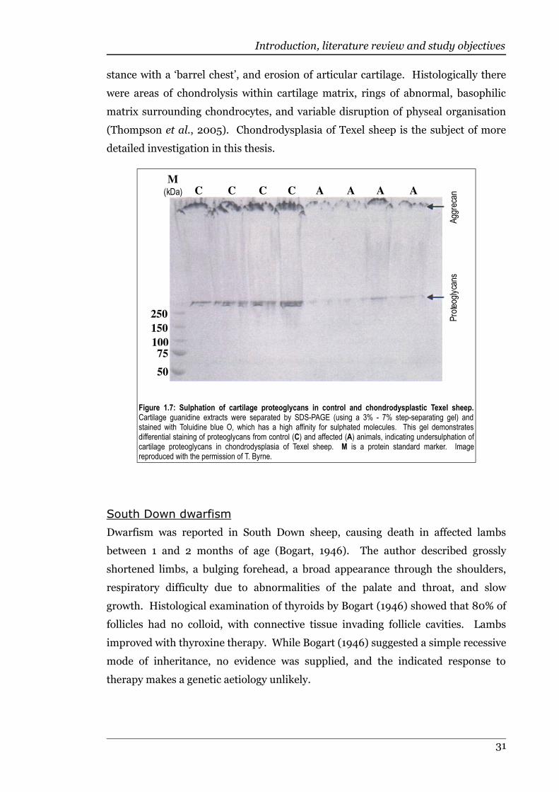
Introduction, literature review and study objectives
stance with a ‘barrel chest’, and erosion of articular cartilage. Histologically there
were areas of chondrolysis within cartilage matrix, rings of abnormal, basophilic
matrix surrounding chondrocytes, and variable disruption of physeal organisation
(Thompson et al., 2005). Chondrodysplasia of Texel sheep is the subject of more
detailed investigation in this thesis.
South Down dwarfism
Dwarfism was reported in South Down sheep, causing death in affected lambs
between 1 and 2 months of age (Bogart, 1946). The author described grossly
shortened limbs, a bulging forehead, a broad appearance through the shoulders,
respiratory difficulty due to abnormalities of the palate and throat, and slow
growth. Histological examination of thyroids by Bogart (1946) showed that 80% of
follicles had no colloid, with connective tissue invading follicle cavities. Lambs
improved with thyroxine therapy. While Bogart (1946) suggested a simple recessive
mode of inheritance, no evidence was supplied, and the indicated response to
therapy makes a genetic aetiology unlikely.
31
Figure 1.7: Sulphation of cartilage proteoglycans in control and chondrodysplastic Texel sheep.Cartilage guanidine extracts were separated by SDS-PAGE (using a 3% - 7% step-separating gel) andstained with Toluidine blue O, which has a high affinity for sulphated molecules. This gel demonstratesdifferential staining of proteoglycans from control (C) and affected (A) animals, indicating undersulphation ofcartilage proteoglycans in chondrodysplasia of Texel sheep. M is a protein standard marker. Imagereproduced with the permission of T. Byrne.
M
(kDa) C A A A A
Aggrecan
250
150
100
75
50
C C C
Proteoglycans
Introduction, literature review and study objectives
Ectrodactyly
A chondrodysplasia of unknown aetiology and characterised by ectrodactyly has
been described in Cheviot sheep (Wray et al., 1971). Clinically, the eyes protruded,
the ears and tail were short, forelimbs were shortened and forehooves were
replaced by an area of hard skin at the end of the limb. Hindhooves were reduced
in size and abnormally shaped, and metapodial bones were slightly distorted at the
distal epiphysis. The authors described an abnormal proximal phalanx in the
forelimbs and absence of the middle and distal phalanges. The tendons of the
superficial and deep digital flexors ended in a mass of fibrous tissue at the distal
extremity of each digit in the forelimbs (Wray et al., 1971).
Similar outbreaks occur occasionally on sheep farms in New Zealand, and have
been attributed to various non-genetic causes, including nitrate toxicity (Gumbrell,
1990). A case of ectrodactyly and hemimelia in a Najdi sheep has been described
(Ramadan, 1993), but no signs of chondrodysplasia were recorded in that
occurence.
1.3.3 Cattle
Brachycephalic "snorter"
Brachycephalic "snorter" dwarfism, named for the upper respiratory noises that
accompany the disease, was at one time an important autosomal recessive disorder
in Hereford cattle (Julian et al., 1957; Jones & Jolly, 1982), but is now uncommon.
The disease has also been recorded in the Angus breed (Baker et al., 1951; Bovard &
Proide, 1965). Detectable by day 125-130 of gestation, the disease featured
disproportionate dwarfism, mandibular prognathia, a characteristically shortened
head relative to its width, accompanied by a bulging frontal area due to early
closure of the spheno-occipital synchondrosis (in normal cattle full closure is
achieved at 24-36 months, while in the dwarf cattle it occurred as early as 5.5
months of age) (Julian et al., 1957). Distal long bones were reduced in length, but
with normal diameter, resulting in apparent thickening (Bovard & Hazel, 1963;
Jones & Jolly, 1982).
Radiographs demonstrated bony projections into the cranium, especially from the
posterior intra-occipital synchondrosis, ventrally flattened lumbar vertebrae with
an undulating ventral surface and a concave dip in the dorsal surface, and
32
Introduction, literature review and study objectives
shortened lumbar transverse processes with forward hooking at the ends (Jones &
Jolly, 1982).
Histologically, chondrocyte columns in the physes were shortened and irregular
(Julian et al., 1957). Ultrastructural investigation revealed chondrocytes
occasionally containing dilated endoplasmic reticulum enclosing an amorphous,
slightly granular material (Jones & Jolly, 1982).
Dolichocephalic dwarfism and Comprest dwarfism have been linked to the
brachycephalic snorter and may be part of the same complex (Gregory & Carroll,
1956; Julian et al., 1959). Dolichocephalic dwarfism was characterised by a very
long head relative to width, and was recognised in Aberdeen-Angus, Hereford and
Shorthorn cattle (Julian et al., 1959).
The Comprest breed was a small-type Hereford. Dwarf calves with or without
crooked legs, and occasional severe lethal forms, occurred when breeding Comprest
with Comprest, and also Comprest with carriers of non-Comprest dwarfism
(Chambers et al., 1954). Some authors suggested that the Comprest dwarf along
with the Dolichocephalic dwarf and Brachycephalic Snorter were all forms of the
same dwarfism complex (Gregory, 1956; Julian et al., 1959). Stonaker, a
stockworker from Colorado Agricultural and Mechanical College, considered the
Comprest Hereford to be the result of a gene acting with partial dominance, and
that homozygosity resulted in dwarfism (Pahnish et al., 1955).
Dexter bulldog
Chondrodysplasia in Dexter cattle is an example of incomplete dominance, where
heterozygous animals develop disproportionate dwarfism while homozygous
affected animals are non-viable “bulldog” calves (Usha et al., 1997). The disease is
caused by mutations in ACAN which codes for the cartilage core protein, aggrecan
(Cavanagh et al., 2007). Bulldog calves have become more common due to a recent
resurgence of the Dexter breed amongst lifestyle farmers. Affected calves are often
aborted prior to 7 months of gestation, and feature a short, scoliotic vertebral
column, severe rhizomelia, hydrocephalus with open cranial sutures, a retruded
muzzle, cleft palate, protruding tongue, and a large ventral abdominal hernia
(Berger & Innes, 1948; Harper et al., 1998).
Histologically, fetal limb bones are composed mainly of highly cellular epiphyseal
cartilage with a central segment of diaphyseal bone of subperiosteal origin
33

Introduction, literature review and study objectives
consisting of dense cancellous bone and some degree of compact bone (Harper et
al., 1998). Limbs lack distinct growth plates, with minimal chondrocyte column
formation or maturation, while periosteal bone formation appears normal. The
histological features and markedly reduced size of the limb bones are consistent
with a failure of endochondral ossification following primary ossification of the
diaphysis of the cartilage anlage (Harper et al., 1998). Genetic tests are available
for detection of the two major disease-causing mutations (University of Sydney,
2007).
A similar condition to the Dexter bulldog has been reported in Jersey cattle (Mead
et al., 1946) and Holstein-Friesians (Bowden, 1970). In the Jersey form of the
disease, the homozygotes were phenotypically very similar to the Dexter lethal, and
the heterozygotes had reduced body and limb length. In contrast, carriers of the
Holstein-friesian form showed no abnormalities, and the disease in this breed can
be considered to be autosomal recessive. Holstein-Friesian bulldog calves also
differed from Dexter bulldog calves in that gestation was completed to full term
(Agerholm et al., 2004b). The authors described a shortened axial skeleton,
marked rhizomelia, a short maxilla and bilateral exophthalmus. Radiographic
lesions included a ventrally rotated splanchnocranium, small and irregular
diaphyses with prominent, rubbery epiphyses causing spinal cord compression in
the vertebral column, a stenotic trachea, a narrow thorax containing hypoplastic
lungs, and bilateral ventricular hypertrophy (Agerholm et al., 2004b).
Histologically, the physes were poorly organised and remnant cartilage cores
extended into the metaphyses (Agerholm et al., 2004b).
A similar autosomal recessive chondrodysplasia was reported in British Friesians,
in which gestation lasted only 200-300 days (Jones et al., 1978). Affected calves
had shortened limbs and muzzle, and had either hydrocephalus and cleft palate or
an oedematous body.
Complex vertebral malformation (CVM)
Although not a form of chondrodysplasia, CVM is mentioned here because it is an
important lethal disorder in Holstein cattle. This disease has an autosomal
recessive mode of inheritance and is caused by a mutation in SLC35A3, for which a
genetic test is available (Ministeriet for Fã~Devarer, Landbrug og Fiskeri Danmarks
Jordbrugsforskning, international patent WO 02/40709 A2, 2003). CVM features
multiple hemivertebrae, synostoses and misshapen vertebrae, scoliosis and
34
Introduction, literature review and study objectives
arthrogryposis. Cardiac malformations are also associated with CVM, and 75% of
affected fetuses are aborted prior to 260 days of gestation (Agerholm et al., 2001;
Nagahata et al., 2002; Agerholm et al., 2004a).
Others
Chondrodysplasia of Guernsey Island cattle, the Telemark lethal mutation,
American Angus dwarfism, chondrodysplasia of Japanese Brown cattle, the
Ayrshire cartilage defect, Stumpy in Shorthorn cattle, and Shorthorn
disproportionate dwarfism are described in Appendix 9. A form of proportionate
dwarfism with an autosomal dominant mode of inheritance complicated by
incomplete penetrance has been identified in commercial Angus herds in Australia
(Latter et al., 2006). As this disease is not considered to be a chondrodysplasia, it is
not discussed further in this review.
1.3.4 Dogs
Alaskan malamute chondrodysplasia
Alaskan Malamute chondrodysplasia is generally regarded as having an autosomal
recessive mode of inheritance (Sande et al., 1982), although the anaemia detectable
in both heterozygous and homozygous animals may suggest an incomplete
dominant rather than recessive mode of inheritance (Fletch & Pinkerton, 1973;
Fletch et al., 1975). While affected pups do not show gross signs of
chondrodysplasia until 5-6 weeks of age, radiographic evidence of abnormalities are
evident as early as 7-10 days, and include flattening of the distal ulnar metaphysis
and delayed ossification of the accessory carpal bone (Sande et al., 1974). In time,
the costochondral junctions develop flaring and cupping, and the antebrachium
becomes shortened and curved (Sande et al., 1974). Histologically, the physes
appear normal at birth (Sande et al., 1982). As the lesions develop, chondrocyte
columns in the proliferative zone become irregular and are separated by a broad
core of matrix. The primary and secondary spongiosa contain a reduced number of
large, irregular spicules, with irregular thickening of physes occasionally associated
with trabecular microfractures (Sande et al., 1982). A reduction in the ratio of
chondroitin 4-sulphate to chondroitin 6-sulphate in physeal cartilage has been
reported (Bingel et al., 1985).
35
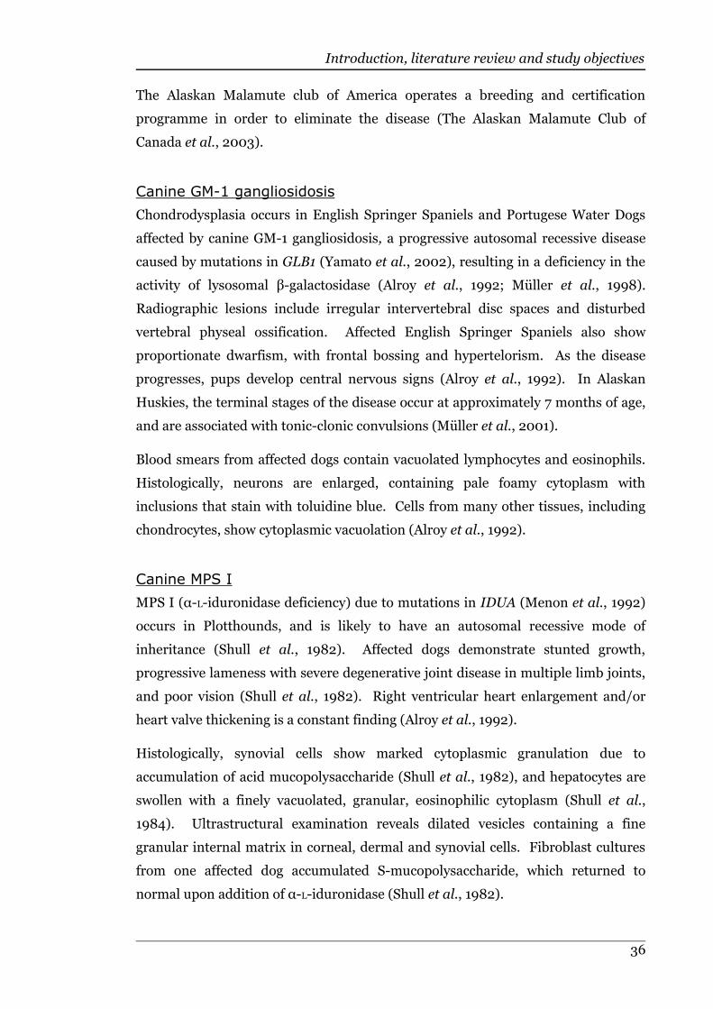
Introduction, literature review and study objectives
The Alaskan Malamute club of America operates a breeding and certification
programme in order to eliminate the disease (The Alaskan Malamute Club of
Canada et al., 2003).
Canine GM-1 gangliosidosis
Chondrodysplasia occurs in English Springer Spaniels and Portugese Water Dogs
affected by canine GM-1 gangliosidosis, a progressive autosomal recessive disease
caused by mutations in GLB1 (Yamato et al., 2002), resulting in a deficiency in the
activity of lysosomal β-galactosidase (Alroy et al., 1992; Müller et al., 1998).
Radiographic lesions include irregular intervertebral disc spaces and disturbed
vertebral physeal ossification. Affected English Springer Spaniels also show
proportionate dwarfism, with frontal bossing and hypertelorism. As the disease
progresses, pups develop central nervous signs (Alroy et al., 1992). In Alaskan
Huskies, the terminal stages of the disease occur at approximately 7 months of age,
and are associated with tonic-clonic convulsions (Müller et al., 2001).
Blood smears from affected dogs contain vacuolated lymphocytes and eosinophils.
Histologically, neurons are enlarged, containing pale foamy cytoplasm with
inclusions that stain with toluidine blue. Cells from many other tissues, including
chondrocytes, show cytoplasmic vacuolation (Alroy et al., 1992).
Canine MPS I
MPS I (α-L-iduronidase deficiency) due to mutations in IDUA (Menon et al., 1992)
occurs in Plotthounds, and is likely to have an autosomal recessive mode of
inheritance (Shull et al., 1982). Affected dogs demonstrate stunted growth,
progressive lameness with severe degenerative joint disease in multiple limb joints,
and poor vision (Shull et al., 1982). Right ventricular heart enlargement and/or
heart valve thickening is a constant finding (Alroy et al., 1992).
Histologically, synovial cells show marked cytoplasmic granulation due to
accumulation of acid mucopolysaccharide (Shull et al., 1982), and hepatocytes are
swollen with a finely vacuolated, granular, eosinophilic cytoplasm (Shull et al.,
1984). Ultrastructural examination reveals dilated vesicles containing a fine
granular internal matrix in corneal, dermal and synovial cells. Fibroblast cultures
from one affected dog accumulated S-mucopolysaccharide, which returned to
normal upon addition of α-L-iduronidase (Shull et al., 1982).
36

Introduction, literature review and study objectives
Canine MPS VII
Canine MPS VII is a disorder with an autosomal recessive mode of inheritance,
caused by a deficiency of β-glucuronidase due to mutations in GUSB (Haskins et
al., 1991; Ray et al., 1998; Ray et al., 1999). Affected animals show a reduced
growth rate and develop a broad, short face, broad chest and corneal clouding.
Cytoplasmic vacuolation occurs in cells of many tissues, including chondrocytes.
Radiographs demonstrate severe progressive epiphyseal dysplasia and bilateral hip
subluxation or luxation. Cardiac abnormalities, hepatomegaly and a misshapen,
narrow trachea with overlapping rings are additional features, and there is
excessive urinary excretion of mucopolysaccharides (chondroitin 4- and 6-sulphate
and dermatan sulphate) (Haskins et al., 1991).
English pointer enchondrodystrophy
Last reported in 1984, an enchondrodystrophy with an autosomal recessive mode of
inheritance in the English Pointer breed was identified in England and Australia
(Whitbread et al., 1983; Lavelle, 1984). Signs of deformity were apparent prior to
weaning, and progressed to include flaring of long bone metaphyses and valgus
deformity of the distal forelimbs. Marked degenerative changes occurred in many
joints, and the thorax appeared dorso-ventrally flattened. In some severe cases
kyphosis developed (Whitbread et al., 1983). Histology of the physes demonstrated
thickened hypertrophic zones in some bones and a limited, disorganised
proliferative zone in others. The transitional zone of articular cartilage underwent
degenerative changes, beginning as decreased staining intensity and progressing to
the formation of cystic spaces (Whitbread et al., 1983).
Miniature poodle pseudoachondroplasia
Pseudoachondroplastic dysplasia with an autosomal mode of inheritance was
identified in Miniature Poodles, associated with variable undersulphation of
cartilage matrix proteoglycans (Riser et al., 1980; Bingel et al., 1986). Gross lesions
appeared at 10-21 days of age, including brachygnathia superior, thickened tracheal
rings, a dorso-ventrally flattened thorax, mild kyphosis and enlarged costochondral
junctions (Gardner, 1959; Amlöff, 1961; Riser et al., 1980). Radiographic lesions
consisted of enlarged, stippled epiphyses with malformation of both the long bones
and vertebral column accompanied by flared metaphyses of long bones.
Histologically there was delayed ossification of long bone epiphyses (Riser et al.,
37
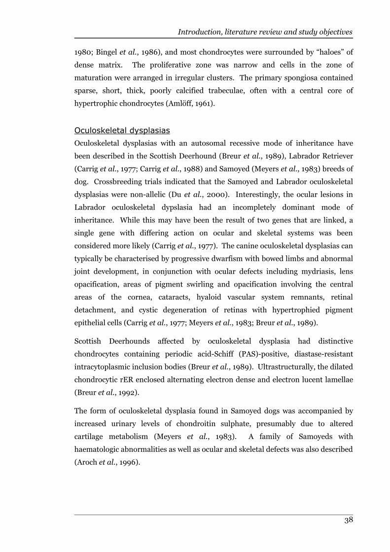
Introduction, literature review and study objectives
1980; Bingel et al., 1986), and most chondrocytes were surrounded by “haloes” of
dense matrix. The proliferative zone was narrow and cells in the zone of
maturation were arranged in irregular clusters. The primary spongiosa contained
sparse, short, thick, poorly calcified trabeculae, often with a central core of
hypertrophic chondrocytes (Amlöff, 1961).
Oculoskeletal dysplasias
Oculoskeletal dysplasias with an autosomal recessive mode of inheritance have
been described in the Scottish Deerhound (Breur et al., 1989), Labrador Retriever
(Carrig et al., 1977; Carrig et al., 1988) and Samoyed (Meyers et al., 1983) breeds of
dog. Crossbreeding trials indicated that the Samoyed and Labrador oculoskeletal
dysplasias were non-allelic (Du et al., 2000). Interestingly, the ocular lesions in
Labrador oculoskeletal dypslasia had an incompletely dominant mode of
inheritance. While this may have been the result of two genes that are linked, a
single gene with differing action on ocular and skeletal systems was been
considered more likely (Carrig et al., 1977). The canine oculoskeletal dysplasias can
typically be characterised by progressive dwarfism with bowed limbs and abnormal
joint development, in conjunction with ocular defects including mydriasis, lens
opacification, areas of pigment swirling and opacification involving the central
areas of the cornea, cataracts, hyaloid vascular system remnants, retinal
detachment, and cystic degeneration of retinas with hypertrophied pigment
epithelial cells (Carrig et al., 1977; Meyers et al., 1983; Breur et al., 1989).
Scottish Deerhounds affected by oculoskeletal dysplasia had distinctive
chondrocytes containing periodic acid-Schiff (PAS)-positive, diastase-resistant
intracytoplasmic inclusion bodies (Breur et al., 1989). Ultrastructurally, the dilated
chondrocytic rER enclosed alternating electron dense and electron lucent lamellae
(Breur et al., 1992).
The form of oculoskeletal dysplasia found in Samoyed dogs was accompanied by
increased urinary levels of chondroitin sulphate, presumably due to altered
cartilage metabolism (Meyers et al., 1983). A family of Samoyeds with
haematologic abnormalities as well as ocular and skeletal defects was also described
(Aroch et al., 1996).
38
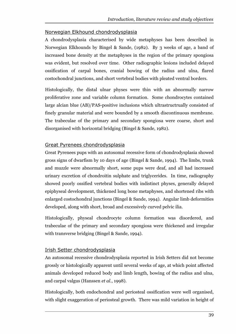
Introduction, literature review and study objectives
Norwegian Elkhound chondrodysplasia
A chondrodysplasia characterised by wide metaphyses has been described in
Norwegian Elkhounds by Bingel & Sande, (1982). By 3 weeks of age, a band of
increased bone density at the metaphyses in the region of the primary spongiosa
was evident, but resolved over time. Other radiographic lesions included delayed
ossification of carpal bones, cranial bowing of the radius and ulna, flared
costochondral junctions, and short vertebral bodies with pleated ventral borders.
Histologically, the distal ulnar physes were thin with an abnormally narrow
proliferative zone and variable column formation. Some chondrocytes contained
large alcian blue (AB)/PAS-positive inclusions which ultrastructrually consisted of
finely granular material and were bounded by a smooth discontinuous membrane.
The trabeculae of the primary and secondary spongiosa were coarse, short and
disorganised with horizontal bridging (Bingel & Sande, 1982).
Great Pyrenees chondrodysplasia
Great Pyrenees pups with an autosomal recessive form of chondrodysplasia showed
gross signs of dwarfism by 10 days of age (Bingel & Sande, 1994). The limbs, trunk
and muzzle were abnormally short, some pups were deaf, and all had increased
urinary excretion of chondroitin sulphate and triglycerides. In time, radiography
showed poorly ossified vertebral bodies with indistinct physes, generally delayed
epiphyseal development, thickened long bone metaphyses, and shortened ribs with
enlarged costochondral junctions (Bingel & Sande, 1994). Angular limb deformities
developed, along with short, broad and excessively curved pelvic ilia.
Histologically, physeal chondrocyte column formation was disordered, and
trabeculae of the primary and secondary spongiosa were thickened and irregular
with transverse bridging (Bingel & Sande, 1994).
Irish Setter chondrodysplasia
An autosomal recessive chondrodysplasia reported in Irish Setters did not become
grossly or histologically apparent until several weeks of age, at which point affected
animals developed reduced body and limb length, bowing of the radius and ulna,
and carpal valgus (Hanssen et al., 1998).
Histologically, both endochondral and periosteal ossification were well organised,
with slight exaggeration of periosteal growth. There was mild variation in height of
39
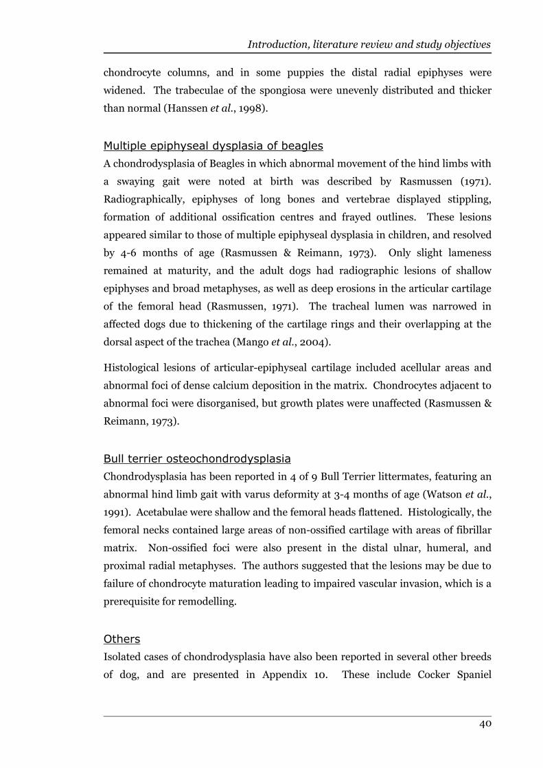
Introduction, literature review and study objectives
chondrocyte columns, and in some puppies the distal radial epiphyses were
widened. The trabeculae of the spongiosa were unevenly distributed and thicker
than normal (Hanssen et al., 1998).
Multiple epiphyseal dysplasia of beagles
A chondrodysplasia of Beagles in which abnormal movement of the hind limbs with
a swaying gait were noted at birth was described by Rasmussen (1971).
Radiographically, epiphyses of long bones and vertebrae displayed stippling,
formation of additional ossification centres and frayed outlines. These lesions
appeared similar to those of multiple epiphyseal dysplasia in children, and resolved
by 4-6 months of age (Rasmussen & Reimann, 1973). Only slight lameness
remained at maturity, and the adult dogs had radiographic lesions of shallow
epiphyses and broad metaphyses, as well as deep erosions in the articular cartilage
of the femoral head (Rasmussen, 1971). The tracheal lumen was narrowed in
affected dogs due to thickening of the cartilage rings and their overlapping at the
dorsal aspect of the trachea (Mango et al., 2004).
Histological lesions of articular-epiphyseal cartilage included acellular areas and
abnormal foci of dense calcium deposition in the matrix. Chondrocytes adjacent to
abnormal foci were disorganised, but growth plates were unaffected (Rasmussen &
Reimann, 1973).
Bull terrier osteochondrodysplasia
Chondrodysplasia has been reported in 4 of 9 Bull Terrier littermates, featuring an
abnormal hind limb gait with varus deformity at 3-4 months of age (Watson et al.,
1991). Acetabulae were shallow and the femoral heads flattened. Histologically, the
femoral necks contained large areas of non-ossified cartilage with areas of fibrillar
matrix. Non-ossified foci were also present in the distal ulnar, humeral, and
proximal radial metaphyses. The authors suggested that the lesions may be due to
failure of chondrocyte maturation leading to impaired vascular invasion, which is a
prerequisite for remodelling.
Others
Isolated cases of chondrodysplasia have also been reported in several other breeds
of dog, and are presented in Appendix 10. These include Cocker Spaniel
40

Introduction, literature review and study objectives
chondrodystrophy, German Shepherd chondrodysplasia, Scottish Terrier
achondroplasia and Akita achondrogenesis.
1.3.5 Cats
Feline MPS VI
Chondrodysplasia is described in Siamese cats with MPS VI, which is caused by a
recessively inherited deficiency of aryl-sulfatase B (Haskins et al., 1980a) and can
be considered an animal model for Maroteaux-Lamy Syndrome in humans (Jezyk
et al., 1977). Clinically, at 8 weeks of age affected cats have a small head with a
broad, short maxilla, odontoid hypoplasia, small ears, cloudy corneas and pectus
excavatum. Skeletal lesions, including vertebral fusion or subluxation, and
degenerative joint disease progress in severity with age (Haskins et al., 1980a;
Haskins et al., 1981).
Histologically, there is evidence of retarded endochondral ossification with focal
necrosis and physeal dysplasia (Orgad et al., 1989). Articular cartilage is
hypercellular and supported by a sclerotic subchondral bony plate containing
islands of cartilage. Chondrocytes, and cells in many other tissues, are packed with
membrane-bound inclusions (Haskins et al., 1980a). Coarse metachromatic
granules are present in peripheral blood neutrophils (Haskins et al., 1981), and
excessive glycosaminoglycans, mostly dermatan sulphate, are excreted in the urine
(Jezyk et al., 1977; Haskins et al., 1980a).
Feline MPS VII
Feline MPS VII is caused by β-glucuronidase deficiency (Fyfe et al., 1999). Lesions
are apparent by 2 months of age, and by 6 months of age there is retarded growth,
corneal clouding, delayed dental eruption, abdominal enlargement and a shortened
maxilla. Radiographic abnormalities include fused cervical vertebrae, a flattened
thorax, pectus excavatum, short thoracic and lumbar vertebrae, and bilateral
coxofemoral subluxation. Metachromatic granules are present in peripheral blood
neutrophils, and there is excessive urinary excretion of chondroitin sulphate (Fyfe
et al., 1999).
41
Introduction, literature review and study objectives
Scottish Fold osteochondrodysplasia
Scottish Fold cats are known for the typical ear-fold, believed by some to be a result
of a cartilage abnormality resulting in inability of the pinna to remain pricked
(Malik et al., 1999). The causative “folded-ear” gene acts in a dominant manner.
Osteochondrodysplasia in the Scottish Fold breed is characterised by lameness,
shortened and misshapen distal limbs, and short, broad caudal vertebrae.
Intertarsal and tarsometatarsal joint spaces progressively diminish and
periarticular osteophytes form, eventually leading to ankylosis (Chang et al., 2007;
Malik et al., 1999). Skeletal lesions were first thought to occur only in cats
homozygous for the folded-ear gene (Malik et al., 1999), but later studies indicated
milder skeletal lesions may also occur in heterozygous animals (Hubler et al.,
2004).
Histologically, articular cartilage is irregularly thickened with necrotic foci, flaking
and fibrillation, accompanied by chronic synovitis. Persistent islands of cartilage
are evident in epiphyseal bone (Malik et al., 1999).
Others
The Munchkin breed and rare forms of chondrodysplasia in cats such as
metaphyseal chondrodysplasia, chondrodysplasia with Pelger-Hüet anomaly and
feline congential hypothyroidism are listed in Appendix 10.
1.3.6 Pigs
Dwarfism
A naturally-occurring mutation in type X collagen has been identified in domestic
pigs by Nielsen (2000), representing an animals model for Schmid metaphyseal
chondrodysplasia in humans. The disease in pigs is characterised by an autosomal
dominant mode of inheritance, shortened long-bone with abnormal metaphyses
containing tongues of physeal cartilage. Histologically, physes contain a
disorganised hypertrophic zone and stain poorly with H&E or TB.
Danish Landrace chondrodysplasia
An autosomal recessive form of chondrodysplasia described in Danish Landrace
pigs was characterised by shortening of the limbs, especially the forelimbs, with an
apparently normal axial skeleton and skull (Jensen et al., 1984). The joints had
42

Introduction, literature review and study objectives
increased mobility and became degenerate over time. The hair coat in affected
animals was abnormally long.
Radiographically, long bones were abnormally short and thick, with wide
metaphyses and mushroom-shaped epiphyses. Histologically, physeal lesions
included reduction of the proliferative zone and an irregular hypertrophic zone
(Jensen et al., 1984).
Hereditary dwarfism
Spontaneously occurring hereditary dwarfism has been reported in Large White X
Landrace pigs, featuring shortened limbs, muzzle, ribs and spinal column (Mores et
al., 1989). Histologically, physes were enlarged with foci of degenerate matrix at
the junction of the physis and metaphysis, and metaphyseal trabeculae were
irregular.
1.3.7 Deer
Four cases of chondrodystrophy were reported in an inbred herd of fallow deer
(Baker et al., 1979). Most bones, but particularly those of the limbs, were reduced
in length and there was varus deformity of the forelimbs. Phalanges were less
severely affected than the other limb bones. Epiphyses were distorted, and in some
areas physeal closure was premature. The sternebrae were enlarged and
misshapen. A cleft palate, domed skull and large fontanelles were also noted.
Histologically, both the proliferative and hypertrophic zones of the physes were
reduced (Baker et al., 1979).
1.3.8 Goats
A form of dwarfism resembling “bulldog” calves was described in an inbred herd of
goats (Kaikini & Malkhede, 1969). The disorder featured an overly prominent
cranial vault, short, wide facial bones, brachygnathia superior, protruding tongue,
short neck, markedly shortened limbs and a ventral hernia.
1.3.9 Mice
Some chondrodysplasias in mice are caused by naturally occurring mutations while
many others are the result of intentional manipulations for the purpose of research.
Appendix 11 includes a table of chondrodysplasias in transgenic mice. Only
naturally occurring chondrodysplasias will be presented here.
43
Introduction, literature review and study objectives
Achondroplasia ( cn )
Achondroplastic mice can be distinguished from normal mice at birth (Bonucci et
al., 1976). The authors described grossly shortened long bones and small caudal
vertebrae. Sannasgala & Johnson, (1990) identified delayed formation of secondary
centres of ossification and thin physes containing abnormally small chondrocytes.
Histologically, the interterritorial matrix stained less intensely than normal with
either AB or colloidal iron, suggesting a reduced proteoglycan content (Bonucci et
al., 1976). Primary spongiosa was scant, with short, thick longitudinal septae
(Sannasgala & Johnson, 1990).
The skeletal changes seen in homozygous affected (cn/cn) mice are very similar to
those seen in human achondroplasia and the mouse has been considered to be a
model of the human disease (Bonucci et al., 1976). The causative mutation for
achondroplasia in mice, however is in NPR2, the atrial natriuretic peptide receptor-
B (Tsuji & Kunieda, 2005), rather than in FGFR3.
Brachymorphic mouse
Brachymorphism in mice is an autosomal recessive disease associated with
mutations in PAPSS2 resulting in decreased PAPS synthase activity (Pennypacker
et al., 1981; Kurima et al., 1998). Developing during the first 4 weeks of life, gross
lesions included shortened long bones and tail, and a domed skull (Orkin et al.,
1977; Kurima et al., 1998). Histologically, hypertrophic zones were narrowed and
the physes became less organised with time. This disorder is considered to be
homologous to Pakistani spondyloepimetaphyseal dysplasia in humans (ul Haque
et al., 1998).
Cartilage matrix deficient ( cmd) mice
Cartilage matrix deficiency in mice is a lethal autosomal recessive disorder
characterised by short, wide, endochondral bones, a short head, thoracic
hypoplasia, thickened tracheal rings, and subcutaneous oedema (Bell et al., 1986).
Near complete deletion of the aggrecan gene has been identified (Krueger et al.,
1999). Kobayakawa et al (1985) described epiphyseal cartilage with reduced
extracellular matrix and poor physeal organisation, occasionally containing
degenerate foci. A deficiency of acid mucopolysaccharides in the cartilage
extracellular matrix has been recognised (Bell et al., 1986). Ultrastructurally,
abnormally dense networks of thickened, mostly parallel, collagen fibrils were
44
Introduction, literature review and study objectives
closely associated with chondrocytes, and the number of matrix vesicles was
reduced (Kobayakawa et al., 1985).
Others
Chondrodysplasia “cho”, murine brachypodism “bp”, Chubby “cby”,
dispropoprtionate micromelia “Dmm”, cartilage anomaly “can”, dwarfism, and
MPS VII are listed in Appendix 11.
1.3.10 Rabbits
Few chondrodysplasias are described in rabbits. These include chondrodysplasia in
the presence of Pelger-Hüet anomaly, where the skeletal lesions have a recessive
mode of inheritance while the leukocytic changes demonstrate an incomplete
dominant mode of inheritance (Nachtsheim, 1950). Autosomal recessive “da”
achondroplasia has been identified in the New Zealand White rabbit (Crary &
Sawin, 1952), “ac” chondrodysplasia is an incompletely dominant disorder (Crary et
al., 1958) where mitochondria lack one phosphorylation site (Mackler et al., 1972),
and “cd” chondrodystrophy is a lethal autosomal recessive disorder that appears
similar to metatropic dwarfism in human beings (Fox & Crary, 1975). These
diseases are listed in more detail in Appendix 11.
1.3.11 Birds
Several forms of chondrodysplasia have been described in domestic birds. These
are summarised in Appendix 12.
1.3.12 Non-human primates
A case of lethal short-limbed dwarfism resembling thanatophoric dwarfism in
humans was described in a rhesus monkey (Macaca mulatta) (Zeman & Baskin,
1986). Grossly, all limbs were markedly rhizomelic and bones were curved with
flared and irregular metaphyses. The head was relatively large, the face was flat
with slight protusion of the mandible and the thorax was narrow due to short,
flared ribs. A normal female reproductive tract was identified along with vestigial
structures of undifferentiated tissue grossly resembling testicles in the inguinal
area. Endochondral ossification was markedly disorganised, with no chondrocyte
columns in physes and failure of orderly vascular penetration. The authors
45
Introduction, literature review and study objectives
described a poorly organised primary spongiosa with thick, irregular and often
horizontal trabeculae (Zeman & Baskin, 1986).
1.4 Nutritional chondrodysplasia
1.4.1 Manganese deficiency
Manganese (Mn) is crucial for normal cartilage formation due to its function in two
enzyme systems required for chondroitin sulphate synthesis: polymerase enzyme
which links UDP-N-acetyl-galactosamine and UDP-glucuronic acid to form the
chondroitin polysaccharide, and galactosyltransferase which incorporates galactose
from UDP-galactose to the galactose-galactose-xylose trisaccharide linking the
polysaccharide and the associated protein (Leach et al., 1969; Tsopanakis &
Herries, 1976). Because of this, Mn deficiency causes reversible reduction of the
incorporation of sulphate into cartilage and impaired glycosaminoglycan
metabolism (Leach & Muenster, 1962; Bolze et al., 1985).
Skeletal abnormalities secondary to Mn deficiency have been reported in several
species (Neher et al., 1956; Tsai & Everson, 1967; Leach, 1968; Valero et al., 1990)
and typically occur in the offspring of deficient dams (Caskey et al., 1944; Frost et
al., 1959; Valero et al., 1990), but can also occur in young growing animals fed a
deficient diet (Leach, 1968).
Mn deficiency in adult cattle results in reproductive disorders manifesting as
increased returns to service (Rojas et al., 1965). Calves born to deficient dams,
however, show generalised chondrodysplasia characterised by short limbs with
enlarged joints, enlarged costochondral junctions and excessive cartilage in long
bone epiphyses (Valero et al., 1990). Tracheal rings are thickened, and affected
calves are prone to tracheal collapse. The animals recover slowly after birth,
although in severe cases the legs remain bent and short for several months.
Histologically, the costochondral junctions are irregular, with poorly organised
proliferating chondrocytes and an absence of hypertrophic chondrocytes (Valero et
al., 1990). In many areas the matrix contains numerous fine basophilic fibrils and
large areas of rarefaction. "Collagen rings" surrounding chondrocytes appear
similar to those seen in achondrogenesis 1b and Texel chondrodysplasia. Many
small PAS-positive droplets are present in the matrix close to the physes, consisting
of interwoven fibres with a stellate appearance ultrastructurally. Similar changes
are seen in articular cartilage of the long bones (Valero et al., 1990), and cartilage
46
Introduction, literature review and study objectives
from affected calves has a low mucopolysaccharide content (Dyer & Rojas, 1965).
Repeated outbreaks of chondrodysplasia in calves born following periods of
drought in Australia are likely to be due to Mn deficiency (McLaren, et al., 2007).
An increased susceptibility to convulsions was identified in Mn-deficient rats
(Hurley et al., 1963), along with ataxia resulting from abnormal development of the
middle ear (Hurley et al., 1960). Mn deficiency has also been associated with ataxia
in chickens (Caskey et al., 1944). Although Mn deficiency has not been assessed in
the acorn calf abnormality or bovine congenital joint laxity and dwarfism, the
history of poor diet and the type of lesions involved are suggestive of this
nutritional aetiology (Barry & Murphy, 1964; Ribble & Janzen, 1987).
1.4.2 Plant toxicity
Exposure to certain plant toxins, especially during fetal development, can result in
generalised skeletal defects. These can be due to direct action of a toxin or
secondary to metabolic disturbance, and plant toxicity is usually a differential
diagnosis in outbreaks of skeletal disease in grazing animals. Skeletal deformities
resembling crooked calf disease, including arthrogryposis, scliosis, torticollis and
cleft palate, can be seen in offspring from dams fed Conium maculatum (poison-
hemlock), Nicotiana glauca (tree tobacco) or Lupinus formosus (lunara lupine)
during gestation (Panter et al., 1988b; Panter et al., 1990). While primary
myopathy is not considered to be responsible for the skeletal lesions (Abbot et al.,
1986), altered maternal uterine contraction may be involved in their development.
Congenital conium toxicity in lambs results in carpal flexural deformities, angular
limb deformities and kinked tails, all resolving by 8 weeks of age (Panter et al.,
1988a). Osteolathyrism is a non-congenital skeletal disorder caused by ingestion of
Lathyrus odoratus (sweet pea) (Bélanger et al., 1959). The resulting interference
with lysyl oxidase activity increases connective tissue fragility, leading to skeletal
effects that include joint deformities and spontaneous fractures.
Some plants may interfere with mineral absorption either directly or indirectly by
concentrating other interacting minerals. For example, if high levels of calcium or
phosphorus were present in forage, Mn absorption may be hindered, especially in
ruminants (Spears, 2003). Various components of plants may directly interfere
with mineral absorption, for example, some varieties of fava bean are able to inhibit
the intestinal absorption of Mn and zinc (Rubio et al., 1992), both of which are
47
Introduction, literature review and study objectives
minerals required for normal cartilage development and endochondral ossification
(Leach et al., 1969; Ortega et al., 2004). Alternatively, mycotoxins produced by
some fungi on decomposing plant matter may interfere with growth or
development of many tissues (Wangikar et al., 2005).
1.5 Conclusion and study objectives
The roles of matrix components and the pathways involved in the regulation of
cartilage growth and endochondral ossification are complex and incompletely
understood. Much progress has been made through the study of disorders which
accent the effects of an abnormality in a single component of cartilage or its
regulation. In order to learn most efficiently from a naturally occurring
chondrodysplasia, its character needs to be thoroughly described microscopically,
biochemically, and, ideally, genetically. More tools, especially with respect to
molecular and genetic studies, are available than ever before and are proving
increasingly valuable in improving our understanding of cartilage development and
chondrodysplastic disease.
The chondrodysplasia reported recently in Texel sheep has presented an
opportunity to study a new hereditary cartilage disorder in sheep, an animal with
great potential as a model of skeletal disease in human beings. The preliminary
studies already performed on chondrodysplastic Texel sheep have indicated strong
similarities to the chondrodysplasias in human beings that involve reduced
sulphate uptake. The objectives of this study are:
i) To describe in detail the gross, histological, histochemical and
ultrastructural lesions of chondrodysplasia in Texel sheep in order to support the
classification of this disorder as a new heritable disease of sheep and to assist in
determining the likely mechanism.
ii) To measure the pattern of skeletal growth in chondrodysplastic lambs
in order to assess the phenotypic effect of this genetic defect on different skeletal
components, and to test the hypothesis that bone lengths and growth rates in
chondrodysplastic lambs are less than in controls.
iii) To quantify the transport of sulphate into fibroblasts in order to test
the hypothesis that undersulphation of hyaline cartilage in chondrodysplasia of
Texel sheep is due to reduced cellular uptake of sulphate.
48

Introduction, literature review and study objectives
iv) To use a collection of microsatellite markers on samples from animals
of known phenotype in order to test for linkage disequilibrium, which may be able
to localise the causative mutation in chondrodysplasia of Texel sheep to an area
within a chromosome. This will be the first step in the development of a genetic
test capable of identifying carrier animals.
v) To quantify the chondroitin 4-sulphate and chondroitin 6-sulphate
dissacharide content in chondrodysplastic and normal lambs of differing ages to (a)
compare the disaccharide ratios with those found in disorders of sulphate transport
in human beings, to (b) investigate the change in proteoglycan sulphation with age
in affected and control animals, and to (c) test the hypothesis that a difference in
chondroitin sulphate disaccharides is detectable in affected newborn lambs prior to
the appearance of gross or microscopic lesions.
49
2 Pathology of chondrodysplasia in Texel
sheep
Pathology of chondrodysplasia in Texel sheep
2.1 Introduction
Chondrodysplasias are defined as abnormalities in the growth and/or development
of cartilage. The aetiology may be genetic or environmental, and disproportionate
dwarfism is a common manifestation due to the requirement for normal cartilage in
the development of bones by endochondral ossification (Rimoin et al., 1976; van
der Harten et al., 1988; Ala-Kokko et al., 1990). Several forms of chondrodysplasia
have been described in sheep, most notably "spider lamb syndrome" (SLS) in the
Suffolk and Hampshire breeds, and the historical Ancon mutant (Landauer &
Chang, 1949; Oberbauer et al., 1995). Others include Cheviot achondroplasia
(Wray et al., 1971), Southdown dwarfism (Bogart, 1946), lethal dwarfism (a
sporadic outbreak in a mixed breed flock) (Duffell et al., 1985), and the recently
identified chondrodysplasia in Texels (Thompson et al., 2005).
Chondrodysplasia in Texel sheep was first recorded in a commercial sheep breeding
and lamb fattening property in Southland, New Zealand. Preliminary investigation
of affected lambs revealed what appeared to be an autosomal recessive form of
chondrodysplasia pathologically distinct from other chondrodysplasias described in
sheep (Byrne, 2005; Thompson et al., 2005). Biochemical and histological features
bore a strong resemblance to a group of chondrodysplasias caused by mutations in
the gene for the diastrophic dysplasia sulphate transporter (DTDST), SLC26A2, in
human beings. This group includes achondrogenesis type 1b, atelosteogenesis type
II, diastrophic dysplasia and recessive multiple epiphyseal dysplasia (Hästbacka et
al., 1994; Superti-Furga, 1994; Hästbacka et al., 1996b; Rimoin et al., 1998; Rossi &
Superti-Furga, 2001). These disorders are characterised by disproportionate
dwarfism, deficient uptake of sulphate by chondrocytes and fibroblasts in vitro, and
decreased sulphation of cartilage extracellular matrix proteoglycans (Hästbacka et
al., 1994; Rossi & Superti-Furga, 2001; Superti-Furga, 2001).
The objective of this investigation was to conduct a detailed morphological study of
chondrodysplasia in Texel sheep. This information will support the classification of
Texel chondrodysplasia as a new disease, distinct from other ovine
chondrodysplasias, and provide an indication of the suitability of chondrodysplasia
of Texel sheep as a model for similar disorders in human beings.
51
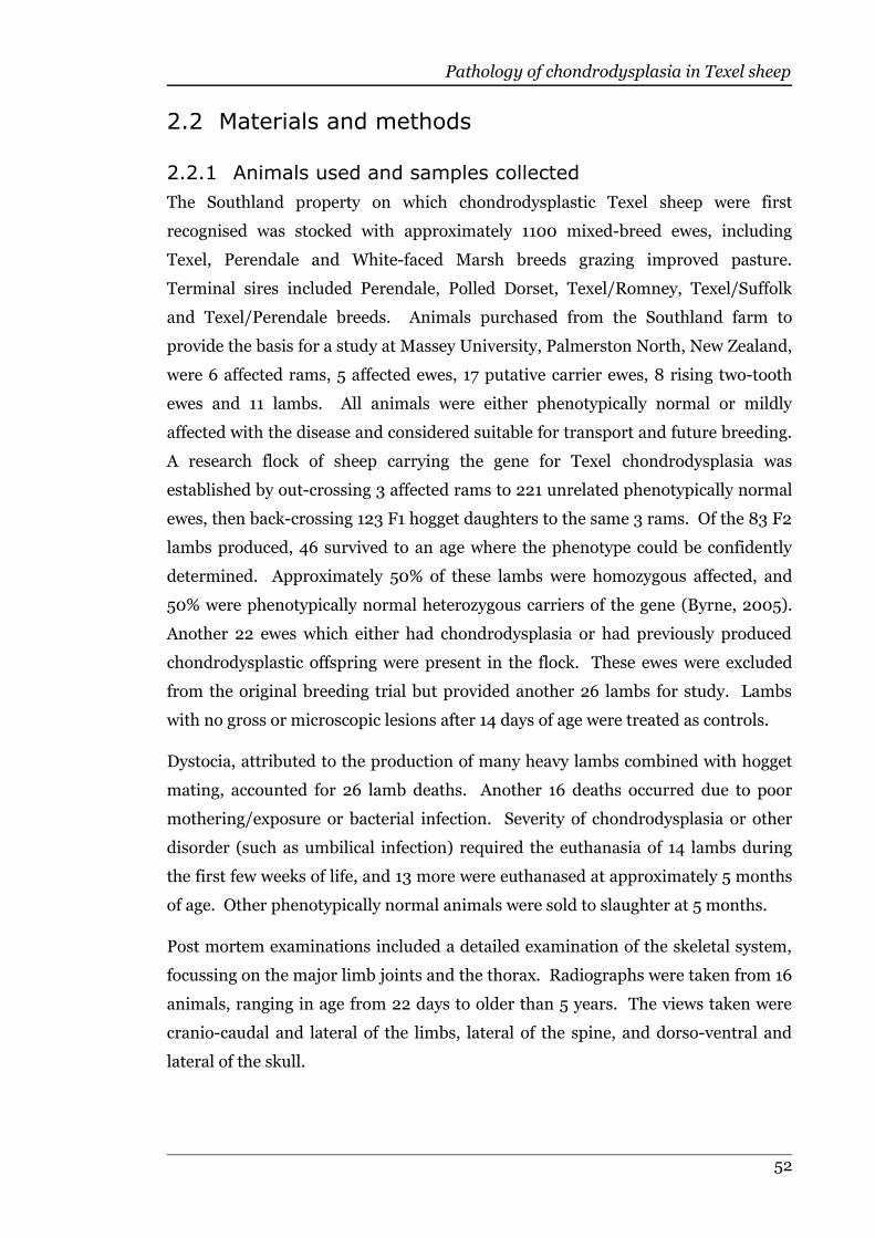
Pathology of chondrodysplasia in Texel sheep
2.2 Materials and methods
2.2.1 Animals used and samples collected
The Southland property on which chondrodysplastic Texel sheep were first
recognised was stocked with approximately 1100 mixed-breed ewes, including
Texel, Perendale and White-faced Marsh breeds grazing improved pasture.
Terminal sires included Perendale, Polled Dorset, Texel/Romney, Texel/Suffolk
and Texel/Perendale breeds. Animals purchased from the Southland farm to
provide the basis for a study at Massey University, Palmerston North, New Zealand,
were 6 affected rams, 5 affected ewes, 17 putative carrier ewes, 8 rising two-tooth
ewes and 11 lambs. All animals were either phenotypically normal or mildly
affected with the disease and considered suitable for transport and future breeding.
A research flock of sheep carrying the gene for Texel chondrodysplasia was
established by out-crossing 3 affected rams to 221 unrelated phenotypically normal
ewes, then back-crossing 123 F1 hogget daughters to the same 3 rams. Of the 83 F2
lambs produced, 46 survived to an age where the phenotype could be confidently
determined. Approximately 50% of these lambs were homozygous affected, and
50% were phenotypically normal heterozygous carriers of the gene (Byrne, 2005).
Another 22 ewes which either had chondrodysplasia or had previously produced
chondrodysplastic offspring were present in the flock. These ewes were excluded
from the original breeding trial but provided another 26 lambs for study. Lambs
with no gross or microscopic lesions after 14 days of age were treated as controls.
Dystocia, attributed to the production of many heavy lambs combined with hogget
mating, accounted for 26 lamb deaths. Another 16 deaths occurred due to poor
mothering/exposure or bacterial infection. Severity of chondrodysplasia or other
disorder (such as umbilical infection) required the euthanasia of 14 lambs during
the first few weeks of life, and 13 more were euthanased at approximately 5 months
of age. Other phenotypically normal animals were sold to slaughter at 5 months.
Post mortem examinations included a detailed examination of the skeletal system,
focussing on the major limb joints and the thorax. Radiographs were taken from 16
animals, ranging in age from 22 days to older than 5 years. The views taken were
cranio-caudal and lateral of the limbs, lateral of the spine, and dorso-ventral and
lateral of the skull.
52
Pathology of chondrodysplasia in Texel sheep
Samples collected routinely for histology included humerus, femur, lumbar
vertebrae 3 and 4, costochondral junctions 6 and 7, trachea (a 2-3cm section
approximately 5cm from the larynx), larynx, heart, lung, liver, and kidney. Bones
from young lambs were fixed entire; those from older animals were sectioned
longitudinally prior to fixation.
2.2.2 Tissue processing and analysis
All tissues for histology were fixed in 10% neutral buffered formalin for at least 48
hours, after which bones were cut into 2mm thick slabs using a low-speed band-
saw. Bone slabs were decalcified in deltaCAL (Delta Products Groups, Aurora, IL)
overnight, followed by rinsing in tap water. All tissues were embedded in paraffin
wax using standard techniques, prior to sectioning at 4µm thickness. Sections from
all samples were stained with haematoxylin and eosin (H&E), with an additional
section from cartilaginous tissues stained with toluidine blue (TB). All tissues were
processed in batches containing samples from both affected and control animals.
Chondrocyte density was investigated in the articular-epiphyseal complex, the
transitional zone of the articular cartilage, and the central area of the tracheal
cartilage in animals aged 14 days and over, by which time their disease status was
clear. Sections of proximal humerus and trachea stained with H&E were examined
at 400X magnification using a grid. For each of the 3 tissue areas, cells within the
grid were counted at 3 random locations. If a point of observation fell in an area of
severe chondrolysis or contained a blood vessel, another was selected.
Cartilage thickness was measured using Olympus AnalySIS software.
Measurements were taken from the central articular and physeal cartilages of the
proximal humerus. The thickness of tracheal cartilage was assessed by measuring
the distance between abluminal and luminal perichondrium at the ventral-most
point of a sectioned tracheal ring taken approximately 5cm from the larynx. Each
measurement was performed in triplicate.
Cell density and cartilage thickness results were divided into the age groups of 2
weeks to 2 months, and 3 months to 5 months in order to account for the expected
changes associated with age. These groups also reflected the age pattern of lambs
available for study due to required euthanasia and farm management practices.
The Student’s t-test was used to assess the significance of differences in cell density
53
Pathology of chondrodysplasia in Texel sheep
and cartilage thickness between groups, using Prism 3.0 (GraphPad Software Inc.,
1999).
2.2.3 Immunohistochemistry
Immunohistochemical staining for type II and type VI collagen was carried out by
Prof. C. Anthony Poole (Section of Orthopaedic Surgery, Department of Medical
and Surgical Sciences, Dunedin School of Medicine, University of Otago, Dunedin,
New Zealand). Standard paraffin-embedded sections of articular and physeal
cartilage, articular-epiphyseal complex and trachea were taken from a
phenotypically normal and a chondrodysplastic lamb at 47 and 50 days of age,
respectively. Previous histological investigation revealed that although the severity
of lesions in chondrodysplasia of Texel sheep was variable, the nature of the lesions
was not. Because of this, and constraints on resources, it was considered that a
single representative chondrodysplastic lamb with a similarly-aged control would
be acceptable for an immunohistochemical investigation.
2.2.4 Electron microscopy
Tissues for electron microscopy were collected from 16 lambs immediately post-
euthanasia, and consisted of tracheal ring, approximately 5cm from the larynx, and
articular cartilage from the proximal humerus. Samples were prepared using both
the standard Karnovsky’s method and a modified technique using ruthenium
hexammine trichloride (RHT). The use of RHT in the fixation steps reduces
fixation artifact in cartilage by precipitating proteoglycans, causing the cell
membrane to remain attached to the pericellular matrix (Eggli et al., 1985;
Hunziker et al., 1992). In this method, the samples were transported in phosphate-
buffered saline, diced to 1mm3, and fixed for 2.5h using 0.7% RHT and 2%
glutaraldehyde in 0.1M cacodylate buffer (pH 7.4). Samples were then rinsed in
0.1M cacodylate buffer (pH 7.4), soaked in the same buffer, and further fixed for
2.5h using 0.7% RHT and 1% osmium tetroxide in 0.1M cacodylate buffer (pH 7.4),
followed by another rinsing and soaking step with 0.1M cacodylate buffer (pH 7.4).
The specimens were dehydrated through an acetone series then infiltrated in a 1:1
mixture of Spurr’s resin and dry acetone overnight. This was replaced by pure resin
the following morning and left to infiltrate for at least 4h. Specimens were moulded
into pure resin and cured at 60°C for at least 48h. Sections 70nm in thickness were
cut, placed on copper grids and stained with uranyl acetate and lead citrate.
54
Pathology of chondrodysplasia in Texel sheep
2.3 Results
2.3.1 Clinical signs
Affected lambs were phenotypically normal at birth, but began to lag behind their
peers in size by 2-3 weeks of age (occasionally as early as 1 week). This was
particularly noticeable when only one lamb in a set of twins was affected. The
chondrodysplastic lambs typically developed a stocky appearance with shortened
neck and limbs, bilateral varus deformity of forelimbs centred on the carpus, flexion
of the carpal joints, and a wide-based stance (Figures 2.1 and 2.2). The apparent
stockiness was due to both the combination of reduced length of long bones (see
Chapter 3) with relatively normal soft tissue growth, and the dorsoventrally
flattened thorax creating a “barrel-like” appearance. The pectoral girdle was
typically lower than the pelvic girdle in the standing animal, particularly in lambs
with severe forelimb varus deformities and carpal flexion. Elbow and carpal joints
were enlarged with palpable metaphyseal flaring of the distal radius. Hooves were
often deformed, becoming rotated medially so that many animals walked on the
abaxial surface of the lateral digit. This was considered to be secondary to the varus
limb deformity and abnormal positioning. Lamb with severe varus and foot
deformities also had marked hyperextension of the metacarpophalangeal joints
(Figure 2.2B).
Chondrodysplastic lambs typically had a “stiff-legged” gait with a side-to-side
rocking motion about their long axis. In severe cases, lambs were often reluctant to
rise from recumbency. Following exercise, such lambs showed occasional episodes
of collapse accompanied by marked dyspnoea and cyanosis. Sudden death occurred
sporadically in severe cases. Affected lambs were sometimes observed walking
slowly backwards, although no other clinical findings suggested a neurological
disturbance.
Occasionally lambs could not be classified as chondrodysplastic until they were
greater than 1 month of age. These tended to be relatively slow-growing lambs,
possibly as a result of an underlying visceral infection or, in some cases,
competition from siblings for milk. Some of these lambs did not develop the
characteristic chondrodysplastic appearance until approximately 4 months of age,
and tended to survive much longer than the more severely affected lambs. This was
a similar pattern of development to that of the dwarf rams used as sires in the back-
cross breeding trial (Thompson et al., 2005).
55

Pathology of chondrodysplasia in Texel sheep
56
Figure 2.1: Chondrodysplastic Texel lambs. A. Forty-one-day-old triplets and their dam. Two lambs demonstrate moderatechondrodysplasia, and are accompanied by one phenotypically normal sibling (far left). B. A 32-day-old affected lamb displayingthe characteristic wide-based stance. C. A 30-day-old lamb with severe chondrodysplasia resulting in limb deformity with carpalflexion and enlarged joints.
CB
A
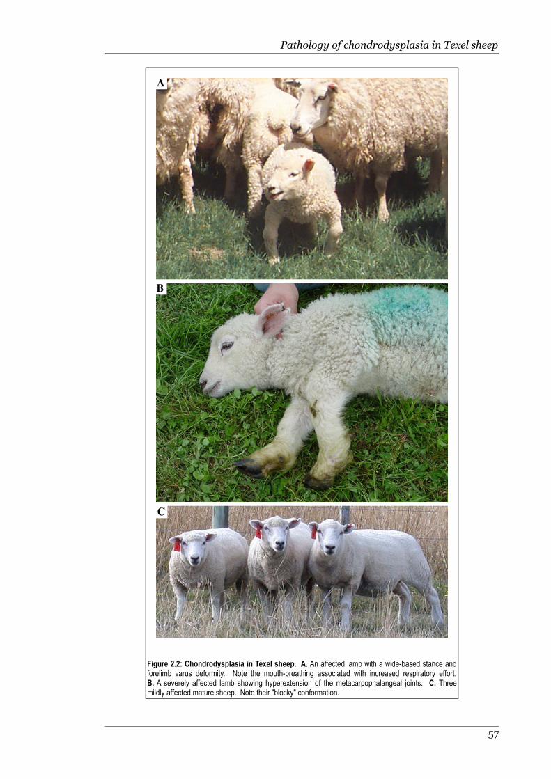
Pathology of chondrodysplasia in Texel sheep
57
Figure 2.2: Chondrodysplasia in Texel sheep. A. An affected lamb with a wide-based stance andforelimb varus deformity. Note the mouth-breathing associated with increased respiratory effort.B. A severely affected lamb showing hyperextension of the metacarpophalangeal joints. C. Threemildly affected mature sheep. Note their "blocky" conformation.
A
C
B
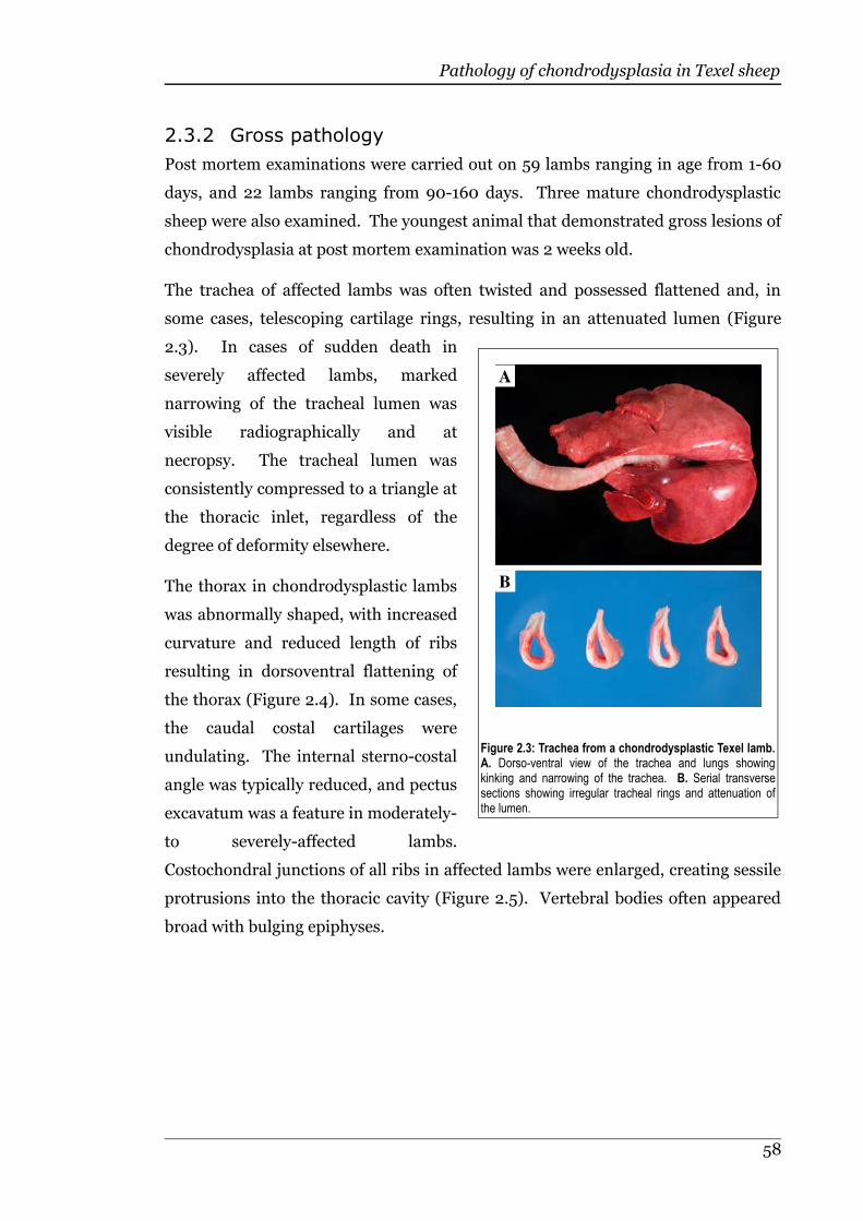
Pathology of chondrodysplasia in Texel sheep
2.3.2 Gross pathology
Post mortem examinations were carried out on 59 lambs ranging in age from 1-60
days, and 22 lambs ranging from 90-160 days. Three mature chondrodysplastic
sheep were also examined. The youngest animal that demonstrated gross lesions of
chondrodysplasia at post mortem examination was 2 weeks old.
The trachea of affected lambs was often twisted and possessed flattened and, in
some cases, telescoping cartilage rings, resulting in an attenuated lumen (Figure
2.3). In cases of sudden death in
severely affected lambs, marked
narrowing of the tracheal lumen was
visible radiographically and at
necropsy. The tracheal lumen was
consistently compressed to a triangle at
the thoracic inlet, regardless of the
degree of deformity elsewhere.
The thorax in chondrodysplastic lambs
was abnormally shaped, with increased
curvature and reduced length of ribs
resulting in dorsoventral flattening of
the thorax (Figure 2.4). In some cases,
the caudal costal cartilages were
undulating. The internal sterno-costal
angle was typically reduced, and pectus
excavatum was a feature in moderately-
to severely-affected lambs.
Costochondral junctions of all ribs in affected lambs were enlarged, creating sessile
protrusions into the thoracic cavity (Figure 2.5). Vertebral bodies often appeared
broad with bulging epiphyses.
58
Figure 2.3: Trachea from a chondrodysplastic Texel lamb.A. Dorso-ventral view of the trachea and lungs showingkinking and narrowing of the trachea. B. Serial transversesections showing irregular tracheal rings and attenuation ofthe lumen.
A
B
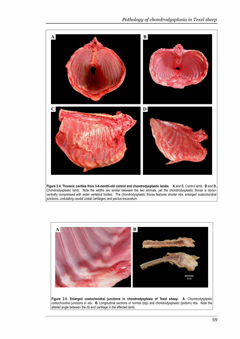
Pathology of chondrodysplasia in Texel sheep
59
Figure 2.4: Thoracic cavities from 3-4-month-old control and chondrodysplastic lambs. A and C. Control lamb. B and D.Chondrodysplastic lamb. Note the widths are similar between the two animals, yet the chondrodysplastic thorax is dorso-ventrally compressed with wider vertebral bodies. The chondrodysplastic thorax features shorter ribs, enlarged costochondraljunctions, undulating caudal costal cartilages, and pectus excavatum.
C
BA
D
Figure 2.5: Enlarged costochondral junctions in chondrodysplasia of Texel sheep. A. Chondrodysplasticcostochondral junctions in situ. B. Longitudinal sections of normal (top) and chondrodysplastic (bottom) ribs. Note thealtered angle between the rib and cartilage in the affected lamb.
BA

Pathology of chondrodysplasia in Texel sheep
In severely affected lambs, erosion of articular cartilage from weight-bearing
surfaces of major limb joints often occurred during the first 3 months of life and led
to degenerative joint disease (Figure 2.6). Bone length appeared to be reduced and
joints were enlarged in these animals (Figure 2.6B). In mildly affected animals that
survived to adulthood, degenerative joint disease was largely confined to the
shoulder and featured erosion of articular cartilage, osteophyte formation around
chondro-osseous margins and development of loose bodies (Figure 2.6C). Articular
and physeal cartilage of long bones was grossly thickened compared with age-
matched controls, and was far easier to cut with a scalpel.
60
Figure 2.6: Joint lesions in chondrodysplasia of Texel sheep. A. Control (left) andchondrodysplastic distal humeri from 3-4-month-old lambs. Note the whiter colour of thechondrodysplastic cartilage, at least partly due to the increased cartilage thickness, and the erosion ofthe medial surface of the trochlear ridge. B. Extensive erosion of proximal humeral articular cartilagewith exposure of subchondral bone in a severely affected 3-month-old lamb. Note the shortappearance of the bone in conjunction with enlarged bone ends. C. Erosion of cartilage andosteophyte formation in the shoulder joint of a mature ram. Note the large loose body (arrow).
A
CB

Pathology of chondrodysplasia in Texel sheep
The only non-cartilaginous soft tissue lesion was bilateral hydronephrosis (Figure
2.7), identified in 2 month-old chondrodysplastic lambs and in 1 newborn lamb of
unknown phenotype. In the latter, the hydronephrosis was accompanied by severe
bladder distension and signs of dystocia. Hydronephrosis was not a constant
finding in chondrodysplastic lambs, and may be unrelated.
2.3.3 Histological findings
The microscopic lesions of chondrodysplasia in Texel sheep were progressive and
consistent in nature. They varied in severity depending on the age of affected lambs
and tended to reflect the degree of gross deformity. No lesions were present in non-
cartilaginous tissues other than bone, which frequently had abnormalities
secondary to cartilaginous lesions.
Chondrocytes of hyaline cartilage throughout the articular-epiphyseal complex,
deeper zones of articular and physeal cartilage, trachea and costochondral junction
were surrounded by dense, concentric rings of abnormal, deeply basophilic fibrillar
material (Figure 2.8). The interterritorial cartilage matrix often contained multiple,
coalescing foci of rarefaction, merging in severe cases to form large areas of
61
Figure 2.7: Hydronephrosis in chondrodysplasia of Texel sheep. Hydronephrosis was observed occasionallyin chondrodysplastic lambs, and may be an unrelated finding.
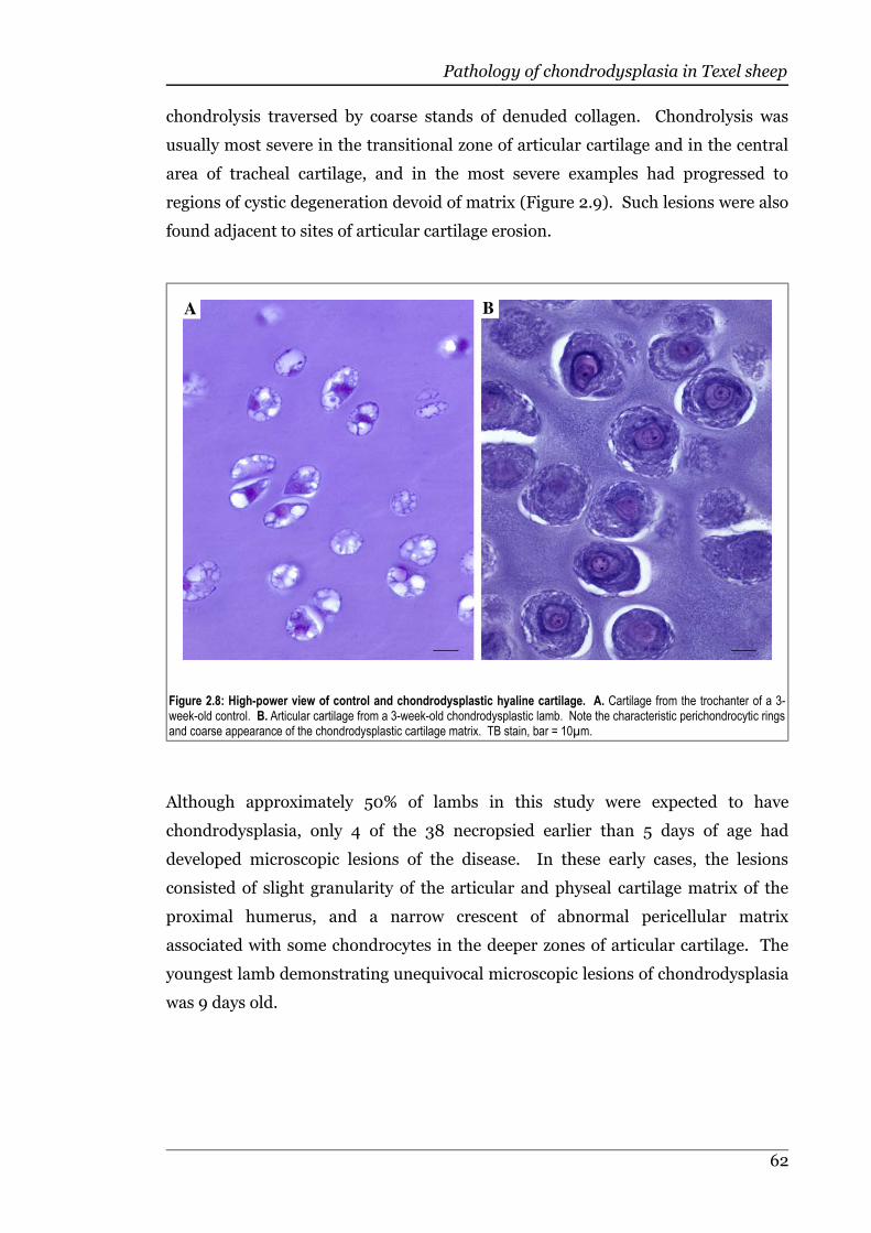
Pathology of chondrodysplasia in Texel sheep
chondrolysis traversed by coarse stands of denuded collagen. Chondrolysis was
usually most severe in the transitional zone of articular cartilage and in the central
area of tracheal cartilage, and in the most severe examples had progressed to
regions of cystic degeneration devoid of matrix (Figure 2.9). Such lesions were also
found adjacent to sites of articular cartilage erosion.
Although approximately 50% of lambs in this study were expected to have
chondrodysplasia, only 4 of the 38 necropsied earlier than 5 days of age had
developed microscopic lesions of the disease. In these early cases, the lesions
consisted of slight granularity of the articular and physeal cartilage matrix of the
proximal humerus, and a narrow crescent of abnormal pericellular matrix
associated with some chondrocytes in the deeper zones of articular cartilage. The
youngest lamb demonstrating unequivocal microscopic lesions of chondrodysplasia
was 9 days old.
62
Figure 2.8: High-power view of control and chondrodysplastic hyaline cartilage. A. Cartilage from the trochanter of a 3-week-old control. B. Articular cartilage from a 3-week-old chondrodysplastic lamb. Note the characteristic perichondrocytic ringsand coarse appearance of the chondrodysplastic cartilage matrix. TB stain, bar = 10µm.
A B

Pathology of chondrodysplasia in Texel sheep
Microscopic lesions were present in all examined sections of hyaline cartilage from
lambs showing gross lesions of chondrodysplasia. Although the severity of lesions
varied between sites and between individual lambs, it did not appear to be related
to the parental genotype. For example, one 22-day-old chondrodysplastic lamb
with 2 chondrodysplastic parents had only mild lesions, including rarefaction of
matrix in the transitional zone of the articular cartilage of the proximal humerus.
In contrast, a 23-day-old chondrodysplastic lamb with only 1 affected parent
showed far more pronounced lesions, including severe matrix rarefaction with
fibrillar areas of denuded collagen.
The microscopic lesions in the cartilage of affected lambs surviving to 5 months of
age were considerably milder than in severely chondrodysplastic lambs that died or
were euthanased at 1 month of age. Pericellular rings of dense matrix were still
prominent but there was little evidence of matrix rarefaction. The distal humeral
63
Figure 2.9: Articular cartilage from the proximal humerus of a 30-day-old chondrodysplastic lamb. Articular surface is attop-left. Note the severe chondrolysis and cystic degeneration of cartilage matrix. TB stain, bar = 200µm.
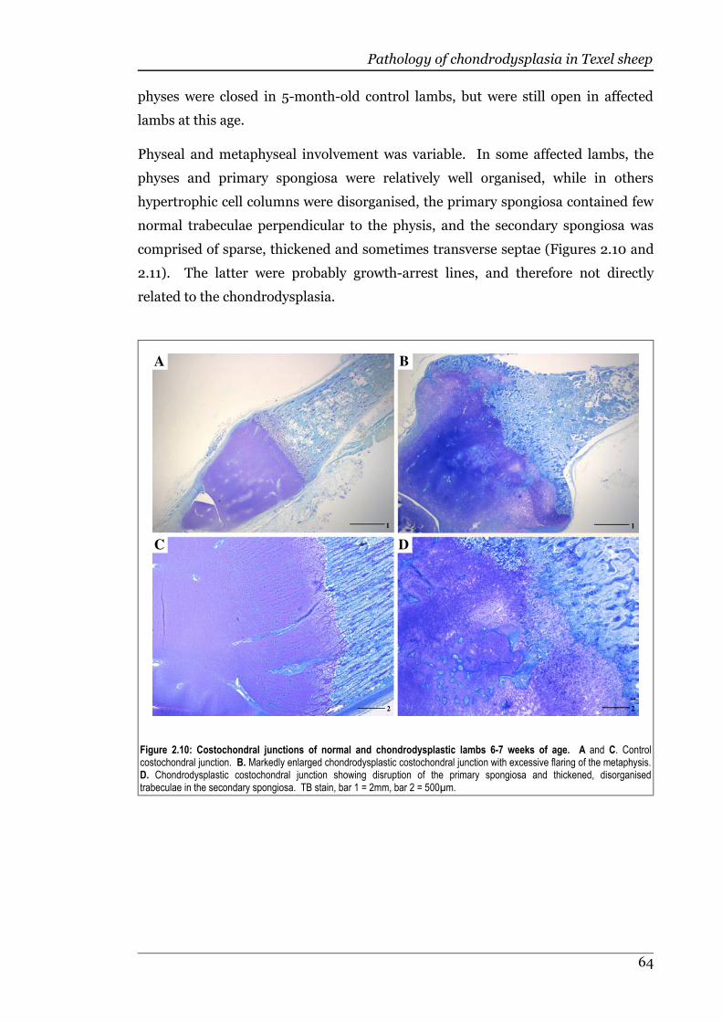
Pathology of chondrodysplasia in Texel sheep
physes were closed in 5-month-old control lambs, but were still open in affected
lambs at this age.
Physeal and metaphyseal involvement was variable. In some affected lambs, the
physes and primary spongiosa were relatively well organised, while in others
hypertrophic cell columns were disorganised, the primary spongiosa contained few
normal trabeculae perpendicular to the physis, and the secondary spongiosa was
comprised of sparse, thickened and sometimes transverse septae (Figures 2.10 and
2.11). The latter were probably growth-arrest lines, and therefore not directly
related to the chondrodysplasia.
64
Figure 2.10: Costochondral junctions of normal and chondrodysplastic lambs 6-7 weeks of age. A and C. Controlcostochondral junction. B. Markedly enlarged chondrodysplastic costochondral junction with excessive flaring of the metaphysis.D. Chondrodysplastic costochondral junction showing disruption of the primary spongiosa and thickened, disorganisedtrabeculae in the secondary spongiosa. TB stain, bar 1 = 2mm, bar 2 = 500µm.
A B
C D
1 1
2 2
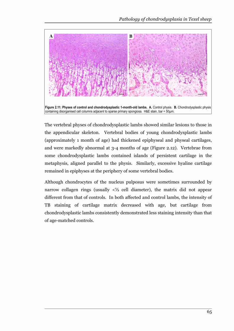
Pathology of chondrodysplasia in Texel sheep
The vertebral physes of chondrodysplastic lambs showed similar lesions to those in
the appendicular skeleton. Vertebral bodies of young chondrodysplastic lambs
(approximately 1 month of age) had thickened epiphyseal and physeal cartilages,
and were markedly abnormal at 3-4 months of age (Figure 2.12). Vertebrae from
some chondrodysplastic lambs contained islands of persistent cartilage in the
metaphysis, aligned parallel to the physis. Similarly, excessive hyaline cartilage
remained in epiphyses at the periphery of some vertebral bodies.
Although chondrocytes of the nucleus pulposus were sometimes surrounded by
⅓narrow collagen rings (usually < cell diameter), the matrix did not appear
different from that of controls. In both affected and control lambs, the intensity of
TB staining of cartilage matrix decreased with age, but cartilage from
chondrodysplastic lambs consistently demonstrated less staining intensity than that
of age-matched controls.
65
Figure 2.11: Physes of control and chondrodysplastic 1-month-old lambs. A. Control physis. B. Chondrodysplastic physiscontaining disorganised cell columns adjacent to sparse primary spongiosa. H&E stain, bar = 50µm.
A B
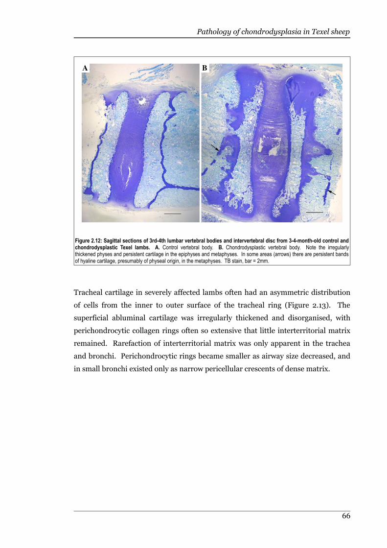
Pathology of chondrodysplasia in Texel sheep
Tracheal cartilage in severely affected lambs often had an asymmetric distribution
of cells from the inner to outer surface of the tracheal ring (Figure 2.13). The
superficial abluminal cartilage was irregularly thickened and disorganised, with
perichondrocytic collagen rings often so extensive that little interterritorial matrix
remained. Rarefaction of interterritorial matrix was only apparent in the trachea
and bronchi. Perichondrocytic rings became smaller as airway size decreased, and
in small bronchi existed only as narrow pericellular crescents of dense matrix.
66
Figure 2.12: Sagittal sections of 3rd-4th lumbar vertebral bodies and intervertebral disc from 3-4-month-old control andchondrodysplastic Texel lambs. A. Control vertebral body. B. Chondrodysplastic vertebral body. Note the irregularlythickened physes and persistent cartilage in the epiphyses and metaphyses. In some areas (arrows) there are persistent bandsof hyaline cartilage, presumably of physeal origin, in the metaphyses. TB stain, bar = 2mm.
A B
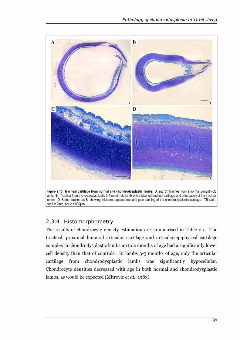
Pathology of chondrodysplasia in Texel sheep
2.3.4 Histomorphometry
The results of chondrocyte density estimation are summarised in Table 2.1. The
tracheal, proximal humeral articular cartilage and articular-epiphyseal cartilage
complex in chondrodysplastic lambs up to 2 months of age had a significantly lower
cell density than that of controls. In lambs 3-5 months of age, only the articular
cartilage from chondrodysplastic lambs was significantly hypocellular.
Chondrocyte densities decreased with age in both normal and chondrodysplastic
lambs, as would be expected (Mitrovic et al., 1983).
67
Figure 2.13: Tracheal cartilage from normal and chondrodysplastic lambs. A and C. Trachea from a normal 5-month-oldlamb. B. Trachea from a chondrodysplastic 3-4-month-old lamb with thickened tracheal cartilage and attenuation of the tracheallumen. D. Same trachea as B, showing thickened appearance and pale staining of the chondrodysplastic cartilage. TB stain,bar 1 = 2mm, bar 2 = 500µm.
A B
C D
1 1
2 2

Pathology of chondrodysplasia in Texel sheep
Table 2.1: Chondrocyte density expressed as cells per 400X field in cartilage of chondrodysplastic and control Texellambs. Cells were counted in the transitional zone of the proximal humeral articular cartilage, the articular-epiphyseal complexof the proximal humerus, and the central cartilage of the trachea.
Group 2 weeks to 2 months of age
Location Articular cartilage Articular-epiphyseal complex Tracheal cartilage
Status Control Affected Control Affected Control Affected
Mean ± SEM 185 ± 29 115 ± 10 156 ± 15 102 ± 12 212 ± 32 140 ± 16
Difference between means
69 ± 24* 54 ± 22* 72 ± 32*
Group 3 months to 5 months of age
Location Articular cartilage Articular-epiphyseal complex Tracheal cartilage
Status Control Affected Control Affected Control Affected
Mean ±SEM 129 ± 11 82 ± 7 94 ± 10 74 ± 8 94 ± 5 76 ± 7
Difference between means
46 ± 14** 20 ± 14 18 ± 8
* = P ≤ 0.05, ** = P ≤ 0.01 using the Student’s t-test. Averaged results were rounded to the nearest whole number.
The results of cartilage thickness measurements are summarised in Table 2.2.
Although some severely affected lambs had markedly thickened tracheal rings (see
Figures 2.3 and 2.13), this was highly variable. In spite of their gross appearance,
the mean thickness of the ventral tracheal rings was not significantly different from
that of phenotypically normal carrier animals. The mean thickness of articular and
physeal cartilage in chondrodysplastic lambs was significantly greater than that of
controls at all ages. The articular cartilage of the proximal humerus was thicker by
approximately 700µm in the younger group of chondrodysplastic lambs, and this
difference declined to 317µm in older lambs. The mean thickness of the central
physeal cartilage of the proximal humerus in chondrodysplastic lambs 2 weeks to 2
months of age was 360µm greater than in age-matched control lambs, the
difference increasing to 563µm in the older age group.
68
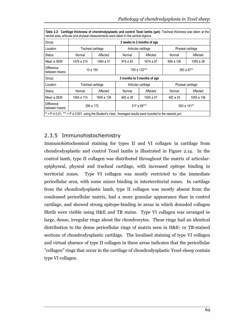
Pathology of chondrodysplasia in Texel sheep
Table 2.2: Cartilage thickness of chondrodysplastic and control Texel lambs (µm). Tracheal thickness was taken at theventral area, articular and physeal measurements were taken in the central regions.
Group 2 weeks to 2 months of age
Location Tracheal cartilage Articular cartilage Physeal cartilage
Status Normal Affected Normal Affected Normal Affected
Mean ± SEM 1476 ± 214 1464 ± 51 974 ± 83 1674 ± 87 699 ± 106 1059 ± 38
Differencebetween means
13 ± 155 700 ± 132*** 360 ± 87**
Group 3 months to 5 months of age
Location Tracheal cartilage Articular cartilage Physeal cartilage
Status Normal Affected Normal Affected Normal Affected
Mean ± SEM 1594 ± 114 1849 ± 128 683 ± 38 1000 ± 57 492 ± 33 1055 ± 146
Differencebetween means
256 ± 172 317 ± 69*** 563 ± 141**
** = P ≤ 0.01, *** = P ≤ 0.001, using the Student’s t-test. Averaged results were rounded to the nearest µm.
2.3.5 Immunohistochemistry
Immunohistochemical staining for types II and VI collagen in cartilage from
chondrodysplastic and control Texel lambs is illustrated in Figure 2.14. In the
control lamb, type II collagen was distributed throughout the matrix of articular-
epiphyseal, physeal and tracheal cartilage, with increased epitope binding in
territorial zones. Type VI collagen was mostly restricted to the immediate
pericellular area, with some minor binding in interterritorial zones. In cartilage
from the chondrodysplastic lamb, type II collagen was mostly absent from the
condensed pericellular matrix, had a more granular appearance than in control
cartilage, and showed strong epitope-binding in areas in which denuded collagen
fibrils were visible using H&E and TB stains. Type VI collagen was arranged in
large, dense, irregular rings about the chondrocytes. These rings had an identical
distribution to the dense pericellular rings of matrix seen in H&E- or TB-stained
sections of chondrodysplastic cartilage. The localised staining of type VI collagen
and virtual absence of type II collagen in these areas indicates that the pericellular
"collagen" rings that occur in the cartilage of chondrodysplastic Texel sheep contain
type VI collagen.
69
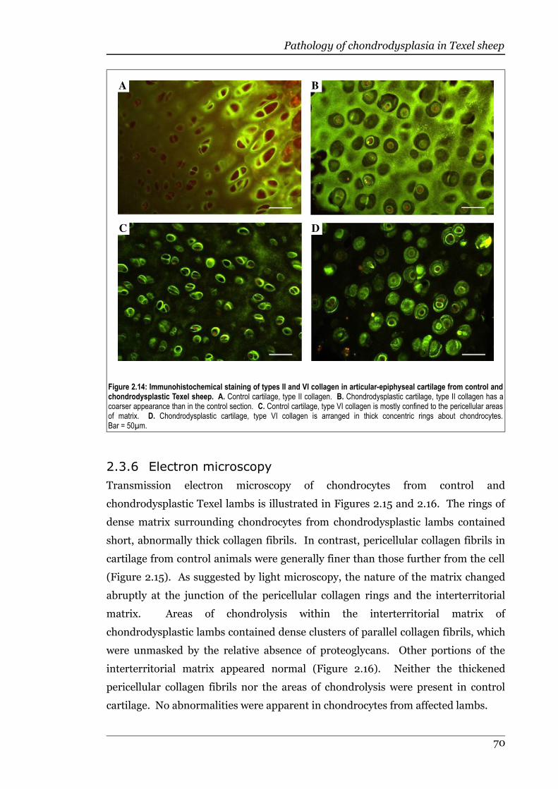
Pathology of chondrodysplasia in Texel sheep
2.3.6 Electron microscopy
Transmission electron microscopy of chondrocytes from control and
chondrodysplastic Texel lambs is illustrated in Figures 2.15 and 2.16. The rings of
dense matrix surrounding chondrocytes from chondrodysplastic lambs contained
short, abnormally thick collagen fibrils. In contrast, pericellular collagen fibrils in
cartilage from control animals were generally finer than those further from the cell
(Figure 2.15). As suggested by light microscopy, the nature of the matrix changed
abruptly at the junction of the pericellular collagen rings and the interterritorial
matrix. Areas of chondrolysis within the interterritorial matrix of
chondrodysplastic lambs contained dense clusters of parallel collagen fibrils, which
were unmasked by the relative absence of proteoglycans. Other portions of the
interterritorial matrix appeared normal (Figure 2.16). Neither the thickened
pericellular collagen fibrils nor the areas of chondrolysis were present in control
cartilage. No abnormalities were apparent in chondrocytes from affected lambs.
70
Figure 2.14: Immunohistochemical staining of types II and VI collagen in articular-epiphyseal cartilage from control andchondrodysplastic Texel sheep. A. Control cartilage, type II collagen. B. Chondrodysplastic cartilage, type II collagen has acoarser appearance than in the control section. C. Control cartilage, type VI collagen is mostly confined to the pericellular areasof matrix. D. Chondrodysplastic cartilage, type VI collagen is arranged in thick concentric rings about chondrocytes.Bar = 50µm.
A B
C D

Pathology of chondrodysplasia in Texel sheep
71
Figure 2.15: Articular cartilage chondrocytes and pericellular matrix from control and chondrodysplastic Texel lambs.A. Control chondrocytes. B. Chondrodysplastic chondrocyte. Note the concentric rings of dense, abnormal matrix surroundingthe chondrocyte. RHT fixation, bar = 2µm.
A B
Figure 2.16: High power views of pericellular and interterritorial matrix of cartilage from control andchondrodysplastic Texel lambs. A. Control pericellular matrix. B. Chondrodysplastic pericellular matrixdemonstrating irregular clumping of matrix components. C. Control interterritorial matrix. D. Interterritorialmatrix from a chondrodysplastic lamb with similar appearance to control tissue, D. RHT fixation, bar = 0.2µm.
A B
C D
Pathology of chondrodysplasia in Texel sheep
2.4 Discussion
Chondrodysplasia of Texel sheep is characterised grossly by disproportionate
dwarfism, angular limb deformity, degenerative joint disease in severely affected
animals, a barrel-shaped chest, twisting of the trachea with attenuation of the
tracheal lumen and soft, thickened articular and physeal cartilage. Histologically,
chondrocytes were surrounded by "collagen rings" containing type VI collagen,
cartilage matrix contained areas of rarefaction and chondrolysis, and articular,
physeal and tracheal cartilages were hypocellular compared with controls, at least
in lambs younger than 2 months. The gross and microscopic lesions described here
in Texel sheep confirm that this inherited chondrodysplasia is distinct from "spider
lamb syndrome" (SLS) and other chondrodysplasias of sheep.
Disproportionate dwarfism, angular limb deformity and an abnormally shaped
thorax are features of many chondrodysplasias in man and animals, and reflect the
dependency of skeletal development on normal cartilage growth and maturation
(Erlebacher et al., 1995). The microscopic and ultrastructural lesions in hyaline
cartilage matrix of affected Texel lambs are highly distinctive and closely match
those seen in human achondrogenesis type 1b, atelosteogenesis type II and
diastrophic dysplasia, all of which are recessively inherited disorders caused by
mutations in the diastrophic dysplasia sulphate transporter, SLC26A2 (van der
Harten et al., 1988; Shapiro, 1992; Sillence et al., 1997; Superti-Furga, 2001).
Neonatal death occurs in children with achondrogenesis type 1b and
atelosteogenesis type II (van der Harten et al., 1988; Superti-Furga, 1994;
Hästbacka et al., 1996b). Histologically, there may be failure of physeal
organisation, degeneration and rarefaction of the interterritorial matrix, and
condensation of matrix debris around chondrocytes, variably referred to as collagen
rings, haloes, or pericellular debris (van der Harten et al., 1988; Shapiro, 1992;
Superti-Furga, 1994).
The decreased intensity of staining with TB in cartilage from chondrodysplastic
Texel sheep compared with controls supports a reduction in sulphate ions in the
matrix, which is consistent with the biochemical findings of Byrne (2005) (see
Figure 1.7). An SLC26A2 knock-out mutant mouse also demonstrated reduced
staining with TB, and, although not discussed by the authors, histological images
illustrate abnormal pericellular matrix (Forlino et al., 2006).
72
Pathology of chondrodysplasia in Texel sheep
Chondrodysplasia of Texel sheep progresses similarly to disorders of sulphate
transport (Superti-Furga, 2001; Forlino et al., 2005). Shapiro (1992) described the
development of microscopic lesions in diastrophic dysplasia in two patients
throughout childhood. The matrix rarefaction in these cases appeared in the
earliest cartilage examined, while lesions corresponding to collagen rings (referred
to as “target” cells) were not clearly evident until several years of age. A similar
development of lesions was seen in Texel chondrodysplasia, where the earliest
lesions involved matrix rarefaction, while collagen rings became more pronounced
with age. An apparent deviation from this pattern occurred in affected lambs older
than 1 month, in which microscopic lesions appeared to be less severe than in 1-
month-old affected lambs. While this could be due to ‘regression’ of the lesions, it
more likely reflects the death or euthanasia of the more severely affected lambs at a
younger age. Severely affected lambs that either died following exercise or were
euthanased due to respiratory distress had tracheal lesions that were consistent
with trachal collapse. The soft, thickened cartilage of the tracheal rings was
frequently deformed to a slim oval (see Figure 2.3B), and the increased respiratory
effort clinically evident in severely affected Texel lambs probably generated enough
negative pressure in the cervical airway to cause the lumen to collapse.
The lesions in chondrodysplasia of Texel sheep were highly variable in severity,
corresponding to the variability of expression demonstrated in children with
SLC26A2 mutations (Superti-Furga, 2001). Although the nature of the mutation
itself influences the phenotype through the residual level of function, other factors
must be involved in determining the severity of disease, as first-degree relatives
with the same SLC26A2 mutation have demonstrated differing phenotypes
(Superti-Furga, 2001). Variability in the capacity to utilise sulphate from other
sources, such as intracellular sulphydral compounds (Rossi et al., 2003), may
explain some of the phenotypic irregularities in disorders caused by SLC26A2
mutations.
Variability of the phenotype in diastrophic dysplasia may also reflect a complex
pathogenesis, possibly involving both a cartilage matrix defect and secondary
changes in the fibroblast growth factor (FGF) signalling pathway, which utilises
heparan sulphate (Superti-Furga, 2001; Harmer, 2006). Undersulphation of
heparan sulphate in disorders of sulphate transport has not been thoroughly
investigated, but is considered likely to occur (Forlino et al., 2005). This is
supported by the undersulphation of heparan sulphate in vitro in the presence of
73
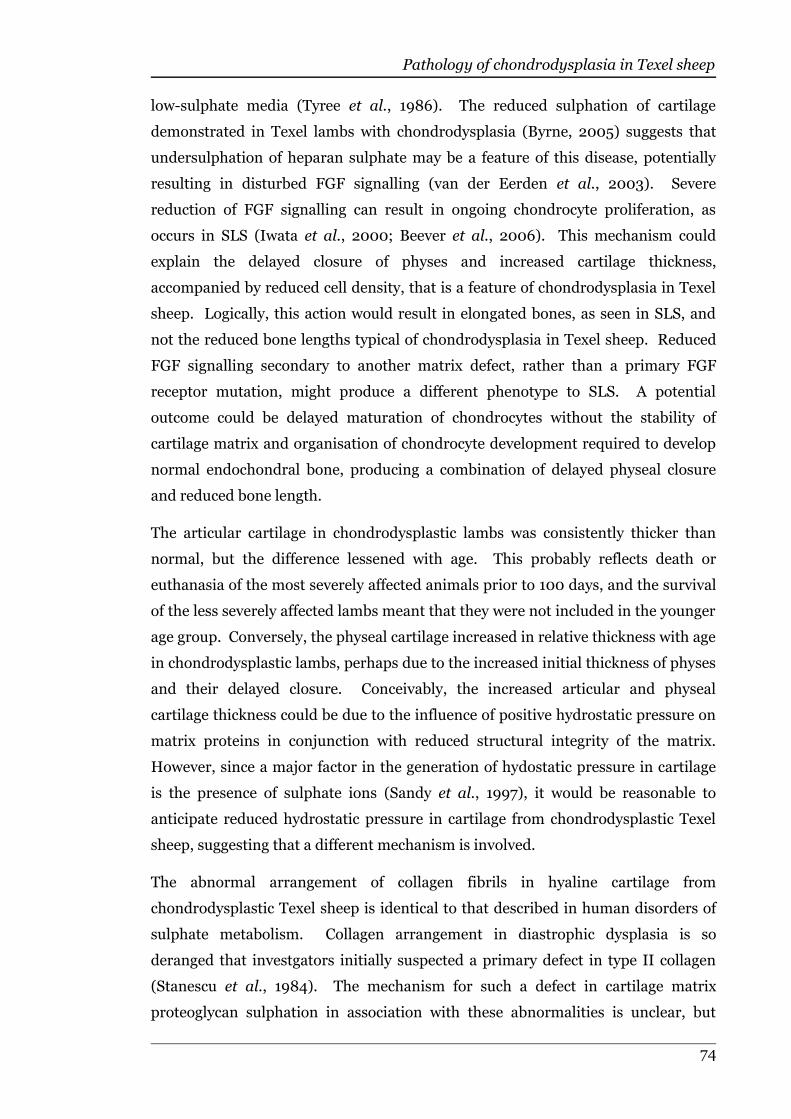
Pathology of chondrodysplasia in Texel sheep
low-sulphate media (Tyree et al., 1986). The reduced sulphation of cartilage
demonstrated in Texel lambs with chondrodysplasia (Byrne, 2005) suggests that
undersulphation of heparan sulphate may be a feature of this disease, potentially
resulting in disturbed FGF signalling (van der Eerden et al., 2003). Severe
reduction of FGF signalling can result in ongoing chondrocyte proliferation, as
occurs in SLS (Iwata et al., 2000; Beever et al., 2006). This mechanism could
explain the delayed closure of physes and increased cartilage thickness,
accompanied by reduced cell density, that is a feature of chondrodysplasia in Texel
sheep. Logically, this action would result in elongated bones, as seen in SLS, and
not the reduced bone lengths typical of chondrodysplasia in Texel sheep. Reduced
FGF signalling secondary to another matrix defect, rather than a primary FGF
receptor mutation, might produce a different phenotype to SLS. A potential
outcome could be delayed maturation of chondrocytes without the stability of
cartilage matrix and organisation of chondrocyte development required to develop
normal endochondral bone, producing a combination of delayed physeal closure
and reduced bone length.
The articular cartilage in chondrodysplastic lambs was consistently thicker than
normal, but the difference lessened with age. This probably reflects death or
euthanasia of the most severely affected animals prior to 100 days, and the survival
of the less severely affected lambs meant that they were not included in the younger
age group. Conversely, the physeal cartilage increased in relative thickness with age
in chondrodysplastic lambs, perhaps due to the increased initial thickness of physes
and their delayed closure. Conceivably, the increased articular and physeal
cartilage thickness could be due to the influence of positive hydrostatic pressure on
matrix proteins in conjunction with reduced structural integrity of the matrix.
However, since a major factor in the generation of hydostatic pressure in cartilage
is the presence of sulphate ions (Sandy et al., 1997), it would be reasonable to
anticipate reduced hydrostatic pressure in cartilage from chondrodysplastic Texel
sheep, suggesting that a different mechanism is involved.
The abnormal arrangement of collagen fibrils in hyaline cartilage from
chondrodysplastic Texel sheep is identical to that described in human disorders of
sulphate metabolism. Collagen arrangement in diastrophic dysplasia is so
deranged that investgators initially suspected a primary defect in type II collagen
(Stanescu et al., 1984). The mechanism for such a defect in cartilage matrix
proteoglycan sulphation in association with these abnormalities is unclear, but
74
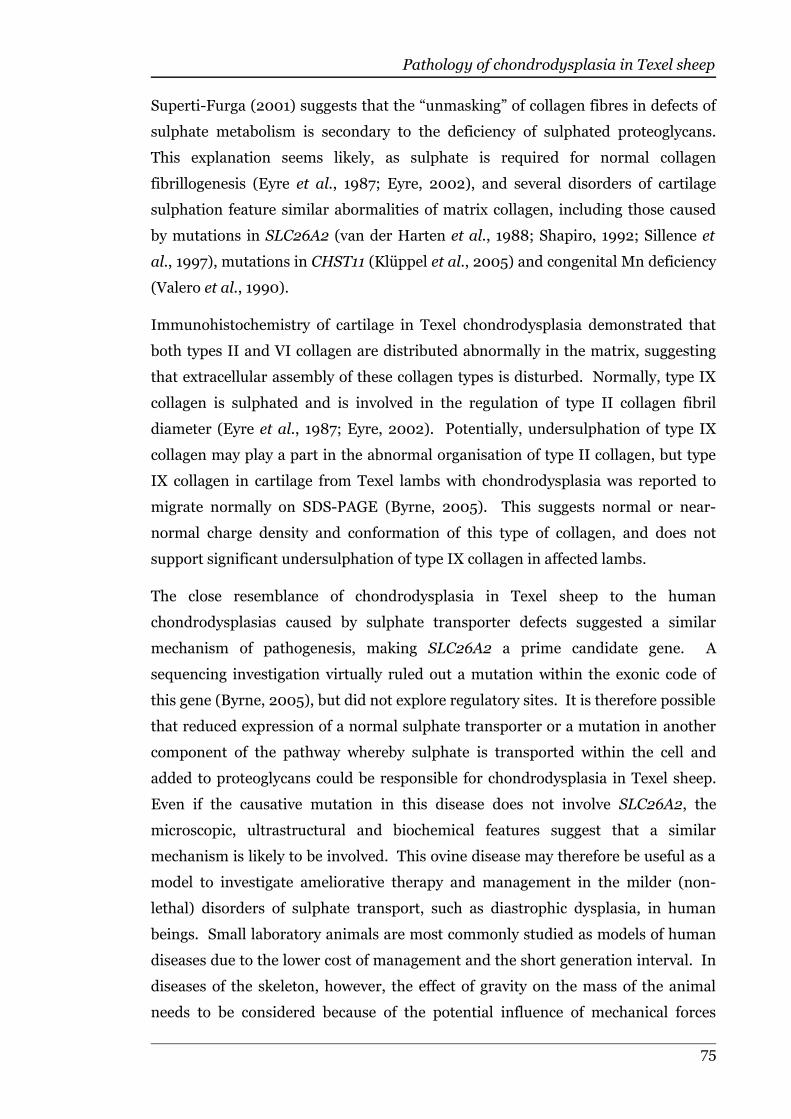
Pathology of chondrodysplasia in Texel sheep
Superti-Furga (2001) suggests that the “unmasking” of collagen fibres in defects of
sulphate metabolism is secondary to the deficiency of sulphated proteoglycans.
This explanation seems likely, as sulphate is required for normal collagen
fibrillogenesis (Eyre et al., 1987; Eyre, 2002), and several disorders of cartilage
sulphation feature similar abormalities of matrix collagen, including those caused
by mutations in SLC26A2 (van der Harten et al., 1988; Shapiro, 1992; Sillence et
al., 1997), mutations in CHST11 (Klüppel et al., 2005) and congenital Mn deficiency
(Valero et al., 1990).
Immunohistochemistry of cartilage in Texel chondrodysplasia demonstrated that
both types II and VI collagen are distributed abnormally in the matrix, suggesting
that extracellular assembly of these collagen types is disturbed. Normally, type IX
collagen is sulphated and is involved in the regulation of type II collagen fibril
diameter (Eyre et al., 1987; Eyre, 2002). Potentially, undersulphation of type IX
collagen may play a part in the abnormal organisation of type II collagen, but type
IX collagen in cartilage from Texel lambs with chondrodysplasia was reported to
migrate normally on SDS-PAGE (Byrne, 2005). This suggests normal or near-
normal charge density and conformation of this type of collagen, and does not
support significant undersulphation of type IX collagen in affected lambs.
The close resemblance of chondrodysplasia in Texel sheep to the human
chondrodysplasias caused by sulphate transporter defects suggested a similar
mechanism of pathogenesis, making SLC26A2 a prime candidate gene. A
sequencing investigation virtually ruled out a mutation within the exonic code of
this gene (Byrne, 2005), but did not explore regulatory sites. It is therefore possible
that reduced expression of a normal sulphate transporter or a mutation in another
component of the pathway whereby sulphate is transported within the cell and
added to proteoglycans could be responsible for chondrodysplasia in Texel sheep.
Even if the causative mutation in this disease does not involve SLC26A2, the
microscopic, ultrastructural and biochemical features suggest that a similar
mechanism is likely to be involved. This ovine disease may therefore be useful as a
model to investigate ameliorative therapy and management in the milder (non-
lethal) disorders of sulphate transport, such as diastrophic dysplasia, in human
beings. Small laboratory animals are most commonly studied as models of human
diseases due to the lower cost of management and the short generation interval. In
diseases of the skeleton, however, the effect of gravity on the mass of the animal
needs to be considered because of the potential influence of mechanical forces
75
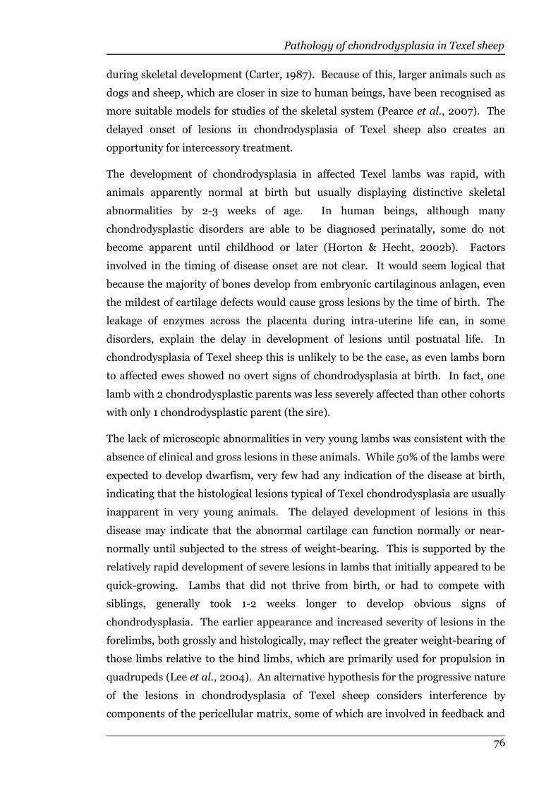
Pathology of chondrodysplasia in Texel sheep
during skeletal development (Carter, 1987). Because of this, larger animals such as
dogs and sheep, which are closer in size to human beings, have been recognised as
more suitable models for studies of the skeletal system (Pearce et al., 2007). The
delayed onset of lesions in chondrodysplasia of Texel sheep also creates an
opportunity for intercessory treatment.
The development of chondrodysplasia in affected Texel lambs was rapid, with
animals apparently normal at birth but usually displaying distinctive skeletal
abnormalities by 2-3 weeks of age. In human beings, although many
chondrodysplastic disorders are able to be diagnosed perinatally, some do not
become apparent until childhood or later (Horton & Hecht, 2002b). Factors
involved in the timing of disease onset are not clear. It would seem logical that
because the majority of bones develop from embryonic cartilaginous anlagen, even
the mildest of cartilage defects would cause gross lesions by the time of birth. The
leakage of enzymes across the placenta during intra-uterine life can, in some
disorders, explain the delay in development of lesions until postnatal life. In
chondrodysplasia of Texel sheep this is unlikely to be the case, as even lambs born
to affected ewes showed no overt signs of chondrodysplasia at birth. In fact, one
lamb with 2 chondrodysplastic parents was less severely affected than other cohorts
with only 1 chondrodysplastic parent (the sire).
The lack of microscopic abnormalities in very young lambs was consistent with the
absence of clinical and gross lesions in these animals. While 50% of the lambs were
expected to develop dwarfism, very few had any indication of the disease at birth,
indicating that the histological lesions typical of Texel chondrodysplasia are usually
inapparent in very young animals. The delayed development of lesions in this
disease may indicate that the abnormal cartilage can function normally or near-
normally until subjected to the stress of weight-bearing. This is supported by the
relatively rapid development of severe lesions in lambs that initially appeared to be
quick-growing. Lambs that did not thrive from birth, or had to compete with
siblings, generally took 1-2 weeks longer to develop obvious signs of
chondrodysplasia. The earlier appearance and increased severity of lesions in the
forelimbs, both grossly and histologically, may reflect the greater weight-bearing of
those limbs relative to the hind limbs, which are primarily used for propulsion in
quadrupeds (Lee et al., 2004). An alternative hypothesis for the progressive nature
of the lesions in chondrodysplasia of Texel sheep considers interference by
components of the pericellular matrix, some of which are involved in feedback and
76
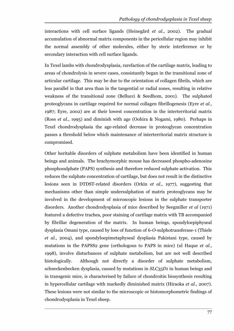
Pathology of chondrodysplasia in Texel sheep
interactions with cell surface ligands (Heinegård et al., 2002). The gradual
accumulation of abnormal matrix components in the pericellular region may inhibit
the normal assembly of other molecules, either by steric interference or by
secondary interaction with cell surface ligands.
In Texel lambs with chondrodysplasia, rarefaction of the cartilage matrix, leading to
areas of chondrolysis in severe cases, consistantly began in the transitional zone of
articular cartilage. This may be due to the orientation of collagen fibrils, which are
less parallel in that area than in the tangential or radial zones, resulting in relative
weakness of the transitional zone (Bellucci & Seedhom, 2001). The sulphated
proteoglycans in cartilage required for normal collagen fibrillogenesis (Eyre et al.,
1987; Eyre, 2002) are at their lowest concentration in the interterritorial matrix
(Ross et al., 1995) and diminish with age (Oohira & Nogami, 1980). Perhaps in
Texel chondrodysplasia the age-related decrease in proteoglycan concentration
passes a threshold below which maintenance of interterritorial matrix structure is
compromised.
Other heritable disorders of sulphate metabolism have been identified in human
beings and animals. The brachymorphic mouse has decreased phospho-adenosine
phosphosulphate (PAPS) synthesis and therefore reduced sulphate activation. This
reduces the sulphate concentration of cartilage, but does not result in the distinctive
lesions seen in DTDST-related disorders (Orkin et al., 1977), suggesting that
mechanisms other than simple undersulphation of matrix proteoglycans may be
involved in the development of microscopic lesions in the sulphate transporter
disorders. Another chondrodysplasia of mice described by Seegmiller et al (1971)
featured a defective trachea, poor staining of cartilage matrix with TB accompanied
by fibrillar degeneration of the matrix. In human beings, spondyloepiphyseal
dysplasia Omani type, caused by loss of function of 6-O-sulphotransferase-1 (Thiele
et al., 2004), and spondyloepimetaphyseal dysplasia Pakistani type, caused by
mutations in the PAPSS2 gene (orthologous to PAPS in mice) (ul Haque et al.,
1998), involve disturbances of sulphate metabolism, but are not well described
histologically. Although not directly a disorder of sulphate metabolism,
schneckenbecken dysplasia, caused by mutations in SLC35D1 in human beings and
in transgenic mice, is characterised by failure of chondroitin biosynthesis resulting
in hypercellular cartilage with markedly diminished matrix (Hiraoka et al., 2007).
These lesions were not similar to the microscopic or histomorphometric findings of
chondrodysplasia in Texel sheep.
77
Pathology of chondrodysplasia in Texel sheep
English pointer enchondrodystrophy shares several gross and microscopic
similarities with Texel chondrodysplasia. Pups appear normal at birth, but develop
enlarged physes with metaphyseal flaring and angular limb deformities, erosion of
cartilage in the shoulder joints, and dorso-ventral flattening of the thorax
(Whitbread et al., 1983; Lavelle, 1984). Histologically, the physes sometimes
contain a thickened hypertrophic zone and a narrow, disorganised, proliferative
zone. Degenerative changes in the transitional to radial zones in articular cartilage
appear from 12 weeks of age, beginning as a reduction in staining density and
progressing to the formation of cystic spaces (Lavelle, 1984). Unlike
chondrodysplasia of Texel sheep, some dogs severely affected by with this disease
have inferior prognathism and kyphosis.
Pseudoachondroplasia of miniature poodles develops similarly to chondrodysplasia
of Texel sheep. Newborn pseudoachondroplastic pups appear phenotypically
normal, but develop thickened tracheal rings, flared metaphyses, excessively curved
ribs and a dorso-ventrally flattened thorax (Riser et al., 1980). Proteoglycans are
undersulphated and large “haloes” of dense matrix surround chondrocytes in these
pups (Riser et al., 1980; Bingel et al., 1986), but the disease differs radiographically
and microscopically from Texel chondrodysplasia.
Lesions similar to those of chondrodysplasia in Texel sheep are described in calves
and lambs with congenital manganese (Mn) deficiency (Caskey et al., 1944; Frost et
al., 1959; Valero et al., 1990), and can also occur in young growing animals fed a
deficient diet (Leach, 1968). Affected newborn calves typically have short limbs and
excessive cartilage in long bone epiphyses, enlarged costochondral junctions and
thickened tracheal rings, occasionally resulting in tracheal collapse (Valero et al.,
1990). Mn is required for the synthesis of chondroitin sulphate due to its function
as a cofactor for polymerase enzyme which links UDP-N-acetyl-galactosamine and
UDP-glucuronic acid to form the polysaccharide, and galactotransferase which
incorporates galactose from UDP-galactose to the galactose-galactose-xylose
trisaccharide and links the polysaccharide to the associated protein (Leach et al.,
1969). Deficiency of Mn has been shown to cause a reversible reduction in the
incorporation of sulphate into cartilage with impaired glycosaminoglycan
metabolism (Leach & Muenster, 1962; Bolze et al., 1985).
The gross and microscopic similarities between chondrodysplasia of Texel sheep
and certain chondrodysplasias in humans and other animals strongly supports a
78
Pathology of chondrodysplasia in Texel sheep
defect in sulphate metabolism in affected Texel sheep. The lesions most specific to
these disorders were the grossly thickened tracheal rings and the formation of
microscopic perichondrocytic collagen rings with rarefaction of cartilage
interterritorial matrix.
2.5 Summary
Chondrodysplasia of Texel sheep is a newly described heritable disorder which is
distinct from other chondrodysplasias described in sheep. Phenotypically normal
at birth, affected lambs develop microscopic lesions as early as 9 days of age, and
usually show gross deformities by 2-3 weeks. The disorder is characterised by short
stature, angular limb deformities, a barrel-shaped chest and wide-based stance.
Gross pathological findings include tracheal narrowing and contortion, enlarged
costochondral junctions, and erosion of articular cartilage in major limb joints.
Primary microscopic lesions are confined to hyaline cartilage and are characterised
by the presence of dense pericellular rings of matrix (predominantly type VI
collagen) and degeneration of the interterritorial matrix. Articular and physeal
cartilage is thickened in affected lambs, and hypocellular in lambs up to 2 months
of age. Physeal closure is delayed. Severity of lesions is variable between
chondrodysplastic lambs and between sites within lambs.
The microscopic lesions in chondrodysplasia of Texel sheep resemble those of
sulphate transport disorders due to SLC26A2 mutations in human beings. The
variable expression and progressive nature of lesions in affected lambs is consistent
with those seen in human diastrophic dysplasia. The similarities between
chondrodysplasia of Texel sheep and certain disorders in human beings indicate
that this disease of sheep has potential as an animal model.
79
3 Morphometric studies

Morphometric studies
3.1 Introduction
Skeletal growth and development is a complex event governed by many internal
and external factors (Stokes, 2002; Cohen, 2006). Skeletal morphogenesis
principally involves 4 distinct developmental processes: patterning, organogenesis,
growth and homeostasis (Mundlos, 2001). Skeletal patterning includes functions
influencing the size, shape and number of skeletal components. Regional defects in
patterning alter specific parts of the skeleton, for example, the fusion and
duplication of digits in synpolydactyly (Muragaki et al., 1996). Skeletal
organogenesis involves the differentiation of cell types and the subsequent
formation of bone and cartilage tissue. An example of disruption of these processes
is cleidocranial dysplasia, featuring persistently open skull sutures, due to failure of
osteoblast differentiation (Mundlos et al., 1997; Mundlos, 1999). Examples of
defects in skeletal growth include the many disorders of growth plate function,
which result in altered (usually decreased) growth rates and consequently dwarfism
(Mundlos, 2001; Cohen, 2006) (refer to Chapter 1, Sections 1.3and 1.4). Skeletal
homeostasis maintains bone mass, shape and strength, and is disrupted in Paget's
disease, which is characterised by excessive resorption and formation of bone
(Roodman, 1996; Mundlos, 2001). The lesions of chondrodysplasia in Texel sheep
suggest that the disease is primarily associated with an abnormality of skeletal
growth.
Chondrodysplasias in humans and animals may alter skeletal growth in a variety of
ways (Rimoin et al., 1998). Effects on function are varied, ranging from almost
normal bone growth and shape to deformities so severe that they are incompatible
with life (Vikkula et al., 1995; Harper et al., 1998). Depending on the disease,
lesions may be evident at birth, as is typical of lethal disorders, or may manifest
gradually, in some cases only becoming evident when skeletal growth is nearly
complete (Superti-Furga, 1996; Superti-Furga et al., 2000).
Chondrodysplastic Texel lambs develop the appearance of a wide, barrel-shaped
chest and disproportionately shortened limbs, and show a decreased overall rate of
growth (see Chapter 2, Figure 2.1). These changes usually become apparent in
chondrodysplastic lambs at 2-3 weeks of age, but identifying the point of change
and whether all skeletal components change at the same time is difficult or
impossible through subjective means. The purpose of this investigation was to
objectively describe the morphometric and allometric development of the skeleton
81
Morphometric studies
in Texel lambs affected by chondrodysplasia by comparing bone lengths, patterns of
bone growth, and proportional sizes of anatomical features with control lambs.
This information was used to test the hypothesis that bone lengths and growth rates
in chondrodysplasia of Texel sheep are less than in controls. The findings were
related to pathological features in lambs at different ages in order to characterise
the development of the disease in more detail and contribute to a greater
understanding of the pathogenesis of lesions.
3.2 Materials and methods
3.2.1 Animals used and measurements taken
An experimental flock of ewes either carrying or affected by Texel chondrodysplasia
and mated to affected rams produced 109 lambs over a period of 1 month in the
2004 season. All lambs were either affected by chondrodysplasia or were
phenotypically normal heterozygotes. The lambs were measured within 16 hours of
birth, and at approximately 10-day intervals until 30 days of age, then at
approximately 20-day intervals until 61-80 days of age, and once again at
approximately 5 months of age. Some measurements were obtained post-mortem.
All lambs were weighed at birth using a spring scale. Lambs were not weighed as
they aged due to limited availability of equipment and the impracticality of driving
them to yards.
The skeletal dimensions illustrated in Figure 3.1 were measured using a retractable
metal tape with a small wire loop at the free end, adjusted so the thumbnail of the
person taking measurements was at '0mm' on the tape when the thumb was
inserted vertically through the loop and top of the nail positioned perpendicular to
the tape. In this way, the free end of the tape measure was attached to the left
thumb while the cassette holding the retractable tape was held in the right hand
with the right thumb free. The exact points listed below were palpated with the
thumbnails, which were facing each other and held perpendicular to the tape
measure, and the reading in mm on the tape measure adjacent to the right
thumbnail was recorded. Lengths measured were those of the humerus from the
proximal border of the greater tubercle to the distal extent on the lateral condyle,
ulna from the proximal tip of the olecranon to the distal point of the ulnar styloid
process, metacarpus from the dorsal aspect of the carpometacarpal joint space to
the most distal aspect of the convexity of the large metacarpal bone with the limb in
82
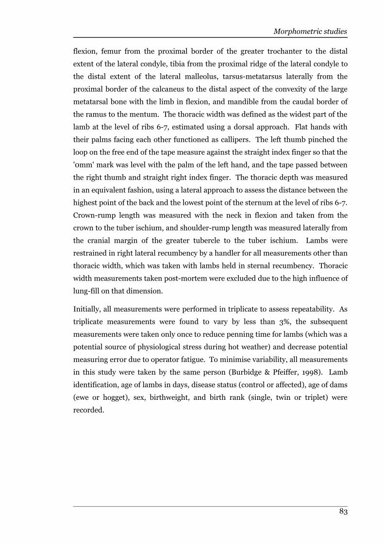
Morphometric studies
flexion, femur from the proximal border of the greater trochanter to the distal
extent of the lateral condyle, tibia from the proximal ridge of the lateral condyle to
the distal extent of the lateral malleolus, tarsus-metatarsus laterally from the
proximal border of the calcaneus to the distal aspect of the convexity of the large
metatarsal bone with the limb in flexion, and mandible from the caudal border of
the ramus to the mentum. The thoracic width was defined as the widest part of the
lamb at the level of ribs 6-7, estimated using a dorsal approach. Flat hands with
their palms facing each other functioned as callipers. The left thumb pinched the
loop on the free end of the tape measure against the straight index finger so that the
'0mm' mark was level with the palm of the left hand, and the tape passed between
the right thumb and straight right index finger. The thoracic depth was measured
in an equivalent fashion, using a lateral approach to assess the distance between the
highest point of the back and the lowest point of the sternum at the level of ribs 6-7.
Crown-rump length was measured with the neck in flexion and taken from the
crown to the tuber ischium, and shoulder-rump length was measured laterally from
the cranial margin of the greater tubercle to the tuber ischium. Lambs were
restrained in right lateral recumbency by a handler for all measurements other than
thoracic width, which was taken with lambs held in sternal recumbency. Thoracic
width measurements taken post-mortem were excluded due to the high influence of
lung-fill on that dimension.
Initially, all measurements were performed in triplicate to assess repeatability. As
triplicate measurements were found to vary by less than 3%, the subsequent
measurements were taken only once to reduce penning time for lambs (which was a
potential source of physiological stress during hot weather) and decrease potential
measuring error due to operator fatigue. To minimise variability, all measurements
in this study were taken by the same person (Burbidge & Pfeiffer, 1998). Lamb
identification, age of lambs in days, disease status (control or affected), age of dams
(ewe or hogget), sex, birthweight, and birth rank (single, twin or triplet) were
recorded.
83
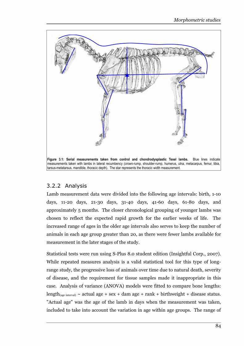
Morphometric studies
3.2.2 Analysis
Lamb measurement data were divided into the following age intervals: birth, 1-10
days, 11-20 days, 21-30 days, 31-40 days, 41-60 days, 61-80 days, and
approximately 5 months. The closer chronological grouping of younger lambs was
chosen to reflect the expected rapid growth for the earlier weeks of life. The
increased range of ages in the older age intervals also serves to keep the number of
animals in each age group greater than 20, as there were fewer lambs available for
measurement in the later stages of the study.
Statistical tests were run using S-Plus 8.0 student edition (Insightful Corp., 2007).
While repeated measures analysis is a valid statistical tool for this type of long-
range study, the progressive loss of animals over time due to natural death, severity
of disease, and the requirement for tissue samples made it inappropriate in this
case. Analysis of variance (ANOVA) models were fitted to compare bone lengths:
length(age interval) ~ actual age + sex + dam age + rank + birthweight + disease status.
"Actual age" was the age of the lamb in days when the measurement was taken,
included to take into account the variation in age within age groups. The range of
84
Figure 3.1: Serial measurements taken from control and chondrodysplastic Texel lambs. Blue lines indicatemeasurements taken with lambs in lateral recumbency (crown-rump, shoulder-rump, humerus, ulna, metacarpus, femur, tibia,tarsus-metatarsus, mandible, thoracic depth). The star represents the thoracic width measurement.
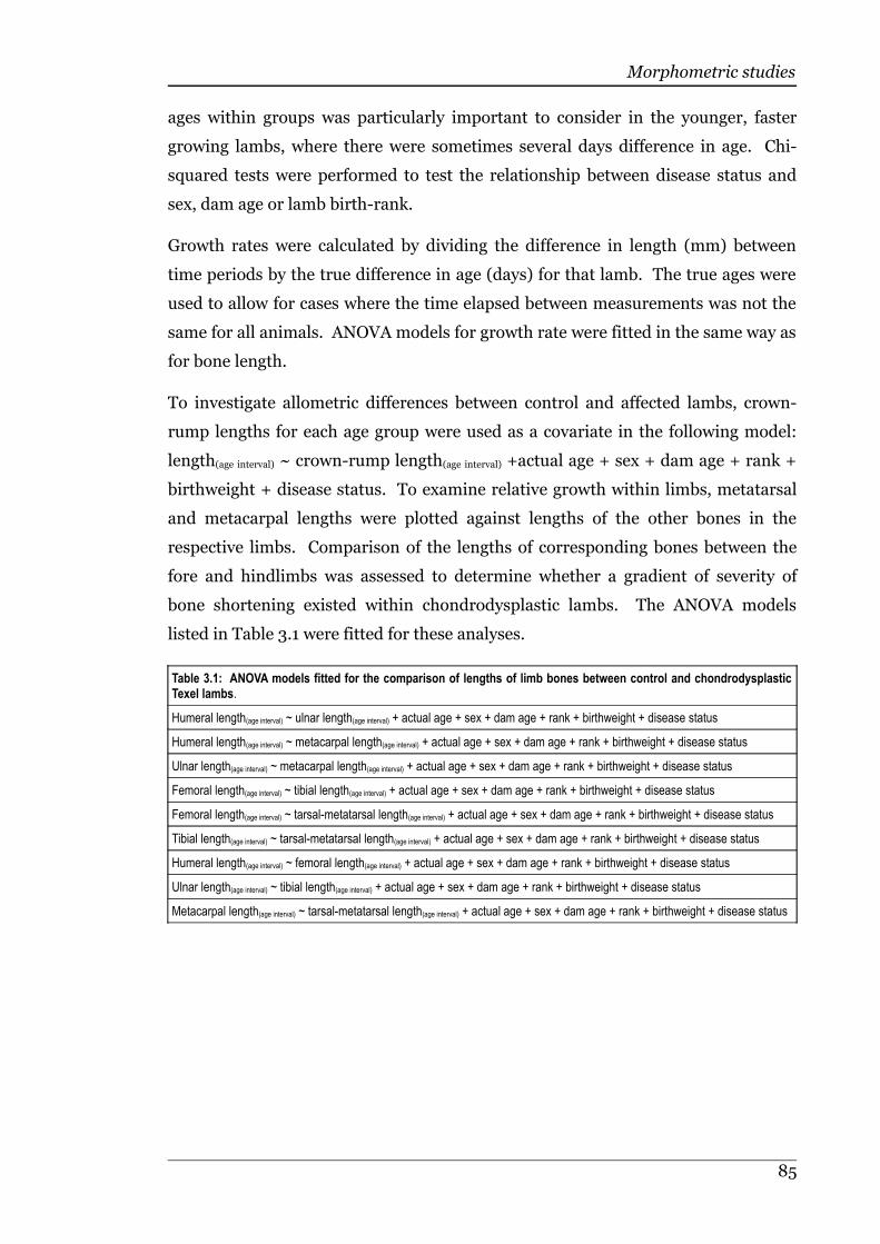
Morphometric studies
ages within groups was particularly important to consider in the younger, faster
growing lambs, where there were sometimes several days difference in age. Chi-
squared tests were performed to test the relationship between disease status and
sex, dam age or lamb birth-rank.
Growth rates were calculated by dividing the difference in length (mm) between
time periods by the true difference in age (days) for that lamb. The true ages were
used to allow for cases where the time elapsed between measurements was not the
same for all animals. ANOVA models for growth rate were fitted in the same way as
for bone length.
To investigate allometric differences between control and affected lambs, crown-
rump lengths for each age group were used as a covariate in the following model:
length(age interval) ~ crown-rump length(age interval) +actual age + sex + dam age + rank +
birthweight + disease status. To examine relative growth within limbs, metatarsal
and metacarpal lengths were plotted against lengths of the other bones in the
respective limbs. Comparison of the lengths of corresponding bones between the
fore and hindlimbs was assessed to determine whether a gradient of severity of
bone shortening existed within chondrodysplastic lambs. The ANOVA models
listed in Table 3.1 were fitted for these analyses.
Table 3.1: ANOVA models fitted for the comparison of lengths of limb bones between control and chondrodysplasticTexel lambs.
Humeral length(age interval) ~ ulnar length(age interval) + actual age + sex + dam age + rank + birthweight + disease status
Humeral length(age interval) ~ metacarpal length(age interval) + actual age + sex + dam age + rank + birthweight + disease status
Ulnar length(age interval) ~ metacarpal length(age interval) + actual age + sex + dam age + rank + birthweight + disease status
Femoral length(age interval) ~ tibial length(age interval) + actual age + sex + dam age + rank + birthweight + disease status
Femoral length(age interval) ~ tarsal-metatarsal length(age interval) + actual age + sex + dam age + rank + birthweight + disease status
Tibial length(age interval) ~ tarsal-metatarsal length(age interval) + actual age + sex + dam age + rank + birthweight + disease status
Humeral length(age interval) ~ femoral length(age interval) + actual age + sex + dam age + rank + birthweight + disease status
Ulnar length(age interval) ~ tibial length(age interval) + actual age + sex + dam age + rank + birthweight + disease status
Metacarpal length(age interval) ~ tarsal-metatarsal length(age interval) + actual age + sex + dam age + rank + birthweight + disease status
85
Morphometric studies
3.3 Results
3.3.1 Overview
Subjectively, chondrodysplastic lambs were indistinguishable from controls at
birth, but appeared to grow less rapidly and were noticeably smaller by 2-3 weeks of
age. The thorax of affected animals acquired a distinctive barrel-shape, and the
forelimbs often developed varus deformities. Affected lambs that appeared to grow
quickly from an early age, compared with their cohorts, seemed to have a more
rapid onset of the chondrodysplastic phenotype.
The variation in birthweight was influenced by sex, age of dam and birth rank. The
mean birthweight of male lambs was approximately 0.6kg heavier than that of
female lambs (P≤0.005), and ewes produced lambs approximately 0.7kg heavier
than hoggets (P≤0.0001). Chi-squared tests demonstrated no significant
relationship between disease status and sex or birth rank of lambs, or age of dam.
All dimensions other than thoracic width were significantly smaller in affected than
normal lambs by 1 month of age, and this difference continued to be significant for
the duration of the study. The mean percentage difference in length of bones in
affected lambs relative to controls was greatest at 61-80 days for every dimension.
The greatest difference was in thoracic depth, which was 20% less in
chondrodysplastic lambs. The smallest difference was in thoracic width, which was
5% less in chondrodysplastic lambs. Growth rates of the crown-rump, shoulder-
rump, humerus, femur, tibia and tarsus-metatarsus, mandibular length and
thoracic depth were significantly reduced for part of the measured period but were
not significantly different from controls in the last time period (5 months). Ulnar
and metacarpal growth rates were less in chondrodysplastic lambs compared with
controls at 5 months of age (P≤0.05). There was no significant difference in growth
rate of thoracic width between control and affected lambs at any age. Details of
these results are presented in the following sections and a digital copy of the raw
data is included in the back of this thesis.
86
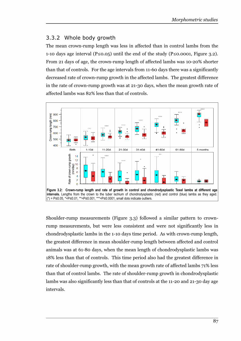
Morphometric studies
3.3.2 Whole body growth
The mean crown-rump length was less in affected than in control lambs from the
1-10 days age interval (P≤0.05) until the end of the study (P≤0.0001, Figure 3.2).
From 21 days of age, the crown-rump length of affected lambs was 10-20% shorter
than that of controls. For the age intervals from 11-60 days there was a significantly
decreased rate of crown-rump growth in the affected lambs. The greatest difference
in the rate of crown-rump growth was at 21-30 days, when the mean growth rate of
affected lambs was 82% less than that of controls.
Shoulder-rump measurements (Figure 3.3) followed a similar pattern to crown-
rump measurements, but were less consistent and were not significantly less in
chondrodysplastic lambs in the 1-10 days time period. As with crown-rump length,
the greatest difference in mean shoulder-rump length between affected and control
animals was at 61-80 days, when the mean length of chondrodysplastic lambs was
18% less than that of controls. This time period also had the greatest difference in
rate of shoulder-rump growth, with the mean growth rate of affected lambs 71% less
than that of control lambs. The rate of shoulder-rump growth in chondrodysplastic
lambs was also significantly less than that of controls at the 11-20 and 21-30 day age
intervals.
87
Figure 3.2: Crown-rump length and rate of growth in control and chondrodysplastic Texel lambs at different ageintervals. Lengths from the crown to the tuber ischium of chondrodysplastic (red) and control (blue) lambs as they aged.(*) = P≤0.05, *=P≤0.01, **=P≤0.001, ***=P≤0.0001, small dots indicate outliers.
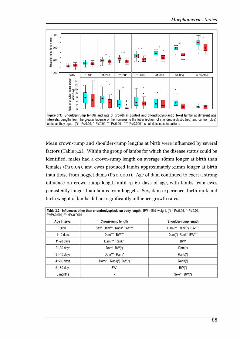
Morphometric studies
Mean crown-rump and shoulder-rump lengths at birth were influenced by several
factors (Table 3.2). Within the group of lambs for which the disease status could be
identified, males had a crown-rump length on average 18mm longer at birth than
females (P≤0.05), and ewes produced lambs approximately 31mm longer at birth
than those from hogget dams (P≤0.0001). Age of dam continued to exert a strong
influence on crown-rump length until 41-60 days of age, with lambs from ewes
persistently longer than lambs from hoggets. Sex, dam experience, birth rank and
birth weight of lambs did not significantly influence growth rates.
Table 3.2: Influences other than chondrodysplasia on body length. BW = Birthweight, (*) = P≤0.05, *=P≤0.01,**=P≤0.001, ***=P≤0.0001
Age interval Crown-rump length Shoulder-rump length
Birth Sex* Dam*** Rank* BW*** Dam*** Rank(*) BW***
1-10 days Dam*** BW*** Dam(*) Rank* BW***
11-20 days Dam*** Rank* BW*
21-30 days Dam* BW(*) Dam(*)
31-40 days Dam*** Rank* Rank(*)
41-60 days Dam(*) Rank(*) BW(*) Rank(*)
61-80 days BW* BW(*)
5 months - Sex(*) BW(*)
88
Figure 3.3: Shoulder-rump length and rate of growth in control and chondrodysplastic Texel lambs at different ageintervals. Lengths from the greater tubercle of the humerus to the tuber ischium of chondrodysplastic (red) and control (blue)lambs as they aged. (*) = P≤0.05, *=P≤0.01, **=P≤0.001, ***=P≤0.0001, small dots indicate outliers.
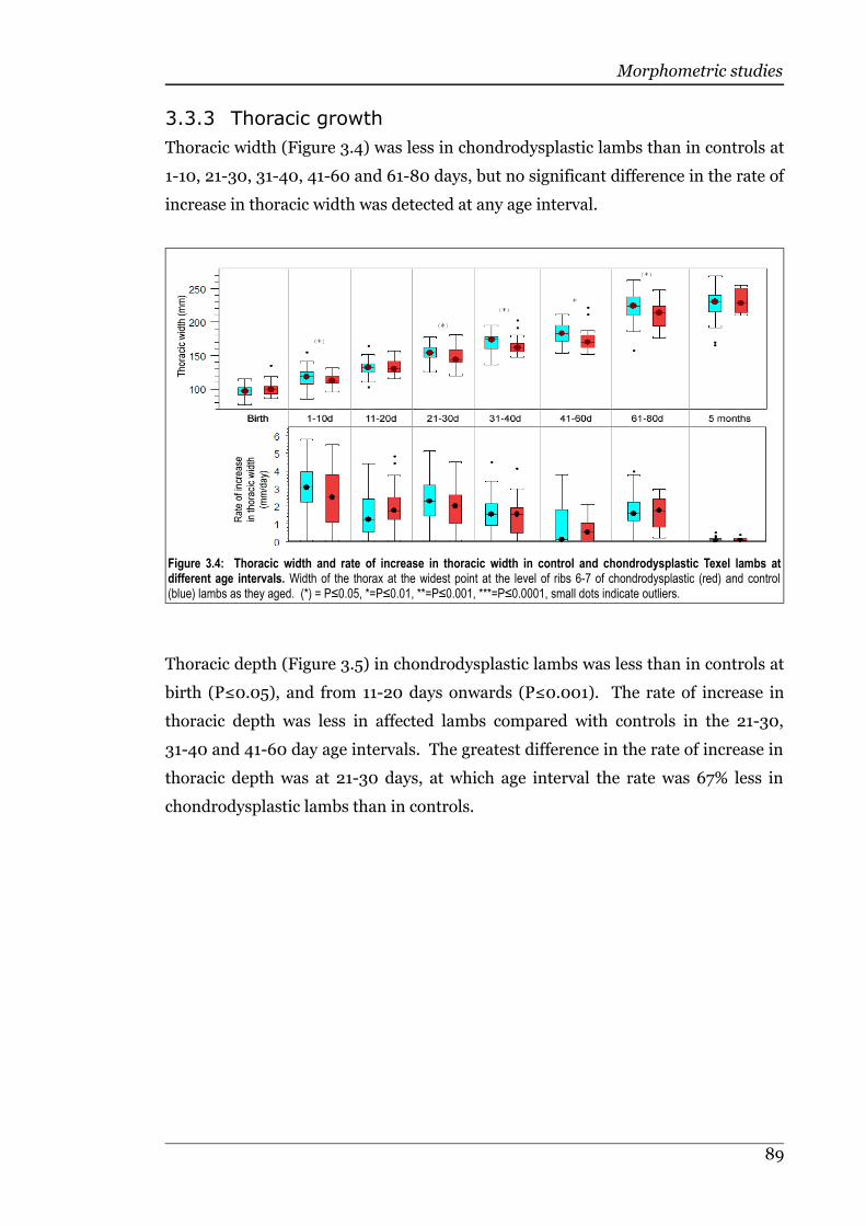
Morphometric studies
3.3.3 Thoracic growth
Thoracic width (Figure 3.4) was less in chondrodysplastic lambs than in controls at
1-10, 21-30, 31-40, 41-60 and 61-80 days, but no significant difference in the rate of
increase in thoracic width was detected at any age interval.
Thoracic depth (Figure 3.5) in chondrodysplastic lambs was less than in controls at
birth (P≤0.05), and from 11-20 days onwards (P≤0.001). The rate of increase in
thoracic depth was less in affected lambs compared with controls in the 21-30,
31-40 and 41-60 day age intervals. The greatest difference in the rate of increase in
thoracic depth was at 21-30 days, at which age interval the rate was 67% less in
chondrodysplastic lambs than in controls.
89
Figure 3.4: Thoracic width and rate of increase in thoracic width in control and chondrodysplastic Texel lambs atdifferent age intervals. Width of the thorax at the widest point at the level of ribs 6-7 of chondrodysplastic (red) and control(blue) lambs as they aged. (*) = P≤0.05, *=P≤0.01, **=P≤0.001, ***=P≤0.0001, small dots indicate outliers.

Morphometric studies
Thoracic width was positively correlated with birth weight up to and including the
41-60 days age interval (Table 3.3). Dam experience influenced thoracic depth up
to 41-60 days, with lambs from ewes having a deeper thorax than lambs from
hoggets. At birth, thoracic depth was 4.6mm deeper in male than in female lambs
(P≤0.01). Birth weight was positively correlated with thoracic depth until the 11-20
days age interval, and birth rank had a sporadic influence, lambs from multiple
births tending to have deeper thoraxes than singleton lambs.
Table 3.3: Influences other than chondrodysplasia on thoracic dimensions. BW = Birthweight, (*) = P≤0.05, *=P≤0.01,**=P≤0.001, ***=P≤0.0001
Age Thoracic width Thoracic depth
Birth Dam(*) Rank(*) BW*** Sex* Dam*** Rank** BW***
1-10 days Dam(*) Dam** BW**
11-20 days Rank* BW** Sex* Dam* BW***
21-30 days Rank* BW(*) Dam*
31-40 days Dam(*) Rank(*) Dam** Rank*
41-60 days Rank(*) BW(*) Dam*
61-80 days BW(*) BW(*)
5 months - -
3.3.4 Mandibular growth
Although there was no clear difference in mean mandibular length between
chondrodysplastic and control lambs at the 11-20 and 31-40 day age intervals
(Figure 3.6), when the other variables in the ANOVA model were accounted for, a
90
Figure 3.5: Thoracic depth and rate of increase in thoracic depth in control and chondrodysplastic Texel lambs atdifferent age intervals. Depth of the thorax at the level of ribs 6-7 of chondrodysplastic (red) and control (blue) lambs as theyaged. (*) = P≤0.05, *=P≤0.01, **=P≤0.001, ***=P≤0.0001, small dots indicate outliers.
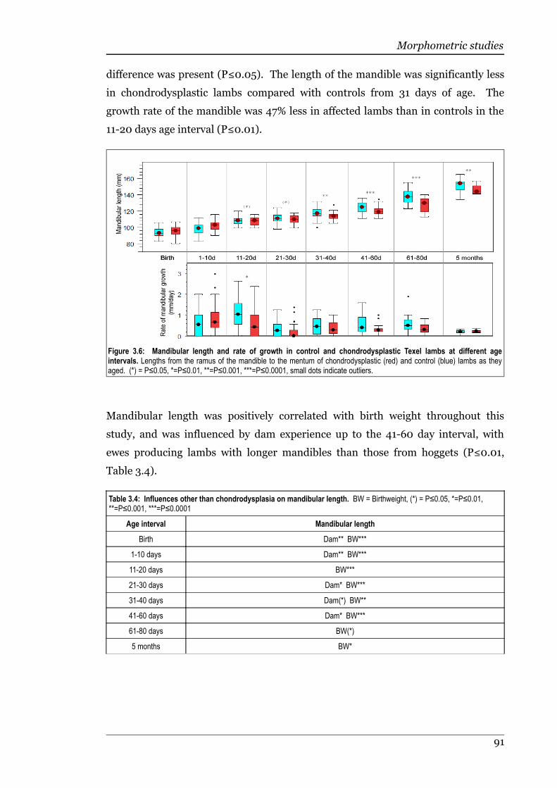
Morphometric studies
difference was present (P≤0.05). The length of the mandible was significantly less
in chondrodysplastic lambs compared with controls from 31 days of age. The
growth rate of the mandible was 47% less in affected lambs than in controls in the
11-20 days age interval (P≤0.01).
Mandibular length was positively correlated with birth weight throughout this
study, and was influenced by dam experience up to the 41-60 day interval, with
ewes producing lambs with longer mandibles than those from hoggets (P≤0.01,
Table 3.4).
Table 3.4: Influences other than chondrodysplasia on mandibular length. BW = Birthweight, (*) = P≤0.05, *=P≤0.01,**=P≤0.001, ***=P≤0.0001
Age interval Mandibular length
Birth Dam** BW***
1-10 days Dam** BW***
11-20 days BW***
21-30 days Dam* BW***
31-40 days Dam(*) BW**
41-60 days Dam* BW***
61-80 days BW(*)
5 months BW*
91
Figure 3.6: Mandibular length and rate of growth in control and chondrodysplastic Texel lambs at different ageintervals. Lengths from the ramus of the mandible to the mentum of chondrodysplastic (red) and control (blue) lambs as theyaged. (*) = P≤0.05, *=P≤0.01, **=P≤0.001, ***=P≤0.0001, small dots indicate outliers.
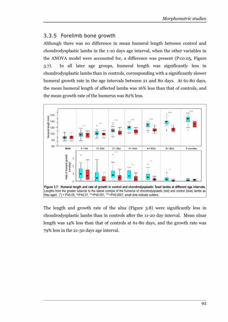
Morphometric studies
3.3.5 Forelimb bone growth
Although there was no difference in mean humeral length between control and
chondrodysplastic lambs in the 1-10 days age interval, when the other variables in
the ANOVA model were accounted for, a difference was present (P≤0.05, Figure
3.7). In all later age groups, humeral length was significantly less in
chondrodysplastic lambs than in controls, corresponding with a significantly slower
humeral growth rate in the age intervals between 21 and 80 days. At 61-80 days,
the mean humeral length of affected lambs was 16% less than that of controls, and
the mean growth rate of the humerus was 82% less.
The length and growth rate of the ulna (Figure 3.8) were significantly less in
chondrodysplastic lambs than in controls after the 11-20 day interval. Mean ulnar
length was 14% less than that of controls at 61-80 days, and the growth rate was
79% less in the 21-30 days age interval.
92
Figure 3.7: Humeral length and rate of growth in control and chondrodysplastic Texel lambs at different age intervals.Lengths from the greater tubercle to the lateral condyle of the humerus of chondrodysplastic (red) and control (blue) lambs asthey aged. (*) = P≤0.05, *=P≤0.01, **=P≤0.001, ***=P≤0.0001, small dots indicate outliers.
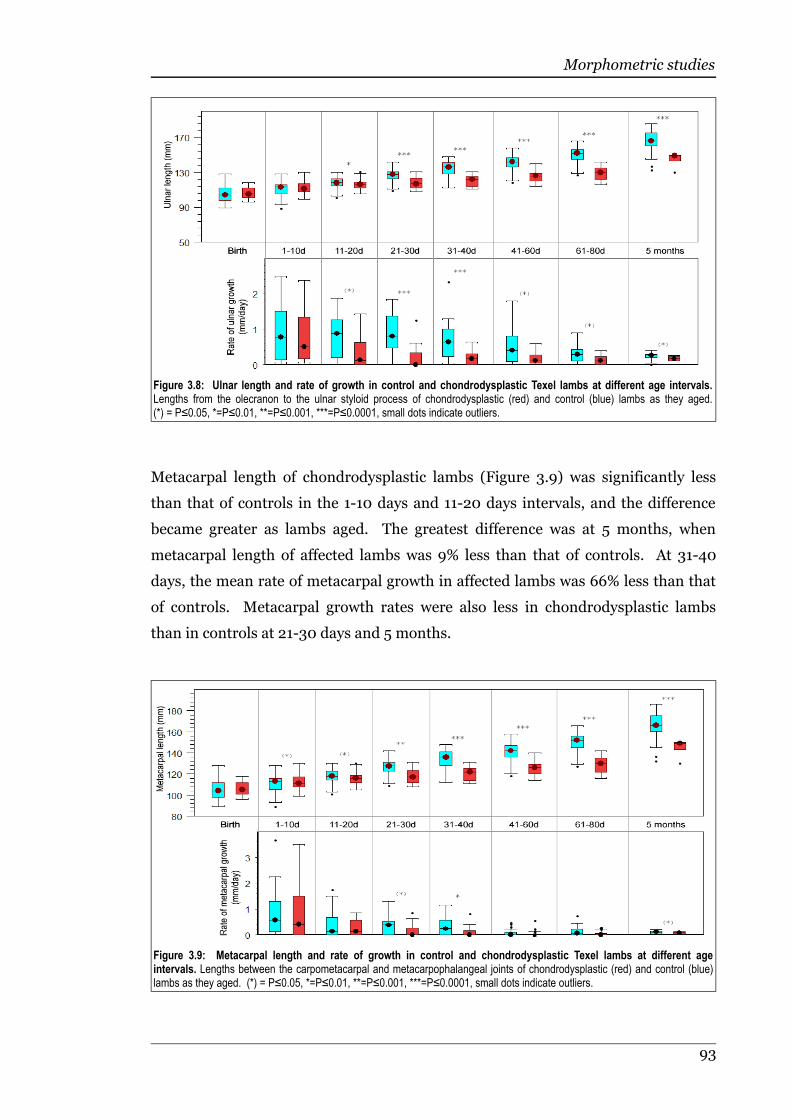
Morphometric studies
Metacarpal length of chondrodysplastic lambs (Figure 3.9) was significantly less
than that of controls in the 1-10 days and 11-20 days intervals, and the difference
became greater as lambs aged. The greatest difference was at 5 months, when
metacarpal length of affected lambs was 9% less than that of controls. At 31-40
days, the mean rate of metacarpal growth in affected lambs was 66% less than that
of controls. Metacarpal growth rates were also less in chondrodysplastic lambs
than in controls at 21-30 days and 5 months.
93
Figure 3.9: Metacarpal length and rate of growth in control and chondrodysplastic Texel lambs at different ageintervals. Lengths between the carpometacarpal and metacarpophalangeal joints of chondrodysplastic (red) and control (blue)lambs as they aged. (*) = P≤0.05, *=P≤0.01, **=P≤0.001, ***=P≤0.0001, small dots indicate outliers.
Figure 3.8: Ulnar length and rate of growth in control and chondrodysplastic Texel lambs at different age intervals.Lengths from the olecranon to the ulnar styloid process of chondrodysplastic (red) and control (blue) lambs as they aged.(*) = P≤0.05, *=P≤0.01, **=P≤0.001, ***=P≤0.0001, small dots indicate outliers.

Morphometric studies
Maternal age had a strong influence on humeral and ulnar length until 41-60 days
of age, adult ewes producing lambs with longer bones than those from hoggets
(Table 3.5). The metacarpal bone appeared to be influenced by different factors to
the other forelimb bones. In the age intervals for which sex had a significant
influence of metacarpal length, male lambs had a greater mean length than did
female lambs. This difference was significant at birth, with a mean difference of
4.2mm (P≤0.05), 1-10 days with a mean difference of 4.2mm (P≤0.01), 11-20 days
with a mean difference of 4.4mm (P≤0.001), 31-40 days with a mean difference of
3.5mm (P≤0.01), and at 41-60 days with a mean difference of 3.6mm (P≤0.001).
Dam experience had a significant effect until 61-80 days, adult ewes having lambs
with longer metacarpi than those from hoggets. Rank had a significant effect on
metacarpal length, with lambs from multiple births having longer metacarpi than
singletons by 2.9mm at 61-80 days (P≤0.001) and 4.8mm at 5 months (P≤0.05).
Table 3.5: Influences other than chondrodysplasia on forelimb bone length. BW = Birthweight, (*) = P≤0.05, *=P≤0.01,**=P≤0.001, ***=P≤0.0001
Age interval Humeral length Ulnar length Metacarpal length
Birth Dam** Rank* BW*** Dam*** Rank(*) BW*** Sex(*) Dam*** BW**
1-10 days Sex* Dam** BW*** Dam*** BW*** Sex* Dam** BW***
11-20 days Dam*** Rank(*) BW*** Dam*** Rank(*) BW*** Sex* Dam* BW**
21-30 days Dam*** BW** Dam*** Rank(*) BW** Dam*
31-40 days Dam* BW(*) Sex(*) Dam*** Rank(*) BW* Sex* Dam**
41-60 days - Dam(*) BW(*) Sex* Dam* BW(*)
61-80 days BW* BW* Rank(*) BW***
5 months BW(*) Sex(*) BW* Dam(*) BW**
3.3.6 Hindlimb bone growth
Femoral length (Figure 3.10) was significantly different in control and
chondrodysplastic lambs at 1-10 days and the differences increased with age,
becoming most marked at 61-80 days, when mean femoral length of affected lambs
was 16% less than that of controls. Mean femoral growth rate was significantly less
in chondrodysplastic lambs than in controls at 1-10 and at 41-60 days, when the
mean femoral growth rate was 77% slower than that of controls.
94
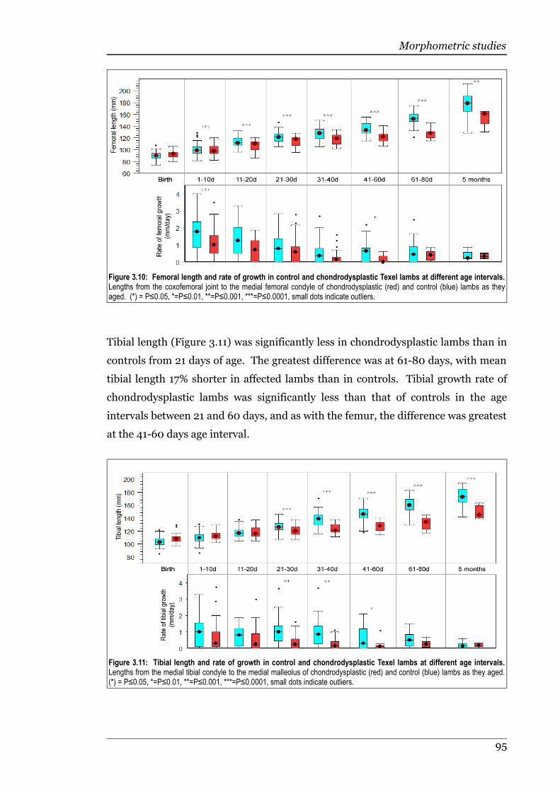
Morphometric studies
Tibial length (Figure 3.11) was significantly less in chondrodysplastic lambs than in
controls from 21 days of age. The greatest difference was at 61-80 days, with mean
tibial length 17% shorter in affected lambs than in controls. Tibial growth rate of
chondrodysplastic lambs was significantly less than that of controls in the age
intervals between 21 and 60 days, and as with the femur, the difference was greatest
at the 41-60 days age interval.
95
Figure 3.10: Femoral length and rate of growth in control and chondrodysplastic Texel lambs at different age intervals.Lengths from the coxofemoral joint to the medial femoral condyle of chondrodysplastic (red) and control (blue) lambs as theyaged. (*) = P≤0.05, *=P≤0.01, **=P≤0.001, ***=P≤0.0001, small dots indicate outliers.
Figure 3.11: Tibial length and rate of growth in control and chondrodysplastic Texel lambs at different age intervals.Lengths from the medial tibial condyle to the medial malleolus of chondrodysplastic (red) and control (blue) lambs as they aged.(*) = P≤0.05, *=P≤0.01, **=P≤0.001, ***=P≤0.0001, small dots indicate outliers.

Morphometric studies
Tarsal-metatarsal length (Figure 3.12) was less in chondrodysplastic lambs than in
controls at 1-10 days, and the difference became more pronounced with age. The
disparity peaked at 61-80 days, when tarsal-metatarsal length in affected lambs was
11% less than in controls. Tarsal-metatarsal growth rate was less in
chondrodysplastic lambs than in controls at the 11-20, 21-30, 31-40 and 41-60 days
age intervals. The greatest difference was at 31-40 days, when the mean growth
rate in affected lambs was 73% less than in controls.
As with the metacarpus, rank had some influence on femoral length (Table 3.6),
although in contrast with the findings for metacarpal length, singleton lambs had
longer femurs than lambs from multiple births by 0.8mm at 1-10 days (P≤0.01),
3mm at 11-20 days (P≤0.05), 3.4mm at 21-30 days (P≤0.01), 2.2mm at 31-40 days
(P≤0.05), and 3.2mm at 41-60 days (P≤0.05). Birthweight was positively
correlated with femoral length until the 21-30 days age interval, and dam age was
only a significant factor until 1-10 days, with lambs from adult ewes having longer
femurs than those from hoggets. There was a significant interaction of dam age and
birthweight up to 61-80 days of age, with adult ewes producing lambs with greater
tarsal-metatarsal lengths than lambs from hoggets. Tibial length was positively
correlated with birth weight up to 31-40 days of age, and was influenced by dam age
up to 61-80 days of age, with adult ewes producing lambs with longer tibias than
did hoggets.
96
Figure 3.12: Tarsal-metatarsal length and rate of growth in control and chondrodysplastic Texel lambs at different ageintervals. Lengths from the calcaneus to the metatarsophalangeal joint of chondrodysplastic (red) and control (blue) lambs asthey aged. (*) = P≤0.05, *=P≤0.01, **=P≤0.001, ***=P≤0.0001, small dots indicate outliers.

Morphometric studies
Table 3.6: Influences other than chondrodysplasia on hindlimb bone length. BW = Birthweight, (*) = P≤0.05, *=P≤0.01,**=P≤0.001, ***=P≤0.0001
Age interval Femoral length Tibial length Tarsal-metatarsal length
Birth Sex(*) Dam*** BW*** Sex(*) Dam* BW*** Dam*** Rank(*) BW***
1-10 days Rank(*) BW*** Dam* BW*** Dam*** Rank* BW***
11-20 days Rank(*) BW** Sex(*) Dam* Rank** BW*** Sex(*) Dam** Rank(*) BW***
21-30 days Rank* BW** Dam* Rank* BW** Dam*** BW*
31-40 days Rank(*) BW(*) Sex(*) Dam* Rank(*) BW(*) Dam* BW*
41-60 days Rank(*) BW(*) Sex(*) Dam(*) Dam(*) BW*
61-80 days BW(*) - Dam* BW**
5 months - - BW(*)
3.3.7 Allometry
Bone lengths in relation to crown-rump length
When crown-rump length was used as a covariate for other measurements within
each age group, birthweight was responsible for most variation in the majority of
measurements until 21-30 days (Table 3.7). Sex influenced metacarpal length in
most age groups other than newborn, with male lambs having longer metacarpi
relative to crown-rump length than female lambs. Tibial length was influenced by
sex at the 11-20, 31-40 and 41-60 day age intervals, with male lambs having longer
tibias relative to crown-rump length compared with female lambs. Other body
parts were inconsistently affected by rank, sex and dam age. Measurements in
which chondrodysplastic lambs showed a disproportionate shortening relative to
crown-rump length when compared with control lambs were shoulder-rump length
at 61-80 days (P≤0.05), ulna at 21-30 days (P≤0.01) and 31-40 days (P≤0.05),
metacarpus at 61-80 days (P≤0.05), femur at 1-10 and 11-20 days (P≤0.05), tibia at
41-60 days (P≤0.01), and thoracic depth at birth (P≤0.05), 21-30 days (P≤0.01),
61-80 days (P≤0.05) and 5 months (P≤0.001). The tarsal-metatarsal length at
21-30 days was proportionally longer relative to crown-rump length in
chondrodysplastic lambs when compared with control lambs (P≤0.05).
97
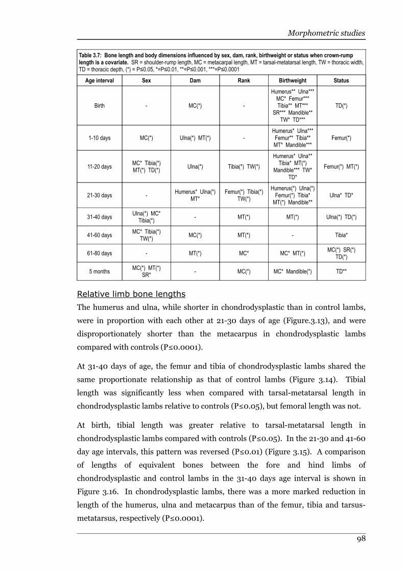
Morphometric studies
Table 3.7: Bone length and body dimensions influenced by sex, dam, rank, birthweight or status when crown-rumplength is a covariate. SR = shoulder-rump length, MC = metacarpal length, MT = tarsal-metatarsal length, TW = thoracic width,TD = thoracic depth, (*) = P≤0.05, *=P≤0.01, **=P≤0.001, ***=P≤0.0001
Age interval Sex Dam Rank Birthweight Status
Birth - MC(*) -
Humerus** Ulna***MC* Femur***Tibia** MT***
SR*** Mandible**TW* TD***
TD(*)
1-10 days MC(*) Ulna(*) MT(*) -Humerus* Ulna***Femur** Tibia**MT* Mandible***
Femur(*)
11-20 daysMC* Tibia(*)MT(*) TD(*)
Ulna(*) Tibia(*) TW(*)
Humerus* Ulna**Tibia* MT(*)
Mandible*** TW*TD*
Femur(*) MT(*)
21-30 days -Humerus* Ulna(*)
MT*Femur(*) Tibia(*)
TW(*)
Humerus(*) Ulna(*)Femur(*) Tibia*MT(*) Mandible**
Ulna* TD*
31-40 daysUlna(*) MC*
Tibia(*)- MT(*) MT(*) Ulna(*) TD(*)
41-60 daysMC* Tibia(*)
TW(*)MC(*) MT(*) - Tibia*
61-80 days - MT(*) MC* MC* MT(*)MC(*) SR(*)
TD(*)
5 monthsMC(*) MT(*)
SR*- MC(*) MC* Mandible(*) TD**
Relative limb bone lengths
The humerus and ulna, while shorter in chondrodysplastic than in control lambs,
were in proportion with each other at 21-30 days of age (Figure.3.13), and were
disproportionately shorter than the metacarpus in chondrodysplastic lambs
compared with controls (P≤0.0001).
At 31-40 days of age, the femur and tibia of chondrodysplastic lambs shared the
same proportionate relationship as that of control lambs (Figure 3.14). Tibial
length was significantly less when compared with tarsal-metatarsal length in
chondrodysplastic lambs relative to controls (P≤0.05), but femoral length was not.
At birth, tibial length was greater relative to tarsal-metatarsal length in
chondrodysplastic lambs compared with controls (P≤0.05). In the 21-30 and 41-60
day age intervals, this pattern was reversed (P≤0.01) (Figure 3.15). A comparison
of lengths of equivalent bones between the fore and hind limbs of
chondrodysplastic and control lambs in the 31-40 days age interval is shown in
Figure 3.16. In chondrodysplastic lambs, there was a more marked reduction in
length of the humerus, ulna and metacarpus than of the femur, tibia and tarsus-
metatarsus, respectively (P≤0.0001).
98
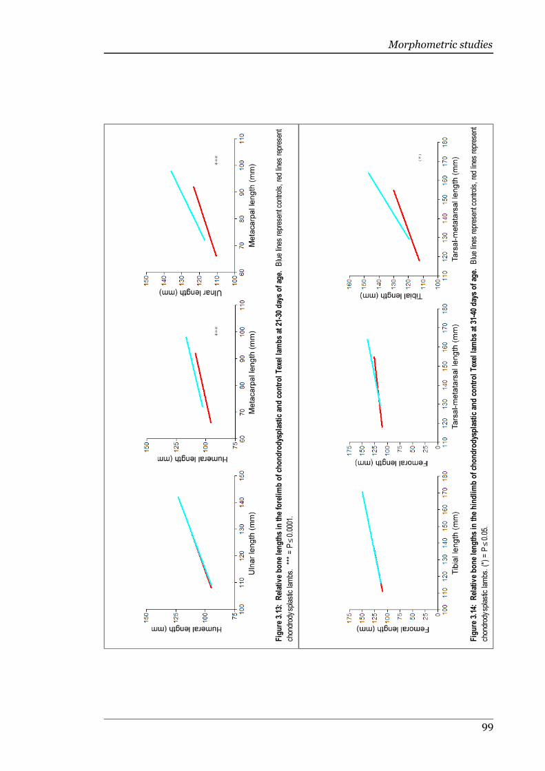
Morphometric studies
99
Figure 3.13: Relative bone lengths in the forelimb of ch
ondrodysplastic and control Texel lam
bs at 21-30 days of ag
e. Blue lines represent controls, red lines represent
chondrodysplastic lambs. *** = P≤0.0001.
Figure 3.14: Relative bone lengths in the hindlimb of ch
ondrodysplastic and control Texel lam
bs at 31-40 days of ag
e. Blue lines represent controls, red lines represent
chondrodysplastic lambs. (*) = P≤0.05.
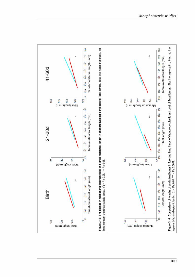
Morphometric studies
100
Figure 3.15: The ch
ange in relationsh
ip between tibial an
d tarsal-metatarsal length in chondrodysplastic and control Texel lam
bs. Blue lines represent controls, red
lines represent chondrody
splastic lambs. (*) = P≤0.05, * = P≤0.01.
Figure 3.16: Comparison of lengths of eq
uivalen
t bones in fore and hind lim
bs of ch
ondrodysplastic and control Texel lam
bs. Blue lines represent controls, red lines
represent chondrody
splastic lambs. (*) = P≤0.05, *** = P≤0.0001.
Morphometric studies
3.4 Discussion
The results of this study support the hypothesis that bone lengths and growth rates
in chondrodysplastic Texel lambs are less than in controls. The findings also
confirm the clinical impression of gradual progression of the phenotype in
chondrodysplasia of Texel sheep. Chondrodysplastic Texel sheep have an overall
disproportionately reduced stature, which becomes apparent over the first 2 weeks
of life. Chondrodysplastic lambs were indistinguishable from control lambs at
birth. Although statistically significant differences in thoracic depth and in the
relationship between tibial and tarsal-metatarsal lengths could be identified when
comparing a number of animals, the overlap in values between affected and
controls lambs at birth was too great to confidently diagnose chondrodysplasia in
any one lamb using these criteria.
The greatest differences in mean bone lengths and body dimensions between
chondrodysplastic and control lambs were generally in the 61-80 days age interval,
with somewhat less marked differences in mean length at 5 months. The latter is
most likely explained by the removal of the most severely affected
chondrodysplastic lambs from the flock prior to 5 months, rather than any "catch-
up" in growth at a later stage.
The results for rate of increase in many of the measured dimensions indicated that
bone growth in chondrodysplastic lambs was initially the same as in controls,
slowed down during the first two weeks of life, then apparently returned to normal
by 5 months of age. This may have been due to the removal of the more severely
affected lambs earlier on leaving only mildly affected lambs that do not have such a
reduced rate of growth. However, if this was the case, the actual measurements of
each bone would not be expected to be as short as they were, relative to controls at
that point. An alternative explanation may be that by 5 months of age long bone
growth in control lambs was slowing and some physes were beginning to ossify,
while those in chondrodysplastic lambs remained open. This was supported
histologically, where there was complete bony union across the whole physis of the
distal humerus in control animals by this time. In chondrodysplastic lambs, no
physes were fused at 5 months (see Chapter 2), and long bone physes were
significantly thicker than in control lambs (see Chapter 2, Table 2.2). This does not
necessarily mean that all bones were still increasing in length at this age, as the
cartilaginous physis persists for some time after effective longitudinal growth has
101
Morphometric studies
ceased (Roach et al., 2003). Nonetheless, it is possible that the bones of
chondrodysplastic lambs surviving to 5 months of age continued to grow slowly due
to delayed physeal closure, while those of control lambs grew quicker earlier on but
slowed or ceased in growth by 5 months of age.
The allometric comparisons of bone lengths in chondrodysplastic and control Texel
lambs demonstrated that shortening of limb bones was more marked in the
proximal limb than in the distal limb (rhizomelia), and that the forelimbs were
more severely affected than the hindlimbs. This pattern of development reflects to
some degree the spectrum of severity of histological lesions within affected animals
(see Chapter 2). The mechanism behind this uneven distribution of the effects of
chondrodysplasia in Texel sheep may be related to relative growth rate gradients
within young lambs. In normal lambs, the proximal limb bones and caudal end of
the skeleton are growing faster than the distal limb bones and cranial end of the
skeleton (Davies et al., 1984). A similar cranio-caudal gradient of increasing
relative growth rate has been described in young pigs (Liu et al., 1999). Therefore,
within limbs, the bones with the fastest expected rate of growth relative to their
potential final length were more severely shortened in chondrodysplastic lambs.
Physeal cartilage from faster-growing bones has an increased rate of chondrocyte
cell division and sulphate incorporation into matrix per wet weight than physeal
cartilage from slower-growing bones, indicating the former has a higher level of
metabolic activity than the latter (Seinsheimer & Sledge, 1981). It is likely that cells
with a higher metabolic rate would be more affected by disorders involving
insufficiency of a metabolic product or enzymatic cofactor. If such a mechanism is
involved in chondrodysplasia of Texel sheep, it would help to explain the pattern of
lesions, both gross and microscopic, that develop as affected lambs grow. The idea
of a cartilage defect which is dependent to some degree upon the local metabolic
rate is also supported by the subjective observation that the lambs that were
initially faster-growing appeared to have a more sudden onset of lesions of
chondrodysplasia. The data collected in this study was not able to provide great
enough resolution to verify this hypothesis.
While the pattern of limb growth in chondrodysplastic lambs appeared to be
inversely related to expected relative growth rate, the converse was seen when
comparing relative reduction of bone length between fore and hindlimbs. If the
degree of reduction in bone length was determined only by the expected rate of
growth for that bone, the hindlimbs should have been more severely affected than
102

Morphometric studies
the forelimbs, as is seen in Hyena disease (Yamamoto et al., 2003). An explanation
for the increased effects of chondrodysplasia in the forelimb may simply be that
more weight is carried on the forelimbs in quadrupeds, due to the position of the
centre of mass immediately posterior to the point of the elbow (Jayes & Alexander,
1978). Mechanical compression of growth plates can constrain longitudinal growth
(Stokes, 2002), and perhaps the defective cartilage, which is soft compared with
control cartilage (see Chapter 2), may be less able to resist compressive forces,
resulting in disturbance of normal physeal growth (Horton & Hecht, 2002a; Remes
et al., 2002).
As with the reduction in bone lengths, angular limb deformities were also more
severe in the forelimbs than in the hindlimbs. This may be because abnormal
loading can exacerbate the severity of angular limb deformities, as mechanical
compression slows longitudinal bone growth, while sustained traction causes a
degree of increased growth (Frost, 1997; Stokes, 2002; Stokes et al., 2006).
The data on the relationship between thoracic width and depth supports the
appearance of a broad, barrel-shaped chest, even though both thoracic width and
depth measurements were less in chondrodysplastic lambs than in controls. This
broadening of the thorax could be due to the relatively soft chondrodysplastic
cartilage (see Chapter 2): on inspiration, the bony and cartilaginous portions of the
rib cage are moving against the negative pressure within the thoracic cavity. The
soft cartilage would deform towards the core of the animal on each inspiration,
creating an abnormally acute internal costochondral angle (see Chapter 2, Figures
2.4 and 2.5). As the animal grows, this deformity would develop until the
sternocostal angle is nearly flat or inverted, leading to pectus excavatum, a gross
lesion present in severely affected lambs. The greater inspiratory effort seen with
partial obstruction of the cervical trachea due to collapse of tracheal rings (see
Chapter 2, Figure 2.3) may add to these pressures on the costochondral cartilage.
In children, even minor respiratory obstruction is considered to be able to induce
retraction of the sternum (Goretsky et al., 2004).
The differences in bone lengths and body dimensions between chondrodysplastic
and control lambs were often significant, but were contradicted by the lack of
significant differences in the rate of bone growth. This can be explained by the
relative variability of the measurements. While variability of the measurements
was relatively low, <3% of the total length of the dimension measured, it became a
103
Morphometric studies
much larger proportion of the difference between consecutive measurements, from
which the growth rates were calculated. For this reason, while the growth rate
graphs often showed an apparent difference between control and chondrodysplastic
lambs, the difference was not statistically significant.
Shoulder-rump measurements were more variable than other dimensions
measured, and were influenced by rumen-fill and thoracic shape to some extent due
to the measurement method. This greater variability could account for some of the
differences between shoulder-rump and crown-rump results. By 61-80 days of age,
subjective assessment indicated that some of the more severely affected lambs were
losing body condition. This could explain the significant difference in the rate of
increase in shoulder-rump length between affected and control lambs in this age
group, as the difference in muscle mass, fat deposits and gut-fill may have distorted
the shoulder-rump measurements more than other dimensions. By 5 months, a
difference in the rate of increase in shoulder-rump length was no longer
identifiable, most likely because the most severely affected lambs with poor body
condition had either died or been euthanased on humane grounds by this time.
3.5 Summary
Chondrodysplasia of Texel sheep is a gradual-onset disorder of skeletal growth,
resulting in disproporitonate shortening of all body dimensions. Overt signs of the
disease became evident in the first two to three weeks of life. Growth rates of all
measured dimensions in affected and control lambs were initially similar, but
decreased in chondrodyslastic lambs between 11 and 80 days of age. The difference
in body dimensions between controls and affected lambs became more marked with
age, although because the most severely affected animals were removed from the
study population before the end of the study, differences between
chondrodysplastic and control lambs at the final age interval were usually less
marked than in the previous age interval. Thoracic depth and crown-rump length
showed the greatest difference in affected lambs compared with controls, while
thoracic width and mandibular length were least affected by the chondrodysplastic
phenotype.
Within the limbs, a gradient of severity of bone shortening was identified in affected
lambs. The proximal bones of the limbs were more severely shortened than the
metapodial bones, and the forelimb bones were more severely shortened than the
104

Morphometric studies
corresponding hindlimb bones. This pattern may in part reflect the relative rates of
postnatal bone growth in lambs, as the greatest shortening within limbs occurred in
the bones with the fastest expected growth rates for normal lambs.
105

4 Sulphate uptake by dermal fibroblasts
Sulphate uptake by dermal fibroblasts
4.1 Introduction
Chondrodysplasia of Texel sheep is an inherited disease characterised by dwarfism,
limb deformities, distinctive microscopic lesions and undersulphation of hyaline
cartilage (see Figure 1.7) (Byrne, 2005; Thompson et al., 2005). Reduced
sulphation of cartilage matrix proteoglycans occurs with age (Bayliss et al., 1999),
in osteoarthritis (Plaas et al., 1998) and in some genetic disorders (Superti-Fuga et
al., 2001). Undersulphation of cartilage can be caused by decreased function of
transporters or enzymes involved in the metabolic pathway whereby sulphate
groups are added to cartilage matrix proteoglycans (see Chapter, 1, Figure 1.3). Of
the genetic disorders that result in undersulphated cartilage, those caused by
mutations in SLC26A2, which encodes the diastrophic dysplasia sulphate
transporter (DTDST), are the best described. The mutations in SLC26A2 that are
associated with chondrodysplasia result in varying degrees of dysfunction of the
DTDST (Satoh et al., 1998; Superti-Furga, 2001). This results in reduced uptake of
sulphate by chondrocytes, and consequently in undersulphation of proteoglycans in
hyaline cartilage (Rossi et al., 1996a).
The activity of the DTDST in human beings, mice and cattle has been assessed
using radiolabelled sulphate uptake assays performed in vitro on chondrocytes
(Rossi et al., 1996a; Rossi et al., 1997) and fibroblasts (Rossi et al., 1996b; Cetta et
al., 1997; Brenig et al., 2003). While chondrocytes demonstrate a greater uptake of
sulphate than do fibroblasts in vitro (Rossi et al., 1996a), the latter are easier to
obtain and grow in culture (Grinnel, 1992).
The microscopic and clinical characteristics of chondrodysplasia in Texel sheep
resemble those that occur in heritable disorders caused by mutations in SLC26A2
in human beings. Mutations in SLC26A2 have been identified in achondrogenesis
type 1b, atelosteogenesis type II, diastrophic dysplasia and recessive multiple
epiphyseal dysplasia (rMED) (Hästbacka et al., 1994; Hästbacka et al., 1996b;
Superti-Furga et al., 1996b; Cai et al., 1998; Ballhausen et al., 2003).
Achondrogenesis type 1b and atelosteogenesis type II are both lethal in the
perinatal period (van der Harten et al., 1988; Rossi et al., 1996b). Diastrophic
dysplasia is not lethal, but results in severe dwarfism with multiple joint
contractures (Langer, 1965), while rMED is a relatively mild disorder characterised
by short to normal stature and early-onset degenerative joint disease (Superti-
Furga et al., 1999). Microscopic lesions have not been assessed in rMED, but the
107
Sulphate uptake by dermal fibroblasts
more severe chondrodysplasias caused by SLC26A2 mutations have distinctive
lesions in hyaline cartilage, characterised by rarefaction of interterritorial matrix
and perichondrocytic collagen rings (van der Harten et al., 1988; Shapiro, 1992;
Superti-Furga, 1994). These lesions are identical to those of chondrodysplasia in
Texel sheep (see Chapter 2, Figures 2.8 and 2.9). The gross skeletal changes of
chondrodysplasia of Texel sheep are less severe than those of diastrophic dysplasia,
but more severe than those of rMED. Gross lesions of diastrophic dysplasia and
rMED become more pronounced throughout childhood (Superti-Furga, 2001;
Mäkitie et al., 2003), with microscopic lesions of diastrophic dysplasia initially
limited to hyaline cartilage matrix rarefaction, and followed by the development of
pericellular collagen rings (Shapiro, 1992). Gross and microscopic lesions
developed similarly in chondrodysplasia of Texel sheep (see Chapter 2).
The resemblance of chondrodysplasia in Texel sheep to the chondrodysplasias
caused by mutations in SLC26A2 in human beings suggests a similar mechanism,
possibly involving defective cellular uptake or intracellular processing of sulphate.
The aim of this investigation was to test the hypothesis that undersulphation of
cartilage matrix in chondrodysplasia of Texel sheep is due to reduced cellular
uptake of sulphate.
4.2 Materials and methods
4.2.1 Cell culture
Dermal fibroblasts were harvested from 2 chondrodysplastic Texel lambs and 2
control lambs 1-2 months of age, in addition to a mature chondrodysplastic ram
and a mature control ewe. Full-thickness skin biopsies were collected from the
dorsolateral neck of either living animals or dead animals immediately after
euthanasia. The live animals were sedated with a slow intravenous injection of
diazepam into the jugular vein (5mg/ml at a rate of 1ml per 10kg estimated
bodyweight, Pamlin, Parnell Laboratories Ltd., NZ), given local anaesthesia via a
cutaneous ring-block with lignocaine hydrochloride (20mg/ml at a rate of 10ml per
animal, Bomacaine, Bomac Laboratories Ltd., NZ), and received amoxycillin
trihydrate injected subcutaneously in the dorsal thorax for antibiotic prophylaxis
(150mg/ml at a rate of 1ml per 10kg estimated bodyweight, Betamox LA, Norbrook
NZ Ltd., NZ). The protocol for isolation and culture of fibroblasts was adapted
from a previously described method (Takashima, 1998). Briefly, skin samples were
108
Sulphate uptake by dermal fibroblasts
immediately placed in phosphate-buffered saline (PBS) consisting of 137mM NaCl,
10mM phosphate and 2.7mM KCl at pH 7.4 in distilled water. Samples were
washed in PBS, and incubated in 0.3% trypsin in PBS overnight at 4°C. The
epidermis was mechanically removed from each sample using forceps, and the
remaining tissue was diced to 2mm3 and air-dried for 30min in 6-well culture
plates (Raylab, NZ) before the addition of culture media: Dulbecco's modified
Eagle's medium (DMEM)/Ham’s F-12 1:1 with penicillin/streptomycin at 100U/ml
and 100µg/ml respectively, and Fungizone at 2.5 µg/ml (Gibco, Invitrogen). Cells
were passaged once confluence was reached. At the 3rd-6th passage the cells were
seeded into type I collagen cellcoat 24-well plates (Raylab, NZ) at 10,000 cells per
well and cultured to confluence.
4.2.2 Radiolabelled sulphate uptake
The protocol for radiolabelled sulphate uptake was adapted from a method
described by Satoh et al (1998). Radiolabelled sulphate was purchased from GE
Healthcare Biosciences. Cell cultures were washed 3 times in 37ºC low ionic
strength washing solution (300mM sucrose, 1mM MgCl2, 10mM N - 2
-hydroxyethylpiperazine - N' - 2 - ethanesulphonic acid/ tris - (hydroxymethyl) -
aminomethane (HEPES/Tris), pH 7.5), and preincubated for 2min at 37°C. Cells
were then incubated in 500µL of uptake solution (300mM sucrose, 1mM MgCl2,
10mM HEPES/Tris, pH 7.5, 40µCi/ml [35S]sulphate) with non-radiolabelled
Na2SO4 at 2µM, 25µM, 50µM, 100µM, for 5min at 37ºC. Cells were washed 4 times
with ice-cold washing solution containing 100mM sucrose, 100mM NaNO3, 1mM
MgCl2, 10mM HEPES/Tris, pH 7.5, then lysed in 0.3mL 10% sodium dodecyl
sulphate (SDS). Cell lysate aliquots of 150µl were mixed with 3ml scintillation fluid
(toluene, 2,5-diphenyloxazole, 2-p-phenylenebis 5-phenyloxazole) and allowed to
settle overnight. Each reaction was performed in triplicate, and evaluated for 5min
using a Tri-Carb 2900TR liquid scintillation analyser (Perkin-Elmer).
4.2.3 Protein determination
The cell lysate protein concentrations were determined in 96-well microtitre plates
(Raylab, NZ) using the modified Lowry procedure (Lowry et al., 1951; Hartree,
1972; Oostra et al., 1978; Caprette, 1995). Lowry concentrate was prepared by
mixing 3 parts copper reagent (0.63M Na2CO3, 5mM CuSO4.5H2O, 2mM sodium
potassium tartrate in water), 1 part 1% SDS and 1 part 1M NaOH. Folin reagent was
109
Sulphate uptake by dermal fibroblasts
prepared by dilution of 2N Folin reagent (Sigma-Aldrich Corp., St. Louis, MO, USA)
to 0.2N with water.
To dilute the cell lysate, 20µl was mixed with 60µl distilled water, followed by 80µl
of Lowry concentrate. The solution was pipette-mixed and incubated at room
temperature. After 10min, 40µl Folin reagent was added and the solution was
pipette-mixed and incubated at room temperature for 30min. Cell lysate samples
were assayed in triplicate and absorbance was read at 490nm. A standard curve
was constructed using bovine serum albumin (Sigma-Aldrich Corp., St. Louis, MO,
USA).
4.2.4 Analysis
Protein concentration in µg per 20µl was multiplied by 7.5 to find the total µg
protein per 150µl. Scintillation counts per minute (cpm) were corrected for
quenching by dividing by 0.01 multiplied by the SIS number generated by the Tri-
Carb analyser for each sample. The counts were then corrected for protein
concentration by dividing by total protein per 150µl, giving cpm per µg protein.
Scintillation cpm corrected for quenching and protein concentration were analysed
using S-Plus 8.0 student edition (Insightful Corp., 2007). The ANOVA model fitted
was: cpm ~ age + sex + disease status, and the Student's t-test was used to compare
individual animals of interest.
4.3 Results
The radiolabelled sulphate uptake by fibroblasts from chondrodysplastic and
control Texel sheep is illustrated in Figure 4.1. One animal, the mature control ewe,
was an outlier, with 3-5 times the sulphate uptake of other animals. For this
animal, radiolabelled sulphate uptake decreased with increasing non-radiolabelled
sulphate concentration. This was the expected result, as non-radiolabelled sulphate
should compete with the radiolabelled sulphate for transport into cells. When this
animal was removed from analysis and the results were fitted for age, sex and
disease status, there was no difference in radiolabelled sulphate uptake between the
different concentrations of non-radiolabelled sulphate concentrations. Because of
this outcome, the results for each animal were pooled.
The Student's t-test demonstrated that the control female lamb had a greater mean
uptake of radiolabelled sulphate than the affected female by 249.3 ± 36.84cpm
110

Sulphate uptake by dermal fibroblasts
(P≤0.0005), but there was no significant difference between the affected and
control male lambs. The control female lamb had a greater mean uptake of
radiolabelled sulphate than the control male lamb by 261.3 ± 38.77cpm
(P≤0.0005). There was no difference in mean or variance between affected female
and male lambs and between the affected lambs and the control male. Sulphate
uptake from female lambs was far more variable than from males.
4.4 Discussion
The results of this study did not support the hypothesis that undersulphation
cartilage matrix in chondrodysplasia of Texel sheep is the result of reduced cellular
uptake of sulphate. While there was a difference in sulphate uptake between the
chondrodysplastic and control female lambs, there was a slightly greater difference
between the female and male control lambs. This outcome was not consistent with
a relationship between disease status and sulphate uptake, although it cannot be
ruled out. These findings in chondrodysplasia of Texel sheep were consistent with
those of a previous study in which the examined exonic sequence of the SLC26A2
gene in an affected sheep was found to be mutation-free (Byrne, 2005). While it
has been suggested that the recessive nature of diseases caused by mutations in the
SLC26A2 gene may mean that 50% functionality of the DTDST is adequate for
normal skeletal development (Cohn, 2001), there have been reports of studies in
111
Figure 4.1: Fibroblast sulphate uptake in chondrodysplastic and control Texel sheep. This histogram shows the sulphate
uptake (cpm/mg protein) for the 4 different concentrations of non-radiolabelled sulphate for each animal: 100µM (■), 50µM (■),
25µM (■) and 2µM (■). Age (mth = month), gender (M = male, F = female) and disease status (A = affected, C = control) for
each animal is presented on the X axis. The mature female control animal was considered an outlier. No significant difference insulphate uptake was detected between chondrodysplastic and control lambs.
Sulphate uptake by dermal fibroblasts
vitro in which no difference in activity between some affected and wild-type
transporters could be detected (Karniski, 2001). It is also possible that a very mild
reduction in DTDST activity may not have been able to be detected using fibroblast
cultures. Since chondrocytes have a greater potential for sulphate uptake than
fibroblasts (Rossi et al., 1996a), chondrocyte cultures may be better able to expose
small differences in sulphate uptake. Unfortunately, initial efforts to conduct the
trial with cultured chondrocytes were unsuccessful. At the later stage, fewer
affected sheep were available, and dermal fibroblasts were used instead in order to
avoid euthanasia of potentially valuable experimental animals. A trial using more
sheep and higher concentrations of radiolabelled sulphate may have been able to
detect slight differences in sulphate uptake, but laboratory constraints on the use of
radioactive substances prevented this, even had the animals been available.
Chondrodysplasia of Texel sheep appears to be due to a disorder of sulphate
metabolism. Evidence for this is provided by reduced staining of extracted
cartilage proteoglycans by toluidine blue, as described by Byrne (2005) (see Figure
1.7), by the reduced staining of cartilage in histological sections, and by the
distinctive histological lesions, which strongly resemble disorders of sulphate
metabolism in human beings (see Chapter 2). Even if sulphate is able to enter cells
normally, there are several other points at which the pathway involved in the
sulphation of proteoglycans could be interrupted (see Chapter 1, Figure 1.3).
Reduced activity of phosphoadenosine-phosphosulphate synthase 2 (PAPSS2)
would decrease the amount of phosphoadenosine-phosphosulphate (PAPS)
available, as occurs in murine brachymorphism (Orkin et al., 1976; Kurima et al.,
1998) and spondyloepimetaphyseal dysplasia, Pakistani type (ul Haque et al.,
1998). Normal quantities of PAPS must then be translocated across the golgi
membrane and sulphate must be transferred to the recipient proteoglycan via
sulphotransferases, such as chondroitin 6-sulphotransferase and chondroitin 4-
sulphotransferase 1 (Fukuta et al., 1998; Yamauchi et al., 2000; Hiraoka et al.,
2001). Defects in any of these components, or their regulation, could result in
chondrodysplasia with undersulphated cartilage matrix (Thiele et al., 2004;
Klüppel et al., 2005).
4.5 Summary
Chondrodysplasia of Texel sheep is associated with reduced sulphation of cartilage
proteoglycans, suggesting a defect in sulphate metabolism, but no reduction in
112
Sulphate uptake by dermal fibroblasts
radiolabelled sulphate uptake by cultured dermal fibroblasts was demonstrated in
this study. Although the number of animals used was small and the technique may
have lacked sensitivity, the results suggest that the defect may involve intracellular
sulphate activation, transport or incorporation into glycosaminoglycans, rather
than cellular uptake.
113
5 Genetics of Texel chondrodysplasia
Genetics of Texel chondrodysplasia
5.1 Introduction
Chondrodysplasia of Texel sheep is a genetic disorder with a simple autosomal
recessive mode of inheritance (Byrne, 2005) and with striking microscopic and
biochemical similarities to human achondrogenesis 1b and diastrophic dysplasia
(see Chapter 2, Figures 2.8, 2.9 & 2.15). Achondrogenesis 1b and diastrophic
dysplasia are two examples of a group of diseases caused by mutations in SLC26A2,
which encodes the diastrophic dysplasia sulphate transporter (DTDST) (Cai et al.,
1998; Satoh et al., 1998; Superti-Furga et al., 1999). These mutations result in
decreased transport of sulphate into chondrocytes and fibroblasts (Karniski, 2004)
and SLC26A2 was therefore considered a prime candidate gene for
chondrodysplasia of Texel sheep, although no mutation was identified in the
sequenced 85.4% of exonic DNA from SLC26A2 in chondrodysplastic Texel sheep
(Byrne, 2005). This result did not preclude the possibility of a mutation in the
remaining 14.6%, or of reduced expression of a normal transporter due to a
mutation in a non-transcribed regulatory region. Another candidate gene involved
in the pathway for activation and transfer of sulphate to cartilage proteoglycans is
PAPSS2 (see Chapter 1, Figure 1.3). Although the precise locus of this gene had not
been determined at the time of this investigation, comparative maps indicated that
it may be located on ovine chromosome 22 (Australian Sheep Gene Mapping Web
Site, 2004; Entrez Gene, 2007). Identification of the gene responsible for
chondrodysplasia in Texel sheep would enhance understanding of the pathogenesis
of the disease, focus the study of metabolic features, potentially extend current
knowledge about cartilage matrix synthesis, and may lead to the development of a
genetic test able to identify animals carrying the defective gene.
Approaches to the identification of disease-causing mutations may include direct
examination of individual genes which are considered to be candidates because of
their biological function, scanning of chromosomes and scanning the entire genome
(Gut, 2004; Zhu & Zhao, 2007). While it is usually cheaper and less resource-
intensive to examine individual candidate genes than to perform chromosome or
genome scans, it is frequently impossible due to the poor level of existing
knowledge about the biology of the phenotype under investigation. Furthermore, it
is of limited use for polygenic traits (Tabor et al., 2002; Zhu & Zhao, 2007), and in
species for which large areas of the genome is unmapped, such as the ovine genome
(Australian Sheep Gene Mapping Web Site, 2004). Scanning techniques, however,
115
Genetics of Texel chondrodysplasia
will generally identify a relatively large chromosomal region which may still contain
many candidates, depending on the density of markers available. Because in the
case of chondrodysplasia of Texel sheep the initial candidate gene, SLC26A2, did
not contain a mutation (Byrne, 2005), the next step in the genetic investigation
became a scan of several chromosomes.
An opportunity for collaborative research on the genetics of chondrodysplasia in
Texel sheep arose with the Centre for Integrated Animal Genomics in the
Department of Animal Science at Iowa State University. Previous studies within
this group had successfully employed microsatellite marker linkage disequilibrium
(LD) mapping to locate the causative mutation for a form of dwarfism in American
Angus cattle (Mishra et al., 2004). The following study was performed with the
assistance of Dr J. Reecy and Dr J. Koltes.
Microsatellites, or "short tandem repeats", are short arrays of repeated sequences
which tend to be highly polymorphic due to their high rate of mutation (Lynch &
Walsch, 1998). Different alleles are identified by the number of repeats in an array.
Because microsatellite alleles are scored by array length, they are codominant,
allowing heterozygotes to be differentiated from homozygotes. A potential
drawback of microsatellite markers is the presence of null alleles, which fail to
amplify to detectable levels in PCR assays (Dakin & Avise, 2004).
Linkage disequilibrium is the non-random association of alleles, and can be used
with several markers on a chromosome as a tool to identify the location of a gene of
interest, even if a marker does not lie at that exact locus (Lynch & Walsch, 1998;
Gaut & Long, 2003). This process requires DNA from a group of animals for which
the pedigree and phenotypes (affected and normal) are known. Inbreeding, cross-
breeding and selection are examples of influences that tend to increase LD. If the
LD is extensive, it can mask the true chromosomal region of interest. In this way
LD is both necessary in order to refine the region of interest, and a hindrance when
present in large amounts (Lynch & Walsch, 1998).
The aim of this study was to identify genetic linkage of the chondrodysplastic
phenotype in Texel sheep to a chromosome and refine an area of interest on that
chromosome, thus assisting with the identification of likely candidate genes, and to
rule out others.
116

Genetics of Texel chondrodysplasia
5.2 Materials and methods
5.2.1 Animals used
Only DNA from animals for which the phenotypes were known was used in this
study. DNA samples were available from 2 of the 3 sires (all affected) of the lambs
from the original back-cross breeding trial, 18 chondrodysplastic lambs and 9
carrier lambs. Lambing took place in a common paddock, all animals were checked
twice daily, and new lambs were tagged at these times. The ewe that was attentive
to a lamb at the time of tagging was recorded as the dam.
5.2.2 DNA extraction
Samples of spleen were collected during necropsy and stored at -20ºC until DNA
extraction. While most samples were extracted using a Wizard genomic DNA
extraction kit (Promega, Madison, WI, USA), samples from 03-04 and 70-04 were
extracted using a GenElute mammalian genomic DNA miniprep kit (Sigma-Aldrich
Corp., St. Louis, MO, USA), and 59-04 using a high pure PCR template preparation
kit (Roche Diagnostics, Mannheim, Germany). Briefly, the Promega protocol
involved freezing 10mg samples of spleen in liquid nitrogen and grinding the tissue
to powder, lysing the cells and nuclei, and incubating the lysate with a proteinase.
The proteins were precipitated and the samples centrifuged at 13,000g for 4min to
separate protein from the solution. The supernatant containing the DNA was
transferred to isopropanol and gently inverted until a mass of DNA was visible,
then centrifuged at 13,000g for 1min to obtain a DNA pellet. This pellet was then
washed in ethanol and air-dried.
5.2.3 Microsatellite selection
Microsatellite markers were selected from ovine chromosomes 1, 5, 6, 13 and 22
using information from Sheep Predicted Map version 1.4 (Australian Sheep Gene
Mapping Web Site, 2004). These chromosomes were selected due to either the
known location of candidate genes or their linkage to a high number of
chondrodysplastic diseases in cattle and human beings. Ideally the microsatellite
markers would be spaced less than 20cM apart, but their limited availability in the
ovine genome prevented this in some areas. Markers used in this study are listed in
Table 5.1, microsatellite primer sequences are listed in Appendix 13. Primers were
obtained from Integrated DNA Technologies (IDT, Coralville, IA, USA).
117
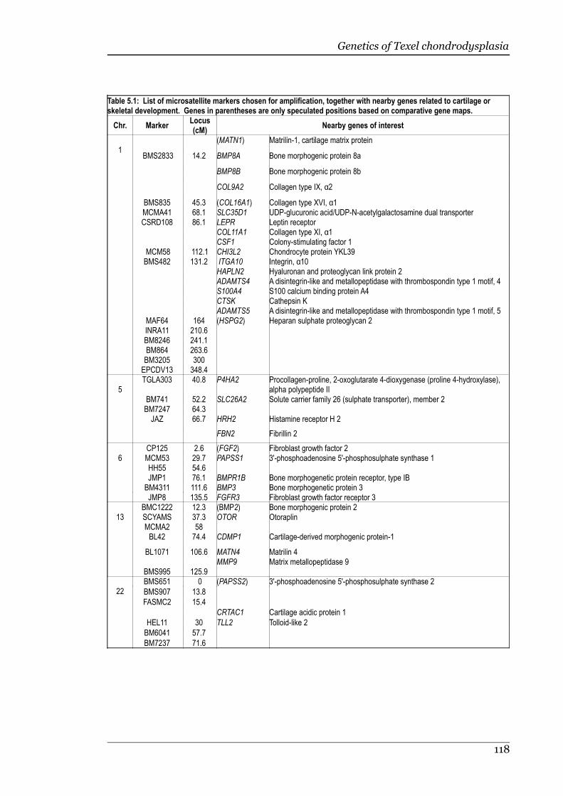
Genetics of Texel chondrodysplasia
Table 5.1: List of microsatellite markers chosen for amplification, together with nearby genes related to cartilage orskeletal development. Genes in parentheses are only speculated positions based on comparative gene maps.
Chr. Marker Locus(cM)
Nearby genes of interest
1 (MATN1) Matrilin-1, cartilage matrix protein
BMS2833 14.2 BMP8A Bone morphogenic protein 8a
BMP8B Bone morphogenic protein 8b
COL9A2 Collagen type IX, α2
BMS835 45.3 (COL16A1) Collagen type XVI, α1MCMA41 68.1 SLC35D1 UDP-glucuronic acid/UDP-N-acetylgalactosamine dual transporterCSRD108 86.1 LEPR Leptin receptor
COL11A1 Collagen type XI, α1CSF1 Colony-stimulating factor 1
MCM58 112.1 CHI3L2 Chondrocyte protein YKL39BMS482 131.2 ITGA10 Integrin, α10
HAPLN2 Hyaluronan and proteoglycan link protein 2ADAMTS4 A disintegrin-like and metallopeptidase with thrombospondin type 1 motif, 4 S100A4 S100 calcium binding protein A4 CTSK Cathepsin K ADAMTS5 A disintegrin-like and metallopeptidase with thrombospondin type 1 motif, 5
MAF64 164 (HSPG2) Heparan sulphate proteoglycan 2 INRA11 210.6 BM8246 241.1 BM864 263.6 BM3205 300 EPCDV13 348.4
5TGLA303 40.8 P4HA2 Procollagen-proline, 2-oxoglutarate 4-dioxygenase (proline 4-hydroxylase),
alpha polypeptide II BM741 52.2 SLC26A2 Solute carrier family 26 (sulphate transporter), member 2 BM7247 64.3 JAZ 66.7 HRH2 Histamine receptor H 2
FBN2 Fibrillin 2
6CP125 2.6 (FGF2) Fibroblast growth factor 2 MCM53 29.7 PAPSS1 3'-phosphoadenosine 5'-phosphosulphate synthase 1 HH55 54.6 JMP1 76.1 BMPR1B Bone morphogenetic protein receptor, type IB BM4311 111.6 BMP3 Bone morphogenetic protein 3 JMP8 135.5 FGFR3 Fibroblast growth factor receptor 3
13BMC1222 12.3 (BMP2) Bone morphogenic protein 2SCYAMS 37.3 OTOR Otoraplin MCMA2 58 BL42 74.4 CDMP1 Cartilage-derived morphogenic protein-1
BL1071 106.6 MATN4 Matrilin 4 MMP9 Matrix metallopeptidase 9
BMS995 125.9
22BMS651 0 (PAPSS2) 3'-phosphoadenosine 5'-phosphosulphate synthase 2
BMS907 13.8
FASMC2 15.4
CRTAC1 Cartilage acidic protein 1
HEL11 30 TLL2 Tolloid-like 2
BM6041 57.7
BM7237 71.6
118
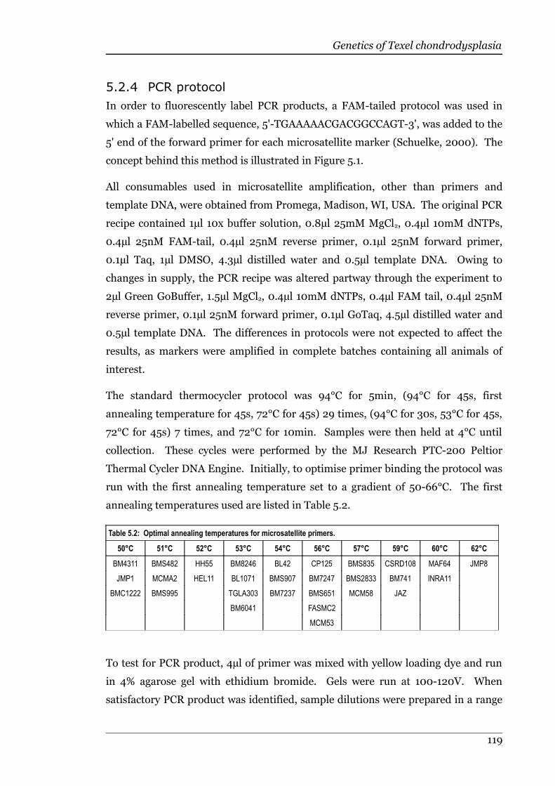
Genetics of Texel chondrodysplasia
5.2.4 PCR protocol
In order to fluorescently label PCR products, a FAM-tailed protocol was used in
which a FAM-labelled sequence, 5'-TGAAAAACGACGGCCAGT-3', was added to the
5' end of the forward primer for each microsatellite marker (Schuelke, 2000). The
concept behind this method is illustrated in Figure 5.1.
All consumables used in microsatellite amplification, other than primers and
template DNA, were obtained from Promega, Madison, WI, USA. The original PCR
recipe contained 1µl 10x buffer solution, 0.8µl 25mM MgCl2, 0.4µl 10mM dNTPs,
0.4µl 25nM FAM-tail, 0.4µl 25nM reverse primer, 0.1µl 25nM forward primer,
0.1µl Taq, 1µl DMSO, 4.3µl distilled water and 0.5µl template DNA. Owing to
changes in supply, the PCR recipe was altered partway through the experiment to
2µl Green GoBuffer, 1.5µl MgCl2, 0.4µl 10mM dNTPs, 0.4µl FAM tail, 0.4µl 25nM
reverse primer, 0.1µl 25nM forward primer, 0.1µl GoTaq, 4.5µl distilled water and
0.5µl template DNA. The differences in protocols were not expected to affect the
results, as markers were amplified in complete batches containing all animals of
interest.
The standard thermocycler protocol was 94°C for 5min, (94°C for 45s, first
annealing temperature for 45s, 72°C for 45s) 29 times, (94°C for 30s, 53°C for 45s,
72°C for 45s) 7 times, and 72°C for 10min. Samples were then held at 4°C until
collection. These cycles were performed by the MJ Research PTC-200 Peltior
Thermal Cycler DNA Engine. Initially, to optimise primer binding the protocol was
run with the first annealing temperature set to a gradient of 50-66°C. The first
annealing temperatures used are listed in Table 5.2.
Table 5.2: Optimal annealing temperatures for microsatellite primers.
50°C 51°C 52°C 53°C 54°C 56°C 57°C 59°C 60°C 62°C
BM4311 BMS482 HH55 BM8246 BL42 CP125 BMS835 CSRD108 MAF64 JMP8
JMP1 MCMA2 HEL11 BL1071 BMS907 BM7247 BMS2833 BM741 INRA11
BMC1222 BMS995 TGLA303 BM7237 BMS651 MCM58 JAZ
BM6041 FASMC2
MCM53
To test for PCR product, 4µl of primer was mixed with yellow loading dye and run
in 4% agarose gel with ethidium bromide. Gels were run at 100-120V. When
satisfactory PCR product was identified, sample dilutions were prepared in a range
119

Genetics of Texel chondrodysplasia
from full strength to ten-times dilution based on the brightness of the bands, and
submitted for genotyping at the Iowa State University DNA facility (IA, USA).
Genotyping was performed using an Applied Biosystems Prism 3100 Genetic
Analyser and a 6-carboxyl-X-rhodamine (ROX)-labelled size standard (500-ROX,
GeneScan, Applied Biosystems, Foster City, CA, USA).
5.2.5 Analysis
Microsatellite amplification was scored using Genoprofiler version 2.0.2 (UC Davis,
CA, USA) and Chromas version 2.31 (Technelysium Pty. Ltd., Helensville, QLD,
Australia). Chi-squared tests were performed to assess changes in heterozygosity,
allele frequency and genotype in association with the chondrodysplastic phenotype.
A pedigree was then constructed using the pooled microsatellite data, and the
phenotype for each animal was indicated by "0" in phenotypically normal animals
and "1" in chondrodysplastic animals. Half-sib regression interval mapping was
performed using QTL express (University of Edinburgh, qtl.cap.ed.ac.uk) using
Haseman-Elston regression, with bootstrap resampling set to 2000 iterations in
120
Figure 5.1: Amplification scheme for fluorescently labelled universal primers. A.Forward primer with added tail at 5' end. B. Reverse primer. C. Fluorescent FAM-labelleduniversal primer. 1. The primer tail is incorporated into PCR products. 2. The universalFAM-labelled primer takes over from the forward primer, selected for by a lower annealingtemperature. 3. The final product is FAM-labelled.
A B C
1
2
3

Genetics of Texel chondrodysplasia
order to generate a 95% confidence interval for the locus of interest. To test for
significance, the analysis was run with chromosome-wide permutations set to 2000
iterations. Haseman-Elston regression evaluates pairs of relations and the number
of alleles identical by descent. This technique regresses the squared difference in
trait score of each pair of relations on the fraction of alleles identical by descent at a
given marker locus. A significant negative slope indicates genetic linkage
(Haseman & Elston, 1972; Lynch & Walsch, 1998).
5.3 Results
5.3.1 Pedigree information
The sires of all but two of the lambs were able to be identified using microsatellite
data. The pedigree information is illustrated in Appendix 14. Because genetic
information was only available for Rams 1 and 2, only their combined 18 offspring
were able to be used for half-sib regression interval mapping.
5.3.2 Microsatellite scoring
Not all markers amplified clearly and time constraints prevented further reaction
attempts or substitution with other markers where available. Markers that either
failed to amplify or were unable to be reliably scored were MCMA41, BM864,
BM3205, EPCDV13, BM741, BM7247 and SCYAMS. Although there was no
previous record of a null allele for BM741 in sheep (Australian Sheep Gene Mapping
Web Site et al., 2006), repeated amplification attempts failed to demonstrate any
alleles in several animals, one of which was a sire. In other animals, the same
technique resulted in amplification of a single allele 200 base pairs in length.
Because the genotype of animals could not be established for BM741 (homozygosity
for the amplified allele could not be distinguished from heterozygosity for the allele
and a null allele), this marker could not be included in the analysis.
Alleles were assigned arbitrary identification numbers based on the size of the
amplified microsatellite, with higher numbers indicating a longer amplified
sequence (Figure 5.2). Full genotyping results are available in Appendix 15.
121

Genetics of Texel chondrodysplasia
5.3.3 Chi-squared analysis
The only significant result of chi-squared testing was an increased level of
heterozygosity in affected sheep relative to phenotypically normal carrier animals at
marker BMS2833, chromosome 1 (P≤0.05) (Figure 5.3). Some differences in allele
frequency or genotype between affected and carrier sheep appeared at markers
SCYAMS and BMS995 on chromosome 13 and FASMC2 on chromosome 22, but
did not reach significance. Chi-squared scores low enough to support no loss of
heterozygosity occurred at MAF64 on chromosome 1, BM4311 on chromosome 6,
SCYAMS on chromosome 13, and both BMS651 and FASMC2 on chromosome 22.
122
Figure 5.2: Typical chromatogram showing amplified microsatellite sequence. A. An individualhomozygous for the 136bp allele. B. An individual heterozygous for the 136bp and 144bp alleles. Red traceindicates the 500-ROX size standard.

Genetics of Texel chondrodysplasia
5.3.4 Half-sib regression interval mapping
The initial results for the half-sib regression interval mapping are listed in Table
5.3. Because 2 of the 4 markers on chromosome 5 amplified poorly and another
marker demonstrated nearly total homozygosity, half-sib regression interval
mapping was unable to be performed on this chromosome.
Table 5.3: Half-sib regression interval mapping results for microsatellite data from chondrodysplastic and control Texelsheep. The peak loci listed here are the points at which linkage disequilibrium was most evident for ovine chromosomes 1, 6,13 and 22. Significance was not reached at any point.
Chromosome 1 Chromosome 6 Chromosome 13 Chromosome 22
Peak Locus 128cM 0cM 94cM 0cM
95% CI 2-228cM 0-131cM 2-112cM 0-71cM
Likelihood ratio 4.6 3.37 2.75 3.58
Significance none none none none
5.4 Discussion
The results of linkage disequilibrium testing for chondrodysplasia of Texel sheep in
this study failed to identify a likely candidate gene. Although chromosome 1
demonstrated a significant difference in the level of heterozygosity between
123
Figure 5.3: Chi-squared test results for differences between control and chondrodysplastic Texel sheep inheterozygosity, allele frequency and genotype of microsatellite markers. A. A significant difference in the level ofheterozygosity in the marker BMS2833 was due increased heterozygosity in chondrodysplastic sheep (P≤0.05). B. A differencein allele frequency between affected and control sheep at the marker SCYAMS failed to reach significance. C. Marker BMS995demonstrated an increase in heterozygosity in chondrodysplastic sheep, but this was not significant. D. The marker FASMC2demonstrated a slight, but non-significant, difference in allele frequency and genotype between affected and control sheep.* = markers at which chi-squared testing supported no loss of heterozygosity in affected animals relative to carriers.
Genetics of Texel chondrodysplasia
chondrodysplastic and control sheep (P≤0.05), given the number of chi-squared
tests performed on the microsatellite data, this one-off outcome may have been
coincidental (Moore & McCabe, 1993). The difference consisted of an increase in
heterozygosity in chondrodysplastic lambs, which is not consistent with linkage to a
recessively inherited disease (Futuyma, 1998), suggesting that the difference
between genotypes of affected and control lambs at this locus was not linked to
disease status.
The main difficulties experienced in this study were the due to the limited number
of animals for which the phenotype was known, and poor amplification of several of
the markers. Had it been possible to verify the phenotype of newborn lambs, the
number of samples available would have increased from 26 to 64. Such an increase
in sample size may have generated sufficient statistical power to produce a more
definitive result (Moore & McCabe, 1993). Another potential limitation to this
study was the practice of lambing in a common paddock, allowing the opportunity
for lamb-stealing by ewes to occur (Alexander et al., 1983). Although all animals
were checked twice-daily, lamb-stealing could not be absolutely ruled out, affecting
the integrity of the constructed pedigree. Ideally, the dams would have been
genotyped along with the lambs, enabling the detection of cases of lamb-stealing.
Unfortunately, tissue samples were not routinely collected from the dams as the
genetic study described in this chapter had not been anticipated at the time of the
breeding trial, which was originally established for a different research purpose.
Because the animals available for use in this study were closely related and
originated from a population where the disease frequency was artificially increased
(through back-crossing), there was a risk of spurious association of the disease to
any marker allele in high frequency (Pritchard et al., 2000). Transmission
disequilibrium testing (TDT) is a statistical tool able to overcome this difficulty by
comparing the transmission of a disease-linked marker allele from a heterozygous
parent to its affected offspring (Spielman et al., 1993). Unfortunately, in this study
DNA was only available from affected parents. This prevented the use of TDT, as
chondrodysplasia of Texel sheep is a recessive disorder and heterozygosity of sires
does not support linkage of the marker to the disease, while homozygosity may be
due to high allele frequencies in the inbred population (Lynch & Walsch, 1998).
The relatively recent discovery of chondrodysplasia in Texel sheep (Thompson et
al., 2005) indicates that this disease is due to either a reasonably new mutation in
124
Genetics of Texel chondrodysplasia
the population, or an old mutation that has gone unrecognised or unreported for
many generations. If the former is the case, markers some distance from the
causative mutation would be expected to show linkage because there has been less
opportunity for recombination. This effect is useful for detection, but would hinder
fine mapping using LD methods (Devlin & Risch, 1995; Lynch & Walsch, 1998).
Further genetic research on chondrodysplasia of Texel sheep will require an
increased number of samples, preferably not sourced from a back-cross trial. An
increased number of unaffected relations to the chondrodysplastic sheep would
make the analysis more powerful, as would an increase in marker density. An
alternative to undertaking another large-scale breeding trial would be to approach
Texel breeders. If a farm was found to regularly produce chondrodysplastic lambs,
samples from a related group of affected and non-affected animals and their
parents could be used to perform another genome-scanning experiment. As more
markers, both microsatellites and single nucleotide polymorphisms (SNPs), are
identified for the ovine genome (Cockett, 2006; Pariset et al., 2006), LD mapping
will likely become an increasingly useful tool for this type of study. Because
chondrodysplasia of Texel sheep is believed to have a simple mendelian mode of
inheritance (Byrne, 2005), a candidate gene approach in further genetic research
may be appropriate (Zhu & Zhao, 2007), especially if further classification of the
disease phenotype reveals candidate genes that have not already been investigated.
An alternative type of investigation would be to examine differential expression of
genes in chondrodysplastic Texel sheep, although studies of this kind often identify
a large number of differently expressed genes, and additional supporting evidence
should be considered before using this technique to identify candidate genes
(Okuda et al., 2002; Zhu & Zhao, 2007).
5.5 Summary
This study did not find evidence for genetic linkage of chondrodysplasia of Texel
sheep to ovine chromosomes 1, 5, 6, 13 or 22. The effectiveness of the study was
hampered by the low number of samples, the level of inbreeding involved in the
sample animals, and the poor amplification of several microsatellite markers. A
new breeding trial to generate samples, involving multiple affected and unaffected
parents and minimised inbreeding would be advisable before repeating this style of
genetic investigation.
125
6 Sulphation of chondroitin disaccharides
in cartilage

Sulphation of chondroitin disaccharides in cartilage
6.1 Introduction
Chondroitin sulphate is the major sulphated polysaccharide attached to the core
protein, aggrecan, in hyaline cartilage matrix (Krueger et al., 1990; Sandy et al.,
1997) (see Chapter 1, Figure 1.2). Sulphation of cartilage matrix polysaccharide is
vital for normal matrix integrity and compressive stiffness of the tissue, and is
therefore crucial to normal cartilage formation, and consequently to endochondral
ossification (Sandy et al., 1997). Several forms of chondrodysplasia characterised
by reduced levels of sulphate in cartilage have been identified in both human beings
and animals (Rossi et al., 1996a; Sandy et al., 1997; Rossi & Superti-Furga, 2001;
Forlino et al., 2005). These can occur as a result of mutations reducing the ability
of chondrocytes to extract sulphate from the extracellular fluid (Rossi & Superti-
Furga, 2001), mutations affecting the activation and addition of sulphate to
proteoglycans within the chondrocyte (ul Haque et al., 1998), or fetal deficiency of
manganese, which is required for normal function of enzyme systems involved in
sulphate activation and transfer to proteoglycans (Leach & Muenster, 1962; Bolze
et al., 1985).
Chondrodysplasia of Texel sheep shares microscopic lesions, and certain
developmental characteristics, with several diseases resulting from defective
sulphate metabolism in human beings and other animals (van der Harten et al.,
1988; Valero et al., 1990; Shapiro, 1992; Superti-Furga, 1994). This finding
suggests a metabolic defect in sulphate metabolism is the likely underlying defect in
affected sheep. Support for this hypothesis was provided by a previous study where
hyaline cartilage from chondrodysplastic Texel lambs was shown to contain
undersulphated matrix proteoglycans (see Figure 1.7) (Byrne, 2005).
Undersulphation of hyaline cartilage matrix was also apparent in histological
sections stained with toluidine blue (see Chapter 2, Figure 2.13). A more sensitive
study of the cartilage proteoglycan sulphation profile in chondrodysplasia of Texel
sheep would provide additional information about the pathogenesis of this disease,
and could help to relate biochemical changes to the development of cartilage
lesions. An investigation of cartilage sulphation in control and chondrodysplastic
Texel sheep of varying ages would also help to determine whether the
undersulphation of cartilage proteoglycans is progressive or remains constant.
Furthermore, if the biochemical defect is detectable in newborn lambs, it would
provide a means for neonatal diagnosis of this chondrodysplasia, which otherwise
127
Sulphation of chondroitin disaccharides in cartilage
cannot be unequivocally recognised until 2-3 weeks of age. The inability to
diagnose the chondrodysplastic phenotype in newborn lambs has previously limited
the number of samples available for genetic analysis (see Chapter 5).
Historically, studies to compare chondroitin disaccharides have typically used high
performance liquid chromatography (HPLC) (Plaas et al., 1986; Rossi et al., 1996a).
More recently, capillary electrophoresis (CE) has been used for some of these
investigations (Theocharis & Theocharis, 2002; Zamfir et al., 2003). CE allows
separation of ionic species by their charge and frictional forces (Gordon et al.,
1988) and is able to provide greater resolution of ionic species while requiring a
smaller sample size than HPLC (Karamanos et al., 1995).
This pilot study aimed to use CE to quantify chondroitin 4-sulphate and
chondroitin 6-sulphate disaccharide concentrations in articular cartilage of
chondrodysplastic Texel and control sheep, investigate age-related differences in
concentrations of these disaccharides, and to test the hypothesis that a difference in
chondroitin sulphate disaccharides is detectable in affected newborn lambs prior to
the development of lesions.
6.2 Materials and methods
6.2.1 Animals used and samples collected
An entire humerus was collected from all animals that underwent post mortem
examination, and was stored at -20°C for 6-24 months. Articular cartilage shavings
(10-15mg) were harvested from the central humeral heads of 8 animals of various
ages. Samples 1 and 2 were from obligate affected newborn lambs (for which both
parents were chondrodysplastic), samples 3 and 4 were from control newborn
lambs, sample 5 was from a 1-month-old lamb severely affected by
chondrodysplasia, sample 6 was from a 1-month-old control lamb, sample 7 was
from a mature chondrodysplastic ram, sample 8 was from a mature control sheep.
Because newborn lambs from the flock carrying the defective gene could not be
positively identified as chondrodysplastic by gross or microscopic examination,
control samples were collected from mixed-breed newborn lambs that did not have
ancestors in common with the affected Texel flock or its contributing members.
128
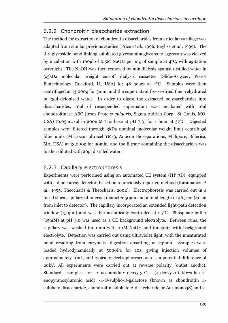
Sulphation of chondroitin disaccharides in cartilage
6.2.2 Chondroitin disaccharide extraction
The method for extraction of chondroitin disaccharides from articular cartilage was
adapted from similar previous studies (Price et al., 1996; Bayliss et al., 1999). The
β-0-glycosidic bond linking sulphated glycosaminoglycans to aggrecan was cleaved
by incubation with 100µl of 0.5M NaOH per mg of sample at 4°C, with agitation
overnight. The NaOH was then removed by minidialysis against distilled water in
3.5kDa molecular weight cut-off dialysis cassettes (Slide-A-Lyzer, Pierce
Biotechnology, Rockford, IL, USA) for 48 hours at 4°C. Samples were then
centrifuged at 13,000g for 3min, and the supernatant freeze-dried then rehydrated
in 25µl deionised water. In order to digest the extracted polysaccharides into
disaccharides, 10µl of resuspended supernatant was incubated with 10µl
chondroitinase ABC (from Proteus vulgaris, Sigma-Aldrich Corp., St. Louis, MO,
USA) (0.05mU/µl in 200mM Tris base at pH 7.3) for 1 hour at 37°C. Digested
samples were filtered through 3kDa nominal molecular weight limit centrifugal
filter units (Microcon ultracel YM-3, Amicon Bioseparations, Millipore, Billerica,
MA, USA) at 13,000g for 90min, and the filtrate containing the disaccharides was
further diluted with 20µl distilled water.
6.2.3 Capillary electrophoresis
Experiments were performed using an automated CE system (HP 3D), equipped
with a diode array detector, based on a previously reported method (Karamanos et
al., 1995; Theocharis & Theocharis, 2002). Electrophoresis was carried out in a
fused silica capillary of internal diameter 50µm and a total length of 46.5cm (40cm
from inlet to detector). The capillary incorporated an extended light-path detection
window (150µm) and was thermostatically controlled at 25°C. Phosphate buffer
(15mM) at pH 3.0 was used as a CE background electrolyte. Between runs, the
capillary was washed for 1min with 0.1M NaOH and for 4min with background
electrolyte. Detection was carried out using ultraviolet light, with the unsaturated
bond resulting from enzymatic digestion absorbing at 232nm. Samples were
loaded hydrodynamically at 5000Pa for 10s, giving injection volumes of
approximately 10nL, and typically electrophoresed across a potential difference of
20kV. All experiments were carried out at reverse polarity (outlet anodic).
Standard samples of 2-acetamido-2-deoxy-3-O- (4-deoxy-α-L-threo-hex-4-
enopyranosyluronic acid) -4-O-sulpho-D-galactose (known as chondroitin 4-
sulphate disaccharide, chondroitin sulphate A disaccharide or ∆di-mono4S) and 2-
129

Sulphation of chondroitin disaccharides in cartilage
acetamido-2-deoxy-3-O- (4-deoxy-α-L-threo-hex-4-enopyranosyluronic acid) -6-O-
sulpho-D-galactose (known as chondroitin 6-sulphate disaccharide, chondroitin
sulphate C or ∆di-mono6S) (Sigma-Aldrich Corp., St. Louis, MO, USA) were used to
identify ionic species and to construct standard curves. Standard preparations of
unsulphated chondroitin disaccharide were unavailable commercially.
All samples were run at least 4 times and the peaks integrated using HP
Chemstation software (Agilent Technologies, Santa Clara, CA, USA). Where a
particular disaccharide exhibited a double peak, both anomeric forms were pooled.
The peak areas were subsequently normalised according to their migration times, in
order to take into account the differing amount of time that compounds with
different electrophoretic velocities spend in the detector window (Goodall et al.,
1991). The normalised peak areas were subsequently used, via a calibration of
absorptivity using the standard samples, to produce concentrations in mg/L.
6.2.4 Analysis
Disaccharide concentrations were corrected for the original cartilage sample size by
multiplying the raw concentration by 10 and dividing by the wet weight in mg of the
original cartilage sample. This gave the concentration of disaccharide in mg/L per
10mg of cartilage. In order to compensate for slight differences in injection volume,
the concentration of ∆di-mono4S was divided by the concentration of ∆di-mono6S
for each sample. This number is referred to as the ratio of ∆di-mono4S to ∆di-
mono6S. Statistical analyses were performed using S-Plus 8.0 student edition
(Insightful Corp., 2007). The ANOVA model was: ratio of ∆di-mono4S to ∆di-
mono6S ~ Age + Status, where "Age" was the age of the animal from which cartilage
was taken, and "Status" was the presence or absence of chondrodysplasia. The
Student's T-test was used to compare individual animals.
6.3 Results
Although some run to run variation was observed for the absolute amounts of the
differently sulphated disaccharides, the ratios of ∆di-mono4S to ∆di-mono6S
varied little for each cartilage sample. A typical electropherogram is shown in
Figure 6.1, demonstrating peaks caused by ∆di-mono4S, ∆di-mono6S and a peak at
the expected location for unsulphated chondroitin disaccharide, as demonstrated
by Karamanos et al. (1995). Double peaks were routinely observed, owing to the
different anomeric forms of the hexosamine present at the reducing terminal of the
130
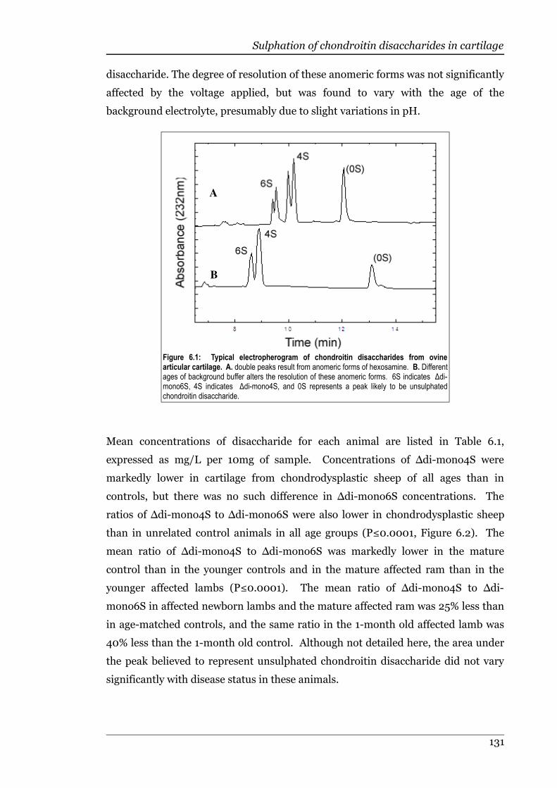
Sulphation of chondroitin disaccharides in cartilage
disaccharide. The degree of resolution of these anomeric forms was not significantly
affected by the voltage applied, but was found to vary with the age of the
background electrolyte, presumably due to slight variations in pH.
Mean concentrations of disaccharide for each animal are listed in Table 6.1,
expressed as mg/L per 10mg of sample. Concentrations of ∆di-mono4S were
markedly lower in cartilage from chondrodysplastic sheep of all ages than in
controls, but there was no such difference in ∆di-mono6S concentrations. The
ratios of ∆di-mono4S to ∆di-mono6S were also lower in chondrodysplastic sheep
than in unrelated control animals in all age groups (P≤0.0001, Figure 6.2). The
mean ratio of ∆di-mono4S to ∆di-mono6S was markedly lower in the mature
control than in the younger controls and in the mature affected ram than in the
younger affected lambs (P≤0.0001). The mean ratio of ∆di-mono4S to ∆di-
mono6S in affected newborn lambs and the mature affected ram was 25% less than
in age-matched controls, and the same ratio in the 1-month old affected lamb was
40% less than the 1-month old control. Although not detailed here, the area under
the peak believed to represent unsulphated chondroitin disaccharide did not vary
significantly with disease status in these animals.
131
Figure 6.1: Typical electropherogram of chondroitin disaccharides from ovinearticular cartilage. A. double peaks result from anomeric forms of hexosamine. B. Differentages of background buffer alters the resolution of these anomeric forms. 6S indicates ∆di-mono6S, 4S indicates ∆di-mono4S, and 0S represents a peak likely to be unsulphatedchondroitin disaccharide.
A
B

Sulphation of chondroitin disaccharides in cartilage
Table 6.1: Concentrations of chondroitin sulphate disaccharides from chondrodysplastic andcontrol Texel sheep of varying ages. Concentrations are expressed as mg/L per 10mg of sample.
Animal Status Age ∆di-mono6S ∆di-mono4S
1 Affected Newborn 24.2 48.6
2 Affected Newborn 28.8 55.2
3 Control Newborn 26.1 74.3
4 Control Newborn 38.7 91.1
5 Affected 1 month 41.2 61.9
6 Control 1 month 28.6 71.4
7 Affected Mature 38.3 36.1
8 Control Mature 44.5 56.3
6.4 Discussion
The results of this pilot study indicate that capillary electrophoresis can be used to
detect a difference in the profile of cartilage chondroitin sulphate disaccharides in
chondrodysplastic Texel sheep when compared with controls, and that the
decreased cartilage sulphate content in this disease is most likely due to reduced
levels of chondroitin 4-sulphate. Cartilage from Texel sheep with chondrodysplasia
had a lower ratio of ∆di-mono4S to ∆di-mono6S than age-matched controls at all
132
Figure 6.2: Ratios of ∆di-mono4S to ∆di-mono6S in chondrodysplastic andcontrol Texel sheep of different ages. Red plots are chondrodysplastic animals,blue plots are control animals. The ratios are lower in affected animals of all agesP≤0.0001.

Sulphation of chondroitin disaccharides in cartilage
ages measured, supporting the hypothesis that a difference in chondroitin sulphate
disaccharides is detectable in affected newborn lambs prior to the development of
lesions. A decreased ratio of ∆di-mono4S to ∆di-mono6S has been recorded in
chondrodysplasias resulting from sulphate transport disorders due to reduced
activity of the DTDST (Rossi et al., 1998), and in mutations in the gene encoding
chondroitin 4-sulphotransferase-1, CHST11 (Klüppel et al., 2005). This finding
indicates that CHST11, which maps to ovine chromosome 3 at approximately
221.4cM (Australian Sheep Gene Mapping Web Site, 2004), is a potential candidate
gene. A naturally occurring decrease in the ratio of ∆di-mono4S to ∆di-mono6S
has been described in ageing articular cartilage in humans (Roughley et al., 1981;
Bayliss et al., 1999; Lauder et al., 2001). A similar change was evident in the
articular cartilage of mature control and chondrodysplastic sheep in this study.
The similarity in ∆di-mono6S concentration between chondrodysplastic and
control lambs indicates that a defect in chondroitin 6-0-sulphotransferase-1,
encoded by CHST3, is unlikely. Loss-of-function mutations in CHST3 result in
spondyloepiphyseal dysplasia Omani type, in which cartilage matrix concentrations
of chondroitin 6-sulphate are severely reduced (Thiele et al., 2004). Mutations in
SLC26A2 that decrease DTDST function result in an increase in unsulphated
chondroitin disaccharide, along with decreased ∆di-mono4S, and sometimes ∆di-
mono6S, in cartilage matrix (Rossi et al., 1997; Rossi et al., 1998). If the
unclassified peak identified in the present study is truly representative of
unsulphated chondroitin disaccharide, it would suggest that abnormalities in
DTDST function are not involved in chondrodysplasia of Texel sheep. This is
supported by previous in vitro and genetic studies, neither of which were able to
support an abnormality of chondrocyte sulphate uptake (see Chapters 4 and 5).
The decreased ratio of ∆di-mono4S to ∆di-mono6S in newborn chondrodysplastic
lambs indicates that although affected lambs cannot be distinguished
phenotypically from normal or heterozygous lambs at birth, the underlying
cartilage abnormality is present. Capillary electrophoresis techniques may
therefore be useful in identifying the phenotype of newborn animals that are yet to
demonstrate gross or microscopic lesions of chondrodysplasia. DNA and articular
cartilages are available from all lambs that could not be included in the previous
genetic study (see Chapter 5) because of their undetermined disease status.
Although funding constraints do not allow it at this time, the phenotypes of these
lambs could be estimated using capillary electrophoresis, thereby increasing the
133
Sulphation of chondroitin disaccharides in cartilage
number of animals available for a renewed genetic investigation. While more
samples would be required to confirm the true diagnostic power of the ratio of
cartilage ∆di-mono4S to ∆di-mono6S, this study indicates that the information
would at least be useful as quantitative trait data associated with the
chondrodysplastic phenotype (Lynch & Walsch, 1998).
The newborn affected lambs, which had no gross or microscopic lesions, and the
mature affected ram, which had mild lesions of chondrodysplasia, all had ratios of
∆di-mono4S to ∆di-mono6S closer to the values for control animals than did the 1-
month old affected lamb, which demonstrated severe lesions of the disease. This
suggested that the magnitude of the difference in the ratio of ∆di-mono4S to ∆di-
mono6S may have been related to the severity of lesions. It cannot be discerned
from this study whether the difference was solely due to the severity of disease, age-
related changes in cartilage sulphation specific to chondrodysplasia of Texel sheep,
or a combination of the two. Further studies using more animals of different ages
representing the full spectrum of severity in this disease would be able to answer
this question, as would a study using serial cartilage biopsies. Capillary
electrophoresis would be an ideal tool for the latter type of experiment due to the
small sample size required (10mg of cartilage) (Price et al., 1996).
The regulation of the proportion of sulphate added to either the 4- or 6-position on
the chondroitin disaccharide is poorly understood. While the decrease of ∆di-
mono4S but not of ∆di-mono6S found in cartilage from chondrodysplastic Texel
sheep in this study intuitively suggests a defect specifically interfering in the
addition of sulphate to the 4-position, this is not necessarily the case. In defects of
DTDST and PAPSS function, where the available intracellular sulphate and
consequently PAPS is reduced (Kurima et al., 1998; Rossi et al., 2003),
concentrations of ∆di-mono4S and ∆di-mono6S are not always decreased by equal
amounts. In the brachymorphic mouse, which has a mutation in PAPSS2 and
therefore decreased activity of PAPS, there is an increase in unsulphated
chondroitin disaccharide and a decrease in both ∆di-mono4S and ∆di-mono6S
(Orkin et al., 1976). In cases of diastrophic dysplasia, atelosteogenesis type 2 or
achondrogenesis 1b, however, which are all caused by defects in the DTDST,
cartilage has increased levels of unsulphated chondroitin disaccharide and tends to
show a more marked reduction of ∆di-mono4S than of ∆di-mono6S (Rossi et al.,
1997). This difference in chondroitin disaccharide profile was not able to be
explained by a difference in PAPS binding affinity of the different
134
Sulphation of chondroitin disaccharides in cartilage
sulphotransferases, as the apparent Km of chondroitin 4-sulphotransferase has been
found to be the same as of chondroitin 6-sulphotransferase (Sugumaran et al.,
1986). Because of these past findings, the pattern of altered sulphation of
chondroitin disaccharides identified in the present study must be interpreted with
care.
The inferences made in this study, while based on striking differences between
affected and control animals, are drawn from a very low number of samples. As a
result, this study may have underestimated the normal level of variation in
concentrations of chondroitin disaccharide species in articular cartilage of sheep. If
this is the case, a repeated experiment using cartilage samples from a greater
number of animals may reveal less marked differences between control and
chondrodysplastic Texel sheep.
6.5 Summary
The concentration of chondroitin 4-sulphate disaccharide and the ratio of
chondroitin 4-sulphate to chondroitin 6-sulphate were consistently lower in
articular cartilage from chondrodysplastic Texel sheep than in age-matched
controls. These differences were detectable in newborn affected lambs that were
not showing gross or microscopic lesions of chondrodysplasia, indicating that the
underlying cartilage abnormality is present at birth.
135

7 General discussion
General discussion
7.1 Introduction
Discovery of a form of chondrodysplasia on a commercial sheep farm in Southland,
New Zealand, created an opportunity to investigate the gross, microscopic,
biochemical and genetic characteristics of this disease in Texel sheep. Animals for
study were generated using a back-cross breeding trial on a Massey University
research farm. The results of this trial indicated a simple autosomal recessive mode
of inheritance and an apparent defect of cartilage sulphation in chondrodysplasia of
Texel sheep (Byrne, 2005). Later studies, discussed in the following sections,
generated a more detailed description of the gross and microscopic development of
lesions in this disease, and investigated aspects of sulphate metabolism and genetic
linkage.
7.2 Development of lesions
Chondrodysplastic Texel lambs were indistinguishable from controls at birth by
gross, morphometric or microscopic examination (see Chapters 2 and 3), but
appeared to have a lower ratio of chondroitin 4-sulphate disaccharide (∆di-
mono4S) to chondroitin 6-sulphate disaccharide (∆di-mono6S) in the central
articular cartilage of the humeral head (see Chapter 6, Figure 6.2). This finding
indicates that although lesions of chondrodysplasia cannot be identified at birth,
the underlying biochemical defect is present at that time. While in some genetic
diseases the leakage of critical metabolites across the placenta may delay the onset
of lesions until postnatal life (Leonard & Morris, 2006), the presence of a
biochemical defect at birth in affected lambs suggests that this phenomenon does
not occur in chondrodysplasia of Texel sheep. This was also supported by the
observation that chondrodysplastic lambs born to affected ewes were identical to
those born to phenotypically normal carrier ewes, both at birth and as they grew
(see Chapter 2). If the absence of gross and microscopic lesions at birth cannot be
explained by metabolic support in utero, the question of why the lesions develop so
rapidly in the first weeks of life must be raised.
The morphometric analysis of growing control and chondrodysplastic Texel lambs
revealed that some disturbance in growth was detectable in the 1-10 day age
interval, and there were usually marked differences between affected and control
lambs from 11 days of age (see Chapter 3). A similar pattern was seen in the
development of microscopic lesions. While subtle lesions were sometimes visible
137
General discussion
prior to 5 days of age, the youngest lamb able to be unequivocally diagnosed as
chondrodysplastic was 9 days old. These findings suggest that the onset and
development of gross lesions in chondrodysplasia of Texel sheep are closely related
to the emergence of microscopic lesions. This was also seen in the varying nature of
the microscopic lesions, which tended to be milder in the less severely affected
animals than in the severely affected animals, and were very mild in the few
animals that survived to maturity. The histologically observed rarefaction and
cystic degeneration of interterritorial cartilage matrix appeared to be more closely
related to the gross phenotype than did the later-developing perichondrocytic
collagen rings. The chronology of the development of microscopic lesions in
affected lambs was similar to that seen in diastrophic dysplasia, in which
longitudinal studies demonstrated relatively early appearance of interterritorial
matrix lesions and later appearance of collagen rings (Shapiro, 1992). These
microscopic findings support the hypothesis that the development of the gross
phenotype in chondrodysplasia of Texel sheep is primarily due to a gradual failure
of the structural integrity of the hyaline cartilage matrix, rather than to a disorder of
cartilage regulation. This would explain the absence of lesions in neonatal lambs.
There are minimal physical stresses on articular cartilage during fetal development,
but forces increase considerably with weight bearing and postnatal growth
(Hamrick, 1999). This physical loading, accompanied by compressive and shear
stresses, may be a precipitating factor in the development of microscopic and gross
lesions in chondrodysplasia of Texel sheep. Credibility is given to this idea by (a)
the tendency for lesions to appear earliest and to be most severe in the large joints
that combine high loading with extensive mobility; (b) the subjective observation
that initially faster-growing lambs seemed to develop severe lesions of
chondrodysplasia more rapidly than slower growing lambs; and (c) the preferential
appearance of interterritorial matrix lesions in the transitional zone of articular
cartilage, which is structurally weaker than the adjacent superficial or radial zones
(Bellucci & Seedhom, 2001). Interterritorial cartilage matrix normally contains
lower concentrations of sulphated proteoglycans (Ross et al., 1995), which are
required for collagen fibrillogenesis (Eyre, 2002). Because the concentration of
sulphated proteoglycans in cartilage is decreased in chondrodysplasia of Texel
sheep (Byrne, 2005) (see Figure 1.7 and Chapter 6), it is not surprising that the first
microscopic lesions appear in parts of cartilage where their concentration would be
expected to be lowest. It is possible that the reduction in sulphated proteoglycan
138
General discussion
concentration in the interterritorial matrix of the transitional zone of articular
cartilage alters the nature of the collagen fibrils, which may further contribute to
the weakening of this zone in affected cartilage.
The apparent relationship between lesions of the interterritorial matrix in
chondrodysplasia of Texel sheep and severity of the disease did not extend to the
perichondrocytic collagen rings. These were present even in mildly affected
animals, while the interterritorial matrix lesions tended to be very mild or absent in
such cases. Perichondrocytic collagen rings are a distinguishing feature in
chondrodysplasia of Texel sheep (Thompson et al., 2005), along with
achondrogenesis 1b (Superti-Furga, 1994), diastrophic dysplasia (Stanescu et al.,
1984), congenital Mn deficiency (Valero et al., 1990), and apparently in the
SLC26A2 knock-out mouse (Forlino et al., 2006). Immunohistochemistry
performed on cartilage from chondrodysplastic Texel lambs revealed that the
collagen rings consisted of chiefly type VI collagen (see Chapter 2, Figure 2.14),
which, although an important part of the pericellular envelope (Poole et al., 1988b),
is not considered to be a major structural component of hyaline cartilage matrix
(Wardale & Duance, 1993). Because of this, it may be supposed that the presence of
the collagen rings do not have a great effect on the structural integrity of the
affected cartilage, and may explain the ability of animals affected by congenital Mn
deficiency to recover once a complete diet is available (Valero et al., 1990). This
assumes that the collagen rings in congenital Mn deficiency have the same
composition as those in chondrodysplasia of Texel sheep. Although defective type
VI collagen has been associated with several types of myopathy and muscular
dystrophy in human beings (Lamandé et al., 1998; Giusti et al., 2005), there are no
published reports of chondrodysplasia either in conjunction with these disorders or
as a separate disease.
Many of the arguments in support of a mechanical stimulus for lesion development
in chondrodysplasia of Texel sheep could also be applied to a metabolic aetiology,
primarily because the metabolic rate of chondrocytes is influenced by mechanical
stimulus. From birth until skeletal maturity, articular cartilage is predicted to be
under increasing peak pressure due to thinning of cartilage, increased force
generation and decreased relative joint size (Hamrick, 1999). High peak pressures
in dynamic loading induce chondrocyte division and matrix synthesis (Urban,
1994), possibly in response to mechanically-induced matrix degradation (Archer,
1994). This suggests that as animals become ambulatory and increase in mass,
139
General discussion
there will be an increase in metabolic activity of chondrocytes in weight-bearing
cartilage. A relationship between local metabolic rate and lesion development was
identified in Chapter 3, where the limb-bones with the fastest potential growth rate
were also the most severely shortened. It would be reasonable to suppose that cells
with a higher metabolic rate would be more severely affected by a disorder
involving enzymatic insufficiency or limited substrate. If such a pathogenesis is
involved in chondrodysplasia of Texel sheep, it could in part explain the pattern of
lesions in this disease, and may be a reason for the marked variability in severity of
lesions both between animals, and between sites within animals. Similar variability
has been described in diastrophic dysplasia, where relatives with the same
SLC26A2 mutation had differing expression of the disease (Superti-Furga, 2001),
potentially due to a varying ability for chondrocytes to use alternative sources of
intracellular sulphate to synthesise sulphated glycosaminoglycans (Rossi et al.,
2003). Perhaps in chondrodysplasia of Texel sheep there is a similar compensatory
mechanism, such as has been proposed in diseases caused by mutations in PAPSS2,
where the PAPSS1 gene product may be able to partially recover the deficiency in
phosphoadenosine-phosphosulphate (PAPS) (ul Haque et al., 1998). Should the
mutation in chondrodysplasia of Texel sheep lie in a sulphotransferase, it would be
reasonable to suspect that another sulphotransferase may be able to catalyse the
required reaction to varying degrees. In addition, abnormal transforming growth-
factor-β (TGF-β) and bone morphogenic protein (BMP) signalling (see Chapter 1,
Figure 1.6) have been identified in CHST11 mutant mice with defective chondroitin
4-sulphotransferase activity (Klüppel et al., 2005), and may provide an increased
opportunity for individual genotypes to influence the presentation of a homologous
disease. Another explanation for the variable severity of chondrodysplasia in Texel
sheep may lie in the different rates of growth, as suggested above, or in the
involvement of sulphate in some signalling and regulation pathways, such as the
requirement for heparan sulphate in fibroblast growth factor (FGF) signalling
(Forlino et al., 2005; Harmer, 2006).
In all likelihood, both mechanical and metabolic processes are involved in the
development of lesions in chondrodysplasia of Texel sheep.
7.3 Possible aetiology
An abnormality in cartilage sulphate metabolism is likely to be the defect in
chondrodysplasia of Texel sheep. This is supported by the microscopic lesions,
140
General discussion
which mimic those found in defects of the diastrophic dysplasia sulphate
transporter (DTDST) (Thompson et al., 2005) and in congenital Mn deficiency (see
Chapter 2). The former reduces the cellular uptake of sulphate (Karniski, 2001),
while the latter affects polymerase and galactosyltransferase, enzymes required for
synthesis of chondroitin sulphate (Leach et al., 1969; Tsopanakis & Herries, 1976).
The pale staining of hyaline cartilage matrix with toluidine blue, compared with
equally-thick sections from control animals, suggests that there were fewer
negatively charged ions in the abnormal cartilage. This would be expected if there
were a deficiency of sulphate in the cartilage matrix. A biochemical study identified
a decreased amount of sulphate relative to cartilage protein in chondrodysplastic
Texel sheep (see Figure 1.7) (Byrne, 2005), and capillary electrophoresis of articular
cartilage chondroitin disaccharides revealed a decreased concentration of
chondroitin 4-sulphate in cartilage from chondrodysplastic animals of all ages
tested (see Chapter 6).
The pathway involved in the sulphation of cartilage matrix proteoglycans includes
several steps (see Chapter 1, Figure 1.3). Initially, sulphate must enter the cell
through the DTDST, an antiporter that exchanges intracellular chloride ions for
extracellular sulphate (Vincourt et al., 2003). This protein is encoded by SLC26A2,
and mutations in this gene cause human achondrogenesis 1b, atelosteogenesis type
II, diastrophic dysplasia and recessive multiple epiphyseal dysplasia (rMED)
(Hästbacka et al., 1996b; Cai et al., 1998; Rossi & Superti-Furga, 2001; Ballhausen
et al., 2003), and chondrodysplasia in a transgenic mouse model (Forlino et al.,
2005). The importance of this gene in human diseases that share microscopic
lesions, and some of their developmental aspects, with chondrodysplasia of Texel
sheep made SLC26A2 the initial candidate for this disease. In light of this, 85.4% of
the exonic DNA of SLC26A2 was sequenced in chondrodysplastic and control
sheep, but no mutation was identified (Byrne, 2005). While this study decreased
the likelihood of a causative mutation in this gene, it could not rule out a mutation
in the remaining 14.6% of exonic sequence, or a mutation in a non-expressed
regulatory region. Because of these points, and the strong similarity between
microscopic lesions of chondrodysplastic Texel sheep and those described in
diseases caused by SLC26A2 mutations, further investigation of the DTDST was
performed via an assay of radiolabelled sulphate uptake by dermal fibroblasts in
chondrodysplastic and control sheep. This study did not show a difference in
sulphate uptake between chondrodysplastic and age-matched control lambs, but
141
General discussion
was limited by the low number of animals able to be used and the omission of
results from one animal due to its outlying nature (see Chapter 4). It must also be
noted that while identifying a decreased level of sulphate uptake would have
provided strong evidence for a defect of DTDST function, failure to detect a
difference has occurred in some cases where a disease-causing mutation in
SLC26A2 was known to exist (Karniski, 2001).
Because of the remaining possibility that the causative mutation in
chondrodysplasia of Texel sheep involved this gene, the area of ovine chromosome
5 containing SLC26A2 was included in a microsatellite marker study. While some
of the marker alleles used for this chromosome did not amplify as well as expected
(one failed to amplify clearly enough to be accurately scored, one contained a
suspected null allele, and one was not polymorphic enough to provide useful
information), the results of chi-squared testing did not support any loss of
heterozygosity in association with the chondrodysplastic phenotype at marker
TGLA303, 9.251cM from the SLC26A2 gene locus (Australian Sheep Gene Mapping
Web Site, 2004). Although none of the individual studies performed on DTDST
function or the SLC26A2 gene in chondrodysplasia of Texel sheep were powerful
enough to rule out a defect of sulphate uptake of chondrocytes in affected sheep,
collectively they suggest that SLC26A2 is unlikely to contain a causative mutation in
this disease.
Given the similarity of lesions and biochemistry in chondrodysplasia of Texel sheep
to those caused by SLC26A2 mutations, a defect within the cartilage proteoglycan
sulphation pathway was still considered to be the most likely cause of the disease.
Following the uptake of sulphate into cells, it is converted into an "active" form
known as PAPS by phosphoadenosine-phosphosulphate synthase (PAPSS),
encoded by PAPSS2 (Superti-Furga, 2001). A defect in this enzyme results in
reduced concentrations of PAPS and undersulphation of cartilage proteoglycans
(Orkin et al., 1976). Mutations in PAPSS2 have been identified in
spondyloepimetaphyseal dysplasia (SEMD) Pakistani type in humans (ul Haque et
al., 1998), and mutations in the orthologous gene, ATPSK2, are responsible for
brachymorphism in mice (Pennypacker et al., 1981). Microscopic cartilage lesions
have not been described in SEMD, Pakistani type, and in the brachymorphic mouse
the lesions do not include degeneration of the interterritorial matrix or formation of
the perichondrocytic collagen rings typical of chondrodysplasia in Texel sheep
(Orkin et al., 1977; Kurima et al., 1998; ul Haque et al., 1998). The profile of
142
General discussion
chondroitin disaccharide sulphation has not been described in PAPS deficiency.
While the precise location of PAPSS2 has not been mapped in the ovine genome, it
is mapped to chromosome 10q22-24 in the human genome (Xu et al., 2002),
corresponding to the tip of ovine chromosome 22 at a position below 15.4cM
(Australian Sheep Gene Mapping Web Site, 2004). For this reason, the genetic
study included microsatellite markers from the area of ovine chromosome 22, but
was unable to detect any difference in the level of heterozygosity between affected
and control sheep. This suggests either that chondrodysplasia of Texel sheep is
unlikely to be caused by a mutation in PAPSS2 or that PAPSS2 in sheep exists at
another locus not investigated during the genetic study. Because of this
uncertainty, a mutation in PAPSS2 cannot be excluded with confidence in
chondrodysplasia of Texel sheep, although the lack of similar microscopic lesions in
the brachymorphic mouse suggests that disturbed PAPSS function may not be
involved in chondrodysplasia of Texel sheep.
Following the activation of sulphate to PAPS, sulphate is translocated from the
cytosol into the golgi apparatus by PAPS/PAP antiporters, PAPS transporter 1
encoded by SLC35B2 (Kamiyama et al., 2003) and PAPS transporter 2 encoded by
SLC35B3 (Kamiyama et al., 2006), neither of which have been mapped in the ovine
genome (Australian Sheep Gene Mapping Web Site, 2004). No disease-causing
mutations in either PAPS transporter have been described. While it is reasonable
to suppose that loss of PAPS transporter function could prevent sulphation of
cartilage matrix proteoglycans and result in chondrodysplasia, the relatively equal
expression of SLC35B2 and SLC35B3 in most tissues examined indicates that
deficient action of one transporter could be compensated for by the other
(Kamiyama et al., 2006). It is therefore reasonable to presume that a disease
involving PAPS transporter function would not have a simple autosomal recessive
mode of inheritance, and is consequently unlikely to be the cause of
chondrodysplasia in Texel sheep.
Once PAPS has been translocated to the golgi apparatus, sulphate is transferred to
glycosaminoglycans by various sulphotransferases (Schwartz et al., 1998). In
cartilage, chondroitin 4-sulphotransferase, encoded by CHST11 (Yamauchi et al.,
2000), and chondroitin 6-sulphotransferase, encoded by CHST3 (Tsutsumi et al.,
1998), catalyse the addition of sulphate to cartilage glycosaminoglycans.
Chondroitin 6-sulphotransferase transfers sulphate from PAPS to position 6 on
chondroitin (Tsutsumi et al., 1998). Defects in chondroitin 6-sulphotransferase
143
General discussion
caused by mutations in CHST3 have been identified in humans, and result in
spondyloepiphyseal dysplasia Omani type, characterised biochemically by a
decreased concentration of chondroitin 6-sulphate in cartilage but unchanged
concentrations of chondroitin 4-sulphate and unsulphated chondroitin (Thiele et
al., 2004). The results of the capillary electrophoresis study in chondrodysplasia of
Texel sheep revealed decreased levels of ∆di-mono4S, unchanged concentrations of
∆di-mono6S, and what appeared to be unchanged levels of unsulphated
chondroitin disaccharide, although the identity of that peak could not be confirmed
(see Chapter 6). These results suggest that it is unlikely that chondrodysplasia of
Texel sheep is caused by a mutation in CHST3.
Defects in the other major sulphotransferase in cartilage, chondroitin 4-
sulphotransferase, have been induced in transgenic mice, and result in severe
chondrodysplasia with fibrillation of cartilage extracellular matrix and a reduced
ratio of ∆di-mono4S to ∆di-mono6S (Klüppel et al., 2005). Although the disease in
transgenic mice was very severe relative to the milder phenotype of
chondrodysplasia in Texel sheep, it was caused by total absence of functional
chondroitin 4-sulphotransferase, which may not always simulate a naturally
occurring mutation. Because of this, the nature of the lesions and biochemical
changes induced may be more informative than their level of severity when
comparing the CHST11-mutant mouse with naturally occurring diseases. Due to
the similar nature of the chondroitin disaccharide sulphation profile in the CHST11-
mutant mouse and chondrodysplastic Texel sheep (see Chapter 6, Figure 6.2),
CHST11 is considered a likely candidate gene for this disease.
7.4 Importance of chondrodysplasia in Texel sheep
7.4.1 Relevance to industry
Chondrodysplasia of Texel sheep represents a concern to sheep farmers in New
Zealand largely because of the potential for defective genes to be widespread in the
population due to a founder effect. This is reasonably likely to be the case, since the
Texel breed is comparatively new in New Zealand (released from quarantine in
1990), and upgraded Texel sheep in this country are descended from relatively few
founding animals. It is also possible that the frequency of the disease allele could
be increased through a selective advantage for carriers, such as occurs in "spider
lamb syndrome" (Smith et al., 2006). Although animals carrying a single copy of
144

General discussion
the gene for chondrodysplasia in Texel sheep appear normal and have no
microscopic lesions of the disease, it is still possible they have a slightly different
conformation that may be appealing to Texel breeders, for example, a solid,
"blocky" appearance such as occurs in mildly affected mature animals (see Chapter
2, Figure 2.2C). The occurrence of affected lambs on farms would result in
economic loss due to the effect of the disease on skeletal development. Affected
lambs are smaller than normal and have angular limb deformities that would
render the animal undesirable for commercial slaughter (see Chapter 2, Figures 2.1
and 2.2).
Until recently, the Texel breed in New Zealand has been used mainly as a terminal
sire and the problem of a recessive disease would be of concern only to Texel
breeders. Since the late 1990s, new composite breeds such as Greeline (Greeline
Flock and Stud Rams, 2007), Highlander (Rissington Breedline, 2007) and
TEFRom (Twin Farm, 2007), in which Texel genetics are included on both sides of
the pedigree, have achieved a modest degree of popularity. Because of this, there is
potential for the defective gene to become more widespread in the population and
that the defect will be exhibited due to the mutation appearing in both sides of the
pedigree. A genetic test able to identify animals carrying the gene would become a
valuable tool to eradicate this disorder from flocks using composite breeds
containing Texel genetics.
7.4.2 Biomedical research
The similarity between the lesions in chondrodysplasia of Texel sheep and those of
achondrogenesis 1b and diastrophic dysplasia in humans beings suggests that
chondrodysplastic Texel sheep may be a model for these types of diseases
(Thompson et al., 2005). While no studies have been able to support a defect in
DTDST function, the common features of the pathogenesis may mean that
chondrodysplastic Texel sheep could be suitable as experimental models for
therapeutic studies, other than gene therapy. Although a mouse model for
diastrophic dysplasia already exists (Forlino et al., 2005), this species has less in
common with humans in terms of metabolism and joint-loading than do larger
animals, such as dogs or sheep (Barsoum et al., 2000; Casal & Haskins, 2005;
Pearce et al., 2007). The delay in the development of lesions until postnatal life
makes chondrodysplasia of Texel sheep a candidate for research into preventive
therapy
145

General discussion
Naturally occurring genetic disorders have the potential to reveal new information
about complex biological mechanisms. Chondrodysplasia of Texel sheep is an
example of a disease closely mimicking the microscopic phenotype of diseases in
human beings, but appearing to be caused by a different genetic defect. This
provides a potential to increase understanding of the way lesions form in hyaline
cartilage, and the metabolic requirements of the tissue.
7.5 Limitations to this study
The primary limitation to the studies on chondrodysplasia of Texel sheep presented
here was the level of inbreeding involved in the original back-cross breeding trial.
While this design represented the best option for rapidly increasing the number of
animals for study and determining the mode of inheritance involved (Byrne, 2005),
the closely related nature of the flock generated potential limitations to the
phenotypic characterisation and genetic analysis of the disease.
The inbreeding in the original breeding trial was likely to have reduced the level of
genetic variation within the experimental flock (Futuyma, 1998), and may have
misrepresented the characteristics of this disease. While reduced genetic variation
would have been of great importance if studying a disease with a polygenic
aetiology, the simple autosomal recessive mode of inheritance in chondrodysplasia
of Texel sheep lessened the likely impact of inbreeding on the disease phenotype.
Because affected sheep from the back-cross trial had a similar range of lesions to
those in the affected lambs originally identified on private farms, the findings from
this study can still be considered relevant to chondrodysplasia of Texel sheep
outside of the experimental flock.
In the genetic study, the high level of inbreeding generated the potential for
spurious linkage of markers with the disease phenotype (Pritchard et al., 2000).
Transmission disequilibrium testing (TDT) is a statistical method which can
overcome this problem (Spielman et al., 1993), but unfortunately the required
samples from non-affected parents of affected lambs were not available to be used
in this test. Because no genetic linkage could be identified in the microsatellite
marker experiment, however, spurious linkage was not a concern in this study.
Other than the effects of inbreeding, the genetic study was limited by several other
factors. The level of genetic information available for the ovine genome is low in
some chromosomes, although ongoing research is constantly addressing this
146

General discussion
deficiency (Cockett, 2006; Pariset et al., 2006). Poor amplification of some
markers, despite optimisation of laboratory conditions, reduced the effectiveness of
the genetic study in chondrodysplasia of Texel sheep. Time constraints did not
allow for the purchase of primers for alternative microsatellite markers in some
cases, and in others, no alternative markers were available. Another limitation in
the genetic study was the dependency of linkage disequilibrium analysis on
accurate pedigree information, which may have been compromised in this
experiment due to the absence of maternal genetic information. Such genotypic
data would have identified errors due to lamb-stealing, which is well recognised in
sheep at the time of birth (Alexander et al., 1983). Furthermore, the number of
samples available for use in the genetic study was relatively low due to the inability
to verify the phenotype of newborn lambs at the time of the experiment.
Sample size was also a limitation in both the capillary electrophoresis and the
sulphate uptake studies. In the former, this may have resulted in the
underestimation of the normal variation between animals in chondroitin
disaccharide ratios. For the sulphate uptake study, the number of samples was
limited by animal availability and laboratory regulations for radioactive substances.
Unsuccessful attempts to culture chondrocytes for sulphate uptake study led to
fibroblasts being used as a substitute cell type. Although fibroblasts have been used
successfully in this type of study (Rossi et al., 1996b; Cetta et al., 1997; Brenig et al.,
2003), the greater sulphate uptake potential of chondrocytes (Rossi et al., 1996a)
may have increased the sensitivity of the study, had they been used.
7.6 Future studies
The next stage of research into chondrodysplasia of Texel sheep would likely
involve sequencing of CHST11, the gene encoding chondroitin 4-
sulphotransferase-1, in chondrodysplastic and unaffected Texel sheep in an attempt
to find a causative mutation and therefore allow development of a diagnostic test.
The histological and biochemical studies in this thesis have indicated that the
pathology of chondrodysplasia in Texel sheep is consistent with a potential defect in
the function of chondroitin 4-sulphotransferase, while current and earlier genetic
studies combined with biochemical findings and fibroblast sulphate uptake in vitro
suggest that genes involved in earlier steps of the cartilage proteoglycan sulphation
pathway are less likely to be involved.
147
General discussion
Should sequencing of CHST11 fail to find a mutation causing chondrodysplasia of
Texel sheep, another chromosome or genome scanning experiment could be
conducted. This would require a new breeding trial designed to provide an
increased number of samples from a new pedigree containing a large number of
unaffected relations to affected animals, and a greater marker density than that
used in the initial linkage disequilibrium study. Ongoing genomics research will
result in more markers being identified within the ovine genome, including both
microsatellites and single-nucleotide polymorphisms (SNPs) (Cockett, 2006;
Pariset et al., 2006). This, combined with the increasing availability and cost-
effectiveness of large-scale scanning techniques such as SNP-chips, will increase the
potential sensitivity of future scanning studies within the ovine genome. A more
cost-effective alternative may be to work closely with Texel breeders known to have
had affected animals in their flocks. A major difficulty would lie in farmer
compliance, since the identification of a genetic disease on a stud farm generates
significant stigma. Additionally, pedigree accuracy may not be assured, although
genotyping of parents and offspring would be able to reinforce the pedigree
information. A scanning study could then be followed up by candidate gene
analysis, preferably supported by knowledge of gene function and its potential
involvement with the pathogenesis of the disease. Once the causative mutation has
been identified, a genetic test to detect phenotypically normal carrier sheep may be
developed, enabling eradication of the mutant gene from farms if the allele
frequency is low, or control of the disease by breeding only from tested sires if the
allele frequency is very high. A genetic test may also be used to determine whether
the gene for chondrodysplasia in Texel sheep can be found in Texel populations in
countries such as Denmark and Finland, from which the founding embryos for the
New Zealand Texels were sourced.
If a new breeding trial is not feasible, and an appropriate collection of samples
cannot be obtained from farms, the difference in chondroitin sulphate disaccharide
ratios between chondrodysplastic and unaffected sheep could be used to increase
the number of usable samples from the original back-cross trial by providing
phenotypic information about the tissue collected from newborn lambs produced in
that trial. This would increase the power of genetic analyses even though the
pedigree structure was not ideal. To do this, the capillary electrophoresis
experiment would ideally be repeated on a larger number of samples from animals
of known phenotype in order to adequately assess the normal variation in the ratio
148
General discussion
of ∆di-mono4S to ∆di-mono6S between animals. This would determine whether
the ratio itself can be used as a diagnostic tool, or if it would be more properly used
as a quantitative trait in scanning genetic studies designed to test for quantitative
trait linkage (QTL testing). Unfortunately, the generation of newborn lambs known
to be affected by chondrodysplasia is difficult since it requires both parents to be
affected by the disease, and many chondrodysplastic Texel sheep do not survive to
maturity. Because of this, it would be difficult to gain enough samples to
thoroughly test the diagnostic power of the ∆di-mono4S to ∆di-mono6S ratio in
newborn lambs.
Further biochemical characterisation of chondrodysplasia of Texel sheep in order to
help identify candidate genes, either alone or in conjunction with genetic scanning
studies, may be of use in ongoing research. These could include an assessment of
PAPSS function in vitro using radiolabelled sulphate to quantify PAPS (Rossi et al.,
1996a). This experiment would test the integrity of the proteoglycan sulphation
pathway up to and including PAPSS function. Alternatively, mRNA analysis could
be used to measure the level of expression of various enzymes and transporters
involved in the sulphation pathway.
A potential future study on the development of chondrodysplasia in Texel sheep
could consist of serial capillary electrophoresis studies using cartilage biopsies to
determine the way in which the ratio of ∆di-mono4S to ∆di-mono6S changes with
age in chondrodysplastic Texel sheep, and whether the deviation from age-matched
control cartilage is influenced by the severity of the disease. This would provide
more information about the pathogenesis of the lesions in chondrodysplasia of
Texel sheep, and may help to elucidate the relationship between the altered ratio in
other diseases and their development.
7.7 Conclusion
Chondrodysplasia in Texel sheep is caused by defective sulphation of cartilage
matrix proteoglycans, possibly due to a mutation in CHST11. The gross lesions of
this disease develop over the first few weeks of life. These lesions include
rhizomelia with forelimb varus deformity and erosion of articular cartilage in large
joints, enlarged costochondral junctions with pectus excavatum, and a twisted
trachea with soft, thick tracheal rings prone to collapse. The most distinctive
microscopic lesions are perichondrocytic collagen rings and degeneration of
149
General discussion
interterritorial cartilage matrix with denuded collagen fibrils. These microscopic
lesions are shared by other disorders of cartilage sulphation. The development of
gross and microscopic lesions appears to be the result of physical forces on
structurally weak cartilage matrix. Cartilage from affected sheep had a lower
concentration of chondroitin 4-sulphate than did controls, in conjunction with
unchanged levels of chondroitin 6-sulphate.
Chondrodysplasia of Texel sheep is of concern to the sheep farming industry in New
Zealand due to the potential for economic loss. Conversely, the disease may be
useful as a model for disorders of cartilage sulphation in humans, and could be used
in studies exploring preventive therapies for non-lethal forms of these diseases.
Identification of the causative mutation in chondrodysplasia of Texel sheep would
allow for the development of a genetic test to identify carrier animals on sheep
farms, and would provide more information about the pathogenesis of the disease
and its usefulness as a model for disease in humans.
150
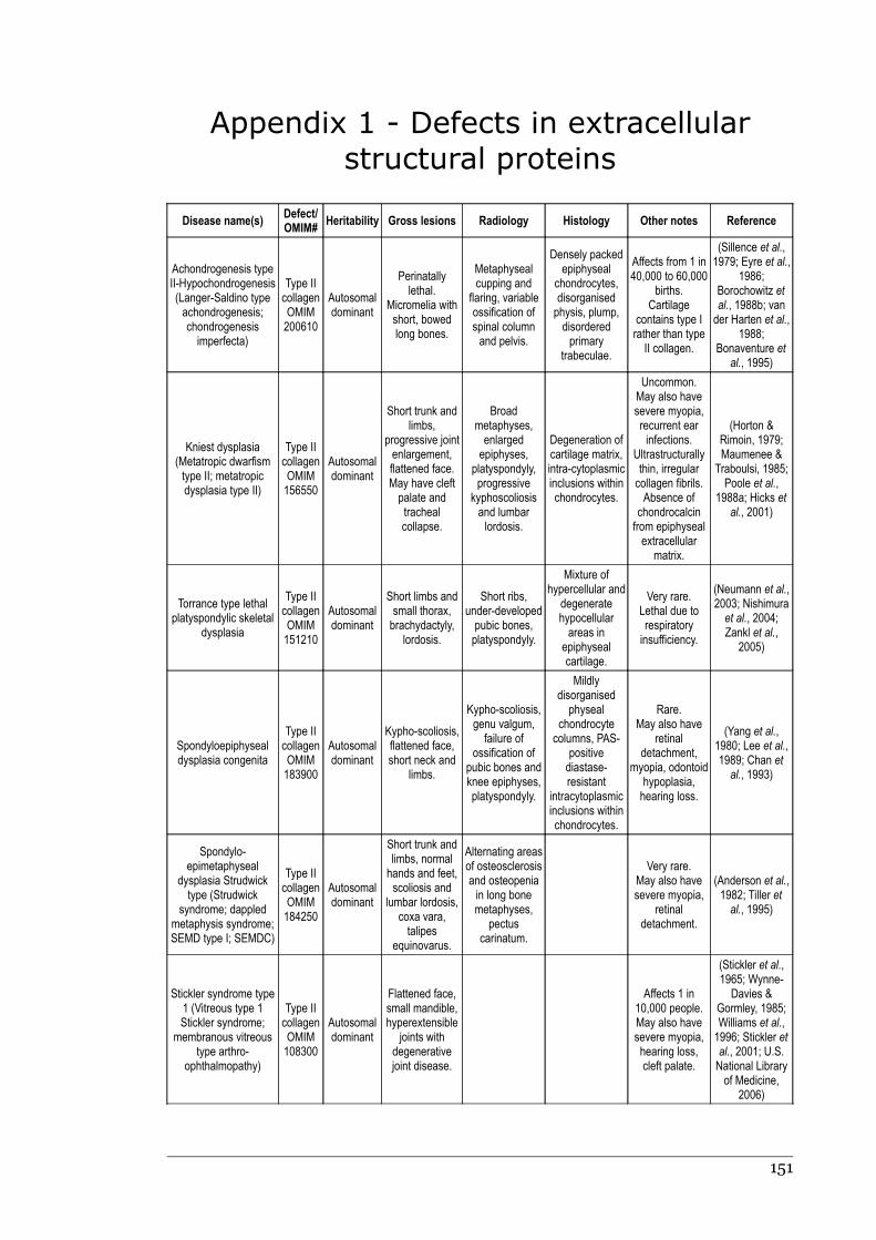
Appendix 1 - Defects in extracellular
structural proteins
Disease name(s)Defect/OMIM#
Heritability Gross lesions Radiology Histology Other notes Reference
Achondrogenesis typeII-Hypochondrogenesis(Langer-Saldino typeachondrogenesis;chondrogenesisimperfecta)
Type IIcollagenOMIM200610
Autosomaldominant
Perinatallylethal.
Micromelia withshort, bowedlong bones.
Metaphysealcupping and
flaring, variableossification ofspinal columnand pelvis.
Densely packedepiphyseal
chondrocytes,disorganisedphysis, plump,disorderedprimary
trabeculae.
Affects from 1 in40,000 to 60,000
births.Cartilage
contains type Irather than typeII collagen.
(Sillence et al.,1979; Eyre et al.,
1986;Borochowitz etal., 1988b; vander Harten et al.,
1988;Bonaventure et
al., 1995)
Kniest dysplasia(Metatropic dwarfismtype II; metatropicdysplasia type II)
Type IIcollagenOMIM156550
Autosomaldominant
Short trunk andlimbs,
progressive jointenlargement,flattened face.May have cleftpalate andtrachealcollapse.
Broadmetaphyses,enlargedepiphyses,
platyspondyly,progressive
kyphoscoliosisand lumbarlordosis.
Degeneration ofcartilage matrix,intra-cytoplasmicinclusions withinchondrocytes.
Uncommon.May also havesevere myopia,recurrent earinfections.
Ultrastructurallythin, irregularcollagen fibrils.Absence of
chondrocalcinfrom epiphysealextracellular
matrix.
(Horton &Rimoin, 1979;Maumenee &
Traboulsi, 1985;Poole et al.,
1988a; Hicks etal., 2001)
Torrance type lethalplatyspondylic skeletal
dysplasia
Type IIcollagenOMIM151210
Autosomaldominant
Short limbs andsmall thorax,brachydactyly,
lordosis.
Short ribs,under-developedpubic bones,platyspondyly.
Mixture ofhypercellular and
degeneratehypocellularareas in
epiphysealcartilage.
Very rare.Lethal due torespiratoryinsufficiency.
(Neumann et al.,2003; Nishimuraet al., 2004;Zankl et al.,
2005)
Spondyloepiphysealdysplasia congenita
Type IIcollagenOMIM183900
Autosomaldominant
Kypho-scoliosis,flattened face,short neck and
limbs.
Kypho-scoliosis,genu valgum,failure of
ossification ofpubic bones andknee epiphyses,platyspondyly.
Mildlydisorganised
physealchondrocytecolumns, PAS-
positivediastase-resistant
intracytoplasmicinclusions withinchondrocytes.
Rare.May also have
retinaldetachment,
myopia, odontoidhypoplasia,hearing loss.
(Yang et al.,1980; Lee et al.,1989; Chan etal., 1993)
Spondylo-epimetaphyseal
dysplasia Strudwicktype (Strudwick
syndrome; dappledmetaphysis syndrome;SEMD type I; SEMDC)
Type IIcollagenOMIM184250
Autosomaldominant
Short trunk andlimbs, normalhands and feet,scoliosis and
lumbar lordosis,coxa vara,talipes
equinovarus.
Alternating areasof osteosclerosisand osteopeniain long bonemetaphyses,
pectuscarinatum.
Very rare.May also havesevere myopia,
retinaldetachment.
(Anderson et al.,1982; Tiller etal., 1995)
Stickler syndrome type1 (Vitreous type 1Stickler syndrome;
membranous vitreoustype arthro-
ophthalmopathy)
Type IIcollagenOMIM108300
Autosomaldominant
Flattened face,small mandible,hyperextensible
joints withdegenerativejoint disease.
Affects 1 in10,000 people.May also havesevere myopia,hearing loss,cleft palate.
(Stickler et al.,1965; Wynne-Davies &
Gormley, 1985;Williams et al.,1996; Stickler etal., 2001; U.S.National Libraryof Medicine,
2006)
151

Appendix 1 - Defects in extracellular structural proteins
Disease name(s)Defect/OMIM#
Heritability Gross lesions Radiology Histology Other notes Reference
Spondyloperipheraldysplasia
Type IIcollagenOMIM271700
Autosomaldominant
Brachydactyly.Platyspondyly,short ulna.
Very rare.May have shortstature, myopia,hearing loss,
mentalretardation.
(Kelly et al.,1977; Vanek,1983; Zabel etal., 1996)
Multiple epiphysealdysplasia
Type IXcollagen, COMP,MATN3OMIM600969132400607078
Autosomaldominant
Short stature,scoliosis, valgusdeformity, early
onsetosteoarthritis.
Irregularossification ofcarpals and
tarsals, stippledcalcification ofepiphyses,subchondralsclerosis,
flattened femoralheads.
Chondrocyteshave
intracytoplasmicinclusions andare surroundedby slightly fibrillar
matrix.Epiphyseal
cartilage is mostseverelyaffected.
Affects 1 in10,000 peopleUltrastructurally
dilatedchondrocyte rER
containsalternatingbands of
electron denseand lucentmaterial.
(Rasmussen &Reimann, 1973;Wynne-Davies &Gormley, 1985;Ballo et al.,
1997;Bönnemann et
al., 2000;Maddox et al.,2000; Czarny-Ratajczak et al.,2001; Mäkitie etal., 2004a; U.S.National Libraryof Medicine,
2006)
Schmid metaphysealchondrodysplasia(Japanese type
spondylometaphysealdysplasia)
Type XcollagenOMIM156500
Autosomaldominant
Disproportionatedwarfism,bowed legs,coxa vara,
lumbar lordosis.
Metaphyses arewidened,
sclerotic andirregular,
occasional mildplatyspondyly.
Absence ofidentifiablehypertrophicchondrocytes,disorganised.
The mostcommon type ofmetaphysealchondro-dysplasia.
(Lachman et al.,1988; Hasegawaet al., 1994;Chan et al.,
1995b; Ikegawaet al., 1997;
Savarirayan etal., 2000;
Bateman et al.,2003; Batemanet al., 2004)
Stickler syndrome typeII (Vitreous type 2Stickler syndrome;beaded vitreous typeStickler syndrome)
Type XIcollagenOMIM604841
Autosomaldominant
Short stature,midface
hypoplasia, cleftpalate,
micrognathia,hyperextensiblejoints, osteo-arthropathy.
Mild epiphysealdysplasia, widemetaphyses.
Hearing loss,myopia, maydevelop
glaucoma andretinal
detachment.
(Ayme & Preus,1984; Richardset al., 1996;
Spranger, 1998)
Marshall syndrome
Type XIcollagenOMIM154780
Autosomaldominant
Short stature,midface
hypoplasia, lessfrequent cleft
palate.
Narrowed jointspaces with hip
and kneeosteophytes.
Very similar toStickler
syndrome typeII. Myopia,
cataracts. Lessfrequently
detached retina.
(Ayme & Preus,1984; Annunenet al., 1999a)
Otospondylomega-epiphyseal dysplasia(Chondrodystrophywith sensorineural
deafness; Nance-Insleysyndrome; Nance-
Sweeneychondrodysplasia)
Type XIcollagenOMIM215150
Autosomaldominant
andrecessiveforms
Short limbs andfingers,
degenerativejoint disease, flat
face, smallmandible,
sometimes cleftpalate.
Platyspondyly,progressive
fusion of carpalbones.
Very rare.Hearing loss.
(Nance &Sweeney, 1970;Insley & Astley,1974; Vikkula et
al., 1995;Pihlajamaa etal., 1998)
Marfan syndromeFBN1OMIM154700
Autosomaldominant
Increasedheight, long
digits, anteriorchest
deformities, jointlaxity.
Scoliosis,thoracic lordosis.
Fibroblasticchondrocytes.
Affected 2-3 per10,000 births.Clinically
variable, alsohas ocular andcardiovascularabnormalities.
(Nogami et al.,1979; Judge &Dietz, 2005;Tekin et al.,
2007)
152
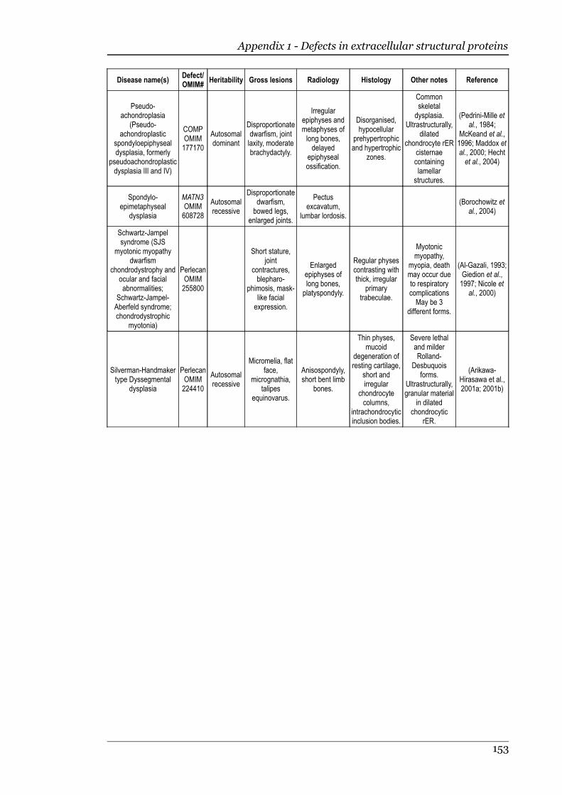
Appendix 1 - Defects in extracellular structural proteins
Disease name(s)Defect/OMIM#
Heritability Gross lesions Radiology Histology Other notes Reference
Pseudo-achondroplasia
(Pseudo-achondroplastic
spondyloepiphysealdysplasia, formerly
pseudoachondroplasticdysplasia III and IV)
COMPOMIM177170
Autosomaldominant
Disproportionatedwarfism, jointlaxity, moderatebrachydactyly.
Irregularepiphyses andmetaphyses oflong bones,delayed
epiphysealossification.
Disorganised,hypocellular
prehypertrophicand hypertrophic
zones.
Commonskeletaldysplasia.
Ultrastructurally,dilated
chondrocyte rERcisternaecontaininglamellar
structures.
(Pedrini-Mille etal., 1984;
McKeand et al.,1996; Maddox etal., 2000; Hechtet al., 2004)
Spondylo-epimetaphyseal
dysplasia
MATN3OMIM608728
Autosomalrecessive
Disproportionatedwarfism,bowed legs,
enlarged joints.
Pectusexcavatum,
lumbar lordosis.
(Borochowitz etal., 2004)
Schwartz-Jampelsyndrome (SJS
myotonic myopathydwarfism
chondrodystrophy andocular and facialabnormalities;
Schwartz-Jampel-Aberfeld syndrome;chondrodystrophic
myotonia)
PerlecanOMIM255800
Short stature,joint
contractures,blepharo-
phimosis, mask-like facialexpression.
Enlargedepiphyses oflong bones,platyspondyly.
Regular physescontrasting withthick, irregular
primarytrabeculae.
Myotonicmyopathy,
myopia, deathmay occur dueto respiratorycomplicationsMay be 3
different forms.
(Al-Gazali, 1993;Giedion et al.,1997; Nicole et
al., 2000)
Silverman-Handmakertype Dyssegmental
dysplasia
PerlecanOMIM224410
Autosomalrecessive
Micromelia, flatface,
micrognathia,talipes
equinovarus.
Anisospondyly,short bent limb
bones.
Thin physes,mucoid
degeneration ofresting cartilage,
short andirregular
chondrocytecolumns,
intrachondrocyticinclusion bodies.
Severe lethaland milderRolland-
Desbuquoisforms.
Ultrastructurally,granular material
in dilatedchondrocytic
rER.
(Arikawa-Hirasawa et al.,2001a; 2001b)
153
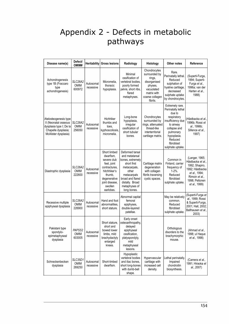
Appendix 2 - Defects in metabolic
pathways
Disease name(s)Defect/OMIM#
Heritability Gross lesions Radiology Histology Other notes Reference
Achondrogenesistype 1B (Fraccaro
typeachondrogenesis)
SLC26A2OMIM600972
Autosomalrecessive
Micromelia,thoracic
hypoplasia.
Minimalossification of
vertebral bodies,poorly formed
pelvis, short ribs,flared
metaphyses.
Chondrocytessurrounded by
rings,disorganised
physes,vacuolatedmatrix with
coarse collagenfibrils.
Rare.Perinatally lethal.
Reducedsulphation of
hyaline cartilage,decreased
sulphate uptakeby chondrocytes.
(Superti-Furga,1994; Superti-Furga et al.,
1996a; van derHarten et al.,
1988)
Atelosteogenesis typeII (Neonatal osseousdysplasia type I; De laChapelle dysplasia;McAlister dysplasia)
SLC26A2OMIM256050
Autosomalrecessive
Hichhikerthumbs and
toes,kyphoscoliosis,micromelia.
Long-bonehypoplasia,irregular
ossification ofshort tubular
bones.
Chondrocytessurrounded byrings, attenuated
thread-likeinterterritorial
cartilage matrix.
Extremely rare.Perinatally lethal
due torespiratory
insufficiency dueto airway
collapse andpulmonaryhypoplasia.Reducedfibroblast
sulphate uptake.
(Hästbacka et al.,1996b; Rossi etal., 1996b;
Sillence et al.,1997)
Diastrophic dysplasiaSLC26A2OMIM222600
Autosomalrecessive
Short limbeddwarfism,severe clubfeet, joint
contractures,hitchhiker’sthumb,
degenerativejoint disease,
swollenearlobes.
Deformed tarsaland metatarsalbones, extremely
short firstmetacarpals,
othermetacarpals
broad and flareddistally. Broadmetaphyses oflong bones.
Cartilage matrixdegenerationwith collagen
fibrils traversingcystic spaces.
Common inFinland, carrierfrequency of
1-2%.Reducedfibroblast
sulphate uptake.
(Langer, 1965;Hästbacka et al.,1992; Shapiro,1992; Hästbackaet al., 1994;Rimoin et al.,1998; Peltonenet al., 1999)
Recessive multipleepiphyseal dysplasia
SLC26A2OMIM226900
Autosomalrecessive
Hand and footabnormalities,short stature.
Abnormal capitalfemoral
epiphyses,double-layered
patellae.
May be relativelycommon.Reducedfibroblast
sulphate uptake.
(Superti-Furga etal., 1999; Rossi& Superti-Furga,2001; Hall, 2002;Ballhausen et al.,
2003)
Pakistani typespondylo-
epimetaphysealdysplasia
PAPSS2OMIM603005
Autosomalrecessive
Short stature,short and
bowed lowerlimbs, mild
brachydactylyenlargedknees.
Early onsetosteoarthropathy,
delayedepiphysealossification,platyspondyly,
mildmetaphyseal
lesions.
Orthologousdisorders to thebrachymorphic
mouse.
(Ahmad et al.,1998; ul Haqueet al., 1998)
Schneckenbeckendysplasia
SLC35D1OMIM269250
Autosomalrecessive
Short-limbeddwarfism.
Hypoplasticvertebral bodiesand iliac bones,short long-boneswith dumb-bell
shape.
Hypervascularcartilage withincreased cell
density.
Lethal perinatally.Impairedchondroitinbiosynthesis.
(Camera et al.,1991; Hiraoka et
al., 2007)
154
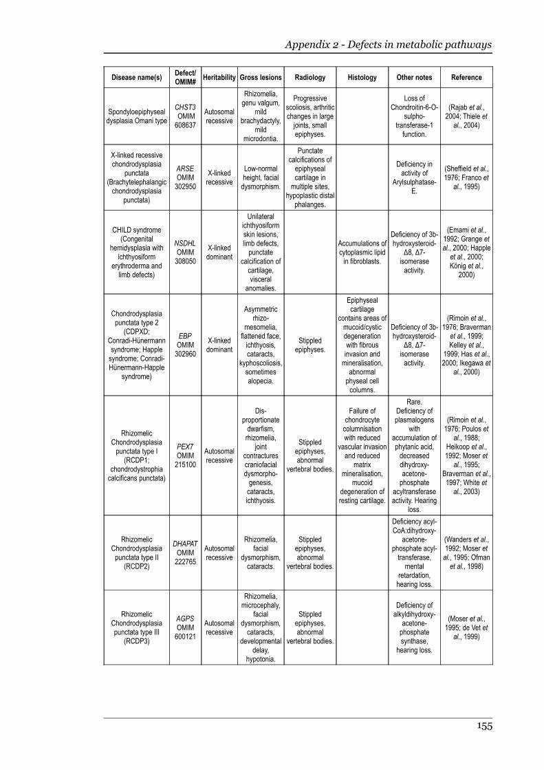
Appendix 2 - Defects in metabolic pathways
Disease name(s)Defect/OMIM#
Heritability Gross lesions Radiology Histology Other notes Reference
Spondyloepiphysealdysplasia Omani type
CHST3 OMIM608637
Autosomalrecessive
Rhizomelia,genu valgum,
mildbrachydactyly,
mildmicrodontia.
Progressivescoliosis, arthriticchanges in largejoints, smallepiphyses.
Loss ofChondroitin-6-O-
sulpho-transferase-1function.
(Rajab et al.,2004; Thiele et
al., 2004)
X-linked recessivechondrodysplasia
punctata(Brachytelephalangicchondrodysplasia
punctata)
ARSEOMIM302950
X-linkedrecessive
Low-normalheight, facialdysmorphism.
Punctatecalcifications ofepiphysealcartilage in
multiple sites,hypoplastic distal
phalanges.
Deficiency inactivity of
Arylsulphatase-E.
(Sheffield et al.,1976; Franco et
al., 1995)
CHILD syndrome(Congenital
hemidysplasia withichthyosiform
erythroderma andlimb defects)
NSDHLOMIM308050
X-linkeddominant
Unilateralichthyosiformskin lesions,limb defects,punctate
calcification ofcartilage,visceral
anomalies.
Accumulations ofcytoplasmic lipidin fibroblasts.
Deficiency of 3b-hydroxysteroid-
∆8, ∆7-isomeraseactivity.
(Emami et al.,1992; Grange etal., 2000; Happleet al., 2000;König et al.,
2000)
Chondrodysplasiapunctata type 2
(CDPXD;Conradi-Hünermannsyndrome; Happlesyndrome; Conradi-Hünermann-Happle
syndrome)
EBPOMIM302960
X-linkeddominant
Asymmetricrhizo-
mesomelia,flattened face,ichthyosis,cataracts,
kyphoscoliosis,sometimesalopecia.
Stippledepiphyses.
Epiphysealcartilage
contains areas ofmucoid/cysticdegenerationwith fibrousinvasion andmineralisation,
abnormalphyseal cellcolumns.
Deficiency of 3b-hydroxysteroid-
∆8, ∆7-isomeraseactivity.
(Rimoin et al.,1976; Braverman
et al., 1999;Kelley et al.,
1999; Has et al.,2000; Ikegawa et
al., 2000)
RhizomelicChondrodysplasiapunctata type I
(RCDP1;chondrodystrophiacalcificans punctata)
PEX7OMIM215100
Autosomalrecessive
Dis-proportionatedwarfism,rhizomelia,
jointcontracturescraniofacialdysmorpho-genesis,cataracts,ichthyosis.
Stippledepiphyses,abnormal
vertebral bodies.
Failure ofchondrocytecolumnisationwith reduced
vascular invasionand reduced
matrixmineralisation,
mucoiddegeneration ofresting cartilage.
Rare.Deficiency ofplasmalogens
withaccumulation ofphytanic acid,decreaseddihydroxy-acetone-phosphate
acyltransferaseactivity. Hearing
loss.
(Rimoin et al.,1976; Poulos et
al., 1988;Heikoop et al.,1992; Moser et
al., 1995;Braverman et al.,1997; White etal., 2003)
RhizomelicChondrodysplasiapunctata type II
(RCDP2)
DHAPATOMIM222765
Autosomalrecessive
Rhizomelia,facial
dysmorphism,cataracts.
Stippledepiphyses,abnormal
vertebral bodies.
Deficiency acyl-CoA:dihydroxy-
acetone-phosphate acyl-transferase,
mentalretardation,hearing loss.
(Wanders et al.,1992; Moser etal., 1995; Ofmanet al., 1998)
RhizomelicChondrodysplasiapunctata type III
(RCDP3)
AGPSOMIM600121
Autosomalrecessive
Rhizomelia,microcephaly,
facialdysmorphism,cataracts,
developmentaldelay,
hypotonia.
Stippledepiphyses,abnormal
vertebral bodies.
Deficiency ofalkyldihydroxy-
acetone-phosphatesynthase,
hearing loss.
(Moser et al.,1995; de Vet et
al., 1999)
155
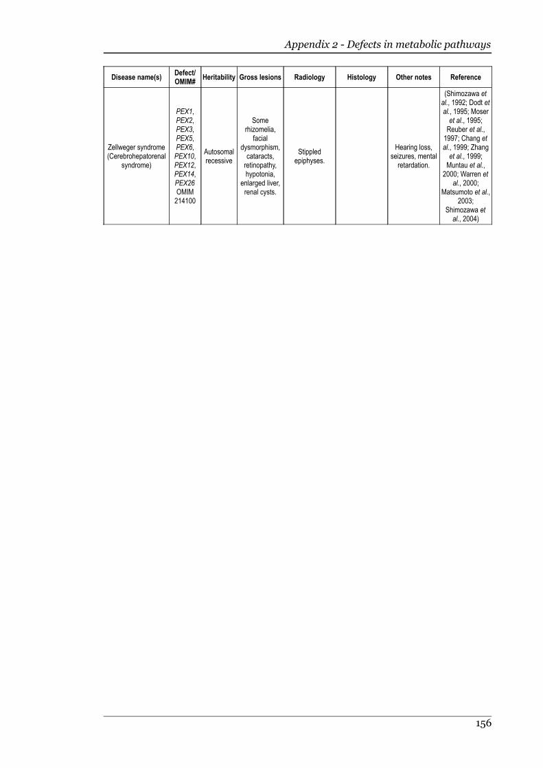
Appendix 2 - Defects in metabolic pathways
Disease name(s)Defect/OMIM#
Heritability Gross lesions Radiology Histology Other notes Reference
Zellweger syndrome(Cerebrohepatorenal
syndrome)
PEX1,PEX2,PEX3,PEX5,PEX6,PEX10,PEX12,PEX14,PEX26OMIM214100
Autosomalrecessive
Somerhizomelia,
facialdysmorphism,cataracts,retinopathy,hypotonia,
enlarged liver,renal cysts.
Stippledepiphyses.
Hearing loss,seizures, mentalretardation.
(Shimozawa etal., 1992; Dodt etal., 1995; Moseret al., 1995;Reuber et al.,1997; Chang etal., 1999; Zhanget al., 1999;Muntau et al.,
2000; Warren etal., 2000;
Matsumoto et al.,2003;
Shimozawa etal., 2004)
156
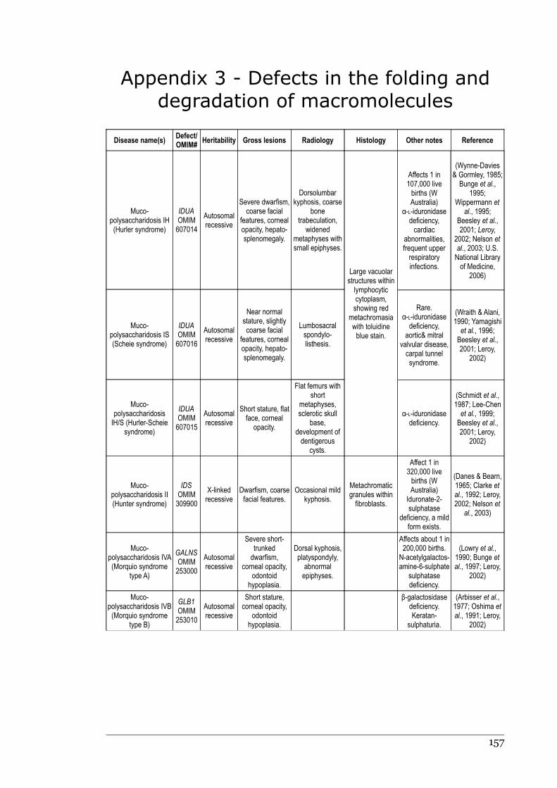
Appendix 3 - Defects in the folding and
degradation of macromolecules
Disease name(s)Defect/OMIM#
Heritability Gross lesions Radiology Histology Other notes Reference
Muco-polysaccharidosis IH(Hurler syndrome)
IDUAOMIM607014
Autosomalrecessive
Severe dwarfism,coarse facial
features, cornealopacity, hepato-splenomegaly.
Dorsolumbarkyphosis, coarse
bonetrabeculation,
widenedmetaphyses withsmall epiphyses.
Muco-polysaccharidosis IS(Scheie syndrome)
IDUAOMIM607016
Autosomalrecessive
Near normalstature, slightlycoarse facial
features, cornealopacity, hepato-splenomegaly.
Lumbosacralspondylo-listhesis.
Muco-polysaccharidosisIH/S (Hurler-Scheie
syndrome)
IDUAOMIM607015
Autosomalrecessive
Short stature, flatface, corneal
opacity.
Flat femurs withshort
metaphyses,sclerotic skull
base,development ofdentigerous
cysts.
Large vacuolarstructures withinlymphocyticcytoplasm,showing red
metachromasiawith toluidineblue stain.
Affects 1 in107,000 livebirths (WAustralia)
α-L-iduronidasedeficiency,cardiac
abnormalities,frequent upperrespiratoryinfections.
(Wynne-Davies& Gormley, 1985;Bunge et al.,
1995;Wippermann et
al., 1995;Beesley et al.,2001; Leroy,
2002; Nelson etal., 2003; U.S.National Libraryof Medicine,
2006)
Rare.α-L-iduronidasedeficiency,
aortic& mitralvalvular disease,carpal tunnelsyndrome.
(Wraith & Alani,1990; Yamagishiet al., 1996;Beesley et al.,2001; Leroy,
2002)
α-L-iduronidasedeficiency.
(Schmidt et al.,1987; Lee-Chenet al., 1999;Beesley et al.,2001; Leroy,
2002)
Muco-polysaccharidosis II(Hunter syndrome)
IDSOMIM309900
X-linkedrecessive
Dwarfism, coarsefacial features.
Occasional mildkyphosis.
Metachromaticgranules withinfibroblasts.
Affect 1 in320,000 livebirths (WAustralia)
Iduronate-2-sulphatase
deficiency, a mildform exists.
(Danes & Bearn,1965; Clarke etal., 1992; Leroy,2002; Nelson et
al., 2003)
Muco-polysaccharidosis IVA(Morquio syndrome
type A)
GALNSOMIM253000
Autosomalrecessive
Severe short-trunkeddwarfism,
corneal opacity,odontoid
hypoplasia.
Dorsal kyphosis,platyspondyly,abnormalepiphyses.
Affects about 1 in200,000 births.N-acetylgalactos-amine-6-sulphate
sulphatasedeficiency.
(Lowry et al.,1990; Bunge etal., 1997; Leroy,
2002)
Muco-polysaccharidosis IVB(Morquio syndrome
type B)
GLB1OMIM253010
Autosomalrecessive
Short stature,corneal opacity,
odontoidhypoplasia.
β-galactosidasedeficiency.Keratan-
sulphaturia.
(Arbisser et al.,1977; Oshima etal., 1991; Leroy,
2002)
157
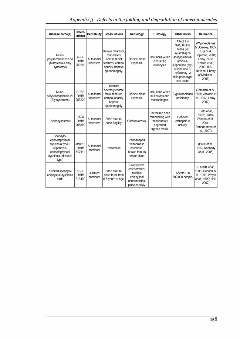
Appendix 3 - Defects in the folding and degradation of macromolecules
Disease name(s)Defect/OMIM#
Heritability Gross lesions Radiology Histology Other notes Reference
Muco-polysaccharidosis VI(Maroteaux-Lamy
syndrome)
ARSBOMIM253200
Autosomalrecessive
Severe dwarfism,moderatelycoarse facial
features, cornealopacity, hepato-splenomegaly.
Dorsolumbarkyphosis.
Inclusions withincirculatingleukocytes.
Affect 1 in320,000 livebirths (W
Australia) N-acetylgalactos-
amine-4-sulphatase (aryl-sulphatase B)deficiency. Amild phenotype
can occur.
(Wynne-Davies& Gormley, 1985;
Litjens &Hopwood, 2001;Leroy, 2002;Nelson et al.,2003; U.S.
National Libraryof Medicine,
2006)
Muco-polysaccharidosis VII
(Sly syndrome)
GUSBOMIM253220
Autosomalrecessive
Dwarfism,severely coarsefacial features,corneal opacity,
hepato-splenomegaly.
Dorsolumbarkyphosis.
Inclusions withinleukocytes andmacrophages.
β-glucuronidasedeficiency.
(Tomatsu et al.,1991; Vervoort etal., 1997; Leroy,
2002)
PycnodysostosisCTSKOMIM265800
Autosomalrecessive
Short stature,bone fragility.
Osteosclerosis.
Decreased boneremodelling withinadequatelydegraded
organic matrix.
Deficientcathepsin-Kactivity.
(Gelb et al.,1996; Fratzl-Zelman et al.,
2004;Donnarumma et
al., 2007)
Spondylo-epimetaphysealdysplasia type II
(Spondylo-epimetaphyseal
dysplasia, Missouritype)
MMP13OMIM602111
Autosomaldominant
Rhizomelia.
Pear-shapedvertebrae inchildhood,
bowed femursand/or tibias.
(Patel et al.,1993; Kennedyet al., 2005)
X-linked spondylo-epiphyseal dysplasia
tarda
SEDLOMIM313400
X-linkeddominant
Short stature,short trunk from6-8 years of age.
Progressiveosteoarthritis,
multipleepiphyseal
abnormalities,platyspondyly.
Affects 1 in500,000 people.
(Heuertz et al.,1993; Gedeon etal., 1999; Whyteet al., 1999; Hall,
2002)
158

Appendix 4 - Defects in hormones and
signal transduction mechanisms
Disease name(s)Defect/OMIM #
Heritability Gross lesions Radiology Histology Other notes Reference
AchondroplasiaFGFR3OMIM100800
Autosomaldominant
Short staturewith a relativelynormal trunk,rhizomelia,
megalocephaly,frontal bossingwith midfacehypoplasia,
brachydactyly.
Lordosis, bowedlegs, shortened
vertebralpedicles withdecreasedlumbar
interpedicularspace, abnormal
pelvicconformation,
small skull base.
Reducedendochondralgrowth, normal
periostealgrowth, short butwell organisedchondrocytecolumns.
Affects 1 in15,000 – 40,000births, the mostcommon form ofshort-limbeddwarfism.
(Langer, 1967;Rimoin et al.,
1970; Rimoin etal., 1976;
Rousseau et al.,1994; Shiang etal., 1994; Belluset al., 1995a)
HypochondroplasiaFGFR3OMIM146000
Autosomaldominant
Short stature,megalocephalywith mild frontal
bossing,micromelia,
lumbar lordosis.
Shortenedvertebral
pedicles withdecreasedlumbar
interpedicularspace.
Well organisedendochondralossification.
Similar toachondroplasia,
but milder.
(Rimoin et al.,1976; Bellus etal., 1995b;
Ramaswami etal., 1998)
Thanatophoricdysplasia Type I
(TD1; platyspondylicskeletal dysplasia,San Diego type)
Thanatophoricdysplasia Type 2
(TD2)
FGFR3OMIM187600
FGFR3OMIM187601
Autosomaldominant
Autosomaldominant
Extremely shortlimbs, redundant
skin folds,narrow chest,large head withfrontal bossingand wide-
spaced eyes.
Small ribs,platyspondyly,curved femurs.
Small ribs,“clover leaf” skull.
Physealchondrocytecolumns are
disorganised andshort, withmetaphyseal
boneovergrowing thephyses, andshort, thickprimary
trabeculae.
Lethal. Affects 1in 20,000-50,000
births.Pulmonary
hypoplasia. TypeI is more
common thantype II.
(Orioli et al.,1986; Tavorminaet al., 1995;
Delezoide et al.,1997; Kitoh et al.,1998; Wilcox etal., 1998; Brodieet al., 1999)
Severeachondroplasia,developmentaldelay, and
acanthosis nigricans(SADDAN)
FGFR3OMIM134934
Autosomaldominant
Severerhizomelia,acanthosisnigricans,
redundant skinfolds, midfacehypoplasia,
frontal bossing.
Femoral bowing,platyspondylywith reducedinterpedicular
space, short ribs,pelvic
abnormalities.
Rare.Developmental
delay.
(Bellus et al.,1999; Tavorminaet al., 1999)
Jansen metaphysealchondrodysplasia(Metaphyseal
chondrodysplasiaMurk Jansen type,
formerlymetaphysealdysostosis)
PTHR1OMIM156400
Autosomaldominant
Short stature,short and bowed
limb bones.
Genu valgum,broad
metaphyses oflong bones.
Delayedmaturation ofchondrocytes.
Rare.Asymptomatichypercalcaemia
and hypo-phosphataemia.ConstitutivePTH/PTHrPreceptoractivation.
(Charrow &Poznanski, 1984;Schipani et al.,
1995; Schipani etal., 1997a; David,1998; Kozlowskiet al., 1999; Hall,2002; Bastepe et
al., 2004)
Blomstrandmetaphysealdysplasia
PTHR1OMIM215045
Autosomalrecessive
Severemicromelia,narrow chest,short neck,
micrognathia.
Osteosclerosis,advancedskeletal
maturation,bowed limbs,
widemetaphyses,small vertebral
bodies.
Short irregularchondrocytecolumns,
thickened, denseprimary
trabeculae.
Rare. Absence ofPTH/PTHrPreceptors.Pulmonaryhypoplasia.
(Jobert et al.,1998; Zhang etal., 1998; Galeraet al., 1999;
Karperien et al.,1999)
159
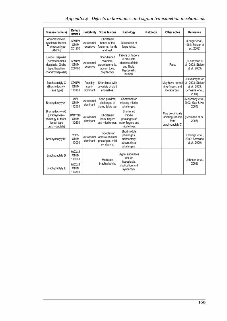
Appendix 4 - Defects in hormones and signal transduction mechanisms
Disease name(s)Defect/OMIM #
Heritability Gross lesions Radiology Histology Other notes Reference
Acromesomelicdysplasia, Hunter-Thompson type
(AMDH)
CDMP1OMIM201250
Autosomalrecessive
Shortenedbones of the
forearms, handsand feet.
Dislocation oflarge joints.
(Langer et al.,1989; Stelzer et
al., 2003)
Grebe Dysplasia(Acromesomelicdysplasia, Grebetype; Brazilian
chondrodysplasia)
CDMP1OMIM200700
Autosomalrecessive
Short-limbeddwarfism,
acromesomelia,absent toes,polydactyly.
Failure of fingersto articulate,
absence of tibiaand fibula.Hypoplastichumeri.
Rare.(Al-Yahyaee etal., 2003; Stelzeret al., 2003)
Brachydactyly C(Brachydactyly,Haws type)
CDMP1OMIM113100
Possiblysemi-
dominant
Short limbs witha variety of digitanomalies.
May have normalring-fingers andmetacarpals.
(Savarirayan etal., 2003; Stelzeret al., 2003;
Schwabe et al.,2004)
Brachydactyly A1IHHOMIM112500
Autosomaldominant
Short proximalphalanges of
thumb & big toe.
Shortened ormissing middlephalanges.
(McCready et al.,2002; Gao & He,
2004)
Brachydactyly A2(Brachymeso-
phalangy II; Mohr-Wriedt type
brachydactyly)
BMPR1BOMIM112600
Autosomaldominant
Shortenedindex-fingers
and middle toes.
Shortenedmiddle
phalanges ofindex-fingers and
middle toes.
May be clinicallyindistinguishable
frombrachydactyly C.
(Lehmann et al.,2003)
Brachydactyly B1ROR2OMIM113000
Autosomaldominant
Hypoplasia/aplasia of distalphalanges, mildsyndactyly.
Short middlephalanges,rudimentary/absent distalphalanges.
(Oldridge et al.,2000; Schwabeet al., 2000)
Brachydactyly D
Brachydactyly E
HOX13OMIM113200
HOX13OMIM113300
Moderatebrachydactyly.
Digital anomaliesinclude
hypoplasia,duplication andsyndactyly.
(Johnson et al.,2003)
160
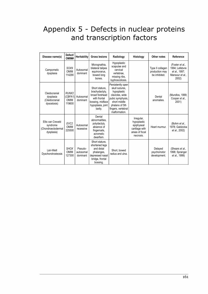
Appendix 5 - Defects in nuclear proteins
and transcription factors
Disease name(s)Defect/OMIM#
Heritability Gross lesions Radiology Histology Other notes Reference
Campomelicdysplasia
SOX9OMIM114290
Autosomaldominant
Micrognathia,bilateral talipesequinovarus,bowed longbones.
Hypoplasticscapulae and
cervicalvertebrae,missing ribs,
kyphoscoliosis.
Type II collagenproduction maybe inhibited.
(Foster et al.,1994; Lefebvreet al., 1997;
Mansour et al.,2002)
Cleidocranialdysplasia
(Cleidocranialdysostosis)
RUNX2(CBFA1)OMIM119600
Autosomaldominant
Short stature,brachydactyly,broad foreheadwith frontal
bossing, midfacehypoplasia, joint
laxity.
Persistently openskull sutures,hypoplastic
clavicles, widepubic symphysis,short middlephalanx of 5th
fingers, vertebralmalformation.
Dentalanomalies.
(Mundlos, 1999;Cooper et al.,
2001)
Ellis van Creveldsyndrome
(Chondroectodermaldysplasia)
EVC2OMIM225500
Autosomalrecessive
Dentalabnormalities,polydactyly,absence offingernails,acromelicdwarfism.
Irregular,hypoplasticepiphysealcartilage withareas of focalnecrosis.
Heart murmur.(Bohm et al.,
1978; Galdzickaet al., 2002)
Léri-WeillDyschondrosteosis
SHOXOMIM127300
Pseudo-autosomaldominant
Short stature,shortened legs
and distalphalanges,
depressed nasalbridge, frontal
bossing.
Short, bowedradius and ulna.
Delayedpsychomotordevelopment.
(Shears et al.,1998; Sprangeret al., 1999)
161
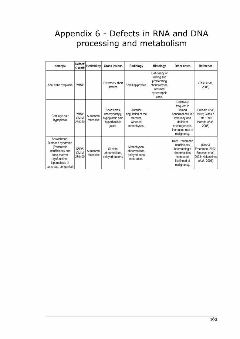
Appendix 6 - Defects in RNA and DNA
processing and metabolism
Name(s)Defect/OMIM#
Heritability Gross lesions Radiology Histology Other notes Reference
Anauxetic dysplasia RMRPExtremely short
stature.Small epiphyses.
Deficiency ofresting andproliferatingchondrocytes,
reducedhypertrophic
zone.
(Thiel et al.,2005)
Cartilage-hairhypoplasia
RMRPOMIM250250
Autosomalrecessive
Short limbs,brachydactyly,hypoplastic hair,hyperflexibile
joints.
Anteriorangulation of the
sternum,widened
metaphyses.
Relativelyfrequent inFinland.
Abnormal cellularimmunity and
deficienterythrogenesis.Increased rate of
malignancy.
(Sulisalo et al.,1993; Glass &Tifft, 1999;
Harada et al.,2005)
Shwachman-Diamond syndrome
(Pancreaticinsufficiency andbone-marrowdysfunction;
Lipomatosis ofpancreas, congenital)
SBDSOMIM260400
Autosomalrecessive
Skeletalabnormalities,delayed puberty.
Metaphysealabnormalities,delayed bonematuration.
Rare. Pancreaticinsufficiency,haematologicabnormalities,increasedlikelihood ofmalignancy.
(Dror &Freedman, 2002;Boocock et al.,
2003; Nakashimaet al., 2004)
162
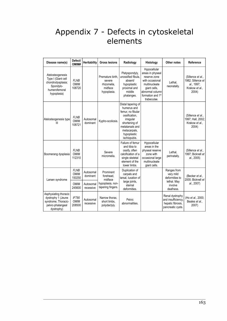
Appendix 7 - Defects in cytoskeletal
elements
Disease name(s)Defect/OMIM#
Heritability Gross lesions Radiology Histology Other notes Reference
AtelosteogenesisType I (Giant cellchondrodysplasia;
Spondylo-humerofemoralhypoplasia)
FLNBOMIM108720
Premature birth,severe
rhizomelia,midface
hypoplasia.
Platyspondyly,unossified fibula,
absent/hypoplasticproximal and
middlephalanges.
Hypocellularareas in physealreserve zonewith occasionalmultinucleategiant cells,
abnormal columnformation and 1º
trabeculae.
Lethal,neonatally.
(Sillence et al.,1982; Sillence et
al., 1997;Krakow et al.,
2004)
Atelosteogenesis typeIII
FLNBOMIM108721
Autosomaldominant
Kypho-scoliosis.
Distal tapering ofhumerus and
femur, no fibularossification,irregular
shortening ofmetatarsals andmetacarpals,hypoplasticischiopubis.
(Sillence et al.,1997; Hall, 2002;Krakow et al.,
2004)
Boomerang dysplasiaFLNBOMIM112310
Severemicromelia.
Failure of femurand tibia toossify, often
calcification of asingle skeletalelement of thelower limbs.
Hypocellularareas in the
physeal reservezone with
occasional largemultinucleategiant cells.
Lethal,perinatally.
(Sillence et al.,1997; Bicknell et
al., 2005)
Larsen syndrome
FLNBOMIM150250
Autosomaldominant
Prominentforehead,midface
hypoplasia, non-tapering fingers.
Duplication ofcarpals and
tarsal, luxation oflarge joints,sternal
deformities.
Ranges fromvery mild
deformities tolethal. Mayinvolve
deafness.
(Becker et al.,2000; Bicknell et
al., 2007)OMIM245600
Autosomalrecessive
Asphyxiating thoracicdystrophy 1 (Jeunesyndrome; Thoracic-pelvic-phalangeal
dystrophy)
IFT80OMIM208500
Autosomalrecessive
Narrow thorax,short limbs,polydactyly.
Pelvicabnormalities.
Renal dystrophyand insufficiency,hepatic fibrosis,pancreatic cysts.
(Ho et al., 2000;Beales et al.,
2007)
163

Appendix 8 - Chondrodysplasias of
unknown aetiology in humans
Disease name(s)Defect/OMIM#
Heritability Gross lesions Radiology Histology Other notes Reference
Chondrodysplasia punctata group
Chondrodysplasiapunctata Tibia-Metacarpal type
OMIM118651
Short stature,midface
hypoplasia.
Abnormalvertebral bodies,short tibiae and2nd and 3rdmetacarpals.
Lesions improvewith age.
(Rittler et al.,1990;
Savarirayan etal., 2004)
Chondrodysplasiapunctata
Facialabnormalities,rhizomelia.
Cervicalplatyspondyly,
stippledcalcifications inendochondral
bones.
Psychomotorand
developmentaldelay, weakrespiratory
muscles, lethalby 2 years.
(Kumada et al.,2001)
Short rib polydactyly syndromes
Short rib polydactyly I(Saldino-Noonan
syndrome;Polydactyly with
neonatalchondrodystrophy
type I)
OMIM263530
Autosomalrecessive
Micromelia,polydactyly.
Severe thoracicdystrophy,irregular
metaphyses,small ilia.
Lethalperinatally.
Cardiac defects,visceral
abnormalities,imperforate
anus.
(Balci et al.,2003; Verma,
2005)
Short rib polydactylyII (Majewskisyndrome;
Polydactyly withneonatal
chondrodystrophytype II)
OMIM263520
Autosomalrecessive
Cleft lip andpalate,
polydactyly.
Short ribs andlong-bones,rounded
metaphyses.Oval-shapedtibiae, delayedossification ofsternum and
fibulae.
Lethalperinatally.
Various visceralabnormalitiesincluding cystic
kidneys.
(Cooper & Hall,1982; Walley et
al., 1983)
Short rib polydactylyIII – Verma-Naumoff
type
OMIM264510
Autosomalrecessive
Rhizomelia.Narrow thorax,metaphyseal
spurs.
Disorganisedendochondralossification,short cellcolumns inhypertrophic
zone.
Respiratoryinfections. Maybe a variant ofasphyxiatingthoracic
dystrophy 1.
(Naumoff et al.,1977; Ho et al.,
2000)
Short rib polydactylyIV (Beemer-Langer
syndrome)
OMIM269860
Autosomalrecessive
Short, bowedlimbs, facialabnormalities,protuberantabdomen.
Narrow thorax,short ribs andlong-bones.
(Beemer et al.,1983; Elçioğlu et
al., 1996)
164
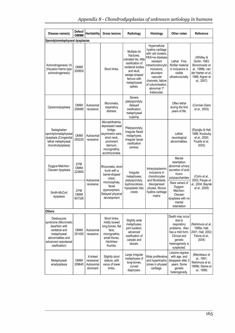
Appendix 8 - Chondrodysplasias of unknown aetiology in humans
Disease name(s)Defect/OMIM#
Heritability Gross lesions Radiology Histology Other notes Reference
Spondylometaphyseal dysplasias
Achondrogenesis 1A(Houston-Harris typeachondrogenesis)
OMIM200600
Short limbs.
Multiple ribfractures,
crenated ilia, littleossification ofvertebral bodies
and skull,wedge-shapedfemurs withmetaphyseal
spikes.
Hypercellularhyaline cartilagewith cell clusters,PAS+ve diastase
resistantintrachondrocytic
inclusions,abundantvascular
channels, failureof columnisation,abnormal 1ºtrabeculae.
Lethal. Fine,fibrillar materialin inclusions is
visibleultrastructurally.
(Whitley &Gorlin, 1983;Borochowitz etal., 1988b; vander Harten et al.,1988; Aigner et
al., 2007)
OpsismodysplasiaOMIM258480
Autosomalrecessive
Micromelia,respiratorydistress.
Severeplatyspondyly,
delayedossification,metaphysealcupping.
Often lethalduring the firstyears of life.
(Cormier-Daireet al., 2003)
Sedaghatianspondylometaphysealdysplasia (Congenitallethal metaphysealchondrodysplasia)
OMIM250220
Autosomalrecessive
Microphthalmia,depressed nasal
bridge,asymmetric ears,
short neck,prominentsternum,
micrognathia,acrorhizomelia.
Platyspondyly,irregular flaredmetaphyses,irregular tarsalossificationcentres.
Lethal,neurologicalabnormalities.
(Elçioğlu & Hall,1998; Koutoubyet al., 2000;Foulds et al.,
2003)
Dyggve-Melchior-Clausen dysplasia
DYMOMIM223800
Autosomalrecessive
Rhizomelia, shorttrunk with abarrel-shaped
chest,microcephaly,
facialdysmorphism.
Delayed physicaldevelopment
Irregularmetaphyses,platyspondyly,kyphoscoliosis,hypoplastic iliac
crests
Intracytoplasmicinclusions inchondrocytesand fibroblasts,disorganisedphyses, fibroushyaline cartilage
matrix
Mentalretardation,
abnormal urinaryexcretion of acid
muco-polysaccharides (Cohn et al.,
2003; Paupe etal., 2004; Bayraket al., 2005)
Smith-McCortdysplasia
DYMOMIM607326
Rare variant ofDyggve-Melchior-Clausen
dysplasia with nomental
retardation
Others
Desbuquoissyndrome (Micromelic
dwarfism withvertebral andmetaphyseal
abnormalities andadvanced carpotarsal
ossification)
OMIM251450
Autosomalrecessive
Short limbs,mildly bowedlong bones, flat
face,micrognathia,small thorax,hitchhikerthumbs.
Slightly widemetaphyses,joint luxation,advanced
ossification ofcarpals andtarsals.
Death may occurdue to
respiratoryproblems. Alsohas a mild form.Clinical andgenetic
heterogeneity issuspected.
(Nishimura et al.,1999a; Hall,
2001; Hall, 2002;Faivre et al.,
2004)
Metaphysealanadysplasia
OMIM309645
X-linkedrecessive/Autosomaldominant
Slightly shortstature, withvarus of lower
limbs.
Large irregularmetaphyses oflong-bones,
curveddiaphyses.
Wide proliferativeand hypertrophiczones in physeal
cartilage.
Lesions regresswith age, and
disappear after 2years. Some
geneticheterogeneity.
(Maroteaux etal., 1991;
Nishimura et al.,1999b; Slama et
al., 1999)
165
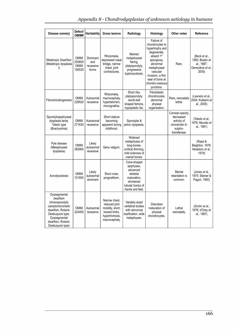
Appendix 8 - Chondrodysplasias of unknown aetiology in humans
Disease name(s)Defect/OMIM#
Heritability Gross lesions Radiology Histology Other notes Reference
Metatropic Dwarfism(Metatropic dysplasia
I)
OMIM250600OMIM156530
Dominantand
recessiveforms
Rhizomelia,depressed nasalbridge, narrowchest, jointcontractures.
Markedmetaphyseal
flaring,platyspondyly,progressive
kyphoscoliosis.
Failure ofchondrocytes tohypertrophy anddegenerate,absent 1ºspongiosa,abnormal
metaphysealvascular
invasion, a thinseal of bone atchondro-osseous
junctions.
Rare.
(Beck et al.,1983; Boden et
al., 1987;Geneviève et al.,
2005)
FibrochondrogenesisOMIM228520
Autosomalrecessive
Rhizomelia,macrocephaly,hypertelorism,micrognathia.
Short ribs,platyspondyly,dumb-bell
shaped femora,hypoplastic ilia.
Fibroblasticchondrocytes,abnormalphyseal
organisation.
Rare, neonatallylethal.
(Leeners et al.,2004; Kulkarni et
al., 2005)
Spondyloepiphysealdysplasia tarda,Toledo type(Brachyolmia)
OMIM271630
Autosomalrecessive
Short staturebecoming
apparent duringchildhood.
Spondylar &pelvic dysplasia.
Corneal opacity,decreasedactivity of
chondroitin 6-sulpho-
transferase.
(Toledo et al.,1978; Mourão et
al., 1981)
Pyle disease(Metaphysealdysplasia)
OMIM265900
Likelyautosomalrecessive
Genu valgum.
Widenedmetaphyses oflong-bones,
cortical thinning,mild sclerosis ofcranial bones.
(Raad &Beighton, 1978;Heselson et al.,
1979)
AcrodysostosisOMIM101800
Likelyautosomaldominant
Short nose,prognathism.
Cone-shapedepiphyses,advancedskeletal
maturation,shortened
tubular bones ofhands and feet.
Mentalretardation iscommon.
(Jones et al.,1975; Steiner &Pagon, 1992)
Dyssegmentaldwarfism
(Anisospondyliccamptomicromelicdwarfism, Roland-Desbuquois type;Dyssegmental
dwarfism, Roland-Desbuquois type)
OMIM224400
Autosomalrecessive
Narrow chest,reduced jointmobility, short,bowed limbs,hypertrichosis,macrocephaly.
Variably-sizedvertebral bodieswith abnormal
ossification, widemetaphyses.
Disturbedmaturation of
physealchondrocytes.
Lethalneonatally.
(Gruhn et al.,1978; d'Orey et
al., 1997)
166

Appendix 9 - Chondrodysplasias of
production animals
Diseasename
BreedDefect/OMIA#
Heritability Gross lesions Radiology Histology Other notes Reference
Sheep
Spider lambsyndrome
Suffolk,Hampshire
FGFR3OMIA000187
May be in-completelydominant
Long limbs,carpal valgus,
skullmalformations,kyphoscoliosis,
erosion ofarticularcartilage.
Poordevelopment of
anconealprocess,cuboidal
sternebrae,pectus
excavatum.
Thickened,irregular physes(esp. thoracicvertebral), withpoorly organisedcell columns.
Onset from birth.
(Vanek et al.,1987; Vanek et
al., 1989;Oberbauer et al.,1995; Cockett et
al., 1999;Beever et al.,2006; Smith etal., 2006)
AnconOMIA000302
Likelyautosomalrecessive
Short stature,elbow varus,carpal valgus.
Extinct.
(Landauer &Chang, 1949,Landauer,1950))
NorwegianAncon
OMIA000302
Bowed femurs.Thin physes,irregular
columnisation.
Onset from birth.Extinct.
(Chang, 1949)
Achondro-plasia
CheviotOMIA000004
Ectrodactyly,exophthalmus,short ears & tail.
Abnormalproximalphalanx,absence ofmiddle and
distalphalanges.
(Wray et al.,1971)
Lethaldwarfism
Domed head,short muzzle,micromelia,swollen
abdomen, cleftpalate.
Narrow thoraxwith short ribs,bowed long
bones with largeepiphyses.
Lack of physealchondrocyte
columns, matrixreduced to
narrow strands.
Lethal. Thyroidhyperplasia.Scrotal herniamay occur.
(Duffell et al.,1985)
DwarfismSouthDown
Autosomalrecessive
Short limbs,bulging cranium,throat & palateabnormalities.
Thyroid follicleslack colloid.
Lethal at 1-2months.
(Bogart, 1946)
TexelChondro-dysplasia
TexelAutosomalrecessive
Short, bentlimbs, barrel
chest, thickenedtrachea.
Enlargedcostochondraljunctions.
Rarefaction ofcartilage matrix,"collagen rings"
aroundchondrocytes.
May causetracheal
collapse andsubsequent
death.
(Byrne, 2005;Thompson et al.,
2005)
Cattle
Brachy-cephalicSnorter
HerefordOMIA000310
Autosomalrecessive
Short head,inferior
prognathia,shortened distallong bones.
Early closure ofspheno-occipitalsynchondrosis,bony projectionsinto cranium,
ventral flatteningof lumbarvertebrae.
Short irregularchondrocytecolumns,occasional
dilations of ERcontaininggranularmaterial.
Onset from birth.(Julian et al.,1957; Jones &Jolly., 1982)
Dolicho-cephalicDwarf
Aberdeen-Angus,Hereford,Shorthorn
Long head.
May be part of acomplex with thebrachycephalic
snorter.
(Gregory, 1956;Julian et al.,
1959)
ComprestDwarf
Small-typeHereford
May be part of acomplex with thebrachycephalic
snorter.
(Gregory, 1956;Julian et al.,
1959)
167

Appendix 9 - Chondrodysplasias of production animals
Diseasename
BreedDefect/OMIA#
Heritability Gross lesions Radiology Histology Other notes Reference
Bulldogcalves
DexterACANOMIA001271
Autosomalin-
completelydominant
Short spine,severe
rhizomelia,hydrocephalus,reduced maxilla,cleft palate,protrudingtongue,
umbilical hernia.
Limb bonesconsist mostly ofhighly cellularepiphyseal
cartilage withoutdistinct physes.
Usually abortedby 7 monthsgestation.
Heterozygotesare short-limbed.
(Harper et al.,1998; Cavanaghet al., 2007)
JerseyJersey, onsetfrom birth
(Mead et al.,1946)
Holstein-Friesian
Short maxilla,protrudingtongue,enlarged
cranium, eyedeformities,short spine,rhizomelia,
umbilical hernia,stenotic trachea,narrow thorax,hypoplastic
lungs, bilateralventricularhypertrophy.
Domination ofhypertrophic
chondrocytes inepiphyses, poororganisation ofphysis, cartilage
cores inmetaphysis.
Gestation incompleted to
term.
(Bowden, 1970;Agerholm et al.,
2004b)
GuernseyAutosomalrecessive
Short maxillaand lower jaw,
dis-proportionatedwarfism.
Rhizomelia.
Completegestation toterm, die
minutes afterbirth.
(Jones, 1961)
TelemarkLethal
Telemark RecessiveShort heads &maxillae, shortcrooked legs.
Completegestation to
term, die due torespiratoryobstruction.
(Mohr, 1926;Brandt, 1941)
Angusdwarfism
AmericanAngus
PRKG2mutation
Autosomalrecessive
Short limbs.Disorganised
physes.(Mishra et al.,
2004)
Brachy-gnathia anddegener-ative jointdisease
AngusOMIA000150
Likely to beheritable
Brachygnathiasuperior, erosionof cartilage inmajor joints.
Osteophytes,reduced jointspaces andsclerosis ofsubchondral
bone.
Degeneration ofcartilage matrix
withchondrocytenecrosis andfibrillation.Poorly
organisedphyses andabnormal
ossification ofcalcified matrix.
Lethal.(Jayo et al.,
1987)
Chondro-dysplasia
JapaneseBrownCattle
LIMBINmutationOMIA000305
Autosomalrecessive
Short longbones with
abnormalities atthe ends.
Narrow irregularphysis withcartilage
extending intospongiosa,hypocellularhypertrophiczone, cystic
degeneration ofmatrix. Irregular
restingchondrocytes
with vacuolationand acidophilicintracytoplasmic
granules.
(Moritomo et al.,1992; Takeda et
al., 2002)
168
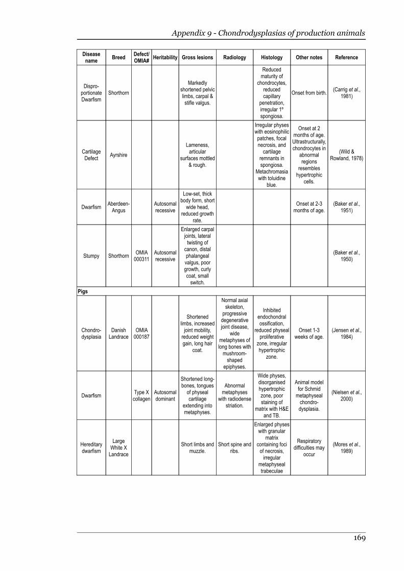
Appendix 9 - Chondrodysplasias of production animals
Diseasename
BreedDefect/OMIA#
Heritability Gross lesions Radiology Histology Other notes Reference
Dispro-portionateDwarfism
Shorthorn
Markedlyshortened pelviclimbs, carpal &stifle valgus.
Reducedmaturity of
chondrocytes,reducedcapillary
penetration,irregular 1ºspongiosa.
Onset from birth.(Carrig et al.,
1981)
CartilageDefect
Ayrshire
Lameness,articular
surfaces mottled& rough.
Irregular physeswith eosinophilicpatches, focalnecrosis, andcartilage
remnants inspongiosa.
Metachromasiawith toluidine
blue.
Onset at 2months of age.Ultrastructurally,chondrocytes in
abnormalregions
resembleshypertrophic
cells.
(Wild &Rowland, 1978)
DwarfismAberdeen-Angus
Autosomalrecessive
Low-set, thickbody form, short
wide head,reduced growth
rate.
Onset at 2-3months of age.
(Baker et al.,1951)
Stumpy ShorthornOMIA000311
Autosomalrecessive
Enlarged carpaljoints, lateraltwisting of
canon, distalphalangealvalgus, poorgrowth, curlycoat, smallswitch.
(Baker et al.,1950)
Pigs
Chondro-dysplasia
DanishLandrace
OMIA000187
Shortenedlimbs, increasedjoint mobility,reduced weightgain, long hair
coat.
Normal axialskeleton,
progressivedegenerativejoint disease,
widemetaphyses oflong bones withmushroom-shaped
epiphyses.
Inhibitedendochondralossification,
reduced physealproliferative
zone, irregularhypertrophic
zone.
Onset 1-3weeks of age.
(Jensen et al.,1984)
DwarfismType Xcollagen
Autosomaldominant
Shortened long-bones, tongues
of physealcartilage
extending intometaphyses.
Abnormalmetaphyses
with radiodensestriation.
Wide physes,disorganisedhypertrophiczone, poorstaining of
matrix with H&Eand TB.
Animal modelfor Schmidmetaphysealchondro-dysplasia.
(Nielsen et al.,2000)
Hereditarydwarfism
LargeWhite XLandrace
Short limbs andmuzzle.
Short spine andribs.
Enlarged physeswith granular
matrixcontaining fociof necrosis,irregular
metaphysealtrabeculae
Respiratorydifficulties may
occur
(Mores et al.,1989)
169
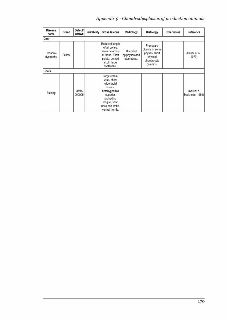
Appendix 9 - Chondrodysplasias of production animals
Diseasename
BreedDefect/OMIA#
Heritability Gross lesions Radiology Histology Other notes Reference
Deer
Chondro-dystrophy
Fallow
Reduced lengthof all bones,
varus deformityof limbs. Cleftpalate, domedskull, largefontanelle.
Distortedepiphyses andsternebrae.
Prematureclosure of somephyses, short
physealchondrocytecolumns.
(Baker et al.,1979)
Goats
BulldogOMIA000005
Large cranialvault, short,wide facialbones,
brachygnathiasuperior,protruding
tongue, shortneck and limbs,ventral hernia.
(Kaikini &Malkhede, 1969)
170
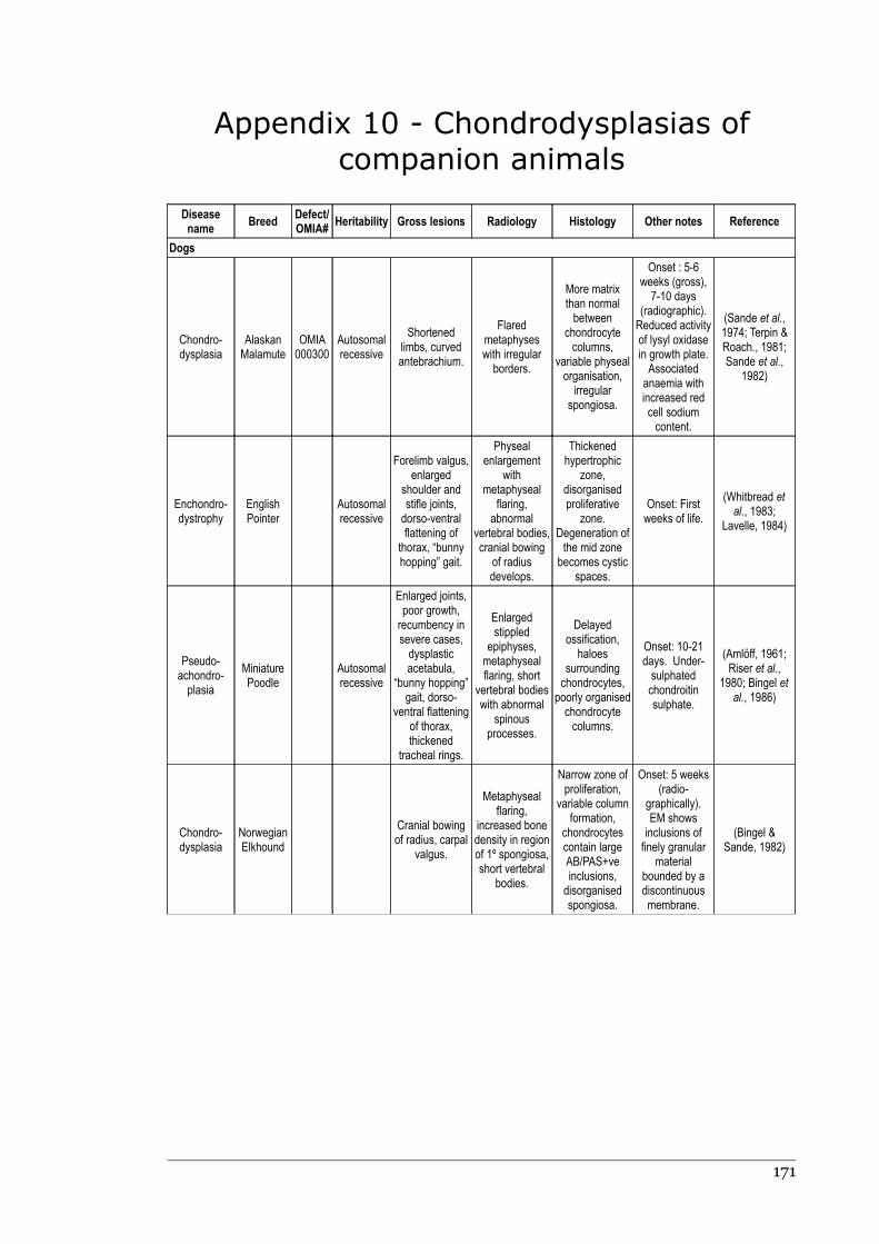
Appendix 10 - Chondrodysplasias of
companion animals
Diseasename
BreedDefect/OMIA#
Heritability Gross lesions Radiology Histology Other notes Reference
Dogs
Chondro-dysplasia
AlaskanMalamute
OMIA000300
Autosomalrecessive
Shortenedlimbs, curvedantebrachium.
Flaredmetaphyseswith irregularborders.
More matrixthan normalbetween
chondrocytecolumns,
variable physealorganisation,irregular
spongiosa.
Onset : 5-6weeks (gross),7-10 days
(radiographic).Reduced activityof lysyl oxidasein growth plate.Associatedanaemia withincreased redcell sodiumcontent.
(Sande et al.,1974; Terpin &Roach., 1981;Sande et al.,
1982)
Enchondro-dystrophy
EnglishPointer
Autosomalrecessive
Forelimb valgus,enlarged
shoulder andstifle joints,dorso-ventralflattening of
thorax, “bunnyhopping” gait.
Physealenlargement
withmetaphyseal
flaring,abnormal
vertebral bodies,cranial bowing
of radiusdevelops.
Thickenedhypertrophic
zone,disorganisedproliferative
zone.Degeneration ofthe mid zone
becomes cysticspaces.
Onset: Firstweeks of life.
(Whitbread etal., 1983;
Lavelle, 1984)
Pseudo-achondro-plasia
MiniaturePoodle
Autosomalrecessive
Enlarged joints,poor growth,recumbency insevere cases,dysplasticacetabula,
“bunny hopping”gait, dorso-
ventral flatteningof thorax,thickened
tracheal rings.
Enlargedstippled
epiphyses,metaphysealflaring, short
vertebral bodieswith abnormal
spinousprocesses.
Delayedossification,
haloessurroundingchondrocytes,
poorly organisedchondrocytecolumns.
Onset: 10-21days. Under-sulphatedchondroitinsulphate.
(Amlöff, 1961;Riser et al.,
1980; Bingel etal., 1986)
Chondro-dysplasia
NorwegianElkhound
Cranial bowingof radius, carpal
valgus.
Metaphysealflaring,
increased bonedensity in regionof 1º spongiosa,short vertebral
bodies.
Narrow zone ofproliferation,
variable columnformation,
chondrocytescontain largeAB/PAS+veinclusions,disorganisedspongiosa.
Onset: 5 weeks(radio-
graphically).EM showsinclusions offinely granular
materialbounded by adiscontinuousmembrane.
(Bingel &Sande, 1982)
171
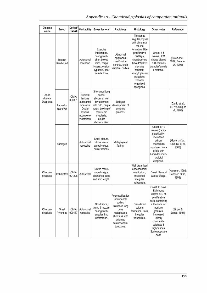
Appendix 10 - Chondrodysplasias of companion animals
Diseasename
BreedDefect/OMIA#
Heritability Gross lesions Radiology Histology Other notes Reference
Oculo-skeletalDysplasia
ScottishDeerhound
LabradorRetriever
Samoyed
OMIA000301
Autosomalrecessive
Exerciseintolerance,poor growth,short bowedlimbs, carpal
hyperextension,kyphosis, poormuscle tone.
Abnormalepiphysealossificationcentres, short
vertebral bodies.
Thickenedirregular physeswith abnormal
columnformation, littleproliferativecartilage,
chondrocyteshave PAS+ve
diastaseresistant
intracytoplasmicinclusions,variablyorganisedspongiosa.
Onset: 4-5weeks. EMshows dilatedrER contains
granular/lamellar material.
(Breur et al.,1989; Breur etal., 1992)
Skeletallesions
autosomalrecessive.Ocularlesions
incomplete-ly dominant
Shortened longbones,
abnormal jointdevelopment
with DJD, carpalvarus, bowing of
radius, hipdysplasia,ocular
abnormalities.
Delayeddevelopment of
anconealprocess.
(Carrig et al.,1977; Carrig et
al., 1988)
Autosomalrecessive
Small stature,elbow varus,carpal valgus,ocular lesions.
Metaphysealflaring.
Onset: 8-12weeks (radio-graphically).Increasedurinary
chondroitinsulphate. Non-allelic with
Labrador oculo-skeletaldysplasia.
(Meyers et al.,1983; Du et al.,
2000)
Chondro-dysplasia
Irish SetterOMIA001296
Autosomal
Bowed radius,carpal valgus,shortened bodyand limb length.
Well organisedendochondralossification,thickenedirregular
trabeculae.
Onset: Severalweeks of age.
(Hanssen, 1992;Hanssen et al.,
1998)
Chondro-dysplasia
GreatPyrenees
OMIA000187
Autosomalrecessive
Short limbs,trunk, & muzzle,poor growth,angular limbdeformities.
Poor ossificationof vertebralbodies,
thickened longbone
metaphyses,short ribs with
enlargedcostochondraljunctions.
Disorderedcolumn
formation, thickirregular
trabeculae.
Onset 10 days.EM shows
dilated rER ofproliferative
cells, containingruthenium red
positivegranules.Increasedurinary
chondroitinsulphate &triglycerides.
Some pups aredeaf.
(Bingel &Sande, 1994)
172
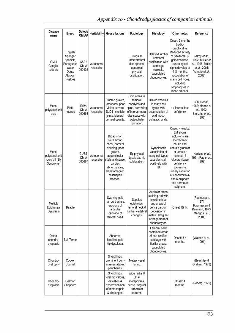
Appendix 10 - Chondrodysplasias of companion animals
Diseasename
BreedDefect/OMIA#
Heritability Gross lesions Radiology Histology Other notes Reference
GM-1Ganglio-sidosis
EnglishSpringerSpaniels,Portuguese
WaterDogs,AlaskanHuskies
GLB1OMIA000402
Autosomalrecessive
Irregularintervertebraldisc spaces,abnormalphyseal
ossification.
Delayed lumbarvertebral
ossification withcartilagenecrosis,vacuolated
chondrocytes.
Onset: 2 months(radio-
graphically).Reduced activityof lysosomal β-galactosidase.Neurological
signs develop at4 ½ months,vacuolation ofmany cell types,
includinglymphocytes inblood smears.
(Alroy et al.,1992; Müller etal., 1998; Mülleret al., 2001;Yamato et al.,
2002)
Muco-polysaccharid
-osis I
Plott-hounds
IDUAOMIA000664
Autosomalrecessive
Stunted growth,lameness, poorvision, severeDJD in multiplejoints, bilateralcorneal opacity.
Lytic areas infemoral
condyles andspine, narrowingof intervertebraldisc space withosteophyteformation.
Dilated vesiclesin many celltypes with
accumulation ofacid muco-
polysaccharide.
α-L-iduronidasedeficiency.
(Shull et al.,1982; Menon et
al., 1992;Stoltzfus et al.,
1992)
Muco-polysaccharid-osis VII (SlySyndrome)
GUSBOMIA000667
Autosomalrecessive
Broad shortskull, broadchest, cornealclouding, poor
growth,appendicular
skeletal disease,cardiac
abnormalities,hepatomegaly,misshapentrachea.
Epiphysealdysplasia, hipsubluxation.
Cytoplasmicvacuolation ofmany cell types,vacuoles stainpositively with
TB.
Onset: 4 weeks.EM shows
inclusions aremembrane-bound and
contain granularor lamellarmaterial. β-glucuronidasedeficiency.Excessive
urinary excretionof chondroitin-4-and 6-sulphateand dermatansulphate.
(Haskins et al.,1991; Ray et al.,
1998)
MultipleEpiphysealDysplasia
Beagle
Swaying gait,narrow trachea,erosions ofarticular
cartilage offemoral head.
Stipplesepiphyses,
femoral neck &lumber vertebral
changes.
Acellular areasstaining red withtoluidine blueand areas ofdense calciumdeposition in
matrix. Irregulararrangement ofchondrocytes.
Onset: Birth.
(Rasmussen,1971;
Rasmussen &Reimann, 1973;Mango et al.,
2004)
Osteo-chondro-dysplasia
Bull TerrierAbnormal
hindlimb gait,hip dysplasia.
Femoral neckcontained areasof non-ossifiedcartilage withfibrillar areas,vacuolated
chondrocytes.
Onset: 3-4months.
(Watson et al.,1991)
Chondro-dystrophy
CockerSpaniel
Short limbs,prominent bonymasses at jointperipheries.
Metaphysealflaring.
(Beachley &Graham, 1973)
Chondro-dysplasia
GermanShepherd
Short limbs,forelimb valgus,deviation &
hyperextensionof metacarpals& phalanges.
Wide radial &ulnar
metaphyses,dense irregulartrabecularpatterns.
Onset: 4months.
(Roberg, 1979)
173
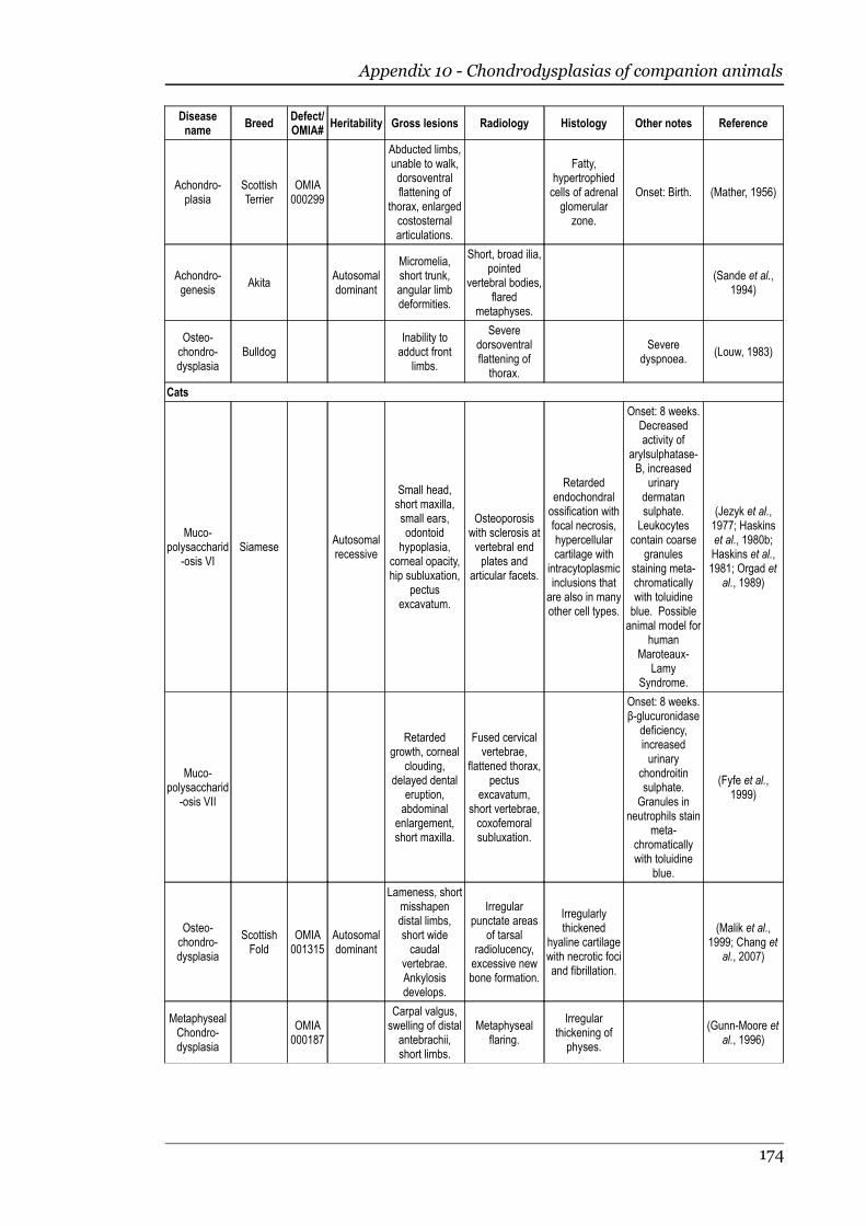
Appendix 10 - Chondrodysplasias of companion animals
Diseasename
BreedDefect/OMIA#
Heritability Gross lesions Radiology Histology Other notes Reference
Achondro-plasia
ScottishTerrier
OMIA000299
Abducted limbs,unable to walk,dorsoventralflattening of
thorax, enlargedcostosternalarticulations.
Fatty,hypertrophiedcells of adrenalglomerular
zone.
Onset: Birth. (Mather, 1956)
Achondro-genesis
AkitaAutosomaldominant
Micromelia,short trunk,angular limbdeformities.
Short, broad ilia,pointed
vertebral bodies,flared
metaphyses.
(Sande et al.,1994)
Osteo-chondro-dysplasia
BulldogInability toadduct front
limbs.
Severedorsoventralflattening ofthorax.
Severedyspnoea.
(Louw, 1983)
Cats
Muco-polysaccharid
-osis VISiamese
Autosomalrecessive
Small head,short maxilla,small ears,odontoid
hypoplasia,corneal opacity,hip subluxation,
pectusexcavatum.
Osteoporosiswith sclerosis atvertebral endplates and
articular facets.
Retardedendochondralossification withfocal necrosis,hypercellularcartilage with
intracytoplasmicinclusions thatare also in manyother cell types.
Onset: 8 weeks.Decreasedactivity of
arylsulphatase-B, increased
urinarydermatansulphate.Leukocytes
contain coarsegranules
staining meta-chromaticallywith toluidineblue. Possibleanimal model for
humanMaroteaux-
LamySyndrome.
(Jezyk et al.,1977; Haskinset al., 1980b;Haskins et al.,1981; Orgad et
al., 1989)
Muco-polysaccharid
-osis VII
Retardedgrowth, corneal
clouding,delayed dental
eruption,abdominal
enlargement,short maxilla.
Fused cervicalvertebrae,
flattened thorax,pectus
excavatum,short vertebrae,coxofemoralsubluxation.
Onset: 8 weeks.β-glucuronidase
deficiency,increasedurinary
chondroitinsulphate.Granules in
neutrophils stainmeta-
chromaticallywith toluidine
blue.
(Fyfe et al.,1999)
Osteo-chondro-dysplasia
ScottishFold
OMIA001315
Autosomaldominant
Lameness, shortmisshapendistal limbs,short widecaudal
vertebrae.Ankylosisdevelops.
Irregularpunctate areas
of tarsalradiolucency,excessive newbone formation.
Irregularlythickened
hyaline cartilagewith necrotic fociand fibrillation.
(Malik et al.,1999; Chang et
al., 2007)
MetaphysealChondro-dysplasia
OMIA000187
Carpal valgus,swelling of distalantebrachii,short limbs.
Metaphysealflaring.
Irregularthickening ofphyses.
(Gunn-Moore etal., 1996)
174
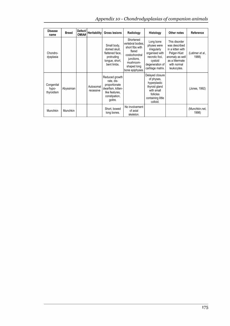
Appendix 10 - Chondrodysplasias of companion animals
Diseasename
BreedDefect/OMIA#
Heritability Gross lesions Radiology Histology Other notes Reference
Chondro-dysplasia
Small body,domed skull,flattened face,protruding
tongue, short,bent limbs.
Shortenedvertebral bodies,short fibs with
flaredcostochondraljunctions,mushroom-shaped long
bone epiphyses.
Long bonephyses wereirregularly
organised withnecrotic foci,
cystoiddegeneration ofcartilage matrix.
This disorderwas describedin a kitten withPelger-Hüet
anomaly as wellas a littermatewith normalleukocytes.
(Latimer et al.,1988)
Congenitalhypo-
thyroidismAbyssinian
Autosomalrecessive
Reduced growthrate, dis-
proportionatedwarfism, kitten-like features,constipation,
goitre.
Delayed closureof physes,hyperplasticthyroid glandwith smallfollicles
containing littlecolloid.
(Jones, 1992)
Munchkin MunchkinShort, bowedlong bones.
No involvementof axialskeleton.
(Munchkin.net,1998)
175
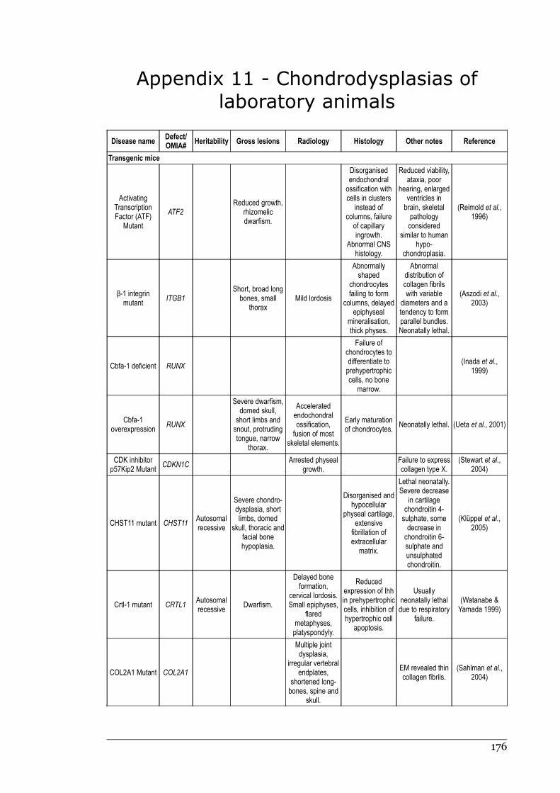
Appendix 11 - Chondrodysplasias of
laboratory animals
Disease nameDefect/OMIA#
Heritability Gross lesions Radiology Histology Other notes Reference
Transgenic mice
ActivatingTranscriptionFactor (ATF)
Mutant
ATF2Reduced growth,
rhizomelicdwarfism.
Disorganisedendochondralossification withcells in clusters
instead ofcolumns, failureof capillaryingrowth.
Abnormal CNShistology.
Reduced viability,ataxia, poor
hearing, enlargedventricles inbrain, skeletalpathologyconsidered
similar to humanhypo-
chondroplasia.
(Reimold et al.,1996)
β-1 integrinmutant
ITGB1Short, broad longbones, small
thoraxMild lordosis
Abnormallyshaped
chondrocytesfailing to form
columns, delayedepiphyseal
mineralisation,thick physes.
Abnormaldistribution ofcollagen fibrilswith variable
diameters and atendency to formparallel bundles.Neonatally lethal.
(Aszodi et al.,2003)
Cbfa-1 deficient RUNX
Failure ofchondrocytes todifferentiate toprehypertrophiccells, no bone
marrow.
(Inada et al.,1999)
Cbfa-1overexpression
RUNX
Severe dwarfism,domed skull,
short limbs andsnout, protrudingtongue, narrow
thorax.
Acceleratedendochondralossification,fusion of most
skeletal elements.
Early maturationof chondrocytes.
Neonatally lethal. (Ueta et al., 2001)
CDK inhibitorp57Kip2 Mutant
CDKN1CArrested physeal
growth.Failure to expresscollagen type X.
(Stewart et al.,2004)
CHST11 mutant CHST11Autosomalrecessive
Severe chondro-dysplasia, shortlimbs, domed
skull, thoracic andfacial bonehypoplasia.
Disorganised andhypocellular
physeal cartilage,extensive
fibrillation ofextracellular
matrix.
Lethal neonatally.Severe decrease
in cartilagechondroitin 4-sulphate, somedecrease inchondroitin 6-sulphate andunsulphatedchondroitin.
(Klüppel et al.,2005)
Crtl-1 mutant CRTL1Autosomalrecessive
Dwarfism.
Delayed boneformation,
cervical lordosis.Small epiphyses,
flaredmetaphyses,platyspondyly.
Reducedexpression of Ihhin prehypertrophiccells, inhibition ofhypertrophic cell
apoptosis.
Usuallyneonatally lethaldue to respiratory
failure.
(Watanabe &Yamada 1999)
COL2A1 Mutant COL2A1
Multiple jointdysplasia,
irregular vertebralendplates,
shortened long-bones, spine and
skull.
EM revealed thincollagen fibrils.
(Sahlman et al.,2004)
176

Appendix 11 - Chondrodysplasias of laboratory animals
Disease nameDefect/OMIA#
Heritability Gross lesions Radiology Histology Other notes Reference
COLXTransgene
COLX
Inset eyes,narrow snout,
variable severityof lesions.
Cervical lordosis,thoracolumbarkyphosis.
Little or no matrixin a compressedhypertrophiczone, in some
cases absence ofcell columns.
EM revealedpoorly visible
collagen fibrils inhypertrophiczone. Often
lethal in perinatalperiod.
(Jacenko et al.,1993; Jacenko et
al., 2001)
PTH/PTHrPReceptor Null
PTHR1
Small size,domed skull,short face,protruding
tongue, shortlimbs.
Chondrocytecolumns irregular
& short.Accelerated
mineralisation ofendochondral
bones.
Often lethal mid-gestation.
(Lanske et al.,1996)
FGF2 Mutant FGF2 Megalocephaly.
Reducedchondrocyte
proliferation andincreasedapoptosis.
Removal ofSTAT1 functioncorrected thechondro-dysplasia.
(Sahni et al.,2001)
FGFR3 Mutant FGFR3Autosomaldominant
Short bowedlimbs, curvedspine, rounded
skull, small thoraxwith caudalwidening.
Decreasedossification,enlarged
costochondraljunctions &sternebrae.
Hypercellularcartilage,
short/absent cellcolumns,incompletematuration ofhypertrophic
cells.
Lethal neonatally,similar to humanthanatophoricdysplasia IIConstitutiveactivation ofFGFR3.
(Iwata et al.,2000)
FGFR3 Mutant FGFR3Autosomalrecessive
Excessive growthof endochondralbones, long tail,kyphoscoliosis,bowed legs.
Long vertebralbodies and limb
bones.
Expansion ofphyseal
hypertrophiczone.
Disruption ofFGFR3.
(Deng et al.,1996)
Ihh mutant IHH
Domed skull,shortened snout,mandible, tail and
limbs.
Reducedchondrocyteproliferation,inappropriatematuration,
degradation ofmatrix, no cellcolumns.
(St-Jacques etal., 1999)
Pex7 Knockout PEX7
Impairedossification of
distal limbs, partsof skull andvertebrae.
Model ofchondro-dysplasiapunctata inhumansImpaired
oxidation of somefatty acids.
(Brites et al.,2003)
PTHrP Over-expression
PTHRPShortened limbs& tail, bowed long
bones.
Delayedendochondralossification.
Delayedendochondralossification,
persistent anlageof proliferating &prehypertrophic
cells.
Becomeshistologicallynormal by 7
weeksChondrocyte-
specificoverexpression of
PTHrP.
(Weir et al., 1996)
RANK Null RANK
Small body, shortlimbs, domedskull, failure oftooth eruption.
Osteopetroticskeleton.
Thickenedphyses withdisorganised
cells, occlusion ofmedullary cavity.
(Dougall et al.,1999)
177
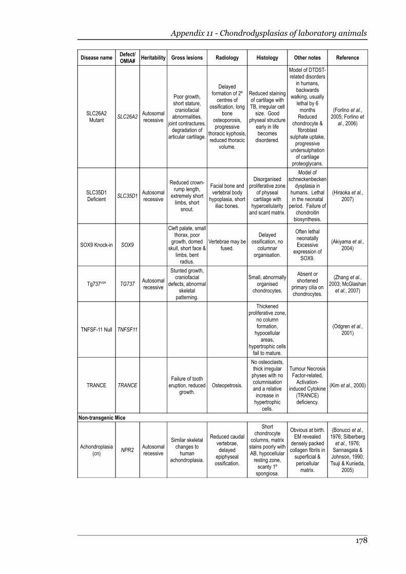
Appendix 11 - Chondrodysplasias of laboratory animals
Disease nameDefect/OMIA#
Heritability Gross lesions Radiology Histology Other notes Reference
SLC26A2Mutant
SLC26A2Autosomalrecessive
Poor growth,short stature,craniofacial
abnormalities,joint contractures,degradation of
articular cartilage.
Delayedformation of 2ºcentres of
ossification, longbone
osteoporosis,progressive
thoracic kyphosis,reduced thoracic
volume.
Reduced stainingof cartilage withTB, irregular cell
size. Goodphyseal structure
early in lifebecomesdisordered.
Model of DTDST-related disorders
in humans,backwards
walking, usuallylethal by 6monthsReduced
chondrocyte &fibroblast
sulphate uptake,progressive
undersulphationof cartilage
proteoglycans.
(Forlino et al.,2005; Forlino et
al., 2006)
SLC35D1Deficient
SLC35D1Autosomalrecessive
Reduced crown-rump length,
extremely shortlimbs, short
snout.
Facial bone andvertebral body
hypoplasia, shortiliac bones.
Disorganisedproliferative zone
of physealcartilage withhypercellularityand scant matrix.
Model ofschneckenbecken
dysplasia inhumans. Lethalin the neonatal
period. Failure ofchondroitinbiosynthesis.
(Hiraoka et al.,2007)
SOX9 Knock-in SOX9
Cleft palate, smallthorax, poor
growth, domedskull, short face &
limbs, bentradius.
Vertebrae may befused.
Delayedossification, no
columnarorganisation.
Often lethalneonatallyExcessive
expression ofSOX9.
(Akiyama et al.,2004)
Tg737orpk TG737Autosomalrecessive
Stunted growth,craniofacial
defects, abnormalskeletal
patterning.
Small, abnormallyorganised
chondrocytes.
Absent orshortened
primary cilia onchondrocytes.
(Zhang et al.,2003; McGlashan
et al., 2007)
TNFSF-11 Null TNFSF11
Thickenedproliferative zone,
no columnformation,hypocellular
areas,hypertrophic cellsfail to mature.
(Odgren et al.,2001)
TRANCE TRANCEFailure of tootheruption, reduced
growth.Osteopetrosis.
No osteoclasts,thick irregularphyses with nocolumnisationand a relativeincrease inhypertrophic
cells.
Tumour NecrosisFactor-related,Activation-
induced Cytokine(TRANCE)deficiency.
(Kim et al., 2000)
Non-transgenic Mice
Achondroplasia(cn)
NPR2Autosomalrecessive
Similar skeletalchanges tohuman
achondroplasia.
Reduced caudalvertebrae,delayed
epiphysealossification.
Shortchondrocyte
columns, matrixstains poorly withAB, hypocellularresting zone,scanty 1ºspongiosa.
Obvious at birth.EM revealed
densely packedcollagen fibrils insuperficial &pericellularmatrix.
(Bonucci et al.,1976; Silberberget al., 1976;Sannasgala &Johnson, 1990;Tsuji & Kunieda,
2005)
178
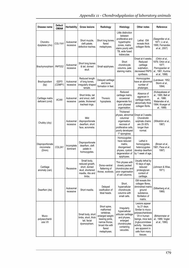
Appendix 11 - Chondrodysplasias of laboratory animals
Disease nameDefect/OMIA#
Heritability Gross lesions Radiology Histology Other notes Reference
Chondro-dysplasia (cho)
COL11A1Autosomalrecessive
Short muzzle,cleft palate,
defective trachea.
Short long bones,flared
metaphyses.
Little distinctionbetween
proliferative andhypertrophiczones, matrix
stains poorly withTB, wide fusedtrabeculae.
Lethal. EMreveals thickcollagen fibrils.
(Seegmiller et al.,1971; Li et al.,
1995; Fernandeset al., 2007)
Brachymorphism PAPSS2Autosomalrecessive
Short long bones& tail, domed
skull.Small epiphyses.
Shortchondrocytecolumns, palestaining matrix.
Onset at 4 weeks.Reducedcartilagesulphation,
decreased PAPSsynthesis.
(Orkin et al.,1976; Orkin et al.,
1977;Pennypacker etal., 1981; Kurimaet al., 1998)
Brachypodism(bp)
GDF5(CDMP1)
Autosomalrecessive
Reduced lengthof long bones,
irregularly shapedtarsals.
Delayed cartilageand bone
formation in feet.
Homozygoteshave an abnormal
number ofphalanges.
(Landauer, 1952;Storm et al.,
1994)
Cartilage matrixdeficient (cmd)
ACANAutosomalrecessive
Short limbs, tailand snout, cleftpalate, thickenedtracheal rings.
Thoracichypoplasia.
Reducedcartilage matrix,
occasionaldegenerate foci,poor physealorganisation.
Absence ofaggrecan in
cartilage matrix,abnormally thickcollagen fibrils.
(Kobayakawa etal., 1985; Bell et
al., 1986;Watanabe et al.,1994; Krueger et
al., 1999)
Chubby (cby)Autosomalrecessive
Milddisproportionatedwarfism, shortface, acromelia.
Thickenedphyses, abnormal
columnarorganisation,necrosis of
proliferative cells,poorly developed1º spongiosa.
Onset at 3 weeks.Chondroitin
sulphate chainsare 20-30%longer thannormal.
(Wikström et al.,1987)
Disproportionatemicromelia(Dmm)
COL2A1Incompletedominant
Disproportionatedwarfism, cleft
palate inhomozygotes.
Homozygoteshave reduced
matrix,disorganised
physes, cystoiddegeneration ofepiphyses.
Lethal inhomozygotes,heterozygotes
develop dwarfismby 1 week of age.
(Brown et al.,1981; Pace et al.,
1997)
Cartilageanomaly (can)
Recessive
Small body,reduced growth,short, domed
skull, shortenedmaxilla, ribs and
limbs.
Dorso-ventralflattening of
thorax, scoliosis.
Thin physes withclosely packed
chondrocytes andpoor organisationof cell columns.
Usually lethal by10 days of age,
reducedproteoglycancontent ofcartilage.
(Johnson & Wise,1971)
Dwarfism (dw)Autosomalrecessive
Short maxilla.Delayed
ossification oftibial heads.
Short,hypercellularchondrocytecolumns withsmall cells.
EM reveals thincollagen fibres,diminished matrix
groundsubstance,fibrillation of
matrix.
(Silberberg et al.,1966)
Muco-polysaccharid-
osis VII
Small body, shortlimbs, short, thick
tail, facialdysmorphism.
Short spine,malformedvertebrae,
enlarged weight-bearing joints,narrow thorax,broad ribs with
flaredmetaphyses.
Irregularlyhypercellular
articular cartilageand physes,enlarged
chondrocytescontainingvacuoles.
Lesions appearby 21 days.
Similar to muco-polysaccharidosis
VII in humanbeings, mice lackβ-glucuronidaseactivity. Vacuolesare apparent incells from many
tissues.
(Birkenmeier etal., 1989; Vogleret al., 1990)
179
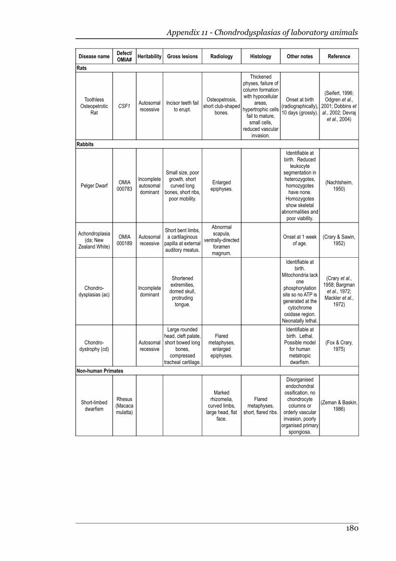
Appendix 11 - Chondrodysplasias of laboratory animals
Disease nameDefect/OMIA#
Heritability Gross lesions Radiology Histology Other notes Reference
Rats
ToothlessOsteopetrotic
RatCSF1
Autosomalrecessive
Incisor teeth failto erupt.
Osteopetrosis,short club-shaped
bones.
Thickenedphyses, failure ofcolumn formationwith hypocellular
areas,hypertrophic cellsfail to mature,small cells,
reduced vascularinvasion.
Onset at birth(radiographically),10 days (grossly).
(Seifert, 1996;Odgren et al.,
2001; Dobbins etal., 2002; Devrajet al., 2004)
Rabbits
Pelger DwarfOMIA000783
Incompleteautosomaldominant
Small size, poorgrowth, shortcurved long
bones, short ribs,poor mobility.
Enlargedepiphyses.
Identifiable atbirth. Reduced
leukocytesegmentation inheterozygotes,homozygoteshave none.
Homozygotesshow skeletal
abnormalities andpoor viability.
(Nachtsheim,1950)
Achondroplasia(da; New
Zealand White)
OMIA000189
Autosomalrecessive
Short bent limbs,a cartilaginous
papilla at externalauditory meatus.
Abnormalscapula,
ventrally-directedforamenmagnum.
Onset at 1 weekof age.
(Crary & Sawin,1952)
Chondro-dysplasias (ac)
Incompletedominant
Shortenedextremities,domed skull,protrudingtongue.
Identifiable atbirth.
Mitochondria lackone
phosphorylationsite so no ATP isgenerated at thecytochrome
oxidase region.Neonatally lethal.
(Crary et al.,1958; Bargmanet al., 1972;Mackler et al.,
1972)
Chondro-dystrophy (cd)
Autosomalrecessive
Large roundedhead, cleft palate,short bowed long
bones,compressed
tracheal cartilage.
Flaredmetaphyses,enlargedepiphyses.
Identifiable atbirth. Lethal.Possible model
for humanmetatropicdwarfism.
(Fox & Crary,1975)
Non-human Primates
Short-limbeddwarfism
Rhesus(Macacamulatta)
Markedrhizomelia,curved limbs,large head, flat
face.
Flaredmetaphyses,
short, flared ribs.
Disorganisedendochondralossification, nochondrocytecolumns or
orderly vascularinvasion, poorlyorganised primary
spongiosa.
(Zeman & Baskin,1986)
180

Appendix 12 - Chondrodysplasias of birds
Diseasename
Species/Breed
Defect/OMIA#
Heritability Gross lesions Radiology Histology Other notes Reference
Nanomeliadomesticchicken,
Gallus gallusACAN
Describedas
autosomalrecessive,but this
may not bethe case ifhetero-zygotesalso showlesions
Extremelyshort, twistedlimbs, benttibiotarsus,
brachycephaly,parrot-like beakwith inferior
brachygnathia.
Mushroom-shaped
proximal headof the humerus.
Lethal duringincubation,markedlyreduced
synthesis ofaggrecan andchondroitinsulphate.
Heterozygotesmay haveslightly
shortened longbones.
(Landauer,1965; Mathews,1967; Stirpe et
al., 1987;Primorac et al.,
1994)
Fowlchondro-dystrophy
domesticchicken,
Gallus gallus
OMIA000189
Autosomalrecessive
Deformed beakand limbs,
shortened longbones.
Lethal due toinability tohatch.
(Lamoreux,1942)
Fowlmicromelia
domesticchicken,
Gallus gallus
OMIA000648
Autosomalrecessive
Parrot-like beakwith inferior
brachygnathia,domed skull,micromelia.
(Asmundson,1942)
Chondro-dystrophy
Japanesequail, Coturnix
japonica
Autosomalrecessive
Short, bentlong-bones,
flattened head,bulging eyes,
inferiorbrachygnathia.
Usually lethalwithin 4 days of
hatching.
(Hermes et al.,1990)
MicromeliaJapanese
quail, Coturnixjaponica
Autosomalrecessive
Short, broadhead with
bulging eyes,inferior
brachygnathia,broad trunk,generalisedmicromelia.
Lethal duringincubation.
(Hill et al.,1963)
Earlyembryonicchondro-dystrophy
Japanesequail, Coturnix
japonica
Autosomalrecessive
Short curvedupper mandiblewith virtually nolower mandible,short curved
tibia and tarso-metatarsus withno evident hock
joint.
Lethal duringincubation.Normal axialskeleton.
(Collins et al.,1968)
Short leg
Turkey, LargeWhite,
Bourbon Red,Bronze
Autosomalrecessive
Shortenedtarso-
metatarsus,apparent
thickening oflong bones.
Reducedhatchability and
postnatalviability,
Survivors inLarge Whiteform are
reproductivelysterile.
(Asmundson,1939;
Asmundson,1944; Nestor,
1978)
181

Appendix 12 - Chondrodysplasias of birds
Diseasename
Species/Breed
Defect/OMIA#
Heritability Gross lesions Radiology Histology Other notes Reference
Lethalchondro-dystrophy
TurkeyOMIA000189
Autosomalrecessive
Micromelia,bent femur and
tibia,brachycephaly,bulging eyes,brachygnathia
inferior.
Lethal duringincubation.
(Gaffney, 1975)
Achondro-plasia
PigeonExtremely shortlimbs and beak.
Lethal.(Hollander,
1945)
Micromelia Pekin duckAutosomalrecessive
Shortenedlimbs and upper
mandible,subcutaneous
oedema,abnormal,
curled feathers,epidermalthickening.
Little cellularorganisation ofepiphyseal
cartilage and noevidence ofchondrocytehypertrophy.
Lethal duringincubation.
(Ash, 1966; Ashet al., 1970)
182

Appendix 13 - Microsatellite primer
sequences
Chr. Marker Locus(cM)
Forward primer sequence Reverse primer sequence
1 BMS2833 14.2 5'-ACCAGATGACCAGACCTTGTG-3' 5'-TGATCTGTGGATCCTCCCTC-3'
1 BMS835 45.3 5'-TCATGTGCATGGGGTTTG-3' 5'-ATCTGCCTACCTGGGCATC-3'
1 MCMA41 68.1 5'-TTCCTGTTTTCTCTTAGATTTGGC-3' 5'-TGGCAAGTGTGTTCTTTACCAC-3'
1 CSRD108 86.1 5'-CATGGAATCACAAAGAGTTGGACA-3' 5'-CCTGGTAAGACAGTCAGTATACAA-3'
1 MCM58 112.1 5'-CTGGGTCTGTATAAGCACGTCTCC-3' 5'-CAGAACAATAAACGCTAAACCAGAGC-3'
1 BMS482 131.2 5'-ACTTCCCCAGTCTTCCCAGT-3' 5'-TGGTGGACAGTCCCATACAG-3'
1 MAF64 164 5'-AATAGACCATTCAGAGAAACGTTGAC-3' 5'-CTCATGGAATCAGACAAAAGGTAGG-3'
1 INRA11 210.6 5'-CGAGTTTCTTTCCTCGTGGTAGGC-3' 5'-GCTCGGCACATCTTCCTTAGCAAC-3'
1 BM8246 241.1 5'-AATGACAAATTGAGGGAGACG-3' 5'-AGAGCCCAGTATCAATTCTTCC-3'
1 BM864 263.6 5'-TGGTAGAGCAATATGAAGGCC-3' 5'-GGAAATCCAAGAAAGAGGGG-3'
1 BM3205 300 5'-TCTTGCTTCCTTCCAAATCTC-3' 5'-TGCCCTTATTTTAACAGTCTGC-3'
1 EPCDV13 348.4 5'-CCATCTGTGCTGTCAGTCA-3' 5'-GAGCCTGGTGGGTTATAGT-3'
5 TGLA303 40.8 5'-CTTGTGTGCCAGACCCAGGAATCC-3' 5'-CATAAGTCAAAGTAACAGTTTAGATGTCC-3'
5 BM741 52.2 5'-GCCCCTGAAGGAATGGTG-3' 5'-CCAAAAGGTCCTATCTCCAAA-3'
5 BM7247 64.3 5'-AAGGCCTGCAGTATTTATATCC-3' 5'-AATCTTTCCCTAGAACTTACAAAGG-3'
5 JAZ 66.7 5'-CCCTGTCCCTTCGCCTGTAAAT-3' 5'-GCAGTCTGGCTCATGGGAGAAAT-3'
6 CP125 2.6 5'-GCAAATAGCCTCTTGTATGATCCTTGG-3' 5'-ACCAAAACAAGACCTTTATTTTTCATGG-3'
6 MCM53 29.7 5'-CATGGAGTTGTAGAGTCAGACATGA-3' 5'-GAGCAAAGGTCATGTCAGGTGT-3'
6 HH55 54.6 5'-GTTATTCCATATTCTTTCCTCCATCATAAGC-3' 5'-CCACACAGAGCAACTAAAACCCAGC-3'
6 JMP1 76.1 5'-CAAAGAGTCAGACATGACTGAACGAG-3' 5'-CAAAAGGACAAAGCTTGTCTGAGGCAC-3'
6 BM4311 111.6 5'-TCCACTTCTTCCCTCATCTCC-3' 5'-GAAGTATATGTGTGCCTGGCC-3'
6 JMP8 135.5 5'-CGGGATGATCTTCTGTCCAAATATGC-3' 5'-CATTTGCTTTGGCTTCAGAACCAGAG-3'
13 BMC1222 12.3 5'-CCAATTTTGCAGATAAGAAAACA-3' 5'-CCTGAGTGTTCCTCCTGAGT-3'
13 SCYAMS 37.3 5'-CTGAGCAGGAGACCATAGCACT-3' 5'-CAAAAGCAGTAATCCCAAAGCACA-3'
13 MCMA2 58 5'-TCACCCAACAATCATGAAAC-3' 5'-TTAAATCGAGTGTGAATGGG-3'
13 BL42 74.4 5'-CAAGGTCAAGTCCAAATGCC-3' 5'-GCATTTTTGTGTTAATTTCATGC-3'
13 BL1071 106.6 5'-AGAAGGACAGAGACCACAGGC-3' 5'-TTGAGGTGAAGAGGTCCACC-3'
13 BMS995 125.9 5'-AATTCTTCCAACCTCCAGTGC-3' 5'-ACTTTTCAAGCAGGGCTCAC-3'
22 BMS651 0 5'-AATATGTGAAAACAAGTCAAAGCA-3' 5'-CCTGGCAAGCAACAGTTAAT-3'
22 BMS907 13.8 5'-AGTTTCTACTCTGCCACTGTCC-3' 5'-TAAAGTCCTGTCTGCCTCTTTC-3'
22 FASMC2 15.4 5'-GAAGATGGGCCCTATAGCTG-3' 5'-AAATGCCACACATTCAAACTC-3'
22 HEL11 30 5'-CTTTGTGGAAGGCTAAGATG-3' 5'-TCCCACATGATCTATGGTGC-3'
22 BM6041 57.7 5'-GGCTGCTGCATGTCAGTG-3' 5'-GACTTGAGCTCCTCCAGGG-3'
22 BM7237 71.6 5'-TTTCTGCTAATGGCATCATTT-3' 5'-TGGATAAAGAAGATGTGGTGTG-3'
Note that all forward primers had FAM-5'-TGAAAAACGACGGCCAGT-3' added to the 5' end.
183
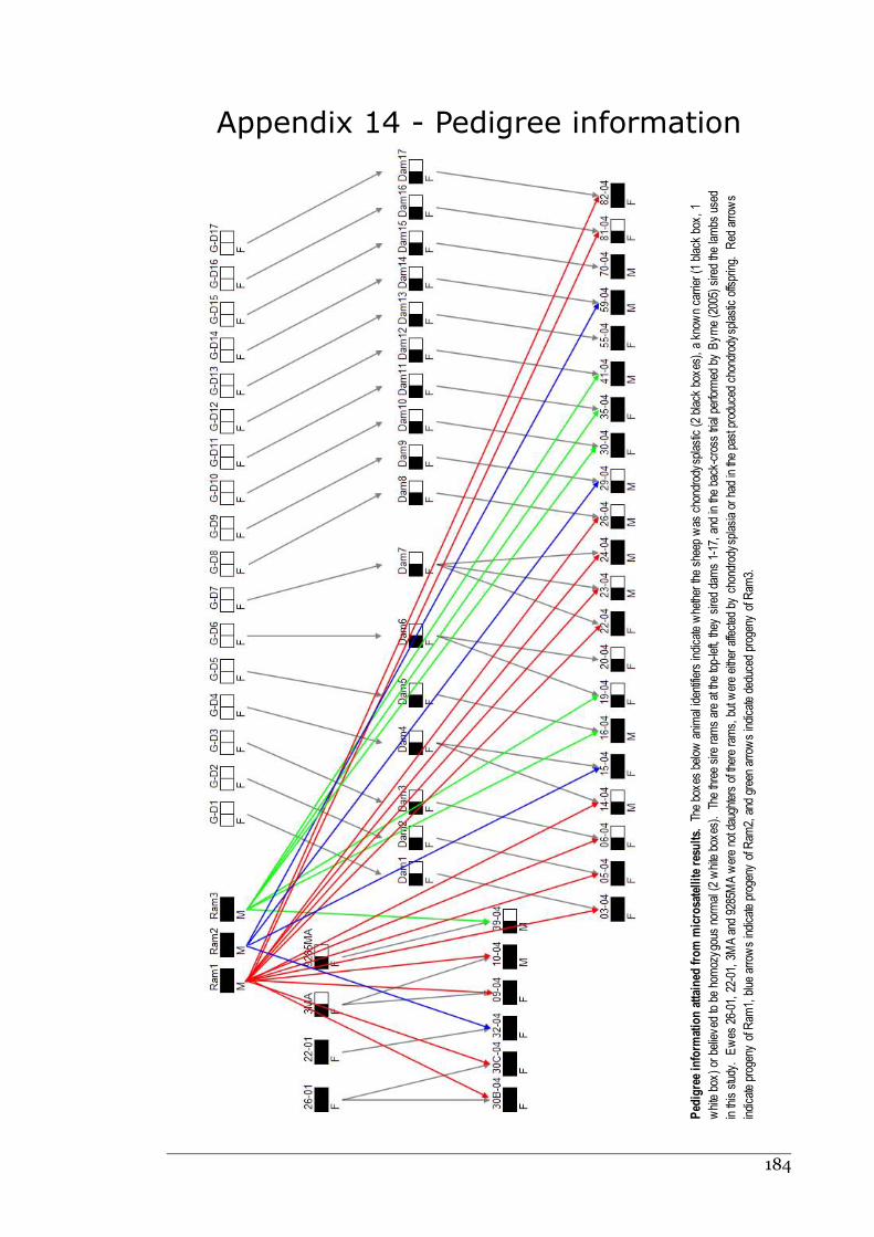
Appendix 14 - Pedigree information
184
Ped
igree inform
ation attained
from m
icrosatellite resu
lts. The boxes below
animal identifiers indicate w
hether the sheep was chondrody
splastic (2 black boxes), a known carrier (1 black box
, 1
white box
) or believed to be homozygous normal (2 white box
es). The three sire ram
s are at the top-left, they
sired dams 1-17, and in the back-cross trial perform
ed by Byrne (2005) sired the lambs used
in this study. Ewes 26-01, 22-01, 3MA and 9285M
A w
ere not daughters of there ram
s, but w
ere either affected by chondrody
splasia or had in the past produced chondrodysplastic offspring. Red arrow
s
indicate progeny
of R
am1, blue arrows indicate progeny
of R
am2, and green arrow
s indicate deduced progeny
of R
am3.
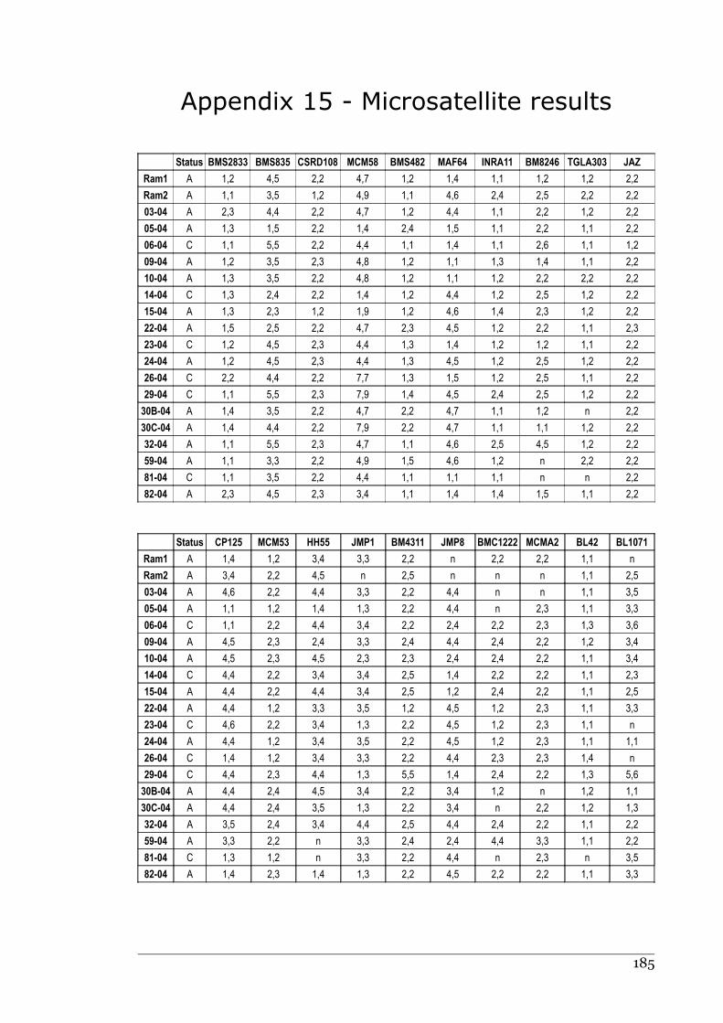
Appendix 15 - Microsatellite results
Status BMS2833 BMS835 CSRD108 MCM58 BMS482 MAF64 INRA11 BM8246 TGLA303 JAZ
Ram1 A 1,2 4,5 2,2 4,7 1,2 1,4 1,1 1,2 1,2 2,2
Ram2 A 1,1 3,5 1,2 4,9 1,1 4,6 2,4 2,5 2,2 2,2
03-04 A 2,3 4,4 2,2 4,7 1,2 4,4 1,1 2,2 1,2 2,2
05-04 A 1,3 1,5 2,2 1,4 2,4 1,5 1,1 2,2 1,1 2,2
06-04 C 1,1 5,5 2,2 4,4 1,1 1,4 1,1 2,6 1,1 1,2
09-04 A 1,2 3,5 2,3 4,8 1,2 1,1 1,3 1,4 1,1 2,2
10-04 A 1,3 3,5 2,2 4,8 1,2 1,1 1,2 2,2 2,2 2,2
14-04 C 1,3 2,4 2,2 1,4 1,2 4,4 1,2 2,5 1,2 2,2
15-04 A 1,3 2,3 1,2 1,9 1,2 4,6 1,4 2,3 1,2 2,2
22-04 A 1,5 2,5 2,2 4,7 2,3 4,5 1,2 2,2 1,1 2,3
23-04 C 1,2 4,5 2,3 4,4 1,3 1,4 1,2 1,2 1,1 2,2
24-04 A 1,2 4,5 2,3 4,4 1,3 4,5 1,2 2,5 1,2 2,2
26-04 C 2,2 4,4 2,2 7,7 1,3 1,5 1,2 2,5 1,1 2,2
29-04 C 1,1 5,5 2,3 7,9 1,4 4,5 2,4 2,5 1,2 2,2
30B-04 A 1,4 3,5 2,2 4,7 2,2 4,7 1,1 1,2 n 2,2
30C-04 A 1,4 4,4 2,2 7,9 2,2 4,7 1,1 1,1 1,2 2,2
32-04 A 1,1 5,5 2,3 4,7 1,1 4,6 2,5 4,5 1,2 2,2
59-04 A 1,1 3,3 2,2 4,9 1,5 4,6 1,2 n 2,2 2,2
81-04 C 1,1 3,5 2,2 4,4 1,1 1,1 1,1 n n 2,2
82-04 A 2,3 4,5 2,3 3,4 1,1 1,4 1,4 1,5 1,1 2,2
Status CP125 MCM53 HH55 JMP1 BM4311 JMP8 BMC1222 MCMA2 BL42 BL1071
Ram1 A 1,4 1,2 3,4 3,3 2,2 n 2,2 2,2 1,1 n
Ram2 A 3,4 2,2 4,5 n 2,5 n n n 1,1 2,5
03-04 A 4,6 2,2 4,4 3,3 2,2 4,4 n n 1,1 3,5
05-04 A 1,1 1,2 1,4 1,3 2,2 4,4 n 2,3 1,1 3,3
06-04 C 1,1 2,2 4,4 3,4 2,2 2,4 2,2 2,3 1,3 3,6
09-04 A 4,5 2,3 2,4 3,3 2,4 4,4 2,4 2,2 1,2 3,4
10-04 A 4,5 2,3 4,5 2,3 2,3 2,4 2,4 2,2 1,1 3,4
14-04 C 4,4 2,2 3,4 3,4 2,5 1,4 2,2 2,2 1,1 2,3
15-04 A 4,4 2,2 4,4 3,4 2,5 1,2 2,4 2,2 1,1 2,5
22-04 A 4,4 1,2 3,3 3,5 1,2 4,5 1,2 2,3 1,1 3,3
23-04 C 4,6 2,2 3,4 1,3 2,2 4,5 1,2 2,3 1,1 n
24-04 A 4,4 1,2 3,4 3,5 2,2 4,5 1,2 2,3 1,1 1,1
26-04 C 1,4 1,2 3,4 3,3 2,2 4,4 2,3 2,3 1,4 n
29-04 C 4,4 2,3 4,4 1,3 5,5 1,4 2,4 2,2 1,3 5,6
30B-04 A 4,4 2,4 4,5 3,4 2,2 3,4 1,2 n 1,2 1,1
30C-04 A 4,4 2,4 3,5 1,3 2,2 3,4 n 2,2 1,2 1,3
32-04 A 3,5 2,4 3,4 4,4 2,5 4,4 2,4 2,2 1,1 2,2
59-04 A 3,3 2,2 n 3,3 2,4 2,4 4,4 3,3 1,1 2,2
81-04 C 1,3 1,2 n 3,3 2,2 4,4 n 2,3 n 3,5
82-04 A 1,4 2,3 1,4 1,3 2,2 4,5 2,2 2,2 1,1 3,3
185
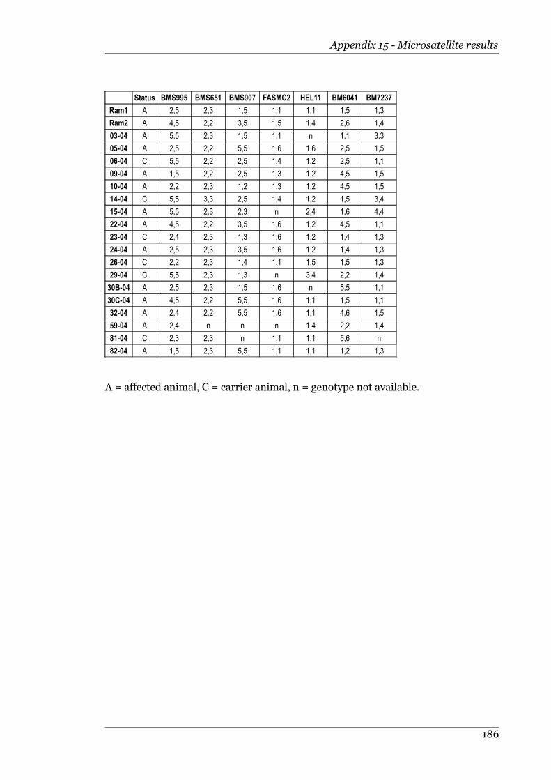
Appendix 15 - Microsatellite results
Status BMS995 BMS651 BMS907 FASMC2 HEL11 BM6041 BM7237
Ram1 A 2,5 2,3 1,5 1,1 1,1 1,5 1,3
Ram2 A 4,5 2,2 3,5 1,5 1,4 2,6 1,4
03-04 A 5,5 2,3 1,5 1,1 n 1,1 3,3
05-04 A 2,5 2,2 5,5 1,6 1,6 2,5 1,5
06-04 C 5,5 2,2 2,5 1,4 1,2 2,5 1,1
09-04 A 1,5 2,2 2,5 1,3 1,2 4,5 1,5
10-04 A 2,2 2,3 1,2 1,3 1,2 4,5 1,5
14-04 C 5,5 3,3 2,5 1,4 1,2 1,5 3,4
15-04 A 5,5 2,3 2,3 n 2,4 1,6 4,4
22-04 A 4,5 2,2 3,5 1,6 1,2 4,5 1,1
23-04 C 2,4 2,3 1,3 1,6 1,2 1,4 1,3
24-04 A 2,5 2,3 3,5 1,6 1,2 1,4 1,3
26-04 C 2,2 2,3 1,4 1,1 1,5 1,5 1,3
29-04 C 5,5 2,3 1,3 n 3,4 2,2 1,4
30B-04 A 2,5 2,3 1,5 1,6 n 5,5 1,1
30C-04 A 4,5 2,2 5,5 1,6 1,1 1,5 1,1
32-04 A 2,4 2,2 5,5 1,6 1,1 4,6 1,5
59-04 A 2,4 n n n 1,4 2,2 1,4
81-04 C 2,3 2,3 n 1,1 1,1 5,6 n
82-04 A 1,5 2,3 5,5 1,1 1,1 1,2 1,3
A = affected animal, C = carrier animal, n = genotype not available.
186
ReferencesABBOT, L. C., FINNELL, R. H., CHERNOFF, G. F., PARISH, S. M. & GAY, C.C. (1986). Crooked calf disease: A
histological and histochemical examination of eight affected calves. Veterinary Pathology, 23(6):734-40.
AGERHOLM, J. S., ANDERSEN, O., ALMSKOU, M. B., BENDIXEN, C., ARNBJERG, J., AAMAND, G. P., NIELSEN, U. S., PANITZ,F. & PETERSEN, A.H. (2004a). Evaluation of the inheritance of the complex vertebral malformationsyndrome by breeding studies. Acta Veterinaria Scandinavica, 45(3-4): 133-7.
AGERHOLM, J. S., ARNBJERG, J. & ANDERSEN, O. (2004b). Familial chondrodysplasia in Holstein calves. Journalof Veterinary Diagnostic Investigation, 16(4): 293-8.
AGERHOLM, J. S., BENDIXEN, C., ANDERSEN, O. & ARNBJERG, J. (2001). Complex vertebral malformation inHolstein calves. Journal of Veterinary Diagnostic Investigation, 13(4): 283-9.
AHMAD, M., UL HAQUE, M. F., AHMAD, W., ABBAS, H., HAQUE, S., KRAKOW, D., RIMOIN, D. L., LACHMAN, R. S. & COHN,D.H. (1998). Distinct, autosomal recessive form of spondyloepimetaphyseal dysplasia segregating inan inbred Pakistani kindred. American Journal of Medical Genetics, 78(5): 468-73.
AIGNER, T., RAU, T., NIEDERHAGEN, M., ZAUCKE, F., SCHMITZ, M., PÖHLS, U., STÖSS, H., RAUCH, A. & THIEL, C.T.(2007). Achondrogenesis Type IA (Houston-Harris): A still-unresolved molecular phenotype.Pediatric Developmental Pathology, 10(4): 328-34.
AIKAWA, T., SEGRE, G. V. & LEE, K. (2001). Fibroblast growth factor inhibits chondrocytic growth throughinduction of p21 and subsequent inactivation of cyclin E-Cdk2. Journal of Biological Chemistry,276(31): 29347-52.
AKIYAMA, H., LYONS, J. P., MORI-AKIYAMA, Y., YANG, X. H., ZHANG, R., ZHANG, Z. P., DENG, J. M., TAKETO, M. M.,NAKAMURA, T., BEHRINGER, R. R., MCCREA, P. D. & DE CROMBRUGGHE, B. (2004). Interactions betweenSOX9 and b-catenin control chondrocyte differentiation. Genes & Development, 18(9): 1072-87.
AL-GAZALI, L. I. (1993). The Schwartz-Jampel syndrome. Clinical Dysmorphology, 2(1): 47-54.AL-YAHYAEE, S. A. S., AL-KINDI, M. N., HABBAL, O. & KUMAR, D.S. (2003). Clinical and molecular analysis of
Grebe acromesomelic dysplasia in an Omani family. American Journal of Medical Genetics,121A(1): 9-14.
ALA-KOKKO, L., BALDWIN, C. T., MOSKOWITZ, R. W. & PROCKOP, D.J. (1990). Single base mutation in the type-IIprocollagen gene (COL2A1) as a cause of primary osteoarthritis associated with a mildchondrodysplasia. Proceedings of the National Academy of Sciences, 87(17): 6565-8.
ALEXANDER, G., STEVENS, D., MOTTERSHEAD, B. (1983). Problems in the accurate recording of lambing data.Australian Journal of Experimental Agriculture and Animal Husbandry, 23(123): 361-8.
ALINI, M., MATSUI, Y., DODGE, G. R. & POOLE, A.R. (1992). The extracellular matrix of cartilage in the growthplate before and during calcification - Changes in composition and degradation of type II collagen.Calcified Tissue International, 50(4): 327-35.
ALROY, A., ORGAD, U., DEGASPERI, R., RICHARD, R., WARREN, C. D., KNOWLES, K., THALHAMMER, J. G. & RAGHAVAN, S.S.(1992). Canine GM1-gangliosidosis. A clinical, morphologic, histochemical, and biochemicalcomparison of two different models. American Journal of Pathology, 140(3): 675-89.
AMLÖFF, J. (1961). On achondroplasia in the dog. Zentralblatt Fur Veterinaermedizin, 8: 43-56.ANDERSON, C. E., SILLENCE, D. O., LACHMAN, R. S., TOOMEY, K., BULL, M., DORST, J. & RIMOIN, D.L. (1982).
Spondylometepiphyseal dysplasia, Strudwick type. American Journal of Medical Genetics, 13A(3):243–256.
ANNUNEN, S., KORKKO, J., CZARNY, M., WARMAN, M. L., BRUNNER, H. G., KAARIAINEN, H., MULLIKEN, J. B.,TRANEBJAERG, L., BROOKS, D. G., COX, G. F., CRUYSBERG, J. R., CURTIS, M. A., DAVENPORT, S. L. H., FRIEDRICH,C. A., KAITILA, I., KRAWCZYNSKI, M. R., LATOS-BIELENSKA, A., MUKAI, S., OLSEN, B. R., SHINNO, N., SOMER, M.,VIKKULA, M., ZLOTOGORA, J., PROCKOP, D. J. & ALA-KOKKO, L. (1999a). Splicing mutations of 54-bp exonsin the COL11A1 gene cause Marshall syndrome, but other mutations cause overlappingMarshall/Stickler phenotypes. American Journal of Human Genetics, 65(4): 974-83.
ANNUNEN, S., PAASALITA, P., LOHINLVA, J., PERALA, M., PIHLAJAMAA, T., KARPPINEN, J., TERVONEN, O., KROGER, H.,LAHDE, S., VANHARANTA, H., RYHANEN, L., GORING, H. H. H., OTT, J., PROCKOP, D. J. & ALA-KOKKO, L.(1999b). An allele of COL9A2 associated with intervertebral disc disease. Science, 285(5426):409-11.
ARBISSER, A. I., DONNELLY, K. A., SCOTT JR., C. I., DI FERRANTE, N. M., SINGH, J., STEVENSON, R. E., AYLSWORTH, A. S.& HOWELL, R.R. (1977). Morquio-like syndrome with beta-galactosidase deficiency and normalhexosamine sulfatase activity: Mucopolysaccharidosis IV B. American Journal of Medical Genetics,1A(2): 195-205.
ARCHER, C. W. (1994). Skeletal development and osteoarthritis. Annals of the Rheumatic Diseases, 53(10):624-30.
187

ARCHER, C. W., MORRISON, E. H., BAYLISS, M. T. & FERGUSON, M.W.J. (1996). The development of articularcartilage. 2. The spatial and temporal patterns of glycosaminoglycans and small leucine-richproteoglycans. Journal of Anatomy, 189(1): 23-35.
ARIKAWA-HIRASAWA, E., WILCOX, W. R. & YAMADA, Y. (2001a). Dyssegmental dysplasia, Silverman-Handmakertype: Unexpected role of perlecan in cartilage development. American Journal of Medical Genetics,106A(4): 254-7.
ARIKAWA-HIRASAWA, E., WILCOX, W. R., LE, A. H., SILVERMAN, N., GOVINDRAJ, P., HASSELL, J. R. & YAMADA, Y.(2001b). Dyssegmental dysplasia, Silverman-Handmaker type, is caused by functional nullmutations of the perlecan gene. Nature Genetics, 27(4): 431-4.
AROCH, I., OFRI, R. & AIZENBERG, I. (1996). Haematological, ocular and skeletal abnormalities in a Samoyedfamily. Journal of Small Animal Practice, 37(7): 333-9.
ASAMURA, K., ABE, S., IMAMURA, Y., ASZODI, A., SUZUKI, N., HASHIMOTO, S., TAKUMI, Y., HAYASHI, T., FASSLER, R.,NAKAMURA, Y. & USAMI, S. (2005). Type IX collagen is crucial for normal hearing. Neuroscience,132(2): 493-500.
ASH, W. J. (1966). Micromelia - A lethal mutation in white Pekin ducks. Journal of Heredity, 57(4): 137-41.ASH, W. J., MICHAEL, B. A. & FEDELE, L. (1970). Parakeratosis in the micromelic syndrome of Anas
platyrhynchos. Poultry Science, 49(5): 1365.ASMUNDSON, V. S. (1939). Some factors affecting hatchability of eggs (abstract). Poultry Science, 18: 399.ASMUNDSON, V. S. (1942). An inherited micromelia in the domestic fowl. Journal of Heredity, 33(9): 328-30.ASMUNDSON, V. S. (1944). Inherited shortening of the long bones in the turkey. Journal of Heredity, 35(10):
295-301.ASZODI, A., HUNZIKER, E. B., BRAKEBUSCH, C. & FASSLER, R. (2003). b1 integrins regulate chondrocyte rotation,
G1 progression, and cytokinesis. Genes & Development, 17(19): 2465-79.AUSTRALIAN SHEEP GENE MAPPING WEB SITE (2004). Sheep Predicted Map - version 1.4.
http://www1.angis.org.au/jmaddox/cgi-bin/shp_predict_map.pl, accessed June 2006AUSTRALIAN SHEEP GENE MAPPING WEB SITE (2006). Markers: Query form. www1.angis.org.au/jmaddox/cgi-
bin/marker.pl, accessed June 2006AYME, S. & PREUS, M. (1984). The Marshall and Stickler syndromes: Objective rejection of lumping. Journal
of Medical Genetics, 21(1): 34-8.BAKER, J. R., ASHTON, D. G., JONES, D. M. & NOODLE, B.A. (1979). Four cases of chondrodystrophy in Fallow
deer. Veterinary Record, 104(20): 450-3.BAKER, M. L., BLUNN, C. T. & OLOUFA, M.M. (1950). Stumpy, a recessive achondroplasia in Shorthorn cattle.
Journal of Heredity, 41(9): 243-6.BAKER, M. L., BLUNN, C. T. & PLUM, M. (1951). "Dwarfism" in Aberdeen-Angus cattle. Journal of Heredity,
42(3): 141-3.BALCI, S., ALTINOK, G., TEKSEN, F. & ERYILMAZ, M. (2003). A 34-week-old male fetus with short rib polydactyly
syndrome (SRPS) type I (Saldino-Noonan) with pancreatic cysts. Turkish Journal of Pediatrics,45(2): 174-8.
BALLHAUSEN, D., BONAFE, L., TERHAL, P., UNGER, S. L., BELLUS, G., CLASSEN, M., HAMEL, B. C., SPRANGER, J., ZABEL,B., COHN, D. H., COLE, W. G., HECHT, J. T. & SUPERTI-FURGA, A. (2003). Recessive multiple epiphysealdysplasia (RMED): Phenotype delineation in eighteen homozygotes for DTDST mutation R279W.Journal of Medical Genetics, 40(1): 65-71.
BALLO, R., BRIGGS, M. D., COHN, D. H., KNOWLTON, R. G., BEIGHTON, P. H. & RAMESAR, R.S. (1997). Multipleepiphyseal dysplasia, ribbing type: A novel point mutation in the COMP gene in a South Africanfamily. American Journal of Medical Genetics, 68(4): 396–400.
BALLOCK, R. T. & O'KEEFE, R.J. (2003). Physiology and pathophysiology of the growth plate. Birth DefectsResearch part C: Embryo Today, 69(2): 123-43.
BALLOCK, R. T., HEYDEMANN, A., WAKEFIELD, L. M., FLANDERS, K. C., ROBERTS, A. B. & SPORN, M.B. (1993). TGFb-1prevents hypertrophy of epiphyseal chondrocytes - Regulation of gene expression for cartilagematrix proteins and metalloproteases. Developmental Biology, 158(2): 414-29.
BARGMAN, G. J., MACKLER, B. & SHEPHERD, T.H. (1972). Studies of oxidative energy deficiency I.Achondroplasia in the rabbit. Archives of Biochemistry and Biophysics, 150(1): 137-46.
BARRY, M. R. & MURPHY, W.J.B. (1964). Acorn calves in the Albury district of New South Wales. AustralianVeterinary Journal, 40: 195-8.
BARSOUM, S. C., CALLAHAN, H. M., ROBINSON, K. & CHANG, P.L. (2000). Canine models for human geneticneurodegenerative diseases. Progress in Neuropsychopharmacology & Biological Psychiatry,24(5): 811-23.
BASTEPE, M., RAAS-ROTHSCHILD, A., SILVER, J., WEISSMAN, I., WIENTROUB, S., JÜPPNER, H. & GILLIS, D. (2004). Aform of Jansen's metaphyseal chondrodysplasia with limited metabolic and skeletal abnormalitiesis caused by a novel activating parathyroid hormone (PTH)/PTH-related peptide receptor mutation.Journal of Clinical Endocrinology & Metabolism, 89(7): 3595-600.
188

BATEMAN, J. F., FREDDI, S., MCNEIL, R., THOMPSON, E., HERMANNS, P., SAVARIRAYAN, R. & LAMANDE, S.R. (2004).Identification of four novel COL10A1 missense mutations in Schmid metaphyseal chondrodysplasia:Further evidence that collagen X NC1 mutations impair trimer assembly. Human Mutation, 23(4):396-402.
BATEMAN, J. F., FREDDI, S., NATTRASS, G. & SAVARIRAYAN, R. (2003). Tissue-specific RNA surveillance?Nonsense-mediated mRNA decay causes collagen X haploinsufficiency in Schmid metaphysealchondrodysplasia cartilage. Human Molecular Genetics, 12(3): 217-25.
BAYLISS, M. T., OSBORNE, D., WOODHOUSE, S. & DAVIDSON, C. (1999). Sulfation of chondroitin sulfate in humanarticular cartilage. The effect of age, topographical position, and zone of cartilage on tissuecomposition. Journal of Biological Chemistry, 274(22): 15892-900.
BAYRAK, I. K., NURAL, M. S. & DIREN, H.B. (2005). Dyggve-Melchior-Clausen syndrome without mentalretardation (Smith-McCort dysplasia). Diagnostic & Interventional Radiology, 11(3): 163-5.
BEACHLEY, M. C. & GRAHAM, F.H. (1973). Hypochondroplastic dwarfism (endochondral chondrodystrophy) ina dog. Journal of the American Veterinary Medical Association, 163(3): 283-4.
BEALES, P. L., BLAND, E., TOBIN, J. L., BACCHELLI, C., TUYSUZ, B., HILL, J., RIX, S., PEARSON, C. G., KAI, M., HARTLEY,J., JOHNSON, C., IRVING, M., ELÇIOĞLU, N., WINEY, M., TADA, M. & SCAMBLER, P.J. (2007). IFT80, whichencodes a conserved intraflagellar transport protein, is mutated in Jeune asphyxiating thoracicdystrophy. Nature Genetics, 39(6): 727-9.
BECK, M., ROUBICEK, M., ROGERS, J. G., NAUMOFF, P. & SPRANGER, J. (1983). Heterogeneity of metatropicdysplasia. European Journal of Pediatrics, 140(3): 231-7.
BECKER, R., WEGNER, R. D., KUNZE, J., RUNKEL, S., VOGEL, M. & ENTEZAMI, M. (2000). Clinical variability ofLarsen syndrome: Diagnosis in a father after sonographic detection of a severely affected fetus.Clinical Genetics, 57(2): 148-50.
BEEMER, F. A., LANGER JR., L. O., KLEP-DE PATER, J. M., HEMMES, A. M., BYLSMA, J. B., PAULI, R. M., MYERS, T. L. &HAWS, C.C. (1983). A new short rib syndrome: Report of two cases. American Journal of MedicalGenetics, 14(1): 115-23.
BEESLEY, C. E., MEANEY, C. A., GREENLAND, G., ADAMS, V., VELLODI, A., YOUNG, E. P. & WINCHESTER, B.G. (2001).Mutational analysis of 85 mucopolysaccharidosis type I families: Frequency of known mutations,identification of 17 novel mutations and in vitro expression of missense mutations. HumanGenetics, 109(5): 503-11.
BEEVER, J. E. & COCKETT, N.E. (2001). Screening for the molecular defect causing Spider Lamb syndrome insheep. US Patent No, 6,306,591 B1.
BEEVER, J. E., SMIT, M. A., MEYERS, S. N., HADFIELD, T. S., BOTTEMA, C., ALBRETSEN, J. & COCKETT, N.E. (2006). Asingle-base change in the tyrosine kinase II domain of ovine FGFR3 causes hereditarychondrodysplasia in sheep. Animal Genetics, 37(1): 66-71.
BÉLANGER, L. F. (1959). Observations on the manifestations of osteolathyrism in the chick. Journal of Bone& Joint Surgery-British Volume, 41B: 581-9.
BELIN, V., CUSIN, V., VIOT, G., GIRLICH, D., TOUTAIN, A., MONCLA, A., VEKEMANS, M., LE MERRER, M., MUNNICH, A. &CORMIER-DAIRE, V. (1998). SHOX mutations in dyschondrosteosis (Léri-Weill Syndrome). NatureGenetics, 19(1): 67-9.
BELL, L., JURILOFF, D. M. & HARRIS, M.J. (1986). A new mutation at the cmd locus in the mouse. Journal ofHeredity, 77(3): 205-6.
BELLUCCI, G. & SEEDHOM, B.B. (2001). Mechanical behaviour of articular cartilage under tensile cyclic load.Rheumatology (Oxford), 40(12): 1337-45.
BELLUS, G. A., BAMSHAD, M. J., PRZYLEPA, K. A., DORST, J., LEE, R. R., HURKO, O., JABS, E. W., CURRY, J. C., WILCOX,W. R., LACHMAN, R. S., RIMOIN, D. L. & FRANCOMANO, C.A. (1999). Severe achondroplasia withdevelopmental delay and acanthosis nigricans (SADDAN): Phenotypic analysis of a new skeletaldysplasia caused by a Lys650Met mutation in fibroblast growth factor receptor 3. AmericanJournal of Medical Genetics, 85A(1): 53-65.
BELLUS, G. A., HEFFERON, T. W., ORTIZ DE LUNA, R. I., HECHT, J. T., HORTON, W. A., MACHADO, M., KAITILA, I. &MCINTOSH, I. (1995a). Achondroplasia is defined by recurrent G380R mutations of FGFR3.American Journal of Human Genetics, 56(2): 368-73.
BELLUS, G. A., MCINTOSH, I., SMITH, E. A., AYLSWORTH, A. S. & KAITILA, I. (1995b). A recurrent mutation in thetyrosine kinase domain of fibroblast growth factor 3 causes hypochondroplasia. Nature Genetics,10(3): 357-9.
BERGER, J. & INNES, J.R.M. (1948). "Bull-dog" calves (chondrodystrophy, achondroplasia) in a Friesian herd.Veterinary Record, 60(6): 57-8.
BI, W. M., DENG, J. M., ZHANG, Z. P., BEHRINGER, R. R. & DE CROMBRUGGHE, B. (1999). SOX9 is required forcartilage formation. Nature Genetics, 22(1): 85-9.
BI, W. M., HUANG, W. D., WHITWORTH, D. J., DENG, J. M., ZHANG, Z. P., BEHRINGER, R. R. & DE CROMBRUGGHE, B.(2001). Haploinsufficiency of Sox9 results in defective cartilage primordia and premature skeletalmineralization. Proceedings of the National Academy of Sciences, 98(12): 6698-703.
189
BICKNELL, L. S., FARRINGTON-ROCK, C., SHAFEGHATI, Y., RUMP, P., ALANAY, Y., ALEMBIK, Y., AL-MADANI, N., FIRTH, H.,KARIMI-NEJAD, M. H., KIM, C. A., LEASK, K., MAISENBACHER, M., MORAN, E., PAPPAS, J. G., PRONTERA, P., DERAVEL, T., FRYNS, J., SWEENEY, E., FRYER, A., UNGER, S., WILSON, L. C., LACHMAN, R. S., RIMOIN, D. L., COHN,D. H., KRAKOW, D. & ROBERTSON, S.P. (2007). A molecular and clinical study of Larsen syndromecaused by mutations in FLNB. Journal of Medical Genetics, 44(2): 89-98.
BICKNELL, L. S., MORGAN, T., BONAFÉ, L., WESSELS, M. W., BIALER, M. G., WILLEMS, P. J., COHN, D. H., KRAKOW, D. &ROBERTSON, S.P. (2005). Mutations in FLNB cause boomerang dysplasia. Journal of MedicalGenetics, 42(7): e43.
BINGEL, S. A. & SANDE, R.D. (1982). Chondrodysplasia in the Norwegian Elkhound. American Journal ofPathology, 107(2): 219-29.
BINGEL, S. A. & SANDE, R.D. (1994). Chondrodysplasia in five Great Pyrenees. Journal of the AmericanVeterinary Medical Association, 205(6): 845-8.
BINGEL, S. A., SANDE, R. D. & WIGHT, T.D. (1985). Chondrodysplasia in the Alaskan Malamute.Characterization of proteoglycans dissociatively extracted from dwarf growth plates. LaboratoryInvestigation, 53(4): 479-85.
BINGEL, S. A., SANDE, R. D. & WIGHT, T.D. (1986). Undersulfated chondroitin sulfate in cartilage from aminiature poodle with spondylo-epiphyseal dysplasia. Connective Tissue Research, 15(4): 283-302.
BIRKENMEIER, E. H., DAVISSON, M. T., BEAMER, W. G., GANSCHOW, R. E., VOGLER, C. A., GWYNN, B., LYFORD, K. A.,MALTAIS, L. M. & WAWRZYNIAK, C.J. (1989). Murine mucopolysaccharidosis type VII - Characterizationof a mouse with beta-glucuronidase deficiency. Journal of Clinical Investigation, 83(4): 1258-66.
BODEN, S. D., KAPLAN, F. S., FALLON, M. D., RUDDY, R., BELIK, J., ANDAY, E., ZACKAI, E. & ELLIS, J. (1987).Metatropic dwarfism - Uncoupling of endochondral and perichondral growth. Journal of Bone &Joint Surgery-American Volume, 69A(2): 174-84.
BOGART, R. (1946). Inheritance and physiology of dwarfism in sheep (abstract). Genetics, 31(2): 211-2.BOHM, N., FUKUDA, M., STAUDT, R. & HELWIG, H. (1978). Chondroectodermal dysplasia (Ellis-Van Creveld
syndrome) with dysplasia of renal medulla and bile-ducts. Histopathology, 2(4): 267-81.BOLZE, M. S., REEVES, R. D., LINDBECK, F. E., KEMP, S. F. & ELDERS, M.J. (1985). Influence of manganese on
growth, somatomedin and glycosaminoglycan metabolism. Journal of Nutrition, 115(3): 352-8.BONAVENTURE, J., COHEN-SOLAL, L., RITVANIEMI, P., VAN MALDERGEM, L., KADHOM, N., DELEZOIDE, A. L., MAROTEAUX, P.,
PROCKOP, D. J. & ALA-KOKKO, L. (1995). Substitution of aspartic acid for glycine at position 310 in typeII collagen produces achondrogenesis II, and substitution of serine at position 805 produceshypochondrogenesis: Analysis of genotype-phenotype relationships. Biochemical Journal, 307(3):823-30.
BÖNNEMANN, C. G., COX, G. F., SHAPIRO, F., WU, J. J., FEENER, C. A., THOMPSON, T. G., ANTHONY, D. C., EYRE, D. R.,DARRAS, B. T. & KUNKEL, L.M. (2000). A mutation in the alpha 3 chain of type IX collagen causesautosomal dominant multiple epiphyseal dysplasia with mild myopathy. Proceedings of theNational Academy of Sciences, 97(3): 1212-7.
BONUCCI, E., DEL MARCO, A. & NICOLETTI, B. (1976). Histological and histochemical investigations ofachondroplastic mice: A possible model of human achondroplasia. Growth, 40(3): 241-51.
BOOCOCK, G. R. B., MORRISON, J. A., POPOVIC, M., RICHARDS, N., ELLIS, L., DURIE, P. R. & ROMMENS, J.M. (2003).Mutations in SBDS are associated with Shwachman-Diamond syndrome. Nature Genetics, 33(1):97-101.
BOROCHOWITZ, Z. U., SCHEFFER, D., ADIR, V., DAGONEAU, N., MUNNICH, A. & CORMIER-DAIRE, V. (2004). Spondylo-epi-metaphyseal dysplasia (SEMD) matrilin 3 type: Homozygote matrilin 3 mutation in a novelform of SEMD. (Letter). Journal of Medical Genetics, 41(5): 366-72.
BOROCHOWITZ, Z., BERANT, M. & KRISTAL, H. (1988a). Spondylometaphyseal dysplasia: Further hetergeneity.Skeletal Radiology, 17(3): 181-6.
BOROCHOWITZ, Z., LACHMAN, R., ADOMAIN, G. E., SPEAR, G., JONES, K. & RIMOIN, D.L. (1988b). Achondrogenesistype I: Delineation of further heterogeneity and identification of two distinct subgroups. Journal ofPediatrics, 112(1): 23-31.
BOROCHOWITZ, Z., ORNOY, A., LACHMAN, R. & RIMOIN, D.L. (1986). Achondrogenesis-II/hypochondrogenesis -Variability versus heterogeneity. American Journal of Medical Genetics, 24(2): 273-88.
BOVARD, K. P. & HAZEL, L.N. (1963). Growth patterns in snorter dwarf and normal Hereford calves. Journalof Animal Science, 22(1): 188-96.
BOVARD, K. P. & PROIDE, B.M. (1965). Snorter dwarfism in an Angus inbred line. Journal of Heredity, 56(5):243-6.
BOWDEN, D. M. (1970). Achondroplasia in Holstein-Friesian cattle. Journal of Heredity, 61(4): 163-4.BRANDT, G. W. (1941). Achondroplasia in calves. Journal of Heredity, 32(6): 183-6.BRAVERMAN, N., LIN, P., MOEBIUS, F. F., OBIE, C., MOSER, A., GLOSSMANN, H., WILCOX, W. R., RIMOIN, D. L., SMITH,
M., KRATZ, L., KELLEY, R. I. & VALLE, D. (1999). Mutations in the gene encoding 3 beta-hydroxysteroid-delta(8),delta(7)-isomerase cause X-linked dominant Conradi-Hunermann syndrome. NatureGenetics, 22(3): 291-4.
190
BRAVERMAN, N., STEEL, G., OBIE, C., MOSER, A., MOSER, H., GOULD, S. J. & VALLE, D. (1997). Human PEX7encodes the peroxisomal PTS2 receptor and is responsible for rhizomelic chondrodysplasiapunctata. Nature Genetics, 15(4): 369-76.
BRENIG, B., BAUMGARTNER, B. G., KRIEGESMANN, B., HABERMANN, F., FRIES, R. & SWALVE, H.H. (2003). Molecularcloning, mapping, and functional analysis of the bovine sulfate transporter SLC26a2 gene. Gene,319: 161-6.
BREUR, G. J., FARNUM, C. E., PADGETT, G. A. & WILSMAN, N.J. (1992). Cellular basis of decreased rate oflongitudinal growth of bone in pseudoachondroplastic dogs. Journal of Bone & Joint Surgery-American Volume, 74A(4): 516-28.
BREUR, G. J., TURGAI, J., VANENKEVORT, B. A., FARNUM, C. E. & WILSMAN, N.J. (1994). Stereological and serialsection analysis of chondrocytic enlargement in the proximal tibial growth plate of the rat.Anatomical Record, 239(3): 255-68.
BREUR, G. J., ZERBE, C. A., SLOCOMBE, R. F., PADGETT, G. A. & BRADEN, T.D. (1989). Clinical, radiographic,pathologic, and genetic features of osteochondrodysplasia in Scottish Deerhounds. Journal of theAmerican Veterinary Medical Association, 195(5): 606-12.
BRITES, P., MOTLEY, A. M., GRESSENS, P., MOOYER, P. A. W., PLOEGAERT, I., EVERTS, V., EVRARD, P., CARMELIET, P.,DEWERCHIN, M., SCHOONJANS, L., DURAN, M., WATERHAM, H. R., WANDERS, R. J. A. & BAES, M. (2003).Impaired neuronal migration and endochondral ossification in PEX7 knockout mice: A model forrhizomelic chondrodysplasia punctata. Human Molecular Genetics, 12(18): 2255-67.
BRODIE, S. G., KITOH, H., LACHMAN, R. S., NOLASCO, L. M., MEKIKIAN, P. B. & WILCOX, W.R. (1999). Platyspondyliclethal skeletal dysplasia, San Diego type, is caused by FGFR3 mutations. American Journal ofMedical Genetics, 84(5): 476-80.
BROWN, K. S., CRANLEY, R. E., GREENE, R., KLEINMAN, H. K. & PENNYPACKER, J.P. (1981). Disproportionatemicromelia (Dmm) - An incomplete dominant mouse dwarfism with abnormal cartilage matrix.Journal of Embryology & Experimental Morphology, 62: 165-82.
BUNGE, S., KLEIJER, W. J., STEGLICH, C., BECK, M., SCHWINGER, E. & GAL, A. (1995). Mucopolysaccharidosis type I:Identification of 13 novel mutations of the alpha-L-iduronidase gene. Human Mutation, 6(1): 91-4.
BUNGE, S., KLEIJER, W. J., TYLKI-SZYMANSKA, A., STEGLICH, C., BECK, M., TOMATSU, S., FUKUDA, S., POORTHUIS, B. J. H.M., CZARTORYSKA, B., ORII, T. & GAL, A. (1997). Identification of 31 novel mutations in the N-acetylgalactosamine-6-sulfatase gene reveals excessive allelic heterogeneity among patients withMorquio A syndrome. Human Mutation, 10(3): 223-32.
BURBIDGE, H. M. & PFEIFFER, D.U. (1998). The accuracy and reliability of linear measurements of the ulna foranthropometrical studies in dogs. Research in Veterinary Science, 65(1): 53-7.
BYRNE, T. J. (2005). Investigation into the inheritance and biochemistry of chondrodysplasia in Texelsheep. Masterate Thesis, Massey University, New Zealand
CAI, G., NAKAYAMA, M., HIRAKI, Y. & OZONO, K. (1998). Mutational analysis of the DTDST gene in a fetus withachondrogenesis type 1B. American Journal of Medical Genetics, 78A(1): 58-60.
CAMERA, G., SCARANO, G., TRONCI, A., LA CAVA, G. & MASTROIACOVO, P. (1991). "Snail-like pelvis"chondrodysplasia: A further case report. American Journal of Medical Genetics, 40(4): 513-4.
CAPRETTE, D. R. (1995). Hartree-Lowry and Modified Lowry Protein Assays.http://www.ruf.rice.edu/~bioslabs/methods/protein/lowry.html, accessed Nov. 2006
CARRIG, C. B., GRANDAGE, J. & SEAWRIGHT, A.A. (1981). Disproportionate dwarfism in two bovine siblings.Veterinary Radiology, 22(2): 78-82.
CARRIG, C. B., MACMILLAN, A. & BRUNDAGE, S. (1977). Retinal dysplasia associated with skeletal abnormalitiesin Labrador retrievers. Journal of the American Veterinary Medical Association, 170(1): 49-57.
CARRIG, C. B., SPONENBERG, D. P., SCHMIDT, G. M. & TVEDTEN, H.W. (1988). Inheritance of associated ocular andskeletal dysplasia in Labrador Retrievers. Journal of the American Veterinary Medical Association,193(10): 1269-72.
CARTER, D. R. (1987). Mechanical loading history and skeletal biology. Journal of Biomechanics, 20(11-12):1095-109.
CASAL, M. & HASKINS, M. (2005). Large animal models and gene therapy. European Journal of HumanGenetics, 14(3): 266–272.
CASKEY, C. D., NORRIS, L. C. & HEUSER, G.F. (1944). A chronic congenital ataxia in chicks due to manganesedeficiency in the maternal diet. Poultry Science, 23: 516-20.
CAVANAGH, J. A. L., TAMMEN, I., WINDSOR, P. A., BATEMAN, J. F., SAVARIRAYAN, R., NICHOLAS, F. W. & RAADSMA, H.W.(2007). Bulldog dwarfism in Dexter cattle is caused by mutations in ACAN. Mammalian Genome,18(10): 808-14.
CETTA, G., ROSSI, A., BURGIO, G. R. & BELUFFI, G. (1997). Diastrophic dysplasia sulfate transporter (DTDST)gene is not involved in pseudodiastrophic dysplasia. American Journal of Medical Genetics,73A(4): 493-4.
CHAMBERS, D., WHATLEY, J. A. J. & STEPHENS, F.F. (1954). The inheritance of dwarfism in a comprest Herefordherd. Journal of Animal Science, 13: 956-7.
191
CHAN, D., COLE, W. G., CHOW, C. W., MUNDLOS, S. & BATEMAN, J.F. (1995a). A COL2A1 mutation inachondrogenesis type-II results in the replacement of type-II collagen by type-I and type-IIIcollagens in cartilage. Journal of Biological Chemistry, 270(4): 1747-53.
CHAN, D., COLE, W. G., ROGERS, J. G. & BATEMAN, J.F. (1995b). Type X collagen multimer assembly in vitro isprevented by a Gly(618) to Val mutation in the alpha-1(X) NC1 domain resulting in Schmidmetaphyseal chondrodysplasia. Journal of Biological Chemistry, 270(9): 4558-62.
CHAN, D., TAYLOR, T. K. F. & COLE, W.G. (1993). Characterization of an arginine-789 to cysteine substitutionin alpha-1(II) collagen chains of a patient with spondyloepiphyseal dysplasia. Journal of BiologicalChemistry, 268(20): 15238-45.
CHAN, D., WENG, Y. M., GRAHAM, H. K., SILLENCE, D. O. & BATEMAN, J.F. (1998). A nonsense mutation in thecarboxyl-terminal domain of type X collagen causes haploinsufficiency in Schmid metaphysealchondrodysplasia. Journal of Clinical Investigation, 101(7): 1490-9.
CHANG, C. C., WARREN, D. S., SACKSTEDER, K. A. & GOULD, S.J. (1999). PEX12 interacts with PEX5 and PEX10and acts downstream of receptor docking in peroxisomal matrix protein import. Journal of CellBiology, 147(4): 761-73.
CHANG, J., JUNG, J., OH, S., LEE, S., KIM, G., KIM, H., KWEON, O., YOON, J. & CHOI, M. (2007).Osteochondrodysplasia in three Scottish Fold cats. Journal of Veterinary Science, 8(3): 307-9.
CHANG, S. C., HOANG, B., THOMAS, J. T., VUKICEVIC, S. & LUYTEN, F.P. (1994). Cartilage-derived morphogenicproteins. Journal of Biological Chemistry, 269(45): 28227-34.
CHANG, T. K. (1949). Morphological study on the skeleton of Ancon sheep. Growth, 13: 269-97.CHARROW, J. & POZNANSKI, A.K. (1984). The Jansen type of metaphyseal chondrodysplasia: Confirmation of
dominant inheritance and review of radiographic manifestations in the newborn and adult.American Journal of Medical Genetics, 18A(2): 321-7.
CHEN, H., DEERE, M., HECHT, J. T. & LAWLER, J. (2000). Cartilage oligomeric matrix protein is a calciumbinding protein, and a mutation in its type 3 repeats causes conformational changes. Journal ofBiological Chemistry, 275(34): 26538-44.
CLARKE, J. T. R., WILSON, P. J., MORRIS, C. P., HOPWOOD, J. J., RICHARDS, R. I., SUTHERLAND, G. R. & RAY, P.N.(1992). Characterization of a deletion at Xq27-q28 associated with unbalanced inactivation of thenonmutant X chromosome. American Journal of Human Genetics, 51(2): 316-22.
COCKETT, N. E. (2006). The sheep genome. In: Genome Dynamics, Vol. 2: Vertebrate Genomes. J. Volff(Ed.), (pp. 79-85). Karger, Basel.
COCKETT, N. E., SHAY, T. L., BEEVER, J. E., NIELSEN, D., ALBRETSEN, J., GEORGES, M., PETERSON, K., STEPHENS, A.,VERNON, W., TIMOFEEVSKAIA, O., SOUTH, S., MORK, J., MACIULIS, A. & BUNCH, T.D. (1999). Localization ofthe locus causing Spider Lamb syndrome to the distal end of ovine chromosome 6. MammalianGenome, 10(1): 35-8.
COHEN, M. M. (2006). The new bone biology: Pathologic, molecular, and clinical correlates. AmericanJournal of Medical Genetics, 140A(23): 2646-706.
COHN, D. H. (2001). Defects in extracellular matrix structural proteins in the osteochondrodysplasias. In:The molecular basis of skeletogenesis, (pp. 195-212). Wiley, Chichester (Novartis FoundationSymposium 232).
COHN, D. H., EHTESHAM, N., KRAKOW, D., UNGER, S., SHANSKE, A., REINKER, K., POWELL, B. R. & RIMOIN, D.L.(2003). Mental retardation and abnormal skeletal development (Dyggve-Melchior-Clausendysplasia) due to mutations in a novel, evolutionarily conserved gene. American Journal of HumanGenetics, 72(2): 419-28.
COLLINS, W. M., ABPLANALP, H. & YOSHIDA, S. (1968). Early embryonic chondrodystrophy in Japanese quail.Journal of Heredity, 59(4): 248-50.
COOPER, C. P. & HALL, C.M. (1982). Lethal short-rib polydactyly syndrome of the Majewski type: A report ofthree cases. Radiology, 144(3): 513-7.
COOPER, S. C., FLAITZ, C. M., JOHNSTON, D. A., LEE, B. & HECHT, J.T. (2001). A natural history of cleidocranialdysplasia. American Journal of Medical Genetics, 104A(1): 1-6.
CORMIER-DAIRE, V., DELEZOIDE, A. L., PHILIP, N., MARCORELLES, P., CASAS, K., HILLION, Y., FAIVRE, L., RIMOIN, D. L.,MUNNICH, A., MAROTEAUX, P. & LE MERRER, M. (2003). Clinical, radiological, and chondro-osseousfindings in opsismodysplasia: Survey of a series of 12 unreported cases. Journal of MedicalGenetics, 40(3): 195-200.
CRARY, D. D. & SAWIN, P.B. (1952). A second recessive achondroplasia in the domestic rabbit. Journal ofHeredity, 43(6): 254-9.
CRARY, D. D., SAWIN, P. B. & ATKINSON, N. (1958). Morphogenetic studies of the rabbit. XXI. The nature ofdisproportionate dwarfism induced by the Da gene revealed by the early fetal ossification pattern.American Journal of Anatomy, 103(1): 69-97.
CURRARINO, G. (2000). Double-layered manubrium sterni in young children with diastrophic dysplasia.Pediatric Radiology, 30(6): 404-9.
192

CZARNY-RATAJCZAK, M., LOHINIVA, J., ROGALA, P., KOZLOWSKI, K., PERALA, M., CARTER, L., SPECTOR, T. D., KOLODZIEJ,L., SEPPANEN, U., GLAZAR, R., KROLEWSKI, J., LATOS-BIELENSKA, A. & ALA-KOKKO, L. (2001). A mutation inCOL9A1 causes multiple epiphyseal dysplasia: Further evidence for locus heterogeneity. AmericanJournal of Human Genetics, 69(5): 969-80.
D'ANGELO, M., BILLINGS, P. C., PACIFICI, M., LEBOY, P. S. & KIRSCH, T. (2001). Authentic matrix vesicles containactive metalloproteases (MMP) - A role for matrix vesicle-associated MMP-13 in activation oftransforming growth factor-beta. Journal of Biological Chemistry, 276(14): 11347-53.
D'OREY, M. C., MATEUS, M., GUIMARÃES, H., MIGUEL, C., COSTEIRA, M. J., NOGUEIRA, R., MONTENEGRO, N., SANTOS, N.T. & MAROTEAUX, P. (1997). Dyssegmental dysplasia: A case report of a Rolland-Desbuquois type.Pediatric Radiology, 27(12): 948-50.
DAKIN, E. E. & AVISE, J.C. (2004). Microsatellite null alleles in parentage analysis. Heredity, 93(5): 504-9.DANES, B. S. & BEARN, A.G. (1965). Hurler's syndrome: Demonstration of an inherited disorder of connective
tissue in cell culture. Science, 149(3687): 987-9.DAVID, L. (1998). Activating mutations of the receptors of parathyroid hormone and parathyroid hormone
related peptide. Archives De Pediatrie, 5(S4): S385-389.DAVIES, A. S., TAN, G. Y. & BROAD, T.E. (1984). Growth gradients in the skeleton of cattle, sheep and pigs.
Anatomia, Histologia, Embryologia, 13(3): 222-30.DE VET, E., IJLST, L., OOSTHEIM, W., DEKKER, C., MOSER, H., VAN DEN BOSCH, H. & WANDERS, R.J.A. (1999). Ether
lipid biosynthesis: Alkyl-dihydroxyacetonephosphate synthase protein deficiency leads to reduceddihydroxyacetonephosphate acyltransferase activities. Journal of Lipid Research, 40(11):1998-2003.
DELEZOIDE, A. L., LASSELIN-BENOIST, C., LEGEAIMALLET, L., BRICE, P., SENEE, V., YAYON, A., MUNNICH, A., VEKEMANS,M. & BONAVENTURE, J. (1997). Abnormal FGFR 3 expression in cartilage of thanatophoric dysplasiafetuses. Human Molecular Genetics, 6(11): 1899-906.
DELISE, A. M., FISCHER, L. & TUAN, R.S. (2000). Cellular interactions and signaling in cartilage development.Osteoarthritis and Cartilage, 8(5): 309-34.
DENG, C., WYNSHAW-BORIS, A., ZHOU, F., KUO, A. & LEDER, P. (1996). Fibroblast growth factor 3 is a negativeregulator of bone growth. Cell, 84(6): 911-21.
DEVLIN, B. & RISCH, N. (1995). A comparison of linkage disequilibrium measures for fine-scale mapping.Genomics, 29(2): 311-22.
DEVRAJ, K., BONASSAR, L. J., MACKAY, C. A., MASON-SAVAS, A., GARTLAND, A. & ODGREN, P.R. (2004). A newhistomorphometric method to assess growth plate chondrodysplasia and its application to thetoothless (tl, Csf1null) osteopetrotic rat. Connective Tissue Research, 45(1): 1-10.
DIAB, M., WU, J. J., SHAPIRO, F. & EYRE, D. (1994). Abnormality of type-IX collagen in a patient withdiastrophic dysplasia. American Journal of Medical Genetics, 49A(4): 402-9.
DISTEL, B., ERDMANN, R., GOULD, S. J., BLOBEL, G., CRANE, D. I., CREGG, J. M., DODT, G., FUJIKI, Y., GOODMAN, J. M.,JUST, W. W., KIEL, J. A., KUNAU, W. H., LAZAROW, P. B., MANNAERTS, G. P., MOSER, H. W., OSUMI, T.,RACHUBINSKI, R. A., ROSCHER, A., SUBRAMANI, S., TABAK, H. F., TSUKAMOTO, T., VALLE, D., VAN DER KLEI, I., VANVELDHOVEN, P. P. & VEENHUIS, M. (1996). A unified nomenclature for peroxisome biogenesis factors.Journal of Cell Biology, 135(1): 1-3.
DOBBINS, D. E., SOOD, R., HASHIRAMOTO, A., HANSEN, C. T., WILDER, R. L. & REMMERS, E.F. (2002). Mutation ofmacrophage colony stimulating factor (Csf1) causes osteopetrosis in the tl rat. Biochemical &Biophysical Research Communications, 294(5): 1114-20.
DODT, G., BRAVERMAN, N., WONG, C., MOSER, A., MOSER, H. W., WATKINS, P., VALLE, D. & GOULD, S.J. (1995).Mutations in the PTS1 receptor gene, PXR1, define complementation group 2 of the peroxisomebiogenesis disorders. Nature Genetics, 9(2): 115-25.
DONNARUMMA, M., REGIS, S., TAPPINO, B., ROSANO, C., ASSERETO, S., CORSOLINI, F., DI ROCCO, M. & FILOCAMO, M.(2007). Molecular analysis and characterization of nine novel CTSK mutations in twelve patientsaffected by pycnodysostosis. Human Mutation, 28(5): 524.
DOUGALL, W. C., GLACCUM, M., CHARRIER, K., ROHRBACH, K., BRASEL, K., DE SMEDT, T., DARO, E., SMITH, J., TOMETSKO,M. E., MALISZEWSKI, C. R., ARMSTRONG, A., SHEN, V., BAIN, S., COSMAN, D., ANDERSON, D., MORRISSEY, P. J.,PESCHON, J. J. & SCHUH, J. (1999). RANK is essential for osteoclast and lymph node development.Genes & Development, 13(18): 2412-24.
DROR, Y. & FREEDMAN, M.H. (2002). Shwachman-Diamond syndrome. British Journal of Haematology,118(3): 701-13.
DU, F. L., ACLAND, G. M. & RAY, J. (2000). Cloning and expression of type II collagen mRNA: Evaluation as acandidate for canine oculo-skeletal dysplasia. Gene, 255(2): 307-16.
DUFFELL, S. J., LANSDOWN, A. B. G. & RICHARDSON, C. (1985). Skeletal abnormality of sheep - Clinicalradiological and pathological account of occurrence of dwarf lambs. Veterinary Record, 117(22):571-6.
DYER, I. A. & ROJAS, M.A. (1965). Manganese requirements and functions in cattle. Journal of the AmericanVeterinary Medical Association, 147(12): 1393-6.
193
EGGLI, P. S., HERRMANN, W., HUNZIKER, E. B. & SCHENK, R.K. (1985). Matrix compartments in the growth plateof the proximal tibia of rats. Anatomical Record, 211(3): 246-57.
ELÇIOĞLU, N. & HALL, C.M. (1998). Spondylometaphyseal dysplasia - Sedaghatian type. American Journal ofMedical Genetics, 76A(5): 410-4.
ELÇIOĞLU, N., KARATEKIN, G., SEZGIN, B., NUHOGLU, A. & CENANI, A. (1996). Short rib polydactyly syndrome intwins: Beemer-Langer type with polydactyly. Clinical Genetics, 50(3): 159-63.
EMAMI, S., RIZZO, W. B., HANLEY, K. P., TAYLOR, J. M., GOLDYNE, M. E. & WILLIAMS, M.L. (1992). Peroxisomalabnormality in fibroblasts from involved skin of CHILD syndrome: Case study and review ofperoxisomal disorders in relation to skin disease. Archives of Dermatology, 128(9): 1213-22.
ENOMOTO-IWAMOTO, M., IWAMOTO, M., MUKUDAI, Y., KAWAKAMI, Y., NOHNO, T., HIGUCHI, Y., TAKEMOTO, S., OHUCHI, H.,NOJI, S. & KURISU, K. (1998). Bone morphogenetic protein signaling is required for maintenance ofdifferentiated phenotype, control of proliferation, and hypertrophy in chondrocytes. Journal of CellBiology, 140(2): 409-18.
ENTREZ GENE (2007). Entrez Gene: PAPSS2 3'-phosphoadenosine 5'-phosphosulfate synthase 2 [ Homosapiens ]. http:// www.ncbi.nlm.nih.gov / sites / entrez? Db = gene & Cmd = Show Detail View &TermToSearch=9060&ordinalpos=2&itool=EntrezSystem2.PEntrez.Gene.Gene_ResultsPanel.Gene_RVDocSum, accessed July 2007
ERLEBACHER, A., FILVAROFF, E. H., GITELMAN, S. E. & DERYNCK, R. (1995). Toward a molecular understanding ofskeletal development. Cell, 80(3): 371-8.
EYRE, D. R. (2002). Collagen of articular cartilage. Arthritis Research, 4(1): 30-5.EYRE, D. R. & WU, J.J. (1987). Type XI Collagen. In: Structure and Function of Collagen Types. R. Mayne
& R. Burgeson (Eds.), (pp. 261-282). Academic Press, Inc, Florida.EYRE, D. R., APON, S., WU, J. J., ERICSSON, L. H. & WALSH, K.A. (1987). Collagen type IX: Evidence for covalent
linkages to type II collagen in cartilage. FEBS Letters, 220(2): 337-41.EYRE, D. R., UPTON, M. P., SHAPIRO, F. D., WILKINSON, R. H. & VAWTER, G.F. (1986). Nonexpression of cartilage
type II collagen in a case of Langer-Saldino achondrogenesis. American Journal of HumanGenetics, 39(1): 52-67.
FAIVRE, L., LE MERRER, M., ZERRES, K., BEN HARIZ, M., SCHEFFER, D., YOUNG, I. D., MAROTEAUX, P., MUNNICH, A. &CORMIER-DAIRE, V. (2004). Clinical and genetic heterogeneity in Desbuquois dysplasia. AmericanJournal of Medical Genetics, 128A(1): 29-32.
FARQUHARSON, C. & JEFFERIES, D. (2000). Chondrocytes and longitudinal bone growth: The development oftibial dyschondroplasia. Poultry Science, 79(7): 994-1004.
FERGUSON, C. M., SCHWARZ, E. M., PUZAS, J. E., ZUSCIK, M. J., DRISSI, H. & O'KEEFE, R.J. (2004). Transforminggrowth factor-b1 induced alteration of skeletal morphogenesis in vivo. Journal of OrthopaedicResearch, 22(4): 687-96.
FERGUSON, C. M., SCHWARZ, E. M., REYNOLDS, P. R., PUZAS, J. E., ROSIER, R. N. & O'KEEFE, R.J. (2000). Smad2and 3 mediate transforming growth factor-beta 1-induced inhibition of chondrocyte maturation.Endocrinology, 141(12): 4728-35.
FERNANDES, R. J., WEIS, M., SCOTT, M. A., SEEGMILLER, R. E. & EYRE, D.R. (2007). Collagen XI chainmisassembly in cartilage of the chondrodysplasia (cho) mouse. Matrix Biology, 26(8): 597-603.
FLETCH, S. M. & PINKERTON, P.H. (1973). Animal model for human disease: Inherited hemolytic anemia withstomatocytosis in the Alaskan Malamute dog. American Journal of Pathology, 71(3): 477-80.
FLETCH, S. M., PINKERTON, P. H. & BRUECKNER, P.J. (1975). The Alaskan Malamute chondrodysplasia(dwarfism-anemia) syndrome - In review. Journal of the American Animal Hospital Association,11(3): 353-61.
FORLINO, A., GUALENI, B., PECORA, F., TORRE, S. D., PIAZZA, R., TIVERON, C., TATANGELO, L., SUPERTI-FURGA, A., CETTA,G. & ROSSI, A. (2006). Insights from a transgenic mouse model on the role of SLC26A2 in health anddisease. Novartis Foundation Symposium, 273: 193-206.
FORLINO, A., PIAZZA, R., TORRE, S. D., TATANGELO, L., BONAFE, L., GUALENI, B., ROMANO, A., PECORA, F., SUPERTI-FURGA, A., CETTA, G. & ROSSI, A. (2005). A diastrophic dysplasia sulfate transporter (SLC26A2)mutant mouse: Morphological and biochemical characterization of the resulting chondrodysplasiaphenotype. Human Molecular Genetics, 14(6): 859-71.
FOSTER, J. W., DOMINGUEZSTEGLICH, M. A., GUIOLI, S., KWOK, C., WELLER, P. A., STEVANOVIC, M., WEISSENBACH, J.,MANSOUR, S., YOUNG, I. D., GOODFELLOW, P. N., BROOK, J. D. & SCHAFER, A.J. (1994). Campomelicdysplasia and autosomal sex reversal caused by mutations in an sry-related gene. Nature,372(6506): 525-30.
FOULDS, N., FAIRHURST, J., TEMPLE, I. K., CADE, S., GROVES, C. & LANCASTER, T. (2003). A female case ofSedaghatian type spondylometaphyseal dysplasia. American Journal of Medical Genetics, 118A(4):377-81.
FOX, R. R. & CRARY, D.D. (1975). Hereditary chondrodystrophy in the rabbit: Genetics and pathology of anew mutant, a model for metatropic dwarfism. Journal of Heredity, 66(5): 271-6.
194
FRANCO, B., MERONI, G., PARENTI, G., LEVILLIERS, J., BERNARD, L., GEBBIA, M., COX, L., MAROTEAUX, P., SHEFFIELD, L.,RAPPOLD, G. A., ANDRIA, G., PETIT, C. & BALLABIO, A. (1995). A cluster of sulfatase genes on Xp22.3 -Mutations in chondrodysplasia punctata (CDPX) and implications for warfarin embryopathy. Cell,81(1): 15-25.
FRATZL-ZELMAN, N., VALENTA, A., ROSCHGER, P., NADER, A., GELB, B. D., FRATZL, P. & KLAUSHOFER, K. (2004).Decreased bone turnover and deterioration of bone structure in two cases of pycnodysostosis.Journal of Clinical Endocrinology & Metabolism, 89(4): 1538-47.
FROST, G., ASLING, C. W. & NELSON, M.M. (1959). Skeletal deformities in manganese-deficient rats.Anatomical Record, 134(1): 37-53.
FROST, H. M. (1997). Biomechanical control of knee alignment: Some insights from a new paradigm.Clinical Orthopaedics & Related Research, Feb: 335-42.
FUKUTA, M., KOBAYASHI, Y., UCHIMURA, K., KIMATA, K. & HABUCHI, O. (1998). Molecular cloning and expression ofhuman chondroitin 6-sulfotransferase. Biochimica et Biophysica Acta, 1399(1): 57-61.
FUTUYMA, D. J. (1998). Evolutionary Biology, 3rd edition. Sinauer Associates, Sunderland, Massachusetts.FYFE, J. C., KURZHALS, R. L., LASSALINE, M. E., HENTHORN, P. S., ALUR, P. R. K., WANG, P., WOLFE, J. H., GIGER, U.,
HASKINS, M. E., PATTERSON, D. F., SUN, H. C., JAIN, S. & YUHKI, N. (1999). Molecular basis of feline b-glucuronidase deficiency: An animal model of mucopolysaccharidosis VII. Genomics, 58(2): 121-8.
GAFFNEY, L. J. (1975). Chondrodystrophy: An inherited lethal condition in turkey embryos. Journal ofHeredity, 66(6): 339-43.
GALDZICKA, M., PATNALA, S., HIRSHMAN, M. G., CAI, J. F., NITOWSKY, H., EGELAND, J. A. & GINNS, E.I. (2002). A newgene, EVC2, is mutated in Ellis-Van Creveld syndrome. Molecular Genetics & Metabolism, 77(4):291-5.
GALERA, M. F., PATRICIO, F. R. D., LEDERMAN, H. M., PORCIUNCULA, C. G. G., MONLLEO, I. L. & BRUNONI, D. (1999).Blomstrand chondrodysplasia: A lethal sclerosing skeletal dysplasia - Case report and review.Pediatric Radiology, 29(11): 842-5.
GAO, B. & HE, L. (2004). Answering a century old riddle: Brachydactyly type A1. Cell Research, 14(3):179-87.
GAO, B., GU, J. Z., SHE, C. W., SHU, A. L., YANG, M. S., TAN, Z., YANG, X. P., GUO, S. Z., FENG, G. Y. & HE, L.(2001). Mutations in IHH, encoding Indian hedgehog, cause brachydactyly type A-1. NatureGenetics, 28(4): 386-8.
GARDNER, D. L. (1959). Familial canine chondrodystrophia foetalis (achondroplasia). Journal of Pathology& Bacteriology, 77(1): 243-7.
GAUT, B. S. & LONG, A.D. (2003). The lowdown on linkage disequilibrium. Plant Cell, 15(7): 1502-6.GEDEON, A. K., COLLEY, A., JAMIESON, R., THOMPSON, E. M., ROGERS, J., SILLENCE, D., TILLER, G. E., MULLEY, J. C. &
GECZ, J. (1999). Identification of the gene (SEDL) causing X-linked spondyloepiphyseal dysplasiatarda. Nature Genetics, 22(4): 400-4.
GEHRING, W. J., AFFOLTER, M. & BURGLIN, T. (1994). Homeodomain proteins. Annual Review of Biochemistry,63: 487-526.
GELB, B. D., SHI, G. P., CHAPMAN, H. A. & DESNICK, R.J. (1996). Pycnodysostosis, a lysosomal disease caused bycathepsin K deficiency. Science, 273(5279): 1236-8.
GENEVIÈVE, D., LE MERRER, M., MUNNICH, A., MAROTEAUX, P. & CORMIER-DAIRE, V. (2005). Long-term follow-up ina patient with metatropic dysplasia. American Journal of Medical Genetics, 135A(3): 342-3.
GIEDION, A., BOLTSHAUSER, E., BRINER, J., EICH, G., EXNER, G., FENDEL, H., KAUFMANN, L., STEINMANN, B., SPRANGER, J.& SUPERTI-FURGA, A. (1997). Heterogeneity in Schwartz-Jampel chondrodystrophic myotonia.European Journal of Pediatrics, 156(3): 214-23.
GIUSTI, B., LUCARINI, L., PIETRONI, V., LUCIOLI, S., BANDINELLI, B., SABATELLI, P., SQUARZONI, S., PETRINI, S., GARTIOUX,C., TALIM, B., ROELENS, F., MERLINI, L., TOPALOGLU, H., BERTINI, E., GUICHENEY, P. & PEPE, G. (2005).Dominant and recessive COL6A1 mutations in Ullrich scleroatonic muscular dystrophy. Annals ofNeurology, 58(3): 400-10.
GLASS, R. B. & TIFFT, C.J. (1999). Radiologic changes in infancy in McKusick cartilage hair hypoplasia.American Journal of Medical Genetics, 86(4): 312-5.
GOODALL, D. M., WILLIAMS, S. J. & LLOYD, D.K. (1991). Quantitative aspects of capillary electrophoresis. Trac -Trends in Analytical Chemistry, 10(9): 272-9.
GORDON, M. J., HUANG, X., PENTONEY JR., S. L. & ZARE, R.N. (1988). Capillary Electrophoresis. Science,242(4876): 224-8.
GORETSKY, M. J., KELLY JR., R. E., CROITORU, D. & NUSS, D. (2004). Chest wall anomalies: Pectus excavatumand pectus carinatum. Adolescent Medicine Clinics, 15(3): 455-71.
GRANGE, D. K., KRATZ, L. E., BRAVERMAN, N. E. & KELLEY, R.I. (2000). CHILD syndrome caused by deficiency of3 b-hydroxysteroid-D(8), D(7)-isomerase. American Journal of Medical Genetics, 90A(4): 328-35.
GREELINE FLOCK AND STUD RAMS (2007). Greeline composite sheep. www.greelinesheep.co.nz/index.html,accessed Nov. 2007
195
GREGORY, P. W. (1956). Phenotypic forms and genetic relationships of the bovine dwarf complex. Journal ofAnimal Science, 15: 1207.
GREGORY, P. W. & CARROLL, F.D. (1956). Evidence for the same dwarf gene in Hereford, Aberdeen Angus andcertain other breeds of cattle. Journal of Heredity, 47(3): 107-11.
GRIMSRUD, C. D., ROMANO, P. R., D'SOUZA, M., PUZAS, J. E., REYNOLDS, P. R., ROSIER, R. N. & O'KEEFE, R.J. (1999).BMP-6 is an autocrine stimulator of chondrocyte differentiation. Journal of Bone & MineralResearch, 14(4): 475-82.
GRIMSRUD, C. D., ROMANO, P. R., D'SOUZA, M., PUZAS, J. E., SCHWARZ, E. M., REYNOLDS, P. R., ROISER, R. N. &O'KEEFE, R.J. (2001). BMP signaling stimulates chondrocyte maturation and the expression ofIndian hedgehog. Journal of Orthopaedic Research, 19(1): 18-25.
GRINNEL, F. (1992). The Scientific Attitude, 2nd edition. Guilford Press, New York.GRUBER, H. E., LACHMAN, R. S. & RIMOIN, D.L. (1990). Quantitative histology of cartilage vascular canals in the
human rib. Findings in normal neonates and children and in achondrogenesis II-hypochondrogenesis. Journal of Anatomy, 173: 69-75.
GRUHN, J. G., GORLIN, R. J. & LANGER, L.O. (1978). Dyssegmental dwarfism. A lethal anisospondyliccamptomicromelic dwarfism. American Journal of Diseases of Children, 132(4): 382-6.
GUMBRELL, R. C. (1990). Nitrate poisoning and foetal abnormalities in sheep. Surveillance, 17(1): 10-1.GUNN-MOORE, D. A., HAGARD, G., TURNER, C., DUNCAN, A. W. & BARR, F.J. (1996). Unusual metaphyseal
disturbance in two kittens. Journal of Small Animal Practice, 37(12): 583-90.GUT, I. G. (2004). An overview of genotyping and single nucleotide polymorphisms (SNP). In: Molecular
Analysis and Genome Discovery. R. Rapley & S. Habron (Eds.), (pp. 43-54). John Wiley & SonsLtd., Chichester.
HALL, B. D. (2001). Lethality in Desbuquois dysplasia: Three new cases. Pediatric Radiology, 31(1): 43-7.HALL, C. M. (1978). International nomenclature of constitutional diseases of bone. Journal of Pediatrics,
93(4): 614-6.HALL, C. M. (2002). International nosology and classification of constitutional disorders of bone (2001).
American Journal of Medical Genetics, 113(1): 65-77.HAMILTON, W. J. & MOSSMAN, H.W. (1972). Human Embryology. W. Heffer & Sons Ltd, Cambridge.HAMRICK, M. W. (1999). A chondral modeling theory revisited. Journal of Theoretical Biology, 201(3):
201-8.HANSSEN, I. (1992). Dwarfism in Irish Setters. Norsk Veterinaertidsskrift, 104(8/9): 643-6.HANSSEN, I., FALCK, G., GRAMMELTVED, A. T., HAUG, E. & ISAKSEN, C.V. (1998). Hypochondroplastic dwarfism in
the Irish Setter. Journal of Small Animal Practice, 39(1): 10-4.HAPPLE, R., KONIG, A. & GRZESCHIK, K.H. (2000). Behold the CHILD, it's only one: CHILD syndrome is not
caused by deficiency of 3 b-Hydroxysteroid-D8, D7-isomerase. American Journal of MedicalGenetics, 94A(4): 341.
HARADA, D., YAMANAKA, Y., UEDA, K., SHIMIZU, J., INOUE, M., SEINO, Y. & TANAKA, H. (2005). An effective case ofgrowth hormone treatment on cartilage-hair hypoplasia. Bone, 36(2): 317-22.
HARMER, N. J. (2006). Insights into the role of heparan sulphate in fibroblast growth factor signalling.Biochemical Society Transactions, 34(3): 442-5.
HARPER, P. A. W., LATTER, M. R., NICHOLAS, F. W., COOK, R. W. & GILL, P.A. (1998). Chondrodysplasia inAustralian Dexter cattle. Australian Veterinary Journal, 76(3): 199-202.
HARTREE, E. F. (1972). Determination of protein: A modification of the Lowry method that gives a linearphotometric response. Analytical Biochemistry, 48(2): 422-7.
HAS, C., BRUCKNER-TUDERMAN, L., MULLER, D., FLOETH, M., FOLKERS, E., DONNAI, D. & TRAUPE, H. (2000). TheConradi-Hunermann-Happle syndrome (CDPX2) and emopamil binding protein: Novel mutations,and somatic and gonadal mosaicism. Human Molecular Genetics, 9(13): 1951-5.
HASCALL, V. & LAURENT, T. (1997). Hyaluronan: Structure and physical properties. http://www.glycoforum.gr.jp /science/hyaluronan/HA01/HA01E.html, accessed Jan. 2005
HASEGAWA, T., KOZLOWSKI, K., NISHIMURA, G., HARA, H., HASEGAWA, Y., ASO, T., KOTO, S., NAGAI, T. & TSUCHIYA, Y.(1994). Japanese type of spondylo-metaphyseal dysplasia. Pediatric Radiology, 24(3): 194-7.
HASEMAN, J. K. & ELSTON, R.C. (1972). The investigation of linkage between a quantitative trait and a markerlocus. Behavior Genetics, 2(1): 3-19.
HASKINS, M. E., AGUIRRE, G. D., JEZYK, P. F. & PATTERSON, D.F. (1980a). The pathology of the feline model ofmucopolysaccharidosis VI. American Journal of Pathology, 101(3): 657-74.
HASKINS, M. E., AGUIRRE, G. D., JEZYK, P. F., SCHUCHMAN, E. H., DESNICK, R. J. & PATTERSON, D.F. (1991).Mucopolysaccharidosis type-VII (Sly syndrome) - b-glucuronidase-deficient mucopolysaccharidosisin the dog. American Journal of Pathology, 138(6): 1553-5.
HASKINS, M. E., JEZYK, P. F., DESNICK, R. J. & PATTERSON, D.F. (1980b). Feline models ofmucopolysaccharidosis. AR Liss, New York.
196
HASKINS, M. E., JEZYK, P. F., DESNICK, R. J. & PATTERSON, D.F. (1981). Animal model of human disease:Mucopolysaccharidosis VI, Maroteaux-Lamy syndrome, arylsulfatase B-deficientmucopolysaccharidosis in the Siamese cat. American Journal of Pathology, 105(2): 191-3.
HASSELL, J., YAMADA, Y. & ARIKAWA-HIRASAWA, E. (2002). Role of perlecan in skeletal development anddiseases. Glycoconjugate Journal, 19(4-5): 263-7.
HÄSTBACKA, J., DE LA CHAPELLE, A., KAITILA, I., SISTONEN, P., WEAVER, A. & LANDER, E. (1992). Linkagedisequilibrium mapping in isolated founder populations - Diastrophic dysplasia in Finland. NatureGenetics, 2(3): 204-11.
HÄSTBACKA, J., DE LA CHAPELLE, A., MAHTANI, M. M., CLINES, G., REEVEDALY, M. P., DALY, M., HAMILTON, B. A.,KUSUMI, K., TRIVEDI, B., WEAVER, A., COLOMA, A., LOVETT, M., BUCKLER, A., KAITILA, I. & LANDER, E.S. (1994).The diastrophic dysplasia gene encodes a novel sulfate transporter - Positional cloning by fine-structure linkage disequilibrium mapping. Cell, 78(6): 1073-87.
HÄSTBACKA, J., KERREBROCK, A., MOKKALA, K., CLINES, G., LOVETT, M., KAITILA, I., DE LA CHAPELLE, A. & LANDER, E.S.(1999). Identification of the Finnish founder mutation for diastrophic dysplasia (DTD). EuropeanJournal of Human Genetics, 7(6): 664-70.
HÄSTBACKA, J., SUPERTI-FURGA, A., WILCOX, W. R., RIMOIN, D. L., COHN, D. H. & LANDER, E.S. (1996a). Sulphatetransport in chondrodysplasia. Annals of the New York Academy of Sciences, 785: 131-136.
HÄSTBACKA, J., SUPERTI-FURGA, A., WILCOX, W. R., RIMOIN, D. L., COHN, D. H. & LANDER, E.S. (1996b).Atelosteogenesis type II is caused by mutations in the diastrophic dysplasia sulfate transporter gene(DTDST): Evidence for a phenotypic series involving three chondrodysplasias. American Journal ofHuman Genetics, 58(2): 255-62.
HAUSER, N., PAULSSON, M., HEINEGÅRD, D. & MORGELIN, M. (1996). Interaction of cartilage matrix protein withaggrecan. Increased covalent cross-linking with tissue maturation. Journal of Biological Chemistry,271(50): 32247-52.
HEALY, C., UWANOGHO, D. & SHARPE, P.T. (1999). Regulation and role of SOX9 in cartilage formation.Developmental Dynamics, 215(1): 69-78.
HECHT, J. T., MÄKITIE, O., HAYES, E., HAYNES, R., SUSIC, M., MONTUFAR-SOLIS, D., DUKE, P. J. & COLE, W.G. (2004).Chondrocyte cell death and intracellular distribution of COMP and type IX collagen in thepseudoachondroplasia growth plate. Journal of Orthopaedic Research, 22(4): 759-67.
HEIKOOP, J. C., WANDERS, R. J. A., STRIJLAND, A., PURVIS, R., SCHUTGENS, R. B. H. & TAGER, J.M. (1992). Geneticand biochemical heterogeneity in patients with the rhizomelic form of chondrodysplasia punctata -A complementation study. Human Genetics, 89(4): 439-44.
HEINEGÅRD, D., ASPBERG, A., FRANZÉN, A. & LORENZO, P. (2002). Glycosylated matrix proteins. In: Connectivetissue and its heritable disorders: Molecular, genetic, and medical aspects, Vol. 2. P. M. Royce &B. Steinmann (Eds.), (pp. 271-91). Wilely-Liss Inc.
HERMES, J. C., ABBOTT, U. K., JOHNSTON, E. & OWENS, M. (1990). A new chondrodystrophy mutation inJapanese quail (Corturnix japonica). Journal of Heredity, 81(3): 222-4.
HESELSON, N. G., RAAD, M. S., HAMERSMA, H., CREMIN, B. J. & BEIGHTON, P. (1979). The radiologicalmanifestations of metaphyseal dysplasia (Pyle disease). British Journal of Radiology, 52(618):431-40.
HEUERTZ, S., NELEN, M., WILKIE, A., LE MERRER, M., DELRIEU, O., LARGET-PIET, L., TRANEBJAERG, L., BICK, D., HAMEL,B., VAN OOST, B. A., MAROTEAUX, P. & HORS-CAYLA, M.C. (1993). The gene for spondyloepiphysealdysplasia (SEDL) maps to Xp22 between DXS16 and DXS92. Genomics, 18(1): 100-4.
HICKS, J., DE JONG, A., BARRISH, J., ZHU, S. H. & POPEK, E. (2001). Tracheomalacia in a neonate with Kniestdysplasia: Histopathologic and ultrastructural features. Ultrastructural Pathology, 25(1): 79-83.
HILL, W. G., LLOYD, G. L. & ABPLANALP, H. (1963). Micromelia in Japanese quail. An embryonic lethalmutant. Journal of Heredity, 54(4): 188-90.
HILTON, M. J., GUTIERREZ, L., MARTINEZ, D. A. & WELLS, D.E. (2005a). EXT1 regulates chondrocyteproliferation and differentiation during endochondral bone development. Bone, 36(3): 379-86.
HILTON, M. J., TU, X. L., COOK, J., HU, H. L. & LONG, F.X. (2005b). Ihh controls cartilage development byantagonizing Gli3, but requires additional effectors to regulate osteoblast and vasculardevelopment. Development, 132(19): 4339-51.
HIRAOKA, N., MISRA, A., BELOT, F., HINDSGAUL, O. & FUKUDA, M. (2001). Molecular cloning and expression oftwo distinct human N-acetylgalactosamine 4-O-sulfotransferases that transfer sulfate to GalNAcbeta 1-->4GlcNAc beta 1-->R in both N- and O-glycans. Glycobiology, 11(6): 495-504.
HIRAOKA, S., FURUICHI, T., NISHIMURA, G., SHIBATA, S., YANAGISHITA, M., RIMOIN, D. L., SUPERTI-FURGA, A., NIKKELS, P.G., OGAWA, M., KATSUYAMA, K., TOYODA, H., KINOSHITA-TOYODA, A., ISHIDA, N., ISONO, K., SANAI, Y., COHN, D.H., KOSEKI, H. & IKEGAWA, S. (2007). Nucleotide-sugar transporter SLC35D1 is critical to chondroitinsulfate synthesis in cartilage and skeletal development in mouse and human. Nature Medicine,13(11): 1363-7.
HO, N. C., FRANCOMANO, C. A. & VAN ALLEN, M. (2000). Jeune asphyxiating thoracic dystrophy and short-ribpolydactyly type III (Verma-Naumoff) are variants of the same disorder. American Journal ofMedical Genetics, 90A(4): 310-4.
197
HOLLANDER, W. F. (1945). Lethal achondroplasia in the pigeon. Journal of Heredity, 36(10): 297-300.HORTON, W. A. (2006). Recent milestones in achondroplasia research. American Journal of Medical
Genetics, 140A(2): 166-9.HORTON, W. A. & HECHT, J.T. (2002a). Chondrodysplasias: Disorders of cartilage matrix proteins. In:
Connective tissue and its heritable disorders: Molecular, genetic, and medical aspects, Vol. 2. P.M. Royce & B. Steinmann (Eds.), (pp.909-37). Wiley-Liss, New York.
HORTON, W. A. & HECHT, J.T. (2002b). Chondrodysplasias: General concepts and diagnostic andmanagement considerations. In: Connective tissue and its heritable disorders: Molecular, genetic,and medical aspects, Vol. 2. P. M. Royce & B. Steinmann (Eds.), (pp.901-8). Wiley-Liss, NewYork.
HORTON, W. A. & RIMOIN, D.L. (1979). Kniest Dysplasia - Histochemical study of the growth plate. PediatricResearch, 13(11): 1266-70.
HUANG, W. D., CHUNG, U., KRONENBERG, H. M. & DE CROMBRUGGHE, B. (2001). The chondrogenic transcriptionfactor SOX9 is a target of signaling by the parathyroid hormone-related peptide in the growth plateof endochondral bones. Proceedings of the National Academy of Sciences, 98(1): 160-5.
HUBLER, M., VOLKERT, M., KASER-HOTZ, B. & ARNOLD, S. (2004). Palliative irradiation of Scottish Foldosteochondrodysplasia. Veterinary Radiology & Ultrasound, 45(6): 582-5.
HUNZIKER, E. B., LUDI, A. & HERRMANN, W. (1992). Preservation of cartilage matrix proteoglycans usingcationic dyes chemically related to ruthenium hexaammine trichloride. Journal of Histochemistry& Cytochemistry, 40(7): 909-17.
HURLEY, L. S., WOOLLEY, D. E., ROSENTHAL, E. & TIMIRAS, P.S. (1963). Influence of manganese of susceptibilityof rats to convulsions. American Journal of Physiology, 204: 493-6.
HURLEY, L. S., WOOTEN, E., EVERSON, G. J. & ASLING, C.W. (1960). Anomalous development in inner ear inoffspring of manganese-deficient rats. Federation Proceedings, 19: 327.
IKEGAWA, S., NAKAMURA, K., NAGANO, A., HAGA, N. & NAKAMURA, Y. (1997). Mutations in the N-terminal globulardomain of the type X collagen gene (COL10A1) in patients with Schmid metaphysealchondrodysplasia. Human Mutation, 9(2): 131-5.
IKEGAWA, S., OHASHI, H., NISHIMURA, G., KIM, K. C., SANNOHE, A., KIMIZUKA, M., FUKUSHIMA, Y., NAGAI, T. &NAKAMURA, Y. (1998). Novel and recurrent COMP (cartilage oligomeric matrix protein) mutations inpseudoachondroplasia and multiple epiphyseal dysplasia. Human Genetics, 103(6): 633-8.
IKEGAWA, S., OHASHI, H., OGATA, T., HONDA, A., TSUKAHARA, M., KUBO, T., KIMIZUKA, M., SHIMODE, M., HASEGAWA, T.,NISHIMURA, G. & NAKAMURA, Y. (2000). Novel and recurrent EBP mutations in X-linked dominantchondrodysplasia punctata. American Journal of Medical Genetics, 94A(4): 300-5.
INADA, M., YASUI, T., NOMURA, S., MIYAKE, S., DEGUCHI, K., HIMENO, M., SATO, M., YAMAGIWA, H., KIMURA, T., YASUI,N., OCHI, T., ENDO, N., KITAMURA, Y., KISHIMOTO, T. & KOMORI, T. (1999). Maturational disturbance ofchondrocytes in Cbfa1-deficient mice. Developmental Dynamics, 214(4): 279-90.
INSLEY, J. & ASTLEY, R. (1974). A bone dysplasia with deafness. British Journal of Radiology, 47(557):244-51.
ITO, K., KIMATA, K., SOBUE, M. & SUZUKI, S. (1982). Altered proteoglycan synthesis by epiphyseal cartilages inculture at low SO4
2- concentration. Journal of Biological Chemistry, 257(2): 917-23.IWATA, T., CHEN, L., LI, C., OVCHINNIKOV, D. A., BEHRINGER, R. R., FRANCOMANO, C. A. & DENG, C.X. (2000). A
neonatal lethal mutation in FGFR3 uncouples proliferation and differentiation of growth platechondrocytes in embryos. Human Molecular Genetics, 9(11): 1603-13.
JACENKO, O., CHAN, D., FRANKLIN, A., ITO, S., UNDERHILL, C. B., BATEMAN, J. F. & CAMPBELL, M.R. (2001). Adominant interference collagen X mutation disrupts hypertrophic chondrocyte pericellular matrixand glycosaminoglycan and proteoglycan distribution in transgenic mice. American Journal ofPathology, 159(6): 2257-69.
JACENKO, O., LUVALLE, P. A. & OLSEN, B.R. (1993). Spondylometaphyseal dysplasia in mice carrying adominant-negative mutation in a matrix protein specific for cartilage-to-bone transition. Nature,365(6441): 56-61.
JAYES, A. S. & ALEXANDER, R.M. (1978). Mechanics of locomotion of dogs (Canis familiaris) and sheep (Ovisaries). Journal of Zoology, 185(3): 289-308.
JAYO, M., LEIPOLD, H. W., DENNIS, S. M. & ELDRIDGE, F.E. (1987). Brachygnathia superior and degenerativejoint disease: A new lethal syndrome in Angus calves. Veterinary Pathology, 24(2): 148-55.
JENSEN, P. T., NIELSEN, D. H., JENSEN, P. & BILLE, N. (1984). Hereditary dwarfism in pigs. NordiskVeterinaermedicin, 36(1-2): 32-7.
JEZYK, P. F., HASKINS, M. E. & PATTERSON, D.F. (1977). Mucopolysaccharidosis in a cat with arylsulfatase-Bdeficiency: A model of Maroteaux-Lamy syndrome. Science, 198(4319): 834-6.
JOBERT, A. S., ZHANG, P., COUVINEAU, A., BONAVENTURE, J., ROUME, J., LE MERRER, M. & SILVE, C. (1998). Absence offunctional receptors for parathyroid hormone and parathyroid hormone-related peptide inBlomstrand chondrodysplasia. Journal of Clinical Investigation, 102(1): 34-40.
JOHNSON, D. R. & WISE, J.N. (1971). Cartilage anomaly (can); a new mutant gene in the mouse. Journal ofEmbryology & Experimental Morphology, 25(1): 21-31.
198

JOHNSON, D., KAN, S., OLDRIDGE, M., TREMBATH, R. C., ROCHE, P., ESNOUF, R. M., GIELE, H. & WILKIE, A.O.M.(2003). Missense mutations in the homeodomain of HOXD13 are associated with brachydactylytypes D and E. American Journal of Human Genetics, 72(4): 984-97.
JONES, B. R. (1992). Preliminary studies on congenital hypothyroidism in a family of Abyssinian cats.Veterinary Record, 131(7): 145-8.
JONES, J. M. & JOLLY, R.D. (1982). Dwarfism in Hereford cattle: A genetic morphological and biochemicalstudy. New Zealand Veterinary Journal, 30(12): 185-9.
JONES, K. L., SMITH, D. W., HARVEY, M. A., HALL, B. D. & QUAN, L. (1975). Older paternal age and fresh genemutation: Data on additional disorders. Journal of Pediatrics, 86(1): 84-8.
JONES, T. H., MCCLINTOCK, A. E., SMITH, G. F. & WILLIAMS, G. (1978). Achondroplasia in British Friesians.Veterinary Record, 102(18): 404.
JONES, W. A. (1961). The lethal recessive problem in cattle in Guernsey. Veterinary Record, 73(39): 937-41.JUDGE, D. P. & DIETZ, H.C. (2005). Marfan's syndrome. Lancet, 366(9501): 1965-76.JULIAN, L. M., TYLER, W. S. & GREGORY, P.W. (1959). The current status of bovine dwarfism. Journal of the
American Veterinary Medical Association, 135(2): 104-9.JULIAN, L. M., TYLER, W. S., HAGE, T. J. & GREGORY, P.W. (1957). Premature closure of the spheno-occipital
synchondrosis in the horned Hereford dwarf of the 'short-headed' variety. American Journal ofAnatomy, 100(2): 269-97.
KAIKINI, A. S. & MALKHEDE, P.S. (1969). Achondroplasia in a goat (Capra hirus). Indian Veterinary Journal,46(10): 904-5.
KAMIYAMA, S., SASAKI, N., GODA, E., UI-TEI, K., SAIGO, K., NARIMATSU, H., JIGAMI, Y., KANNAGI, R., IRIMURA, T. &NISHIHARA, S. (2006). Molecular cloning and characterization of a novel 3'-phosphoadenosine 5'-phosphosulfate transporter, PAPST2. Journal of Biological Chemistry, 281(16): 10945-53.
KAMIYAMA, S., SUDA, T., UEDA, R., SUZUKI, M., OKUBO, R., KIKUCHI, N., CHIBA, Y., GOTO, S., TOYODA, H., SAIGO, K.,WATANABE, M., NARIMATSU, H., JIGAMI, Y. & NISHIHARA, S. (2003). Molecular cloning and identificationof 3'-phosphoadenosine 5'-phosphosulfate transporter. Journal of Biological Chemistry, 278(28):25958-63.
KARAMANOS, N. K., AXELSSON, S., VANKY, P., TZANAKAKIS, G. N. & HJERPE, A. (1995). Determination of hyaluronanand galactosaminoglycan disaccharides by high-performance capillary electrophoresis at theattomole level. Applications to analyses of tissue and cell culture proteoglycans. Journal ofChromatography, 696A(2): 295-305.
KARNISKI, L. P. (2001). Mutations in the diastrophic dysplasia sulfate transporter (DTDST) gene: Correlationbetween sulfate transport activity and chondrodysplasia phenotype. Human Molecular Genetics,10(14): 1485-90.
KARNISKI, L. P. (2004). Functional expression and cellular distribution of Diastrophic Dysplasia SulfateTransporter (DTDST) gene mutations in HEK cells. Human Molecular Genetics, 13(19): 2165-71.
KARPERIEN, M., VAN DER HARTEN, H. J., VAN SCHOOTEN, R., FARIH-SIPS, H., DEN HOLLANDER, N. S., KNEPPERS, S. L. J.,NIJWEIDE, P., PAPAPOULOS, S. E. & LOWIK, C. (1999). A frame-shift mutation in the type I parathyroidhormone (PTH)/PTH-related peptide receptor causing Blomstrand lethal osteochondrodysplasia.Journal of Clinical Endocrinology & Metabolism, 84(10): 3713-20.
KELLEY, R. I., WILCOX, W. G., SMITH, M., KRATZ, L. E., MOSER, A. & RIMOIN, D.S. (1999). Abnormal sterolmetabolism in patients with Conradi-Hunermann-Happle syndrome and sporadic lethalchondrodysplasia punctata. American Journal of Medical Genetics, 83A(3): 213-9.
KELLY, T. E., LICHTENSTEIN, J. R. & DORST, J.P. (1977). An unusual familial spondyloepiphyseal dysplasia:'Spondyloperipheral dysplasia'. Birth Defects Original Article Series, 13(3B): 149-65.
KENNEDY, A. M., INADA, M., KRANE, S. M., CHRISTIE, P. T., HARDING, B., LÓPEZ-OTÍN, C., SÁNCHEZ, L. M., PANNETT, A.A. J., DEARLOVE, A., HARTLEY, C., BYRNE, M. H., REED, A. A. C., NESBIT, M. A., WHYTE, M. P. & THAKKER,R.V. (2005). MMP13 mutation causes spondyloepimetaphyseal dysplasia, Missouri type(SEMD(MO). Journal of Clinical Investigation, 115(10): 2832-42.
KIM, N., ODGREN, P. R., KIM, D. K., MARKS, S. C. & CHOI, Y.W. (2000). Diverse roles of the tumor necrosisfactor family member TRANCE in skeletal physiology revealed by TRANCE deficiency and partialrescue by a lymphocyte-expressed TRANCE transgene. Proceedings of the National Academy ofSciences, 97(20): 10905-10.
KIRSCH, T. & PFÄFFLE, M. (1992). Selective binding of anchorin CII (annexin V) to type II and X collagen andto chondrocalcin (C-propeptide of type II collagen): Implications for anchoring function betweenmatrix vesicles and matrix proteins. FEBS Letters, 310(2): 143–147.
KITOH, H., LACHMAN, R. S., BRODIE, S. G., MEKIKIAN, P. B., RIMOIN, D. L. & WILCOX, W.R. (1998). Extra pelvicossification centers in thanatophoric dysplasia and platyspondylic lethal skeletal dysplasia-SanDiego type. Pediatric Radiology, 28(10): 759-63.
KLEEMANN-FISCHER, D., KLEEMANN, G. R., ENGEL, D., YATES, J. R., WU, J. J. & EYRE, D.R. (2001). Molecularproperties of matrilin-3 isolated from human growth cartilage. Archives of Biochemistry andBiophysics, 387(2): 209-15.
199
KLÜPPEL, M., WIGHT, T. N., CHAN, C., HINEK, A. & WRANA, J.L. (2005). Maintenance of chondroitin sulfationbalance by chondroitin-4-sulfotransferase 1 is required for chondrocyte development and growthfactor signaling during cartilage morphogenesis. Development & Disease, 132(17): 3989-4003.
KOBAYAKAWA, M., IWATA, H., BROWN, K. S. & KIMATA, K. (1985). Abnormal collagen fibrillogenesis in epiphysealcartilage of cmd (cartilage matrix deficiency) mouse. Collagen & Related Research, 5(2): 137-47.
KOBAYASHI, T., LYONS, K. M., MCMAHON, A. P. & KRONENBERG, H.M. (2005). BMP signaling stimulates cellulardifferentiation at multiple steps during cartilage development. Proceedings of the NationalAcademy of Sciences, 102(50): 18023-7.
KÖNIG, A., HAPPLE, R., BORNHOLDT, D., ENGEL, H. & GRZESCHIK, K. (2000). Mutations in the NSDHL gene,encoding a 3 b-hydroxysteroid dehydrogenase, cause CHILD syndrome. American Journal ofMedical Genetics, 90A(4): 339-46.
KOUTOUBY, A., HABIBULLAH, J. & MOINUDDIN, F.A. (2000). Spondylometaphyseal dysplasia: Sedaghatian type.American Journal of Medical Genetics, 90A(3): 199-202.
KOZLOWSKI, K., CAMPBELL, J. B., AZOUZ, M. E. & SPRAGUE, P. (1999). Metaphsyeal chondrodysplasia, typeJansen. Australasian Radiology, 43(4): 544-7.
KRAKOW, D., ROBERTSON, S. P., KING, L. M., MORGAN, T., SEBALD, E. T., BERTOLOTTO, C., WACHSMANN-HOGIU, S.,ACUNA, D., SHAPIRO, S. S., TAKAFUTA, T., AFTIMOS, S., KIM, C. A., FIRTH, H., STEINER, C. E., CORMIER-DAIRE,V., SUPERTI-FURGA, A., BONAFE, L., GRAHAM, J. M. J., GRIX, A., BACINO, C. A., ALLANSON, J., BIALER, M. G.,LACHMAN, R. S., RIMOIN, D. L. & COHN, D.H. (2004). Mutations in the gene encoding filamin B disruptvertebral segmentation, joint formation and skeletogenesis. Nature Genetics, 36(4): 405-10.
KRUEGER JR., R. C., FIELDS, T. A., HILDRETH, J. & SCHWARTZ, N.B. (1990). Chick cartilage chondroitin sulfateproteoglycan core protein. I. Generation and characterization of peptides and specificity forglycosaminoglycan attachment. Journal of Biological Chemistry, 265(20): 12075-87.
KRUEGER JR., R. C., KURIMA, K. & SCHWARTZ, N.B. (1999). Completion of the mouse aggrecan gene structureand identification of the defect in the cmd-Bc mouse as a near complete deletion of the murineaggrecan gene. Mammalian Genome, 10(12): 1119-25.
KÜHN, K. (1987). The classical collagens: Types I, II and III. In: Structure and Function of Collagen Types,(pp. 1-42). Academic Press, Inc, Florida.
KULKARNI, M. L., PRAKASH, S., MATADH, S. P., PRABHU, P. & KULKARNI, P.M. (2005). Fibrochondrogenesis. IndianJournal of Pediatrics, 72(4): 355-7.
KUMADA, S., HAYASHI, M., KENMOCHI, J., KUROSAWA, S., SHIMOZAWA, N., KRATZ, L. E., KELLEY, R. I., TAKI, K. &OKANIWA, M. (2001). Lethal form of chondrodysplasia punctata with normal plasmalogen andcholesterol biosynthesis. American Journal of Medical Genetics, 98A(3): 250-5.
KUMAR, P., OKA, M., TOGUCHIDA, J., KOBAYASHI, M., UCHIDA, E., NAKAMURA, T. & TANAKA, K. (2001). Role ofuppermost superficial surface layer of articular cartilage in the lubrication mechanism of joints.Journal of Anatomy, 199(3): 241-50.
KURIMA, K., WARMAN, M., KRISHNAN, S., DOMOWICZ, M., KRUEGER JR., R. C., DEYRUP, A. & SCHWARTZ, N.B. (1998). Amember of a family of sulfate-activating enzymes causes murine brachymorphism. Proceedings ofthe National Academy of Sciences, 95(15): 8681-5.
KVIST, A. J., JOHNSON, A. E., MÖRGELIN, M., GUSTAFSSON, E., BENGTSSON, E., LINDBLOM, K., ASZÓDI, A., FÄSSLER, R.,SASAKI, T., TIMPL, R. & ASPBERG, A. (2006). Chondroitin sulfate perlecan enhances collagen fibrilformation. Implications for perlecan chondrodysplasias. Journal of Biological Chemistry, 281(44):33127-39.
LACHMAN, R. S., RIMOIN, D. L. & SPRANGER, J. (1988). Metaphyseal chondrodysplasia, Schmid type. Clinicaland radiographic delineation with a review of the literature. Pediatric Radiology, 18(2): 93-102.
LAMANDÉ, S. R., BATEMAN, J. F., HUTCHISON, W., MCKINLAY GARDNER, R. J., BOWER, S. P., BYRNE, E. & DAHL, H.H.(1998). Reduced collagen VI causes Bethlem myopathy: A heterozygous COL6A1 nonsense mutationresults in mRNA decay and functional haploinsufficiency. Human Molecular Genetics, 7(6): 981-9.
LAMOREUX, W. F. (1942). Hereditary chondrodystrophy in the fowl. Journal of Heredity, 33(8): 275-83.LANDAUER, W. J. (1950). The Massachusetts Ancon sheep. Journal of Heredity, 41(6): 144.LANDAUER, W. J. (1952). Brachypodism: A recessive mutation of house-mice. Journal of Heredity, 43(6):
293-8.LANDAUER, W. J. (1965). Nanomelia, a lethal mutation of the fowl. Journal of Heredity, 56(3): 131-8.LANDAUER, W. J. & CHANG, T.K. (1949). The Ancon or Otter sheep. History and genetics. Journal of Heredity,
40(4): 105-12.LANGER JR., L. O. (1965). Diastrophic dwarfism in early infancy. American Journal of Roentgenology,
93(2): 399-404.LANGER JR., L. O., BAUMANN, F. A. & GORLIN, R.J. (1967). Achondroplasia. American Journal of
Roentgenology, 100(1): 12-26.LANGER JR., L. O., CERVENKA, J. & CAMARGO, M. (1989). A severe autosomal recessive acromesomelic dysplasia,
the Hunter-Thompson type, and comparison with the Grebe type. Human Genetics, 81(4): 323-8.
200
LANSKE, B., KARAPALIS, A. C., LEE, K., LUZ, A., VORTKAMP, A., PIRRO, A., KARPERIEN, M., DEFIZE, L. H. K., HO, C.,MULLIGAN, R. C., ABOU-SAMRA, A. B., JÜPPNER, H., SEGRE, G. V. & KRONENBERG, H.M. (1996). PTH/PTHrPreceptor in early development and Indian hedgehog-regulated bone growth. Science, 273(5275):663-6.
LATIMER, K. S., ROWLAND, G. N. & MAHAFFEY, M.B. (1988). Homozygous Pelger-Hüet anomaly andchondrodysplasia in a stillborn kitten. Veterinary Pathology, 25(4): 325-8.
LATTER, M. R., LATTER, B. D., WILKINS, J. F. & WINDSOR, P.A. (2006). Inheritance of proportionate dwarfism inAngus cattle. Australian Veterinary Journal, 84(4): 122-8.
LAUDER, R. M., HUCKERBY, T. N., BROWN, G. M., BAYLISS, M. T. & NIEDUSZYNSKI, I.A. (2001). Age-related changesin the sulphation of the chondroitin sulphate linkage region from human articular cartilageaggrecan. Biochemical Journal, 358(2): 523-8.
LAVELLE, R. B. (1984). Inherited enchondrodystrophic dwarfism in English Pointers. Australian VeterinaryJournal, 61(8): 268.
LEACH, R. M. J. (1968). Effect of manganese upon the epiphyseal growth plate in the young chick. PoultryScience, 47: 828-30.
LEACH, R. M. J. & MUENSTER, A.M. (1962). Studies on the role of manganese in bone formation I. Effect onthe mucopolysaccharidal content of chick bone. Journal of Nutrition, 78: 51.
LEACH, R. M. J., MUENSTER, A. M. & WEIN, E.M. (1969). Studies on the role of manganese on bone formationII. Effect upon chondroitin sulphate synthesis in chick epiphyseal cartilage. Archives ofBiochemistry and Biophysics, 133(1): 22-8.
LEE-CHEN, G. J., LIN, S. P., TANQ, Y. F. & CHIN, Y.W. (1999). Mucopolysaccharidosis type I: Characterizationof novel mutations affecting alpha-L-iduronidase activity. Clinical Genetics, 56(1): 66-70.
LEE, B., VISSING, H., RAMIREZ, F., ROGERS, D. & RIMOIN, D.L. (1989). Identification of the molecular defect in afamily with spondyloepiphyseal dysplasia. Science, 244(4907): 978-80.
LEE, D. V., STAKEBAKE, E. F., WALTER, R. M. & CARRIER, D.R. (2004). Effects of mass distribution on themechanics of level trotting in dogs. Journal of Experimental Biology, 207(10): 1715-28.
LEE, E. R., LAMPLUGH, L., SHEPARD, N. L. & MORT, J.S. (1995). The septoclast, a cathepsin B-rich cell involvedin the resorption of growth-plate cartilage. Journal of Histochemistry & Cytochemistry, 43(5):525-36.
LEENERS, B., FUNK, A., COTARELO, C. L. & SAUER, I. (2004). Two sibs with fibrochondrogenesis. AmericanJournal of Medial Genetics, 127A(3): 318-20.
LEFEBVRE, V., HUANG, W. D., HARLEY, V. R., GOODFELLOW, P. N. & DE CROMBRUGGHE, B. (1997). SOX9 is a potentactivator of the chondrocyte-specific enhancer of the proa1(II) collagen gene. Molecular & CellularBiology, 17(4): 2336-46.
LEGEAI-MALLET, L., BENOIST-LASSELIN, C., DELEZOIDE, A. L., MUNNICH, A. & BONAVENTURE, J. (1998). Fibroblastgrowth factor receptor 3 mutations promote apoptosis but do not alter chondrocyte proliferation inthanatophoric dysplasia. Journal of Biological Chemistry, 273(21): 13007-14.
LEHMANN, K., SEEMANN, P., STRICKER, S., SAMMAR, M., MEYER, B., SURING, K., MAJEWSKI, F., TINSCHERT, S., GRZESCHIK,K. H., MULLER, D., KNAUS, P., NURNBERG, P. & MUNDLOS, S. (2003). Mutations in bone morphogeneticprotein receptor 1B cause brachydactyly type A2. Proceedings of the National Academy of Sciences,100(21): 12277-82.
LEONARD, J. V. & MORRIS, A.A.M. (2006). Diagnosis and early management of inborn errors of metabolismpresenting around the time of birth. Acta Paediatrica, 95(1): 6-14.
LEROY, J. G. (2002). Disorders of lysosomal enzymes. In: Connective tissue and its heritable disorders:Molecular, genetic, and medical aspects, Vol. 2. P. M. Royce & B. Steinmann (Eds.), (pp. 849-99).Wilely-Liss Inc.
LI, J., KIM, K. S., PARK, J. S., ELMER, W. A., HUTTON, W. C. & YOON, S.T. (2003). BMP-2 and CDMP-2:Stimulation of chondrocyte production of proteoglycan. Journal of Orthopaedic Science, 8(6):829-35.
LI, Y., LACERDA, D. A., WARMAN, M. L., BEIER, D. R., YOSHIOKA, H., NINOMIYA, Y., OXFORD, J. T., MORRIS, N. P.,ANDRIKOPOULOS, K. & RAMIREZ, F. (1995). A fibrillar collagen gene, Col11a1, is essential for skeletalmorphogenesis. Cell, 80(3): 423-30.
LITJENS, T. & HOPWOOD, J.J. (2001). Mucopolysaccharidosis type VI: Structural and clinical implications ofmutations in N-acetylgalactosamine-4-sulfatase. Human Mutation, 18(4): 282-95.
LIU, M. F., HE, P., AHERNE, F. X. & BERG, R.T. (1999). Postnatal limb bone growth in relation to live weight inpigs from birth to 84 days of age. Journal of Animal Science, 77(7): 1693-701.
LOUW, G. J. (1983). Osteochondrodysplasia in a litter of Bulldog puppies. Journal of the South AfricanVeterinary Association, 54(2): 129-31.
LOWRY, O. H., ROSEBROUGH, N. J., FARR, A. L. & RANDALL, R.J. (1951). Protein measurement with the Folinphenol reagent. Journal of Biological Chemistry, 193(1): 265-75.
LOWRY, R. B., APPLEGARTH, D. A., TOONE, J. R., MACDONALD, E. & THUNEM, N.Y. (1990). An update on thefrequency of mucopolysaccharide syndromes in British Columbia. Human Genetics, 85(3): 389-90.
201

LUYTEN, F. P. (1997). Cartilage-derived morphogenetic protein-1. International Journal of Biochemistry &Cell Biology, 29(11): 1241-4.
LYNCH, M. & WALSCH, B. (1998). Genetics and Analysis of Quantitative Traits. Sinauer Associates, Inc.,Massachusetts.
MACKLER, B., BARGMAN, G. J. & SHEPARD, T.H. (1972). Etiology of achondroplasia in the rabbit: A defect inoxidative energy metabolism (Abstract). Teratology, 5: 261.
MADDOX, B. K., MOKASHI, A., KEENE, D. R. & BACHINGER, H.P. (2000). A cartilage oligomeric matrix proteinmutation associated with pseudoachondroplasia changes the structural and functional properties ofthe type 3 domain. Journal of Biological Chemistry, 275(15): 11412-7.
MÄKITIE, O., ELLIS, L., DURIE, P. R., MORRISON, J. A., SOCHETT, E. B., ROMMENS, J. M. & COLE, W.G. (2004a).Skeletal phenotype in patients with Shwachman-Diamond syndrome and mutations in SBDS.Clinical Genetics, 65(2): 101-12.
MÄKITIE, O., MORTIER, G. R., CZARNY-RATAJCZAK, M., WRIGHT, M. J., SURI, M., ROGALA, P., FREUND, M., JACKSON, G.C., JAKKULA, E., ALA-KOKKO, L., BRIGGS, M. D. & COLE, W.G. (2004b). Clinical and radiographic findingsin multiple epiphyseal dysplasia caused by MATN3 mutations: Description of 12 patients. AmericanJournal of Medical Genetics, 125A(3): 278-84.
MÄKITIE, O., SAVARIRAYAN, R., BONAFE, L., ROBERTSON, S., SUSIC, M., SUPERTI-FURGA, A. & COLE, W.G. (2003).Autosomal recessive multiple epiphyseal dysplasia with homozygosity for C653S in the DTDSTgene: Double-layer patella as a reliable Sign. American Journal of Medical Genetics, 122A(3):187-92.
MALIK, R., ALLAN, G. S., HOWLETT, C. R., THOMPSON, D. E., JAMES, G., MCWHIRTER, C. & KENDALL, K. (1999).Osteochondrodysplasia in Scottish Fold cats. Australian Veterinary Journal, 77(2): 85-92.
MANABE, N., NAKAMURA, K., IKEGAWA, S. & KIMIZUKA, M. (1998). A mild form of pseudoachondroplasia: Minimalepi-metaphyseal involvement of long bones. European Journal of Radiology, 28(2): 155-9.
MANGO, R. L., XU, L. F., SANDS, M. S., VOGLER, C., SEILER, G., SCHWARZ, T., HASKINS, M. E. & PONDER, K.P. (2004).Neonatal retroviral vector-mediated hepatic gene therapy reduces bone, joint, and cartilage diseasein mucopolysaccharidosis VII mice and dogs. Molecular Genetics & Metabolism, 82(1): 4-19.
MANKIN, H. J. & RADIN, E.L. (1997). Structure and function of joints. In: Arthritis and Allied Conditions: ATextbook of Rheumatology (13th Ed.). W. J. Koopman (Ed.), (pp. 175-191). Williams & Wilkins,Maryland.
MANSOUR, S., OFFIAH, A. C., MCDOWALL, S., SIM, P., TOLMIE, J. & HALL, C. (2002). The phenotype of survivors ofcampomelic dysplasia. Journal of Medical Genetics, 39(8): 597-602.
MAROTEAUX, P., VERLOES, A., STANESCU, V. & STANESCU, R. (1991). Metaphyseal anadysplasia: A metaphysealdysplasia of early onset with radiological regression and benign course. American Journal ofMedical Genetics, 39(1): 4-10.
MARTIN, R. B., BURR, D. B. & SHARKEY, N.A. (1998). Skeletal Tissue Mechanics. Springer-Verlag, New York.MATHER, G. W. (1956). Achondroplasia in a litter of pups. Journal of the American Veterinary Medical
Association, 128(7): 327-8.MATHEWS, M. B. (1967). Chondroitin sulphate and collagen in inherited skeletal defects of chickens. Nature,
213(5082): 1255-6.MATSUMOTO, N., TAMURA, S. & FUJIKI, Y. (2003). The pathogenic peroxin Pex26p recruits the Pex1p-Pex6p
AAA ATPase complexes to peroxisomes. Nature Cell Biology, 5(5): 454-60.MAUMENEE, I. H. & TRABOULSI, E.I. (1985). The ocular findings in Kniest dysplasia. American Journal of
Ophthalmology, 100(1): 155-60.MAYNARD, J. A., IPPOLITO, E. G., PONSETI, I. V. & MICKELSON, M.R. (1981). Histochemistry and ultrastructure of
the growth plate in achondroplasia. Journal of Bone & Joint Surgery-American Volume, 63A(6):969-79.
MCCREADY, M. E., SWEENEY, E., FRYER, A. E., DONNAI, D., BAIG, A., RACACHO, L., WARMAN, M. L., HUNTER, A. G. W.& BULMAN, D.E. (2002). A novel mutation in the IHH gene causes brachydactyly type A1: A 95-year-old mystery resolved. Human Genetics, 111(4-5): 368-75.
MCEWEN, D. G., GREEN, R. P., NASKI, M. C., TOWLER, D. A. & ORNITZ, D.M. (1999). Fibroblast growth factorreceptor 3 gene transcription is suppressed by cyclic adenosine 3 ',5 '-monophosphate -Identification of a chondrocytic regulatory element. Journal of Biological Chemistry, 274(43):30934-42.
MCGLASHAN, S. R., HAYCRAFT, C. J., JENSEN, C. G., YODER, B. K. & POOLE, C.A. (2007). Articular cartilage andgrowth plate defects are associated with chondrocyte cytoskeletal abnormalities in Tg737orpk micelacking the primary cilia protein polaris. Matrix Biology, 26(4): 234-46.
MCKEAND, J., ROTTA, J. & HECHT, J.T. (1996). Natural history study of pseudoachondroplasia. AmericanJournal of Medical Genetics, 63A(2): 406-10.
MCLAREN, P. J., CAVE, J. G., PARKER, E. M. & SLOCOMBE, R.F. (2007). Chondrodysplastic calves in NortheastVictoria. Veterinary Pathology, 44(3): 342-54.
MEAD, S. W., GREGORY, P. W. & REGAN, W.M. (1946). A recurrent mutation of dominant achondroplasia incattle. Journal of Heredity, 37(6): 183-8.
202

MENON, K. P., TIEU, P. T. & NEUFELD, E.F. (1992). Architecture of the canine IDUA gene and mutationunderlying canine mucopolysaccharidosis-I. Genomics, 14(3): 763-8.
MEREDITH, S. P., RICHARDS, A. J., BEARCROFT, P., POULSON, A. V. & SNEAD, M.P. (2007). Significant ocular findingsare a feature of heritable bone dysplasias resulting from defects in type II collagen. British Journalof Ophthalmology, 91(9): 1148-51.
METTLER, G. & FRASER, F.C. (2000). Recurrence risk for sibs of children with 'sporadic' achondroplasia.American Journal of Medical Genetics, 90(3): 250-1.
MEYERS, V. N., JEZYK, P. F., AGUIRRE, G. D. & PATTERSON, D.F. (1983). Short-limbed dwarfism and oculardefects in the Samoyed dog. Journal of the American Veterinary Medical Association, 183(9):975-9.
MININA, E., KRESCHEL, C., NASKI, M. C., ORNITZ, D. M. & VORTKAMP, A. (2002). Interaction of FGF, Ihh/Pth1h,and BMP signaling integrates chondrocyte proliferation and hypertrophic differentiation.Developmental Cell, 3(3): 439-49.
MISHRA, B., KOLTES, J., TOTIR, R., FERNANDO, R., CAVANAGH, J., GEORGES, M., COPPIETERS, W. & REECY, J. (2004).Mapping of the gene causing disproportionate dwarfism in American Angus cattle (Abstract).Proceedings of the International Society of Animal Genetics, Sept., 11-16.
MITROVIC, D., QUINTERO, M., STANKOVIC, A. & RYCKEWAERT, A. (1983). Cell density of adult human femoralcondylar articular cartilage. Joints with normal and fibrillated surfaces. Laboratory Investigation,49(3): 309-16.
MOHR, O. L. (1926). Uber Letalfaktoren mit Berücksichtigung ihres Verhaltens bei Haustieren und beimMenschen. Zeitschrift fur induktive Abstammungs und Vererbungslehre, 41(1): 59-109.
MOORE, D. S. & MCCABE, G.P. (1993). Introduction to the Practice of Statistics (2nd Edition). W.H. Freemanand Co., New York.
MORES, N., SOBESTIANSKY, J. & BOMBANA, V.T. (1989). Hereditary dwarfism in pigs: Clinical and pathologicalcharacteristics of spontaneous cases. Arquivo Brasileiro De Medicina Veterinaria E Zootecnia,41(2): 163-9.
MORITOMO, Y., ISHIBASHI, T. & MIYAMOTO, H. (1992). Morphological changes of epiphyseal plate in the longbone of chondrodysplastic dwarfism in Japanese Brown cattle. Journal of Veterinary MedicalScience, 54(3): 453-9.
MORRIS, N. P., KEENE, D. R. & HORTON, W.A. (2002). Morphology and chemical composition of connectivetissue: Cartilage. In: Connective tissue and its heritable disorders: Molecular, genetic, andmedical aspects, Vol. 2. P. M. Royce & B. Steinmann (Eds.), (41-65). Wiley-Liss, New York.
MOSER, A. B., RASMUSSEN, M., NAIDU, S., WATKINS, P. A., MCGUINNESS, M., HAJRA, A. K., CHEN, G., RAYMOND, G.,LIU, A. & GORDON, D. (1995). Phenotype of patients with peroxisomal disorders subdivided intosixteen complementation groups. Journal of Pediatrics, 127(1): 13-22.
MOURÃO, P. A., KATO, S. & DONNELLY, P.V. (1981). Spondyloepiphyseal dysplasia, chondroitin sulfate type: Apossible defect of PAPS-chondroitin sulfate sulfotransferase in humans. Biochemical & BiophysicalResearch Communications, 98(2): 388-96.
MÜLLER, G., ALLDINGER, S., MORITZ, A., ZURBRIGGEN, A., KIRCHHOF, N., SEWELL, A. & BAUMGARTNER, W. (2001). GM1-gangliosidosis in Alaskan Huskies: Clinical and pathologic findings. Veterinary Pathology, 38(3):281-90.
MÜLLER, G., BAUMGARTNER, W., MORITZ, A., SEWELL, A. & KUSTERMANN-KUHN, B. (1998). Biochemical findings in abreeding colony of Alaskan Huskies suffering from GM1-gangliosidosis. Journal of InheritedMetabolic Disease, 21(4): 430-1.
MUNCHKIN.NET (1998). Munchkin Information, History and Standards. www.munchkin.net/info.html,accessed Sept. 2004
MUNDLOS, S. (1999). Cleidocranial dysplasia: Clinical and molecular genetics. Journal of Medical Genetics,36(3): 177-82.
MUNDLOS, S. (2001). Defects of human skeletogenesis - models and mechanisms. In: The molecular basis ofskeletogenesis, (pp. 81-101). Wiley, Chichester (Novartis Foundation Symposium 232).
MUNDLOS, S. & OLSEN, B.R. (1997). Heritable diseases of the skeleton. 1. Molecular insights into skeletaldevelopment-transcription factors and signaling pathways. FASEB Journal, 11(2): 125-32.
MUNDLOS, S., OTTO, F., MUNDLOS, C., MULLIKEN, J. B., AYLSWORTH, A. S., ALBRIGHT, S., LINDHOUT, D., COLE, W. G.,HENN, W., KNOLL, J. H. M., OWEN, M. J., MERTELSMANN, R., ZABEL, B. U. & OLSEN, B.R. (1997). Mutationsinvolving the transcription factor CBFA1 cause cleidocranial dysplasia. Cell, 89(5): 773-9.
MUNTAU, A. C., HOLZINGER, A., MAYERHOFER, P. U., GARTNER, J., ROSCHER, A. A. & KAMMERER, S. (2000). Thehuman PEX3 gene encoding a peroxisomal assembly protein: Genomic organization, positionalmapping, and mutation analysis in candidate phenotypes. Biochemical & Biophysical ResearchCommunications, 268(3): 704-10.
MURAGAKI, Y., MUNDLOS, S., UPTON, J. & OLSEN, B.R. (1996). Altered growth and branching patterns insynpolydactyly caused by mutations in HOXD13. Science, 272(5261): 548-51.
NACHTSHEIM, H. (1950). The Pelger-Hüet anomaly in man and rabbit: A Mendelian character of the nuclei ofleukocytes. Journal of Heredity, 41(5): 131-7.
203
NAGAHATA, H., OOTA, H., NITANAI, A., OIKAWA, S., HIGUCHI, H., NAKADE, T., KUROSAWA, T., MORITA, M. & OGAWA, H.(2002). Complex vertebral malformation in a stillborn Holstein calf in Japan. Journal ofVeterinary Medical Science, 64(12): 1107-12.
NAKANO, T., WALKER, B. & YOUNG, B.A. (1994). Analysis of tissues from normal lambs and those with Spidersyndrome. Canadian Journal of Animal Science, 74(3): 583-5.
NAKASHIMA, E., MABUCHI, A., MAKITA, Y., MASUNO, M., OHASHI, H., NISHIMURA, G. & IKEGAWA, S. (2004). NovelSBDS mutations caused by gene conversion in Japanese patients with Shwachman-Diamondsyndrome. Human Genetics, 114(4): 345-8.
NANCE, W. E. & SWEENEY, A. (1970). A recessively inherited chondrodystrophy. Birth Defects Original ArticleSeries, 6(4): 25-7.
NASKI, M. C., COLVIN, J. S., COFFIN, J. D. & ORNITZ, D.M. (1998). Repression of hedgehog signaling and BMP4expression in growth plate cartilage by fibroblast growth factor receptor 3. Development, 125(24):4977-88.
NAUMOFF, P., YOUNG, L. W., MAZER, J. & AMORTEGUI, A.J. (1977). Short rib polydactyly syndrome type-3.Radiology, 122(2): 443-7.
NEHER, G. M., DOYLE, L. P., THRASHER, D. M. & PLUMLEE, M.P. (1956). Radiographic and histopathologicalfindings in the bones of swine deficient in manganese. American Journal of Veterinary Research,17(62): 121-8.
NELSON, J., CROWHURST, J., CAREY, B. & GREED, L. (2003). Incidence of the mucopolysaccharidoses in westernAustralia. American Journal of Medical Genetics, 123A(3): 310-3.
NESTOR, K. E. (1978). Hereditary chondrodystrophy in the turkey. Poultry Science, 57(3): 577-80.NEUMANN, L., KUNZE, J., UHL, M., STOVER, B., ZABEL, B. & SPRANGER, J. (2003). Survival to adulthood and
dominant inheritance of platyspondylic skeletal dysplasia, Torrance-Luton type. PediatricRadiology, 33(11): 786-90.
NICOLE, S., DAVOINE, C. S., TOPALOGLU, H., CATTOLICO, L., BARRAL, D., BEIGHTON, P., HAMIDA, C. B., HAMMOUDA, H.,CRUAUD, C., WHITE, P. S., SAMSON, D., URTIZBEREA, J. A., LEHMANN-HORN, F., WEISSENBACH, J., HENTATI, F. &FONTAINE, B. (2000). Perlecan, the major proteoglycan of basement membranes, is altered inpatients with Schwartz-Jampel syndrome (chondrodystrophic myotonia). Nature Genetics, 26(4):480–483.
NIELSEN, V. H., BENDIXEN, C., ARNBJERG, J., SORENSEN, C. M., JENSEN, H. E., SHUKRI, N. M. & THOMSEN, B. (2000).Abnormal growth plate function in pigs carrying a dominant mutation in type X collagen.Mammalian Genome, 11(12): 1087-92.
NISHIMURA, G., HONG, H. S., KAWAME, H., SATO, S., CAI, G. & OZONO, K. (1999a). A mild variant of Desbuquoisdysplasia. European Journal of Pediatrics, 158(6): 479-83.
NISHIMURA, G., IKEGAWA, S., SAGA, T., NAGAI, T., AYA, M. & KAWANO, T. (1999b). Metaphyseal anadysplasia:Evidence of genetic heterogeneity. American Journal of Medical Genetics, 82A(1): 43-8.
NISHIMURA, G., NAKASHIMA, E., MABUCHI, A., SHIMAMOTO, K., SHIMAMOTO, T., SHIMAO, Y., NAGAI, T., YAMAGUCHI, T.,KOSAKI, R., OHASHI, H., MAKITA, Y. & IKEGAWA, S. (2004). Identification of COL2A1 mutations inplatyspondylic skeletal dysplasia, Torrance type. Journal of Medical Genetics, 41(1): 75-9.
NOGAMI, H., ATSUHIKO, O., OZEKI, B. S., OKI, T., OGINO, T. & MURACHI, S. (1979). Ultrastructure of cartilage inheritable disorders of connective tissue. Clinical Orthopaedics, 143: 251-9.
NOONAN, K. J., HUNZIKER, E. B., NESSLER, J. & BUCKWALTER, J.A. (1998). Changes in cell, matrix compartment,and fibrillar collagen volumes between growth-plate zones. Journal of Orthopaedic Research,16(4): 500-8.
OBERBAUER, A. M., EAST, N. E., POOL, R., ROWE, J. D. & BONDURANT, R.H. (1995). Developmental progression ofthe Spider lamb syndrome. Small Ruminant Research, 18(2): 179-84.
ODGREN, P. R., KIM, N., VAN WESENBEECK, L., MACKAY, C., MASON-SAVAS, A., SAFADI, F. F., POPOFF, S. N., LENGNER, C.,VAN-HUL, W., CHOI, Y. & MARKS, S.C. (2001). Evidence that the rat osteopetrotic mutation toothless(TL) is not in the TNFSF11 (TRANCE, RANKL, ODF, OPGL) gene. International Journal ofDevelopmental Biology, 45(8): 853-9.
OFMAN, R., HETTEMA, E. H., HOGENHOUT, E. M., CARUSO, U., MUIJSERS, A. O. & WANDERS, R.J.A. (1998). Acyl-CoA:dihydroxyacetonephosphate acyltransferase: Cloning of the human cDNA and resolution of themolecular basis in rhizomelic chondrodysplasia punctata type 2. Human Molecular Genetics, 7(5):847-53.
OKUDA, T., SUMIYA, T., MIZUTANI, K., TAGO, N., MIYATA, T., TANABE, T., KATO, H., KATSUYA, T., HIGAKI, J., OGIHARA,T., TSUJITA, Y. & IWAI, N. (2002). Analyses of differential gene expression in genetic hypertensive ratsby microarray. Hypertension Research, 25(2): 249-55.
OLDRIDGE, M., FORTUNA, A. M., MARINGA, M., PROPPING, P., MANSOUR, S., POLLITT, C., DECHIARA, T. M., KIMBLE, R.B., VALENZUELA, D. M., YANCOPOULOS, G. D. & WILKIE, A.O. (2000). Dominant mutations in ROR2,encoding an orphan receptor tyrosine kinase, cause brachydactyly type B. Nature Genetics, 24(3):275-8.
OLSEN, B. R. (1996). Role of cartilage collagens in formation of the skeleton. In: Molecular andDevelopmental Biology of Cartilage, (pp. 124-130).
204

OLSEN, B. R., REGINATO, A. M. & WANG, W.F. (2000). Bone development. Annual Review of Cell andDevelopmental Biology, 16: 191-220.
OOHIRA, A. & NOGAMI, H. (1980). Age-related changes in physical and chemical properties of proteoglycanssynthesized by costal and matrix-induced cartilages in the rat. Journal of Biological Chemistry,255(4): 1346-50.
OOSTRA, G. M., MATHEWSON, N. S. & CATRAVAS, G.N. (1978). Optimization of Folin-Ciocalteu reagentconcentration in an automated Lowry protein assay. Analytical Biochemistry, 89(1): 31-4.
ORGAD, U., SCHELLING, S., ALROY, J., ROSENBERG, A. & SCHILLER, A. (1989). Skeletal lesions in lysosomal storagediseases (Abstract). Laboratory Investigation, 60(1): A68.
ORIOLI, I. M., CASTILLA, E. E. & BARBOSANETO, J.G. (1986). The birth prevalence rates for the skeletal dysplasias.Journal of Medical Genetics, 23(4): 328-32.
ORKIN, R. W., PRATT, R. M. & MARTIN, G.R. (1976). Undersulfated chondroitin sulfate in the cartilage matrixof Brachymorphic mice. Developmental Biology, 50(1): 82-94.
ORKIN, R. W., WILLIAMS, B. R., CRANLEY, R. E., POPPKE, D. C. & BROWN, K.S. (1977). Defects in the cartilaginousgrowth plates of brachymorphic mice. Journal of Cell Biology, 73(2): 287-99.
ORTEGA, N., BEHONICK, D. J. & WERB, Z. (2004). Matrix remodeling during endochondral ossification. Trendsin Cell Biology, 14(2): 86-93.
OSHIMA, A., YOSHIDA, K., SHIMMOTO, M., FUKUHARA, Y., SAKURABA, H. & SUZUKI, Y. (1991). Human beta-galactosidase gene mutation in morquio B disease. American Journal of Human Genetics, 49(5):1091-3.
OZERAN, J. D., WESTLEY, J. & SCHWARTZ, N.B. (1996). Kinetics of PAPS translocase: Evidence for an antiportmechanism. Biochemistry, 35(12): 3685-94.
PACE, J. M., LI, Y., SEEGMILLER, R. E., TEUSCHER, C., TAYLOR, B. A. & OLSEN, B.R. (1997). Disproportionatemicromelia (Dmm) in mice caused by a mutation in the C-propeptide coding region of Col2a1.Developmental Dynamics, 208(1): 25-33.
PAHNISH, O. F., STANLEY, E. B. & SAFLEY, C.E. (1955). The inheritance of a dwarf anomaly in beef cattle.Journal of Animal Science, 14: 200-7.
PANTER, K. E., BUNCH, T. D. & KEELER, R.F. (1988a). Maternal and fetal toxicity of poison hemlock (Coniummaculatum) in sheep. American Journal of Veterinary Research, 49(2): 281-3.
PANTER, K. E., KEELER, R. F. & BAKER, D.C. (1988b). Toxicoses in livestock from the hemlocks (Conium andCicuta spp.). Journal of Animal Science, 66(9): 2407-13.
PANTER, K. E., KEELER, R. F., BUNCH, T. D. & CALLAN, R.J. (1990). Congenital skeletal malformations and cleftpalate induced in goats by ingestion of Lupinus, Conium and Nicotiana species. Toxicon, 28(12):1377-85.
PARISET, L., CAPPUCCIO, I., AJMONE-MARSAN, P., BRUFORD, M., DUNNER, S., CORTES, O., ERHARDT, G., PRINZENBERG, E.,GUTSCHER, K., JOOST, S., PINTO-JUMA, G., NIJMAN, I. J., LENSTRA, J. A., PEREZ, T. & VALENTINI, A. (2006).Characterization of 37 breed-specific single-nucleotide polymorphisms in sheep. Journal ofHeredity, 97(5): 531-4.
PARK, J. G. & WALLERSTEIN, R. (2003). Achondroplasia. http://www.emedicine.com/PED/topic12.htm,accessed April 2005
PATEL, A. C., MCALISTER, W. H. & WHYTE, M.P. (1993). Spondyloepimetaphyseal dysplasia: Clinical andradiologic investigation of a large kindred manifesting autosomal dominant inheritance, and areview of the literature. Medicine (Baltimore), 72(5): 326-42.
PAULI, R. M., CONROY, M. M., LANGER JR., L. O., MCLONE, D. G., NAIDICH, T., FRANCIOSI, R., RATNER, I. M., COPPS, S.C. & OPITZ, J.M. (2005). Homozygous achondroplasia with survival beyond infancy. AmericanJournal of Medical Genetics, 16(4): 459-73.
PAUPE, V., GILBERT, T., LE MERRER, M., MUNNICH, A., CORMIER-DAIRE, V. & EL GHOUZZI, V. (2004). Recentadvances in Dyggve-Melchior-Clausen syndrome. Molecular Genetics & Metabolism, 83(1-2): 51-9.
PEARCE, A. I., RICHARDS, R. G., MILZ, S., SCHNEIDER, E. & PEARCE, S.G. (2007). Animal models for implantbiomaterial research in bone: A review. European Cells & Materials, 13: 1-10.
PEDRINI-MILLE, A., MAYNARD, J. A. & PEDRINI, V.A. (1984). Pseudoachondroplasia - Biochemical andhistochemical studies of cartilage. Journal of Bone & Joint Surgery-American Volume, 66A(9):1408-14.
PELTONEN, J., VAARA, P., MARTTINEN, E., RYOPPY, S. & POUSSA, M. (1999). The knee joint in diastrophic dysplasia- A clinical and radiological study. Journal of Bone & Joint Surgery-British Volume, 81B(4):625-31.
PENNYPACKER, J. P., KIMATA, K. & BROWN, K.S. (1981). Brachymorphic mice (bm-bm) - A generalizedbiochemical defect expressed primarily in cartilage. Developmental Biology, 81(2): 280-7.
PIHLAJAMAA, T., PROCKOP, D. J., FABER, J., WINTERPACHT, A., ZABEL, B G, A, WIESBAUER, P., SPRANGER, J. & ALA-KOKKO, L. (1998). Heterozygous glycine substitution in the COL11A2 gene in the original patient withthe Weissenbacher-Zweymuller syndrome demonstrates its identity with heterozygous OSMED(nonocular Stickler syndrome). American Journal of Medical Genetics, 80(2): 115-20.
205
PLAAS, A. H., HASCALL, V. C. & MIDURA, R.J. (1986). Ion exchange HPLC microanalysis of chondroitin sulfate:Quantification of derivatization of chondroitin lyase digestion products with 2-aminopyridine.Glycobiology, 6(8): 823-9.
PLAAS, A. H., WEST, L. A., WONG-PALMS, S. & NELSON, F.R. (1998). Glycosaminoglycan sulfation in humanosteoarthritis. Disease-related alterations at the non-reducing termini of chondroitin and dermatansulfate. Journal of Biological Chemistry, 273(20): 12642-9.
POOLE, A. R., PIDOUX, I., REINER, A., ROSENBERG, L., HOLLISTER, D., MURRAY, L. & RIMOIN, D.L. (1988a). Kniestdysplasia is characterized by an apparent abnormal processing of the C-propeptide of type IIcartilage collagen resulting in imperfect fibril assembly. Journal of Clinical Investigation, 81(2):579-89.
POOLE, C. A. (1997a). Articular cartilage chondrons: Form, function and failure. Journal of Anatomy, 191(1):1-13.
POOLE, C. A., AYAD, S. & GILBERT, R.T. (1992). Chondrons from articular cartilage. 5. Immunohistochemicalevaluation of type-VI collagen organization in isolated chondrons by light, confocal and electron-microscopy. Journal of Cell Science, 103(4): 1101-10.
POOLE, C. A., AYAD, S. & SCHOFIELD, J.R. (1988b). Chondrons from articular cartilage. 1. Immunolocalizationof type VI collagen in the pericellular capsule of isolated canine tibial chondrons. Journal of CellScience, 90(4): 635-43.
POOLE, C. A., FLINT, M. H. & BEAUMONT, B.W. (1987). Chondrons in cartilage - Ultrastructural analysis of thepericellular microenvironment in adult human articular cartilages. Journal of OrthopaedicResearch, 5(4): 509-22.
POOLE, C. A., GILBERT, R. T., HERBAGE, D. & HARTMANN, D.J. (1997b). Immunolocalization of type IX collagen innormal and spontaneously osteoarthritic canine tibial cartilage and isolated chondrons.Osteoarthritis and Cartilage, 5(3): 191-204.
POULOS, A., SHEFFIELD, L., SHARP, P., SHERWOOD, G., JOHNSON, D., BECKMAN, K., FELLENBERG, A. J., WRAITH, J. E.,CHOW, C. W., USHER, S. & SINGH, H. (1988). Rhizomelic chondrodysplasia punctata: Clinical,pathologic, and biochemical findings in two patients. Journal of Pediatrics, 113(4): 685-90.
PRICE, F. M., LEVICK, J. R. & MASON, R.M. (1996). Glycosaminoglycan concentration in synovium and othertissues of rabbit knee in relation to synovial hydraulic resistance. Journal of Physiology, 495(3):803-20.
PRIMORAC, D., STOVER, M. L., CLARK, S. H. & ROWE, D.W. (1994). Molecular basis of nanomelia, a heritablechondrodystrophy of chicken. Matrix Biology, 14(4): 297-305.
PRINSTER, C., CARRERA, P., DEL MASCHIO, M., WEBER, G., MAGHNIE, M., VIGONE, M. C., MORA, S., TONINI, G., RIGON,F., BELUFFI, G., SEVERI, F., CHIUMELLO, G. & FERRARI, M. (1998). Comparison of clinical-radiological andmolecular findings in hypochondroplasia. American Journal of Medical Genetics, 75(1): 109-12.
PRITCHARD, J. K., STEPHENS, M., ROSENBERG, N. A. & DONNELLY, P. (2000). Association mapping in structuredpopulations. American Journal of Human Genetics, 67(1): 170-81.
RAAD, M. S. & BEIGHTON, P. (1978). Autosomal recessive inheritance of metaphyseal dysplasia (Pyle disease).Clinical Genetics, 14(5): 251-6.
RAJAB, A., KUNZE, J. & MUNDLOS, S. (2004). Spondyloepiphyseal dysplasia Omani type: A new recessive typeof SED with progressive spinal involvement. American Journal of Medical Genetics, 126A(4):413-9.
RAMADAN, R. O. (1993). A Case Report - Hemimelia and Ectrodactyly in a Najdi Sheep. Agri - Practice,14(9): 30-2.
RAMASWAMI, U., RUMSBY, G., HINDMARSH, P. C. & BROOK, C.G.D. (1998). Genotype and phenotype inhypochondroplasia. Journal of Pediatrics, 133(1): 99-102.
RASMUSSEN, P. G. (1971). Multiple epiphyseal dysplasia in a litter of Beagle puppies. Journal of SmallAnimal Practice, 12(2): 91-6.
RASMUSSEN, P. G. & REIMANN, I. (1973). Multiple epiphyseal dysplasia, with special references to histologicalfindings. Acta Pathologica Microbiologica et Immunologica Scandinavica, 81(4): 381-9.
RAY, J., BOUVET, A., DESANTO, C., FYFE, J. C., XU, D. B., WOLFE, J. H., AGUIRRE, G. D., PATTERSON, D. F., HASKINS,M. E. & HENTHORN, P.S. (1998). Cloning of the canine beta-glucuronidase cDNA, mutationidentification in canine MPS VII, and retroviral vector-mediated correction of MPS VII cells.Genomics, 48(2): 248-53.
RAY, J., SCARPINO, V., LAING, C. & HASKINS, M.E. (1999). Biochemical basis of the beta-glucuronidase genedefect causing canine mucopolysaccharidosis VII. Journal of Heredity, 90(1): 119-23.
REIMOLD, A. M., GRUSBY, M. J., KOSARAS, B., FRIES, J. W. U., MORI, R., MANIWA, S., CLAUSS, I. M., COLLINS, T.,SIDMAN, R. L., GLIMCHER, M. J. & GLIMCHER, L.H. (1996). Chondrodysplasia and neurologicalabnormalities in ATF-2-deficient mice. Nature, 379(6562): 262-5.
REMES, V., HELENIUS, I., PELTONEN, J., POUSSA, M. & SOVIJÄRVI, A. (2002). Lung function in diastrophicdysplasia. Pediatric Pulmonology, 33(4): 277-82.
206
REUBER, B. E., GERMAIN-LEE, E., COLLINS, C. S., MORRELL, J. C., AMERITUNGA, R., MOSER, H. W., VALLE, D. & GOULD,S.J. (1997). Mutations in PEX1 are the most common cause of peroxisome biogenesis disorders.Nature Genetics, 17(4): 445-8.
RIBBLE, C. S. & JANZEN, E.D. (1987). Congenital skeletal deformity syndrome. Canadian Veterinary Journal,28(4): 160.
RICHARDS, A. J., YATES, J. R. W., WILLIAMS, R., PAYNE, S. J., POPE, F. M., SCOTT, J. D. & SNEAD, M.P. (1996). Afamily with Stickler syndrome type 2 has a mutation in the COL11A1 gene resulting in thesubstitution of glycine 97 by valine in alpha-1(XI) collagen. Human Molecular Genetics, 5(9):1339-43.
RIMOIN, D. L., FRANCOMANO, C. A., GIEDION, A., HALL, C., KAITILA, I., COHN, D., GORLIN, R., HALL, J., HORTON, W.,KRAKOW, D., LE MERRER, M., LACHMAN, R., MUNDLOS, S., POZNANSKI, A. K., SILLENCE, D., SUPERTI-FURGA, A. &WILCOX, W. (1998). International nomenclature and classification of the osteochondrodysplasias(1997). American Journal of Medical Genetics, 79A(5): 376-82.
RIMOIN, D. L., HUGHES, G. N. & KAUFMAN, R.L. (1970). Endochondral ossification in achondroplasticdwarfism. New England Journal of Medicine, 283(14): 728-35.
RIMOIN, D. L., SILBERBERG, R. & HOLLISTER, D.W. (1976). Chondro-osseous pathology in thechondrodystrophies. Clinical Orthopedics, 114: 137-52.
RISER, W. H., HASKINS, M. E., JEZYK, P. F. & PATTERSON, D.F. (1980). Psudoachondroplastic dysplasia inminiature poodles: Clinical, radiologic, and pathologic features. Journal of the AmericanVeterinary Medical Association, 176(4): 335-41.
RISSINGTON BREEDLINE (2007). Highlander. http://www.rissington.com/dynamicpage.asp?PageID=21,accessed Nov. 2007
RITTLER, M., MENGER, H. & SPRANGER, J. (1990). Chondrodysplasia punctata, tibia-metacarpal (Mt) type.American Journal of Medical Genetics, 37(2): 200-8.
ROACH, H. I., MEHTA, G., OREFFO, R. O. C., CLARKE, N. M. P. & COOPER, C. (2003). Temporal analysis of ratgrowth plates: Cessation of growth with age despite presence of a physis. Journal of Histochemistry& Cytochemistry, 51(3): 373-83.
ROBERG, J. W. (1979). Dwarfism in the German Shepherd. Canine Practice, 6: 42-4.ROJAS, M. A., DYER, I. A. & CASSATT, W.A. (1965). Manganese deficiency in the bovine. Journal of Animal
Science, 24: 664-7.ROODMAN, G. D. (1996). Advances in bone biology: The osteoclast. Endocrinology Reviews, 17(4): 308-32.ROOK, J. S., TRAPP, A. L., KREHBIEL, J., YAMINI, B. & BENSON, M. (1988). Diagnosis of hereditary
chondrodysplasia (Spider lamb syndrome) in sheep. Journal of the American Veterinary MedicalAssociation, 193(6): 713-8.
ROSS, M. H., ROMRELL, L. J. & KAYE, G.I. (1995). Histology: A text and atlas. Williams & Wilkins, Maryland.ROSSI, A. & SUPERTI-FURGA, A. (2001). Mutations in the diastrophic dysplasia sulfate transporter (DTDST)
gene (SLC26A2): 22 novel mutations, mutation review, associated skeletal phenotypes, anddiagnostic relevance. Human Mutation, 17(3): 159-71.
ROSSI, A., BONAVENTURE, J., DELEZOIDE, A. L., CETTA, G. & SUPERTI-FURGA, A. (1996a). Undersulfation ofproteoglycans synthesized by chondrocytes from a patient with achondrogenesis type 1bhomozygous for an L483p substitution in the diastrophic dysplasia sulfate transporter. Journal ofBiological Chemistry, 271(31): 18456-64.
ROSSI, A., BONAVENTURE, J., DELEZOIDE, A. L., SUPERTI-FURGA, A. & CETTA, G. (1997). Undersulfation of cartilageproteoglycans ex vivo and increased contribution of amino acid sulfur to sulfation in vitro inMcAlister dysplasia atelosteogenesis type 2. European Journal of Biochemistry, 248(3): 741-7.
ROSSI, A., CETTA, G., PIAZZA, R., BONAVENTURE, J., STEINMANN, B. & SUPERTI-FURGA, A. (2003). In vitroproteoglycan sulfation derived from sulfhydryl compounds in sulfate transporterchondrodysplasias. Pediatric Pathology & Molecular Medicine, 22(4): 311-21.
ROSSI, A., KAITILA, I., WILCOX, W. R., RIMOIN, D. L., STEINMANN, B., CETTA, G. & SUPERTI-FURGA, A. (1998).Proteoglycan sulfation in cartilage and cell cultures from patients with sulfate transporterchondrodysplasias: Relationship to clinical severity and indications on the role of intracellularsulfate production. Matrix Biology, 17(5): 361-9.
ROSSI, A., VAN DER HARTEN, H. J., BEEMER, F. A., KLEIJER, W. J., GITZELMANN, R., STEINMANN, B. & SUPERTI-FURGA, A.(1996b). Phenotypic and genotypic overlap between atelosteogenesis type 2 and diastrophicdysplasia. Human Genetics, 98(6): 657-61.
ROUGHLEY, P. J., WHITE, R. J. & SANTER, V. (1981). Comparison of proteoglycans extracted from high and lowweight-bearing human articular cartilage, with particular reference to sialic acid content. Journal ofBiological Chemistry, 256(24): 12699-704.
ROUSSEAU, F., BONAVENTURE, J. & LEGEAI-MALLET, L. (1994). Mutations in the gene encoding fibroblast growthfactor receptor-3 in achondroplasia. Nature, 371(6494): 252–254.
ROZENBLATT-ROSEN, O., MOSONEGO-ORNAN, E., SADOT, E., MADAR-SHAPIRO, L., SHEININ, Y., GINSBERG, D. & YAYON, A.(2002). Induction of chondrocyte growth arrest by FGF: Transcriptional and cytoskeletal alterations.Journal of Cell Science, 115(3): 553-62.
207
RUBIO, L. A., GRANT, G., BARDOCZ, S., DEWEY, P. & PUSZTAI, A. (1992). Mineral excretion of rats fed on dietscontaining faba beans (Vicia faba L.) or faba bean fractions. British Journal of Nutrition, 67(2):295-302.
SAHLMAN, J., PITKANEN, M. T., PROCKOP, D. J., ARITA, M., LI, S. W., HELMINEN, H. J., LANGSJO, T. K., PUUSTJARVI, K.& LAMMI, M.J. (2004). A human COL2A1 gene with an Arg519Cys mutation causesosteochondrodysplasia in transgenic mice. Arthritis & Rheumatism, 50(10): 3153-60.
SAHNI, M., RAZ, R., COFFIN, J. D., LEVY, D. & BASILICO, C. (2001). STAT1 mediates the increased apoptosis andreduced chondrocyte proliferation in mice overexpressing FGF2. Development, 128(11): 2119-29.
SAKAI, L. Y., KEENE, D. R. & ENGVALL, E. (1986). Fibrillin, a new 350-kD glycoprotein, is a component ofextracellular microfibrils. Journal of Cell Biology, 103(6): 2499-509.
SANDE, R. D. & BINGEL, S.A. (1983). Animal models of dwarfism. Veterinary Clinics of North America, SmallAnimal Practice, 13(1): 71-89.
SANDE, R. D., ALEXANDER, J. E. & PADGETT, G.A. (1974). Dwarfism in the Alaskan Malamute: Its radiographicpathogenesis. Journal of the American Veterinary Radiology Society, 15: 10-7.
SANDE, R. D., ALEXANDER, J. E., PLUHAR, G. E. & TUCKER, R.L. (1994). Achondrogenesis in an Akita: Aspontaneous mutation (abstract). Veterinary Radiology and Ultrasound, 35(4): 245.
SANDE, R. D., ALEXANDER, J. E., SPENCER, G. R., PADGETT, G. A. & DAVIS, W.C. (1982). Dwarfism in AlaskanMalamutes: A disease resembling metaphyseal dysplasia in human beings. American Journal ofPathology, 106(2): 224-36.
SANDY, J. D., PLAAS, A. H. K. & ROSENBERG, L. (1997). Structure, Function and Metabolism of CartilageProteoglycans. In: Arthritis and Allied Conditions: A Textbook of Rheumatology (13th Ed.). W. J.Koopman (Ed.)(pp. 229-244. Williams & Wilkins, Maryland.
SANNASGALA, S. & JOHNSON, D. R. (1990). Kinetic parameters in the growth plate of normal andachondroplastic (cn cn) mice. Journal of Anatomy, 172: 245-58.
SASANO, Y., FURUSAWA, M., OHTANI, H., MIZOGUCHI, I., TAKAHASHI, I. & KAGAYAMA, M. (1996). Chondrocytessynthesize type I collagen and accumulate the protein in the matrix during development of rat tibialarticular cartilage. Anatomy & Embryology, 194(3): 247-52.
SATOH, H., SUSAKI, M., SHUKUNAMI, C., IYAMA, K., NEGORO, T. & HIRAKI, Y. (1998). Functional analysis ofdiastrophic dysplasia sulfate transporter - Its involvement in growth regulation of chondrocytesmediated by sulfated proteoglycans. Journal of Biological Chemistry, 273(20): 12307-15.
SAVARIRAYAN, R., BOYLE, R. J., MASEL, J., ROGERS, J. G. & SHEFFIELD, L.J. (2004). Long term follow-up inchondrodysplasia punctata, tibia-metacarpal type, demonstrating natural history. AmericanJournal of Medical Genetics, 124A(2): 148-57.
SAVARIRAYAN, R., CORMIER-DAIRE, V., LACHMAN, R. S. & RIMOIN, D.L. (2000). Schmid type metaphysealchondrodysplasia: A spondylometaphyseal dysplasia identical to the "Japanese" type. PediatricRadiology, 30(7): 460-3.
SAVARIRAYAN, R., WHITE, S. M., GOODMAN, F. R., GRAHAM, J. M., DELATYCKI, M. B., LACHMAN, R. S., RIMOIN, D. L.,EVERMAN, D. B. & WARMAN, M.L. (2003). Broad phenotypic spectrum caused by an identicalheterozygous cDMP-1 mutation in three unrelated families. American Journal of Medical Genetics,117A(2): 136-42.
SCHIPANI, E., JENSEN, G. S., PINCUS, J., NISSENSON, R. A., GARDELLA, T. J. & JÜPPNER, H. (1997a). Constitutiveactivation of the cyclic adenosine 3-prime,5-prime monophosphate signaling pathway byparathyroid hormone (PTH)/PTH-related peptide receptors mutated at the two loci for Jansen'smetaphyseal chondrodysplasia. Molecular Endocrinology, 11(7): 851-8.
SCHIPANI, E., KRUSE, K. & JÜPPNER, H. (1995). A constitutively active mutant PTH-PTHrP receptor in Jansen-type metaphyseal chondrodysplasia. Science, 268(5207): 98-100.
SCHIPANI, E., LANGMAN, C., HUNZELMAN, J., LE MERRER, M., LOKE, K. Y., DILLON, M. J., SILVE, C. & JÜPPNER, H.(1999). A novel parathyroid hormone (PTH)/PTH-related peptide receptor mutation in Jansen'smetaphyseal chondrodysplasia. Journal of Clinical Endocrinology & Metabolism, 84(9): 3052-7.
SCHIPANI, E., LANSKE, B., HUNZELMAN, J., LUZ, A., KOVACS, C. S., LEE, K., PIRRO, A., KRONENBERG, H. M. & JÜPPNER,H. (1997b). Targeted expression of constitutively active receptors for parathyroid hormone andparathyroid hormone-related peptide delays endochondral bone formation and rescues mice thatlack parathyroid hormone-related peptide. Proceedings of the National Academy of Sciences,94(25): 13689-94.
SCHMID, T. M. & LINSENMAYER, T.F. (1987). Type X Collagen. In: Structure and Function of Collagen Types,(pp. 195-222). Academic Press, Inc, Florida.
SCHMIDT, H., ULLRICH, K., VON LENGERKE, H. J., KLEINE, M. & BRAMSWIG, J. (1987). Radiological findings inpatients with mucopolysaccharidosis I H/S (Hurler-Scheie syndrome). Pediatric Radiology, 17(5):409-14.
SCHUELKE, M. (2000). An economic method for the fluorescent labeling of PCR fragments. NatureBiotechnology, 18(2): 233-4.
208
SCHWABE, G. C., TINSCHERT, S., BUSCHOW, C., MEINECKE, P., WOLFF, G., GILLESSEN-KAESBACH, G., OLDRIDGE, M.,WILKIE, A. O. M., KOMEC, R. & MUNDLOS, S. (2000). Distinct mutations in the receptor tyrosine kinasegene ROR2 cause brachydactyly type B. American Journal of Human Genetics, 67(4): 822-31.
SCHWABE, G. C., TÜRKMEN, S., LESCHIK, G., PALANDUZ, S., STÖVER, B., GOECKE, T. O. & MUNDLOS, S. (2004).Brachydactyly type C caused by a homozygous missense mutation in the prodomain of CDMP1.American Journal of Medical Genetics, 124A(4): 356-63.
SCHWARTZ, N. B., LYLE, S., OZERAN, J. D., LI, H., DEYRUP, A., NG, K. & WESTLEY, J. (1998). Sulfate activation andtransport in mammals: System components and mechanisms. Chemico-biological Interactions,109(1-3): 143-51.
SEEGMILLER, R., FRASER, F. C. & SHELDON, H. (1971). A new chondrodystrophic mutant in mice. Electronmicroscopy of normal and abnormal chondrogenesis. Journal of Cell Biology, 48(3): 580-93.
SEIFERT, M. F. (1996). Abnormalities in bone cell function and endochondral ossification in theosteopetrotic toothless rat. Bone, 19(4): 329-38.
SEINSHEIMER, F. & SLEDGE, C.B. (1981). Parameters of longitudinal growth rate in rabbit epiphyseal growthplates. Journal of Bone & Joint Surgery-American Volume, 63A(4): 627-30.
SEKI, K. & HATA, A. (2004). Indian hedgehog gene is a target of the bone morphogenetic protein signalingpathway. Journal of Biological Chemistry, 279(18): 18544-9.
SHAPIRO, F. (1992). Light and electron-microscopic abnormalities in diastrophic dysplasia growth cartilage.Calcified Tissue International, 51(4): 324-31.
SHEARS, D. J., VASSAL, H. J., GOODMAN, F. R., PALMER, R. W., REARDON, W., SUPERTI-FURGA, A., SCAMBLER, P. J. &WINTER, R.M. (1998). Mutation and deletion of the pseudoautosomal gene SHOX causes Léri-Weilldyschondrosteosis. Nature Genetics, 19(1): 70-3.
SHEFFIELD, L. J., DANKS, D. M., MAYNE, V. & HUTCHINSON, L.A. (1976). Chondrodysplasia punctata - 23 cases ofa mild and relatively common variety. Journal of Pediatrics, 89(6): 916-23.
SHELTON, M. A. (1968). A recurrence of the Ancon dwarf in Merino sheep. Journal of Heredity, 59(5):267-8.
SHIANG, R., THOMPSON, L. M., ZHU, Y. Z., CHURCH, D. M., FIELDER, T. J., BOCIAN, M., WINOKUR, S. T. & WASMUTH,J.J. (1994). Mutations in the transmembrane domain of FGFR3 cause the most common geneticform of dwarfism, achondroplasia. Cell, 78(2): 335-42.
SHIMOZAWA, N., TSUKAMOTO, T., NAGASE, T., TAKEMOTO, Y., KOYAMA, N., SUZUKI, Y., KOMORI, M., OSUMI, T., JEANNETTE,G., WANDERS, R. J. A. & KONDO, N. (2004). Identification of a new complementation group of theperoxisome biogenesis disorders and PEX14 as the mutated gene. Human Mutation, 23(6): 552-8.
SHIMOZAWA, N., TSUKAMOTO, T., SUZUKI, Y., ORII, T., SHIRAYOSHI, Y., MORI, T. & FUJIKI, Y. (1992). A human generesponsible for Zellweger syndrome that affects peroxisome assembly. Science, 255(5048): 1132-4.
SHOTELERSUK, V. & TONGKOBPETCH, S. (2005). Two novel frameshift mutations of the EBP gene in twounrelated Thai girls with Conradi-Hunermann-Happle syndrome. Clinical & ExperimentalDermatology, 30(4): 419-21.
SHULL, R. M., HELMAN, R. G., SPELLACY, E., CONSTANTOPOULOS, G., MUNGER, R. J. & NEUFELD, E.F. (1984).Morphologic and biochemical studies of canine mucopolysaccharidosis-I. American Journal ofPathology, 114(3): 487-95.
SHULL, R. M., KAKKIS, E. D., MCENTEE, M. F., KANIA, S. A., JONAS, A. J. & NEUFELD, E.F. (1994). Enzymereplacement in a canine model of Hurler syndrome. Proceedings of the National Academy ofSciences, 91(26): 12937-41.
SHULL, R. M., MUNGER, R. J., SPELLACY, E., HALL, C. W., CONSTANTOPOULOS, G. & NEUFELD, E.F. (1982). Canine a-L-iduronidase deficiency. A model of mucopolysaccharidosis I. American Journal of Pathology,109(2): 244-8.
SILBERBERG, R., HASLER, M. & LESKER, P. (1976). Ultrastructure of articular cartilage of achondroplastic mice.Acta Anatomica, 96(2): 162-75.
SILBERBERG, R., HASLER, M. & SILBERBERG, M. (1966). Articular cartilage of dwarf mice: Light andelectronmicroscopic studies. Acta Anatomica, 65(1): 275-98.
SILLENCE, D. O., HORTON, W. A. & RIMOIN, D.L. (1979). Morphologic studies in the skeletal dysplasias.American Journal of Pathology, 96(3): 813-60.
SILLENCE, D. O., LACHMAN, R. S., JENKINS, T., RICCARDI, V. M. & RIMOIN, D.L. (1982). Spondylohumerofemoralhypoplasia (giant cell chondrodysplasia): A neonatally lethal short-limb skeletal dysplasia.American Journal of Medical Genetics, 13A(1): 7-14.
SILLENCE, D. O., WORTHINGTON, S., DIXON, J., OSBORN, R. & KOZLOWSKI, K. (1997). Atelosteogenesis syndromes: Areview, with comments on their pathogenesis. Pediatric Radiology, 27(5): 388-96.
SLAMA, M., MATHIEU, M., DEHOUCK, I., AL HOSRI, J., VAN THOURNOUT, I., BARATTE, B. & GRUMBACH, Y. (1999).Metaphyseal anadysplasia in two sisters. Pediatric Radiology, 29(5): 372-5.
SMITH, L. B., DALLY, M. R., SAINZ, R. D., RODRIGUE, K. L. & OBERBAUER, A.M. (2006). Enhanced skeletal growthof sheep heterozygous for an inactivated fibroblast growth factor receptor 3. Journal of AnimalScience, 84(11): 2942-9.
209
SPEARS, J. W. (2003). Trace mineral bioavailability in ruminants. Journal of Nutrition, 133(5 S1): 1506S-9S.SPIELMAN, R. S., MCGINNIS, R. E. & EWENS, W.J. (1993). Transmission test for linkage disequilibrium: The
insulin gene region and insulin-dependent diabetes mellitus (IDDM). American Journal of HumanGenetics, 52(3): 506-16.
SPRANGER, J. (1998). The type XI collagenopathies. Pediatric Radiology, 28(10): 745-50.SPRANGER, S., SCHILLER, S., JAUCH, A., WOLFF, K., RAUTERBERG-RULAND, I., HAGER, D., TARIVERDIAN, G., TROGER, J. &
RAPPOLD, G. (1999). Léri-Weill syndrome as part of a contiguous gene syndrome at Xp22.3.American Journal of Medical Genetics, 83A(5): 367-71.
ST-JACQUES, B., HAMMERSCHMIDT, M. & MCMAHON, A.P. (1999). Indian hedgehog signaling regulatesproliferation and differentiation of chondrocytes and is essential for bone formation. Genes &Development, 13(16): 2072-86.
STANESCU, V., STANESCU, R. & MAROTEAUX, P. (1984). Pathogenic mechanisms in osteochondrodysplasias.Journal of Bone & Joint Surgery-American Volume, 66A(6): 817-36.
STEINER, R. D. & PAGON, R.A. (1992). Autosomal dominant transmission of acrodysostosis. ClinicalDysmorphology, 1(4): 201-6.
STELZER, C., WINTERPACHT, A., SPRANGER, J. & ZABEL, B. (2003). Grebe dysplasia and the spectrum of CDMP1mutations. Pediatric Pathology & Molecular Medicine, 22(1): 77-85.
STEWART, M. C., KADLCEK, R. M., ROBBINS, P. D., MACLEOD, J. N. & BALLOCK, R.T. (2004). Expression and activityof the CDK inhibitor p57Kip2 in chondrocytes undergoing hypertrophic differentiation. Journal ofBone & Mineral Research, 19(1): 123-32.
STICKLER, G. B., BELAU, P. G., FARRELL, F. J., JONES, J. D., PUGH, D. G., STEINBERG, A. G. & WARD, L.E. (1965).Hereditary progressive arthro-ophthalmopathy. Mayo Clinic Proceedings, 40: 433-55.
STICKLER, G. B., HUGHES, W. & HOUCHIN, P. (2001). Clinical features of hereditary progressive arthro-ophthalmopathy (Stickler syndrome): a survey. Genetics in Medicine, 3(3): 192-6.
STIRPE, N. S., ARGRAVES, W. S. & GOETINCK, P.F. (1987). Chondrocytes from the cartilage proteoglycan deficientmutant, Nanomelia, synthesize greatly reduced levels of the proteoglycan core protein transcript.Developmental Biology, 124(1): 77-81.
STOKES, I. A. F. (2002). Mechanical effects on skeletal growth. Journal of Musculoskeletal & NeuronalInteractions, 2(3): 277-80.
STOKES, I. A. F., ARONSSON, D. D., DIMOCK, A. N., CORTRIGHT, V. & BECK, S. (2006). Endochondral growth ingrowth plates of three species at two anatomical locations modulated by mechanical compressionand tension. Journal of Orthopaedic Research, 24(6): 1327-34.
STOLTZFUS, L. J., SOSAPINEDA, B., MOSKOWITZ, S. M., MENON, K. P., DLOTT, B., HOOPER, L., TEPLOW, D. B., SHULL, R.M. & NEUFELD, E.F. (1992). Cloning and characterization of cDNA-encoding canine alpha-L-iduronidase - messenger-RNA deficiency in mucopolysaccharidosis-I dog. Journal of BiologicalChemistry, 267(10): 6570-5.
STORM, E. E., HUYNH, T. V., COPELAND, N. G., JENKINS, N. A., KINGSLEY, D. M. & LEE, S.J. (1994). Limb alterationsin brachypodism mice due to mutations in a new member of the TGF-b superfamily. Nature,368(6472): 639-43.
STOSSEL, T. P., CONDEELIS, J., COOLEY, L., HARTWIG, J. H., NOEGEL, A., SCHLEICHER, M. & SHAPIRO, S.S. (2001).Filamins as integrators of cell mechanics and signalling. Nature Reviews. Molecular Cell Biology,2(2): 138-45.
SUGUMARAN, G., COGBURN, J. N. & SILBERT, J.E. (1986). Simultaneous sulfation of endogenous chondroitinsulfate and chondroitin-derived oligosaccharides. Studies with separate 4-sulfating and 6-sulfatingmicrosomal systems. Journal of Biological Chemistry, 261(27): 12659-64.
SULISALO, T., SISTONEN, P., HÄSTBACKA, J., WADELIUS, C., MÄKITIE, O., DE LA CHAPELLE, A. & KAITILA, I. (1993).Cartilage-hair hypoplasia gene assigned to chromosome-9 by linkage analysis. Cytogenetics & CellGenetics, 64(2): 120.
SUNDARRAJ, N., FITE, D., LEDBETTER, S., CHAKRAVARTI, S. & HASSELL, J.R. (1995). Perlecan is a component ofcartilage matrix and promotes chondrocyte attachment. Journal of Cell Science, 108(7): 2663-72.
SUPERTI-FURGA, A. (1994). A defect in the metabolic activation of sulfate in a patient with achondrogenesistype IB. American Journal of Human Genetics, 55(6): 1137-45.
SUPERTI-FURGA, A. (1996). Achondrogenesis type 1B. Journal of Medical Genetics, 33(11): 957-61.SUPERTI-FURGA, A. (2001). Defects in sulfate metabolism and skeletal dysplasias. In: The Metabolic &
Molecular Bases of Inherited Disease, (pp. 5189-5201). McGraw-Hill, USA.SUPERTI-FURGA, A., BONAFÉ, L. & RIMOIN, D.L. (2001). Molecular-pathogenetic classification of genetic
disorders of the skeleton. American Journal of Medical Genetics, 106A(4): 282-93.SUPERTI-FURGA, A., HÄSTBACKA, J., WILCOX, W. R., COHN, D. H., VAN DER HARTEN, H. J., ROSSI, A., BLAU, N., RIMOIN,
D. L., STEINMANN, B., LANDER, E. S. & GITZELNAMM, R. (1996a). Achondrogenesis type IB is caused bymutations in the diastrophic dysplasia sulphate transporter gene. Nature Genetics, 12(1): 100-2.
210
SUPERTI-FURGA, A., HECHT, J., UNGER, S., COLE, W., HAMEL, B., BELLUS, G., CLASSEN, M., LE MERRER, M., ZABEL, B.,LANGER, L., SPRANGER, J., COHN, D. & SOBETZKO, D. (2000). Recessive multiple epiphyseal dysplasia(rMED): Phenotype delineation twelve individuals homozygous for DTDST mutation R279w.American Journal of Human Genetics, 67(4): 379.
SUPERTI-FURGA, A., NEUMANN, L., RIEBEL, T., EICH, G., STEINMANN, B., SPRANGER, J. & KUNZE, J. (1999). Recessivelyinherited multiple epiphyseal dysplasia with normal stature, club foot, and double layered patellacaused by a DTDST mutation. Journal of Medical Genetics, 36(8): 621-4.
SUPERTI-FURGA, A., ROSSI, A., STEINMANN, B. & GITZELMANN, R. (1996b). A chondrodysplasia family produced bymutations in the diastrophic dysplasia sulfate transporter gene: Genotype/phenotype correlations.American Journal of Medical Genetics, 63A(1): 144-7.
TABOR, H. K., RISCH, N. J. & MYERS, R.M. (2002). Candidate-gene approaches for studying complex genetictraits: Practical considerations. Nature Reviews. Genetics, 3(5): 391-7.
TAKASHIMA, A. (1998). Establishment of Fibroblast Cultures. Current Protocols in Cell Biology.TAKEDA, H., TAKAMI, M., OGUNI, T., TSUJI, T., YONEDA, K., SATO, H., IHARA, N., ITOH, T., KATA, S. R., MISHINA, Y.,
WOMACK, J. E., MORITOMO, Y., SUGIMOTO, Y. & KUNIEDA, T. (2002). Positional cloning of the geneLIMBIN responsible for bovine chondrodysplastic dwarfism. Proceedings of the National Academyof Sciences, 99(16): 10549-54.
TAVORMINA, P. L. (1997). Thanatophoric dysplasia (types I and II) caused by distinct mutations in fibroblastgrowth factor receptor 3. Nature Genetics, 9(3): 321–328.
TAVORMINA, P. L., BELLUS, G. A., WEBSTER, M. K., BAMSHAD, M. J., FRALEY, A. E., MCINTOSH, I., SZABO, J., JIANG, W.,JABS, E. W., WILCOX, W. R., WASMUTH, J. J., DONOGHUE, D. J., THOMPSON, L. M. & FRANCOMANO, C.A.(1999). A novel skeletal dysplasia with developmental delay and acanthosis nigricans is caused by aLys650Met mutation in the fibroblast growth factor receptor 3 gene. American Journal of HumanGenetics, 64(3): 722-31.
TAVORMINA, P. L., SHIANG, R. & THOMPSON, G.M. (1995). Thanatophoric dysplasia (Types I & II) caused bydistinct mutations in fibroblast growth factor receptor 3. Nature Genetics, 12(3): 390-3.
TEKIN, M., CENGIZ, F. B., AYBERKIN, E., KENDIRLI, T., FITOZ, S., TUTAR, E., CIFTÇI, E. & CONBA, A. (2007). Familialneonatal Marfan syndrome due to parental mosaicism of a missense mutation in the FBN1 gene.American Journal of Medical Genetics, 143A(8): 875-80.
TERPIN, T. & ROACH, M.R. (1981). Chondrodysplasia in the Alaskan Malamute: Involvement of arteries, aswell as bone and blood. American Journal of Veterinary Research, 42(11): 1865-73.
THE ALASKAN MALAMUTE CLUB OF CANADA (2003). Genetic medical disorders and the Alaskan Malamute.www.quadrant.net/amcc/, accessed June 2005
THEOCHARIS, A. D. & THEOCHARIS, D.A. (2002). High-performance capillary electrophoretic analysis ofhyaluronan and galactosaminoglycan-disaccharides in gastrointestinal carcinomas. Differentialdisaccharide composition as a possible tool-indicator for malignancies. BiomedicalChromatography, 16(2): 157-61.
THIEL, C. T., HORN, D., ZABEL, B., EKICI, A. B., SALINAS, K., GEBHART, E., RUSCHENDORF, F., STICHT, H., SPRANGER, J.,MULLER, D., ZWEIER, C., SCHMITT, M. E., REIS, A. & RAUCH, A. (2005). Severely incapacitating mutationsin patients with extreme short stature identify RNA-processing endoribonuclease RMRP as anessential cell growth regulator. American Journal of Human Genetics, 77(5): 795-806.
THIELE, H., SAKANO, M., KITAGAWA, H., SUGAHARA, K., RAJAB, A., HOHNE, W., RITTER, H., LESCHIK, G., NURNBERG, P. &MUNDLOS, S. (2004). Loss of chondroitin 6-O-sulfotransferase-1 function results in severe humanchondrodysplasia with progressive spinal involvement. Proceedings of the National Academy ofSciences, 101(27): 10155-60.
THOMPSON, K. G., BLAIR, H. T., LINNEY, L. E., WEST, D. M. & BYRNE, T. (2005). Inherited chondrodysplasia inTexel sheep. New Zealand Veterinary Journal, 53(3): 208-12.
TILLER, G. E., POLUMBO, P. A., WEIS, M. A., BOGAERT, R., LACHMAN, R. S., COHN, D. H., RIMOIN, D. L. & EYRE, D.R.(1995). Dominant mutations in the type-II collagen gene, COL2A1, producespondyloepimetaphyseal dysplasia, Strudwick type. Nature Genetics, 11(1): 87-9.
TIMPL, R. & ENGEL, J. (1987). Type VI Collagen. In: Structure and Function of Collagen Types, (pp.105-144). Academic Press, Inc, Florida.
TOLEDO, S. P., Mourão, P. A., LAMEGO, C., ALVES, C. A., DIETRICH, C. P., ASSIS, L. M. & MATTAR, E. (1978).Recessively inherited, late onset spondylar dysplasia and peripheral corneal opacity with anomaliesin urinary mucopolysaccharides: A possible error of chondroitin-6-sulfate synthesis. AmericanJournal of Medical Genetics, 2: 385-95.
TOMATSU, S., FUKUDA, S., SUKEGAWA, K., IKEDO, Y., YAMADA, S., SASAKI, T., OKAMOTO, H., KUWAHARA, T. & YAMAQUCHI,S. (1991). Mucopolysaccharidosis type VII; characterization of mutations and molecularheterogeneity. American Journal of Human Genetics, 48(1): 89-96.
TOYDEMIR, R. M., BRASSINGTON, A. E., BAYRAK-TOYDEMIR, P., KRAKOWIAK, P. A., JORDE, L. B., WHITBY, F. G., LONGO,N., VISKOCHIL, D. H., CAREY, J. C. & BAMSHAD, M.J. (2006). A novel mutation in FGFR3 causescamptodactyly, tall stature, and hearing loss (CATSHL) syndrome. American Journal of HumanGenetics, 79(5): 935-41.
211
TSAI, H. C. & EVERSON, G.J. (1967). Effect of manganese deficiency on the acid mucopolysaccharides incartilage of guinea pigs. Journal of Nutrition, 91(4): 447-52.
TSOPANAKIS, A. D. & HERRIES, D.G. (1976). Galactosyl transferase: The role of manganese ions in themechanism. Biochemical & Biophysical Research Communications, 68(4): 1102-8.
TSUJI, T. & KUNIEDA, T. (2005). A loss-of-function mutation in natriuretic peptide receptor 2 (Npr2) gene isresponsible for disproportionate dwarfism in cn/cn mouse. Journal of Biological Chemistry,280(14): 14288-92.
TSUJI, T., NAKAMURA, H., HIRATA, A. & YAMAMOTO, T. (2004). Expression of the Ellis-van Creveld (EVC) gene inthe rat tibial growth plate. Anatomical Record, 279A(2): 729-36.
TSUMAKI, N., TANAKA, K., ARIKAWA-HIRASAWA, E., NAKASE, T., KIMURA, T., THOMAS, J. T., OCHI, T., LUYTEN, F. P. &YAMADA, Y. (1999). Role of CDMP-1 in skeletal morphogenesis: Promotion of mesenchymal cellrecruitment and chondrocyte differentiation. Journal of Cell Biology, 144(1): 161-73.
TSUTSUMI, K., SHIMAKAWA, H., KITAGAWA, H. & SUGAHARA, K. (1998). Functional expression and genomicstructure of human chondroitin 6-sulfotransferase. FEBS Lett, 441(2): 235-41.
TUAN, R. S. (2003). Cellular signaling in developmental chondrogenesis: N-cadherin, Wnts, and BMP-2.Journal of Bone & Joint Surgery-American Volume, 85A(S2): 137-41.
TWIN FARM (2007). Romney & TEFRom solutions. http://www.twin-farm.com/index.html, accessed Nov.2007
TYREE, B., HASSELL, J. R. & HASCALL, V.C. (1986). Altered synthesis of heparan sulfate proteoglycans at lowsulfate concentration. Archives of Biochemistry and Biophysics, 250(1): 202-10.
U.S. NATIONAL LIBRARY OF MEDICINE (2006). Genetics Home Reference. http://ghr.nlm.nih.gov/, accessedApril 2006
UETA, C., IWAMOTO, M., KANATANI, N., YOSHIDA, C., YANG, L., ENOMOTO-IWAMOTO, M., OHMORI, T., ENOMOTO, H.,NAKATA, K., TAKADA, K., KURISU, K. & KOMORI, T. (2001). Skeletal malformations caused byoverexpression of CBFA1 or its dominant negative form in chondrocytes. Journal of Cell Biology,153(1): 87-100.
UL HAQUE, M. F., KING, L. M., KRAKOW, D., CANTOR, R. M., RUSINIAK, M. E., SWANK, R. T., SUPERTI-FURGA, A.,HAQUE, S., ABBAS, H., AHMAD, M. & COHN, D.H. (1998). Mutations in orthologous genes in humanspondyloepimetaphyseal dysplasia and the brachymorphic mouse. Nature Genetics, 20(2): 157-62.
UNGER, S. (2002). A genetic approach to the diagnosis of skeletal dysplasia. Clinical Orthopaedics &Related Research, 401: 32-8.
UNIVERSITY OF SYDNEY (2007). Centre for Advanced Technologies in Animal Genetics and Reproduction: DNAanalysis. www.vetsci.usyd.edu.au/reprogen/services/DNA_Analysis.php, accessed Oct. 2007
URBAN, J. P. (1994). The chondrocyte: A cell under pressure. British Journal of Rheumatology, 33(10):901-8.
USHA, A. P., LESTER, D. H. & WILLIAMS, J.L. (1997). Dwarfism in Dexter cattle is not caused by the mutationsin FGFR3 responsible for achondroplasia in humans. Animal Genetics, 28(1): 55-7.
VAJO, Z., FRANCOMANO, C. A. & WILKIN, D.J. (2000). The molecular and genetic basis of fibroblast growthfactor receptor 3 disorders: The achondroplasia family of skeletal dysplasias, Muenkecraniosynostosis, and Crouzon syndrome with acanthosis nigricans. Endocrinology Reviews, 21(1):23-39.
VALERO, G., ALLEY, M. R., BADCOE, L. M., MANKTELOW, B. W., MERRALL, M. & LAWES, G.S. (1990).Chondrodystrophy in calves associated with manganese deficiency. New Zealand VeterinaryJournal, 38(4): 161-7.
VAN DER EERDEN, B. C. J., KARPERIEN, M. & WIT, J.M. (2003). Systemic and local regulation of the growth plate.Endocrinology Reviews, 24(6): 782-801.
VAN DER EERDEN, B. C. J., KARPERIEN, M., GEVERS, E. F., LOWIK, C. & WIT, J.M. (2000). Expression of Indianhedgehog, parathyroid hormone-related protein, and their receptors in the postnatal growth plateof the rat: Evidence for a locally acting growth restraining feedback loop after birth. Journal ofBone & Mineral Research, 15(6): 1045-55.
VAN DER HARTEN, H. J., BRONS, J. T., DIJKSTRA, P. F., NIERMEYER, M. F., MEIJER, C. J., VAN GIEJN, H. P. & ARTS, N.F.(1988). Achondrogenesis-hypochondrogenesis: The spectrum of chondrogenesis imperfecta. Aradiological, ultrasonographic, and histopathologic study of 23 cases. Pediatric Pathology, 8(6):571-97.
VAN DER WEYDEN, L., WEI, L., LUO, J., YANG, X., BIRK, D. E., ADAMS, D. J., BRADLEY, A. & CHEN, Q. (2006).Functional knockout of the matrilin-3 gene causes premature chondrocyte maturation tohypertrophy and increases bone mineral density and osteoarthritis. American Journal ofPathology, 169(2): 515-27.
VANEK, J. (1983). Spondyloperipheral dysplasia. Journal of Medical Genetics, 20(2): 117-21.VANEK, J. A., WALTER, P. A. & ALSTAD, A.D. (1987). Comparing Spider syndrome in Hampshire and Suffolk
sheep. Veterinary Medicine, 82: 430-7.VANEK, J. A., WALTER, P. A. & ALSTAD, A.D. (1989). Radiographic diagnosis of hereditary chondrodysplasia in
newborn lambs. Journal of the American Veterinary Medical Association, 194(2): 244-8.
212
VERMA, A. (2005). Short rib polydactyly syndrome type I (Saldino-Noonan syndrome). Indian Pediatrics,42(4): 389.
VERVOORT, R., BUIST, N. R. M., KLEIJER, W. J., WEVERS, R., FRYNS, J. P., LIEBAERS, I. & LISSENS, W. (1997).Molecular analysis of the beta-glucuronidase gene: Novel mutations in mucopolysaccharidosis typeVII and heterogeneity of the polyadenylation region. Human Genetics, 99(4): 462-8.
VIKKULA, M., MARIMAN, E. C. M., LUI, V. C. H., ZHIDKOVA, N. I., TILLER, G. E., GOLDRING, M. B., VAN BEERSUM, S. E.C., MALEFIJT, M. C. D., VAN DEN HOOGEN, F. H. J., ROPERS, H. H., MAYNE, R., CHEAH, K. S. E., OLSEN, B. R.,WARMAN, M. L. & BRUNNER, H.G. (1995). Autosomal-dominant and recessive osteochondrodysplasiasassociated with the COL11A2 locus. Cell, 80(3): 431-7.
VINCOURT, J. B., JULLIEN, D., AMALRIC, F. & GIRARD, J.P. (2003). Molecular and functional characterization ofSLC26A11, a sodium-independent sulfate transporter from high endothelial venules. FASEBJournal, 17(3): 890-2.
VOGLER, C., BIRKENMEIER, E. H., SLY, W. S., LEVY, B., PEGORS, C., KYLE, J. W. & BEAMER, W.G. (1990). A murinemodel of mucopolysaccharidosis-VII - Gross and microscopic findings in beta-glucuronidase-deficient mice. American Journal of Pathology, 136(1): 207-17.
VORTKAMP, A., LEE, K., LANSKE, B., SEGRE, G. V., KRONENBERG, H. M. & TABIN, C.J. (1996). Regulation of the rateof cartilage differentiation by Indian Hedgehog and PTH-related protein. Science, 273(5275):613-22.
WALLEY, V. M., COATES, C. F., GILBERT, J. J., VALENTINE, G. H. & DAVIES, E.M. (1983). Brief clinical report: Shortrib-polydactyly syndrome, Majewski type. American Journal of Medical Genetics, 14(3): 445-52.
WANDERS, R. J. A., SCHUMACHER, H., HEIKOOP, J., SCHUTGENS, R. B. H. & TAGER, J.M. (1992). Humandihydroxyacetonephosphate acyltransferase deficiency - A new peroxisomal disorder. Journal ofInherited Metabolic Disease, 15(3): 389-91.
WANGIKAR, P. B., DWIVEDI, P., SINHA, N., SHARMA, A. K. & TELANG, A.G. (2005). Teratogenic effects in rabbits ofsimultaneous exposure to ochratoxin A and aflatoxin B1 with special reference to microscopiceffects. Toxicology, 215(1-2): 37-47.
WARDALE, R. J. & DUANCE, V.C. (1993). Quantification and immunolocalisation of porcine articular andgrowth plate cartilage collagens. Journal of Cell Science, 105(4): 975-84.
WARREN, D. S., WOLFE, B. D. & GOULD, S.J. (2000). Phenotype-genotype relationships in PEX10-deficientperoxisome biogenesis disorder patients. Human Mutation, 15(6): 509-21.
WATANABE, H. & YAMADA, Y. (1999). Mice lacking link protein develop dwarfism and craniofacialabnormalities. Nature Genetics, 21(2): 225-9.
WATANABE, H., KIMATA, K., LINE, S., STRONG, D., GAO, L. Y., KOZAK, C. A. & YAMADA, Y. (1994). Mouse cartilagematrix deficiency (cmd) caused by a 7 bp deletion in the aggrecan gene. Nature Genetics, 7(2):154-7.
WATSON, A. D. J., MILLER, A. C., ALLAN, G. S., DAVIS, P. E. & HOWLETT, C.R. (1991). Osteochondrodysplasia inBull Terrier littermates. Journal of Small Animal Practice, 32(6): 312-7.
WEIR, E. C., PHILBRICK, W. M., AMLING, M., NEFF, L. A., BARON, R. & BROADUS, A.E. (1996). Targetedoverexpression of parathyroid hormone-related peptide in chondrocytes causes chondrodysplasiaand delayed endochondral bone formation. Proceeding of the National Academy of Sciences,93(19): 10240-5.
WEKSLER, N. B., LUNSTRUM, G. P., REID, E. S. & HORTON, W.A. (1999). Differential effects of fibroblast growthfactor (FGF) 9 and FGF2 on proliferation, differentiation and terminal differentiation ofchondrocytic cells in vitro. Biochemical Journal, 342(3): 677-82.
WEST, D. M., BURBIDGE, H. M., VERMUNT, J. J. & ARTHUR, D.G. (1995). Hereditary chondrodysplasia (spidersyndrome) in a New Zealand Suffolk lamb of American origin. New Zealand Veterinary Journal,43(3): 118-22.
WHITBREAD, T. J., GILL, J. J. B. & LEWIS, D.G. (1983). An inherited enchondrodystrophy in the English Pointerdog. A new disease. Journal of Small Animal Practice, 24(7): 399-411.
WHITE, A. L., MODAFF, P., HOLLAND-MORRIS, F. & PAULI, R.M. (2003). Natural history of rhizomelicchondrodysplasia punctata. American Journal of Medical Genetics, 118A(4): 332-42.
WHITLEY, C. B. & GORLIN, R.J. (1983). Achondrogenesis: New nosology with evidence of geneticheterogeneity. Radiology, 148(3): 693-8.
WHYTE, M. P., GOTTESMAN, G. S., EDDY, M. C. & MCALISTER, W.H. (1999). X-linked recessivespondyloepiphyseal dysplasia tarda: Clinical and radiographic evolution in a 6-generation kindredand review of the literature. Medicine, 78(1): 9-25.
WIBERG, C., KLATT, A. R., WAGENER, R., PAULSSON, M., BATEMAN, J. F., HEINEGÅRD, D. & MORGELIN, M. (2003).Complexes of matrilin-1 and biglycan or decorin connect collagen VI microfibrils to both collagen IIand aggrecan. Journal of Biological Chemistry, 278(39): 37698-704.
WIKSTRÖM, B., WALLACE, M. E., HJERPE, A. & ENGFELDT, B. (1987). Chubby: A new autosomal recessive skeletalmutation producing dwarfism in the mouse. Journal of Heredity, 78(1): 8-14.
WILCOX, R. L., TAVORMINA, P. L. & KRAKOW, D. (1998). Molecular, radiological and histopathologicalcorrelations in Thanatophoric dysplasia. American Journal of Medical Genetics, 78(3): 274-81.
213

WILD, P. & ROWLAND, A.C. (1978). Systemic cartilage defect in a calf. Veterinary Pathology, 15(3): 332-8.WILLIAMS, C. J., GANGULY, A., CONSIDINE, E., MCCARRON, S., PROCKOP, D. J., WALSH-VOCKLEY, C. & MICHELS, V.
(1996). A to G transition at the 3-prime acceptor splice site of IVS17 characterizes the COL2A1 genemutation in the original Stickler syndrome kindred. American Journal of Medical Genetics, 63(3):461-7.
WINTERBOTTOM, N., TONDRAVI, M. M., HARRINGTON, T. L., KLIER, F. G., VERTEL, B. M. & GOETINCK, P.F. (1992).Cartilage matrix protein is a component of the collagen fibril of cartilage. DevelopmentalDynamics, 193(3): 266-76.
WINTERPACHT, A., HILBERT, K., STELZER, C., SCHWEIKARDT, T., DECKER, H., SEGERER, H., SPRANGER, J. & ZABEL, B.(2000). A novel mutation in FGFR-3 disrupts a putative N-glycosylation site and results inhypochondroplasia. Physiological Genomics, 2(1): 9-12.
WIPPERMANN, C. F., BECK, M., SCHRANZ, D., HUTH, R., MICHEL-BEHNKE, I. & JUNGST, B.K. (1995). Mitral and aorticregurgitation in 84 patients with mucopolysaccharidoses. European Journal of Pediatrics, 154(2):98-101.
WRAITH, J. E. & ALANI, S.M. (1990). Carpal tunnel syndrome in the mucopolysaccharidoses and relateddisorders. Archives of Disease in Childhood, 65(9): 962-3.
WRAY, C., MATHIESON, A. O. & COPLAND, A.N. (1971). An achondroplastic syndrome in South country Cheviotsheep. Veterinary Record, 88(20): 521-2.
WU, J. J. & EYRE, D.R. (1998). Matrilin-3 forms disulfide-linked oligomers with matrilin-1 in bovineepiphyseal cartilage. Journal of Biological Chemistry, 273(28): 17433-8.
WU, J. J., MURRAY, J. & EYRE, D.R. (1996). Evidence for copolymeric crosslinking between types II and IIIcollagens in human articular cartilage [abstract]. Transactions of the Orthopedic Research Society,21: 42.
WYNNE-DAVIES, R. & GORMLEY, J. (1985). The prevalence of skeletal dysplasias: An estimate of their minimumfrequency and the number of patients requiring orthopaedic care. Journal of Bone & Joint Surgery-British Volume, 67B(1): 133-7.
WYNNE-DAVIES, R., HALL, C. M. & YOUNG, I.D. (1986). Pseudoachondroplasia - Clinical diagnosis at differentages and comparison of autosomal dominant and recessive types - A review of 32 patients (26kindreds). Journal of Medical Genetics, 23(5): 425-34.
XU, Z., FREIMUTH, R. R., ECKLOFF, B., WIEBEN, E. & WEINSHILBOUM, R.M. (2002). Human 3'-phosphoadenosine5'-phosphosulfate synthetase 2 (PAPSS2) pharmacogenetics: Gene resequencing, geneticpolymorphisms and functional characterization of variant allozymes. Pharmacogenetics, 12(1):11-21.
YAMAGISHI, A., TOMATSU, S., FUKUDA, S., UCHIYAMA, A., SHIMOZAWA, N., SUZUKI, Y., KONDO, N., SUKEGAWA, K. & ORII,T. (1996). Mucopolysaccharidosis type I: Identification of common mutations that cause Hurler andScheie syndromes in Japanese populations. Human Mutation, 7(1): 23-9.
YAMAMOTO, K., KOBAYASHI, S., YOSHIKAWA, M., ONIZUKA, N., MIKAMI, O., YAMADA, M., NAKAMURA, K. & NAKAJIMA, Y.(2003). Hyena disease (premature physeal closure) in calves due to overdose of vitamins A, D-3, E.Veterinary & Human Toxicology, 45(2): 85-7.
YAMATO, O., ENDOH, D., KOBAYASHI, A., MASUOKA, Y., YONEMURA, M., HATAKEYAMA, A., SATOH, H., TAJIMA, M.,YAMASAKI, M. & MAEDE, Y. (2002). A novel mutation in the gene for canine acid beta-galactosidasethat causes GM1-gangliosidosis in Shiba dogs. Journal of Inherited Metabolic Disease, 25(6):525-6.
YAMAUCHI, S., MITA, S., MATSUBARA, T., FUKUTA, M., HABUCHI, H., KIMATA, K. & HABUCHI, O. (2000). Molecularcloning and expression of chondroitin 4-sulfotransferase. Journal of Biological Chemistry, 275(12):8975-81.
YANG, S. S., CHEN, H., WILLIAMS, P., CACCIARELLI, A., MISRA, R. P. & BERNSTEIN, J. (1980). Spondyloepiphysealdysplasia congenita - A comparative study of chondrocytic inclusions. Archives of Pathology &Laboratory Medicine, 104(4): 208-11.
YANG, S. S., HEIDELBERGER, K. P. & BERNSTEIN, J. (1976). Intracytoplasmic inclusion bodies in the chondrocytesof type I lethal achondrogenesis. Human Pathology, 7(6): 667-73.
YANG, Y. Z., TOPOL, L., LEE, H. & WU, J.L. (2003). Wnt5a and Wnt5b exhibit distinct activities in coordinatingchondrocyte proliferation and differentiation. Development, 130(5): 1003-15.
ZABEL, B., HILBERT, K., STOSS, H., SUPERTI-FURGA, A., SPRANGER, J. & WINTERPACHT, A. (1996). A specific collagentype II gene (COL2A1) mutation presenting as spondyloperipheral dysplasia. American Journal ofMedical Genetics, 63(1): 123-8.
ZAMFIR, A., SEIDLER, D. G., KRESSE, H. & PETER-KATALINIĆ, J. (2003). Structural investigation ofchondroitin/dermatan sulfate oligosaccharides from human skin fibroblast decorin. Glycobiology,13(11): 733-42.
214

ZANKL, A., NEUMANN, L., IGNATIUS, J., NIKKELS, P., SCHRANDER-STUMPEL, C., MORTIER, G., OMRAN, H., WRIGHT, M.,HILBERT, K., BONAFE, L., SPRANGER, J., ZABEL, B. & SUPERTI-FURGA, A. (2005). Dominant negativemutations in the C-propeptide of COL2A1 cause platyspondylic lethal skeletal dysplasia, Torrancetype, and define a novel subfamily within the type 2 collagenopathies. American Journal of MedicalGenetics, 133(1): 61-7.
ZEMAN, D. H. & BASKIN, G.B. (1986). Lethal short-limbed dwarfism in a Rhesus monkey. VeterinaryPathology, 23(2): 198-200.
ZHANG, P. (1998). A homozygous inactivating mutation in the parathyroid hormone/parathyroid hormone-related peptide receptor causing Blomstrand chondrodysplasia. Journal of Clinical Endocrinology& Metabolism, 83(9): 3365-8.
ZHANG, Q., MURCIA, N. S., CHITTENDEN, L. R., RICHARDS, W. G., MICHAUD, E. J., WOYCHIK, R. P. & YODER, B.K.(2003). Loss of the Tg737 protein results in skeletal patterning defects. Developmental Dynamics,227(1): 78-90.
ZHANG, Y. & CHEN, Q. (2000). Changes of matrilin forms during endochondral ossification. Molecular basisof oligomeric assembly. Journal of Biological Chemistry, 275(42): 32628-34.
ZHANG, Z. Y., SUZUKI, Y., SHIMOZAWA, N., FUKUDA, S., IMAMURA, A., TSUKAMOTO, T., OSUMI, T., FUJIKI, Y., ORII, T.,WANDERS, R. J. A., BARTH, P. G., MOSER, H. W., PATON, B. C., BESLEY, G. T. & KONDO, N. (1999). Genomicstructure and identification of 11 novel mutations of the PEX6 (peroxisome assembly factor-2) genein patients with peroxisome biogenesis disorders. Human Mutation, 13(6): 487-96.
ZHU, M. & ZHAO, S. (2007). Candidate gene identification approach: Progress and challenges. InternationalJournal of Biological Sciences, 3(7): 420-7.
215