Sheep and Goat Medicine.pdf - SA Boerbok Telersvereniging
633
-
Upload
khangminh22 -
Category
Documents
-
view
0 -
download
0
Transcript of Sheep and Goat Medicine.pdf - SA Boerbok Telersvereniging


Editors
D.G. Pugh, DVM, MSDiplomate, American College of TheriogenologistsDiplomate, American College of Veterinary NutritionSouthernTraxx Veterinary ServicesWaverly, Alabama
A.N. Baird, DVM, MSDiplomate, American College of Veterinary SurgeonsSection Chief, Large Animal SurgeryDepartment of Veterinary Clinical SciencesPurdue University, School of Veterinary MedicineWest Lafayette, Indiana
With 225 illustrations

3251 Riverport LaneMaryland Heights, Missouri 63043 ISBN: 9781437723533
SHEEP AND GOAT MEDICINECopyright © 2012 by Saunders, an imprint of Elsevier Inc.
All rights reserved. No part of this publication may be reproduced or transmitted in any form or by any means, electronic or mechanical, including photocopying, recording, or any information storage and retrieval system, without permission in writing from the publisher. Permissions may be sought directly from Elsevier’s Rights Department: phone: (+1) 215 239 3804 (US) or (+44) 1865 843830 (UK); fax: (+44) 1865 853333; e-mail: [email protected]. You may also complete your request on-line via the Elsevier website at http://www.elsevier.com/permissions.
Notice
Knowledge and best practice in this field are constantly changing. As new research and experience broaden our knowledge, changes in practice, treatment and drug therapy may become necessary or appropriate. Readers are advised to check the most current information provided (i) on procedures featured or (ii) by the manufacturer of each product to be administered, to verify the recommended dose or formula, the method and duration of administration, and contraindications. It is the respon-sibility of the practitioner, relying on their own experience and knowledge of the patient, to make diagnoses, to determine dosages and the best treatment for each individual patient, and to take all appropriate safety precautions. To the fullest extent of the law, neither the Publisher nor the [Editors/Authors] [delete as appropriate] assumes any liability for any injury and/or damage to persons or property arising out of or related to any use of the material contained in this book.
The Publisher
Previous edition copyrighted 2002
Library of Congress Cataloging-in-Publication Data
Sheep and goat medicine / editors, D.G. Pugh, A.N. Baird. -- 2nd ed. p. ; cm. Rev. ed. of: Sheep & goat medicine / edited by D.G. Pugh. c2002. Includes bibliographical references and index. ISBN 978-1-4377-2353-3 (hardcover : alk. paper) 1. Sheep--Diseases. 2. Goats--Diseases. I. Pugh, D. G. (David G.) II. Baird, A. N. (Aubrey Nickie) III. Sheep & goat medicine. [DNLM: 1. Sheep Diseases--therapy. 2. Goat Diseases--therapy. 3. Veterinary Medicine. SF 968] SF968.S54 2012 636.3--dc23 2011018653
Vice President and Publisher: Linda DuncanPublisher: Penny RudolphAcquisitions Editor: Teri MerchantPublishing Services Manager: Catherine JacksonProject Manager: Sara AlsupDesign Direction: Teresa McBryan
Printed in
Last digit is the print number: 9 8 7 6 5 4 3 2 1

To my parents, Terry and the late Jack Pugh, who struck the match
To my bride, soul mate, best friend, and love of my life, Jayne Moore Pugh, who fans the flames
To my children, Rebekah, Natalie, Dylan, my grandchildren, Ella and Elijah, and my sons-in-law, Aaron and Brent, all who keep the fire burning bright
And to the Lord, who has blessed me with so many wonderful opportunities
Keep the Faith
D.G. Pugh
To the memory of Aubrey and Arline, who taught me to always give my best and that with opportunity comes responsibility. I can only hope to be as good at parenting as you were.
To Debra, my love and my life with whom I absolutely enjoy each step of life’s journey.
To Taylor, Tanner, and Kaycee, who give Debra and me so much enjoyment each day and great reason to look forward to all the tomorrows.
And most important, may this work be, as all things, to the glory of God.
A.N. Baird


vii
Contributors
A. N. (Nickie) Baird, DVM, MS, DACVSSection Chief, Large Animal SurgeryDepartment of Veterinary Clinical SciencesSchool of Veterinary MedicinePurdue University, West Lafayette, Indiana
Debra K. Baird, DVM, PhD, DACVRDepartment of Veterinary Clinical SciencesSchool of Veterinary MedicinePurdue University, West Lafayette, Indiana
Melanie J. Boileau, DVM, MS, DACVIMAssistant Professor, Food Animal Medicine and SurgeryDepartment of Veterinary Clinical SciencesOklahoma State University Center for Veterinary
Health SciencesStillwater, Oklahoma
Stan Bychawski, DVM, Dipl ACTOptimum Genetics Ltd.Regina, Saskatchewan, Canada
Fred Caldwell, DVM, DACVSDepartment of Clinical SciencesCollege of Veterinary MedicineAuburn University, Auburn, Alabama
Christopher Cebra, VMD, MA, MS, DACVIMDepartment Head, Clinical SciencesOregon State University, Corvallis, Oregon
Margaret Cebra, VMD, DACVIMPhilomouth, Oregon
John A. Christian, DVM, PhDAssociate Professor of Clinical BiologyLaboratory DirectorVTH Clinical Pathology LaboratorySchool of Veterinary MedicinePurdue University, West Lafayette, Indiana
Elizabeth A. Coffman, DVMDepartment of Large Animal Clinical SciencesCollege of Veterinary MedicineUniversity of Tennessee, Knoxville, Tennessee
Misty A. Edmondson, DVM, MS, DACTAssistant ProfessorDepartment of Clinical SciencesCollege of Veterinary MedicineAuburn University, Auburn, Alabama
Virginia R. Fajt, DVM, PhDClinical Assistant ProfessorDepartment of Veterinary Physiology and PharmacologyCollege of Veterinary Medicine and Biomedical SciencesTexas A&M University, College Station, Texas
Margi A. Gilmour, DVM, DACVOAssociate ProfessorDepartment of Veterinary Clinical SciencesOklahoma State University Center for Veterinary
Health SciencesStillwater, Oklahoma
Jason W. Johnson, DVM, MS, DACTClinical Sciences, TheriogenologyRoss University School of Veterinary MedicineBasseterre, St. Kitts
Meredyth Jones, DVM, MS, DACVIM-LAClinical Assistant ProfessorVeterinary Medicine Teaching HospitalCollege of Veterinary MedicineKansas State University, Manhattan, Kansas
Ray M. Kaplan, DVM, PhD, DEVPCDepartment of Infectious DiseasesCollege of Veterinary MedicineUniversity of Georgia, Athens, Georgia
Hui-Chu Lin, DVM, MS, DACVASection Chief, Equine Medicine and SurgeryDepartment of Clinical SciencesCollege of Veterinary MedicineAuburn University, Auburn, Alabama
Matt D. Miesner, DVM, MS, DACVIMVeterinary Medicine Teaching HospitalCollege of Veterinary MedicineKansas State University, Manhattan, Kansas

viii Contributors
James E. Miller, DVM, MPVM, PhDProfessor, Department of Pathobiological SciencesCollege of Veterinary MedicineLouisiana State University, Baton Rouge, Louisiana
Seyedmehdi Mobini, DVM, MS, DACTProfessor and HeadDepartment of Veterinary ScienceFort Valley State University, Fort Valley, Georgia
Dusty W. Nagy, DVM, PhD, DACVIMFood Animal Medicine and SurgeryDepartment of Veterinary Medicine and SurgeryCollege of Veterinary MedicineUniversity of Missouri, Columbia, Missouri
Christine B. Navarre, DVM, MS, DACVIMExtension Veterinarian, LSU AgCenterDepartment of Veterinary ScienceLouisiana State University, Baton Rouge, Louisiana
Thomas Passler, DVM, DACVIMAssistant ProfessorDepartment of Clinical SciencesCollege of Veterinary MedicineAuburn University, Auburn, Alabama
Cassandra Plummer, DVMSmall Ruminant Medicine and Surgery, TheriogenologyCollege of Veterinary MedicineIowa State University, Ames, Iowa
Paul J. Plummer, DVM, DACVIMFood Supply Veterinary ServicesVeterinary Diagnostic and Production Animal MedicineCollege of Veterinary Medicine,Iowa State University, Ames, Iowa
D.G. Pugh, DVM, MS, DACT, DACVNSouthernTraxx Veterinary ServicesWaverly, Alabama
Darrell L. Rankins Jr., MS, PhDExtension SpecialistDepartment of Animal SciencesAuburn University, Alabama
Laura K. Reilly, VMD, DACVIMNew Bolton CenterUniversity of PennsylvaniaKennett Square, Pennsylvania
Jerry R. Roberson, DVM, PhD, DACVIMDepartment of Large Animal Clinical SciencesCollege of Veterinary MedicineThe University of Tennessee, Knoxville, Tennessee
John F. Roberts, DVM, DACVPPathologistThompson-Bishop-Sparks Alabama State Diagnostic
LaboratoryAlabama Department of Agriculture and IndustriesAuburn, Alabama
Patty Scharko, DVM, MPH, DACVPMField/Extension VeterinarianLivestock Poultry HealthClemson University, Columbia, South Carolina
Kelly M. Still, DVMVisiting InstructorFood Supply Veterinary ServicesVeterinary Diagnostic and Production Animal MedicineCollege of Veterinary MedicineIowa State University, Ames, Iowa
Debra Taylor, DVM, MS, DACVIMDepartment of Clinical SciencesCollege of Veterinary MedicineAuburn University, Auburn, Alabama
Paul H. Walz, DVM, PhD, DACVIMDepartments of Clinical Sciences and PathobiologyCollege of Veterinary MedicineAuburn University, Alabama
Brian K. Whitlock, PhD, DVM, DACTField ServicesDepartment of Large Animal Clinical SciencesCollege of Veterinary MedicineUniversity of Tennessee, Knoxville, Tennessee

ix
In 2002, the first edition of the book Sheep and Goat Medicine was published. That first edition was the culmination of two long years of writing and editing, mixed daily with communications to the editorial staff at Saunders and the great group of that book’s chapter authors. It was a phenomenal experience. I benefited from the experience, learned a lot, and was sure I never, ever wanted to edit or write that much of a textbook ever again. The first edition was well received and suc-cessful. I received emails from US Army veterinarians in Afghanistan and Iraq, veterinary missionaries from all over the world, and emails and phone calls from practi-tioners throughout North America, all who were using the book on a daily basis. But I was determined never to edit another book, or write that many words. In 2004 I left my position as Professor of Large Animal Medicine at Auburn University to join an erudite group of profes-sionals, as a technical services veterinarian at Fort Dodge Animal Health. During 2009, I was contacted by Teri Merchant, a Managing Editor at Elsevier, about putting together a 2nd edition of the book. Also in 2009, Pfizer Animal Health purchased Fort Dodge Animal Health. My career path was going to change again, Ms. Jayne (my bride of 37 years) convinced me to revise the book. I agreed, but only after I persuaded my good friend and colleague Dr. Nickie Baird to be the co-editor. I have had the pleasure of being in practice twice, working at 4 universities, and visiting countless schools over the past 30 years. I have never known a finer surgeon, nor had a better friend than Dr. Nickie Baird. In mid March of 2009, we started laying out the new edition. Nickie authored or co-authored two chapters outright. He edited and or wrote all the surgery throughout this edition of the book, and contributed, gathered, and col-lected more than half of the figures in the book. I could not have had a better partner in this process. Without his tireless work, there would be no 2nd edition of Sheep and Goat Medicine. As we went into the finishing stages of the book, I found myself working within a small ruminant private practice and as a veterinarian for an ongoing research project at Auburn University. These are both fun endeavors, but not conducive to writing-editing books. If Dr Baird had not been available, I fear this project would have failed.
The first edition of this text had an exceptional group of chapter authors. We made authorship changes only because some of the original group were unavailable, as they had changed career directions. However, other authors did become available. From the first edition, we asked Drs. Darrell Rankins, Jr., (Chapter 2: Feeding & Nutrition), Debra Taylor (Chapter-3: Parenteral Nutrition), Christine Navarre (Chapter -5: GI System), Laura Reilly (Chapter 11: Musculoskeletal), Chris Cebra and Margaret Cebra (Chapter 16: Multisystem Diseases, and Chapter 17: Cardiovascular System), Hui-Chu Lin (Chapter 18: Anes-thesia), Seyedmehdi Mobini (Chapter 19: Flock/Herd Health), and Virginia Fajt (Appendix I: Suggested Dosages) to all re-write their original chapters. We enlisted Drs. Patty Scharko (Extension Veterinarian at Clemson University) and Jason Johnson (Theriogenologist at Ross University) to help Dr. Mobini with Chapter 19. Dr. Hui-Chu Lin convinced Dr. Fred Caldwell to help us with Chapter 18, and Dr. Baird recruited Dr. John Chris-tian to review and update Appendix II.
In organizing the new edition, we felt we should make a few structural changes to the original edition. These included the addition of a stand-alone chapter on fluid therapy and nutritional support (Chapter 3- written by Drs. Walz and Taylor), a chapter on para-site control (Chapter 6, Drs. Miller and Kaplan) and a chapter on Necropsy Procedures (Chapter 20-written by Dr. Roberts). We also expanded the author list from 24 to 34. We were able to persuade folks from different parts of the USA and Canada help us as either chapter authors or co-authors. The six years at Fort Dodge Animal Health allowed me to travel and meet many outstanding folks. That experience greatly affected the authorship of this second edition. While visiting the University of Missouri, Dr. Dusty Nagy and I were teaching handling and physical examination of sheep and goats to students from six veterinary colleges. After watching her explain physical examination, I knew we needed her involved in this project. While I was at LSU, helping with a sheep/goat producer short course, I was able to talk Dr. Jim Miller into being the primary author for the chapter on parasite control. Dr. Miller in turn solicited the help of Dr. Ray Kaplan. Both men are two of my parasite gurus. During a visit to Iowa State for a small ruminant
Preface

x Preface
meeting, I learned so much from Drs. Plummer and Plummer. I was very glad when they also agreed to add their names to “the list.” Living just north of Auburn University, I have been allowed to visit the Tuesday morning food animal rounds. We were so pleased when Drs. Walz, Edmondson, and Passler all agreed to help in the book. They are the small ruminant ‘backbone’ for one of the finest food animal teaching groups in the world. Dr. Jerry Roberson invited me to speak at a goat health care short course at the University of Tennessee. While there I learned much more from him than he from me. I was relieved when he agreed to be part of this book. Drs. Jones, Miesner, and Boileau were added after I heard them speak and read some of their publications. We were elated when all agreed to participate as authors. I was fortunate to spend 2 weeks with the great Stanislaw Bychawski, learning semen handling in small rumi-nants. We were thankful that he agreed to contribute to this text. I am so proud that several former students are part of this project (Drs. Caldwell, Edmondson, Fajt, Roberts, and Whitlock). All of these folks rode in a truck I drove while they were students, and all are so much better veterinary clinicians, researchers, and writ-ers than their old ambulatory instructor. We recruited
chapter authors from different backgrounds and differ-ent parts of North America: from the northeastern - USA Dr. Reilly (Kennett Square, Pa); from the southeastern USA – Drs. Kaplan (Athens, Ga), Caldwell, Edmondson, Lin, Passler, Rankins, Roberts, Taylor, and Walz (Auburn, Al), S Mobini (Fort Valley, Ga), Scharko (Clemson, SC), Coffman, Roberson, and Whitlock (Knoxville, Tn); from the western Gulf Coast – Drs. Navarre and Miller (Baton Rouge, La), and Fajt (College Station, TX); from the central USA – Drs. Baird, Baird, and Christian (West Lafayette, In), Nagy (Columbia, Mo), Jones and Miesner (Manhattan, Ks), Plummer, Plummer, and Still (Ames, Ia) and Boileau and Gilmour (Stillwater, Ok); from the west coast of the USA – Drs. Cebra and Cebra, from Canada - Dr Bychawski (Regina, Saskatchewan); and from the West Indies – Dr. Johnson (Basseterre, St. Kitts). We tried to incorporate several different types of expertise. We included one radiologist, 2 surgeons, 13 Internists, 6 theriogenologists, 2 nutritionists, 1 anesthe-siologist, 1 clinical pathologist, 1 anatomic pathologist, 1 ophthalmologist, 2 parasitologists, 1 epidemiologist, and 1 pharmacologist.
D.G. Pugh

xi
Like the first edition of this text, unfortunately, my finger prints are on too many pages. Thankfully, Dr. Baird worked to overcome my biases and make this edition better than the last. This edition of Sheep and Goat Medicine, as did the last, reflects the many teachers, professors, and colleagues that affected my career and were able to drive large animal medicine into my thick skull. I was blessed to have had the opportunity to work with some very fine theriogenologists. These include the late D. John Williams, Al Caudle, RG Elmore, Dave Hardin, Jim Bowen, and Beverly Purswell. I learned
much of my ideas on Herd Health Medicine from John McCormack and the late Tom McDaniel. I was taught nutrition by Drs. Jack Miller, Tom Meacham, LaRue Johnson, and Gatz Riddell. I am blessed to have worked with so many talented veterinarians. Of those, Drs. Dilmus Blackmon, Tommy Divers, Dwight Wolfe, Bobby Lee Carson, Christine Navarre, and Gatz Riddell left an indelible mark on my career. If this book is of value, all of the above folks, Dr. Nickie Baird, and the Lord deserve the credit.
D.G. Pugh
Acknowledgments
In the preface Dr. Pugh has outlined this book, the topics discussed in it, and the many people that made it happen. It is my desire that it serves as an important resource for clinicians, students, and even producers. I must give a special thank you to my friend and col-league of 25 years, David Pugh, for allowing me the honor to assist in this project. It was quite an edu-cational and challenging experience for me. I have respected David as a man and professional since we first worked together as the “two southeastern boys” at Texas A&M. After this exercise, I have new respect for his patience, faithfulness, persistence, and willingness to put in the extra effort to make this book the best it can be.
Thanks to my Purdue colleagues, especially the resi-dents (medicine and surgery) who helped secure many of the photographs and ultrasound images used in this
text. I also appreciate the work of the surgery and imag-ing techs, especially Jessica Engen. The former teachers, residents, co-workers, and students who each had input into my professional development are too many to mention but you all deserve a word of thanks and my appreciation for your influence and inspiration.
Finally, there were a lot of role models (most did not even realize they were) that had a tremendous impact on turning this small-town boy into the person I have become. These mentors in addition to my Dad included men like Elton, Van, Arthur, J.R., B.L., J.B., Cecil and Doc who have all passed on. It is a privilege to be able to thank two gentlemen still there in my home town, Cirven Burnette and Tom Willey. They are community leaders, church workers, friends, and true role models. Thank you one and all.
A.N. Baird

1
Handling and Examining Sheep and Goats
Dusty W. Nagy and D.G. Pugh
C H A P T E R
PHYSICAL EXAMINATIONA complete physical examination is the foundation of all medical, surgical, and herd health maintenance of a herd or flock. Appropriate identification of a clinical problem and its localization to an organ system allows the clinician to make a list of disorders for the differen-tial diagnosis. From there, a diagnostic and treatment plan can be developed and prevention protocols can be instituted, if necessary.
The physical examination begins with gathering the signalment and history for both affected animals and the herd or flock. Next, the animal is physically evaluated first from a distance and then by a traditional “hands-on” examination. Elimination of any of the steps described for a complete examination may result in missed infor-mation and an impaired ability to appropriately and efficiently address any problems that might exist.
Signalment and HistoryAscertaining the signalment and taking a relevant his-tory constitute an important aspect of the physical examination. Noting the age, breed, and sex of the ani-mal will help guide the clinician in obtaining the medi-cal history and performing the physical assessment, because many diseases are more prevalent within differ-ent groups (e.g., scrapie in Suffolk sheep). Specific ques-tions associated with the history may vary in accordance with the particular case, the familiarity of the veterinar-ian with the farm, and the degree of owner experience and observation. Information gathered should include chief complaint, duration and persistence of clinical manifestations, signs and symptoms present, and repro-ductive or lactational phase of the individual animal. Management and herd or flock details also are impor-tant aspects of the history for any clinical case. Informa-tion gathered should include the following:Housing—including shelter type, pasture size and rota-
tion, and pasture availabilityFeeding—including type of feed, feeding regimen,
water source and any recent changes in feeds or feeding regimen, and availability of browse
Animal contact information—including recent intro-ductions to the herd, animal source for recently purchased animals, transportation to shows, fairs, or other facilities, and any contact with non–farm origin animals.
Herd health information—including the status of diseases monitored at the herd level such as caprine arthritis encephalitis (CAE) virus infection, case-ous lymphadenitis, or internal parasitism; results of routine surveillance testing; previous diseases pres-ent on the premises; any vaccination programs or anthelminthic, anticoccidial, or routine treatments completed on the farm; and any standard operating procedures (SOPs) that may be in place
Intended animal use (pet, fleece, leather, meat, milk)—dictating all aspects of care and management
Distance ExaminationTypically, the animals of interest are confined to facili-tate efficient veterinary visits. However, this practice compromises or potentially eliminates the ability to do an appropriate distance examination. This component of the physical examination allows accurate observation of the interaction of the animal with its environment and herd mates. As prey animals, sheep and goats will attempt to remain with the group as long as this is physically possible, even when they are sick.
Animals that are lagging behind the group or have separated themselves from the group require closer scru-tiny. In addition, abnormal respiratory pattern, droopy ears, nasal discharge, and fecal staining of the perineum may be signs that the affected animal is in need of fur-ther evaluation. Initial assessments of lameness (altered posture or gait), body condition, conformation, body symmetry, and neurologic status also can be made dur-ing a distance examination. This examination also may allow the veterinarian to identify additional animals in need of care that have not been observed by the pro-ducer. Once the distance examination is complete, the animal can be appropriately restrained for a hands-on physical examination.
1

2 Chapter 1 • Handling and Examining Sheep and Goats
Approach to the Hands-on ExaminationHands-on examination can be performed in a variety of ways. Each clinician should adopt an appropriate routine and use it consistently. Consistency in the execution of the physical examination process makes it unlikely that important information will be missed. Our preferred routine, presented in this chapter, is to start at the head and continue to the tail. Other effective approaches to the hands-on examination, however, may be developed to meet the needs of the individual clini-cian. Even with a systematic approach, some overlap of information acquired on body systems or structures is inevitable, but such repetition serves to ensure a com-plete examination.
Gloves and protective clothing should always be worn for handling animals, both to decrease the poten-tial for the transmission of zoonotic diseases and to limit the movement of pathogens from farm to farm on the clothing of the veterinarian.
Body Condition ScoreDetermination of body condition score (BCS) is an effective tool for managing both individual animals and herds (Chapter 2). In an individual animal, low BCS may indicate disease or poor access to feed. In a flock or herd, a trend toward low BCSs may be indicative of inadequate feed quantity or quality or of management-related diseases such as internal parasitism. A prepon-derance of low BCSs should be a trigger for investigating management diseases or introducing supplemental feeding. Conversely, a preponderance of high scores may indicate the need to decrease supplemental feeding.
Body condition scoring requires hands-on assess-ment of the animal. This is not a visual examination. Evaluation of the muscle and fat covering over the lum-bar region between the dorsal and transverse spinous processes as well as the fat covering on the sternum is used to determine BCS. Tables and charts with pic-tures are available and are useful tools for reference for
scoring. Sheep and goats are scored on a 1 to 5 system, with 1 representing emaciation and 5 representing extreme obesity (Table 1-1). Half-scores (in 0.5-point increments) may be assigned when an animal’s condi-tion falls between two traditional scores. Ideally, BCS should be between 2.5 and 4.0, depending on the ani-mal’s stage in the reproductive and production cycles.
The entire body surface of the animal should be manually explored and palpated. Hair and wool have the ability to mask swellings and abnormalities of the skin. General quality of the hair and wool should be noted, because a poor coat may be a sign of illness. Systemic disease or severe nutritional stress may cause wool break in sheep or telogen arrest in goats and haired sheep, which leads to alopecia with normal underlying skin. Local or patchy wool or hair loss may be indicative of pruritus or other evidence of underlying skin disease. Micronutrient deficiencies, particularly of copper, may cause loss of crimp with a steely appearance to the wool in sheep and a generalized dull-appearing, poor-quality hair coat in goats. Zinc deficiencies may cause alopecia with scaling, crusting, and hyperkeratosis. In addition, animals with zinc deficiencies may have overgrown or deformed hooves.
Wool or hair should be parted to permit close inspec-tion of the fiber and underlying skin. This aspect of the examination is particularly important in sheep, because thick wool can hide dramatic disease of the skin. Close examination of the hair or wool and at the level of the skin will allow for identification of mites, lice, keds, and fly strike. Ectoparasites typically are more common in winter, when animals are housed in more crowded con-ditions. Pruritic diseases such as scrapie may be associ-ated with patchy losses of wool with excoriations of the underlying skin. In both mycotic and bacterial forms of dermatitis, the presenting manifestation may be mat-ting of the wool or hair with exudate. Dermatophilus infections often manifests with thick scab lesions with underlying exudate, but nonpruritic areas of hair loss may be the only clinical sign in milder cases.
Light-skinned breeds or animals with severe liver disease may suffer from photodermatitis or
TABLE 1-1 Body Condition Scoring in Sheep and Goats
Assigned Score
Physical Finding
Spinous Processes Transverse ProcessesLoin Eye Muscle
Fat Cover Over Loin Eye Muscle
Condition 1 Sharp and prominent Sharp Shallow NoneCondition 2 Sharp and prominent Smooth, slightly rounded Medium depth LittleCondition 3 Smooth and rounded Smooth, well covered Full MediumCondition 4 Palpable as firm line with pressure Not palpable Full ThickCondition 5 Not palpable Not palpable Very full Very thick

Chapter 1 • Handling and Examining Sheep and Goats 3
photosensitization. In such instances, erythema and edema accompanied by pruritus and severe pain may be noted on lightly haired or lightly wooled skin. In severe cases, aseptic necrosis and sloughing of skin may be pres-ent. In colder months, frostbite may lead to alopecia with swelling and erythema; severe cases may be characterized by dry gangrene, necrosis, or sloughing of skin of distal extremities.
Examination by Body Systems and StructuresHead and NeckGeneral symmetry of the head should be evaluated. The lips, nostrils, muzzle, cheeks, eyes, and ears all should be symmetric, and the animal should carry the head square on the neck, with no evidence of lateral, dorsal, or ventral deviation. Asymmetry in the head and neck may indicate cranial nerve deficits secondary to listerio-sis or possible infection in one or both ears. Retained cud or masses in the oral cavity may manifest as a swell-ing of the cheeks. This can be further evaluated with an oral examination. The muzzle should be examined, to include a good look at the lips, nares, and oral mucosa. Presence of vesicles or crusty lesions at the mucocutane-ous junctions of the face commonly is associated with contagious ecthyma. Lesions associated with contagious ecthyma may also be found at the coronary bands, pre-puce, udder, and the site of recent shearing wounds or tail docks. An atypical form of contagious ecthyma also has been described in which the typical crusty prolifera-tive lesions are found on the head and hind legs and in other nonmucocutaneous locations. Swelling under the chin is consistent with submandibular edema (often caused by hypoproteinemia secondary to endoparasit-ism) or may be an enlarged submandibular lymph node. Swelling at the level of the larynx may be indica-tive of goiter with an enlarged thyroid gland.
The ears and eyes should get at least a cursory exami-nation in every animal. Ears should be evaluated for evidence of trauma and exudative lesions. Ear mites, bacterial otitis, and debris within the ear canal may be the cause of head shaking or abnormal carriage of the head. The eyes should be clear and free of discharge and conjunctival inflammation. The presence of dis-charge may be indicative of viral or bacterial respiratory infection, traumatic lesion, foreign body, or entro-pion, whereas a bluish hue to the cornea is indicative of edema. Corneal edema most often is secondary to trauma or keratoconjunctivitis. This finding warrants a more detailed examination of the deeper structures of the eye. Pupils should be symmetric. Direct and consen-sual pupillary light responses should be present in both eyes. Evaluations of pupil diameter and function should take into account the ambient lighting, because pupils may be near maximally contracted on a sunny day.
Evaluation of the oral and conjunctival membranes is not complete without inspection for color change and estimate of perfusion. This aspect of the examina-tion is important for parasite control with use of the FAMACHA method (see Chapter 6). Some breeds may have pigmented oral mucous membranes, making these assessments difficult. In such animals, preputial or vul-var membranes may be used instead. Pale membranes may indicate anemia, most likely caused by Haemonchus contortus infestation. Jaundice may be present in ani-mals with liver disease or, alternatively, those that have undergone a hemolytic event, such as that related to copper toxicity. Reddish congested membranes may be indicative of fever, septicemia, or toxemia.
A crude assessment of hydration status may be made by pinching the skin over the upper eyelid. In a normally hydrated animal, the skin should snap back into place quickly. Normal structures of the head such as horns and wattles also can be examined. Naturally polled goats will have a central whorl of hair, whereas horned goats may have palpable horn buds with overly-ing whorls of hair. Wattles may be present in both males and females.
The oral cavity should be evaluated for structural abnormalities and smell. The teeth can be used to esti-mate the age of the animal (Chapter 4). Prognathism and brachygnathism are readily apparent on inspection of the head. Subtler lesions, however, will be more evident when the mouth is open and the maxilla and mandible can be better evaluated for alignment. Cleft palate can be seen as a gap in the dorsal mouth where the hard palate failed to fuse. In animals in which the mouth cannot be opened wide enough for visualization of the hard palate, sweeping a finger over the palatal surface should reveal any defect. A normal hard palate in a ruminant animal has a rough feel similar to that of corrugated cardboard.
Odor of the breath may indicate disease of the oral cavity, rumen, or respiratory tract. Abscessed teeth or infections within the mouth or laryngeal area may result in a foul odor with or without an accompanying exudate. Neonates with cleft palate may have a rancid milk odor to the breath related to the presence of milk regurgitated through the mouth and nose. Animals with pharyngeal or esophageal obstructions and possible forestomach motility disorders may regurgitate and have a rumen odor to the breath. Ketoacidotic does or ewes with preg-nancy toxemia may have a sweet smell to the breath.
Teeth should be evaluated for wear and the presence of disease. Animals with abnormal wear patterns or poor dentition (no teeth, lost teeth) may have difficulty eating and maintaining body condition, particularly in situations involving competition for food. Both sheep and goats also can be aged on the basis of eruption of the dentition. Age typically is estimated using the time of eruption and wear patterns present on the incisors.

4 Chapter 1 • Handling and Examining Sheep and Goats
After the permanent incisors have erupted, aging by dentition becomes less accurate owing to the effects of certain feedstuffs and behavior on tooth wear. Eruption times for sheep and for goats are similar, although some individual and breed variability has been documented.
Deciduous incisors erupt as follows:I1 at birth to 1 weekI2 at 1 to 2 weeksI3 at 2 to 3 weeksI4 at 3 to 4 weeks
Permanent incisors erupt as follows:I1 at 1 to 1.5 yearsI2 at 1.5 to 2 yearsI3 at 2.5 to 3 yearsI4 at 3.5 to 4 years
Cardiovascular SystemA good-quality stethoscope is critical to effective aus-cultation. In sheep and fiber-breed goats, thick wool or hair may impede sound transmission, making the quality of the stethoscope of greater importance than in animals without such impediment.
Auscultation of the heart is performed by slowly moving the stethoscope over the valves and locating the point of maximal intensity. On the left side of the tho-rax, the clinician can auscultate the pulmonic valve (at the low third intercostal space, below the elbow), the aortic valve (at the high fourth intercostal space, above the elbow), and the left atrioventricular (AV) valve also known as the mitral or bicuspid valve (at the low fifth intercostal space, at the level of the elbow). On the right side of the thorax, the clinician can auscultate the right AV valve or tricuspid valve (at the high fourth intercostal space, above the elbow).
Rate, rhythm, character, and intensity of the heart sounds should be assessed. The normal heart rate ranges between 70 and 90 beats/minute in an adult goat and 70 and 80 beats/minute in an adult sheep (Table 1- 2). Heart rate in kids and lambs is more variable at 90 to 150 beats/minute and 80 to 130 beats/minute, respectively (Table 1-3). Synchrony of the heart beat and peripheral pulse can be assessed by simultaneous auscultation of the heart and palpation of the femoral artery on the medial aspect of the pelvic limb in the proximal third of the distance between the hip and stifle.
Tachycardia is not an uncommon finding on physical examination of both sheep and goats and may be a nor-mal variation in an excited animal or may indicate some pathologic process. Tachycardia may be considered nor-mal in young, ruminating, lactating, late-pregnancy, or excited sheep and goats. Pathologic conditions that may cause tachycardia include anemia, heart failure, pain, and inflammation. Bradycardia may result from a conduction block (AV node block) or vagal syndromes. A sinus arrhythmia often is detectable during late inspi-ration and is considered to be a normal finding. Atrial
fibrillation is the most common rhythm abnormality in ruminant species, but other arrhythmias occasionally can be heard. Generally, animals with abnormal cardiac rhythms will have an irregular pulse.
Estimates of peripheral perfusion may be made by evaluating the relative warmth of distal appendages such as ears and feet, mucous membrane color, capil-lary refill time, and jugular filling time. Poor peripheral perfusion may be noted in animals with heart failure, hypocalcemia, hypovolemia, or profound hypother-mia. Distention of the jugular veins and the presence of pulsations may indicate heart failure. Peripheral edema also is consistent with heart failure, but other causes of edema such as hypoproteinemia, vasculitis, and lymphatic obstruction should be ruled out. Bilat-eral abdominal distention with ascitic fluid also may be present in animals with heart failure.
Respiratory SystemThe clinician can determine the respiratory rate by observing the movements of the costal arch or nostrils at a distance. The average respiratory rate for an adult goat is 15 to 30 breaths/minute, and for an adult sheep, 12 to 20 breaths/minute (see Table 1-2); kids and lambs have a respiratory rate of 20 to 40 breaths/minute (see Table 1-3). An increased respiratory rate may be a sign of excitement, high environmental temperature or humidity, pain, fever, respiratory or cardiovascular disease, or respiratory compensation for metabolic acidosis. A decreased respiratory rate may result from respiratory compensation for metabolic alkalosis.
TABLE 1-2 Temperature, Pulse, and Respiratory Rates in Adult Sheep and Goats
Parameter Sheep Goats
Rectal temperature (° F) 102-103.5 100.5-103.5Rectal temperature (° C) 39-40 38-40Pulse (beats/minute) 70-80 70-90Respiration (breaths/minute) 12-20 15-30
TABLE 1-3 Temperature, Pulse, and Respiratory Rates in Lambs and Kids
Parameter Lambs Kids
Rectal temperature (° F) 102.5-104 102-104Rectal temperature (° C) 39.5-40.5 39.5-40.5Pulse (beats/minute) 80-130 90-150Respiration (breaths/minute) 20-40 20-40

Chapter 1 • Handling and Examining Sheep and Goats 5
The clinician should carefully look for and note signs of dyspnea or respiratory distress, including tachypnea, extended head and neck, open-mouth breathing, flar-ing nostrils, abducted elbows, exaggerated abdominal movements, and anal pumping.
The cranial border of the lung field is deep to the triceps, the dorsal border extends from the point of the shoulder to the last rib, and the caudoventral border arches from the point of the elbow to the last rib. The clinician can place a stethoscope well forward under the triceps to auscultate the cranial lung fields. Because of the goat’s relatively thin chest wall, normal breath and bronchial sounds are readily detectable and may have a harsh quality (louder on inspiration than on expira-tion). Bronchial sounds usually are loudest over the craniodorsal lung field at the level of the tracheal bifur-cation. Increased breath sounds suggest the conditions causing tachypnea be considered. Decreased breath sounds may be appreciated with pneumothorax.
Abnormal lung sounds include crackles (air moving through inflammatory fluid in the alveoli) and wheezes (air moving through inflamed, narrowed airways). Respiratory conditions causing abnormal lung sounds include pulmonary edema and pneumonia. Because significant lung disease can be present without causing an audible abnormality, other signs of respiratory dis-ease (e.g., signs of dyspnea along with fever, cough, and nasal discharge) must be assessed. An awareness of the interrelationship of the respiratory and cardiovascular systems is essential; detection of disease in one system warrants careful examination of the other.
Symmetry of airflow from the nostrils can be assessed using the back of the hand or a feather. Uneven air-flow may be caused by blockage of a nasal passage by a foreign body or, rarely, nasal adenocarcinoma. The character of any nasal discharge should be noted (i.e., consistency, volume, unilateral or bilateral, con-tinuous versus intermittent). Food and water containers should be examined for nasal exudate. A “scalded skin” appearance or hair loss below the nostrils suggests an intermittent discharge. Small-volume bilateral serous discharge may be normal in animals, particularly sheep, maintained in poorly ventilated conditions. However, serous discharge also may be a sign of nasal inflamma-tion or early viral infection. A mucoid discharge may be a manifestation of early pneumonia, lungworm infes-tation, Oestrus ovis larval infection (a disease of sheep that occasionally is seen in goats), traumatic injury, or abscessation. A mucopurulent nasal discharge may be seen in advanced pneumonia with bacterial infection. A hemorrhagic discharge usually indicates more severe nasal trauma. Unilateral hemorrhagic discharge indi-cates disease rostral to the nasal septum, while bilateral discharge accompanies disease caudal to the septum.
A foul, rotten-smelling breath suggests pharyngitis, laryngitis, or fungal pneumonia. A dull sound produced
on percussion of the sinus area indicates fluid accumu-lation caused by an inflammatory disease (e.g., tooth root abscess [in the maxillary sinuses], infected dehorn-ing site, ascending respiratory infection [in the frontal sinuses]). Rarely, tissue masses (e.g., polyp, tumor) cause abnormalities on sinus percussion.
The clinician should auscultate the trachea for wheez-ing (as heard with tracheal collapse or an obstructive lesion) and crackling sounds (characteristic of trache-itis). A cough sometimes can be elicited by palpating the larynx and squeezing the trachea. A normal animal may cough once or twice, whereas a diseased ani-mal will cough repeatedly after tracheal compression. Upper airway disease (e.g., rhinitis, tracheitis, foreign body, compressive lesion) usually is characterized by a loud, harsh, dry, nonproductive cough of acute onset. Affected animals do not swallow after coughing. Lower airway disease usually is characterized by a chronic, soft, productive cough. Animals with lower airway dis-ease typically cough infrequently and will swallow after coughing. Examples of lower airway disease are chronic pneumonia, lung abscess, and lungworm infection. Coughing up blood suggests aspiration pneumonia or pharyngeal abscess (Chapter 7).
Gastrointestinal SystemThe gastrointestinal system is one of the largest, most expansive in the body, extending from the mouth to the rectum. It should be evaluated in segments as the prac-titioner performs the physical examination. The mouth should be observed for any erosions, ulcerations, swell-ings, ptyalism, or signs of periodontal disease. Teeth should be evaluated for presence and soundness. Ani-mals with excessive wear, malocclusion, or damaged or missing teeth should be evaluated closely. Poor denti-tion is a major impedance to eating and may lead to the demise of the animal. Teeth should be checked in all kids before they are retained in the herd. Dentition in adults should be checked annually. Wear patterns will vary dramatically depending on feed and soil type. In harsh environments, animals may have premature den-tal abnormalities that require removal from the herd. Evaluation of the molars is difficult, because most sheep and goats will resist this examination. Use of a mouth gag and a bright light source will help. It is important that animals have good molars because these teeth are critical to grinding forages in both primary and rumina-tion phases of eating.
The neck should be palpated along its course to feel for any swellings that may impede passage of feed or ingesta through the esophagus. Animals with esopha-geal disease or an inability to swallow may present with excessive salivation or focal pain at the affected area of the esophagus.
Because the gastrointestinal system occupies the major portion of the abdominal cavity, abdominal

6 Chapter 1 • Handling and Examining Sheep and Goats
contour is an important part of the examination of this body system. Animals should be observed from behind to compare both sides. The presence of the rumen on the left causes a natural mild asymmetry in abdomi-nal contour in both sheep and goats. The presence of a heavy wool or hair coat can mask abnormalities in contour, so these animals should be palpated for nor-mal contour. The clinician should evaluate all areas of the abdomen, alternating percussion and ballottement. Rumen contractions can be auscultated and palpated in the left paralumbar fossa. In healthy sheep and goats, occurrence of one to two primary rumenal contrac-tions (ingesta mixing) and one secondary contraction (eructation) per minute is characteristic (Table 1-4). In healthy animals, a gas cap will be present dorsally on clinical examination, with the fiber mat sitting directly below. Normal fiber mat should be firm but indent-able. The normal fluid layer will lie below the fiber mat. Decreased rumen contraction rate and abnormal stria-tion of contents may be due primarily to indigestion or disease of the rumen. However, rumen contraction rate often is abnormal in animals as a result of other, non-gastrointestinal illnesses. The presence of a “ping” indi-cates a fluid-gas interface, typically in a distended viscus. Secussable fluid may be trapped within a viscus or free in the abdomen. Large abdominal masses or fetuses may be detectable by ballottement, depending on size.
A clear understanding of normal ruminant gastro-intestinal anatomy is necessary for accurate evaluation for abdominal distention. Distention high on the left side with a ping would suggest rumen tympany. Severe rumen tympany may cause distention present on the lower right side of the abdomen as the ventral sac of the rumen moves toward the right. Rumen impaction may cause distention beginning on the left and progressing
ventrally to the right. In such cases, the lower left and ventral right swelling will be firm.
Distention of the upper right quadrant of the abdo-men typically is associated with cecum, spiral colon, or small intestinal distention. Depending on the amount of fluid and gas accumulated, a ping and fluid may be present. Distention of the lower right quadrant typically is due to abomasal impaction or, in late gestation, the presence of fetuses. Rarely, severe rumen impaction will manifest with distention of both the lower right quad-rant and the left side.
Bilateral ventral abdominal distention is often caused by abdominal disease outside the gastrointestinal tract, although chronic indigestion or ileus may manifest in this fashion. Fluid distention of the abdomen may occur as a consequence of liver failure, endoparasitism, or severe congestive heart failure.
The normal rectal temperature in sheep and goats ranges between 102° and 103.5° F and 100.5° and 104.0° F, respectively (see Tables 1-2 and 1-3). Hyper-thermia may result from elevated environmental temperature and humidity, stress and excitement, or inflammatory disease. Hypothermia may occur in mal-nourished or older animals. Diseases of the rectum are uncommon in mature sheep and goats. Sheep with excessively short tail docks or certain feeding regimens are prone to rectal prolapse. Fecal consistency should be evaluated. Of note, increased fecal water is attribut-able to many physiologic processes and is not always a sign of infectious disease. Fecal soiling of the perineum and the back of the hindlegs is a consistent finding in animals with persistent diarrhea.
The abdomen of young kids should be palpated for pain and swelling. Particular attention should be paid to both the internal and external umbilical structures. The remnants of the umbilical vein can be palpated in the abdomen moving cranially toward the liver, whereas the remnants of the urachus and both umbili-cal arteries course caudally toward the urinary blad-der. Pain in any remnant with or without swelling is indicative of infection. The perineum and pelvis of lambs should be evaluated for fecal staining. Diarrhea can quickly lead to life-threatening acid-base and electrolyte abnormalities in young kids and lambs. In neonates, the presence or absence (atresia ani) of the anus should be noted (Chapter 5).
Urogenital TractOn the distance examination the abdominal contour may give some indication of disease of the urogenital tract. Abdominal distention may indicate a rupture of the urinary bladder, whereas caudal ventral edema may be indicative of a ruptured urethra. Animals with obstructive urolithiasis may stand stretched out, with the thoracic limbs in front and the pelvic limbs behind them. In addition, they may vocalize, strain, or flag the
TABLE 1-4 Some Physiologic Parameters in Sheep and Goats
Parameter Sheep Goats
Rumen contraction rate (number/minute)
1-2 1-2
Age at puberty (months) 5-12 4-12Estrus duration (hours) 36 12-24Estrus cycle (days) 16-17 18-23Gestation (days) 147 150Average birth weight (lb) Breed-
dependentBreed-
dependentSingle 8-13Twins 7-10Dairy 6.5-9.5Meat 6-15
Fleece weight (lb) 7-15

Chapter 1 • Handling and Examining Sheep and Goats 7
tail during micturition. Urine samples in both sheep and goats often can be obtained by briefly occluding the nostrils. Catheterization of the urethra is difficult in females owing to the presence of the urethral diverticu-lum at the floor of the pelvis and close to impossible in males, because multiple anatomic locations in male anatomy (urethral process, sigmoid flexure, urethral diverticulum) are difficult to traverse with a catheter.
The external genitalia of both males and females should be examined (Chapter 8). The prepuce should be examined for traumatic lesions and swellings. Lac-erations, abscesses, and hematomas all may potentially impair fertility and the passage of urine if not managed appropriately. The preputial opening should be evalu-ated for the presence of crystals, blood, excessive dry-ness, scabs, or ulcerations, because any of these may be indicative of urethral calculi, obstructive urolithiasis, or ulcerative posthitis. In both sheep and goats, the penis is difficult to examine without the use of sedation. The examination can be performed with the animal in lat-eral recumbency or sitting up on the rump (we prefer this method), by pushing backward on the prepuce while pushing cranially on the sigmoid flexure begin-ning at the perineum (see Chapter 8). This maneuver often is more easily accomplished with an assistant. Once exteriorized, the penis can be grasped. Using a piece of saline-soaked gauze makes holding onto the penis easier. The surface of the penis should be exam-ined for color, scabs, and any traumatic lesions. Palpa-tion of the penis may reveal the presence of uroliths or swelling or focal area of pain. The urethral process should be examined closely for the presence of a urolith or sandy grit, which may be indicative of urolithiasis or urethral blockage (Chapter 12).
The scrotum should be free of lesions, with intact skin and uniform hair or wool. Mange, traumatic inju-ries, hernias, and frostbite all may be the cause of scrotal abnormalities. The testes and epididymes should be palpated carefully for abnormal shape (epididymitis) or size (orchitis, hypoplasia), freedom of movement in the scrotum (adhesions, spermatocele or varicocele, abscesses), and turgidity (poor testicular tone, usually associated with suboptimal sperm production). The phrase “big is beautiful, mobility meaningful, resilience respectable, softness suspicious” is helpful to remember in evaluating males for breeding soundness. Rams and bucks selected for breeding should always have symmet-ric scrotal contents and meet the breed and age criteria for scrotal circumference measurements. The urethral process is normally visible at the end of the penis.
The vulva and udder of the female should be exam-ined for color and size. Swelling and hyperemia may indicate estrus or impending parturition. Crystals on the vulva hairs below the urethral orifice suggest a urinary tract infection. The clinician should note the color, con-sistency, and volume of any discharge from the vulva.
A moderate serous to cloudy discharge is common in late estrus. A reddish-brown, odorless discharge seen 1 to 3 weeks after parturition probably is lochia, the normal breakdown product of the cotyledonary attach-ments. The finding of large protruding vulva lips or clitoris or a short anogenital distance is suggestive of an intersex condition.
Abdominal palpation is of some utility in evaluating the genitourinary system. In neonates, the umbilicus and internal structures including the urachus should be evaluated for enlargement, pain, or secretions, which may be indicative of infection or patent urachus. In adults, fluid in the abdomen (e.g., urine) often can be ballotted to produce a fluid wave. The left kidney, pal-pable in the middorsal abdomen, should be evaluated for size, shape, consistency, and the presence of pain. Animals with obstructive urolithiasis may have a pal-pable, enlarged urinary bladder that extends from the pelvis into the abdomen. Finally, fetuses may be pal-pable in ewes and does, depending on stage of gestation (Chapters 8 and 12).
Musculoskeletal SystemExamination of the musculoskeletal system of both sheep and goats should begin at a distance. Posture and gait should be evaluated. Gait is best evaluated while the animal is walking away from and toward the examiner, as well as from the side. Animals with a sore leg may prefer to not bear weight on the limb at rest and use it sparingly while in motion. Sheep and goats with footrot or goats with CAE may graze or crawl on their carpi because of bilateral forelimb pain. Particular attention should be paid to conformation as poor conformation is a fatal flaw in extensive grazing operations. Feet should be observed for appropriate wear, separation of the hoof wall from the underlying sensitive lamina, and defects in the sole. The interdigital space should be checked for pain, exudate, or foul odor. The coronary bands should be observed for pain, swell-ing, or separation from the foot. All joints should be palpated and checked for appropriate range of motion. A pain assessment should be made throughout the range of motion. In neonates, septic joints may become painful, particularly during motion, before swelling is evident. In adults, hygromas and synovitis secondary to CAE infection may be differentiated on clinical exami-nation: Joint swelling due to CAE typically is painful during motion, whereas that due to a hygroma is not.
Nervous SystemDisease of the nervous system may be localized either centrally or peripherally. A complete neurologic exami-nation is a critical start to generating an appropriate list of differential diagnoses of the neurologic patient. This examination should begin at a distance and the ani-mal’s posture, gait, and interaction with its environment

8 Chapter 1 • Handling and Examining Sheep and Goats
should be noted. Traumatic and infectious peripheral nerve disorders occur rarely in both sheep and goats. A variety of peripheral nerves can be damaged that will alter limb posture or the animal’s ability to bear weight or to advance a limb. Damage to the femoral (inabil-ity to bear weight and advance limb, absent patellar reflex), sciatic (knuckled fetlock with dropped hock, intact patellar reflex), peroneal (hyperflexion of fetlock, overextension of hock, inability to extend digit), tibial (knuckling of fetlock, no dropped hock), or obturator (inability to adduct limbs) nerves may affect the pelvic limb. Sciatic and obturator nerve paresis and paralysis are the most common peripheral pelvic limb disorders in sheep and goats. Sciatic nerve deficits typically are associated with injection site lesions, whereas obturator nerve problems result from pressure ischemia second-ary to prolonged wedging of a fetus in the pelvis. Radial nerve paralysis, resulting in inability to advance the limb, is the most common nerve palsy affecting the tho-racic limb. Both botulism and tick paralysis may cause a progressive flaccid paralysis, although these conditions are uncommon in both sheep and goats.
The central nervous system can be divided into four major anatomic sites to which clinical signs may be localized: cortical, cerebral, cerebellar, and spinal cord. Furthermore, disease at any of these locations may be characterized by alterations in mentation (interaction of animal with environment), gait, posture, and spinal reflexes. Cortical or cerebral diseases are characterized by changes in mentation, with normal gait, posture, and spinal reflexes. Head pressing, propulsive walking, con-vulsions, and blindness also are common in sheep and goats with cortical disease. Animals with cerebellar and spinal cord diseases typically will have altered gait and posture with normal mentation. Spinal reflexes in both cerebellar and spinal cord disease may be present or absent depending on the disease process and exact location of the lesion. Ataxia with normal strength and proprioception, truncal sway, hypermetria, and head tremor are common signs in animals affected with cer-ebellar disease. Animals with spinal cord disease may exhibit increased extensor tone and exaggerated spinal reflexes or paresis to paralysis with decreased spinal reflexes, depending on the portion of the spinal cord affected. Disease of the brainstem is perhaps the most variable in presentation, because changes in mentation, gait, or posture and spinal reflexes may be present or absent, depending on the disease process. Typically, brainstem disease will be associated with cranial nerve deficits, which may manifest as head tilt, flaccid tongue, facial paralysis, circling, or ptosis (Chapter 13).
Lymphatic SystemSuperficial lymph nodes should be palpated for con-sistency and size as part of a routine examination. In sheep, careful technique is especially important,
because smaller nodes may be difficult to identify through thick wool. Enlargement of the lymph nodes may occur owing to drainage of an infectious process, Corynebacterium pseudotuberculosis infection, or rarely lymphosarcoma or another cancer that has spread to the regional lymph nodes. Evaluation of internal lymph nodes generally requires diagnostic imaging, although extreme enlargements occasionally may be palpable externally. The routinely palpable superficial lymph nodes include the submandibular, retropharyngeal, parotid, prescapular, prefemoral, supramammary (in females), popliteal, and scrotal (in males).
Mammary GlandThe mammary gland should be palpated for symmetry, size, shape, color, consistency, and temperature. Conta-gious ecthyma, udder impetigo, and bites or abrasions from suckling can cause external lesions at the base of the udder or on the teats. A physiologic prepartal udder edema occurs in some sheep and goats. This condition generally is symmetric in distribution and ventrally located on the udder. A diffusely hard or firm udder noted in the first few days after lambing may indi-cate ovine progressive pneumonia (OPP) infection in sheep or CAE infection in goats. Affected glands secrete scant quantities of normal-appearing milk. No signs of inflammation are present in most cases of OPP and CAE, and both glands are equally affected. Asymme-try, enlargement, abnormal color, and abnormal tem-perature (hot or cold) all may be indicative of mastitis. Abnormal shape or symmetry may reflect presence of a mass (tumor or abscess) in the udder. A few streams of milk should be stripped from each gland in all lactating animals. This maneuver allows for evaluation of teat patency as well as secretion evaluation. Abnormally thin or thick milk with or without clots, flakes, or dis-coloration is indicative of mastitis.
It is important to recognize that the first signs of a diseased mammary gland may be appreciated as problems in the lambs or kids or as maternal-neonatal bonding issues. Weak, malnourished neonates may reflect poor milk production or painful udder condi-tions in the dam (Chapter 15).
Skin and Wool or Hair CoatThe skin over the entire animal should be examined for abrasions, lacerations, papules, pustules, scabs, and hair or wool loss. Haired sheep (e.g., Barbados, Katahdin, Wiltshire Horn, St. Croix) and goats will shed winter coats in the spring. In sheep, excessive wool may cover the eyes, physically impairing sight—a condition termed wool blindness. During colder months, snow or ice may freeze to the surface wool, exacerbating preexisting wool blindness. If matted wool with exudation is noted, mycotic dermatitis is likely. If the wool is matted with-out exudation, the affected sheep probably has more

Chapter 1 • Handling and Examining Sheep and Goats 9
than 1 year of wool growth or has been chronically ill or underfed. With the onset of warm weather and sweat-ing, wool can become even more matted. When numer-ous sheep are found to have a loss of crimp and the wool takes on a steely appearance, a nutrient (copper) deficiency should be suspected. Fleece rot results from prolonged wetness accompanied by bacterial multipli-cation. Grass seed infestation may occur in range- and browse-grazing sheep. Hairiness or abnormal wool pigmentation, such as presence of brown fibers over the nape of the neck in wool sheep, may indicate border disease infection (Chapter 10).
Some common clinical signs of skin and hair or wool coat diseases and their associated causes are as follows:Pruritus—Mange, allergy, and scrapie are three com-
mon causes of pruritus.Hair loss—Ringworm, mange, and poor or improper
nutrition all can result in loss of hair over the entire body or in small, circumscribed areas.
Skin nodules—Abscesses, pustules, and demodectic mange cause most skin nodules.
Dandruff—Dandruff and skin flecks generally are nonspecific signs of illness or of poor or improper nutrition.
Crustiness—Crustiness, most notably under the dew claws, may indicate chorioptic mange.
Sunburn—Animals with white, thin skin can become sunburned, especially on the udders and top line.
RESTRAINING AND HANDLING SHEEP AND GOATSSafety and Health ConsiderationsIn 2007 the U.S. Bureau of Labor and Statistics placed farming as the number 6 most hazardous occupation in the United States, with 37.1 fatalities per 100,000 work-ers. This statistic highlights the importance of facility planning for optimal human and animal welfare. Poorly designed and maintained facilities may lead to human or animal injury, as well as decreased efficiency and loss of time and money. Stress and trauma to livestock during handling should be avoided. Hyperexcitability during processing is dangerous both for the handlers and for the animals themselves. This problem can be exacerbated by conditions in substandard facilities. Pro-ducers who are able to have frequent, nonthreatening interactions with their sheep and goats will reduce the flock or herd animals’ apprehension on being handled, thereby creating a safer environment overall.
The potential for exposure to zoonotic diseases during routine handling of animals is an important consideration. Assessment of the herd’s health status through the use of historical information and physical examination should identify the potential risk for the presence of zoonotic disease within a flock or herd. Lack of evidence of disease on such assessment, however,
is not foolproof. Accordingly, protective clothing and gloves should be worn to ensure optimal protection of all animal handlers.
BehaviorA clear understanding of sheep and goat behavior will be an advantage to clinicians working with these species (Table 1-5). One of the most basic concepts in handling sheep and goats is the flight zone—an animal’s personal space in which it feels comfortable and unthreatened. When a handler is outside the animal’s flight zone, the animal will turn and face the person. If the handler enters the flight zone quietly and calmly, the animal will move away from the handler in a similar manner. If the flight zone is penetrated too deeply, or in an aggressive or erratic fashion, animal behavior can be unpredictable and dangerous. Sheep and goats are not large, but they are quick on their feet and strong for their size. Pile-ups of panicked animals in small enclosures can result in injury, especially in small or weak animals.
The size of an animal’s flight zone varies and will depend on the sum total of that animal’s experiences with people. Sheep and goats that have not had much human contact will have a large flight zone, whereas pets may have a very limited or no flight zone. Sheep confined to a small space will have a smaller flight zone than sheep confined to a large area. Frequent, gentle handling tends to diminish the size of the flight zone. Mishandling will make animals wary of future confine-ment and restraint. Patience and an easygoing manner in treatment hold rewards for the clinician.
Point of balance is another important livestock han-dling concept. The point of balance is located at the animal’s shoulder. Animals of all livestock species will move forward if the handler steps behind the point of balance, and they will back up if the handler stands in front of the point of balance. Many people make the mistake of standing in front of the point of balance while trying to get livestock to move forward through a chute. Sheep and goats usually will refuse to move for-ward if they see people or large objects in front of them.
Taking advantage of the flight zone and point of bal-ance is a fundamental part of successful handling. These principles also can be applied successfully with groups of animals to facilitate movement. Sheep and goats will readily follow one another and will move away from things that frighten them. They move better around slight corners or curves and will not move toward an area that appears to be a dead end. They will move away from buildings and prefer to move uphill. They prefer lighted areas and will resist movement into dark barns, alleys, and chutes. Handling areas should be well-lit and free of objects that may project shadows into the animals’ visual path. Solid sides in alleyways will help maintain forward momentum and minimize attempts at escape.
10 Chapter 1 • Handling and Examining Sheep and Goats
Sheep have very little means of defense. In the face of perceived danger, they may stamp their feet or “head butt,” but generally they will attempt to run away. The presence of the flock provides some protection for the individuals that make up the group. However, in situa-tions in which predation is a problem, a few individuals may fall prey, allowing some relative safety to the rest of the flock. Sheep have an extremely strong flocking instinct. Under normal circumstances healthy animals will rarely be far from the group. Therefore any indi-vidual animal that separates itself from the flock should be suspected to have a condition requiring further investigation.
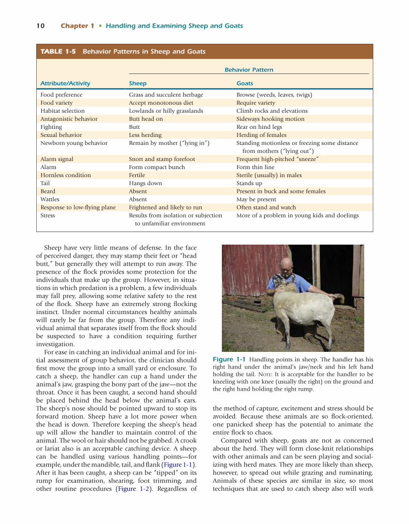
For ease in catching an individual animal and for ini-tial assessment of group behavior, the clinician should first move the group into a small yard or enclosure. To catch a sheep, the handler can cup a hand under the animal’s jaw, grasping the bony part of the jaw—not the throat. Once it has been caught, a second hand should be placed behind the head below the animal’s ears. The sheep’s nose should be pointed upward to stop its forward motion. Sheep have a lot more power when the head is down. Therefore keeping the sheep’s head up will allow the handler to maintain control of the animal. The wool or hair should not be grabbed. A crook or lariat also is an acceptable catching device. A sheep can be handled using various handling points—for example, under the mandible, tail, and flank (Figure 1-1). After it has been caught, a sheep can be “tipped” on its rump for examination, shearing, foot trimming, and other routine procedures (Figure 1-2). Regardless of
the method of capture, excitement and stress should be avoided. Because these animals are so flock-oriented, one panicked sheep has the potential to animate the entire flock to chaos.
Compared with sheep, goats are not as concerned about the herd. They will form close-knit relationships with other animals and can be seen playing and social-izing with herd mates. They are more likely than sheep, however, to spread out while grazing and ruminating. Animals of these species are similar in size, so most techniques that are used to catch sheep also will work
TABLE 1-5 Behavior Patterns in Sheep and Goats
Attribute/Activity
Behavior Pattern
Sheep Goats
Food preference Grass and succulent herbage Browse (weeds, leaves, twigs)Food variety Accept monotonous diet Require varietyHabitat selection Lowlands or hilly grasslands Climb rocks and elevationsAntagonistic behavior Butt head on Sideways hooking motionFighting Butt Rear on hind legsSexual behavior Less herding Herding of femalesNewborn young behavior Remain by mother (“lying in”) Standing motionless or freezing some distance
from mothers (“lying out”)Alarm signal Snort and stamp forefoot Frequent high-pitched “sneeze”Alarm Form compact bunch Form thin lineHornless condition Fertile Sterile (usually) in malesTail Hangs down Stands upBeard Absent Present in buck and some femalesWattles Absent May be presentResponse to low-flying plane Frightened and likely to run Often stand and watchStress Results from isolation or subjection
to unfamiliar environmentMore of a problem in young kids and doelings
Figure 1-1 Handling points in sheep. The handler has his right hand under the animal’s jaw/neck and his left hand holding the tail. Note: It is acceptable for the handler to be kneeling with one knee (usually the right) on the ground and the right hand holding the right rump.

Chapter 1 • Handling and Examining Sheep and Goats 11
on goats. Unlike with sheep, the horns or beard of a goat are acceptable to use in restraint. The ears, however, are not. Goats find restraint by their ears painful, and owners consider it abusive. Animals that are housed with a collar or halter can be led using these imple-ments. Techniques to catch and hold a goat include looping an arm around the goat’s neck and grabbing its gastrocnemius tendon. A goat being held by a hind limb, particularly more distally on the limb, may pos-sibly dislocate a hip joint in an attempt to jerk itself free.
Restraint for Physical ExaminationClinicians should consider the layout and surround-ings of the working facility, the physical condition and temperament of the animal to be restrained, and both human and animal safety when planning procedures that require physical restraint of sheep and goats. Ani-mals that are well socialized and have been handled frequently and in a quiet, nonaggressive manner often can be restrained and treated by one person. Handling animals that have had only occasional human con-tact or those that have been aggressively handled will require an assistant or use of a restraint device.
The use of an assistant or restraining device facili-tates physical examinations, vaccinations, blood collec-tions, artificial insemination, hoof trimming, and other
procedures. Equipment such as stanchions, tilt tables, squeeze chutes, cages, and raceways can be used. Some procedures can be completed while an assistant steadies the sheep or goat against a wall or fence by firmly hold-ing a leg against the animal’s flank or thorax behind its elbow. Both sheep and goats can be rolled up on their rump and restrained in this fashion for a variety of pro-cedures. Another useful strategy is to have the handler straddle the goat and back it into a corner and then firmly press the knees against the goat’s shoulders or neck. This maneuver may frighten and cause struggling in sheep that are unused to restraint. A handler also can gently “flip” a sheep or goat into lateral recumbency, where it can be held by a knee placed on the animal’s neck (Figure 1-3, A and B). Kids weighing up to 30 lb that are used to being handled can be placed with their legs folded under them on the lap of an assistant, to permit the clinician to examine the head. The choice of restraint technique is dependent on the preference and experience of the clinician, the clinical condition and temperament of the animal involved, and requirements for the procedure to be performed. As a general rule, the handler should use the least restraint possible to permit safe handling of the animal.
Restraining the HeadFor procedures in goats, the clinician can control the head by gripping the animal’s cheeks, beard, or horns while straddling the withers or neck. One method for head restraint is to place one hand on each cheek and wrap the fingers under the mandible, with care taken to avoid pressure on the trachea. Alternatively, the clini-cian can hold the beard with one hand and wrap the other arm around the goat’s neck (Figure 1-4). A third method involves gripping the horns. The ability to con-trol a horned goat’s head depends on the temperament of the animal as well as on the skill and strength of the handler.
After the head is stabilized, the goat’s ears, eyes, nose, and mouth can be inspected. For an oral examination, the use of a speculum is recommended to ensure a clear view of the oral cavity and prevent the goat from biting instruments or the clinician’s fingers.
Restraint for Administering MedicationsVeterinarians and sheep and goat producers working as a team can ensure that only wholesome meat and milk products enter the human food chain. Inappropriate premilking and preslaughter drug withdrawal regimens and chemical contamination of feed and pasture give rise to drug residues in products for human consump-tion. Although some sheep and goats are considered pets by their owners, an important point is that the U.S.
Figure 1-2 Sitting a sheep on its rump can be accomplished in various ways. The following technique is recommended: The handler’s left arm is placed around the animal’s neck at the level of the shoulder. The right hand reaches under the sheep, grasping the right hindfoot and setting it on its rump. In this photograph, the ewe has been sat up, and the handler is keeping her stationary.

12 Chapter 1 • Handling and Examining Sheep and Goats
Food and Drug Administration (FDA) classifies sheep and goats as food-producing animals no matter what the owner’s intended use.
Owing to the limited number of pharmaceuticals labeled for use in sheep and goats, the veterinarian often is in the position of prescribing drugs to be used in an extra label fashion. According to the Animal Medicinal Drug Use Clarification Act of 1996 (AMDUCA), extral-abel use of a drug is permissible only under the follow-ing conditions: • The drug must be given only under the supervision
of a veterinarian. • Only FDA-approved human and animal drugs can
be used. • A valid veterinarian-client-patient relationship must
exist. • The drug can be given for therapeutic use only. • Only dosage-form drugs and drugs administered in
water can be given for extralabel applications.
• Drugs given for extralabel indications are prohibited in feed.
• Extralabel drug use is prohibited if it results in viola-tive food residue.
• FDA prohibition of a specific extralabel drug use precludes its administration for that purpose.If a drug is to be used in an extralabel fashion,
appropriate labeling and record-keeping criteria must be met. In addition, it is the responsibility of the pre-scribing veterinarian to institute a withdrawal regimen appropriate for meat and milk production, to avoid potential contamination of the food chain. Record-keeping requirements to comply with AMDUCA should include the individual or group animal identification; species being treated; number of animals treated; con-dition being treated; drug name and active ingredient; dosage prescribed and the duration of treatment; and appropriate withdrawal, withholding, or discard times. In addition, the records must be kept for a minimum of 2 years, and the FDA must have access to these records. At the level of the farm, the producer also may want to consider keeping records of the date(s) of the extralabel drug use and contact information for the person who administered the treatment. A prescription label that conforms to AMDUCA should include the name and address of the prescribing veterinarian; the drug name; specific instructions for use including identification of the animal(s) to be treated, dose, dosing interval, route of administration, and the duration of therapy; cautionary statements; and an appropriate withdrawal, withholding, or discard time.
Veterinarians should advise their clients on the ethi-cal and legal ramifications of not following all labeled guidelines for drugs used in food-producing animals. Awareness of potential risk factors for disease and adherence to conscientious management practices by the producer will lead to reduced disease incidence and
A B
Figure 1-3 Goats can be restrained in lateral recumbency if increased restraint is required. The handler leans over the goat (in this case, from the left) and grasps the goat’s left pelvic limb with the right hand and the goat’s left thoracic limb with the left hand. A, The goat is lifted and leaned into the handler. B, The goat is placed on the ground and a knee is placed on its neck.
Figure 1-4 Goats can be restrained and led by placing one hand under the animal’s jaw and slightly lifting the chin. The second hand is placed behind the head under the ears.

Chapter 1 • Handling and Examining Sheep and Goats 13
the need for drug therapies. In these ways, individual owners can ensure that products from their sheep and goats are wholesome and safe for human use or consumption.
The veterinarian should always ascertain the animal’s intended use (e.g., leather, meat, breeding, exhibition, pet) before administering any medications. Reactions to vaccines and antibiotics can cause lesions in commer-cially valuable skin and muscle and cosmetic flaws in hobby, pet, and show goats. Meat producers prefer that injections be placed in the neck, which yields a meat cut of low value. Breeders prefer the axilla, in which a nodu-lar mass of scar tissue will not be visible and cannot be readily mistaken for caseous lymphadenitis.
Oral DrugsWhen drenching, dispensing boluses, or passing an orogastric tube, the clinician should hold the animal’s head in a straight, natural position with the mandible parallel to the ground. The dose syringe is inserted well into the cheek pouch at the commissure of the lips. The animal must be given time to swallow as the fluid is slowly dispensed. Tilting the head upward can lead to aspiration pneumonia. To safely and properly place a bolus, the clinician moves the balling gun over the base of the tongue, but not into the pharynx. After admin-istering the tablet, the clinician maintains the position of the animal’s head and holds the mouth closed until it swallows. This maneuver prevents the animal from spitting out the medication. Using a speculum, the clinician can pass a 1.2- to 1.5-cm-diameter stomach
tube through the mouth of an adult sheep or goat. An 8 French red rubber urethral catheter with an attached 60-mL catheter-tip syringe can be used as an orogastric tube to feed or provide oral medications to very young or weak lambs or kids.
Injectable DrugsIntramuscular Injections. Intramuscular injec-
tions commonly are given in the area of the neck enclosed by the cervical vertebrae ventrally, the nuchal ligament dorsally, and the shoulder caudally. Other muscles used for injections include the longissimus in the lumbar region as well as the gluteals, semitendino-sus, semimembranosus, and triceps. The clinician must pay special attention to the location of the sciatic nerve in the thighs, because irritating drugs introduced in this region can cause permanent damage. Additionally, the small muscle masses in young goats limit the volume of the injectable substance.
Subcutaneous Injections. Subcutaneous injec-tions can be given in the axilla or on the chest wall. The triangular area of the neck, as described previously, also is used. Any injection site reactions near the prescapular lymph node, however, may be erroneously diagnosed as caseous lymphadenitis.
Intravenous Injections. The jugular vein often is used to administer intravenous drugs and collect blood samples (Figure 1-5, A and B). In sheep and goats the jugular vein can be found lying in a line starting at the base of the ear running down the neck to the thoracic inlet. In sheep it may be necessary to part the wool
A B
Figure 1-5 A, A single handler can draw blood or give an intravenous injection by straddling the goat and holding the head against the handler’s leg using the elbow. The hand on the restraining arm can then be used to hold off the vein while the free hand is used to draw a sample. B, A helper is holding the ewe while the clinician in drawing from the right juglar vein.

14 Chapter 1 • Handling and Examining Sheep and Goats
to give adequate visualization of the vein. Adequate restraint is critical to avoid inadvertent puncture of other structures such as the trachea or esophagus. A 4-cm, 20-gauge needle can be used for venipuncture.
Additional Injection Routes. Intradermal injec-tions call for 1.8-cm, 25-gauge tuberculin needles. Intra-peritoneal injections, primarily used in neonates, are given with 1-inch, 18- or 20-gauge needles inserted no deeper than 1.8 cm. While the kid is held hanging by its front legs, the clinician inserts the needle perpendicular to the skin approximately 1 cm to the left of the navel.
The clinician should clean and swab the teat with alcohol before giving intramammary infusions. Single-use teat cannulas are used for each teat and inserted just deep enough to gain entry into the teat cistern. In ewes and does with small teat orifices, sterile tomcat catheters can be used to infuse medications.
Restraint for Hoof TrimmingSetting a sheep on its rump (tipping) is the easiest mode of restraint for trimming hooves (Box 1-1). Sheep in this position struggle very little and are easy to handle, even for a single person. To rest comfortably on its rump, the sheep should be off center, so that it is sitting on its hip and not its dock. If the sheep struggles, the handler can place a hand on its brisket to move it into a better position.
Unlike in sheep, trimming goat hooves typically is done with the animal standing. With the goat standing, the clinician flexes the pastern and raises the foot and limb until the sole faces upward. The clinician can then work facing the head or tail of the goat while positioned at the animal’s side. An assistant can hold the goat, place it in a stanchion, or halter tie the animal while the clinician trims the hooves. In caring for fractious animals, sitting them on the rump (as is done with sheep) may be useful.
Restraint of the NeonateLambs and kids can be restrained easily in a standing position for physical examination by a single handler. Castration, horn disbudding, and tail docking typically will require one person to restrain the animal and another to perform the procedure. Both lambs and kids can be restrained for castration, and for tail docking in lambs, in a similar fashion. With the handler in a seated position, the animal’s right thoracic and pelvic limbs can be held in the restrainer’s right hand while the left thoracic and pelvic limbs are held in the restrainer’s left hand. The animal’s back is then supported between the handler’s legs (Figure 1-6). Disbudding can be accomplished by the handler holding the animal in sternal recumbency. The person completing the disbudding can restrain the head by holding it down on the table. A rolled-up towel
placed under the neck will better support the head and neck for the procedure. Alternatively, the animal can be placed in a disbudding box, which allows a single person to perform the procedure (Figure 1-7).
FacilitiesAn important goal of management in small ruminant operations is to gather, restrain, and handle animals with minimal stress. Injury prevention for both animals and personnel also should be considered. Well-planned working facilities will promote achievement of these aims.
FencingFencing is used to confine stock, separate animals into management subgroups, exclude predators, and protect ornamental and commercial crops from consumption by goats. Goats typically are more difficult to con-fine than sheep. The selection of appropriate fencing material is dictated by purpose, size, and cost. When planning permanent enclosures, owners may want to consult fence contractors and suppliers of commercial handling equipment, as well as other sheep and goat keepers, who may have proven ideas for consideration.
Smooth-wire, high-tensile, and multiple-strand elec-tric fencing deters goats and predators alike and is easily maintained. Appropriate training of animals to electric fencing will minimize escapes and maximize animal respect of the fence. Such training can be particularly important in sheep with heavy wool, because they may not perceive shocks through the wool. The potential for loss of power to the fence is the major drawback of electric fencing, because the current is the major deter-rent for both livestock and predators to penetration of the fence.
Goats can damage field fencing by standing on their hindlimbs and leaning their front feet against the fence. A single strand of electric wire placed near the top and and another at the bottom of a woven wire fence will discourage goats from leaning on the fence and may prevent predators from crawling underneath. Horned sheep and goats, particularly those with backward-pointing horns, are likely to get caught when with-drawing the head from the 6-inch-wide by 5-inch-high spaces in the woven fencing. The entrapped animal is then indefensible against butting by herd mates or attack by predators. A possible fatal outcome is stran-gulation. Similarly, sheep and goats can catch a foot in the open spaces of a chain-link or 2×2 woven wire fence and fracture a leg in the struggle to free it.
Although welded-wire panels can withstand the pressure of goats, they can entrap horned animals if the panel openings are the standard size of 8 inches wide by 6 inches high. Furthermore, lambs and kids weigh-ing less than 15 lb can step through these spaces to

Chapter 1 • Handling and Examining Sheep and Goats 15
escape confinement. Welded-wire panels with openings smaller than 8 inches wide by 4 inches high can elimi-nate these concerns.
HousingHousing facilities may provide shelter for livestock; storage for feed, equipment, and supplies; and a work area for routine animal care procedures. Shelter that provides warmth, shade, and protection from wind, pre-cipitation, and predators establishes an environment of comfort and calmness in the herd. Unlike cattle, sheep and goats will interrupt their grazing to seek cover from rain. The shelter should promote the productivity and well-being of the animals it houses.
Heating and cooling, ventilation, flooring, and water supply should be considered under the guidance of a person knowledgeable and experienced in farm build-ing construction. Additionally, space should be planned for loafing areas, feed and water troughs, lambing and kidding stalls, and shelter for disabled animals and ani-mal groupings specific to the facility (e.g., young does and bucks, recently shorn goats, research study groups). Feed and equipment storage areas must be designed to
eliminate the potential for consumption of excessive amounts of grain and ingestion of stored chemicals.
Floor space alone does not determine adequate living space for animals. Enclosure shape, floor type, ceiling height, the location and dimensions of feeders and waterers, and other physical and social elements affect the usefulness of the space. Mature ewes and does require an average of 16 square feet of stall space, excluding troughs. A buck or ram may need as much as 30 square feet, whereas a lamb or kid needs approxi-mately 10 square feet. Shelters in outside pens or pas-tures should allow 5.5 square feet per animal. Fence heights vary, ranging between 4 to 5 feet for ewes and does and 5 to 6 feet for rams and bucks in rut.
A clean, dry, draft-free, well-bedded stall or pen is ideal for “for lambing and kidding and bonding of the dam with her offspring. Housing for ewes and does at parturition facilitates observation and enables the pro-ducer to manage difficulties experienced by the dam or her lambs or kids in a timely manner. Limiting the dam to a stall gives her a quiet, undisturbed environment for bonding, which is a crucial event requiring a minimum of 5 minutes.
1. Stand to the left side of the sheep. 2. Hold the sheep’s head in your left hand by placing
your hand under its jaw. 3. Your left knee should be near or just behind the
sheep’s left shoulder. 4. Your right leg should be touching the sheep’s side near
its left hip. 5. Place your right hand on the sheep’s back over the hips. 6. Turn the sheep’s nose away from you toward its shoulder.
7. You should feel the weight of the sheep leaning against your legs.
8. Put pressure on the sheep’s hips with your right hand so the animal cannot pick its back feet off the floor.
9. Take a step back with your right leg. 10. The hind leg of the sheep should start to go down. 11. Continue to bring the animal’s head around until it is
sitting down with its back leaning against your legs.
BOX 1-1 How to Tip a Sheep
Figure 1-6 Demonstration of restraint for castration and tail docking in sheep.
Figure 1-7 Demonstration of the use of a goat or ‘disbud-ding box for horn disbudding.

16 Chapter 1 • Handling and Examining Sheep and Goats
In large herds, it may be impractical to provide indi-vidual stalls for preparturient ewes and does. In such cases, they may be group-penned 2 to 4 weeks before parturition, giving the owner ample opportunity to monitor for potential problems. Neonates are highly susceptible to hypothermia in cold or wet weather. Therefore owners should take precautions to ensure that their ewes and does do not deliver in the field during adverse weather.
Feed and Water. Good feed and water hygienics are essential to promote healthy animals and reduce wastage of feed. Sheep are not as particular as goats about the cleanliness of feeders and waterers. In con-trast with tales of tin can–eating goats, these animals will decline wet or moldy feed and dirty water. Feed-stuffs should be provided in well-designed troughs that minimize contamination with feces, urine, and dirt deposited by hooves.
A 15-inch-long feed trough space can accommodate one adult sheep or goat. Lambs and kids should have free access to a creep feeder when penned or pastured with mature sheep and goats, effectively eliminating competition with adult animals. Bunk space in the creep area should be 10 inches per kid. Despite ample trough space and the use of creep feeders, animals low in the herd hierarchy may not be allowed access to feed. To alleviate the effect of dominance, multiple feeders may be used, or distressed animals can be isolated and fed individually. Alternatively, animals simultaneously restrained and fed in individual stanchions or keyhole mangers will be unable to dominate or be dominated by herd mates.
The volume of water consumed by livestock is influenced by the water content of feed sources, envi-ronmental temperature, and water quality. A rough estimate is 3 L per 45 kg of body weight per day (¾ gal-lon per 100 lb of body weight). To encourage consump-tion, the producer should offer clean water ad libitum. Typical water troughs or self-waterers can be used. In cold climates the use of water heaters improves intake. Adequate drainage should be provided under watering devices.
BiosecuritySick or Injured AnimalsSick animals pose a great risk for disease transmission. Accordingly, they should be removed from the general population of the herd or flock. Producers should have a dedicated “hospital” area separate from the herd where disabled animals can be housed while receiving veterinary and nursing care. It is critical that this area not be a multipurpose facility (e.g., maternity pens, fitting areas for show animals), because this practice may facilitate disease transmission in the herd or flock. The hospital stalls or pens should be constructed of
nonporous materials to minimize the potential for microbial colonization. The area must be cleaned and disinfected after discharge of recovered animals, to pre-vent pathogen transmission to the next occupant.
Introducing New Animals to the HerdA pre-purchase health examination by a veterinar-ian is an important aspect of disease prevention and the establishment of a new veterinarian-client-patient relationship. Taking precautions to reduce the risk of introducing disease into a stable herd is a sound busi-ness decision that contributes to increased profitability through reduced veterinary expenses, time, and effort. An ideal pre-purchase evaluation includes a health assessment of the herd of origin as well as the individu-als being considered for purchase. As dictated by the history, physical examination findings, and knowledge of diseases occurring on nearby livestock farms, the veterinarian may perform diagnostic tests to assess the health and reproductive soundness of animals offered for sale. It also is important to consider screening ani-mals of interest for diseases that the purchaser is actively trying to eliminate or obtain a free status for before their arrival on the farm (e.g., caprine arthritis encephalitis virus, caseous lymphadenitis). In areas in which endo-parasitism is a concern, prearrival fecal flotation and treatment, if indicated, should be considered for all new acquisitions.
All new additions to a flock or herd should be quar-antined for a minimum of 4 weeks. Quarantine facili-ties should minimize physical and aerosol contact with the existing animals. Although the hospital facility is a tempting place to quarantine new additions, its use for this purpose may allow exposure of sick animals to new pathogens during a time of extreme susceptibility and is not recommended. In addition to permitting observa-tion for incubating disease, nutritional requirements, and behavior patterns, quarantine allows the animal to become acclimated to its environment with minimal stress (Chapter 19).
RECOMMENDED READINGAmoah EA, et al: Breeding season and aspects of reproduction in
female goats, J Anim Sci 74:723–728, 1996.Animal Medicinal Drug Use Clarification Act Brochure and Extralabel
drug use algorithm. http://www.avma.org/reference/amduca/amduca1.asp
Clinical examination and making a diagnosis. In Radostits OM, et al, editors: Veterinary medicine, ed 10, Philadelphia, 2007, Saunders.
Constable PD: Clinical examination of the ruminant nervous system, Vet Clin Food Anim 20:185–214, 2004.
Fajt VR: Label and extralabel drugs in small ruminants, Vet Clin Food Anim 17:403–420, 2001.
Grandin T: Design of loading facilities and holding pens, Appl Anim Behav Sci 28:187–201, 1990.
Hutson GD: Behavioural principles of sheep handling. In Grandin T, editor: Livestock handling and transport, Wallingford, UK, 1993, CAB International, pp 127–146.

Chapter 1 • Handling and Examining Sheep and Goats 17
Jackson PGG, Cockcroft PD: Clinical examination of farm animals, Oxford, UK, 2002, Blackwell Science.
Leahy JR, Barrow P: Restraint of animals, ed 2, Ithaca, NY, 1953, Cornell Campus Store.
New South Wales Department of Agriculture, Division of Animal Pro-duction: How to tell the age of sheep, Agfact A3.0.1 , 2003.
Ramírez A, et al: Effects if immediate and early post-partum separa-tion on maintenance of maternal responsiveness in parturient multiparous goats, Appl Anim Behav Sci 48:215–224, 1996.
Sheldon CC, Sonsthagen, Topel JA: Animal restraint for veterinary pro-fessionals, St Louis, 2006, Mosby.
Sherman DM, Robinson RA: Clinical examination of sheep and goats, Vet Clin North Am Large Anim Pract 5:409–426, 1983.
Smith GW, et al: Atypical parapoxvirus infection in sheep, J Vet Intern Med 16:287–292, 2008.
U.S. Food and Drug Administration Animal and Veterinary webpage. http://www.fda.gov/AnimalVeterinary/default.htm
Wilson JH: The art of physical diagnosis, Vet Clin North Am Food Anim 8:169–176, 1992.

18
Feeding and NutritionDarrell L. Rankins, Jr., and D.G. Pugh
C H A P T E R
More than any other factor identified in veterinary man-agement of sheep and goats, diet has a profound effect on general health of both the individual animal and the flock or herd. The diet will have an impact on all aspects of animal health and productivity and therefore is discussed in almost every chapter in this book. The goal in feeding sheep and goats is optimal health as reflected in productivity, reproduction, and performance.
Sheep and goats are able to optimally convert browse, forages, and other feedstuffs barely usable for more commonly encountered livestock species into usable animal products (e.g., meat, milk, fiber) or to reach peak performance (e.g., pet, show, breeding). These two small ruminant species exhibit a high degree of mobility of the lips and tongue, which allows selective consump-tion in the diet, choosing from among plants and other foodstuffs available in the environment. Like other ruminants, both sheep and goats can be characterized by their grazing preferences.1 Sheep are grass or rough-age grazers and tend to graze higher-quality portions of the plant. Goats, as active foragers, tend to select highly digestible portions of grasses. They also can use browse that is woody or stemmy and will readily consume flow-ers, fruits, and leaves; they generally select grass over legumes and browse over grass; and they prefer to graze along fence lines and in rough or rocky pasture areas. Goats typically perform poorly compared with sheep or cattle on flat, improved, monoculture pastures but usually flourish in areas featuring browse or numerous plant species to graze. If given a choice, many meat goats (e.g., Kiko, Spanish, Boer, Tennessee Wooden Leg) prefer a diet of 15% to 20% grasses and 80% to 85% browse.1
Goats are extremely particular about their diet and refuse to consume feeds that have been soiled but are used for brush management in many regions of the world. Goats maintained for brush control should be closely monitored for changes in body weight, body condition score (BCS), and hair coat; the clinician also should look for any signs of toxicosis. Whenever browse, with its deeper root systems, is the predominant forage consumed, mineral uptake may be greater than that expected with consumption of grasses grown on the same land. Both sheep and goats also are excellent
converters of browse and brush to meat, fiber, and milk, but they are raised mostly as grazing animals.1
WATERAlthough often taken for granted, water is an extremely important nutrient. It is the major constituent of an ani-mal’s body. If an animal were deprived of all nutrients, it would succumb to water deprivation first. Although sheep and goats may survive despite loss of most of their body fat and up to 40% to 50% of their total body protein, a water loss of only 10% can prove fatal.
Both sheep and goats are particular about the quality of their water sources. A fresh, clean, non-stagnant source of water should be available at all times. Water sources should be easily accessible, safe, and should be moni-tored so they are not a source of toxins and/or patho-genic organisms. A paved surface 8 to 10 feet around the water tanks/troughs helps prevent unsanitary conditions conducive to many diseases, including footrot.
Daily water intake can be affected by several factors. Pregnancy and lactation increase water requirements and consumption—water intake is increased 126% from months 1 to 5 of gestation. In addition, water intake is greater for females carrying twins than for those carrying only a single.2 Likewise, lactating ewes or does consume twice as much water as that typical for non-lactating females: 7 to 15 L/day versus 3.5 to 7 L/day, respectively. Animals grazing lush spring pastures, for which the forage water content may exceed 80%, con-sume markedly less water than those restricted to dry hay, which may be only 12% to 15% water. Obviously, lactating dairy animals require even greater quantities of water. When high-protein diets are being fed or when mineral consumption increases, water consumption also increases. Sheep may increase their water intake 12-fold during summer over that during the winter months.2 Water quality also can affect daily water con-sumption. For maintenance, individual goats and sheep usually consume 3.5 to 15 L of water/day.3
Water varies in quality according to the amount and type of contaminant. The most common dissolved substances in water are calcium, magnesium, sodium
2

Chapter 2 • Feeding and Nutrition 19
chloride, sulfate, and bicarbonate.3 If the salts of these minerals are present in high-enough concentrations, depressed performance, illness, and occasionally death can result. In addition to causing various specific prob-lems in animals, dissolved salts have additive effects on suppression of production and health. As salt concentra-tions increase, water consumption usually is depressed, with young animals generally being more affected than adults. Over time, animals tend to adapt to water with high concentrations of dissolved salts. Rapid or abrupt changes from water with relatively low concentrations to water with high concentrations of dissolved sub-stances are poorly tolerated, however.3-6 High sulfate concentrations in the range of 3500 to 5000 parts per million (ppm) may result in suppressed copper absorp-tion from the intestine. Nitrates and, less commonly, nitrites occasionally are encountered in toxic concentra-tions from ground water. Most safe, drinkable water has a pH of 7 to 8. As the alkalinity of water increases, its suitability for consumption decreases.
Although water contaminated with coliform bacteria has been associated with disease in humans, only rarely is coliform contamination of drinking water implicated as an agent of disease in sheep and goats. In general, only very young animals are affected. Goats tend to adapt to high ambient temperatures better than do other domestic ruminants and require less water evapo-ration to control body temperature.7 In addition, they possess the ability to reduce urine and fecal water losses during times of water deprivation.
In summary, sheep and goats should have access to a continuous supply of fresh, clean water, to ensure that productivity is not compromised.
ENERGYEnergy generally is the first limiting nutrient under most practical conditions where sheep and goats are maintained throughout the world. Energy requirements vary greatly depending on level and stage of produc-tion, level of activity, and intended animal use. Except in situations in which rapid growth rates are desired or milk production is to be maximized, the energy require-ment usually can be met with medium- to high-quality forage. Under maximal production pressures, however, some sort of supplementation may be required. Energy-deficient diets can result in poor growth rates, lower BCSs, decreased fiber production, reduced fiber diam-eter, decreased immune function, and increased sus-ceptibility to parasitic diseases and other pathologic conditions. Angora goats and many wool breeds of sheep are prone to various fiber production changes, whereas cashmere goats may be less susceptible.
The greater part of the energy that is used by sheep and goats comes from the breakdown of structural car-bohydrates from roughage. Therefore roughage should
constitute the bulk of their diet. Energy can be expressed in terms of the net energy system (calories) or in terms of total digestible nutrients (TDN) as a percentage of the feed. The two expressions are interchangeable with use of various prediction equations; in this chapter, TDN is used as the measure. Currently, most feed and forage testing laboratories estimate TDN using the Van Soest fiber analysis. A representative sample is analyzed for neutral and acid detergent fiber contents, and then TDN is predicted based on one or both of these values. This system works effectively for most forages but is less reliable for feeds that are high in starch (e.g., corn). In general, warm-season, perennial grass hays are approxi-mately 50% to 54% TDN, whereas many of the cereal grains usually are 80% to 90% TDN. Most forages in the green, vegetative state are approximately 62% to 70% TDN on a dry matter basis. Stemmy, dry, poor-quality hay is less than 50% TDN. By comparing these typical values with the requirements of various classes of sheep and goats, keepers can ascertain when supplemental energy sources are needed for forage-based rations. For example, a 150-lb ewe requires a diet containing 52.5% TDN for maintenance and 66% for the first few weeks of lactation, with a steady increase from 53% to 66% TDN during gestation. Therefore the dry (nonlactating), nonpregnant ewe could use low-quality forage, but the pregnant or lactating ewe needs a diet of lush, vegetative forage. If a good to excellent forage is unavailable, some type of energy supplement is required for the ewe in late pregnancy or while lactating. Similar supplementa-tion may be indicated for goats: A 110-lb doe requires a diet containing 53% TDN for maintenance but higher amounts during pregnancy and lactation.2
A variety of choices are available for energy supple-mentation. The most common choice is cereal grains, corn being the most common of these. Corn is dense in energy, and most of that energy is in the form of starch. When appreciable levels of starch are supplemented to ruminants consuming forage-based diets, the general response is a decrease in forage intake and digestibil-ity. However, the energy status in the sheep or goat receiving corn supplementation will still be improved because of the energy from the corn. Several other cereal grains are available for use as energy supplements for ruminants consuming forage-based diets (e.g., grain sorghum, oats, barley, rye). Two other nontraditional energy supplements are soybean hulls and wheat mid-dlings. Soybean hulls are the outermost layer of the soybean and are composed of abundant quantities of digestible fiber. Unlike corn, soybean hulls do not sup-press fiber digestion but may increase hay digestibility. Even though soybean hulls have a TDN value 62% less than corn, they produce similar results when used as an energy supplement for ruminants consuming forages. Wheat middlings, a byproduct of wheat milling, elicit similar responses. Beet pulp, citrus pulp, and brewer’s

20 Chapter 2 • Feeding and Nutrition
grains all are byproduct feedstuffs that can be effec-tively used in both sheep and goat feeding, and these byproduct-type feeds often are much more economical than corn. All byproduct feeds should be analyzed for composition and used accordingly in diet formulation.
Another source of energy supplementation is fat. In general, total fat content should not exceed 8% of the diet, or 4% to 5% as supplemental fat. In the southern United States, where cotton production is prevalent, whole cottonseed (which contains approximately 24% fat) is used as an energy supplement for both sheep and goats. In animals of both species, the diet should be supplemented with no more than 20% of the daily intake as whole cottonseed, assuming that the remain-der of the diet contains no fat.
PROTEINAs a general rule, a minimum of 7% dietary crude pro-tein is needed for normal rumen bacterial growth and function for sheep and goats. If dietary protein drops below 7%, forage intake and digestibility are depressed. Protein deficiency is associated with decreased fiber production, slowed growth, decreased immune func-tion, anemia, depressed feed use, edema, and death. All of the protein reaching the small intestine is found in bacteria or protozoa or dietary protein that escaped ruminal digestion. The quality (amino acid content) of the bacterial protein is surprisingly quite good. There-fore the quantity of dietary protein provided to adult ruminants is much more important than the quality. The opposite is true of the preruminant lamb or kid. If lambs or kids are fed a milk replacer, it should be composed of milk byproducts to provide an adequate amino acid composition for maximal growth.
Crude protein content varies widely among the various feedstuffs. Warm-season, perennial grass hay samples can range from less than 6% to more than 12% crude protein, whereas legumes in the vegetative state may occasionally be more than 28% crude protein. The protein content of plants declines with maturity. As with energy needs, crude protein requirements vary with the animal’s stage of production. For maintenance, ewes and does of most weight classes require a diet containing 7% to 8% protein. During lactation, both ewe and doe require 13% to 15% crude protein in the diet, depending on the number of offspring suckling. Supplementation of protein may be necessary for heavy-producing animals. Whenever grass hay is fed, protein deficiency should be a concern, particularly for growing or lactating animals. The most consistent sign of protein deficiency in lactating animals is poor weight gain or slow growth in their lambs or kids, particularly with twins or triplets.2
Typical protein supplements include the oilseed meals (cottonseed meal, soybean meal), commercially
blended supplements containing both natural protein and nonprotein nitrogen (NPN) (e.g., as range cubes or pellets or molasses-based products), and various byproducts (whole cottonseed, corn gluten feed, dried distiller’s grains). Protein should be fed to meet, but not greatly exceed, requirements. Excess protein usually results in increased feed costs and higher rates of disease (e.g., heat stress, pizzle rot).
Giving NPN is an inexpensive way to increase the protein concentration of rations for sheep or goats. NPN is any source of nitrogen in the nonprotein form, but the most commonly used type is urea. Whenever NPN is used, the diet should have sufficient amounts of highly fermentable energy components. Feeding grain with NPN can result in a decrease in rumen pH. In this altered environment, the ability of the ruminal urease enzyme to ferment urea is depressed, resulting in a slower release of or breakdown to ammonia and carbon dioxide (CO2). Slowing this metabolic pathway allows more efficient protein synthesis by the rumen microbes. By contrast, diets of poor-quality roughage result in a higher rumen pH and enhanced urease activity. These conditions result in a quicker release of ammonia, a poorer “marriage” of chains of carbon atoms and nitro-gen for microbial protein synthesis, and a potential increase in the incidence of urea or ammonia toxicity. Whenever NPN is added to the diet, feeds containing a urease enzyme should be limited or avoided. Such urease-containing feeds include raw soybeans and wild mustard. Signs of urea or ammonia toxicity, which may be fatal, include dull or depressed demeanor, muscle tremors, frequent urination and defecation, excessive salivation, increased respiration, ataxia, and tetanic spasms. Treatment includes the infusion of a 5% ace-tic acid solution (vinegar and water) into the rumen through a stomach tube. In severe cases, rumenotomy and fluid therapy may be required.
The following guidelines are useful when urea is fed as a protein source: 1. Never use urea for more than one third of the
protein in the diet or more than 3% of the grain portion of the diet.
2. Ensure that a highly fermentable source of carbohy-drates (e.g., corn, milo) is fed along with NPN.
3. Avoid the sudden introduction of urea into the diet (allow at least 8 to 10 days for its introduction).
4. Ensure proper mixing of feedstuffs whenever urea is used.
5. If 1 lb of urea plus 6 lb of ground corn is cheaper than 7 lb of cottonseed meal or soybean meal, then the former diet may be efficiently fed. However, if 7 lb of either the cottonseed or the soybean meal is less expensive, the urea should be avoided.
6. If the crude protein of the diet is greater than 14% of the dietary TDN, NPN is of little value. For example, if TDN is 45%, which is typical of many

Chapter 2 • Feeding and Nutrition 21
dry hays during winter, NPN is of limited or no value if the crude protein of the diet is greater than 6.3% (45 × 0.14 = 6.3).Because of variable dietary intake and its relation-
ship to body condition scoring, NPN is best used in sheep or goats with BCSs greater than 2.5; they should be avoided in animals with a BCS of less than 2. If NPN is offered to animals, it should be fed daily; less is used for protein synthesis if the supplement is fed less fre-quently. In one report, the inclusion of NPN in poorly digestible forage diets for lambs resulted in increased weight gain and wool production and decreased signs of parasitic nematode infestation.8
MINERALSClinicians generally consider seven macrominerals and eight microminerals when assessing mineral nutrition for sheep and goats. The designations macro and micro do not reflect the minerals’ relative importance but rather characterize the amount of each that is required as a proportion of the diet. Macromineral needs usually are expressed as percentage of the diet, whereas micro-mineral needs generally are expressed as ppm or mg/kg.
The seven commonly assessed macrominerals are calcium, phosphorus, sodium, chlorine, magnesium, potassium, and sulfur. The eight microminerals are copper, molybdenum, cobalt, iron, iodine, zinc, man-ganese, and selenium. Trace mineral deficiency is less common than energy, protein, or macromineral defi-ciency. Such deficiencies evolve slowly over time and rarely lead to the dramatic effects on productivity and body condition seen in protein deficiency.2 In some cases of mineral deficiency, liver biopsy is the diagnostic tool of choice. The technique for liver biopsy is covered in Chapter 5.
Calcium and PhosphorusCalcium and phosphorus are interrelated in body func-tions and are therefore discussed together. Nearly all of the calcium in the body and most of the phosphorus is found in the skeletal tissues. Diets deficient in calcium and phosphorus may delay growth and development in young lambs and kids and predispose them to metabolic bone disease (e.g., rickets, osteochondrosis) (see Chapter 11). Likewise, calcium and phosphorus deficiencies in lactating ewes and does can dramatically reduce milk production.
Serum phosphorus concentrations are not highly regulated but are still maintained between 4 and 7 mg/dL for sheep and between 4 and 9.5 mg/dL for goats. Phosphorus deficiency is the most commonly encoun-tered mineral deficiency in range- or winter-pastured animals. Most forage tends to be high in calcium and relatively low in phosphorus; this is true especially for
legumes. Beet pulp and legumes (such as clover and alfalfa) are good to excellent sources of calcium. For lactating dairy goats and sheep, supplemental calcium and phosphorus are necessary to meet high demands for milk production. Range goats may need less supple-mental phosphorus than sheep because of their prefer-ence for browse and plants that tend to accumulate phosphorus. Phosphorus serum concentrations of less than 4 mg/dL may indicate phosphorus deficiency.2 Phosphorus deficiency results in slow growth, listless-ness, an “unkempt” appearance, depressed fertility, and depraved appetite or pica.2
Sheep and goats fed high-grain or high-concentrate diets typically need supplemental calcium and little to no additional phosphorus. Grains are relatively low in calcium but contain moderate to high concentrations of phosphorus. Although serum calcium is tightly held in a narrow range, serum concentrations consistently below 9 mg/dl are suggestive of chronic calcium defi-ciency.2 Chronic parasitism can lead to decrease in body stores of both calcium and phosphorus.2 Common cal-cium supplements include oyster shells and limestone. Defluorinated rock phosphate is an excellent source of phosphorus. Dicalcium phosphate or steamed bone meal (when available) are good sources for both. The calcium-to-phosphorus ratio should be maintained between 1:1 and 2:1.2
Sodium and ChlorineSodium and chlorine are integral components of many bodily functions. Salt (sodium chloride [NaCl]) is the carrier for most ad libitum mineral supplements. If salt is not offered ad libitum, it should be incorporated into a complete ration at a level of 0.5% of the diet. Sodium is predominantly an extracellular ion and is important for normal water metabolism, intracellular and extracel-lular function, and acid-base balance. Conversely, chlo-ride is an intracellular ion, functions in normal osmotic balance, and is a component of gastric secretions. Sheep or goats that are deficient in salt intake routinely chew wood, lick the soil, or consume other unlikely plants or debris. The NaCl content of feeds may be increased to 5%, particularly for feeding males, to help increase water intake and reduce the incidence of urolithiasis (see Chapter 12).
Salt commonly is used as a carrier to ensure trace mineral intake, because sheep and goats have a natural drive for NaCl in the diet. An important consideration in the decision to use a salt-containing mineral mixture to ensure mineral intake is that individual consump-tion may vary drastically. Furthermore, improperly pre-pared salt mixtures or blocks, feed supplements, liquid feeds, or certain types of food or water contamination may be associated with drastically altered mineral consumption.

22 Chapter 2 • Feeding and Nutrition
Salt also is useful as an intake limiter for energy-protein supplements. A 10% to 15% NaCl mixture of two parts ground corn and one part soybean meal is approximately 20% crude protein. The added salt usually limits intake of this mixture to 0.45 kg/day in the adult goat or sheep. Whenever using salt-limited feeding, the keeper should take care to introduce the feedstuffs slowly over 2 to 3 weeks and provide access to adequate quantities of fresh clean water. Only white salt should be used as an intake limiter. If trace mineral salt or ionized salt is used, mineral (e.g., copper, iodine) toxicity is likely, particularly in sheep.
MagnesiumMagnesium is important for normal functioning of the nervous system and is required for many enzymatic reactions. Skeletal magnesium can be used by the ani-mal during times of deficiency, but the skeletal magne-sium reserve is much smaller than the calcium reserve. Many fast-growing–heavily-fertilized-cereal grains or grass pastures are deficient in magnesium. Magnesium absorption is depressed by high concentrations of plant potassium or rumen ammonia. Legume and legume-grass mixed pastures are good sources of magnesium. A magnesium deficiency can lead to a clinical mani-festation known as grass tetany in either sheep or goats. Magnesium toxicity is very rare.
PotassiumPotassium is required for normal acid-base balance and is an integral component of many enzymatic pathways; it functions as an intracellular ion. The requirement is between 0.5% and 0.8% of the diet, depending on the stage of production. Most grains contain less than 0.4% potassium, whereas fresh green forages generally contain more than 1%. Dormant forages, however, may have much lower potassium concentrations.
Potassium deficiency or toxicity is rare in sheep and goats. However, deficiency may occur in highly stressed animals being fed diets composed mostly of grain. Therefore, in stressful situations (such as weaning), supplemental potassium may be indicated for animals fed predominantly on grain.2
SulfurSulfur is a component of many bodily proteins. It is found in high concentrations in wool and mohair, in keeping with the large amounts of sulfur-containing amino acids (cystine, cysteine, and methionine) in kera-tin. Sulfur deficiency can reduce mohair production in Angora goats.9 The general recommendation is to main-tain a 10:1 nitrogen-to-sulfur ratio in sheep and goat diets.2 Ideal ratios of 10.4:1 for maximal gains and 9.5:1
for maximal intake in growing goats.10 However, a ratio as low as 7.2:1 has been suggested for optimal mohair production.11 If the forage has a low sulfur content or if large quantities of urea are used in the diet, weight gain and fiber production can be increased by providing supplemental sulfur.
In both sheep and goats, sulfur deficiency may result in anorexia, reduced weight gain, decreased milk pro-duction, decreased wool growth, excessive tearing, exces-sive salivation, and, eventually, death. Browsing animals such as goats may ingest enough tannins to decrease sulfur availability. Sulfur deficiency also depresses diges-tion, decreases microbial protein synthesis, decreases use of NPN, and lowers the rumen microbial popula-tion. Whenever NPN is fed to fiber-producing animals, sulfur supplementation is indicated. With the possible exception of oats and barley, the sulfur content of most cereal grains usually is low to deficient, although corn-soybean diets usually meet requirements for the rumi-nal synthesis of sulfur-containing amino acids.
Sulfur toxicity occasionally is seen in settings in which calcium sulfate is used as a feed intake limiter. It also occurs when ammonium sulfate is fed as a source of NPN or as a urinary acidifier. If sulfur is supple-mented in the form of sulfate, toxicity may occur, par-ticularly if the sulfur content is greater than 0.4% of the diet.2 Sulfate can be reduced to sulfide in the rumen or lower bowel. Sulfide in large enough concentrations can result in polioencephalomalacia that is only partially responsive to thiamine (see Chapter 13).
In the southeastern United States, use of ammonium sulfate as fertilizer has increased appreciably with the rising cost of commercial nitrogen. If signs of marginal trace mineral deficiencies begin to appear in any group of sheep or goats, forage sulfur concentrations should be measured. An excess of dietary sulfur can lead to deficiency of any of several trace minerals (e.g., copper, zinc) without causing any overt toxicity problems.
CopperCopper deficiencies can be primary (as a result of low intake) or secondary (caused by high concentrations of molybdenum, sulfur and/or iron, or other substances in feedstuffs). In the rumen, copper, molybdenum, and sulfur form thiomolybdates, which reduce copper avail-ability. Specifically, copper’s ability to function as part of the enzyme systems needed for specific biochemical reactions is depressed. This impairment in metabolism results in clinical signs of deficiency. Other factors that alter copper absorption include high concentrations of dietary cadmium, iron, selenium, zinc, and vitamin C as well as alkaline soils. Zinc supplementation in the diet (to a concentration higher than 100 ppm) will reduce availability and liver stores of copper. Roughage grown on “improved” (fertilized, limed) pastures is more
Chapter 2 • Feeding and Nutrition 23
likely to be deficient. Liming reduces copper uptake by plants, and many fertilizers contain molybdenum. Good-quality lush grass forages have less available cop-per than that typical for most hays, and legumes have more available copper than most grasses. Liver copper reserves last up to 6 months in sheep.2
Copper DeficiencySigns of copper deficiency include microcytic anemia, depressed milk production, lighter or faded-looking hair color, poor-quality fleeces, heart failure, infertil-ity, increased susceptibility to disease, slowed growth, enlarged joints, lameness, gastric ulcers, and diarrhea. These signs appear to be more severe with primary copper deficiencies than with a lowered copper-molybdenum ratio. Sheep with copper deficiency have inferior wool, which usually is characterized as “stringy” or “steely.” Such wool lacks both tensile strength and crimp. Growing lambs and kids are most susceptible to copper deficiency, followed by, in order of predisposi-tion, lactating females.
Several breed differences have been observed with regard to copper metabolism. For example, some Finnish-Landrace sheep may have lower serum copper concen-trations than in Merinos, which in turn have lower serum copper levels than in British breeds at similar levels of intake.12 Milk usually is deficient in copper, whereas molybdenum is concentrated. In lambs sus-pected of having “swayback,” liver copper concentra-tions usually are less than 80 ppm dry weight.
Anecdotal reports indicate that goats offered only sheep mineral (with low to absent added copper but with added molybdenum) may succumb to copper deficiency. The risk of this deficiency may be magnified in pygmy goats and young, growing animals. Merino sheep and dwarf goat breeds require 1 to 2 ppm more copper than other breeds. Copper is absorbed more efficiently by young animals than by adults.2 Copper supplementation appears to have some effect on the control of nematode parasites.
Very young lambs or kids can present with enzootic ataxia. Affected animals are born from copper-deficient ewes or does. The swayback condition of lambs or kids usually is seen at birth but may be diagnosed in ani-mals up to 3 months of age. Neonates may experience a progressive ascending paralysis. Manifestations of this ataxia include muscular incoordination (especially in the hindlegs) and failure to nurse. Most neonates die within 3 to 4 days of onset of the first clinical signs and symptoms. Affected older animals may survive or die, depending on severity. Rear limb ataxia, muscle atrophy, and weakness are noted in lambs or kids from 2 weeks to 3 months of age.
A definitive diagnosis is made with necropsy. His-topathologic examination of the spinal cord reveals myelin degeneration and cavitations of cerebral white
matter. Liver copper concentrations are invariably depressed. Prevention and treatment consist of cop-per supplementation (using oral supplements, copper needles, a trace mineral mixture, or injectable copper) and maintaining an appropriate dietary copper-to-molybdenum ratio (see Chapter 13).
If copper deficiency is suspected, the copper, molyb-denum, sulfur, and iron concentrations of the diet should be determined. To confirm copper deficiency, the nutritionist or clinician should measure body tis-sue concentration. Serum copper commonly is used to determine body copper status, but much of the copper is bound in the clot, making plasma a more reliable indicator of body copper status. Unfortunately, from a body assessment standpoint, blood copper concen-trations may be falsely increased by stress or disease. If serum copper is overtly low and animals were not stressed during sampling, copper deficiency is likely. If serum copper concentrations are used for assessment, and copper concentrations fall within normal ranges, additional copper supplementation is of little or no value. An exception is those cases in which serum cop-per is normal but dietary molybdenum is high, or the copper-to-molybdenum ratio is less than 4:1. In such cases the assayed copper may not be available for use in body metabolism. The dietary copper-to-molybdenum ratio should be maintained between 5:1 and 10:1. Liver is the best tissue to use in determining body copper status, but among other limitations, it is a poor indi-cator of short-term copper balance. If liver copper is marginal, but plasma or serum copper is in the normal range, the animal may have a favorable response to copper supplementation. In such instances, dietary cop-per probably is deficient, and the liver stores of copper are being depleted. If a herd problem seems likely, the clinician should sample not only a cross-section of ages and production status but also as many symptomatic animals as possible.
Forage samples should be taken for copper and trace mineral analysis. Core samples of hay should be properly collected. Feed samples should be placed in plastic bags, not brown paper boxes or bags. Dietary copper should range between 4 and 15 ppm. In areas in which copper deficiency is a problem in goats, a mineral mixture with 0.5% copper sulfate should be offered on a free-choice basis. This level of copper, however, may be toxic for sheep.2 In extremely deficient areas, copper needles can be administered orally, or copper can be injected parenterally.
Copper ToxicityCopper toxicity is a much larger problem in sheep than in goats. In sheep, the magnitude of difference between copper deficiency and copper toxicity is quite small. Copper toxicity can occur in sheep as a result of simple mixing errors during the formulation of mineral

24 Chapter 2 • Feeding and Nutrition
premixes, or from feeding mineral mixes formulated for species other than sheep, and can be exacerbated by the ingestion of toxic plants (e.g., lupines, alkaloid-containing species) and stress. Sources of toxic concentrations of copper include premixes, trace mineral supplements made for species other than sheep, copper sulfate–containing foot baths, feedstuffs containing high levels of copper (horse, hog, or chicken feeds), and some nontraditional feedstuffs (broiler litter). Signs of cop-per toxicity include increased respiration, depression, weakness, hemoglobinuria, and icterus, with sudden death in some instances. Gross histopathologic findings in affected animals include signs of a massive hemolytic crisis and dark, hemoglobin-filled kidneys. Treatment includes administration of d-penicillamine (26 mg/kg once a day for 6 days) and ammonium tetrathiomolyb-date (1.7 mg/kg IV every other day for three treatments). The control of methemoglobinemia should be spe-cifically addressed (see Chapter 12). Goats are closer to cattle than to sheep in susceptibility to copper toxicity.
CobaltCobalt is used by rumen bacteria in the formation of vitamin B12. It is deficient in some highly organic or poorly drained soils. Cobalt deficiency in sheep or goats is characterized as a classic B12 deficiency, with signs and symptoms including lack of appetite, emaciation, anemia, and “wasting disease.” Cobalt deficiency is associated with white liver disease, although phospho-rus and copper deficiencies and chronic parasitism also play roles in pathogenesis. Animals with this condition have excessive ophthalmic discharge, and their skin becomes extremely pale. Necropsy reveals a fatty liver (see Chapter 5).
To determine whether a cobalt deficiency exists, the clinician must evaluate the complete diet. Serum or urinary methylmalonic acid is increased and serum vita-min B12 and liver cobalt concentrations are depressed in cobalt deficiency. Diagnosis may be difficult, however, because of the normally low tissue concentration of cobalt. A diet with a cobalt concentration of 0.1 ppm is adequate in most instances, but dietary levels below 0.06 ppm should be considered deficient. If a frank deficiency exists, a cobalt-supplemented trace mineral mixture should be fed ad libitum. Cobalt toxicity is of minimal concern with most sheep and goat operations under typical conditions in North America.2
IronIron deficiency in sheep and goats is quite rare under grazing conditions. Lambs or kids raised in total confinement and deprived of access to pasture and earth-floored stalls or paddocks may become deficient, however. Iron deficiency is exacerbated when young
animals are fed a milk replacer deficient in iron. New-born kids and lambs are born with minimal iron stores. Iron is an important component of hemoglobin, and a deficiency can result in microcytic-hypochromic ane-mia. Iron deficiency is a rare problem in adults, except in cases of excessive parasitism.
In kids and lambs with diagnosed iron deficiency, iron dextran (150 mg given intramuscularly) at 2- to 3-week intervals may prove a valuable therapy.9 Paren-teral iron dextran may be toxic, and caution is indicated with its use.9 If selenium deficiency also exists, the use of iron dextran can result in painful muscle reactions. The dietary iron requirement generally is 30 to 40 ppm.
IodineIodine deficiency is more common in certain geo-graphic regions of North America, particularly the “Northern Tier” of the United States. Iodine availability is depressed by methylthiouracil, nitrates, perchlorates, soybean meal, and thiocyanates. Minerals that interfere with iodine absorption include rubidium, arsenic, fluo-rine, calcium, and potassium. Iodine appears to be most available for use by the body during winter months and during lactation. The form or “state” in which iodine exists in the feed alters availability—iodates are absorbed more readily than iodides. Signs of iodine deficiency include goiter, poor growth, depressed milk yield, pregnancy toxemia, and reproductive abnormali-ties including abortion, stillbirth, retained placentas, irregular estrus, infertility, depressed libido, and birth of small, weak, and either hairless or short- and fuzzy-haired newborns. Lambs or kids born to iodine- deficient dams may have enlarged thyroid glands. Affected kids can be treated with 3 to 6 drops of iodine (Lugol’s solution) daily for 7 days.
An enlarged thyroid in the kid commonly is a con-genital problem unassociated with dietary iodine (see Chapter 9). After a thorough examination of the diet, if iodine deficiency is still suspected, the clinician can measure the serum or plasma thyroxine levels, which are lowered in deficient states, to assess the body status. Iodine is readily absorbed, so most sources will work well in salt-mineral mixtures or feed supplements. Iodine levels of 0.8 ppm for lactating animals and 0.2 ppm for nonlactating ewes or does usually are suf-ficient for normal function. Applying iodine (1 to 2 mL of tincture of iodine or Lugol’s solution) to the skin of a pregnant female once each week is a labor-intensive but rewarding method of preventing iodine deficiency–induced hypothyroidism. Hyperiodinism occasionally is associated with the feeding of kelp or related plants in mineral mixtures. This clinical problem may be encountered in the occasional pet or dairy goat. Simply removing the iodine source may be all that is required for treatment of toxicity.2

Chapter 2 • Feeding and Nutrition 25
ZincZinc deficiency–related disease or dysfunction has been reported in sheep and goats. Zinc availability is improved with the presence of vitamin C, lactose, and citrate in the diet. Oxalates, phytates, and large dietary concentrations of calcium, cadmium, iron, molybde-num, and orthophosphate all depress zinc availability. Zinc concentrations usually are higher in legumes than in grasses, but legumes invariably contain large concen-trations of calcium, which can depress zinc availability. Zinc tends to be less available from cereal grain. Signs of zinc deficiency include dermatitis and parakeratosis, depressed milk production, impaired appetite, poor feed utilization, slowed growth, increased susceptibil-ity to footrot, diminished hair growth on legs and head, swollen joints, poor growth, decreased repro-ductive performance, reduced testicular development, impaired vitamin A metabolism, and increased vitamin E requirements. Male goats appear to be more sensi-tive to the potential for adverse effects of marginal zinc intake.
When zinc deficiency is suspected, the clinician should carefully sample all constituents of the diet. Serum or plasma should be properly collected into tubes specifically designed for trace mineral analysis (royal blue top or trace mineral tubes). Hemolysis alters the accuracy of serum and plasma samples, because red blood cells have high zinc concentrations. Liver samples yield the most reproducible measurements of the zinc status of the animal. Both polystyrene containers and brown paper bags may be contaminated with zinc and should not be used for sample collection. Diets contain-ing 20 to 50 ppm of zinc usually are sufficient, except for animals that consume a high percentage of legumes in their diets. In these instances, a chelated form of zinc is indicated. Providing trace mineral–salt mixes with 0.5% to 2% zinc usually prevents deficiency. The differ-ence between required and toxic amounts is quite large, so zinc toxicity is rare under most conditions.2
SeleniumThe absorption of selenium from the small intestine is enhanced by adequate dietary levels of vitamins E and A and histidine. Large dietary quantities of arsenic, cal-cium, vitamin C, copper, nitrates, sulfates, and unsatu-rated fats inhibit selenium absorption. Legumes usually are a better source of selenium than are grasses, which in turn are superior to cereal grains (see also Chapter 11).
The signs of selenium deficiency include nutritional muscular dystrophy, particularly of the skeletal and cardiac muscles of fast-growing young lambs or kids, and retained placentas. Other signs associated with insufficient selenium include poor growth, weakness or premature birth of lambs or kids, depressed immune
function, mastitis, and metritis. Most often, selenium deficiency is observed in lambs between birth and 8 weeks of age.
Serum selenium concentrations are difficult to inter-pret because they may reflect dietary intake over the past 2 to 4 weeks. Whole blood selenium is reflective of dietary selenium intake over the past 100-plus days.
Liver biopsy is the the most accurate method for diagnosing selenium deficiency. Our own preference, however, is to use whole blood selenium to determine selenium adequacy. Diets containing 0.1 to 0.3 ppm of selenium usually are adequate. The upper limit (0.3 ppm) should be fed during the final trimester of pregnancy. Mineral-salt mixes should contain between 24 and 90 ppm selenium in deficient regions. Of course, dietary limits may be restricted to different levels in different countries and regions of the United States. In cases of frank deficiency, injectable vitamin E and selenium preparations may be given. Selenium toxicity may occur, but deficiency is the more prevalent prob-lem. Toxicity is characterized by wool break, anorexia, depression, incoordination, and death.2
VITAMINSBecause the rumen normally synthesizes B vitamins in healthy sheep and goats, the only vitamins needed in the diets of nonstressed animals are the fat-soluble vitamins: A, D, E, and K. If an animal has altered rumen function, is parasitized, is on a low-fiber diet, or is being given long-term antibiotic therapy, supplemental B vita-mins may be of value.
Vitamin AVitamin A is involved in numerous bodily functions. It is essential for growth, proper skeletal development, normal reproduction, vision, and epithelial tissue integ-rity. Signs of vitamin A deficiency include weight loss, depressed immune function, night blindness, decreased fertility, and hair loss. Vitamin A can be stored in the liver for 4 to 6 months or longer. Green, vegetative for-age meets the daily vitamin A requirement for sheep and goats, which is 105 international units (IU)/kg of body weight/day for nonlactating animals.2 During late gestation, the requirement increases to 150 IU/kg/day, and for lactation, 175 IU/kg/day. For conversion purposes, one retinol equivalent (RE) is equal to 3.33 IU. Plants are not a source of preformed vitamin A but instead contain vitamin A’s carotenoid precursors.2
Hay that is brown and dry and has been stored for long periods probably is deficient. Vitamin-mineral supplements that also contain oxidizing agents (e.g., copper, iron) are subject to oxidative destruction during storage. Although the label may indicate that vitamin A is present, its activity may be minimal.

26 Chapter 2 • Feeding and Nutrition
Vitamin DVitamin D requirements generally are met if the ani-mals are exposed to sunlight. In confinement feeding operations or during sustained overcast or cloudy con-ditions, vitamin D should be supplemented. Vitamin D deficiency can occur in heavily wooled lambs raised with limited access to sunlight or sun-cured forages. Winter months tend to be the most common time for marginal blood vitamin D concentrations.
Vitamin D, along with calcium and phosphorus, is important for normal bone integrity. Deficiencies can result in rickets (see Chapter 11). Plants, both fresh and in the form of hay (particularly sun-cured hay), con-tain abundant quantities of ergocalciferol (vitamins D2 and D3). The vitamin D requirement for sheep is 5 to 6 IU/kg of body weight/day, except for early-weaned lambs, which have a requirement of 6 to 7 IU/kg/day.2 For conversions, 1 IU of vitamin D equals 0.025 μg of crystalline D3.2
Vitamin EVitamin E is a biologic antioxidant that plays a major role in maintaining cell membrane integrity. It is closely associated with selenium in its mode of action, and a deficiency of either can lead to white muscle disease, depressed immune function, and depressed fertility in sheep and goats. Lambs from vitamin E– deficient ewes may exhibit stiffness, paralysis, and pneumonia. If a higher-than-expected incidence of infection and disease is noted in the herd or flock, the keeper or clinician should investigate adequacy of vitamin E intake. In selenium-deficient areas, young lambs generally should be given extra vitamin E and selenium by injection. Vitamin E is poorly stored in the body, so daily intake is crucial. Although vitamin E is found in most good-quality forages, if females are consuming poor-quality hay (particularly in selenium-deficient areas), supplementation is required. Feeds rich in vitamin E include alfalfa meal, cottonseed meal, and brewer’s grains. Some feedstuffs (e.g., corn, feeds containing high levels of sulfur, onions) decrease vitamin E availability. The 2007 National Research Council (NRC) recommendation for vita-min E requirements of small ruminants is 5.3 IU/kg of body weight/day. This recommendation is for all classes of sheep and goats.2
Vitamin KIf a ruminant animal is healthy, the keeper does not need to supplement vitamin K. Vitamin K is important for normal blood clotting and vision. In healthy ani-mals it is produced in sufficient quantities in the rumen and lower gut.
MINERAL FEEDINGA salt block or loose salt is just that—a block or loose mixture of NaCl. Trace mineral salt in block or loose form is composed of NaCl (usually 98% to 99%) with added trace microminerals. The adequacy or content of certain minerals in the block or loose salt mixture generally is not specified. The nutritionist or clinician should carefully evaluate the type of salt-mineral sup-plement that is being offered to sheep or goats.
Most adult ewes consume around 0.3 to 0.8 kg of a mineral mix per month, or approximately 10 to 28 g daily. Sheep and goats maintained in dry lots usually consume more than this, whereas those that graze or browse on range consume less. Although commonly used, salt blocks are inappropriate for both sheep and goats, and their use can lead to inadequate mineral intake and the occasional broken tooth.
Complete mineral mixtures should be used for ani-mals grazing poor-quality forages, and for breeding, pregnant, and lactating animals. A useful mixture of 40% dicalcium phosphate and 60% trace mineral salt offered ad libitum generally provides an effective yet inexpensive salt-mineral supplement. If vitamin E sup-plementation is required, 1 kg (2¼ lb) of a vitamin E supplement containing 44,100 IU/kg can be combined with 22.7 kg (50 lb) of trace mineral salt. If animals consume 10 to 17 g of the mixture daily, requirements for vitamin E should be met. In situations in which the amount consumed may not be adequate to meet these requirements, the keeper can monitor intake by weighing the mineral being offered weekly. If animals are not consuming enough of the supplement, the addi-tion of corn, molasses, or soybean meal may enhance intake. If too much of the mixture is being consumed, the addition of white salt will curtail intake. Mineral supplementation should be based on individual farm practices, forage analysis, stage of production, and breed. As a general guide, mineral supplementation should be year round.
FEED ADDITIVESTo date, very few feed additives have been approved by the U.S. Food and Drug Administration (FDA) for use in sheep and goats. Two antibiotics, chlortetra-cycline and oxytetracycline, have been approved as feed additives for sheep in the United States. Dietary antibiotics may improve average daily gain, increase feed conversion, and reduce the losses associated with certain diseases (e.g., pneumonia, enterotoxemia) of lambs and kids when incorporated into creep feeds or finishing diets. Responses are variable and depend on management and the degree of stress the lambs are experiencing. Chlortetracycline and tetracycline are labeled in the United States for increased feed

Chapter 2 • Feeding and Nutrition 27
efficiency and improved body weight gain (20 to 60 g/ton of feed), for the prevention of Campylobacter fetus-associated abortion in breeding ewes (80 mg/animal/day), and for the treatment of bacterial pneu-monia caused by Pasteurella multocida and enteritis caused by Escherichia coli (22 mg/kg of body weight/day). Both of these antibiotics have been successfully used (off label) in similar dosages in goats to treat the conditions listed for sheep. These antibiotics may be milled into complete diets or top-dressed onto feeds to treat footrot or conjunctivitis in situations in which individual animal treatment is difficult. Individual animal intake may vary, with resultant differences in response to therapy. Whenever feed-based antibiotics are used, anorexic animals will have insufficient intake for proper therapy.
Two ionophores, lasalocid and monensin, are approved by the FDA as feed additives for control of coccidiosis in sheep and goats, respectively. Both are approved for confinement feeding only, and neither is approved for use in animals whose milk is to be used for human consumption in the United States. Feed-ing these ionophores to ewes or does 30 days before they give birth can reduce the shedding of infective oocysts and may decrease pasture contamination and resultant coccidiosis infection in young lambs or kids. Both agents have value in improving weight gain and feed efficiency in adults and young growing animals. Ionophores also enhance propionic acid fermentation in the rumen, thereby increasing the pool of glucose precursors and aiding in the prevention of pregnancy toxemia in late-term ewes and does. These drugs have the added benefit of decreasing the incidence of free-gas bloat in animals on high grain–low forage diets (e.g., show lambs, feedlot lambs).
Decoquinate is another anticoccidial feed addi-tive that is licensed for use in sheep and goats in the United States. However, it is not approved for use in animals producing milk for human consump-tion. Decoquinate acts early in the life cycle of coc-cidia, before they can cause gastrointestinal damage, therebye preventing some of the more serious conse-quences of infection. Decoquinate is very safe and can be added to feed, mineral mixtures, and milk or milk replacers. Lambs or kids at risk for the development of coccidiosis secondary to stress or environmental contamination and ewes or does in late gestation are likely candidates for the use of this feed additive. To maximize their effectiveness, decoquinate-containing feeds should be provided continually for a minimum of 28 days.
The dewormer morantel is approved as a feed addi-tive for goats to control gastrointestinal nematodes. Feed additive anthelmintics are valuable for use in animals that are difficult to handle individually because of tem-perament or lack of facilities. However, if anthelmintics
are fed continuously and consistent therapeutic intake is not met, anthelmintic resistance will occur.
The anionic salts ammonium chloride and ammo-nium sulfate both are urinary acidifying agents that help prevent certain types of urolithiasis when added to the diets of rams, bucks, and wethers. Urolithiasis may occur in males (in which the urethral diameter is smaller than in females) consuming high-grain diets. This is particularly true in pet goats, breeding bucks or rams, and feedlot lambs. These anionic salts tend to be unpalatable, however, and in effective doses (200 mg/kg/day), their use may result in depressed feed intake.
The term yeast culture refers to yeast and the medium on which it is grown. This product can be dried, pre-served, and used as a feed additive. Although the mode of action has not yet been determined, the feeding of some yeast cultures may stimulate dry matter intake and fiber digestion, especially in mildly stressed animals. These yeast cultures may stimulate the growth of rumi-nal bacteria, which utilize lactic acid. The quality of these preparations should be examined closely before their use. Yeast culture may be useful in easing animals into grain-rich diets and minimizing rumen upset dur-ing the diet transition phase.
Buffers are salts that resist pH changes, whereas neu-tralizing agents neutralize acid and therefore increase pH. Some feed-grade buffers include sodium bicar-bonate, sodium sesquicarbonate, sodium bentonite, and calcium carbonate. Magnesium oxide, sodium carbonate, and sodium hydroxide are neutralizing agents. Buffers and neutralizing agents can be added to high-grain diets (e.g., diets fed to feedlot lambs, show lambs, and dairy animals) to help limit the rapid changes in ruminal pH associated with the ingestion of excessive concentrates. Sodium bicarbonate prob-ably is the most widely used of these chemicals. The response to feeding buffers appears to be variable, except when they are used in dairy animals receiving high-grain diets. Buffers are of less value when forage-based diets are fed. In dairy goats and sheep, buffering agents improve milk production, minimize milk fat depression, decrease the incidence of lactic acidosis–rumenitis complex, and improve overall health. These buffers may be fed ad libitum to dairy goats, included in a total mixed diet at around 1%, or top-dressed onto the feed.
FIBERFiber is an important component of the diet of a rumi-nant animal. Without adequate fiber in the diet, normal rumination does not occur. In sheep, feeding a concen-trate-based diet with limited amounts of fiber results in “wool pulling” as the animals seek a roughage source. To promote a healthy rumen, the dietary fiber content generally should be greater than 50%.

28 Chapter 2 • Feeding and Nutrition
Fiber also is required in the diet to maintain accept-able levels of milk fat. The particle size of the fiber is important. It is generally accepted that a minimum particle size of 1 to 2.5 cm is appropriate to stimulate normal rumination, although the effect of smaller par-ticles is not well documented in sheep and goats. Pel-leted roughage does not meet the requirement for fiber size. Animals being fed pelleted forage or lush pasture should be offered hay.13
PELLETED FEEDSThe process of pelleting compacts feeds by forcing them through a die. Pelleting of feeds decreases waste, allows for easier storage and mechanization, and decreases labor. However, it usually increases the total feed-ing cost. Compacting the feed ingredients reduces or eliminates fines and dust particles, thereby increasing palatability. The pelleting process reduces separation and feed sorting by the animal, preventing the intake of only certain parts of the total feed. Because pelleting usually entails grinding, particle size usually is reduced, somewhat improving digestibility. However, feeding pellets can result in decreased milk fat in dairy ani-mals, an increased incidence of ulcers and choke, and urolithiasis in males. Pelleted rations may increase the incidence of phosphatic calculi, owing to decreases in saliva production, thus lowering phosphate excretion by the gastrointestinal tract. Pelleted rations can there-fore increase urinary excretion of phosphorus. Pelleted rations also are associated with increased mucoprotein excretion in the urine. Pelleting also may reduce the content of vitamins A, E, and K, or destroy these nutri-ents outright, in the feed. In formulating pelleted feeds, manufacturers should fortify these nutrients in the pel-let. The animal keeper or producer should weigh the costs versus benefits of pelleted feedstuffs.
FEED ANALYSISBoth sheep and goats can derive nutritional value from numerous feeds. A listing of a wide array of feeds and their nutritional content can be found in the 2007 NRC recommendations for small ruminants.2 For simplicity, energy values are reported as TDN. Many feeds have limitations on their use because of such factors as fat content, palatability, moisture content, antinutritional factors, and other attributes beyond the scope of this discussion.
To analyze the nutrient content of a given feedstuff, the clinician must obtain a representative sample. For hay analysis, random sampling of approximately 10% of the bales is adequate. With large round bales, a core sample into the round surface of the bale to a depth of approximately 78 cm is ideal. Most sampling devices provide an approximate 2.5-cm-diameter core from
the bale. All of the core samples should be combined into one container and thoroughly mixed. From this combined mix, the clinician should properly package a subsample of approximately 0.22 kg and send it to a laboratory for analysis. Samples of silage and other high-moisture feeds should be frozen before shipment to the testing laboratory. To analyze bulk feeds that are stored in bins or other storage facilities, the clinician should take several random grab samples as the feed is being augered or unloaded.
Forage can be evaluated by appearance, albeit with much less accuracy than with some sort of laboratory analysis. Green, leafy forage that is free of mold or weeds usually is more nutritious. Goats tend to select leaves when fed hay; thus hay analysis may not always apply to nutritional intake.
After a representative sample arrives at the laboratory, it is analyzed for a variety of nutritive components. First, the sample is assayed for moisture content. Most feeds contain approximately 10% to 15% moisture, or pos-sibly less in arid environments. The dry matter of a feed is therefore important, and for comparison the nutrient content of the feed is reported as percent dry matter. If the moisture content exceeds 15%, mold contamina-tion is typically a problem. In addition, total ash con-tent also may be determined and amounts of individual minerals measured. Total ash content may be of value for analysis of various byproduct feeds in which dust or soil contamination may be a problem.
The fiber content also should be determined. Most laboratories use the Van Soest procedure, which is based on the use of detergents. The first step is to boil the sample in a neutral detergent solution and separate the cell contents from the fiber. The undissolved fraction is referred to as the neutral detergent fiber (NDF). This NDF fraction is then boiled in an acid detergent solution to dissolve the hemicellulose, which leaves behind the acid detergent fiber (ADF). This fraction is dissolved in 72% sulfuric acid, which solubilizes the cellulose. The remaining lignin and silica are separated by ashing the sample. The NDF is an estimate of the amount of hemi-cellulose, cellulose, and lignin the sample contains, whereas the ADF estimates the amount of only cellu-lose and lignin. As the NDF content of a feedstuff rises, the bulkiness of the feed also increases—that is, NDF is negatively correlated with dry matter intake. As the ADF content of a feed rises, its digestibility is decreased. Pelleting or grinding usually results in a greater dry mat-ter intake, even for feedstuffs with relatively high NDF content. Based on the determined levels of the various fiber fractions, prediction equations are used to com-pute TDN content and various other values for energy content (e.g., metabolizable energy, net energy).
The last major nutrient that is measured is crude protein. The sample is analyzed for nitrogen con-tent, and then crude protein is calculated as percent
Chapter 2 • Feeding and Nutrition 29
nitrogen multiplied by 6.25. The crude protein value cannot indicate if any or how much of the protein has been damaged by heat. Heat damage often results in decreased digestibility. This method of protein analysis does not differentiate between NPN and natural pro-tein. Protein content reported as digestible protein is formulated from crude protein content. Unfortunately, digestible protein is of limited practical value in devel-oping rations. Additionally, samples may sometimes be analyzed for fat. Table 2-1 illustrates sample hay analyses.
Different testing laboratories use different equations to predict energy values. One such equation in common use is as follows:
TDN (%) = 88.9 - [0.79 x ADF (%)]
The equation balances using either the ADF (39%) or the TDN (58.09%) values from the analysis provided in Table 2-1. In contrast with this simple equation, the various net energy prediction equations use cubic and quadratic terms, which are much more complex. The NDF fraction can be used to estimate the animal’s vol-untary dry matter intake:
Dry matter intake (% of body weight) = 120 ÷ NDF (%)
Again, using the information from Table 2-1, the equation is solved as follows:
Dry matter intake =120 ÷ 62% = 1.94% of body weight
Thus animals provided with the hay in Table 2-1 would consume approximately 1.9% of their body weight in dry matter.
Another nutritional measure that may be reported on a forage analysis is relative feed value (RFV), which is calculated as follows:
RFV = digestible dry matter (%) × dry matter intake (%) ÷ 1.29
where digestible dry matter (%) = 88.9 – (0.779 × ADF [%]) For this example, therefore, the equation is completed as follows:
RFV = (58.52 × 1.94) ÷ 1.29 = 88
RFVs can exceed 100 and often do so for good-quality alfalfa. However, this measure does not take into account the crude protein content of the forage, which must be evaluated separately. The poorer the quality of a forage, the longer it requires for digestion. Poor-quality forage remains in the rumen for a longer period, thereby indirectly limiting feed intake. Keepers purchas-ing feeds would do well to make decisions based on RFV. During diet formulation, however, TDN and pro-tein concentrations most often are used as guidelines.
BALANCING A RATIONSubstitution MethodThe substitution method for balancing a ration works best when only two or three feedstuffs are to be used in the animal nutritional plan In this chapter, pounds, rather than kilograms, are used in demonstrating this method of ration calculation. In the following example, a diet composition is determined for a group of ewes with an average body weight of 150 lb. These animals also are in late gestation, with a high expectation for twinning. Some grass hay is available and has been analyzed to contain 51% TDN and 8.8% crude pro-tein. Both corn and soybean meal can be purchased as needed. Daily requirements can be determined from the NRC recommendations.2 Dry matter intake is predicted to be 4.0 lb/day, and the ewes require 2.7 lb of TDN and 0.42 lb of protein. If x = lb of hay, then 4.0 – x = lb of corn. TDN can then be determined as follows:
0.51 (x) + 0.881 (4.0 − x) = 2.7
where 0.51 and 0.881 are, respectively, the proportion of TDN in the hay and in the corn and 2.7 is the daily TDN requirement in pounds. Solving for x indicates that feeding 2.2 lb of hay and 1.8 lb of corn per day (dry matter basis) will provide the ewes’ energy needs.
The next step is to determine the protein adequacy. The provided hay contributes 0.19 lb of protein (2.2 × 0.088); the corn contributes 0.18 lb of protein (1.8 × 0.1). Total daily intake of protein is therefore 0.37 lb (0.19 + 0.18). However, because the protein requirement was determined to be 0.42 lb, the diet is still deficient by 0.05 lb (0.42 – 0.37). A protein source such as soybean meal can be used to supplement the grain (corn). The net gain in protein for this substitution is 0.34 lb for every pound of soybean meal substituted for corn (0.44 – 0.1). Dividing the deficiency (0.05 lb) by the net gain in protein gained by substituting soybean meal for corn (0.34 lb) indicates that the ration can be balanced by adding 0.15 lb of soybean meal and subtracting 0.15 lb
TABLE 2-1 A Sample Analysis for Fescue Hay
ConstituentContent Determined on Dry-Matter Basis
Moisture 12.75%Dry matter 87.25%Crude protein 12.31%FiberNDF 62.00%ADF 39.00%Total digestible nutrients* 58.09%Net energy—lactation* 1.31 mcal/kgNet energy—maintenance* 1.25 mcal/kgNet energy—grain* 0.58 mcal/kg
*Calculated from prediction equations.ADF, Acid detergent fiber; NDF, neutral detergent fiber.

30 Chapter 2 • Feeding and Nutrition
of corn. The final daily ration is therefore 1.65 lb of corn, 0.15 lb of soybean meal, and 2.2 lb of hay.
For conversion of this ration composition to an as-fed basis, and for simplicity’s sake in this example, all feeds are assumed to be 90% dry matter. Therefore the amount of each feedstuff should be divided by 0.9, resulting in 1.8 lb of corn, 0.17 lb of soybean meal, and 2.4 lb of hay.
From a practical standpoint, we recommend offering the ewes free-choice hay, supplemented with 2 lb of a corn–soybean meal mixture that contains 90% corn and 10% soybean meal. This ration is fed until lambing commences, at which time the diet is reformulated to meet the demands of lactation.
Fixed Ingredients MethodPresented next is a method of balancing a ration using a fixed set of ingredients. In this example, three different grain sources are used: corn, oats, and wheat. The diet is balanced for 30-lb kids growing at a rate of 0.20 lb/day. In addition, cottonseed hulls are available as a rough-age source and cottonseed meal is a source of protein. The wheat was purchased at a bargain price, but feeding wheat in large amounts is associated with potential prob-lems. Therefore wheat is limited to 15% of the diet. In this example, the owners have requested that equal quan-tities of corn and oats be used in the diet formulation. The daily requirements for these goats are as follows: dry matter intake of 0.9 lb, protein intake of 0.119 lb, and TDN intake of 0.59 lb. First, the nutrients being provided by the fixed level of wheat should be taken into account:
Daily intake = 0.90lb × 15% = 0.135lb of wheat / day
TDN from wheat = 0.135lb × 0.873 = 0.12lb of TDN
Protein = 0.135lb × 0.151 = 0.020lb of protein
Subtracting these amounts from the requirement yields the following results:
Dry matter = 0.90 − 0.135 = 0.765
TDN = 0.59 − 0.12 = 0.47lb
Crude protein = 0.119 − 0.020 = 0.02lb
An equation similar to that in the previous example, in which x = pounds of cottonseed hulls and 0.765 – x = 1:1 mixture of corn and oats, is now used to solve for TDN:
0.451 (x) + 0.8275 (0.765 − x) = 0.47
where 0.451 is the TDN content of the cottonseed hulls and 0.8275 is the TDN content of a mixture of equal parts of corn (0.881) and oats (0.774)[0.881 + 0.774] ÷ 2 = 0.8275. Solving for x reveals that balancing the ration requires 0.52 lb of cottonseed hulls and 0.24 lb of the corn-plus-oats mix, which equates to 0.12 lb (0.24 lb ÷ 2) of each.
So far, the ration consists of 0.52 lb of cottonseed hulls, 0.135 lb of wheat, 0.12 lb of corn, and 0.12 lb of oats.
The hulls provide 0.022 lb of protein (0.52 lb × 0.042), the corn provides 0.012 lb of protein (0.12 lb × 0.10), and the oats provide 0.016 lb of protein (0.12 × 0.132). Total protein in the ration thus far is 0.05 lb (0.022 + 0.012 + 0.016). The requirement is 0.099 lb, leaving a deficit of 0.049 lb.
Cottonseed meal can be substituted for some of the grain. An equal mix contains 11.6%, so the net gain of the substitution is 32.7% (44.3% – 11.6%). Therefore, to balance the ration, the keeper should add 0.15 lb (0.47 lb ÷ 0.327) of cottonseed meal and take out 0.075 lb of corn and 0.075 lb of oats from the diet. The final daily ration is shown in Table 2-2.
Pearson SquareThe Pearson square is a simple tool that is quite useful for blending two ingredients on the basis of one nutri-ent. In the following example, corn and soybean meal are blended to attain a concentrate mixture of 16% crude protein. The square is formed by placing the per-centage of the nutrient that is desired in the center and then placing the percentages of the nutrient present in the two feeds at the left corners:
Corn 10.0%
48.1%
16%
32.1 parts corn
6.0 parts soybean mealSoybean meal
38.1 total parts
The square is solved by subtracting diagonally across the square without regard to the sign of the differences (in other words, no negative numbers) and recording the dif-ference at the right corners. Using the individual and total parts, the percentage of each ingredient can be calculated:
32.1/38.1 = 84.25% corn
6/38.1 = 15.75% soybean meal
TABLE 2-2 Final Daily Ration Determined Using a Fixed Set of Ingredients
Componentlb Dry Matter
lb as Fed*
% as Fed
Cottonseed hulls 0.52 0.58 58.0Wheat 0.135 0.15 15%Corn 0.045 0.05 5.0Oats 0.045 0.05 5.0Cottonseed meal 0.15 0.15 17.0
*To calculate “lb as fed” values, the keeper should determine the percentage of dry matter and divide it into the amount of dry matter being fed (e.g., cottonseed hulls 0.52 lb at 90% dry matter, or 0.52 ÷ 0.9 = 0.58 lb of feed). In this example, all feeds are 90% dry matter.

Chapter 2 • Feeding and Nutrition 31
Therefore a mixture composed of 84.25% corn and 15.75% soybean meal mixture yields a feed with a crude protein content of 16%. This quick method can be used to determine content for any class of nutrient.
Calculating Requirements for Phosphorus and Calcium SupplementationThe next example illustrates a method for calculating requirements for phosphorus and calcium supplemen-tation, using the 84.25% corn–15.75% soybean meal mixture from the previous example. Values for the cal-cium and phosphorus content of these two feedstuffs are from the 2007 NRC recommendations for small ruminants.2
Calcium Content
Phosphorus Content
Corn grain, grade 2 0.02% 0.33%
Soybean meal, mechanically extracted
0.26% 0.62%
All values are provided on a dry matter basis. Calcium supplementation is with limestone, and phosphorus supplementation is with dicalcium phosphate. The corn–soybean meal mixture composes 97% of the diet. This allows for the addition of a calcium and phospho-rus source (dicalcium phosphate) and a calcium source (limestone) can be added for needed trace minerals, as well as a urine acidifier (if needed). Corn therefore makes up 81.7% of the diet (84.25% × 0.97), whereas soybean meal composes 15.3% of the diet (15.75% × 0.97). Based on an assumed requirement of 0.5% for phosphorus and the percentage of phosphorus in dical-cium phosphate (18.5%), the amount of phosphorus supplementation (as dicalcium phosphate) can be calculated by multiplying each feed ingredient by the percent phosphorus in that feed and adding the results:
0.5% = (81.75% × 0.0033) + (15.3% × 0.0062) + (x × 0.185%)
where 0.5% is the daily phosphorus requirement, 81.75% is the percentage of corn in the diet, 0.0033 is the percentage of phosphorus found in corn, 15.3% is the percentage of soybean meal in the diet, 0.0062 is the percentage of phosphorus found in soybean meal, x is the amount of dicalcium phosphate required for supplementation, and 0.185% is the percentage of phosphorus in dicalcium phosphate. The equation is solved as follows:
0.5% = 0.27% + 0.095% + (x × 0.185%)
0.5% = 0.365% + (x × 0.185%)
(0.5% - 0.365%) ÷ 0.185% = x
x = 0.73%
Therefore dicalcium phosphate must make up 0.73% of the diet to satisfy the phosphorus requirement.
It is now possible to solve for the required calcium supplementation in the form of limestone, based on a daily requirement of 0.6% and the percentages of dical-cium phosphate in the diet (0.73%) and of calcium in limestone (38%):
0.6% = (81.75% × 0.0002) + (15.3% × 0.0026)
+ (0.73% × 0.22%) + (x × 0.38%)
where 0.6% is the daily calcium requirement, 81.75% is the percentage of corn in the diet, 0.0002 is the percent-age of calcium found in corn, 15.3% is the percentage of soybean meal in the diet, 0.0026 is the percentage of calcium found in soybean meal, 0.73% is the percent-age of dicalcium phosphate in the diet, 0.22% is the percentage of calcium in dicalcium phosphate, x is the amount of limestone required for supplementation, and 0.38% is the percentage of calcium in limestone. The equation is solved as follows:
0.6% = 0.016% + 0.04% + 0.16% + (x × 0.38%)
0.6% = 0.216% + (x × 0.38%)
(0.6% - 0.216%) ÷ 0.38% = x
x = 1%
Therefore limestone must make up 1% of the diet to satisfy the calcium requirement.
The ration calculated in the previous examples would therefore be composed of the following:Corn 81.7%Soybean meal 15.3%Dicalcium phosphate 0.73%Limestone 1%Total ration 98.73%Values for this ration are on an as-fed basis. With the standard addition of 0.5 lb (equivalent to 0.5%) of sodium chloride and 0.05 lb (equivalent to 0.05%) of trace minerals, the resultant mixture is 99.28% com-plete on a dry matter basis.
Langston University in Langston, Oklahoma, sponsors a website that allows owners, producers, caregivers, nutritionists, and veterinarians to bal-ance total rations, calculate calcium and phospho-rus requirements, estimate supplemental concentrate, and other diet and nutritional calculations for goats (http://www.luresext.edu/goats/research/nutreqgoats.html).
REFERENCES 1. Hofmann RR: Anatomy of the gastrointestinal tract. In Church DC,
editor: The ruminant animal digestive physiology and nutrition, Englewood Cliffs, NJ, 1988, Prentice-Hall.
2. Nutrient requirements of small ruminants; sheep, goats, cervids and New World camelids, Washington, DC, 2007, National Academy Press.
3. Water requirements for livestock, Alberta Agriculture Food and Rural Development Extension Bulletin , 2000.

32 Chapter 2 • Feeding and Nutrition
4. Bauder J: When is water good enough for livestock? Montana State Extension Bulletin , 2000.
5. Guyer PQ: Livestock water quality, University of Nebraska Extension Service Bulletin G79-46A.
6. Meyer KB: Water quality in animals, Purdue University Extension Bul-letin WQ9 , 1999.
7. Maloiy GMO, Taylor CR: Water requirements of African goats and haired sheep, J Agr Sci 77:203, 1971.
8. Knox MR, Steel JW: The effects of urea supplementation on pro-duction and parasitological responses of sheep infected with Hae-monchus contortus and, Trichostrongylus colubriformis, Vet Parasitol 3:123, 1999.
9. Bratzlaff K, Henlein G, Huston J: Common nutritional problems feeding the sick goat. In Naylor JM, Ralston SL, editors: Large animal clinical nutrition, St Louis, 1991, Mosby.
10. Qi K, Lu CD, Owens FN: Sulfate supplementation of growing goats: effects on performance, acid-base balance, and nutrient digestibilities, J Anim Sci 71:1579, 1993.
11. Qi K, et al: Sulfate supplementation of Angora goats: metabolic and mohair responses, J Anim Sci 70:2828, 1992.
12. Hayter S, Wiener G: Variation in the concentration of copper in the blood plasma of Finnish-Landrace and Merino sheep and their crosses with reference to reproductive performance and age, Anim Prod 16:261, 1973.
13. Holland C, Kezar W: Pioneer forage manual. A nutritional guide, Des Moines, IA, 1995, Pioneer Hi-Bred International.
FURTHER READINGEnsminger ME, Oldfield JE, Heinemann WW: Feeds and nutrition, ed 2,
Clovis, Calif, 1990, Ensminger Publishing.Naylor JM, Ralston SL: Large animal clinical nutrition, St Louis, 1991,
Mosby.
BODY CONDITION SCORINGTheoretically, animals in livestock enterprises should be provided with the exact amount of nutrients required for each stage of production; however, this usually is not practicable under field conditions. The animals will therefore be subject to seasonal periods of undernutri-tion and overnutrition. A useful tool in assessing the overall nutritional status of the flock or herd is body condition scoring. The scoring system most commonly used for sheep and goats has a range of 1 to 5, with a body condition score (BCS) of 1 assigned to extremely thin animals and a BCS of 5 to those that are extremely obese. Body condition scoring is accomplished by pal-pating a relaxed sheep or goat for the degree of fat cov-ering on the spinous processes and transverse processes in the lumbar region1 (Figure 2-1). Because more than 85% to 90% of all healthy ewes receive a score of 2, 3, or 4, half-scores often are assigned for greater accuracy. For example, if the general score is higher than a 3 but not quite a 4, the animal should be assigned a BCS of 3.5. Ideally, a majority of the flock should have a BCS of 2.5 to 3 at breeding and parturition.
If the flock animals were scored 45 days before partu-rition, and the average was less than 2.5 to 3, the keeper should increase the flock’s energy intake so that an aver-age BCS of 2.5 to 3 is attained by the time of parturition. Animals in thin body condition at parturition give birth to weaker babies and generally produce less milk during early lactation. An ideal BCS is especially important in accelerated breeding systems, in which the females are rebred within 60 to 90 days after parturition. Likewise, if the average BCS at 30 days before breeding is less than 3, the keeper should consider “flushing” the females (rec-ommended flushing regimens are discussed later in the chapter). Moreover, condition scoring all of the females allows the keeper to move the thin females (those with a BCS less than 2) into one feeding group while leaving
the others (those with a BCS higher than 3) in another feeding group. In this way the thin females can receive additional supplementation without the risk of over-conditioning in the remainder of the ewes. A univer-sally accepted condition scoring system is not available for goats. In our own practice, we follow the system presented in Figure 2-1 and use many of the same prin-ciples described for scoring both sheep and goats.2
FEEDING PROGRAMSIn North America, most farm flocks of sheep and goats are maintained on pasture- or range-based systems. Worldwide, approximately 80% of all nutrients for sheep and goats are derived from forage.3 Both species are adept at converting forage into high-quality products for human consumption and use. Whenever sheep (and possibly goats) graze large pastures or range, their maintenance energy requirements may be more than 60% higher than those of animals raised in dry lots.4 The more walking required or the larger the range, the more work the ani-mal must perform to consume enough forage to support maintenance, growth, lactation, and fiber production.
PASTURESProducing and providing good-quality forage ultimately will reduce feeding costs and increase overall health and usually results in a more profitable farming operation. In a typical fall breeding–spring lambing operation, supplemental grain feeding can be kept to a minimum if a good forage management program is followed.
A variety of perennial grasses (e.g., fescue, orchard) can be used by sheep and goats. Strategic incorporation of legumes (e.g., clover) and some annual grasses (e.g., ryegrass or small grains) can provide excellent nutrition for the flock. The addition of 30% legumes to a grass pasture improves the nitrogen content of the soil and
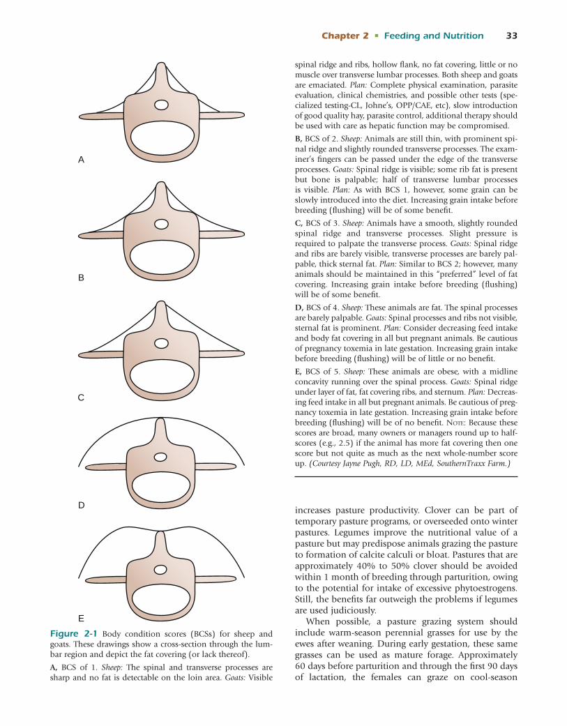
Chapter 2 • Feeding and Nutrition 33
increases pasture productivity. Clover can be part of temporary pasture programs, or overseeded onto winter pastures. Legumes improve the nutritional value of a pasture but may predispose animals grazing the pasture to formation of calcite calculi or bloat. Pastures that are approximately 40% to 50% clover should be avoided within 1 month of breeding through parturition, owing to the potential for intake of excessive phytoestrogens. Still, the benefits far outweigh the problems if legumes are used judiciously.
When possible, a pasture grazing system should include warm-season perennial grasses for use by the ewes after weaning. During early gestation, these same grasses can be used as mature forage. Approximately 60 days before parturition and through the first 90 days of lactation, the females can graze on cool-season
B
D
E
C
A
Figure 2-1 Body condition scores (BCSs) for sheep and goats. These drawings show a cross-section through the lum-bar region and depict the fat covering (or lack thereof).
A, BCS of 1. Sheep: The spinal and transverse processes are sharp and no fat is detectable on the loin area. Goats: Visible
spinal ridge and ribs, hollow flank, no fat covering, little or no muscle over transverse lumbar processes. Both sheep and goats are emaciated. Plan: Complete physical examination, parasite evaluation, clinical chemistries, and possible other tests (spe-cialized testing-CL, Johne’s, OPP/CAE, etc), slow introduction of good quality hay, parasite control, additional therapy should be used with care as hepatic function may be compromised.
B, BCS of 2. Sheep: Animals are still thin, with prominent spi-nal ridge and slightly rounded transverse processes. The exam-iner’s fingers can be passed under the edge of the transverse processes. Goats: Spinal ridge is visible; some rib fat is present but bone is palpable; half of transverse lumbar processes is visible. Plan: As with BCS 1, however, some grain can be slowly introduced into the diet. Increasing grain intake before breeding (flushing) will be of some benefit.
C, BCS of 3. Sheep: Animals have a smooth, slightly rounded spinal ridge and transverse processes. Slight pressure is required to palpate the transverse process. Goats: Spinal ridge and ribs are barely visible, transverse processes are barely pal-pable, thick sternal fat. Plan: Similar to BCS 2; however, many animals should be maintained in this “preferred” level of fat covering. Increasing grain intake before breeding (flushing) will be of some benefit.
D, BCS of 4. Sheep: These animals are fat. The spinal processes are barely palpable. Goats: Spinal processes and ribs not visible, sternal fat is prominent. Plan: Consider decreasing feed intake and body fat covering in all but pregnant animals. Be cautious of pregnancy toxemia in late gestation. Increasing grain intake before breeding (flushing) will be of little or no benefit.
E, BCS of 5. Sheep: These animals are obese, with a midline concavity running over the spinal process. Goats: Spinal ridge under layer of fat, fat covering ribs, and sternum. Plan: Decreas-ing feed intake in all but pregnant animals. Be cautious of preg-nancy toxemia in late gestation. Increasing grain intake before breeding (flushing) will be of no benefit. Note: Because these scores are broad, many owners or managers round up to half-scores (e.g., 2.5) if the animal has more fat covering then one score but not quite as much as the next whole-number score up. (Courtesy Jayne Pugh, RD, LD, MEd, SouthernTraxx Farm.)

34 Chapter 2 • Feeding and Nutrition
annual grasses. In some environments the warm-season grasses then begin their seasonal production. With this system, very little supplemental feeding is required. So long as quantity of the various forages is not limited, grain supplementation usually is not required. In prac-tice, however, weather typically limits forage quantity for 60 days or more each year. A good-quality, pasture-based forage feeding system often requires minimal energy and protein supplements for nonlactating, nongrowing ani-mals. Stockpiled forages can be an economical source of nonharvested feedstuffs and are most efficiently utilized when “strip grazed.” With use of winter annual pastures for grazing, allowing the forage to reach a height of 8 inches before access by animals, strip grazing, and lim-iting grazing all enhance efficiency of utilization.
For proper forage management, adequate acreage in grazeable land and several pastures are needed for a rotational grazing program. Forage requires some peri-ods of rest from grazing to maintain optimal productiv-ity. Therefore pasture rotation is essential.
The pasture layout does not need to be elaborate or to comprise many small paddocks. However, pas-tures do need to be divided for proper maximization of forage production. Approximately 6 to 10 separate paddocks or pastures are desirable, and further subdivi-sions can be added as needed. The divisions should be based on the productivity of the soil and natural breaks in the topography. They will not necessarily be of equal size. The forage should be grazed in a way that optimal leaf material is produced. Depending on the time of year and amount of moisture, the length of time graz-ing an area and rest between each rotation will vary. For example, the keeper may decide to have the flock graze each of 10 paddocks for an average of 3 days at a time; at the end of the rotation, the first paddock has had 30 days of rest and should have good forage regrowth. This type of grazing management may not necessarily increase animal gains, but it may increase the land’s carrying capacity as well as the overall quality of the pastures. Pasture rotation systems that increase grass production do not necessarily aid in parasite control. Between four and six ewes (and their lambs) and five to eight does (and their kids) can be maintained on the same amount of land that will support one cow and her calf. In woodland or brushy areas, the same land that will nutritionally support one cow and her calf will provide enough forage for approximately 10 goats and their kids.
A complete mineral supplement should be offered at all times. An adequate mineral supplement for animals grazing grass pasture contains 15% to 30% salt, 6% to 12% calcium, 6% to 12% phosphorus, and 1% to 4% magnesium (except in early spring when magnesium should be 8% to 14% of the minerals). Trace miner-als suitable for the area and soil type also should be offered.
RANGEMany of the world’s sheep and goats graze on range lands. The common goal for all range land enterprises is to use as much standing forage as possible, with minimal use of harvested forage or other supplements. Supplemental feeding should be practiced only when nutrient demands far exceed the nutrient supply of the forage. Some deficiencies are acceptable because of the female’s ability to regain body condition during the period from weaning until breeding.
The amount and type of supplementation needed are variable across range conditions. The two most important factors in supplementation decisions are stage of animal production (e.g., lactation) and weather conditions (e.g., moisture or snow cover). A good range mineral mixture includes equal parts of dicalcium phosphate and trace mineral salt. The trace mineral–salt component should be designed for the local forage and soil types. In general, phosphorus should be supple-mented under most range land conditions. Regardless of its composition, the salt-mineral supplement should be made available on a free-choice basis as the only source of salt.
Additional supplements containing protein or energy may be used as needed. Body condition scoring can help in making the decision to supplement energy. If the level of desirable performance can be attained by using a supplemental grain in amounts equal to 0.5% of body weight or greater, feeding grain can be econom-ical. If greater quantities of grain are needed, however, negative effects on forage use (e.g., depressed digest-ibility of forage) are possible. Several grain byproducts are acceptable supplements for ruminants consuming a forage-based diet. For example, soybean hulls and wheat middlings can provide economical supplies of energy without negative effects on forage use. Protein supplements in the form of soybean meal or cotton-seed meal are often used and may actually enhance the digestibility of moderate- or poor-quality forage.
Whenever hand feeding is difficult, salt-limited rations may be useful for range-fed sheep or goats used for brush control. Depending on requirements, supple-mental energy (e.g., corn, oats) or protein (e.g., cotton-seed meal, soybean meal) should be ground and mixed with salt in a 3:1 to a 6:1 salt-to-grain ratio, depending on intake. If intake is too great, more salt should be added. If intake is too low, salt should be reduced. In all cases, only white salt (NaCl) should be used. The use of salt-limited feeds decreases trace mineral intake. If trace mineral deficiencies exist locally, and salt-limited feeds are to be used, the keeper should add a suitable trace mineral salt to the feed in quantity such that trace min-eral consumption does not exceed 0.02% of the ani-mal’s weight. Salt-limited supplemental feeding should be introduced slowly over 2 to 3 weeks, and the animals

Chapter 2 • Feeding and Nutrition 35
performance should be monitored daily, particularly in times of stress (predator attacks, weather changes).
CONFINEMENT FEEDINGConfinement feeding of sheep or goats in various small vegetation-free enclosures or dry lots is used in certain locales for all or part of the year. In climates with colder winters and areas that lack winter grazing, some produc-ers move sheep (and occasionally goats) to a sheltered dry lot or barn for protection. Such situations usually require more start-up money (for construction of a barn to house animals, feeding floor or lot, and water system) than that needed for range or pasture opera-tions. Confinement management also may increase the incidence of some contagious diseases, external para-sites (particularly during winter), feeding costs, bedding costs, and the need to handle and dispose of manure. Nevertheless, the advantages can more than outweigh the disadvantages in operations for which a cheap source of feed and labor is available.
When properly performed, confinement or dry lot feeding can all but eliminate two of the most serious problems with sheep and goat production: internal parasites and predators. However, during confinement feeding, some access to outdoor dry lots is needed to improve hoof and udder health and to decrease the need for supplemental vitamin D. Because no grazing is allowed and feedstuffs (hay, silage, grains) are fed in bunks or other types of feeders, production losses resulting from parasites can be curtailed. Also, less energy is required for maintenance (walking to a feed bunk versus grazing). Animals require 2 to 4 hours to consume the same amount of dry matter from hay that is consumed in 16 to 22 hours of grazing pasture. Heavy-wooled breeds of sheep in full fleece require 1.5 times more space in a confined area than those that have been shorn. Adult sheep and goats require 0.6 and 0.3 m, respectively, of linear bunk space per animal.
With confinement systems, ewes and does are more easily separated by age (ewe lambs, adult ewes) and production (lactating, dry or early lactation, late lacta-tion). The ability to feed groups separately can improve the use and efficiency of available feedstuffs and help decrease the incidence of some production diseases (e.g., pregnancy toxemia, hypocalcemia). A dry lot pro-gram can be used not only during winter but also when pasture becomes scarce or for feeding young lambs or kids for rapid gains.
In dry lot feeding, sheep or goats may be fed hay, silage, “haylage” (hay silage), or green chop. For hay feeding, “square” bales are associated with less waste, but they tend to be more expensive than larger, round bales. Nutrient loss from round bales can be as high as 50%. Storing hay in a shelter, above the ground, and feeding in a hay feeder will reduce waste.
The dietary habits of sheep and goats vary and affect intake. However, dietary preference appears to be a more important limiting factor in the use of certain feedstuffs in goats. The smell, taste, and variety of feeds also affect intake. Silage can be fed to sheep and goats, but animals of both species may take time to adapt to its smell and consistency. Sheep and goats appear to be more suscep-tible to infection with Listeria monocytogenes, and silage that has been poorly packed or exposed to air or has not attained a low enough pH (less than 4.5) may be con-taminated with Listeria or mold. Such silage should be avoided, as should bundle-fed, uneaten, frozen, moldy, or spoiled silage. Hay silages (haylage) and corn silage should have pH values of 3.8 to 4.5 and 4.0 to 4.2, respectively. Corn silage will invariably be deficient in both protein and calcium. (Note: Adding 20 lb of urea, 2 lb of limestone, and 4 lb of dicalcium phosphate/ton of silage will improve nutritional content.)
Feed bunk design should minimize animal contami-nation. Adults and kids (lambs) should be prevented from crawling into feed containers and soiling the feed.
Dry lot feeding also is of value in implementing a parasite control program. If oral anthelmintics of the benzimidazole class are to be used in a deworming program, forcing the animals to fast or feeding dry hay for 12 to 24 hours before deworming and then provid-ing dry lot feeding for as long as 72 hours will improve the results. This technique also allows for parasite egg–laden feces to be “cleaned” or passed from the bowel before the animals are placed on a safe pasture. Animals may then be moved to pasture after deworming in a relatively parasite-free state, reducing pasture contami-nation. Examples of confinement or dry lot rations are shown in Table 2-3.
FEEDING THE ADULT MALEMales should enter the breeding season in good body con-dition without excessive fat. Rams and bucks should be maintained at a prebreeding body condition score of 3 to 4, because they may lose more than 10% to 12% of their body weight in 1½ months of a breeding season. Condi-tion scores should be assessed as part of a breeding sound-ness evaluation approximately 2 months before breeding.
It usually is beneficial to feed a concentrated energy-protein supplement to the males beginning approximately 4 to 6 weeks before the breeding season. Depending on the body condition and size of the males and quality of forage, a daily ration of 6 to 8 lb of forage and 1 to 2 lb of 12% to 14% crude protein concentrate usually suffices. A good-quality supplement for grass-based forage is 80% corn and 20% soybean meal. After the breeding season, some concentrate may need to be fed to help the animals regain an adequate body condition. For the remainder of the year, adult males can be maintained on good-quality hay. If grass forage is fed, animals should have

36 Chapter 2 • Feeding and Nutrition
free access to a mixture of 50% dicalcium phosphate and 50% trace mineral salt. If legumes constitute a signifi-cant portion of the diet, a mixture of 50% trace mineral salt, 25% dicalcium phosphate, and 25% defluorinated rock phosphate can be offered. In both instances, these mineral-salt mixtures should be the only source of salt offered to encourage adequate intake. The trace mineral component should be designed for the local soil types. For sheep, low-copper mineral mixtures are optimal, but goats can safely consume trace mineral mixtures made for cattle. Because of the possibility of urolithiasis in males, the keeper should take steps to prevent stone formation by adding ammonium chloride or other urine acidifiers to the mineral mixture.
FEEDING THE FEMALEBreeding females have different nutrient requirements as the stage of production changes. Although require-ments are much lower for maintenance than for lac-tation, meeting these requirements is important for efficient production. Body condition scoring all females every 2 to 3 weeks is an important and cost-effective management tool. Mineral feeding as described for the adult male is applicable for the female.
MaintenanceDuring maintenance, the objective is to maintain the female’s weight and health and replenish any losses expe-rienced during lactation. Most pasture or range settings provide adequate levels of nutrient intake to maintain dry, nonpregnant sheep and goats for this entire period. If extremes in environmental conditions occur (e.g., drought, snowfall), some supplemental feeding is required.
BreedingAt the time of breeding, the practice of flushing females has been used with some success. The basic premise is that increased nutrition, specifically energy, just before
and during the early breeding season increases the ovu-lation rate and thus the lambing or kidding rate. The female’s age and body condition and the time of year all affect the response to flushing. Mature females in mar-ginal body condition usually respond best to flushing. Moreover, the practice appears to be more beneficial in attempts to breed the group early or late, as opposed to during the peak of breeding season. Overconditioned females either do not respond or appear to respond only marginally to flushing. Flushing can be accom-plished by the provision of lush pastures or by supple-mentation with approximately 0.14 kg (0.33 lb) to 0.45 kg (1 lb) of a 10% to 12% crude protein grain/head/day. It is best to begin the hypernutrition approxi-mately 2 weeks before the males are introduced and continue for an additional 2 to 3 weeks into the breed-ing season.
The benefits of flushing include increased body con-dition, increased ovulation rate, and increased number of lambs born. Adequate body condition is neces-sary for acceptable conception rates. Outside certain biologic limits, a flushing effect is not observed. For example, an extremely thin (BCS 1) female probably will not achieve an increased ovulation rate because she is too thin to have normal reproductive cycles. However, within normal ranges (BCS 2.5 to 3) the ovulation rate appears to respond to a short-duration increase in energy and, to a lesser extent, to increased protein intake (see Figure 2-1). Flushing does not always increase lambing or kidding rates; however, it does increase the number of females cycling early in the breeding season, resulting in birth of a greater propor-tion of the offspring early in the lambing or kidding season. In females, a BCS at or just under 2.5 to 3 is optimal for most breeding flocks.
Early to Middle GestationAfter the female has conceived, early gestation is the time of partial fetal and placental development. Nutrition is important for adequate development, but
TABLE 2-3 Example Rations for Dry Lot Feeding for Nondairy Animals (lb/day)
150-Pound Ewe 70-Pound Doe
Maintenance Gestation Lactation Maintenance Gestation Lactation
Ingredient* A† B† A B A B A B A B A B
Alfalfa hay 2.9 4.25 5.5 2.0 2.8 2.6Grass hay 2.9 3.6 4.8 1.67 2.3 2.2Corn 0.25 0.75 1.0 1.0 0.33 0.5 0.4 0.4Soybean meal 0.15 0.75 0.4
*See Table 2-5 for average nutrient contents for each ingredient.†A and B are different sample diets for each stage of production.

Chapter 2 • Feeding and Nutrition 37
requirements are not greatly increased over those of maintenance. If the diet is lacking in energy, protein, and certain minerals, placental development may be poor, resulting in poor fetal growth. A reduction in lamb survival rates at birth can result from inadequate feeding during early gestation. Likewise, adequate nutri-tion is required for proper attachment of the embryo to the uterus. Midterm stress abortions can occur in Angora goats as a result of energy deficiency. This stress effect is more common in range conditions, particularly after a weather change, predator attack, or decreased feed intake. The incidence can be minimized by not breeding the female until she has attained 60% to 70% of her projected mature weight, and by maintaining a steady nutritional state during pregnancy.5
During early gestation, ewes and does can be main-tained on winter range or pasture or moderate-quality hay, but grass-quality grass hay, grass-legume, or small grain pasture would be best. If corn silage is fed, ewes and does at this stage may need ¼ to 1⁄3 lb of a pro-tein supplement daily. With seasonal decreases in feed availability or with weather-associated increases in feed requirements, some supplemental grain may be required. Females should be fed to maintain a BCS of 2.5 to 3 during early gestation. The scores should be assessed every 2 to 3 weeks, and any flock condition score change acted on immediately.
Late GestationThe nutrition of the female during the last 6 weeks of gestation is extremely important. Approximately 70% of fetal growth occurs during this period. Inadequate nutrition can result in poor colostrum production, low birth weight in both lambs and kids, lower energy reserves in the newborn animals, and increased death losses, especially during cold and inclement weather. Birth weight is an important factor affecting newborn survival. It can be influenced by breed, number born, age of dam, and the dam’s preparturient diet. Extremely low birth weights (less than 2 kg) in lambs can result in increased mortality during the first 24 hours. Con-versely, overfeeding of energy can result in obesity and contribute to dystocia. Proper nutrition is crucial. In general, more problems result from underfeeding than from overfeeding during late gestation.
The process of converting dietary energy into fetal growth is quite inefficient. Because 70% to 80% of fetal growth occurs during the final 6 weeks of gestation, the dam’s energy requirements increase substantially. In many instances the only way to provide the extra nutrition is to increase the amount of concentrate being offered. This sharp increase in energy requirements is compounded if the pregnancy involves multiple fetuses. A large uterus filled with several fetuses physically limits rumen capacity. In such cases, the mature female may
not be able to consume enough forage to meet her needs. The keeper may then wish to feed a supplement of between 1⁄3 and 1 lb and between ¾ and 2 lb of grain per day for goats and sheep, respectively. A ration of free-choice grass hay, 1 to 2 lb of a 20% protein range cube (depending on female size), usually will suffice.
During late gestation, feeding regimens should be designed to minimize use of energy supplied by body fat reserves. This consideration is especially crucial for ewes during late gestation. Excessive catabolism of body fat can result in pregnancy toxemia. The dam is at greater risk for this condition with concurrent stress from some environ-mental factor or disease. Pregnancy toxemia is character-ized by a buildup of ketones in the blood secondary to accelerated fat catabolism. Affected ewes appear listless and have a distinct acetone smell to the breath.
Maintaining the flock at BCSs of 2.5 to 3 and pro-moting adequate energy intake during late gestation will help prevent pregnancy toxemia. During late gesta-tion, ewes with a single fetus may consume as much as 3.5% to 4% of their body weight in dry matter in grain or excellent-quality forages. Intake may reach 5% in some does. If poor-quality forage is fed, these preg-nant females may be able to consume only 2% to 3% of their body weight in dry matter. Treatment can be successful, but as is the case with all nutritional prob-lems, prevention is the best strategy. Ewes should be fed approximately 1 kg (2.2 lb) of a cereal grain (e.g., corn, oats) during the final month of gestation to prevent pregnancy toxemia.
Goats also can develop pregnancy toxemia but appear to be more resistant (see Chapter 5). Dairy goats that are grazing or being fed good-quality grass hay can be fed 0.5 to 1 kg (1 to 2 lb) of a 16% crude protein grain/100 lb of body weight daily during the final 1½ months of gestation. The amount of grain may need to be adjusted depending on body condition.
In addition to promoting the birth of healthy lambs and/or kids, and preventing pregnancy toxemia, ade-quate nutrition during this time frame promotes sig-nificant mammary development during the last 30 days of gestation. Stillbirths, pregnancy toxemia, and poor milk production all are indicators of feeding an energy- deficient diet in late gestation. Adequate nutrition should be provided to support milk and colostrum production.
Feed or mineral supplements that contain added ion-ophore, antibiotics, or decoquinate may help control or prevent coccidiosis, abortion, and pregnancy toxemia (see earlier under “Feed Additives,” as well as Chapters 5 and 6 and Appendix 1).
LactationIn both sheep and goats, milk production peaks within 2 to 3 weeks after parturition and then declines rather rapidly to a low by 8 to 10 weeks after parturition.

38 Chapter 2 • Feeding and Nutrition
In dairy breed animals, this drop in milk production is less profound. A dam nursing a single kid or lamb pro-duces less milk than a female nursing twins or triplets. This is because one lamb or kid is unable to consume the full amount being produced, allowing a reduction in total mammary output. A dam nursing twins pro-duces approximately 30% more milk than one nursing a single. Likewise, a lactating dairy goat being milked two to three times per day for maximal production also produces greater amounts. A dairy goat usually weighs 10% of a dairy cow’s weight but may require 12% to 14% of the nutrients. Lactating does may be capable of consuming 4% to 5% or more (up to 10% to 11% in some females) of their body weight in dry matter, making feed intake the most important limiting factor affecting milk production.
Milk production during the first 4 weeks of lactation is important for good lamb and kid growth. If milk pro-duction is lacking, the lamb or kid can compensate by increasing solid feed consumption. Because feed is less digestible than milk, however, suckling animals cannot consume enough feed to make up for a milk deficiency and may therefore exhibit suppressed growth rates dur-ing early lactation.
Underfeeding energy during late gestation or early lactation results in greater-than-expected death losses in lambs, particularly twin lambs. Depressed milk pro-duction results in lambs that are “scruffy,” poorly kept, thin, and weak. Necropsy findings in affected lambs are nondescript—the gastrointestinal tract is filled with straw, and the animal has little or no abdominal fat. Lambs older than 1 month are less likely to starve as they begin to eat on their own (See Chapter 20).
During peak lactation it is nearly impossible for a ewe or doe to consume enough feed to meet her nutrient demands. During this time, good- to excellent- performing dairy animals use body fat to make up for this deficit and therefore experience a downward shift (often by more than 1 point) in BCS. This degree of loss is the reason why an adequate body condition before parturition is paramount. To make efficient use of her body fat, a ewe or doe must have adequate levels of protein in the diet. Whenever diets containing large quantities of cereal grain are fed, some form of rumen buffer should be included in the diet or offered on a free-choice basis.
Because feed intake can limit production in heavy-producing dairy animals, increasing the diet’s energy density in early lactation may be required. The addi-tion of fat to the diet is an excellent way to increase the energy density of the diet. As a general rule, supplemen-tal fats should not exceed 4% to 5% of the diet. Oil seed (whole cottonseed), if locally available, is an excellent source of additional energy in the diet. Approximately 2% to 3% of the added fat can effectively come from oil seeds. If more fat is needed, 2% to 3% more fat can be added in the form of specialty feed fats, including
calcium or magnesium salts or fatty acids. These spe-cialty fats are expensive but for the most part bypass the rumen. The fatty acids and calcium or magnesium salts are broken apart for digestion in the small intestine.
Obviously, the concentrate portion of the grain can be adjusted on the basis of BCSs. These recommendations show the importance of adequate protein concentra-tions for maximal milk production. Whole cottonseed can be included in the diet of lactating animals as an excellent source of both energy (greater than 90% TDN) and protein (21% to 23% crude protein). Whole cottonseed should account for no more than 20% of the diet. The requirements of most lactating ewes can be met by feeding 3.2 to 3.6 kg (7 to 8 lb) of a 12% to 14% crude protein, 55% to 60% TDN diet. If hay is fed, a grass-legume or legume-only hay helps supply protein demands. If silage (approximately 2 to 3 lb) is fed, then a protein supplement (¼ to ½ lb), grain supplement (1 to 1.5 lb), and ground limestone (0.02 to 0.04 lb) should be offered. If grass hay is offered (approximately 2 to 4 lb), then a protein supplement (1⁄3 lb) and grain (1 to 2 lb) should be provided. With the exception of that in dairy goats or ewes, milk production decreases quickly; by 8 to 10 weeks post partum it has become an insignificant nutrition source for the suckling lambs or kids. Up to this point the dam’s requirements can be met by grazing moderate- to good-quality pasture or range. If animals are grouped and fed by production, first-lactation dams with one kid or lamb should be fed with mature females with twins. Also, if these first-lac-tation dams have twins, they should be fed with mature dams with triplets.
Some dairy goats are susceptible to production of “off-flavor” milk. Cabbage, onions, wild garlic, and some species of weeds or browse all can negatively affect milk flavor. If certain feed sources cannot be avoided, feeding these off-flavor producers just after milking may limit some of their ill effects. Still, avoiding the offend-ing feedstuffs is the best method of prevention. Other nonfeed influences on milk flavor are disease (metritis, mastitis), filthy living conditions, and gastrointestinal upset.
FEEDING THE LAMB OR KIDBottle FeedingRearing orphaned lambs or kids on milk replacer is quite expensive and labor-intensive. If at all possible, keepers should attempt to graft the orphans onto another dam, rearing them on milk replacer only if this cannot be accomplished. Ideally, orphans need to consume small quantities of milk many times per day, which generally is not possible for most sheep and goat producers. Most producers feed “bottle babies” only one to three times each day. Many dairy kids or lambs are removed from their dams somewhere between birth and 72 hours of

Chapter 2 • Feeding and Nutrition 39
age and fed as orphans. The most economical way to raise orphans is to get them onto a dry concentrate feed as soon as possible.
The newborn needs to receive 10% to 20% of its body weight in colostrum, preferably within 3 to 12 hours after birth. If it is not available from the dam, fro-zen colostrum supplies can be thawed and used. Colos-trum absorption decreases rapidly from birth through 36 hours of age. Hemolytic crisis has been observed in some lambs fed cow colostrum. Still, cross-species colostrum often is better than no colostrum. Dairy cow colostrum usually is available but is relatively dilute in its immunoglobulin content. Any colostrum fed to an orphan should be free of caprine arthritis encephalitis (CAE) and Johne’s disease. If lambs or kids are unable to nurse, they need to be tubed. To pass a tube for feeding, lay an 14-18 French, rubber feeding urethral catheter along the lamb or goat from the tip of it’s nose, along the neck so the tip lies at the last rib. Mark the tube at the nose. Gently open the lamb/kids mouth and pass the tube over the tongue, into the esophagus, and on until the mark is just in front of the mouth. The tube can usually be palpated just to the left of the larynx as it passes into and down the esophagus.
After the initial amount of colostrum is fed, addi-tional feeding should be withheld for as long as 5 hours in newborn animals that are to be bottle-raised. This strategy encourages sucking, easing the transition and aiding in training to a bottle, nipple pail, or bucket. If the owner wishes to feed by hand, a lamb nipple attached to a soda bottle is a good system. The nipple should be placed in the mouth and the newborn’s jaw moved in a chewing motion by the handler. This usu-ally stimulates the nursing reflex in all but very weak newborn animals. Lambs or kids left with their dams for more than 2 days require longer training to become accustomed to a bottle or pail.
When a ewe or doe has too little milk to support more than one newborn, it is imperative that sufficient colostrum be given to all. The keeper should then leave the strongest, most vigorous newborn with the dam and raise the weakest artificially. Although immunoglobulin may not be absorbed after 12 to 36 hours, colostrum is a rich source of vitamin A, energy, protein, and local gut-acting antibodies. It also acts as a laxative. If pos-sible, colostrum should be fed for 2 to 3 days.
If lambs or kids are to be hand-fed, feeding 10% to 20% of their body weight in the form of good-quality milk replacer divided into four equal daily feedings usually is acceptable. Milk replacers for goats should be around 20% protein and 20% fat, with most of the protein supplied by an animal source (whey pro-teins). If the milk replacer appears brown, the pro-tein sources may have been overheated, resulting in decreased digestibility. Antibiotics commonly are added to help reduce the incidence of bacterial respiratory
and enteric diseases. Milk replacers should be fortified with vitamin A (20,000 to 30,000 IU/kg of dry matter), vitamin E (30 to 40 mg/kg of dry matter), and vitamin D (2500 to 3500 IU/kg of dry matter). The composition of sheep and goat milk is available in several publica-tions.4 If lamb milk replacers are used for goats, they should be diluted, because they contain more fat than naturally occurs in goat milk. Good-quality milk replac-ers designed for calves may be fed to goats and lambs in small quantities in several feedings (10% to 20% of body weight divided into four to six equal feedings). When mixing milk replacers, the keeper should take care to ensure that the powder and water are properly mixed into a suspension. Frequent feeding of small quantities will help reduce the incidence of bloat. By the third week of life, some kids or lambs can be switched to a twice-daily feeding regimen. Because milk replacers are expensive, animals should be weaned as soon as possible. If lambs or kids are underfed or fed a poorly digestible replacer, they may become emaciated, weak, or comatose. Death is possible. Inadequately fed lambs or kids have lower-than-normal blood glucose and at necropsy will be found to be devoid of fat stores. The abomasum in starved neonates often becomes impacted with hair or poorly digestible items.
The most efficient and least labor-intensive system is to place the orphans on a self-feeder using refriger-ated milk or milk replacer. This strategy helps limit milk consumption so that the orphans nurse more frequently throughout a 24-hour period. A self-feeder regimen in effect imitates the normal dam-newborn nursing regimen. Keeping the milk cold also may help prevent spoilage and lessen the extent to which the milk replacer separates out of suspension. In addition, kids or lambs using a self-feeder should have access to an extremely palatable dry feed. In orphaned lambs or kids, solid feed should be introduced as soon as possi-ble. Offering ¼ lb/day of a mixture of corn, oats, alfalfa pellets, molasses, and soybean meal that provides 14% to 16% crude protein works well. Top-dressing the feed with a dry milk replacer also may stimulate early intake of the dry feed. Other ingredients known to be extremely palatable to young ruminants are soybean hulls and various sources of bran, including wheat bran.
Creep FeedingThe term creep feeding refers to the use of supplemental feed for the nursing lamb or kid. The goals of a creep feeding program are to promote an adequate intake with a palatable feed and to provide all necessary nutri-ents in the most economical regimen possible. Both lambs and kids use feedstuffs more efficiently before weaning.
Lambs and kids will only nibble at the creep feed until they are 3 to 4 weeks old. Nevertheless, the creep

40 Chapter 2 • Feeding and Nutrition
feed should be made available as soon as possible to help the orphans get used to eating from one loca-tion and to help establish rumen function. The feeder should be placed in a dry, well-lighted area where lambs or kids can easily gain access but still retain visual con-tact with their dams, and kept clean, with a minimum of 2 inches of bunk space per lamb or kid. A variety of methods can be employed to maximize the acceptance of the creep area. Strategies include hanging a light over the creep feeder, retaining one or two dams and their offspring in the area for a few days (with limited feed, of course), and putting all of the animals in a small, confined space adjacent to the creep area.
Creep feeds need not be complex, but they must be palatable because they are competing with milk. Pellet-ing or coarse grinding feeds usually increases intake. Fine grinding usually results in decreased intake as animals (particularly lambs) age. Pellets should be small enough for consumption. In goats, pellet size larger than 5 to 7 mm may decrease intake. After the lambs or kids have begun to consume the creep feed, cheaper ingredients can be used for a more cost-effective regimen. Until the animals reach 3 to 4 weeks of age, however, palatability is the key to successful creep feeding. If increased perfor-mance is to be attained from creep feeding lambs, they must consume more than 0.23 kg (0.5 lb) daily from 3 weeks of age to weaning. Low-fiber creep feeds contain-ing 16% to 20% protein usually work best. Enhanced performance may be attained if salt (0.5% of the creep feed), ammonium chloride (0.2 kg/440 kg of feed, or 10 lb/ton), and vitamin E are added to most creep feeds. Some examples of creep feeds are shown in Table 2-4.
In general, creep feeding should provide an addi-tional 0.5 kg of gain for each 1.8 to 3.2 kg (4 to 7 lb) of feed consumed. The level of efficiency will vary from one set of conditions to another, but generally when feed costs are low and sale prices are high, creep feeding usually is profitable. It is less profitable when feed costs
are high and sale prices are low. In the final analysis, the feasibility of creep feeding is determined simply as a matter of feed costs versus animal sale prices.
WeaningLambs and kids can be weaned as early as 3 or 4 weeks, but better results may be obtained if weaning is delayed until 8 to 12 weeks. Because of labor constraints, many keepers attempt to wean milk replacer–fed young as soon as possible. Kids of most meat and dairy breeds should weigh at least 9.1 to 11.4 kg (20 to 25 lb) and consume 0.23 kg (0.5 lb)/day of a 16% to 18% crude protein grain.
Because weaning is such a stressful event, the imme-diate goal should be to get the lamb or kid accustomed to eating out of feedbunks and drinking from a water trough. The decision to wean lambs or kids depends on age, season of birth, whether they have been consuming creep feed, existing parasite or predator problems on the farm, market price, and available labor. Feedbunk location is important in encouraging newly weaned animals to consume adequate amounts of dry matter. If excellent-quality forage is available, however, it can be used as the sole source of feed. A good strategy is to place the feedbunks perpendicular to the fence line so that the weanlings are forced to see and possibly inves-tigate the feed as they walk (usually continually) the fence line. For the first 2 days of the weaning period, good-quality hay should be offered on a free-choice basis. The weanlings should then be introduced to a concentrate feed offered at a level of approximately 1% of body weight/day. A lamb weighing 31.8 kg (70 lb) therefore consumes approximately 0.32 kg (0.75 lb)/day. After the lambs or kids have been introduced to the grain, the keeper can gradually increase the amount.
Some managers prefer to remove all grain supple-ments and place the dam on a poor-quality forage
TABLE 2-4 Sample Creep Diets for Lambs and Kids*
Element Sample 1 Sample 2 Sample 3 Sample 4
Ground corn 33% 60% 63% 40%Oats — — — 11%Soybean hulls — — 10% —Soybean meal 6% 8.5% 10% 6.5%Alfalfa hay 55% 25% — 35%Bran — — 10% —Molasses 5% 5% 5% 6%Trace mineral salt 0.5% 0.5% 0.5% 0.5%Ammonium chloride 0.5% 0.5% 0.5% 0.5%Limestone — 0.5% 1% 0.5%
*Diets 1, 2, and 3 should be fed with an excellent-quality hay offered on a free-choice basis. Diet 4 is a complete, pelleted feed.

Chapter 2 • Feeding and Nutrition 41
1 week before weaning. This reduces milk production and decreases the incidence of mastitis. By 7 months, most dairy breed kids should weigh between 27.3 and 36.4 kg (60 to 80 lb). A good-quality mineral mixture should be offered on a free-choice basis. Potential replacement animals should be identified and fed a regimen to minimize excessive fat deposition and maximize postweaning growth rates. The same guide-lines described for mineral feeding in the male (50% dicalcium phosphate and 50% trace mineral salt) are applicable for weanlings.
FinishingFinishing of lambs for slaughter can be accomplished in a variety of ways. No one perfect diet for finish-ing has been defined. Instead, each feeding facility accomplishes the goal by using feedstuffs that are avail-able and economical to the geographic area. Feedlots designed specifically for goats are not as common as those designed for lambs. Most goats are slaughtered off forage-based diets with little use of concentrate feeding.
At slaughter the lamb should have approximately 0.23 to 0.46 cm of backfat. However, the amount of backfat often depends on specific market preferences. Slaughter weights have a wide range because of the variation in frame size among North American sheep, although they generally fall between slightly below 45.4 kg (100 lb) and 68 kg (150 lb). Ideally, the lambs should be marketed at the proper degree of finish, regardless of their weight. Feeding beyond the lamb’s ideal finish results in higher cost of gain because of decreased feed efficiency. Adding lean muscle is much more energy-efficient than adding body fat. Blackfaced sheep and meat goat breeds generally finish at greater body weights.
If high-quality forage is available, lambs can be finished on it. This regimen generally works best for smaller, younger lambs. Older, heavier lambs require some concentrate feeding. For example, a small-framed lamb born in January in the southeastern United States could be ready for slaughter in June having been grazed on only cool-season annual grasses (ryegrass) or grass-legume pastures. By contrast, a large-framed, spring-born lamb in the western United States may come off range in the fall at 6 months weighing 31.8 to 41 kg (70 to 90 lb) and need a concentrate-based diet to be finished by 1 year.
Many lambs in North America are finished in a feed-lot or dry lot. Such lots vary in size and may be open areas, confinement barns, or a combination of both. An excellent feeding regimen is stepwise feeding, whereby lambs (and occasionally kids) are given more grain as they get larger. By the end of the finishing period, many animals typically are consuming approximately 80% concentrate and 20% roughage. However, when given
free access to both roughage and concentrate, lambs consume approximately 60% to 70% concentrate and 30% to 40% roughage. A variety of cereal grains, includ-ing corn, oats, barley, milo, and to some extent wheat, can be used by lambs. Amounts used are based on local economics. A protein supplement may be included depending on the amount of protein being provided by the roughage source. Alfalfa commonly is used as a roughage source because of its wide availability, and animals feeding on it may not need additional protein. Mineral and vitamin premixes also are added to some diets. Because the finishing period usually involves institution of diets that emphasize grains, the nutrition-ist or clinician must be aware that excessive grain intake can predispose animals to urolithiasis, enterotoxemia, and bloat.
Processing of grains, with the possible exception of sorghum, does not appreciably increase lamb perfor-mance. Cracking, rolling, or flaking milo to break its hard seed coat increases its usability in lambs. Feeding other grains whole may actually tend to decrease the incidence of acidosis and other digestive disturbances. Pelleting bulky rations may be of some benefit because of the increased level of consumption. Pelleted feeds help ensure a more uniform intake and are less dusty and easier to handle. The most important factor to consider with regard to pelleting or other process-ing is the potential for the lamb or kid to “sort” the feed and consume only a portion of the diet. Sort-ing feed is more of a problem with self-feeding and group feeding. Thus pelleted feeds are best used in free-choice, self-feeding systems. For example, if the protein, mineral, and vitamin premix is a loose meal, cracking the grain may be beneficial in minimizing sorting, despite its lack of effect on usability. Such feed formulations, however, are more expensive, and their use may be associated with an increased incidence of some diseases.
As stated earlier, goats generally are not finished in commercial settings. In North America, most meat goats are slaughtered by the consumer; in small, local processing facilities; or within niche marketing sys-tems. With some exceptions, goats tend to be sold in small groups over the course of the year. Because of this method of marketing, goats generally are kept on a forage-based diet rather than maintained with year-round feeding of grain. Still, some feedlots, or “grain on grass” operations, do exist. If a group of kids is placed on a concentrate-based diet for finishing, the same basic principles discussed previously for lambs apply. Table 2-5 provides examples of growing and finishing diets for lambs and kids. Growing diets, which contain 14.5% protein and 68% TDN, are used for younger, lighter lambs and kids. Finishing diets, which contain 10% protein and 80% TDN, are more effectively fed to older, heavier animals.

42 Chapter 2 • Feeding and Nutrition
Regardless of the species being fed, the introduction of energy-dense diets in a feedlot setting is stressful and associated with many metabolic diseases. The nutrition-ist or clinician should ensure that animals being fed in the finishing stages be slowly introduced to these diets and vaccinated for Clostridium perfringens serotypes C and D infection and possibly other diseases (e.g., contagious ecthyma, pasteurellosis) that are locally problematic. On arrival at the finishing facility, animals should be offered free access to a good-quality legume-grass hay, fresh clean water, and a mineral mixture. Animals should be introduced to the finishing diet slowly over a 2- to 4-week period. For males, ammo-nium chloride or other urine acidifiers should be fed to prevent urolithiasis (see Chapter 12). Rumen buffers, antibiotics, ionophores (see earlier under “Feed Additives”), and free-choice hay all are effective in mini-mizing some production diseases.
FEEDING YEARLINGSFemalesEach sheep and goat enterprise is unique in terms of overall goals. Some operations place importance on breeding ewes and does so that they have their first offspring by 1 year of age. Other farms or ranches may find it much more practical and economical to breed their animals to have their first offspring as 2-year-olds. A ewe’s lifetime production can be as much as 20% greater if she is bred as a ewe lamb rather than as a yearling.6
If the goal is to have the females lamb or kid as yearlings, nutrition is crucial from weaning to breed-ing. Yearlings should be maintained on a steady growth plane.
Depending on their weaning weights, most females need to gain between 0.11 and 0.23 kg (0.25 to 0.5 lb) per day from weaning until breeding. Replacement yearlings should be kept on the best available pasture.
In most instances, however, this management approach will require some supplemental energy or concentrate feeding. Concentrate feeding of ½ to 2 lb (depending on breed, species, size, and so on) of a 12% to 14% crude protein should be offered in settings of poor-quality forage. Overfeeding young females, however, can result in excessive fat deposition in the mammary glands and decreased lifetime milk production.
If females are to be bred as yearlings, a moderate growth rate is most desirable. The female should obtain 65% of her projected mature weight by the time of breed-ing. In reality, a range of weights probably exists within which small-framed sheep and goats may have acceptable conception rates at 55% to 60% of their projected mature weights, whereas some large-framed animals may need to be closer to 70% of their mature weights. So long as a good, well-planned forage system is available, females can achieve desired weight gains with little or no grain supplementation. Sheep or goats that can successfully breed out of season should be bred at 13 months so they can lamb or kid at 18 months. This approach requires less nutritional input than breeding 7-month-old females but still provides an acceptable generation interval for increased female productivity. After the females have been bred, moderate and steady weight gain is desirable until parturition, with a weight goal of 85% to 90% of the projected mature weight by 1 year of age.
Good-quality grass pasture will need to be supple-mented with additional energy and protein sources. Animals maintained on grass-legume mixtures will require less supplementation.
Regardless of the breeding system, animals should be weighed and body condition scored regularly whenever possible. If the BCSs of the group begin to drop below 2.5, the keeper should offer a source of supplemental energy; conversely, if the scores rise above 3.5, less energy supplementation is needed. A good-quality min-eral mixture as described for adult males is appropriate for use in yearlings.
TABLE 2-5 Sample Grower and Finisher Diets for Lambs and Kids
Element Grower 1 Grower 2 Grower 3 Finisher 1* Finisher 2* Finisher 3*
Corn 33.5% 28.5% 32.1% 73.2% 76.0% 74.6%Alfalfa 55% — — 20% — —Grass hay — 50% — — 17% —Cottonseed hulls — — 40% — — 14%Soybean meal 5.5% 15% 21% — — 4%Molasses 5% 5% 5% 5% 5% 5%Trace mineral salt 0.5% 0.5% 0.5% 0.5% 0.5% 0.5%Limestone — 0.5% 0.9% 0.8% 1.0% 1.4%NH4Cl 0.5% 0.5% 0.5% 0.5% 0.5% 0.5%
*Finisher diets should contain enough limestone (or other calcium source) to provide a 2:1 calcium-to-phosphorus ratio.

Chapter 2 • Feeding and Nutrition 43
MalesFeeding developing males is quite straightforward. They should be developed using as much forage as possible, with just enough supplemental feeding to produce desirable gains (0.34 kg or 0.75 lb/day). This goal is easily accomplished with good genetics. Growing males should be offered a good-quality mineral mixture as described previously, with the keeper taking steps to prevent urolithiasis and other production-related diseases.
FEEDING SHOW ANIMALSAll show animals should be offered a good-quality min-eral mixture and given free access to fresh, clean water.
LambsFeeding show lambs should be as simple as possible while providing the desired rate of gain and appropri-ate “bloom.” Ideally, the lamb should be fed 30% to 40% of its total daily intake as good-quality hay or forage; the remaining 60% to 70% of the diet should be in the form of a concentrate or grain mixture (Table 2-6). A lamb can eat as much as 3% to 4% of its body weight daily. At least 0.45 kg (1 lb) of hay/day should be fed with the concentrate. Lambs should be gradually exposed to increasing concentrate portions of the feed, taking 10 to 14 days to make the transition from little grain to the full amount. Feeds should never be switched abruptly, and fresh, clean water should always be offered on a free-choice basis.
Mature SheepMature show ewes and rams should consume approxi-mately 1.36 to 2.27 kg (3 to 5 lb) of concentrate/day, depending on their size. They also should be offered good-quality hay on a free-choice basis. The require-ments for mature sheep are found in the 2007 NRC recommendations for small ruminants.4 Adult show animals should be maintained in good condition but should not be obese. A good exercise regimen is neces-sary to prevent overconditioning. When possible, forc-ing animals to graze or walk some distance from grain to hay to water may prove valuable.
Show GoatsThe feeding of young meat goats for show is similar to the feeding of show lambs, as discussed previously. The recommended approach is to use a simple diet that pro-vides the desired level of gain and degree of bloom. The forages and concentrates for lambs presented in Table 2-6 also are appropriate for young goats.
FEEDING FOR FIBER PRODUCTIONSheepWool production is highly heritable; however, nutrition can affect wool growth and character. Within certain bio-logic limits, energy intake is directly proportional to wool production,6,7 although separating protein effects from energy effects is difficult. So long as the minimal protein requirement is met, additional dietary protein does not appear to increase wool growth. Wool does contain an abundance of the sulfur-containing amino acid cystine. Accordingly, feedstuffs rich in sulfur-containing amino acids are important for optimizing wool growth.
In general, the effects of nutrition on wool produc-tion are associated with quantity rather than quality. Increased nutrient intake can increase wool production, within limits. However, quality can be affected during periods of severe nutrient deprivation. Under these con-ditions, fiber diameter is decreased. Extreme underfeed-ing can result in weak fiber with limited value.4
The nutritional status of the ewe during gestation can influence the wool production of subsequent offspring. Kelly and colleagues8 bisected embryos to produce clones that were then placed in ewes fed at maintenance or sub-maintenance energy and protein levels from day 50 to 140 of gestation. The lambs that were born to the ewes fed a submaintenance diet produced 0.136 kg (0.3 lb) less wool from 0.4 to 1.4 years of age. The wool from these lambs was coarser than that produced by lambs born to ewes fed at a maintenance level. These effects have been attributed to decreased hair follicle develop-ment in fetuses whose dams were fed deficient diets, and they continue for the rest of the offspring’s life.
GoatsAngora goats produce large quantities of fiber per unit of body weight. The 2 to 3.6 kg (4.5 to 8 lb) of mohair obtained with each cutting can greatly increase nutri-tional demands. As with wool, mohair production can
TABLE 2-6 Concentrate Mixes for Show Lambs*
Element Sample 1 Sample 2
Corn 50% 45%Oats 35% —Soybean hulls — 40%Soybean meal 10% 10%Molasses 4% 4%Mineral mix 1% 1%
*Animals should be introduced to high-grain diets slowly over 2 to 3 weeks.
44 Chapter 2 • Feeding and Nutrition
be improved with increased energy intake. However, protein appears to elicit more of a effect on mohair growth than that on wool growth. Whereas cashmere wool appears to be only minimally affected by dietary manipulation, increasing dietary protein above require-ments increases mohair volume and fiber diameter.6,9 In Angora does fed isocaloric diets containing either 12% or 19% protein, an increase in grease fleece weight (of approximately 0.57 kg [1.25 lb]) and fiber diameter was noted with the higher protein intake.6 Mohair also con-tains abundant amounts of sulfur, so sulfur-containing amino acids are important in Angora goat nutrition. Qi and co-workers9 indicated that the NRC-recommended4 10:1 nitrogen-to-sulfur ratio for maximal mohair produc-tion may be on the low side and suggested that a ratio of 7.2:1 may be more useful. Therefore, if NPN is used as a protein source, sulfur supplementation is necessary.
Ranged Angora goats should receive nutritional sup-plementation during late gestation and early lactation. Salt-limited feeds can be used to control both energy and protein consumption under range conditions. Cottonseed or soybean meal (or other protein sources), corn, and salt (noniodized, nonmineralized) can be added at a 1:3:1 ratio. The keeper should introduce the supplement slowly, adding more white salt if the animals are overconsuming and decreasing salt if they are underconsuming. This salt-limited feeding system can be an effective way to increase energy and protein intake for range-fed goats (and possi-bly sheep). Careful intake monitoring is important.
Adequate shelter should be provided to fiber- producing animals, particularly young animals and Angora goats, that have just been sheared. In early spring or late fall, animals may be susceptible to cold stress for as long as 4 weeks after shearing. The pro-vision of shelters or wind breaks and an additional 0.23 to 4.5 kg (0.5 to 1 lb)/day of an energy supplement (cracked corn) above the normal feeding regimen can help minimize freezing and stress loss.
FEEDING PET AND GERIATRIC SHEEP AND GOATSPet sheep and goats can live much longer than animals in production units. The principles of nutrition presented throughout this chapter apply to the proper feeding of pet animals as well. The only dietary formulation, manipula-tion, or plan that appears to be associated with increased longevity is lowered intake. Thus keepers should strive to maintain a proper body condition and weight in pet animals to help them achieve a long healthy life.
Obesity is a constant and major problem in the pet population, to include both sheep and goats. Pet goats tend to be more commonly affected by overfeeding-related diseases than are sheep or goats in production units. With the exception of feedlot animals and those being prepared for shows, pet sheep and goats are
overrepresented in cases of obesity, bloat, acidosis or ruminitis, and urinary calculi. The increased prevalence of these disorders is related to a lack of knowledge about feeding in many owners, inactivity of the ani-mals, and pet status with its lack of performance or production goals. A barn or paddock layout that neces-sitates walking, client education, and proper diet design all are essential to combat obesity and will increase the pet animal’s long-term health. Forcing animals that are capable of doing so to walk (e.g., for grazing or access to water, salt, or minerals) will enhance the chances of the weight loss program’s success. Weight loss programs should never be instituted in pregnant animals and should be avoided until after midlactation.
A weight loss program should begin with a complete physical examination, an accurate weight measurement, determination of BCS, and a complete blood count and serum chemistry analysis when possible. If the animal has no overt disease, the weight loss program should set goals for weight and BCS and a plan to attain these goals in approximately 4 to 8 months. A generic weight loss diet modification might be as follows: (1) first 4 to 6 weeks: feed moderate-quality grass hay at 2% of body weight (accurate weight of animal and hay); (2) second 4 to 6 weeks: feed moderate-quality grass hay at 2% of target weight. (Note: The hay should be 8% to 10% crude protein; free-choice water and a mineral mixture designed for the farm should be provided; and accom-modation for some form of exercise should be made.)
Body weight and condition loss are common prob-lems among geriatric animals. A complete physical examination, complete blood count, and serum chem-istry analysis may be indicated to identify ongoing disease processes. Older animals may require special feeding to address dental disease, parasite damage to the bowel, and other general health problems. Good-quality hay, moistened pellets, lush forage, and palat-able concentrates often are required for animals with dental disease (see Chapter 4).
Allowing older animals ready access to feed, particu-larly if their social status has changed, along with longer periods of noncompetitive time to consume it, will help maintain good body condition. Because many geriatric animals have arthritic conditions, minimizing excess body weight, properly trimming feet, and placement of water and feed such that animals are not forced to walk great distances all are valuable in case management. Diets designed for the geriatric horse can be used. With copper concentrations greater than 10 ppm, however, these diets should not be fed to any sheep or to goats with a history of hepatic disease. If the animal has renal disease, the protein content of the diet should be maintained at 7% to 8%, and the calcium-to-phosphorus ratio should be kept at 1:1. A good-quality granular mineral mixture of equal parts dicalcium phosphate and trace mineral salt should be offered on a free-choice basis.

Chapter 2 • Feeding and Nutrition 45
If older animals are losing weight, the keeper can slowly increase caloric intake by 7% to 10% in the form of fat. However, protein, fats, and copper should be restricted in animals with hepatic disease. Animals with hepatic or renal disease may benefit from the addition of B vitamins, given orally or parenterally. If renal dis-ease is present, the protein requirement should be met but not exceeded. If anorexia is a problem, varying the diet, offering lush grazing, and adding energy-dense feeds are useful strategies. Obviously, all husbandry practices that aid in overall health (e.g., proper shelter, deworming) will enhance long-term survival.
NUTRITIONAL DISORDERSThe most common nutrition-related diseases seen in late gestation in goats and sheep are pregnancy toxemia (discussed in Chapter8), hypocalcemia, and hypomagnesemia.
HypocalcemiaHypocalcemia can be a problem in dairy goats and, to some extent, in ewes, meat and fiber goats, and pet animals. It usually becomes apparent shortly before or after parturition and is a result of low concentrations of serum calcium. Some cases also are complicated by hypophosphatemia and hypermagnesemia or hypo-magnesemia. Ewes appear to be most susceptible in late gestation and early lactation, particularly when experiencing some sort of stress (e.g., hauling, predator attack, lack of feed). Sheep may succumb to hypocal-cemia 6 weeks before to 10 weeks after parturition. The greatest demand for calcium for the nondairy animal occurs 3 to 4 weeks before parturition in females with more than one fetus, as a result of the calcification of fetal bones. Goats may have hypocalcemia before par-turition; in high-producing dairy goats, however, the disease generally occurs after the dam gives birth. With any abrupt demand for calcium, the body requires 1 or more days to accrue the necessary enzymes capable of mobilizing bone stores of calcium. High intake of calcium, phosphorus, or some cations (potassium, sodium) decreases the production of parathyroid hor-mones. During decreased parathyroid function, less 1,25-dihydroxycholecalciferol is produced. Lack of this hormone results in lowered absorption and mobiliza-tion of calcium from the intestines and bones. Low dietary calcium or increased amounts of dietary anions enhances the production and release of parathyroid hormones.
Clinical SignsEarly in the course of the disease, affected animals exhibit a stiff gait, tremors, and tetany; they also have decreased rumen motility and may be ataxic or
constipated. As the disease progresses, increased heart and respiratory rates, regurgitation of rumen content, bloat, and depression to the point of opisthotonos may be noted. Corneal and pupillary light reflexes are nor-mal at first but become depressed before disappearing entirely. The rectal temperature usually remains in the normal range but may be slightly low.
DiagnosisDiagnosis usually is based on a history and signalment conducive to development of hypocalcemia, as well as on response to therapy. Serum calcium concentrations less than 4 to 5 mL/dL in sheep and goats are fairly diagnostic of this condition.
TreatmentIn clinical cases, immediate treatment is needed, usu-ally in the form of intravenous administration of cal-cium borogluconate (50 to 100 mL of a 23% solution). Subcutaneous delivery of these calcium solutions or the oral administration of a calcium gel designed for cattle, but based on sheep or goat body weight, will help prevent relapse. If the subcutaneous route is chosen to develop a “reservoir” of calcium for affected animals, solutions containing dextrose or numerous electrolytes should be avoided if possible, because use of some of these preparations has been associated with abscess formation. During treatment, cardiac monitoring is indicated, and therapy should be slowed or stopped if arrhythmias occur. If the treatment is successful, the animal will stand and urinate within 20 minutes. If left untreated, affected animals usually die.
PreventionTo prevent or minimize the risk of hypocalcemia, partic-ularly in dairy goats, the diet should be low in calcium, with a low cation-to-anion ratio. The dietary modifica-tions used for the prevention of milk fever in cattle may be of value in dairy sheep and goats. Therefore reducing or eliminating diets rich in cations (alfalfa) or in calcium and phosphorus in the late dry period may aid in prevention. Many legumes are rich sources of potassium and calcium and can therefore contribute to hypocalcemia. Immediately after parturition the cal-cium levels in the diet should be increased. This strategy improves calcium reabsorption for bones and absorp-tion from the intestine. Hauling or other forms of stress should be minimized in sheep during the final 8 weeks of gestation. Even with this strategy, some incidence of hypocalcemia may be experienced.4
HypomagnesemiaHypomagnesemia, manifesting as grass tetany, can be a problem in sheep and, to a lesser extent, goats grazing on lush, rapidly growing forage. It usually occurs during the

46 Chapter 2 • Feeding and Nutrition
early spring on pastures that are well fertilized with nitro-gen and potassium. A combination of elevated nitrogen and potassium levels in the forage leads to a reduced absorption of magnesium from the gastrointestinal tract.
The primary problem in hypomagnesemia is reduced absorption by the animal, rather than low plant concen-trations. Sheep, and goats that graze lush cereal grains (e.g., wheat, rye), particularly in early lactation or late gestation, are predisposed to this condition. Any type of stress (e.g., weather changes, transportation, predator attack) can increase blood concentrations of free fatty acids, and excess blood from fatty acid concentrations depresses blood magnesium. Other forms of hypomag-nesemia occur during winter when animals are fed poor-quality grass hay (with low magnesium content) and in lambs or kids fed only low-magnesium milk replacers. Kids or lambs with access to grain or legume-grass hay are more resistant to hypomagnesemia. Ewes with poor dentition and those that lose excessive weight during winter are prone to develop the condition.
Clinical SignsHypomagnesemia generally occurs in ewes 2 to 4 weeks after lambing. It is more common in females that had twins. Affected animals are excitable and may exhibit paddling convulsions, clonic-tonic muscle spasms, and an increased respiratory rate. They also may simply be found dead in the pasture. Rectal temperature commonly is normal. Convulsions may be triggered by any num-ber of stimuli, from being chased by predators to acute changes in weather patterns. Lambs or kids with the milk replacer–associated form of hypomagnesemia usually are anorexic and hyperexcitable and may salivate profusely.
DiagnosisDiagnosis often is based on signalment and history, as well as response to treatment. Serum magnesium levels less than 1.5 mg/dL may be indicative of this disease; levels less than 1 mg/dL should be considered diagnos-tic. Postmortem serum samples are of limited value. Magnesium concentrations in cerebrospinal fluid (for 12 hours after death), urine (for 24 hours after death), or anterior eye chamber fluid (for 48 hours after death) are good postmortem tests.
TreatmentTreatment consists of the intravenous administration of a solution which contains 20% to 25% calcium boroglu-conate and 4% to 5% magnesium (50 mL). Oral calcium magnesium gel and subcutaneous injection of calcium-magnesium salts both are beneficial to prevent relapse.
PreventionBecause grass tetany results from a reduction in avail-able magnesium, a number of methods can be used to increase consumption. Properly balanced fertilizers
and magnesium compounds can be applied to the soil to increase plant concentrations of magnesium. The addition of such compounds is helpful but not very economical, because as noted, the primary problem with the occurrence of hypomagnesemia is reduced absorption by the animal, rather than low plant concentrations. Therefore prevention is best accomplished by offering high-magnesium mineral supplements before the growth of lush spring forage and several weeks prior to lambing or kidding. Most mineral supplements with high levels of magnesium are unpalatable; feeders should be checked frequently to ensure proper consumption. To enhance intake the keeper can mix magnesium oxide with molasses, corn, salt, or other feedstuffs. Daily consumption is impor-tant, because magnesium in a readily usable form is poorly stored in the body. An average adult lactating ewe needs 7 to 9 g of magnesium oxide daily. An eco-nomical supplement is a 1:1 mix of trace mineral salt and magnesium oxide, but this combination appears to be unpalatable. A more acceptable substitute may be equal parts of ground corn, trace mineral salt, and magnesium oxide. Other palatable grains can be used in place of the corn. Legumes (e.g., alfalfa, clover, bird’s foot, kudzu) are much better sources of both calcium and magnesium, and their inclusion in a pas-ture helps reduce the incidence of hypomagnesemia.4 Maintaining a high soil pH (greater than 5.5) enhances magnesium availability and intake by plants. The inclusion of vitamin D (5 to 10 IU/kg/day) in a milk replacer helps prevent hypomagnesemia in lambs or kids fed indoors.
UrolithiasisUrolithiasis is a common and frustrating problem for owners of male sheep and goats, particularly pet goats, and for clinicians involved in their management. In Chapter 12 the pathophysiology and clinical signs of urolithiasis and therapeutic modalities of relevance are covered in greater detail than that provided here.
Formation of phosphatic calculi is seen with man-agement practices that allow feeding of high-concen-trate, low-roughage, low calcium-to-phosphorus ratio, high-magnesium diets, and alkaline urine. High-grain diets result in the excretion of large amounts of phos-phorus in the urine. Oxalate calculus formation is associated with the consumption of oxalate-containing plants (Table 2-7). Urinary stones are composed of salts and minerals arrayed in a crystal lattice surround-ing an organic nidus.10 The nidus forms when urine mucoproteins or mucopolysaccharides and saturated urine precipitate to form crystals. Urinary mucopro-tein-mucopolysaccharide production is increased with ingestion of estrogenic compounds, inadequate lev-els of vitamin A, consumption of certain feedstuffs

Chapter 2 • Feeding and Nutrition 47
(e.g., cottonseed meal, milo), use of pelleted diets, and rapid growth of the animal.4,10
Dietary risk factors for urolithiasis include high-grain–low-roughage diets, decreased formation of saliva, increased amount of phosphorus excreted in the urine, and increased levels of dietary magnesium. Calcium-to-phosphorus ratio should be maintained between 1:1 and 2:1. Cereal grains have an abnormal calcium-to-phosphorus ratio of 1:4 to 1:6.4,10 Low-forage high-concentrate diets traditionally are deficient in vitamin A or its precursors. Vitamin A deficiency can result in desquamation of cells lining the urinary blad-der, which may serve as a nidus for stone formation. Clinical signs may include dysuria, stranguria, hematu-ria, urine dribbling, vocalization, prolonged urination, tail flagging, colic, and bruxism. A complete examina-tion should be performed, appropriate diagnosis made, and immediate medical or surgical therapy instituted4,10 (see Chapter 12).
Access to fresh, clean water is crucial to the pre-vention of this condition. Water should be abun-dant, fresh, clean, palatable, and readily accessible. In many geographic regions, maintaining water supplies requires more attention during winter months. The addition of sodium chloride to the diet (3% to 5% of the dietary dry matter intake) will increase water consumption, and the excess chloride ions may reduce production of calculus-forming salts.10 Diets and feed-stuffs rich in cations (e.g., alfalfa, molasses-sweet feed) should be avoided. An anionic diet increases the uri-nary excretion of hydrogen ions, decreases urinary pH, increases urinary excretion of calcium, and decreases
the precipitation of struvite.10 The diet should be bal-anced for macrominerals (i.e., calcium, phosphorus, magnesium, and sulfur). The addition of calcium carbonate or calcium chloride to the diet to attain a 2:1 calcium-to-phosphorus ratio, with the dietary phos-phorus content kept under 0.45%, may be required. Pelleted rations probably should be avoided or used at a minimum in animals with a history of urolithiasis or in those prone to it, because such feed is associated with both an increase in mucoprotein matrix formation and urinary excretion of phosphorus. All cereal grains (e.g., corn, oats, milo) are high in phosphorus and relatively low in calcium, so their consumption should be minimized. If cereal grains are fed, calcium should be added to the diet to maintain the proper calcium-to-phosphorus ratio (2:1). The addition of chlortetracy-cline or tetracycline and beta-carotene or vitamin A to complete diets, mineral mixtures, or feed supplements also can be helpful. Diets containing 30% green forage probably are sufficient in beta-carotene content.10
In cases of calcium oxalate or calcium carbonate calculi, feeding legumes (e.g., alfalfa, clover, kudzu) should be limited. All of the plants listed in Table 2-7 are associated with formation of oxalate calculi, so their ingestion should be avoided or minimized. Management practices used to minimize oxalate stone formation include slow introduction to new grazing or browse and control of plants that accumulate oxalates (e.g., by application of 2,4-D to pastures).4
Dietary protein should be fed to meet but not greatly exceed requirements for maintenance or growth, because excessive protein intake (e.g., pet goats, feedlot lambs) can result in an increased urinary output of the mucoprotein. Dietary estrogenic compounds, includ-ing phytoestrogens, should be minimized or avoided because they may be associated with an increase in secondary sex gland size and in the output of urinary mucoprotein. Many legumes (e.g., white clover) contain estrogenic compounds or have inappropriate calcium-to-phosphorus ratios and a larger-than-necessary pro-tein content, contributing to formation of some types of stones. Although legumes in hay and forage may improve growth and productivity, they should be used and fed to calculi-prone males with caution. Addition of ammonium chloride (200 to 300 mg/kg/day, or 2% of the total diet) appears to be effective in maintaining proper pH. Ammonium chloride can be added to the feed or mixed with honey and sprayed onto forage to ensure adequate intake. When individual medication is cost-prohibitive, providing a loose mineral mixture with an anionic salt can provide some protection (example: 2.5 lb of ammonium chloride well mixed with 50 lb of trace mineral salt, provided as the only source of available salt). Vitamin C (3 to 4 mg/kg/day) also can help maintain pH balance, but administering the vita-min often enough for it to be of practical value may be
TABLE 2-7 Plants With a High Oxalate Content
Common Name Species Designation
Halogeton Halogeton glomeratusLamb’s quarters
or fat henChenopodium album
Pokeweed Phytolacca americanaRussian thistle Salsola kaliPurslane Portulaca oleraceaBassia Bassia hyssopifoliaPigweed Amaranthus retroflexusSoursob Oxalis cernua and Oxalis
pes-capraeGreasewood Sarcobatus vermiculatusDock and orchard
sorrelRumex acetosella
and Rumex acetosaCultivated rhubarb Rheum rhaponticumSugar beet leaves Beta vulgarisFungi Aspergillus niger
and Aspergillus niger
48 Chapter 2 • Feeding and Nutrition
difficult and may predispose animals to urinary oxalate crystal formation. Urine pH should be maintained at or slightly less than 6.8. All urinary stones should be sub-mitted for laboratory analysis to aid in the development of a preventive plan for the rest of the flock.4,10
Gastrointestinal ParasitesGastrointestinal parasitism has a negative effect on both energy and amino acid metabolism and also increases requirements of these nutrients in sheep and goats. This effect is due to an increase in gastrointestinal protein turnover, a nutritional cost for increased immune stimu-lation by the parasites, direct gastrointestinal blood loss, and possibly a reduced feed intake.4,11-13 Gastrointesti-nal parasites appear to have a greater effect on protein requirements than on energy. Increasing dietary metabo-lizable protein intake in the face of subclinical parasitism will help meet production goals.4 Compared with most other amino acids, the sulfur-containing amino acids are influenced to a greater extent by parasitism, which can have a negative effect on wool and fiber production.
The strategy of feeding supplemental metabolizable protein improves resilience and resistance to parasites in sheep, particularly if the protein source is not degraded in the rumen.14-16 The quality of the diet appears to be more significant than the quantity.17 Feeding the bypass protein fish meal (8%) to parasitized sheep in late gestation has been shown to reduce Trichostrongylus colubriformis and Trichostrongylus circumcincta burdens by a factor of 3 to 4 and to improve body condition and body weight.15 Rumen-protected methionine also has a positive effect on wool production and weight gain in T. colubriformis–infected lambs.18 Supplementation with soybean meal and energy also will be beneficial to maximize resilience.4 Dietary supplementation appears to affect parasitism most profoundly if targeted. That is, when specific nutrients are deficient in the diet and the animal’s stage of development or health status dictates requirements for those nutrients are greatest, then those nutrients (e.g., protein, carbohydrates, cop-per, phosphorus) are supplemented. An example of targeted supplementation is the addition of protein to the diet during early pregnancy, which may promote immunity to parasitism at parturition in sheep.16,19,20 In animals with access to forage containing plants with condensed tannins, expected benefits include reduced gastrointestinal parasite burden, altered parasite life cycle, reduced parasite larval numbers, and stimulation of the host’s immune system.21-23 Many plants contain-ing condensed tannins also are legumes, which also will improve protein intake (e.g., sulla, lucerne, Sericea lespe-deza).23 However, tannin feeding is not without draw-backs. Some associated problems include depression of food intake, binding of dietary proteins and digestive enzymes (resulting in decrease in protein supply and
digestion), and injury to parts of the gastrointestinal tract.24 With feeding of condensed tannins, a balance must be drawn to maximize the positive effects on gas-trointestinal parasite control while minimizing some of their deleterious effects.
Ensuring adequate nutrient intake (energy, protein, macrominerals, and trace minerals), supplementation with rumen bypass protein, targeted nutritional supple-mentation, allowing access to browse containing con-densed tannins, good grazing strategies (e.g., reduced stocking rates, pasture rotations), logical anthelmintic usage (e.g., targeted parasite control, use of only effec-tive anthelmintics), and selecting for animals with para-site resistance all are needed in implementing a parasite control program20,25 (see Chapter 6).
REFERENCES 1. Russell A: Body condition scoring of sheep. In Boden E, editor:
Sheep and goat practice, London, 1991, Baillière Tindall. 2. Santucci PM, et al: Body condition scoring of goats in extensive
conditions. In Morand-Fehr P, editor: Goat nutrition, Wageningen, Netherlands, 1991, Pudoc.
3. Holland C, Kezar W: Pioneer forage manual. A nutritional guide, Des Moines, Iowa, 1995, Pioneer Hi-Bred International.
4. Nutrient requirements of small ruminants; sheep, goats, cervids and New World camelids, Washington, DC, 2007, National Academy Press.
5. Bratzlaff K, Henlein G, Huston J: Common nutritional problems feeding the sick goat. In Naylor JM, Ralston SL, editors: Large animal clinical nutrition, St Louis, 1991, Mosby.
6. Sahlu T, et al: Dietary protein level and ruminal degradability for mohair production in Angora goats, J Anim Sci 70:1526, 1992.
7. Allden WG: Undernutrition of the Merino sheep and its sequelae: II. The influence of finite periods of arrested growth on the sub-sequent wool growth, fleece development, and utilization of feed for wool production of lambs, Austr J Agr Res 19:639, 1968.
8. Kelly RW, et al: Nutrition during fetal life alters annual wool production and quality in young Merino sheep, Austr J Exp Agr 36:259, 1996.
9. Qi K, et al: Sulfate supplementation of Angora goats: metabolic and mohair responses, J Anim Sci 70:2828, 1992.
10. Belknap EB, Pugh DG: Diseases of the urinary system. In Pugh DG, editor: Sheep and goat medicine, Philadelphia, 2002, WB Saunders, pp 255–276.
11. Adams NR, Liu SM: Principles of nutrient partitioning for wool, growth and reproduction: implications for nematode parasitism, Aust J Exp Agr 43:1399–1407, 2003.
12. Sykes AR, Greer AW: Effects of parasitism on the nutrient econ-omy in sheep: an overview, Aust J Exp Agr 43:1393–1398, 2003.
13. Sykes AR: Parasitism and production in farm animals, Anim Prod 59:155–172, 1994.
14. Coop RL, Kyriazakis I: Nutrition-parasite interaction, Vet Parasitol 84:187–204, 1999.
15. Donaldson JD, van Field MFJ, Sykes AR: The effect of nutrition on the periparturient status of mature sheep, Anim Sci 67:523–533, 1998.
16. Kidane A, et al: Consequences of infection pressure and protein nutrition on periparturient resistance to Teldorsagia circumcincta and performance in ewes, Vet Parasitol 165:78–87, 2009.
17. McClure SJ: Dietary impacts on the resistance of Merino lambs to Trichostrongylus colubriformis, N Z Vet J 57:102–108, 2009.
18. Coop RL, et al: The influence of protein and amino acid on the resilience of sheep to intestinal parasitism, Proceedings of the 4th International Congress for Sheep Vets, Armidale, New South Wales, Australia, 196–198, 1997.

Chapter 2 • Feeding and Nutrition 49
19. Valderrabano J, Gomez-Rincon C, Uriarte J: Effect of nutritional status and fat reserves on the periparturient immune response to Haemonchus contortus infection in sheep, Vet Parasitol 141:122–131, 2006.
20. Kahn LP, et al: Enhancing immunity to nematode parasites in single-bearing Merino ewes through nutrition and genetic selec-tion, Vet Parasitol 112:211–225, 2003.
21. Min BR, Hart SP: Tannins for suppression of internal parasites, J Anim Sci 81(Suppl 2):E102–E109, 2003.
22. Athanasiadou S, et al: Direct anthelmintic effects of condensed tannins towards different gastrointestinal nematodes of sheep: in vitro and in vivo studies, Vet Parasitol 99:205–219, 2001.
23. Zafar I, et al: Review. Anthelmintic effects of condensed tannins, Int J Agr Biol 4:438–440, 2002.
24. Athanasiadou S, Kyriazakis I: Plant antimetabolites: antiparasitic effects and their role in ruminant production systems, Proc Nutr Soc 63:631–639, 2004.
25. Kahn LP, et al: Regulation of the resistance to nematode para-sites of single- and twin-bearing Merino ewes through nutrition and genetic selection, Vet Parasitol 114:15–31, 2003.
FURTHER READINGEnsminger ME, Oldfield JE, Heinemann WW: Feeds and nutrition, ed 2,
Clovis, Calif, 1990, Ensminger Publishing.Naylor JM, Ralston SL: Large animal clinical nutrition, St Louis, 1991,
Mosby.
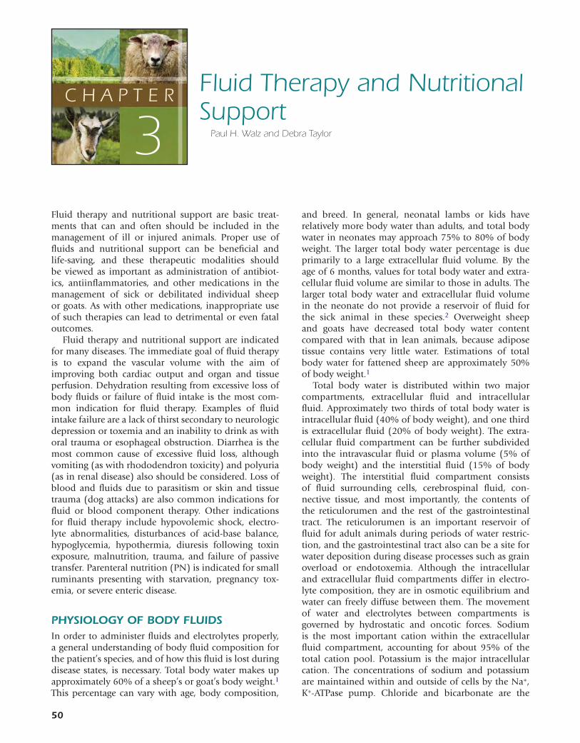
50
Fluid Therapy and Nutritional Support
Paul H. Walz and Debra Taylor
C H A P T E R
Fluid therapy and nutritional support are basic treat-ments that can and often should be included in the management of ill or injured animals. Proper use of fluids and nutritional support can be beneficial and life-saving, and these therapeutic modalities should be viewed as important as administration of antibiot-ics, antiinflammatories, and other medications in the management of sick or debilitated individual sheep or goats. As with other medications, inappropriate use of such therapies can lead to detrimental or even fatal outcomes.
Fluid therapy and nutritional support are indicated for many diseases. The immediate goal of fluid therapy is to expand the vascular volume with the aim of improving both cardiac output and organ and tissue perfusion. Dehydration resulting from excessive loss of body fluids or failure of fluid intake is the most com-mon indication for fluid therapy. Examples of fluid intake failure are a lack of thirst secondary to neurologic depression or toxemia and an inability to drink as with oral trauma or esophageal obstruction. Diarrhea is the most common cause of excessive fluid loss, although vomiting (as with rhododendron toxicity) and polyuria (as in renal disease) also should be considered. Loss of blood and fluids due to parasitism or skin and tissue trauma (dog attacks) are also common indications for fluid or blood component therapy. Other indications for fluid therapy include hypovolemic shock, electro-lyte abnormalities, disturbances of acid-base balance, hypoglycemia, hypothermia, diuresis following toxin exposure, malnutrition, trauma, and failure of passive transfer. Parenteral nutrition (PN) is indicated for small ruminants presenting with starvation, pregnancy tox-emia, or severe enteric disease.
PHYSIOLOGY OF BODY FLUIDSIn order to administer fluids and electrolytes properly, a general understanding of body fluid composition for the patient’s species, and of how this fluid is lost during disease states, is necessary. Total body water makes up approximately 60% of a sheep’s or goat’s body weight.1 This percentage can vary with age, body composition,
and breed. In general, neonatal lambs or kids have relatively more body water than adults, and total body water in neonates may approach 75% to 80% of body weight. The larger total body water percentage is due primarily to a large extracellular fluid volume. By the age of 6 months, values for total body water and extra-cellular fluid volume are similar to those in adults. The larger total body water and extracellular fluid volume in the neonate do not provide a reservoir of fluid for the sick animal in these species.2 Overweight sheep and goats have decreased total body water content compared with that in lean animals, because adipose tissue contains very little water. Estimations of total body water for fattened sheep are approximately 50% of body weight.1
Total body water is distributed within two major compartments, extracellular fluid and intracellular fluid. Approximately two thirds of total body water is intracellular fluid (40% of body weight), and one third is extracellular fluid (20% of body weight). The extra-cellular fluid compartment can be further subdivided into the intravascular fluid or plasma volume (5% of body weight) and the interstitial fluid (15% of body weight). The interstitial fluid compartment consists of fluid surrounding cells, cerebrospinal fluid, con-nective tissue, and most importantly, the contents of the reticulorumen and the rest of the gastrointestinal tract. The reticulorumen is an important reservoir of fluid for adult animals during periods of water restric-tion, and the gastrointestinal tract also can be a site for water deposition during disease processes such as grain overload or endotoxemia. Although the intracellular and extracellular fluid compartments differ in electro-lyte composition, they are in osmotic equilibrium and water can freely diffuse between them. The movement of water and electrolytes between compartments is governed by hydrostatic and oncotic forces. Sodium is the most important cation within the extracellular fluid compartment, accounting for about 95% of the total cation pool. Potassium is the major intracellular cation. The concentrations of sodium and potassium are maintained within and outside of cells by the Na+,K+-ATPase pump. Chloride and bicarbonate are the
3

Chapter 3 • Fluid Therapy and Nutritional Support 51
major anions within the extracellular fluid space, whereas phosphates, proteins, and other anions main-tain electroneutrality with the potassium cation in the intracellular fluid compartment. When fluids are administered to dehydrated sheep or goats, fluid losses are replaced within the extracellular fluid compartment; therefore the fluids being administered should contain concentrations of ions similar to those found in the extracellular fluid compartment. Assessment of blood electrolyte concentrations and determination of acid-base status are performed using techniques that reflect conditions in the extracellular fluid compartment.
Dehydration is a common feature of many diseases. Dehydration results from inadequate fluid intake in the presence of increased fluid losses. With dehydration, all fluid compartments are affected. Dehydration initially results in reduction of the intravascular fluid volume, followed by contraction of the interstitial and intra-vascular fluid compartments. Most disease processes result in concurrent losses of fluids and electrolytes—resulting in a pathophysiologic condition referred to as isotonic or isoosmolar dehydration. In such cases, providing both fluid and electrolytes (mainly sodium) is important. Hypertonic dehydration, or a relative water deficit, occurs when water losses exceed losses of electrolytes; water deprivation is an example. By con-trast, hypotonic dehydration, or relative water excess, occurs when electrolyte losses exceed water losses. Hypotonic dehydration also occurs with diarrhea, when losses of electrolytes and water occur concurrently (iso-tonic dehydration) but the water deficit is replaced by water consumption or administration of 5% dextrose solutions. Another example of hypotonic dehydration occurs in goats or sheep with obstructive urolithiasis, when sodium depletion exceeds water loss as sodium moves into the peritoneal cavity. In cases of urolithiasis with urinary bladder rupture, administration of fluids containing sodium is important (see Chapter 12).
PATIENT ASSESSMENTPhysical examination of hypovolemic or dehydrated sheep and goats is very important to ensure that the correct fluid type is administered at an appropriate rate, as well as for identifying the underlying disease process. Intercurrent disease processes, hypothermia, and peri-natal asphyxia can lead to difficulty in accommodat-ing intravenous fluids. Fluid therapy can have adverse effects such as volume overload and edema, so particu-lar attention should be given to the cardiovascular and renal systems. Cardiovascular diseases may result in an inability to cope with an acute fluid load, and oliguric renal failure results in an impaired ability to excrete excess fluid (see Chapter 17).
Dehydration is most accurately assessed by changes in body weight before and after a disease event.
Because this information usually is not available to the clinician, clinical assessment is used to assess degree of dehydration. The packed cell volume and total plasma protein can be used as tools to assess hydration status, but these measurements cannot be used to replace an estimation of hydration status from physical examina-tion. The reference ranges for packed cell volume in healthy sheep and goats are 27% to 45% and 22% to 38%, respectively3; these ranges are too wide to be useful in estimation of hydration status. Total plasma protein concentration is dependent on colostral intake in neonates and may be elevated with chronic inflam-mation. In addition, a sheep or goat with anemia and hypoproteinemia in conjunction with dehydration, as occurs with intestinal parasitism, can nevertheless have a normal packed cell volume and total plasma protein concentration. Packed cell volume and total plasma protein concentrations are most useful in monitoring the progress of fluid therapy to prevent overhydration.
Although no standard method is available for assess-ing dehydration in sheep and goats, percent dehydra-tion can be estimated by assessing heart rate; eyeball recession; mucous membranes for tackiness, color, and capillary refill time; and skin elasticity or turgor (Table 3-1). Degree of enophthalmos has been used to determine hydration status in calves4 and can be used to assess hydration in sheep and goats as well. The percent dehydration can be estimated by measuring
TABLE 3-1 Estimation of Percent Dehydration from Physical Examination Findings
Percent Dehydration Physical Findings
<5% History of fluid loss but no other abnormalities
5% Minimal depression, normal to mildly tacky mucous membranes, minimal enophthalmos, normal heart rate, normal capillary refill time (<2 seconds)
8% Depression, mild to moderate decrease in skin turgor (skin tent duration 2-4 seconds), obvious enophthalmos, slight tachycardia (heart rate >90 beats/minute), increased capillary refill time (3-4 seconds)
≥ 10% Severe depression, weakness, moderate to marked degree of decreased skin turgor (skin tent duration >5 seconds), dry and dark mucous membranes, tachycardia (>120 beats/minute), increased capillary refill time (>5 seconds), cold extremities

52 Chapter 3 • Fluid Therapy and Nutritional Support
the eyeball recession in mm and multiplying by 2. For example, a sheep or goat with eyeball recession of 4 mm is estimated to be 8% dehydrated. The duration of skin tenting also can be used to estimate hydration status. The percent dehydration is estimated by measuring the skin tent (in seconds), multiplying by 2, and then sub-tracting 4. For example, a sheep or goat with a skin tent duration of 6 seconds is estimated to be 8% dehydrated (6 × 2 = 12 − 4 = 8%). Some caveats to this clinical assessment of dehydration exist, and it is important to recognize that the poor skin elasticity and enophthal-mos of dehydration also are seen in emaciated small ruminants.
Acid-base and electrolyte abnormalities are more difficult to assess without the aid of laboratory testing. Serum biochemistry and blood gas analyses can provide information on serum electrolyte (sodium, potassium, chloride, bicarbonate, calcium, magnesium, and phos-phorus) abnormalities, acid-base disorders, or glucose abnormalities that require correction. In general, most cases of diarrhea in neonates and grain engorgement in adults will be characterized by a metabolic acidosis, whereas intestinal obstructions and renal disease will be associated with a metabolic alkalosis. Although the degree of acidosis can be estimated in calves with diar-rhea using a clinical scoring system,5 no similar clinical scoring system has been created for use in neonatal sheep or goats with diarrhea (see Appendix 11).
FLUID AND ELECTROLYTE REPLACEMENT THERAPYFluid and electrolyte replacement therapy in sheep and goats is required when fluid intake by the animal is not enough to meet metabolic needs. Rehydration, replace-ment of lost electrolytes, and restoration of acid-base balance are the goals of fluid therapy. Provision of intra-venous fluids can restore the circulatory capacity and mental status sufficiently that nutrition and replace-ment of ongoing losses can be provided through oral fluids. The aggressiveness of treatment is dictated by the severity of the condition as well as economic consider-ations. In developing the fluid and electrolyte replace-ment therapy plan, three basic assessments should be made: (1) quantity and rate of fluid administration, (2) fluid type, and (3) method of fluid administration.
QUANTITY AND RATE OF FLUID ADMINISTRATIONThe fluid therapy plan is designed to replace deficits while supplying maintenance fluid needs and account-ing for ongoing loss of fluids associated with the disease process (Table 3-2). The first priority for treating dehy-dration in a sheep or goat is to restore the extracellular fluid volume to normal. The following simple formula
can be used to calculate the recommended amount of fluid for restoration of the animal to a normal hydrated state:
(Estimation of Dehydration) X (Body Weight in kg)
= Liters of Fluid Needed (in L)
Intravenous fluid therapy is recommended in cases in which the estimation of dehydration is 8% or greater, because oral fluid therapy will be ineffective.6 For exam-ple, a 50-kg sheep that is 8% dehydrated will need 4 L of fluid to replace the deficit (0.08 × 50 = 4.00). A 5-kg kid with diarrhea that is 10% dehydrated will need 500 mL of fluid to replace the deficit. Because methods for pre-cise measurement of the degree of dehydration are not available, it is important for the clinician to recognize that replacing the exact fluid deficit is not of chief con-cern. Rather, the clinician should replace a fluid deficit to restore tissue perfusion and improve mental capacity so oral fluids can be utilized. A general rule is to replace half of the fluid deficit over 4 to 6 hours, with the bal-ance given over 12 to 24 hours. More often, the fluid deficit is replaced more rapidly (6 hours); however, care should be taken in the hypothermic neonate or in cases of sepsis, because generalized edema may result. Specifi-cally, too-rapid delivery of intravenous fluid therapy can result in pulmonary and cerebral edema. Alternatively, the rate of fluid administration can be set at 50 mL/kg/hour, which is less than the shock therapy rate of 90 mL/kg/hour.
Calculation of maintenance fluids is based on spe-cies- and age-specific physiologic requirements. The normal, adult sheep or goat requires approximately 50 mL/kg/day to provide enough fluids for digestion and to replace losses through urine and defecation (sensible water loss) and sweat and respiration (insensible water loss). As stated previously, neonates have higher total body water than that in adults and therefore have higher maintenance fluid requirements. The maintenance fluid needs for lambs and kids can be up to 80 mL/kg/day.
TABLE 3-2 Components and Formulas for a Fluid Therapy Plan
Category Amount/Formula
Deficit % dehydration × body weight in kgMaintenance Adults: 50 mL/kg/day
Neonates: 80 mL/kg/dayOngoing losses Up to 5% of body weight in kgBicarbonate deficit Adults: body weight (in kg) ×
0.3 × base deficitNeonates: body weight (in kg) ×
0.6 × base deficitShock rate 90 mL/kg/hour

Chapter 3 • Fluid Therapy and Nutritional Support 53
In developing the fluid therapy plan, both replacement of the deficit and inclusion of maintenance fluid require-ments should be addressed. The maintenance fluid needs of sheep and goats, in 50 mL/kg/day, can be sim-ply converted to 1 mL/lb/hour or 2 to 4 mL/kg/hour. An important consideration is that this maintenance fluid requirement can change with ambient temperature and feed intake, because diets may vary in moisture content. Careful monitoring is warranted for patients that are on continuous intravenous fluid therapy or receiving large volumes of fluids over a short period of time, because they can become hypoproteinemic and develop edema. Serial measurements of packed cell volume and total plasma protein are needed to prevent overhydration.
Pathologic water losses can continue to occur even with ongoing fluid therapy (e.g., with infectious diar-rhea). Losses may continue as well with disorders char-acterized by third space sequestration of fluids, such as grain engorgement or ruptured urinary bladder. Those losses that are expected should be included in the fluid therapy plan. Although it is difficult to estimate these losses, up to 5% of the body weight per day may be estimated as extra fluid losses for animals with severe diarrhea.
Fluid TypeMany different and appropriately formulated fluid types are available for intravenous administration in sheep and goats. The decision to administer intravenous flu-ids to expand fluid volume is far more important than the specific choice of fluid type and rate. A majority of patients will respond adequately to any balanced electrolyte fluid. The type of fluid to be administered ideally should be based on the individual patient’s dis-ease process and the measured or predicted acid-base or electrolyte deficits that must be corrected. Four basic categories of fluids are available for intravenous use in clinical practice: crystalloids, colloids, whole blood, and PN solutions.
Crystalloid fluids are the mainstay of fluid therapy and consist primarily of water with a sodium or glu-cose base. Crystalloid fluids can be divided into four groups based on purpose: replacement, maintenance, hypertonic saline, and dextrose in water. Alternatively, crystalloid fluids can be classified by tonicity, as hypo-tonic, isotonic, or hypertonic. Replacement crystalloid fluids are required for sheep or goats with deficits of either electrolytes or water or, most commonly, both. The commonly used replacement crystalloids in small ruminant medicine are isotonic solutions and include the unbalanced solutions, such as normal saline (0.9% NaCl) and sodium bicarbonate (1.3% NaHCO3), and the balanced electrolyte solutions, such as Ringer’s solu-tion (with lactate or acetate), Normosol-R (Hospira, Inc., Lake Forest, Illinois), and Plasma-Lyte A (Baxter
Healthcare Corp., Deerfield, Illinois). Balanced solu-tions resemble the extracellular fluid in composition. All replacement and maintenance crystalloids have an osmolality similar to that of plasma and can enter all body fluid compartments. These fluids will equilibrate quickly with interstitial fluids, so only 20% to 25% of the infused fluid remains within the intravascular com-partment 1 hour after infusion.
Normal saline (0.9% NaCl) has slightly higher sodium and higher chloride concentrations than those for plasma. Normal saline is considered an acidifying solution because it lowers plasma bicarbonate levels through volume expansion, reduction in renal bicar-bonate absorption, and increased renal tubular chloride levels, which in turn promotes bicarbonate excretion in the renal collecting ducts. For sheep and goats with met-abolic acidosis, such as occurs with grain engorgement or diarrhea, normal saline is not the fluid of choice. Normal saline is a crystalloid fluid indicated for sheep and goats with metabolic alkalosis or hyponatremia as would occur with gastrointestinal obstruction or stasis, obstructive urolithiasis, or ruptured urinary bladder.
Balanced crystalloid fluids vary in their content of electrolytes, but in general, balanced crystalloid solu-tions contain electrolyte concentrations similar to those in plasma. Ringer’s solution is an acidifying solution and is fairly similar to normal saline except for its slightly lower levels of sodium, higher levels of chlo-ride, and additional potassium and calcium. Lactated Ringer’s solution is considered an alkalizing solution, but the lactate present requires hepatic metabolism to produce bicarbonate, and only the l-isomer of lactate is metabolized efficiently to produce bicarbonate. The balanced crystalloids Normosol-R and Plasma-Lyte A also are considered alkalizing fluids because they con-tain sodium acetate and sodium gluconate. Acetate and gluconate are bicarbonate precursors. Unlike lactate, which is metabolized by the liver, acetate is metabolized by muscle tissue. Gluconate has been shown to be inef-fective as an alkalizing agent in calves when adminis-tered intravenously but is effective when given orally.7 Although some of these balanced crystalloid solutions are considered to be alkalizing, they are regarded as inferior in alkalizing ability to 1.3%, 5%, or 8.4% sodium bicarbonate.2
Sodium bicarbonate is the crystalloid fluid of choice for metabolic acidosis and can either be given as isotonic sodium bicarbonate (1.3%), or hypertonic sodium bicarbonate (5% or 8.4%), or added to other crystalloid solutions. Common causes of metabolic acidosis include absorption of d-lactate from the gastro-intestinal tract (e.g., with grain engorgement or entero-colitis) and sodium loss with secretory diarrhea. Sepsis or other causes of systemic shock also can lead to meta-bolic acidosis as a result of l-lactate accumulation from poor tissue perfusion. To correct metabolic acidosis, a

54 Chapter 3 • Fluid Therapy and Nutritional Support
total carbon dioxide (CO2) measurement from a serum biochemistry panel or blood gas analysis is needed. The base deficit is calculated by subtracting the measured total CO2 from the normal total CO2 (approximate nor-mal total CO2 is 25 mEq/mL). The amount of bicarbon-ate to administer can be calculated as follows:
Neonates : mEq bicarbonate needed
= (base deficit) x (body weight in kg) x 0.6
Adults : mEq bicarbonate needed
= (base deficit) x (body weight in kg) x 0.3
Neonates have a larger bicarbonate space than that in adult animals and thus have greater bicarbonate replacement needs when losses occur.6 For example, a 5-kg kid with a base deficit of 15 will need 45 mEq of bicarbonate to correct the metabolic acidosis: 15 (base deficit) × 5 kg (body weight) × 0.6 = 45 mEq. If the 5-kg kid is 10% dehydrated, the fluid deficit is 500 mL. None of the crystalloid fluids previously discussed can provide enough base in 500 mL to correct the acidosis in this specific example, emphasizing the need for bicarbonate therapy in cases of metabolic acidosis in ruminants. The bicarbonate needed to correct metabolic acidosis can be given to sheep or goats as a 1.3% isotonic solution; alternatively, the deficit can be added to normal saline and administered intravenously. As a general rule, half of the calculated base deficit should be corrected if the metabolic acidosis results from dehydration only. The entire deficit can be corrected if the dehydration is due to neonatal diarrhea or grain engorgement. Sodium bicarbonate is available commercially as hypertonic solutions of either 5% (0.6 mEq/mL) or 8.4% (1 mEq/mL). Solutions of 5% sodium bicarbonate can be given intravenously without dilution so long as the dehydra-tion is corrected at the same time. The administration rate for 5% sodium bicarbonate should not exceed 2 mL/kg/minute.8
Dextrose-containing intravenous crystalloid solu-tions such as 5% dextrose in water (D5W) should not be used routinely in small ruminant practice for stand-alone therapy, because once the dextrose is metabo-lized, the fluid contains no active solute. Infusion of 5% dextrose can lead to dilution of serum electrolytes along with the development of edema. However, glu-cose supplementation is important for hypoglycemic, hypothermic neonatal small ruminants and in ewes with pregnancy toxemia. Blood glucose can easily be measured with commercially available hand-held glucometers. To treat hypoglycemia, dextrose can be administered intravenously as a 50% solution (at a dose of 0.2 mL/kg of body weight) or as a 5% to 10% solu-tion. Dextrose can be added to other crystalloid fluids to make a 1% or 2% solution (20 mL of 50% dextrose/L for each 1% of dextrose needed). For ewes with preg-nancy toxemia, 5 to 7 g of glucose given intravenously six to eight times daily has been recommended.9
Hypertonic saline solutions are a type of crystalloid fluid that has gained increased use in ruminants over the past 2 decades. Hypertonic saline solutions (7.2% NaCl) rapidly increase intravascular volume by increas-ing intravascular hyperosmolality, thereby drawing fluid from the intracellular and interstitial fluid compart-ments. The effect is transient and must be supple-mented by additional volume replacement. Hypertonic saline solutions should be administered at a dose of 4 mL/kg of body weight over 3 to 10 minutes. Indica-tions for hypertonic saline solution administration in small ruminants are severe dehydration, endotoxic shock, and hemorrhagic shock. In cases of hemorrhagic shock, blood transfusions should be performed after therapy with hypertonic saline solutions. In cases of dehydration, hypertonic saline solutions should be fol-lowed with intravenous isotonic solutions or oral fluid therapy.
Colloids are high-molecular-weight compounds that, unlike crystalloids, do not readily leave the intravascular space. Examples of colloids are plasma, human serum albumin and synthetic compounds such as hetastarch, dextrans, and modified gelatin solutions. Plasma is used primarily in cases of failure of passive transfer and hypoproteinemia and is administered at a dosage rate of 20 to 40 mL/kg of body weight.10 The use of plasma for colloidal effects is relatively ineffective, because 50 to 100 mL/kg of body weight is required to raise serum albumin concentration by 1 g/dL. Sheep and goat plasma preparations are available commercially (Midwest Animal Blood Services, Inc., Stockbridge, Michigan). The use of colloids for expanding intravascular volume in ill small ruminants has not been evaluated.
Whole blood transfusions are indicated primarily when the red blood cell mass is inadequate to carry oxygen to the peripheral tissues. Whole blood transfusions are recommended in animals with clinical signs suggestive of tissue hypoxia, such as elevated heart and respiratory rates, weakness, and lethargy, or in which the packed cell volume drops below 15% to 20% in acute anemia and below 10% to 15% in chronic anemia. Whole blood can be administered at a dose of 10 to 15 mL/kg of body weight; however, this will result in an increase in packed cell volume of the recipient by only 3% to 4%. For hemorrhagic shock, at least half of the esti-mated blood loss should be replaced by whole blood.10 Whole blood transfusions also can be used as a source of plasma and can be given at a dose of 40 to 80 mL/kg of body weight. Monitoring of the transfusion is impor-tant, and the whole blood transfusion should be started at a slow rate (0.1 mL/kg/hour), with vital signs evalu-ated every 5 minutes. Clinical signs of anaphylactic reactions to whole-blood transfusions include fever, dyspnea, hiccoughing, muscle tremors, salivation, and lacrimation. If a transfusion reaction is noted, blood administration is ceased and epinephrine (1:1000) can

Chapter 3 • Fluid Therapy and Nutritional Support 55
be administered at a dose of 0.01 to 0.02 mL/kg of body weight intravenously.
Method of AdministrationAdministration of fluids is primarily by either the oral or the intravenous route. The choice of route is based on hydration status and on whether the patient can take in oral fluids. Subcutaneous, intraperitoneal, and intraosseus routes are not commonly used. Subcuta-neous fluids are contraindicated in animals that are severely dehydrated as a result of poor absorption. With severe dehydration, the intravenous route is required. Intravenous fluids can be given as bolus injections or as a constant rate infusion. Oral fluids have the advantage of being the least costly, but this route is less effec-tive in cases involving gastrointestinal stasis or severe dehydration
Intravenous AdministrationThe jugular vein is very superficial and easy to visualize once the hair or fleece has been clipped in most sheep and goats. Placement of a jugular catheter is best accom-plished with the small ruminant patient positioned in lateral recumbency if possible. After aseptic preparation of the jugular furrow, a local block can be performed over the catheterization site with 0.5 to 1 mL of 2% lidocaine using a 3-mL syringe and 25-gauge, 5/8-inch needle. After the site is blocked, a small stab incision through the skin is made with a No. 15 scalpel blade. To prevent inadvertent incision of the jugular vein, the skin can be either pinched up away from the jugular or slid dorsally over the neck muscles. Use of the small stab skin incision for this procedure is very helpful in decreasing the amount of tissue drag with potential collapse of the jugular vein during catheter placement. Before insertion of the catheter, the jugular vein is maxi-mally expanded by holding off at the thoracic inlet and allowing time for it to distend. In severely dehydrated or hypotensive sheep and goats, adequate vessel expansion may take additional time. The catheter is held horizon-tally and flushed out with sterile heparinized saline. Heparinized saline can be purchased commercially or formulated from normal saline in a concentration of 10 U/mL. The catheter is held by its hub with the index finger placed over and occluding the end of the hub. The catheter is inserted through the stab incision. If any skin drag is noted, the catheter is set aside and the skin incision is deepened or extended with the scalpel blade. Once the catheter is through the skin and lined up with the distended jugular at an angle of 45 to 60 degrees, it is advanced with a quick, short, forceful insertion. To check for proper position of the catheter, the finger is lifted off the hub and the catheter contents are observed for a flash of blood. If a flash of blood is evident, the catheter and stylet are advanced an additional 1 cm.
If another check for blood flash reconfirms its position in the jugular vein, the catheter is slid into the vein off the stylet, which is then removed. With severe hypo-volemia or hemoconcentration, a flash of blood may not be observed in the catheter. Absence of this sign is more common if the animal is standing or in sternal recumbency than if the animal is in lateral recumbency. In such instances, the position of the catheter within the jugular vein is assessed as follows: A syringe filled with heparinized saline is attached to an extension set and used for periodic aspiration during performance of the initial venipuncture. Once the catheter is inserted, an injection port or extension set can be attached to the catheter, and the catheter should be checked for blood withdrawal and then flushed with heparinized saline.
Several methods can be used for securing and pro-tecting jugular catheters: The catheter and extension set or injection port can be sutured to the skin. White porous tape also can be attached to the catheter injec-tion port or extension set and this tape can be sutured to the skin. The catheter can be additionally secured to the skin using a cyanoacrylate glue (Superglue). Glue also can be applied at the point of entry of the hub end and catheter into the skin. Extension sets are very useful but must be secured at another site on the body to prevent the catheter from being pulled out if the extension set or fluid line gets caught or tangled. Heparinized saline can be used to keep catheters patent when intravenous fluids are not running, and catheters should be flushed every 6 to 8 hours with heparinized saline.
Intravenous catheters are available in various lengths, diameters, and construction materials. Teflon, polypro-pylene, polyurethane, and Silastic catheter types are available for use in small ruminant patients. Teflon catheters need to be changed every 3 days, whereas polyurethane catheters can remain in place for up to 2 weeks. For adult sheep and goats, a 16-gauge, 3.25-inch Teflon catheter often is used. For kids and lambs, an 18-gauge, 2-inch catheter is of the appropriate size and length for jugular vein catheterization. The rate of fluid administration is proportional to the diameter of the catheter and inversely proportional to the length of the catheter.
Oral AdministrationOral fluid therapy can be used as indicated in both neo-natal and adult animals. In general, oral fluid therapy is preferred over other modalities because it is the most physiologic. Oral fluids can be administered by orogas-tric or nasogastric intubation or through a previously created rumenostomy. A syringe barrel or syringe case can be adapted for use as a speculum. Milk tubing or foal orogastric tubes can be passed all the way down the esophagus in adult small ruminants. For neonates, small red rubber feeding tubes are suitable, and these are ideally passed down to the midesophageal region.
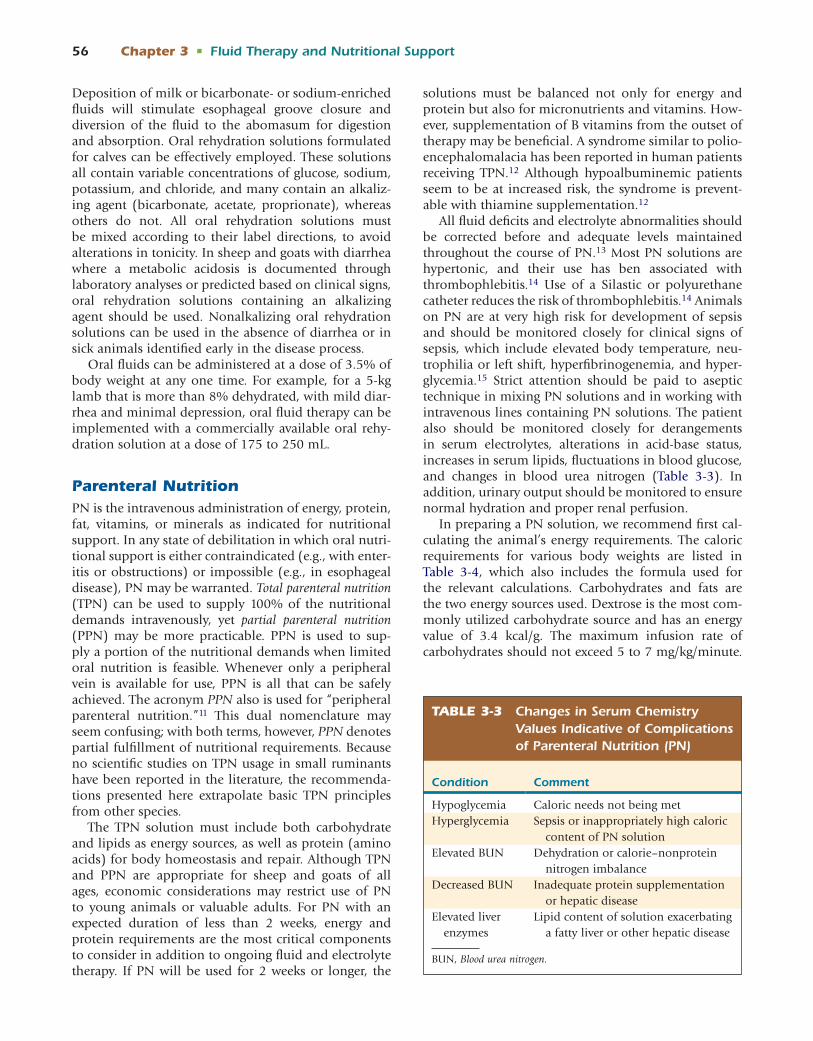
56 Chapter 3 • Fluid Therapy and Nutritional Support
Deposition of milk or bicarbonate- or sodium-enriched fluids will stimulate esophageal groove closure and diversion of the fluid to the abomasum for digestion and absorption. Oral rehydration solutions formulated for calves can be effectively employed. These solutions all contain variable concentrations of glucose, sodium, potassium, and chloride, and many contain an alkaliz-ing agent (bicarbonate, acetate, proprionate), whereas others do not. All oral rehydration solutions must be mixed according to their label directions, to avoid alterations in tonicity. In sheep and goats with diarrhea where a metabolic acidosis is documented through laboratory analyses or predicted based on clinical signs, oral rehydration solutions containing an alkalizing agent should be used. Nonalkalizing oral rehydration solutions can be used in the absence of diarrhea or in sick animals identified early in the disease process.
Oral fluids can be administered at a dose of 3.5% of body weight at any one time. For example, for a 5-kg lamb that is more than 8% dehydrated, with mild diar-rhea and minimal depression, oral fluid therapy can be implemented with a commercially available oral rehy-dration solution at a dose of 175 to 250 mL.
Parenteral NutritionPN is the intravenous administration of energy, protein, fat, vitamins, or minerals as indicated for nutritional support. In any state of debilitation in which oral nutri-tional support is either contraindicated (e.g., with enter-itis or obstructions) or impossible (e.g., in esophageal disease), PN may be warranted. Total parenteral nutrition (TPN) can be used to supply 100% of the nutritional demands intravenously, yet partial parenteral nutrition (PPN) may be more practicable. PPN is used to sup-ply a portion of the nutritional demands when limited oral nutrition is feasible. Whenever only a peripheral vein is available for use, PPN is all that can be safely achieved. The acronym PPN also is used for “peripheral parenteral nutrition.”11 This dual nomenclature may seem confusing; with both terms, however, PPN denotes partial fulfillment of nutritional requirements. Because no scientific studies on TPN usage in small ruminants have been reported in the literature, the recommenda-tions presented here extrapolate basic TPN principles from other species.
The TPN solution must include both carbohydrate and lipids as energy sources, as well as protein (amino acids) for body homeostasis and repair. Although TPN and PPN are appropriate for sheep and goats of all ages, economic considerations may restrict use of PN to young animals or valuable adults. For PN with an expected duration of less than 2 weeks, energy and protein requirements are the most critical components to consider in addition to ongoing fluid and electrolyte therapy. If PN will be used for 2 weeks or longer, the
solutions must be balanced not only for energy and protein but also for micronutrients and vitamins. How-ever, supplementation of B vitamins from the outset of therapy may be beneficial. A syndrome similar to polio-encephalomalacia has been reported in human patients receiving TPN.12 Although hypoalbuminemic patients seem to be at increased risk, the syndrome is prevent-able with thiamine supplementation.12
All fluid deficits and electrolyte abnormalities should be corrected before and adequate levels maintained throughout the course of PN.13 Most PN solutions are hypertonic, and their use has ben associated with thrombophlebitis.14 Use of a Silastic or polyurethane catheter reduces the risk of thrombophlebitis.14 Animals on PN are at very high risk for development of sepsis and should be monitored closely for clinical signs of sepsis, which include elevated body temperature, neu-trophilia or left shift, hyperfibrinogenemia, and hyper-glycemia.15 Strict attention should be paid to aseptic technique in mixing PN solutions and in working with intravenous lines containing PN solutions. The patient also should be monitored closely for derangements in serum electrolytes, alterations in acid-base status, increases in serum lipids, fluctuations in blood glucose, and changes in blood urea nitrogen (Table 3-3). In addition, urinary output should be monitored to ensure normal hydration and proper renal perfusion.
In preparing a PN solution, we recommend first cal-culating the animal’s energy requirements. The caloric requirements for various body weights are listed in Table 3-4, which also includes the formula used for the relevant calculations. Carbohydrates and fats are the two energy sources used. Dextrose is the most com-monly utilized carbohydrate source and has an energy value of 3.4 kcal/g. The maximum infusion rate of carbohydrates should not exceed 5 to 7 mg/kg/minute.
TABLE 3-3 Changes in Serum Chemistry Values Indicative of Complications of Parenteral Nutrition (PN)
Condition Comment
Hypoglycemia Caloric needs not being metHyperglycemia Sepsis or inappropriately high caloric
content of PN solutionElevated BUN Dehydration or calorie–nonprotein
nitrogen imbalanceDecreased BUN Inadequate protein supplementation
or hepatic diseaseElevated liver
enzymesLipid content of solution exacerbating
a fatty liver or other hepatic disease
BUN, Blood urea nitrogen.

Chapter 3 • Fluid Therapy and Nutritional Support 57
Highly concentrated dextrose solutions are hypertonic and will increase risk for thrombosis of peripheral ves-sels if infused at a rate greater than 10% in a peripheral vein. Adult animals generally will tolerate up to a 20% dextrose solution via a jugular venous catheter.
Lipids are isotonic and energy-dense, supplying 9.1 kcal of energy per gram. Lipids for PN are formulated as a 20% solution, which can be infused at a maximum rate of 2.5 to 3 g/kg/day (15 mL of 20% solution/kg/day). From 30% to 60% of the calories in a PN solution should be supplied as lipids. In animals with evidence of hepatic disease, the low end of this lipid range (30%) should be targeted, and liver enzymes such as serum sorbitol dehydrogenase should be monitored closely.
Protein is supplied as 8.5% amino acid solution, which has approximately 1.3 g of nitrogen/100 mL. The protein requirement for young lambs and kids (extrapo-lated from data for other species) is likely to be between 2.0 and 3.75 g of amino acids/kg/day.13,16 The require-ment for adult animals (extrapolated from other spe-cies) is 1.0 to 1.5 g of amino acids/kg/day.13 The ratio of nonprotein calories to grams of nitrogen should be maintained at 100:1 to 300:1 to avoid negative protein balance.16,17 Grams of nitrogen can be calculated by dividing the number of grams of amino acids / 6.25.17
(gms amino acids / 6.25 = gms nitrogen)
A central venous catheter is required for adminis-tration of more than 1 to 1.5 g of protein/kg of body weight/day. Juvenile and adult animals require 23 and 11 mL, respectively, of the 8.5% amino acid solution/kg/day to achieve TPN.
It is most convenient and practical to mix solutions in 1-L aliquots. Both glass and plastic sterile contain-ers are suitable for mixing.18 When plastic bags are used, they should be made of ethylene vinyl acetate.19 The order of addition is an important consideration in preparation of TPN solutions, because adding lipid directly to 50% dextrose can result in destruction of the lipid droplets. As a useful mnemonic, the l in lipid can be said to stand for “last when mixing.”
Because the maintenance fluid requirement is 50 mL/kg/day, the volumes of nutrient solutions administered must be subtracted from the total daily requirement to avoid overhydration of the animal. Continuing or ongoing fluid losses also should be factored into total replacement volume, as previously described.
Complications of PN can be avoided by minimizing the handling of the PN line with the following practices: • Place the catheter in aseptic fashion. • Cover with a sterile dressing. • Use this line solely for PN. • Do not disconnect except to add a new PN bag.The separate fluid line for electrolyte-fluid supple-mentation also will prevent precipitation of PPN with concentrated electrolyte solutions if required. Sterile povidone-iodine ointment may be placed on all con-nections and covered with tape to decrease the chance of contamination.19,20
It is best to acclimate the patient by initiating PN with a solution containing only 25% of the calculated amounts of dextrose, lipid, and amino acids for TPN.15 The fluid difference should be supplied with a polyionic isotonic fluid during acclimation. The energy and pro-tein density should then be gradually increased over a period of 2 to 3 days. During this acclimation period, the animal should be monitored closely for compli-cations. Monitoring these parameters while slowly increasing the energy and protein density of the PN solution will allow the clinician to adjust the protein, lipid, carbohydrate, and electrolyte composition of the solution to the individual needs and tolerance of the animal. If the animal’s blood work remains normal dur-ing the acclimation period, the full maintenance TPN regimen can be begun on the third or fourth day if indi-cated. The patient should be weighed, if possible, on a daily basis to ensure that it maintains or gains weight during treatment. The transition back to enteral nutri-tion should be made very gradually as well.15 Whenever possible, enteral feeding in small amounts should be continued during PN.
TABLE 3-4 Daily Caloric Requirements for Various Body Weights in Sheep and Goats
Body weight (kg) 2 3 4 5 8 10 15 20 25 30 35 40 50
kcal/day 235 319 395 468 666 787 1067 1324 1565 1795 2015 2227 2632
Values shown were calculated using the following formula*:Caloric requirement = 140 kcal/kg0.75 of body weight/dayUsing a scientific calculator, punch in weight in kg, hit xy key, then punch in 0.75 and hit xy key again; multiply this number by 140 = kcal needed per day.Note: The equation above yields a caloric requirement very close to that calculated by the total energy requirement (TER) equation: (30 × body weight (kg) + 70 × “illness factor” of 1.8 to 2.0. Use of illness factors this large has been suggested to lead to oversupplementation.20 We recommend starting TPN at 25% of the caloric requirement listed above and increasing nutrients as tolerated by the patient.*From Nutrient requirements of small ruminants; sheep, goats, cervids and new world camelids. Washington, DC, 2007, National Academy Press.

58 Chapter 3 • Fluid Therapy and Nutritional Support
Mineral supplementation is rarely necessary until after day 7 to 10 of PN. Minerals should be monitored by measurement of serum electrolytes and supple-mented if needed (Table 3-5). Both trauma and diarrhea may increase loss of zinc (and possibly other nutrients). Vitamin supplementation (Table 3-6) generally is not needed until after day 7 to 10 of PN, with the possible exception of the B vitamins. The B vitamins may be of benefit if given every 2 to 3 days at labeled dosages from the onset of TPN. As a general rule, vitamin K, 10 mg administered by intramuscular injection, can be given once a week.
Dextrose solutions concentrated enough to meet full caloric requirements (i.e., greater than 10% concentra-tion) are thrombogenic to peripheral veins. Peripheral PN—as noted, also commonly abbreviated PPN—has been utilized (MacEntire D: Personal communication, 2009) and evaluated clinically in dogs.21 The goal with this form of PN is to supply a portion of the daily caloric requirements through a peripheral line with-out inducing thrombophlebitis secondary to excessive hypertonicity. Administration of such solutions has been shown to improve nitrogen balance.21 Chandler and co-workers performed a prospective study using a commercially available three-in-one solution.21 This solution, which has higher osmolarity, caloric den-sity (0.63 kcal/mL), and amino acid concentration than the “custom” solution described further on, was administered to dogs at 2 mL/kg/hour. Venous throm-bosis occurred in the peripheral veins at 36 hours if the solution was administered around the clock (i.e., for 24 hours a day). If the solution was administered for 10 to 12 hours each day, followed by isotonic fluid for the other 12 to 14 hours, thrombosis did not occur.
A custom-made peripheral PN solution that has been well tolerated in the small animal critical care setting
(MacEntire D: Personal communication, 2009) is pre-pared as follows: • Remove 400 mL of polyionic intravenous fluid from
a 1-L bag. • Add 100 mL of 50% dextrose, 200 mL of 8.5%
amino acids, and 100 mL of 20% lipid solution.This solution, which provides 0.35 kcal/mL, can be given as a continuous infusion (i.e., for 24 hours a day) at the maintenance fluid rate. Administration at the mainte-nance rate provides 30% of the caloric requirement for a 10-kg animal and approximately 40% for a 40-kg animal. This regimen would be calorically similar to one provid-ing 25% of the values calculated from a worksheet.
REFERENCES 1. Carlson GP, Bruss ML: Fluid, electrolyte, and acid-base balance.
In Kaneko JJ, Harvey JW, Bruss ML, editors: Clinical biochemistry of domestic animals, ed 6, San Diego, 2008, Elsevier, pp 529–560.
2. Berchtold J: Intravenous fluid therapy of calves, Vet Clin North Am Food Anim Pract 15:505–531, 1999.
3. Kaneko JJ, et al: Appendix VIII. Blood analyte reference values in large animals. In Kaneko JJ, Harvey JW, Bruss ML, editors: Clinical biochemistry of domestic animals, ed 5, San Diego, 1997, Academic Press, pp 890–894.
4. Constable PD, et al: Clinical and laboratory assessment of hydra-tion status of neonatal calves with diarrhea, J Am Vet Med Assoc 212:991–996, 1998.
5. Naylor JM: A retrospective study of the relationship between clinical signs and severity of acidosis in diarrheic calves, Can Vet J 30:577–580, 1989.
6. Roussel AJ Jr, Navarre CB: Fluid therapy, transfusion, and shock therapy. In Anderson DE, Rings DM, editors: Current veterinary therapy: food animal practice, St Louis, 2009, Saunders, pp 526–533.
7. Naylor JM, Forsyth GW: The alkalinizing effects of metabolizable bases in the healthy calf, Can J Vet Res 50:509–516, 1986.
8. Constable PD: Fluid and electrolyte therapy in ruminants, Vet Clin North Am Food Anim Pract 19:557–597, 2003.
9. Rook JS: Pregnancy toxemia of ewes, does, and beef cows, Vet Clin North Am Food Anim Pract 16:293–317, 2000.
10. Divers TJ: Blood component transfusions, Vet Clin North Am Food Anim Pract 21:615–622, 2005.
11. Chandler ML, Payne-James JJ: Prospective evaluation of a periph-erally administered three-in-one parenteral nutrition product in dogs, J Small Anim Pract 47:518–523, 2006.
12. Francini-Pesenti F, et al: Wernicke’s syndrome during parenteral feeding: not an unusual complication, Nutrition 25:142–146, 2009.
TABLE 3-5 General Guidelines for Mineral Supplementation
Mineral Amount*
Calcium 0.8 mEq (× 2 for growth or lactation)Magnesium 0.33 mEqSodium 2.26 mEqPhophorus 3.6 mmol/day (× 2 for growth or lactation)Zinc 0.6 mgIron 0.6 mgIodine 1.5 μg
When PPN or TPN is used for less than 7 days, minerals will rarely need to be added to PN solutions, but when PN is used for longer periods, close monitoring of serum electrolyte status may indicate a need for supplementation.*Per kg of body weight per day
TABLE 3-6 Estimated Nutritional Requirements for Some Vitamins for Prolonged Parenteral Nutrition*
Vitamin Amount†
Vitamin A 25 IUVitamin D 6.6 IUVitamin E 224 μg
*More than 7 days in duration.†Per kg of body weight per day.

Chapter 3 • Fluid Therapy and Nutritional Support 59
13. Spurlock SL, Ward MV: Parenteral nutrition in equine patients: principles and theory, Comp Cont Educ Pract Vet 13:461–469, 1991.
14. Chandler ML, Guilford WG, Payne-James J: Use of peripheral par-enteral nutritional support in dogs and cats, J Am Vet Med Assoc 216:669–673, 2000.
15. Lippert AC, Fulton RB, Parr AM: A retrospective study of the use of total parenteral nutrition in dogs and cats, J Vet Intern Med 7:52–64, 1993.
16. Baker JC, Lippert AC: Total parenteral nutrition in the calf, Com-pend Food Anim 9:F71–F80, 1987.
17. Spurlock SL, Ward MV: Providing parenteral nutritional support for equine patients, Vet Med 85:883–890, 1990.
18. Gonyon T, et al: Container effects on the physicochemical prop-erties of parenteral lipid emulsions, Nutrition 24:1182–1188, 2008.
19. Thomovsky E, et al: Parenteral nutrition: formulation, monitor-ing and complications, Compend Contin Educ Vet 29:88–102, 2007.
20. Thomovsky E, et al: Parenteral nutrition: uses, indications and compounding, Compend Contin Educ Vet 29:76–85, 2007.
21. Chandler ML, Payne-James JJ: Prospective evaluation of a periph-erally administered three-in-one prarenteral nutrition product in dogs, J Small Anim Pract 47:518–523, 2006.

60
Oral-Esophageal Diseases
A.N. Baird and Debra K. Baird
C H A P T E R
Although pathologic oral-esophageal conditions make up a very small part of small ruminant practice, such conditions can be responsible for significant productive and economic losses in the affected flock or herd. For the practitioner called on to investigate possible oral-esophageal disease, it is very important to gather a thor-ough history of illness, management procedures, and treatments; to observe the flock or herd as the animals eat, move about, and ruminate; and to evaluate body condition in several individual animals.
Initial assessment of the oral cavity can be done with the aid of physical restraint, a mouth gag, and a good light. The gingiva normally is pale pink in color. Although mild gingivitis is very common, association with significant oral disease is unusual. More severe gin-givitis, is frequently associated with more serious tooth problems, including tooth loss within 1 or 2 years. With more pronounced redness and edema present diffusely throughout the mouth, deeper tissues may be affected, leading to periodontal disease, with the potential for tooth loss and consequent conditioning problems. If such disease becomes a significant herd problem, regular oral exams are indicated to identify changes in the teeth and to determine if any specific management or dietary changes are likely to help herd performance.1
A thorough oral examination can be a challenge in sheep and goats. The small ruminant has a relatively narrow intermandibular space, and the mouth does not open as widely as in some other species. Sedation or use of a short-acting anesthetic should be considered when a very thorough oral examination must be performed in an individual animal. With appropriate restraint, the lips can be reflected to expose the buccal surface of the incisors and gingiva. Further retraction of the lips may induce the animal to open the mouth, permitting inspection of the lingual surface of the incisors and part of the tongue. The incisors should be checked for normal tooth eruption, wear, and loss of teeth. Any abnormal inclination of the incisors leading to incorrect occlusion with the dental pad also should be noted. During the examination, continual movement of the mouth struc-tures in a chewing motion is to be expected, making pro-longed study of the oral cavity impractical in this setting.
Palpation of the cheeks can give some insight regard-ing the health of the cheek teeth. Direct visualization of the cheek teeth requires use of a mouth gag and light source. Even then, a thorough exam is difficult because the animal will continue to chew against the mouth gag. The cheek teeth should be checked for signs of abnormal wear, such as wave mouth, and loss of teeth, which may lead to overgrowth of opposing teeth or food impaction in empty spaces. Molars often will be black because of grass staining, which has no deleteri-ous effect.2 The mandible also should be carefully pal-pated to detect any bony swelling, which may coincide with tooth root disease.1
Dental care such as floating or clipping abnormally growing teeth may be considered on a case-by-case basis. Implementation of a management program that includes a lot of such dental care probably is best avoided. It can be time-consuming, and each interven-tion may propagate further imbalance of the dentition, leading to more widespread dental problems within the herd. In treated animals, it also runs the risk of mak-ing tooth problems worse if the sensitive pulp cavity is exposed in the shortened tooth. In the case of short-ening a cheek tooth overgrown because of a missing opposing tooth, floating is of only short-term benefit, because the missing tooth is the real reason for the problem. The owner must make a decision regarding the management of an animal with sufficiently abnor-mal teeth that grazing and maintaining body condition have become a problem. This decision centers on sup-plemental feeding to maintain the animal in produc-tion versus culling. Specific considerations include the costs of supplemental feeding as well as replacement costs and availability of confinement facilities to allow such feeding.1
Loss of incisors has important consequences for productivity in most animal management systems that require a lot of grazing. The normal dentition in this area of the mouth should consist of short, closely arrayed incisors. Incisors may become short and peg-like in some young animals pastured on rough grazing. The impaired dentition may become a herd problem over time as a result of decreased grazing efficiency in
4

Chapter 4 • Oral-Esophageal Diseases 61
affected animals. Abnormally long teeth with spaces in between may be a predictor of eventual tooth loss, with consequent significant nutritional problems.
In small ruminant operations, animals with oral-esophageal disease frequently are found to have chronic conditions by the time they are brought to the attention of a veterinarian, largely because they are herd or flock animals: Specific changes in food intake, body condi-tion, and production are not noticed as quickly in the flock as they are in animals that are raised as pets or show specimens. Sheep with poor teeth may have lost a lot of weight before being noticed by owners, because these animals stay with the flock and are observed to eat (although not very efficiently) and move normally.3
REFERENCES 1. Spence J, Aitchison G: Clinical aspects of dental disease in sheep.
In Boden E, editor: Sheep and goat practice, London, 1991, Baillière Tindall.
2. Bruere AN, West DM: Dental abnormalities. In The sheep: health, disease and production, Palmerston North, NZ, 1993, Foundation for Continuing Education of the New Zealand Veterinary Association.
3. Scott PR: Digestive system. In Sheep medicine, London, 2007, Manson Publishing.
DIAGNOSTIC PROCEDURESUltrasonography has become increasingly popular in all aspects of veterinary medicine, including small ruminant practice. Although ultrasound examinations have been used for some time in reproductive exami-nation, more and more reasons are emerging for use of this modality to investigate any soft tissue abnor-mality. Conditions affecting the head, oral cavity, and esophagus discussed in this chapter are no exception. Ultrasonography can delineate abscess cavities, provide follow-up monitoring of draining tracts, look for for-eign bodies, and also help in evaluation of esophageal obstruction. It also is a helpful imaging tool for use with biopsy of soft tissue masses or lymph nodes. The wool in the area will inhibit good contact with the probe, so clipping will be more important in sheep than in goats. Although depending on the location and length of hair, the goat often will need to be clipped to allow a mean-ingful study of the area. Thoroughly soaking the area to be examined with alcohol before application of ultra-sound gel is helpful to obtain a good-quality image, because such preparation removes small air pockets, thereby permitting uniform coupling of the gel with the skin. Superficial lesions such as lymph nodes or pal-pable masses are best visualized using a high-frequency probe, with a range of 8.0 to 7.5 MHz. Deeper structures such as muscle abscesses and retropharyngeal lymph nodes often require imaging with a lower-frequency, 5.0-MHz probe for better penetration of the ultrasound beam into the deeper tissues. The tradeoff is a loss in
resolution, but visualization of the deeper tissues is gained. The sonographic appearance of abscesses can vary. Depending on maturity and contents, an abscess can look anechoic (black) to hypoechoic (gray) with ultrasound imaging. If the abscess contains thick case-ous material, it may be hypoechoic in appearance and similar in echotexture to a lymph node. Gas within an abscess often will be evident as small, hyperechoic foci that have an associated “comet tail.” Abscesses with a very fluid-like center often appear black, or anechoic.
Ultrasound imaging performed to search for a for-eign body often is very rewarding. Foreign material such as wood can be missed on plain radiographs but is eas-ily seen with ultrasonography. Material such as wood, bone, or metal will produce a linear, hyperechoic focus with “shadowing.” The foreign material strongly reflects the ultrasound beam, so that a black shadow, or “tail,” is formed below the foreign body. Draining tracts often have fluid or gas within them; in such cases, ultrasound imaging is useful to monitor resolution. The foreign body itself often is surrounded by a hypoechoic rim of fluid. Ultrasound-guided biopsy of a soft tissue mass is extremely useful for obtaining a sample for histopatho-logic evaluation and diagnosis. The biopsy instrument can be visualized using ultrasound imaging to guide the needle to the correct location while avoiding vascula-ture within the mass to be biopsied. Ultrasound-guided biopsy is commonplace in veterinary medicine but underutilized in ruminants and other larger animals.
Radiography can add important information on con-ditions of the head, particularly the teeth. Tangential views, typically lateral (Figure 4-1) and dorsoventral projections, often are needed to make an accurate diag-nosis and to localize an abnormality. Oblique views are especially helpful in imaging tooth conditions.
Figure 4-1 Skull radiograph, lateral view, of a 6- month-old Suffolk wether, obtained with the animal under general anesthesia.

62 Chapter 4 • Oral-Esophageal Diseases
In oblique views, tooth roots can be evaluated without superimposition of the contralateral arcade. The angle of obliquity is approximately 30 degrees (from lateral), with the x-ray beam directed from ventral to dorsal (Figure 4-2). The affected side should be placed against the cassette. In the 30-degree left ventral–right dorsal view, the right mandible and left maxilla will be profiled on the image. Oftentimes it also is helpful to obtain the opposite oblique view, so that the tooth roots of both arcades can be compared without superimposition of other teeth. Occasionally a 45-degree oblique view can be useful to evaluate the crowns of the teeth without superimposition (Figure 4-3). Tooth root abscesses,
broken teeth, skull fractures, and nasal or sinus masses are a few conditions for which skull radiography pro-vides important diagnostic information.
Contrast radiography can be quite useful in investi-gating conditions of the esophagus. Barium is the con-trast medium of choice unless a perforation is suspected, in which case an iodine-based contrast agent should be used. A contrast study of the esophagus (esophagram) will determine type and location of diverticula and will provide some information on obstructions of the esophagus. Fistulogram is another valuable contrast study that can be used in evaluating draining tracts. This procedure can determine the extent of a draining tract, outline radiolucent foreign bodies, and identify a piece of infected bone or other structure that needs to be removed surgically. A fistulogram is performed by injecting an iodinated contrast agent into the opening of the draining tract. Typically a small catheter such as a polyethylene urinary catheter is used so that it can be inserted a short way into the tract. Enough contrast should be injected to completely fill the draining tract. If the opening of the draining tract is ventral to the bulk of the tract, a Foley catheter can be used with the balloon inflated to keep the contrast within the tract. Another method to overcome gravity’s effect on the contrast is to perform the study with the animal under general anesthesia, with the patient positioned so that the opening is dorsal. Towel clamps also can be used to help close the opening around the catheter. In all instances, an initial film or image should be made before contrast is injected. Multiple films may be required to ensure that the draining tract is completely filled. In some cases, the radiograph is best exposed toward the end of the injection, so that the tract con-tents are under pressure.
Endoscopic examination may be useful for the diagnosis of pharyngeal and esophageal conditions. The relatively small size of the nasal passages in sheep and goats prohibits nasal endoscopy with most of the endoscopes of 10 mm or greater diameter that are used in large animal practice. Smaller-diameter “pediatric” endoscopes may be used, but again, adequate restraint for a thorough examination that is safe for the animal and the equipment is difficult to accomplish in the nonsedated small ruminant. The oral pharyngeal region and esophagus may be examined through an endoscope placed through the mouth, with use of a tube speculum to protect the scope from the teeth, but we still advise heavy sedation or anesthesia for the best results and maximum safety.
ORAL CAVITYThe muzzle and oral cavity in sheep and goats are characterized by very mobile lips that are thin relative to those in larger ruminants such as cattle. An obvious
Figure 4-2 Skull radiograph, 30-degree ventromedial–dorsolateral oblique view, of a 6-month-old Suffolk wether, obtained with the animal under general anesthesia. Compare with Figure 4-3.
Figure 4-3 Skull radiograph, 45-degree ventromedial–dorsolateral oblique view, of the same animal as in Figure 4-2. Different oblique views may be required for optimal imaging of tooth roots and crowns.

Chapter 4 • Oral-Esophageal Diseases 63
philtrum is present in the upper lip. The tongue and palate are smoother than in cattle. The mouth opening is relatively narrow in sheep and goats, compared with that in cattle, making examination of the teeth and oral cavity more difficult. Consistent with findings in all ruminants, the dental pad is located rostral to the pal-ate, where upper incisors are found in other species.1 Small ruminants have three pairs of lower incisors and one pair of lower canine teeth, which look and func-tion just like the incisors. (For the purposes of this discussion, therefore, those canine teeth are referred to as incisors in considering the front teeth as a group.) The dental formula for sheep and goats is 2(Di0/3, Dc0/0, Dp3/3) for deciduous teeth and 2(I0/3, C0/1, P3/3, M3/3) for permanent teeth. Deciduous teeth are in place by the age of 4 weeks in sheep and goats. Aging based on tooth eruption is done by looking at the inci-sors and canines, which make up the four pairs of ros-tral mandibular teeth in the small ruminant (Table 4-1). The eruption time for these teeth may vary by 6 months or more, depending mostly on nutrition. The canine is the most unpredictable of these teeth in time of erup-tion and may even be absent in some mature sheep. One study determined that up to 15.4% of 266 sheep examined lacked either one or both canine teeth, which can interfere with aging by tooth eruption.2
The periodontal ligament holding the incisors is relatively large compared with that in other animals of similar size. This wider ligament allows the move-ment of the incisors normally seen in ruminants. The normal incisors in sheep and goats are loose enough to be moved a couple of millimeters with gentle digi-tal pressure. The movement minimizes trauma to the cartilaginous dental pad with occlusion and actually aids in cutting plant stuffs when grazing. However, this movability also predisposes small ruminant species to loss of the incisors over time with grazing. Although loss of the incisors can be problematic to the individual affected animal, it may lead to a serious herd manage-ment problem with certain rough-grazing pastures if a
large percentage of the herd or flock suffers incisor loss, especially at a relatively young age. With loss of inci-sors, inadequate nutrition related to impaired intake may lead in turn to poor performance by the individual animal or herd3 (Figure 4-4). Loss of incisors is not as dramatic an issue for goats, which are primarily brows-ers, as opposed to sheep, which graze closely. Goats normally will lose incisors at an older age than is typi-cal for sheep but maintain body condition better than sheep after incisor loss.4
Incisor loss may be due to the use of pasture with sandy soils and consequent wear on teeth from picking up soil during grazing. The teeth are seen to wear on their sides as well as the crown, raising the possibility of other reasons for the excessive wear of the incisors. Acid soils may contribute to this tooth loss, because tooth dentin is demineralized when exposed to (ionized) calcium and phosphate at a pH consistent with some forage and soils.5
Dental health related to ability (or inability) to graze is a very important factor in determining cull rates of sheep. This is true especially in regions or countries in which grazing may be a more important nutritional factor than those in which a lot of supple-mental feeding is done. In some management systems it is financially feasible to move older ewes with a bad mouth to supplemental feeding to get another year or two of production, rather than culling and replacing the animals in the flock. The true cost of incisor loss will include increased costs of supplemental feed, lost productive years of ewes, replacement costs for culled ewes, lost production of wool and offspring in ewes with poor dentition, and decreased price of ewes sold with unsound mouths. Broken-mouthed is a term used to describe sheep with one or more missing incisors; gummy describes sheep with all of the incisors missing.6
TABLE 4-1 Ages for Permanent Tooth Eruption in Sheep and Goats
Permanent Tooth Age at Eruption
Incisor 1 1 to 1.5 yearsIncisor 2 1.5 to 2 yearsIncisor 3 2.5 to 3 yearsIncisor 4 3.5 to 4 yearsPremolars 1.5 to 2 yearsMolar 1 3 monthsMolar 2 9 to 12 monthsMolar 3 1.5 to 2 years Figure 4-4 Missing incisors in sheep may lead to early
culling from the flock, as was the case with this animal.

64 Chapter 4 • Oral-Esophageal Diseases
In contrast with the incisors, the cheek teeth are very stable, with ligamentous support and bone to help grind foodstuffs and cud. Improper wear of cheek teeth may lead to a herd health problem when ewes develop higher-than-normal rates of pregnancy toxemia related to inability to take in enough nutrition to maintain the pregnancy and good body condition. Abnormal wear or loss of cheek teeth may lead to buccal mucosal and gingival abrasions from the remaining teeth as they grow overlong in the absence of opposing teeth, causing trauma to tissues of the oral cavity. The inefficient chew-ing and pain in the oral cavity will impair nutritional intake, with consequent poor body condition.3
As mentioned earlier, sheep are more adversely affected by lost incisors than goats; however, dental issues that can affect body condition may arise in goats as well: Cheek teeth may wear unevenly, resulting in sharp points that can damage soft tissues of the mouth and make chewing painful. In some animals, tooth root abscesses may form that result in cold sensitivity, lead-ing to decreased water intake. The cheek teeth normally have sharp edges on the lateral aspect of the maxillary teeth and on the medial aspect of the mandibular teeth. If these sharp edges are the likely cause of soft tissue injury, abnormal chewing, and loss of condition, the abnormal points may need to be reduced by filing or cutting. Either dental floats of appropriate size can be used to file the hooks, or the points can be removed by cutting with pliers or Gigli wire. Goats that are hav-ing trouble with cheek teeth may be observed to chew on only one side of the mouth, or to drop food while chewing. Some will act hungry but will not eat because of mouth pain. In older goats, oral tumors such as sarcoma, adenosarcoma, osteoma, fibrosarcoma, and fibroma may be the cause of loose teeth, tooth loss, and mouth pain. Cheek tooth root abscesses may cause firm swelling of the area of the affected root. In some cases, at least a temporary response to broad-spectrum antibi-otics administered for several weeks may be obtained. Most such abscesses will not heal with use of antibiot-ics alone, and it often is difficult to financially justify surgical extraction or periapical curettage on any but the most valuable goats.4
Cheek teeth abnormalities are more difficult to deter-mine, because examination and visualization of the cheek teeth can be a challenge. Although gingivitis may lead to abnormal wear and even loss of cheek teeth, the first clinical sign of cheek tooth loss may be loss of body condition. Closer observation may reveal cheek swelling from impacted foodstuffs, or palpation may demonstrate loss of specific teeth. With loss of a tooth, the opposing tooth then grows longer without normal wear. Impact of food on tissue where the tooth was lost or sharp points that form on the remaining tooth may damage soft tissue structures of the cheek, gum, and tongue.7
Sheep with poor dentition that has caused lacera-tions in the oral cavity will lose body condition, because the oral pain will prevent proper food intake. In some animals, wetting of the jaw with saliva from drooling may be noted. Halitosis also may be noted. Cheek teeth abnormalities frequently cause swellings in the cheek from either oral lesions or impacted foodstuffs. Occa-sionally the swelling may be retained cud, which can be mistaken for a soft tissue swelling by visual observation alone. Oral examination as already described will make this distinction. Molar teeth abnormalities may mani-fest as short jerky jaw movements, sometimes with the mouth slightly open. With excessive quidding, fibrous feed may be seen at the commissure of the mouth. Radiographs can be helpful to evaluate cheek teeth; the appropriate oblique view should be obtained to avoid superimposition of tooth roots. The most useful infor-mation can be gained if the animal is under general anesthesia for the radiographic study. Palpation of the mandible may detect missing teeth or sharp points on cheek teeth. Cheek tooth abscesses with draining tracts are not frequently seen in sheep.8
The mandible may develop osseous swellings that can be readily discovered on physical examination by palpation of the mandible. Some of these swellings are due to periostitis around tooth roots. Many are of little significance and resolve without treatment. Indeed, some lesions may go unnoticed by the owner. Ones that become too large to ignore or that impair grazing ability probably are due to abscessation of tooth roots. These osseous lesions seldom constitute a herd problem, and although surgical intervention may provide improve-ment, it often is more costly and time consuming than is reasonable for all but the most valuable of small ruminants. Conflicting results have been achieved with antibiotic therapy, but this usually is worth an attempt to improve the animal’s condition, especially in certain cases, such as that of a pregnant female in which antibi-otics may help ensure healthy offspring.3
FluorosisThe skeletal lesions of fluorosis usually are not apparent until after dental fluorosis is appreciated in the animal. The pathomechanism of the dental abnormalities is fluoride-induced disruption of the normal deposition of mineral in developing teeth. The extent and severity of the fluorotic changes are therefore dependent on the duration of exposure and the age of the animal. The clinical manifestations do not emerge until long after the exposure to fluoride. The clinical signs of abnormal development of dentition include a faster wearing of the teeth that have discolored and chalky and pitted enamel. The dental abnormality observed may be as simple as a groove around a pair of teeth subsequent to short-term exposure of the affected animal to toxic

Chapter 4 • Oral-Esophageal Diseases 65
levels of fluoride.7 Details of the pathogenesis and skel-etal lesions are presented in Chapter 11.
MalocclusionMalocclusion of the incisors with the dental pad can have a negative effect on grazing efficiency and, conse-quently, body condition and production. Brachygnathia (parrot mouth) occasionally occurs as a congenital defect in which the incisors meet caudally on the dental pad or, in severe cases, behind the dental pad on the palate.3 Some reports suggest that brachygnathia inferior is heretable (the relevant genetics being far from simple) in an oligogenic pattern including dominant and reces-sive loci, with further modifying loci likely. Craniofacial abnormalities seen with brachygnathia inferior may be related to viral infections, plant alkaloids, or teratogenic drugs.9 Surgical treatment for this condition has been described in horses,10 but the cost of treatment and low chance of perfect results would make such intervention feasible in very few small ruminants. The lack of upper incisors to serve as an anchor for growth retardation wires also presents difficulty in treatment.
The possibly hereditary nature of the condition raises the question of the ethics of attempting treatment: If the owner chooses to have the condition treated, early surgical intervention, with refererence to equine data as required, is indicated. The best course of action in such cases, however, probably is to simply cull the animal to decrease losses. More often the incisors are anterior to the dental pad, which interferes with grazing, whether because of the abnormal angle or length of the incisors or the mismatch in depth of the mandible and the max-illa. The length and angle of the incisors change with age. The angulation of the incisors leading to abnormal occlusion is thought by some investigators to be more a product of periodontal disease than of any heritable predisposition3 (Figure 4-5). However, others believe incisor malocclusion of both undershot (brachynathia) and overshot (prognathia) types to be hereditary.6
Pharyngeal LesionsGrowing lambs can have a necrotic stomatitis caused by Fusobacterium necrophorum. This condition also has been reported in goats.11 Etiologic factors may include poor hygiene related to use of milk replacer regimens for raising lambs and trauma from oral dosing with medication or oral fluids. Breaks in the oral mucosa will then become infected. Affected lambs will be poor growers because of oral pain leading to decreased feed intake. Wet, matted hair around the mouth from exces-sive salivation may be an indicator of this condition. Respiratory signs may develop as the infectious agent migrates to the lungs, where abscesses form. Pleuritis occurring secondary to abscess formation often is not
responsive to treatment. The breath is malodorous. Administration of penicillin, 50,000 IU/kg daily for at least 1 week, is the treatment of choice. Prevention is superior to treatment and involves simply attention to good hygiene in bottle-raising lambs and proper care in use of any dosing instrument to give oral medications.
Pharyngeal lesions are common after balling gun use or drenching young lambs. Unfortunately this injury may not be appreciated until the wound has abscessed and either compressed the larynx to cause abnormal breathing or even spread to involve the cervi-cal veretebral canal to cause neurologic signs. Thorough examination and visualization (or endoscopy) require at least very heavy sedation and are best done with the animal under general anesthesia.8 Pharyngeal abscesses due to infection from any organism may be secondary to trauma of the pharynx incurred during administra-tion of oral medication, whether liquid or capsule formulations. Trauma to the pharyngeal wall by the tip of the instrument not only may allow secondary infec-tion of the wound but often introduces medication directly into the tissue planes, where it acts as a foreign substance, causing irritation and an inflammatory reac-tion. The infection may migrate to the cervical spinal cord, where swelling places compression on the cord, potentially leading to paresis.8,12 Traumatic injury also may be the underlying cause of compromised breathing or painful swallowing. The pharyngeal discomfort will lead to decreased food intake, so weight loss may be one of the first signs noticed by the owner. A history of recent drenching (less than 2 weeks earlier) will assist with the diagnosis. By the time clinical signs are seen, the likelihood of response to treatment with antibiot-ics is poor. Euthanasia probably should be considered. Certainly, proper technique and prevention are greatly preferred over attempts at treatment.8
Figure 4-5 Malocclusion in a 3-year-old Suffolk sheep in poor body condition.

66 Chapter 4 • Oral-Esophageal Diseases
Herd outbreaks of pharyngeal abscesses sometimes follow programs of medication administration by drenching gun. Morbidity rates of up to 15% have been reported. Affected animals may exhibit acute signs that progress rapidly to death (malignant edema), whereas others may linger for several months with weight loss before dying or requiring euthanasia. In some animals, an abscess forms in the mouth or pharynx and then fistulizes to drain through the skin of the face.12
Actinobacillosis occasionally will be associated with facial subcutaneous abscesses, which can drain through the skin or, rarely, into the pharyngeal region. In sheep, this infection usually is due to grazing on pas-tures with thorns or some potentially traumatic plant capable of causing oral lesions, which subsequently become infected. In the absence of any clinical signs of decreased food intake or difficult breathing, no treat-ment is required. If breathing noise is due to pharyngeal compression, treatment with steroids and antibiotics is indicated, although the prognosis in such cases is poor.
REFERENCES 1. Habel RE: Ruminant digestive system. In Getty R, editor:
Sisson and Grossman’s The anatomy of the domestic animals, ed 5, Philadelphia, 1975, WB Saunders.
2. Cocquyt G, et al: Variations of the canine teeth in sheep, Vlaams Diergeneesk Tijdschr 72:332–339, 2003.
3. Spence J, Aitchison G: Clinical aspects of dental disease in sheep. In Boden E, editor: Sheep and goat practice, London, 1991, Baillière Tindall.
4. Matthews J: The geriatric goat. In Diseases of the goat, ed 3, Oxford, 2009, Wiley-Blackwell.
5. Bloxham GP, Purton DG: Demineralisation and incisor wear: an in vitro study, N Z J Agr Res 34:277–279, 1991.
6. Bruere AN, West DM: Dental abnormailites. In The sheep: health, disease and production, Palmerston North, NZ, 1993, Founda-tion for Continuing Education of the New Zealand Veterinary Association.
7. West DM, Spence JA: Disease of the oral cavity. In Martin WB, Aitken ID, editors: Disease of sheep, ed 3, Oxford, 2000, Blackwell Science.
8. Scott PR: Digestive system. Sheep medicine, London, 2007, Manson Publishing.
9. Kerkmann A, et al: Review of literature and results from test matings of East Friesian milk sheep affected with brachygnathia inferior, Berl Munch Tierarztl Wochenschr 121:292–305, 2008.
10. DeBowes RM: Brachynathia. In White NA, Moore JN, editors: Current practice of equine surgery, Grand Rapids, Mich, 1990, JB Lippincott.
11. Yeruham I, Elad D: Necrotizing stomatitis associated with Fuso-bacterium necrophorum in two goats, J Vet Med B Infect Dis Vet Public Health 51 1:46–47, 2004.
12. de Sant’Ana FJF, et al: Oropharyngeal and neurologic lesions in sheep associated with the use of drenching guns, Pesq Vet Bras 27:282–286, 2007.
CONDITIONS OF THE HEAD AND NECKDevelopment of firm swellings of the rostral mandible, known as dentigerous or odontogenic cysts, has been reported in 2- to 4-year-old sheep. The incidence may be high enough to result in a significant effect on the
flock as a whole. The swellings are osseous in composi-tion and result in the displacement or absence of one or more teeth. Radiographically, the swelling demonstrates a classic “cystic” appearance with involvement of teeth in or near the cystic area. Microscopically, a cavity of thin alveolar bone lined with stratified epithelium and filled with sterile fluid can be appreciated. The cause of this cystic lesion is not known. The swelling occa-sionally is seen in sheep flocks with abnormal wear of temporary teeth. Some investigators have suggested this disease to be a type of dental malpositioning and male-ruption. One as-yet unproven theory is that the cysts are due to an abscess of the periodontal tissues during the development of the permanent incisors.1 The affected animals usually are culled when the tooth loss prevents normal grazing to maintain body condition. A rostral mandibulectomy is a treatment option for this condi-tion only if the owner wishes to alleviate the animal’s pain and is willing to supplement feeding, because the animal will be rendered unable to graze.
A list of diagnostic possibilities for the cause of soft tissue swellings of the head and neck region will include thymic hyperplasia, thymoma, wattle cysts, salivary mucocele, and caseous lymphadenitis (CLA), as well as some esophageal lesions covered elsewhere in this chapter.2,3 Thymic hyperplasia is seen as a soft swelling on the ventral aspect of the neck in very young goats. This normal enlargement may be seen as early as 2 weeks postnatally and usually will resolve by the age of 6 months. No treatment is required. The clinician merely needs to recognize this entity as the cause of the swelling, to provide reassurance to a concerned owner.
A thymoma is a tumor that affects older goats. The swelling associated with this tumor may be observed at the thoracic inlet; in some cases, the tumor will be in the thoracic cavity. Thymomas often have no clinical significance and are an incidental finding at necropsy. They can become large enough at the thoracic inlet to impinge on the esophagus, causing signs of esophageal obstruction such as bloat secondary to difficulty in swallowing or eructation.
Wattle cysts are swellings at the base of a wattle. The wattle itself serves no real purpose, and some produc-ers may request wattle removal for cosmetic reasons if these features are not symmetric. The presence of wattle cysts may be especially undesirable in a show animal. Although the location of the swelling is diagnostic for these cysts, the lesion may range in size from barely noticeable to several centimeters in diameter at the base of the wattle. The tendency to cyst formation is inherited and therefore will be seen more often in some family lines. Aspiration is not curative. Histopathologic examination of the resected cyst reveals stratified squa-mous epithelium with mature hair follicles.
Another potential cause of facial swellings is the pres-ence of salivary cysts, also known as salivary mucoceles.

Chapter 4 • Oral-Esophageal Diseases 67
These lesions are fluid-filled swellings that do not cause pain to the animal. They arise either on the side of the head or in the intermandibular area, depending on whether they are associated with the parotid or the sub-mandibular salivary glands. The cysts can be surgically removed and the salivary duct ligated.3 Occasionally the duct may be lacerated, resulting in a chronic draining tract that discharges copious saliva when the animal eats. These lesions also should be treated by ligation of the duct, which will be followed by shrinkage of the associated gland.
CLA will cause enlargement and abscessation of lymph nodes of the head and neck. The causative agent of CLA is Corynebactrium pseudotuberculosis. The disease is present on all continents and can affect all breeds of goats. The spread may have been enhanced by the popularity and importation of Boer goats since the early 1990’s. The organism can survive for several months in the environment after drainage of an abscess. Then other animals are infected by contamination of an open wound. The wound does not have to be more than a skin break from head butting or even browsing forage. The organism can be spread through use of contami-nated equipment such as shears or tattoo pliers or by contact with infected animals. The incubation period is 2 to 6 months. More common in sheep than in goats are internal lymph node abscesses and abscesses of internal organs with hematogenous spread of CLA. Animals with external lymph node abscesses may not demonstrate other clinical signs, but those with internal abscesses may exhibit progressive weight loss or even respiratory signs if the thoracic nodes are involved.2 Further discussion of diagnosis and treatment (control) is available in other sources.
REFERENCES 1. Gardner DG, Orr MB: Dentigerous cysts (ovine odontogenic cysts)
in sheep, N Z Vet J 38:148–150, 1990. 2. West DM, Spence JA: Disease of the oral cavity. In Martin WB,
Aitken ID, editors: Disease of sheep, ed 3, Oxford, 2000, Blackwell Science.
3. Matthews J: External swellings. Disease of the goat, ed 3, Oxford, 2009, Wiley- Blackwell.
VIRAL DISEASESFoot-and-Mouth DiseaseFoot-and-mouth disease (FMD) is a highly contagious viral disease of tremendous economic and biosecurity importance to the cloven-hoof livestock industry as a whole. The etiologic agent is a picornavirus. When affected, small ruminants may exhibit only mild clini-cal signs but, more important, may serve as a source of infection for other animals.1,2 In particular, a mild clini-cal course is usual in sheep, which in some cases may become carriers of the disease after clinical recovery, so
the risk is increased for exposure of other susceptible species on the premises (or in more remote locations consequent to management operations) to potentially infectious animals.3 Young sheep and goats that are infected will exhibit more severe signs and suffer a higher death rate. The oral lesions of FMD begin as vesicles that progress to mucosal erosions.1,2 With oral lesions in sheep, considerations in the differential diagnosis also must include contagious ecthyma (orf), as well as traumatic lesions that have no infectious component.2
The ulcers also are seen at the coronary band, as implied by the designation foot-and-mouth.4 The pre-senting clinical manifestation may be acute severe lameness in sheep.5,6 Erosions on dental pads also are commonly seen.5 In one report, however, up to 27% of sheep known to be infected with FMD did not exhibit clinical signs of erosions or lameness.7 Sheep are sus-ceptible to infection acquired through respiratory expo-sure and contamination of skin breaks with the virus.8
Laboratory testing is required to determine the specific vesicular disease, because clinical signs for all such conditions are similar. In the United States (and many other countries), government agencies should be enlisted to help with diagnosis and disposition when FMD is suspected. FMD is the most important disease constraint to international trade of livestock and ani-mal products. The FMD virus is sensitive to pH ranges below 6 and above 9 but is resistant to alcohol, ether, chloroform, and detergents.8 Chapter 11 presents more detailed information on the musculoskeletal compo-nents of FMD.
Contagious Ecthyma (Orf)Contagious ecthyma, also known as orf and sore mouth, is a quite common disease of sheep and goats caused by a poxvirus. The classic clinical sign is crusting at the muco-cutaneous junction of the nose and mouth (Figure 4-6). Proliferative lesions affecting the oral mucosa also may be present.9 The oral lesions usually are seen in young animals born into endemic herds. In immunologically naive older animals, clinical signs may develop after exposure to clinically normal carrier animals.10 Orf lesions are differentiated from oral lesions of FMD and bluetongue by the clinical finding of crusty scabs, as opposed to erosions and ulcerative lesions. The clinical signs of contagious ecthyma usually are self-limiting, with resolution in 3 to 6 weeks. Severely affected animals may require supportive care and assisted feeding if the mouth is sore enough to preclude nursing or if ewes have udder lesions of significant severity to prevent the young from nursing. Humans can be infected by the virus, as well as acting as vectors transmitting the virus from one animal to another, so extreme care should be taken to use protective gloves in handling affected animals.

68 Chapter 4 • Oral-Esophageal Diseases
Animals usually maintain immunity for 2 to 3 years after a clinical case of orf, although some may show clinical signs of reinfection at 1 year after resolution of the initial infection. Lesions usually are milder and resolve more quickly during subsequent infections.11 In 18 outbreaks of orf over 4 years in India (6 in sheep and 12 in goats), reported morbidity rates were 18.93% for goats and 21.50% in sheep; mortality rates were 2.53% and 1.10% in those species, respectively. Kids were more likely than older animals to have lesions on the gums and tongue.12 Contagious ecthyma is endemic in Northeastern Brazil.13 One lamb flock affected by an orf outbreak exhibited significant facial swelling with pitting edema in addition to crusting of nostrils, lips, and muzzle. The disease ran its course, but the healed animals showed some hair loss at the sites of the facial edema.14 Although most cases of orf demonstrate healing of the clinical lesions in weeks, a report of the disease in sheep noted persistence of clinical signs for as long as 6 months. The scabs of the chronic form were well adhered to the skin, causing bleeding on removal.15
In a survey of 48 goat flocks in Argentina, 81.2% of keepers identified contagious ecthyma as an infectious disease problem on the premises.16 In a report from the United States, five sheep with orf from three dif-ferent flocks exhibited proliferative skin lesions on the limbs that were painful to touch and caused the affected animals to be reluctant to move. Unlike in most cases of orf, the lesions did not spontaneously resolve. The disease also appeared to be less contagious than clas-sic orf in sheep. All five animals were euthanized after lack of response to empirical treatment with antibiotics and topical medications. As is evident from this report, although orf usually is diagnosed on the basis of typical clinical signs and disease course, the classic presentation is not an invariable feature.17
BluetongueBluetongue is an arthropod-transmitted orbivirus that affects all ruminants. Clinical signs are seen more often in sheep than in other ruminants. Bluetongue is more often associated with reproductive disorders, but it is discussed here because the associated vasculitis causes clinical signs manifesting in the head and oral cavity as well as in other organs. Rarely, the tongue may indeed be cyanotic (or blue), but a more common finding is edema involving the muzzle. Oral lesions when pres-ent consist of erosions progressing to ulcers of the dental pad and commissures of the lips. Treatment is basically supportive care, especially feeding in cases in which the mouth becomes very sore from the oral ulcers.18
Bluetongue causes economic losses from mortality, reduced production, poor wool growth, and reduced reproductive performance including ram infertility.19 Among domestic ruminants, sheep may be more fre-quently affected with clinical signs than other animals. The clinical signs of bluetongue are associated with injury to small blood vessels. Fetal infection can be due to transplacental transmission.20,21
REFERENCES 1. Smith BP: Foot-and-mouth disease (Aftosa, Aphthous Fever).
In Smith BP, editor: Large animal internal medicine, ed 4, St Louis, 2009, Mosby Elsevier, pp 802–803.
2. Watson P: Differential diagnosis of oral lesions and FMD in sheep, In Practice 26:182–191, 2004.
3. Donaldson AI, Sellers RF: Foot-and-mouth disease. In Martin WB, Aitken ID, editors: Disease of sheep, ed 3, Oxford, 2000, Blackwell Science.
4. Cottral GE, Callis JJ: Foot-and-mouth disease. In Commission on Foreign Animal Disease, editor: Foreign animal diseases, their diagnosis and control, Richmond, Va, 1975, US Animal Health Association.
5. Geering WA: Foot-and-mouth disease in sheep, Aust Vet J 43:485–489, 1967.
6. Scott PR: Miscellaneous diseases. In Sheep medicine, London, 2007, Manson Publishing.
7. Gibson CF, Donaldson AI, Ferris NP: Response of sheep vacci-nated with large doses of vaccine to challenge by airborne foot-and-mouth disease virus, Vaccine 2:157–161, 1984.
8. Donaldson AI: Foot-and-mouth disease: the principal features, Irish Vet J 41:325–327, 1987.
9. Michelsen PGE: Contagious ecthyma. In Smith BP, editor: Large animal internal medicine, ed 3, St Louis, 2002, Mosby.
10. Nettleton PF, et al: Natural transmission of orf virus from clini-cally normal ewes to orf-naive sheep, Vet Rec 139:364–366, 1996.
11. Matthews J: External swellings. In Diseases of the goat, ed 3, Oxford, 2009, Wiley-Blackwell.
12. Ramesh A, et al: Confirmatory diagnosis of contagious ecthyma by PCR and electron microscopy, Indian Vet J 86:770–772, 2009.
13. Nobrega JE Jr, et al: Contagious ecthyma in sheep and goats in the semi-arid area of Paraiba, Brazil, Pesq Vet Bras 28:135–139, 2008.
14. Casey MJ, Robinson JHM, Sammin DJ: Severe facial oedema asso-ciated with orf in an Irish sheep flock, Vet Rec 161:600, 2007.
15. Housawi FMT: Chronic and acute natural sheep orf infection: comparative clinico-pathological observations, Vet Med J Giza 56:89–96, 2008.
Figure 4-6 Orf lesions around the mouth in a mixed-breed sheep. (Courtesy Dr. Janice Ktitchevsky, Purdue University.)

Chapter 4 • Oral-Esophageal Diseases 69
16. Bedotti DO, Snachez RM: Observations on animal health prob-lems of goats in the west of the province of La Pampa (Argentina), Vet Arg 19:100–112, 2002.
17. Smith GW, et al: Atypical parapoxvirus infection in sheep, J Vet Int Med 16:287–292, 2002.
18. Michelsen PGE: Bluetongue. In Smith BP, editor: Large animal internal medicine, ed 3, St Louis, 2002, Mosby.
19. Scott PR: Miscellaneous diseases. In Sheep medicine, London, 2007, Manson Publishing.
20. Maclachlan NJ, et al: The pathology and pathogenesis of blue-tongue, J Comp Pathol 141:1–16, 2009.
21. Worwa G, et al: Experimental transplacental infection of sheep with bluetongue virus serotype 8, Vet Rec 164:499–500, 2009.
DISEASES OF THE ESOPHAGUSThe esophagus is dorsal to the trachea in the anterior third of the neck; it then passes just to the left of the trachea until coursing dorsally again near the thoracic inlet. The thoracic esophagus passes in the mediastinum dorsal to the base of the heart and tracheal bifurca-tion. Then it continues straight back through the mediastinum in a location ventral to the aorta and through the esophageal hiatus of the diaphragm.
The muscular tunic of the esophagus is made up of striated muscles in outer and inner layers of spiral fibers. The muscular layer readily separates from the submu-cosa and mucosa when incised. The submucosa is very loose, whereas the mucosa normally lies in longitudinal folds in the normal relaxed esophagus. The folds are seen to flatten as the esophagus dilates for passage of food material. The vascular supply to the esophagus is segmental, with little collateral circulation, which makes preservation of vasculature very important in esophageal surgery.1
Esophageal ObstructionObstruction of the esophagus is less common in small ruminants than in cattle. It is more common in sheep than in goats. Fortunately, choke in sheep usually is due to rapid consumption of feed pellets, before saliva can moisten them. Accordingly, most cases of obstruction by feed material will resolve relatively quickly as the feed takes on moisture in the esophagus. Affected ani-mals may appear anxious and exhibit excess salivation related to the inability to swallow.
Because of normal ruminant physiology and the associated need to eructate, ruminal bloat will develop in an animal with a complete esophageal obstruction.2 The practitioner should first attempt to pass a stomach tube to resolve the obstruction. With use of a mouth gag to hold the mouth open, the well-lubricated tube is passed with care to avoid causing more trauma to the esophagus. If this intervention is not successful and the bloating persists, an emergency rumenostomy is indicated to relieve the bloat, or at least decompression should be accomplished by placement of a large-gauge
needle or intravenous catheter into the rumen through the left flank. Although the latter catheter technique may be quicker, it also may allow contamination of the abdominal cavity with rumen contents.
The emergency rumenostomy can be performed with use of a small volume of lidocaine to achieve local anesthesia in the left flank. A 2-inch skin and body wall incision is adequate to permit grasping of the distended rumen, which is then exteriorized enough to secure the serosal layer to the muscular body wall with four lengths of absorbable suture in a continous stitch. The rumen may then be incised and the mucosal layer sutured to the skin. This rumenostomy can be reversed once the esophageal obstruction and secondary bloat-ing have resolved.
If the obstruction does not resolve on its own in a reasonable period (hours), further intervention is war-ranted. A second careful attempt to pass a stomach tube to break down the obstruction may be undertaken. Another method is to anesthetize the animal and intubate with the cuff inflated to prevent aspiration as a stomach tube is passed down to the level of the obstruction, where it is used to lavage the esophagus in an effort to hydrate and break down the obstruc-tion. The clinician also may attempt to massage the obstruction toward the mouth during this lavage. With obstruction of prolonged duration, mucosal damage of the esophagus and subsequent scarring with stricture formation may result. Such strictures may be associated with future obstructive episodes, with a poor prognosis for the affected animal.
EsophagotomyIf the esophageal obstruction does not resolve with conservative management, surgery may be indicated. Esophagotomy is not a commonly performed procedure in sheep or goats; accordingly, the practitioner may wish to make an appropriate referral for any animal valuable enough to warrant this treatment, rather than attempt-ing the procedure under less than ideal situations. This management approach is recommended especially because esophagotomy is not an emergency procedure in the ruminant; a more commonly done and less dif-ficult rumenostomy can prevent life-threatening aspects of esophageal obstruction, allowing release of ruminal gases and providing a path whereby the animal’s caloric and hydration needs can be met in case of complete esophageal obstruction.
With that said, the esophagotomy is best done with the animal under general anesthesia for ease of exposure and closure. The animal is positioned in dorsal recumbency. An orogastric tube is passed to the level of the obstruction to help identify the proper location. Alternatively, an endoscope may be placed into the esophagus to the level of the obstruction. The

70 Chapter 4 • Oral-Esophageal Diseases
scope allows visualization of the obstruction as well as transillumination to direct the incision and dis-section. It is preferable to make the esophageal inci-sion immediately distal to the obstruction in healthy esophageal tissue. An obstruction near the thoracic inlet may dictate that the esophageal incision be made proximal to the obstruction. This placement allows for primary closure of the healthy tissue or gives the option of leaving the esophageal incision open as an esophagostomy to facilitate feeding while allow-ing the inflamed part of the esophagus to return to normal before the temporary esophagostomy heals by second intention. This point is actually less critical in ruminants, because the rumenostomy can provide the same feeding access.
Primary closure can be more difficult to manage in the ruminant because of eructation, in contrast with that in other species, in which a primary closure of an esophageal incision would be managed by prohibiting oral intake for several days, thus allowing the incision to heal without stretching to accommodate food passage, or to prevent potential leak of liquids through the clo-sure. On incision of the mucosa, it easily separates from the muscular layer of the esophagus. The mucosa is the holding layer of the esophageal closure. It is recom-mended that the mucosa be closed with small sutures in a simple continuous pattern, with knots placed in the lumen. The muscular layer can then be closed. Care is taken to preserve the blood supply of the esophagus. The skin and muscle incisions are closed in a routine manner. A drain should be placed next to the esophagus to remove (and detect) any leakage from the esophageal closure.3
MegaesophagusMegaesophagus has been reported in a 2-year-old goat that presented with intermittent regurgitation and swelling of the distal neck. The diagnosis was made by endoscopic examination and positive contrast radiogra-phy. The animal was not treated.4
Megaesophagus also has been reported in a ram5 and is uncommon in other ruminants.6
The most common clinical sign associated with megaesophagus is regurgitation occurring soon after eating.5
Miscellaneous Esophageal ConditionsFormation of an esophageal diverticulum may follow trauma from intraluminal obstruction or extraluminal injury. Obstruction that develops in association with a diverticulum is secondary to packing of foodstuffs into the lesion. The diverticula are described as either trac-tion or pulsion in type, depending on the shape. Clini-cal signs may include distention of the esophagus noted in the neck area and recurrent, usually mild esophageal obstruction. The practitioner may obtain visualization of the diverticulum with endoscopy, but contrast radi-ography is the best imaging modality for identifying the type and full extent of the diverticulum.
In one report, a kid presented shortly after birth with a subcutaneous swelling that was found to contain milk on aspiration. With repeated aspiration, the swelling was observed to decrease in size but to enlarge again after nursing. Further examination determined the kid to have a congenital fistula of the proximal esophagus that com-municated with the subcutaneous space.7 This particular lesion certainly is rare, but goats are prone to a number of congenital malformations of variable nature and severity.
REFERENCES 1. Habel RE: Ruminant digestive system. In Getty R, editor: Sisson and
Grossman’s The anatomy of the domestic animals, ed 5, Philadelphia, 1975, WB Saunders.
2. Guard C: Choke and esophageal disorders. In Smith BP, editor: Large animal internal medicine, ed 3, St Louis, 2002, Mosby.
3. Stick JA: Esophageal obstruction. In White NA, Moore JN, editors: Current practice of equine surgery, Grand Rapids, Mich, 1990, JB Lippincott.
4. Mozaffari AA, Vosough D: Idiopathic megaesophagus in a goat: clinical and radiologic features, Iranian J Vet Surg 2:94–97, 2007.
5. Braun U, et al: Regurgitation due to megaesophagus in a ram, Can Vet J 31:391–392, 1990.
6. Guard C: Choke and esophageal disorders. In Smith BP, editor: Large animal internal medicine, ed 3, St Louis, 2002, Mosby.
7. Prasad A, et al: Congenital esophageal diverticulum in a kid, Tamilnadu J Vet Anim Sci 4:29, 2008.

71
Diseases of the Gastrointestinal System
Christine B. Navarre, A.N. Baird, and D.G. Pugh
C H A P T E R
In the sheep or goat, the gastrointestinal system is argu-ably more prone to disease than any other body system or structure. There is no substitute for a thorough physi-cal examination in trying to determine which body sys-tems of a sick animal are affected; this is true especially with diseases of the gastrointestinal system. A com-plete physical examination should include palpation for body condition, assessment of abdominal shape and rumen motility, observation of the consistency of the stool, and evaluation for the presence of bloat. Rectal palpation cannot be performed in sheep and goats, however, so localization of a disease process to a particular segment of the gastrointestinal system can be difficult. Therefore ancillary diagnostic procedures may be needed to characterize gastrointestinal diseases properly.
DIAGNOSTIC PROCEDURESBasic Laboratory StudiesClinicopathologic data from laboratory studies consist-ing of a complete blood count (CBC), serum biochemi-cal evaluation (SBE), and urinalysis can be helpful in eliciting the presence of gastrointestinal disease, devel-oping a prognosis and plan for treatment, and moni-toring response to treatment. A CBC rarely identifies a specific disease but can be helpful in evaluating the severity of dehydration, anemia, and hypoproteinemia. The clinician must take care to interpret the packed cell volume (PCV) and total protein in light of the hydra-tion status of the animal as noted on physical exami-nation. An anemic or dehydrated hypoproteinemic animal may have normal PCV and total protein values. Both the CBC and SBE can be helpful in determining the presence and severity of an inflammatory disease process. Changes in the total and differential white blood cell counts indicate acute or chronic inflam-mation; increases in globulins or fibrinogen suggest a chronic inflammatory disease. Low protein levels, espe-cially of albumin, can point to chronic blood loss from intestinal parasitism or infiltrative bowel disease. Liver disease should be suspected if liver enzyme or biliru-bin levels are elevated. Of note, however, liver enzyme
concentrations can be normal in chronic liver disease. Also, albumin levels rarely drop in ruminants with liver disease as they do in other species.1 Changes in electro-lytes can occur with gastrointestinal disease, especially if affected animals are anorexic. Electrolyte measure-ments also are helpful in formulating a treatment plan. Hypochloremia and metabolic alkalosis occasionally occur in abomasal disease. A mild hypocalcemia may be encountered in some small ruminants with gastrointes-tinal atony. Many animals with gastrointestinal disease are dehydrated, azotemic, and possibly hypoprotein-emic; therefore it may be helpful to rule out urinary tract disorders in these cases.
Normal ranges for clinicopathologic laboratory val-ues are available in Appendix 11 Tables A-D and also have been published in several other textbooks.2-5 How-ever, clinicians would do well to learn the normal val-ues, especially serum biochemistry values, established by the laboratory most commonly used for analysis in their practice.5
Rumen Fluid AnalysisAnalysis of rumen fluid can help differentiate among diseases of the forestomachs. An appropriately sized orogastric tube can be passed through the oral or nasal cavity for fluid collection (Figure 5-1). For this proce-dure, proper restraint of the animal, using a mouth speculum to prevent chewing of the tube if it is passed orally, is essential. If the tube is chewed, its roughened surface may damage the esophagus, and parts of a bro-ken tube can be swallowed. Rumen fluid also can be col-lected using percutaneous rumenocentesis.1,6-10 For this percutaneous technique, a 16-gauge needle is inserted in the rumen through the abdominal wall caudal to the xyphoid and to the left of midline. The clinician then aspirates fluid with a syringe. Local anesthesia and sedation of the animal may be necessary. This technique avoids the saliva contamination that can occur dur-ing collection with an orogastric tube, and it appears to be less stressful. Rumenocentesis carries a slight risk of peritonitis, but this risk can be minimized with immobilization of the animal through proper restraint.
5

72 Chapter 5 • Diseases of the Gastrointestinal System
Percutaneous rumenocentesis should not be performed on pregnant females.
After the fluid is collected, it can be analyzed for color, odor, pH, protozoal species and motility, methylene blue reduction time (MBR), Gram staining characteristics, and chloride levels (Figure 5-2). Nor-mal values are listed in Table 5-1. Anorexia may cause the fluid to appear darker, the pH to increase, and the number and motility of protozoa to decrease. A gray color, low pH, and dead or no protozoa are seen in rumen acidosis from grain overload. The MBR is pro-longed with any type of indigestion/digestive disorder.
Large numbers of gram-positive rods (Lactobacillus species) also may be seen in rumen acidosis. Elevated rumen chloride concentrations indicate an abomasal or proximal small intestinal obstruction (either functional or mechanical).
AbdominocentesisAbdominocentesis is useful in discerning the causes of fluid distention in the abdomen. Two methods can be used. The first technique involves tapping the lowest point of the abdomen slightly to the right of midline; it is useful in ruling out a ruptured bladder as the cause of general ascites1,11 (Figure 5-3). The clinician should take care to avoid the prepuce in males and mammary veins in females. The second technique is useful if peritonitis is suspected. Because localized peritonitis is more common than generalized peritonitis, four sites are tapped.12 The two cranial sites are slightly caudal to the xyphoid and medial to the milk veins on both sides. The two caudal sites are slightly cranial to the mammary gland and to the left and right of midline. For either technique, manual restraint with sedation is recom-mended; the use of real-time ultrasonography may help locate fluid pockets.
A 20-gauge needle or teat cannula can be used for fluid collection.11 The clinician should prepare the site using sterile technique and provide local anesthesia when a teat cannula is to be used. Fluid should be collected in a small ethylenediamine tetraacetic acid (EDTA) tube for analysis and a sterile tube for culture. Abdominal fluid can be difficult to obtain because of the small amounts normally present in both sheep and goats. It is important to minimize the ratio of EDTA to fluid in the sample, because EDTA can falsely elevate protein levels. Using EDTA tubes made for small ani-mals or shaking excess EDTA out of large tubes resolves this problem. Normal culture results are similar to those for cattle (clear, colorless to slightly yellow, 1 to 5 g/dL protein, less than 10,000 cells)12 (see Appendix Table H). Cytologic examination is needed to character-ize the cell population and assess for the presence of phagocytized bacteria.
RadiographyRadiography of the abdomen can be performed in small ruminants using small animal techniques. In adults, the rumen normally fills the entire abdomen. Radiography can detect gas distention of the small intestine, abdomi-nal fluid, and foreign bodies.12,13 Contrast techniques are useful for diagnosing atresia of the rectum or colon. Unlike in other small animals, contrast techniques are not practical for characterizing small intestinal prob-lems in sheep and goats, because the rumen dilutes and slows passage of the contrast media.14
Figure 5-1 Passage of an orogastric tube through a mouth speculum made from a polyvinylchloride (PVC) pipe. To avoid oral and esophageal trauma, the animal should be well restrained, and the tube should be lubricated and passed slowly down the esophagus.
Figure 5-2 Fluid obtained by rumenocentesis should be examined for both bacteria and protozoa. A drop of rumen fluid is placed on a microscopic slide and viewed under a coverslip. At low power (40×), normal rumen fluid will be observed to contain 35 to 40 organisms/field from several populations of protozoa, as seen here. Both low numbers and loss of motility signal a need for medical intervention or transfaunation.

Chapter 5 • Diseases of the Gastrointestinal System 73
UltrasonographyUltrasonography can be used to provide better charac-terization of abdominal distention, internal and exter-nal abdominal masses, and gross lesions of the liver. With this imaging modality, ascites can be differenti-ated from fluid in the intestinal tract, and gas disten-tion of the intestines can be differentiated from fluid distention. The normal ultrasonographic examination of the liver in sheep and goats has been described.15,16 The liver can be viewed on the right side from the sev-enth or eighth rib caudally to the 13th rib (Figure 5-4, A and B).
Ultrasonography also can be used to guide tissue sampling for biopsy of other organs or masses and to locate pockets of fluid.
LaparoscopyLaparoscopy more commonly is used as a reproductive tool, but it also can be used diagnostically as an alterna-tive to exploratory laparotomy in small ruminants.17-21 General anesthesia is recommended to allow a greater degree of inflation of the abdominal cavity for a more thorough examination, but laparoscopy can be done with use of sedation and local anesthesia at portal incision sites.
The technique for laparoscopic exploration of the abdomen in cattle and llamas can be modified for use in sheep and goats.18-21 Laparoscopic evaluation of the abdominal cavity is usually done through a ventral approach, with the animal secured in dorsal recum-bency. The abdominal cavity can be inflated with CO2 delivered by a needle or teat cannula or after placement of a laparoscopic cannula. A time-saving method is to make a “bite” through the skin and into the external rectus sheath with a suture, which can be pulled tight in order to tense the body wall. A stab incision can then be made in the skin and external rectus sheath before introduction of a guarded trocar into the abdomi-nal cavity while tension is applied to the abdominal wall using the previously placed suture. Next, the laparoscope is placed through the trocar and the abdo-men inflated under visualization through the scope. The cannula is then placed in the inguinal area as described for laparoscopic insemination (Chapter 8). This technique allows a more efficient use of time and minimizes the likelihood that the omentum will be “ballooned.”
Laparoscopic placement into the right side allows visualization of most of the abdominal organs (Figure 5-5, A to C). Obviously, the clinician should avoid the rumen when introducing the laparoscope into the abdomen. This procedure may be enhanced by lower-ing the head or rear of the animal, allowing better visu-alization of the entire abdomen. Visualization of the abdominal cavity and the ability to manipulate organs will be greatly improved by fasting the animal for 24 to 48 hours or at least decreasing the bulk in the diet. Respiration must be monitored closely, and assisted ventilation should be available during this procedure, because inflation of the abdomen and lowering of the head can put pressure on the diaphragm.
TABLE 5-1 Normal Rumen Fluid Characteristics of Sheep and Goats
Characteristic Normal Finding
Color GreenOdor AromaticpH* 6.5 to 7.5Protozoa† Mixed sizes and species rapidly
movingMethylene blue
reduction time‡3 to 6 minutes
Gram stain Gram-negative rods predominateRumen chloride Less than 25 to 30 mEq/L
Data from Nordlund KV, Garrett EF: Rumenocentesis: a technique for collecting rumen fluid for diagnosis of subacute rumen acidosis in dairy herds, Bovine Pract 28:109, 1994; Keefe GP, Ogilvie TH: Comparison of oro-ruminal probe and rumenocentesis for prediction of rumen pH in dairy cattle, Proceedings of the 30th Annual Ameri-can Association of Bovine Practice Convention, 1997, p 168; and Smith MC, Sherman DM: Goat medicine, ed 2, Ames, Iowa, 2009, Wiley-Blackwell.*Use pH paper with at least 0.5-unit gradations.†Place a drop of fluid on a warm slide and cover with a coverslip. Examine under 100× magnification.‡Mix one part 0.03% methylene blue to 20 parts rumen fluid. Measure time for blue color to clear to match a control tube of fluid.
Figure 5-3 Ventral and caudal sites for performing abdomi-nocentesis. The needle indicates the ventral site. The caudal site is the clipped area below the flank.

74 Chapter 5 • Diseases of the Gastrointestinal System
Exploratory LaparotomyExploratory laparotomy can be a valuable diagnostic tool in evaluating gastrointestinal diseases when other tests indicate abdominal disease. In some cases, thera-peutic surgical procedures can be performed at the same time. The technique of exploratory laparotomy used in cattle can be adopted for sheep and goats with the understanding that these animals are more likely to lie down during surgery; therefore standing surgery should be attempted only rarely.22
For this procedure, small ruminants should be heav-ily sedated or placed under general anesthesia. Animals that show signs of postoperative pain, anorexia, and depression should be treated accordingly with a non-steroidal antiinflammatory drug (NSAID) (e.g., flunixin meglumine, 1.1 to 2.2 mg/kg intravenously [IV]).12 The decision to use perioperative and postoperative antimicrobial agents should be based on the condi-tions under which the surgery is performed and the diagnosis made at surgery. Antimicrobial agents are not necessary for elective exploratory surgery performed aseptically, in a hospital setting, and without complica-tions. However, they are indicated in field conditions, if infection is already present, and if the intestinal tract is opened. A combination of ceftiofur (1.1 to 2.2 mg/kg intramuscularly [IM] or subcutaneously [SC] twice a day) and procaine penicillin G (22,000 international units [IU]/kg IM twice daily) can be administered until culture results indicate an absence of microbes (see also Appendix 1).
Liver BiopsyLiver biopsy in sheep and goats is performed using the same technique and instruments as in cattle and llamas.23,24 Sedation and ultrasound guidance are recommended.24 The recommended biopsy site is in the ninth to tenth intercostal space slightly above an imaginary line from the tuber coxae to the point of the elbow (Figure 5-6).
The site should be surgically prepared, and a local anesthetic (2% lidocaine hydrochloride) infused sub-cutaneously. A small scalpel blade is used to make a stab incision through the skin. A 14-gauge, 11.5-cm liver biopsy instrument is inserted through the inci-sion and the intercostal muscles and into the liver. The biopsy instrument should be directed toward the opposite elbow in most cases, but the use of real-time ultrasonography can help determine the direction and depth needed (2 to 4 cm). The clinician should attempt to avoid large vessels along the caudal border of the ribs. On reaching the liver, the clinician will note a slight increase in resistance. Samples can be submitted for culture (in a sterile plastic or glass vial or tube), histopathologic study (in formalin at a 10:1 ratio of formalin to tissue), or mineral analysis (in a plastic tube). When performing a liver biopsy for mineral analysis, the clinician should rinse the biopsy site with distilled and deionized water after ster-ile preparation to minimize sample contamination. Samples for mineral analysis should not be placed in formalin.
A B
Figure 5-4 A, Ultrasound image of the right abdomen obtained from the right eighth intercostal space in a 3-year-old LaMancha cross doe, showing the right liver lobe with the characteristic hepatic and portal veins, represented by the small, tubular anechoic structures within the liver parenchyma. The ventral border of the lung is seen on the left side of the image. This ultrasound scan was obtained using a 7-MHz micro-convex transducer. Dorsal is to the left of the image. B, Ultrasound image of the right abdomen obtained from the right ninth intercostal space of the same animal as in A, demonstrating normal right liver lobe and gallbladder. The gallbladder appears as an anechoic, fluid filled structure directly adjacent to the right liver lobe. This ultrasound scan was obtained using a 7-MHz microconvex transducer. Dorsal is to the left of the image. (Courtesy Dr. Karine Pader, Purdue University.)

Chapter 5 • Diseases of the Gastrointestinal System 75
Closure of the skin incision can be accomplished by suturing or stapling, or if it is small enough, the wound can be left alone to heal by second intention. The clinician or the keeper should apply fly repellent to the area. The animal’s production record should show Clostridium prophylaxis; if it does not, vaccination dur-ing or before the biopsy is indicated.
REFERENCES 1. Roussel AJ, Whitney MS, Cole DJ: Interpreting a bovine serum
chemistry profile: part 1, Vet Med 92:553, 1997. 2. Smith MC, Sherman DM: Goat medicine, ed 2, Ames, Iowa, 2009,
Wiley-Blackwell. 3. Howard JL, Smith RA: Current veterinary therapy 4: food animal
practice, Philadelphia, 1999, WB Saunders. 4. Howard JL: Current veterinary therapy 3: food animal practice,
Philadelphia, 1993, WB Saunders. 5. Keneko JJ: Clinical biochemistry of domestic animals, San Diego,
1989, Academic Press.
A B
C
Figure 5-5 A laparoscopic examination (peformed using a 10-mm-diameter direct vision scope) of the abdomen in a 2-year-old Pygmy buck. A, The muscle fibers of the diaphragm are evident cranially in the center of this photograph. A small part of the liver is in the lower left of the image. B, The larger organ in the center of this photograph is the cecum. It normally appears darker in comparison with other portions of the intestine and contains ingesta of a doughy consistency. C, This photograph shows part of the liver on the right body wall.
Figure 5-6 Liver biopsy: After the skin is clipped, anes-thetized, and aseptically prepared, the surgeon makes a stab incision in the skin and introduces a 14-gauge biopsy needle.

76 Chapter 5 • Diseases of the Gastrointestinal System
6. Navarre CB, et al: Analysis of gastric first compartment fluid col-lected via percutaneous centesis from healthy llamas, J Am Vet Med Assoc 214:812, 1999.
7. Nordlund KV, Garrett EF: Rumenocentesis: a technique for col-lecting rumen fluid for diagnosis of subacute rumen acidosis in dairy herds, Bovine Pract 28:109, 1994.
8. Keefe GP, Ogilvie TH: Comparison of oro-ruminal probe and rumenocentesis for prediction of rumen pH in dairy cattle, Proceedings of the 30th Annual American Association of Bovine Practitioners Convention, Guelph, Ontario, Canada, p 168, 1997.
9. VanMetre DC, Tyler JW, Stehman SM: Diagnosis of enteric disease in small ruminants, Vet Clin North Am Food Anim Pract 16:87, 2000.
10. Smith MC: Commonly encountered diseases of goats, Proceedings of the 1996 Symposium on the Health and Disease of Small Rumi-nants, Kansas City, Mo, 1996.
11. Matthews J: Colic, ed 2, Oxford, UK, 1999, Blackwell Science. 12. House JK, et al: Ancillary tests for the assessment of the ruminant
digestive system, Vet Clin North Am Food Anim Pract 8:203, 1992. 13. Tanwar RK, Saxena AK: Radiographic detection of foreign bodies
(goat), Vet Med 79:1195, 1984. 14. Cegarra IJ, Lewis RE: Contrast study of the gastrointestinal tract in
the goat (Capra hircus), Am J Vet Res 38:1121, 1977.
15. Braun U, Hausammann K: Ultrasonographic examination of the liver in sheep, Am J Vet Res 53:198, 1992.
16. Soroori S, et al: Ultrasonographic examination of the goat liver, Turk J Vet Anim Sci 32:385–388, 2008.
17. Seeger KH, Klatt PR: Laparoscopy in the sheep and goat. In Harrison RM, Wildt DE, editors: Animal laparoscopy, Baltimore, 1990, Williams & Wilkins.
18. Anderson DE, Gaughan EM, St-Jean G: Normal laparoscopic anatomy of the bovine abdomen, Am J Vet Res 54:1170, 1993.
19. Lin HC, et al: Effects of carbon dioxide insufflation combined with changes in body position on blood gas and acid-base status in anesthetized llamas (Lama glama), Vet Anesth 26:444, 1997.
20. Baird AN, Rodgerson DH, Pugh DG: Laparoscopic ovariectomy in llamas, Vet Surg 25:419, 1996.
21. Anderson DE, et al: Laparoscopic surgical approach and anatomy of the abdomen in llamas, J Am Vet Med Assoc 208:111, 1996.
22. Hooper RN: Abdominal surgery in small ruminants, Presented at the 1998 Symposium on the Health and Disease of Small Ruminants, Las Vegas, 1998, Nev.
23. Montes AJ, Pugh DG: A technique for liver biopsy in sheep. In Dziuk P, Wheeler M, editors: Handbook of methods for study of reproduction physiology in domestic animals, Chicago, 1992, Univer-sity of Illinois, Section VI, D2, Sheep, p 1.
24. Welles EG, et al: Liver biopsy in llamas, Equine Pract 19:24, 1997.
DISEASES OF THE FORESTOMACHSBloatBloat is less common in small ruminants than in cattle, with goats being affected less commonly than sheep. Bloat is the accumulation of either free gas or froth in the rumen, which causes rumen distention. The causes of bloat can be divided into three categories1,2:Frothy bloat—caused by diets that promote the forma-
tion of stable frothFree gas bloat—caused by diets that promote excessive
free gas productionFree gas bloat—caused by failure to eructate
PathogenesisFrothy bloat usually is associated with the ingestion of legume forages or hay (particularly alfalfa) and with grazing on lush cereal grain pastures, but it also may occur with high-grain diets.3 In the case of frothy bloat from a fine-ground diet (usually corn), mucoprotein released from rumen protozoa stabilizes the foam at a low pH. In legume-associated frothy bloat, plant chloro-plasts released into the rumen trap gas bubbles. Regard-less of the form of frothy bloat, the small bubbles fill much of the rumen, preventing clearance of the rumen’s cardia and resulting in a cessation of eructation. Free gas bloat also occurs with grain diets, especially if the ani-mals are not adapted to the diet. Failure to eructate has a variety of causes. Physical obstructions of the esopha-gus such as with choke or swollen mediastinal lymph nodes can cause free gas bloat. Any disease of the rumen wall may result in impairment of contractions and eruc-tation. Hypocalcemia, endotoxemia, pain, peritonitis,
and some pharmaceutical agents (especially xylazine) all produce conditions that interfere with rumen func-tion and eructation.1,2,4,5
Clinical SignsClinical signs of frothy bloat and free gas bloat from either food intake or physical obstruction of the esoph-agus usually are more severe and immediately life-threatening than those associated with bloat due to rumen wall diseases and systemic influences. Abdomi-nal enlargement occurs, particularly in the dorsal left paralumbar fossa. This ruminal enlargement may be subtle in sheep or Angora goats with full fleece. Signs of colic and anxiety are common. The rumen may be either hypomotile or hypermotile. Respiratory distress is obvious, with mouth breathing evident in some ani-mals; death can ensue if the bloat is not treated.3
Diagnosis and TreatmentPresence of bloat constitutes a medical emergency, so diagnosis and treatment should occur almost simulta-neously. If the animal is not in immediate danger of dying, an orogastric tube can be passed. Most cases of free gas bloat are relieved with passage of the tube. A thorough history and complete physical examination are then indicated to find the cause of the bloat. If the bloat is not relieved with passage of an orogastric tube, the tube should be removed and examined for evidence of froth. Frothy bloat can be treated with poloxalene (44 mg/kg) or dioctyl sodium sulfosuccinate (DSS) (28 mL [1 oz]) delivered by orogastric tube. The froth encountered in frothy bloat, caused by the ingestion of finely ground grain, has a pH of less than 5.5. If frothy
Chapter 5 • Diseases of the Gastrointestinal System 77
bloat develops while animals are being fed concen-trates, mineral oil (100 mL) may work better. Peanut oil (20 to 50 mg/kg), vegetable oil (100 to 200 mL), and hand soap (10 mL) also have been recommended in emergency situations.3
If the animal is in severe respiratory distress, the clinician should insert a trocar or large needle into the rumen at the paralumbar fossa. If gas does not escape, or froth is seen coming out of the trocar, an emergency rumenotomy is indicated (see later under “Rumenotomy”).3 With occurrence of bloat in multiple animals of a pastured group, the entire group should be removed from the pasture and reintroduced slowly after gradual acclimation. If only one or two cases of bloat are encountered, the healthy animals can remain on the offending pasture, but grazing should be limited, to ensure gradual acclimation.
PreventionPrevention of frothy bloat involves limiting access to offending pastures or feedstuffs; providing supple-mental feed and providing poloxalene in mineral supplements; and adding ionophores to the ration or supplement. When grazing or consuming legumes as green chop animals should be introduced to the feed or pasture slowly, preferably over 2 to 3 weeks. Animals should be closely monitored after a frost and during the rapid growth phase of plants, because legumes, particularly alfalfa, may be more likely to cause bloat at this time. Certain varieties of legumes that are designed for intensive grazing systems (e.g., Alfagraze) should be planted and managed in a manner that decreases the incidence of bloat (e.g., limited or creep grazing). Feed-ing dry, stemmy hay for 1 to 2 hours before allowing access to the legume pasture also may help minimize occurrence of bloat. Grass-legume pastures in which legumes are limited to less than 50% of the forage are safer but can still pose a problem with animals that are selective grazers. Grazing legumes with high leaf tannin concentrations (e.g., arrowleaf clover, kudzu) usually is safer, because tannins help break down rumen foam. The inclusion of poloxalene (10 to 20 mg/kg daily) in the feed or mineral supplement is useful in preventing frothy bloat. If poloxalene supplements are used, keep-ers should feed them for 1 to 2 weeks before moving animals onto a problem pasture.
Free gas bloat from concentrate feeds can be con-trolled by slow introduction to these feeds to allow for rumen adaptation and by the inclusion of ionophores in the diet.1 Monensin (15 mg/head/day in ewes and 1 mg/kg/day in goats) and lasalocid (0.5 to 1 mg/kg/day in sheep and goats) both decrease the formation of free ruminal gas. By enhancing propionic acid for-mation, these drugs not only reduce the amount of methane produced in the rumen but also improve the efficiency of nutrient assimilation from feedstuffs.
Bloat in lambs and kids can have the same causes as in adults but also can be caused by improper milk feed-ing. Overfeeding, feeding of large infrequent meals, and feeding spoiled or cold milk all have been associated with bloat in lambs and kids.6 Rapid overdistention of the abomasum and improper chemical or physical composition of milk replacers both will inhibit rumen motility, leading to bloat. Even though the feeding of cold milk has been associated with bloat, the practice can be used effectively in orphan feeding programs. Lambs and kids tend to limit their intake of cold milk after they have become accustomed to a free-choice feeding system that delivers refrigerated milk. Milk usu-ally is placed in the rumen when animals are tube-fed; this may result in milk spoilage.1,6
Simple IndigestionSimple indigestion is a mild form of upset of reticulo-rumen function caused by a change in feeding routine. Such changes typically involve alterations in the type of feed or in the amount of feed offered. The most com-mon causes of simple indigestion are the addition of grain to the diet, an increase in the amount of grain fed, and an increase in the energy density of the diet. Examples of such dietary changes are replacing oats with corn and changing from whole to ground corn. If the changes are drastic, rumen acidosis (discussed next) can occur. Other common causes are changes in hay or pasture, consumption of moldy hay, and ingestion of weeds and toxic plants after overgrazing or drought. Clinical manifestations include mild anorexia that lasts for 1 to 2 days; mild diarrhea and bloat also may be present. Rumen fluid pH may be unchanged, increased, or decreased, depending on the inciting cause. Most animals improve with no treatment.1
Rumen AcidosisPathogenesisRumen acidosis is caused by the rapid rumen fermenta-tion of highly digestible carbohydrates that are ingested in excessive amounts. Although corn commonly is implicated, other cereal grains (oats, wheat, barley) may be the offending feedstuffs, particularly if they are finely ground. The smaller the particle size, the more quickly rumen bacteria are able to ferment the carbohydrates contained in the feed. The common name for this con-dition is “grain overload,” but breads, candy, apples and other fruits, beets, and potatoes also have been impli-cated as sources of the excess carbohydrates.
Rumen acidosis usually occurs in animals that have been fed predominantly forage-based rations and sud-denly are given access to large amounts of highly fer-mentable concentrates or concentrated forms of energy. It also can occur in animals that have been receiving

78 Chapter 5 • Diseases of the Gastrointestinal System
concentrates previously if the amount is suddenly and drastically increased; if access is denied for a time and then suddenly returned (e.g., during weather changes or with alterations in water availability); or if ration mix-ing errors occur (e.g., leaving out monensin and rumen buffers).
As highly digestible carbohydrates are fermented, rumen pH drops. Lactobacillus species, which are lactic acid producers, proliferate in the acidic rumen environ-ment and further lower rumen pH. As the rumen pH drops, rumen protozoa and many of the lactate users begin to die. Lactic acid production causes the osmotic pressure in the rumen to increase. Fluid is drawn from the systemic circulation into the rumen, resulting in dehydration and possibly hypovolemic shock. Lactate concentrations increase in the blood, potentially lead-ing to systemic lactic acidosis. The lactic acid in the rumen also is toxic to the rumen epithelium. Damage to the epithelium can result in leakage of bacteria and toxins into the portal and systemic circulation. Chronic sequelae to rumen acidosis include fungal rumenitis and occasionally formation of liver abscesses.1,7 Liver abscesses are less commonly encountered in sheep and goats than in cattle. Laminitis also can occur but may be more of a problem in sheep than in goats.8 The severity of the disease depends on the composition of the feed, particle size, amount of feed consumed, and the period of adaptation to the diet.
Clinical SignsClinical manifestations vary with the amount and type of feed ingested and the time since ingestion. Signs first appear 12 to 36 hours after ingestion of the offending feed; they range from anorexia, depression, and weak-ness to recumbency in an animal suffering from severe circulatory shock. Dehydration usually is severe, and evi-dence of toxemia is present (e.g., injected mucous mem-branes, increased scleral injection). Colic, bilateral ventral abdominal distention, rumen stasis, and a “splashy” feel to the rumen also may be noted. Diarrhea can develop, adding to dehydration.1,8,9 The diarrheal output can range from paste-like feces to very watery droppings with foam, occasionally with pieces of easily recognizable grain. Dehydration, lactic acidosis, and toxemia may result in ataxia, head pressing, opisthotonos, seizures, and other neurologic abnormalities.10 The body temperature ini-tially is elevated but may drop as the condition worsens or the animal becomes toxic. Secondary thiamine defi-ciency also can contribute to neurologic changes.11
DiagnosisThe rumen fluid pH may fall below 5.5. The fluid itself is milky gray, and particles of the inciting feed may be noted. Protozoa usually are reduced in number or absent, and large gram-positive rods (Lactobacillus spe-cies) may be seen on Gram staining.9 Clinicopathologic
laboratory data are consistent with dehydration (increased PCV and total protein, prerenal azotemia) and metabolic acidosis.9 Liver enzymes (gamma- glutamyl transpeptidase [GGT], aspartate aminotrans-ferase [AST], lactate dehydrogenase [LDH]) may be elevated on serum biochemical analysis.1,11 The leuko-gram can vary in appearance, ranging from normal to a degenerative left shift, depending on the severity of the case. Urinalysis reveals an increased specific gravity.
TreatmentTreatment is aimed at correcting cardiovascular shock, dehydration, acidosis, and toxemia and removing or neutralizing the offending feedstuffs. Intravenous flu-ids containing 5% sodium bicarbonate should be administered.1,12 Oral fluids are contraindicated because absorption is diminished, potentially increas-ing the rumen distention and worsening the animal’s discomfort. NSAIDs are indicated to control the pain and inflammation of toxemia (flunixin meglumine, 1.1 to 2.2 mg/kg IV).1,12 Oral administration of mag-nesium hydroxide and magnesium oxide (1 g/kg) may neutralize the acidic pH and is sufficient in mild cases. However, if much of the feed is still in the rumen, these two alkalizing agents will only work temporarily. Oral antibiotics have been recommended to kill rumen microflora and stop fermentation. We believe that these agents are contraindicated, however, because the gram-negative anaerobes that need to flourish to reestablish normal rumen microflora are susceptible to most anti-microbials effective against Lactobacillus species. Remov-ing the substrate for growth of Lactobacillus organisms is more effective. Because orogastric tubes with large-enough bores for reflux of feedstuffs are too large for sheep and goats, rumenotomy is indicated in severe cases to remove the feed (see earlier under “Rumen-otomy”). After the rumen pH is corrected, transfauna-tion of the rumen microflora with approximately 1 qt of rumen fluid from another small ruminant is beneficial (Box 5-1). Thiamine supplementation (vitamin B1, 5 mg/lb SC, three to four times a day) is indicated until rumen function returns.11 In certain instances, calcium may be indicated and can be added to the intravenous fluids (as calcium gluconate). The clinician should avoid mixing calcium salts and sodium bicarbonate.
Bacterial leakage into the rumen wall, liver, and systemic circulation makes antimicrobial therapy nec-essary. The systemic antimicrobial agent of choice is penicillin (procaine penicillin G, 22,000 IU/kg IM twice daily), because anaerobes are the most likely offending organisms. With aggressive treatment, the prognosis for short-term survival is good. Feed (grass hay only) and water should be limited until rumen contractions return, to prevent overdistention of the rumen. The chronic sequelae discussed previously influence long-term survival.

Chapter 5 • Diseases of the Gastrointestinal System 79
PreventionPrevention involves introducing concentrate feeds slowly to allow rumen microflora adaptation. Dietary change from a lower to a higher fermentable energy concentration should occur slowly, preferably over a 2- to 3-week period. In the case of animals being fed high-grain rations (e.g., club lambs, feedlot lambs, dairy goats), buffering agents can be added to the diet. Rumen buffers may improve milk production, increase feed intake, and increase rate of gain. The crude fiber content should constitute a minimum of 20% of the diet’s total digestible nutrients (TDN). For example, if the TDN is 75%, the minimum acceptable crude fiber is 15%. Crude fiber levels lower than this can be fed for short periods if the rumen is properly adapted, but problems may nevertheless occur. Sodium bicarbonate probably is the most commonly used buffer; it can be offered on a free-choice basis or included in the diet as 1% of dry matter intake. Calcium carbonate or lime-stone (both of which have low rumen solubility) and magnesium oxide (which has poor palatability) also can be included in the feed. Magnesium oxide should be limited to 0.5% to 0.8% of the dry matter intake.
Reticulitis, Rumenitis, and ParakeratosisPathogenesisReticulitis and rumenitis can result from chemical or mechanical damage to the mucosal lining of the reticu-lorumen. The most common cause of chemical damage is rumen acidosis. However, ingestion of caustic toxins also can damage the mucosa. Mechanical damage can occur from ingested foreign bodies or rumen bezoars. In cattle, viruses such as the agents of bovine virus diar-rhea and infectious bovine rhinotracheitis can infect the rumen wall. Similar viruses have yet to be identified in sheep and goats.
After the mucosa has been damaged, secondary infection by bacteria or fungi can occur.13 Previous
treatment with oral antibiotics may predispose animals to development of fungal infections of the rumen wall, especially if the mucosa is already damaged. Actinobacillosis, actinomycosis, and tuberculosis rarely affect the rumen wall. Tumors of the rumen wall also have been reported.1,14 Not all of these causes of reticu-litis and rumenitis have been reported in sheep and goats, but all are potential problems.
Clinical SignsThe clinical manifestations of these diseases are vague. Anorexia and forestomach hypomotility may be the only clinical signs.
DiagnosisConfirming a diagnosis of these diseases also may prove difficult. Samples of rumen fluid may show only changes associated with anorexia (alkaline pH, decreased num-bers and motility of protozoa, prolonged MBR time; see Table 5-1 for normal values). Occasionally, fungal organisms may be seen on Romanowski (Diff-Quick)-stained slides of rumen fluid. In such cases, a diagnosis of fungal rumenitis should be made. An exploratory lap-arotomy and rumenotomy may be required to identify foreign bodies or masses. Rumen parakeratosis is charac-terized by dark, thickened, and clumped rumen papillae. It is seen mainly in feedlot lambs that consume finely ground or pelleted rations.15 The parakeratotic rumen papillae are fragile and vulnerable to damage, which can increase the risk for development of rumenitis.1
Treatment and PreventionTreatment depends on the inciting cause. Dietary changes should be made to decrease energy density and increase fiber intake. Mild rumenitis may subside with time and supportive care (i.e., transfaunation, fluid support, high-quality feed). Fungal rumenitis can be treated with oral thiabendazole, 25-44 mg/kg, when available.16 Severe changes may lead to scarring and permanent impairment of rumen function.
COLLECTIONCollection is easiest from the rumen of a fistulated adult cow.
If a fistulated cow is unavailable, fluid can be collected through a weighted orogastric tube. Alternatively, fluid can be collected from any normal ruminant at slaughter.
HANDLINGRumen contents collected from a fistulated cow or at
slaughter can be strained through gauze or cheesecloth to separate the fluid from the fibrous contents. Fluid
collected through a weighted tube should be ready for storage.
STORAGERumen fluid ideally should be administered immediately.
However, it can be stored for 24 to 48 hours, as follows: The surface of the fluid is covered with a layer of mineral oil to maintain an anaerobic environment and the open container is refrigerated. Caution: Do not store rumen fluid in a closed container, because it may explode.
BOX 5-1 Collection, Handling, and Storage of Rumen Fluid for Transfaunation

80 Chapter 5 • Diseases of the Gastrointestinal System
DISEASES OF THE RETICULORUMENTraumatic ReticuloperitonitisTraumatic reticuloperitonitis is not as common in small ruminants as in cattle, but it has been reported. Goats are affected more commonly than sheep. The overall lower incidence probably is related to the dietary habits of small ruminants, which tend to be selective grazers and do not “vacuum” the ground as cattle do. Offend-ing foreign bodies that cause traumatic reticuloperitoni-tis include pieces of wire and needles.17-19 The clinical signs are identical to those in cattle and may include anorexia, depression, colic, signs of heart failure, and evidence of draining tracts from the chest cavity. Treat-ment usually is difficult.
Rumen ImpactionRumen impaction can occur after dehydration, block-age of the omasal orifice by a foreign body, sand inges-tion, or consumption of diets high in fiber and low in digestibility.20 Rumen impaction with plastic trash bags present in the environment has become a grow-ing problem worldwide.18 Clinical manifestations are nonspecific, but the firm rumen usually can be palpated in the left flank. The feces may be scant and dry. Oral fluids containing magnesium sulfate (60 g) may loosen impactions, but a rumenotomy is required in severe cases.20
RumenotomyTo reduce rumen fill in sheep or goats requiring rumen-otomy, ideally feed should be withheld for 24 hours before surgery. Such preparation usually is impossible, however, because in most cases rumenotomy is an emergency procedure. The perioperative administra-tion of antimicrobial agents is essential, because even with meticulous technique, some contamination of the incision site and possibly the peritoneal cavity is inevitable. Because the rumen microflora is com-posed predominantly of anaerobic bacteria, penicillin (22,000 IU/kg) is the antimicrobial agent of choice and should be administered 2 to 4 hours before surgery. If the rumenotomy is being performed on an emergency basis, penicillin salts (potassium or sodium), which can be given by the intravenous route, provide thera-peutic concentrations more rapidly than can procaine penicillin. NSAIDs (e.g., flunixin meglumine, 1.1 to 2.2 mg/kg IV) also are recommended before surgery. If necessary, treatment of cardiovascular shock and dehydration with intravenous fluids also should begin before surgery and continue until the animal is rehy-drated and in stable condition (see Chapter 3).
Rumenotomy more commonly is done with the small ruminant in right lateral recumbency, because the
standing patient is likely to become recumbent anyway during the procedure; either the animal will need to be restrained on some elevated surface, or the practitioner will have to operate while kneeling. The procedure can be safely done with use of local anesthesia, usually assisted by sedation in most animals. The use of general anesthesia may be considered in fractious or very valu-able animals, to decrease the potential for abdominal contamination (see Chapter 18). The clinician should clip and surgically prepare a square area from 5 cm in front of the last rib to the tuber coxae, and from the dorsal midline to the lower abdomen, encompassing the entire left paralumbar fossa.
The routine skin and body wall incision is made in the middle of the left paralumbar fossa. The surgeon makes a skin incision approximately 5 cm longer than a handwidth, 5 cm caudal and parallel to the last rib. The incision is continued through the muscle layers into the abdomen. Because the abdominal wall is relatively thin, the surgeon should take care not to enter the rumen or bowel. The body wall incision must be more than adequate in size to facilitate entry of the practitio-ner’s hand and possibly forearm, to allow exploration of the rumen and evacuation of its contents. The rumen incision will be smaller than the body wall incision after the rumen is secured to the skin. After the body wall incision has been made, a thorough exploration of the abdominal cavity should be performed before the rumen is secured to the skin. (note: Exploration of the abdominal cavity is absolutely contraindicated after the rumen has been opened.) After abdominal explo-ration, the rumen is secured to the skin by creating a watertight seal with continuous suture. The watertight seal is critical in preventing abdominal contamination. The rumen is secured to the skin with use of mono-filament (or coated) suture with minimal drag in the tissue on a cutting needle in a Cushing pattern. The first “bite” is taken in the skin at either 3 or 9 o’clock (Figure 5-7); then the second bite is taken in the rumen at that location in the opposite direction. The suture is then tied and the tail left long. To prevent or minimize leakage, the rumen suture bites should be through the seromuscular layer but not penetrate the mucosa, which could lead to leakage at closure. The Cushing pattern is continued from the midpoint of the incision to the dorsal- and ventralmost aspects of the body wall incision, where a modification resembling a W stroke is done to ensure a seal at those areas. If the practitioner starts at the 9 o’clock point and sutures dorsally, the fol-lowing technique is used at the dorsalmost part of the body wall incision: The standard bite (approximately ¼ inch) is made in the rumen near the end of the body wall incision; then the suture is passed superficial and dorsal to the end of the incision by approximately 2 inches before a bite is taken in the skin toward the incision. The suture then is passed superficially to

Chapter 5 • Diseases of the Gastrointestinal System 81
the dorsalmost part of the rumen, where a transverse bite (perpendicular to the suture line) is taken in the rumen. The suture is again passed superficial and dor-sal to the body wall incision, where a skin bite is made parallel to and at the same level as the previous skin suture. Then the Cushing pattern is continued from the dorsalmost (12 o’clock) position down to the 3 o’clock location, where the suture is tied. The same procedure then is followed from 3 o’clock ventrally to 6 o’clock, where the dorsalmost suture pattern is repeated and continued to the originating 9 o’clock position, where the suture is tied. Two separate suture lines are used to limit the circumferential decrease in lumen size created by one suture line pulled tightly. If an assistant is avail-able, each operator can work simultaneously on the two separate suture lines. The rumen can now be rolled over the skin to create a watertight seal (Figure 5-8). The rumenotomy incision is then made in the center of the exposed, secured rumen (Figure 5-9). Once the rumen
has been secured and opened, no other modifications should be made to the rumenotomy. The rumen con-tents can be evacuated by hand, which will lead to significant contamination of the field—thereby show-ing that a watertight seal is imperative (Figure 5-10). If the contents are very liquid, a large tube can be used to siphon the rumen. In such instances, care must be taken to guard the end of the tube, to prevent occlu-sion of flow by suction of the rumen wall over the end of the tube.
Closure of the rumen is performed in two layers. Absorbable suture in a simple continuous pattern is used to close the rumen lumen for the first layer, and any drapes should be removed. The surgical field should be reprepared; all soiled materials (e.g., gloves, gown, drapes) are removed and replaced, and sterile instruments are readied for the second part of the closure. Absorbable suture in an inverting pat-tern (e.g., Lembert, Cushing) is used for the second
Figure 5-7 Rumenotomy: First suture “bite” in the skin at the 9 o’clock position parallel to the incision placed in a dorsal to ventral direction.
Figure 5-8 Rumenotomy: The rumen is secured to the skin with a watertight seal, ready for the rumenotomy incision.
Figure 5-9 Rumenotomy: Rumen contents are visible through the rumenotomy incision.
Figure 5-10 Rumenotomy: A gloved hand is used to explore the rumen and to evacuate its contents.

82 Chapter 5 • Diseases of the Gastrointestinal System
layer of the rumen closure. Suturing of this second layer should start at one end of the rumen incision, and retention sutures securing the rumen to the skin are removed as needed to free enough rumen for closure. When the second layer closure is com-plete ( Figure 5-11), the rumen is cleaned with moist sponges before being returned to the abdominal cavity. (Note: Exploration of the abdominal cavity at this time is associated with an increased incidence of septic peritonitis.) The muscular body wall and skin are closed in routine fashion using the practitioner’s technique of choice.
The sheep or goat should be observed closely by the clinician for signs of complications, including peri-tonitis, incisional dehiscence, incisional hematoma, abscess, and hernia formation. Penicillin therapy (with procaine penicillin G, 22,000 IU/kg twice daily) should continue for at least 5 days. The skin sutures can be removed 10 to 14 days after surgery.
REFERENCES 1. Garry FB: Indigestion in ruminants. In Smith BP, editor: Large
animal internal medicine, ed 2, St Louis, 1996, Mosby. 2. Guard C: Bloat or ruminal tympany. In Smith BP, editor: Large
animal internal medicine, ed 2, St Louis, 1996, Mosby. 3. Matthews J: Diseases of the goat, ed 2, Oxford, UK, 1999, Blackwell
Science. 4. Brikas P, Tsiamitas C, Wyburn RS: On the effect of xylazine on
forestomach motility in sheep, J Vet Med 33:174, 1986. 5. van Miert AS, van Duin CT, Anika SM: Anorexia during febrile
conditions in dwarf goats: the effect of diazepam, flurbiprofen and naloxone, Vet Q 8:266, 1986.
6. Chennells D: Bloat in kids, Goat Vet Soc J 2:16, 1981. 7. Nour MSM, Abusamra MT, Hago BED: Experimentally induced
lactic acidosis in Nubian goats: clinical, biochemical, and patho-logical investigations, Small Rumin Res 31:7, 1999.
8. VanMetre DC, Tyler JW, Stehman SM: Diagnosis of enteric disease in small ruminants, Vet Clin North Am Food Anim Pract 16:87, 2000.
9. Braun U, Rihs T, Schefer U: Ruminal lactic acidosis in sheep and goats, Vet Rec 130:343, 1992.
10. Lal SB, et al: Biochemical alterations in serum and cerebrospinal fluid in experimental acidosis in goats, Res Vet Sci 50:208, 1991.
11. Karapinar T, et al: Severe thiamine deficiency in sheep with acute ruminal lactic acidosis, J Vet Intern Med 22:662, 2008.
12. Smith MC: Commonly encountered diseases of goats, Proceedings of the 1996 Symposium on the Health and Disease of Small Rumi-nants, Kansas City, Mo, 1996.
13. Perez V, et al: Generalized aspergillosis in dairy sheep, J Vet Med 46:613, 1999.
14. Norval M, et al: Rumen papillomas in sheep, Vet Microbiol 10:219, 1985.
15. Kutas F, Galfi P, Neogrady S: Effect of monensin on development of ruminal parakeratosis in fattening lambs, Zentralbl Veterinar-med 30:506, 1983.
16. Kersting KW, Thompson JR: Lactic acidosis. In Howard JL, Smith RA, editors: Current veterinary therapy 4: food animal practice, Philadelphia, 1999, WB Saunders.
17. Sharma KB, Ranka AK: Foreign body syndrome in goats—a report of five cases, Indian Vet J 55:413, 1978.
18. Maddy KT: Traumatic gastritis in sheep and goats, J Am Vet Med Assoc 124:124, 1954.
19. Akkoc A: Traumatic reticulopericarditis in a Saanen goat, Turk J Vet Anim Sci 31:283, 2007.
20. Smith MC, Sherman DM: Goat medicine, ed 2, Ames, Iowa, 2009, Wiley-Blackwell.
Figure 5-11 Rumenotomy: The final inverted closure of the rumenotomy incision.
DISEASES OF THE ABOMASUMAbomasitis and Abomasal UlcersAbomasitis and abomasal ulcers in adult sheep and goats are associated with rumen acidosis or chronic rumenitis but also can be caused by infections.1-5 Finely ground feeds, pelleted rations, systemic stress, and feed-ing lush forages all have been implicated. Anecdotal associations with mineral deficiency (i.e., copper) have gone unproved.
Clinical Signs and DiagnosisAbomasitis and abomasal ulcers can be asymptomatic or manifest with a variety of clinical signs includ-ing anorexia, bloat, colic, and diarrhea. No definitive
antemortem diagnostic tests are available. Fecal occult blood often is absent in ulcerative disease. Occasion-ally dark stool, altered appetite (e.g., wood chewing), and bruxism are seen. Thus other causes of colic should be eliminated and diagnosis based on clinical signs or postmortem findings.
TreatmentEffective therapy can be difficult to achieve. Oral medica-tions such as coating agents must first pass through the rumen and therefore arrive at the abomasum diluted. Intravenous (not oral) ranitidine (15 mg/kg once a day) may be beneficial.6 Herd problems of rumen acidosis may be addressed with addition of buffers to the feed.

Chapter 5 • Diseases of the Gastrointestinal System 83
Abomasal HemorrhageA syndrome of abomasal hemorrhage, bloat, and ulcer-ation is encountered in lambs and kids 2 to 10 weeks of age. Sarcina-like bacteria, Clostridium falax, Clostridium sordelli, and Clostridium septicum have been isolated in many of these cases.7-11 C. septicum infections of the abomasum commonly are called braxy.1 Free-choice milk replacer feeding regimens, iron deficiency, and bezoars have been implicated as predisposing factors.11,13
Clinical SignsThe clinical manifestations of this syndrome are severe, acute abdominal distention and colic, possible ulcer-ation, with progression to death in all cases.7-10
Diagnosis and TreatmentThe diagnosis of this condition is by postmortem examination. Treatment in suspected antemortem cases is unsuccessful.
PreventionAdding formalin to milk replacers and vaccinating for clostridial diseases may decrease the occurrence of abomasal hemorrhage.12,14 Lambs or kids on farms where such disease has been a problem can be vac-cinated with multivalent bacterins against Clostridium infections during the first week of life.
Abomasal ImpactionSimilar to rumen impaction, abomasal impaction usu-ally occurs when poor-quality roughage is fed, but it also can be seen with foreign body obstruction of the pylorus.4,15-17 Goats appear to be more commonly affected than sheep, and Boer goats are more com-monly affected than Angora goats.17 Pregnant animals may be more prone to this condition.
Clinical Signs and DiagnosisAffected animals usually are anorexic. Mild distention of the ventral abdomen is characteristic, and in some cases the firm abomasum can be palpated through the abdominal wall on the right side.18 Weight loss may be apparent. Clinicopathologic evaluation may be normal, or mild hypochloremic metabolic alkalosis may be present, with elevated rumen chloride concentrations (greater than 50 mEq/L).18
TreatmentDietary changes and oral administration of mineral oil are the most commonly used treatments. Abomasotomy can be attempted, although it has rarely been reported in small ruminants and does not usually improve the long-term prognosis. For this procedure, the animal is positioned in dorsal recumbency and placed under
general anesthesia. The abomasum can best be visual-ized through an incision parallel and to the right of midline, caudal to the xyphoid process. The prognosis is poor with or without surgery.15
PreventionDietary manipulation to improve feed or forage quality is the best mode of prevention.
Abomasal Emptying DefectAbomasal emptying defect is a disease that manifests in similar fashion to that for abomasal impaction but is recognized only in Suffolk sheep. The underlying cause is unknown, but the proposed pathomechanism is an acquired dysautonomia from neurotoxicosis.19 Unlike abomasal impaction, this disease is associated with concentrate feeding and often occurs around lambing time. The clinical signs are chronic weight loss, abdominal distention, and anorexia. Clinicopathologic laboratory findings and rumen chloride levels are the same as those described for abomasal impaction. At necropsy, the abomasum is greatly distended, and the contents may be liquid or dry. Treatment with laxatives, cathartics, motility modifiers, and abomasotomy has been mostly unsuccessful.20-22
Azalea, Laurel, and Rhododendron ToxicityMembers of the azalea, laurel, and rhododendron plant group produce andromedotoxins that alter sodium metabolism, resulting in prolonged nerve depolariza-tion. These plants are cardiotoxic, but affected animals generally exhibit acute gastrointestinal upset. These evergreen shrubs produce thick, dark green leaves. They also have five-lobed, white to pink, saucer-shaped flowers that bloom around May-July. Some of these plants are grown as ornamental shrubbery around homes, whereas others grow wild along streams, cliffs, and rocky slopes. They can be short or tall (up to 10 m) and can form thickets. All parts of these plants are toxic.
Clinical SignsAnimals browsing a new area, those fed clippings from trimmed azalea hedges, and underfed, hungry animals with access to these plants are likely candidates for intoxi-cation. Animals that ingest as few as two or three leaves may show salivation, tooth grinding, nasal discharge, colic, epiphora, and acute digestive upset within 6 hours of ingestion. As the intoxication progresses, animals become depressed, with a slowed pulse, and exhibit projectile vomiting and frequent defecation. Fatally intox-icated animals become paralyzed and comatose. Aspira-tion pneumonia secondary to the intoxication-induced impairments may develop in both sheep and goats.

84 Chapter 5 • Diseases of the Gastrointestinal System
DiagnosisThe diagnosis of this condition usually is based on clinical signs coupled with a history of ingestion of one of the offending plants or the discovery of such plant material in the gastrointestinal tract.
TreatmentIntoxicated animals may recover in 1 to 2 days without any therapy if the offending plants are removed from the diet. In some instances, however, the administra-tion of charcoal (2 to 9 g/kg orally [PO]), atropine (0.05 to 0.2 mg/kg IV), other antiarrhythmic drugs, and intravenous fluids, as appropriate, may be indicated. To manage the aspiration pneumonia, antibiotics (e.g., penicillin 22,000 units/kg IM twice daily) and oral magnesium hydroxide also may be beneficial. Obvi-ously, any existing dehydration should be corrected (see Chapter 3).
PreventionMountainous or hilly areas should be fenced to pre-vent animal access. Feeding shrubbery clippings is discouraged.
REFERENCES 1. Kimberling CV: Jensen and Swift’s diseases of sheep, ed 3,
Philadelphia, 1988, Lea & Febiger. 2. Matthews J: Diseases of the goat, ed 2, Oxford, UK, 1999, Blackwell
Science. 3. Gundula A, Shirley H: Two cases of phycomycotic ulceration in
sheep, Vet Rec 77:675, 1965. 4. Linklater KA, Smith MC: Color atlas of diseases and disorders of the
sheep and goat, London, 1993, Wolfe Publishing. 5. Maratea KA, Miller MA: Abomasal coccidiosis associated with
proliferative abomasitis in sheep, J Vet Diagn Invest 19:118, 2007.
6. Duran SH, et al: pH changes in abomasal fluid of sheep treated with intravenous and oral ranitidine. Proceedings of the Eleventh ACVIM Forum, Washington, DC, 1993, American College of Veterinary Internal Medicine.
7. DeBey BM, Blanchard PC, Durfee PT: Abomasal bloat associ-ated with Sarcina-like bacteria in goat kids, J Am Vet Med Assoc 209:1468, 1996.
8. Vatn S, Tranulis MA, Hofshagen M: Sarcina-like bacteria, Clostrid-ium falax and Clostridium sordelli in lambs with abomasal bloat, haemorrhage and ulcers, J Comp Pathol 122:193, 2000.
9. Ellis TM, Rowe JB, Lloyd JM: Acute abomasitis due to Clostridium septicum infection in experimental sheep, Aust Vet J 60:308, 1983.
10. Eustis SL, Bergeland ME: Suppurative abomasitis associated with Clostridium septicum infection, J Am Vet Med Assoc 178:732, 1981.
11. Edwards GT, et al: Sarcina-like bacteria associated with bloat in young lambs and calves, Vet Rec 163:391, 2008.
12. Vatn S, Torsteinbo WO: Effects of iron dextran injections on the incidence of abomasal bloat, clinical pathology and growth rates in lambs, Vet Rec 146:462, 2000.
13. Vatn S, Ulvund MJ: Abomasal bloat, haemorrhage and ulcers in young Norwegian lambs, Vet Rec 146:35, 2000.
14. Gorrill AO, Nicholson JW, MacIntyre TM: Effects of formalin added to milk replacers on growth, feed intake, digestion and incidence of abomasal bloat in lambs, Can J Anim Sci 55:557, 1975.
15. Bath GF, Bergh T: A specific form of abomasal phytobezoar in goats and sheep, J S Afr Vet Med Assoc 50:69, 1979.
16. Smith MC, Sherman DM: Goat medicine, ed 2, Ames, Iowa, 2009, Wiley-Blackwell.
17. Bath GF: Abomasal phytobezoariasis of goats and sheep, J S Afr Vet Med Assoc 49:133, 1979.
18. Kline EE, et al: Abomasal impaction in sheep, Vet Rec 113:177, 1983.
19. Pruden SJ, et al: Abomasal emptying defect of sheep may be an acquired form of dysautonomia, Vet Pathol 41:164, 2004.
20. Guard C: Abomasal dilation and emptying defect of Suffolk sheep. In Smith BP, editor: Large animal internal medicine, ed 2, St Louis, 1996, Mosby.
21. Rings DM, et al: Abomasal emptying defect in Suffolk sheep, J Am Vet Med Assoc 185:1520, 1984.
22. Ruegg PL, George LW, East NE: Abomasal dilation and emptying defect in a flock of Suffolk ewes, J Am Vet Med Assoc 193:1534, 1988.
DISEASES OF THE INTESTINESDiarrhea in Lambs and Kids: OverviewDiarrhea in lambs and kids is a complex, multifacto-rial disease involving the animal’s susceptibilities, the environment, nutrition, infectious agents, and manage-ment. Decades of research have been devoted to the study of the pathophysiology of infectious diarrhea in calves; the pathphysiologic picture in lambs and kids is quite similar. Despite improvements in management practices and prevention and treatment strategies, diar-rhea is still the most common and costly disease affect-ing neonatal ruminants.1-5
Some general preventive measures (e.g., improved sanitation) will decrease the risk of diarrheal disease from any cause. By contrast, specific control measures such as vaccination require the definition of a specific
cause of diarrhea. Table 5-2 lists the agents most likely to cause diarrhea in lambs and kids, tissues or other samples required for diagnosis, and commonly used test methods. The color and consistency of the feces and any gross lesions can appear similar with numerous dis-eases. Laboratory identification of infectious agents and tissue histopathologic examination are therefore key to establishing a diagnosis (see Chapters 16 and 20). Because autolysis and secondary bacterial invasion of the gut begin within minutes of death, necropsy samples taken immediately from euthanized lambs and kids yield the most reliable diagnostic material. Mixed infections with two or more pathogens are com-mon, and clinically important farm-specific pathogens change from year to year.3-7 In some cases an underlying nutritional deficiency or excess may be present concur-rently with infective disease. The clinician should there-fore take a variety of samples to ensure identification of

Chapter 5 • Diseases of the Gastrointestinal System 85
all pathogens and predisposing factors involved; con-tinued reevaluation of the causes of diarrhea is crucial. Evaluation of material from multiple cases, with a focus on those in the acute phases, is important. Although examination of antemortem fecal samples can be diag-nostic, laboratory testing of tissue samples may yield better results.
Treatment and preventive measures for specific diar-rheal diseases are the focus of the remainder of this section, which is followed by sections on general sup-portive treatment and control measures for all infec-tious diarrheal diseases.
Causes of Diarrhea in Neonatal Lambs and KidsFour major pathogens cause diarrhea in lambs and kids during the first month of life: enterotoxigenic Escherichia coli (ETEC), rotavirus, Cryptosporidium spe-cies, and Salmonella species. The relative prevalence of these infectious agents varies greatly among studies. This variability probably results from differences in location, season, and diagnostic techniques and the
occurrence of mixed infections. Other, less common causes of diarrhea in neonates are Giardia infections and nutritional diarrhea.
Enterotoxigenic Escherichia coliPathogenesisETEC employs two virulence factors to cause disease. The first is the ability to attach and colonize the intestinal villi, which is accomplished by means of fimbriae or pili. The most important fimbriae in lambs are K99 and F41.8 The fimbrial antigens can be rec-ognized from samples sent for analysis in most diag-nostic laboratories and are important in identifying this agent as a cause of diarrhea. After the organism attaches to the villi, it produces the second virulence factor, enterotoxin. Enterotoxin interferes with the normal physiology of the gut, with resultant diarrhea.8 Calves have an age-associated resistance that probably is related to the blocking of fimbrial attachment to the gut, so ETEC diarrheal disease occurs mainly in calves younger than 1 week of age.9,10 The mode of infection is fecal-oral.
TABLE 5-2 Diagnostic Samples and Testing Methods Required for Differentiation of the Most Common Causes of Infectious Diarrhea in Lambs and Kids
Causative Agent Sample Required Test Method*
Escherichia coli 2 to 3 g feces Culture and serotyping for K99 and F41Formalin-fixed small intestine Histopathologic examination
Rotavirus 2 to 3 g feces or colonic contents EM, ELISA, VI, CF test, PCR assayFormalin-fixed small and large intestine Histopathologic examinationFrozen small and large intestine VI, FA test, IP assay
Cryptosporidia 2 to 3 g feces FA test, fecal flotationAir-dried fecal smear Acid-fast stainFormalin-fixed small and large intestine Histopathologic examination
Salmonella 2 to 3 g feces Culture, PCR assayFormalin-fixed small and large intestine Histopathologic examinationFrozen small and large intestine and mesenteric lymph
nodesCulture
Giardia Wet mount of feces Iodine stainingFeces ELISA, FA test
Clostridium perfringens Frozen small intestinal contents and abomasum, small and large intestine
Culture, toxin identification
Formalin-fixed abomasum and small and large intestine Histopathologic examinationCoccidia 2 to 3 g feces Fecal flotation
Formalin-fixed small and large intestine Histopathologic examination
CF, Complement fixation; ELISA, enzyme-linked immunospecific assay; EM, electron microscopy; FA, fluorescent antibody; IP, immunoperoxidase; PCR, polymerase chain reaction; VI, virus isolation.Data from Rings DM, Rings MB: Managing Cryptosporidium and Giardia infections in domestic ruminants, Vet Med 91:1125, 1996; Cohen ND, et al: Comparison of polymerase chain reaction and microbiological culture for detection of salmonella in equine feces and environmental samples, Am J Vet Res 57:780, 1996; and Drolet R, Fairbrother JM, Vaillancourt D: Attaching and effacing Escherichia coli in a goat with diarrhea, Can Vet J 35:122, 1994.

86 Chapter 5 • Diseases of the Gastrointestinal System
Clinical SignsETEC diarrhea is seen in lambs and kids younger than 10 days of age but is most common at 1 to 4 days, so age-related resistance also may be a factor in newborns of these species.3,7 It usually manifests as an outbreak in lambs and kids between 12 and 48 hours of age. Because ETEC causes a secretory-type diarrhea, bicar-bonate loss in the diarrhea leads to severe acidosis, with lambs and kids quickly becoming dehydrated and recumbent. However, many infected animals die before developing diarrhea. Affected neonates are depressed, stop nursing, and may show excessive salivation. Fluid sequestration in the abomasum pro-duces a splashing sound on movement. This condition is associated with high mortality if animals are not treated promptly.7,8
DiagnosisFecal culture and serotyping for the K99 and F41 fim-brial antigens constitute the basis for diagnosis. Because many nonpathogenic E. coli bacteria are normal gut inhabitants, growth of this organism on cultures usually is an insignificant finding.8 Occasionally the bacteria do not express the fimbrial antigens in culture, so ETEC cannot be ruled out if the culture is negative for K99 and F41.11 Histopathologic evidence of colonization of the small intestine can support a diagnosis.
TreatmentSupportive care consisting of fluid therapy with either oral, intravenous, or subcutaneous administration of a polyionic solution is the mainstay of therapy. The use of oral antimicrobial agents is controversial. Although antibiotics may kill the ETEC, they also may interfere with normal gut flora. If fluid support is provided, the diarrhea usually subsides without antibiotic treat-ment. Nevertheless, oral neomycin (10 to 22 mg/kg twice daily) or trimethoprim-sulfa (30 mg/kg PO) and systemic ampicillin (10 to 20 mg/kg IM twice a day) or amoxicillin (10 to 20 mg/kg IM three times a day) may be beneficial. NSAIDs are indicated to decrease inflam-mation of the gut and provide some analgesia. The use of flunixin meglumine (1 to 2 mg/kg IM) has been shown to decrease fecal output in ETEC infections in calves12 and appears to be beneficial in lambs.
PreventionVaccination of ewes and does with bovine ETEC vac-cine before they give birth is recommended to increase passive immunity in the neonate.3,4,8 Monoclonal and polyclonal antibody products for calves may be benefi-cial during an outbreak if administered to lambs or kids within the first 12 hours of life. The use of neomycin (10 to 12 mg/kg PO twice daily) in lambs that appear clinically normal may help stop the progression of an outbreak. Shearing ewes pre-partum to minimize fecal
ingestion by neonates and ensuring that newborns ingest adequate colostrum both will help decrease the incidence of this disease. Making sure that ewes and does have a 2.5 to 3.5 body condition score at parturi-tion and are fed adequate diets in the final 2 months of gestation will increase the chance of adequate colos-trum manufacture by the dam.
RotavirusPathogenesisLambs and kids are infected with group B rotaviruses, whereas most other animals and human beings are infected with group A rotaviruses.13 Rotaviruses infect villus tip cells of the small intestine, which results in villus atrophy and malabsorptive diarrhea.14
Clinical SignsRotavirus generally causes diarrhea in lambs and kids 2 to 14 days of age, but older animals also can be affected. Young animals can become very depressed and dehydrated.3,13,15,16
DiagnosisDetection of the organism by electron microscopy of fecal or colonic samples or by immunologic tech-niques applied to feces or tissue sections is the basis of diagnosis.13,16 Because these organisms are sloughed with the villus tip cells they infect, and viral antigens are complexed with the animal’s antibodies, tissue samples from acutely infected animals are of highest diagnostic value.17 Rotavirus has been detected in animals without diarrhea, so other causes of diarrhea should be investi-gated as well.4,6
Treatment and PreventionRotavirus diarrhea is treated with supportive care. Pre-vention by vaccination of ewes and does with bovine rotavirus vaccines before they give birth is recom-mended to increase passive immunity in neonates.3,4
Cryptosporidium SpeciesPathogenesisCryptosporidium parvum is a protozoan that can cause a malabsorptive diarrhea similar to that seen with rota-virus infection. Unlike other protozoal agents, such as the one that causes coccidiosis, cryptosporidia do not require fecal excretion for sporulation to infec-tive stages.18 They sporulate in the gut, whereupon approximately 20% become immediately infectious to other villus tip cells without leaving the intestines. This method of autoinfection can result in severe disease that may be sustained for long periods. Because some of the oocysts also are immediately infectious when they are shed in feces, spread of infection may be rapid.

Chapter 5 • Diseases of the Gastrointestinal System 87
Clinical SignsCryptosporidia can cause diarrhea in lambs and kids at 5 to 10 days of age.4,19,20 Affected animals often are active, alert, and nursing. The diarrheal stools usually are very liquid and yellow. Diarrhea can range from mild and self-limiting to severe, especially with mixed infections.4,6,19,21 Relapses are quite common, and this organism usually occurs as a component of mixed infections.
DiagnosisAcid-fast staining of air-dried fecal smears is a quick and easy method of diagnosis. Examination under 40× to 100× magnification reveals round protozoa that have taken up the red color of the carbol fuchsin por-tions of the stain on a green background (Figure 5-12). Although cryptosporidial infection can be diagnosed by fecal flotation testing, the very small size (4 to 6 μm) of these organisms makes this method difficult and subject to false-negative results.22,23 Both immunologic and polymerase chain reaction (PCR) techniques have been developed to improve detection limits.22,24 Cryp-tosporidia also can be identified on histopathologic examination. Cryptosporidiosis is a zoonotic disease, and people can become infected from handling infected animals or feces.18
PreventionNo consistently effective treatment for cryptosporidi-osis in ruminants has been identified. However, proper hydration and electrolyte balance should be main-tained, along with other supportive care. Prevention through decreased exposure of lambs and kids to
organisms in the environment is critical, especially exposure of neonates at birth.25 On farms endemic with coccidiosis or during an outbreak, improved hygiene may be of benefit (e.g., pre–colostral intake udder wash, feeding only low-heat-pasteurized colostrums, isola-tion of all exposed animals). Anecdotal reports suggest that decoquinate and monensin sodium may be useful in control of cryptosporosis. Decoquinate (2.5 mg/kg PO) fed to does and kids may be useful in decreasing morbidity and mortality associated with cryptospirosis in goat kids.26 Treatment in all affected animals also should include fluid-electrolyte therapy.
During an outbreak, affected animals should be iso-lated from the rest of the flock. No new animals should be added to a pen in which the disease has been diag-nosed. Keepers should depopulate pens in which the disease has been diagnosed and attempt to clean the environment. Cryptosporidiosis can be particularly dif-ficult to control because of the organism’s persistence in the environment and resistance to most chemical dis-infectants. Ammonia (5% to 10%) and formalin (10%) seem to be the most effective agents, but due to potential for toxic effects caution is indicated with the use of either chemical.19,27 Feeders should be constructed to mini-mize fecal contamination. Early results are favorable for vaccine development in cattle, and vaccination may prove to be the best control method in the future.28 Cry-tosporidiosis is potentially a zoonotic disease; clinicians and keepers should therefore exercise great caution when handling affected animals, and well-planned biosecurity programs should be instituted (see Chapter 19).
Salmonella SpeciesPathogenesisThe bacterial genus Salmonella has thousands of sero-types, all of which can potentially cause diarrhea in animals. Salmonellae can cause diarrhea in lambs and kids of any age.3,4 These microbes produce enterotoxins, are invasive, and cause severe inflammatory disease and necrosis of the lining of the small and large intestines.
Clinical SignsAffected animals younger than 1 week of age are more likely to die acutely before onset of clinical signs, whereas animals older than 1 week are more likely to have diarrhea.4,7,29 An acute onset of fever, depression, tenesmus, and shock occasionally is observed. Salmonella-induced diarrheal stool is more likely to contain blood.4 Enteric salmonellosis also is a zoonotic disease that warrants implementation of protective measures.
DiagnosisA diagnosis of Salmonella diarrhea is based on culture of the organism in feces or tissues and characteristic changes on histopathologic examination of the small
Figure 5-12 Red-staining Cryptosporidium on a blue-green background in a fecal smear prepared with an acid-fast stain. This protozoal parasite induces villus atrophy and decreased digestion.

88 Chapter 5 • Diseases of the Gastrointestinal System
and large intestine.30 More sensitive PCR techniques for identifying Salmonella species in feces are being developed.31 The diarrheal feces occasionally may con-tain fibrin, but many animals die before this develop-ment is observed. The clinician may note leukopenia or leukocytosis in the CBC results.
TreatmentTherapy for Salmonella-induced diarrhea involves sup-portive care and possibly parenteral antimicrobial ther-apy. The use of antimicrobial agents is controversial and probably does not influence the gastrointestinal infec-tion. Nevertheless, because Salmonella is an invasive organism, parenteral use of antimicrobial agents may be beneficial in preventing septicemia. Antimicrobial sus-ceptibility patterns are difficult to predict for Salmonella species, so antimicrobial therapy should be based on culture and sensitivity results. Ceftiofur sodium (1.1 to 2.2 mg/kg IM twice daily) or trimethoprim-sulfadiazine (15 mg/kg SC once a day) can be administered until antimicrobial sensitivity results are available.
PreventionLatent carriers of Salmonella can potentially shed organ-isms to other animals, particularly when they are stressed.4 Newly introduced animals should be isolated for 1 month, and fecal culture should be considered.4 Bleach (sodium hypochlorite) and chlorhexidine are effective disinfectants to apply to the premises and animal handling/feeding equipment during an out-break. Identification of carrier animals by fecal culture is recommended for herd problems. Vaccine efficacy is questionable, and to date its effects have not been thor-oughly evaluated in sheep and goats.32
GiardiaGiardia-induced diarrhea is more commonly seen in, but not limited to, 2- to 4-week-old lambs and kids.4,33 The diarrhea usually is transient, but infected animals can continue to shed cysts for many weeks, even when they appear to be clinically normal.22,34,35 Therefore simply finding the pathogen in feces does not mean that it is the cause of the diarrhea, especially in older animals. Giardia can be found in herds without any history of neonatal diarrhea, so finding Giardia in herds in which newborn animals are experiencing diarrhea is of ques-tionable relevance.36 However, these animals may be a source of infection for others and possibly humans.22,33 Identification of the organism on iodine-stained wet mounts of feces or tissue is the classic method of diag-nosing giardiasis, but more sensitive immunologic techniques are now available.22,33 Infected animals can be treated effectively with fenbendazole (5 to 10 mg/kg twice daily for 3 days or once daily for 5 days).22 Giardiasis has historically been treated with
oral metronidazole (50 mg/kg once a day for 5 days). However, use of this drug class in food animals is cur-rently illegal in the United States. Giardiasis is poten-tially a zoonotic condition.
Nutritional DiarrheaInfectious agents are not the only cause of diarrhea in neonates. Nutritional problems can result in diarrhea, but cases related to nutrition are underreported in the literature because the resulting diarrhea usually is mild and subsides without treatment. Nutritional diarrhea is most common in orphaned animals and usually is a result of improper management practices such as use of poor-quality milk replacers, mixing errors, or infrequent feeding of large amounts (see Chapter 2). Diarrhea resulting from consumption of lush pasture or high-energy rations also is commonly seen and usu-ally is self-limiting. The incidence of this form of gastric upset can be minimized by a slow introduction (over 2 to 3 weeks) to energy-dense diets.
In calves with infectious diarrhea that develop mal-digestion or malabsorption, secondary nutritional diarrhea may result from an inability to digest carbo-hydrates (lactose, xylose).37,38 This digestive defect has been reported in goats and also is probably a cause of diarrhea in lambs.39 Diarrhea resulting from primary lactose deficiency also has been reported in calves.40 Calves on poor-quality milk replacers can develop an overgrowth of normal enteric E. coli, resulting in diarrhea.41 If lactose intolerance is suspected, decreasing the amount of lactose fed and using commercially avail-able lactose enzymes may alleviate clinical problems.
Causes of Diarrhea in Older Lambs and KidsThe most common cause of diarrhea in older lambs and kids is nematode infestation. Other major causes of diarrhea in older lambs and kids are C. perfringens infection and coccidiosis. Coccidiosis is covered in Chapter 6. Giardia has been reported to cause weight loss without diarrhea in 2- to 3-month-old lambs.42
Clostridium perfringensC. perfringens types A, B, C, and D all can cause diarrhea in lambs and kids, but type D is the most common etio-logic agent in the United States.4,7,43,44
PathogenesisClostridial diarrhea occurs in peracute, acute, and chronic forms and commonly is called enterotoxemia or overeating disease. In type C infection, a beta toxin can cause acute hemorrhagic enteritis. Type C infection is seen mostly in lambs or kids younger than 3 weeks of

Chapter 5 • Diseases of the Gastrointestinal System 89
age. An epsilon toxin is responsible for pathologic find-ings in type D infections. Enterotoxemia usually is seen in rapidly growing feedlot lambs on high-concentrate rations. It also is associated with other feeding changes, including changes in type of pasture. However, it occasionally has been reported in the absence of any dietary changes, particularly in goats.4,7,45 This disease commonly occurs in the fastest-growing and most well-conditioned animals. Even vaccinated herds (again, more usually goats) can be affected, so it should not be ruled out despite confirmation of previous vaccination.4
Clinical SignsThe peracute form of clostridial infection is character-ized by the rapid onset of severe depression, abdominal pain, profuse and bloody diarrhea, and neurologic signs. Death occurs within hours of onset of clinical manifestations. Sudden death may occur without diar-rhea. Sudden death following the onset of neurologic signs is more common in sheep, whereas goats are more likely to show signs of diarrhea before death.4 Similar but less severe signs are seen in the acute form of the disease. The chronic form occurs more commonly in goats.4,44
DiagnosisAntemortem diagnosis is based on clinical signs. At necropsy, C. perfringens can be cultured from intestinal tissue samples. The significance of a positive culture can be difficult to interpret, however, because these organisms can be a normal component of the gut flora but subsequently proliferate after death. This is true especially of type A, for which a role in actual disease is controversial.46 Histopathologic examination of sec-tions of the gut can be helpful. Identification of the tox-ins (namely, the epsilon toxin) in intestinal contents is required for a definitive diagnosis.4,7 Because the toxin degrades within several hours of death, its absence does not preclude enterotoxemia as a diagnosis.43
TreatmentTreatment is rarely effective but consists mainly of aggressive supportive care. C. perfringens type D antitox-ins (15 to 20 mL SC) can be administered to animals during an outbreak of enterotoxemia if clinical signs are noted. The antitoxin may be more effectively used as a preventive early in an outbreak of the disease. During an outbreak, any animals that have not been vaccinated should be given the antitoxin and vaccinated with the toxoid simultaneously; those previously vaccinated should receive a booster vaccination.
PreventionRoutine vaccination should start at 4 to 6 weeks of age and be followed by a booster 3 to 4 weeks later. In settings in which the disease has become endemic,
however, lambs or kids can be vaccinated and given antitoxin during the first week of life. Yearly vaccina-tion, preferably a few weeks before the ewes and dams give birth, increases colostral immunity in neonates and improves prevention programs. Goats may not respond as well to vaccination as sheep do, so biannual, trian-nual, or quarterly vaccination is recommended, espe-cially in herds in disease-endemic areas.4,39 Vaccination with only C. perfringens type C and type D vaccines and tetanus toxoid is superior to the use of more polyvalent clostridial vaccines.4 Reducing the energy density of the diet and avoiding sudden dietary changes or alterations in the feeding routine are crucial to prevention. Control of internal parasites, particularly tapeworms, may fur-ther reduce the incidence of these disorders.
Miscellaneous Causes of Diarrhea in Kids and LambsAdenovirus, caprine herpesvirus, coronavirus, Campy-lobacter jejuni, Escherichia fergusonii, Yersinia species, and Strongyloides papillosus can cause diarrhea in lambs and kids of various ages.2,4,6 Enterohemorrhagic E. coli (EHEC) and enteropathogenic E. coli (EPEC) also have been isolated from feces of both diarrheic and normal lambs and kids.48-52 These E. coli serotypes are K99- and F41-negative. Culture and serotyping of these organisms from feces and tissue samples with typical histopatho-logic lesions are diagnostic. Although ETEC disease is not zoonotic, EHEC and EPEC can potentially affect humans and cause food-borne illness.
TREATMENT OF LAMBS AND KIDS WITH DIARRHEAAlthough specific therapies are available for some causes of diarrhea, many animals need to be treated for dehydration and metabolic acidosis regardless of the inciting cause. Animals with only mild diarrhea, especially mild nutritional diarrhea, may not require therapy unless they become dehydrated. If kids or lambs become less than 8% dehydrated and are only mildly depressed but still willing to nurse, they can be treated with oral electrolytes designed for calves. Fluids can be administered by bottle or by feeding tube (~ 18-24 inch, 3/8 inch diameter, catheter tip) if the animal will not nurse. The keeper or the clinician should carefully adjust the amount of fluids for lambs and kids (250 to 500 mL [8 to 16 oz], as opposed to 4 L in a calf). Because most electrolyte solutions designed for calves contain glucose, they should be refrigerated after they have been mixed and any leftovers discarded within 24 hours. Intravenous fluids may be needed to treat more severe dehydration. If the lamb or kid is too weak to stand, intravenous fluids are indicated. Iso-tonic fluids containing electrolytes should be given to

90 Chapter 5 • Diseases of the Gastrointestinal System
replenish losses. Glucose can be added to make a 1% to 2.5% solution. Sodium bicarbonate also may be admin-istered, especially if the dehydration is severe. A rule of thumb is to give one fourth of the calculated fluid needed as isotonic bicarbonate (1.3%). Extra potassium (10 to 20 mEq/L) can be added to fluids, because most animals are severely dehydrated from diarrhea and depleted in potassium, even though their blood potas-sium levels may be elevated. If extra potassium is added, acidosis must be corrected concurrently. After correcting the dehydration, the keeper or the clinician can offer oral electrolyte-enriched fluids to replace ongoing losses caused by continued diarrhea (see also Chapter 3).
Removing milk or milk replacer from the diet is not recommended. Young animals need nutrients, and even high-energy, glucose-containing electrolyte solutions are no substitute for milk. Animals should continue to receive milk replacer in normal amounts or be allowed to nurse; oral electrolytes also can be given if neces-sary. Animals being hand-fed should be offered small amounts frequently to help minimize problems. Elec-trolytes should never be mixed with milk but should instead be given in separate feedings. If lactase defi-ciency is suspected, lactase drops or capsules (available in health food stores) can be added to milk or milk replacer.39
NSAIDs (e.g., flunixin meglumine, 1.1 to 2.2 mg/kg IV, or ketoprofen, 3 mg/kg IV) are beneficial, especially if toxemia is involved, as in ETEC, enterotoxemia, and salmonellosis. Antimicrobial agents should be reserved for proven outbreaks of salmonellosis and for animals with other causes of diarrhea that do not respond to fluid therapy and NSAIDs; these drugs should be admin-istered only parenterally. Oral coating agents and antac-ids are popular, but such agents have not been shown to be beneficial, and their use is not therapeutically logical in light of the pathogenesis of these diseases. The thera-peutic use of probiotics is questionable, but anecdotal reports suggest they may be beneficial in reestablishing the normal flora of the small intestine. Our own rule of thumb is that nothing should be given orally except milk, oral electrolytes, and possibly probiotics.
GENERAL CONTROL MEASURES FOR INFECTIOUS DIARRHEAEnsuring adequate intake of high-quality colostrum and minimizing stress are important for prevention of all neonatal diseases. A normal lamb or kid will stand and nurse within 45 minutes to 1 hour of birth. The ingestion of colostrum within 2 to 3 hours is essential in preventing hypothermia and hypoglycemia and decreasing the incidence of various diseases. Lambs or kids born as twins or triplets, weak or injured neonates, those born during severe weather, those born from a dam with dystocia, and those delivered by cesarean
section all are candidates for colostrum supplementa-tion. Supplemental colostrum should be good-quality colostrum from females that have tested negative for Johne’s disease, ovine progressive pneumonia (OPP), and caprine arthritis encephalitis (CAE). Mixing colos-trum from several cows decreases the incidence of the “cow colostrum–associated” hemolytic disease some-times seen in lambs. If the lamb or kid is unable to nurse, it should be tube fed 50 mL/kg of colostrum. The veterinarian or animal handler can sit comfortably holding the lamb or kid in sternal recumbency in the lap. A 12 to 14 French soft feeding tube is then lubri-cated, inserted into the side of the mouth, and passed slowly to the depth of the thoracic inlet. If the tube is placed in the trachea, the lamb or kid will show signs of discomfort and may shake and cough. The tube may be palpated on the left side of the throat. After correct placement of the tube, colostrum can be administered by gravity flow.
Antepartum shearing of the dam may decrease the likelihood of ingestion of feces by lambs. Good sani-tation in lambing and kidding areas is paramount in management programs that stress prevention. The pres-ence of organic matter interferes with the effectiveness of many disinfectants, so removal and proper disposal of feces, carcasses, and placentas are essential. When disposing of waste material containing either Cryptospo-ridium or Giardia, the keeper should be careful to avoid contaminating water sources. Infected animals should be isolated to prevent spread of the infection through-out the flock or herd. In general, infected animals should remain in the environment where the infection was first diagnosed, because it is already contaminated. Removing pregnant ewes or dams to a clean area before lambing or kidding helps minimize the continued spread of disease. If possible, lambs and kids already born but not showing clinical signs should be removed to a third area. If “safe” pastures are maintained for internal nematode control, they are ideal for use in an emergency situation to control these diseases (see also Chapter 6). Although some animals may appear nor-mal, they may be incubating and possibly shedding the infective agents of disease. If such animals are moved with pregnant females, they can be a source of contami-nation in a clean area. If healthy lambs and kids cannot be moved to a third, relatively safe area, they should be left with the clinically infected animals because they have already been exposed.
REFERENCES 1. Sherman DM: Causes of kid morbidity and mortality: an over-
view, Proceedings of the Fourth International Conference on Goats, Brasilia, Brazil, 1987, EMBRAPA-DDT.
2. Vickers MC: Enteric infections in young goats and their control, Proceedings of the Sixteenth Seminar, Sheep and Beef Cattle Society, Palmerston North, NZ, 1986.

Chapter 5 • Diseases of the Gastrointestinal System 91
3. Blackwell TE: Enteritis and diarrhea, Vet Clin North Am Large Anim Pract 5:557, 1983.
4. Smith MC, Sherman DM: Goat medicine, ed 2, Ames, Iowa, 2009, Wiley-Blackwell.
5. Nagy B, et al: Infectious gastrointestinal diseases of young goats. Proceedings of the Fourth International Conference on Goats, Brasilia, Brazil, 1987, EMBRAPA-DDT.
6. Nagy B, et al: Occurrence of cryptosporidia, rotaviruses, coronavi-rus-like particles and K99+ Escherichia coli in goat kids and lambs. Proceedings of the Third International Symposium on Veterinary Labo-ratory Diagnostics, Ames, Iowa, 1983.
7. Kimberling CV: Jensen and Swift’s diseases of sheep, ed 3, Philadelphia, 1988, Lea & Febiger.
8. Hodgson JC: Escherichia coli in domestic animals and humans, Wallingford, UK, 1994, Cab International.
9. Runnels PL, Moon HW, Schneider RA: Development of resistance with host age to adhesion of K99+ Escherichia coli to isolated intestinal epithelial cells, Infect Immun 28:298, 1980.
10. Zeman DH, Thomson JU, Francis DH: Diagnosis, treatment, and management of enteric colibacillosis, Vet Med 84:794, 1989.
11. Schultheiss P: Diarrheal disease in calves, Large Anim Vet 47:24, 1992.
12. Roussel AJ, et al: Effect of flunixin meglumine on Escherichia coli heat stable enterotoxin-induced diarrhea in calves, Am J Vet Res 49:1431, 1988.
13. Theil KW, et al: Group B rotavirus associated with an outbreak of neonatal lamb diarrhea, J Vet Diagn Invest 7:148, 1995.
14. Babiuk LA, Sabara M, Hudson GR: Rotavirus and coronavirus infections in animals. In Karger S, editor: Infection and immunity in farm animals, Switzerland, 1985, Basel, p 225.
15. Theil KW, Lance SE, McCloskey CM: Rotaviruses associated with neonatal lamb diarrhea in two Wyoming shed-lambing opera-tions, J Vet Diagn Invest 8:245, 1996.
16. Munoz M, et al: Rotavirus excretion by kids in a naturally infected goat herd, Small Rum Res 14:83, 1994.
17. Heath SE: Neonatal diarrhea in calves: diagnosis and intervention in problem herds, Comp Cont Educ Pract Vet 14:995, 1992.
18. Moore JA, Blagburn BL, Lindsay DS: Cryptosporidiosis in animals including humans, Comp Cont Educ Pract Vet 10:275, 1988.
19. Foreyt WJ: Coccidiosis and cryptosporidiosis in sheep and goats, Vet Clin North Am Food Anim Pract 6:655, 1990.
20. Berg IE, Peterson AC, Freeman TP: Ovine cryptosporidiosis, J Am Vet Med Assoc 173:1586, 1978.
21. Sanford SE, et al: Cryptosporidiosis, rotaviral, and combined cryptosporidial and rotaviral infections in goat kids, Can Vet J 32:626, 1991.
22. Rings DM, Rings MB: Managing Cryptosporidium and Giardia infections in domestic ruminants, Vet Med 91:1125, 1996.
23. Corwin RM: Cryptosporidiosis: a coccidiosis of calves, Comp Cont Educ Pract Vet 14:1005, 1992.
24. Webster KA, et al: Detection of Cryptosporidium parvum oocysts in feces: comparison of conventional coproscopical methods and the polymerase chain reaction, Vet Parasitol 61:5, 1996.
25. Delafosse A, et al: Herd-level risk factors for Cryptosporidium infec-tion in dairy-goat kids in western France, Prev Vet Med 77:109, 2006.
26. Ferre I, et al: Effect of different decoquinate treatments on cryp-tosporidiosis in naturally infected cashmere goat kids, Vet Rec 157:261, 2005.
27. Campbell I, et al: Effect of disinfectants on survival of Cryptospo-ridium oocysts, Vet Rec 111:414, 1982.
28. Perryman LE, et al: Protection of calves against cryptosporidiosis with immune bovine colostrum induced by a Cryptosporidium parvum recombinant protein, Vaccine 17:2142, 1999.
29. Bulgin MS, Anderson BC: Salmonellosis in goats, J Am Vet Med Assoc 178:720, 1981.
30. House JD, Smith BP: Current strategies for managing salmonella infections in cattle, Vet Med 93:756, 1998.
31. Cohen ND, et al: Comparison of polymerase chain reaction and microbiological culture for detection of Salmonella in equine feces and environmental samples, Am J Vet Res 57:780, 1996.
32. Li H, McFarlane RG, Wagner J: Vaccination of pregnant ewes against infection with Salmonella Brandenburg, N Z Vet J 53:416, 2005.
33. Kirkpatrick CE: Giardiasis in large animals, Comp Cont Educ Pract Vet 11:80, 1989.
34. Koudela B, Vitovec J: Experimental giardiasis in goat kids, Vet Parasitol 74:9, 1998.
35. Olsen ME, et al: Effects of giardiasis on production in a domestic ruminant (lamb) model, Am J Vet Res 56:1470, 1995.
36. Castro-Hermida JA, et al: Giardia duodenalis and Cryptosporidium parvum infections in adult goats and their implications for neona-tal kids, Vet Rec 157:623–627, 2005.
37. Nappert G, et al: Determination of lactose and xylose malabsorp-tion in preruminant diarrhetic calves, Can J Vet Res 57:152, 1993.
38. Gunn AA, Naylor JA, House JK: Diarrhea. In Smith BP, editor: Large animal internal medicine, ed 4, St Louis, Mosby Elsevier, pp 340–363.
39. Weese JS, Kenney DG, O’Connor A: Secondary lactose intolerance in a neonatal goat, J Am Vet Med Assoc 217:372, 2000.
40. Olchowy TW, et al: Lactose intolerance in a calf, J Vet Intern Med 7:12, 1993.
41. Roy JHB: The calf, ed 4, London, 1980, Butterworth. 42. Aloisio F, et al: Severe weight loss in lambs infected with Giardia
duodenalis assemblage B, J Vet Parasitol 142:154, 2006. 43. Uzal FA, Kelly WR: Enterotoxemia in goats, Vet Res Commun
20:481, 1996. 44. Miyakawa ME, et al: Necrotizing enterocolitis and death in a goat
kid associated with enterotoxin E (CPE)-producing Clostridium perfringens type A, Can Vet J 48:1266, 2007.
45. Songer JG: Clostridial diseases of small ruminants, Vet Res 29:219, 1998.
46. Uzal FA, Songer JG: Diagnosis of Clostridium perfringens intestinal infections in sheep and goats, J Vet Diagn Invest 20:253, 2008.
47. Seimiya YM, et al: Caprine enteritis associated with Yersinia pseu-dotuberculosis infection, J Vet Med Sci 67:887, 2005.
48. Hariharan H, et al: Isolation of Escherichia fergusonii from feces and internal organs of a goat with diarrhea, Can Vet J 48:630, 2007.
49. Duhamel GE, Moxley RA, Maddox CW: Enteric infection of a goat with enterohemorrhagic Escherichia coli (O103:H2), J Vet Diagn Invest 4:197, 1992.
50. Drolet R, Fairbrother JM, Vaillancourt D: Attaching and effacing Escherichia coli in a goat with diarrhea, Can Vet J 35:122, 1994.
51. Barlow AM, et al: Attaching and effacing lesions in the intestines of adult goats associated with natural infection with Escherichia coli O145, Vet Rec 155:807, 2004.
52. Capriolo A, et al: Enterohaemorrhagic Escherichia coli: emerging issues on virulence and modes of transmission, Vet Res 36:289, 2005.
DIARRHEA IN ADULT SHEEP AND GOATSThe list of considerations in the differential diagnosis for acute and chronic diarrhea in small ruminants is extensive.1,2 The most common cause of diarrhea in adult sheep and goats is parasitism; another major cause is Johne’s disease. Parasitism is discussed in Chapter 6.
Other causes of acute diarrhea include rumen acidosis, peritonitis, endotoxemia, and ingestion of toxins. The list of toxins that cause diarrhea also is very long, and often diarrhea is not the primary clinical sign. Some of the more commonly encountered toxins that produce diarrhea are arsenic, salt in toxic amounts, levamisole,

92 Chapter 5 • Diseases of the Gastrointestinal System
copper, oak, selenium, and pyrrolizidine alkaloids.1 Salmonella infection and chronic enterotoxemia can cause diarrhea in adult animals. Coccidiosis can occur in adults under severe stress or in animals that possess limited immunity because of lack of exposure. Hepatic and renal disease and copper deficiency sometimes are accompanied by chronic diarrhea, but weight loss is a more common sign in adults.
Johne’s DiseaseJohne’s disease, also called paratuberculosis, is a chronic wasting and diarrheal disease caused by the bacterium Mycobacterium avium subspecies paratuberculosis. Trans-mission of the organism is primarily by the fecal-oral route. Young animals are more susceptible to infection than adults. It can be transmitted through milk and placenta.
PathogenesisBacterial shedding in feces and milk and transplacental transmission are more common in animals showing clinical signs.3-5 Therefore the offspring of infected ani-mals, and especially the offspring of animals showing clinical signs, are more likely to acquire the infection than other members of the flock/herd. After an animal is exposed, it will either clear the organism or acquire a chronic, persistent infection. The infection most com-monly is isolated to the ileal regions of the small intes-tine, where it causes granulomatous thickening of the intestinal wall and subsequent malabsorptive diarrhea. Infected animals may be asymptomatic for years.
Clinical SignsMorbidity rates are low (approximately 5%), but for every infected animal with clinical signs, several are in the subclinical state and may be a source of both horizontal and vertical transmission.3 Both sheep and goats appear to remain asymptomatic until they reach 2 to 7 years of age. The most consistent clinical sign in sheep and goats is chronic weight loss. Chronic diarrhea occurs in approximately 20% of cases.3 Signs may appear with or be exacerbated by stress, especially after parturition.3,4 Hypoproteinemia and chronic mild anemia are the only consistent findings from clini-copathologic laboratory tests. Submandibular edema may develop as a consequence of low protein levels in infected animals, and because parasitism is ubiquitous, an accurate diagnosis may be difficult.
DiagnosisDiagnosis is by culture of the organism from feces. Such testing unfortunately takes between 8 and 14 weeks but can identify 40% to 60% of clinically infected goats. Feces of noninfected sheep and goats within heavily infected herds can yield a positive culture from oral-fecal
pass-through of the organism. Sheep strains of Johne’s disease and some goat variant strains seem to be more difficult to culture in media used to identify cattle strains of the disease. Therefore fecal culture in sheep and goats appears to be of limited benefit in a clinical setting.4,5 A relatively inexpensive and easily performed method of identifying approximately 50% of all clinically infected animals is acid-fast staining of fecal smears.3,4 A PCR fecal assay also is available, but its sensitivity is lower than that of fecal culture. Good diagnostic results can be obtained with serologic testing for antibodies (e.g., agar gel immunodiffusion [AGID] test, enzyme-linked immunospecific assay [ELISA], complement fixation test) in animals showing clinical signs. The specificity of all of the serologic tests is greater than 95% in sheep and goats with signs of clinical disease, although the sensitiv-ity is not as high.4-7 Therefore a positive serologic test result in an animal showing clinical signs indicates that the animal has Johne’s disease. However, the disease can-not be ruled out with a negative test result. Identification of subclinically infected animals using serologic tests is more problematic. A sensitivity of approximately 50% is all that should be expected. With the ELISA and comple-ment fixation test, cross-reactivity with Corynebacterium pseudotuberculosis may occur, thereby limiting the value of such testing in flocks with caseous lymphadenitis infections.4,8 ELISA performed on milk samples from goats had reduced sensitivity but increased specificity (less cross-reaction) compared with serum ELISA.9 Sheep and goats appear to respond differently with regard to the formation of antibodies. In sheep, antibodies tend to develop in the later stages of the disease, whereas antibodies may be detected much earlier in the goat. Necropsy diagnosis is based on the finding of thickened, corrugated intestines, especially in the area of the ileum. Acid-fast staining of impression smears (taken from the ileum and ileocecal lymph nodes) can help yield a quick diagnosis. The staining of numerous clumps of acid-fast rods is highly suggestive of Johne’s disease.
PreventionJohne’s disease has no effective treatment, so prevention and control are imperative. However, preventing the introduction of Johne’s disease into a herd can be dif-ficult. Because animals with subclinical infection may not shed the organism or shedding may occur in only small quantities, fecal culture is helpful only if a posi-tive culture is obtained. The sensitivity of serologic tests of animals with subclinical disease is low and variable among flocks.4,5 Negative test results in subclinically infected animals are common. However, the specificity of serologic tests remains high, so a positive test result is a valid reason to not purchase an animal.4 Because Johne’s disease also occurs in cattle, supplemental colostrum supplies should come only from dairy herds free of Johne’s disease.

Chapter 5 • Diseases of the Gastrointestinal System 93
After Johne’s disease is diagnosed in a herd, several control measures should be implemented. Sanitation is important, because the organism is highly resistant in the environment (i.e., capable of surviving longer than 1 year under most conditions).5 Reduced stocking rates, frequent cleaning of pens, and use of automatic waterers will decrease fecal transmission. Keepers and herdsmen should cull the offspring of infected animals. Culling animals on the basis of the results of flock/herd -wide AGID testing or ELISA and fecal culture is recom-mended. Animals should be tested at least once a year. More frequent testing as resources allow will speed the identification of infected animals. A vaccine for cattle is available only in some locales, and clinicians or keep-ers may require official permission for its (extralabel) use in sheep and goats. Vaccination for Johne’s disease in cattle does not eliminate infection but can decrease herd prevalence, delay the onset of clinical signs, and decrease cross-transmission by infective bacterial shed-ding in the feces.
REFERENCES 1. Smith BP, Magdesian KG: Alterations in alimentary and hepatic
function. In Smith BP, editor: Large animal internal medicine, ed 4, St Louis, 2009, Mosby Elsevier, pp 92–116.
2. Smith MC, Sherman DM: Goat medicine, ed 2, Ames, Iowa, 2009, Wiley-Blackwell.
3. Greig A: Johne’s disease in sheep and goats, In Pract 22:146, 2000. 4. Smith MC: Paratuberculosis in small ruminants. Proceedings of the
Small Ruminants for the Mixed Animal Practitioner Western Veterinary Conference, Las Vegas, Nev, 1998.
5. Stehman SM: Paratuberculosis in small ruminants, deer, and South American camelids, Vet Clin North Am Food Anim Pract 12:441, 1996.
6. Manning EJB, et al: Diagnostic testing patterns of natural Mycobac-terium paratuberculosis infection in pygmy goats, Can Vet J 67:213, 2003.
7. Clarke CJ, et al: Comparison of the absorbed ELISA and agar gel immunodiffusion test with clinicopathologic findings in ovine clinical paratuberculosis, Vet Rec 139:618, 1996.
8. Pepin M, Marly J, Pardon P: Corynebacterium pseudotuberculosis infection in sheep and the complement fixation test for paratuber-culosis, Vet Rec 120:236, 1987.
9. Salgado M, Manning EJ, Collins MT: Performance of a Johne’s disease enzyme-linked immunosorbent assay adapted for milk samples from goats, J Vet Diagn Invest 17:350, 2005.
INTESTINAL OBSTRUCTIONAny cause of intestinal obstruction that occurs in other ruminants may occur in sheep and goats. Most of these diseases produce abdominal discomfort and occasionally abdominal distention. Diagnosis is made more difficult owing to size restrictions that usually preclude rectal palpation. Abdominal radiographs and ultrasonography may help differentiate among these diseases, but exploratory surgery or laparoscopic evaluation may be required to obtain a definitive diagnosis and to permit selection of appropriate thera-peutic plans.
IntussusceptionAlthough occurring in all ages, intussusception is more common in young animals. In this condition, one seg-ment of the intestine telescopes into an adjacent seg-ment. Any portion of the intestine can be affected, but the ileum and ileocecal junction are the most common areas involved. When intussusception occurs, the intes-tinal lumen narrows to the point of obstruction. The initiating cause is not always known.1,2 The condition is associated with an intestinal mass in adults and enteri-tis in young animals.1 Oesophagostomum infestations have been implicated as a cause in sheep.1
Clinical SignsThe initial complaint is colic (manifested as kicking at the abdomen, repeated rising and lying down, and vocalization), typically followed by chronic low-grade pain. True colic signs are variable in lambs and kids.
In some instances, after the initial colic episode sub-sides, animals show no evidence of pain until the abdo-men becomes distended. The time between the initial intussusception and abdominal distention depends on where the blockage occurs. With intussusception of the ileum, several days may elapse before bilaterally sym-metric abdominal distention becomes evident. Fecal output is scant, and what little there is may be dark or tarry or may contain mucus. Dehydration becomes evi-dent, hypochloremic metabolic alkalosis may develop, and rumen chloride levels may increase with obstruc-tions of the duodenum.
DiagnosisAbdominocentesis may yield fluid compatible with a transudate (increased protein concentration and leuko-cyte numbers).1 Plain radiography and ultrasonography may reveal fluid-distended intestinal loops. Occasion-ally the intussusception itself can be visualized with ultrasonography or palpable through the abdominal wall. If the condition is not treated, intestinal rupture and peritonitis can occur.
TreatmentSurgical correction is required. If the intussusception is corrected early, the prognosis is good in the absence of peritonitis. Fluid support is needed to correct dehydra-tion and metabolic abnormalities. Intravenous fluids should be administered until rumen function returns. Ringer’s injection with added calcium (approximately 25 mL of calcium borogluconate/L) and potassium (10 to 20 mEq/L) are good choices for fluid therapy.
94 Chapter 5 • Diseases of the Gastrointestinal System
Foreign Body ObstructionIngested foreign bodies or bezoars can obstruct por-tions of the intestines.3,4 The signs are similar to those of obstruction caused by intussusception and depend on which part of the intestine is blocked. In some cases the obstructing body can be seen with use of radiogra-phy or ultrasonography. Surgical removal is required for treatment.
Cecal Volvulus and Torsion of the Root of the MesenteryCecal volvulus and torsion of the root of the mesen-tery occur sporadically in sheep and goats.1,3 Signs of extreme abdominal pain, rapidly progressive abdomi-nal distention, and circulatory collapse are characteristic clinical manifestations. Immediate surgical correction and circulatory support are needed.
Intestinal AtresiaAtresia of the colon, rectum, or anus can occur as a congenital problem. The clinical sign of progressive abdominal distention usually is noted in the first week of life. Atresia of the anus can be detected on physical examination, but atresia of the colon and rectum may require contrast radiography for definitive diagnosis. Surgical establishment of anal patency can be per-formed for atresia ani. A permanent colostomy may be required in animals with atresia of the colon and rectum. Atresia of the anus and rectum are considered heritable in cattle.1 In our experience, atresia ani is more common in sheep than in goats.
If surgical correction of atresia ani is attempted, the animal also should be neutered at this time, because of the potential genetic basis for this condition. Although correction can be done with local anesthetic infiltration, we recommend epidural anesthesia in case the correc-tion is not as simple as expected. General anesthesia should be considered for cases that will require deeper dissection to pull rectal tissue to the normal anus loca-tion (see Chapter 18). Ultrasonography can be used to locate a feces-filled rectum before the surgery is begun. Occasionally a slight bulge in the skin may be seen where the anus should be located. This finding is more com-mon in males. In females with atresia ani, rectovaginal fistula with defecation through the vagina is a common finding. As a result, females may not present as early in the course of the condition as males. The owner or man-ager may choose to allow affected females to live until slaughter weight is reached, because surgical correction is difficult and therefore not economically feasible for a market animal. Additionally, attempts at surgical cor-rection at more advanced stages of the condition are seldom successful. An important consideration is that
over time, the fistula may not remain large enough to allow normal defecation. Therefore constipation with subsequent rectal dilation may occur before the female reaches market weight.
The best candidates for corrective surgery are animals that exhibit the aforementioned bulge, whether male or female, without a rectovaginal fistula. This bulge in the skin indicates the rectum is most likely immediately cranial to the imperforate skin. For surgical correc-tion, the area around the anus is clipped if needed and scrubbed for surgery. Epidural or local anesthesia is obtained. The clinician should first make a stab incision through the imperforate skin to identify the rectum. When feces are seen to freely exit this incision, it can be converted to an X-shaped incision; in such cases, a patent anus will be maintained by expulsion of feces. Some surgeons prefer to make a circular incision, which will be less likely to heal together, once again obstruct-ing defecation. With this circular incision, repair is best accomplished by securing the rectal mucosa to the edge of the incised skin with absorbable suture placed at four points equidistant around the circular skin incision. The space between these sutures can then be closed with either short segments of continuous suture or additional separate sutures. The mucosa-to-skin suture procedure is not very difficult in a tractable patient with use of appropriate regional anesthesia. Use of this closure technique is imperative if the rectum terminates further cranial to the skin and some dissection with cau-dal traction on the rectum is required. Postoperatively, the animal should be given mineral oil, DSS, or stool softeners as indicated.
Intestinal IleusIleus of the small intestine is a pseudoobstruction resulting from the absence of intestinal motility. The animal’s failure to pass ingesta leads to signs similar to those of intussusception. The cause of ileus usu-ally is unclear, but the condition often is secondary to systemic disease. The same elements that cause rumen stasis may potentially result in intestinal stasis and ileus. Symptomatic treatment with NSAIDs for pain and inflammation and fluids for dehydration is usually curative.1 If signs persist, however, surgical exploration is indicated to rule out true obstructive diseases.
PeritonitisPathogenesisInfection of the peritoneal lining of the abdominal cavity may lead to septic peritonitis. Common causes include uterine tears; rupture of the rumen or aboma-sum secondary to rumenitis, abomasitis, or abomasal ulcers; trocarization of the rumen for bloat; and rupture of the intestine secondary to obstruction.

Chapter 5 • Diseases of the Gastrointestinal System 95
Clinical SignsSigns of peritonitis depend on the extent and severity of the underlying condition. Abdominal discomfort and distention, dehydration, injected mucous membranes, depression, and death all can occur. The presence of a fever is variable, both heart rate and respiratory rate usu-ally are elevated, and respiratory effort may be guarded. Animals may be febrile early but exhibit a normal to low body temperature as the condition progresses.
DiagnosisAbdominal ultrasound imaging can be useful in locat-ing pockets of fluid for abdominocentesis, which usu-ally yields fluid with increased protein concentration and leukocyte numbers. On occasion, intracellular bacteria are observed on cytologic examination. The presence of extracellular bacteria is not diagnostic, because accidental enterocentesis can occur. Culture of abdominal fluid with antimicrobial sensitivity testing is indicated for proper treatment. The causative organisms vary depending on the source of the bacteria. Rumen bacteria typically are gram-negative anaerobes; E. coli and other enteric bacteria are common if the intestine is the source of infection. Exploratory surgery may be required to diagnose a gastrointestinal rupture. The CBC results can be normal but often show an inflam-matory leukogram and, in severe cases, a degenerative left shift.
TreatmentTreatment includes the prescription of appropriate antimicrobial agents, the administration of NSAIDs for pain and endotoxemia, and fluid support for dehydra-tion. The prognosis is guarded, especially if an intestinal rupture has occurred.
Rectal ProlapsePathogenesis, Clinical Signs, and DiagnosisRectal prolapse is more common in sheep than in goats. This evagination of the rectal mucosa and rectal structures (and possibly the descending colon) usu-ally is associated with excessive straining and short tail docking in lambs. Straining is seen in lambs with diarrhea caused by coccidiosis, Salmonella infection, or dietary imbalances; in ewes or ewe lambs with vaginal prolapse; in males with urolithiasis; and in animals grazing lush forage (particularly legumes such as alfalfa and clover). Rectal prolapse also can be secondary to chronic coughing, short tail docking, the use of growth implants, and rarely rabies.5-11 In lambs, short tail docking (i.e., close to the body) appears to increase the incidence of rectal prolapse, as compared with long tail docking (at the level of the attachment of the caudal tail fold).10,11 Thus short tail docking should be avoided. Regardless of the cause, after the rectal mucosa
becomes everted and exposed, irritation of the mucosa causes further straining, which exacerbates the problem. Venous drainage of the prolapse may be compromised, but the arterial supply usually remains intact and con-tributes to the swelling.
Rectal prolapse is graded as type I to type IV, based on the extent of rectum and distal colon that is everted.5 A description of these grades is presented in Table 5-3.
TreatmentCorrection may be cost-prohibitive for feedlot lambs, and immediate slaughter is recommended. In more valuable animals, very mild, early cases can be treated with frequent application of hemorrhoidal ointment designed for humans and manual replacement of the prolapsed mucosa into the anus. In our own practice, we avoid applying pursestring sutures in the anus, because they tend to serve as a nidus for infection and result in further straining. If less aggressive therapies do not relieve the problem in 24 hours, however, place-ment of a pursestring suture may become necessary, particularly with type I and type II prolapse.
In all cases and with all modes of treatment, restrict-ing feed for 24 to 48 hours while administering mineral oil is recommended. Dusty feedstuffs (concentrates, pellets, hay) should be avoided because they may con-tribute to coughing, which exacerbates this condition. Adding molasses to feeds and lightly wetting hay may help reduce problems with dust.
Placement of a pursestring suture is easily accom-plished. The prolapsed tissue and perineal area are washed with mild soap and lubricated with petroleum jelly or hemorrhoidal ointment before the prolapsed mucosa is replaced.5,9 After replacement of this tis-sue, the clinician inserts a tubular object (syringe case, wooden dowel, gloved finger) into the rectum. A pursestring suture of nonabsorbable suture material (3-5 nylon suture material) is then placed in the skin around the anus, tightened around the tubular object, and tied off. For placement of the suture, a cutting needle is used, entering and exiting at the 12 o’clock position. Tying the knot above the anus ensures that less fecal soiling of the suture will occur. The clini-cian should tie the suture in a bow knot to allow easy identification over the next few days and then remove the tubular object. The suture should be tight enough to prevent prolapse but loose enough to allow feces to pass. The clinician should regularly reevaluate the animal and if possible gradually loosen the pursestring suture at 24-hour intervals until no tension exists. After a full day of no tension, the suture can be removed. If the animal continues to strain, an epidural anesthetic can be administered. Petroleum jelly and hemorrhoid gel should be placed on the anus daily.5,9
The injection of counterirritants (1 mL or less of Lugol’s iodine) around the rectum, either alone or in

96 Chapter 5 • Diseases of the Gastrointestinal System
conjunction with anal pursestring suturing, is a quick and inexpensive treatment.5,6,9 The clinician inserts an 18-gauge needle (5 cm) deeply into the skin around the anus at the 12, 3, and 9 o’clock positions. An injection at the 6 o’clock position should be avoided, because swelling around the urethra can result in obstruction.
For more severe cases, submucosal resection or amputation of involved rectal tissue may be necessary.5,9 Rectal amputation can be performed using either a pro-lapse ring or a suture technique. Placement of a prolapse ring is a salvage procedure. The clinician inserts the pro-lapse ring into the rectum and places an elastrator band or suture around the area to be amputated, to induce vascular compromise and necrosis of tissue. If a liga-ture is used, it should be tightened to allow purchase on the tube or ring. A fibrosis is induced just proximal to the band or suture, and mucosa subsequently grows across the areas.5 Strictures, peritonitis, and abscesses are possible complications, but this technique may be useful as a field procedure.
Submucosal resection can be performed with use of epidural analgesia after the prolapsed tissue and the perineal area have been surgically prepared. The clinician places two spinal needles (9 to 10 cm) at a 90-degree angle to each other 2 to 4 mm distal to the anal sphincter and through the entire depth of pro-lapsed tissue.5
A circular incision is made 2 to 4 mm distal to the spinal needles through the mucosa and around the outside of the anus. Another circular incision is made
just distal to the caudal extent of the prolapse into the point at which the mucosa reflects on itself on the inner side of the prolapse. The clinician connects these two incisions with a longitudinal incision parallel to the prolapse and dissects the mucosa between the circum-ferential incisions.5 The mucosal edges are then brought together using a suitable absorbable suture material in a simple interrupted pattern. After completion of this suturing, the clinician removes the two spinal needles and places a pursestring suture in the anal sphincter. Placement of the suture and follow-up care are the same as described for the pursestring suture technique. Submucosal resection decreases the incidence of both peritonitis and stricture formation compared with other surgical techniques, but it is costly to perform.5
With all of these techniques, use of a caudal epidural anesthetic (e.g., 2% lidocaine, 0.5 mL/45 kg of body weight) is recommended to decrease straining and ease pain associated with the procedure.6,7 A xylazine epidural (0.01 to 0.03 mg/kg in a quantity sufficient to make 2 mL, with 2% lidocaine) may give longer relief (approximately 4 to 6 hours) from straining than that obtainable with lidocaine. An alcohol epidural also may prevent straining for extended periods. Either iso-propyl alcohol or ethanol can be used to demyelinate the motor and sensory nerves.5 This type of anesthesia can be permanent and therefore should be used only in animals intended for slaughter. Because of the potential for some loss of sciatic nerve function, the clinician should perform a test injection of a local anesthetic
TABLE 5-3 Grades of Rectal Prolapse
Grade Description Comments
Type I Small, circular amount of submucosal swelling protrudes through anus; probing reveals a pocket or fornix just inside anus
Good prognosis in the absence of damage to mucosa
Repair: Pursestring suture, iodine injection, submucosal resection
Type II Slightly more circular submucosal and mucosal swelling, possibly containing retroperitoneal rectal tissue from anus; probing reveals a pocket just inside anus
Good prognosis with rapid treatment and no mucosal damage
Repair: Pursestring suture, iodine injection, submucosal resection, rectal amputation
Type III Complete prolapse containing part of the retroperitoneal structures of the rectum and the descending colon; probing reveals a fornix just inside anus; the affected portion of the descending colon does not prolapse through the anus
With vascular injury to the descending colon, prognosis is guarded to poor
Repair: Submucosal resection and rectal amputation are the methods of choice
Type IV The descending colon appears as a tube, and has intussuscepted through the rectum and anus; unlike the previous types, in this case a probe or finger can be inserted into the prolapse through the anal sphincter for a distance of 5 to 10 cm
With vascular injury to the descending colon, prognosis is poor
Repair: Abdominal exploration may be required to determine the extent of damage to the descending colon
From Hooper RN: General surgical techniques for small ruminants: part II, Presented at the Small Ruminants for the Mixed Animal Practitioner Western Veterinary Conference, Las Vegas, Nev, 1998.

Chapter 5 • Diseases of the Gastrointestinal System 97
(2% lidocaine) before using alcohol. If the epidural appears to be effective and no ataxia or muscle weak-ness of the rear limbs is noted, the clinician can inject a mixture of equal parts of lidocaine and alcohol into the sites where the test epidural was performed. Pos-sible problems with alcohol epidural anesthesia include injection site necrosis, sciatic nerve dysfunction, and the inability to pass feces.5
Regardless of the type of epidural used, the clinician clips, washes, and dries the area before placing a small needle (20- to 21-gauge [2.6 cm]) in the cranialmost yet still movable intracaudal vertebral space—usually C1 to C2 or C2 to C3. The needle is placed on the dorsal midline, at 90 degrees to the skin, with the hub moved slightly caudally, and then slowly advanced (see Chapter 18).
PreventionManagement practices that predispose animals to rectal prolapse should be avoided. The most common associa-tion in lambs is with short tail docking.10,11 The clinician should advise keepers and owners that docking closer than the attachment of the caudal tail folds should be avoided.10,11 Other conditions to be avoided include dusty living or keeping quarters (pens, paddocks, or barns), overconditioned status (with excessively fat animals), and relevant disease processes—coccidiosis, internal parasites, respiratory disease, and urinary cal-culi. Although the estimated heritability for the inci-dence of rectal prolapse appears to be low (0.14),11 the
keeper should consider removing animals with a his-tory of rectal prolapse, or whose offspring experienced prolapse, from the breeding flock or herd. Attention to good feeding practices and monitoring BCS also will aid in the prevention of rectal prolapse.
REFERENCES 1. Francoz D, Guard C: Obstructive intestinal diseases. In Smith BP,
editor: Large animal internal medicine, ed 4, St Louis, 2009, Mosby Elsevier, pp 866–870.
2. Mitchell WC: Intussusception in goats, Agri-Practice, Dec 1983, p 1918.
3. Smith MC, Sherman DM: Goat medicine, ed 2, Ames, Iowa, 2009, Wiley-Blackwell.
4. Sherman DM: Duodenal obstruction by a phytobezoar in a goat, J Am Vet Med Assoc 178:139, 1981.
5. Hooper RN: General surgical techniques for small ruminants: part II, presented at the Small Ruminants for the Mixed Animal Practitio-ner Western Veterinary Conference, Las Vegas, Nev, 1998.
6. Halland SK: Rectal prolapse in ruminants and horses. In Smith BP, editor: Large animal internal medicine, ed 4, St Louis, 2009, Mosby Elsevier, pp 891–892.
7. Kimberling CV: Jensen and Swift’s diseases of sheep, ed 3, Philadelphia, 1988, Lea & Febiger.
8. Welker B, Modransky P: Rectal prolapse in food animals: Part 1: cause and conservative management, Comp Cont Educ Pract Vet 13:1869, 1991.
9. Welker B, Modransky P: Rectal prolapse in food animals. Part II: surgical options, Comp Cont Educ Pract Vet 14:554, 1992.
10. Thomas DL, et al: Length of docked tail and the incidence of rectal prolapse in lambs, J Anim Sci 81:2725, 2003.
11. Windels H: Factors causing rectal prolapse in feedlot lambs. Proceedings of the 62nd Annual Sheep and Lamb Feeders Day, 1990, University of Minnesota– St Paul, pp 10-13.
DISEASES OF THE LIVERLiver AbscessFormation of liver abscesses usually is the result of chronic rumenitis in cattle, but these lesions are rare in sheep and goats. They may occur in feedlot lambs and kids and other animals fed rations high in grain. In lambs and kids, septicemia or extension of an umbilical vein infection can lead to formation of liver abscesses.1 In most cases, however, liver abscess is an incidental finding. Weight loss, anorexia, depression, and decreased production (e.g., growth, milk) may be noted in affected animals.
In adults, Corynebacterium pseudotuberculosis is the most common pathogen. Actinomyces pyogenes and Fusobacterium necrophorum also are cultured from abscesses.1,2 Liver enzymes may or may not be elevated. Diagnostic ultrasonography of the liver may help detect abscesses, especially if they are numerous and wide-spread. However, no specific treatment or control measure is available. Many of the preventive protocols used for feeder cattle can be applied to the control of abscesses in sheep and goats. Such strategies include
slowly introducing concentrates into the diet, offering long-stemmed hay on a free-choice basis, and includ-ing rumen buffers (alkalizing agents) and antimicrobial agents in the feed.
Pregnancy Toxemia and Fatty Liver SyndromePathogenesisFatty liver occurs in conjunction with pregnancy tox-emia in ewes and does during the last month of gestation.3,4 It is most common in both thin or obese ewes or does with a single large fetus, twins, or triplets.1,3-5 During late gestation, particularly in obese females, the abdominal space is filled with accumulated fat and an ever-expanding uterus. Because of the lack of rumen space, these animals have difficulty consuming enough feedstuffs to satisfy energy requirements. In most management systems, late gestation occurs during the winter months, when less pasture is available and poorer-quality feedstuffs are offered. Energy require-ments for ewes and does carrying twins or triplets are greatly increased during the final 2 months of gestation,

98 Chapter 5 • Diseases of the Gastrointestinal System
because 70% to 80% of fetal growth occurs during this time. Ewes with twins require 180% more energy, and those with triplets need 200% to 250% more dietary energy. Glucose maintenance in ewes pregnant with twins is significantly more prone to disturbance result-ing in hypoglycemia than in ewes bearing singletons.4 Pregnancy toxemia also occurs in association with anorexia caused by other diseases (e.g., foot rot, OPP, CAE) or sudden stresses (e.g., feed or weather changes, predator attacks, hauling). A period of anorexia or lack of sufficient energy intake will result in a negative energy balance. Affected animals begin to mobilize body stores of fat and transport them to the liver. In the liver, fat is catabolized to glycerol and free fatty acids (FFAs). FFAs can be used in the citric acid cycle (Krebs cycle) as an energy source, but not in the direct formation of glucose. Anorexic animals have less rumi-nal substrate available for production of the glucose precursor propionic acid. However, oxaloacetate, which is an integral part of the citric acid cycle, is removed from the cycle and converted into glucose. Depletion of oxaloacetate inhibits the normal citric acid cycle’s function, thereby inhibiting the use of FFAs. As the pool of FFAs increases, they are converted to ketone bodies or repackaged into lipoproteins. Because ruminants are not efficient at transporting lipoproteins out of the liver and back to the adipose stores, the lipoproteins overwhelm the liver’s ability to handle fats, leading to a massive buildup and resulting in a fatty liver. Because less substrate is available for glucose formation, more oxaloacetate is “cannibalized” from the citric acid cycle, further inhibiting the body’s ability to use FFAs. This impairment in turn results in the continued accumula-tion of ketones. Hypoglycemia, hyperketonemia, and potentially uremia and death can occur.
Clinical SignsAnimals suffering from fatty liver or pregnancy toxemia become anorexic and depressed or dull, with altered behavior patterns, and may lag behind others in the group or become recumbent. Some are constipated, grind their teeth, have a ketone smell to the breath, demonstrate labored breathing or frequent urination, and suffer from dystocia. Neurologic signs include blindness, circling, incoordination, “star-gazing,” trem-ors, and convulsions.6-8 Death can occur if the condi-tion is left untreated. In the case of fetal death in utero, maternal septicemia-endotoxemia and death are com-mon sequelae.
DiagnosisDiagnosis is based on clinical signs, the presence of multiple fetuses, and typical clinicopathologic findings.3 CBC results may be normal or show an eosinophilia, neutropenia, and lymphocytosis. Affected animals may or may not be hypoglycemic, but ketoacidosis,
hypocalcemia, and hypokalemia are common.5-8 Liver enzymes usually are within normal limits but occasion-ally may be increased. Azotemia, both from dehydration and secondary to renal disease, is a common finding, and a fatal uremia may occur. Blood concentrations of β-hydroxybutyric acid greater than 7 mmol/L are consis-tent with pregnancy toxemia. Urinalysis will be positive for both ketones and protein.3 Urine is collected from sheep by holding the nares and from does by frighten-ing them and then allowing them a perceived escape, whereupon they stop, squat, and void.
Although not commonly performed, liver biopsy can help determine the extent of fatty infiltration. Serum protein pattern changes may become an available tool in the diagnosis of this condition in the future.5 This syndrome must be differentiated from hypocalcemia, hypomagnesemia, polioencephalomalacia, encephali-tis, lead toxicity, and cerebral abscesses.
TreatmentVery early cases (before onset of recumbency) may be treated with oral or intravenous glucose. A bal-anced electrolyte solution with extra calcium (25 mL of 23% calcium borogluconate/L), potassium (10 to 20 mEq/L), and 5% dextrose is needed.3 In some cases, sodium bicarbonate is valuable in treating acidosis (see Chapter 3). Energy intake must be increased, and pro-pylene glycol can be administered (15 to 30 mL every 12 hours) as a glucose precursor. Rumen transfaunation and supplementation with vitamin B complex (includ-ing vitamin B12, biotin, niacin, and thiamine) also are recommended.
After affected females become recumbent, treatment must be very aggressive. Flunixin meglumine (2.5 mg/kg once daily) appears to improve survivability, but should be used in conjunction with other therapies.3 Flunixin meglumine can be given daily in depressed anorexic animals, and its use appears to improve feed intake.3 Researchers using recombinant bovine somatotropin showed a response, but it was not significant in com-parison with that in control animals.9 Removal of the fetuses is crucial in these cases. Chemically inducing parturition (by administering 2.5 to 10 mg of prosta-glandin F2α or 0.75 μg/45 kg of cloprostenol in does and 15 to 20 mg of dexamethasone in ewes) and giv-ing the ewe or doe medical support (fluids, B vitamins, glucose) while waiting is a useful protocol in some cases. Unfortunately, during the time before parturition, endotoxemia from dead fetuses further compromises the female’s well-being. For this reason, we recommend immediate cesarean section in depressed moribund ani-mals (see Chapter 8). The owner should be forewarned of the poor prognosis for animals already in a moribund state. Fluid support during and after surgery is crucial.
Regardless of the therapeutic plan, the animal should be offered a palatable, energy-rich, highly digestible

Chapter 5 • Diseases of the Gastrointestinal System 99
feedstuff. The keeper and the clinician should take care to minimize the risk of a confounding disease during con-valescence (e.g., lactic acidosis, polioencephalomalacia).
PreventionFatty liver and pregnancy toxemia can be prevented through proper management and nutrition. Maintain-ing animals in proper body condition throughout the year and making sure energy and protein levels are adequate in late gestation (see Chapter 2) are two key preventive measures.3,6,7 The owner or manager should be taught to assess body condition in individual ani-mals, avoid extremes in body condition, and maintain emergency stores of feed in case of severe weather or natural disasters. In overconditioned females, the keeper should be encouraged to restrict institution of weight loss programs to early gestation (if at all) and to avoid abrupt feeding changes, while promoting exer-cise (e.g., by increasing walking distances from mineral access to shelter). The requirement for energy may be one and a half to two times maintenance for dams with single fetuses and two to three times maintenance for those with multiple fetuses. Prevention of concurrent disease, which may further increase energy demands or cause anorexia (e.g., intestinal parasitism, foot rot), is crucial. The keeper should take care to increase the grain portion of the diet slowly, and ensure the con-sistent availability of fresh, clean water, as anorexia from rumen upset can lead to pregnancy toxemia. Ewes should be offered 0.5 to 1 kg of a cereal grain (corn, oats, barley, or a combination) every day during the final months of gestation; does can be offered ½ to 1 kg of grain. Keepers should maintain ewes and does at a body condition score of 2.5 to 3 (see Chapter 2) throughout gestation and evaluate the animals’ energy intake every 2 to 4 weeks.
Ultrasonography can help identify females with mul-tiple fetuses. These animals should be separated into groups and fed accordingly.3 Ultrasonographic determi-nation of fetal numbers is best accomplished between 35 and 90 days after breeding (see Chapter 8). Deter-mination of fetal number may be enhanced with use of proper technique: shearing the hair or fiber in front of the udder, applying a coupling substance to the skin (e.g., alcohol, oil, lubbricating gel), and interrogating (viewing) as much of the abdomen as possible while systematically moving from one side of the posterior abdomen to the other, to obtain an appreciation of the abdominal structures including any fetuses present.
Animal keepers and clinicians should ensure that ewes are healthy and free of chronic diseases (e.g., OPP, CAE, foot rot, chronic parasitism) and that a good-quality trace mineral salt mixture is available on a free-choice basis. The addition of lasalocid (0.5 to 1 mg/kg/day) or monensin (1 mg/kg/day) to the feed or mineral mixture will enhance the formation of the glucose
precursor propionic acid and improve the efficiency of feed use. Monensin should be used with caution, however, because associated toxicity has been reported; the agent should compose no more than 30 ppm of the complete diet. The inclusion of niacin (1 g/head/day) in a feed supplement or mineral mixture will help prevent pregnancy toxemia. Supplementation with lasalocid, monensin, or niacin should begin 2 to 4 weeks before the animals give birth.
Shearing in the last trimester also is recommended in ewes.7 Many sheep producers routinely clip the wool around the vulva. If complete body shearing is performed, the incidence of fatty liver or pregnancy tox-emia may be decreased, by several mechanisms: Sheared sheep require less energy to walk and graze. Sheared ewes also tend to shiver on cold days, exercising the enzyme systems that promote the more efficient use of FFAs as energy substrate. These ewes tend to seek shelter during cold weather, which may decrease lamb losses resulting from hypothermia. Obviously, if ewes are to be shorn, keepers should make adequate shelter available.
Keepers should avoid hauling or moving females during late gestation. Proper predator control mea-sures should be maintained. Good hoof care programs should be in place on farms or ranches where grazing is the predominant form of nutrient intake. Sheep and goats should have their teeth checked to ensure good dentition before the breeding season. Animals with poor teeth should be culled.
Measuring serum β-hydroxybutyric acid concentra-tions is useful in assessing energy status in ewes. Values of 0.8 to 1.6 mmol/L suggest a negative energy balance. Keepers should take steps to correct the problem by feeding better-quality, more digestible feedstuffs.
White Liver DiseaseWhite liver disease is a form of fatty liver disease reported only in Angora and Angora-cross goats and sheep. It is associated with cobalt deficiency.10-14
PathogenesisCobalt is needed by rumen microflora to produce cyanocobalamin, or vitamin B12, which is a coenzyme for methylmalonyl-coenzyme A (CoA) mutase. This enzyme is in turn needed to convert propionate to glu-cose through the Krebs cycle. Cobalt deficiency leads to the accumulation of methylmalonyl-CoA, or methylma-lonic acid, which is converted to branched-chain fatty acids that accumulate in the liver. Diets high in grain, which is fermented to propionate, coupled with defi-cient or marginal cobalt intake, may predispose to this condition. White liver disease has not been reported in the United States, but ill thrift from cobalt deficiency has been observed. It is therefore possible that the dis-ease goes unrecognized in some cases.11-14

100 Chapter 5 • Diseases of the Gastrointestinal System
Clinical SignsSigns most commonly are seen in young animals and include ill thrift, anorexia, and diarrhea; sheep may exhibit photosensitivity. Clinical laboratory find-ings include a macrocytic-normochromic anemia and hypoproteinemia.1,11,14
DiagnosisAbnormal serum or liver concentrations of vitamin B12 or liver cobalt levels are the basis for diagnosis. Liver cobalt concentrations of 0.08 ± 0.02 ppm determined on a dry matter basis were reported in goats with white liver disease, compared with 0.53 ± 0.11 ppm in control animals.11,12
Treatment and PreventionSheep can be treated with oral cobalt (1 mg/head/day) or vitamin B12 injections. The condition usually can be prevented by including cobalt in the ration by feeding a good-quality trace mineral salt. however in areas in which cobalt is extremely deficient or absent from all feedstuffs, the oral administration of cobalt-containing “bullets” along with supplementation with a cobalt-containing salt-mineral mixture, may be required.13
Copper ToxicosisPathogenesisCopper toxicosis is more common in sheep than in goats.1,6,8 Goats appear to excrete copper more effi-ciently than sheep and are more cow-like in their ability to resist toxicosis, but nevertheless are suscep-tible.1,6,15-17 The use of copper oxide wire particles to treat internal parasitism has been suggested as a cause of copper toxicity in goats. Toxicity results from chronic accumulation in the liver from the ingestion of excess copper in relation to molybdenum or sulfate in the diet. In sheep, a copper-to-molybdenum ratio greater than 10:1 leads to the accumulation of excess copper. The most common sources of excess copper in sheep and goats are trace mineral mixtures and feeds formu-lated for cattle or horses. Clinical signs often are absent during the chronic accumulation phase. Onset of acute disease is related to the sudden release of copper from the liver in large amounts. Stress usually precipitates this acute phase. Acute release of copper and subse-quent high blood copper concentrations cause an acute hemolytic crisis, resulting in anemia, hemoglobinuria, and acute renal failure. Existing hepatic disease (such as that caused by liver flukes) may predispose animals to this condition. Some breeds (e.g., Merino sheep) seem to be prone to copper absorption and storage problems, whereas others (e.g., pygmy goats) tend to be more resistant and prone to deficiency (see Chapter 2).
Clinical SignsAnorexia, depression, diarrhea, and weakness all are signs of copper toxicity. In many instances, affected animals are found dead with hemolysis and icterus. Abdominal pain and diarrhea sometimes are present. Port wine–colored urine is evidence of hemoglobin-uria. Hemoglobinemia produces icterus of the mucosal membranes and fever.
DiagnosisFindings on clinicopathologic examination include ane-mia, hemoglobinemia, hyperbilirubinemia, increased liver enzymes, and azotemia. Urinalysis reveals hemoglo-binuria and isosthenuria. The combination of azotemia and isosthenuria indicates acute renal failure. Definitive diagnosis of acute disease requires measurement of cop-per concentrations in serum. Normal blood copper con-centrations are approximately 50 to 200 μg/dL in sheep and goats.18 These concentrations increase 10- to 20-fold with an acute hemolytic crisis.6 On necropsy, kidney copper concentrations are the most diagnostic tissue, because liver concentrations may be normal after release into the bloodstream. Generally, kidney concentrations greater than 100 ppm and liver concentrations greater than 350 ppm on a dry matter basis are diagnostic. If tissue copper is reported in wet weight, the conversion to dry tissue weight can be estimated by multiplying the tissue concentration by a factor of 3.5.
TreatmentTreatment of acutely affected animals often is futile. Appropriate management consists of supportive ther-apy for the acute renal failure and anemia and attempts to lower liver copper stores. Fluid therapy for the acute renal failure (see Chapter 3) is of clinical benefit, and a blood transfusion may be needed if the PCV drops pre-cipitously. Ammonium tetrathiomolybdate (1.7 mg/kg IV or 3.4 mg/kg SC on alternate days for three treat-ments) is the most economical agent for treatment in acute cases. In valuable animals, oral d-penicillamine (26 to 50 mg/kg twice daily or 52 mg/kg once daily for 6 days) increases urinary copper excretion. Trientine is used in human beings but has shown variable results in sheep. Treatment of the remainder of the flock should include the oral administration of ammonium molyb-date (50 to 500 mg/head/day) and sodium thiosulfate (300 to 1000 mg/head/day) for 3 weeks. Stress should be minimized, so keepers and clinicians should delay routine maintenance procedures such as deworming and hoof trimming until after treatment. When appli-cable, spraying a combination of ammonium molyb-date and sodium sufate onto harvested forages low or deficient in copper to approximate the required thera-peutic amount will decrease the stress required in daily oral dosing of chemicals. Allowing free access to graz-ing of forages high in sulfur (greater than 0.5% sulfur),

Chapter 5 • Diseases of the Gastrointestinal System 101
if available, for all surviving ambulatory animals also may help to minimize death losses in a flock or herd. Overzealous attempts to clear excessive hepatic copper stores may potentially lead to deficiency, excessively stress the animal, and can be costly, thus should be avoided. The offending source of copper should be eliminated. Caution should be taken in such cases to remove ionophores from the diet, because these agents may contribute to copper absorption. 19
PreventionAvoiding high dietary copper (more than 10 ppm), a high copper-to-molybdenum ratio (greater than 10:1) in the feed, use of copper-containing foot baths, and other sources of copper is crucial. Including supple-mental molybdenum in the diet to lower the copper-to-molybdenum ratio to 6:1 to 8:1 is beneficial. Addition of up to 2 to 6 ppm of molybdenum may be required in many instances.
Often too much emphasis is placed on the trace mineral component of the diet. The clinician should be aware that even if no copper is added to the trace mineral mixture and the element does not appear on the product label, the mineral mixture may nevertheless contain copper. Many components of mineral mixes are contaminated with copper (zinc sulfate may contain 400 ppm of copper, dicalcium phosphate may contain more than 30 ppm of copper). Therefore the clinician needs to perform a dietary analysis to find and correct the problem.
Toxic HepatitisPathogenesisThe liver is vulnerable to toxic insult because one of its major functions is detoxification. The most com-mon plants that are gastrointestinal and liver toxins are shown in Table 5-4. Clinical signs will depend on the offending agent. Acute, severe toxicity is more common with chemical toxicosis, whereas plant toxins usually cause chronic disease. A thorough history is important, and in many cases, inspection of the animals’ environ-ment is required.
Clinical SignsThe clinical signs of toxic hepatitis can be subtle and nonspecific. Animals may exhibit only anorexia and depression. Icterus is more common with hemolytic diseases and is not always seen with liver disease. Pho-tosensitivity is a common clinical feature in ruminants, and hepatoencephalopathy also can occur.
DiagnosisClinicopathologic data are more helpful in diagnosing acute toxicity. Serum AST and LDH levels can increase with hepatocellular necrosis, but such changes are
not liver-specific, so muscle injury and disease must be ruled out. These enzymes also increase if serum is not separated from a blood clot in a timely fashion.1 Increased levels of alkaline phosphatase (AP) and GGT indicate biliary stasis. AP concentrations also are not liver-specific, but increased serum levels of GGT are very specific for liver disease. GGT also increases in some hepatocellular diseases, so testing for normal concentra-tions is important.18 Unfortunately, levels of all of these enzymes can be normal with liver disease, especially if it is chronic. Hyperbilirubinemia, hypoglycemia, low blood urea nitrogen (BUN), and hypoalbuminemia are not always evident, as is classically taught. If hepatoen-cephalopathy is suspected, blood ammonia concentra-tions may be elevated. Blood ammonia analysis may be impracticable in the field, because the blood should be kept on ice and the test should be performed within 30 minutes of collection. To enhance the accuracy of blood ammonia analysis, the clinician should collect blood from a normal control animal for comparison. Ammonia concentrations three times those in the control animal are diagnostic.20 Liver biopsy remains the most valuable tool for diagnosing liver disease. Although clotting dysfunction may occur in liver dis-ease, it is an uncommon complication in ruminants, and risk of bleeding should not discourage the clinician from performing a liver biopsy.
TreatmentIf the intoxication is caught in the acute stage, activated charcoal (500 g in the adult animal) can be given. Sup-portive care, especially fluid support with dextrose solu-tions, is the mainstay of therapy. Low-protein diets may suppress ammonia production temporarily, but they can be detrimental over time, depending on the pro-duction status of the animal. Animals exhibiting pho-tosensitivity should be housed indoors if possible, and broad-spectrum (systemic or topical) antibiotics may be necessary to control secondary bacterial dermatitis. Corticosteroids (e.g., dexamethasone 0.1 to 1 mg/kg IV or IM) may be indicated in early cases of photosensiti-zation to decrease inflammation. Neurologic signs can be controlled with phenobarbital (initial dose: 10 to 20 mg/kg IV diluted in saline and administered over 30 minutes; subsequent doses: 1 to 9 mg/kg IV diluted in saline, as needed, up to three times daily). Diazepam (Valium) is contraindicated in hepatoencephalopathy because it may worsen deficits.21
Miscellaneous Liver DiseasesCongenital hyperbilirubinemia, or black liver dis-ease, occurs in certain mutant Corriedale sheep.1 The underlying disorder, the very similar to Dubin-Johnson syndrome in humans, is a genetically recessive condi-tion characterized by an abnormality in the excretion

102 Chapter 5 • Diseases of the Gastrointestinal System
TABLE 5-4 Plants That May Cause Gastrointestinal or Hepatic Disease
Plant Comments Signs
Cocklebur Erect annual herbage in sandy soils, flood plains, and overgrazed pastures; seeds are toxic
Within hours to days of ingestion: anorexia, vomiting, colic, dyspnea, gastroenteritis, chronic hepatitis, hepatic damage, death
Senecio (groundsel), Crotalaria, heliotrope, Amsinckia (fiddleneck), Echium
Pyrrolizidine alkaloids; excreted in milk and urine and can cross placenta; young more susceptible
Dullness, weakness, weight loss, icterus, fibrosis, hepatocytomegaly, bile duct proliferation, photosensitivity; subcutaneous edema, diarrhea
Lantana Found in sandy, tropical areas; berries, leaves, and hay are toxic
Chronic toxicity—slow hepatic failure; icterus, photosensitization, weakness, bloody diarrhea, cholestasis, hepatic failure
Sneezeweed, bitterweed, rubberweed
Grows in overgrazed pastures; all parts of plant are toxic
Acute toxicity—gastrointestinal upset, depression, serous nasal discharge, salivation, bloat; chronic toxicity—vomiting, hepatic and renal congestion, gastric edema, aspiration pneumonia; pulmonary edema
Cabbage, kale, rape, mustard, wild mushroom
Remove from diet; add iodine to diet (for goiter)
Gastroenteritis, hepatic necrosis, photo-sensitization, goiter, hemolysis
Horsebrush Stop grazing, keep animals indoors Itching, uneasiness, inflamed eyes, blindness, serum discharge from scabs; degenerative changes in liver and elevated liver enzymes
Clover (crimson, red, subterranean burclover)
Photosensitization
St. John’s wort Perennial herb; grows along roadsides and in overgrazed fields; remove from diet and keep animals in shade
Increased respiration, diarrhea, pruritus, dermatitis, death
Blue-green algae Toxic after a bloom Vomiting, diarrhea, liver failure, photosensitization; necropsy findings include swollen bloody liver, edema around gallbladder, centrolumbar apoptosis, necrosis
Pokeweed Vomiting, cramps, diarrhea, weakness, dyspnea, prostration, tremors, convulsions
Gossypol (cottonseed) Toxicity seen in younger preruminants
Poor performance, convulsions, cardiac toxicity
Rhubarb Contains oxalic acid Gastrointestinal toxicityOak Acorns and oak buds are most
toxicAbdominal pain, pseudomembranes in
gastrointestinal tract, bloody diarrhea, depression, renal toxicity
Castor bean Beans most toxic Gastrointestinal irritation, bloody diarrhea, central nervous system disturbances
Mistletoe Berries not toxic Nausea, diarrheaOther potentially pathogenic
plantsEnglish ivy
SesbaniaNarcissusElderberrySpurgeBuckwheatQueen Anne’s laceMilkweedParsley, giant hogweed

Chapter 5 • Diseases of the Gastrointestinal System 103
of conjugated bilirubin and phylloerythrin. Appear-ance of disease manifestations in animals often is related to consumption of green forage. Clinical signs include anorexia, photodermatitis, and icterus. Liver biopsy in affected animals reveals dark pink to black granules in otherwise normal hepatocytes. The syn-drome first manifests itself in lambs around 5 months of age.22
A similar condition, termed Gilbert’s syndrome in people, occurs in Southdown lambs around 6 months of age. It appears to be a recessive condition charac-terized by decreased hepatic uptake of phylloerythrin and bilirubin, with concurrent renal failure.22 Clinical signs include icterus, photodermatitis, and ulceration around the ears and mouth. Liver biopsy reveals nor-mal hepatic tissue. In both of these conditions, affected animals should be kept out of sunlight and fed mini-mal amounts of green forage. Obviously, these animals should be neutered or culled.
Various tumors of the liver, including fibrosarcoma, lymphosarcoma, and cholangiocellular carcinoma, have been reported in sheep and goats.21,22 The use of ultrasonography and ultrasound-guided liver biopsy may aid in diagnosis.
REFERENCES 1. Fetcher A: Liver diseases of sheep and goats, Vet Clin North Am
Large Anim Pract 5:525, 1983. 2. Santa Rosa J, et al: A retrospective study of hepatic abscesses in
goats: pathological and microbiological findings, Br Vet J 145:73, 1989.
3. Zamir S, Rozov A, Gootwine E: Treatment of pregnancy toxaemia in sheep with flunixin meglumine, Vet Rec 165:265–266, 2009.
4. Schlumbohm C, Harmeyer J: Twin-pregnancy increases of ewes to hypoglycaemic stress and pregnancy toxaemia, Res Vet Sci 84:286–299, 2007.
5. Yarim GF, Ciftci G: Serum protein pattern in ewe with pregnancy toxemia, Vet Res Commun 33:431, 2009.
6. Smith MC, Sherman DM: Goat medicine, ed 2, Ames, Iowa, 2009, Wiley-Blackwell.
7. Maas J, Pearson EG: Hepatic lipidosis. In Smith BP, editor: Large animal internal medicine, ed 4, St Louis, 2009, Mosby Elsevier, pp 912–916.
8. Kimberling CV: Jensen and Swift’s Diseases of sheep, ed 3, Philadelphia, 1988, Lea & Febiger.
9. Scott PR, Sargison ND, Penny CD: Evaluation of recombinant bovine somatotropin in the treatment of ovine pregnancy toxae-mia, Vet J 155:197–199, 1998.
10. Johnson EH, et al: Hepatic lipidosis associated with cobalt defi-ciency in Omani goats, Vet Res Commun 23:215, 1999.
11. Black H, et al: White liver disease in goats, N Z Vet J 36:15–17, 1988.
12. Kennedy S, et al: Histopathologic and ultrastructural alterations of white liver disease in sheep experimentally depleted of cobalt, Vet Pathol 34:575–584, 1997.
13. Mitchell PJ, et al: White liver disease in sheep, Aust Vet J 58:181–184, 1982.
14. Sargison ND, et al: Hepatic encephalopathy associated with cobalt defiency and white liver disease in lambs, Vet Rec 149:770–772, 2001.
15. Adam SEI, Wasfi IA, Magzoub M: Chronic copper toxicity in Nubian goats, J Comp Pathol 87:623–627, 1977.
16. Belford CJ, Raven CR, Black H: Chronic copper poisoning in Angora kids, N Z Vet J 37:152–154, 1989.
17. Solaiman SG, et al: Effescts of high copper supplements on per-formance, health, plasma copper, and enzymes in goats, Small Rumin Res 41:127–139, 2001.
18. Kaneko JJ: Clinical biochemistry of domestic animals, ed 4, San Diego, 1989, Academic Press.
19. George LW: Copper Toxicoses. In Smith BP, editor: Large animal internal medicine, ed 4, St Louis, 2009, Mosby Elsevier, pp 1166–1169.
20. Roussel AJ, Whitney MS, Cole DJ: Interpreting a bovine serum chemistry profile: part I, Vet Med 92:553, 1997.
21. Divers TJ: Therapy of liver failure. In Smith BP, editor: Large animal internal medicine, ed 4, St Louis, 2009, Mosby Elsevier, pp 921–923.
22. Ogilvie TH: Large animal internal medicine, Baltimore, 1998, Williams & Wilkins.
PATHOLOGIC CONDITIONS OF THE UMBILICUSUmbilical HerniaThe umbilicus is an opening in the ventral abdominal wall that allows passage of the umbilical vessels and allantoic stalks. This opening should close within a few day of birth. The failure of this opening to close prop-erly is termed umbilical hernia.
The hernial sac has an inner peritoneal layer and an outer layer of skin. Umbilical hernias probably are of genetic origin but may occur as sequelae to umbilical remnant infection. The opening in the abdominal wall is perceived as a ring on palpation. If the clinician can insert more than one finger into the hernial ring or if the hernia persists for more than 3 to 4 weeks, surgical intervention is indicated.
PinningUse of clamps or elastrator bands may be of value for closing small hernias (those less than 4 cm in diam-eter). The clinician should either lightly sedate the ani-mal or infiltrate the skin around the hernia with a local anesthetic (e.g., 2% lidocaine). The animal is placed on its back and held by a technician helper. Any viscera prolapsing into the hernial sac should be replaced into the abdomen. The empty hernia sac and skin should then be tented away from the body wall to allow place-ment of an elastrator band as close as possible to the body wall. The clinician then inserts two metal pins (old fashioned baby diaper pins can be used) through the skin and hernia sac in a crossing fashion just distal to the elastrator band, so as to keep the elastrator band in place immediately adjacent to the external rectus sheath. Pinning in this fashion will result in ischemic

104 Chapter 5 • Diseases of the Gastrointestinal System
necrosis of the tissue distal to the elastrator band and enough inflammation of the tissue that the hernia ring adheres closed. The skin and hernia sac distal to the elastrator band will slough and the abdominal defect will heal in 7 to 14 days. Lambs should be given tetanus prophylaxis. This procedure and other clamping tech-niques are useful in females and some males. However, urine scalding of the skin may occur in some males. Clinicians should closely monitor animals that have undergone clamping for signs of abdominal discomfort or wound complications.
Surgical ResectionIn cases in which the hernial ring is larger than 5 cm, surgical intervention will yield the most reli-able results. The animal can be sedated and the skin of the umbilical region then infiltrated with a local anesthetic, or general anesthesia can be used. The area around the hernia is clipped and surgically prepared. The clinician makes an elliptical skin incision around the hernia sac and dissects down to the hernia ring at the external rectus sheath. The abdominal cavity is opened just cranial (or caudal) to the hernia ring on the linea alba to allow introduction of a finger into the abdominal cavity. The clinician uses this finger to digi-tally palpate the hernia ring to ensure that no viscera have adhered to the inner lining of the ring and that no enlarged or infected umbilical remnants (umbilical vein, umbilical arteries, or urachus) are present. The surgeon then carefully excises the hernial sac at the hernial ring. Any adhesions or abnormal umbilical remnants present are then excised before closure of the defect in the abdominal wall. This closure can be accomplished by simply opposing the incised edges of the external rectus sheath with absorbable suture in a simple continuous pattern. In the case of large hernias with tension on the body wall closure, a near-far-far-near suture pattern may be used. As a matter of personal preference (that of A.N.B.), a near-far-far-near suture pattern can be used in the middle of the inci-sion to relieve tension, with completion of the closure in a simple continuous suture pattern. This approach provides a secure repair with rapid healing. The sub-cutaneous tissue should be closed with absorbable suture in a simple continuous pattern, and the skin can be closed using whatever pattern the clinician prefers. Animals should be given tetanus prophylaxis and anti-biotics. They should be closely monitored for signs of sepsis and surgical failure. Exercise should be limited for 7 to 14 days after surgery.
Umbilical InfectionsInfections of the umbilical arteries (omphaloarteritis) and veins (omphalophlebitis) and urachal disease can occur as a consequence of failure or partial failure of
passive transfer of colostral antibodies and subsequent sepsis. Contamination of the umbilicus, retracting of these structures after stretching and breaking, and chemical damage (from strong tincture of iodine) to the amniotic remnants are other possible causes.1-3 Dipping the umbilicus in iodine or iodine-chloriodine solutions is a common practice. Aggressive use of these chemicals, however, may precipitate severe inflamma-tion of the cord. Excessive torsion of the umbilical cord, distention of the proximal urachus, and some genetic factors all may be associated with patent urachus, which also may occur as a sequela to omphaloarteritis or omphalophlebitis.
Clinical Signs and DiagnosisThe clinical signs include umbilical swelling, pain, and occasionally drainage or discharge of the umbilical stump. Palpation and transabdominal ultrasonographic evaluation will reveal an enlarged cord-like structure ascending from the umbilicus cranially (the umbili-cal vein) or caudally (the urachus or umbilical artery). Ultrasonographic evaluation may indicate presence of an abscess or thickened tissue. Patent urachus is associated with dermatitis, urine scalding of the ventral abdomen, and urine dribbling. If the urachus becomes infected, it may leak urine intraperitoneally or subcutaneously. Both of these developments may be identified by abdominal palpation, ballottement, ultrasonographic evaluation, and, when indicated, paracentesis.1 The CBC may reveal neutrophilia. Blood culture is indicated if sepsis occurs simultaneously. Occasionally, infection of the internal structures may occur with no outward umbilical swelling. Deep abdominal palpation and the use of real-time ultrasound imaging are necessary to obtain a diagnosis. Animals with umbilical infections also may exhibit signs of septicemia, anorexia, depres-sion, joint distention, and fever.
TreatmentRepair of Patent Urachus. If a patent urachus
occurs without inflammation of the associated tissues, it can be cauterized daily with iodine or silver nitrate. However, if it remains patent for more than 5 days, it should be surgically closed. For the surgical repair pro-cedure, the animal should be placed under general anes-thesia (see Chapter 18). The area around the umbilicus is clipped and surgically prepared, and broad-spectrum antimicrobial therapy is instituted 2 to 4 hours before surgery. The clinician opens the abdomen lateral to the umbilicus and digitally explores the adjacent area for adhesion formation. The urachus should be identified and followed to the urinary bladder. The urachal attach-ment to the bladder is then amputated, and the bladder is closed using a double-layered inverting (Cushing) pattern. The abdominal wall, subcutaneous tissue, and skin are closed as described for umbilical hernia repair.

Chapter 5 • Diseases of the Gastrointestinal System 105
Medical versus Surgical Management of Infection. On occasion, some cases of omphalo-phlebitis-omphaloarteritis can be effectively treated medically. Prolonged antibiotic therapy with a broad-spectrum antimicrobial agent (ceftiofur, 2.2 mg/kg once a day, or oxytetracycline, 20 mg/kg SC every 72 hours) may be attempted. If medical therapy is ineffective, however, the infected umbilical remnants should be marsupialized or excised. We prefer more aggressive surgical removal of the umbilical remnants. As with urachal surgery, the abdomen should be opened lateral to the umbilicus. Depending on the severity of infection and the amount of tissue involved, the clinician may need to perform extensive dissection of necrotic tissue and possibly intestinal resection.3
Surgical Management of Extensive Infection. If the infection of the umbilical vein extends to and involves the liver, marsupialization of the umbilical vein is an effective method of therapy.2,3 The clinician can pull the vein to the cranialmost portion of the abdominal incision and suture it to the muscle layers and skin before closing the abdomen as described for umbilical hernia repair. Our own preference, however, is to cover the transected end of the umbilical vein (with a sterile glove or surgical sponge) and pull it through a stab incision cranial to the hernia. It can then be secured in place with suture. The abdominal incision is then closed as described in hernia repair. This method
of repair will minimize the incidence of abdomi-nal wall herniation. Only monofilament, absorbable, non–gut suture material should be used.3 The venous stump should be flushed daily with antiseptic solution (1% chlorhexidine or 0.1% povidone-iodine), and the animal should be maintained on antibiotics for more than 14 days. The venous stump usually closes within a month.3 Very rarely, a second operation may be required to resect the previously infected umbilical vein.
PreventionUmbilical infections can be prevented or their inci-dence drastically reduced by ensuring adequate intake of good-quality colostrum. In addition, lambs and kids should be exposed to only minimal stress (particularly during the first 2 to 3 days of life), to enhance colostral absorption. In some management scenarios, dipping of the navel with noncaustic materials also helps reduce the incidence of such infections.
REFERENCES 1. Madigan JE: Omphalitis and Omphalophebitis. In Smith BP,
editor: Large animal internal medicine, ed 4, St Louis, 2009, Mosby Elsevier, pp 321–322.
2. Rings DM: Umbilical hernia, umbilical abscess, and auricle fistula, Vet Clin North Am Food Anim Pract 11:137, 1995.
3. Hooper RN: General surgical techniques for small ruminants: part II. Proceedings of the Small Ruminants for the Mixed Animal Practitioner Western Veterinary Conference, 1998, Las Vegas, Nev.

106
Internal ParasitesJames E. Miller, Ray M. Kaplan, and D.G. Pugh
C H A P T E R
Internal parasitism of sheep and goats is the most significant medical problem affecting animal health and production throughout much of the world. Inter-nal parasites are the most common cause of diarrhea, weight loss, anemia, poor production, poor reproduc-tion, and general ill health in animals. The economic losses for producers of animals with uncontrolled internal parasites can be devastating. Internal parasit-ism results in decreases in growth and in milk and fiber production, as well as increasing production costs (e.g., labor and drug cost for treatment). Lack of adequate forage (or pasture) or intensive management programs may result in increased animal concentration, which usually results in increased flock or herd parasitism. The inappropriate use of deworming chemicals further exacerbates and increases the potential severity of flock or herd parasitism.
This chapter reviews the biology, diagnostic proce-dures, treatment, and prevention strategies for the con-trol of nematodes, cestodes, coccidia, and liver flukes in animals, with emphasis on a clinical perspective.
NEMATODE INFECTIONEtiology and PathogenesisGastrointestinal nematode parasites of sheep and goats are very similar to those of cattle, but the species encountered will differ as a result of host specificity (e.g., cattle species do not readily infect sheep and goats, and vice versa). The major gastrointestinal nema-todes that parasitize pastured sheep and goats alike are Haemonchus contortus, Teladorsagia (formerly Osterta-gia) circumcincta, Trichostrongylus spp. (predominantly Trichostrongylus colubriformis), Cooperia curticei, and Oesophagostomum spp. Of minor clinical importance are Nematodirus spp., Trichuris ovis, Bunostomum trigonoceph-alum, and Strongyloides papillosus. The particular species of nematodes that are primarily responsible for produc-ing disease vary from region to region, where climate usually determines which pathogens are of greatest importance and weather determines the epidemiology of transmission. Most of these nematodes affect the
abomasum or small intestine and cause greatest levels of clinical disease in young, growing animals. In much of the United States, H. contortus is the most important nematode with respect to clinical disease and economic impact. Although adult animals also are infected, clini-cal disease is not common in sheep (and, to a lesser extent, goats) that are older than 18 months, owing to development of immunity. High stocking rates (over-crowding) lead to overgrazing, which increases both the contamination of the pasture and the rates of expo-sure to infective larvae. Such pasture mismanagement often exists concurrently with malnutrition (inadequate nutrient or protein intake); this combination usually results in greater susceptibility to infection no matter what the age of the animals.1
The life cycle for all of the aforementioned parasitic nematodes is essentially the same: Eggs deposited by female worms are passed in the feces and hatch under favorable environmental (temperature and moisture) conditions, releasing the first-stage larvae. The first-stage larvae feed on organic matter in the feces and molt to the second-stage larvae, which continue to feed before molting to the third-stage larvae, the infective form. The third-stage larvae do not feed, because the cuticle of the second-stage larvae is retained, which serves as a protective outer sheath. The first- and second-stage larvae are susceptible to adverse environmental condi-tions such as high temperatures and desiccation, but the third-stage larvae are protected by the outer cuticle and can survive for an extended period of time (sometimes many months) either within feces or on pasture. During dry environmental conditions, fecal pellets tend to trap the third-stage larvae, because no moisture is available to facilitate migration out of the feces; therefore drought conditions followed by rain can result in devastatingly high rates of pasture contamination as larvae that have remained in the feces are released.1 Whether as an immediate or a delayed event, the third-stage larvae migrate out of the feces when adequate moisture (rain, flooding, heavy dew) is present and are then ingested by the host during grazing. The larvae exsheath in the rumen and find their way to the appropriate organ
6

Chapter 6 • Internal Parasites 107
and penetrate into the mucosa, where they molt to the fourth-stage larvae. Under normal development condi-tions, the fourth-stage larvae migrates back out into the lumen and molt to the fifth stage of the life cycle (the immature adult), which then develop into the repro-ductive mature adult to complete the life cycle. Under environmental conditions that are harmful to survival and development of the free-living larval stages outside the host (winter cold or summer heat), the fourth-stage larvae will go into a state of arrested development (or hypobiosis) in the gastrointestinal mucosa and remain in that state for 3 to 4 months, at which time they resume development. The end of hypobiosis coincides with changes in the weather, which causes the environment to once again become conducive to development and survival of free-living parasitic stages.
Exceptions to this life cycle are recognized. The development to third-stage infective larvae takes place inside the egg for Nematodirus spp. and Trichylostrongus ovis. For Nematodirus spp., the eggs can survive for extended periods (up to 1 to 2 years) on pasture, eventually hatching when environmental conditions are right. Therefore very few generations per year are produced, and accumulation of infection is slow. For T. ovis, the eggs do not hatch, and free-living larval stages are not part of the life cycle; the infective eggs can survive for extended periods (4 to 5 years) on pas-ture and in dry lots or barns. Both Nematodirus spp. and T. ovis usually are associated with minimal clinical disease, but one species of the former, Nematodirus bat-tus, may pose a threat to young, newly weaned grazing lambs. This species, however, is currently not present in the United States. The hookworm, B. trigonocephalum, may infect the host by either oral ingestion or percuta-neous penetration. Infective larvae of S. papillosus can be ingested while grazing but also may be sequestered in the mammary tissue of the dam, from which they can be transmitted to the young in the milk during suckling.
An important point in the context of nematode control is that reinfection depends on forage consump-tion. Maintaining animals indoors or in dry pens thus removes them from reinfection sources, thereby inter-rupting the life cycle of these pathogens.
Clinical SignsEach nematode parasite induces its own specific pathologic changes in the host, but infections rarely are due to a single species but rather are caused by a mixture of many. Most infected animals will not dis-play any outward manifestations of disease; however, when infections are severe enough to cause clinical disease, signs may include anemia, diarrhea, poor growth, weight loss, submandibular edema (bottle-jaw), midline edema, decreased feed conversion,
decreased milk production, and death. Which of these manifestations predominates depends on which species of pathogen are most numerous. Haemonchus contortus infection is the most devastating, particularly in hot and humid tropical and subtropical regions but also is a problem in many cooler, temperate regions1 (see Chapter 20).
DiagnosisAntemortem diagnosis of gastrointestinal nematode infections usually is made by examining the feces for nematode eggs. Although a direct fecal smear can be examined, the mere presence of nematode eggs is not helpful in determining the parasite load of an individ-ual animal or group of animals. Quantifying the fecal egg count (FEC) is the best way of estimating parasite loads. A quantitative McMaster technique for deter-mining the FEC (reported as number of eggs per gram [EPG]) is illustrated in Box 6-1 and Figure 6-1.
Common trichostrongyle eggs are shown in Figure 6-2, A. In addition, all eggs of trichostrongyle nema-todes look similar and cannot readily be identified as to species. Knowing which species are predominant, however, has important implications for selection of control strategies. Thus culture of feces followed by larval isolation and microscopic identification has been the traditional method of identifying nematode species that are infecting an animal. However, this method requires a 10- to 14-day incubation period and considerable parasitologic expertise. A quicker and easier method is therefore highly desirable. Such a test for identifying Haemonchus eggs now exists.2,3 It has been demonstrated that peanut lectin will bind specifi-cally to eggs of Haemonchus, but not to those of other trichostrongyle species. Therefore, by adding peanut lectin that is conjugated with fluorescein isothiocyanate to trichostrongyle eggs isolated from feces, it becomes possible to identify Haemonchus eggs using fluorescence microscopy. This test is easy to perform, and results are available the same day the sample is received. The only limiting factor is the requirement for a fluorescence microscope. Thus this test can be performed only in a reference laboratory.
In settings in which anemia causing nematodes (primarily H. contortus) are predominant, blood packed cell volume (PCV) and FAMACHA score both are good indicators of the level of blood loss and associ-ated problems (Box 6-2). The FAMACHA card depicts five colors from red (healthy) to white (very anemic), which is then matched to the color of the inside of the lower eyelid of the affected animal (Figure 6- 3). The score correlates well with PCV and can be used easily in the field.4 (Note: the term FAMACHA is derived from it’s originators name, Professor Francois ‘Fafa’ Malan’s Chart.)

108 Chapter 6 • Internal Parasites
Fecal worm egg examination methods are based on the principle of differential density. Parasite eggs sink in water, but they will float in various chemical solutions that are more dense than water because the eggs are lighter than the fluid used as a flotation solution. The most inexpen-sive and easiest flotation solution to make is one using table salt. One quart of flotation solution is sufficient for approximately 30 McMaster examinations.
MaterialsCompound microscopeScaleSaturated sodium chloride (table salt) solution
(prepared as described below)50-mL centrifuge tube with screw cap
(Note: tube should be marked in 1-mL increments)Tongue depressorPipette (Note: a 1-mL syringe or eye dropper works well)McMaster egg-counting slidePaper towelsFresh fecal sample (kept refrigerated until tested, as
described below)Table salt, 1-pound boxTap water, 3 quartsPreparation of Saturated Salt SolutionHeat 1 lb of table salt in 3 quarts of tap water in pan
while stirring until boiling; then let cool at room tempera-ture. The solution will look cloudy, and some material will precipitate (this is to be expected). Pour the clear part of solution into a dispensing container of some kind. Store at room temperature. Do not refrigerate, because additional solute will precipitate under such conditions.
Alternative Solution Mixture: Add ¼ cup of white table sugar to 1 cup of water, to achieve a specific gravity greater than 1.20 (Note: cap mixture to shake; additional water or sugar may be needed to reach proper specific gravity).
Note: Fecal flotation solutions also are commercially available but are significantly more expensive than using these recipes.
Collection of Fecal Sample 1. Collect fresh feces that are uncontaminated by soil
or bedding. (Note: Using a rubber glove, extract feces directly from the rectum. Alternatively, feces can be picked up off the ground if done soon after deposition.)
2. Label container with the name or number of the animal and the date of collection. Fresh samples work best, but accurate results can be obtained if the samples are kept refrigerated during the interim. If samples are not refrigerated, the eggs will hatch within 12 to 24 hours. Once hatched, they cannot be counted.
McMaster Egg-Counting Procedure 1. Weigh out 2 g of feces into a 50-mL centrifuge tube
and fill to 30 mL with salt solution. It is recommended to purchase a small scale for accurate weighing of feces, but if a scale is not available, a close estimation can be achieved by placing 28 mL of salt solution into a
50- mL centrifuge tube and then adding feces until a volume of 30 mL is achieved.
2. Pour off approximately 25 mL of the salt solution into another small container, keeping feces in the tube (a tongue depressor can be used for this purpose).
3. Let soak for a few minutes and mix (soft feces) or break up (fecal pellets) with a tongue blade.
4. Add back approximately half of the salt solution and mix well, breaking up any remaining feces as well as possible.
5. Add back the remaining salt solution and screw the cap back onto the tube.
6. Shake tube vigorously for approximately 1 minute to homogenize any remaining feces as much as possible.
7. Set tube aside for a few minutes to let bubbles dissipate. 8. Wet McMaster chamber with water, and dry top and
bottom on paper towels. 9. Rock (do not shake) tube side to side several times
to thoroughly mix solution without causing large air bubbles to form.
10. Using 1-mL syringe or eye dropper, immediately take up a sample of the suspension and fill both sides of counting chamber. Work quickly. If it takes more than a few seconds to load the first chamber, then mix fecal solution again and refill pipette before loading the second chamber.
11. Let stand for 1 to 2 minutes to allow eggs to float to top. 12. Count all eggs inside of the two grid areas viewed
under low power (using a 10x objective). Focus on the top layer, which contains the very small air bubbles (seen as small black circles; if numerous large air bubbles are visible, remove the fluid and refill).
13. Count only trichostrongyle or strongyle eggs (oval, approximately 80 to 90 μm long). Do not count Strongy-loides (oval, approximately 50 μm long), tapeworm eggs (triangular or D-shaped), or coccidia (of various sizes). Notation is made regarding the presence of other species, but only the trichostrongyle or strongyle eggs are counted.
14. Once filled, the chambers can sit for no longer than 60 minutes before counting without causing problems. If the samples are permitted to sit for any longer, drying or crystal formation may begin.
15. Multiply the total egg count (from both chambers) by 50 to determine EPG (eggs per gram).
AdditioNAl CoNsiderAtioNs
This is a dilution technique, so theoretically the ratio of feces to flotation solution will not detect infections with less than 50 eggs/g of feces (1 egg seen on slide), so it is not very accurate for samples with low numbers of eggs. From a clinical standpoint, however, slight differences in results with low egg counts are of little consequence.
Fairly soon after counting is complete, thoroughly rinse out the McMaster chamber with warm running water.
BOX 6-1 Modified McMaster Egg Counting for Quantitation of Nematode Eggs
Continued
Chapter 6 • Internal Parasites 109
Treatment and Control ProgramsDeveloping an effective modern worm control program is a dynamic process that requires regular periodic review and updating of management practices.1 The clinician should first take a thorough history and deter-mine strengths and weaknesses of current practices to direct the design and implementation of an updated control program. Before deciding on which control measures to implement, however, the clinician needs to know which nematode parasites are predominant and which anthelmintics are effective against the species of concern.
Traditionally, control programs have relied on the use of broad-spectrum anthelmintics since their intro-duction in the mid-1900s. The overuse of these anthel-mintics, however, has led to development of widespread resistance of nematode populations to many and some-times all available anthelmintics, thereby making con-trol extremely difficult.5 Over the past few decades, traditional approaches to worm control have been based on deworming all of the animals in the herd or flock. After deworming, the small number of surviv-ing parasites will have little or no noticeable effect on animal health or production until approximately 25% of the worm population is resistant. The continous use of the same dewormer (or members of its class) in a flock or herd will over time result in a population of nematodes resistant to that particular class of deworm-ing chemicals. This type of deworming program thus merely selects for resistant nematodes.
Nematode infections are not distributed evenly in an animal population; only approximately 30% to 35% of the animals harbor a majority of the nematodes. Thus nematode eggs shed in the feces of those animals constitute the vast majority of pasture contamination. Because anthelmintic resistance is genetically based, once resistance is present, no reversion to susceptibility occurs. Consequently, it is necessary to retain suscep-tible genes in a nematode population to extend the life of those anthelmintics that are effective. To achieve this end, it is necessary to leave some animals untreated. This requirement demands a change in mindset to a targeted selective treatment (TST) approach. It is now
broadly accepted that this approach will help ensure that a refugium (portion of the worm population that is not selected by anthelmintic treatment) of suscep-tible nematode larvae (from nontreated animals) is maintained on the pasture, to help dilute out resistant genes from resistant nematodes that survived treatment. TST has been used successfully in settings in which H. contortus was the primary nematode.6,7 This success was made possible by using the FAMACHA system to identify the anemic animals needing dewormng, thereby leaving the nonanemic animals to provide the refugium. In areas in which other nematode species are predominant, FAMACHA is of little use, and FECs and body condition scoring should be used to identify the animals with heaviest infections. Another field method that has been used in young growing animals to identify the more highly parasitized of the group is evaluation for reduced weight gain (an indicator of the effect of parasites on production).6 This strategy requires regular weighing of all animals in the group; those that do not meet the desired weight gain threshold are separated and dewormed.
The Southern Consortium of Small Ruminant Para-site Control (SCSRPC) is a good resource for informa-tion concerning FAMACHA and parasite control in general. Specific links are available on the SCSRPC website (www.scsrpc.org).
Strategic deworming targets specific aspects of the epi-demiology of the parasite. Transmission of nematode parasites depends on consumption of infective larvae during grazing, so the forage growing season is the primary target for controlling infection. Increasing tem-perature and available moisture (natural or artificial) provide the conditions for nutritious forage growth that gazing animals depend on for their growth and devel-opment. It is best to establish routine parasite evalua-tions (FEC, PCV, FAMACHA, or weight gain) at specified intervals (usually 2 to 4 weeks, depending on expected severity of problems) from parturition throughout the grazing season. TST can then be applied to establish or maintain refugia, with the aim of extending the useful life of the anthelmintics used. An exception to the rec-ommendation for using TST is with lambs or kids to be sold for fattening or slaughter. Because these animals
Doing so will keep the chamber clean and ready it to be used again. If fecal solution dries in the chamber, do not soak in soapy water for long periods, because this will cause the chamber to become cloudy. If the chamber gets dirty, soak for only a few minutes in water containing dish-washing soap and then rinse completely with tap water.
This is one method for performing a McMaster fecal egg count. Other different but similar protocols are routinely used in many labs, so a slightly different procedure may be recommended elsewhere. The important thing is to use the same procedure each time.
BOX 6-1 Modified McMaster Egg Counting for Quantitation of Nematode Eggs—cont’d

110 Chapter 6 • Internal Parasites
will be moved to confinement pens and then slaugh-tered, deworming them all before shipping will help put them in better condition without risk of contaminat-ing pastures with resistant worms. For lambs and kids (or older breeding stock) to be sold as replacements,
however, TST is appropriate, to minimize transporting surviving resistant worms onto the buyer’s farm.
Another strategic concept is deworming and then moving to “safe” pastures—areas in which the level of parasite contamination is low and reinfection of graz-ing animals will take longer. Examples of safe pastures are those in which sheep or goats have not grazed for 3 to 6 months (depending on the climate), pastures used for hay production, “new” pastures (i.e., those that have been used previously for crops or have been out of production), and pastures grazed by horses or cattle. The use of safe pastures can be beneficial, but it is para-mount that TST be implemented to ensure that a refugia of susceptible worms will be maintained on the new pasture. If all animals are dewormed, then only resistant worms will contaminate the new pasture, which will hasten development of resistance to the anthelmintics being used. During the grazing season, pasture rotation at approximately 30-day intervals is commonly done to take advantage of the nutritive value of growing for-age. Unfortunately, 25 to 30 days also is the optimal period for developing larvae to be readily available for reinfection when temperature and moisture conditions are right. Accordingly, rotating pastures at less than 3-month intervals during the grazing season may be ineffective in significantly reducing numbers of infective larvae unless high temperatures and low rainfall result in rapid desiccation of the fecal pellets, which will kill a large proportion of developing larvae. In warm climates (with mild winters), during cooler months, pasture rotation generally is ineffective, because low tempera-tures and retained moisture provide an environment for extended larval survival.8 However, if eggs or larvae are exposed to frequent hard freezes separated by thawing temperatures, then most will be killed.
When feasible, if pastures can be tilled or burned and replanted, by the time new forage is available for grazing, a majority of infective larvae will be dead, or their numbers significantly decreased. Proper fertiliza-tion based on soil testing will enhance the productivity of forages and the overall health of animals grazing. Annual forage crop utilization and moving sheep and goats to increased tannin–containing browse (e.g., chicory, sericea lespedeza) during periods of hot and humid weather both will favor decreased parasite expo-sure. An important point, however, is that such pastures will then be relatively safe, so use of TST in animals to be placed on them is a rational strategy.
In northern temperate climates, when ewes or does are moved to a dry lot or barn for the winter, a stra-tegic TST deworming as they are moved off pasture will help keep the parasite burden low throughout the winter. Hypobiosis also is common in northern tem-perate climates, where a majority of the parasites (the ones of concern are the most pathogenic, primarily T. circumcincta and H. contortus) are inside the animals
A
B
C
Figure 6-1 A, McMaster chamber used for quantitative counting of trichostrongyle-type nematode eggs. B, Three nematode eggs are located more centrally in the slide; two air bubbles are seen to the right. C, A McMaster slide being evalu-ated and counted in field conditions. Note: A water displace-ment method is being used for estimation of “grams of feces” in this “on-farm” method for counting eggs per gram (EPG). The clinician is using a watch to ensure adequate time for flota-tion before counting “inside the grid.” (B, Courtesy Eddy Krecek, St. Kitts, West Indies.)

Chapter 6 • Internal Parasites 111
and not on the pasture. Decreasing the numbers of hypobiotic larvae before emergence in the spring helps to reduce the periparturient rise in FEC and resultant pasture contamination. To this end, the anthelmintic has to be effective against hypobiotic larvae. The mac-rocylic lactones have the best efficacy, but some of the benzimidazoles may be partially effective. Because it is not possible to determine which animals harbor the highest number of hypobiotic larvae, TST cannot
be used effectively. So in this particular circumstance (usually in midwinter), deworming all animals may be justified. However, some adult animals may not require treatment during the grazing season and probably also have relatively low levels of infection with hypobiotic larvae. So if multiple-drug resistance is a problem on a given farm, it still would be wise to leave some of the animals untreated (based on previous low infec-tion history) to supply refugia in the spring. If ewes or
A B
D
C
Figure 6-2 Parasite diagnostic stages seen on fecal examination: A, trichostrongyle-type eggs; B, tapeworm egg; C, coccidial oocyst; and D, fluke egg with operculum.
1. Ensure that all personnel using FAMACHA have been properly trained.
2. Because Haemonchus is the only parasite monitored with FAMACHA, attention should be paid to identifying and controlling other parasites when needed (e.g., by fecal egg count [FEC] or fecal culture)
3. Herd or flock examinations should be properly carried out every 2 to 3 weeks, and more often during peak par-asite transmission times (i.e., hot and humid weather)
4. Monitor lambs and kids and animals that lag behind very carefully
5. Identify individual animals and keep accurate records 6. All animals needing deworming treatment three times
more often than the flock or herd average should be culled
7. Institute sound nutritional, management, and selection practices that maximize health and minimize parasitic disease.
BOX 6-2 FAMACHA Guidelines

112 Chapter 6 • Internal Parasites
does are dewormed as described, spring periparturient deworming may not be necessary, but it is prudent to do an evaluation and then deworm (i.e., using TST) those animals with the heaviest infections. This strategy will help keep FEC and pasture contamination low during the spring periparturient period. Unfortunately, in warmer temperate to subtropical environments, this method is less effective, because larvae can survive on pasture during the winter months and hypobiosis is not a major factor in the winter or summer as it is in cattle (e.g., Ostertagia ostertagi in the summer). In these regions, routine but reduced interval infection moni-toring can identify ewes and does with the heaviest infections warranting TST, which will help during the spring periparturient period to reduce FEC and pas-ture contamination. Well-nourished animals also are recognized to tolerate parasitism better, and the addi-tion of a protein supplement overlapping the expected periparturient period has been shown to decrease the number of worm eggs shed around the time of parturi-tion; however, the cost of the protein supplement may outweigh its benefits.9
Tactical deworming programs are used to remove worms from their hosts before they enter their repro-ductive phase, in which they can contaminate the pasture. An example of tactical deworming is treating animals 10 to 14 days after a long heavy period of rain, particularly following a drought. Worm transmission can have especially devastating consequences during this time as pastures become heavily contaminated. If preexisting parasitism is left untreated, clinical dis-ease can occur quickly, especially when H. contortus is predominant, because the fourth-stage larvae also are blood feeders, so FECs would not be that informative. Deworming all animals in this situation might be war-ranted, but if records have been kept on which animals have been dewormed the most (e.g., most susceptible),
then deworming only those animals (i.e., TST) probably will be sufficient.
Opportunistic deworming and salvage deworming usu-ally are less effective in long-term management. Many times salvage deworming programs are used to save the lives of heavily parasitized animals. If animals are dewormed only after showing severe signs of parasit-ism (e.g., bottlejaw, severe anemia), animal and flock or herd productivity will already have been depressed. Deworming during handling for other procedures (e.g., castration, vaccination, shearing) is an example of an opportunistic program. It is convenient but is not conducive to long-term productivity. In view of the seri-ous potential impact of parasitism on the health and productivity of small ruminants, animal work should be scheduled around parasite management programs, not vice versa.10 Integrating these other management procedures into the regularly scheduled parasite moni-toring program works quite well.
Suppressive deworming programs entail the use of anthelmintics at regular intervals, usually every 2 to 4 weeks. Such suppressive programs have been in com-mon use over the past couple of decades and initially were quite effective. Of note, however, they are labor-intensive, tend to be very expensive, fail to identify animals with superior immunity to parasites, and ultimately (and most important) have been the main cause in the development of anthelmintic resistance. As a general rule, the more frequently deworming occurs, the quicker resistance develops to anthelmin-tics. After deworming, as mentioned earlier, only resis-tant parasites remain to reproduce freely, resulting in proliferation of resistant worms. Using anthelmintics that remain in tissues at subtherapeutic concentra-tions for extended periods and treating and retaining immunocompromised animals will further encourage the development of anthelmintic resistance. Practices that ensure adequate dosages, proper dosing tech-niques, and appropriate types of anthelmintics should be emphasized.11 Animals can still be wormed at 2- to 4-week intervals, but only for the purpose of regular infection monitoring, and instead of mass deworming, TST should be implemented.
In view of the current situation, in which multi-ple-drug resistance is highly prevalent and few new anthelmintics are in development, clinicians should do everything they can to minimize the further develop-ment of anthelmintic resistance, both through their own actions and by counseling owners on the impor-tance of proper management to enhance control pro-grams and use of anthelmintics. The anthelmintics that have been used previously, the route of administration (e.g., PO, SC, IM, pour-on), frequency of use, and dura-tion of use should be determined. Records of the results of infection evaluation (e.g., FEC, FAMACHA, PCV) should be maintained.
Figure 6-3 In this goat the conjunctiva of the eye is being evaluated for color, on a scale of 1 to 5 (with 1 = normally pink and 5 = very pale), during a FAMACHA evaluation. (Courtesy Dusty Nagy, University of Missouri.)

Chapter 6 • Internal Parasites 113
Anthelmintic ResistanceBenzimidazoles (thiabendazole, fenbendazole, alben-dazole, and others) belong to the first class of broad-spectrum anthelmintics introduced, and now resistance to all agents in this class has been documented. In gen-eral, resistance to one anthelmintic confers resistance to all members of that class—a property referred to as side-resistance.1,9,10 Anthelmintics within a given class differ in potency, however, so some more potent ones still will be effective in the short term after resistance is diagnosed in other, less potent ones. Benzimidazole-resistant worms do not reacquire susceptibility, even after many years of withholding use of these agents. Benzimidazole efficacy can be improved somewhat, however, by increasing dosages, dividing dosages into two treatments administered at 12-hour intervals, and instituting pretreatment fasting.12 The concept is that with fasting at least 24 hours before deworming, the rate of passage of gastrointestinal contents down the gastro-intestinal tract is reduced, so the anthelmintic stays in the system longer, with increased contact time with the worms. However, feed should never be withheld from sick or debilitated animals or late-term females.
Imidazothiazoles and tetrahydropyrimidines (levamisole, morantel tartrate, and pyrantel pamoate) belong to the second class of broad-spectrum anthelmintics. Resis-tance is not as prevalent or widespread as with the benz-imidazoles for a number of reasons, including a lower frequency of use. Nevertheless, side-resistance occurs in this class as well. According to a few reports, once resistance has been established, temporary reversion to susceptibility may occur if the worms have not been exposed to these anthelmintics for several years, but long-term true reversion has not been demonstrated.
Macrocyclic lactones (ivermectin, moxidectin, and oth-ers) belong to a recently introduced class of broad- spectrum anthelmintics. Resistance to these agents (especially ivermectin) is now widespread. Moxidectin has been approved for use in sheep (but not goats) in the United States, and when first used it is effective in regions in which ivermectin resistance is encoun-tered.1,9,13 Side-resistance is still a problem, however, and ivermectin-resistant worms can rapidly become moxidec-tin-resistant if the latter is used frequently. Moxidectin should therefore be used very prudently, and its selection as a first-choice agent is not recommended until all other anthelmintics have failed. It is suggested that clinicians refrain from injecting or using pour-on preparations designed for cattle in small ruminants, because this prac-tice may enhance the development of resistant strains of some internal parasites related to inappropriately low drug absorption (with pour-on use) or long-term sub-therapeutic levels (with injection).11 However, oral and injectable forms of moxidectin have demonstrated simi-lar elimination curves in goats, so injectable moxidectin may be useful in goats.13 Still needed are studies showing
an association between the route and dosage of moxidec-tin and resistance to the entire macrocyclic lactone class; accordingly, the clinician should use this drug and all members of this class cautiously.
Amino-Acetonitrite DerivativesMonepantel (Zolvix) is an agent representing a new class of anthelmintics currently being marketed in New Zea-land.14 When it will be approved for use in North America remains unknown, but until such approval is obtained, judicious use in its country of origin is recommended so as not to allow development of resistance in the same manner as for the currently available anthelmintics.
General GuidelinesIf resistance to all classes of anthelmintics is recognized in a particular setting (e.g., farm, breeding facility), combining two anthelmintics from two classes (e.g., fenbendazole-levamisole, albendazole-ivermectin) has been shown to improve efficacy.11,15 When using com-bined anthelmintics, the clinician should administer the full therapeutic dosage of each.
Sheep and goats metabolize anthelmintics at differ-ent rates, which requires higher dosages of most of them in goats than in sheep.16 For extralabel use, the cattle dosage (in mg/kg), can be administered to sheep, but twice the cattle dose should be administered to goats, except for levamisole, which is given at only 1.5 times the cattle dose, owing to potential neurotoxicity issues. Pour-on anthelmintics designed for cattle tend to be of limited value when used topically on either sheep or goats.11 Table 6-1 lists approved anthelmintics useful in sheep and goats. For extralabel use, current withdrawal times (WDTs) can be obtained through the Food Animal Residue Avoidance and Depletion (FARAD) databank (a component of the U.S. Chemical Food Safety Program).
The current recommendation for optimizing the use of anthelmintics in a parasite control program is to use one agent until it fails and then switch to another.11 More frequent rotation of anthelmintics between classes can hasten resistance and should be avoided whenever pos-sible. For individual animals needing to be dewormed (i.e., using TST), dosing should be weight-based if at all possible, because underdosing also can hasten the development of resistance.
Anthelmintic efficacy can be determined by a fecal egg count reduction test. The World Association for the Advancement of Veterinary Parasitology (WAAVP) recommends that such testing be done by comparing the McMaster FEC of dewormed animals with that of control (not dewormed) animals 7 to 14 days after deworming. If maintaining a control group is not possible or practical, the alternative is to compare the pre- and post-deworming FECs; interpretation can be misleading, however, because FEC can change drasti-cally over a short period, especially with Haemonchus.

114 Chapter 6 • Internal Parasites
TABLE 6-1 Commonly Used Antiparasitic Drugs in Sheep and Goats
Drug Parasite
Approved DosageProduct Formulation
Meat WDT (days)* Milk WDT (days)* Goat RemarksSheep Goat Sheep Goat Sheep Goat
Fenbendazole (Safeguard, Panacur) Nematodes No Yes 5.0 mg/kg 5.0 mg/kg Suspension NE 6 NE Big-horn sheepIn goats, approved dose of 10 mg/kg is recom-
mended but is considered extralabel use and will require an extended WDT*
Albendazole (Valbazen) NematodesTrematodesCestodes
Yes Yes 7.5 mg/kg 10/15 mg/kg Suspension 7 7 NE In goats, 10 mg/kg dose and WDT of 7 days in meat animals are approved for liver flukes only; 15 mg/kg is fornematodes
Do not use within 30 days of conceptionLevamisole (Levisole, Tramisol,
Prohibit)Nematodes Yes No 8.0 mg/kg, 1
oblet/23 kg12.0 mg/kg Soluble drench powder;
sheep: oblets3 NE NE Toxic side effects: salivation, restlessness, muscle
fasciculations Weighing before treatment is recommended
Be careful with use of this agent in hot weather, because dehydration increases risk of toxicity
Morantel (Rumatel) Nematodes No Yes 10.0 mg/kg 10.0 mg/kg Feed premix NE 30 0 Approved for use in lactating dairy goatsIvermectin (Ivomec for Sheep) Nematodes,
arthropodsYes No 0.2 mg/kg 0.4 mg/kg Sheep: oral drench 11 NE NE Cattle injectable form not recommended
Morantel (Rumatel) Nematodes No Yes 10.0 mg/kg 10.0 mg/kg Feed premix NE 30 0 Approved for use in lactating dairy goatsIvermectin (Ivomec for Sheep) Nematodes
ArthropodsYes No 0.2 mg/kg 0.4 mg/kg Sheep: oral drench 11 NE NE Cattle injectable form not recommended
Moxidectin (Cydectin) Nematodes Arthropods
Yes No 0.2 mg/kg 0.4 mg/kg Sheep: oral drench 7 NE NE In goats, use cattle injectable dose of 0.2 mg/kgUse of cattle pour-on formulation is highly
discouragedDecocquinate (Deccox) Coccidia Yes Yes 13.6 g/ton 13.6 g/ton Feed additive 0 0 NE In both sheep and goats, feed to provide 0.5 mg/kg
of decocquinateFeed for at least 28 days during
periods of exposureLasalosid (Bovatec) Coccidia Yes No 20-30 g/ton 20-30 g/ton Feed additive 0 NE NE Feed continuously to provide not less than 15 mg
or more than 70 mg/head/day of lasalosidMonensin (Rumensin) Coccidia No Yes 20 g/ton 20 g/ton Feed additive NE 0 NE Feed continuously
Do not allow horses or other equine species access to formulations containing monensin, because ingestion can be fatal
*NE, Not established. This constitutes extralabel use, so a withdrawal time (WDT) has not been established. Current recommendations are available on the Food Animal Residue Avoidance and Depletion (FARAD) program website (www.farad.org).
Testing should always be done using individual ani-mals (10%, or a minimum of 10, animals on the farm). Use of composite samples (combining equal amounts of feces from a number of animals) will reduce the accuracy of the test, owing to individual variation in FEC among animals, and is not recommended. If the test is conducted under WAAVP guidelines, less than a 95% drop in FEC indicates the presence of resistance, and switching anthelmintics is warranted. If the test was conducted using pre- and post-treatment FECs, resistance is likely although unproven, and switching anthelmintics should be considered. FEC reduction test-ing should be performed every 2 to 3 years or whenever
resistance is suspected. In vitro methods of assessing anthelmintic resistance also are available at some diagnostic laboratories. These tests give highly accurate results but tend to be quite expensive.
The most effective method to prevent anthelmintic resistance is not to use these agents at all. This approach is not really practical, but one of the most overlooked management practices is the identification and selection of parasite-resistant animals to build the flock or herd (see later under “Genetic Selection”).17 Selection for resis-tance in the host is possible because a minority of the animals (approximately 30% to 35%) harbor the heavi-est infections, and because this trait has been shown

Chapter 6 • Internal Parasites 115
TABLE 6-1 Commonly Used Antiparasitic Drugs in Sheep and Goats
Drug Parasite
Approved DosageProduct Formulation
Meat WDT (days)* Milk WDT (days)* Goat RemarksSheep Goat Sheep Goat Sheep Goat
Fenbendazole (Safeguard, Panacur) Nematodes No Yes 5.0 mg/kg 5.0 mg/kg Suspension NE 6 NE Big-horn sheepIn goats, approved dose of 10 mg/kg is recom-
mended but is considered extralabel use and will require an extended WDT*
Albendazole (Valbazen) NematodesTrematodesCestodes
Yes Yes 7.5 mg/kg 10/15 mg/kg Suspension 7 7 NE In goats, 10 mg/kg dose and WDT of 7 days in meat animals are approved for liver flukes only; 15 mg/kg is fornematodes
Do not use within 30 days of conceptionLevamisole (Levisole, Tramisol,
Prohibit)Nematodes Yes No 8.0 mg/kg, 1
oblet/23 kg12.0 mg/kg Soluble drench powder;
sheep: oblets3 NE NE Toxic side effects: salivation, restlessness, muscle
fasciculations Weighing before treatment is recommended
Be careful with use of this agent in hot weather, because dehydration increases risk of toxicity
Morantel (Rumatel) Nematodes No Yes 10.0 mg/kg 10.0 mg/kg Feed premix NE 30 0 Approved for use in lactating dairy goatsIvermectin (Ivomec for Sheep) Nematodes,
arthropodsYes No 0.2 mg/kg 0.4 mg/kg Sheep: oral drench 11 NE NE Cattle injectable form not recommended
Morantel (Rumatel) Nematodes No Yes 10.0 mg/kg 10.0 mg/kg Feed premix NE 30 0 Approved for use in lactating dairy goatsIvermectin (Ivomec for Sheep) Nematodes
ArthropodsYes No 0.2 mg/kg 0.4 mg/kg Sheep: oral drench 11 NE NE Cattle injectable form not recommended
Moxidectin (Cydectin) Nematodes Arthropods
Yes No 0.2 mg/kg 0.4 mg/kg Sheep: oral drench 7 NE NE In goats, use cattle injectable dose of 0.2 mg/kgUse of cattle pour-on formulation is highly
discouragedDecocquinate (Deccox) Coccidia Yes Yes 13.6 g/ton 13.6 g/ton Feed additive 0 0 NE In both sheep and goats, feed to provide 0.5 mg/kg
of decocquinateFeed for at least 28 days during
periods of exposureLasalosid (Bovatec) Coccidia Yes No 20-30 g/ton 20-30 g/ton Feed additive 0 NE NE Feed continuously to provide not less than 15 mg
or more than 70 mg/head/day of lasalosidMonensin (Rumensin) Coccidia No Yes 20 g/ton 20 g/ton Feed additive NE 0 NE Feed continuously
Do not allow horses or other equine species access to formulations containing monensin, because ingestion can be fatal
*NE, Not established. This constitutes extralabel use, so a withdrawal time (WDT) has not been established. Current recommendations are available on the Food Animal Residue Avoidance and Depletion (FARAD) program website (www.farad.org).
to be heritable. Therefore these animals should be consid-ered for culling. Salvage deworming programs generally should be avoided, but they may be used to determine aggressive selection criteria. That is, animals that do well with little or no deworming, particularly those grazing heavily contaminated pastures, should be retained and those that need repeated deworming should be culled when possible. Proper record keeping and identification of all animals are paramount in selecting for parasite resistance. This aggressive approach can yield excellent results if it is carefully implemented, but devastating losses are possible if it is poorly managed. General guide-lines for use of deworming drugs are presented in Box 6-3.
With introduction of new animals to a farm, bio-se curity programs need to be in place to limit the corresponding introduction of potentially anthelmin-tic-resistant worms. New additions should be kept in confinement for at least 3 to 4 weeks (preferably on an easily cleaned floor such as cement) and dewormed at least twice with two (and preferably three) different classes of anthelmintics during this period. The effec-tiveness of the anthelmintic used should be confirmed by reduction in FEC to as close to zero as possible before the animal is allowed to be put out with the flock or herd1,11 (Box 6-4). Thus any contamination with resistant worms will be minimal (see Chapter 19).
116 Chapter 6 • Internal Parasites
Alternative Control MethodsNutritionNutritional status of the host can have an important influence on the effects of parasitic infection.18 Animals on a good plane of nutrition are better equipped to with-stand parasite infection than animals on an inadequate diet. Nutrition plays a role in both the resilience (ability to cope with the infection) and the resistance (the ability to resist becoming infected) of the animal. With regard to resilience, an animal receiving an adequate level of nutrition can more effectively cope with the negative physiologic effects that the parasite induces. With regard to resistance, the immune response has a high require-ment for protein; inadequate nutrition can therefore lead to an inadequate immune response and higher infection levels. Parasites also can interfere with the efficiency with which the host utilizes nutrients and can affect appetite. Therefore no matter how well an animal is fed, increasing infection levels may eventually reach a
point at which parasitism overwhelms the host’s ability to function properly.
Nutrients are absorbed from the gut and are par-titioned to where they are most needed (for growth, breeding, pregnancy, lactation, immunity, and so on), and some nematode parasites alter normal gut tissue health. To ensure appropriate use of nutrients, a proper balance of this partitioning is essential (Chapter 2). As mucosal damage occurs as a result of worm activ-ity, nutrient absorption is compromised, leading to the host’s use of more body reserves. In addition, parasites often cause loss of protein through the gut epithelium—a condition referred to as protein-losing enteropathy. If proteins, as essential components of the immune system, are less available, immune function is impaired, so the affected animal becomes more suscep-tible to subsequent infection. Inadequate feeding will result in loss of productivity and an increased risk of parasitic disease. Increasing dietary protein, specifically
1. Treat only those animals in need (targeted deworming). 2. Treat with an effective deworming agent. 3. Determine dose on the basis of an accurate body
weight. 4. For oral administration, administer over the tongue
(place the deworming applicator toward the back of the mouth, over the tongue, to maximize the full dose that reaches the rumen).
5. For oral administration, reduce feed intake 24 hours before treatment. (This strategy works most efficiently for the benzimidazole class but may have some benefit for improved efficacy for other drug classes.)
6. Repeat dosage within 12 hours for benzimidazoles and macrocyclic lactones. (This will improve efficacy, particularly in regions where marginal resistance is encountered.)
7. Use two different classes of dewormers together if resis-tance to both is a local problem.
Note: Emphasis should be on minimizing the need to use dewormers and maximizing management programs that include selective deworming, improved grazing strate-gies, and enhance overall animal health.
BOX 6-3 Guidelines for Use of Deworming Drugs
1. Place all new arrivals in a secure dry lot, where they are fed, cleaned, and cared for in order to minimize ANy contact with the herd
2. Perform a fecal examination for the presence of nema-tode eggs.
3. Within 2 to 3 days of arrival, treat with three differ-ent classes of anthelmintics on the same day. (Note: Ensure that all animals are settled and overtly healthy before deworming; then administer a milbemycin [e.g., moxidectin], benzimidazole [e.g., albendazole], and membrane depolarizing agent [e.g., levamisole].)
4. Perform a second fecal examination 10 to 14 days after deworming to check for presence of nematode eggs. (Note: If results are negative, repeat the exam in 5 to 7 days. If parasite eggs are detected, repeat deworming, or do not allow inclusion of affected animals into the herd or flock.)
5. Allow only animals with negative findings on two sepa-rate tests to be placed with the herd.
BOX 6-4 Generic Biosecurity Program for Management of Internal Parasites*
*Approach used in clinical practice by D.G. Pugh, DVM, SouthernTraxx Veterinary Services.

Chapter 6 • Internal Parasites 117
of the types with greater rumen bypass ability (e.g., fish meal), appears to maximize both resistance and resil-ience. Dietary supplementation (e.g., protein, energy, mineral, vitamin) appears to be most beneficial in times of natural weight or body condition loss and increased nutrient demands (e.g., late gestation, parturition, early lactation) and most effective when a quality-balanced diet is used. Most mineral mixtures for sheep will be low in to devoid of copper, and many are supplemented with molybdenum. These types of sheep mineral mix-tures should be avoided in goats, because they may precipitate copper deficiency, suppress animal health and immune function, and enhance parasitic disease. Optimizing the overall health of the animal (through provision of adequate energy, appropriate macro- and micromineral intake, and adequate access to browse) is paramount in parasite control programs.
Mixing Livestock SpeciesMost nematodes are host-specific. Thus different spe-cies of livestock each have their own fauna of nematode species, with the exception that sheep and goats share many of the same species. If cattle or horses are grazed together with sheep or goats, each consumes the other’s worms, but those worms do not infect the inappropri-ate host. Thus one species essentially helps to clean up the pasture for the other species. Along the same line, cattle and sheep or horses and goats can alternate graz-ing the same pasture. Through this practice, pasture contamination and reinfection should decrease over time. A strategy utilizing only sheep and goats is not effective, and may even serve to exacerbate the problem as they are both infected by the same parasitic worms. Llamas living with sheep or goats are also at risk for the development of large parasite burdens, because they also share parasitic worms. Careful monitoring of the parasite status of any camelids living and co-grazing with sheep and goats is recommended.
Pasture RotationPasture rotation is used primarily to provide grazing animals with the most nutritious forage for growth and development. The period required for most forages to recover from grazing to once again reach the most nutritious stage is approximately 30 days, so this rota-tion interval is common. However, this 30-day interval also is about the same as that needed to create a high level of larval contamination of pasture for the next grazing group, so this rotation practice may actually lead to increased worm infection. Evidence suggests that pasture infectivity can be effectively reduced in tropical and subtropical environments with rotation at 3-month intervals, because the larvae are relatively short-lived under hot environmental conditions. This time interval, however, is not practical for efficient forage utilization. In more temperate environments, pasture infectivity
can extend out to 8 to 12 months, so rotation is of little use in controlling worms. Leaving pastures ungrazed for such extended periods also may not be practical unless they are to be cut for hay. An important consideration is that stocking rates often are higher than normal with rotation schemes, which may make matters worse with the increased fecal and therefore larval contamination. No matter what rotation scheme is selected, routine use of TST is indicated to allow establishment of refugia.
Copper Oxide Wire ParticlesCopper oxide wire particles (COWP) are available in the United States for treating copper deficiency in cattle. In addition, COWP have demonstrated anthelmintic activ-ity against abomasal worms (specifically H. contortus) of sheep and goats, but not intestinal worms.19,20 Sheep are relatively sensitive to copper toxicity, so COWP should be used with caution in sheep, and low-dose treatments are recommended. By contrast, goats are not nearly as sensitive, and toxicity usually is not a major concern. For both sheep and goats, dosages of 0.5 to 2 g for lambs or kids and 2 g for ewes or does are effective. The particles are best administered orally in gel caps for TST. They also can be mixed in loose feed or milled into pelleted feed but should be administered to individual animals for TST.
Condensed Tannin–Containing ForagesCondensed tannin–containing plants have been shown to reduce FEC and sometimes worm numbers, specifi-cally H. contortus in the abomasum. Egg hatching and larval development in feces also may be hindered, which can have indirect benefits by decreasing the numbers of larvae that make it onto pasture. In the United States, sericea lespedeza (Lespedeza cuneata) is a perennial warm-season condensed tannin–containing legume that can be grazed or fed as hay or pellets to control H. contortus.21-23 When sericea lespedeza is grazed, up to 4 weeks may be required for the ani-mals to acclimate to eating it, and if it is provided as a supplement (hay or pellets), the animal’s total intake of sericea lespedeza needs to be around 50% or higher. This forage appears to have a great deal of promise in providing a natural adjunct to parasite control. Because many condensed tannin–containing plants are browse and some are legumes, potential added benefits can be realized in using them in a management program—that is, consuming nutrients above the ground (to reduce parasite exposure) and increasing protein intake (to enhance resistance and resilience).
Genetic SelectionAn animal’s innate level of resistance to worm infec-tion has a genetic basis that is expressed through host immunity. Because parasite resistance is a heritable trait, keepers can improve the overall level of resistance

118 Chapter 6 • Internal Parasites
by culling those animals that are most susceptible and retaining those that are more resistant. This goal is readily achievable, because parasite levels vary greatly among animals, with most of the worms being present in a minority of the animals. Culling heavily infected animals, identified by FEC, PCV, FAMACHA, or similar assessment, would eliminate the susceptibility genes carried by those animals; the more resistant genetics would then increase with each generation for which this culling strategy was used. To speed up this pro-cess, it would help to identify sires that produce rela-tively resistant offspring or to have genetic markers to identify resistant animals. The genetic improvement approach takes time but may be quite worthwhile in the long run.17
Another genetic approach is to use breeds that have demonstrated relative resistance to nematode infection. Several breeds of sheep (St. Croix, Gulf Coast Native, Katahdin, Red Maasai, Santa Ines, and possibly others) and goats (East African Dwarf and Saanen and per-haps others) have demonstrated some level of innate resistance to nematodes relative to that in most other breeds.24-29 An important consideration in this context, however, is that such breeds may be less productive, which may dictate against their exclusive use in the flock or herd. Nevertheless, appropriate genetic selection will bring a real benefit in less frequent use of anthelmin-tics, thereby conserving their efficacy for when they are needed.
Nematode-Trapping FungiUsing fungi as a biologic control agent against nema-todes is not a new concept.30 Nematode trapping fungi are found naturally in soils worldwide and normally feed on free-living soil-dwelling nematodes by produc-ing sticky loop hyphal traps that capture and kill the nematodes.
In order to kill developing parasitic nematode lar-vae, the fungi must be present in large quantities in the feces. This critical level can be achieved by feed-ing fungal spores to animals, which pass through the gastrointestinal tract unchanged and then germinate in the feces, where they trap and kill the developing larvae. Numerous species of fungi have demonstrated the ability to kill nematode larvae; however, the spores of only one species, Duddingtonia flagrans, have demon-strated the ability to pass through the gastrointestinal tract of ruminants with high survivability. Ultimately, by killing many of the developing larvae in the feces, fewer are left to migrate out of the feces onto the for-age, thereby decreasing parasite transmission.31,32 This method works well and is an environmentally safe biologic approach for controlling worms; however, no commercial products containing these fungi have yet to be marketed. If such a product becomes available, this approach would be a useful component of an integrated
parasite control program, but because it does not kill the parasites in the animals, it can never be the sole means of control.
VaccinesNew technology in antigen identification has made it possible to pursue functional vaccine development. Vaccines have been successful in controlling lungworms in cattle and tapeworms in sheep,33,34 and antigens derived from the gut mucosal cells of H. contortus have shown promise.35-37 Antibodies produced by vaccina-tion are ingested during blood feeding and attack the antigens on the worm’s gut mucosal cells. The resulting cellular injury impairs the worm’s ability to process nutrients to maintain proper growth and maintenance, and the worm dies. The antigens are in the gut of the worm, where they are “hidden” from the host’s immune system; therefore, without the capability for an anam-nestic response, multiple vaccinations are necessary to maintain antibody levels high enough to combat infection. Recombinant technology is being investi-gated to produce a product that is less expensive than extraction of the natural antigen from worms, but this approach has thus far failed. More recent technology has improved the extraction of natural antigens, and a marketable product may become available.38
Secretory and excretory products have been the focus of development of other vaccines, and those antigens do have contact with the host’s immune system to stimulate antibody production. Protection achieved has been variable, however, and not worth marketing efforts at present.
Integrated ControlTraditional control of worms has relied on grazing man-agement and treatment with anthelmintics. Grazing management schemes often are impractical owing to the expense of their implementation and the hardiness of infective larvae on pasture. Currently, in the United States, few FDA-approved anthelmintics for small rumi-nants are available, and they have been extensively used to the point of development of widespread resistance. The use of non-approved anthelmintics (i.e., extralabel use) also has resulted in development of resistance. The evolution of this resistance issue has led to devel-opment of the aforementioned alternative strategies, which ultimately may constitute major components of sustainable worm control programs. The most promis-ing of those methods that are immediately applicable are smart use of anthelmintics, COWP, sericea lespe-deza, and FAMACHA.
Integrated control aims to take advantage of multiple methods by combining them in a logical framework, with the goal of providing sustainable and environ-mentally sound control of worms. This approach should permit efficient and profitable production
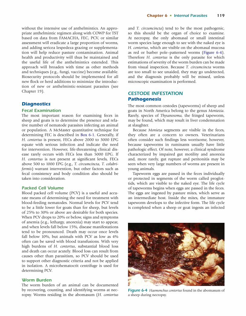
Chapter 6 • Internal Parasites 119
without the intensive use of anthelmintics. An appro-priate anthelmintic regimen along with COWP for TST based on data from FAMACHA, FEC, PCV, or similar assessment will eradicate a large proportion of worms, and adding sericea lespedeza grazing or supplementa-tion will help reduce pasture contamination. Animal health and productivity will thus be maintained and the useful life of the anthelmintics extended. This approach will broaden with time as other methods and techniques (e.g., fungi, vaccine) become available. Biosecurity protocols should be implemented for all new flock or herd additions to minimize the introduc-tion of new or anthelmintic-resistant parasites (see Chapter 19).
DiagnosticsFecal ExaminationThe most important reason for examining feces in sheep and goats is to determine the presence and rela-tive number of nematode parasites infecting an animal or population. A McMaster quantitative technique for determining FEC is described in Box 6-1. Generally, if H. contortus is present, FECs above 2000 to 3000 EPG equate with serious infection and indicate the need for intervention. However, life-threatening clinical dis-ease rarely occurs with FECs less than 5000 EPG. If H. contortus is not present at significant levels, FECs above 500 to 1000 EPG (e.g., T. circumcincta, T. colubri-formis) warrant intervention, but other factors such as fecal consistency and body condition also should be taken into consideration.
Packed Cell VolumeBlood packed cell volume (PCV) is a useful and accu-rate means of determining the need for treatment with blood-feeding nematodes. Normal levels for PCV tend to be a little lower for goats than for sheep, but levels of 25% to 30% or above are desirable for both species. When PCV drops to 20% or below, signs and symptoms of anemia (e.g., lethargy, anorexia) may start to appear, and when levels fall below 15%, disease manifestations tend to be pronounced. Death may occur once levels fall below 10%, but animals with PCV as low as 4% often can be saved with blood transfusions. With very high burdens of H. contortus, substantial blood loss and death can occur acutely. Blood loss can result from causes other than parasitism, so PCV should be used to support other diagnostic criteria and not be applied in isolation. A microhematocrit centrifuge is used for determining PCV.
Worm BurdenThe worm burden of an animal can be documented by recovering, counting, and identifying worms at nec-ropsy. Worms residing in the abomasum (H. contortus
and T. circumcincta) tend to be the most pathogenic, so this should be the organ of choice to examine. At necropsy, the only abomasal or small intestinal worm species large enough to see with the naked eye is H. contortus, which are visible on the abomasal mucosa as red or barber pole–patterned worms (Figure 6-4). Therefore H. contortus is the only parasite for which estimations of severity of the worm burden can be made from visual inspection. Because T. circumcincta worms are too small to see unaided, they may go undetected, and the diagnosis probably will be missed, unless microscopic examination is performed.
CESTODE INFESTATIONPathogenesisThe most common cestodes (tapeworms) of sheep and goats in North America belong to the genus Moniezia. Rarely, species of Thysanosoma, the fringed tapeworm, may be found, which may result in liver condemnation at slaughter.
Because Monieza segments are visible in the feces, they often are a concern to owners. Veterinarians often consider such findings less worrisome, however, because tapeworms in ruminants usually have little pathologic effect. Of note, however, a clinical syndrome characterized by impaired gut motility and anorexia and, more rarely, gut rupture and peritonitis may be seen when very large numbers of worms are present in young animals.
Tapeworm eggs are passed in the feces individually or protected in segments of the worm called proglot-tids, which are visible to the naked eye. The life cycle of tapeworms begins when eggs are passed in the feces. The eggs are ingested by pasture mites, which serve as an intermediate host. Inside the mites, the immature tapeworm develops to the infective form. The life cycle is completed when a sheep or goat ingests an infected
Figure 6-4 Haemonchus contortus found in the abomasum of a sheep during necropsy.

120 Chapter 6 • Internal Parasites
mite while grazing, whereupon the immature form is released from the mite and develops into the adult worm within the small intestine.
Diagnosis and TreatmentA presumptive diagnosis can be made by finding pro-glottids in the feces or eggs in fecal flotation prepara-tions (see Figure 6-2, B). Treatment with albendazole, fenbendazole, or praziquantel may be effective, either with a single dose or with daily therapy (e.g., fenben-dazole daily for 3 to 5 days). Because of the free-living nature of the arthropod intermediate host, animals are readily reinfected after treatment, which may give rise to the false assumption that the therapy was ineffec-tive. Again, tapeworm infection may result in disease, but often it is more convenient to blame the tapeworm segment seen in the stool as a cause of disease than to implicate the unseen thousands of more pathogenic worms in the abomasum and small intestine of the animal.8,10
CYSTICERCOSISCysticercus tenuicollis is the larval stage of the dog tapeworm Taenia hydatigena, and sheep and goats are intermediate hosts. Eggs containing a larval stage are ingested from sources contaminated with dog feces, and after the egg’s shell is digested away, the larvae pen-etrate the wall of the intestine and migrate to the liver. The larvae may remain in the liver or migrate to the surface of the liver, mesentery, or omentum, where they attach themselves and develop into fluid-filled bladder-like cysts called cysticerci. Acute disease occurs only with large numbers of cysticerci and is characterized by depression and weakness secondary to liver dam-age. The chronic cystic stage usually is asymptomatic. No treatment is available, and control is problematic because it requires treating infected dogs and preventing contact with dogs.
COCCIDIAL INFECTIONPathogenesisCoccidiosis is a parasitic disease caused by protozoa (Eimeria spp.) that is a common cause of diarrhea in lambs and kids. In contrast with the nematodes, para-sites for which sheep and goats share the same species, each animal has its own species of Eimeria. Clinical disease results from destruction of the small intestinal epithelium during the asexual reproduction phase of infection. Sexual reproduction then produces oocysts, which are unsporulated and must sporulate outside the host to become infective. Sporulation usually occurs in less than a week under moderate temperatures and high moisture conditions. Sporulated oocysts can survive a
wide range of temperatures and may survive for years under favorable conditions.
Clinical SignsLambs and kids 1 to 6 months of age are most suscep-tible to coccidiosis, and disease most commonly is seen in association with stressful conditions such as with weaning, feed changes, or shipping. Crowded condi-tions with excessive manure are ideal for buildup and sporulation of the oocysts and may promote transmis-sion of coccidia. Under these conditions, animals may be exposed to high numbers of infective oocysts, lead-ing to diarrhea. In severe cases the diarrheal stool may contain blood or mucus and be very watery. Anorexia, dehydration, weakness, rough hair coat, and weight loss, possibly eventuating in death, are encountered in many cases.39 Constant straining with defecation can result in rectal prolapse. In severe cases the major portion of the intestinal mucosa is destroyed, and even if affected animals are treated appropriately, diarrhea can continue for days to several weeks until the mucosa heals. Per-manent scarring of the mucosa is common, potentially resulting in chronic poor growth and development.
DiagnosisAcute coccidiosis can be difficult to diagnose, because clinical disease and death can occur before oocysts (see Figure 6-2, C) appear in the feces (approximately 2 weeks after infection). If high numbers of oocysts are found on fecal examination, the disease is peaking, and most of the damage has been done. In the chronic stages, low numbers of oocysts are typical. Because clinically normal animals often shed small numbers of oocysts, which contain a mixture of pathogenic and nonpathogenic species, interpretation of the findings on fecal examination for animals in the chronic stages of coccidiosis or for those with diarrhea from other causes can be difficult.40,41 In such cases the clinician should rule out other diseases before making a diagno-sis of coccidiosis.
Treatment and PreventionTreatment of affected animals with clinical disease includes supportive care and administration of anticoc-cidial drugs. If the clinical scenario is that of an out-break, with numerous animals affected, then all animals in the group should be treated. The use of coccidiostats, which interfere with asexual reproduction, is useful mainly to prevent further development of infection in the affected animals that have been exposed to infective oocysts.40 Many coccidiostats are used prophylactically when conditions conducive to coccidiosis are expected (i.e., prestress: shipping, weaning, parturition, and so on).

Chapter 6 • Internal Parasites 121
Coccidiostats are of minimal value if given after the onset of clinical disease. Sulfa drugs (not approved for coccidia control in small ruminants) appear to be clinically beneficial, but they may simply decrease secondary or concurrent bacteria-induced diarrhea.40 Table 6-1 lists the anticoccidial drugs approved for use in the United States.
To avoid toxicity in growing animals, dosages must be adjusted to the changing levels of feed intake as animals grow. Coccidiostats should be fed for at least 4 weeks to allow exposure and subsequent development of immunity to occur while preventing the detrimental effects of clinical disease. Fortunately, a strong immu-nity to coccidia develops naturally, but a continual low level of infection is necessary to maintain such immunity. Also, excessive stress in older animals may predispose them to immunosuppression, with resultant clinical coccidiosis.
Coccidia can become resistant to coccidiostats with extended and inappropriate use.40 Therefore this class of chemicals should be fed only during times of expected risk. The inclusion of lasalocid (1 kg of 6% premix) or decoquinate (1 kg of 13% premix) in 22 kg of trace mineralized salt, fed as the only source of salt for 30 days ante partum, can reduce the number of oocysts shed in ewe or doe feces. This practice can reduce the levels of contamination with coccidia and thereby reduce a source of infection for lambs and kids. The benefits of administering lasalocid and monensin beyond the time for coccidia control include increased feed efficiency, enhanced growth rate, and decreased incidence of free gas bloat. However, if coccidiostats are included in either mineral or feed supplements, incon-sistent or depressed intake may result in subtherapeutic drug dosing.
Control also involves improved management and sanitation. Preventing overcrowding decreases the buildup of manure and infective oocysts. In addition, proper design of facilities to prevent fecal contamina-tion of feed and water sources is important. Exposure to sunlight and desiccation are two of the most effec-tive means of killing the oocysts. Minimizing stress and optimizing nutritional intake are important hus-bandry and management techniques to minimize clini-cal coccidiosis.
LIVER FLUKE INFECTIONBoth Fasciola hepatica and Fascioloides magna can infect sheep and goats. Unlike the other parasites, liver flukes are restricted in geographic distribution. This specificity of distribution is due to their requirement for an inter-mediate host, a snail, that only lives in specific aquatic and semiaquatic habitats and soil types. These parasites are common in the Gulf Coast, Pacific Northwest, and Great Lakes regions of the United States.
PathogenesisThe life cycles of F. hepatica and F. magna are similar in that each requires an aquatic snail as an intermediate host. Sheep and goats are definitive hosts for F. hepatica, whereas only some species of deer and elk are definitive hosts for F. magna. Fluke eggs are passed in feces and hatch in water, releasing the larval form (miracidium), which finds and penetrates a snail. After penetration, the miracidium changes forms several times while undergo-ing asexual reproduction. Through this process, each miracidium produces many cercariae, which emerge from the snail to encyst as infective metacercariae on forage. Sheep or goats ingest the metacercariae during grazing, which subsequently excyst in the small intes-tine. The immature fluke penetrates the wall of the small intestine and migrates to the liver, where it penetrates through the liver capsule into the parenchyma. Penetra-tion of the liver can produce significant blood loss, and migration through the liver can leave large tracts of scar tissue (Figure 6-5, A). Adult F. hepatica flukes end up in the bile ducts after migrating through the liver (see Figure 6-5, B) and can live there for years if no treatment
B
A
Figure 6-5 During a necropsy of a sheep, liver fluke infec-tion. A, Fibrotic scarring and necrotic tracts. B, Cut surface with biliary hyperplasia and fluke being squeezed from duct (arrow). (Courtesy Dr. John Malone, Louisiana State University.)

122 Chapter 6 • Internal Parasites
is given. By contrast, F. magna does not mature in sheep and goats, and these flukes continue to migrate in the liver, causing severe damage and death.42 One of the most serious complications of acute liver fluke infec-tion is black disease (Clostridium novyi infection), which causes sudden death. It is imperative that animals be properly vaccinated for this condition in areas in which the risk of fluke-associated disease is recognized.
Clinical SignsF. hepatica infection can cause both acute and chronic disease conditions. Acute disease results when large numbers of immature flukes migrate through the liver, particularly in animals with limited immunity to these parasites. Clinical signs include anorexia, depres-sion, weakness, dyspnea, anemia, ascites, colic-like signs, dry feces, and sudden death. Similar but more severe manifestations are typical for F. magna infec-tion, which usually is fatal. Chronic F. hepatica disease is the result of presence of mature flukes in the bile ducts and is manifested in depressed growth and milk production.
DiagnosisAntemortem diagnosis of fluke infection can be dif-ficult. Finding eggs in feces is diagnostic for F. hepatica. Eggs are produced only by adults and then not in great numbers, so a negative fecal test result cannot rule out acute or chronic fascioliasis. Fluke eggs are dense and therefore do not float in routine fecal flotation prepara-tions used for nematode diagnosis.
A sedimentation technique should be used for sus-pected fluke infections. To perform a sedimentation test, 2 to 3 g of feces is mixed with 50 mL of tap water; the mix-ture is then strained through a tea strainer and funnel into a 50-mL centrifuge tube. The strained fecal sample is then left to sit for a few minutes, to allow the eggs to settle. The supernatant is then poured off and the tube refilled with water while the sediment is resuspended. Repeating this procedure two or three times will clean up the sample so that eggs can be visualized. After the last supernatant is poured off, leaving 5 to 10 mL, methylene blue can be added to stain particulate matter so that the golden-brown eggs (with an operculum at one end) show up easier against the blue background when examined under a dissecting microscope (see Figure 6-2, D). F. magna does not complete it’s life cycle in sheep and goats, so eggs are not produced and fecal examination is of no value.
In the absence of finding fluke eggs in feces, fluke-infected animals may demonstrate eosinophilia, ane-mia, increased liver enzymes, and hypoalbunemia. Infection often is discovered by finding the mature or immature flukes at necropsy or slaughter.
TreatmentIf F. hepatica infection is diagnosed by fecal exam or at necropsy, all animals in the herd should be treated. Because flukicides available in the United States are highly effective only against mature flukes, the timing of treatment is important. In the southern portions of North America, the snails are present year-round, and transmission occurs in the late winter and spring when the snails become active. Flukes migrate and mature in the late spring and summer. Thus treatment in the southern United States usually is recommended in the late summer or early fall. In cooler, northern climates, transmission does not start until late in the spring and may continue into the summer, so flukes can mature into the fall and winter. Therefore treatment is recom-mended in late fall or winter.
The most effective flukicide for removing both immature and mature flukes has been clorsulon (not available in the United States). Ivermectin products containing an adulticidal dose of clorsulon are avail-able but are not approved for use in sheep or goats. Albendazole can kill some adult F. hepatica organisms but is approved only for worm control (7.5 mg/kg) in sheep. A higher dose (10 mg/kg) is approved in goats only for F. hepatica. Albendazole and a clorsulon-iver-mectin combination are somewhat useful in controlling F. magna, but unfortunately, neither is 100% effective, and only a few remaining flukes can be fatal.
PreventionControl of fluke infections is difficult, but decreasing exposure is one key to success. Properly timed treat-ment of animals not only provides immediate benefit but also can decrease infections in successive years by decreasing egg shedding when snails are present on pastures. Eliminating the snail is impractical, but fenc-ing off low-lying wet areas to prevent animal access may limit exposure. Depending on local fluke conditions, efforts should be made to avoid grazing animals in high-risk areas (e.g., where water stands or flows over grazing pastures, streams, and irrigation ditches) during peak infection times.
LUNGWORM INFECTIONPathogenesisAmong the parasites known to cause bronchitis in sheep and goats, Dictyocaulus filaria is the most patho-genic, Muellerius capillaris is the most common and least pathogenic, and Protostrongylus rufescens is inter-mediate in pathogenicity. The life cycle of D. filaria is direct, and the life cycle of both M. capillaris and P. rufescens requires either a snail or slug as an interme-diate host. D. filaria transmission in North America is during the cooler months (fall and winter) of the year,

Chapter 6 • Internal Parasites 123
and transmission of M. capillaris and P. rufescens occurs when snails or slugs are present, which is usually spring or summer. Infected snails and slugs that survive the winter can carry over transmission from one year to the next.43
Infection with D. filaria usually appears in 2- to 18-month-old sheep. Affected animals generally have chronic fever, cough, nasal discharge, tachypnea, anorexia, and weight loss. Treatment with antibiotics is not reward-ing. At necropsy, the worms usually are observed in the bronchi, especially in the diaphragmatic lobes (Figure 6-6). Pulmonary edema, emphysema, and atelectatic and pus-filled lobules also may be evident. Damage due to D. filaria (and other lungworm) infection can predispose animals to secondary bacterial and viral infections and reduce general health (see Figure 6-6, A and B).
Infection with P. rufescens can cause serious disease in sheep, although infection is rarely reported in North America.44 In addition to larvae in feces, they also may be found in nasal secretions. Adult nematodes live in the small bronchioles, and clinical signs include diar-rhea, weight loss, mucopurulent nasal discharge, tachy-pnea, and increased respiratory sounds.
Infection with M. capillaris causes few clinical signs. At necropsy in infected animals, gray or greenish sub-pleural granulomas are seen in the caudal lobes. Goats can have widespread interstitial pneumonia without nodular lesions.45,46
DiagnosisInfection is diagnosed by the presence of first-stage larvae (species are easily differentiated) in fresh (rectal collection is preferred) feces prepared using a simple Baermann technique. Because larvae may be few in
number, a fair amount of feces (10 g or so) is wrapped in cheesecloth or other porous material (e.g., Chem-wipes) and suspended (using an applicator stick) in warm water in a plastic wine glass over night. The warm water stimu-lates the larvae to move and migrate out of the feces and sediment to the bottom of the conical stem. After the fecal ball is removed and the supernatant is poured off, a pipette is used to remove the remaining small amount of water, which is placed on a microscope slide; larvae can then be identified under a microscope. Larvae of D. filaria have straight tails (Figure 6-7), whereas larvae of M. capillaris and P. rufescens have kinked tails.
TreatmentTreatment is with either ivermectin (200 μg/kg), fen-bendazole (7.5 mg/kg), or albendazole (10 mg/kg). Refractory cases of M. capillaris may require larger doses
BA
Figure 6-6 Lungs from a sheep with Dictyocaulus filaria infection. Atelectasis is evident in diaphragmatic lobes (A), and worms can be seen in the trachea (B). (Courtesy Dr. John Malone, Louisiana State University.)
Figure 6-7 Lungworm first-stage larvae of Dictyocaulus recovered with the Baermann technique. (Courtesy Dr. John Malone, Louisiana State University.)

124 Chapter 6 • Internal Parasites
of either fenbendazole (15 mg/kg at 35-day intervals to 30 mg/kg at 30-day intervals) or ivermectin (300 μg/kg), because immature stages of M. capillaris may survive lower doses or single treatments.47
PreventionEliminating the intermediate host mollusk will be help-ful but is very difficult to accomplish (see Chapter 7).
REFERENCES 1. Pugh DG, Mobini SM, Hilton CD: Control programs for gastroin-
testinal nematodes in sheep and goats, Comp Cont Educ Pract Vet 20:S112, 1998.
2. Palmer DG, McCombe IL: Lectin staining of trichostrongylid nematode eggs of sheep: rapid identification of Haemonchus contortus eggs with peanut lectin, Int J Parasitol 26:447–450, 1996.
3. Jurasek ME, et al: Modification and further evaluation of a fluorescein-labeled peanut agglutinin test for identification of Haemonchus contortus eggs, Vet Parasitol 169:209–213, 2010.
4. Kaplan RM, et al: Validation of the FAMACHA8 eye color chart for detecting clinical anemia on sheep and goat farms in the south-ern United States, Vet Parasitol 123:105–120, 2004.
5. Howell SB, et al: Prevalence of anthelmintic resistance on sheep and goat farms in the southeastern United States, J Am Vet Med Assoc 233:1913–1919, 2008.
6. Van Wyk JA, et al: Targeted selective treatment for worm manage-ment—how do we sell rational programs to farmers? Vet Parasitol 139:336–346, 2006.
7. Kenyon F, et al: The role of targeted selective treatments in the development of refugia-based approaches to the control of gastrointestinal nematodes of small ruminants, Vet Parasitol 164:3–11, 2009.
8. Donaldson J: The effects of dietary protein on establishment and maturation of nematode populations in adult sheep. In Barrel GK, editor: Sustainable control of internal parasites in ruminants, Canterbury, New Zealand, 1997, Lincoln University.
9. Pugh DG, Navarre CB: Parasite control programs in sheep and goats, Vet Clin North Am Food Anim Pract 17:231–244, 2001.
10. Kaplan RM: Anthelmintic treatment in the era of resistance. In Anderson DE, Rings M, editors: Current veterinary therapy: food animal practice, ed 5, St Louis, 2009, Saunders, pp 470–478.
11. Craig TM: Epidemiology of internal parasites: effects of climate and host reproductive cycles on parasite survival, Proceedings of the Small Ruminant Mixed Animal Practitioner Western Veterinary Conference , 1998, Las Vegas, Nev.
12. Hennessy DR: Modifying the formulation or delivery mechanism to increase the activity of anthelmintic compounds, Vet Parasitol 72:367–390, 1997.
13. Escudero E, et al: Pharmacokinetics of moxidectin and doramec-tin in goats, Res Vet Sci 67:177–181, 1999.
14. Kaminski R, et al: A new class of anthelmintis effective against drug-resistant nematodes, Nature 452, 2008:U176-U119.
15. Miller DK, Craig TM: Use of anthelmintic combinations against multiple resistant Haemonchus contortus in Angora goats, Small Rumin Res 19:281–283, 1996.
16. Hennessy D: The disposition of antiparasitic drugs in relation to the development of resistance by parasites of livestock, Acta Trop 56:125–141, 1994.
17. Hunt PW, McEwan JC, Miller JE: Future perspectives for the implementation of genetic markers for parasite resistance in sheep, Trop Biomed 25:18–33, 2008.
18. Coop R, Kyriazakis I: Influence of host nutrition on the develop-ment and consequences of nematode parasitism in ruminants, Trends Parasitol 17:325–330, 2001.
19. Burke JM, et al: Dose of copper oxide wire particles (COWP) and feed supplement level influences Haemonchus contortus infection in lambs, Vet Parasitol 123:235–243, 2004.
20. Burke JM, et al: Use of copper oxide wire particles to control gas-trointestinal nematodes in goats, J Anim Sci 85:2753–2761, 2007.
21. Min BR, et al: The effect of grazing forage containing condensed tannins on gastro-intestinal parasite infection and milk composi-tion in Angora does, Vet Parasitol 130:105–113, 2005.
22. Lange KC, et al: Effect of sericea lespedeza, fed as hay, on natural and experimental Haemonchus contortus infections in lambs, Vet Parasitol 141:273–278, 2006.
23. Terrill TH, et al: Effect of pelleting on efficacy of sericea lespedeza hay as a natural dewormer in goats, Vet Parasitol 146:117–122, 2007.
24. Preston JM, Allonby EW: The influence of breed on the suscepti-bility of sheep and goats to a single experimental infection with Haemonchus contortus, Vet Rec 103:509–512, 1978.
25. Courtney CH, et al: Resistance of nonlambing exotic and domes-tic ewes to naturally acquired gastrointestinal nematodes, Int J Parasitol 15:239–243, 1985.
26. Mugambi JM, et al: Response of Dorper and red Maasai lambs to trickle Haemonchus contortus infections, Res Vet Sci 61:218–221, 1996.
27. Miller JE, et al: Epidemiology of gastrointestinal nematode parasitism in Suffolk and Gulf Coast Native sheep with special emphasis on relative susceptibility to Haemonchus contortus infec-tion, Vet Parasitol 74:55–74, 1998.
28. Baker RL, et al: Resistance of Galla and Small East African goats in the sub-humid tropics to gastrointestinal nematode infections and the peri-parturient rise in faecal egg counts, Vet Parasitol 79:53–64, 1998.
29. Amarante AF, et al: Resistance of Santa Ines, Suffolk and Ile de France sheep to naturally acquired gastrointestinal nematode infections, Vet Parasitol 120:91–106, 2004.
30. Larsen M, et al: Biological control of gastro-intestinal nema-todes—facts, future, or fiction? Vet Parasitol 72:479–485, 1997.
31. Chandrawathani P, et al: Biological control of nematode parasites of small ruminants in Malaysia using the nematophagous fungus Duddingtonia flagrans, Vet Parasitol 117:173–183, 2003.
32. Fontenot ME, et al: Efficiency of feeding Duddingtonia flagrans chlamydospores to grazing ewes on reducing availability of para-sitic nematode larvae on pasture, Vet Parasitol 118:203–213, 2004.
33. Bain RK, Urquhart GM: Parenteral vaccination of calves against the cattle lungworm, Dictyocaulus viviparus, Res Vet Sci 45:270–271, 1988.
34. Lightowlers MW: Cestode vaccines: origins, current status and future prospects, Parasitol 133(Suppl):S27–S42, 2006.
35. Kabagambe EK, et al: Attempts to control haemonchosis in graz-ing ewes by vaccination with gut membrane proteins of the para-site, Vet Parasitol 92:15–23, 2000.
36. Smith WD, van Wyk JA, van Strijp MF: Preliminary observations on the potential of gut membrane proteins of Haemonchus contor-tus as candidate vaccine antigens in sheep on naturally infected pasture, Vet Parasitol 98:285–297, 2001.
37. Olcott DD, et al: Effect of vaccination of goats with H-gal-GP and H11 antigens from intestinal membrane cells of Haemonchus con-tortus, Proceedings of the 21st International Conference of the World Association for the Advancement of Veterinary Parasitology, August 19-23, 2007:Ghent, Belgium.
38. Smith WD, Taylor S: Twists and turns en route to a vaccine for Haemonchus contortus, Proceedings of the 22nd International Con-ference of the World Association for the Advancement of Veterinary Parasitology, August 8-13, 2009:Calgary, Canada.
39. Foreyt WJ: Coccidiosis and cryptosporidiosis in sheep and goats, Vet Clin North Am Food Anim Pract 6:655, 1990.
40. Craig TM: Coccidiosis in small ruminants, Proceedings of the Small Ruminant Mixed Animal Practitioner Western Veterinary Conference, 1998b:Las Vegas, Nev.
41. Smith MC: Parasitic diseases of goats, Proceedings of the 1996 Sym-posium on Health and Disease of Small Ruminants, 1996:Nashville, Tenn.

Chapter 6 • Internal Parasites 125
42. Westcott RB, Foreyt WJ: Epidemiology and control of trematodes in small ruminants, Vet Clin North Am Food Anim Pract 17:373, 1986.
43. Helle O: The efficacy of fenbendazole and albendazole against the lungworm Muellerius capillaris in goats, Vet Parasitol 22:293, 1986.
44. Mansfield LS, et al: Lungworm infection in a sheep flock in Mary-land, J Am Vet Med Assoc 202:601–606, 1993.
45. Nimmo JS: Case report: six cases of verminous pneumonia (Muel-lerius sp.) in goats, Can Vet J 21:49, 1979.
46. Kanwar NS, Paliwal OP, Ram K: Verminous pneumonia in goats, J Vet Parasitol 12:139, 1998.
47. McCraw BM, Mensies PI: Treatment of goats infected with the lungworm Muellerius capillaris, Can Vet J 27:287, 1986.

126
Diseases of the Respiratory System
Paul J. Plummer, Cassandra L. Plummer, and Kelly M. Still
C H A P T E R
ANATOMYClinically significant upper airway structures in the small ruminant include the frontal and maxillary sinuses, phar-ynx, larynx, and trachea. The nasopharynx is the primary path for respiration, but oral respirations are anatomi-cally possible, and “panting” occurs under some fairly normal conditions such as high ambient temperature. Laryngeal structure is similar to that in other species, with small V-shaped vocal folds just caudal and ventral to the arytenoid cartilages.1,2 The retropharyngeal lymph node is located dorsocaudal to the pharynx and can com-press the larynx or trachea when enlarged or abscessed. The trachea runs down the ventromedial aspect of the neck from the larynx to the bronchial bifurcation in the thorax. It is composed of incomplete tracheal rings con-nected by a membranous wall. The tracheal diameter in small ruminants generally is smaller than might be expected and changes at the thoracic inlet: In goats the trachea narrows, whereas in sheep it enlarges.2
In the thorax, the trachea bifurcates into two main bronchi. Just cranial to this bifurcation a separate bron-chus branches out to the right cranial lung lobe. The major lung divisions include left and right cranial lobes, each with a cranial and caudal part; the right middle (cardiac) lobe; the right accessory lobe; and the left and right caudal (diaphragmatic) lobes. When enlarged, the mediastinal lymph nodes and thymus may compress or shift the thoracic trachea or lung. The caudal lung border is demarcated by the sixth rib ventrally, by the seventh rib at the lateral midthorax, and by the eleventh rib dorso-caudally. The intercostal vessels and nerves run caudally along each rib, and care should be taken to avoid these structures during thoracocentesis or biopsy procedures.2
PHYSIOLOGYThe respiratory system permits reoxygenation of pul-monary venous blood and release of carbon dioxide formed by cellular respiration. Effective respiration requires both alveolar ventilation and gas diffusion across the respiratory membrane; together, these two processes can be quantified by the ventilation-perfusion ratio, which may be altered during disease. Alveolar
ventilation occurs through movement of gas from the terminal bronchioles and depends on inspiratory tidal volume and expiratory functional reserve in addition to respiratory rate. Anatomic dead space (e.g., nasal pas-sages, pharynx, trachea, bronchi) does not contribute to alveolar ventilation. Once in the alveolus, respiratory gases must diffuse between the lung and capillaries. Gas movement across membranes is affected by the diffu-sion coefficient of the gas, the thickness of the septum, and the surface area available for diffusion. Because carbon dioxide diffuses much more readily than oxygen and is the direct stimulus for respiration, hypoxia may occur without significant increases in respiratory rate. Alveolar septum thickness can be increased by edema and fibrosis. Surface area can be physically decreased by consolidation and emphysema or physiologically reduced by alteration in the ventilation-perfusion ratio stemming from increased physiologic dead space or shunting of blood away from ventilated alveoli.3
Significant innate immune defenses are present in the lung. The sneeze and cough reflexes forcibly expel large particles and irritants from the upper airway. Nasal hairs and air turbulence over the nasal concha will filter out airborne particles as small as 6 μm. Gravitational precipitation will filter smaller particles (1 to 5 μm) in the small bronchioles. Mucociliary clearance efficiently moves trapped particles to the pharynx, where they are either swallowed or coughed out. This system is formed by mucus-producing goblet cells and ciliated epithelial cells that line the respiratory tract from the nasal passages to the terminal bronchioles. Once in the alveoli, particles larger than 0.5 μm will come to lie against the alveolar wall and be cleared by alveolar macrophages or the lung lymphatic system. Particles less than 0.5 μm in size remain suspended and will be exhaled without consequence.4
DIAGNOSTIC APPROACHESPhysical Examination and AuscultationA thorough and unbiased physical examination is the most important component of the diagnostic evalua-tion of small ruminants presented for abnormalities of
7

Chapter 7 • Diseases of the Respiratory System 127
the respiratory tract. Without a complete physical exam, important primary or secondary physiologic problems may be missed, and the diagnostic plan may be incom-plete or result in failure to obtain a definitive diagnosis.
The physical exam should be conducted in a system-atic manner and must include all aspects of the respira-tory system. Before restraining the animal, the clinician should spend a few minutes observing its attitude, stance, respiratory rate, and respiratory pattern from a distance, because significant elevations in respiratory rate and pattern can occur after capture and restraint, particularly in animals that are less socialized. In conse-quence of the flocking instincts of sheep and goats, ani-mals observed to be standing apart from the rest of the flock or herd are likely to be significantly ill. Once the animal is caught and restrained, the practitioner should begin by evaluating the respiratory system starting at the head (see Chapter 1). The nares should be examined for evidence of serous, mucopurulent, or hemorrhagic dis-charge from one or both nostrils (Figure 7-1). Unilateral nasal discharge may provide significant localization of a lesion and should be noted on the examination form. Both nares should be accessed for patency by placing either a small cotton ball or a mirror in front of the nose and observing for movement or fogging, respectively. The remainder of the head should be evaluated for evi-dence of facial deformity or soft tissue swelling indica-tive of a localized lesion. The pharyngeal area should be palpated, with particular attention paid to the local lymph nodes. When possible, the palpation should include an attempt to feel the area lateral and dorsal to the pharynx by placing a hand alongside the trachea and palpating with gentle dorsal pressure. This area is a common site for retropharyngeal abscesses (often caused by Corynebacterium pseudotuberculosis), which may result in considerable respiratory stridor and effort. The extrathoracic trachea should be palpated from the
pharynx down to the mediastinal entrance for any evi-dence of stricture, dilatation, or external compression. During this portion of the evaluation, occasional gentle squeezing pressure should be applied to the trachea, to determine how easily coughing can be induced. The mediastinal opening is another area that warrants palpation for evidence of space-occupying lesions or tracheal deviation associated with such findings.
Attention should then turn to performing a complete auscultatory examination of the thorax, when pos-sible. Owing to the heavy wool cover on the thorax of sheep, this exam may be of limited usefulness without adequate shearing. At a minimum, the cranioventral aspect of the thorax of sheep can be auscultated in the nonwooled area located immediately behind the elbow. In sheared or haired sheep and lambs and goats, the entire thorax generally can be auscultated without fur-ther removal of fiber or hair. Attention should be paid to the intensity, duration, and character of the breath sounds, as well as the stage of respiration (i.e., inhala-tion or exhalation, early or late) during which they occur. In comparison with those in cattle, the normal airway sounds heard in sheep and goats are much more obvious, owing to the thinner body wall. This perceived magnification often results in the erroneous impression of abnormal respiratory sounds.
Abnormal sounds should be classified as either of two different descriptive types: Wheezes are high-pitched, continuous musical sounds associated with altered airflow through larger airways. They are indica-tive of either fluid in the airway or increased velocity of air movement in the airway. Crackles are noncontinuous brief “popping” sounds associated with sudden open-ing of small airways or alveoli. They most commonly are heard during inspiration, particularly late inspira-tion, and previously were described as “rales.” If any abnormal breath sounds are auscultated, they should be localized and their anatomic location recorded on the examination form. In most instances, the use of a rebreathing bag, as is common in respiratory evaluation of horses, is not necessary for small ruminants, owing to their relatively thin body wall.
After completion of the auscultation exam, several additional pieces of information should be collected. The rectal temperature will reveal whether the animal is febrile, normothermic, or hypothermic. The presence of fever may provide additional evidence of an inflam-matory process that may warrant additional diagnostic effort. Additionally, the nutritional status of the animal should be evaluated, because immune dysfunction is more common in young animals with less than ade-quate reserves of body fat. This assessment is perhaps best performed by body condition scoring of multiple animals in the same management group. Finally, the practitioner should spend some time evaluating the environment in which the diseased animal is housed.
Figure 7-1 The clinician is carefully examining the nares in this well-restrained ewe. (Note: Both a light source and saline are available to flush out any material that precludes proper evaluation.) (Courtesy Dr. A.N. Baird, Purdue University.)

128 Chapter 7 • Diseases of the Respiratory System
Environments with poor ventilation, drafts, dust, or high stocking densities may predispose resident ani-mals to the development of respiratory disease; in such instances, appropriate treatment may require address-ing the environmental conditions.
With respiratory disease in preweaned animals, it also is worthwhile to consider the role of colostrum management and failure of passive transfer in the dis-ease process. When warranted, serum samples from several animals can be collected and assayed for failure of passive transfer status. Our own preference is to test a group of 10 animals between 24 and 72 hours of age; at least 8 of the 10 animals should demonstrate adequate evidence of passive transfer. If increased rates of failure of passive transfer are identified, then herd- or flock-level changes are needed to improve immunity of this at-risk group.
After the physical exam has been completed, the clinician should use the findings to develop a compre-hensive problem list that will serve as a basis for devel-opment of a complete diagnostic plan and differential list. Although this step often is skipped in the interest of time, it is one of the few ways to ensure consideration of all possible clinical entities in the differential diagnosis.
Diagnostic ProceduresOnce a complete list of diagnostic possibilities has been generated, the clinician can turn to the development of a useful and cost-effective diagnostic approach specific to the case. In this context, it is important to ascertain the expectations of the client with regard to desired out-come. For instance, the producer with 29 weaned kids in group housing of which 10 were lost to pneumonia in the past week may have very different expectations and motivations to pursue diagnostic investigation from those for a producer with a single animal showing clinical signs. Many of the usual procedures for such investigation, as described next, may not be economi-cally feasible or desirable if the producer perceives that the cost does not justify the return on investment. By contrast, if the results can be used to prevent disease in multiple animals, the motivation to pay for the diag-nostics may be increased.
Blood Gas AnalysisBlood gas analysis provides a rapid and useful assess-ment of hemoglobin oxygenation and alveolar diffu-sion of gases. Its usefulness is, however, limited by the need for rapid testing and appropriate sample handling to prevent erroneous results. The advent of portable blood gas analyzers that can be carried on the ambu-latory care truck make this test feasible in the farm situation; in most instances, however, its application is limited to high-value cases in referral hospital set-tings. In our experience, an arterial blood gas sample is
best collected from small ruminants using the brachial artery located on the medial aspect of the proximal portion of the front legs. Special blood gas syringes are commercially available and should be used if accurate assessment of partial pressures is required, as would be the case in respiratory disease. While the animal is lying in lateral recumbency, the lower limb is extended and the pulsation of the artery is palpated between the index and middle fingers while the needle is inserted at a 90-degree angle to the skin. Once the artery is pen-etrated, the syringe is held steady and should self-fill. Negative pressure should not be applied to the syringe, because this will alter gas partial pressures in the sam-ple. Once the blood is collected, the needle should be rapidly sealed, typically with the rubber stopper sup-plied with blood gas syringes. Care should be taken to not introduce any bubbles into the syringe during this process. Arterial partial pressures of O2 (Pao2) should be above 70 to 80 mm Hg in an animal with normal oxygenation. Partial pressures below that level may be indicative of inappropriate ventilation, poor alveolar ventilation, or thickened alveolar walls that impair oxy-gen diffusion. Normal partial pressures of CO2 in an arterial sample should be below 40 mm H, and if the sample yields a Pao2 greater than that value in associa-tion with a very low oxygen partial pressure, the pos-sibility that a venous sample has been obtained needs to be considered.
RadiographyRadiographs of the thorax, neck, or head often are required and can be of significant diagnostic ben-efit. Radiographs can easily be obtained using portable radiographic equipment commonly available to vet-erinary practitioners. When unilateral nasal discharge or facial deformities are observed during the physical exam, radiographic evaluation with both lateral and dorso-ventral views of the head may elucidate the etiology. In many instances, nasal foreign bodies or sinusitis can be confirmed on the basis of the radiographic interpreta-tion of the head views. Similarly, radiographs of the neck may provide additional evidence of tracheal compres-sion or retropharyngeal masses that may be associated with coughing in affected animals. Thoracic radiographs can be obtained with the animal either standing or in lateral recumbency, depending on the facilities available to the practitioner (Figure 7-2, A and B).
Lung field consolidation can be readily identified by observing radiographic opacities in the cranial ventral lung fields, and mediastinal masses, often associated with caseous lymphadenitis abscesses, generally are revealed as a line of masses of increased density cours-ing through the thorax at the level of the trachea. In rare instances, a thymoma may result in the appearance of a mass cranial to the heart that gives the appearance of the animal’s having two hearts.

Chapter 7 • Diseases of the Respiratory System 129
Ultrasound ImagingPortable ultrasound units are becoming standard equip-ment in many large animal clinics, affording easy access to this imaging modality. Many units used for reproduc-tive practice are equipped with a linear, 5- to 7.5-MHz transducer. This type of machine can provide reasonably good-quality images of the thorax and adjacent soft tissues. When available, a curvilinear probe will pro-vide a superior image but certainly is not required for
diagnostic use. Appropriate patient preparation is para-mount for obtaining a good-quality image. Wool or hair over the site of interest should be clipped, although the use of coupling agents (e.g., gel, vegetable oil, alcohol) can be helpful in some instances. Owing to the nature of the functioning, gas-filled lung, ultrasonography of the respiratory tract is more limited than that of other body systems. For example, ultrasound examination of the pharyngeal region may provide an easy means of identifying retropharyngeal abscesses when they are suspected from findings on palpation. In such cases the probe should be placed parallel to the lateral aspect of the trachea and directed dorsomedially towards the opposite ear. Abscesses typically have a hyperechoic wall, with variable echotexture of the contents.
Ultrasound imaging also can provide useful informa-tion in evaluation of the thorax. The clinician should become familiar with the appearance of normal aerated lung, allowing rapid identification of areas that lack the normal appearance. Normal lung is recognizable by the bright hyperechoic line of the visceral pleura above a classic reverberation artifact induced by the aerated lung. The reverberation artifact is typical of ultrasound waves hitting a gas interface and consists of sequential hyper-echoic lines spaced at regular intervals. It is important to realize that any images appear on the screen deep to the start of the reverberation artifact are indeed artifacts and not images of the lung parenchyma (Figure 7-3). Once an appreciation for the normal appearance of lung has been achieved, the thoracic exam can be systematically performed. With use of a linear or a curvilinear probe, the probe should be oriented parallel to the ribs in the intercostal space. We prefer to start at the most dorsal aspect of each intercostal space and slowly move down-wards to the ventral thorax observing the lung surface along the path. This is repeated in each intercostal space moving caudally. The image quality is maximized by following the natural “lay” of the wool or hair (in a dorsal-ventral direction). As the exam progresses cau-dally, the diaphragm will come into the image while moving ventrally, often with the adjacent liver filling the space below. With use of this method, the extent of the thoracic lung field can be determined. The three primary lesions that will be observed are parenchymal masses in the lung that are adjacent to the visceral pleura, lung consolidation, and the characteristic “comet tail” lesions associated with pleural thickening and inflam-mation. The first of these lesions is readily identified by the observation of echodense masses interrupting the normal reverberation artifact of the lung. Such masses can be measured to allow for sequential ultrasono-graphic examination as a means of assessing treatment success or resolution of the lesion. In our experience, these lesions most commonly are associated with paren-chymal abscesses. Consolidated lung is recognized on deeper imaging, beyond the normal lung reverberation.
A
B
Figure 7-2 Radiograph showing pulmonary edema related to a tracheal obstruction in a 6-year-old Suffolk ewe. A, Increased opacity is evident throughout the lungs, along with peribronchial cuffing and lack of visualization of the vas-cular markings. It is difficult to distinguish the borders of the caudal vena cava because of the increased interstitial opacity. Other considerations in the differential diagnosis for this pul-monary pattern would be pneumonia and pulmonary hemor-rhage. B, Lateral view of the caudal thorax of the same patient 24 hours after treatment with a tracheotomy, diuretics, and antibiotics. The vascular margins are better delineated, as are the borders of the caudal vena cava. Interstitial opacity within the lungs is less than on the previous radiograph. Although mild interstitial opacity persists, the pulmonary edema is resolving. (Courtesy Dr. Debra Baird, Purdue University.)

130 Chapter 7 • Diseases of the Respiratory System
In many instances, the consolidated lung may have an appearance similar to that of liver (“hepatized lung”) or may be seen to contain scattered gas shadows associated with presence of gas in the larger airways or in abscesses. “Comet tails” are recognizable as small, hyperechoic spots with a comet tail–shaped artifact located deep to the spot. These lesions are nonspecific but often are asso-ciated with thickening or inflammation of the pleura.
If pleural fluid is present, it will be imaged as an anechoic or hypoechoic area in the ventral thorax, with normal lung reverberation noted at the lung-fluid interface. Because the mediastinum is not always eas-ily imaged, radiographs remain the preferred imaging modality for identification of mediastinal masses.
Nasal SwabNasal swabs are very useful as a means of obtaining material for microbiologic culture in cases of respiratory disease. The laboratory that will process the cultures should be contacted for recommendations on swab type and submission recommendations. For instance, calcium alginate swabs and bleached cotton swabs have the potential to interfere with polymerase chain reac-tion (PCR) testing. Many swabs with wooden sticks have formaldehyde as a preservative in the wood, which can adversely affect bacterial growth. Similar considerations apply regarding the selection of transport media. The use of a guarded swab (i.e., mare uterine swabs), when available, should be considered to minimize contami-nation of oral flora. Use of an oral speculum (0.5- to 1.0-inch internal diameter [ID] polyvinylchloride (PVC) pipe, cut to length, with the ends sanded smooth) may
help in obtaining a more reliable sample. Collection of the diagnostic sample involves simply rolling the swab surface on the pharyngeal mucosa around the palatine tonsil. Once prepared, the swab should be placed in the transport medium and refrigerated unless otherwise directed by the diagnostic laboratory.
Sinus-CentesisThe technique of sinus-centesis provides the practitio-ner with an option for collecting representative culture material from a nasal sinus. Owing to the comparatively smaller nasal sinuses of sheep and goats, proper selec-tion of a site for sampling is critical to ensure entry into the sinus cavity. Radiographic assistance in localizing the involved sinus cavity is recommended. If necessary, radiopaque markers can be placed on the skin before exposure to ensure appropriate site selection. Once the site is verified, the area should be clipped and surgi-cally prepared. Raising a small bleb under the skin with lidocaine will provide adequate anesthesia to the external surfaces but will not achieve anesthesia to the periosteum. Thus the animal should be sedated or anes-thetized (see Chapter 18). If the goal is to collect a small sample of material for culture, a small-diameter bone pin or heavy-gauge circlage wire can be guided through a stab incision in the skin and used to drill a small hole through the bone. A hypodermic needle can then be introduced into the sinus and a sample aspirated. Samples should be submitted for aerobic bacterial and fungal culture. In cases in which drainage and lavage will be needed, a small sinus trephine can be used to create a large-bore opening into the sinus.
After collection of the sample, the incision should be kept clean and allowed to heal by second intention. The operated animals should be fed low to the ground to help facilitate sinus drainage, and use of elevated hay racks should be avoided until the wound is fully healed.
Tracheal WashWith large herd outbreaks of respiratory disease or in the face of a high incidence of treatment failures, diagnostic sampling for determining the etiologic agent and anti-microbial susceptibility profile should be considered. Nasal swabs can be used in some circumstances but will yield less reliable results than a tracheal wash. A tracheal wash provides the clinician with the opportunity to col-lect a sterile deep lung sample with minimal effort.
The animal should be standing and adequately restrained for this procedure. Sedation may be war-ranted. If a fiberoptic endoscope is available (8 to 9 mm diameter), it may be inserted through the nasal passage in some adult sheep or goats. If this is not possible, the endoscope can be passed through an oral speculum. An endoscopic examination may allow visualization of the respiratory tract, identify exudates, and enhance sample collection. If an endoscope is unavailable,
Figure 7-3 Ultrasound image of the right cranioventral thorax obtained at the sixth intercostal space in a 3-year-old LaMancha cross doe with normal lungs. The linear hyper-echoic structure with reflective echoes represents the normal, aerated pleural surface. This ultrasound image was obtained using a 10-MHz linear array transducer. Dorsal is to the left of the image. (Courtesy Dr. Karine Pader, Purdue University.)

Chapter 7 • Diseases of the Respiratory System 131
a percutaneous transtracheal wash (TTW) procedure can be performed. Use of a commercially available prester-ilized, complete kit designed for foals, when available, will enhance the ease of this procedure (Figure 7-4, A and B). Alternatively, a hypodermic needle of appro-priate size to allow a sterile tube catheter (220 polyeth-ylene) to pass through the bore may be used. On the ventral aspect of the neck, the hair or fiber should be removed over at least a 6-inch square of skin centered on the midline and located roughly one third of the way down the neck from the throatlatch. The trachea should be identified and easily palpated at this level. The site should be disinfected using a standard surgical preparation, and a small bleb can be raised with lido-caine placed under the skin directly over the midline of the trachea in the center of the site. A stab incision should be made through the skin using a scalpel blade. The procedure should be performed using sterile tech-nique. If a commercial kit is to be used, the blunt-tipped needle and associated placement trochar should be identified in the kit, and the clinician should become familiar with their design and use before performing the procedure. The unit should be placed through the skin incision and the tip of the trocar used to palpate the tracheal rings while the operator’s opposite hand is used to stabilize the trachea. The tip of the trocar should be positioned on midline between the tracheal rings and firm pressure applied to facilitate passage of the trochar into the trachea. A slight “pop” may be felt as the tip of the stylet enters the cavity. The trocar should be advanced until it can be felt to fully penetrate the trachea and can be slightly advanced in a ventral
direction (Figure 7-5, A). The stylet should be removed and the aspiration catheter passed (using sterile gloves) through the trocar to roughly the level of the tracheal bifurcation. Twelve to 15 mL of sterile saline should be infused into the trachea and a sterile syringe used to apply gentle suction as the catheter is moved back and forth in the trachea (see Figure 7-5, B). The goal is to move the catheter so that its tip is in the pool of fluid created just cranial to the tracheal carina. Although this cannot be visualized, it can be located with practice in a majority of cases. If needed, additional normal saline (another 5 to 10 mL as indicated) can be given to increase the recovery volume. The catheter should be removed, followed by the trocar. In cases in which a needle and polypropylene catheter are used, the needle should be removed from the trachea before the catheter, to minimize the risk of cutting off the distal tip of the catheter during its withdrawal.
ThoracocentesisThoracocentesis provides a reliable means of collecting a sample of pleural fluid for diagnostic submission. This is best performed in the cranial-ventral portion of the chest, where pleural fluid pools in the most depen-dent part of the thorax. While the animal is standing, this area can be evaluated by ultrasound imaging and an appropriate site selected as indicated by presence of fluid and absence of other viscera. The body wall thick-ness can be measured to assist in determining how deep to advance the needle to acquire the sample. Once col-lected, the sample should be evaluated for appearance, odor, and turbidity, in addition to being submitted for
B
1
2
3
4
A
Figure 7-4 Transtracheal wash. A, Site of access for transtracheal wash on the ventral midline in the midcervical region. B, Supplies for transtracheal wash procedure: 18-inch flexible wash catheter (1), sample vial (2), trochar removed from cannula for visualization (3), and wash cannula (4).

132 Chapter 7 • Diseases of the Respiratory System
bacterial culture and cytologic study. The presence of a pungent foul odor often is associated with anaerobic infection, and treatment decisions should consider this possibility.
UPPER AIRWAY DISEASEStertor and stridor, sneezing, and nasal discharge are hallmark signs that suggest upper airway disease over pneumonia.
RhinitisPossible causes of rhinitis in sheep and goats include foreign material such as from regurgitation, parasites, tumor, and other respiratory infections.
Oestrus ovis InfestationPathogenesisNasal bot infestation is more common in sheep than in goats, and infected goats have a lower larval burden than that typical for sheep.5 Clinical signs during the first spring infestation generally are mild, but disease sever-ity markedly increases during subsequent infestations, probably owing to hypersensitization; goats may acquire immunity after repeated infections.5 The adult Oestrus ovis fly deposits larvae at the animal’s nostrils. The first instar larvae migrate up the nasal passages into the
dorsal turbinates and sinuses. There they develop over a 2- to 10-month period to the third instar stage,1,6 return to the nostril, and are sneezed out to pupate in the soil.6 Both first instar larvae and pupae may overwinter.6
Clinical SignsIrritation from the adult flies will induce avoidance activities such as head shaking, head rubbing, and feet stomping; if the animal’s distress level is severe, grazing activity will decrease.6 Larval passage and development can cause inflammatory rhinitis characterized by sneez-ing, mucopurulent discharge, and decreased airflow through the nares. Sequelae can include bacterial rhini-tis or sinusitis and, infrequently, interstitial pneumonia secondary to antigen aspiration.5
DiagnosisO. ovis infection is associated with a profuse nasal discharge containing numerous eosinophils and mast cells.5,7 Direct visualization of the bots or mineralized remains may be possible with endoscopic or radio-graphic imaging. In commercial herds, clinical signs, cytologic examination of the discharge, and response to therapy usually are sufficient to make the diagnosis.
TherapyTreatment usually is administered for heavy late sum-mer infestations or to kill overwintering bots. Iver-mectin (0.2 mg/kg SC) is effective in killing the O. ovis larvae1,6,7 but requires an extended milk withdrawal period: 40 days if administered subcutaneously and 6 days if administered orally (if administered at a higher oral dose of 0.4 mg/kg, an 11-day milk withdrawal is recommended).8 Pour-on eprinomectin (0.5 mg/kg) may be a better choice for commercial dairies because it has been shown to be effective in sheep against nasal bot infestation and has a zero-day milk withdrawal period.9,10
Once the bots are killed, secondary bacterial infec-tions usually will resolve without further intervention. If indicated, however, treatment is with broad-spectrum antimicrobials.7
Other ParasitesIn the Himalayas, a nasal leech from standing water pools can cause similar clinical signs. Systemic ivermec-tin is ineffective, but direct application of ivermectin solution (0.1 mg/mL) to the leech will kill it within a few hours. Wetting the animal’s muzzle will encourage the leech to migrate down to the nostril opening so that the ivermectin can be applied.1
On the Indian subcontinent, Schistosoma nasale infec-tion (“snoring disease”) has been reported as a cause of nasal obstruction from parasite-associated inflamma-tion and tissue proliferation.1,11
A B
Figure 7-5 A, The middle third of the neck is aseptically scrubbed, the skin and subcutaneous tissue are anesthetized, a stab incision is made through the skin and subcutaneous tissue, a 14-gauge needle or trocar is passed through the inci-sion and into the trachea, and tubing is passed using sterile technique just beyond the tracheal bifurcation. B, The clini-cian injects sterile isotonic solution (12 to 30 mL) through the tubing and then immediately performs fluid retrieval by aspiration with the syringe. (Note: With this method, caution should be taken to avoid cutting off the tube in the trachea with the sharp needle.)

Chapter 7 • Diseases of the Respiratory System 133
Enzootic Nasal TumorPathogenesisEnzootic nasal tumors are transmissible, sporadically occurring tumors of the nasal passages of sheep and goats.12,13 This condition has been reported in ani-mals as young as 15 and 7 months, respectively,14,15 and is believed to be caused by type D or B retrovirus infection.12,16,17 These tumors can occur unilaterally or bilaterally and are locally invasive but not usually meta-static.13,15 They originate from the olfactory mucosa and ethmoid or nasal turbinates and usually are classified as adenomas, adenopapillomas, and adenocarcinomas.13-15 Other conditions on a differential diagnosis list for nasal masses include lymphosarcoma and fungal granuloma.1
Clinical SignsThe tumor starts as small nodules that grow to form large nodular cystic masses, causing progressive inspi-ratory dyspnea and secondary emaciation.13,15 Inflam-matory polyps may be present near the tumor.15,17 Primary clinical signs include unilateral or bilateral copious seromucous to mucopurulent nasal discharge with inspiratory stridor. Additional signs may include exercise intolerance, decreased airflow and open-mouth breathing, anorexia, head shaking and sneezing, exoph-thalmos, and bony facial asymmetry.13-15
DiagnosisA preliminary diagnosis can be made from the clinical signs and findings on sinus percussion. Radiographic or endoscopic imaging may be indicated. Definitive diag-nosis requires surgical excisional biopsy.14
TreatmentIf enzootic nasal tumor is untreated, death will occur within 90 days of appearance of clinical signs.13,14 Sur-gical debulking is a palliative option14 but may not be curative.18 The mass can be accessed for excision by creat-ing an I-shaped incision in the skin and then the nasal bones along the dorsal facial midline axis, reflecting the cutaneous and bony flaps, and removing the nasal sep-tum. Profuse hemorrhage is to be expected; epinephrine (1:100,000)-soaked gauze pads can help with hemo-stasis, and a blood donor should be readily available.18 A temporary tracheostomy may be needed during the surgical procedure and the postsurgical period.14 Herd or flock control of enzootic tumor is difficult in the absence of a serologic test to identify animals with preclinical dis-ease. Enzootic nasal tumors can be spread by nasal dis-charge; infected animals should be isolated and culled.17
Other Respiratory InfectionsOther respiratory infections involved in small ruminant rhinitis include herpesvirus and Pasteurella multocida infections. Herpesvirus infection causes fibronecrotic
ulceration of the nasal septum with a marked catarrhal rhinitis, usually accompanied by additional severe systemic signs.1 P. multocida infection causes nasal tur-binate atrophy, which can be identified at necropsy by cross-sectioning the head at the level of the first premo-lar.1 In tropical and subtropical regions, an important consideration in the differential diagnosis for bacterial rhinitis is nasal melioidosis (caused by Burkholderia pseudomallei).1 Respiratory involvement is particularly common in small ruminant species and may include oculonasal discharge, coughing, lymphadenopathy, and pulmonary disease, all characterized by multiple case-ous abscesses. Melioidosis is zoonotic and reportable in many parts of the world.19
SinusitisPathogenesisSinusitis is a relatively rare condition in sheep and goats, usually related to dehorning infections (with consequent involvement of the frontal sinus) or den-tal abnormalities (with maxillary sinus involvement). Signs of frontal sinus infection may appear weeks to months after the dehorning process. Multisinus infec-tion can result from nasal bot infestation, neoplasia, facial fractures, and horn injuries.7
Clinical SignsIndications of sinusitis include drainage from dehorn-ing sites as well as swelling, softening, or deformities of the overlying facial bones. Malodor, unequal airflow, head shaking and rubbing, extension of the head and neck, or head resting or pressing also may be noted. Systemic signs such as pyrexia, anorexia, and lethargy may develop as well, and chronic sinusitis may lead to neurologic symptoms.7
DiagnosisA presumptive diagnosis can be made from the clinical presentation and findings on percussion. Radiographic imaging is indicated for investigation of recurring or refractory cases. In one instance of chronic sinusitis in a pet goat, computed tomography was used to accurately characterize the lesion.20 An oral exam with the animal under light sedation should be performed if dental abnormalities are suspected. Culture and sensitivity testing of the sinus exudate can help direct antimicro-bial selection.
TreatmentBasic therapy involves daily lavage of the dehorning site and sinus with a dilute antiseptic such as 0.1% chlorhex-idine. Lavage solution can be introduced through a teat cannula or 16-18 French catheter. Multiple trephination sites may be needed, especially in the highly compart-mentalized ovine frontal sinus.7 Trephine holes need to
134 Chapter 7 • Diseases of the Respiratory System
be large enough to establish drainage; 14-gauge needles commonly are used for diagnostic sampling but are too small for lavage. Placement and ease of trephination are facilitated by the softer bone and the bone deformity found in typical chronic sinusitis cases.21 The caudal frontal sinus can be accessed 5 mm from the base of the horn while avoiding the frontal vein in the supraorbital groove; the rostral frontal sinus lies medial to the orbit. Trephination borders for the maxillary sinus are cranial to the orbit, caudal and dorsal to the facial tuberosity, and ventral to the infraorbital foramen.22
Complete resolution may require a couple weeks of daily treatment, because the sinus structure is com-plex and biofilm development is common. Sheep have been used in experimental models for antibiofilm approaches to sinusitis; early results are promising.23 Animals showing systemic signs should be treated with antibiotics (penicillin, 22,000 IU/kg twice daily) and nonsteroidal antiinflammatories (e.g., flunixin meglumine, 1.1 mg/kg IV twice daily, or ketoprofen, 3.0 mg/kg IV or IM once a day).
Sinusitis may be prevented by bandaging open dehorning sites for 5 to 7 days after the procedure and by gauze-packing extracted tooth sockets.7
PharyngitisPathogenesisIn sheep and goats, pharyngitis typically develops sec-ondary to traumatic injury, with subsequent bacterial colonization. Inciting trauma usually is caused by dos-ing equipment, rough feeds, or foreign objects. Plastic animal health devices (e.g., dosing and balling guns, stomach tubes, speculum), especially older devices that have been roughened from chewing, are notorious for traumatizing the pharynx. Commonly involved patho-gens include Arcanobacterium pyogene, Fusobacterium necrophorum, and C. pseudotuberculosis.7
Clinical SignsCoughing, painful swallowing, anorexia, and drooling are typical for pharyngitis. Oral malodor and dyspnea or stridor may be present, and the animal may stand with an extended head and neck. Systemic signs may include fever, dehydration, and aspiration pneumonia. Hyperemia, swelling, exudate, and foreign material may be identified on oral exam. Mild lesions may resolve spontaneously, but more severe infections can lead to cellulitis and formation of abscesses or granulomas.
DiagnosisCough and a pain response will occur on palpation of the pharyngeal region. An oral exam should be performed; light xylazine sedation will facilitate this process. Radiographic and endoscopic imaging may be indicated in some cases. Bacterial culture and sensitivity
testing may help with antimicrobial selection if uncon-taminated samples can be obtained.
TreatmentPharyngitis should be treated with parenteral broad-spectrum antibiotics and NSAIDs. Oral medications and forced tube feeding are contraindicated; a tempo-rary rumen fistula can be placed if nutritional support is needed. Abscesses can be drained into the pharyngeal cavity and flushed with a dilute antiseptic, such as 0.1% or 0.2% povidone-iodine (Betadine).7
Retropharyngeal AbscessesAlthough retropharyngeal abscesses can develop in association with pharyngitis, they more commonly are due to C. pseudotuberculosis infection. Clinical signs result from pressure on the pharynx and trachea and include stridor, cough, and difficulty swallowing. Diag-nosis is based on clinical signs, palpation, and possibly radiographic imaging. To avoid contamination of the environment, C. pseudotuberculosis abscesses should not be lanced. Surgical removal of the retropharyngeal lymph node is technically possible but difficult owing to the presence of vital anatomic structures in the region. Closed-system lavage along with either intralesional or subcutaneous tulathromycin (2.5 mg/kg) is as effective as traditional methods of lancing, draining, and flush-ing subcutaneous caseous lymphadenitis abscesses24; this approach may be an option if the retropharyngeal abscess can be accessed percutaneously.
Laryngitis and TracheitisNecrotic laryngitis (necrobacillosis, “calf diphtheria”) is caused by invasion by the opportunistic anaerobe F. necrophorum through breaks or ulcers in the laryngeal mucosa. This condition is rare in sheep and goats but is seen more commonly with indoor housing systems and in feedlot environments. Clinical signs include a moist-sounding painful cough, inspiratory dyspnea, difficulty swallowing, and salivation. A presumptive diagnosis usually can be made on the basis of clinical signs; laryn-goscopic and endoscopic examinations are warranted with recurring or refractory cases. In cattle, most early cases respond well to broad-spectrum antimicrobial therapy25 and NSAIDs. A temporary tracheostomy may be needed until medical therapy takes effect.
Laryngeal chondritis is characterized by edema, sup-puration, necrosis, and abscessation of the arytenoid cartilages. This disease has been described in Texel sheep as well as in cattle and horses.25-27 Breed predilec-tions have been documented, but mode of inheritance is unknown.27 Clinical signs may resemble those of necrotic laryngitis and include increased upper airway noise, dyspnea, cyanosis, and possibly halitosis; if the

Chapter 7 • Diseases of the Respiratory System 135
condition goes untreated, clinical progression and death are expected.26,27 Diagnosis in the live animal requires endoscopic evaluation of the arytenoids. Partial aryte-noidectomy has been suggested as a treatment,7,26 but subsequent aspiration pneumonia has been observed in cattle.27 Goulding and associates reported a success-ful standing permanent tracheostomy in a heifer; the surgery was intended as a salvage procedure, but the heifer was retained and bred successfully.25 If laryngeal chondritis is caught before cartilage necrosis, abscess formation, or granulation, early ovine and bovine cases have been successfully treated with broad-spectrum antibiotics (lincomycin) and dexamethasone.26,27
Laryngeal hemiplegia has been reported in an Alpine goat. No cause was identified on necropsy.1
Tracheitis most commonly is caused by pressure from collars and tethers or may result from airborne irritants such as dust and ammonia.1
Tracheal collapse is a rarely reported congenital condi-tion in the goat. In view of the surprisingly small diameter of goat tracheas, animals in which the condition is sus-pected should be evaluated by comparison with healthy peers. Clinical signs include stridor, exercise intolerance, and coughing. Affected animals may lag behind their peers in growth and performance.28 One case has been reported in a previously asymptomatic adult goat; clinical onset presumably was triggered by increased respiratory effort secondary to pneumonia.29 Diagnosis is based on recognition of clinical signs and tracheal palpation aided by radiologic or endoscopic examination. Suc-cessful treatment in cattle and one kid using surgically implanted prosthetic rings has been described.28
Cilia-associated respiratory bacillus (CAR) is a bacte-rium that causes tracheitis in laboratory rats and cattle. This bacillus also has been identified in tracheas from goats with chronic caprine tracheitis and in lungs from kids and adult animals with enzootic pneumonia.30,31 The significance of CAR in small ruminant respiratory disease is not yet known.
The viral agent of infectious bovine rhinotrache-itis (IBR), although rarely isolated from field cases, is capable of causing tracheitis, cough, and nasal discharge in experimentally infected goats. Goat isolates are indistinguishable from those in bovine cases, and some researchers theorize that goats may be latent carriers. IBR vaccination in goats is not recommended, because it is not clear that the causative organism has an actual role in caprine respiratory disease.1
LOWER RESPIRATORY DISEASEGeneral Approach to Ovine and Caprine Respiratory DiseaseRespiratory disease can affect small ruminant patients of all ages and breeds, although certain etiologic dis-orders will be more common in specific age groups
or management systems. In general, a small ruminant patient with respiratory disease will exhibit a variety of clinical signs associated with the respiratory system, including but not limited to nasal discharge, tachypnea, dyspnea, and coughing. Auscultation of the lungs may reveal increased respiratory sounds, crackles, or wheezes or loss of respiratory sounds. Many cases that involve the lower respiratory tract are of mixed etiology, with both bacterial and viral components. Although the disease initially may have started as a condition caused by a single agent, frequently, by the time of presenta-tion to a veterinarian for examination, secondary infec-tions have emerged, thereby complicating diagnostic interpretation.
Pathogens of Mixed DiseasePasteurella and Mannheimia should be considered together in regards to pneumonia. In recent years, some of these organisms have undergone name changes, as pointed out when applicable. Pasteurella multocida and Mannheimia haemolytica (previously Pasteurella hae-molytica) both cause pneumonia in goats and sheep. M. haemolytica previously was divided into two bio-types, A and T. The biotype T organisms, named for their ability to utilize trehalose, subsequently were reclassified as Pasteurella trehalosi and then reassigned to a new genus named Bibersteinia trehalosi.32 These organisms are gram-negative coccobacilli that grow well on blood agar, forming 1- to 2-mm-diameter colonies. M. haemolytica type A2 has been isolated most com-monly from goats and sheep, which is a different strain from that commonly isolated in cattle pneumonia cases, M. haemolytica type A1.
Pasteurella infections frequently are secondary infec-tions that follow an initial infection with one of several different viral or bacterial agents such as parainfluenza type 3, adenovirus type 6, respiratory syncytial virus, Bordetella parapertussis, and Mycoplasma ovipneumoniae.33 These predisposing pathogens interact with Pasteurella to overwhelm the immune system, allowing secondary infection to take hold. Pasteurella produces several viru-lence factors, including lipopolysaccharide and endo-toxin, which are responsible for inducing physiologic changes in the respiratory tract that allow Pasteurella to grow and colonize.33 Stress also is thought to play a role in predisposing animals so affected to development of Pasteurella infections. In experimental infections, combined infection with Pasteurella and other agents resulted in a more severe disease process with slower resolution of the lung lesions.
Pasteurella infections result in pneumonia along with septicemia, arthritis, and otitis media. Spring outbreaks are more likely in lambs 2 weeks to 2 months of age and frequently are seen in association with severe weather. Fall outbreaks are more likely to occur in 5- to 7-month-old

136 Chapter 7 • Diseases of the Respiratory System
lambs after shipment to feedlots. Pasteurella outbreaks are associated with morbidity rates of up to 50% of the flock or herd, but mortality rates typically are low.
Transmission of Pasteurella is through several methods. Inhalation of infectious droplets from carrier animals, direct contact with infected animals, and lambs nursing ewes with Pasteurella mastitis all are possible sources of infection. A wide range of signs may be observed in asso-ciation with Pasteurella infections. In some cases, the clin-ical presentation may be sudden death.33 In other cases, clinical signs may include fever (temperatures of 105° to 108° F), depression, anorexia, weight loss, mucopu-rulent nasal discharge or lacrimation, tachypnea, cough-ing, and increased lung sounds. The affected animal also may self-isolate from the flock. The typical course of the disease lasts anywhere from 12 hours up to 3 days. Full recovery usually requires 14 to 20 days. Chronic infec-tions in lambs or kids can result in decreases in lung capacity, weight gain, and feed efficiency.33
Tentative diagnosis of Pasteurella infections can be made on the basis of a history of stress, presence of clinical signs of acute bronchopneumonia, and appropriate gross lesions observed at necropsy. Typical necropsy findings will include pneumonitis with focal areas of acute fibrinopurulent bronchopneumonia, coagulative necrosis, and fibrinous pleuritis. Isolation of M. haemolytica or Bibersteinia trehalosi from tissues confirms a tentative diagnosis.
Treatment for Pasteurella consists of long-acting oxy-tetracycline. Sulfonamides can be given orally or added to the drinking water, but inconsistent dosing may result with delivery of medication in drinking water. A variety of other antibiotics have been reported to be efficacious in the treatment of pasteurellosis, including ampicillin or penicillin, tylosin, ceftiofur, tulathromy-cin, and florfenicol. Low levels of antibiotic resistance are seen within Pasteurella and Mannheimia species.34 Tilmicosin should be avoided in goats on account of anecdotal reports of fatal toxicity. Treatment often involves extralabel drug use, and readers should refer to the section later in this chapter regarding therapeutics. Culture and sensitivity testing of a transtracheal wash sample or material obtained at necropsy can be used to direct antibiotic selection in herd outbreaks or chronic cases, or for very valuable animals.
Prevention of pasteurellosis should be aimed at minimizing stress, an important factor in the develop-ment of the disease. At this time no commercial vac-cines are available for sheep and goats against Pasteurella or Mannheimia spp. Although commercial vaccines are available for cattle, they are aimed at a different strain from that typically seen in sheep and goats. Research has shown low efficacy of the commercial vaccine against P. haemolytica serotype A1 when used in goats.35 Experimental intranasal vaccination produced elevated antibody levels in vaccinated goats but did not decrease
disease in the vaccinated animals.36 A study of vaccina-tion of sheep in New Zealand with a commercially avail-able vaccine did not show any difference in severity of disease or isolation of organism between vaccinated and unvaccinated animals.37 Vaccination for predisposing infectious agents such as parainfluenza type 3, adeno-virus type 6, respiratory syncytial virus, Chlamydophila, B. parapertussis, and M. ovipneumoniae could potentially be done using cattle vaccines when available. At present, no vaccines aimed at respiratory pathogens are labeled for use in small ruminants. Therefore, with institution of a vaccine program using cattle vaccines, a small sample group should be vaccinated first and monitored for potential reactions or side effects before vaccinating the entire herd or flock. Other management areas that should be evaluated in the face of a respiratory disease outbreak are ventilation and nutrition. Ventilation should be improved in barns to decrease the relative humidity and ensure adequate air exchange.
Mycoplasma Pneumonia of SheepMycoplasma pneumonia of sheep also is referred to as enzootic pneumonia or atypical pneumonia. It is a chronic nonprogressive pneumonia of sheep caused by M. ovipneumoniae.38,39 P. haemolytica, other Mycoplasma species, and Chlamydophila psittaci ovis all can act as sec-ondary invaders after a mycoplasmal infection. In one study, Mycoplasma was isolated from 90% of animals with proven pneumonia in a slaughterhouse survey.40
Several predisposing factors may allow the develop-ment of Mycoplasma pneumonias. In addition to stress, minor viral pathogens also can predispose animals to Mycoplasma pneumonias. Intensively reared lambs in conditions of poor ventilation or assembled groups of lambs in feedlots are examples of groups in which Myco-plasma pneumonias are common. Older or convalescent animals can act as a reservoir for the other animals in the pen. Encapsulation of the organism allows it to evade the host immune system and is conducive to long-term colonization of the upper respiratory tract. Although pneumonia associated with M. ovipneumoniae is not common in goats, it has been reported occasionally.41
Transmission of Mycoplasma ovipneumoniae is primar-ily through a respiratory route, either direct contact or inhalation of an aerosol. Mycoplasma infections cause ciliostasis in the lungs and the production of exu-date—factors that may predispose affected animals to secondary bacterial infections.42 In some research trials, Mycoplasma infections appeared to limit the severity of Pasteurella infections.
Clinical SignsMycoplasma pneumonia in sheep usually is a mild disease. Typical clinical signs include chronic cough and dyspnea on exertion. When Pasteurella is involved,

Chapter 7 • Diseases of the Respiratory System 137
mucopurulent nasal discharge, fever, and depression also may be noted. Even in the presence of only mild clinical signs, Mycoplasma pneumonia will cause a decrease in productivity in affected animals as well. Overall, Mycoplasma pneumonia is associated with high morbidity but low mortality, in keeping with the non-progressive and subclinical nature of the infection.
DiagnosisDiagnosis of mycoplasmal infection can be based on characteristic findings at necropsy. Such findings include consolidation of the cranial lung lobes and occasionally the anterior border of the caudal lobes. The consoli-dated areas will appear gray to reddish-brown with red atelectatic areas. Gray-white nodules of a firm consis-tency also will be visible on cut surfaces. Evidence of pleuritis may be seen as well. Histopathologic features are those of an interstitial, cuffing-type pneumonia, with nodular lymphoid hyperplasia and mononuclear lymphocytic cuffing around bronchioles and blood ves-selsl. Exudate composed mainly of macrophages and a few neutrophils is observed within the alveoli. A charac-teristic feature of Mycoplasma infections is the presence of nodular hyaline “scars” in the bronchial walls. In recent research, however, these necropsy findings were present in only 60% of cases, and the remaining 40% of cases did not exhibit these pathologic features.40 In addition to necropsy findings, culture of the organism in broth medium will confirm the diagnosis. When samples are submitted for testing for Mycoplasma, it is important to specifically request this test from the diag-nostic laboratory, because routine bacterial culture will fail to grow this organism. Serologic studies can be per-formed to look for antibodies using the enzyme-linked immunosorbent assay (ELISA), but cross-reactivity is a possible concern.
TreatmentTreatment of Mycoplasma infections includes the use of oxytetracycline, tilmicosin, and florfenicol. Strategies for prevention of mycoplasmal disease include decreas-ing the stocking density of housed lambs, ensuring adequate ventilation in barns, and segregating lambs by age. No vaccine against Mycoplasma ovipneumonia is cur-rently available (see Appendix 1).
Mycoplasma Infection in GoatsSeveral different but related Mycoplasma species are recognized to be associated with pneumonia in goats, with important differences in geographic distribution of the individual species. Collectively, these organisms are categorized as the Mycoplasma mycoides cluster of strains traditionally associated with caprine disease, including M. mycoides subsp. mycoides Large Colony (MmmLC), M. mycoides subsp. capri (Mmc), Mycoplasma capricolum
subsp. capripneumoniae (Mccp), and Mycoplasma capri-colum subsp. capricolum (Mcc). Recently this nomencla-ture has been slightly modified, with MmmLC being subsumed under the Mmc designation.43 This change was based on a lack of ability to distinguish biochemi-cally the two subspecies, 16S rRNA gene sequencing, 16S-23S rRNA intergenic sequencing, and phylogenetic analysis of multiple protein-encoding genes.44-48 This reclassification leaves three significant subspecies that are associated with disease in goats (and, in some cases, sheep). Both M. mycoides subsp. mycoides and M. capricolum subsp. capricolum are associated with mastitis, arthritis, keratitis, pneumonia, and septicemia in goats.49 Both organisms have a worldwide distribu-tion and have been documented in herds located within the United States50-52 and elsewhere. One report found significant disease of dairy goat kids associated with M. mycoides subsp. mycoides after apparent introduction of the organism into the herd by acquisition of a new group of animals that were found to be shedding these mycoplasmas in milk.50 Significant morbidity and mor-tality were reported over a 1-year period, with necropsy demonstrating evidence of fibrinous arthritis, fibrinous pleuritis, interstitial pneumonia, and bronchopneumo-nia in some kids. Management changes associated with heat treatment of colostrum and feeding of pasteurized milk were successful at terminating the outbreak. In set-tings in which use of pasteurized milk is not practical, use of appropriately formulated milk replacer may be considered as an alternative intervention. This reported case also underscores the importance of biosecurity during herd introductions and inadequacy of colos-trum management as a source of respiratory disease (Chapter 16 & 19). Giaginis and co-workers reported the successful treatment of a herd-level outbreak of Mycoplasma capricolum subsp. capricolum disease with parenteral long-acting oxytetracylcine therapy; however, other researchers in Jordan have demonstrated signifi-cant resistance of this organism to oxytetracycline.49,53
Mycoplasma spp. should be considered in any group of small ruminants demonstrating respiratory disease in conjunction with polyarthritis or mastitis. Because mycoplasmas do not grow well on routine media used for bacterial culture, it is important to notify the diag-nostic laboratory that Mycoplasma culture is required in addition to routine procedures. Also important is confirmation of the laboratory’s preferred methods for sample collection and transport, to ensure accurate results. A variety of reports have demonstrated a role of the ear mite Raillietia caprae in transmission and main-tenance of Mycoplasma spp. in goats.54-56 The likelihood of Mycoplasma culture–positive earwax is increased in animals carrying the ear mite55 compared with animals not infected with the ear mite; however, the exact role of the mites in transmission is still unclear. Sterile swabs can be collected from the ears of goats to test for the

138 Chapter 7 • Diseases of the Respiratory System
presence of subclinical carrier state. These swabs can be subjected to routine Mycoplasma culture or to newer PCR-based techniques that have a higher sensitivity and negative predictive value for the carrier state.54 Clinical experience (specifically, of PJP) suggests that Myco-plasma culture is most effective when multiple types of Mycoplasma media are inoculated simultaneously, owing to differential growth on different media types.
Contagious caprine pleuropneumonia is a serious, highly transmissible respiratory disease of goats in Africa and Asia. It is caused by Mycoplasma capricolum subsp. capripneumoniae and is considered a foreign animal dis-ease in the United States. Reports suggest that in many cases, entire herds of goats are affected, with mortality rates of 60% to 70%.57,58 The clinical picture is that of an acute fulminate fibrinous pleuropneumonia, typically in the absence of polyarthritis or mastitis. Clinical suspi-cion of this disease process warrants contacting appro-priate state health officials for further diagnostic input.
Chlamydophila InfectionChamydophila has been associated with cases of pneu-monia in goats and sheep, but the clinical significance has not been fully determined. It has been theorized that Chlamydophila may cause a primary infection, with subsequent secondary invasion by Pasteurella or Mannheimia, but this possibility has not yet been proved. Clinical signs of Chlamydophila infection include depression, fever, dry, hacking cough, nasal discharge, dyspnea, and diarrhea. As suggested by our own experi-ence, this organism should be considered when clinical respiratory disease appears simultaneously with herd or flock problems with septic arthritis, infertility, or abortions, because these are common signs of systemic chlaymdial infection. Diagnosis includes identification of the organism on stained impression smears, immu-nofluorescence on fixed tissue sections, Gimenez stain-ing, and yolk sac inoculation and isolation. Antibody titers using ELISA or complement fixation as well as real-time quantitative PCR assay are now commercially available and may provide more rapid and reproduc-ible results. Available research data suggest that the PCR assay and ELISA show significant improvement in sen-sitivity over the complement fixation test.59,60 Necropsy findings include consolidation of cranial lung lobes with interstitial changes. Histopathologic examina-tion reveals intracytoplasmic elementary bodies within alveolar macrophages. Edematous septa and thickened bronchioles also are observed. Turgid exudate can be seen when the lungs are compressed. Chlamydophila infections usually are treatable with tetracycline anti-biotics, although long-term therapy may be necessary. Tetracyclines also can be used during an outbreak in an attempt to slow or decrease spread of the disease. Published research in cattle and unpublished data in
goats suggest that this organism may be more wide-spread than was previously believed and that disease outbreaks tend to be associated with changes in stress, environmental conditions, or immune status.61 Further research is required; confirmation of these findings, however, would result in a situation in which manage-ment of stressors could provide the primary mechanism of disease control, as opposed to biosecurity.
Viral PneumoniasViral pneumonias generally are associated with fairly mild disease and clinical signs but can act as a predis-posing factor for bacterial pneumonias. A number of viral agents have been identified as potential causes of viral pneumonia in sheep and goats.
Parainfluenza Type 3Parainfluenza type 3 (PI3) is a member of the para-myxovirus family of RNA viruses. PI3 virus infections in sheep appear to be caused by a serotype other than that responsible for PI3 viral infections in cattle and humans. Seroprevalence rates for PI3 are reported at 24% to 87.2%.62-65 The high end of this range suggests that many infections with PI3 are very mild, with few clinical disease manifestations.
Clinical signs associated with PI3 infections include frequent coughing, serous nasal discharge, and occa-sional ocular discharge. Fever is rare. Clinically appar-ent disease is more common in animals younger than 1 year of age. Diagnosis can be made using virus isola-tion, but infections should be less than 1 week in dura-tion to permit a reasonable chance of isolating the virus. In herds or flocks in which PI3 infections are a problem, vaccination with a live intranasal vaccine aimed at PI3 may be attempted to decrease the incidence of disease. Live intranasal vaccine is available for cattle that could be used off-label in sheep and goats. One research trial showed protective effects of a commercial cattle vaccine in ewes and a decrease in the incidence of pneumonia in that flock.66
AdenovirusAdenovirus is a DNA virus with multiple antigenic types. Depending on the serotype, seroprevalence ranges from 7% to 83%.64,65 At this time, the clinical significance of adenovirus infection is not completely understood. Generally, adenovirus-associated disease is fairly mild, but the severity increases when a secondary bacterial infection is present. Adenovirus infections typically are seen in young lambs with both respiratory and enteric disease. Clinical signs of adenovirus infection include fever, anorexia, sneezing, and serous nasal discharge. Necropsy findings include atelectasis and hyperemia,

Chapter 7 • Diseases of the Respiratory System 139
mainly in the cranioventral portions of the lungs.67 His-topathologic lesions include detachment and sloughing of foci of epithelial cells of the terminal bronchioles and alveoli.67 Diagnosis of adenovirus infections is based on either virus isolation or paired serology sam-ples. No vaccine for adenovirus is currently available in the United States.
Respiratory Syncytial VirusRespiratory syncytial virus (RSV) is a pneumovirus that is a member of the paramyxovirus family. RSV infec-tion is an important respiratory disease in cattle, but at present its importance in sheep and goats is unclear. As with the other viral agents, RSV is believed to predispose affected animals to secondary bacterial pneumonias. Prevalence studies have shown a range of seropreva-lence rates from 27.5% to 84.5%.62-65,68,69 Two different subgroups of RSV have been recognized, one in calves and goats and the other in sheep. Necropsy findings in experimental infections in lambs included bronchiolitis obliterans with destruction of the mucociliary appa-ratus, the presence of syncytial cells in alveoli, and a progressive interstitial reaction.70
Clinical SignsClinical signs of RSV infection include anorexia, fever, conjunctivitis, cough, tachypnea, and tachycardia. Tho-racic auscultation reveals increased bronchial sounds and crackles in some cases. Friction rubs also may be auscultated in cases of mixed infection.
DiagnosisNecropsy findings include a diffuse interstitial pneu-monia. Lungs will be firm and edematous. Observation of syncytial cells on histopathologic examination is considered to be characteristic of RSV infection. Immu-noperoxidase staining may reveal the presence of RSV antigen in epithelial cells of alveolar and bronchial walls and syncytial and alveolar lumens.
PreventionCurrently, no vaccine for RSV is available for use in sheep and goats. The use of a commercial cattle mon-ovalent modified live virus vaccine against RSV has been recommended by some investigators in face of an out-break of RSV disease in a herd or flock, but no research has been done on the efficacy of this vaccine in sheep or goats. Furthermore, commercial monovalent vaccines for cattle are not yet available.
HerpesvirusAn ovine and caprine herpesvirus has been isolated from lung and nasal swabs during Pastuerella outbreaks. The role that this virus plays in the development of disease is
unclear at this time. Ovine and caprine herpesvirus has been associated with rhinitis, vulvovaginitis, and abor-tions in some reports. After an experimental challenge with ovine-caprine herpesvirus, clinical rhinitis, along with histopathologic lesions of tracheitis, was observed. None of the animals in the study, however, exhibited severe clinical disease. Some reports have shown that the virus may go into a latent state. A PCR assay specific to caprine herpesvirus is now commercially available.
Diagnostic PlanBox 7-1 shows an approach to diagnosis of a respiratory disease outbreak.
TreatmentTreatment of lower respiratory disease in sheep and goats is aimed primarily at the bacterial infection. Viral infections may predispose affected animals to second-ary bacterial infections, but viral infections alone do not typically cause severe clinical disease. Treatment of bacterial pneumonias should be based on culture and sensitivity testing of the organism in either tracheal or transtracheal wash samples or swabs obtained at nec-ropsy. Until culture results are obtained, empirical anti-biotic therapy should be initiated. Research has shown little antibiotic resistance in respiratory pathogens, most of which apparently are susceptible to commonly used antibiotics. If no response is seen within 48 hours after administration of a specific agent, then an alternative antibiotic should be tried. Evidence of clinical response may include improved appetite, decreased fever (unless antiinflammatories have been used), and return to the animal’s usual attitude/demeanor. In addition to antibi-otic therapy, fluid support and use of an antiinflamma-tory should be considered in the systemically ill patient. In valuable animals with severe respiratory disease, if severe dyspnea is present, oxygen therapy also may be of some benefit. All patients with respiratory disease should be separated from the rest of the flock or herd if possible and given easy access to food and water.
CONTROL OF RESPIRATORY DISEASEControl and prevention of respiratory disease in sheep and goats revolve primarily around environmental and stress management. Animals should be housed in well-ventilated but not drafty environments with an adequate number of air changes to prevent accumulation of nox-ious odors. Adequate transfer of passive immunity from dam to kid through the colostrum is of utmost impor-tance in the prevention and control of respiratory dis-ease in young small ruminants. On account of the lack of commercially available vaccines against all clinically important bacterial and viral small ruminant strains, most herd management programs do not include a vac-cination plan for control of respiratory disease.

140 Chapter 7 • Diseases of the Respiratory System
OTHER ACUTE RESPIRATORY DISEASEVerminous PneumoniaThree primary lungworms of small ruminants are of clinical and economic importance: Dictyocaulus filaria, Muellerius capillaris, and Protostrongylus rufescens. Of these, M. capillaris seems to be the most prevalent in the United States, with two studies performed in the eastern states showing prevalence rates upwards of 60% in goat herds.71,72 In other parts of the world, prevalence rates of 100% in adult goats have been reported.73 D. filaria has a direct life cycle, with a prepatent period of roughly 4 weeks after ingestion of infective larvae.74 By contrast, both M. capillaris and P. rufescens have an indirect life cycle and require an intermediate molluscan host.74 Goats appear to be more likely than sheep to demon-strate clinical disease after infection with M. capillaris, and the lesions more typically are interstitial in goats, whereas they more often are subpleural in sheep.74
Clinical signs are highly variable and are completely absent in some infected animals. The most common sign of disease is a cough, and in some cases, secondary bac-terial infections may occur.74 Diagnosis is made at nec-ropsy; the diaphragmatic lung lobes are seen to be most affected, and nodular (M. capillaris) or lobular lesions that contain the worm may be present.74 One study that evaluated severity of the lesions showed that an average of 35.1% (in kids) and 23.5% (in adults) of the lung surface was effected by parasite lesions.73 Antemortem
diagnosis traditionally has been obtained by means of a standard Baermann fecal exam; however, some evidence indicates that the Baermann procedure using the flask recovery method is more reliable than the funnel method commonly used in some laboratories.75 In that study, 175% higher recovery rates were obtained with the flask method than with the funnel method. Therapy relies on traditional anthelmintics including moxidectin, fenben-dazole, albendazole, oxfendazole, and ivermectin.75-80 Research suggests that some immature stages of the worms may not be sensitive to all products, and that two or three doses administered at 35-day intervals may pro-vide the greatest cure rates (see Chapter 6).81
Aspiration PneumoniaInhalation of significant amounts of feedstuffs or liquids leads to an intense inflammatory response and the devel-opment of aspiration pneumonia. This clinical scenario may be secondary to dysphagia or laryngeal paralysis. Aspiration pneumonia also may occur as an iatrogenic disorder secondary to forced delivery of liquids or appli-cation of drenches. The severity of the condition will reflect the type of material present and the amount of material inhaled. Treatment consists of broad-spectrum antibiotics and antiinflammatory drugs. Prognosis for animals with this condition is guarded, and the condition often progresses until death or euthanasia supervenes.
Outbreak Assessment • Set case definition based on clinical signs. • Determine morbidity and mortality data based on case
definition. • Identify age of animal affected. • Monitor clinical progression of disease and response to
treatment.Diagnostic SamplingDesired Samples With Acute Infections
○ Select on basis of case definition above. ○ Sample before treatment. ○ Sample 4 to 6 animals minimum (ideally). ○ Select appropriate test for suspected disease process.
Necropsy Results and Diagnostics ○ Do these fit with the results for acute sampling?
Development of Standard Operating ProceduresProcedures must be established that support compliance with extralabel drug use requirements and ensure appro-priate and consistent management. Protocol components include:
○ Detailed case definition and selection criteria described in a manner understandable by all personnel
○ Decision tree to determine if treatment is necessary ○ Treatment instructions ◼ Drug to use ◼ Frequency ◼ Route ◼ Withdrawal ○ Assessment of treatment efficacy and retreatment
algorithm ○ Assessment and modification of vaccine protocols ○ Assessment and modification of management (failure
of passive transfer) and facilitiesRecord Keeping
• Record identification data and findings for all animals examined and treated.
• Maintain drug use and withdrawal paperwork. • Assess disease outbreak progression and improvement. • Generate evidence-based medicine data on response to
therapy.
BOX 7-1 Approach to Respiratory Disease Outbreak Management

Chapter 7 • Diseases of the Respiratory System 141
LENTIVIRAL DISEASEOvine Progressive PneumoniaOvine progressive pneumonia (OPP) is a chronic progressive pneumonia of sheep caused by a nononco-genic, single-stranded RNA lentivirus of the Retroviridae family. OPP also is referred to as maedi-visna outside of North America. This disease plays an important economic role in the sheep industry in North America, causing economic losses related to decreased produc-tion, and decreased sales. The magnitude of effect OPP has on the economics of sheep production varies with the reported study. One set of studies showed no negative effect on the number of lambs produced or on grease weight of fleece in a comparison of seropositive ewes with seronegative ewes within the same flocks.82 On the other hand, research also has shown an esti-mated 10% decrease in milk production associated with indurative mastitis.83 Additional research also has shown that OPP infections can decrease weight gain in lambs and increase 30-day mortality rates in lambs.83,84
Once a sheep is infected with OPP, the virus persists in infected monocytes and macrophages and is capable of entering a latent stage for an undetermined period. OPP may occur in goats, but very infrequently.85,86 Instances of cross-species transmission as well as recom-bination between the two viruses in vivo in mixed-species flocks have been reported.87,88 OPP has a long incubation time, averaging 2 to 4 years. Owing to this prolonged incubation, clinical signs of OPP usually are seen in older animals. The seroprevalence of OPP varies depending on the region. One study showed a seroprevalence of 0.5% in a group of 2040 sheep in West Texas.89 Another study showed a seroprevalence of 49% in sheep in the Rocky Mountain region.89 Sub-sequent research showed a prevalence of 26.8% in cull ewes in Alberta, Canada, and 44% seroprevalence in a slaughterhouse survey reported from Quebec.90,91 In all published studies, seroprevalence increases with age.
TransmissionTransmission of OPP is through several means. The most common route of transmission is through ingestion of infected colostrum or milk by a neonate.92 Direct trans-mission also has been reported. Vertical transmission has been rarely observed.92 Close confinement and more than transient exposure of uninfected animals to infected ones both play an important role in transmission.
The OPP virus has a strong predilection to mutate and form new serovars. This antigenic drift, with con-tinual production of new serovars, results in different patterns of disease. Some animals will remain asymp-tomatic carriers for life, without ever developing clini-cal disease, but will shed infective organisms into the environment. Once an animal is infected, it will remain viremic for life.
After infection has occurred, the virus localizes to the lungs, central nervous system, and hematopoietic tis-sues. Within the lungs, the virus stimulates the reticular cells and lymphocytes to proliferate. This proliferative process leads to the thickening of the intraveolar septa and produces adenomatosis of the alveolar lining.93 OPP is a chronic degenerative condition with a slow, progressive nature.
Clinical SignsInitial clinical signs may be subtle and may even go unnoticed, generally appearing only after periods of stress, exertion, or inclement weather. Initially the pro-ducer may happen to observe an animal that just seems listless or dragging behind the flock. Regional lymph-adenopathy is common in infected animals. Other disease manifestations may include indurative lympho-cytic mastitis (“hardbag”), proliferative arthritis, and, less commonly, nonsuppurative encephalitis.93 The affected animal gradually becomes emaciated despite a good appetite. Dyspnea develops and initially will be apparent only after exertion or exercise. In most cases, fever is absent unless a secondary bacterial pneumo-nia develops. Other findings may include nasal dis-charge and coughing, but lung auscultation reveals no abnormalities. As the disease progresses, open-mouth breathing, flaring of the nostrils, forced expirations, and worsening of coughing will be noted. The clinical course may be as short as 3 to 6 months, but in some animals the illness may persist for years. Although the primary clinical signs are respiratory in nature, arthritis, vasculitis, mastitis, encephalitis, and rarely posterior paresis also may be observed. The mastitis associated with OPP is described as an indurative mastitis in which a large, hard udder is palpated but no abnormal secretions are observed. This condition frequently is referred to as “hardbag.” Posterior neurologic mani-festations frequently begin as ataxia, stumbling, and unilateral proprioceptive deficits, which progress over weeks to months to rear limb paralysis or occasionally quadriplegia.93 Clinicopathologic studies occasionally will reveal a moderate hypochromic anemia and leu-kocytosis; hypergammaglobulinemia will be observed in advanced cases. Unfortunately, the mortality rate is 100%, with most animals either dying or culled within 1 year of onset of clinical signs.
DiagnosisSeveral options are available for diagnosis of OPP. Serologic testing includes both the ELISA and the agar gel immunodiffusion (AGID) test. Several ELISAs are available, and ELISA testing in general has been shown to be more sensitive than AGID testing.83,94 PCR test-ing also is available and is more economical than virus isolation. PCR testing can be used for confirma-tion after a positive result on AGID or ELISA testing.83

142 Chapter 7 • Diseases of the Respiratory System
For eradication purposes, it is recommended to use both tests and then to repeat testing two to three times over a period of several months.
Necropsy of OPP-infected animals reveals large, heavy lungs, two to three times the normal weight. Occasionally vertical rib impressions can be seen in the lungs owing to the degree of swelling of the lung tissue. The lungs are firm in consistency and gray-blue to gray-yellow and do not collapse.93 In some instances, a secondary bacterial pneumonia will be observed. Tracheobronchial and mediastinal lymph nodes are enlarged and gray to white in appearance, and bulge on cut surface.93 If evidence of arthritis is observed, it is generally the appendicular joints that are involved. Extensive proliferation of the synovium, fibrosis of the joint capsule, and degenerative changes of the articular cartilage and bone will be observed.93 Findings on gross examination of the spinal cord and brain will be nor-mal. Histopathologic lesions will be those of a chronic, diffuse interstitial pneumonia. Hyperplasia of lym-phoid cells around airways and blood vessels also will be seen with an accumulation of mononuclear cells in the interstitium. Occasionally, characteristic changes of lymphocytic meningitis, choroiditis, or leukoencephali-tis also will be observed.
PreventionPrevention of OPP requires eliminating the virus from the flock. No vaccines are available for OPP. In order to eliminate OPP from a flock, the flock must be closed to new additions, and a rigorous testing and cull program must be instituted. The entire flock should be tested for OPP, and all seropositive animals along with any offspring that are younger than 1 year of age should be removed from the flock and raised at a separate facil-ity.95 All lambs should be fed OPP-negative colostrum, milk, or milk replacer. The entire flock should be tested two times a year until two consecutive negative results are obtained. All seropositive animals must be removed from the herd. Once OPP has been eradicated from a flock, any new additions should be quarantined and tested for OPP before introduction into the flock.
Caprine Arthritis-EncephalitisCaprine arthritis-encephalitis (CAE) is caused by a virus closely related to the agent of OPP (both are lentivi-ruses). Although respiratory disease is not the typical primary clinical manifestation of CAE, respiratory signs can be seen as a part of the disease process. Transmis-sion of CAE is similar to that of OPP, primarily through ingestion of virus-infected colostrum or milk from an infected animal.96 Horizontal transmission also is pos-sible. Interstitial disease occurs with CAE and manifests as a chronic pneumonia with weight loss and dyspnea. The pulmonary lesions typically are distributed in the
caudal or cranioventral lung lobes and closely resemble those seen in the lungs of animals affected with OPP. Diagnosis can be made through serologic testing or histopathologic examination (see Chapters 13 and 16).
Caseous LymphadenitisCaseous lymphadenitis is an abcess disease in sheep and goats caused by the bacterium C. pseudotuberculosis. This gram-positive rod is found in manure, soil, and on the skin of infected herd or flock animals, and can be seen in infected organs upon necropsy examination. The organism is capable of surviving in the environment for long periods, so the environment can be a potential source of infection or reinfection. The organism enters the body through superficial wounds or mucous mem-branes or on contact with fomites such as shearing blades, feeders, grooming equipment, and bedding. Once C. pseudotuberculosis enters the body, it follows the lymphatics and migrates to the local lymph nodes; it then disseminates to the rest of the body, where it forms abcesses in lymph nodes.97 These abcesses can be found in either peripheral or internal lymph nodes. The location of the affected lymph nodes affects the clinical presentation. With involvement of the thoracic lymph nodes, the affected animal may display clinical signs of respiratory disease such as dyspnea, tachypnea, and chronic cough, in addition to chronic weight loss.
DiagnosisDiagnosis of caseous lymphadenitis can be based on identification of abcesses on thoracic radiographs or culture of the organism from either a transtracheal wash sample or an abscess. Within the thoracic cavity, abcesses can be seen in the lung parenchyma, medias-tinal lymph nodes, or the bronchial lymph nodes.97 Abdominal and skeletal lymph nodes are less com-monly affected. Internal involvement is seen more com-monly in older animals.
PreventionPrevention of caseous lymphadenitis is aimed at iden-tifying all affected animals and removing them from the herd or flock. Serologic testing using the synergistic hemolysis inhibition (SHI) test can be used to identify potential infected animals before the development of clinical signs or animals with internal involvement.98 Animals that have been previously vaccinated will test positive on the SHI test, so serologic testing is of little benefit in a vaccinated flock or herd.98 Owing to the organism’s ability to survive in the environment, it also is important to prevent contamination of the environ-ment and transmission to other animals whenever possible. Good hygenic practices such as cleaning clip-per and shearing blades can help to limit the spread of this disease. A vaccine is available and is labeled for

Chapter 7 • Diseases of the Respiratory System 143
use in sheep. The vaccine does not eliminate caseous lymphadenitis from a herd or flock but will decrease the incidence of disease and reduce its severity.98 Although use of the vaccine in goats constitutes an extralabel application, a vaccination program has been used suc-cessfully in goat herds to limit the spread of disease. Severe local reactions consisting of large, firm swellings at the vaccination site have been reported; owners must be cautioned regarding the potential for such reactions before use of this vaccine in goats.
CoccidioidomycosisCoccidioidomycosis is caused by the soil fungus Coccidi-oides immitis. It is transmitted through inhalation, and possibly ingestion or cutaneous abrasions. This disease is enzootic in the southwestern United States.
Coccidioidomycosis is not a contagious disease. Clinical signs include chronic weight loss and a persis-tent cough. Occasionally fever and peripheral lymph node abcesses also are observed. On necropsy, granu-lomas containing creamy purulent material are seen and frequently are located in the bronchial or medi-astinal lymph nodes. Diagnosis of coccidioidomyco-sis relies on the use of either an intradermal test or a complement fixation test. Culture of the organism or identification on microbiologic exam also can be diagnostic. No treatment or vaccination is available for coccidioidomycosis.
TuberculosisTuberculosis in sheep and goats is caused by the bacte-rium Mycobacterium bovis. Goats are affected more com-monly than sheep. Occasionally Mycobacterium avium and Mycobacterium tuberculosis also have been reported to cause small ruminant disease. An increase in preva-lence of tuberculosis has been seen in herds or flocks that are in close proximity to infected cattle or wildlife.
Transmission of tuberculosis generally is through the respiratory tract. Infectious organisms can be found in respiratory secretions, feces, milk, urine, vaginal secretions, semen, and draining lymph nodes. Once the tuberculosis bacillus enters through the respira-tory tract, it invades the local lymph nodes and causes granuloma formation with central necrosis of the lymph node. Occasionally abdominal involvement is observed, suggesting that ingestion may be a possible route of transmission.
Clinical SignsClinical signs of tuberculosis include weight loss and mild respiratory signs. Early in the course of the dis-ease, affected animals exhibit a deep, moist-sounding, chronic cough. As the disease progresses, tachypnea, dyspnea, and abnormal lung sounds develop.
DiagnosisDiagnosis in suspected cases can be made using the intradermal skin test at the caudal tail fold. False-positive results can occur with this test owing to cross-reactivity with Mycobacterium paratuberculosis, M. avium, or M. tuberculosis. In the United States, all positive or suspect test results must be reported to the state vet-erinarian, and tuberculosis itself is a reportable disease. Necropsy will reveal granulomatous lymph nodes. The lymph nodes are encapsulated and contain yel-low to orange, creamy to caseous purulent material and gritty foci. Respiratory lymph nodes are affected more frequently than liver or mesenteric lymph nodes. Histopathologic findings include presence of acid-fast organisms and central calcification and caseation sur-rounded by zones of epithelioid cells and Langhans giant cells, all enclosed in fibrous capsules.1
PreventionPrevention of tuberculosis is based on the identification and culling of all seropositive animals. All animals older than 12 months of age on the farm should be tested annually; with two consecutive all-negative results, the herd or flock can be considered to be free of tuberculo-sis. A national program is in place in the United States to eradicate tuberculosis from all livestock species and is based on an aggressive testing and cull program.
M. bovis is a zoonotic agent, and care should be taken in handling these animals.
Pneumocystis jiroveci (Pneumocystis carinii) PneumoniaPneumocystis jiroveci (formerly called Pneumocystis carinii) is a sporozoan more familiar as the cause of debilitating pneumonia in people with acquired immunodeficiency syndrome, although this fungus can cause infection in small ruminants as well. Affected animals usually have a history of chronic disease associated with some form of immunosuppression, allowing the pathogen to become established. Clinical signs include fever, weight loss, tachypnea, mucopurulent nasal discharge, chronic cough, weakness, and tachycardia, with progression to death. Necropsy of lungs from affected animals reveals diffuse and locally extensive interstitial pneumonia. Important diseases to rule out in making a diagnosis are tuberculosis and caseous lymphadenitis. No effec-tive treatment is available for Pneumocystis pneumonia.
Ovine Pulmonary CarcinomaOvine pulmonary carcinoma (OPA) also is known as sheep pulmonary carcinoma (SPA), or jaagsiekte. Ovine pulmonary carcinoma is a slowly progressive, contagious viral infection caused by a retrovirus. An age-related susceptibility pattern has been observed, with
144 Chapter 7 • Diseases of the Respiratory System
neonates and lambs younger than 10 weeks of age being most susceptible to the disease. The natural occurrence rate is low in goats, but OPA has been experimentally transmitted in kids. OPA is seen worldwide (except for Australia) and may be either a sporadic occurrence or endemic within a region. High virus concentrations within lung fluids or nasal exudates are characteristic. The disease has been observed as a concomitant finding in some animals with OPP. Clinical signs typically are seen in 2- to 4-year-old animals and include progres-sive respiratory distress, tachypnea, and weight loss. Auscultation of the lungs after exertion reveals harsh lung sounds and sometimes crackles and wheezes. Coughing is only an occasional sign and is not a con-sistent finding. Fluid draining from the nostrils can be observed when the animal lowers its head or the rear end of the animal is elevated. Fever typically is absent, and most animals maintain their appetite unless a secondary bacterial pneumonia develops. OPA is a progressive disease, and death occurs within weeks to months of the development of clinical signs. Diagnosis is based on necropsy findings. On examination of the abnormally heavy lungs, a clear exudate is present on cut surfaces, and clear, foamy fluid is seen within the trachea. Large gray masses with a firm consistency are observed in the cranioventral lobes; smaller masses are present in the caudodorsal lung lobes. The tumors have been described as alveolar type II or nonciliated bron-chiolar cells. Metastasis to the bronchial or mediastinal lymph nodes occurs in 10% of the cases. At present, no treatment or vaccine is available. Eradication programs have been based on extensive slaughtering, because no antemortem test is available at this time.
EXTRAPULMONARY DISEASEPleuritis and Pleural AbscessesPathogenesisPleuritis is rare in the small ruminant, in which the condition usually is secondary to another pathologic process such as pneumonia, abscesses (pleural, pseu-dotuberculosis, liver, or sternal), trauma, hypoprotein-emia, septicemia (including clostridial), and tumors.1,7 Mannheimia, Pasteurella, and Mycoplasma are the most common bacterial causes of caprine pleuropneumo-nia1; Helcococcus ovis also has been reported to cause pleuritis and bronchopneumonia in sheep.99 Pleural transudates result from hypoproteinemia, right heart failure, neoplasia, or acorn toxicity.7
Clinical SignsAffected animals may present with weight loss, decreased production, fever, depression, pain and posturing, dys-pnea, and restricted respiratory effort. Percussible fluid lines, friction rubs, and attenuated lung sounds may be present on auscultation; however, these clinical findings
are not present in all cases, and normal findings on auscultation are possible with focal pleural abscesses.100
DiagnosisClinicopathologic findings may include an inflamma-tory leukogram, mild anemia of chronic disease, and hyperglobulinemia in chronic cases. An ultrasound exam is helpful and may be necessary to diagnose focal pleural abscesses.100 Thoracocentesis, fluid analysis, and culture (when indicated) can help determine the cause of the effusion, and findings will help guide the thera-peutic plan.
TreatmentThe underlying disease needs to be treated. Lavage through a chest tube with a commercially available lavage system is indicated in cases of pleuritis and can be performed as a standing procedure with use of local anesthesia. A large-bore chest tube should be inserted caudally in the fifth or sixth intercostal space, at the costochondral junction (level of the elbow). Use of ultrasound guidance, if available, is recommended. Generally, a single tube can be used for both lavage and drainage.101 Focal pleural abscesses may respond to prolonged antibiotic therapy.100
Diaphragmatic HerniaDiaphragmatic hernias may be congenital or acquired, usually secondary to parturition, breeding (in males), or trauma. The clinical signs will vary, depending on which organ or organs herniate; in small ruminants, the reticulum most commonly is involved.7 Dyspnea, weakness, cachexia, muffled lung sounds, and thoracic borborygmi may be noted.101 Diagnosis is made by radiographic or ultrasound imaging. Surgical repair has been described in other species and should be appli-cable in sheep and goats. An important point in this context is that the relatively small size of sheep and goats will make the surgical exposure of the diaphragm more like that in a large dog than in other farm animals. The biggest challenge in repairing diaphragmatic her-nias in adult cattle and horses is the considerable depth of the abdominal cavity and size of the diaphragm. Size considerations are why clinicians occasionally need to resort to paracostal, paramedian thoracotomy or some combination of incisions to repair diaphragmatic rents in those adult species. Adequate exposure of any dia-phragmatic hernia in sheep and goats, however, should be possible through a cranially placed ventral midline laparotomy incision. Use of a tilt table for surgery and large visceral retractors also will enhance the surgical exposure. The animal should be fasted for 48 hours before the procedure to decrease rumen fill. A rumen-otomy may be indicated for the same reason if the ani-mal’s condition dictates emergency surgery rather than

Chapter 7 • Diseases of the Respiratory System 145
waiting 48 hours (see Chapter 5). The hernia can be repaired with large monofilament (absorbable or non-absorbable) suture in a continuous mattress stitch pat-tern. Mesh should be used only when the hernia cannot be closed otherwise, and a clean surgical environment is imperative.101
PneumothoraxPneumothorax is uncommon in small ruminants. When it occurs, it generally is unilateral. Causes include trauma, predator attack, and rupture of emphysematous bulla. Animals will present with inspiratory dyspnea, increased abdominal effort, and decreased lung sounds on the affected side. During percussion, a difference in resonance can be appreciated between the two sides. The diagnosis is confirmed with chest radiographs. A chest tube with a one-way valve should be placed dorsally in the caudal lung field on the affected side. The tube should be inserted in the caudal portion of the intercostal space, to avoid damaging the intercostal ves-sels running along the caudal rib margins. The skin inci-sion should be placed 1 to 2 cm further caudally so that the tube will then tunnel under the skin to penetrate the body wall, creating a seal. Prophylactic antibiotics are indicated to prevent pleuritis.101
NEOPLASIAParenchymal TumorsOvine pulmonary carcinoma has already been dis-cussed in detail (under “Lentiviral Disease”). Other reported rare parenchymal tumors are rhabdomyosar-coma in lambs and multiple pulmonary papillae in Angora goats.102
Thoracic Cavity TumorsThe more common thoracic cavity tumors include thy-momas, thymic lymphoma, mediastinal lymphoma, pleural mesothelioma, and squamous cell carcinoma. Thymomas are by far the most common tumor in goats103,104 and are characterized as epithelial or lym-phocytic origin tumors of adult goats and sheep.103,105 By contrast, thymic lymphoma is a form of lymphoma that originates at the thymus and may metastasize to other organs and structures such as lymph nodes, liver, spleen, kidney, and lung. Thymic lymphoma is more common in young animals, although it has been reported in adult sheep.105
Clinical SignsThese thoracic tumors are space-occupying masses and as such may cause additional pleuritis and effusion.102 Pseudotuberculosis abscesses also may act as a space-occupying thoracic mass and should be included in
the differential diagnosis. Animals may be asymptom-atic (thymomas often are an incidental finding dur-ing slaughter102) or present with progressive dyspnea, cachexia, and exercise intolerance. An enlargement at the thoracic inlet may be palpated. Coughing secondary to tracheal displacement and congestive heart failure may occur.103,104 Although myasthenia gravis is not associated with caprine thymomas,103 one case of sec-ondary megaesophagus has been reported in a goat.106
DiagnosisTumor margins, mineralization, and organ displace-ment may be seen on radiographic or ultrasound images.103 Cytologic analysis of pleural fluid or tumor aspirate can be performed, but thymomas and thymic lymphomas often require a biopsy sample for diag-nosis. Even ultrasound-guided biopsy may not pro-vide sufficient tissue; the definitive diagnosis often is made at necropsy with histopathologic examination. In theory, thymomas may be surgically removed; thymic lymphomas and mesotheliomas frequently are meta-static or widespread in the pleural cavity and are not amenable to surgical excision.102
PLANT TOXICITYAtypical Interstitial PneumoniaPathogenesisPerilla mint (Perilla frutescens) contains a pneumotoxin in the leaves and seeds that when metabolized in the rumen produces toxic intermediaries. These substances damage type I pneumocytes and bronchiolar epithelial cells. The cellular injury results in formation of hyaline membranes, type II pneumocyte proliferation, and ade-nomatosis. The plant is most toxic during the flowering and seed stages of growth (August to October). Other similarly toxic plants include moldy sweet potato (the mold Fusarium solani produces the toxin 4-ipomeanol) and the Brassica genus of plants (e.g., rape, kale, turnip and beet tops), which contain d,l-tryptophan. d,l- Tryptophan is converted in the rumen to the toxic 3-methyl indole intermediate.1,7
Clinical SignsClinical signs include acute dyspnea and tachypnea, open-mouth breathing, extended head and neck postur-ing, and acute death. Signs may be induced or exacer-bated by exertion and stress.
DiagnosisDiagnosis is based on clinical presentation and history. On necropsy, the lungs will be wet, heavy, emphyse-matous, and noncollapsing; rib impressions may be observed. Histopathologic examination should confirm interstitial edema, emphysema, congestion, and alveo-lar epithelial hyperplasia.7

146 Chapter 7 • Diseases of the Respiratory System
TreatmentTreatment should focus on minimizing stress and excitement and providing general supportive care.
Hydrogen Cyanide ToxicityPathogenesisUnder stress or in response to damage (such as from wilt, frost, or drought), cyanogenic glycoside plants will pro-duce hydrogen cyanide (HCN) (Table 7-1). Subsequent to ingestion of the offending plant, HCN blocks cellular respiration, resulting in tissue hypoxia. Under normal conditions, the liver can detoxify HCN; sheep can toler-ate 22 mg of HCN/50 kg of body weight/hour.107
Clinical SignsClinical signs appear when the liver capacity is over-whelmed; rapid intake of 2 to 4 mg HCN/kg of body weight is fatal. Sheep and cattle are more susceptible than goats.7 Affected animals often are found dead.
If HCN disease causing exposure is recognized early, permitting tracking of the clinical course, dyspnea and other signs of cerebral anoxia such as anxiety, stagger-ing, tremors, and terminal convulsions can be observed. The blood will be bright red from hemoglobin-bound oxygen.107
DiagnosisThe rumen contents may have a characteristic “bitter almond” odor.1 Liver and rumen contents can be tested for HCN concentrations; threshold values are 1.4 μg and 10 μg, respectively.7 Samples should be quickly frozen or treated with 1% to 3% mercuric chloride to prevent postsampling loss of HCN.107 Forage and plants also can be tested; HCN levels above 200 ppm are toxic.107 The “picrate paper” test is easily performed in the field, athough less-than-toxic levels may generate a positive result. (Picrate paper is prepared by treating filter paper with a solution of 5 g of sodium bicarbonate and 0.5 g of picric acid in 100 mL of water.) The suspect
TABLE 7-1 Nitrate-Accumulating and Cyanogenic Glycoside–Producing Plants
Nitrate-Accumulating Plants Cyanogenic Glycoside–Producing Plants
Weeds Apple Pyrus malus
Canada thistle Cirsium arvense Arrow grass Triglochin maritimaCheeseweed Malva parviflora Birdsfoot trefoil Lotus corniculatusDock Rumex spp. Cassava Manihot esculentaFireweed Kochia scoparia Cherry, apricot, peach Prunus spp.Jimsonweed Datura spp. Corn Zea maysLambsquarters, goosefoot Chenopodium spp. Elderberry Sambucus canadensisNightshades Solanum spp. Flax LinumPigweed Amaranthus Hydrangea Hydrangea spp.Russian thistle Salsola pestifer Poison suckleya Suckleya suckleyanaSmartweed Polygonum spp. Quick or star grass Cynodon spp.Sudan or Johnson grass Sorghum spp. Sudan or Johnson grass Sorghum spp.Sweet clover Melilotus officinalis Sugar gum Eucalyptus cladocalyxWild sunflower Helianthus anuus Toyon, California holly Heteromeles arbutifoliaCrop plants Lima Bean Phaseolus lunatus L.Alfalfa Medicago sativa Velvet grass Hoecus lunatusBeet Beta vulgaris Vetch seed Vicia sativaCorn Zea mays White clover Trifolium repensFlax Linum usitatissimumOats Avena sativaRape Brassica napusRye Secale cerealeSoybean Glycine maxSudan or Johnson grass Sorghum spp.Wheat Triticum aestivum
Data from: Osweiler GD et al. Clinical and Diagnostic Veterinary Toxicology, 3rd edition, Dubuque, IA, 1985, Kendall/Hunt; Smith MC, Sherman DM: Respiratory System. In Smith MC, Sherman DM, editors: Goat Medicine, 2nd edition, Ames, IA, 2009, Wiley-Blackwell; and Smith, Bradford P. Large Animal Internal Medicine, 4th Edition. Mosby

Chapter 7 • Diseases of the Respiratory System 147
plant material is crushed and infused in water. The pic-rate paper is wetted with that solution and heated to 86° to 95° F. A positive test result consists of a change to a brick-red color after a few minutes.107
TreatmentSodium nitrite (22 mg/kg) should be given by intra-venous infusion as soon as possible.107 In addition, sodium thiosulfate (67- 660 mg/kg IV1107) or methylene blue (4 to 15 mg/kg of a 1% solution IV1) should be given immediately and repeated if necessary. Methy-lene blue also may be given alone at the higher end of the dose range.108 In goats, sodium thiosulfate also may be given orally every hour at 6 g/head to bind free HCN in the rumen.1 Of note, these treatments fall outside Animal Medicinal Drug Use Clarification Act (AMDUCA) guidelines in the United States. Withdrawal times will vary, but the U.S. Department of Agriculture (UDSA)-sponsored Food Animal Residue Avoidance and Depletion (FARAD) Program has published rec-ommendations of 48 hours for milk and 24 hours for meat after sodium nitrite and sodium thiosulfate use.109 Because methylene blue may be carcinogenic, this drug should not be used in lactating animals, and an extended 180-day meat withdrawal protocol should be followed.109
Nitrate-Nitrite ToxicosisPathogenesisIngested nitrates from plants and water are converted to nitrite in the rumen. Nitrite will then bind iron ions in the blood, converting hemoglobin to methemoglobin. Methemoglobin has a reduced ability to carry oxygen, resulting in hypoxia and death. Nitrate is accumulated during the vegetative state (Table 7-1), especially after droughts and during rapid growth and on highly fer-tilized soils; nitrates do not accumulate in the fruit or grains.107 Acacia nilotica subsp. kraussiana (the acacia tree) toxicity also can lead to methemoglobin formation.1
Clinical SignsClinical signs are consistent with generalized hypoxia and include dyspnea, tachycardia, cyanotic mucus membranes, exercise intolerance, and sudden death; abortions may occur days to a week after a sublethal exposure.110 Clinical onset occurs once methemoglo-bin formation has reached the 30% to 40% threshold; death occurs once 80% to 90% of the hemoglobin has converted.107
DiagnosisThe clinical picture and history should be suggestive. Formation of methemoglobin will cause the blood to appear dark brown; this change is concurrent with onset of clinical signs at the 30% methemoglobin threshold.1
Toxicity can be confirmed by establishing definitive nitrite levels in the blood, urine, or aqueous humor; field samples should be frozen.107 Feed nitrate levels should be below 1% of the diet, and water levels should be below 1500 ppm.107
TreatmentAnimals with low levels of toxicity (as indicated by 40% to 50% methemoglobin concentrations) may recover spontaneously.107 A 1% solution of methylene blue should be given intravenously at 4 to 15 mg/kg every 6 to 8 hours; methylene blue overdose in ruminants (requires greater than 30 mg/kg in sheep) is difficult to achieve in a clinical setting.107 This treatment falls outside of AMDUCA guidelines in the United States. Because methylene blue may be carcinogenic, this drug should not be used in lactating animals, and an extended 180-day meat withdrawal protocol should be followed.109 Cold-water rumenal lavage and oral peni-cillin may be used to slow down nitrate conversion.110
REFERENCES 1. Smith M, Sherman D: Respiratory system. In Smith M, Sherman D,
editors: Goat medicine, Ames, Iowa, 2009, Wiley-Blackwell. 2. Constantinescu G: Guide to regional anatomy based on the dissec-
tion of the goat, Ames, Iowa, 2001, Iowa State University Press. 3. Guyton A, Hall J: Physical principles of gas exchange; diffusion
of oxygen and carbon dioxide through the respiratory mem-brane. In Guyton A, Hall J, editors: Textbook of medical physiology, Philadelphia, 1996, WB Saunders.
4. Guyton A, Hall J: Pulmonary ventilation. In Guyton A, Hall J, editors: Textbook of medical physiology, Philadelphia, 1996, WB Saunders.
5. Dorchies P, Duranton C, Jacquiet P: Pathophysiology of Oestrus ovis infection in sheep and goats: a review, Vet Rec 142:487–489, 1998.
6. Bowman D, editor: Georgis’ parasitology for veterinarians, St. Louis, 2003, WB Saunders.
7. Belknap E: Diseases of the respiratory system. In Pugh D, editor: Sheep and goat medicine, Philadelphia, 2002, WB Saunders.
8. Baynes RE, et al: Extralabel use of ivermectin and moxidectin in food animals, J Am Vet Med Assoc 217:668–671, 2000.
9. Hoste H, et al: Efficacy of eprinomectin pour-on against gas-trointestinal nematodes and the nasal bot fly (Oestrus ovis) in sheep, Vet Rec 154:782–785, 2004.
10. Habela M, et al: Efficacy of eprinomectin pour-on in naturally Oestrus ovis infested merino sheep in Extremadura, South-West Spain, Parasitol Res 99:275–280, 2006.
11. Nithiuthai S, et al: Waterborne zoonotic helminthiases, Vet Para-sitol 126:167–193, 2004.
12. De las Heras M, et al: Experimental transmission of enzootic intranasal tumors of goats, Vet Pathol 32:19–23, 1995.
13. McKinnon AO, et al: Enzootic nasal adenocarcinoma of sheep in Canada, Can Vet J 23:88–94, 1982.
14. Rings DM, Rojko J: Naturally occurring nasal obstructions in 11 sheep, Cornell Vet 75:269–276, 1985.
15. De las Heras M, Garcia de Jalon J, Sharp J: Pathology of enzootic intranasal tumor in thirty-eight goats, Vet Pathol 28:474, 1991.
16. De las Heras M, et al: Evidence for a type D-like retrovirus in enzootic nasal tumour of sheep, Vet Rec 132:441, 1993.
17. DeMartini JC, York DF: Retrovirus-associated neoplasms of the respiratory system of sheep and goats. Ovine pulmonary carci-noma and enzootic nasal tumor, Vet Clin North Am Food Anim Pract 13:55–70, 1997.

148 Chapter 7 • Diseases of the Respiratory System
18. Trent A, Smart M, Fretz P: Surgical management of nasal adeno-carcinomas in sheep, J Am Vet Med Assoc 193:227–229, 1988.
19. Spickler A, Roth J: Emerging and exotic diseases of animals, ed 2, Ames, Iowa, 2004, Iowa State University Press.
20. Barrington G, Tucker R: Use of computed tomography to diag-nise sinusitis in a goat, Vet Radiol Ultrasound 37:118, 2005.
21. Lakritz J, Rings D, Hull B: Disorders of the upper respiratory tract in food animals In. In Anderson D, Rings D, editors: Current vet-erinary therapy: food animal practice, Philadelphia, 2009, Saunders.
22. de Lahunta A, Habel R: Paranasal sinuses. In de Lahunta A, Habel R, editors: Applied veterinary anatomy, Philadelphia, 1986, WB Saunders.
23. Le T, et al: The efficacy of topical antibiofilm agents in a sheep model of rhinosinusitis, Am J Rhinol 22:560–567, 2008.
24. Washburn KE, et al: Comparison of three treatment regimens for sheep and goats with caseous lymphadenitis, J Am Vet Med Assoc 234:1162–1166, 2009.
25. Goulding R, et al: Use of a permanent tracheostomy to treat laryn-geal chondritis and stenosis in a heifer, Vet Rec 152:809–811, 2003.
26. Lane JG, et al: Laryngeal chondritis in Texel sheep, Vet Rec 121:81–84, 1987.
27. Milne MH, et al: Successful medical treatment of laryngeal chon-dritis in cattle, Vet Rec 147:305–306, 2000.
28. Jackson PG, et al: Tracheal collapse in a goat, Vet Rec 119:160, 1986.
29. Belli CB, et al: Trachael collapse in an adult goat, Can Vet J 44:835–836, 2003.
30. Fernandez A, et al: Morphological evidence of a filamentous cilia-associated respiratory (CAR) bacillus in goats, Vet Pathol 33:445–447, 1996.
31. Oros J, et al: Association of cilia-associated respiratory (CAR) bacillus with natural chronic tracheitis in goats, J Comp Pathol 117:289–294, 1997.
32. Blackall PJ, et al: Reclassification of [Pasteurella] trehalosi as Bibersteinia trehalosi gen nov comb nov, Int J Syst Evol Microbiol 57:666–674, 2007.
33. Brogden KA, Lehmkuhl HD, Cutlip RC: Pasteurella haemolytica complicated respiratory infections in sheep and goats, Vet Res 29:233–254, 1998.
34. Berge AC, Sischo WM, Craigmill AL: Antimicrobial susceptibil-ity patterns of respiratory tract pathogens from sheep and goats, J Am Vet Med Assoc 229:1279–1281, 2006.
35. Ward AC, et al: Characterization of Pasteurella spp isolated from healthy domestic pack goats and evaluation of the effects of a commercial Pasteurella vaccine, Am J Vet Res 63:119–123, 2002.
36. Zamri-Saad M, Ernie ZA, Sabri MY: Protective effect following intranasal exposure of goats to live Pasteurella multocida B:2, Trop Anim Health Prod 38:541–546, 2006.
37. Goodwin-Ray KA, Stevenson MA, Heuer C: Effect of vaccinating lambs against pneumonic pasteurellosis under New Zealand field conditions on their weight gain and pneumonic lung lesions at slaughter, Vet Rec 162:9–11, 2008.
38. Ayling RD, Bashiruddin SE, Nicholas RA: Mycoplasma species and related organisms isolated from ruminants in Britain between 1990 and 2000, Vet Rec 155:413–416, 2004.
39. Lin YC, et al: Isolation and immunological detection of Myco-plasma ovipneumoniae in sheep with atypical pneumonia, and lack of a role for, Mycoplasma arginini, Res Vet Sci 84:367–373, 2008.
40. Sheehan M, et al: An aetiopathological study of chronic bron-chopneumonia in lambs in Ireland, Vet J 173:630–637, 2007.
41. Goncalves R, et al: Atypical non-progressive pneumonia in goats, Vet J 183:219–221, 2010.
42. Alley MR, Ionas G, Clarke JK: Chronic non-progressive pneu-monia of sheep in New Zealand—a review of the role of Myco-plasma ovipneumoniae, N Z Vet J 47:155–160, 1999.
43. Manso-Silvan L, et al: Mycoplasma leachii sp. nov. as a new spe-cies designation for Mycoplasma sp. bovine group 7 of Leach, and reclassification of Mycoplasma mycoides subsp. mycoides LC as a serovar of Mycoplasma mycoides subsp capri, Int J Syst Evol Microbiol 59:1353–1358, 2009.
44. Abu-Groun EA, et al: Biochemical diversity within the “ Myco-plasma mycoides” cluster, Microbiology 140:2033–2042, 1994.
45. Harasawa R, Hotzel H, Sachse K: Comparison of the 16S-23S rRNA intergenic spacer regions among strains of the Mycoplasma mycoides cluster, and reassessment of the taxonomic position of Mycoplasma sp. bovine group 7, Int J Syst Evol Microbiol 50:1325–1329, 2000.
46. Manso-Silvan L, Perrier X, Thiaucourt F: Phylogeny of the Mycoplasma mycoides cluster based on analysis of five conserved protein-coding sequences and possible implications for the taxonomy of the group, Int J Syst Evol Microbiol 57:2247–2258, 2007.
47. Thiaucourt F, et al: Phylogeny of the Mycoplasma mycoides cluster as shown by sequencing of a putative membrane protein gene, Vet Microbiol 72:251–268, 2000.
48. Vilei EM, Korczak BM, Frey J: Mycoplasma mycoides subsp. capri and Mycoplasma mycoides subsp. mycoides LC can be grouped into a single subspecies, Vet Res 37:779–790, 2006.
49. Giadinis ND, et al: Mortality in adult goats attributed to Myco-plasma capricolum subspecies capricolum, Vet Rec 163:278–279, 2008.
50. East NE, et al: Milkborne outbreak of Mycoplasma mycoides sub-species mycoides infection in a commercial goat dairy, J Am Vet Med Assoc 182:1338-1341.
51. DaMassa AJ, et al: Caprine mycoplasmosis: acute pulmonary disease in newborn kids given Mycoplasma capricolum orally, Aust Vet J 60:125-126.
52. DaMassa AJ, Brooks DL, Adler HE: Caprine mycoplasmosis: widespread infection in goats with Mycoplasma mycoides subsp mycoides (large-colony type), Am J Vet Res 44:322–325, 1983.
53. Jayaraman A, Wood TK: Bacterial quorum sensing: signals, circuits, and implications for biofilms and disease, Annu Rev Biomed Eng 10:145–167, 2008.
54. Amores J, et al: Comparison of culture and PCR to detect Myco-plasma agalactiae and Mycoplasma mycoides subsp. capri in ear swabs taken from goats, Vet Microbiol 140:105–108, 2010.
55. Jimena ON, et al: Association of Raillietia caprae with the pres-ence of mycoplasmas in the external ear canal of goats, Prev Vet Med 92:150–153, 2009.
56. DaMassa AJ, Brooks DL: The external ear canal of goats and other animals as a mycoplasma habitat, Small Rumin Res 4:85–93, 1991.
57. Msami H, et al: Contagious caprine pleuropneumonia in Tanza-nia, Vet Rec 148:22–23, 2001.
58. Kaliner G, MacOwan K: The pathology of experimental and nat-ural contagious caprine pleuropneuominia in Kenya, Zentralbl Veterinarmed 23:652–661, 1976.
59. Kaltenboeck B, et al: Use of synthetic antigens improves detec-tion by enzyme-linked immunosorbent assay of antibodies against abortigenic Chlamydia psittaci in ruminants, J Clin Micro-biol 35:2293–2298, 1997.
60. Huang J, et al: Quantitative detection of Chlamydia spp. by fluo-rescent PCR in the LightCycler, Biotechniques 30:150–157, 2001.
61. DeGraves FJ, et al: Quantitative detection of Chlamydia psit-taci and C. pecorum by high-sensitivity real-time PCR reveals high prevalence of vaginal infection in cattle, J Clin Microbiol 41:1726–1729, 2003.
62. Lamontagne L, Descoteaux JP, Roy R: Epizootiological survey of parainfluenza-3, reovirus-3, respiratory syncytial and infectious bovine rhinotracheitis viral antibodies in sheep and goat flocks in Quebec, Can J Comp Med 49:424–428, 1985.
63. Lehmkuhl HD, et al: Seroepidemiologic survey for antibodies to selected viruses in the respiratory tract of lambs, Am J Vet Res 46:2601–2604, 1985.
64. Goyal SM, et al: Prevalence of antibodies to seven viruses in a flock of ewes in Minnesota, Am J Vet Res 49:464–467, 1988.
65. Giangaspero M, et al: Prevalence of antibodies against respira-tory viruses (parainfluenza virus type 3, respiratory syncytial virus, reovirus and adenovirus) in relation to productivity in Syrian Awassi sheep, Trop Anim Health Prod 29:83–91, 1997.

Chapter 7 • Diseases of the Respiratory System 149
66. Rodger JL: Parainfluenza 3 vaccination of sheep, Vet Rec 125:453–456, 1989.
67. Lehmkuhl HD, et al: Pathogenesis of infection induced by an adenovirus isolated from a goat, Am J Vet Res 58:608–611, 1997.
68. Van der Poel WH, et al: Bovine respiratory syncytial virus anti-bodies in non-bovine species, Arch Virol 140:1549–1555, 1995.
69. Gaffuri A, et al: Serosurvey of roe deer, chamois and domestic sheep in the central Italian Alps, J Wildl Dis 42:685–690, 2006.
70. Masot AJ, et al: Lesions in lambs experimentally infected with bovine respiratory syncytial virus, Histol Histopathol 10:71–77, 1995.
71. Ashraf M, Nepote KH: Prevalence of gastrointestinal nematodes, coccidia and lungworms in Maryland dairy goats, Small Rumin Res 3:291–298, 1990.
72. Anderson DL, Roberson EL: Gastrointestinal and respiratory parasitism in Georgia goats, Agri-Practice 17:20–24, 1996.
73. Berrag B, Cabaret J: Assessment of the severity of natural infec-tions of kids and adult goats by small lungworms (Protostron-gylidae, Nematoda) using macroscopic lesion scores, Vet Res 28:143–148, 1997.
74. Smith B, editor: Large animal internal medicine, St Louis, 2007, Mosby.
75. McKenna PB: Comparative evaluation of two emigration/sedi-mentation techniques for the recovery of dictyocaulid and pro-tostrongylid larvae from faeces, Vet Parasitol 80:345–351, 1999.
76. Bliss EL, Greiner EC: Efficacy of fenbendazole and camben-dazole against Muellerius capillaris in dairy goats, Am J Vet Res 46:1923–1925, 1985.
77. Geurden T, Vercruysse J: Field efficacy of eprinomectin against a natural Muellerius capillaris infection in dairy goats, Vet Parasitol 147:190–193, 2007.
78. Helle O: The efficacy of fenbendazole and albendazole against the lungworm Muellerius capillaris in goats, Vet Parasitol 22:293–301, 1986.
79. McCraw BM, Menzies PI: Treatment of goats infected with the lungworm Muellerius capillaris, Can Vet J 27:287–290, 1986.
80. Papadopoulos E, et al: Treatment of small lungworm infestation in sheep by using moxidectin, Vet Parasitol 121:329–336, 2004.
81. Smith M, Sherman D, editors: Goat medicine, Philadelphia, 1994, Lea & Febiger.
82. Snowder GD, et al: Prevalence and effect of subclinical ovine progressive pneumonia virus infection on ewe wool and lamb production, J Am Vet Med Assoc 197:475–479, 1990.
83. Peterhans E, et al: Routes of transmission and consequences of small ruminant lentiviruses (SRLVs) infection and eradication schemes, Vet Res 35:257–274, 2004.
84. Arsenault J, et al: Maedi-visna impact on productivity in Quebec sheep flocks (Canada), Prev Vet Med 59:125–137, 2003.
85. Banks KL, et al: Experimental infection of sheep by caprine arthritis-encephalitis virus and goats by progressive pneumonia virus, Am J Vet Res 44:2307–2311, 1983.
86. Shah C, et al: Direct evidence for natural transmission of small-ruminant lentiviruses of subtype A4 from goats to sheep and vice versa, J Virol 78:7518–7522, 2004.
87. Gjerset B, Jonassen CM, Rimstad E: Natural transmission and wcomparative analysis of small ruminant lentiviruses in the Nor-wegian sheep and goat populations, Virus Res 125:153–161, 2007.
88. Pisoni G, et al: Demonstration of coinfection with and recom-bination by caprine arthritis-encephalitis virus and maedi-visna virus in naturally infected goats, J Virol 81:4948–4955, 2007.
89. de la Concha-Bermejillo A, Magnus-Corral SSM: Seroprevalence of ovine progressive pneumonia in Texas sheep, Texas Agricul-tural Experiment Station Research Reports PR-5223:34–35, 1994.
90. Fournier D, Campbell JR, Middleton DM: Prevalence of maedi-visna infection in culled ewes in Alberta, Can Vet J 47:460–466, 2006.
91. Arsenault J, et al: Prevalence of and carcass condemnation from maedi-visna, paratuberculosis and caseous lymphadenitis in culled sheep from Quebec, Canada, Prev Vet Med 59:67–81, 2003.
92. Leroux C, Mornex JF: Retroviral infections in sheep and the associated diseases, Small Rumin Res 76:68–76, 2008.
93. de la Concha-Bermejillo A: Maedi-visna and ovine progressive pneumonia, Vet Clin North Am Food Anim Pract 13:13–33, 1997.
94. de Andres D, et al: Diagnostic tests for small ruminant lentivi-ruses, Vet Microbiol 107:49–62, 2005.
95. Reina R, et al: Prevention strategies against small ruminant len-tiviruses: an update, Vet J 182:31–37, 2009.
96. Rowe JD, East NE: Risk factors for transmission and methods for control of caprine arthritis-encephalitis virus infection, Vet Clin North Am Food Anim Pract 13:35–53, 1997.
97. Fontaine MC, Baird GJ: Caseous lymphadenitis, Small Rumin Res 76:42–48, 2008.
98. Williamson LH: Caseous lymphadenitis in small ruminants, Vet Clin North Am Food Anim Pract 17:359–371, 2001.
99. Zhang Y, et al: Isolation of Helcococcus ovis from sheep with pleu-ritis and bronchopneumonia, J Vet Diagn Invest 21:164–166, 2009.
100. Scott P, et al: Relationship between thoracic auscultation and lung pathology detected by ultrasonography in sheep, Vet J 186:53–57, 2010.
101. Gaughan E, Provo-Klimek J, Ducharme N: Surgery of the bovine respiratory and cardiovascular systems. In Fubini S, Ducharme N, editors: Farm animal surgery, St Louis, 2004, WB Saunders.
102. Valentine B, Neoplasia: In Fubini S, Ducharme N, editors: Farm animal surgery, St Louis, 2004, WB Saunders.
103. Olchowy TW, et al: Metastatic thymoma in a goat, Can Vet J 37:165–167, 1996.
104. Rostkowski CM, Stirtzinger T, Baird JD: Congestive heart fail-ure associated with thymoma in two Nubian goats, Can Vet J 26:267–269, 19885.
105. Sandison AT, Anderson LJ: Tumors of the thymus in cattle, sheep, and pigs, Cancer Res 29:1146–1150, 1969.
106. Parish SM, Middleton JR, Baldwin TJ: Clinical megaoesophagus in a goat with thymoma, Vet Rec 139:94, 1996.
107. Osweiler G: Clinical and diagnostic veterinary toxicology, ed 3, Dubuque, Iowa, 1985, Kendall/Hunt.
108. Pickerell J, Oehme F: Cyanogenic glycosides. In Plumlee K, editor: Clinical veterinary toxicology, St Louis, 2004, Mosby.
109. Bright S, Post L: Veterinary antidotes and availability: an update. Retrieved April 16, 2010, from www.abvy.org/public/docs/antidoteupdate08.
110. Casteel S, Evans T: Nitrate. In Plumlee K, editor: Clinical veteri-nary toxicology, St Louis, 2004, Mosby.

150
Theriogenology of Sheep and Goats
Misty A. Edmondson, John F. Roberts, A.N. Baird, Stan Bychawski, and D.G. Pugh
C H A P T E R
Theriogenology is the area of veterinary medicine con-cerned with reproductive physiology, pathology, surgery, and medicine. This chapter focuses on theriogenology of sheep and goats. Each species is considered sepa-rately when relevant data are available, but when appli-cable, the two are discussed together.
Sheep and goats are very fertile animals, with repro-ductive potential far superior to that of most other domestic animals. Specific assessment of the reproduc-tive system should always be preceded by a complete physical examination to determine general health sta-tus and to detect problems that warrant therapeutic or management intervention (see Chapter 1). Animals need to be productive (i.e., healthy) before they are able to be reproductive—in other words, sex is a luxury. Of relevance in this context, a single range ewe usually does not undergo the same sort of reproductive manipula-tion or physiologic stress as that typical for a donor used in an embryo transfer (ET) program.
MALE REPRODUCTIONAnatomy and Physiology of the MaleThe anatomy of the reproductive organs of the ram and buck is similar to that of other ruminants. The penile urethra is surrounded by the corpus spongiosum penis (CSP) throughout its length. The urethra terminates as a vermiform appendage. Blood enters the CSP proximally and exits through two exhaust veins located on the free portion of the penis. Contractions of the urethralis and bulbospongiosus muscles force blood rhythmically through the CSP, producing the characteristic pulses of urine observed during normal micturition. The most prominent structure of the penis is the corpus caver-nosum penis (CCP). It consists of cavernous space supported by fibrous trabeculae. This cavernous tissue is located on the dorsal surface and partially surrounds the CSP. At its origin in the pelvis the CCP is composed of two crura that join before leaving the pelvis. The entire penis is surrounded by the tunica albuginea. The two paired retractor penis muscles arise from the coccy-geal vertebrae and pass around the anus to become two distinct muscles that attach to the ventrolateral surface
of the penis at the distal bend of the sigmoid flexure. The penis normally is held in an S-shaped bend (the sigmoid flexure) except during erection and ejaculation by the retractor penis muscles.1
The testicles are suspended away from the body within the pendulous scrotum. The scrotum is com-posed of undulating epidermis that may or may not be covered by wool or hair, depending on the breed and husbandry practices. A rich plexus of blood ves-sels, lymphatics, and sweat glands lies beneath the skin. The dartos, a smooth muscle layer, is connected to the vaginal tunics of the testicle by the scrotal fascia. The scrotal fascia is the connective tissue that typically is broken down in separation of the skin from the testicle during routine castration. The vaginal tunics are outcroppings of the peritoneum and form a pro-tective covering over the testicles. The space between the two layers of vaginal tunic (parietal and visceral) as it reflects around the testicle normally contains a small amount of peritoneal fluid. The scrotal septum, composed primarily of the dartos muscle, divides the scrotum into two halves.2
The testicle itself is surrounded by a thick layer of fibrous connective tissue known as the tunica albu-ginea. The parenchyma of the testicle is composed of seminiferous tubules that contain the germ cells and their supporting cells (Sertoli cells). The seminifer-ous tubules drain into the rete testes, which in turn is drained by 10 to 12 efferent ducts. These ducts drain into the head of the epididymis, which is located on the dorsal craniolateral aspect of the testicle. The body of the epididymis curves around the lateral portion of the testes and ends caudomedially as the tail. The tubu-lar structure is reflected dorsally and becomes the vas deferens.2 Rams and bucks have a full complement of accessory sex glands. The small bulbourethral glands are located caudally in the pelvic cavity on either side of the pelvic urethra and can be palpated rectally. These ani-mals also have lobulated vesicular glands, disseminate prostates, and a widening of the vas deferens known as the ampulla.3 Spermatogenesis requires approximately 49 to 60 days from the start of germ cell division until the spermatozoa are released from the seminiferous
8

Chapter 8 • Theriogenology of Sheep and Goats 151
tubules. Another 10 days to 2 weeks are required for the sperm to pass from the seminiferous tubules through the epididymis.4
Puberty and SeasonalityRamPuberty typically occurs in the ram at 6 months. It is defined as the point at which the ram develops an interest in sexual activity and produces spermatozoa in sufficient numbers to achieve pregnancy in ewes. The exact age at puberty depends somewhat on breed and time of birth. Rams born early in the spring are older at puberty than late-born lambs. Moreover, rams that are periodically exposed to cycling ewes tend to reach puberty earlier.5 Rams are seasonal breeders: Sperm quality, daily sperm output, and sexual activity are modulated by the increased periods of darkness that are typical of fall (in the Northern Hemisphere). This seasonality in the ram also is manifested by an increase in the testicular circumference (by approximately 1 to 2 cm). The increase in melatonin, which is secreted from the pineal gland during the dark hours as day length shortens, is responsible for many of the physiologic mechanisms associated with transition of the ram from the nonbreeding to the breeding season.6 Manipulation of light-dark intervals and the use of melatonin can alter the breeding season of rams, but the practicality of these procedures is debatable.7
A change in the sexual attitude of the ram toward the ewe as day length decreases defines the onset of the breeding season. He becomes more sexually interested in the female, and courtship behavior occurs more frequently. Rams display a typical flehmen response to females in estrus after sniffing the vulva region and urine from the estrus female. The ram often strikes out at the female with one front leg before mounting her.6
The physiologic changes in testicular size, mating behavior, and semen quality are caused by the activa-tion of the hypothalamus and a decrease in the effec-tiveness of testosterone on the negative inhibition of gonadotropin-releasing hormone (GnRH). Significant differences are seen between the breeding and the non-breeding season with respect to the pattern of GnRH and luteinizing hormone (LH) pulses and the response of the pituitary gland to GnRH.
BuckBreed, age, and nutrition contribute to the onset of sex-ual maturity in the buck.8 The age at puberty depends on the breed, ranging from 2 to 3 months in pygmy breeds to 4 to 5 months in Nubian and Boer bucks. In most breeds of goats raised in the temperate environ-ment of the Northern Hemisphere, sperm is present in the ejaculate at 4 to 5 months. At this age, however, semen quality is poor, and the animals are not suitable
for breeding.9 Nubian and Boer bucks begin exhibiting libido behaviors at 10 to 12 weeks and start producing good-quality semen at approximately 8 months.8,9
Natural adhesions of the urethral process and glans penis to the prepuce make the immature buck inca-pable of copulation. This attachment begins to sepa-rate at 3 months, and fertile mating is possible at 4 to 5 months.8,9 Fast-growing, well-fed, and well-managed kids are able to breed sooner than starved males of the same age.
Outside the normal breeding season, many bucks have depressed libido, reduced pheromones, decreased scrotal circumference (SC), lower rates of viability of spermatozoa after freezing, and a larger number of abnormal spermatozoa. All of these changes reflect lower levels of LH and testosterone. LH and testos-terone concentration, libido, and odor presence in the buck peaks in the fall.10,11 Sexual behavior of the buck includes actively seeking does in estrus, courtship (kicking, pawing, muzzling, grunting, and flehmen), mounting, intromission, and ejaculation. Ejaculation occurs spontaneously and is characterized by a strong pelvic thrust with a rapid backward movement of the head.9 After ejaculation, the buck dismounts and shows no sexual arousal for a few minutes to several hours.
REFERENCES 1. Beckett SD, Wolfe DF: Anatomy of the penis, prepuce, and sheath.
In Wolfe DF, Moll HD, editors: Large animal urogenital surgery, ed 2, Baltimore, 1998, Williams & Wilkins.
2. Heath AM, Purohit RC: Anatomy of the scrotum, testes, epididy-mis, and spermatic cord (bulls, rams, and bucks). In Wolfe DF, Moll HD, editors: Large animal urogenital surgery, ed 2, Baltimore, 1998, Williams & Wilkins.
3. Ashdown RR, Hancock JL: Functional anatomy of male repro-duction. In Hafez ESE, editor: Reproduction in farm animals, ed 4, Philadelphia, 1980, Lea & Febiger.
4. Pineda MH, Faulkner LC: The biology of sex. In McDonald LE, editor: Veterinary endocrinology and reproduction, ed 3, Philadel-phia, 1980, Lea & Febiger.
5. Price EO, Borgwardt R, Dally MR: Heterosexual experience dif-ferentially affects the expression of sexual behavior in 6- and 8-month old ram lambs, Appl Anim Behav Sci 46:193, 1996.
6. Fitzgerald J, Morgan G: Reproductive physiology of the ram. In Youngquist RS, Threlfall W, editors: Current therapy in large animal theriogenology, ed 2, Philadelphia, 2007, WB Saunders.
7. Nett TM: Controlling seasonal reproduction: emphasis on the male, Proceedings Annual Conference of the of the Society for Therio-genology, Nashville, 1991, Tenn.
8. Smith MC, Sherman DM: Goat medicine, Philadelphia, 1994, Lea & Febiger.
9. Goyal HO, Memon MA: Clinical reproductive anatomy and physiology of the buck. In Youngquist RS, Threlfall WR, editors: Current therapy in large animal theriogenology, ed 2, Philadelphia, 2007, WB Saunders.
10. Hill J: Goat reproductive management, Proceedings of the Sympo-sium on Health and Diseases of Small Ruminants, American Associa-tion of Small Ruminant Practitioners, Nashville, 1996, Tenn.
11. Wilddeus S: Reproductive management for meat goat production, Proceedings of the Southeast Region Meat Goat Producers Symposium, Tallahassee, Fla, 1998, Florida A & M University Press.

152 Chapter 8 • Theriogenology of Sheep and Goats
BREEDING SOUNDNESS EXAMINATION IN THE RAMA breeding soundness examination (BSE) should be performed on all rams before the beginning of the breeding season. With the ram being expected to breed as many as 100 ewes during a season, his individual worth far outweighs the cost of a BSE. A proper BSE consists of a thorough physical examination with spe-cial attention to the scrotum and testicles, as well as an evaluation of the semen quality.
Most BSEs do not routinely include an evaluation of the ram’s libido or his physical ability to make intro-mission. The veterinarian should communicate clearly with the client regarding the limitations of the BSE performed and the need for some sort of libido test-ing. Such testing often can be accomplished by directly observing the animal in the first part of the breeding season. Large sheep producers may be encouraged to keep an extra 10% of rams deemed satisfactory for reporductive purposes by veterinary examination, to ensure adequate “ram power.”
Physical ExaminationA complete physical examination should be per-formed on all rams. The ram can be restrained by placing him on his rump in a sitting position1-11 (see Chapter 1).
Examination of Reproductive TractThe scrotum should be palpated to ensure that both testicles are present, approximately equal in size, and of firm consistency; any localized swellings or areas of induration should be noted. The head and tail of the epididymis is palpated to detect swelling, pain, or signs of inflammation. Epididymitis is a relatively common problem in rams. Any ram exhibiting signs of epididymitis should be considered to be infected with Brucella ovis until proved otherwise. The spermatic cord should be examined specifically for deformities in the vascular plexus and vas deferens. The penis usually can be extended by pressing down around the external preputial orifice and grasping the protruding penis with a gauze pad (Figure 8-1). Occasionally the sigmoid flexure may need to be straightened to assist in extending the penis. The penis is then carefully inspected for evidence of active lesions or old scars. The penis can be held in extension by wrapping a strip of gauze around the junction between the free por-tion of the penis and the prepuce. This method also is helpful in collecting semen by electroejaculation. The penis generally is easier to extend when the animal is being held up on the rump than when it is in lateral recumbency.
Scrotal CircumferenceTo determine the SC, the clinician should pull both of the ram’s testicles ventrally into the scrotum and mea-sure it at its largest circumference, using a tape measure marked in centimeters. Care must be taken with breeds that have heavy scrotal wool, because wool may falsely enlarge the measured circumference. Taking the average of several measurements can increase the accuracy of the SC value obtained. The tape should be snug on the scrotum, while barely indenting the skin, so that the tape does not slide out of position (Figure 8-2). SC in the ram is highly heritable and appears to be related to sperm output and age at puberty.1,2
Figure 8-1 A gauze strip is wrapped around the penis at the junction of the free portion of the penis and the prepuce to prevent retraction into the sheath. A prominent vermiform appendage can be seen.
Figure 8-2 Measuring the scrotal circumference of a ram. The procedure is the same for bucks. The tape measure should be held tight enough to indent the skin slightly, and the exam-iner should firmly push the testicles into the scrotum with the free hand. Care should be taken to read the measurement at the correct location on the measuring tape.

Chapter 8 • Theriogenology of Sheep and Goats 153
For selection of ram lambs, the testicular diameter at 170 days provides a long-range prediction of postpu-bertal testicular size and sperm output.3-5 SC is a major criterion for selecting replacement rams. Minimum accepted SCs of 30 cm for ram lambs weighing more than 150 lb, 33 cm for 12- to 18-month-old rams, and 36 cm for rams weighing more than 250 lb have been suggested.1 Strictly on the basis of age, rams at 8 to 14 months should have SCs of 28 to 36 cm to be classified as satisfactory and more than 36 cm to be classified as exceptional. Rams older than 14 months should have SCs of 32 to 40 cm to be classified as satisfactory and more than 40 cm to be classified as exceptional.10
Scrotal size usually is greatest from August to Octo-ber. Smaller testicular measurements (0.5 to 1.5 cm less) are to be expected when rams are tested outside of the normal breeding season (February to April) or during periods of extreme sexual activity.1,2 (Table 8-1)
Semen CollectionThe penis is extended as described previously. The ram is then placed in lateral recumbency to collect semen by electroejaculation. The same electroejacula-tors described for use in bucks are used for rams (Figure 8-3). The clinician inserts the tip of the ram’s penis and the urethral process into the warmed glass or plastic tube. Some rams ejaculate at this point of the exami-nation. The rectum is cleared of feces and a lubricated electric rectal probe is carefully inserted. The clinician massages the accessory sex glands by moving the probe back and forth in a cranial to caudal direction 8 to 10 times while gently forcing the tip of the probe ventrally. Mild electrical stimulation is then applied for 5 seconds. The ram typically vocalizes during this procedure and attempts to escape. After the ram relaxes, the massage and electrical stimulation are repeated until the ram ejaculates into the tube. The spiraled urethral process straightens during the ejaculatory process. The collected semen is evaluated for sperm motility and morphology and the presence of inflammatory cells.2,11
Semen EvaluationSperm MotilityA drop of raw semen is first examined under low power (100×) to estimate the concentration and motility of spermatozoa. A drop of warmed saline is placed on the slide. The clinician then dips the corner of a coverslip into the drop of raw semen and mixes it with the drop of warmed saline. The resultant mixture should allow observation of the motion of individual spermatozoa. If the semen mixture is too concentrated to allow iden-tification of individual spermatozoa, a new preparation should be made with less semen. With experience, the observer will be able to determine the amount of semen
to place on the coverslip to make an adequate slide. The examiner should visually estimate the number of progressively motile sperm. A common error is to over-estimate the percentage of progressively motile sperm. Such errors can be minimized by mentally “freezing” the microscopic image before making the motility esti-mate. One helpful technique is to determine whether more or less than 50% of the spermatozoa are motile. After making that determination, the observer can try to arrive at the nearest 25% and then the nearest 10%. The observer also should record the number of round cells present in each image. If more than two round cells are seen in each medium-power field, a smear of the semen should be made for cytologic evaluation (e.g., with Wright’s stain). The presence of white blood cells indicates inflammation or infection. The presence of early nucleated round germ cells indicates an aberration of spermatogenesis. Rams should have more than 30% progressively motile cells for a “satisfactory” rating and more than 70% for an “exceptional” rating 2,11,12 (Table 8-2). Motility usually is depressed outside the breeding season.
Sperm MorphologyA slide is next prepared for examination of spermatozoa morphology. A small drop of semen is placed on the edge of a slide, and a ribbon of eosin-nigrosin stain is placed slightly closer to the center of the slide. The cor-ner of a second slide is dipped into the semen drop, and the resultant “hanging drop” of semen is mixed with the ribbon of stain. The second slide is then pulled across the first slide in a manner similar to that for creating a
Figure 8-3 Many types and models of electroejaculators are available. The choice is a matter of operator preference (the device shown is owned and in constant use by D.G.P.). An electroejaculator that can draw power from a stationary source (e.g., a truck battery) and has a built-in battery source is pref-erable. The ram probe shown here is too large for some goats but with patience is usable most of the time.

154 Chapter 8 • Theriogenology of Sheep and Goats
blood smear. The amount of semen placed on the edge of the second slide is determined by experience. The resultant smear should have an even distribution of cells. Spermatozoa should be spaced so that individual cells are easily distinguished but each field has approxi-mately 10 cells. The slide is allowed to dry and then examined at 1000× with an oil-immersion lens. The observer should count at least 100 cells and determine a percentage of normal spermatozoa.
Abnormalities usually are recorded as either primary or secondary. Primary abnormalities involve the head and midpiece of the spermatozoa, whereas secondary abnormalities involve the tail (Figure 8-4). The type of abnormality can be used to estimate the severity of problems in rams with an excessive number of abnor-mal cells. Abnormalities of the head and the acrosome are associated with severe testicular aberrations. Tail abnormalities often are associated with less severe prob-lems or diseases of the epididymis. Round droplets of cytoplasm on the tail usually are seen in young rams and are associated with overuse, immaturity, or mild testicular degeneration. Droplets also can occur in sam-ples taken from rams out of season. At least 50% to 70% of the observed spermatozoa should be morphologi-cally normal for the ram to be considered a satisfactory breeder; more than 80% to 90% normal is considered exceptional11 (Table 8-2).
Breeding Soundness PredictionThe SC, progressive motility, and percentage of normal spermatozoa can be combined to classify rams into categories to help predict their usefulness in a breed-ing flock. Rams that are classified as satisfactory in all categories can be expected to impregnate approximately
50 ewes in a 60-day breeding season. Rams that receive exceptional ratings can be expected to impregnate 100 ewes during a 60-day breeding season. Any ram that does not receive at least a satisfactory rating in all cat-egories should either be culled or retested in 60 days. The decision to cull or retest should be based on the severity of observed lesions and the economic value of the individual animal.2,10-13
Ancillary TestsUltrasonographyUltrasonography can be used to evaluate the testicles of rams (or bucks). Changes from the normal homoge-neous testicular parenchyma such as hyperechoic and hypoechoic areas are indicative of fibrotic changes or cystic structures. The examiner should not confuse the
TABLE 8-1 Scrotal Circumference* by Age Group in Breeding Soundness Evaluation of Rams
8 to 14 Months Older Than 14 Months
Size†* Rating Size†* Rating
Smaller than 28 cm
Questionable Smaller than 32 cm
Questionable
28 to 36 cm Satisfactory 32 to 40 cm SatisfactoryLarger than
36 cmExceptional Larger than
40 cmExceptional
From Yarney TA, Sanford LM: Pubertal development of ram lambs: physical and endocrinological traits in combination as indices of postpubertal reproductive function, Therio 40:735, 1993.*Values are the average of three separate measurements.†Testicles may be 2 to 3 cm smaller in the off season.
Figure 8-4 Eosin-nigrosin stain of semen obtained from a 2-year-old mixed-breed goat. Abnormalities of the spermato-zoa include an enlarged head and proximal droplet, as well as other head and acrosome anomalies; normal spermatozoa also are present.
TABLE 8-2 Sperm Motility and Morphology Percentages Required for Classification of Reproductive Potential in Rams
Sperm Attribute Exceptional Satisfactory Unsatisfactory
Motility Greater than 70%
Greater than 30%
Less than 30%
Morphology Greater than 90%
Greater than 50%
Less than 50% normal
From Yarney TA, Sanford LM: Pubertal development of ram lambs: physical and endocrinological traits in combination as indices of post-pubertal reproductive function, Therio 40:735, 1993.

Chapter 8 • Theriogenology of Sheep and Goats 155
normal hyperechoic mediastinum that is found in the center of the testicle for a fibrotic lesion. The medias-tinum appears as a distinct round area in the center of transverse images of the testicle and as a hyperechoic line on longitudinal images (Figure 8-5). The epididy-mis and spermatic cord also can be examined for fibro-sis and cystic structures. Areas of fibrosis or degeneration and testicular abscesses usually can be visualized. 2
Testicular BiopsyTesticular biopsy performed using a 14-gauge biopsy needle will retrieve tissue for direct examination of the testicular architecture. Testicular biopsy plus examina-tion is useful in determining atrophy, degeneration, and hypoplasia. This technique usually is relegated to use in valuable animals. The clinician aseptically pre-pares the testicle and anesthetizes an area of skin. The biopsy needle is then inserted into the dorsum of the testicle while avoiding the epididymis, with care taken not to penetrate the mediastinum. Tissue can be fixed in either Bouin’s solution or 10% formalin for routine histopathologic analysis.14
Serologic ScreeningSerologic screening in the form of an enzyme-linked immunosorbent assay (ELISA) for Brucella ovis should be performed annually in all rams at the time of the BSE.2,13 Brucella ovis is a major cause of decreased infertility in flocks with multiple rams. B. ovis infection can have a
significant impact on the production level of the flock by decreasing the number of multiple births, decreasing con-ception rates, and increasing the lambing interval. Ram epididymitis due to B. ovis is a contagious venereal disease that generally affects mature rams. The bacterium is trans-mitted through homosexual activities or by ewes during the breeding season. Ewes exposed to infected rams do not become permanently infected but serve as mechani-cal vectors for the spread of the disease. Thus all rams that test positive for B. ovis should be culled immediately.11
BREEDING SOUNDNESS EXAMINATION IN THE BUCKAll breeding bucks need to be evaluated for breeding soundness 3 to 4 weeks before onset of the mating season. As in the ram, the examination of the buck should include a physical examination, reproductive examination, measurement of SC, and semen collec-tion and evaluation. BSEs are able to evaluate only the physical soundness and semen quality of the buck. A satisfactory rating cannot guarantee the buck’s ability to produce live offspring.2,15 Attempts to assess libido in the buck greatly aid in a complete reproductive evalua-tion. The libido measurement described for the ram can be adapted for the buck. 2
Physical ExaminationPhysical examination of the buck must include a gen-eral examination for health, with particular attention to assessment of body condition and musculoskeletal condition (feet and legs). To be a satisfactory breeder, a buck should be in good body condition. Use of thin or excessively fat animals should be avoided for breed-ing (see Chapter 2).15,16 The buck should be free of known genetic defects such as hernias, jaw malforma-tion, cryptorchidism, supernumerary teats, and intersex condition. Bucks should not be phenotypically polled.
Examination of Reproductive TractExaminaton of the reproductive tract includes evalua-tion of the testes, epididymis, spermatic cord, and penis. Testes should be examined for size, symmetry, and consistency. A buck should have two large, oval testes of equal size; they are firm during the breeding season and slightly softer during the nonbreeding season. If only one testicle is present, the male should be disqualified as a potential breeder. Ultrasonography may be useful in aiding detection or confirmation of abnormalities.14-16 Gross changes in the epididymis are fairly rare in goats. The clinician should examine the penis for abnormali-ties when collecting a semen sample. The penis must be manually extended from the sheath so that a careful examination can be made. The urethra extends beyond
Figure 8-5 Ultrasound image of a normal testicle from a 2-year-old crossbred ram demonstrating the normal echo-genic, homogeneous appearance of the testicular parenchyma. The epididymis appears more hypoechoic relative to the tes-ticular parenchyma, and the epididymal duct is seen running alongside the testicle. The parietal vaginal tunic is seen as a clearly defined hyperechoic linear structure surrounding the testicle. The ultrasound study was performed using a 7-MHz microconvex transducer. (Courtesy Dr. Karine Pader, Purdue University.)
156 Chapter 8 • Theriogenology of Sheep and Goats
the tip of the penis for approximately 2 to 3 cm, form-ing the urethral process. When bucks have a history of urinary calculi, the urethral process usually is removed during treatment because it is a common area of obstruc-tion. The loss or removal of the urethral process appears to have no detrimental effect on the buck’s fertility.16
Scrotal CircumferenceBecause of its high correlation with testicular size and capacity for sperm production, SC is important in the buck. Its use in the evaluation of breeding soundness, however, is not well defined. SC is measured in the buck as described for BSE in the ram. SC in 45-kg dairy goats has been reported to be 25 to 28 cm, with larger bucks having SCs of 34 to 36 cm.15,16 No age and breed stan-dards exist for SC in meat goats. In 1999, as measured with the Georgia and Southeast Meat Goat Buck Perfor-mance Test, SCs in 45-kg, 7-month-old Kiko and Boer bucks averaged 26 to 29 cm.17
Semen CollectionSemen may be collected with an artificial vagina (AV) in a trained buck or by means of electroejaculation.14 Electroejaculators should be 25 to 30 cm long and 2 to 3 cm in diameter. Many ram probes can be adapted for buck use. An AV can be built from a polyvinyl chloride (PVC) pipe or radiator hose with an inner liner made of a cut section of bicycle inner tube. An AV also can be purchased. The length of the AV should be 18 to 22 cm, and its outside diameter should be 6 cm. It should be filled with warm water to maintain proper turgor and warmth (38° to 40° C). A semen collection cone should be placed at one end. A nonspermicidal lubri-cant is placed in the open end.
For the electroejaculation procedure, bucks are restrained in chutes or held against the wall. The rec-tum is cleaned of feces and a well-lubricated probe is inserted. The prostate is massaged five to six times, electrical current is applied through the probe for 4 to 6 seconds, and then the probe turned off for 3 to 4 seconds. This pattern is maintained until ejaculation occurs (usually four to five cycles). Libido cannot be assessed when semen is collected using an electroejacu-lator. During and after collection, semen should be pro-tected from direct sunlight and temperature shock, and sperm motility should be evaluated within 10 minutes.2
Semen EvaluationThe volume of normal buck ejaculate is 0.5 to 1.5 mL (with an average of 1 mL). Semen is evaluated for color and for sperm characteristics of gross and progressive motility, morphology, and concentration.16 Both semen quality and semen quantity may vary with age, season, temperature, and breed and even between individual
animals within the same breed. Normal values for semen evaluation in the buck are as follows27:Volume of semen: 1 mL (with a range of 0.5 to 1.5 mL)Sperm motility: 80% (with a range of 70% to 90%)Sperm concentration: 4 billion (with a range of 2 to 5
billion)/mLNormal sperm morphology: 80% (with a range of 70%
to 90%)Minimum acceptable values are shown in Box 8-1.
Volume is measured directly from the graduated collection vial. Volume is of some value in evaluat-ing semen collected using an AV, but of limited value when electroejaculators are used. The color of semen depends on the number of spermatozoa per milliliter; it can range from whey-like to milky to creamy. Gross motility is measured as described for the ram. Even though concentration is not routinely assessed in field conditions, it is advisable to include this measurement in the evaluation. Concentration can be easily assessed using a hemocytometer and a commercial Unopette system for white blood cell count.16 Morphology can be determined by examination. An eosin-nigrosin–stained smear is evaluated using a 1000× objective; the exam-iner measures primary or secondary abnormalities in 100 to 200 spermatozoa per slide, as described for the ram (see Figure 8-4). Normal values for a buck to be classified as a satisfactory potential breeder are listed in Box 8-1. A questionable potential breeder may require reevaluation after 8 weeks or need to be culled. The classification of unsatisfactory as a potential breeder may be assigned for reasons other than semen quality (e.g., cryptorchid, lameness). Bucks showing depressed libido, slightly decreased SC, and increased sperm abnormalities should be identified and culled.14,15
SELECTION AND MANAGEMENT OF MALESRamA ram with good-quality semen, adequate testicular size, and good libido can breed 100 ewes in a 17-day breeding season.2,7,11 Most producers in North America,
Volume: 0.5 mLMotile sperm: 70%Concentration: 2 billionMorphology: 80% normal
BOX 8-1 Minimum Acceptable Reproduc-
tive Criteria for a Satisfactory Potential Breeder Buck
Modified from Memon MA, Mickelsen WD, Goyal HO: In Youngquist RS, editor: Current therapy in large animal theriogenology, Philadelphia, 1997, WB Saunders.

Chapter 8 • Theriogenology of Sheep and Goats 157
however, use 3 to 3.5 rams per 100 ewes. Yearlings and mature rams can be expected to service 35 to 50 ewes, whereas ram lambs should be expected to service only 15 to 25 ewes.2 Adjustments should be made for mul-tiple sire breeding units. It is desirable to always have more than three rams to a multiple sire unit, because this tends to alleviate some of the territorial fighting among rams.2
Libido and serving capacity testing can provide useful information regarding how many ewes a ram can be expected to service or even if a ram should be retained.7-9 Serving capacity tests are performed to measure how many times a ram services ewes during a defined period. One report suggests that the test serving pen should be approximately 3 m by 5 m and in clear view of rams that are to be tested.7-9 Larger or smaller pens may be used, however.
Typically the ram is placed in a pen with two to four cycling, unrestrained ewes for a period of 20 to 40 minutes. The keeper monitors and records all sexual behavior, with emphasis on the number of breedings. Such tests are the most reliable predictor of the ade-quacy of an animal’s libido. Ewes used for libido test-ing may be synchronized to estrus, or ovariectomized and administered estrogen. Use of animals identified as high-performing or having a high degree of libido, in comparison with low-libido rams, will result in higher lambing percentages and greater numbers of liveborn lambs per exposed ewe.
Serving capacity tests also may be used to determine proper ram-to-ewe stocking ratios. These tests of flock reproduction can produce a shorter, more uniform lambing season.7-9 Adult rams achieving four to six or more breedings during 30 minutes are preferred. Rams achieving two or three breedings during 30 minutes are acceptable. Rams that appear to be sexually inactive can be tested twice. If they still appear to be sexually inac-tive, the keeper can paint the rumps of the test ewes with different colors of ink and leave the tested ram overnight in the pen with them. The next day the keeper should examine the ram’s chest for the colored ink.2 Still, fertility is maximized if only acceptable groups of rams are kept for breeding.
Selection of rams from high-producing ewes as measured by the number of lambs born, the weight of lambs weaned, and a history of having lambs early in the season also may have a positive relationship with fertility.2 It appears that rams born co-twin to male siblings have higher serving capacities than those born co-twin to females.2,10 Rams also should be selected for structural soundness and for the genetic traits they can pass on to their offspring, because they contribute approximately 60% to 80% of the genetics of the aver-age flock.3
Rams should be maintained on a good nutritional, vaccination, and deworming program. Their body
condition scores before breeding season should be 3.5 to 4 (See Chapter 2, Figure 2-1). Obesity minimizes willingness to breed. Rams should be sheared and their hooves trimmed before breeding season. During breeding season, free access to shelter or shady areas should be provided to minimize heat stress–associated infertility. Special care should be taken during the ini-tial examination of rams to eliminate those that have diseases of the reproductive tract. 2
BuckBucks are chosen on the basis of on individual perfor-mance or progeny testing for traits such as milk produc-tion, meat traits, adaptability, and twinning rate. Prolific bucks are preferred. Birth, weaning, and yearling infor-mation is valuable in establishing the superiority or inferiority of a potential sire. Selection for growth rate and meat production should be a high priority for meat goat producers. Bucks should have good conformation and be large and muscular. Selection based on testicle size is important; bucks with the largest testicles usually produce the highest-quality sperm.2
The same serving capacity tests used for rams are applicable to bucks. Bucks with apparent defects in pos-ture and genital tract abnormalities should be avoided. Because the intersex condition has been linked to the polled gene, the use of phenotypically polled bucks should be avoided. Changing bucks every 2 years pre-vents loss of vigor and reduces inbreeding in the herd. Bucks should be kept separate from does in a group on pasture or in single housing. They should be introduced with females only during the established mating sea-son, after which their job for the year is finished. Bucks require proper nutrition, routine foot care, vaccination, deworming, and exercise (see Chapter 19).
REFERENCES 1. Burfening PJ, Rossi D: Serving capacity and scrotal circumference
of ram lambs as affected by selection for reproductive rate, Small Rumin Res 9:61, 1992.
2. Goyal HO, Memon MA: Clinical reproductive anatomy and physiology of the buck. In Youngquist RS, Threllfall WR, editors: Current therapy in large animal theriogenology, ed 2, Philadelphia, 2007, WB Saunders, pp 511–514.
3. Fitzgerald JA, Perkins A, Hemenway K: Relationship of sex and number of siblings in utero with sexual behavior of mature rams, Appl Anim Behav Sci 38:283, 1993.
4. Grotelueschen DM, Doster AR: Reproductive problems in rams, NebGuide (online resource): http://www.ianr.unl.edu (Lincoln, Neb, 2000, University of Nebraska Cooperative Extension). Accessed January 12, 2010.
5. Fitzgerald J, Morgan M: Reproductive physiology of the ram. In Youngquist RS, Threllfall WR, editors: Current therapy in large animal theriogenology, ed 2, Philadelphia, 2007, WB Saunders, pp 617–619.
6. Katz LS: Sexual performance tests in sexually inexperienced rams. In Dziuk PJ, Wheeler M, editors: Handbook of methods for study of reproductive physiology in domestic animals, Urbana, Ill, 1991, University of Illinois Press.

158 Chapter 8 • Theriogenology of Sheep and Goats
7. Fitzgerald J, Perkins A: Serving capacity tests for rams. In Dziuk PJ, Wheeler M, editors: Handbook of methods for study of reproduc-tive physiology in domestic animals, Urbana, Ill, 1991, University of Illinois Press.
8. Perkins A, Fitzgerald JA, Price EO: Sexual performance of rams in serving capacity tests predicts success in pen breeding, J Anim Sci 70:2722, 1992.
9. Fitzgerald JA, Perkins A: Ram sexual performance: a relationship with dam productivity, Sheep Res J 7:7, 1991.
10. Yarney TA, Sanford LM: Pubertal development of ram lambs: physical and endocrinological traits in combination as indices of postpubertal reproductive function, Therio 40:735, 1993.
11. Kimberling CV, Parsons GA: Breeding soundness evaluation and surgical sterilization of the ram. In Youngquist RS, Threllfall WR, editors: Current therapy in large animal theriogenology, ed 2, Phila-delphia, 2007, WB Saunders, pp 620–628.
12. Bulgin MS: Ram breeding soundness examination and SFT form, Nashville, Proceedings of the Society for Theriogenology, Nashville, Tenn, 1992.
13. Pugh DG: Examination of the ram for breeding soundness, Proceedings of the Seventh Annual Hudson-Walker/Vaughn The-riogenology Conference, Auburn, Ala, 1996, Auburn University College of Veterinary Medicine, p 19.
14. Carson RL, et al: Examination and special procedures of the scrotum and testes. In Wolfe D, Moll HD, editors: Large animal urogenital surgery, Baltimore, 1997, Williams & Wilkins.
15. Pugh DG: Breeding soundness examination in male goats, Pro-ceedings of the Seventh Annual Hudson-Walker/Vaughn Theriogenol-ogy Conference, Auburn, Ala, 1996, Auburn University College of Veterinary Medicine, p 29.
16. Memon MA, Mickelsen WD, Goyal HO: Examination of the reproductive tract and evaluation of potential breeding sound-ness in the buck. In Youngquist RS, Threllfall WR, editors: Current therapy in large animal theriogenology, ed 2, Philadelphia, 2007, WB Saunders, pp 515–518.
17. Mobini S: Proceedings of the North American Veterinary Conference, Reproductive management in goats, vol 14,Orlando, 2000, Fla.
DISEASES OF THE MALE: TESTICULAR ABNORMALITIESVaricocelesA varicocele is defined as a localized dilatation and thrombosis of the internal spermatic vein and is recog-nized as a fluctuant to hard swelling in the spermatic cord. Varicoceles are more common in rams than in bucks. This condition often is manifested as rear limb lameness and awkward posture as the ram tries to relieve pressure on the swollen cords. Affected animals may become weak and susceptible to other diseases as a result of debilitation brought on by an unwillingness to walk to obtain food and water. Varicoceles can be diag-nosed by palpation and diagnostic ultrasound imaging. Abnormalities such as decreased total sperm count, reduced sperm motility, and morphologic abnormali-ties of the sperm often are associated with varicoceles. The exact etiology of the condition is not known, but a genetic predisposition is suspected. No easy treatment is available, and affected rams or bucks should be culled.1
Epididymitis in Older MalesEpididymitis is a rare condition in the buck but a clini-cally important disease in rams. Epididymitis in rams should be considered to be caused by B. ovis until proved otherwise. This is true especially in older rams that have been actively breeding in multiple sire units. However, one case report involving an outbreak of B. ovis in a group of virgin ram lambs suggests that the disease may be spread in utero or neonatally, before any known sexual activity.2 The primary means of spread is thought to be contact with mucous membranes, which results in bacteremia. The organism localizes in the epi-didymis and secondary sex glands. Contact can occur among rams and with recently infected ewes; venereal and oral-nasal modes of transmission also are possible.3
Swelling of the epididymis is the primary presenting sign, appearing approximately 3 weeks after the initial exposure. On gross examination, localized inflamma-tion is present, followed by hyperplasia and obstruc-tion of the epididymal ducts. This obstruction causes a backup of spermatozoa, the development of sperm granulomas, and pressure necrosis. The seminal vesicles also are commonly affected, which may account for the large number of infected rams that show no palpable signs of epididymitis.3 Semen collected from infected rams usually contains a large number of polymorpho-nuclear neutrophils, which can be seen on the motility preparations or on Wright’s-stained specimens. Micro-scopic evaluation of Stamp’s modified Ziehl-Neelsen–stained semen also can be a useful tool in diagnosis. The coccobacillus Brucella will stain red against a blue background. Culture for the presence of Brucella organ-isms is a good diagnostic tool in suspected cases.
Serologic testing for B. ovis should be considered a routine part of a BSE. An ELISA and a complement fixation test are currently available. An experimental PCR assay appears to give results similar to those with semen culture and may soon be available.3,4 Herd infec-tions with B. ovis can result in a 15% to 30% reduction in lambing rate, depending on the chronicity of the herd problem. This decrease in reproductive efficiency results from lowered fertility in the rams, failure of the ewes to conceive, reabsorption of embryos, abortions, stillbirths, and birth of weak lambs.5,6
Recommendations outlined by Bulgin6 include the following: • Buying virgin rams that have been serologically
tested for brucellosis • Keeping newly purchased rams separate until all
rams are tested free from Brucella • Performing palpation for epididymitis and culling
all affected rams before the breeding season • Culling all B. ovis–positive rams

Chapter 8 • Theriogenology of Sheep and Goats 159
• Retesting all rams in the flock 60 days after any rams are found to be seropositive
• Performing BSEs yearly on all ramsIf a large number of serologically positive rams are
found after a year of adherence to these guidelines, efforts should be made to determine whether a sero-logically negative carrier ram is present in the flock by culturing semen from all rams.
Epididymitis in Young MalesIn younger rams and, less commonly, in bucks, epi-didymitis can be caused by a number of organisms such as Histophilus, Actinobacillus, and Haemophilus spp., as well as Corynebacterium pseudotuberculosis and possibly other pathogens.7,8,9 Lamb epididymitis can be spread from ram to ram by the oral or nasal route. The organ-isms responsible for lamb epididymitis frequently can be cultured from the preputial cavity of rams younger than 2 years of age and commonly are found in the mucous membranes of the prepuce, penis, mouth, and nasal cavity.5 Colonization and subsequent disease of the reproductive tract may depend on the hormonal changes that occur during maturation and puberty, along with other unknown differentiating factors that allow most animals to eliminate the bacterium sponta-neously while causing others to develop clinical signs.10 Experimentally, suppurative epididymitis and spermatic granulomas may be seen within 24 and 72 hours, respectively, after inoculation with some pathogens.11
Diagnosis of lamb epididymitis is made by palpation of the enlarged epididymis and by ruling out B. ovis infection. Semen from infected lambs is characterized by a large number of neutrophils and by the morpho-logically abnormal spermatozoa typical of epididymal disease. Although clinical manifestations in most cases of lamb epididymitis are restricted to the reproductive tract, occasionally fever and hindlimb lameness also are noted.
Lamb epididymitis can be treated with injections of long-acting oxytetracycline (20 mg/kg intramuscularly [IM] or subcutaneously [SC]) for three treatments at 3-day intervals.10,12 Inclusion of tetracycline (20 mg/kg by mouth [PO] daily) products in the ration may be appropriate in herds experiencing a high incidence of lamb epididymitis. Treatment should be reserved for valuable lambs and cases diagnosed in the early stages, because, in most affected lambs, subsequent development of scar tissue in the epididymis prevents functional recovery.
OrchitisOrchitis is a common condition in the ram and is occasionally seen in the buck.13-15 Scrotal abscesses may be caused by trauma or may be an extension of
epididymitis. Whenever testicular trauma or infection is encountered, it should be considered a medical emer-gency in breeding animals. Excessive heat from one tes-ticle can result in potentially irreversible thermal injury to the germinal epithelium of the contralateral testicle.7 All of the organisms discussed in the section on epi-didymitis can cause orchitis. Clinical findings include a hot, swollen scrotum (usually unilateral); inability to move the affected testicle freely in the scrotum; and pain on manipulation of the affected testicle and the scrotum. Some animals may show signs of systemic dis-ease, pain on walking, and decrease in libido.15 In cases affecting valuable animals, hemicastration in the acute phase may prevent permanent infertility.
Sperm GranulomasAlthough testicular tumors are rare in rams and bucks, granulomatous swellings are occasionally encountered. Sperm granulomas are more common in goats than in sheep, and unlike abscesses or other lesions of orchitis, they are bilateral. Formation of sperm granulomas often is caused by a partial or complete blockage of the effer-ent ducts draining into the epididymis.15 As pressure builds, the ducts become distended and may rupture, resulting in a severe inflammation. With accumulation of fluid, pressure continues to build, and testicular degeneration may occur. Some animals initially are fertile but lose fertility after the efferent ducts become completely occluded.
The granulomas are firm swellings found in the head of the epididymis. On palpation the testicles initially may be edematous but eventually become hard. Ulti-mately they may become small and atrophic. Ultraso-nographic evaluation may reveal mineralization of the testicles or the granuloma itself. No treatment is avail-able for sperm granulomas, and the clinician should be cognizant of the potential association with the intersex condition.
Testicular Hypoplasia and DegenerationTesticular hypoplasia and degeneration are difficult to differentiate during an initial examination.13,15 In rams and bucks out of season, changes in testicular size and palpation characteristics may be difficult to differentiate from subtle testicular atrophy. More extreme differences are encountered in rams than in goats, but in general, the testicle in the nonbreeding season is smaller and lacks the usual resilient consistency.13 True hypopla-sia can be associated with the intersex condition in bucks and with a specific chromosomal abnormality in rams.13,15
Other causes of testicular atrophy include zinc defi-ciency, hypothyroidism (iodine deficiency, ingestion of
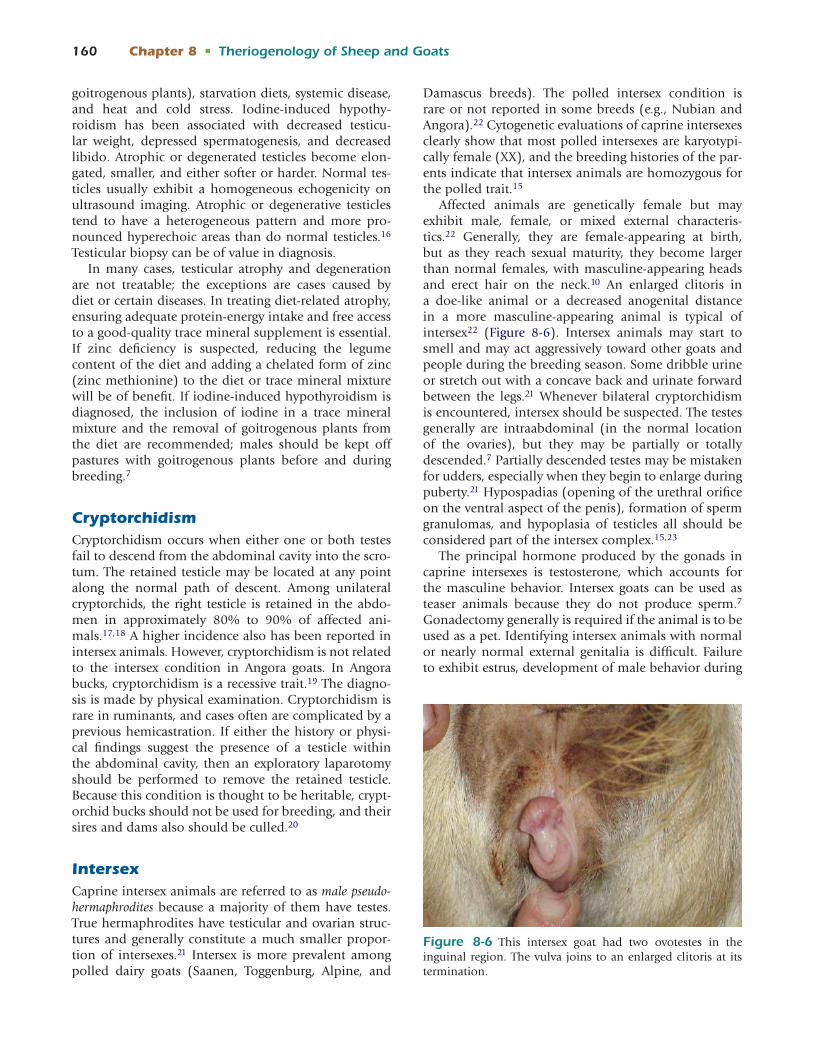
160 Chapter 8 • Theriogenology of Sheep and Goats
goitrogenous plants), starvation diets, systemic disease, and heat and cold stress. Iodine-induced hypothy-roidism has been associated with decreased testicu-lar weight, depressed spermatogenesis, and decreased libido. Atrophic or degenerated testicles become elon-gated, smaller, and either softer or harder. Normal tes-ticles usually exhibit a homogeneous echogenicity on ultrasound imaging. Atrophic or degenerative testicles tend to have a heterogeneous pattern and more pro-nounced hyperechoic areas than do normal testicles.16 Testicular biopsy can be of value in diagnosis.
In many cases, testicular atrophy and degeneration are not treatable; the exceptions are cases caused by diet or certain diseases. In treating diet-related atrophy, ensuring adequate protein-energy intake and free access to a good-quality trace mineral supplement is essential. If zinc deficiency is suspected, reducing the legume content of the diet and adding a chelated form of zinc (zinc methionine) to the diet or trace mineral mixture will be of benefit. If iodine-induced hypothyroidism is diagnosed, the inclusion of iodine in a trace mineral mixture and the removal of goitrogenous plants from the diet are recommended; males should be kept off pastures with goitrogenous plants before and during breeding.7
CryptorchidismCryptorchidism occurs when either one or both testes fail to descend from the abdominal cavity into the scro-tum. The retained testicle may be located at any point along the normal path of descent. Among unilateral cryptorchids, the right testicle is retained in the abdo-men in approximately 80% to 90% of affected ani-mals.17,18 A higher incidence also has been reported in intersex animals. However, cryptorchidism is not related to the intersex condition in Angora goats. In Angora bucks, cryptorchidism is a recessive trait.19 The diagno-sis is made by physical examination. Cryptorchidism is rare in ruminants, and cases often are complicated by a previous hemicastration. If either the history or physi-cal findings suggest the presence of a testicle within the abdominal cavity, then an exploratory laparotomy should be performed to remove the retained testicle. Because this condition is thought to be heritable, crypt-orchid bucks should not be used for breeding, and their sires and dams also should be culled.20
IntersexCaprine intersex animals are referred to as male pseudo-hermaphrodites because a majority of them have testes. True hermaphrodites have testicular and ovarian struc-tures and generally constitute a much smaller propor-tion of intersexes.21 Intersex is more prevalent among polled dairy goats (Saanen, Toggenburg, Alpine, and
Damascus breeds). The polled intersex condition is rare or not reported in some breeds (e.g., Nubian and Angora).22 Cytogenetic evaluations of caprine intersexes clearly show that most polled intersexes are karyotypi-cally female (XX), and the breeding histories of the par-ents indicate that intersex animals are homozygous for the polled trait.15
Affected animals are genetically female but may exhibit male, female, or mixed external characteris-tics.22 Generally, they are female-appearing at birth, but as they reach sexual maturity, they become larger than normal females, with masculine-appearing heads and erect hair on the neck.10 An enlarged clitoris in a doe-like animal or a decreased anogenital distance in a more masculine-appearing animal is typical of intersex22 (Figure 8-6). Intersex animals may start to smell and may act aggressively toward other goats and people during the breeding season. Some dribble urine or stretch out with a concave back and urinate forward between the legs.21 Whenever bilateral cryptorchidism is encountered, intersex should be suspected. The testes generally are intraabdominal (in the normal location of the ovaries), but they may be partially or totally descended.7 Partially descended testes may be mistaken for udders, especially when they begin to enlarge during puberty.21 Hypospadias (opening of the urethral orifice on the ventral aspect of the penis), formation of sperm granulomas, and hypoplasia of testicles all should be considered part of the intersex complex.15,23
The principal hormone produced by the gonads in caprine intersexes is testosterone, which accounts for the masculine behavior. Intersex goats can be used as teaser animals because they do not produce sperm.7 Gonadectomy generally is required if the animal is to be used as a pet. Identifying intersex animals with normal or nearly normal external genitalia is difficult. Failure to exhibit estrus, development of male behavior during
Figure 8-6 This intersex goat had two ovotestes in the inguinal region. The vulva joins to an enlarged clitoris at its termination.
Chapter 8 • Theriogenology of Sheep and Goats 161
the breeding season, a shortened vagina on speculum examination, and smaller-than-normal teats may be the first signs of the intersex condition.22 The breeding of phenotypically polled bucks should be avoided.
DISEASES OF THE MALE: PENILE ABNORMALITIESHypospadiasSeveral penile abnormalities may occur, albeit rarely, in sheep and goats.7,23-25Both hypospadias and short penile length are associated with intersex in goats. Such animals should be culled. Careful examination of the penis in the fully extended state may reveal existing abnormalities. Occasionally urethral rupture (occurring as a complication of urethral stones), balanoposthitis (see Chapter 12), injuries to the vermiform appendage, hair rings, and other abnormalities are identified.7
Ulcerative PosthitisUlcerative posthitis (enzootic posthitis, sheath rot, or pizzle rot) is an infectious, inflammatory condition of the penis, prepuce, and sheath of sheep and goats con-suming high-protein diets.7,26 The disease is caused by an interaction of the local bacterial flora (e.g., C. renale) with excess urinary urea. Excess ammonia may damage the mucosal surfaces, resulting in swelling of the pre-puce, necrosis and ulceration of the preputial mucosa, and straining to urinate. Removing the animals from the high-protein diet and shearing the “prescrotal” wool or hair usually will relieve the condition. Antibiotics, disinfectants, and antiinflamatory drugs may be needed in some cases (see Chapter 12).
PhimosisBoth phimosis (inability to extend the penis) and para-phimosis (inability to withdraw the penis into the pre-puce) occasionally are seen in rams and bucks; both conditions can cause significant loss of libido and fertility. If they are not quickly diagnosed and treated, affected animals may be rendered infertile. These two conditions may be associated with hair ring, trauma, and balanoposthitis. In cases associated with a hair ring on the glans of the penis, inspecting the penis allows the clinician to identify the problem and remove the “ring” of hair.7 Shearing the wool or mohair just ante-rior to the sheath can minimize the incidence of this problem.13,15 Phimosis also may occur as a sequela to trauma, balanoposthitis, and congenital abnormalities. With traumatic injury, an adhesion may form in the sheath or in the sigmoid region, resulting in an inability to extend the penis. As a general rule, these cases may be difficult to treat. When inflammation and scarring result in posthitis, the treatment is the same as that described
for balanoposthitis. The clinician can attempt to “break down” the adhesions manually. The use of nonsteroidal antiinflammatory drugs (NSAIDs) (e.g., flunixin meglu-mine 1 to 2 mg/kg twice a day) or antibiotics (e.g., pro-caine penicillin 22,000 IU/kg twice daily) and lavage of the sheath with mild antiseptics may be of benefit. With phimosis, most animals experience a loss of libido and should be culled, because the prognosis is poor.15
ParaphimosisParaphimosis also is associated with trauma, infec-tion, and balanoposthitis. It is slightly more common in bucks than in rams, but it is rare in both. In cases of paraphimosis, applying antibiotic cream with or without corticosteroids, replacing the penis, and plac-ing a pursestring suture into the preputial orifice may be of value. Placement of a tube in the sheath, exiting through the orifice, allows urine drainage. The clinician should take care to ensure proper urine flow. Flushing the sheath and penis with a mild antiseptic solution, providing penile hydrotherapy, and covering the penis with medicated ointments are valuable treatments for this condition. The penis should be manually extended at least every third day so that the keeper or the clinician can monitor healing. Sexual rest should be enforced throughout recovery. The prognosis in these cases is poor, however, particularly if the condition is of greater than 2 weeks’ duration and the animal makes no attempt to retract the penis.15
REFERENCES 1. Kimberling CV: Disease of rams. In Kimberling CV, editor:
Diseases of sheep, ed 3, Philadelphia, 1988, Lea & Febiger. 2. Bulgin MS: Brucella ovis epizootic in virgin ram lambs, J Am Vet
Med Assoc 196:1120, 1990. 3. Saunders VF, et al: Multiplex PCR for the detection of Brucella ovis,
Actinobacillus seminis and Histophilus somni in ram semen, Aust Vet J 85:72–77, 2007.
4. Manterola L, Tejero-Garces A, et al: Evaluation of a PCR test for the diagnosis of Brucella ovis infection in semen samples for rams, Vet Microbiol 92:65–72, 2003.
5. Kimberling CV: Sheep flock fertility, Proceedings of the Annual meeting of the Society for Theriogenology, Nashville, 1990, Tenn.
6. Bulgin MS: Epididymitis caused by B. ovis, Proceedings of the Annual meeting of the Society for Theriogenology, Nashville, Tenn, 1991.
7. Mobini S, Heath AA, Pugh DG: Theriogenology of sheep and goats. In Pugh DG, editor: Sheep and goat medicine, Philadelphia, 2002, WB Saunders, pp 129–196.
8. Ferreras MC, et al: Unilateral orchitis and epididymitis caused by Salmonella enterica subspecies diarizonae infection in a ram, J Vet Diagn Invest 19:194–197, 2007.
9. Walker RL, Leamaster BR: Prevalence of Histophilus ovis and Actinobacillus seminis in the genital tract of sheep, Am J Vet Res 47 :1928, 1986.
10. Bulgin MS: Ram lamb epididymitis, Proceedings of the Annual meeting of the Society for Theriogenology, Nashville, Tenn, 1991.
11. Al-Katib WA, Dennis SM: Early sequential findings in the geni-talia of rams experimentally infected with Actinobacillus seminis, N Z Vet J 56:50–54, 2008.

162 Chapter 8 • Theriogenology of Sheep and Goats
12. Ley WB: Ram epididymitis, Agri-Pract 14:34, 1993. 13. Bruer AN: Examination of the ram for breeding soundness.
In Morrow DA, editor: Current therapy in theriogenology, ed 2, Philadelphia, 1986, WB Saunders.
14. Pugh DG: Breeding soundness evaluation in male goats, Proceedings of the Seventh Annual Hudson-Walker/Vaughn Theriogenology Confer-ence, Auburn, Ala, 1996, Auburn University College of Veterinary Medicine, p 29.
15. Mickelsen WD, Memon MA: Infertility and diseases of the repro-ductive organs of bucks. In Youngquist RS, Threlfall WR, editors: Current therapy in large animal theriogenology, ed 2, Philadelphia, 2007, WB Saunders.
16. Ahmad N, Noakes DE, Middleton DJ: Use of ultrasound to diag-nose testicular degeneration in a goat, Vet Rec 132:436–439, 1993.
17. Ott RS, Memon MA: Breeding soundness examination of rams and bucks, a review, Theriogenology 13:155, 1980.
18. Smith KC, Brown PJ, et al: Cryptorchidism in North Ronaldsay sheep, Vet Rec 161:658–659, 2007.
19. Skinner JD, Van Heuden JAH, Goris EJ: A note on cryptorchidism in Angora goats, S Afr J Anim Sci 20:10, 1961.
20. Riddell MG: Developmental anomalies of the scrotum and testes. In Wolfe D, Moll HD, editors: Large animal urogenital surgery, Baltimore, 1997, Williams & Wilkins.
21. Basrur PK, Kochhar HS: Inherited sex abnormalities in goats. In Youngquist RS, Threlfall WR, editors: Current therapy in large animal theriogenology, ed 2, Philadelphia, 2007, WB Saunders.
22. Smith MC, Sherman DM: Goat medicine, Philadelphia, 1994, Lea & Febiger.
23. King WW, Young ME, Fox ME: Multiple congenital genitourinary anomalies in a polled goat, Contemp Top Lab Anim Sci 41:39–42, 2002.
24. Smith KC, Brown P, Parkinson TJ: Hypospadias in rams, Vet Rec 158:789–795, 2006.
25. Fuller DT, et al: What is your diagnosis? Hypospadias and ure-thral diverticulum, J Am Vet Med Assoc 201:1232–1431, 1992.
26. Loste A, et al: High prevalence of ulcerative posthitis in Rasa Aragonesa rams associated with a legume-rich diet, J Vet Med A Physiol Pathol Clin Med 52:176–179, 2005.
SPECIAL SURGICAL PROCEDURES IN THE MALECastrationCastration of the normal young male is among the most commonly performed surgical procedures in small ruminants. Kids and rams are castrated for man-agement and production reasons. Mohair production was higher in castrated goats than in intact ones.1 Castration of kids fed to slaughter increased carcass weight, dressing percentage, and external fat score while reducing internal fat score.2 Volumes of publications are available detailing the effects of age at castration and techniques used on carcass quality and growth rates. This component of meat production manage-ment is beyond the scope of this discussion; here, the focus is on the procedures for different techniques of castration.3-5
Some producers delay castration in animals intended to be used as work or pet animals, which will have an extended life span in comparison with those used for meat. An emerging trend is to delay castration in some meat goats that are fed a very high-concentrate diet and used for show before slaughter. This practice often is related to concerns of urolithiasis. Although such find-ings often are debated, at least one study evaluating the effect of castration on penile length and diameter and urethral diameter determined that lambs castrated before 3 months of age did not have the same penile development as in intact lambs or ones castrated at 5 months of age.6 Most important, relative to urolithiasis, the urethral cross-sectional diameter also was signifi-cantly smaller in the lambs castrated before 3 months of age when compared with that in intact lambs. The lambs castrated at 5 months of age did not show a sig-nificant difference in penile or urethral diameter from the intact lambs.6
Most producers castrate their own animals, although veterinarians may be called on to perform routine cas-trations in small flocks or on older animals that were not castrated at an early age for whatever reason. The most frequently used techniques are the bloodless pro-cedures using elastrator bands or the Burdizzo emascu-latome. Alternatively, traditional surgical excision of the testes may be done. Each method has advantages and disadvantages, but the method chosen often becomes a matter of individual operator bias. The elastrator band technique frequently is used in lambs and kids younger than 1 week of age in conjunction with tail docking or dehorning. It can be used up to the age of 3 or 4 months, depending on the size of the animal, but is better done earlier. The animal may be restrained with rear end up by holding hind limbs or smaller animals may be held with front and rear limbs of each side held together. The elastrator band should be placed in a disinfectant before use. Special pliers are used to place the band around the proximal scrotum, with care taken to ensure that both testicles are distal to the band. With proper place-ment, the band will be distal to the rudimentary teats; an important step is to ensure that the penis has not been included within the band (Figure 8-7). The elastra-tor band restricts blood flow to all tissues distal to the band, with subsequent avascular necrosis and sloughing of the tissue of the scrotum and testes. Sloughing usu-ally occurs in 7 to 10 days. The resulting open tissue is a prime area for infection with clostridial organisms. Tetanus is not an uncommon sequela to castration, especially when elastrator bands are used (Figure 8-8). Therefore any animal not previously vaccinated for clostridial diseases should be administered both tetanus antitoxin and tetanus toxoid at the time of castration. The tissue distal to the elastrator band may be excised 1 or 2 days after band application, to lessen the risk of complications. If one (or both) testicles are proximal to

Chapter 8 • Theriogenology of Sheep and Goats 163
the band, the animal will exhibit characteristics of an intact male; nevertheless, it probably will be infertile because of improper thermoregulation of the testes displaced proximally near the external inguinal ring. In such instances, surgical castration will be required to remove the remaining testis. The procedure will be more difficult than a normal castration because of fibrous tis-sue and adhesions. If the penis has been included in the band, the point of capture usually is at the distal bend of the sigmoid flexure. Consequent urethral obstruction and rupture will necessitate euthanasia of the kid or ram, or at the very least an urethrostomy.
Another bloodless technique is the use of the Burdizzo emasculatome. This device is used to crush the spermatic cord within the scrotum, causing atrophy
of the testicle while sparing the scrotal skin. The testicle should be gently pushed into the distal aspect of the scrotum and the spermatic cord held laterally within the scrotum. The Burdizzo should be applied twice, with the second crush made just distal to the first, to each respec-tive spermatic cord and held approximately 10 seconds, without crossing the median raphe of the scrotum. If the Burdizzo crush is applied across the median raphe, the scrotal skin is likely to become avascular and slough. The respective spermatic cords should be crushed at dif-ferent levels, to help maintain viability of the scrotum.
Although both the elastrator band and Burdizzo emasculatome techniques are so-called bloodless tech-niques, many practitioners still prefer surgical castration by excision of the distal one half to one third of the scro-tum with a scalpel blade or using a Newberry knife to leave two flaps of scrotal skin (cranial and caudal). The testicles are exposed and removed after making the skin incision of choice (Figure 8-9). Many clinicians prefer to place ligatures on the spermatic cord or to crush the cord with an emasculator, to ensure appropriate hemorrhage control. Care should be taken in using an emasculator routinely used for larger species such as horses in that an emasculator that functions well in equine castration may not adequately crush the smaller cord of the small rumi-nant. Therefore the clinician may prefer to use ligatures; this method takes very little time and does not involve appreciable added expense. The spermatic cord is then transected distal to the ligature. The testicles may be removed by traction, as is commonly done in calves, but this method has been associated with herniation almost immediately after the castration in some cases. If the traction method is selected, pressure should be applied over the respective external inguinal ring with fingers while pulling the testes with the other hand to minimize trauma to the ring and subsequent herniation. Animals younger than 2 months of age may be castrated in this manner with use of physical restraint, but older animals should have local anesthesia and sedation, if not general anesthesia. An alternative to traditional castration equip-ment is the Henderson castration tool.7-9
Castrating 4- to 5-month-old lambs using the Burdizzo method has been reported to be less stressful than conventional surgical castration.10 Some workers suggest that both Burdizzo and elastrator band tech-niques cause less postoperative pain than that associated with traditional surgical castration in lambs older than 10 weeks of age.11 The Burdizzo procedure is subject to failure in some cases, even when performed by experi-enced practitioners. Histologic sections of the testicles of 34 lambs castrated with the Burdizzo method showed failure of involution of testicular parenchyma in most of the samples.12 Regardless of the method of castration used, local anesthesia in the spermatic cord reduces pain in the animal. Bupivacaine has been shown to be more effective in decreasing pain over time than lidocaine.11
Figure 8-7 A green elastrator band is in place on the proxi-mal scrotum of a 3-week-old Oberhasli-cross buck. Rudimen-tary teats can be seen proximal to the elastrator band in the nonhaired skin.
Figure 8-8 The sloughing scrotum of a 5-week-old Barba-dos lamb approximately 1 week after application of an elas-trator band for castration. The lamb, in lateral recumbency, exhibits the rigidity of tetanus. The owner performed the castration without providing any tetanus prophylaxis.

164 Chapter 8 • Theriogenology of Sheep and Goats
A pinhole castration technique that entails making a percutaneous stab incision in the proximal scrotum through which the spermatic cord is ligated has been described.13 The ligation leads to atrophy of the testis. The technique does not require any special instruments, and the reporting investigators recommend it as a simple, less painful, and cost-effective method of castra-tion. Histopathologic evaluation of the testis 6 months after ligation showed seminiferous tubules with areas of calcification but no sperm.13
As an alternative to surgical castration, some prac-titioners report success with chemical castration. One effective method is to inject 88% lactic acid at a dose of 0.2 mL/kg of body weight into each testicle of adult goats. However, this may be more stressful than surgi-cal castration because cortisol levels remained high for a longer time period in these animals than ones surgically castrated.14 Another chemical castration tech-nique involves injection of 10% formalin into the tes-ticle. With use of this method in 3-week-old goats, the chemical caused marked fibrosis. Accordingly, formalin injection has been suggested to be a viable option for chemical castration in very young goats.15
Unilateral CastrationRams and bucks can be rendered infertile by a number of unilateral scrotal conditions including hydrocele, hematocele, orchitis, and tumors such as mesothe-lioma. Conditions such as those listed can have a detri-mental effect on the thermoregulation of the unaffected testicle, thereby leading to infertility. If the castration is
done early in the course of the unilateral disease, the contralateral testicle frequently returns to normal sperm production, so the affected male will be fertile.
Unilateral castration is done with the animal under general anesthesia in lateral recumbency, with the affected testicle up and the upper leg abducted (see Chapter 18). This surgery probably could be done with sedation and epidural and local anesthesia; however, aseptic technique is critical to avoid infection of the surgical site, which would have quite a negative impact on the return to fertility. The skin incision is made in an elliptical fashion longitudinally on the lateral aspect of the scrotum over the affected testicle. The incision is continued through the skin and subcutaneous tissues, leaving the vaginal tunic intact if possible. The testicle within the tunic is bluntly freed from the scrotum. Next, double ligation of the cord is done with a circumfer-ential suture proximal to a transfixation suture using absorbable #2 suture material. The elliptical incision allows resection of much of the scrotal skin, which in turn eliminates much of the potential dead space as an important consideration in closure. If the practitioner with limited experience performing this approach has concerns regarding removal of too much skin, the initial skin incision can be made as a simple longitudinal inci-sion and later converted to an elliptical one by resecting skin after removal of the affected testicle, when it is clear how much skin is needed for closure. In either case, the subcutaneous tissues should be closed in several layers to obliterate the dead space. The skin is closed in a continuous interlocking pattern. Some surgeons place a drain in the scrotum or leave the ventralmost part of the skin incision open. As a matter of personal preference (specifically, of A.N.B.), skin resection and complete closure can be performed to achieve primary healing of the skin incision (Figure 8-10, A and B). Use of perioperative antibiotics and antiinflammatories is appropriate (see Appendix 1). If primary healing occurs without complications, the semen quality should no longer show negative effects associated with the unilat-eral disease by 90 days after surgery.
Repeated semen evaluations of normal fertile goats after unilateral castration failed to show any detrimen-tal effects on sperm motility compared with control (nonoperated) bucks. No difference was noted based on the particular testicle removed.16
Cryptorchid CastrationCryptorchidism is a rare condition in ruminants.17 Crypt-orchidism is an inherited recessive trait in at least the Angora breed of goats.18 It is most easily diagnosed when a reliable history confirms that no attempts have been made to castrate (or hemicastrate) the animal in ques-tion. Frequently, male characteristics or behavior leads to exploratory surgery to search for the retained testicle(s). If retained, the testicle is not usually located near the inguinal
Figure 8-9 In this photo, the ram lamb is being restrained by a sitting holder (as described in Chapter 1). The surgeon is removing the distal one half to one third of the scrotum to expose the two testicles. The testicles were then separated from the surrounding tissues and removed by “pulling,” and the goat was examined for inguinal hernia and given a tetanus toxoid inoculation. (Note: Routine spraying of insect repellant around the scrotal area after castration is recommended by D.G.P., regardless of the controversy surrounding this practice.)

Chapter 8 • Theriogenology of Sheep and Goats 165
ring or easily retrievable through an inguinal extension of the gubernaculum testis, as in the horse. The retained tes-ticle is more likely to be within the abdomen and usually is closer to the kidney than to the inguinal ring, although it may also be found at the internal inguinal ring.
The surgery can be done using a traditional laparot-omy or laparoscopy. The laparoscopic procedure allows better visualization of the abdomen and is preferred in most instances. The animal should be either sedated or placed under general anesthesia and in dorsal recum-bency after having roughage withheld for 36 to 48 hours to decrease the visceral fill, for better visualiza-tion within the abdominal cavity. Standard laparoscopic technique with a scope portal near the umbilicus and instrument portals between the scope portal and the caudal part of the fold of the flank is used (Figure 8-11). The retained testicle often is freed easily and exterior-ized from the abdominal cavity for ligation. Other tech-niques such as cautery and intracorporeal ligation can be used, depending on the preference of the surgeon.19
Alternatively, the animal can be placed in lateral recumbency for exploratory surgery performed through a flank incision. The flank exploratory affords very lim-ited visualization, so the practitioner must rely more on what can be palpated than what is seen. The retained testicle often may be located with a simple sweep of the practitioner’s finger through the abdominal cavity. If this maneuver fails, then attempts are made to locate the vas deferens as it crosses the ureter and then trace it to the testicle; this is the recommended systematic approach to finding the retained testicle.
Teaser Animals: Infertile Rams and BucksMale small ruminants are rendered infertile for subse-quent use in estrus detection or, more often, to place with females before exposure to a fertile male as a man-agement tool. The females will start cycling in response to the presence of the altered male (i.e., the teaser) before the fertile male is added to the flock. This system will lead to birth of all lambs or kids within a shorter time period, which allows for more efficient labor, management, and disease control and results in a lamb or kid crop of uniform age and size. Regardless of the intended use of the infertile male, several procedures are in common use to obtain infertility.
The vasectomy probably is the most commonly used technique in small ruminants. The technique is described as for a caudal incision in bulls,20 but the vasectomy may be performed through a proximal-cranial incision located near the teats in sheep and goats where the vas deferens is easily palpable. This procedure can be performed with the ram either “sitting” on his
A
B
Figure 8-10 A, A 3-year-old Rambouillet ram with right-sided scrotal swelling. The animal is positioned in left lateral recumbency under general anesthesia, with the right hindlimb abducted, for unilateral castration. B, The right testicle removed from the ram shows a thickened vaginal tunic, edema, and adhesions associated with the tail of the epididymis.
Figure 8-11 Laparoscopic view of a left retained testicle in a 6-month-old Boer goat. The testicle is being held by laparoscopic forceps. The bladder is seen caudally and to the right. The testicle was removed from the abdomen through the instrument portal after enlargement of the opening to allow extracorporeal ligation. A 10-mm-diameter direct-vision laparoscopic scope and camera were used for this procedure.

166 Chapter 8 • Theriogenology of Sheep and Goats
hindquarters or placed in right lateral recumbency. The vasectomy can be done with use of local anesthesia in tractable animals, but sedation or general anesthesia will make the restraint more dependable. Lumbosacral epi-dural anesthesia is another option. The proximal scro-tum is clipped and prepped for surgery. The vas deferens can be palpated as a firm structure within the spermatic cord. A 3- to 4-cm incision is made through the scrotal skin near the teat over the palpable vas deferens. The incision is continued through vaginal tunic to expose the vas, which is elevated with hemostats and isolated. Ligatures can be placed on the vas approximately 6 cm apart; then the section of the vas between the ligatures is resected. The tunic and skin are closed in routine fashion. The procedure is repeated on the other side. Some practitioners perform the vasectomy on one tes-ticle and then perform a unilateral castration to remove the opposite one, to allow for easy identification of the teaser male in case ear tag or other identification is lost. Resecting, collecting, and fixing in formalin a section of the vas for histopathologic examination constitute good practice.8 However, paternity can be diagnosed with DNA techniques in modern veterinary medicine.
The vasectomy also may be done laparoscopically using standard technique to visualize the vas as it enters the abdomen through the internal inguinal ring. The vas can be isolated and transected after ligation with suture or staples. Cautery is a useful tool to quickly perform the laparoscopic ligation. The laparoscopic procedure does not offer any real advantages over the conventional surgical technique, however, so it is dif-ficult to justify the added expense of the equipment required (Figure 8-12, A to C).
In approximately 80% of operated rams, sperm granulomas will develop after vasectomy. Such granulo-mas are found on clinical examination by palpation or ultrasound imaging. The granulomas do not have any clinically significant detrimental effects.21 Vasectomized bucks can be used for heat detection as early as 1 week after surgery.22 Semen evaluation performed 14 days after vasectomy failed to show any viable, motile sperm in a series of five rams.23
The practitioner may chose to perform an epididy-mectomy rather than a vasectomy to create the teaser male. The animal is restrained, and the distal third of the scrotum is prepared for aseptic surgery. Anesthe-sia can be obtained either with an epidural technique or by local infiltration of an anesthetic in the ventral scrotum over the tail of the epididymis. The testicles are forced to the ventral aspect of the scrotum by grasping
B
C
A
Figure 8-12 A and B, Vasectomy in an anesthetized 2-year-old Pygmy buck. A, Laparoscopic evaluation of the internal inguinal ring, urinary bladder, and surrounding structures. The procedure was performed in a veterinary hospital, where a 10-mm-diameter direct-vision laparoscopic vewing scope, camera, and TV monitor all were available. B, The left internal inguinal ring is seen to the left of the bladder. The forceps are holding the vas deferens in the preferred location for occlu-sion by means of a cautery or surgical staples before transec-tion for a laparoscopic vasectomy. C, Laparoscopic view of
the vas deferens of a 1-year-old Alpine buck under general anesthesia. Laparoscopic staples are in place in the left vas deferens. The section of the vas deferens between the staples will be transected after being cauterized near the staples.

Chapter 8 • Theriogenology of Sheep and Goats 167
the neck of the scrotum. A caudal-to-cranial incision is made directly over the prominent tail of the epididymis, approximately 2 to 3 cm long. The incision is extended through the skin and vaginal tunic until the epididy-mis bulges from the incision. Towel clamps placed on the epididymis help to retract the structure from the incision. When adequate tissue is exposed to ensure complete removal of the tail of the epididymis, crushing forceps are placed across the tissue and a scalpel is used to excise the tissue. The excised tissue should be exam-ined to ensure adequate resection. Either the skin can be allowed to heal by second intention or an appropriate suture can be placed. The animal should be healed in 2 weeks and ready for intended use.
Hemorrhage can be a problem with this technique, especially if the tunica albuginea of the testis is incised.20 The bleeding and second intention wound healing may allow contamination and subsequent infection with the potential for abscess formation. Although epididymec-tomy and vasectomy are applicable in both species, the clinician may find that epididymectomy is more easily performed in the buck, whereas a vasectomy may be the better or easier choice in the ram.
Penile TranslocationOne method of preventing intromission is to “free” the penis and move it over to the left flank. This method is useful in some teaser systems. The surgery prevents intromission but not ejaculation. Therefore, if penile translocation is attempted, a vasectomy (or epididy-mectomy) also should be performed. The buck or ram should be given a systemic antimicrobial agent 2 or more hours before surgery. With the animal standing, the operator marks an area 1 cm cranial to the flank fold.1 The buck or ram is then heavily sedated and either anesthetized with injectable anesthetics, intu-bated and maintained on gas anesthesia, or adminis-tered a lumbosacral epidural anesthetic (see Chapter 18).24 The animal is placed in right lateral recumbency, the ventral abdomen and left flank are clipped, and the surgical site is aseptically prepared.20,24
The operator then excises a 4- to 7-cm circle of skin and cutaneous trunci muscle above the fold in the left flank. The lower edge of the circular incision should be 1 cm above the flank fold, just cranial to the mark made on the left flank. In mature rams with a large abdominal girth, some clinicians choose to graft the preputial ori-fice to a location medial to the fold of the flank rather than the further distance to the flank. Hemostasis is achieved, and the area is covered with saline-moistened gauze. The operator then makes a circumferential inci-sion including 1 to 2 cm of haired skin around the preputial orifice. Then a ventral midline incision is made starting at the caudal aspect of the circumferen-tial incision and extending two thirds of the way to
the scrotum.24 Care should be taken not to open the preputial cavity in making this incision. The penis is left inside the prepuce, and the two structures are freed of subcutaneous tissue by blunt dissection, avoiding large vessels. A single “reference” suture is placed in the skin at the cranialmost aspect of the preputial orifice and a sterile glove is placed over the orifice.20
From the circular incision in the flank to the caudal-most aspect of the longitudinal incision, the clinician creates a tunnel using scissors and blunt dissection. The penis (along with the sterile glove covering the preputial orifice to maintain asepsis) is pulled through the tunnel. Alternatively, a sterile palpation sleeve with the hand cut off can be placed in the previously cre-ated tunnel and sponge forceps can be used to pull the freed skin surrounding the preputial orifice through the sleeve to the graft site. The penis should now be at a 45-degree angle to the long axis of the body.20 The penis should not be restricted at any point through the tunnel, and the penis and prepuce should not be in any degree of torsion. The reference suture should be used to align the cranial aspect of the preputial orifice with the dorsal portion of the circular flank incision.20,24
The preputial graft should be sutured to the circum-ferential incision with a two-layer simple interrupted pat-tern using absorbable material. The subcutaneous tissue of the transposed prepuce should be sutured to the cuta-neous trunci, followed by closure of the skin. The opera-tor then closes the longitudinal incision, attempting to diminish all open dead space with absorbable material. Our own preference is for either a simple interrupted or a horizontal mattress-type suture pattern. The skin over the longitudinal incision is closed in a simple interrupted suture pattern in all areas except the cranialmost aspect of the original preputial incision. To allow for ventral drain-age, this area is not sutured.20,24 Some clinicians routinely close the entire incision; with this approach, primary healing is achieved without drainage when good surgical technique is used. Fly control should be maintained, and tetanus prophylaxis should be provided (using tetanus toxoid or antitoxin). The operator then performs a bilat-eral epididymectomy (or vasectomy); antibiotics are con-tinued for 5 to 7 days (procaine penicillin 22,000 IU/kg twice daily), and the sutures are removed in 14 days. The male is ready for use in 1 month.
REFERENCES 1. Barina V, Kuchtik J: Evaluation of fibre diameter, length and
mohair production in mohair goats and castrates, Acta Univ Agricult Silvicult Mend Brun 48:13–17, 2000.
2. Mourad M, Gbanamou G, Balde IB: Carcass characteristics of West African dwarf goats under extensive system, Small Rumin Res 42:83–86, 2001.
3. Tesfaye K, et al: Growth performance and carcass characteristics of Arsi-Bale goats castrated at different ages, J Cell Anim Biol 2:187–194, 2008.

168 Chapter 8 • Theriogenology of Sheep and Goats
4. Okeudo NJ, Moss BW: Production performance and meat quality characteristics of sheep comprising four sex-types over a range of slaughter weights produced following commercial practice, Meat Sci 80:522–528, 2008.
5. Koyuncu M, et al: Effect of castration on growth and carcass traits in hair goat kids under a semi-intensive system in the south-Marmara region of Turkey, Small Rumin Res 72:38–44, 2007.
6. Bani Ismail ZA, et al: Effects of castration on penile and urethral development in Awassi lambs, Bulg J Vet Med 10:29–34, 2007.
7. Baird AN, Wolfe DF: Castration of the normal male. In Wolfe DF, Moll HD, editors: Large animal urogenital surgery, ed 2, Baltimore, 1999, Williams & Wilkins.
8. Scott PR: Husbandry. In Scott PR, editor: Sheep medicine, London, 2007, Manson Publishing.
9. Matthews J: Surgical techniques. In Matthews J, editor: Diseases of the goat, ed 3, Chelmsford, United Kingdom, 2009, Blackwell Publishing.
10. Bonelli P, et al: Stress responses in lambs castrated with three dif-ferent methods, Ital J Anim Sci 7:207–217, 2008.
11. Melches S, et al: Castration of lambs: a welfare comparison of dif-ferent castration techniques in lambs over 10 weeks of age, Vet J 173:554–563, 2007.
12. Stoffel MH, et al: Histologic assessment of testicular residues in lambs and calves after Burdizzo castration, Vet Rec 164:523–528, 2009.
13. Fazili MR, et al: Evaluation of pinhole castration technique in rams, Small Rumin Res 84:61–64, 2009.
14. Okwee-Acai J, et al: An evaluation of non-surgical castration by single intratesticular injection of lactic acid in adult mubende goats, Bull Anim Health Prod Afr 56:116–124, 2008.
15. Awal MA, et al: Formalin affects the male reproduction of black Bengal goats during prepubertal stage even at low concentration, J Med Sci (Pak) 4:84–89, 2004.
16. Oyeymi MO, Akusu MO: Short-term effect of hemi-orchectomy on testicular and ejaculate characteristics of West African Dwarf bucks, Small Rumin Res 31:75–78, 1999.
17. Riddell MG: Developmental anomalies of the scrotum. In Wolfe DF, Moll HD, editors: Large animal urogenital surgery, ed 2, Baltimore, 1999, Williams & Wilkins.
18. Matthews J: Male infertility. In Matthews J, editor: Diseases of the goat, ed 3, Chelmsford, United Kingdom, 2009, Blackwell Publishing.
19. Rutherford DJ, Fnding E: Laparoscopic castration in a cryptorchid pygmy goat, Veterinary Record 165:27–28, 2009.
20. Wolfe DF: Surgical preparation of estrus-detector males. In Wolfe DF, Moll HD, editors: Large animal urogenital surgery, ed 2, Baltimore, 1999, Williams & Wilkins.
21. Gouletsou PG, Galatos AD, Fthenakis GC: Clinical, ultrasono-graphic and pathologic features following unilateral vasectomy in rams, Anim Reprod Sci 103:52–68, 2008.
22. Batista M, et al: Semen characteristics and plasma levels of tes-tosterone after bilateral vasectomy in bucks, Reprod Domest Anim 37:375–378, 2002.
23. Janett F, et al: Semen characteristics after vasectomy in the ram, Theriogenology 56 (3):485–491, 2001.
24. Heath AM, Pugh DG, Edens MS: Urogenital surgery in goats. In Youngquist RS, Threlfall WR, editors: Current therapy in large animal theriogenology, ed 2, Philadelphia, 2007, Saunders.
SEMEN COLLECTION AND STORAGECollection and storage of semen from mature, healthy bucks and rams can be a key component of a reproduc-tive program that maximizes the overall fertility of a flock or herd and enhances genetic progress in a breed and species.1-3 It appears that the semen of mature rams is of better quality and higher fertility than that of young rams.4
The two most common methods for semen collec-tion involve use of an electroejaculator and of an AV, respectively. Use of the AV is the preferred method for collecting semen, because it is faster in trained males and less stressful overall, while yielding a more physiologically normal sample. The electroejaculation method may be considered for single collections. Elec-troejaculator-obtained samples tend to be inconsistent and often contain a large amount of seminal fluid and a lower concentration of spermatozoa than in samples collected with an AV. In uncooperative or untrained males, an electroejaculator can be used with appro-priate anesthesia.1-3,5-7 Dairy bucks are handled more often and are more easily trained to use an AV. Semen from meat bucks is more commonly collected by elec-troejaculation, but they too can be trained to service an AV. Semen obtained with electroejaculation generally is of larger volume but lower concentration than that obtained with use of an AV. Collections in bucks can be performed two or three times daily on alternate days. Intervals of 30 minutes to 1 hour are advisable between
collections in order to obtain good-quality samples5 and maintain adequate libido. The exact frequency at which semen may be collected will depend on the age, condition, and temperament of each animal. The qual-ity of the semen collected also is very dependent on the season of collection. The incidence of morphologically abnormal sperm is much higher under increasing day length conditions owing to dropping LH-testosterone levels (spring and early summer).
On collection, the ejaculate is placed in a warm-water bath (37° C) and evaluated immediately, as follows:Macroscopic examination • Volume • Consistency • Color • Foreign matter in the ejaculateMicroscopic examination • Gross motility • Individual motility • Morphology • Sperm concentrationThe parameters previously described for semen han-dling and evaluation for breeding soundness apply. The normal color of semen is off-white or milky, with tinges of pink (blood contamination), brown (reproductive tract infection), and yellow and dilute (urine contami-nation) all indicating an inferior ejaculate.8
Clean equipment and proper semen handling tech-niques are critical to success.

Chapter 8 • Theriogenology of Sheep and Goats 169
A 2-hour semen stress test also should be performed: Semen is collected and immediately evaluated for progressive motility. The sample is then incubated at room temperature for 2 hours, and progressive motil-ity reevaluated. The incubated sample should maintain 20% of the motility of the initial sample (Figure 8-13).
The fertility potential of cryopreserved semen can be influenced by numerous factors. In addition to inher-ent variation in semen quality among males, manage-ment, nutrition, and environmental stressors can have a transient influence on spermatogenesis for weeks to months.
Semen from rams may be collected and used fresh or frozen for future use. Typically, rams are trained to service an AV in the presence of females in estrus or treated ovariectomized females (i.e., females that have been given 1 mg of estradiol benzoate per week or pre-pared for libido testing). The semen is collected into a warm (39° C) AV and handled carefully to avoid expo-sure to any contaminants or ultraviolet light. Once it has been collected, semen can be used raw (undiluted), extended and chilled, or frozen and thawed. When raw, undiluted semen is used, females can be inseminated with approximately 0.1 mL of normal, good-quality semen immediately after collection. After evaluation, semen ejaculates are diluted with appropriate extend-ers to achieve final semen-to-extender ratios ranging from 1:1 to 1:4, depending on the sperm concentration of the ejaculate. If necessary, semen can be diluted at 30° C with the extender and cooled to 4° C and then kept at this temperature for up to 24 hours.
FreezingRamMany methods of semen freezing are used. The clini-cian is advised to search the current scientific literature before engaging in this ever-evolving methodology.3 Semen intended for freezing should have a concentra-tion of more than 3 × 10⁹/mL and a motility rate of more than 70% of the ejaculate. Normal concentrations range from 3.5 × 10⁹ mL to 6.0 × 10⁹ mL for rams and 2.5 × 10⁹ ml to 5.0 × 10⁹ mL for bucks. Semen extend-ers are commercially available (e.g., Minitube [available at http://www.minitube.com]). The most commonly used semen extenders are formulated to enhance sperm cell maintenance by providing energy, isotonic osmotic pressure, a buffering system, and protection from cold shock.
Temperature control is of utmost importance in the successful freezing of semen. The semen should be placed in an incubator or water bath (at 30° C).1 Semen intended for freezing should have a concen-tration of more than 3 × 10⁹/mL and a motility rate of more than 70% of the ejaculate. Semen should be diluted as appropriate using a warmed extender
(30° C).5 This extender can be as simple as whole milk or Dulbecco’s phosphate-buffered saline (PBS) with 10% fetal calf serum. Both synthetic and milk- or egg yolk–based extenders are currently available. The three most common milk- or egg yolk–based extenders are (1) egg yolk–citrate glycerol, (2) egg yolk–tris-glycerol, and (3) whole homogenized milk–glycerol. The most common extender is the egg yolk–tris-glycerol. For short-term storage, semen can be extended with the egg yolk–tris-glycerol extender and held at 5° C for up to 24 hours without a noticeable decrease in fertilizing capacity. Use of tris-based extenders also is associated with satisfactory sperm survival on thawing when glyc-erol is added to semen kept at 30° C and slowly cooled to 4° C within 2 to 4 hours before freezing (a one-step method). Semen extended with milk diluents shows better survival rates for spermatozoa during the freezing process if glycerol is added to the diluted semen after it is cooled to 4° C.
For freezing semen, several packaging systems have been developed for cryopreservation of sperm (e.g., in straws, pellets, or ampules). Dilution of extender and semen used in the cryopreservation process will depend on what packaging system is used.
BuckMany of the same principles described for the storage of frozen ram semen are applicable for handling of semen from goats. Traditionally, buck semen has been centri-fuged to remove the seminal plasma, thereby enhancing freezability. If semen is adequately diluted, however, centrifugation may be omitted.9 Buck semen can be placed in plastic straws and frozen in liquid nitrogen
Figure 8-13 In this photo, the clinician is evaluating thawed semen in the field (i.e., under typical conditions encountered on the farm). A slide-coverslip warmer, stage warmer, and temperature-controlled thaw box all are being used. The semen will be stained, and spermatozoa morphol-ogy also will be evaluated.

170 Chapter 8 • Theriogenology of Sheep and Goats
vapor by either slow or fast methods. Diluents for freez-ing buck semen should have properties similar to those of diluents used to extend fresh semen. In addition, they should contain an agent to protect the cell membrane during cooling (usually egg yolk) and a cryoprotective agent (usually glycerol) to protect the spermatozoa against membrane damage during freezing.5,10,11
Dilution of semen can be performed in either of two ways. The two-step dilution method is similar to that used for bull semen. Using a diluent containing no glycerol, the semen is diluted at 30° C, shortly after collection, to half of the final diluted volume.5 After cooling at 5° C for 1.5 to 2 hours, the semen is extended to the final volume with a diluent containing glycerol.5 For one-step dilution, the semen is gradually diluted to the final prefreezing volume at 5° C in 1.5 to 2 hours in a refrigerator or cold room. The two-step dilution method has no advantage over the one-step method for buck semen. Therefore the one-step method is preferred, because it simpli-fies diluent preparation and reduces the handling of semen before freezing. Goat semen should be processed so that the post-thaw dose yields 50 to 100 × 109 progressively motile spermatozoa.12
StrawsThe most popular way of freezing semen today is to use plastic straws (0.25 or 0.5 mL). The ejaculate typically is diluted to a ratio between 1:1 and 1:4, with extenders added slowly by constant slow mixing.1,5 The diluent usually is hypertonic; therefore rapid mixing may cause osmotic shock in spermatozoa.1 Box 8-2 presents a for-mula for a semen extender useful for freezing ram semen in straws. Extended semen is slowly cooled to around freezing (4° to 5° C) over a 1.5- to 2-hour period. The cooled, extended semen should be diluted (usually to achieve a ratio of 1:1) with a freeze buffer containing an energy source, a protein, an antibiotic, and a cryoprotec-tant (glycerol) before being placed into labeled straws. The filled straws are laid horizontally on a rack (evenly and not touching each other) and then placed in a hold-ing container in liquid nitrogen vapor, approximately 4 cm over the surface of liquid nitrogen, for 10 minutes. The straws are then chilled to between –80° and –125° C.1,13 The semen is then rapidly cooled to its final storage temperature of –196° C by submersing the straws in liq-uid nitrogen. Formulas for mixing extenders and freeze buffers are available in the current scientific literature.
PelletsThe pellet technique is simple and does not require sophisticated or expensive equipment. The disadvan-tages of freezing semen with this technique include difficulty of identification, poor sanitation control, and inconvenience for use in the field.3
Semen may be frozen into pellets by dropping cooled semen into depressions drilled or scratched into
“dry ice.” This is a convenient method for freezing, but semen treated this way is more difficult to thaw and use. In this method, collected semen can be diluted by volume to contain 4% glycerol and 12% egg yolk in a slightly hypertonic, buffered solution.1,5 Box 8-3 presents a formula for an extender useful for freezing ram semen in pellets. In a cool, ventilated room, the practitioner makes several small circular cuts (0.5 to 0.8 cm diameter) on a flat piece of solid carbon dioxide (“dry ice”). Cooled pipettes (to 4° C) are used to drop 0.1 to 0.3 mL onto the cut surface of the dry ice. A drop of previously extended or cooled semen is then depos-ited in each indentation. After 10 minutes the frozen pellets are shaken into a pan of liquid nitrogen.
The individual pellets can be stored in labeled plastic goblets. For insemination, pellets are put into a thaw-ing solution (spare diluent, saline solution) and pre-warmed to 40° C until they melt.
ThawingThe thawing and handling procedures must be consis-tent with on-farm recommendations. Improper thaw-ing procedure, lack of postthaw thermoprotection, and
Tris(hydroxymethyl) amino methane: 24.2 gCitric acid: 13.6 gFructose: 10 gGlycerol: 64 mLEgg yolk: 200 mLTotal volume: 1 LFinal pH: 6.8
BOX 8-2 Semen extender Formula for Cryopreservation in Straws
Modified from Shipley CFB, et al: Artificial insemination and embryo transfer in sheep. In Youngquist RS, Threlfall WR, editors: Current therapy in large animal theriogenology, ed 2, Philadelphia, 2007, WB Saunders.
Tris(hydroxymethyl) amino methane: 3.63 gCitric acid: 1.99 gGlucose: 0.5 gGlycerol: 5 mLEgg yolk: 15 mLDistilled, deionized water: sufficient to extend volume to 100 mL
BOX 8-3 Formula for Semen Cryopreservation in Pellets
Modified from Evans G: Freezing sheep semen. In Dziuk PJ, Wheeler M, editors: Handbook of methods for study of reproductive physiology in domestic animals, Urbana, Ill, 1991, University of Illinois Press.

Chapter 8 • Theriogenology of Sheep and Goats 171
other factors can further corrupt semen quality. Appro-priate semen handling techniques are of the highest importance. Proper handling of frozen semen is para-mount in maximizing conception rates.3
Cooled SemenRamRam semen also may be collected and chilled for same- or next-day AI. Such preparation will allow the use of semen shipped from other farms.6,7 Before use, the col-lected and chilled semen should be maintained at or below 35° C. Temperatures above 37° C increase sperm metabolic rate and limit longevity.8 Care should be taken to avoid cold shock to the sperm during warming. The best method, when possible, is to allow the cooled semen to be warmed by the female’s reproductive tract.
BuckThe diluents commonly used to dilute buck semen contain either tris or citrate as the buffer, glucose or fructose as the energy source, and egg yolk to protect the spermatozoal cell membranes against cold shock. The concentration of egg yolk should be reduced to 2% to avoid its reacting with the coagulating enzyme present in the seminal plasma.5 This enzyme occurs in greater concentrations when semen is collected by electroejaculation. To overcome any problems with the coagulating enzyme, a low-concentration egg yolk dilu-ent or skim milk can be used or the seminal plasma can be removed by centrifugation immediately after collection.5,14 Under field conditions, the most readily available semen diluent is skim milk. The ultra–heat-treated milk is sterile and may be used directly as a diluent without any further treatment.3,5 When does are inseminated with fresh spermatozoa by laparoscopic technique, PBS with the addition of 1000 IU of sodium penicillin and 1 mg of streptomycin/mL can be used as an extender.3,5 The extended semen also can be inserted by pipette into 0.5-mL straws and cooled gradually over 1 to 2 hours from 30° C to the storage temperature of 5° C. This semen should be used within 6 to 8 hours after collection/dilution for maximum fertility.5 The semen should be cooled gradually over 1 to 2 hours.
Frozen Semen HandlingFrozen semen for bucks and rams is stored in liquid nitrogen (–196° C). Any changes in this storage tem-perature can alter semen quality.15 The straw is placed in a holding goblet, with two goblets attached to a cane. Multiple canes are set inside a canister, which is immersed and maintained in a liquid nitrogen tank. The liquid nitrogen tank lid should be kept closed and the liquid nitrogen level checked and maintained at an ade-quate level. A schedule of tank maintenance should be
planned, followed, and documented.3 A record or “tank map” of where particular straws are stored (goblet, cane, canister) helps expedite semen transfer and retrieval.16 The tank should be kept in a cool, well-ventilated room. Straws should not be removed from the tank unless they are to be used; they should not be transferred from tank to tank unless the procedure takes less than 2 seconds.
To thaw the straws, the clinician should first identify the canister holding the cane. The canister is raised until the cane tops can be seen (5 to 7 cm below the mouth of the tank). Straws should be maintained below the “frost line” in the neck of the tank at all times. Using a light source to identify the correct cane, the clinician removes the straw to be thawed with tweezers or for-ceps. The cane is immediately replaced into the canister, and the canister is reimmersed in the liquid nitrogen. Straws should not be touched by the handler’s hands.
The clinician should quickly identify whether it is the correct straw and them immerse it in a water bath (33° to 35° C). Thawing procedures should follow manufac-turers’ recommendations, but generally thawing requires only 30 to 40 seconds for 0.5-mL straws and 20 to 30 sec-onds for 0.25-mL straws. Only as many straws as can be used in 10 to 15 minutes should be thawed at one time. If possible, the clinician should thaw no more than three straws at one time to avoid lowering the thaw water tem-perature.15 The straw should be thoroughly dried, the air bubble “shaken” to the crimp end, and the straw opened.
As goblets are emptied, they should be discarded, to expedite the retrieval of straws in the lower goblets. If straws are to be retrieved from lower goblets, however, the cane is raised until the straws are even with the other cane tops, and then the straw to be used is removed.15,16
If pellets are used, the clinician removes the pellets from liquid nitrogen storage and places two or three directly in a dry, sterile tube. The tube is kept in a warm-water bath (37° C). The thawed semen should be pulled into a pipette and used for AI immediately.1 Alternatively, some processing techniques may require the addition of a warm diluent to the frozen pellets.
Evaluating Thawed SemenFor the evaluation of frozen semen samples, guidelines on semen handling as just described should be followed. The clinician should thaw the semen using the recom-mendations that were made by the company where it was originally processed or frozen. Generally, when evaluat-ing frozen and thawed semen, the clinician should make all attempts to avoid postthaw coldshock, ensure the straw was properly identified, thoroughly dry the straw, and examine the semen for motility (using prewarmed slides and coverslips) and morphology and perform a 2-hour mobility test (semen stress test). The Society for Theriogenology has presented the following minimal expected standards for ram spermatozoa: 70% normal,

172 Chapter 8 • Theriogenology of Sheep and Goats
50% intact acrosomes, initial motility of 25%, and a 2-hour percent motility of 15%. The Society also recom-mends a motile sperm dose (MSD) of 25 × 106.14 These guidelines appear to be applicable for the buck as well.
REFERENCES 1. Evans G: Freezing sheep semen. In Dziuk PJ, Wheeler M, editors:
Handbook of methods for study of reproductive physiology in domestic animals, Urbana, Ill, 1991, University of Illinois Press.
2. Buckrell BC, et al: Artificial insemination of small ruminants, American College of Theriogenologists/Society for Theriogenology, Small Ruminant Course, pp 87–94, 1994.
3. Mobini S, Heath AM, Pugh DG: Theriogenology in sheep and goats. In Pugh DG, editor: Sheep and goat medicine, Philadelphia, 2002, WB Saunders, pp 129–186.
4. Lymberopoulos AG, Tsakmakidis IA, Khalifa TA: Effect of ram age on structural and funtional competence of frozen-thawed spermatozoa in dairy sheep, Reprod Domest Anim 12:2, 2008.
5. Evans G, Maxwell WMC: Salmon’s Artificial insemination of sheep and goats, Sydney, 1987, Butterworth.
6. Buckrell BC, et al: Reproductive technologies in commercial use for sheep, goats, and farmed deer, Proceedings of the Society for Theriogenology, Nashville, Tenn, 1997.
7. Mylne MJA, Hunton JR, Buckrell BC: Artificial insemination of sheep. In Youngquist RS, editor: Current therapy in large animal theriogenology, Philadelphia, 1997, WB Saunders.
8. Shipley CFB, et al: Artificial insemination and embryo trans-fer in sheep. In Youngquist RS, Threlfall WR, editors: Current therapy in large animal theriogenology, ed 2, Philadelphia, 2007, WB Saunders, pp 629–641.
9. Xu CL, et al: Liquid storage of goat semen in chemically defined extenders, Reprod Domest Anim 44:771–778, 2009.
10. Cehmineau P, et al: Training manual on artificial insemination in sheep and goats, Rome, 1991, FAO of the United Nations.
11. Leboef B, Restall B, Salamon S: Production and storage of goat semen, Anim Reprod Sci 62:113–141, 2000.
12. Nuti L: Techniques for artificial insemination of goats. In Youngquist RS, Threlfall WR, editors: Current therapy in large animal theriogenology, ed 2, Philadelphia, 2007, WB Saunders, pp 529–534.
13. Bag S, et al: Effect of freezing temperaure, at which straws were plunged into liquid nitrogen, on the post-thaw motility and acro-somal status of ram spermatozoa, Anim Reprod Sci 72:175–183, 2002.
14. Chenoweth PJ, et al: Committee report: proposed minimum stan-dards for frozen semen, Society for Theriogenology, Montgomery, Al
15. Buckrell BC: Guelph system for transcervical AI (user manual), Georgetown, Ontario, Canada. Small Ruminant Genetics.
16. Marshall CE: Handling of frozen semen straws. In Dziuk PJ, Wheeler M, editors: Handbook of methods for study of reproductive physiology in domestic animals, Urbana, Ill, 1991, University of Illinois Press.
FEMALE REPRODUCTIONAnatomy of the Ewe and DoeThe reproductive tract of the ewe and doe is similar to that of other domestic animals. It is composed of the external genitalia (vulva, clitoris), vagina, cervix, uterus, oviducts, and ovaries. The vulva has two labia, which are composed of adipose tissue and portions of con-strictor vulvae muscle covered with skin. The labia are marked by dorsal and ventral commissures. On parting the vulvar lips, the inner surface is easily visualized. The clitoris is homologous to the penis and has some erec-tile tissue. The vestibule is located cranial to the clitoris. It is lined with stratified squamous epithelium, rich in mucous glands. The vestibule of the ewe contains para-median glands, located along the urethral orifice; the doe lacks these glands.
The tubular portion of the tract (vagina, cervix, uterus, oviducts) is composed of an outer serosal sur-face, a double layer of muscular tissue, submucosa, and a mucosal layer. The vagina is located cranial to the vestibule. Upon inspection/examination the vaginal walls are collapsed into folds. The vaginal lumen is composed of stratified squamous epithe-lium. The cervix is located at the cranialmost portion of the vagina, in a subtle depression near the vaginal floor. The canal of the cervix has 5 to 6 and 5 to 8 irregularly spaced overlapping rings in the ewe and doe, respectively. The limited access afforded by this tortuous, narrow cervical lumen causes great difficulty
in performing transcervical AI, particularly in the ewe. The cervix of the ewe and doe, unlike the vagina, is not easily dilatable. The cervix opens cranially into the uterine body.
The bicornuate uterus is composed of a short body and two horns that, in the nongravid state, are slightly coiled and lie in the pelvic canal. The serosal surface of the uterus is held in the abdominal cavity by the highly vascular broad ligament. The endometrium is a pink-gray structure with folds that contain convex caruncles. Melanin pigmentation is found in these caruncular regions in some breeds of sheep (e.g., Hampshire). This pigment is rare in goats.
The two oviducts attach the uterine horns to the ovarian bursa. The small (1.5 by 1 to 2 cm) oval ovaries are partially covered by the ovarian bursa. The ovarian surface usually is rough. During the breeding season or during gestation, the ovary may have two or more progesterone-secreting corpora lutea (singular, corpus luteum [CL]).
EwePhysiologyAge, nutritional status, and season of the year all play roles in the development of sexual maturity in the sheep.1-4 The approach of the breeding season or artifi-cial manipulation of light to mimic shorter days hastens the onset of estrus in ewe lambs. Melatonin implants can cause a similar effect.5,6 Reports of differing effects of light manipulation and melatonin implants can

Chapter 8 • Theriogenology of Sheep and Goats 173
be found in the literature.7-9 The sex of siblings in multiple-birth lambings does not seem to affect the age at puberty in the ewe10; however, exposure to intact rams can decrease the time required for the ewe lamb to achieve her first estrus.4
The attainment of puberty depends on the interac-tion of the juvenile hypothalamus, the anterior pitu-itary, and the ovary. Estradiol secreted by developing follicles has a negative feedback on LH secretion. As puberty approaches, this inhibitory influence becomes less important, and GnRH pulses from the hypothala-mus and subsequent pituitary pulses of LH become more frequent. This hormonal activity stimulates fur-ther follicular development. As the follicle develops, it produces more estradiol until a threshold is reached to provide a positive feedback on LH secretion.11 The consequent LH surge induces the luteinization of the follicle and usually ovulation. The life span of the resultant CL usually is shorter than that in subsequent cycles. This first ovulation in the sheep is not associated with behavioral estrus. With the second and subsequent cycles, follicular growth, LH secretion, and CL develop-ment appear to be more normal, and behavioral estrus occurs. Follicle-stimulating hormone (FSH) also is released from the anterior pituitary gland in response to GnRH.12
Sheep are considered short-day breeders because their breeding season is regulated by the length of the day or, more specifically, by the duration of night, with increasing hours of dark associated with onset of characteristic changes.13 Not only do light duration and timing affect the induction of estrus, but short daylight regimens also can affect the length of the breeding season.14
Seasonality is controlled by the visual perception of light as transmitted by the superior cervical ganglion to the pineal gland. The pineal gland produces melatonin and secretes it during the night. Alteration in melato-nin secretion provides cues to the hypothalamus in its pulse generations of GnRH.12 The hypothalamus also changes in its sensitivity from a strictly negative feedback response to estrogen (from the developing follicles) to a positive feedback from increasing con-centrations of estrogen.15 The increased pulses of GnRH appear to be responsible for the induction of estrus during the breeding season.16 In seasonally breeding animals, a similar scenario occurs during puberty, as is observed in the yearly transition from anestrus to the seasonal cycle.
Much variation occurs among breeds with respect to the timing and length of the breeding season. Dorset, Merino, Rambouillet, and Finnish-Landrace sheep tend to have longer breeding seasons, whereas the South-down, Shropshire, and Hampshire breeds respond to day length and adhere to the short-day breeding season. Sheep living near the equator (or breeds that originated
there, such as the Barbados) usually are less sensitive to the effects of the seasons.
PubertySuffolk ewe lambs show first signs of estrus at around 30 weeks of age. Much as in an adult ewe in transition from anestrus to the breeding season, the first estrus in ewe lambs usually is a “silent” ovulation. The onset of sexual maturity in ewe lambs is dependent on nutrition, adequate growth, and photoperiod (long days followed by decreasing day length).12
Estrous CycleEstrus in the ewe lasts between 15 and 45 hours (with an average of 30 hours), and the interval between peri-ods of estrus activity is between 14 and 19 days (with an average of 17 days)—3 to 5 days of metestrus, 7 to 10 days of diestrus, and 2 days of proestrus). Ewe lambs, ewes cycling outside of the normal breeding season, and transitional ewes tend to have shorter estrus periods. As estrus approaches, the larger follicles of the FSH-induced follicular wave begin to produce more estradiol. The hypothalamus is thus signaled to secrete GnRH, which results in the release of LH by the anterior pituitary gland. This LH surge typically occurs approxi-mately 9 hours after the onset of estrus. The high estra-diol concentration is partially responsible for the ewe’s showing signs of estrus. However, the sheep also must have been recently exposed to progesterone.
Sheep ovulate toward the final third of estrus, or occasionally after the end of behavioral estrus.17 Ovula-tion typically occurs 14 to 26 hours after the LH surge. This timing coincides with approximately 21 to 45 hours after the beginning of estrus. The length of estrus may vary depending on the breed, with wool breeds generally having a longer estrus than meat breeds. Signs of estrus include vulvar swelling, anorexia, and seeking out and standing for the ram. The ewe may secrete small amounts of thin mucus, much less than that secreted by the cow.
After ovulation the follicle becomes luteinized and begins producing progesterone. The progesterone con-centration remains elevated for approximately 12 to 13 days. In the absence of a conceptus, the ovaries produce oxytocin, and the uterine endometrium begins to secrete prostaglandin F2α (PGF2α). The PGF2α is transported away from the uterus by the uterine veins and is transferred directly to the ovarian arteries that run adjacent to the veins. The increased concentration of PGF2α in the ovarian arteries leads to regression of the luteal tissue and diminished progesterone secretion. The cycle begins again with a decrease in serum proges-terone, concurrent development of the follicle, and a subsequent increase in serum estrogen concentrations.
Ovum transport to the uterus takes 2 to 4 days in ewes. Approximately 12 days after conception, signals are sent to the endometrium and ovaries to prevent lysis

174 Chapter 8 • Theriogenology of Sheep and Goats
of the luteal tissue and to maintain the pregnancy. The substance that inhibits uterine production of estrogen receptors is interferon-τ; the decrease in estrogen recep-tors in turn inhibits oxytocin receptors. This inhibi-tory effect breaks a link in the production of luteolytic amounts of PGF2α.18 Attachment of the embryo to the uterine endometrium is a slow process, beginning around day 18.
GestationThe normal gestational period in the ewe is 145 to 150 days. Sheep have a cotyledonary, epitheliochorial placenta. The placental cotyledon and the maternal car-uncle together form a placentome. In the pregnant ewe, 90 to 100 cotyledons are dispersed over the chorionic membrane. Around day 16, the chorion begins attach-ing to the uterine caruncles. This type of placenta limits movement of antibodies from the maternal to the fetal circulation, necessitating the ingestion of colostrum by the neonate for antibody transfer. After day 75, the concentration of progestin in the peripheral blood markedly increases. This increase results from the pla-cental production of progestin and is of major clinical significance, because luteolytic agents cannot guarantee abortion after day 75 of gestation.
Parturition occurs as a result of a complex set of interactions involving the uterine musculature and fetus. As the fetal hypothalamus matures, it begins pro-ducing increasing amounts of corticotropin-releasing hormone, which stimulates the pituitary gland to pro-duce and release corticotropin. This in turn stimulates the fetal adrenal glands to produce and release cortisol. Endogenous cortisol results in an increase in estradiol, PGF2α, and prostaglandin E2 (PGE2) concentrations. This increased activity in turn decreases progesterone production and relaxes the cervix. Uterine responsive-ness to oxytocin also increases because of the estrogen-induced recruitment of oxytocin receptors.
Normal parturition occurs over a period of 3 to 8 hours. The first stage of parturition (initiation of orga-nized contractions) lasts from 1 to 4 hours. The second stage (active labor and delivery of the fetus) lasts as long as 2 hours. The final phase of parturition is expulsion of the placenta, which should occur within 8 hours after the fetus is delivered.12
DoePhysiologyFrom a purely physiologic standpoint, sheep and goats have many similarities. Nevertheless, they are dissimilar in length of the estrous cycle and maintenance of preg-nancy. Goats in a temperate region are polyestrous and breed efficiently when day lengths are short (August to March), with a peak breeding season of October through December.19 The transitional periods span approximately 2 months before and after breeding
season, with deepest anestrus in April and May.20,21 In tropical regions near the equator, native breeds show less seasonality and breed year-round (as do sheep).
Variation in seasonality occurs among and within breeds, which allows for selection of out-of-season breeders.20,22 For example, pygmy and Tennessee stiff-legged meat goat breeds tend to cycle year-round in North America, whereas Nubian, Spanish, Boer, and Kiko goats show more seasonality.21 Producers can use this seasonality to advantage in a synchronization pro-gram by introducing bucks during the summer transi-tional period to induce estrus in does. This “buck effect” is lessened when males live year-round with does.
PubertyDoes reach sexual maturity and begin to cycle at 6 to 8 months.19 In pygmy goats, puberty may occur as early as 3 months. Generally a single-born doe has her first ovulation in the fall after her birth. Breeding should be delayed until a doe has attained 60% to 70% of her pre-dicted adult weight (i.e., 60 to 70 lb in meat goats and 70 to 90 lb in dairy breeds).21
Estrous CycleThe length of the estrous cycle in the doe is 21 days (with a range of 18 to 22 days). Although variations exist, estrus tends to be longer in does than in ewes. Short cycles of 5 to 7 days are more common at the beginning and end of the breeding season in does.19 After midsummer, the decreasing day length causes increased melatonin release from the pineal gland, and the sequence of hormonal events is similar to that seen in the ewe. During estrus and seasonal anestrus, plasma progesterone concentrations are less than 1 ng/mL, whereas progesterone levels during the luteal phase are 4 to 8 ng/mL.19,20
Does in estrus are restless, seek out the buck, wag their tails, vocalize, and have swollen vulvas with clear mucous discharge that changes to cloudy toward the end of estrus. Estrous behaviors may be pronounced in the presence of a buck. Milk production and appetite may decrease during estrus in dairy goats. Well-fed, healthy, mature does average two to three ovulations per cycle, which results in a high propor-tion of multiple births. Estrus varies in duration, rang-ing from 24 to 72 hours, with most does exhibiting estrus for 36 hours.
GestationTwins or triplets are more common than single kids. Oviductal transfer of the embryo(s) requires 3 to 4 days in goats. The average duration of gestation is 5 months, with a range of 147 to 155 days.19 Similar to ewes, does have epitheliochorial, cotyledonary placen-tas. Pregnancy is maintained by progesterone, which is produced entirely by the CL in pregnant does and not

Chapter 8 • Theriogenology of Sheep and Goats 175
by the placenta. In ewes, by contrast, sufficient proges-togen output is maintained by the uteroplacental unit. The plasma concentration of progesterone remains high until approximately 4 days before parturition.
REFERENCES 1. Mukasa-Mugerwa E, Kasali OB, Said AN: Effect of nutrition and
endoparasitic treatment on growth, onset of puberty and repro-ductive activity in Menz ewe lambs, Therio 36:319, 1991.
2. Forcada F, Abecia JA, Zarazaga L: A note on attainment of puberty of September-born early-maturing lambs in relation to level of nutrition, Anim Prod 53:407, 1991.
3. McCann MA, et al: Effect of rapid weight gain to puberty on reproduction, mammary development and lactation in ewe lambs, Therio 32:55, 1989.
4. Kassem R, Owen JB, Fadel I: The effect of pre-mating nutrition and exposure to the presence of rams on the onset of puberty in Awassi ewe lambs under semi-arid conditions, Anim Prod 48:393, 1989.
5. Rajkumar RR, Argo CM, Rodway RG: Effect of melatonin on pulsatilla release of luteinizing hormone in female lambs, Horm Metab Res 24:229, 1992.
6. Fitzgerald JA, Butler WR: Sexual maturation of ewes raised with-out ram exposure in a controlled lighting environment, Therio 29:811, 1988.
7. Kennaway DJ, et al: Pituitary response to LHRH, LH pulsability and plasma melatonin and prolactin changes in ewe lambs treated with melatonin implants to delay puberty, J Reprod Fertil 78:137, 1986.
8. Nowak R, Rodway RG: Effect of intravaginal implants of mela-tonin on the onset of ovarian activity in adult prepuberal ewes, J Reprod Fertil 74:287, 1985.
9. Sunderland SJ, et al: Effect of photoperiod before and after birth on puberty in ewe lambs, Biol Reprod 53:1178, 1995.
10. Meridith S, Kiesling DO: Age of puberty in ewes which developed prenatally with either a ram or a ewe fetus, Small Rumin Res 20:137, 1996.
11. Kinder JE, et al: Endocrine basis for puberty in heifers and ewes, J Reprod Fertil Suppl 49:393, 1995.
12. Rawlings NC, Bartlewski PM: Clinical reproductive physiol-ogy in ewes. In Youngquist RS, Threlfall WR, editors: Current therapy in large animal theriogenology, ed 2, Philadelphia, 2007, WB Saunders.
13. Sweeny T, O’Callaghan D: Physiology of seasonal reproductive transitions in the ewe—regulation by photo period and other environmental cues, Reprod Domest Anim 30:178, 1995.
14. O’Callaghan D, et al: Role of short days in timing of onset and duration of reproductive activity in ewes under artificial photope-riods, Biol Reprod 44:23, 1991.
15. Karsch FJ, et al: Seasonal changes in gonadotropin-releasing hor-mone secretion in the ewe: alteration in response to the negative feedback action of estradiol, Biol Reprod 49:1377, 1993.
16. Barrel GK, et al: Seasonal changes of gonadotropin-releasing hor-mone secretion in ewe, Biol Reprod 46:1130, 1992.
17. Keisler DH: Endocrine control of reproduction in the ewe and ram: a review, Proceedings of the Society for Theriogenology, Small Ruminant Short Course, Nashville, Tenn, 1994, p 2.
18. Spencer TE, Becker WC, George P: Ovine interferon-tau regulates expression of endometrial receptors for estrogen and oxytocin but not progesterone, Biol Reprod 53:732, 1995.
19. Smith MC: Clinical reproductive physiology and endocrinol-ogy of does. In Youngquist RS, Threlfall WR, editors: Current therapy in large animal theriogenology, ed 2, Philadelphia, 2007, WB Saunders.
20. Hill J: Goat reproductive management, Proceedings of the Ameri-can Association of Small Ruminant Practitioners Symposium on Health and Disease of Small Ruminants, Nashville, Tenn, 1996, p 114.
21. Mobini S: Reproductive management in goats, Proceedings of the North American Veterinary Conference, vol 14, Orlando, Fla, 2000, p 219.
22. Wilddeus S: Reproductive management for meat goat production, Proceedings of the Southeast Region Meat Goat Production Symposium, Tallahassee, Fla, 1996, Florida A & M University Press.
BREEDING SOUNDNESS EXAMINATION OF THE FEMALEHistoryHistory is an essential component of a BSE of a doe or ewe because of the inaccessibility of the major portion of the reproductive tract to palpation or visual inspec-tion. Historical information of significance includes duration of heat, interestrous intervals, reaction to the male, and breeding and kidding histories. A general physical examination emphasizing body condition, conformation of the mammary glands, and determina-tion of polled or horned phenotype is important in the evaluation of breeding soundness (see Chapter 1).
Physical ExaminationExternal genital examination should include evaluation of the anogenital distance and whether the clitoris is visible without parting the lips of the vulva. The vulva should be examined for abnormalities. A clear AI specu-lum or an endoscope can be used to evaluate the vagina and cervix. The clinician should note any discharges
from the cervix or vagina. A normal, clear mucous vagi-nal discharge in early standing estrus that turns into a cloudy or creamy mucous discharge late in standing estrus is common, particularly in does.
Reproductive UltrasonographyTransabdominal ultrasonography can be used to exam-ine the uterus for pregnancy and pseudopregnancy. Pseu-dopregnancy is a more common problem in does than in ewes. Transrectal probes often allow visualization of the nonpregnant uterus and ovaries and early pregnancy.
BREEDING MANAGEMENTGeneral PrinciplesEweTo maximize reproductive potential, ewes should be maintained in a healthy and disease-free state with a body condition score between 2.5 and 3.5 at the initia-tion of the breeding season (see Chapters 1 and 2). Ewe lambs should be bred so that they go through parturi-tion earlier in the season than older ewes. In order to

176 Chapter 8 • Theriogenology of Sheep and Goats
cycle, ewes require at least one cycle of increasing day length before onset of the period of decreasing day length that signals the breeding season. Replacement lambs should be chosen from a pool of lambs born early in the previous lambing season. Ewe lambs should weigh approximately 70% of their projected mature weight at the time of breeding.
DoeReplacement doe kids should be selected at weaning (4 to 5 months). Selection of meat does should empha-size traits such as reproduction and soundness. Milking does are selected on the basis of production traits such as soundness of the udder and teats, adequate body size, and good body condition. Female goats that were born as twins or triplets, those born early in the season, and those whose dams gave birth more than once each year are preferable replacement does. All females of breeding age should be maintained in a single group. Breeding should be delayed until a doe has attained 60% to 70% of its adult weight at a body condition score of 3 to 3.5. Does that do not kid by the time they are 2 years of age should be culled. Breeding does should not be allowed to become too thin or too fat. Thin does may fail to conceive, have low twinning rates, or produce kids with low weaning rates. Obese does can suffer from pregnancy toxemia or decreased milk production if they are allowed to become fat before the onset of puberty.
Control of the Estrous CycleWith the increasing use of AI and the desire of produc-ers to concentrate their efforts on lambing, control of the estrous cycle of the female is necessary. Estrus synchronization programs useful in goats and sheep
are shown in Table 8-3.1-4 Producers often request estrus synchronization during the fall breeding sea-son and to induce estrus during the winter anestrus period (nonbreeding season) and summer transitional period. To maintain a continuous milk supply from dairy goats and sheep, the flock should be divided into four breeding groups of equal size, with estrus synchronization within each group. In the Northern Hemisphere, the four groups should be assigned to breedings in late August, mid-October, mid-November, and late December, respectively. Appropriate nutrition, estrus detection, and adequate sire or insemination capabilities are essential components of a synchroniza-tion program. To maximize the efficacy of a synchroni-zation program, stress should be minimized as much as possible.
Ram or Buck EffectIntroducing a buck or ram into a group of transitional-period does or ewes is a powerful tool to induce estrus.5-7 Introduction of a ram into a ewe herd induces estrus in most females within 6 days. The females should have no contact with males for the previous 3 to 4 weeks. Suddenly placing the male with females induces an LH surge and ovulation within a few days. Similarly, fence line contact by males can be used to achieve a ram or buck effect for hand mating.
The use of high-performing rams, as defined by serving capacity tests, has been shown to be more effective in inducing early ovulation than the use of low-fertility rams.6 The response to male stimula-tion can be quite variable and is influenced by breed, previous isolation, depth of anestrus, nutrition, and length of time since parturition. This technique can be used in combination with pharmacologic out-of-season breeding programs and appears to enhance
TABLE 8-3 Overview of Methods of Estrous Cycle Manipulation Used in Sheep and Goats in the United States
Breeding Season Transitional Season Out of Breeding Season
Ewe • Prostaglandins • Progestin source 14 days ±
gonadotropin
• Ram effect • Progestin source for 8-14 days +
gonadotropin up to 48 hours before removal
• Progestin source for 8-14 days + ram effect at removal
• Progestin source for 8-14 days + gonadotropin up to 48 hours before removal
• Manipulation of lights • Melatonin administration
Doe • Prostaglandins • Progestin source for
14 days ± gonadotropin
• Buck effect • Progestin source for 14 days +
gonadotropin + prostaglandin • Progestin source for 14 days +
gonadotropin + buck effect
• Progestin source for 14 days + gonadotropin + prostaglandin
• Manipulation of lights • Melatonin administration
Courtesy Dr. Jason Johnson, Ross University.

Chapter 8 • Theriogenology of Sheep and Goats 177
their efficacy. Males should be isolated from females for 30 to 60 days before introduction. With use of the ram or buck effect in out-of-season breeding, the teaser male should be exposed to a cycling female or an ovariectomized female treated with estradiol cypi-onate (2 mg IM) for 2 weeks before breeding begins. Males that have undergone vasectomy or epididymec-tomy and castrated males treated with testosterone (100 mg weekly for 3 weeks) may be used for this purpose. Regardless of the type of teaser male used, the animal should be placed with the females for 2 to 3 weeks to bring them into heat before the desired breeding male is brought in. The first estrus after intro-duction of the male usually is “silent.”
ProstaglandinsPGF2α can be used to lyse the CL and bring diestral females into heat. Goats and sheep generally are sus-ceptible to prostaglandin-induced luteolysis after days 5 to 6 of the estrous cycle (Figure 8-14). This method of estrus synchronization should be used if the producer is sure that a significant number of ewes and does are actively cycling; it is most effective during the middle to late fall (October and November in North America). One shot of PGF2α can be expected to result in demon-stration of signs of estrus in 60% to 70% of the females in the flock within 30 to 60 hours. Ewes or does that do not show estrus after a properly administered pros-taglandin injection either have been in estrus recently or are anestrus. A two-step approach involving a second injection 9 to 11 days after the first results in tighter synchrony within the flock.
An alternative is to observe the flock actively for 4 days, breed all females that come into estrus during this time, and then administer PGF2α on the fourth day, fol-lowed by breeding of all females that come into estrus during the next 3 days. With this protocol, most females will be bred within a 7-day period.
Both PGF2α (10 to 20 mg) and cloprostenol (75 μg/45 kg of body weight) are used for estrus synchro-nization.1 Producers should ensure that none of the ewes or does are pregnant at the time of administration of prostaglandins, because these agents may induce abortion.
ProgestinsProgestins are used to synchronize estrus by delaying its onset. Exogenous progestin can be used during the breeding season to control the length of the luteal phase artificially. The use of progestins is the most common method of estrus synchronization in goats for AI or ET.
The most common route of application of proges-tin is transvaginal. After the progestin products are removed, estrus should occur within a few days. Placing sponges that contain progesterone into the vagina (i.e., controlled intravaginal drug-releasing devices [CIDRs]) is becoming a popular method of estrus control.2 Sev-eral progesterone concentrations are available in CIDRs. The newer commercially available CIDRs contain 300 mg of progesterone (EAZI-BREED CIDR sheep insert, Pfizer Animal Health, 235 East 42nd Street, NY, NY 10017).
Occasionally CIDRs may be difficult to remove, if the string is not visible from the vulvar lips or if the sponge has adhered to the vaginal wall. In such instances the examiner should restrain the female, introduce a gloved finger into the vaginal vault, identify the CIDR, and carefully remove it after separating it from the vaginal wall (Figure 8-15). Norgestomet implants (½ to 1 implant, for delivery of a dose of 3 to 6 mg) inserted between the skin and cartilage of the dorsal aspect of the ears’ pinnae were used, but at present are not available in North America. Synchronization rates after feeding melengestrol acetate are similar to those
Days
0 11 15
ESTRUS24-72 hrs
Prostaglandins:dinoprost
cloprostenol
Figure 8-14 The cycling doe and prostaglandins. (Courtesy Dr. Jason Johnson, Ross University.)
Figure 8-15 A controlled intravaginal drug-releasing device (CIDR) has been placed in this ewe. With use of these devices in goats (an extralabel use in the United States), the external plastic “string” may need to be trimmed to prevent herdmates from pulling out the CIDR.

178 Chapter 8 • Theriogenology of Sheep and Goats
encountered with norgestomet implants.4 The feeding of oral melengestrol acetate (in both ewes and does, 0.22 mg/head for 14 days, or 0.125 mg/head twice daily for 14 days) also is of value in controlling estrus. It generally is recommended, however, that breeding be delayed until the second heat after the melengestrol acetate feeding is discontinued, because the first heat usually is nonfertile.1 Also, if progesterone is added to the feed, a continuous adequate intake is imperative. Ensuring correct dosing may be a problem in goats and some sheep, particularly with inadequate feeder or bunk space. All of these methods require the removal of the progestin after 9 to 14 days in the ewe and 12 to 14 days in the doe. The removal of the progestin source can be used to synchronize the entire flock at one time. The flock can be divided and the progesterone source removed daily, so that one ram can be used for breed-ing or the AI program can be spread out. Estrus can be expected approximately 24 to 48 hours and 24 to 36 hours after the removal of the progesterone source in ewes and does, respectively.
Introducing a teaser male 24 hours after progestin removal enhances synchrony. Administering equine chorionic gonadotropin (eCG) (250 IU) or a combi-nation of eCG and human chorionic gonadotropin (hCG) while removing the progestin can help tighten synchrony in the herd.1 Administering prostaglandins 24 hours before progestin removal, followed by eCG 24 to 48 hours before or at the time of progestin removal, may further tighten estrus synchrony. The use and removal of these progestin products also may hasten estrus in the nonbreeding season.3
Seasonal ManipulationSeasonal manipulation of the female cycle can be used to hasten the onset of estrus, to obtain more than one breeding per year. Seasonal manipulation also can change the time of lambing and kidding and lactation to better match forage availability. Dairy goats should be more than 120 days into the lactation period before the producer attempts an out-of-season breeding pro-gram.8 All animals should be examined with real-time ultrasonography to determine whether any reproductive abnormalities exist that may preclude the effectiveness of an out-of-season breeding program (e.g., pregnancy, hydrometra).8 Artificial lighting, either by itself or in conjunction with exogenous melatonin, can be used for effective manipulation of the breeding season. The sudden introduction of the male maximizes the effi-cacy of light-melatonin programs. Artificial lighting is mostly used to mimic a long day. During the Northern Hemisphere winter, long days (approximately 20 hours of light) can be simulated by keeping the animals for 2 months in an enclosed facility until March 1. Animals are then exposed to natural daily sunlight. After 6 weeks of natural daylight exposure, males are introduced,
and a fertile estrus occurs within 10 to 20 days. Does undergoing this type of estrus manipulation have a short breeding season of around 60 days. Bucks and rams also may benefit from this type of treatment to increase libido and quality of semen. Light manipula-tion, although effective, is rarely practical, however.
Some producers combine hormonal and lighting manipulation for out-of-season breeding. Lighting manipulation is used successfully in many dairy goat operations. Exogenous melatonin can be adminis-tered to supplement the endogenous release, thereby mimicking the short days associated with the onset of breeding season. Exogenous melatonin can be given as a slow-release implant, repeatedly as an injection, or orally over 30 to 60 days to accelerate the onset of breeding. After the cessation of melatonin administra-tion, females begin to cycle in 40 to 70 days. The lack of availability of this agent limits its use. Exogenous melatonin should be combined with the introduction of the male. Melatonin works most efficiently in dairy goats when combined with artificial lighting for out-of-season breeding.
The most commonly used program for out-of-season breeding is a combination of progestin (delivered as an implant in the ear [norgestomet] or vaginally [CIDR]) and eCG. The progestin should be injected, fed, or implanted for 14 days. A gonadotropin, either FSH or PMSG, is administered 48 hours before progestin removal. eCG (300-400 IU) is most commonly used because it requires only one injection. In regions in which single-agent eCG is not available, a product con-taining both hCG and eCG can be substituted. Variable results have been reported with the use of these prod-ucts, depending on the timing and dosage of adminis-tration. The introduction of a buck or ram enhances the synchronization of these programs.
Increasing Twinning RatesMost successful sheep-rearing enterprises depend on the number of lambs raised and sold per ewe per year. Genetic selection for prolific ewes can be a slow process because of the low heritability of the trait (10%), but some breeds tend to have this predisposition (e.g., Finn-ish-Landrace sheep). With regard to this trait, however, large variability among flocks is typical. This variability allows for the selection of superior animals with a good potential for genetic progress.9 A review of several stud-ies suggests that an annual improvement of 1.3 lambs per 100 ewes can be obtained. Although this number may seem small, when results are compiled over several years, a sizable influence on flock revenues becomes apparent. The ability to select prolific females depends highly on accurate records. The more information that is collected on each individual ewe or doe, the more accurate the selection becomes. Replacement females

Chapter 8 • Theriogenology of Sheep and Goats 179
should be selected from lambs or kids born to females that consistently produce a larger than average number of young per year.
The management practice of providing supplemental feeding to ewes 2 to 3 weeks before breeding—com-monly known as flushing—can result in increased ovulation rates (see Chapter 2). The most productive response to supplemental feeding is seen in flocks that are experiencing a low lambing rate and in which nutri-tional status is not adequate. Flushing is of little benefit if the ewes are already in good body condition. Ewes can be flushed by feeding 1 lb of a high-energy supple-ment (e.g., corn, oats, barley, or a combination) per day.
An increase in numbers of twins and triplets requires a concomitant increase in the ovulation rate; the embryos also must be in an acceptable environment for survival. Stressors that may be associated with decreased embryonic survival include the female’s age, body tem-perature of both the male and the female, lactation status during breeding, and overall nutritional status.9 Females bred outside of the normal breeding season may not be as prolific as those bred during it.
ALTERNATIVE BREEDING PROGRAMSCertain sheep breeds (e.g., Rambouillet, Dorset, Finn-ish-Landrace) and goat breeds (e.g., pygmy, Tennessee stiff-legged) can be encouraged to breed outside of the traditional breeding season. This approach may be desirable to match forage sources, decrease some parasite burdens, and improve lamb or kid supplies for some seasonal markets. Ewes and does can be selected to begin a fall lambing or kidding flock.9 Selected females should be highly prolific and should have given birth early in the traditional lambing or kidding season. Normal-appearing ewe lambs or doe kids born to these reproductively efficient females should be saved as replacements.
The producer should plan on retaining 30% to 40% more lambs or kids than are otherwise needed, to select for out-of-season breeding potential.9 This process should be repeated over several years to identify animals that will serve well in off-season breeding programs. Producers who do not have record systems to identify superior females should expose the flock to superior rams or bucks in the spring and retain any females that become pregnant to create a fall lambing flock.
Females that do not lamb or kid can be exposed to males in the fall to follow traditional breeding programs. Males should be selected using a similar approach. Males born to the more prolific females should be selected as replacement males, and older rams or bucks that have a proven history of superior fertility should be used. Good record-keeping systems and individual identification of females are essential in any selection program.
Females born early in the lambing season as twins should be selected as replacements. With respect to growth traits, twins should be compared with other sets of twins, because early selection based on size alone may discriminate against them compared with single-tons. Ewe lambs of most meat breeds should weigh approximately 100 lb by the time they are 7 months old. Selected lambs should be bred at approximately 10 months so that they will lamb at 15 months early in the spring. Ewes that bear twins early during lamb-ing should be selected for the accelerated fall lambing flock.10-12
SheepA similar program for sheep, termed the STAR manage-ment program, has been developed at Cornell University. The STAR program’s unique feature is that it allows for an almost continuous supply of market lambs. This consistency is a good fit with the year-round niche mar-ket that many producers have developed. A chart of the calendar year is made using a 5-pointed star. The time lag between each point on the star is 73 days, which also is approximately half of the normal gestation length of the ewe. Therefore an individual ewe exposed to the ram at the time corresponding to one point on the star will lamb at the time corresponding to the third point on the star (146 days later). She can then be exposed to the ram at the fourth point on the star (216 days after the first breeding). This approach spreads lamb-ing throughout the year and provides five lambings in 3 years.
The use of this system is contingent on the selection of highly prolific ewes that have the ability to cycle and conceive out of season. Accelerated breeding programs place demands on the producer to improve manage-ment of the flock’s nutritional program and ensure that rams have excellent potential reproductive ability. These complex breeding schemes also require the accu-rate identification of individual animals so that ewes capable of out-of-season breeding can be identified and replacement animals can be chosen from among these females.12
GoatsIn a controlled accelerated kidding program in which three kid crops every 2 years are desired, out-of-season breeding is necessary. This practice requires intense management, early weaning, and hormonal manipu-lation of does for induction and synchronization of estrus. Seasonal effects on reproductive characteristics also have been documented in bucks. Buck libido and ejaculate quality and quantity appear to be highest in late summer and fall, which coincides with the sea-sonal breeding patterns of the does. Distinct behavioral

180 Chapter 8 • Theriogenology of Sheep and Goats
changes and development of odors also occur in the males in the fall to trigger the buck effect in bringing the female into heat. Bucks can be used successfully for out-of-season breeding without any additional treatment.13 However, they will benefit from winter light treatment to increase libido and quality of semen. Producers also can accomplish this effect by administering 50-mg GnRH injections three times daily for 4 days to boost testosterone production.3
REFERENCES 1. Rowe JD: Reproductive management of sheep and goats, Proceed-
ings of the Annual Meeting of the American Veterinary Medicine Association, Schaumburg, Ill, 1998, p 616.
2. Wheaton JE, et al: CIDR: a new progesterone releasing intravagi-nal device for induction of estrous and cycle control in sheep and goats, Anim Reprod Sci 33:127, 1993.
3. Bulgin MS: Increasing reproductive performance of the ewe flock, Proceedings of the Society for Theriogenology, Nashville, Tenn, 1990, p 244.
4. Quispe T, et al: Estrous synchronization with melengestrol acetate in cyclic ewes. Insemination with fresh or frozen semen during the first or second estrous post treatment, Therio 41:1385, 1994.
5. Cushwa WT, et al: Ram influence on ovarian and sexual activity in anestrous ewes: effect of isolation of ewes from rams before joining and date of ram introduction, J Anim Sci 70:1195, 1992.
6. Perkins A, Fitzgerald JA: The behavioral component of the ram effect: the influence of ram sexual behavior on the induction of estrous in anovulatory ewes, J Anim Sci 72:51, 1994.
7. Haresign W: Manipulation of reproduction in sheep, J Reprod Fertil Suppl 45:127, 1992.
8. Rowe JD, East NE: Reproductive management—estrous cycles, synchronization, artificial insemination, pregnancy diagnosis: small ruminants for the mixed practitioner, Proceedings of the Western Veterinary Conference, Las Vegas, Nev, 1998, p 137.
9. Thomas DL: Improving reproductive performance of sheep through selection, Proceedings of the Society for Theriogenology, Nashville, Tenn, 1996, p 178.
10. Robinson JJ: Embryo survival in the sheep, Proceedings of the Society for Theriogenology, Nashville, Tenn, 1995, p 270.
11. Fitch GQ: A breeding program for fall lambing, OSU Extension Facts No. 3801, Stillwater, Okla, 2000, Oklahoma State University Cooperative Extension Service.
12. Keisler DH: Sheep breeding strategies. In Youngquist RS, Threlfall WR, editors: Current therapy in large animal theriogenology, ed 2, Philadelphia, 2007, WB Saunders, pp 649–660.
13. Goyal HO, Memon MA: Clinical reproductive anatomy and physiology of the buck. In Youngquist RS, Threlfall WR, editors: Current therapy in large animal theriogenology, ed 2, Philadelphia, 2007, WB Saunders, pp 511–514.
NATURAL BREEDING SYSTEMSNatural breeding is more commonly used in meat and fiber sheep or goat production systems, whereas AI or hand mating is the most common means of breeding in dairy goats or sheep and the method most commonly used by purebred breeders. In a meat production sys-tem, productivity is largely a function of the number of offspring born and weaned and the frequency with which they are produced. The desired date of parturi-tion for a given farm dictates the breeding date and the management of the breeding male. Females usually are bred in the fall for spring kidding or lambing.
Bucks should be kept separate from the does until they are to be used for breeding. After establishing a mating time, the producer should leave the bucks with the does for 32 days (1½ reproductive cycles) and the rams with the ewes for 27 days. This strategy ensures that all kids or lambs are born within approximately 1 month of one another, reducing the amount of super-vision required at kidding time.
The male-to-female mating ratio depends on the age and SC of the male, the size of the mating area, and whether one or more rams or bucks are to be used. Meat goat production systems should have 1 buck per 30 does. A buck may breed 50 to 200 does in a single breeding season, but 3 to 4 bucks should be put with 100 does.1 Most sheep producers should keep 3 to 5.5 adult rams per 100 ewes.
A marking harness should be used on the males to identify which females have been bred. In commercial
flocks, males should be changed at least every 2 years to prevent inbreeding. Bucks or rams of high libido and good semen quality can be used in a staggered breeding program in which seven or eight ewes or does in syn-chronized estrus are placed with the male for breeding. Hand mating of males can be used as a modification of staggered breeding, with the same female-to-male ratio of 7:1 to 8:1. Table 8-4 presents proposed male-to-female ratios for use in different reproductive manage-ment settings.
ARTIFICIAL INSEMINATIONThe goat and sheep industry has used artificial insemi-nation (AI) commercially in North America for many years. The cervix of the doe is less of an obstacle to insemination than the cervix of the ewe. As a result, commercial AI programs using fresh or frozen semen have been developed and are used most commonly in goats. Advantages of AI include the following2: • Maximized use of outstanding sires • Elimination of the need for rams and bucks on the
farm • Relatively inexpensive semen cost • Decreased potential for venereally transmitted
diseases • Improved herd management
Disadvantages of AI include the following2: • Cost for AI equipment and liquid nitrogen • Increased labor for estrus detection and
insemination

Chapter 8 • Theriogenology of Sheep and Goats 181
• Lack of standardization procedures for packing and quality control for goat semen
• Lack of suitable sire proofs for production traits • Potential for spread of less desirable traits
The success of an AI program depends on many factors—fresh versus frozen semen, number and time of inseminations, insemination method, quality and quantity of semen, semen handling practices, and the management of the animals to be inseminated. The method of insemination (laparoscopic versus transcervical versus cervical versus vaginal), semen used (fresh, chilled, or frozen semen), and ability of the animal keeper (e.g., checking estrus, AI skill) all will greatly affect pregnancy rates. Females selected for AI should be in good health, have a body condition score of 2.5 to 3, and be on an improved nutrition plan for 2 to 5 weeks before breeding (Chapter 2). They also should be free of disease and have a history of giving birth to live, healthy young and raising those kids or lambs to weaning. Preference should be given to females that conceive early in the breeding season, those that lambed or kidded during poor weather conditions, and those that gave birth to and raised multiple young.
AI usually is performed in conjunction with estrus synchronization. Although many protocols have been described, most use either eCG, progestin, or PGF2α sin-gly or in combinations.2-4 One such protocol is shown in Figure 8-16. Combinations of GnRH and PGF2α also appear to be of value in both sheep and goats.5,6
This protocol could be implemented in sheep as follows:
Sheep : GnRH = > 5d with PGF2 = > 36 hr GnRH
= > 12 − 14 hrs laparoscopic AI
Because no uniform standards are available for freez-ing goat or sheep semen, any frozen semen to be used should be evaluated before an AI program is begun. Optimal timing of insemination is an important factor in the success of AI programs. Females do not ovulate until late estrus or shortly after the end of standing
estrus. Therefore recognizing the signs of standing heat is important. The optimal timing of insemination, how-ever, is best determined by changes in cervical mucus. As the doe progresses through estrus, the mucus turns from clear and thin at the beginning of standing heat to cloudy and stringy at middle to late heat. Insemination should be performed in does before or at the time the mucus turns cloudy, usually 12 to 15 hours after the onset of estrus.7 If the doe continues to exhibit signs of heat after insemination, she should be inseminated again after 12 hours, particularly if the program uses cooled or frozen semen.
Timed insemination of synchronized meat goats and ewes tends to work well. Fixed-time insemination using fresh semen 50 to 55 hours after removal of the proges-terone source is an excellent labor-saving technique. If a laparoscopic AI is to be performed, both sheep and goats should be inseminated 55 to 60 hours and 52 to 60 hours after progesterone removal for frozen-thawed and fresh semen, respectively.8 Australian workers sug-gest that observation for estrus before breeding has no advantage over timed insemination (laparoscopy) in the ewe.9 In dairy goat breeds, does should be observed for heat using a teaser animal and inseminated accordingly,
TABLE 8-4 Recommended Male-to-Female Ratios for Rams and Bucks
Breeder Animal Age Group Reproductive Practice/Use Male-to-Female Ratio
1-year-olds In a paddock or confined pasture 1:20 to 1:25Adults In a paddock or confined pasture 1:40 to 1:50
Range 1:25 to 1:30Synchronized females in season 1:15 to 1:25Synchronized females out of season 1:5 to 1:10
Modified from Menzies PI: Reproductive health management. In Youngquist RS, editor: Current therapy in large animal theriogenology, Phila-delphia, 1997, WB Saunders; and Pugh DG: Breeding soundness evaluation in male goats, Proceedings of the Seventh Annual Hudson-Walker/Vaughn Theriogenology Conference, Auburn, Ala, 1996, Auburn University College of Veterinary Medicine, p 29.
Days
Remove progestin �/�PMSG (eCG)
Hours
0 2 4 10 12 or 14 �24 �58�53�48�36
Teasing byvasectomized
ram
GnRH30-60
hrs
Inseminationwith fresh
semen
Inseminationwith frozen
semenProgestin:
ImplantVaginal sponge
CIDR
Figure 8-16 Protocol for estrus synchronization in ewes for artificial insemination. (Courtesy Dr. Jason Johnson, Ross University.)

182 Chapter 8 • Theriogenology of Sheep and Goats
whereas meat animals usually are synchronized in groups. Techniques that place the semen into the uterus should be used for frozen semen.
Vaginal InseminationThe vulva is wiped clean with dry cotton or paper tow-els. The practitioner carefully advances a pipette into the cranial vagina by sliding it along the dorsal vaginal roof to avoid entering the urethral orifice. A cleaned, lubricated speculum may afford better visualization for pipette placement. Cassou guns made for cows can be effectively used in sheep and goats, but AI equip-ment for goats is readily available.10 Insemination with 4 × 10⁹ and 3 × 10⁹ progressively motile, fresh sperma-tozoa, placed close to the cervix, maximizes fertility with vaginal insemination in ewes and does, respec-tively.2,4,11-14 The conception rate with this method ranges from 15% to 30%. Results can be improved with experienced technicians using well-conceived and -implemented estrus synchronization detection programs in healthy, well-fed (body condition score 2.5 to 3) disease-free animals.
Cervical InseminationCervical insemination is more time-consuming and requires greater skill, but should yield superior results to those obtained with vaginal methods.2,4,15 In this method, the ewe’s or doe’s hindquarters (not its abdo-men) are elevated and its legs are held over a table or, more commonly, a bale of hay. The operator gently introduces a lubricated vaginal speculum approxi-mately 12 cm through the cleaned vulva and into the vagina. With the help of a good light source such as a transilluminator, the cervix is visualized through the speculum. The operator then introduces an insemina-tion pipette through the speculum and attempts to atraumatically pass the pipette as far into the cervix as possible. The long (7 to 8 cm) cervix and pres-ence of six to eight rear-directed cervical rings make completely traversing the cervix difficult in the ewe, resulting in semen deposition in the caudal cervix. A 12-gauge tube attached to the semen delivery system allows deeper penetration of the cervix. The doe’s cer-vix has a smaller luminal size but is slightly easier to pass through. Approximately 1 × 10⁹ sperm cells from fresh semen are needed to ensure good lambing and kidding rates with use of this method.2,10-14 If cooled or frozen and thawed semen is used, these numbers may need to be expanded by a factor of 1.5 and 2, respectively.10-14 The conception rate with this method ranges from 35% to 50% (and occasionally higher in skilled inseminators). Care should be taken to mini-mize trauma to the cervix, which as might be expected can reduce fertility.
Transcervical InseminationThe more invasive methods of insemination are designed to place semen directly into the uterus. With these methods, a much smaller number of progressively motile sperm are needed. Transcervical insemination of dairy and meat goats is a relatively common procedure and one that can be easily mastered with some practice. The necessary speculum, light source, and insemination equipment are readily available through goat supply companies. All items that come into contact with the internal reproductive tract of the doe should be sterile. Dairy goats usually are restrained on a milking stand. Meat goats are not usually cooperative on stands and should be restrained by having an assistant lift the hind-quarters and hold both hind legs.
EweSeveral transcervical methods of insemination have been described for use in sheep.4,16 The most popu-lar is the Guelph system for transcervical AI.4,10,12 The ewe is restrained on her back with the hindlimbs pulled forward. Special cradles designed for hoof trim-ming or a V-shaped wooden trough can be used. A specially designed Plexiglas vaginal speculum that has a 1-cm opening running along its entire length is introduced into the vagina, and the cervix is identified. A wand-type light source that can be partially intro-duced into the speculum with a retaining clip can be used to provide a light for this procedure. The opera-tor inserts a pair of 25-cm Bozeman forceps into the speculum and grasps tissue near the cervical os. Any mucus preventing visualization of the cervical os can be aspirated with a syringe infusion pipette. The slitlike opening in the speculum allows the introduction of the forceps; after grasping the cervical tissue, the opera-tor can retract the cervix caudally and slide the forceps partially through the slit to allow better visualization of the cervix. Holding the speculum and forceps with one hand, the operator next introduces a special bent-tipped insemination rod into the cervical os and attempts to traverse the cervical rings. Manipulating the AI rod and the cervix with the grasping forceps facilitates the placement of semen directly into the uterus. The tip of the insemination gun can be used to locate the cervical canal. With the gun turned, most of the cervical rings can be traversed.
Proper attachment of the forceps to the cervical os is crucial for maximal cervical penetration. If the AI pipette tip can be moved without resistance, it is in the cervical lumen or uterine body. The closer to the uterine body the semen is deposited, the higher the conception rate. This procedure requires 50 to 100 million progressively motile sperm cells. It also requires much experience, and the reported results (40% to 70% lambing rates) are variable. Operators report a higher pregnancy rate

Chapter 8 • Theriogenology of Sheep and Goats 183
when the insemination pipette enters the uterus, instead of depositing the semen into the cervix. Pregnancy rates may be similar to those achieved with laparoscopic methods when the semen is placed into the uterus.17
DoeFor AI in goats, the perineal area is washed with soapy water, rinsed, and dried with a paper towel. A lubricated clear AI speculum is inserted into the doe’s vagina and directed dorsally first and then slightly ventrally to pass over the ischial arch. The AI light is inserted into the speculum and the cervix is visualized. After locating the cervical opening, the clinician places pressure on the speculum to lock the cervix into the lumen of the speculum. An assistant should hold the speculum in the vagina while the inseminator prepares the semen. The frozen semen straw is placed in water under condi-tions recommended by the processor (usually 30 to 60 seconds in 35° C water). The clinician dries the straw with a paper towel, cuts it, and places it in the AI gun. The insemination gun is manipulated through the cervi-cal opening by gentle rotation and forward movement, slowly depositing the semen in the interior cervix or uterine body. After insemination, the doe is allowed to stand and relax for a few minutes.
Conception rates between 50% and 85% have been reported, depending on the type of equipment used, skill of the operator, and the quality of the semen.2,18 Fresh extended semen produces better pregnancy rates than frozen semen.2,13,14 In the doe, the desired num-ber of motile sperm per insemination for fresh liquid semen is 150 million; a specimen of 200 million sperm is required with use of frozen semen.10,13,14 Both fresh and frozen semen should be evaluated for adequate quality before insemination.
Laparoscopic InseminationLarge animal laparoscopy (especially for abdominal exploration) frequently is performed using a 10-mm-diameter laparoscope with a 30-degree angled field of view. The bigger diameter allows more light for viewing a large cavity, and the angled field of view allows a more panoramic visualization of the abdominal cavity. How-ever, laparoscopic insemination of small ruminants can be done very efficiently with smaller-diameter laparo-scopes (5 to 6.5 mm) that provide a direct (0 degrees of angulation) field of view, because the clinician can focus directly on the uterus and has no need to further examine the abdominal cavity. Although conventional laparoscopic examinations usually are done through a scope portal near the umbilicus, insemination can quickly be done using more caudally placed portals for the laparoscope and the insemination gun.
The ewe (or doe) should be held off feed and water for 24 hours before laparoscopic insemination. The
animal is sedated and placed in dorsal recumbency in Trendelenberg position (head tilted down at a 45-degree angle or more) to allow good visualization and easy access to the uterus during the insemination process. The abdomen should be clipped and prepared for aseptic surgery. Local anesthetics are infiltrated at the two sites of the portals for the laparoscope and the insemination pipette. The portals are located midway on a line between the cranial border of the udder on midline and the cranial aspect of the fold of the flank on each side. The decision of which side to use for the scope and which for the insemination pipette is a mat-ter of operator preference to achieve optimal comfort with instrument handling.
Conventional laparoscopic technique involves use of a needle to inflate the abdominal cavity before insertion of the trocar and cannula in the scope portal. Alterna-tively, the cannula can be efficiently and safely placed into the abdominal cavity and the scope inserted before inflation with 1 to 2 L of carbon dioxide. This approach also allows the clinician to ensure that the cannula is not within the omentum. Filling the omentum with CO2 will inhibit visualization of the uterus, thereby pro-longing the procedure. After inflation of the abdominal cavity, the clinician views the insertion of the opposite cannula through the laparoscope. The scope can be used to transilluminate the site of the portal. The clinician then makes a 6- to 10-mm incision through the skin and body wall into the abdominal cavity. The cannula is placed through that incision (Figure 8-17, A to D). The uterus is visualized and the clinician inserts the insemi-nation pipette (consisting of a special needle on the end of a pipette) into the second cannula to inseminate each horn. Alternatively, an insemination gun fitted with a brass injection tip or an aseptic needle (0.5 to 0.7 cm) is inserted through the cannula into the abdominal cavity.
The injection of the semen is done in an avascular area at the anterior uterine horn. The needle is inserted into the uterine lumen at a right angle to the uterine wall. The clinician should place the needle in the center of the horn, taking care to ensure that the needle is in the lumen of the uterus. Our own preference is to make a quick, con-trolled thrust into the uterine lumen. The semen should readily flow through the insemination device and into the uterus. If it does not, the needle probably is in the wall of the uterus and should be redirected. After insemi-nation, the laparoscope and cannula are removed and the puncture sites are sutured, stapled, or covered with an antibiotic ointment. Ewes and does should be moved to a recovery area and left undisturbed for 1 to 2 hours. The desired number of motile sperm injected into each uter-ine horn for laparoscopic AI using fresh or frozen semen is 20 million for does and 50 million for ewes.12,13,20 Conception rates of 20% to 90% have been reported in sheep and goats.10 With availability of a skilled laparo-scopic surgeon and under good management conditions,

184 Chapter 8 • Theriogenology of Sheep and Goats
use of a laparoscopic technique should yield the best results of all methods of artificial insemination.10,19 The success of insemination depends largely on the quantity and motility of spermatozoa being inseminated. An experienced operator requires only 1.5 to 4 minutes to perform this procedure, and the females should recover uneventfully. Ewes and does may be laparoscopically inseminated many times throughout their lives.
EMBRYO TRANSFERTraditional cross-breeding programs using AI focus on the male to produce many offspring. Breeding programs based on multiple ovulation and ET use genetically
superior females to contribute to this genetic diversity. The limited economic value of most sheep and goats precludes the widespread use of ET for the average pro-duction unit. Also, the invasive nature of the required procedure makes ET less practical in goats than in cattle. Nevertheless, ET is an efficient method for manipu-lating genetics between flocks, across countries, and among continents.
A successful ET program requires advance planning and lots of attention to detail in donor and recipient selection, superovulation, synchronization of donor and recipient, and successful recovery and transfer of high-quality embryos. ET can be performed out in or out of season, but the best response is attained during
A B
Ventral midline
Operativefield
CD
Figure 8-17 Laparoscopic insemination in field conditions. A, The doe is secured on a reclining surgical table. The surgeon has placed both cannulas and is manipulating the laparoscope in order to visualize the uterus and judge the uterine tone. B, The near hand of the surgeon is operating a 5-mm laparoscope while visualizing the uterus. The far hand is manipulating the insemi-nation gun through a cannula. C, The skin incisions and cannula placements should be inside the two marked operative fields. D, A laparoscopic view of the uterine horn being inseminated. Of note, the insemination gun’s needle (not seen) has penetrated the nonvascular curvature of the toned uterine horn and is depositing semen intraluminally. (A and B, Courtesy Dr. Jeff Burroughs, Small Ruminant Reproduction, Cerro Gordo, North Carolina.)

Chapter 8 • Theriogenology of Sheep and Goats 185
the breeding season, when donors and recipients are cycling normally.4,10,21-25
Donor and Recipient ManagementDonor and recipient selection and management are crucial to the success of an ET program. Recipient and donor ewes must be synchronized to cycle together. Donors respond most successfully to estrus synchro-nization and superovulation when they are young, healthy, and cycling normally.10,26 Does and ewes 2 to 5 years of age respond best to synchronization and super-ovulation programs. Unfortunately, does and ewes pre-sented as potential donors often may be older animals and therefore past their peak reproduction performance. Donors should be in good body condition (but not overconditioned) and in good general health.26 They should be vaccinated against any infectious diseases locally prevalent and kept in separate groups for 2 to 4 months before the beginning of the ET program. This helps acclimatize the donors and prevents stress.23 Any changes in environment, feeding, and handling should occur well in advance of the initiation of an ET program. Premature luteal regression, a syndrome common in some breeds (e.g., Boer), appears to be caused by stress.
Recipients should be healthy animals with proven reproductive ability that are in good body condition (with a body condition score of 3 to 3.5) and cycling normally.26 Does and ewes 2 to 4 years of age with good mothering characteristics and adequate potential for milk production are preferred. Recipients also should be current on their vaccinations against diseases preva-lent in the local area.
SynchronizationMost ET programs rely on exogenous hormones to induce and synchronize estrus in donors and recipients, as described earlier in the section on control of the estrous cycle. Synchronization is commonly achieved using progestin sponges or CIDRs. Accurate detection of estrus can be achieved using a teaser buck or ram. The method of estrus synchronization should be the same for both donor and recipient, with the exception that superior results are obtained if progestin sources are removed from recipients 12 hours before they are removed from donors. Figures 8-18 and 8-19 summarize generic synchronization and superovulation programs for goats and sheep, respectively (Johnson J: Personal communication, Ross University, St. Kitts, 2010).
SuperovulationSuperovulation of the donor is accomplished by inject-ing eCG or pituitary extracts of FSH. Equine CG has a longer half-life (approximately 72 hours). Use of this
preparation is associated with overstimulation of the ovaries, resulting in the release of large numbers of eggs, an increased proportion of unfertilized embryos, and poorer-quality embryos. The eCG is administered in a single dose (1000 to 1500 IU) 48 hours before the progesterone source is removed.
The donor also can be superovulated using an FSH product alone or in combination with eCG 2 days before the end of the artificially created luteal phase.25 FSH has a half-life of approximately 6 hours and is given as twice-daily injections beginning 48 hours before progestin removal. FSH is superior to eCG in
Days
Recipient
Donor
Progestin:Implant
Vaginal spongeCIDR
AM FSHPM FSH
0 2 12 13 14 15 16 21 22
Removeprogestin
Ram:give 50
�gGnRH
Ramremainsor lap AI
Removefood/water
Embryorecovery
Transferembryos
Teaserram:
ram 50�g
GnRH
�/�PMSG(eCG)
Figure 8-18 Protocol for superovulation and synchroniza-tion in the doe. (Courtesy Dr. Jason Johnson, Ross University.)
Days
Recipient
Donor
Progestin:Implant
Vaginal spongeCIDR
AM FSHPM FSH
0 2 12 13 14 15 16 21 22
Removeprogestin
Ram:give 50
�gGnRH
Ramremainsor lap AI
Removefood/water
Embryorecovery
Transferembryos
Teaserram:
ram 50�g
GnRH
PMSG(eCG)
Figure 8-19 Protocol for ewe superovulation and synchro-nization. (Courtesy Dr. Jason Johnson, Ross University.)

186 Chapter 8 • Theriogenology of Sheep and Goats
ovulation and fertilization rates achieved and produc-tion of good-quality embryos.
Does and ewes generally exhibit estrus 24 to 36 hours after progestin removal. Frequent observation of does for estrus with the aid of a teaser animal is needed to ensure accurate recording of the time of estrus. Donors can be hand-mated 12 to 24 hours after estrus detection. Laparoscopic deposition of frozen and thawed semen into the uterine horns 24 hours after the animal is first seen to be in estrus yields optimal results with this technique. If donors are to be naturally bred, one buck or ram should be kept with one or two super-ovulated does or ewes.24
Embryo RecoveryEmbryos (in the late morula to early blastocyst stage of gestation) usually are recovered from the donor’s uterus on day 5 to 7 after breeding. In most instances, surgical collection may be used. However, alternative techniques such as laparoscopic and nonsurgical embryo collection also have been developed.4,10,21-25,27-31
Surgical TechniquesDoes or ewes are held off feed and water 36 hours before surgical collection of embryos. Withholding feed and water decreases the chance of reflux and aspiration; decreases weight on the diaphragm, allowing easier respiration during anesthesia; and decreases abdominal fill from ingesta, thus allowing easier manipulation and exteriorization of the uterus.
After the female is placed under general anesthesia in dorsal recumbency, the caudal abdomen is clipped and prepared for aseptic surgery (see Chapter 18). A caudal ventral midline laparotomy incision approxi-mately 8 to 10 cm in length extending cranially from the udder (or pelvic brim depending on udder development) is made. The uterus and ovaries are then carefully exteriorized. Some practitioners pour a physiologic solution (e.g., 250 to 500 mL of physi-ologic saline with or without added ampicillin) into the abdominal cavity before exteriorizing the uterus. The clinician should carefully examine the ovaries to determine the response to superovulation. This ovary examination can be accomplished laparoscopically before the laparotomy is performed, to minimize ovarian handling. A blunt-tipped needle, approxi-mately 20 gauge, is used to pierce the uterine wall near the uterotubal junction; an embryo or “tom cat” catheter is then inserted through the puncture site into the uterine horn. Tracing that uterine horn toward the body, a small artery forceps is used to “push-puncture” into the lumen of the horn near the bifurcation. An 8 to 10 French Foley catheter is placed through this opening into the uterine body. The cuff is inflated with 3 to 5 mL of saline, and approximately 20 mL of
flushing medium (Dubecco’s PBS solution containing 100 IU/mL penicillin supplemented with 2% heat-activated goat serum) is infused through the embryo catheter to lavage the uterus. The lavage is followed by insufflation of 10 to 15 cc of air. The fluid should drain through the Foley catheter and into a collec-tion bowl or Petri dish. An assistant should carefully monitor lavaged fluid flow and collection. The pro-cedure is repeated on the opposite uterine horn. The uterine puncture sites can be left unsutured, or closed with an inverting suture pattern using an absorbable 3-0 suture on a tapered needle. PGF2α should be administered postoperatively to lyse all luteal tissue. If embryos are to be collected before the fourth day after breeding, oviductal flushing (by cannulation of the oviduct near the fimbria) is necessary. The collection medium is flushed in a retrograde direction through the oviduct using a catheter placed at the uterotubular junction.
The abdominal incision is then closed in routine fashion according to clinician preference. The linea alba may be closed using No. 1 absorbable suture in a simple continuous pattern. The subcutaneous layer can be closed with 2-0 or 3-0 absorbable suture in a simple continuous pattern. The skin can then be closed with surgical staples or a suture material of choice, also in a simple continuous pattern.
Laparoscopic TechniquesLaparoscopy-assisted collection can be performed to exteriorize the tip of the uterine horn, with the flush-ing being performed in the same manner described for surgical collection. This method reduces the severity of adhesions that result from the handling required in a laparotomy approach.22 Laparoscopy also can be used to collect the embryos within the abdomen without performing laparotomy. The latter technique requires considerable skill and is not practical for routine field use. A laparoscopy-assisted procedure will help ensure success with embryo collection while decreasing the risk of adhesion formation.10,13 The laparoscopic method allows the operator to visualize the ovary, locate the CL, and more easily exteriorize the uterine horn. Advantages include reduced surgical time and a smaller abdominal incision.13
Nonsurgical TechniquesNonsurgical or transcervical embryo collection tech-niques avoid the risk of postsurgical adhesions and maintain the value of genetically superior donors after multiple embryo collections. Several reports of success-ful nonsurgical collection in sheep and goats have been published.28-31 Embryo recovery rates appear to be com-parable to those achieved with surgical collection, but at present, most commercial embryo recovery procedures are still performed surgically.

Chapter 8 • Theriogenology of Sheep and Goats 187
Embryo HandlingThe flush medium is examined under a dissecting microscope, and the embryos are retrieved and placed in a holding dish after washing. Before freezing or transfer, they are carefully assessed for quality and stage of development. Morula and blastocyst stages are expected when embryos are collected at day 5 or 6. Embryos should be held in Dubecco’s PBS with 5% to 20% fetal calf serum. The International Embryo Transfer Society (IETS) has defined handling procedures to reduce the risk of disease transmission during ET.
Embryos are drawn-pulled into the tip of a small-bore intravenous catheter attached to a 1-mL syringe for immediate transfer into recipients. Alternatively, the embryos may be processed for freezing.
Embryo TransferMost transfers are done surgically, with or without the aid of a laparoscope. Recipients are selected for transfer on the basis of the greatest synchrony of estrus to the donor doe. This synchrony is one of the most important factors in the success of ET programs.
The recipient is prepared as for surgical embryo col-lection, and a small ventral midline incision is made in front of the mammary gland. The ovaries are examined for a CL, and the uterine horn ipsilateral to the CL is exteriorized. Embryos are transferred to the oviducts through the fimbriae using a “tom cat” catheter, Pasteur pipette, or embryo-specific pipettes if the embryos are in an early stage of development (before day 4). Older embryos (those collected after day 4) are transferred to the uterine horns through a small stab incision made with a rounded 20-gauge needle or with the eye end of a suture needle. Before closing the abdominal incision, the clinician should examine the catheter used to make the transfer microscopically, to ensure that no embryos are retained in it. Most clinicians will transfer 2 embryos per recipient.
Recipient animals undergoing laparoscopy-assisted transfer are prepared and placed on a surgical table or cradle as described for laparoscopic AI. Two can-nulas are placed in the abdominal cavity, each 2 to 3 cm from the midline and approximately 10 cm cra-nial to the udder. The ovaries are examined through the laparoscope to identify the horn suitable for the ET. The tip of the uterus is grasped with forceps and gently elevated through the incision to the exterior and then is punctured with a blunt needle, and the embryos are introduced as previously described. The small incision in the midline is sutured.
Although the laparoscopic collection of embryos requires considerable expertise, laparoscopic transfer of embryos is relatively easy and recommended for large
ET programs. However, laparoscopy-assisted transfer and surgical transfer are the techniques used most often for ET in goats.
For laparoscopic transfer, the recipient animals are prepared as described for laparoscopy-assisted ET. The clinician examines the ovaries through the laparo-scope and identifies the horn ipsilateral to the CL for transfer of the embryos. The embryos are loaded in a 0.5-mL straw, which is placed in an AI insemination gun fitted with a brass injection tip. The Cassou gun is inserted into the abdominal cavity through the can-nula, an avascular area at the tip of the uterine horn is identified, and the needle is inserted into the uterine lumen at a right angle to the uterine wall. The clinician gently depresses the plunger of the AI gun to expel the embryos.
Many factors can affect the success of an ET pro-gram. An average of 8 to 10 transferable embryos can be expected per flush, with expected pregnancy rates of 60% to 80% for the transfer of two fresh embryos per recipient.32 Pregnancy rates from the transfer of frozen embryos are much lower.
ADVANCED REPRODUCTIVE TECHNIQUESEmbryo freezing, in vitro fertilization, sexing semen, nuclear transfer (cloning), and other forms of assisted reproductive technology are useful both in research and for propagating desired genetic traits. Information on specific areas of interest is widely available from the current scientific literature.33-39
REFERENCES 1. Alford A, Strickland J: Meat goat production in Georgia, Athens, Ga,
1998, UGA Extension Bulletin. 2. Nuti L: Techniques for artificial insemination of goats. In
Youngquist RS, Threfall WR, editors: Current therapy in large animal theriogenology, ed 2, Philadelphia, 2007, WB Saunders, pp 529–534.
3. Karaca F, Tasal I, Alan M: Preliminary report on induction of estrus with multiple eCG injections in colored Mohair goats dur-ing the anestrus season, Anim Reprod Sci 114:306–310, 2009.
4. Shipley CFB, et al: Artificial insemination and embryo transfer in sheep. In Youngquist RS, Threfall WR, editors: Current therapy in large animal theriogenology, ed 2, Philadelphia, 2007, WB Saun-ders, pp 629–641.
5. Deligiannis C, et al: Synchonization of ovulation and fixed time intrauterine insemination in ewes, Reprod Domest Anim 40:6–10, 2005.
6. Holtz W, et al: Ovsynch synchronization and fixed-time insemi-nation in goats, Therio 69:785–792, 2008.
7. Mobini S: Reproductive management in goats, Proceedings of the North American Veterinary Conference, vol 14, Orlando, Fla, 2000, p 219.
8. Karatzas G, Karagiannidia A, Varsakeli K: Fertility of fresh and frozen-thawed goat semen during the non breeding season, Therio 48:1049, 1997.
9. Moses D, et al: A large-scale program in laparoscopic intrauterine insemination with frozen-thawed semen in Australian Merino sheep in Argentine Patagonia, Therio 48:651, 1997.

188 Chapter 8 • Theriogenology of Sheep and Goats
10. Mobini S, Heath AM, Pugh DG: Theriogenology in sheep and goats. In Pugh DG, editor: Sheep and goat medicine, Philadelphia, 2002, WB Saunders, pp 129–186.
11. Mylne MJA, Hunton JR, Buckrell BC: Artificial insemination of sheep. In Youngquist RS, editor: Current therapy in large animal theriogenology, Philadelphia, 1997, WB Saunders.
12. Buckrell B, et al: Reproductive technologies in commercial use for sheep, goats, and farmed deer, Proceedings of the Society for Theriogenology, Nashville, Tenn, 1997, p 185.
13. Evans G, Maxwell WMC: Salmon’s Artificial insemination of sheep and goats, Sydney, 1987, Butterworth.
14. Chemineau P, et al: Training manual on artificial insemination in sheep and goats, Rome, 1991, FAO of the United Nations.
15. Paulenz H, et al: Effect of cervical and vaginal insemination with liqud semen stored at room temperature on fertility of goats, Anim Reprod Sci 86:109–117, 2005.
16. Wulster-Radcliffe MC, Lewis GS: Development of a new transcer-vical artificial insemination method for sheep: effects of a new transcervical artificial insemination catheter and traversing the cervix on semen quality and fertility, Therio 58:1361–1371, 2002.
17. Halbert GW, Walton JS, Buckrell BC: Evaluation of a technique for transcervical artificial insemination of sheep, Proceedings of the Society for Theriogenology, Nashville, Tenn, 1990, p 293.
18. Sohnrey B, Holtz W: Technical note: transcervical deep cornual insemination of goats, J Anim Sci 83:1543-1548
19. Anel L, et al: Factors influencing the success of vaginal and laparo-scopic artificial insemination in Churra ewes: a field assay, Therio 63:1235–1247, 2005.
20. Eppleston J, Maxwell WMC: Sources of variation in the reproduc-tive performance of ewes inseminated with frozen-thawed ram semen by laparoscopy, Therio 43:777, 1995.
21. Buckrell BC, Pollard J: Embryo transfer in sheep. In Youngquist RS, editor: Current therapy in large animal theriogenology, Philadel-phia, 1997, WB Saunders.
22. Scudamore CL, et al: Laparoscopy for intrauterine insemination and embryo recovery in super ovulated ewes at a commercial embryo transfer unit, Therio 35:329, 1991.
23. Ishwa AK, Memon MA: Embryo transfer in sheep and goats: a review, Small Rumin Res 19:35, 1996.
24. Hill J: Maximizing the results of goat embryo transfer programs, Proceedings of the American Association of Small Ruminant Practitio-ners Research Symposium on Health and Disease, Nashville, Tenn, 1996, p 120.
25. Husein MQ, et al: Effect of eCG on the pregnancy rate of ewes transcervically inseminated with frozen-thawed semen outside the breeding season, Therio 49:997, 1998.
26. Wallace JM: Milne, Aitken RP: Effect of weight and adiposity at conception and wide variations in gestatinal dietary intake on pregnancy outcome and early postnatal performance in young adolescent sheep, Biol Reprod 82:320–330, 2010.
27. Li QY, et al: Technical note: transfer of ovine embryos through a simplified mini-laparoscopic technique, J Anim Sci 86:3224–3227, 2008.
28. Pereira RJTA, Shohnery B, Hollz W: Nonsurgical embryo collec-tion in goats treated with prostaglandin F2α and oxytocin, J Anim Sci 76:360, 1998.
29. Melican D, Gavin W: Repeat superovulation, non-surgical embryo recovery, and surgical embryo transfer in transgenic dairy goats, Therio 69:197–203, 2008.
30. Flohr SF, Wulster-Radcliffe MC, Lewis GS: Technical note: devel-opment of a transcervical oocyte recovery procedure for sheep, J Anim Sci 77:2583–2586, 1999.
31. Agrawal KP, Goel AK, Tyagi S: Successful non-surgical embryo recovery from a goat, Indian J Exp Biol 29:1144, 1991.
32. Rowe JD: Reproductive management in sheep and goats, Proceed-ings of the American Association of Small Ruminant Practitioners Research Symposium on Health and Disease, Nashville, Tenn, 1998, p 39.
33. Keskintepe L, et al: Term development of caprine embryos derived from immature oocytes in vitro, Therio 42:527, 1994.
34. Samake S, et al: in vitro fertilization of goat oocyte during the nonbreeding season, Small Rumin Res 35:49, 2000.
35. Keskintepe L, Simplicio AA, Brackett BG: Caprine blastocyst development after in vitro fertilization with spermatozoa frozen in different extenders, Therio 49:1265, 1998.
36. Leoni GG, et al: A new selection criterionto assess good quality ovine blastocysts after vitrication and to predict their transfer into recipients, Mol Reprod Dev 75:373–382, 3008.
37. Cognie Y, et al: Current status embryo techniques in sheep and goats, Therio 59:171–188, 2003.
38. Baldassarre H, Karatzas CN: Advanced assisted reproduction tech-nologies (ART) in goats, Anim Reprod Sci 82-83:255–266, 2004.
39. Cran DG: XY sperm separation and use in artificial insemination and other ARTs, Soc Reprod Fertil Suppl 65:475–491, 2007.
PREGNANCY DETERMINATION: RATIONALE AND TECHNIQUESEarly pregnancy diagnosis and determination of the number of fetuses are of considerable value in goat and sheep reproductive herd health management. Goat owners frequently use clinical signs such as failure to return to estrus after breeding, enlarging abdomen, and developing mammary glands to make a presumptive diagnosis of pregnancy. However, pathologic conditions of the uterus and ovaries, physiologic anestrus late in the breeding season, and out-of-season breeding also may cause postbreeding anestrus in nonpregnant does.1,2 Many does and some ewes exhibit estrous behavior dur-ing pregnancy. Ultrasonography and hormonal assays are the most useful methods of pregnancy diagnosis. Abdominal palpation or ballottement, radiography, and rectal-abdominal palpation with a rod have limited use or have been abandoned.
The value of pregnancy determination lies in the identification of nonproductive females and ewes bear-ing multiple fetuses; early identification allows appro-priate nutritional and management programs to be implemented. Meaningful grouping of animals within the herd or flock based on pregnancy status and fetal numbers not only improves the overall health of these animals by reducing the incidence of some diseases but also decreases production costs.
UltrasonographyUltrasonographic techniques for pregnancy determina-tion include amplitude modulation (A-mode), Doppler, and real-time (B-mode) imaging.2 A-mode ultrasonogra-phy can be used to detect pregnancy between 60 and 100 days of gestation. Detection of a fluid density is interpreted as pregnancy. Accordingly, hydrometra or a large bladder may give a false-positive result. Therefore

Chapter 8 • Theriogenology of Sheep and Goats 189
A-mode ultrasound imaging is an unreliable method for diagnosing pregnancy.
Doppler ultrasonography can be used to detect move-ment that may indicate pregnancy (blood flow in the middle uterine artery or umbilical arteries, fetal heart beat, and fetal movements). The external Doppler tech-nique has an accuracy of 100% during the second half of gestation but is not as effective at 50 to 75 days or ear-lier. The transrectal technique for Doppler ultrasound imaging may be attempted as early as 25 to 30 days after breeding, but waiting until day 35 to 40 produces better results. False-negative and false-positive results are com-mon, and determining fetal number is difficult.
Pregnancy detection in the ewe and doe is now per-formed almost entirely with real-time ultrasonography. Linear array real-time ultrasound transducers can be used transrectally to diagnose pregnancy as early as 18 days and as late as 60 days.3 A homemade plastic exten-sion can easily be fashioned from PVC pipe to allow easy introduction of the transducer into the rectum. A 5- or a 7.5-MHz transducer is recommended for rectal scans. After 60 days the gravid uterus is pulled down into the abdomen and may be difficult to visualize transrectally. Table 8-5 shows associated ultrasonographic findings for different gestational ages in sheep and goats.
Transabdominal ultrasonography with a 3.5- or 5-MHz linear or sector scanner is used after 30 days of gestation. The transducer is placed on a fiberless area of the abdomen high in the inguinal region, preferably in the right flank.1,2 A bland fluid (e.g., vegetable oil, methylcellulose, alcohol) should be used to couple the ultrasound transducer to the skin. The clinician aims the transducer’s beam toward the pelvis and scans the abdomen by slowly “sweeping” the transducer crani-ally. In goats, shearing the inguinal region increases the accuracy and speed of examination. Identification of the bladder (typically triangular in appearance) provides an excellent landmark. The uterus normally is located dorsal or cranial to the bladder. Pregnancy at this point can be diagnosed on the basis of finding a fetus, placentomes, or, less reliably, numerous fluid-filled uterine luminal sections. The placentomes appear as round “doughnuts” or C-shaped structures (Figure 8-20). Transabdominal ultrasonography can be used as early as 30 days and as late as 120 days. After day 90 to 120, reliable identification of the number of fetuses becomes difficult, because their individual size fills the screens; one fetus may be mistaken for two, or two different fetuses may be mistaken for one.
Twin pregnancies often can be determined between 45 and 90 days of gestation. Sector scanning units provide a wider visual angle or view of the abdomen.1 This capability allows more of the uterus to be seen in the visual field, improving the accurate identification of multiple fetuses. The clinician should shear the belly wool just cranial to the udder and scan the abdomen
slowly for an overall view of the abdominal structures. Generally, the C-shaped placentomes can be seen “pointing” their concave portions toward the fetus. The fetal bones form “shadows,” and the fetal ribs produce a characteristic striated appearance.
Careful attention to a complete and thorough exami-nation helps minimize errors, but viewing a single fetus too long can result in a false diagnosis of twins. After 120 days the fetal bones can produce a very distorted image, but a diagnosis can be made with some effort. To maxi-mize the usefulness of ultrasound imaging, ewes and does should be scanned at 45 to 60 days of gestation so that producers can implement any management changes indicated by pregnancy status or number of fetuses. Cli-nicians also can use ultrasonography to stage pregnan-cies by measuring the biparietal diameter of the fetus.2-4
Abnormal ultrasonographic findings include hydro-metra, pyometra, fetal mummification, and fetal mac-eration. Hydrometra appears as an anechoic, fluid-filled uterus, often with membranous strands visualized in the lumen of the uterus. The uterus also does not have the typical placentomes characteristic of pregnancy. Hydrometra often is seen in does with apparently nor-mal reproductive histories.2,3 Pyometra also is mani-fested as a fluid-filled uterus with more hyperechoic densities and a swirling appearance.
Assessment of fetal viability may be crucial in disor-ders such as pregnancy toxemia.1 Fetal mummification may be identified as hyperechoic areas without any identifiable body parts within a relatively fluid-free, placentome-less uterus. The fetal heartbeat can be eas-ily recognized by 30 to 35 days after breeding.2 Early fetal death may be recognized by finding free-floating fetal masses along with ribbon-like placental mem-branes.4 These ribbon-like membranes may be found contralateral to a normally developing fetus. Lack of fetal movement, amniotic fluid, heartbeat, and blood coursing through the umbilicus can easily differenti-ate a dead lamb fetus from a living one on real-time
TABLE 8-5 Ultrasonographic Findings With Pregnancy
Day(s) Comments
17 to 25 Transrectal; embryo visible after 24 days26 to 35 Transabdominal; hypoechoic amnion and
hyperechoic fetus30 to 75 Transabdominal; doughnut-shaped to
C-shaped placentomes45 to 90 Best time for twin detection; midabdomen
in front of udder90 to term Determination of number of fetuses is less
accurate close to term

190 Chapter 8 • Theriogenology of Sheep and Goats
ultrasonography. Soon after fetal death, the placent-omes lose their “crisp” margins.1
Abdominal Palpation and BallottementDuring the last half of gestation, the gravid uterus or fetus may be palpated through the abdominal wall. Ballottement may be useful in determining pregnancy status during late gestation. Ballottement is performed
by gently pushing a fisted hand low in the right flank of the female. In a pregnant female in late gestation, the fetus should be pushed away from the fisted hand and then return in a fluid wave to “bump” the fist. Ballotte-ment will not determine the viability of the fetus, and false positives have resulted when the ventral sac of the rumen was mistaken for a gravid uterus.
Hormone AssaysMeasurement of hormones in blood, milk, or urine provides an alternative method of pregnancy diagno-sis when ultrasound equipment is not available. The estrone sulfate test, pregnancy-specific protein B (PSPB) assay, and progesterone measurement are examples.
Estrone SulfateEstrone sulfate is a pregnancy-specific hormone pro-duced by the fetoplacental unit. It can be detected in the urine, serum, or milk after day 50 of pregnancy. When performed any time after day 50 after breeding, estrone sulfate assay has been reported to have almost 100% accuracy in the detection of pregnancy. A positive test result indicates a viable fetus. False-positive results are possible with assay of hemolyzed serum samples. False-negative results may be obtained if samples are collected before day 50 of gestation. Commercial laboratories offering this assay are limited, and the test is expensive.1
Pregnancy-Specific Protein BPSPB is produced by binucleate giant cells of the pla-centa throughout gestation. It can be used in sheep and goats to detect pregnancy any time after days 22 and 30 after breeding for sheep and goats, respectively. Both false-positive and false-negative results are possible, and commercial laboratories offering the test are limited.2 Multiple fetuses result in higher levels of PSPB.1
ProgesteroneProgesterone assay is not a test for pregnancy, and it more accurately identifies nonpregnant females than pregnant females. Goats depend on progesterone from the CL to maintain pregnancy throughout gestation. Plasma or serum progesterone concentrations below 1 to 2 ng/mL at 21 days and 18 to 19 days after breeding in the doe and ewe, respectively, indicate nonpregnancy, as dictated by the absence of a functional CL. An elevated progester-one concentration may indicate pregnancy, hydrometra, pyometra, early embryonic death, fetal mummification, or irregular estrous cycle. The accuracy of blood pro-gesterone level analysis is reported as 80% to 100% for nonpregnancy and 67% to 100% for pregnancy. In some management scenarios, particularly in dairy goats, serum or milk progesterone is collected on day 19 to 22 after breeding. Serum or plasma progesterone concentrations more accurately reflect the true endocrine status of the
A
A
A
C
B
B
Figure 8-20 Ultrasound appearance of the transabdomi-nally viewed gravid uterus contrasted with the rectally viewed nongravid uterus. A, Real-time, curvilinear array ultrasound imaging using a 2.5-MHz transducer on a doe at 60 days of gestation. The hyperechoic, C-shaped placentomes (A), the hyperechoic fetal ribs (B), and the hypoechoic amnionic fluid (C) can be seen in this transabdominal view. B, Ultrasound image of a nongravid uterus in a 3-year-old Cheviot ewe, 2 months after delivering twins, demonstrating an alternating echogenic and hypoechoic appearance. The lumen of the uter-ine horn is not visible of this picture. This long-axis view was obtained transrectally with a 5-MHz linear array transducer. Cranial is to the right of the image. (A, Courtesy Dr. Misty Edmondson, Auburn University; B, courtesy Dr. Karine Pader, Purdue University.)

Chapter 8 • Theriogenology of Sheep and Goats 191
doe and are more accurate than milk progesterone analy-sis. Commercial on-farm cattle progesterone test kits can be used in goats with good accuracy.1
RadiographyAbdominal radiography is useful for detecting preg-nancy and fetal numbers in the individual pet goat brought to a clinic. It also provides an accurate alterna-tive when ultrasound equipment is not available. This procedure is applicable but rarely used in ewes and does. Radiography is not practical for examining large numbers of animals. The fetal skeleton may be seen as early as 58 days after breeding and may be radiopaque after day 65. Radiography probably is best performed 90 days or later after breeding in sheep and goats, to avoid false-negative results.
FETAL AGE DETERMINATIONThe size of the fetus can be used to estimate gestational age. The crown-rump length of the fetus in relation to gestational age has been estimated for Saanen and Alpine goat breeds. A crown-rump length of 40 mm is equivalent to 45 days of gestation, 100 mm to 60 days, and 250 mm to 90 days.5 Fetal age also has been esti-mated for dairy goats and pygmy goats between 40 and 100 days of gestational age by measuring the biparietal diameter with real-time ultrasonography.6,7
FETAL SEX DETERMINATIONFetal sex can be determined using real-time ultraso-nography through visualization of the genital tubercle of the developing fetus between 55 and 75 days of gestation. The genital tubercle begins its development between the hindlimbs and moves back toward the tail in females or forward toward the umbilicus in males. Females should have two teats visible between the hind legs with the absence of a triangular-shaped scrotum. The penis and prepuce are located just caudal to the umbilical cord.8 Visualization of the genital tubercle is more difficult with twins or triplets than with a singleton fetus.9
GENERAL FEMALE MANAGEMENTPregnant ewe and doe flocks should be intensely managed to control disease and lessen the chance of reproductive failure. A review of records provides the veterinarian with the opportunity to look at the repro-ductive performance of the flock over the past several years. This analysis can help in the implementation of management changes to enhance productivity. Particu-lar attention should be paid to lambing percentages and dystocia rates, to determine whether more aggressive monitoring and intervention may be necessary around the time of birthing. Some basic guidelines should be followed with respect to control of infectious dis-ease. Producers should attempt to keep flocks closed during gestation and should be vigilant for potential fomite transmission among flocks. Biosecurity mea-sures should be extended to include pest and stray cat control (see Chapter 19).
REFERENCES 1. Rowe JD, East NE: Reproductive management—part I: estrous
cycles, synchronization, artificial insemination, pregnancy diag-nosis, Small Ruminants for the Mixed Animal Practitioner Western Veterinary Conference, Las Vegas, Nev, 1998, p 37.
2. Mastas D: Pregnancy diagnosis in goats. In Youngquist RS, Threl-fall WR, editors: Current therapy in large animal theriogenology, ed 2, Philadelphia, 2007, WB Saunders, pp 547–554.
3. Amer HA: Ultrasonographic assessment of early pregnancy diag-nosis, fetometry and sex determination in goats, Anim Reprod Sci 117:226–231, 2010.
4. Haibel GK: Use of ultrasonography in reproductive management of sheep and goat herds, Vet Clin North Am Food Anim Pract 6:597, 1990.
5. Mialot JP, Levy I, Emery P: Echographie et gestion des troupeaux, Rec Med Vet 168:399–406, 1991.
6. Haibel GK: Real-time ultrasonic fetal head measurement and ges-tational age in dairy goats, Theriogenology 30:1053–1057, 1988.
7. Haibel GK, Perkins NR, Lidl GM: Breed differences in biparietal diameters of second trimester Toggenburg, Nubian, and Angora goat fetuses, Theriogenology 32:827–834, 1989.
8. Santos MHB, et al: Early fetal sexing of Saanen goats by use of transrectal ultrasonography to identify the genital tubercle and external genitalia, Am J Vet Res 68:561-563.
9. Burstel D, Meinecke-Tillman S, Meineke B: Ultrasonographic diag-nosis of fetal sex in small ruminants bearing multiple fetuses, Vet Rec 151:635–636, 2002.
ANTEPARTUM CARE OF THE EWE AND DOEFemales should be maintained at a body condition score of 2.5 to 3 and should be allowed free access to an acceptable mineral salt mixture (see Chapter 2) and clean water. All causes of stress should be avoided. Deworming, hoof trimming, shearing, vaccination, moving, and other stressful procedures should be mini-mized for 1 month before the ewes and does give birth.1
Producers should determine the animals’ pregnancy status and number of fetuses and sort and feed them accordingly. Females should be monitored and assessed for body condition score every 2 to 3 weeks throughout gestation. Basic feeding programs and herd health rec-ommendations are covered elsewhere in this book (see Chapters 2 and 19).
A good herd health program should be planned and implemented to decrease the incidence of disease in the prepartal ewe. The ewe’s energy balance also can be

192 Chapter 8 • Theriogenology of Sheep and Goats
monitored by measuring beta-hydroxybutyrate concen-trations in serum. A clean, dry lambing area that is pro-tected from severe cold and wind should be provided for the ewe. She should be sheared before lambing and the mammary glands examined to ensure that the lambs will be physically able to nurse and that no severe teat and udder lesions are present.2
Dairy does should be “dried off” 60 days before the expected due date. During the final 4 weeks of the dry period, the diet should be supplemented with concentrates or good-quality pasture. The doe should be watched closely for signs of ketosis, hypocalcemia, hypomagnesemia, or abortion diseases.2 When pos-sible, females should receive their annual vaccinations and be dewormed during the final month of pregnancy. Vaccination of females for enterotoxemia, tetanus, and other endemic diseases optimizes the presence of immunoglobulins in the colostrum. Dairy does should be brought into a kidding pen, and the hair around their udders, tails, and perineal areas should be clipped. Meat does should have access to a clean shelter for kid-ding and should be observed regularly.
PARTURITIONNormal parturition requires the functional maturation of the fetal adrenal cortex. Parturition is triggered by activation of the fetal pituitary-adrenal axis. Adrenocor-ticotropic hormone (ACTH) is released by the fetal pitu-itary gland, stimulating the release of corticosteroids by the fetal adrenal glands. An increase in fetal corticoste-roids stimulates placental estrogen biosynthesis, which in turn stimulates the synthesis and release of PGF2α from the placenta and endometrium. PGF2α causes luteolysis, which results in a decrease in progesterone. The combination of increase in estrogen and decrease in progesterone stimulates myometrial activity, with consequent oxytocin release.1
Birth is much more likely to occur during the day-light hours than at night; it is most frequent around midday. When the female is close to birthing, the udder fills up rapidly, the pelvic ligaments relax, and the vulva enlarges and shows small amounts of colorless mucous discharge. The cervical plug often is shed just before parturition, but it may be lost as early as 1 week ante partum.
Parturition can be divided into three stages. The first stage is initiation of myometrial contractions, which last from 2 to 12 hours. The female may leave the flock and act uncomfortable; she is restless, lies down and gets up, and urinates frequently. During this first stage, the cervix relaxes and releases the cervical seal. The second stage is delivery of the fetus, which is fairly quick, lasting approximately 1 to 2 hours. Does and ewes may prefer lateral recumbency during this stage, but some older, more experienced females may remain standing for
delivery. Initially the amnion protrudes from the vulva, which should be followed shortly by the forefeet and the head. The lamb or kid should be in a position such that the dorsum of the lamb or kid is aimed toward the sacrum of the ewe or doe. Note: Any female that fails to continue progressing through parturition should be examined.
Some lambs and kids are born in posterior pre-sentation, which is normal if both legs are extended and delivery occurs rapidly after the feet are delivered. With multiple kids or lambs, the female may rest between deliveries, or the deliveries may occur in quick succession. If a female strains without produc-ing any kid or ewe for longer than an hour, interven-tion is indicated.
The third stage is characterized by delivery of the pla-centa within 6 hours, and involution of the uterus. In the absence of signs of septicemia or systemic toxicity, failure to deliver the placenta should be no cause for concern until 12 to 18 hours. Involution of the uterus is complete by day 28 after birth. Lochia (a nonodorous, reddish-brown discharge) normally is discharged for as long as 3 weeks.
INDUCTION OF PARTURITION AND PREGNANCY TERMINATIONTermination of pregnancy in the doe can be achieved at any time, because of the species-specific depen-dence on progesterone from the CL to maintain preg-nancy throughout gestation. Therefore intentional or accidental administration of prostaglandins induces abortion or parturition at any stage of gestation. The typical reason for a client’s request for early termina-tion of pregnancy is mismating. The drug of choice to induce abortion or parturition in the doe is PGF2α (5 to 10 mg) or cloprostenol (75 to 100 μg/45 kg of body weight). The ewe is similar to the cow in that PGF2α may not induce abortion throughout gestation in all ewes. To allow the CL to mature and become recep-tive to the prostaglandin effect, the doe or ewe should not be treated earlier than 5 to 7 days after breeding. Successfully aborted does typically show estrus in 3 to 5 days.1
EweFarm personnel can use induction of lambing as a management technique to ensure proper attention to the delivery process. Lambing can be reliably induced in ewes after day 137 of gestation with dexamethasone (15 to 20 mg IM), but better lamb survival rates may be expected if induction is initiated within 1 week of the expected due date or after day 142 of gestation. Lambing can be expected within 36 to 48 hours after the injection.3,4

Chapter 8 • Theriogenology of Sheep and Goats 193
DoeThe gestational age of a kid should be at least 144 days at induced parturition for the animal to be viable. Therefore accurate breeding records are very impor-tant. Females with enlarged udders filled with milk are the best candidates for induction. A doe induced in the morning at the correct stage of gestation can be expected to kid by the next afternoon. Prostaglandins may be given all at once (5 to 10 mg of PGF2α or 75 to 100 μg of cloprostenol/45 kg of body weight) or in step-wise fashion (100 μg cloprostenol followed in 10 hours by 50 μg). This protocol allows owners to plan the time and day of kidding so that assistance is available. Unlike cows, does seldom retain placentas after induced partu-rition. If does are to be induced because of pregnancy toxemia, administering a glucocorticosteroid (10 to 20 mg dexamethasone IM) 6 to 12 hours before induction may enhance fetal maturation and improve postinduc-tion survivability.5
DYSTOCIA MANAGEMENTDystocia can be a major cause of economic loss in sheep and goat flocks. The most common cause of dystocia is fetal postural abnormalities. Other causes include incomplete cervical dilation, simultaneous presenta-tion of lambs or kids, cervicovaginal prolapse, uterine inertia, and occasionally fetal-maternal size dispropor-tion. Cases of fetal-maternal size disproportion usually are associated with singleton births and overly finished (overconditioned) ewes or does.6
Most birthing problems are handled by owners, and only the more difficult cases are submitted for veteri-nary assistance. Most kids or lambs are born in cranial, longitudinal presentation. With all manipulative pro-cedures, it is essential to adhere to general principles of veterinary obstetrics such as cleanliness, lubrication, and gentleness. Practitioners with small hands tend to have a technical advantage.
When a ewe or doe is presented for dystocia manage-ment, the clinician should first assess her overall condi-tion and rule out the presence of concurrent disease. The 3-30 rule is used by many practitioners: (1) The ewe or doe should be examined 30 minutes after contrac-tions begin or after the breaking of the chorioallantoic membrane. (2) If the female is normal and parturition is progressing normally, the clinician should wait at least 30 minutes before beginning any treatments or manipulations. (3) Finally, females should be examined 30 minutes after delivery to determine whether another fetus is still in the uterus or birth canal. Some females with dystocia may have a complicating uterine inertia because they have become fatigued; signs of pain and panting may be noted. Hypocalcemia (both primary or secondary to respiratory alkalosis) contributes to poor uterine contractility.
Epidural AnesthesiaAdministration of a caudal epidural analgesic facilitates correction of fetal alignment and helps decrease the associated straining and pain. The area over the first two caudal vertebrae should be clipped and aseptically prepared. An 18- to 21-gauge, 4-cm needle is directed ventrally into the junction between the first two caudal vertebrae perpendicular to the slope of the tail head. In small goats, use of a 25- to 27-gauge needle may be required. After penetrating the skin, the clinician should fill the hub of the needle with 2% lidocaine (0.5 mL/45 kg of body weight) and advance the needle slowly in a ventral direction. When the needle is in the proper posi-tion, the lidocaine should flow into the space because of the negative pressure in the epidural space. Location of the site can be enhanced by moving the tail up and down. Epidural administration provides approximately 1 hour of analgesia.
Physical ExaminationIdeally, the area around the vulva should be clipped of wool and thoroughly cleansed before any obstetric interventions. The clinician should next attempt to palpate the fetus and determine the cause of the dys-tocia. The use of copious amounts of lubricant should be encouraged for performing obstetric maneuvers in the ewe or doe. Disposable gloves should be worn by all people participating in the birth process because of the potential for zoonotic disease transmission. Common causes of dystocia include deviations from normal presentation, position, or posture; flexion of the neck, carpus, or shoulder; fetal-maternal dispropor-tion; and propulsion of more than one fetus at a time into the vaginal canal.5 Not all cases of abnormal fetal presentation, position, or posture, however, will result in dystocia. Some does and ewes may give birth nor-mally if only one forelimb is presented with the head. In dystocia caused by blockage of the vaginal canal by a relatively large head or fetus, one of the forelimbs may be repositioned into shoulder flexion, allowing room to pass the head and remaining forelimb; the kid can then be delivered by traction. In cases in which just the head is presented and both shoulders are in a flexed position, traction of the head with a snare may be sufficient for delivery if the vaginal canal has been well lubricated.
Carpal and shoulder flexions are corrected digitally by hooking a finger around the forelimbs below the flexed carpus and straightening the limb.
BREECH PRESENTATIONA true breech presentation implies that the fetus is in posterior presentation in a dorsosacral position with both back limbs retained beneath the fetal body. Breech

194 Chapter 8 • Theriogenology of Sheep and Goats
fetuses are handled in a fashion similar to that for fetuses with carpal flexion by straightening each flexed hindlimb. In such cases the rear quarters of the fetus and the tail are felt on vaginal examination. If the vet-erinarian’s hands are small enough, manual correction of this dystocia may be possible. The fetus should be pushed cranially and to one side. Raising the female’s hindquarters can make this maneuver much easier. The clinician next should try to pull a hock back into the pelvic canal. After one hock is in the pelvis, it should be rotated laterally in relation to the long axis of the fetus while the foot is pulled ventrally and medially out through the vulva. The veterinarian should take care not to injure the ewe’s vagina with the fetal hooves. The same procedure is then repeated on the contralateral limb, and the fetus is extracted from the ewe.
Head Malposition or Lateral Deviation of the HeadRepulsion should be attempted to gain enough room to pull the head back around in normal position. If this can-not be accomplished, the clinician can place a snare-type device over the laterally retained head and legs to keep the head of the fetus as tight against its body as possible and then extract the fetus by pulling on the forelimbs.
Front Leg MalpositionOne or both front legs can be retained. If both front legs are retained, the head usually is in the pelvis or can be found protruding from the vulva. If the fetus is still viable, it should be repulsed into the pelvic canal to create the room necessary to extract both legs one at a time. Lambs or kids can be delivered with only one foot forward if repeated efforts to extend the second leg are unsuccessful. Care should be taken to ensure that the legs pulled into the pelvic canal are from the same fetus as the head. If no response is elicited from the fetus by pinching or pulling on the tongue and if the veterinar-ian is confident that the fetus is dead, the head can be removed with a guarded wire saw or a fetotomy knife. This may allow for easier correction of the retained legs. The same basic procedure can be done if only one leg is retained. When both legs and the head present at the same time (i.e., the legs do not present before the head), the elbows often lodge against the inner entrance of the pelvic canal, creating an elbow lock. This malposition often can be corrected by mild repulsion of the head followed by traction on one limb at a time.
RingwombFailure of the cervix to dilate properly is encountered as a clinical problem in the ewe and, to a lesser extent, in the doe.7 This condition is referred to as ringwomb and is
considered to be heritable. A similar clinical condition occurs when the natural birth process is disrupted and the cervix is not properly stimulated for normal dilation to occur. If the veterinarian’s hand can fit into the pelvis, manual dilation of the cervix can be attempted. Oxyto-cin can be administered to induce uterine contractions; pushing against the closed cervix may aid in the dilation process. However, a cesarean section usually is required. A fetotomy knife can be used to open the cervix if the animal’s value does not warrant surgery and the fetuses are still viable. Euthanasia should be considered after this procedure, depending on the condition of the cer-vix and uterus.
Cesarean SectionCesarean section is the most common abdominal sur-gery performed in small ruminants. Although aseptic technique in a hospital setting is ideal for better results and fewer complications, the procedure can certainly be performed in the field with good results. In fact, if transportation to a clinic facility dramatically increases the time to delivery of lambs or kids, the field procedure often is a better option than endangering survival by delaying delivery.
Cesarean section can be performed with the animal in dorsal recumbency using a ventral midline approach, but more often it is done with the animal in right lateral recumbency through a left paralumbar fossa incision. Local anesthesia obtained with use of a paravertebral or line block is adequate. We suggest limiting the dose of lidocaine injected for local anesthesia to no more than 6 mg/kg of body weight. Diluting the 2% lidocaine from the bottle with an equal volume of saline will create a 1% solution, which achieves adequate anesthetization of the surgical site without causing toxicity. Paraverte-bral blocks can be performed in small ruminants as in cattle; however, it is frequently easier to achieve adequate anesthesia with a line block or local infiltration. The body wall of small ruminants is relatively thin, so the local infiltration does not need to go as deeply as in cattle. When paravertebral anesthesia is unsuccessful in achiev-ing adequate anesthesia, it is important not to exceed the toxic dose of lidocaine with use of supplemental local infiltration. Some animals may need mild sedation, but most are easily restrained in lateral recumbency without sedation (see Chapter 18).
The left paralumbar fossa approach allows the dam to remain in lateral recumbency, which leads to far fewer respiratory complications than restraint in dorsal recumbency. The rumen is also dorsal aspect up with this positioning, which serves to help retain abdomi-nal viscera within the abdominal cavity. Should the dam bloat in left lateral recumbency, the rumen will have more room to dilate before respiratory compro-mise develops, and it can be decompressed if needed.

Chapter 8 • Theriogenology of Sheep and Goats 195
Experience would suggest that this is not a frequent problem. When bloat does occur, the practitioner usu-ally is wise to quickly complete the surgery and return the dam to sternal recumbency, rather than being overly concerned with rumen distention. The muscular body wall is relatively thin in small ruminants in com-parison with that in other, large animal species. The utmost care is therefore required in making the body wall incision, to avoid inadvertently damaging deeper structures. Sheep, more so than goats, have a great deal of retroperitoneal fat that must be penetrated to reach the abdominal cavity. The gravid uterus usually can be well exteriorized in small ruminants to allow packing off of the uterus with sterile towels before this organ is opened. If possible, both uterine horns should be exteriorized. The uterine incision is best made on the greater curvature of the uterus in an easily accessible area between the ovary and the cervix. This incision can be made over the hindlimbs of the fetus; however, it frequently is easier to make the incision over the head of the lamb or kid, with care taken not to make a laceration in the fetus (Figure 8-21). The practitio-ner’s needs are best served by making an incision over each respective fetus, but occasionally two fetuses in the same uterine horn can be removed through one uterine incision. However, it is difficult to maneuver a fetus from one uterine horn into the other, to permit removal through one incision, so it is best to make at least one uterine incision per uterine horn when fetuses are present in both uterine horns. Any placenta that is not firmly attached to the uterus should be removed before closure of the uterus. The uterine incision should be closed with absorbable suture in an inverting pattern (e.g., Utrecht, Cushing, Lembert) to achieve a fluid-tight seal (Figure 8-22). The practitioner should be careful not to invert too much tissue, because this may lead to a tunneling effect with subsequent leakage of uterine con-tents. One-layer closure usually is sufficient, although a second can be added if the integrity of the first layer is in question. The uterus should then be cleaned of any blood or debris, using a copious amount of fluid, before it is replaced into the correct position in the abdominal cavity, with care taken to limit any contamination of the abdominal cavity. The muscular body wall is closed with absorbable suture in a manner chosen by the practitioner. Closure of each muscle layer separately is recommended, but in the interest of time, layers may be combined without detriment.
The most common reason for cesarean section in small ruminants probably is inadequate cervical dila-tion.8 However, some consideration must be given to overdiagnosis of “ringwomb” when a time delay before attention to a dystocia. The cervix in fact may have been adequately dilated during labor but does not maintain appropriate dilation for the duration of a prolonged dystocia. Other reasons to perform a cesarean section
include pregnancy toxemia in the ewe, relative fetal oversize, absolute fetal oversize, and, less frequently, fetal malposition. Uterine torsion occasionally may lead to cesarean section, in which case the practitioner frequently will encounter severe tissue damage to the avascular uterus. The uterus experiencing torsion may not be viable, and the dam could be very ill.
Retained placenta is the most common postoperative complication after cesarean section. In general, compli-cations are decreased when the dam receives periopera-tive antibiotics.
Pygmy goats as a breed are at greater risk than others for dystocia.5 The increased popularity of some meat breeds may lead to selective breeding of heavily mus-cled animals, which may be more prone to dystocia. Any delay in surgery after a dystocia occurs decreases the fetal survival rate. Many owners will elect not to rebreed dams after a cesarean section. In one study,
Figure 8-21 Cesarean section in a ewe with pregnancy toxemia. The uterus has been exteriorized and packed off. The animal is in left lateral recumbency. The uterine incision will be made over the head of the fetus with this exposure.
Figure 8-22 Cesarean section in the same animal as in Figure 8-21. The uterine incision is closed with absorbable suture in an inverting pattern.

196 Chapter 8 • Theriogenology of Sheep and Goats
however, all ewes and does bred after cesarean section conceived and had uncomplicated vaginal deliveries.8 Another experimental study comparing ewes experienc-ing natural delivery and those having elective cesarean sections did not find any significant difference in sub-sequent fertility.9 It was found that bacterial cultures of deep cervical swabs were positive for a significantly longer period in does that underwent cesarean section than in does that experienced natural delivery.10 The clinical significance of this finding probably is related to intensity of management systems. Lambs delivered prematurely by cesarean section from ewes pretreated with dexamethasone (16 mg) demonstrated better oxy-gen consumption and breathing frequency than control lambs. These lambs exposed to low temperatures also showed improved thermoregulation in comparison with control lambs.11 This finding supports pretreat-ment of ewes with dexamethasone for all elective cesar-ean deliveries.
FetotomyA complete fetotomy, such as that performed in cows, is rarely practiced in either goats or does. Before perform-ing a fetotomy, the clinician should clean the animal’s perineal area and lubricate the entire reproductive tract well; extreme caution is indicated during the procedure to avoid uterine rupture and cervical or vaginal damage. Partial fetotomy of the head in most cases is sufficient to allow adequate room in the vagina for further manipu-lation or passage of the remaining fetal parts. In both sheep and goats, percutaneous fetotomy to remove the front legs may help reduce size so that the fetus may be manipulated through the pelvic canal. If two fetuses are wedged into the pelvic canal and repulsion of one or both is not possible, partial fetotomy may be of benefit. Partial fetotomy is warranted when the fetus has been dead for some time and the female’s uterus is very fri-able. In such instances, pretreatment with NSAIDs (e.g., flunixin meglumine) and antibiotics (e.g., penicillin) may be indicated.
Note: The clinician should be very careful when performing a fetotomy, because the uterus in both the ewe and the doe is quite friable compared with that in the cow. Any tear in the uterine wall opening into the abdominal cavity in an already physiologically or immunologically compromised animal can easily result in peritonitis, long-term infertility, or death.
NEONATAL CARELambsAfter lambing, the ewes and lambs should be placed together in claiming pens for at least 24 hours. This strategy allows observation of the nursing behavior of the lamb and provides an opportunity for any indicated
intervention. Newborn lambs should attempt to stand and nurse within 30 minutes of birth.
Dipping the navel with an iodine solution (7% tincture), a weak iodine solution, or a chlorhexidine solution is recommended. The chlorhexidine solution appears to have a more prolonged residual antibacterial effect, and the strong iodine solutions may be associ-ated with formation of umbilical abscesses or patent urachus. In addition, the availability of 7% tincture of iodine is currently restricted in the United States owing to its potential misuse for production of methamphet-amine drugs. Still, the “test of time” suggests that all of these solutions are safe and effective if used judiciously.
Neonatal lambs are especially prone to hypoglycemia and hypothermia, so careful observation of newborns is mandatory. The newborn lamb should be up and nurs-ing within the first 2 hours of life. If the lamb does not seem satiated after nursing or if the ewe has an udder problem with the potential for inadequate milk produc-tion, colostrum should be supplemented. Table 8-6 sum-marizes how to make a sodium sulfate solution for use in assessing the success or failure of passive transfer of colostral antibodies. Recommendations for supplemen-tation of colostrum are 50 mL/kg in the first 2 hours after birth and a total of 200 mL/kg in the first day. Lambs can be supplemented with either ovine or caprine colostrum. Fresh or frozen colostrum from animal sources generally is considered superior to commercial supplements. If possible, the colostrum donor should be from the same general geographic location as the dam and should be vaccinated against the clostridial diseases.12
KidsAt birth, kids should be observed for abnormal respiration and other evidence of fetal distress such as meconium staining. Mucus and fluids should be removed from the nose and mouth immediately. Normal kids attempt to stand within a few minutes of birth and nurse vigorously within the first few hours. Respiration in the newborn kid is stimulated by the doe’s licking or by vigorous rub-bing with a towel by the owner. The umbilicus should be inspected for hemorrhage or herniation, and the umbilical stump should be disinfected with 7% tincture of iodine or another suitable iodine or chlorhexidine solution.
The way kids are raised and handled after birth depends on the type of goat and the owner’s preference. Meat and fiber goats raise their kids on pasture, whereas dairy kids are removed before they have a chance to nurse. Kids need to receive adequate colostrum within the first 4 hours of birth. Dairy kids are bottle-fed heat-treated (at 56° C for 1 hour) goat colostrum to prevent caprine arthritis-encephalitis virus transmission. Weak kids should receive colostrum through an oral stomach tube or a lamb feeder. Kids should receive 10% of their body weight in colostrum the first day, divided into three

Chapter 8 • Theriogenology of Sheep and Goats 197
or four feedings. Colostrum substitutes are not suitable for kids and do not increase their immunoglobulin levels. Delayed colostrum intake, inadequate colostrum ingestion, and ingestion of poor-quality colostrum are common reasons for failure of passive transfer2 (see Table 8-6). In meat and fiber production herds, adequate colostrum intake can be assessed by observing kids nursing and by palpating their abdomens.
Serum immunoglobulin levels can be assessed using a sodium sulfate test, zinc sulfate turbidity test, or other commercially available screening test. Levels higher than 1600 mg/dL are desirable; levels below 600 mg/dL may indicate failure or partial failure of passive transfer. Intravenous transfusion of 20 to 40 mL/kg of caprine plasma from the dam or another adult goat in the herd may be indicated for a valuable neonate exhibiting fail-ure of passive transfer.
Kids born in selenium-deficient regions should be injected with selenium at birth. (Note: Overdosing may result in death, so it is imperative to carefully calculate the dosage and properly administer all selenium-containing products.) Finally, kids are at greatest risk for hypother-mia and hypoglycemia during the first few days of life. They should be protected from rain and cold weather and treated for hypoglycemia with glucose solution.
POSTPARTUM CARE OF THE EWE AND DOECareful examination of the ewe or doe for the pres-ence of additional fetuses should be performed by either ballottement of the abdomen or transabdominal
ultrasonography. Vital signs and muscle tone also should be assessed as a means of detecting hypocal-cemia. After parturition, the placenta is passed rather quickly but is not considered retained until 6 to 12 hours post partum. Dairy does should be milked soon after parturition. In meat and fiber production herds, the doe’s or ewe’s udder should be palpated for evidence of mastitis and to determine the quality and quantity of colostrum or milk production. Colos-trum or milk should be expressed from each teat to ensure patency of the teat and to detect any abnormal secretions.
Postpartal females should be monitored closely for appropriate infant-maternal bonding and to ensure that the lamb or kid is nursing appropriately. Observation is facilitated by placing the doe or ewe with her litter in a small pen for several days before being moved back into the main herd. Does and ewes also should be monitored closely for signs of hypocalcemia or keto-sis. Maximizing dry matter intake by fresh does and ewes will help prevent metabolic disease and ensure maximum production.
PERIPARTURIENT DISEASEA variety of periparturient conditions such as pregnancy toxemia, vaginal prolapse, milk fever, and uterine iner-tia may interfere with normal parturition or adversely affect the health and fertility of the ewe or doe after parturition. With the exception of pregnancy toxemia, these conditions are encountered more frequently in sheep than in goats.
TABLE 8-6 A Method for Assessing the Success or Failure of Passive Transfer in the Neonatal Lamb or Kid
Sodium Sulfate Test for Passive Transfer
• Place 14, 16, and 18 g of powdered sodium sulfate with 100 mL of distilled water into three labeled containers. • Place 1.9 mL of each of these solutions (14%, 16%, 18%) into three separate sterile tubes. • Add 0.1 mL of serum to each container; then mix thoroughly. • Allow the mixture to stand undisturbed at room temperature for 1 hour to permit maximal precipitation. Assess the
tubes for clarity. A cloudy appearance (manifested by the inability to read newsprint through the tube) is associated with immunoglobulin precipitation.
Immunoglobulin Concentration (mg/dL)
Appearance of Sodium Sulfate Solution
Comment14% 16% 18%
Greater than 1500 Cloudy Cloudy Cloudy Successful passive transferGreater than 1000 Clear Cloudy Cloudy Successful to partially
successful passive transfer500 Clear Clear Cloudy Partial failure of passive
transferLess than 500 Clear Clear Clear Failure of passive transfer

198 Chapter 8 • Theriogenology of Sheep and Goats
Fetal HydropsConsumption of legumes with high concentrations of estrogenic compounds, hypothyroidism secondary to iodine deficiency, and ingestion of goitrogens all are associated with hydrops uteri. Hydrops also may result from placental or uterine disease. Retention of large quantities of fluid may result in rupture of the prepubic tendon. Induction of parturition should be considered in cases of fetal hydrops.
Rupture of the Prepubic TendonRupture of the prepubic tendon occasionally is seen in sheep and goats pregnant with multiple fetuses, preg-nant females with fetal hydrops, and pregnant females that have experienced abdominal trauma. If the owner chooses to keep the female until parturition, applying a homemade canvas girdle (for added abdominal sup-port), reducing rumen fill (increasing concentrate and decreasing forage intake), and reducing salt or trace mineral intake all may be effective in management of the condition. Surgical correction usually is cost- prohibitive and unsuccessful. If an accurate breeding date can be ascertained, the clinician may consider performing an elective cesarean section or inducing parturition. If parturition is induced, the clinician should closely observe the female to determine whether she requires help to deliver. Preventing stress and trauma (as from deworming or shearing) in late-term females and select-ing for animals that do not give birth to quadruplets may help prevent rupture of the prepubic tendon. Females that survive parturition should be culled.
Vaginal ProlapseVaginal prolapse is a relatively common problem in the ewe. It typically occurs during the last 3 weeks of gesta-tion in multiparous ewes. Vaginal prolapse is relatively uncommon in goats but occasionally is encountered in dairy breeds. The ventral vaginal floor usually is the area that protrudes from the vulvar lips. Many differ-ent theories have been advanced regarding the etiology of vaginal prolapse. The consumption of low-quality forage results in increased abdominal filling, which may lead to the vagina’s being forced out of the vulva. The estrogen content of some legumes also has been incriminated. Other nutrition-related problems include over- and underconditioning and poor bunk manage-ment resulting in overcrowding.
Other physical factors that have been implicated include obesity, persistent cough causing repeated epi-sodes of high intraabdominal pressure, and improper or close tail docking in sheep. The tails of sheep should be docked beyond the sixth coccygeal vertebrae or left just long enough to cover the anus when pulled
ventrally. Unfortunately, show animals are often docked closer than this to improve the look of the rump area in the show ring.
Because of a possible genetic component, the off-spring of ewes or does that have experienced vaginal prolapse should not be kept as breeding stock.12 An epidural anesthetic (2% lidocaine, 0.5 mL/45 kg of body weight) helps prevent straining. Alternatively, a combination of xylazine (0.07 mg/kg) and lidocaine (0.5 mg/kg) can be used to provide as much as 24 hours of relief from straining, although they may cause some pelvic limb ataxia.13 The prolapsed vagina should be cleaned with a mild soapy solution before replacement. Occasionally the urinary bladder is found inside the prolapsed tissue. Real-time ultrasonography is benefi-cial in determining the location of the urinary bladder. If the bladder is within the prolapsed tissue, it usually can be drained by locating the urethral orifice beneath the prolapsed tissue (caudal to the vulvar commis-sures), inserting a finger into the orifice, and lifting the prolapsed tissue. A 12 French catheter can be inserted through the urethra and into the bladder if draining is required. The prolapsed tissue should be well lubri-cated with a water-soluble lubricant (methylcellulose), gently massaged, and carefully forced cranially to its natural position. Picking up the ewe by her hindlegs can facilitate replacement of the prolapsed organ. In the event of considerable swelling that makes replacement difficult or impossible, either a hydroscopic agent is applied (e.g., Epsom salts, sugar) or steady pressure can be applied to the prolapsed tissue to decrease edema and reduce size.
A popular method of retaining the prolapsed tissue is through the use of a specially designed plastic prolapse retainer. This retainer has a broad spoon-shaped end that pushes down on the replaced vaginal floor and two retention arms that are tied into the wool or sutured to the skin on either side of the rump. These devices can be successfully used in some goats.
Various types of pursestring and mattress sutures also have been used. Making a shoelace-pattern sup-port across the vulva with soft rolled gauze, using small loops of umbilical tape placed each side of the vulva, works well. The owner can loosen the lacing and check on the progress of parturition. If the female is 1 month from parturition, a Buhner suture can be used, with substitution of a standard cadaver needle for a Buhner needle. The Buhner method results in a suture that may last longer and will rarely tear out.
A retention harness also has been described. A rope or stout cord is placed over the back so that half of the rope is on either side of the body. The rope is then crossed under the front legs and then brought back dor-sally to be crossed over the back legs. The rope is then passed ventrally and under the rear limbs on either side of the udder and crossed again as it is brought dorsally

Chapter 8 • Theriogenology of Sheep and Goats 199
over the perineal area. The two ends are now tied to the rope placed over the back. This configuration discour-ages straining and secures the perineum.
Uterine ProlapseUterine prolapse generally occurs within 12 to 18 hours after lambing or kidding and may be associated with any condition that weakens the ewe or causes difficult delivery. Hypocalcemia may contribute to the flaccidity that predisposes to uterine prolapse. The prolapsed uterus is usually atonic and is slowly expelled from the vulvar lips rather than being force-fully expelled by straining. The prolapsed tissue should be gently washed and well lubricated before replace-ment into the abdomen. The administration of a caudal epidural (lidocaine 2%, 0.5 mL/45 kg) before replacement decreases straining by the ewe or doe. The replacement procedure can be aided by raising the hindquarters off the ground. This maneuver allows the abdominal contents to fall away from the pelvic canal and promotes correct intraabdominal replacement of the prolapsed uterus.
Closure of the vulvar opening is accomplished using a Buhner or shoelace suture as described for vaginal pro-lapse. If hypocalcemia is suspected, the female should be given a calcium solution. Oxytocin is indicated to aid uterine contraction. The prognosis normally is good. Cases involving laceration or heavy soiling of prolapsed tissue may be complicated by infection.
Retained Fetal MembranesThe placenta should be expelled by 6 hours after par-turition. In the absence of toxemia, septicemia, or abnormal vaginal discharge, the clinician should take no action to remove the placenta until 12 to 18 hours post partum. Retained fetal membranes (RFMs) may be caused by deficiency in selenium or vitamin A, infectious abortion (e.g., toxoplasmosis, chlamydiosis, listeriosis), obesity of the dam, hypocalcemia, dystocia, and possibly other factors.14,15 RFMs are uncommon in goats but appear to be a problem in some sheep flocks. A higher incidence of RFMs has been reported in dairy goats and in does or ewes whose young have died or have been removed. A retained placenta with no other concurrent clinical signs is of little significance, except that the condition may be associated with certain dis-eases or deficiencies. Occasionally a vaginal examina-tion can reveal the placenta if it is not visible externally.
If the ewe or doe appears clinically normal, treatment should entail only the removal of the placenta. Manual removal should not be attempted. Instead, the doe or ewe can be given oxytocin (5 to 10 IU two to six times a day) or prostaglandins (PGF2α, 5 to 10 mg; clopros-tenol, 75 μg/45 kg of body weight). Some practitioners
(in particular, D.G.P.) prefer PGF2α or its analogues and avoid using oxytocin in females with nursing young.
Metritis and EndometritisMetritis is uncommon in sheep and goats but is encountered in dairy goat breeds and in association with RFMs; dystocia; retained dead lambs or kids; abortion caused by toxoplasmosis, chlamydiosis, and listeriosis; and possibly other diseases.14,15 A retained placenta may serve as a “wick” between the environ-ment and the uterus.
Clinical Signs and DiagnosisClinical signs include a thin, watery, brown to red, pos-sibly purulent, malodorous vaginal discharge. Infected females may appear relatively normal or extremely ill and toxic. They may be febrile and exhibit decreased rumen motility, dehydration, increased scleral injec-tion, and possibly depression. In severe cases animals can become infected with Clostridium tetani, other Clostridium species, or other toxin-producing bacteria. Peritonitis may develop as a result of severe uterine infection or postpartum uterine tears or ruptures. Uter-ine tears are more common after dystocia, but they also may occur spontaneously.16 As expected, a complete blood count (CBC) indicates toxemia or septicemia (see Appendix 2). Abdominocentesis may reveal increased protein, increased number of leukocytes, and possibly toxic leukocytosis. Ultrasound examination usually reveals an enlarged, fluid-filled uterus containing hyper-echoic fluid. Both goats and ewes normally have a thick, nonodorous, brown to reddish-brown vaginal discharge (lochia) for as long as 4 weeks after birth. This normal discharge requires no treatment. New, relatively inex-perienced sheep or goat owners, particularly those with pet animals, may interpret lochia as a sign of illness (e.g., metritis).
TreatmentAny underlying disease that is resulting in metritis should be treated. Affected ewes or does should be given broad-spectrum antibiotics (oxytetracycline 10 to 20 mg/kg once or twice a day) or antibiotics with good efficacy against anaerobic bacteria (penicillin 20,000 IU/kg twice daily). If a vaginal speculum examination reveals an open cervix, broad-spectrum antibiotics can be infused through the cervix and into the uterus. Uter-ine infusion is controversial, and the clinician under-taking this procedure should take care not to damage the cervix, puncture the uterus, or otherwise cause fur-ther uterine scarring or damage. Intrauterine infusions may damage the endometrium, as well as decreasing the function of the polymorphonuclear cells within the uterus. Uterine evacuation with administration of prostaglandins (PGF2α, 5 to 10 mg; cloprostenol,

200 Chapter 8 • Theriogenology of Sheep and Goats
75 to 100 μg/45 kg of body weight) or oxytocin (5 to 10 IU), rehydration as needed, and NSAIDs (e.g., flu-nixin meglumine, 1 mg/kg) should be included in the therapeutic plan. If the placenta is retained, it should be removed, but not manually. Because of the potential for clostridial infections, particularly in animals with dystocia-induced uterine trauma, macerated fetuses, or uterine bacterial contamination, clostridial disease pro-phylaxis should be undertaken. Previously vaccinated females can be given a booster that includes C. tetani prophylaxis. In animals with no history of clostridial prophylaxis, antitoxin is indicated.14,15
PyometraPyometra can occur as a sequela of metritis in which the cervix has been damaged; it also occurs in females that cycle after parturition during the anestrus season (e.g., Nubians, dwarf goats). The late cycle can result in an ovulation and retention of the resultant CL for a prolonged period. Pyometra is a very uncommon disorder. Pathologic findings include anestrus, occa-sionally sustained elevated serum progesterone, ultra-sonographic evidence of variable amounts of echogenic intrauterine fluid, and occasionally a purulent vaginal discharge.
Treatment should include prostaglandins (PGF2α, 5 to 10 mg; cloprostenol, 75 to 100 μg/45 kg of body weight) or oxytocin (5 to 10 IU twice daily), or both.14,15
Pregnancy ToxemiaPregnancy toxemia (ketosis, hepatic lipidosis) typically develops during the final trimester of pregnancy in ewes and does. The condition usually is seen in females car-rying multiple fetuses and may result from their inabil-ity to consume adequate energy to match metabolic demands. Conditions that increase energy demands or decrease energy intake also can predispose affected animals to this disease.
Pregnancy toxemia can be divided into four etiologic categories: (1) primary pregnancy toxemia, (2) “fat ewe” (or “fat doe”) pregnancy toxemia, (3) starvation preg-nancy toxemia, and (4) secondary pregnancy toxemia. Primary pregnancy toxemia results from a decrease in nutrition such as poor quality feed or due to a brief period of fasting. Fat ewe (doe) pregnancy toxemia is seen in overconditioned sheep and goats in early gesta-tion that suffer from a decline in nutrition during late gestation, which may be due to smaller rumen capacity. Starvation pregnancy toxemia occurs in extremely thin sheep and goats, usually as a result of lack of feed after periods of drought, heavy snow, or flood. Secondary pregnancy toxemia occurs with concurrent disease such as lameness, impaired dentition, and parasites (see Chapter 5 & 6).
In many instances, pregnancy toxemia can be pre-vented by balancing nutritional demands of the dam and the increased requirements of the fetus during late gestation. Ewes and does carrying multiple fetuses have a decreased dry matter intake compared with that in ewes carrying a single fetus. This decreased dry matter intake results from a decrease in rumen volume second-ary to uterine enlargement, an increase in heat produc-tion from the fetuses, and changes in free fatty acid concentrations.17 Obese or extremely thin females may be more prone to developing the condition. Gestating ewes carrying twins require 180% more energy than those carrying singletons, and those carrying triplets require 240% more than ewes carrying singletons. Ewes and does that are obese, have concurrent disease, or with lack of good quality forages may not be capable of consuming enough to meet these demands, result-ing in a negative energy balance. Ewes and does achieve little net glucose absorption from the gastrointestinal tract but instead synthesize it in the liver.5,17 A negative energy balance in late gestation results in changes in the insulin-glucagon ratio and activates lipases that mobi-lize fatty acids and glycerol from body energy reserves. The liver uses these fatty acids and glycerol as energy for fetal growth. If the energy demands are greater than the supply, the liver cannot produce enough glucose and may become overwhelmed with free fatty acids, result-ing in the production of ketones.
Clinical Signs and DiagnosisPregnancy toxemia is most common during the last 2 to 4 weeks of gestation. Very few clinical signs may be present in the early stage of the disease. As the toxemia progresses, neurologic involvement becomes apparent and is characterized by depression and recumbency, with development of tremors, star-gazing, incoordina-tion, circling, and grinding of the teeth. Increased levels of ketones and low glucose concentrations result in the observed clinical signs.18 The diagnosis is confirmed by detecting an increase in urine and blood ketone concentrations. Urine concentrations are more sensi-tive and specific than blood concentrations. Other findings may include decreased serum calcium and potassium, increased blood urea nitrogen, elevated free fatty acid concentrations (more than 500 μg/mL), and elevated beta-hydroxybutyrate concentrations (more than 1 mmol/L).18 Necropsy findings include a pale, swollen liver.
TreatmentTreatment is aimed at correcting energy, electrolyte, and acid-base disturbances as well as stimulating appetite and treating dehydration. In the early stages of disease (while the animal is still ambulatory), a palatable, energy-rich, highly digestible feedstuff should be offered, and oral or intravenous glucose

Chapter 8 • Theriogenology of Sheep and Goats 201
and balanced electrolytes should be given. In the later stages of the disease (when the animal is recum-bent), treatment specific for pregnancy toxemia must be immediate and aggressive. The fetuses must be removed as soon as possible, with treatment aimed at saving the life of the dam at the expense of the lambs or kids. Lambs or kids born more than 7 days prema-ture seldom survive. In critical cases, a cesarean section should be performed. If the animal is not critically ill or its value does not warrant surgery, parturition should be induced.
Glucose should be given to control the increased ketone production by the liver. A single injection of 50% dextrose (100 to 250 mL IV) may be effective, depending on the size of the ewe or doe. More fre-quent administration may result in a rebound hypo-glycemia. If the animal’s value warrants the expense, a slow drip of 5% dextrose can be used after the initial bolus. B vitamins can be given to stimulate the appetite and provide some of the necessary precur-sors for the liver to produce glucose. If hypocalcemia is suspected, the slow infusion of 50 mL of calcium borogluconate (20 mg of calcium/mL) is warranted. If the animal has been anorexic for several days, transfau-nation with the rumen liquor from a healthy ruminant can produce a more favorable rumen environment. Propylene glycol can be given (15 mL twice daily) to treat the hypoglycemia.19 Flunixin meglumine (2.5 mg/kg once a day) appears to improve feed intake and survivability but should be used in conjunction with other therapies.20,21
PreventionPrevention of pregnancy toxemia entails providing a good nutrition plan and decreasing any stressors such as increased workloads and parasitism. The addition of niacin and ionophores may provide an additional means of combating this disease (see Chapter 2 & 5).17,21 Shearing of pregnant sheep also is beneficial because the consequent increased loss of body heat may stimu-late dry matter intake.17,18,21,22 The weight of the fleece coat increases the workload for the ewe. Ewes in late pregnancy carrying multiple fetuses also tend to be larger and more awkward than their flock mates. Management practices must ensure that these animals are being allowed to eat and that adequate bunk space is available for them. Serum beta-hydroxybutyrate con-centrations have been used as indicators of the nutri-tional status of ewes within a herd. Values greater than 0.7 mmol/L indicate that the herd animals are in nega-tive energy balance; the producer should take immedi-ate steps to prevent pregnancy toxemia and not wait for clinical cases to appear. Ultrasonography is used to separate ewes that are carrying twins so that own-ers or caretakers can meet their additional nutritional needs.
HypocalcemiaHypocalcemia typically is seen during the last 2 weeks of gestation. Twin-bearing ewes require as much as 8 g of calcium and 4 g of phosphorus daily.
Clinical Signs and DiagnosisThe clinical signs can overlap those of pregnancy toxemia, because the two diseases often are seen concurrently. Hypocalcemic ewes initially are ataxic and hyperactive but soon become recumbent. Other clinical signs include bloat and absence of pupillary light responses. The initial hyperactivity results from a lack of membrane stabiliza-tion by calcium. The subsequent paralysis occurs because little to no calcium is available to release acetylcholine at the neuromuscular junction and influence muscle contractility. Calcium concentrations can be measured to confirm hypocalcemia. The serum calcium concentration is less than 7 mg/dL in clinically apparent cases.
TreatmentClinical cases are treated with 1 g of calcium/45 kg of body weight, and the response is dramatic. Ewes should have a good supply of calcium in their diets during the final 6 weeks of gestation. Alfalfa hay provides a good source of calcium, as does a mineral mix containing calcium.14
REFERENCES 1. Thomas JO: Survey of causes of dystocia in sheep, Vet Rec
127:574, 1990. 2. Menzies PI: Lambing management and neonatal care. In
Youngquist RS, Threlfall WR, editors: Current therapy in large ani-mal theriogenology, ed 2, Philadelphia, 2007, WB Saunders.
3. Peters AR, Dent CN: Induction of parturition in sheep using dexa-methasone, Vet Rec 131:128, 1992.
4. Owens JL, et al: A note on the effects of dexamethasone-induced parturition on ewe behavior and lamb survival in prolific Boo-roola Merino ewes, Anim Prod 41:417, 1985.
5. Rowe JD: Reproductive management of sheep and goats, Proceed-ings of the American Association of Small Ruminant Practitioners Research Symposium on Health and Disease, Nashville, Tenn, 1998, p 39.
6. Brawn W: Parturition and dystocia in the goat. In Youngquist RS, Threllfall WR, editors: Current therapy in large animal theriogenol-ogy, ed 2, Philadelphia, 2007, WB Saunders.
7. Majeed AF, Taha MB: Preliminary study on treatment of ring-womb in Iraqi goats, Anim Reprod Sci 18 :1989, 1999.
8. Brounts SH, et al: Outcome and subsequent fertility of sheep and goats undergoing cesarean section because of dystocia: 110 cases (1981-2001), J Am Vet Med Assoc 224:275–279, 2004.
9. Veksler Hess J, et al: Post caesarean reproductive performance of ewes, Rev Brasil Reprod Anim 25:343–344, 2001.
10. Makawi SA, Badawi ME: Effect of Caesarean section (C.S.) on uterine aerobic bacteria and post-partum period in Nubian goats, J Anim Vet Adv 6:375–378, 2007.
11. Clarke L, Heasman L, Symonds ME: Influence of maternal dexa-methasone administration on thermoregulation in lambs deliv-ered by caesarean section, J Endocrinol 156:307–314, 1998.
12. Bulgin M: Diseases of the periparturient ewe. In Youngquist RS, Threlfall WR, editors: Current therapy in large animal theriogenology, ed 2, Philadelphia, 2007, WB Saunders.

202 Chapter 8 • Theriogenology of Sheep and Goats
13. Scott PR, et al: The use of combined xylazine and lidocaine epi-dural injection in ewes with vaginal or uterine prolapse, Therio 43:1175, 1995.
14. Rowe JD: Reproductive management—part III, Proceedings of the Small Ruminants for the Mixed Animal Practitioner Western Veterinary Conference, Las Vegas, Nev, 1998, p 147.
15. Braun W: Periparturient infection and structural abnormality. In Youngquist RS, Threlfall WR, editors: Current therapy in large animal theriogenology, ed 2, Philadelphia, 2007, WB Saunders.
16. Pugh DG, Hardin DK: Ovine uterine rupture, Agri-Pract 7:15, 1986. 17. Gessert ME: The use of niacin and other energy modifiers of
energy metabolism for the prevention of pregnancy toxemia in ewes, Proceedings of the Society for Theriogenology, Nashville, Tenn, 1995, p 296.
18. Scott PR, et al: Cerebrospinal fluid and plasma glucose concentra-tion of ovine pregnancy toxemia cases in apparent ewes, Br Vet J 151:39, 1995.
19. Marteniuk JV, Herdt TH: Pregnancy toxemia and ketosis of ewes and does, Vet Clin North Am Food Anim Pract 4:307, 1988.
20. Zamir S, Rozov A, Gootwine E: Treatment of pregnancy toxaemia in sheep with flunixin meglumine, Vet Rec 165:265–266, 2009.
21. Edmondson MA, Pugh DG: Pregnancy toxemia in sheep and goats. In Anderson DA, Rings DM, editors: Current veterinary therapy: food animal practice, Philadelphia, 2009, WB Saunders.
22. Austin AR, Young NE: The effect of shearing pregnant ewes on lamb birth weights, Vet Rec 100:527, 1977.
REPRODUCTIVE DYSFUNCTION AFFECTING OFFSPINGReproductive FailureDuring an investigation of reproductive failure in rumi-nants, the infectious causes always seem to garner the most attention. Noninfectious causes, however, often can be more problematic to diagnose, although they are easier to treat. Deficiencies in iodine, copper, and other nutrients can result in reproductive failure in sheep and goats. These and other nutritional problems are covered in more detail in Chapter 2.
Plant ToxicityVeratrum californicumMembers of the Veratrum genus are associated with numerous congenital abnormalities in lambs. Vera-trum californicum, commonly known as false hellebore, contains a teratogenic alkaloid (cyclopamine) that is responsible for a number of congenital defects in lambs, depending on the stage of gestation at which they are consumed. Exposure to V. californicum during the first 10 days of gestation is associated with early embryonic death. The classic, demonstrable conditions associated with V. californicum ingestion—severe facial abnormalities such as a cyclops-like appearance, anoph-thalmos, and cleft palate—result when exposure takes place between days 12 and 14. Exposure between days 25 and 36 results in hypoplasia of the metacarpals and metatarsals. Exposure also has been reported to lead to inadequate development of the fetal pituitary glands. This can result in prolonged gestations, abnormally large fetuses, and an increased incidence of dystocia. V. californicum is an erect herb with an unbranched stem. Large, wide, alternate, clasping leaves with prominent spiraling parallel veins are characteristic1,2 (Table 8-7).
LocoweedsMembers of the genera Astragalus and Oxytropis com-monly are referred to as locoweeds; they have been implicated as causing abortions, birth of small weak
lambs, and bent legs in newborns. The incidence of abortion and birth of small weak lambs has been reported to be as high as 75% in exposed ewes. The toxin affects the fetal-placental unit, causing delayed placentation, decreased placental vascularization, fetal edema, and altered development of the cotyledons. It also is associated with decreased spermatogenesis in the ram.3
BroomweedBroomweed (Gutierrezia microcephala, Xanthocephalum lucidum) ingestion can cause abortions and birth of small, weak, premature lambs, from the effects of an ecbolic toxin contained in these plants (triterpenoid saponin). Other clinical signs include gastrointestinal upset and hematuria with death in some cases. Broom-weed is a shrub found in arid regions of the western United States.4
Ergot Alkaloids and ErgotThe consumption of fescue (Festuca arundinacea) infected with the fungus Netyphodium coenophialum is associated with decreased reproductive efficiency.4 The ergot alkaloids produced by the fungus have been shown to affect prolactin production in ewes and to increase the interval from introduction of the ram until conception.5 Presence of ergot in concentrations greater than 0.1% to 0.7% of the diet can reduce the number of live births in sheep.6
Estrogen-Producing PlantsSheep appear to be sensitive to the effects of phy-toestrogens from plants such as subterranean clover (Trifolium subterraneum), white clover (T. repens), and alfalfa (Medicago sativa). Clinical signs associated with phytoestrogen consumption include infertil-ity, irregular and prolonged heats, vaginal prolapse, cystic glandular hyperplasia of the cervix and uterus, enlarged teats, and inappropriate lactation.7 Dystocia and uterine inertia also have been reported.6 Plants associated with depressed reproduction are listed in Table 8-7.

Chapter 8 • Theriogenology of Sheep and Goats 203
PharmaceuticalsSome drugs have been associated with abortion and birth defects in sheep and goats when adminis-tered in mid- to late gestation. The agents implicated include chlorpromazine, phenylbutazone, pheno-thiazine anthelmintic, levamisole anthelmintic, and corticosteroids.8
Nutritional AbnormalitiesPoor body condition, depressed energy intake, and decreased mineral and vitamin intake all suppress reproductive activity in ewes and does. Lower overall nutritional intake results in “weak” signs of estrus, depressed ovulation, abnormal estrous cycle length, and delayed puberty. Deficiencies in energy, protein, vitamins A and E, phosphorus, and many trace minerals are most common. Deficiencies in vitamin A, copper, manganese, and iodine are associated with irregular estrous cycles (see Chapter 2).
Heat StressHeat stress depresses reproductive ability and causes fetal wastage. Causes of heat stress include decreased water intake, obesity, exercise intolerance, and fatigue during hot weather. Both very young and very old animals are susceptible to heat stress. High ambient temperatures and high humidity result in poor or compromised cooling. As the ambient temperature approaches body temperature, skin vasodilation no longer aids in heat dissipation. In sheep, the respiratory passages are important in cooling, so animals will pant when they are hot. Unsheared Angora goats and heavily wooled sheep, particularly young sheep, are especially susceptible to heat stress.
Clinical SignsCommon clinical signs include decreased fertility and depressed signs of estrus in females as well as an increased number of abnormal spermatozoa and depressed libido in males. In Angora goats, embryonic
TABLE 8-7 Plants That Affect Reproduction
PLANT COMMENT
Fusarium • Found in moldy corn and wheat. • Produces the estrogenic substance zearlenone. • Clinical manifestations include a decreased lambing and kidding percentage.
Clovers (subterranean, crimson, red, white, alsike)
• Clovers produce estrogen-like substances. • Clinical manifestations include cystic hyperplasia of the cervix and hydrops uteri. • White clover also contains cyanogenic ergot alkaloids. • Alsike also can cause photosensitization, liver disease, and stomatitis.
Ponderosa pine • Clinical manifestations include stillbirths, last-trimester abortions, renal tubular necro-sis, pulmonary congestion, weak uterine contractions, and poor cervical dilation.
Cottonseed • Toxic substance is gossypol. • Clinical manifestations include testicle and spermatozoa abnormalities. • Toxic ingestions occur most often in young preruminants.
Broomweed, Monterey cypress, jumpweed
Veratrum californicum • All parts of the plant are toxic. • Signs include salivation, diuresis, muscular weakness, and incoordination. • Preventive measures include delaying grazing until after the first frost and breeding
ewes 5 weeks before putting on range containing veratrum.
Days of Gestation Effect
0 to 10 Failure to implant12 to 14 Cyclopia12 to 34 Motor nerve paralysis22 to 30 Cleft palate25 to 36 Hypoplasia of metacarpals and tarsals
Tobacco • Toxic effects are more common in swine.Poison hemlock • Toxic effects are more common in cattle.Lupine • Can cause arthrogryposis.Locoweed • Can cause arthrogryposis.Sudan grass • Can cause arthrogryposis and contracted tendons.

204 Chapter 8 • Theriogenology of Sheep and Goats
mortality will be high if the heat stress occurs during the first 3 to 6 weeks of pregnancy. However, all breeds and both species can experience high rates of embryonic loss. Other clinical signs include dullness, depression, rapid respiration, open-mouth breathing, congested conjunctiva, dilated pupils (early), constricted pupils (late), decreased feed intake, increased heart rate, weak rapid pulse, hyperthermia, acid-base disturbances, dehydration, excessive loss of potassium and sodium from sweating, and increased packed cell volume (to greater than 60% red blood cells). Angora goats have a decreased ability to respond to heat stress compared with other breeds of goats. Sheep can tolerate external temperatures higher than 110° F if the humidity is less than 65%, but they will pant if the rectal temperature is higher than 106° F. Secondary bloat and acidosis can occur if high-energy feed is made available at night or if a break in the weather occurs, because animals may then gorge themselves.
DiagnosisDiagnosis is based on recognition of the clinical signs. Necropsy findings include cerebral edema, rapid putre-faction, and large, distended veins. CBC results are unremarkable.
TreatmentTreatment should include lowering the body tempera-ture with cold water submersion, cold water enemas, ice applications, or alcohol rubs. Affected animals should be sheared. Nonpregnant animals can be given glucocorticoids (dexamethasone, 1 to 2 mg/kg IV). Normal hydration should be maintained. If animals are more than 10% dehydrated, intravenous fluids should be administered, but if animals are less than 10% dehydrated, fluids can be administered orally. Keepers should place affected animals in the shade and attempt to improve air circulation around them.
Bucks and rams should undergo a BSE after periods of heat stress. If spermatic abnormalities are noted, the examination should be repeated in 49 to 60 days.
PreventionPrevention is aimed at keeping animals cool. Woolly or hairy animals should be sheared before periods of hot weather. Long scrotal wool also should be shorn. Animals should be maintained at a good body condi-tion score. Providing shade at feed bunks and spraying water on the animals’ backs around the lounging areas are helpful preventive strategies. Spraying or misting at the feed bunks can increase feed intake. On hot, humid days, animals should be worked or handled only in the early morning. Trace mineral salt and cool water should be provided on a free-choice basis. Animals should be fed in the early morning or late afternoon. Access to tox-ins and plants that decrease peripheral vasodilatation
(e.g., fescue) should be specifically avoided. Providing ventilation across the animals’ backs and housing in an open-ridge barn with a high ceiling will help keep animals cool.
For dairy goats or sheep, sprinklers and good venti-lation in holding pens helps minimize heat stress, but these measures may be contraindicated for the preven-tion of mastitis. Increasing the energy concentration of feed may improve production after a period of reduced intake. Feeding rumen bypass protein (blood meal, fish meal, corn gluten meal, roasted soybeans, extruded soy-beans) improves production, particularly if fat has been added to the feed. The addition of sodium bicarbonate (0.85% to 1%) may enhance milk production in hot weather. Less heat is generated from good-quality forage than from poor-quality forage. The acid-detergent fiber (ADF) content of the diet can be dropped to 21% of the dry matter intake for short periods. The addition of ionophores improves productivity and decreases intake for many animals but may not be of benefit in lactat-ing females. The feeding of long-stem hay should be implemented. If green or wet feeds are given, the bunks should be checked for spoilage on a routine basis on hot days.
PseudopregnancyPseudopregnancy (mucometra, hydrometra, “cloud-burst”) is caused by a prolonged luteal phase in goats. The incidence in dairy goats may be as high as 3% to 5% on some farms,9 with the highest incidence occur-ring in November through December. It is much less common in fiber or meat breeds of goats and sheep. The cause of this condition is poorly understood, but the underlying pathophysiology may involve out-of-season breeding, sheep and goat hybrid pregnancy, and the overuse of hormonal manipulation of the reproductive cycle. Some cases probably occur as a sequela of abor-tion or early embryonic loss with a retained CL. Spon-taneous CL retention outside of pregnancy, which may result from hormonal manipulation for superovulation or out-of-season breeding, also has been proposed as a cause of pseudopregnancy.6,9 The condition may occur numerous times during the life of a doe, or it may occur only once.
Clinical Signs and DiagnosisSome affected females may show signs of parturition, udder development, and a bloody vaginal discharge. Pseudopregnancy also is characterized by anestrus, occasionally increased abdominal size, and external and behavioral signs of pregnancy. Blood progesterone concentrations may be consistent with pregnancy and remain elevated for as long as 5 months. Real-time ultrasonography may reveal a uterus with a variable amount of fluid that is either clear, slightly cloudy,

Chapter 8 • Theriogenology of Sheep and Goats 205
or clear with some flecks. The uterus usually appears thin-walled, and no placenta or fetus can be visual-ized. In females that undergo ultrasound examination before placentomes are visible (before day 30 of gesta-tion), this condition may be falsely diagnosed as preg-nancy. Therefore careful ultrasonographic examination with attention to the stage of pregnancy and positive signs of pregnancy (e.g., fetus, placenta, umbilicus) is imperative.
TreatmentThe most common treatment for pseudopregnancy is the injection of PGF2α (10 to 20 mg) or cloprostenol (75 to 100 μg/45 kg of body weight).
VaginitisVaginitis has several causes. Whenever either non-parturient or parturient vaginitis is encountered, par-ticularly in sheep, contagious ecthyma should be ruled out. Other causes of vaginitis include caprine herpes vulvovaginitis (manifested as edema and cloudy gray discharge), granular vulvovaginitis caused by Myco-plasma and Acholeplasma, and Actinomyces pyogenes and Staphylococcus infections.6 Lavaging the vagina with mild antiseptic solutions (such as commercial chlorhexidine) may be all that is required. If the affected animal is in a lot of pain, NSAIDs are useful.
Ectopic mammary tissue on the vulva occasionally is encountered. It appears as vulvar swelling before parturition. Because outflow tracts for milk are rare, this glandular tissue usually undergoes pressure atrophy. The glandular tissue can be surgically removed, but this form of therapy is rarely required.
Cystic Ovarian DiseaseCystic ovaries appear to be more common in goats than in sheep. In one study, 2.4% of more than 1000 female goats examined at slaughterhouses had ovar-ian cysts.10 Owners often make the diagnosis based on short cycles or nymphomania,9 so cystic ovarian disease probably is overdiagnosed. Graafian follicles larger than 12 mm may be considered cystic, but few studies have been performed to document a standard size.9 The normal follicle diameter size in sheep (15 to 19 mm) is larger than that reported in the doe.6 The use of some superovulation protocols (eCG), possibly phosphorus deficiency, and the feeding of estrogenic compounds may be associated with the formation of cystic ovaries. Treatment with hCG (750 to 1000 IU) or GnRH (50 to 100 μg) may be effective.9 Does that have demonstrated repeated development of cystic ovaries can be treated with hCG or GnRH, watched for signs of estrus, bred, and then retreated with hCG or GnRH 24 hours after breeding.
Ovarian TumorsOvarian tumors are rarely reported in sheep and goats.6,11,12 Granulosa cell tumor is the most common type of ovarian tumor occurring in ewes and does. Ani-mals with these tumors may exhibit nymphomania, virilism, and inappropriate lactation syndrome. Ovar-ian ultrasonographic examination, either per rectum or transabdominal, usually reveals an enlarged ovary that is either solid or cystic. The contralateral ovary is devoid of structures and lacks a CL.
A tentative diagnosis can be based on elevated concentrations of testosterone or estradiol, diagnostic ultrasound findings, and clinical signs. The treatment is ovariectomy. As suggested by our own experience and that of others, does with granulosa cell tumors may have elevated concentrations of testosterone and estradiol.11
OvariectomyOvariectomy is not a commonly performed procedure in small ruminants. Ovarian tumors or other ovarian diseases that are best treated by ovariectomy are uncom-mon in both sheep and goats. Practitioners may be called on to perform ovariectomies more frequently to render pet goats infertile. Ovariectomies should be con-sidered in does that have had a mastectomy for chronic mastitis or inappropriate lactation syndrome to prevent unwanted pregnancy and the resultant need for hand-raising kids (Chapters 9 and 15).
The ovaries in ewes and does normally are approxi-mately 1.5 cm long and shaped like an almond. The uterine horns are coiled in a relatively small spiral.14,15 This anatomic arrangement makes it somewhat difficult to exteriorize the ovaries for ligation and removal.
An ovariectomy may be performed through either a flank or a midline incision. The midline approach may allow easier access for a bilateral ovariectomy, rather than maneuvering the opposite ovary into the surgical field with a flank approach. With either approach, the incision should be made as caudal as possible for ease of exteriorization and ligation. The procedure may be done with use of only local anes-thesia; however, heavy sedation or even general anes-thesia will be helpful, because the tension placed on the ovarian pedicle to exteriorize the ovaries for liga-tion can cause the patient visceral discomfort, which is not alleviated with local anesthesia of the skin and body wall. The ovaries can be located by palpating the cervix in the pelvic canal, and tracing the uterine horn to them. Once the ovary is located by palpation, gentle persistent traction on the ovary will bring it into visualization at the incision site. Once the ovary is seen, hemostats should be placed on the vascular pedicle to allow double ligation before transection

206 Chapter 8 • Theriogenology of Sheep and Goats
and removal. After transection, the pedicle should be observed to ascertain control of hemorrhage. With bilateral ovariectomy, the remaining ovary is removed in the same manner. The body wall incision is closed in routine fashion.
Alternatively, the clinician may choose a laparo-scopic technique for ovariectomy. The laparoscopic procedure would be best used with the animal in dorsal recumbency with hindquarters elevated, to move the abdominal viscera cranially away from the pelvic canal (Trendelenburg position). The scope portal is located near the umbilicus, with instrument portals lateral and caudal to the scope portal on the line from the umbi-licus to the caudal part of the fold of the flank. The advantage of this technique is better visualization of the ovaries. Disadvantages are that the animal should be held off feed for up to 36 to 48 hours to empty the abdominal viscera as much as possible, to allow better manipulation and visualization. It also is difficult to secure the ovaries and exteriorize them for extracor-poreal ligation, so intracorporeal ligation or cautery is needed. This aspect of the procedure requires more time and expertise with the laparoscope, which may ultimately outweigh any advantages of this technique over a conventional laparotomy.
Other ProblemsFreemartins are rare in sheep and goats compared with cattle, because both sheep and goats are adapted to multiple births.
REFERENCES 1. Kampen KR, Ellis LC: Prolonged gestation in ewes ingesting Vera-
trum californicum: morphological changes and steroid biosynthe-sis in the endocrine organs of cyclopic lambs, J Endocrinol 52:549, 1972.
2. James LF: Teratological research at the USDA-ARS poisonous plant research laboratory, J Nat Toxins 8:63, 1999.
3. James LF: Effect of locoweed feeding on fetal lamb development, Can J Comp Med 40:380, 1976.
4. Putman MR: Toxicologic problems in livestock affecting repro-duction, Vet Clin North Am Food Anim Pract 5:325, 1989.
5. Porter JK, Thompson FN: Effects of fescue toxicosis on reproduc-tion in livestock, J Anim Sci 70:1594, 1992.
6. Roberts SJ: Veterinary obstetrics and genital diseases (theriogenology), North Pomfret, Vt, 1986, David & Charles.
7. Adams NR: Organizational and activational effects of phytoestro-gens on the reproductive tract of the ewe, Proc Soc Exp Biol Med 208:87, 1995.
8. Smith MC, Sherman DM: Reproductive system. In Smith MC, Sherman DM, editors: Goat medicine, Ames, Iowa, 2009, Wiley-Blackwell.
9. Braun W: Noninfectious infertility in the doe. In Youngquist RS, Threlfall WR, editors: Current therapy in large animal theriogenology, ed 2, Philadelphia, 2007, WB Saunders.
10. Lyngset O: Studies on reproduction in the goat. V. Pathological conditions and malformations of the genital tract of the goat, Acta Vet Scand 9:364, 1968.
11. DeWalque J: Tumeur ovarienne et masculinization chez une chamoisee de Alpas, Ann Med Vet 322 , 1963.
12. Lofstedt RM, Williams R: Granulosa cell tumor in a goat, J Am Vet Med Assoc 189:206, 1986.
13. Sisson S: Ruminant urogenital system. In Getty R, editor: Sisson and Grossman’s The anatomy of the domestic animals, ed 5, Philadel-phia, 1975, WB Saunders.
14. Riddell MG: Ovariectomy. In Wolfe DF, Moll HD, editors: Large animal urogenital surgery, Baltimore, 1999, Williams & Wilkins.
15. Heath AM, Pugh DG, Edens MS: Urogenital surgery in goats. In Youngquist RS, Threlfall WR, editors: Current therapy in large ani-mal theriogenology, ed 2, Philadelphia, 2007, WB Saunders.
ABORTION AND PERINATAL DEATHAbortion is the loss of the conceptus at any time dur-ing gestation, but the loss most commonly is detected during the final 2 months. Perinatal death may be associated with abortifacient disorders or conditions, but also may be caused by environmental or maternal factors. Embryonic loss often is influenced by failure to maintain progesterone levels, which may be of noninfectious or infectious etiology.1,2 Sheep are not luteal dependent during middle and late fetal devel-opment, whereas goats have prolonged luteal depen-dency.1 Clinical signs of embryonic or early fetal loss may include return to estrus, unobserved abortion, or observation of a blood-tinged vaginal discharge.3 The ewe and doe have a high incidence of abortion compared with other farm animals.2 Abortion rates of 5% for these two species are commonplace; rates less than 5% are considered good, and a less than 2% abortion rate is considered excellent.3,5 A majority of abortions are sporadic rather than epizootic and are
caused by a number of “classic” abortifacient diseases as well as maternal pyrogenic infectious disease, tox-icities, or disruptive metabolic diseases.1 “Abortion storms” result in losses of up to 20% of pregnancies and more and also may be associated with neonatal compromise.3
An epidemiologic investigation may be the practi-tioner’s strongest diagnostic tool. Information gathered should include percentage of the herd affected, calendar dates, gestational ages at time of abortion, age of affected ewes or does, information on recently introduced ani-mals and vaccination, anthelmintic administration and nutritional histories. Care should be taken not to dis-tribute an infectious agent on fomites, clothing, boots, or skin. At-risk animals, such as new introductions or primiparous females, may benefit from being separated from older pregnant females with immunity. The use of feeders may limit exposure to discharged pathogens on the ground.6 Infectious agents that can be quickly iden-tified by serologic testing, clinical diagnosis, or necropsy should be ruled out early.

Chapter 8 • Theriogenology of Sheep and Goats 207
Clinical examination of aborting females and the flock or herd may yield information about systemic diseases or nutritional status. Unaffected ewes and does should be examined first.3 Clinical signs produced by abortifacient agents in females may be mild, such as with diarrhea produced by campylobacteriosis, or severe, such as with Listeria encephalitis.
Evidence of vaginal discharge (“lamb stain”) may be detectable on otherwise clinically normal females.7 Ultrasound imaging is an excellent tool to evaluate in utero fetal viability, as well as placental edema and the character of amniotic fluid. A positive result on sero-logic testing is not considered confirmatory for most agents, but comparison of data from multiple animals in a group, evaluation of past years’ results, and mea-surement of acute versus convalescent titers may direct further diagnostic investigation.3
Serologic studies may be performed on fetal blood. The ovine fetus will begin to produce antibodies at the beginning of the second trimester, and antibody pro-duction will increase with gestational age.8
Sheep and goats have similar timelines of fetal development.9 Guidelines for judging gestational age include abdominal wall closure (both species: 5 to 6 weeks), visibility of the female genital tubercle and penile sheath (both species: 6 weeks), recognition of anterior fontanelle (both species: 7 to 8 weeks), eyelid hair (goat: 10 to 11 weeks; sheep: 11 to 12 weeks), hairs along dorsum of neck (goat: 13 to 14 weeks; sheep: 14 to 15 weeks), hardening of the calvaria (goat: 13 to 14 weeks; sheep: 15 to 16 weeks), eyelid separation (both species: 14 to 15 weeks), sparse coverage with wool or hair except limbs (both species: 16 to 17 weeks), dense coverage with wool or hair and prominent teeth buds (both species: 17 to 20 weeks), and eruption of one to three incisors at birth (both species: 21 to 22 weeks)9 (Figure 8-23). Crown-rump length and weight vary between individual fetuses and breeds, but these data may be useful for comparison of herdmates and for rough estimation of gestational age.8,9
Good diagnostic laboratories achieve a definitive diagnosis rate of between 40% and 60%, so multiple submissions often are necessary.10,11 Reasons why a diagnosis may not be achieved from necropsy findings include (1) difficulty in quantitating certain clinical entities, such as a stress-producing induced prostaglan-din level or corticosteroid surge; (2) maternal infections that produce fever or endotoxin release; (3) in utero autolysis and mummification that limit diagnostic value of tissues; (4) exposure to unknown toxins or unde-scribed toxic plants; (5) septicemia caused by organ-isms that do not grow on routine culture; (6) failure to appreciate nutritional problems that produce a nonvi-able fetus; (7) failure to submit the proper sample; (8) local limitations on available diagnostic testing; and (9) lack of experience in or time limitations for
diagnosticians and pathologists. If available for submis-sion, the placenta usually contains the most diagnostic information. Organisms such as Coxiella burnetii (the agent of Q fever) cause severe placentitis but produce minimal fetal lesions. Fetuses with chronic placental insufficiency may appear emaciated. Fetal mummifica-tion is not a common finding in sheep or goats. When it occurs, however, Toxoplasmosis, Chlamydophila infec-tion, Border Disease, and Coxiella infection should be at the top of a list of potential causative disorders10 (Figure 8-24). Skin lesions often are overlooked and may be associated with some types of bacterial or mycotic abor-tions10,13 (Figure 8-25, A and B). Agents that produce pathognomonic teratogenic defects or developmental abnormalities such as bunyaviruses and specific toxic plants can be implicated from necropsy observations. Noninfectious heart defects, cleft palate, gastrointesti-nal atresia, neurologic defects, neural tube defects, and other random congenital abnormalities may contribute to sporadic abortions or postnatal death (Figure 8-26). Etiologic disorders that cause fetal cardiac insufficiency may result in anasarca and hydrops amnion14,15 (Figures 8-27 and 8-28). Many abortifacient microbial agents for sheep and goats are capable of causing disease in humans, as addressed at the conclusion of the chapter.
NONINFECTIOUS CAUSES OF ABORTIONNoninfectious causes of abortion such as chromosomal rearrangement (Robertsonian translocation), creation of sheep-goat hybrids, stress, nutritional deficiencies,
Figure 8-23 This nonautolyzed goat fetus, which was aborted as a result of bacterial metritis, has eyelid hair, hairs along the dorsum of the neck, and hardening of the calvaria, but the eyelids are nonseparable. These characteristics place the gestational age at 15 weeks.

208 Chapter 8 • Theriogenology of Sheep and Goats
pharmaceutical reactions, and toxic plant ingestion can result in pregnancy loss.6 Stress may trigger a higher percentage of abortions in goats than in sheep because of the goat’s dependency on the corpus luteum (CL) for the maintenance of pregnancy.1 Predator attack, severe weather, and shearing may all trigger early regression of the CL in the doe, resulting in abortion. In Angora goats, the etiology of abortion is not that typically seen in other breeds. Young Angora does are susceptible to stress abortions.16 Some older Angora goats, which typi-cally are heavier than average and have very fine mohair, may experience habitual abortions around gestational day 100 as a result of adrenal dysfunction.6,16 Onset of this habitual, familial form of abortion usually occurs when the doe is 4 or 5 years of age, and culling is indi-cated.6 Heat stress also may result in early embryonic losses, abortions, stillbirths, or birth of weak kids.17
Nutrition-Related AbortionEnergy and protein deficiencies can result in embry-onic loss, decreased fetal growth, depressed placental growth, fetal mummification, and the birth of weak young. Fetal wastage resulting from nutritional defi-ciencies often occurs between days 90 and 120 of gesta-tion.16 Increasing dietary protein supplementation of grass-fed goats by providing leguminous tree leaves was shown to reduce abortions.18 Deficiencies in a number of minerals and vitamins such as iodine, copper, mag-nesium, manganese, vitamin A, and selenium can cause abortion or the birth of weak kids and lambs.6,16 High concentrations of dietary sulfur, particularly sulfate, may result in both selenium and copper deficiency.17
Maintaining optimal body condition scores, ensur-ing adequate protein intake, and supplementing the diet with a good-quality, complete trace mineral mix-ture offered on a free-choice basis usually suffice as protective strategies. Overweight does may be prone to development of hepatic lipidosis and pregnancy tox-emia. Although death of the doe is the usual outcome in such cases, abortions may be observed in late-stage disease.16
Iodine Deficiency. Iodine deficiency may be a problem in certain locations around the world. Affected lambs are born with enlarged thyroid glands, a condi-tion commonly known as goiter. Late-term aborted fetuses with no wool and weak newborns are observed with iodine deficiency (see Chapter 2). Supplementa-tion with iodine in iodine-deficient regions has been associated with increased lambing rates and decreased lamb mortality. Flocks grazing on plants that are mem-bers of the Brassica family (e.g., rape, kale, turnips, flixweed) and animals of certain breeds (e.g., polled Dorset) are more susceptible to iodine deficiency.19 Iodine should be supplied at a rate of 0.10 to 0.80 mg/kg of dry matter of feed intake.
Figure 8-24 Placenta and third-trimester fetus with charac-teristic gross lesions of Toxoplasma gondii infection. The cotyle-dons demonstrate areas of necrosis, visible as small white foci. The fetus is mummified. (Courtesy Dr. John F. Edwards, College Station, Texas.)
A B
Figure 8-25 A, Necrobacillosis. B, Ovine fetus with multifocal suppurative dermatitis caused by Fusobacterium necrophorum. (Courtesy Professor Jørgen S Agerholm, University of Copenhagen.)

Chapter 8 • Theriogenology of Sheep and Goats 209
Copper Deficiency. Copper deficiency causes a condition in newborn lambs known as enzootic sway-back. Lambs are typically normal at birth but develop hindlimb paresis or paralysis within a few weeks. The neurologic deficits are caused by a dystrophic demy-elination of the white matter in the spinal cord. This lesion begins during gestation and cannot be corrected after diagnosis. Therefore the focus of attention should be on the gestating ewes and does. Pygmy goats appear to be the most susceptible of the goat breeds. Infertility problems also have been blamed on copper deficien-cies. Regions with sandy soils or years with increased rainfall may be linked with a higher prevalence of copper deficiency in sheep and goats maintained on a diet high in pasture grasses. Copper supplementation in ewes should be done with caution, because cop-per toxicity can result from oversupplementation and also may cause abortions and other systemic disease.
Copper should be fed to ewes at a rate of 5 ppm (5 mg/kg) of the diet on a dry-matter basis. Copper commonly is supplemented in salt mixtures. These salt mixtures should contain between 0.0625% and 0.13% copper in the form of copper sulfate (between 0.25% and 0.50% copper sulfate in the salt mix). Important interactions occur between copper and molybdenum and between copper and sulfur. High concentrations of molybdenum (1 to 2 ppm) and sulfur (more than 2000 ppm) in feed and water sources can decrease copper availability.5 Interaction with other minerals such as iron (more than 400 ppm), cadmium (more than 3 to 7 ppm), and zinc (more than 100 to 400 ppm) also can negatively affect copper absorption and metabolism.
Manganese Deficiency. Manganese deficiency during gestation can result in abortion or birth of weak, small, paralyzed, or deformed offspring.5 As with other deficiencies, the addition of a palatable trace mineral salt mixture offered on a free-choice basis, year-round, usually is an effective preventive measure (see Chapter 2).
Vitamin E and Selenium Deficiency. Vitamin E and selenium deficiency has been implicated as a cause of abortion in Switzerland.20
Toxicologic AbortionHeavy Metal IntoxicationLead toxicity can cause fetal wastage in ewes.17 Over-supplementation of copper may cause abortion. Excess selenium may cause abortion and failure of conception.
RodenticideEnvironmental contamination with brodifacoum, used for rat control, has resulted in abortions in sheep. Clinical signs mimicked those of Rift Valley fever virus infection.21
Figure 8-26 Neural tube defect in a lamb resulting in anen-cephaly, the absence of a major portion of the brain and skull. (Courtesy Dr. Jim Cooley, Starkville, Mississippi.)
Figure 8-27 Fetus that was aborted as a result of ingestion of the toxic plant Tetrapterys multiglandulosa by the ewe at days 91 to 120 of gestation, resulting in cardiac fibrosis, cardiac insufficiency, and anasarca. (Courtesy Dr. Gabriela Riet Correa Rivero, Castanhal, Pará, Brazil.)
Figure 8-28 Hydrops amnion and fetal anasarca resulted in abortion of this ovine fetus. The skin over the dorsum has been excised to reveal severe diffuse subcutaneous edema.

210 Chapter 8 • Theriogenology of Sheep and Goats
Toxic PlantsTeratogenic changes and altered fetal development have been associated with several plant species, including Gutierrezia (broomweed), Lupinus formosus, Conium mac-ulatum, Nicotiana tabacum (tobacco), Nicotiana glasuca (wild tree tobacco), Veratrum californicum (skunk cab-bage), Astragalus (locoweed), Lupinus formosus (Lunara lupine), Conium maculatum (poison hemlock), Lathyrus, Sophora, and Sorghum bicolor (Sudan grass).22 Toxicity induced by these plants must be differentiated from malformations caused by bunyavirus infections.6 Astrag-alus lentiginosus (a locoweed) also may cause early fetal death in goats.14 Descurainia sophia (flixweed) and other goitrogenic plants may cause decreased hair, higher birth weights, and thyroid hyperplasia in fetuses.19 Ateleia glazioviana has been shown to cause abortion, stillbirth, and perinatal death in sheep in a report from Brazil.23 Also reported from Brazil, Tetrapterys multi-glandulosa causes ovine fetal death with anasarca and perinatal mortality preceded by neurologic deficits.15 Phytoestrogens found in legumes may reduce ovulation rates and have been implicated in increased embryonic mortality rates.16
Forage that accumulates nitrate, such as sweet clover, Johnson grass, sorghum, lamb’s-quarter, jimsonweed, sunflower, pigweed, and oat hay, can cause abortion as a result of nitrate-nitrite toxicity.16 Hay that was obtained from heavily fertilized pastures also may concentrate toxic levels of nitrate. If nitrate-nitrite toxicity is sus-pected, diluting the affected forage with other feedstuffs is a useful approach to management. Cutting suspected forage 30 cm above the ground and avoiding the feed-ing of drought-stressed crops will help decrease nitrate concentrations in feeds to less than 1000 ppm nitrate as nitrogen, or less than 0.44% nitrate. Higher concentra-tions should be avoided or diluted with other feeds. Feeds containing more than 3500 ppm of nitrate nitro-gen, or more than 1.76% nitrate, should not be fed to pregnant animals.24 Elevated nitrate and nitrite levels in fetal ocular fluid can be used to confirm nitrate-induced abortion at necropsy.25
Pharmaceuticals Causing AbortionVarious pharmaceuticals are proven abortifacients, or at least their use has been associated with increased abor-tion incidence. Pharmaceuticals also may be unfairly blamed for abortion; rough handling during drug administration may be a cause of abortion as well.16
Anthelmintics such as phenothiazine and levami-sole given in the final months of gestation have been reported to cause abortion.6,26 Use of anthelmintics in the benzimidazole class—netobimin, albendazole, parbendazole, and cambendazole—in pregnant females during the first trimester has been associated with fetal abnormalities.6,27,28 Abortions were observed in 2 of 27 mixed-breed dairy sheep 7 days after administration
of artemether to treat liver fluke infestation. Anecdotal reports of abortion after the use of other dewormers (ivermectin, fenbendazole) are largely unsubstantiated. Xylazine and high doses of acepromazine given in the first half of pregnancy may cause abortion because of their adverse effects on uterine contraction and placen-tal perfusion.16 Administration of corticosteroids in late pregnancy and estrogen and prostaglandins throughout most of the gestational period may induce abortion.16
INFECTIOUS CAUSES OF ABORTIONThe most common microbial agents identified as causes of abortion and placentitis in sheep and goats in North America are Campylobacter fetus subsp. fetus, Campy-lobacter jejuni, Chlamydophila abortus, C. burnetii, and Toxoplasma gondii. 3,6,10,11,30, 31 Although sheep and goats usually have equal susceptibility to many pathogens, exceptions to this rule, influenced by region, breed, and species of pathogen, have been recognized. For example, campylobacteriosis is more common in sheep in some locales, whereas herpesvirus infection is a goat disease.17, 32
Definitive diagnosis of infection with a specific pathogen is dependent on collection of the appropriate sample, knowledge of available tests, and submission of properly prepared material to a qualified laboratory. Most infectious abortions in sheep and goats have a primarily bacterial etiology.
Bacterial AbortionChlamydophila abortus (Chlamydiosis, Enzootic Abortion)Chlamydophila abortus, a gram-negative intracellular bac-terium and zoonotic agent, has been recognized since 1950 as the species responsible for enzootic abortion. C. abortus was previously identified as Chlamydia psittaci, immunotype 1.7,33 The term chlamydiosis may still be rel-evant because Chlamydophila is a bacterial genus belong-ing to the family Chlamydiaceae, order Chlamydiales.34 C. abortus is one of the most common causes of infec-tious abortion in sheep and goats in North America and the United Kingdom but also induces a persistent subclinical infection in nonpregnant and multiparous sheep and goats.10,30,35-37 When this pathogen is intro-duced into naive flocks, 25% to 60% of ewes or does may abort.35 In flocks in which the disease is epizootic, abortion rates tend to drop to between 1% to 15%, with abortion occurring predominantly in flock addi-tions or primiparous ewes and does.3, 38 Chlamydophilia pecorum (previously Chlamydia psittaci, immunotype 2), an intestine-associated bacterium found in sheep and goats, is not implicated in abortion but may cause poly-arthritis and keratoconjunctivitis.3,17,36,39, 40 C. pecorum may cross-react on serologic tests intended to diagnose

Chapter 8 • Theriogenology of Sheep and Goats 211
C. abortus infection, resulting in a false positive, but cross-protection does not occur.7,41 Concomitant infec-tions with C. abortus and C. burnetii are repeatedly reported.42 Recently utilization of species-specific real-time PCR has resulted in incidental discovery of Chlamydophila psittaci, an avian-associated organism and the cause of human “psittacosis” in a fecal sample from a goat.43
Placentitis induced by Parachlamydia acanthamoe-bae, a zoonotic agent, has been reported by research-ers in Switzerland.44 Parachlamydiaceae is a family in order Chlamydiales, which also includes families Chla-mydiaceae, Simkaniaceae, and Waddliaceae.34 Micro-scopic characteristics of necrotizing placentitis caused by P. acanthamoebae are similar to those of C. abortus infection.44
Transmission and PathogenesisThe best-documented transmission route for C. abor-tus is through oronasal contact with aborted tissues, vaginal discharge, or contaminated neonates.1,37,45 Elementary bodies produced by C. abortus are resis-tant to many chemical and physical factors and will survive prolonged extracellular exposure.46 Pigeons and sparrows may serve as reservoirs for the organism, and ticks or insects may play a role in the transmis-sion of this disease.47,48 The incidence of infection from environmental and wildlife sources is unknown. After entering at the tonsils, the intracellular bacteria become latent within an unknown population of cells.37
The immunologic mechanisms of pathogenesis are not completely understood, but the progression of disease is remarkably consistent.45 After experimen-tal subcutaneous inoculation on day 70 of gestation, C. abortus organisms can be detected in cotyledonary trophoblast at day 85 but do not begin rapid multipli-cation until around day 115.45 This finding parallels clinical observations of naturally occurring cases. At around day 115 of gestation, necrosis of the cotyle-don, occurring in response to massive proliferation of intracellular organisms in trophoblast, prevents normal transfer of nutrients across the placentome, resulting in fetal death.6 Aborting females will shed large numbers of the organism in the uterine dis-charge and placenta for approximately 3 weeks after abortion. Ewes that have primiparous abortions from C. abortus may have subsequent normal pregnancies but continue to shed the organism in low numbers in vaginal secretions.49 Although Chlamydophila has been isolated from the semen of experimentally infected rams for as long as 29 days after inoculation, this is not considered the primary mode of transmission.47,48 Outbreaks of abortion among goats from dual infec-tions with Chlamydophila and C. burnetii also have been reported.50
Clinical Signs and PathologyWhen C. abortus is endemic to a farm, abortions caused by this organism usually are limited to primiparous females.6 After a transient fever and general mal-aise at initial infection, persistently infected ewes and does may exhibit no clinical signs until the time of abortion.17,30,51,52 Does and ewes acquire a mild and transient pneumonia and hepatitis, become anorexic and febrile, and demonstrate a bloody vaginal discharge 2 to 3 days before aborting.53 The fetus usually is deliv-ered in a fresh state but can be retained in the uterus for 1 or 2 days, in which case it will be autolyzed. Some weak newborns may survive, and a few females may have a retained placenta.30,35 Occasionally, pneumo-nia may be seen in young animals during an abortion storm. Examination of the placenta will reveal regional to generalized thickening, brownish exudate, opacity of the intercotyledonary space, and white-gray foci on the chorionic surface of cotyledons6,30,35 (Figure 8-29, A). Severity of lesions will vary between animals and between placentomes of the same placenta, and necro-tizing vasculitis of placental vessels is observed micro-scopically.10,53 Goat fetuses may occasionally show white spots on the liver, identifiable in gross specimens, but on histopathologic examination, most fetuses are found to have some combination of hepatitis, splenitis, bronchopneumonia, or encephalitis.10
DiagnosisImmediate diagnosis can be made by demonstration of the elementary bodies in impression smears in tropho-blast made from cotyledons or uterine discharge. Use of Gimenez or modified Ziehl-Neelsen special stains will improve cytologic detection.3 Impression smears of cotyledons can be examined using fluorescent antibody techniques.10 A definitive diagnosis is made by positive culture or PCR analysis of fresh placenta, stomach con-tents, or fetal tissue.45,54 PCR assay conducted on vaginal swabs demonstrates a high specificity and sensitivity.55 Because C. abortus often affects only the placenta, his-topathologic examination, culture, and PCR analysis of fetal tissue may be unrewarding.45 Examination of the placenta with use of special stains (Machiavello, Giemsa, Gimenez, or modified Ziehl-Neelsen) should be diagnostic.6,10 Immunohistochemistry studies or PCR assay performed on formalin-fixed paraffin-embedded placenta is a less sensitive backup method if fresh tissue is not available.56,57 Sensitivity of PCR assay decreases the longer tissues are stored in formalin.58
Ewes and does have significant increases in antibod-ies against C. abortus after abortion caused by this organ-ism.59 Paired serum samples from the aborting female, taken 2 to 3 weeks apart, or the presence of antibodies in fetal serum can aid in diagnosis.30,33,40 Complement fixation testing and various ELISA methods are useful tools to evaluate sera for C. abortus antibodies but may

212 Chapter 8 • Theriogenology of Sheep and Goats
provide false-negative results because of cross-reactivity with C. pecorum antigen. More recently developed ELISA tests that target outer membrane protein fragments are more sensitive and do not demonstrate cross-reactivity with C. pecorum.60
TreatmentOutbreak control in fiber-producing animals has been demonstrated by treating all females in the flock with tetracycline during the final 4 to 6 weeks of gestation.30,35,40 Tetracycline (400 to 500 mg/head/day) mixed into the feed for 2 weeks can prevent the disease.6,61 In dairy herds it is customary to treat indi-vidual nonlactating does with injections of long-acting oxytetracycline (20 mg/kg SC) every 10 to 14 days.6 An effective protocol may be one injection of long-acting oxytetracycline 6 to 8 weeks before parturition, followed by a second injection 3 weeks after parturi-tion.47 Our own preference is to include tetracycline in the feed or energy-protein supplement when possible. Regardless of the route of administration, suppression of the organism with antibiotics may prevent additional placental damage and reduce shedding of C. abortus. Treatment with tylosin (20 mg/kg IM once or twice daily) also may be effective. C. abortus infection also is responsive to rifamicin and chloramphenicol, but use of these drugs is restricted in some countries, including the United States.6
PreventionCulling of ewes and does that have previously aborted from C. abortus infection should be considered as a control measure.49 Acquisition of replacement females from endemic herds into naive populations should be avoided. ET from endemic farms into disease-free recipients has been proposed as a way to maintain
disease-free status of a farm or lower the incidence of spread on a farm or from farm to farm.6
Enzootic abortion is of such serious economic concern in some countries that compulsory vaccination programs have been implemented.62 Killed vaccines for sheep are available in certain locales, and live vaccines are available in Europe.46 These vaccines may be used in goats but have been associated with local and systemic reactions (marked soreness and stiffness).31,63 In the United States, vaccines usually are available only in combination with Campylo-bacter bacterin or Campylobacter plus E. coli bacterin.31,63 Anecdotal evidence suggests that these three-way vaccines may result in fetal wastage if administered during the first month of pregnancy. If used, the vaccine should be given by intramuscular or subcutaneous injection 8 weeks before breeding, with a booster 4 weeks later.63 Even though trials in sheep have demonstrated that protection against abortion lasts for approximately 3 years, annual revaccination is recommended.64 Vaccination helps pre-vent abortion but may not eliminate infection and should therefore not be considered to be 100% effective.6
Aborting females should be removed from the herd for at least 3 weeks, and fetuses and placentas not sub-mitted for diagnostics should be burned or buried. Pro-ducers should take care to prevent the contamination of feed and water. Supplemental feedstuffs should not be offered directly on the ground, and feeders should be designed to prevent animals from crawling into and contaminating feed.6,62
Coxiella burnetii (Q Fever)C. burnetii is a zoonotic obligate, pleomorphic intracel-lular, rickettsial organism that can survive in a dried condition for extended periods.3 Human infection, known as Q fever, was first recognized in abattoir work-ers in Queensland, Australia. C. burnetii can cause
A B
Figure 8-29 Necrotizing placentitis in two different specimens, grossly similar in appearance, was caused by two different intracellular bacteria, respectively: A, Chlamydophilia abortus; B, Coxiella burnetii. Pathologic changes include pinpoint white foci on cotyledons and thickened, opaque intercotyledonary areas. (A, Courtesy Dr. Patricia Blanchard, Tulare, California; B, courtesy Dr. Robert Moeller, Tulare, California.)

Chapter 8 • Theriogenology of Sheep and Goats 213
abortion in sheep and goats and has distribution in most parts of the world.6,17,30,65,66
Transmission and PathogenesisCattle, sheep, goats, dogs, and birds may serve as carriers of C. burnetii, which is shed in placentas, uterine fluids, colostrum, milk, urine, feces, and possibly semen of ruminants.1,6,66-68 Simultaneous abortion storms occur-ring in cattle, sheep, and goats on the same farm have been documented.42 Multiple species of ticks are the primary natural reservoir for C. burnetii and probably are responsible for disease transmission to domestic animals.42,69 One study indicates a seasonal distribution in cold weather months (Northern Hemisphere) for C. burnetii–related abortions.42 C. burnetii exist in a phase I, virulent form and a phase II, less virulent form.6 Sheep and goats may shed the organism for up to 4 months in vaginal secretions.68,70 Sheep have been shown to shed the organism in feces for 5 months and in milk for 4 months.70 On farms experiencing epizootics, seronega-tive does that are kidding normally may shed C. burnetii.71 Cattle have been implicated as the source of infection for sheep and goats when they share pastures, water, feed sources, and handling equipment. Contact with aborted material, vaginal discharge, and mucous membranes and sexual transmission are methods of transmission. C. burnetii organisms may be aerosolized on contaminated particles and transmitted by inhalation.71
Clinical Signs and PathologySusceptible pregnant females will acquire placentitis, whereas nonpregnant ewes and does usually do not develop clinical signs of infection.3 Stress, overcrowding, and poor nutrition probably contribute to development of clinical signs.6 Does may abort without apparent clinical signs or may show anorexia and depression 1 to 2 days before aborting. Abortion or stillbirth usually occurs in the third trimester, but second-trimester abor-tions are possible.10,30,66,72 Unlike C. abortus, C. burnetii may cause abortion in successive parturitions.73
On gross examination, the placenta may have white areas of necrosis and mineralization on cotyledons that also involves intercotyledonary areas (see Figure 8-29, B). The chorionic surface may be covered by thick exudate (Figure 8-30). Fetuses usually show no gross lesions.10
DiagnosisDiagnosis is based on placental findings, serologic stud-ies, and isolation or PCR detection of the organism in the placenta.30,66,71 Submission of the fetus only is likely to result in no diagnosis.10,74 Detection of C. burnetii by PCR assay does not necessarily translate to a diagnosis in cases of abortion, because animals aborting from other causes may shed the organisms.6 Although isolation of C. burnetii is the ideal means for diagnosis of the disease, it usually is not feasible because of the contagious and zoonotic
potential of the organism. If C. burnetii is suspected, fresh tissues can be held frozen until the disease is ruled out with histopathologic analysis.3 Fluorescent antibody testing can be used to identify the organism in frozen placental sections. Some diagnostic laboratories, however, are unwilling to handle fresh tissues because of zoonotic transmission risk.72 Gimenez or Wolbach’s Giemsa stains of histologic sections of cotyledons or fetal abomasum are useful for observing the organism.10 PCR assay or immunohistochemistry studies on paraffin-embedded tissue may provide a risk-free diagnostic technique.57 Even though a variety of serologic tests have been described, a diagnosis of C. burnetii abortion cannot be given solely on the basis of positive results on such testing, because infec-tions without abortion are common.75 Two commercially available ELISA tests were shown to be more sensitive than the complement fixation test but have the potential to miss infection in animals in which no immunoglobulin G (IgG) response has yet developed.76 When the indirect immuno-fluorescence assay was used, titers above 1:32 were consid-ered positive.77 A four-fold increase in titers between acute and convalescent samples indicates recent infection.72 A rapid presumptive diagnosis of infection with C. burnetii is possible by identifying a large number of characteristic organisms in the placental tissue and ruling out other causes of placentitis (Brucella, Toxoplasma, Campylobacter, and Chlamydophilia).31,64
TreatmentTreatment with oxytetracycline did not significantly reduce shedding of C. burnetii.70
PreventionAfter the infection is established, the ewe or doe can carry the organism indefinitely, shedding it in milk and at parturition. Identification and removal of animals
Figure 8-30 Coxiella burnetii infection resulted in placentitis with intercotyledonary thickening, opacity, and exudate in this specimen from a late-term goat abortion. (Courtesy Dr. J.A. Navarro, School of Veterinary Science, University of Murcia, Spain.)

214 Chapter 8 • Theriogenology of Sheep and Goats
serving as permanent reservoirs should be considered.78 Producers should burn or bury placentas and aborted fetuses promptly.66 Prevention in sheep used in medi-cal research is a major problem. Currently available vaccines are unable to prevent infection but may reduce abortion and shedding of the bacterium.68,79,80 Remov-ing cattle, cats, and rodents may aid in control efforts.31
Campylobacter spp. (Campylobacteriosis, Vibriosis)C jejuni and C. fetus subsp. fetus (formerly Vibrio spp.) are zoonotic, gram-negative, microaerophilic rods that are a significant cause of abortion in sheep world-wide.6,81,82 A study by USDA/APHIS sampling 87.4% of the United States sheep industry identified Campylo-bacter as the number one cause of infectious abortion.82 Campylobacteriosis is less often documented as a cause of abortion in goats.6 C. jejuni is the predominant species resulting in more sporadic abortions, whereas C. fetus subsp. fetus is more likely to be involved in abortion storms affecting large flocks in western North America.7 Most of the isolates of C. fetus belong to a single genetic clone.82 C. fetus subsp. fetus demonstrates variation in serotypes that may affect response to vacci-nation or innate immunity.7 Campylobacter lari, a species associated with birds of coastal habitat, has been rec-ognized as a cause of sheep abortion in California.83,84
Transmission and PathogenesisCampylobacter can be a commensal bacterium in the intestines and gallbladder of sheep, dogs, and some birds.3 These organisms can be transmitted farm to farm by carrion-eating birds such as corvids (crows and jays). The organisms can be shed from the intestinal and biliary mucosa of carrier sheep and occasionally guard dogs that have ingested aborted fetuses. Infec-tion occurs through ingestion of infected feces, aborted fetuses, placentas, and vaginal discharge by ewes.3 Fac-tors that suppress the asymptomatic intestinal infection from progression to diarrhea, bacteremia, and abortion are not known and may be associated with serotype.7 Incubation period may range from 8 to 60 days.7 In ewes experimentally inoculated with C. jejuni abor-tion occurred 7 to 12 days after inoculation.85 Ewes that become infected abort and then become immune. However, some become persistently infected and shed the organism in their feces.
Clinical Signs and PathologyLate-gestation abortions, stillbirths, and birth of weak lambs are common. Aborting does usually are asymp-tomatic but may have mild diarrhea and mucopurulent vaginal discharge.86 Aborted fetuses and placentas usu-ally are expelled with little or no autolysis. With reten-tion of fetuses in utero, death of the ewe may occur.82 The placenta often is edematous and may exhibit
necrosis resulting in mottled swollen cotyledons.6 The fetuses may exhibit some degree of subcutaneous edema as well as pleuritis, hepatitis, and peritonitis. Necrotic areas on the livers of aborted lambs may occasionally look like “gray targets.” Although abortion storms may affect as much as 70% to 90% of a flock, the usual abor-tion rate with enzootic infections is less than 20%.5
DiagnosisDefinitive diagnosis of Campylobacter abortion is through isolation of the bacterium and requires inoculation of special plates and use of special microaerophilic culture technique.10 Fetal lung, abomasal fluid, placenta, and vaginal discharge are preferred samples for culture.10,87 Presumptive identification of organisms by direct micro-scopic examination (darkfield or contrast) of fresh coty-ledonary smears is possible.3,6,88 A serologic test can be done at a few specialized laboratories. Whenever the organism is isolated, culture and antibiotic sensitivity patterns are useful for guiding possible flock treatment. Fetal septicemia caused by Helicobacter bilis (previously Flexispira rappini) may be associated with gross lesions that mimic those of campylobacteriosis.89,90
TreatmentThe antibiotic regimen of penicillin or streptomycin injections or tetracycline in feed (300 mg/head/day) may be useful in the face of a disease outbreak.40,86 Tetracycline in the feed (200 to 300 mg/head/day) before and during lambing or kidding season appears to decrease the incidence of abortion, as does the use of injectable long-acting oxytetracycline (20 mg/kg every 48 hours) during an outbreak. More recently a tetracycline-resistant clone of C. jejuni has emerged as the predominant species in sheep abortion in the west-ern United States.82 In cases of apparent tetracycline resistance, sulfamethazine (110 mg/kg PO) or tylosin (30 mg/kg IM once daily) may be given.
PreventionA combined killed C. fetus–C. jejuni bacterin is avail-able for use in sheep. The vaccine initially is adminis-tered before breeding, with a booster in 2 to 3 months. Annual revaccination shortly before or just after breed-ing is recommended.63 If a vaccine or agent for immu-nization Escherichia coli is combined with C. fetus or C. jejuni, it should be avoided in early gestation, because such agents have been anecdotally associated with fetal wastage. On farms where Campylobacter is a confirmed cause of abortion, autogenous bacterins that are strain-specific are valuable. Because of the probable oral route of infection, maintaining sanitary conditions, avoiding fecal contamination of feedstuffs, and isolating abort-ing animals are recommended strategies.86 The placen-tas and aborted fetuses should be burned or buried and kept away from hungry guard dogs.6

Chapter 8 • Theriogenology of Sheep and Goats 215
Brucella spp. (Brucellosis)Brucella organisms are small, gram-negative coccobacilli that may cause abortion, birth of weak kids, mastitis, epididymitis, and the formation of localized lesions in various tissues.91,92 Brucella melitensis is a zoonotic agent that is widespread in goats in the Middle East, India, Pakistan, Africa, Mexico, and parts of South America.5,30
B. melitensis is the agent of Malta fever in humans.5,30 The incidence of brucellosis caused by B. meliten-sis in goats has historically been extremely low in North America, but more recently, sporadic outbreaks have been reported in goats in Texas and Colorado.92 Although considered goat-specific, B. melitensis may cause abortion in sheep.3
Brucella ovis is endemic in sheep throughout the western parts of North America and is associated with epididymitis in rams and may cause abortion in rare instances.1 Occasionally, Brucella abortus infection occurs in goats living in close contact with infected cattle or as a result of inadvertent vaccination of goats with live strains of the organism.91
Transmission and PathogenesisThe bacterium may be transmitted in contaminated feed or water, oral secretions, milk, urine, feces, semen, vagi-nal discharge, or placental membranes.3,5,91 Crowded facilities often are implicated in Brucella outbreaks.93 The pathogen enters through mucous membranes and becomes localized in the udder, uterus, testes, spleen, or lymph nodes.3,5,91 In pregnant animals, localization in the placenta leads to the development of placentitis with subsequent abortion. Goats may excrete the organ-ism for up to 2 to 3 months in vaginal discharge. If ewes become infected and abort, they usually clear the organ-ism within 2 to 4 weeks.3 Nonaborting infected females may give birth to infected kids or lambs that are capable of shedding the organism.5,6,61
Clinical Signs and PathologySheep and goats with brucellosis often abort during the final trimester.91,93 Substantial increases in initial farm abortions are followed by a period of resistance, during which abortions are rare. Again, abortion appears to be more of a problem in goats than in sheep. In goats and rarely in sheep, a systemic disease with fever, depres-sion, weight loss, diarrhea, mastitis, lameness, hygroma, and orchitis may occur.5,91 Infected ewes are rarely ill. Mild placental lesions have been reported with B. melitensis infection in goats, whereas B. ovis infection in sheep is said to result in pronounced placental thicken-ing and grossly visible necrosis of cotyledons.
DiagnosisDiagnosis of brucellosis as the cause of abortion usu-ally is made by isolating the organism from the aborted fetus (abomasal contents), placenta (cotyledon), or
vaginal discharge. Isolation is dependent on specialized culture, often using increased CO2 or less frequently used agars.94 PCR assay may be used to identify Brucella in milk and blood samples.95 Serologic testing alone may lead to a false diagnosis. Various agglutination, pre-cipitation, and complement fixation tests are available to detect carrier animals.6 The milk ring test is an agglu-tination test conducted on milk samples.96 PCR assay or immunohistochemistry studies of paraffin-embedded tissue may provide a risk-free diagnostic technique.57,97
Treatment and PreventionNo treatment is available for brucellosis in goats or sheep. Eradication of Brucella organisms is difficult because livestock populations can be reinfected from wildlife reservoirs.93,98 Grazing in communal pastures places animals at increased risk.99 In countries with a low prevalence of infection, slaughter of the entire flock generally is the control measure of choice.5,6,93 In other situations, a test and slaughter program may be more appropriate.
Culling rams based on palpable signs of epididymi-tis and serologic testing, and vaccinating normal/non affected ram lambs may help control the disease.3 In many countries in which caprine brucellosis is preva-lent, the disease is controlled by an intensive vaccina-tion program. Vaccination of goats is not permitted in the United States. The killed vaccine occasionally used in sheep (B. ovis) appears to have poor efficacy. When permitted, a live attenuated strain of B. melitensis can be given subcutaneously in kids and lambs 3 to 8 months of age.6,93 This vaccine causes abortion in goats and should therefore be avoided in pregnant animals or within 1 month of breeding. Immunity from a single vaccination for B. melitensis is considered to be lifelong.6
All new animals should be serologically tested before introduction to the flock. Rams and bucks should be tested yearly before the start of the breeding season.5 Placentas and aborted fetuses not used for diagnostic purposes should be burned or buried.
Listeria spp. (Listeriosis)Listeria monocytogenes and Listeria ivanovii are zoonotic, gram-positive, non–acid-fast facultative microaerophilic bacteria that cause meningoencephalitis, abortion, and septicemia in goats and sheep. L. monocytogenes causes encephalitis, septicemia, and abortion in sheep and goats, whereas L. ivanovii is reported to cause abortion only in sheep.3
Transmission and PathogenesisL. monocytogenes can be found in soil, water, plant litter, silage, and the digestive tracts of ruminants and human beings.6 The organism can survive in soil and feces for extended periods and grows in poorly fermented silage (with pH levels higher than 5.5).5,6,17,30 Fecal shedding

216 Chapter 8 • Theriogenology of Sheep and Goats
of L. monocytogenes peaks in the winter.100 Abortion has been attributed to the feeding of contaminated silage. Abortions also have been observed in animals fed only hay or that browse on boggy, high-pH soils.101 Experi-mentally infected does aborted 9 to 11 days later.6
Clinical Signs and PathologyInfection in late gestation can result in stillbirth or weak neonates rather than abortion and is preceded by septice-mia.6,30 Signs of septicemia include fever, decreased appe-tite, and reduced milk production. Metritis may develop in sheep and occasionally goats that abort. Kids grafted to the aborting female can contract listeriosis through the milk, with subsequent fatal septicemia.6 The abortifa-cient and encephalitic clinical presentations of listeriosis do not usually occur simultaneously in sheep but may be seen in goat herds.31 The uterus may exhibit minimal lesions of metritis or be filled with necrotic, dark-colored putrid material.102 Other findings may include a suppu-rative placentitis with necrotizing vasculitis (Figure 8-31). In chronically affected animals, the cotyledons may be thickened with leathery texture.6 In the aborted fetuses, microabscesses containing numerous bacteria may be seen in liver and other organs.3 Fetuses may be severely autolyzed or macerated3 (Figure 8-32).
DiagnosisCulture of the organisms from the fetal stomach con-tents, liver, placenta, or uterine discharge is diagnostic. Unfortunately, fetal autolysis can be so severe that cul-ture may be difficult. The organism may be presump-tively identified during histopathologic examination with use of silver stains. PCR assay or immunohisto-chemistry studies of paraffin-embedded tissue may provide a risk-free diagnostic technique.57
TreatmentThe addition of chlortetracycline (300 mg/head/day) to a grain supplement has been reported to stop abortions during a listeriosis outbreak.101 Long-acting oxytetracy-cline (20 mg/kg of body weight every 48 to 72 hours) also is of value.
PreventionFeeding poor-quality or spoiled silage or grazing on pastures linked with outbreaks or cases of listeriosis should be discouraged. Vaccination to produce cellular immunity has been investigated. The administration of two doses of reduced-virulence live vaccine before breeding is reported to have provided significant protec-tion against experimental challenge in pregnant does.6
Salmonella spp. (Salmonellosis)Salmonellae are zoonotic, gram-negative bacteria that can cause abortion, metritis, and systemic illness in ewes and does. The genus has only two species: Salmonella
enterica, divided into 6 subspecies and containing over 2500 serovars, and Salmonella bongori. The pathogenic species and subspecies of most interest in sheep and goats are S. enterica subsp. enterica; in the following discussion, therefore, Salmonella serotypes from this subspecies are identified by genus and serotype name, as is accepted in the literature. Salmonella Abortusovis, Salmonella Brandenburg, Salmonella Indiana, Salmonella Dublin, Salmonella Montevideo, Salmonella Schwarzen-grund, and Salmonella Typhimurium are associated with systemic disease and abortion in does and ewes.3,6,103,104 S. enterica subsp. arizonae is a separate subspecies that has been implicated in small ruminant abortion. S. Abortusovis, which is uncommon in North America and mostly affects animals in Europe and Western Asia, was first implicated as a cause of sheep abortion in 1921. S. Abortusovis is unique among Salmonella
Figure 8-31 Necrotizing placentitis caused by Listeria mono-cytogenes, observed on the chorionic surface of a cotyledon, in this specimen from a midterm goat abortion. The intercoty-ledonary space remains transparent and minimally affected. (Courtesy Dr. Jim Cooley, Starkville, Mississippi.)
Figure 8-32 Listeria monocytogenes infection resulting in fetal maceration. (Courtesy Dr. Jim Cooley, Starkville, Mississippi.)

Chapter 8 • Theriogenology of Sheep and Goats 217
species in that it is more often associated with abortion, rather than septicemia and enteritis.105
Transmission and PathogenesisSources of infection include fecal contaminated feed, carrion feeding birds, cattle, dogs, cats, rodents, and some wildlife species.7 An ovine abortifacient strain of S. Brandenburg was shown to be transmitted by seagulls in New Zealand.104 S. Indiana was shown to be trans-mitted by pigeons and turtledoves in Spain.103 Climatic changes, shipping, overcrowded conditions, feed depri-vation, water deprivation, inappropriate use of antibiot-ics, and other factors and diseases all may predispose a flock to a Salmonella abortion storm.
The route of infection is by oral ingestion of the organism. In ewes infected with S. Abortusovis, PCR assay results may be positive up to 3 months after abor-tion.106 Infections often are related to stressful situa-tions and may not persist into the following season.7,81
Clinical Signs and PathologyAbortion storms affecting as many as 70% of the females can occur. S. Brandenburg abortions are observed as early as 80 days but peak at 100 to 120 days of gestation.104 Infected females may become febrile and depressed and have diarrhea. Metritis and retained placenta are common findings. A high mortal-ity rate among ewes after abortion has been described. Ewes that die may have an enlarged darkened uterus with foul-smelling fetal contents that have undergone rapid autolysis.12 Salmonellae proliferating in the amniotic fluid may cause fetal pneumonia through amniotic inhalation or septicemia (Figure 8-33). Fetuses may become septic and acquire a “cooked” appearance, with bruising and severe edema of subcu-taneous tissues. Placental cotyledons may be pale and edematous.104
DiagnosisA diagnosis can be made by isolating Salmonella organisms cultured on a selective broth inoculated from aborted fetuses, fetal abomasal contents, placen-tas, or uterine discharge. Identification of salmonellae and specific serovars requires biochemical identifica-tion, as well as sequencing of PCR-generated gene fragments from cultures. PCR analysis conducted on fecal and vaginal swabs has proved to be a more rapid and sensitive method for identification of specific species and serovars in tissue.106,107 PCR assay also is capable of detecting S. Abortusovis in samples that have been overgrown with other enteric bacteria.106 Diagnostic immunohistochemistry studies and PCR assay also may be conducted on paraffin-embedded samples for S. Abortusovis.57 Specific agglutinins can be demonstrated in the sera of adults and aborted fetuses.
TreatmentPrevention and control measures include treating preg-nant females with appropriate antibiotics based on cul-ture and antibiotic sensitivity. Once-daily intramuscular administration of enrofloxacin for 3 to 5 days reduced abortion losses during a S. Abortusovis outbreak.103,105
PreventionLimiting contact to vectors by protecting drinking water with use of antibird meshes may be beneficial.103 A vaccine constituted by inactivated S. Abortusovis was protective when challenged by fully virulent S. Abor-tusovis.108 In a similar study, a vaccine constituted of an inactivated virulent S. Brandenburg isolate did not significantly protect sheep experimentally challenged with S. Brandenburg.109 Administering two doses of an autogenous vaccine followed by an annual booster and cleaning the environment may help minimize this disease.6
Leptospira spp. (Leptospirosis)Leptospira interrogans is a zoonotic agent with worldwide distribution that is responsible for a low proportion of infectious abortions in goats and sheep.10 Sheep are considered relatively resistant to L. interrogans infec-tions and may serve as a maintenance host, whereas goats appear to be more susceptible to L interrogans–induced abortion.110-112 Serovars identified in sheep and goats include Hardjo, Pomona, Bratislava, Bal-lum, Icterohaemorrhagica, Grippotyphosa, Sejroe, and Wolffi.3,66,112,113 In a study of goat abortion in Spain, 2.6% of abortions resulted from infection with L. inter-rogans, with the serovar Pomona being the most preva-lent (75%), followed by the serovars Sejroe (12.5%) and Icterohaemorrhagica (12.5%).39 In Brazil, serovar Hardjo was the most frequently identified.113
Figure 8-33 Fetal pneumonia caused by Salmonella infec-tion. The lungs are firm and fill the thoracic cavity. (Courtesy Dr. John F. Edwards, College Station, Texas.)

218 Chapter 8 • Theriogenology of Sheep and Goats
Transmission and PathogenesisExposure to environments contaminated by urine from species other than sheep or goats appears to be the primary source of infection. Direct transmission from infected animals is rarely confirmed.114
Clinical Signs and PathologyClinical signs include anorexia, fever, jaundice, hemoglo-binuria, anemia, neurologic manifestations, and abortion, with death of the dam in some cases.6,112 Flaccid agalactia may occur in ewes.3 Abortions have been reported during the final trimester of gestation in goats and, less often, in sheep.112 Clinical signs and gross lesions in adult animals may mimic those of copper toxicosis.7
DiagnosisDarkfield microscopy, flourescent antibody techniques, and PCR analysis of placenta, fetal tissue, and fluids are used to confirm the diagnosis.6,112 Impression smears of the kidney can be examined by flourescent antibody techniques.10 The organism is difficult to isolate from contaminated specimens and difficult to find in silver-stained histologic sections. Aborted sheep fetuses may have a low positive titer, but an abortifacient organism other than L. interrogans may be diagnosed in seroposi-tive sheep.115 A single positive serum sample from an aborting ewe or doe is likely to be of no value, and evidence of rising titers in paired sera is needed to implicate L. interrogans in a specific abortion event6 (see Chapter 20).
Treatment and PreventionVaccination two to four times a year with multivalent cattle vaccines has been advocated in locales with high-level leptospirosis activity. Other control measures include separating animal species, reducing the number of rodents, maintaining a clean water supply, and feed-ing tetracycline (300 to 500 mg/head/day) during mid- to late pregnancy.6
Class Mollicutes: Mycoplasma Spp. and Ureaplasma SppBacteria in class Mollicutes have the smallest genome of any free-living organisms; these organisms lack a cell wall and are enclosed in an outer cell membrane.116 The species Mycoplasma mycoides contains two taxa from a cluster that have been identified as causes of abortion in sheep and goats.117 M. mycoides subsp. capri is a collec-tive designation for the former taxa M. mycoides subsp. mycoides LC and M. mycoides subsp. capri.117,118 M. mycoi-des subsp. capri infection probably occurs worldwide but has been diagnosed as a cause of goat abortion in California and Hungary.10,119 Mycoplasma capricolum subsp. capricolum infection occurs sporadically in the United States and has been reported as a cause of nec-rotizing placentitis and fetal septicemia in a goat.120
Other species, Mycoplasma agalactia and Mycoplasma putrefaciens, have been reported to cause abortion in goats.6,10,120 M. mycoides also causes septicemia and is capable of causing abortion. Changes in the surface proteins on the cellular membrane allow mycoplasmas to elude the immune system of the host.116
Ureaplasma organisms also have been isolated from ewes with a history of infertility and granular vulvi-tis.121 Ureaplasma parvum causes altered lung develop-ment when given experimentally to sheep in utero, and this is considered a model for fetal Ureaplasma infec-tion in humans.122 Natural infection by Ureoplasma spp. as a cause of perinatal death and birth of weak lambs is not routinely screened for and is minimally documented.
Clinical Signs and PathologyAbortion is not the predominant clinical finding in an outbreak.
Abortion occurs in does (and rarely in ewes) in the final trimester of gestation. Females that abort generally shed the organism in their milk, amniotic fluids, and placenta.
DiagnosisThe organism also may be isolated from cotyledons, fetal abomasum content, liver, and spleen. Diagnosis of abortion caused by Mycoplasma species is by special culture and PCR analysis of the isolate. Because myco-plasmas often are difficult to culture and are not visible on routine histopathologic examination, PCR tech-niques are rapidly evolving as the diagnostic method of choice.
Treatment and PreventionAdministration of tetracyclines or tylosin may be of benefit, but identification followed by culling of all infected animals is the best strategy for prevention.123 No vaccines are currently available for use in sheep and goats.
Anaplasma phagocytophila (Tick-Borne Fever)Anaplasma phagocytophila (formerly Ehrlichia phagocy-tophila) is a gram-negative, obligate intracellular bac-terium that parasitizes neutrophils in sheep and goats and also may infect humans.124 Clinical signs of infec-tion in sheep include high fever, malaise, neutropenia, and thrombocytopenia.124 Neutropenia may predispose affected sheep to infections by other pathogens.124 Ixodes ricinus is the species of tick most recognized as a vector.124 Clinical signs in infected sheep may include abortion and impaired spermatogenesis.124,125 On microscopic examination, aborted sheep fetuses may have cerebral leukomalacia, presumably secondary to hypoxia.126

Chapter 8 • Theriogenology of Sheep and Goats 219
Yersinia spp. (Yersiniosis)Yersinia organisms are zoonotic gram-negative enteric bac-teria with a reported association with abortion in sheep.1 Yersinia pseudotuberculosis has been implicated as an abor-tifacient agent in sheep from the United States, the United Kingdom, Denmark, and Australia.127 In a Nebraska flock outbreak, Y. pseudotuberculosis caused suppurative placen-titis and abortion in ewes from 75 days of gestation, death of term lambs, and death of the ewe in several cases.128 Yersinia enterocolitica (serotype O) was isolated from an aborted ovine fetus and then inoculated experimentally into a group of ewes, resulting in placentitis.129
Fusobacterium necrophorum (Necrobacillosis)Fusobacterium necrophorum, a gram-negative anaerobic pleomorphic bacterium present worldwide, has been reported to cause sporadic abortion in sheep. In a study of small flocks in Denmark, 4 of 24 ewes were found to have aborted from F. necrophorum.127 Examination of the placenta may demonstrate purulent exudate on the chorionic surface, necrosis of cotyledons and intercoty-ledonary areas, edema, and hemorrhage.13,130 The skin of the fetus may have severe multifocal dermatitis13 (see Figure 8-25, A and B). Because routine diagnostic pro-cedures often exclude anaerobic culture, F. necrophorum infections may be an underrecognized cause of abortion in sheep and goats. Advanced techniques of laser cap-ture microdissection and fluorescence in situ hybridiza-tion (FISH) were required in one instance to diagnose F. necrophorum infection.127, 131
Escherichia coliAlthough usually sporadic, E. coli abortion is more com-mon than abortion caused by “classic” abortifacient viruses and bacteria.132 Diagnosis of E. coli abortion usually is based on obtaining a pure culture from lung or abomasum content and collaborating culture results with histopathologic findings.133 Epizootics are pos-sible, however, and in a report from Scotland, a vero-toxigenic strain of E. coli resulted in a 20% abortion rate in one flock. Infections are acquired hematogenously from a septic female or retrograde from an open cer-vix.10 Necropsy findings typically include bronchopneu-monia-related changes, but microscopic lesions and evidence of peritonitis or epicarditis may be absent. Premature or weak lambs may die within 24 hours of birth.133 Fetuses may be either autolyzed or fresh.133 In E. coli abortion storms, ewes may die with metritis and toxemia.133 Results of culture and sensitivity testing, if rapidly available, can be used to select antibiotics.
Helicobacter bilisHelicobacter bilis (previously identified as F. rappini, Helicobacter rappini, and Helicobacter sp. Flexispira-taxa 2, 5, and 8) is a potentially zoonotic, weakly
gram-negative, microaerophilic, motile, spiral, and flagellated enterohepatic bacterium that may colonize in human, dog, cat, and rodent intestine.11,90,134 The bacterium was identified in a significant number of sheep abortions in South Dakota.135 Experimentally, H. bilis may produce placental vasculitis, suppurative placentitis, and fetal septicemia, resulting in multifo-cal hepatic necrosis. Within areas of necrosis, target lesions that are indistinguishable from lesions caused by Campylobacter spp. may be grossly visible.135 Giemsa staining may provide the best visualization during his-topathologic examination, although the organism is considered gram-negative.
Miscellaneous Bacterial AgentsAny bacterium that can cause septicemia, endotox-emia or debilitation in sheep and goats is capable of causing abortion.3 Bacteremia in a dam may result in fever-related abortion.1 Fetal septicemia may produce stillbirths, perinatal death, and birth of weak neonates. Mannheimia hemolytica and Pasteurella multocida may be isolated from sporadically aborted sheep.11 Arcanobacterium pyogenes is consistently iden-tified as a cause of second- and third-trimester abor-tions in sheep and goats.10,11 Histophilus ovis has been implicated as a cause of ascending placentitis, carried by asymptomatic rams or ewes, which may affect up to 50% of a sheep flock.3 Actinobacillus seminis has been associated with abortion and metritis in ewes and may be transmitted from carrier rams.136 Francisella tularensis, the agent responsible for tula-remia, is a highly zoonotic bacterium that caused abortion in 396 of 840 coming 2-year-old ewes during a Wyoming outbreak.137 Abortion from Staphylococcus aureus was reported in more than 50% of ewes that underwent long-term venous catheteriza-tion and also may be an opportunistic pathogen.11,138 Burkholderia pseudomallei is a zoonotic agent that causes goat abortion and most frequently is identi-fied in Australia and Southeast Asia.10 B. pseudomallei is rare in North America. Erysipelothrix rhusiopathiae was identified as the causative agent in an outbreak of ewe septicemia resulting in a 3.5% abortion rate in a Greek flock.139 Bacillus cereus was identified as a bacterium causing sporadic abortion in sheep in a large survey and also confirmed as an abortifacient experimentally.11,140
Fungal AbortionMycotic abortion, although rare, may occur and is likely to be due to an opportunistic pathogen. Candida albicans was reported as a cause of goat abortion in Cali-fornia.10 Aspergillus often is implicated in bovine abor-tion and may be an opportunistic pathogen in smaller ruminants.141

220 Chapter 8 • Theriogenology of Sheep and Goats
Protozoal AbortionToxoplasma gondii (Toxoplasmosis)T. gondii, an apicomplexan protozoan, is a zoonotic agent with worldwide distribution and is considered one of the primary causes of infective ovine abortion in Australia, the United Kingdom, New Zealand, Norway, and the United States.142-144 T. gondii is capable of caus-ing abortion, fetal mummification, stillbirth, and birth of weak lambs and kids.6,143 The regional proportion of abortions attributable to toxoplasmosis is difficult to estimate, because submission of diagnostic samples to labs is limited, serologic test results are not specific, infected females usually are asymptomatic, and the dis-ease is of sporadic occurrence.143
Transmission and PathogenesisCats are the definitive host for T. gondii and are capable of shedding millions of oocysts within 4 to 12 days of initial infection.7 Kittens infected in utero can shed T. gondii oocysts immediately at time of birth.143 Ewes and does become infected by ingesting feed or water con-taminated with low numbers of oocysts and the concep-tus may become infected as early as 14 days later.143 In experimental studies of sheep, abortion occurs 4 weeks after infection.143 After ingestion the organism enters the blood and spreads to lymph nodes, where it under-goes asexual reproduction. Subsequently it invades and multiplies in the placenta, causes a necrotizing placentitis that may invade the fetus.145 Disruption of maternal nutrient and oxygen supply through disrup-tion of placentomes results in fetal death, abortion, stillbirth, or the birth of weak neonates.145 It is possible, however, for infected dams to give birth to nonaffected young.145 It also is possible for ewes to have abortion in the acute phase of systemic infection, and the pathogen-esis is thought to be related to high fever and hormonal dysregulation.143
Although T. gondii is found in goat semen, venereal transmission is an unlikely cause of transmission.142 Ewes that are seropositive for Toxoplasma gain immune protection, but the organism persists in cysts in the brain and muscle of these animals.146 A persistently infected ewe may transmit T. gondii infection to the pla-centa, but rates for vertical transmission are thought to be below 4%.143,146 Goats that have been infected by T. gondii are likely to be resistant to reexposure and abor-tion in subsequent pregnancies.147
Clinical Signs and PathologyGoats appear to be more susceptible to T. gondii infec-tion than sheep.142 Most abortions occur from infec-tions during the latter half of gestation and fetuses within the same litter may be affected differently.1,17 Affected does and ewes often are clinically normal at the time of abortion.142 Rarely, the immunosuppressed
pregnant female becomes febrile and may develop the neurologic form of the disease. Those infected in the first 40 days have embryonic resorption. Infections occurring between 40 and 120 days generally result in fetal mummification, maceration, and abortion. Infec-tion after 120 days of gestation may produce prema-ture, stillborn, or weak lambs.1 The intercotyledonary areas of the aborted placenta usually are normal, with the cotyledons exhibiting gray-white to yellow, small focal areas of necrosis and calcification (1 to 3 mm in diameter)145 (Figure 8-34). Coxiella, Brucella, and Chlamydophila spp. also can cause similar cotyledonary lesions, but the intercotyledonary region is more likely to be involved42 (see Figures 8-29, A and B, and 8-30). On microscopic examination, fetal brains may have leu-komalacia caused by gradual loss of oxygen.148
DiagnosisA presumptive diagnosis can be made from observa-tion of placental lesions alone, but rapid autolytic decomposition of the placenta may limit accuracy.142 T. gondii isolation is a specialized procedure that requires a cooled, not frozen sample of placenta cotyledon, fetal brain, lung, or muscles.143 PCR detection is becoming more available and often is more practical, because tissue samples can be frozen long-term before ship-ment.149 PCR assay or immunohistochemistry studies on paraffin-embedded tissue may be an option if fresh tissue is not available.57 The modified agglutination test (MAT) can be used to detect antibodies in fetal and maternal serum and is more sensitive than other tests.150 The ELISA and IFA tests also are used for fetal serologic diagnosis.142 Thoracic fluid can be used to check T. gondii titers.7 Identification of T. gondii anti-bodies in fetal fluids or presuckling blood also is diag-nostic of transplacental Toxoplasma infection.142 A high
Figure 8-34 Toxoplasma gondii infection resulting in coty-ledonary necrosis and calcification with minimal intercoty-ledonary involvement. (Courtesy Dr. Jim Cooley, Starkville, Mississippi.)

Chapter 8 • Theriogenology of Sheep and Goats 221
antibody titer in the dam is not diagnostic of recent infection, because titers may remain elevated from one season to the next. The absence of T. gondii antibod-ies in the blood of ewes and does within 7 days of an abortion, however, probably rules out the possibility of infection.6,142
TreatmentFeeding decoquinate (2 mg/kg body weight/day) or monensin (15 to 30 mg/head/day) throughout gesta-tion may reduce the incidence of abortion. Lasalo-cid is less effective than monensin in toxoplasmosis control.151
PreventionControl of toxoplasmosis is based on preventing expo-sure of pregnant females to oocyst-contaminated pas-ture, food, water, and bedding. Fetal membranes and dead fetuses should be buried or incinerated so that they do not serve as sources of infection for cats and other animals.6 Cats should not be allowed near pregnant sheep and goats.142 Older spayed cats can be kept on premises, because their presence may help keep younger pregnant queens from occupying barns. Attempts should be made to prevent cats from def-ecating in feeders, hay, and other feedstuffs (e.g., with placement of “kitty litter” boxes). A vaccine containing tachyzoites of a mildly infective strain of live T. gondii (S48 strain) is available in Europe and New Zealand. Ewes vaccinated with the S48 strain vaccine retain immunity for approximately 18 months.152
Other Protozoal Species Causing AbortionNeospora caninum, an apicomplexan protozoan similar to T. gondii, has been identified with much less fre-quency in sheep and goat abortions.153 When pygmy goats were experimentally inoculated at day 51 of gesta-tion, subsequent fetal death was observed in 2 of the 6 goats.154 Fetuses and placentas were autolyzed, with minimal gross lesions (Figure 8-35). Presumptive diag-nosis is dependent on microscopic observation of mild multifocal necrosis in the brain. In a second experi-ment, a doe inoculated at 85 days produced a near-term mummified stillborn fetus with similar brain lesions.154 Mummification of the fetus, caused by prolonged in utero retention after death, also is a characteristic of toxoplasmosis. A serologic study of sheep in Slovakia revealed a low prevalence of N. caninum, suggesting a minor impact on sheep production in that region.155
Sarcocystis spp. have been implicated in experimental and natural abortion. Of the four species of sarcocysts that infect sheep Sarcocystis tennella and Sarcocystis ari-eticanis are considered pathologic.7 S. tennella abortion has been induced experimentally in sheep.156 Abortion from Sarcocystis spp. has been reported in goats exposed to coyote feces.157
Trypanosoma congolense is associated with abortion of does and ewes in Africa.158 Several studies report-ing data from South America and Africa indicate that Trypanosoma vivax infection in sheep and goats also has been linked to an increased rate of abortion.159
Virus-Induced AbortionFamily Bunyaviridae (Cache Valley Virus, Akabane Virus, Rift Valley Fever Virus, Nairobi Sheep Disease Virus)The family Bunyaviridae is composed of spherical negative-stranded RNA viruses that may infect a vari-ety of animals including man. Almost all bunyavirus infections are dependent on an arthropod vector. 160 The incidence of infection is linked to vector activity. Bunyaviruses in the genera Bunyavirus (Bunyamwera serogroup; Cache Valley virus and Simbu serogroup; Akabane virus), Phlebovirus (Rift Valley fever virus) and Nairovirus (Nairobi sheep disease virus) are most com-monly associated with epidemics of fetal loss in sheep and goats.160,161 Other bunyaviruses associated with fetal disease in ruminants include the Simbu serogroup, Aino and Peaton viruses, the Bunyamwera serogroup, Main Drain virus and the California serogroup, and LaCrosse and San Angelo viruses.162
PathologyAt necropsy the fetuses may have a necrotizing non-suppurative encephalomyelitis, polymyositis, hydro-cephalus, axial skeletal deviations, anasarca, or
Figure 8-35 Neospora caninum–induced abortion caused by experimental inoculation in midgestation. The fetus and placenta are autolyzed. Mild multifocal necrosis in the brain is the only lesion detected on microscopic examination. (Cour-tesy Dr. David Lindsay, Blacksburg, Virginia.)

222 Chapter 8 • Theriogenology of Sheep and Goats
oligohydramnios.160,162 Degeneration of the ventral horn of the spinal cord may result in muscle atrophy and tendon contracture (arthrogryposis). These viruses also may cause in utero death of the fetus. Rift Valley fever virus and Nairobi sheep disease can cause fetal malformation but most often cause abortions without congenital deformities.
DiagnosisThe diagnosis is aided by the detection of antibodies in fetal fluids, heart blood, or precolostrum serum, and development of characteristic congenital abnormali-ties.163 Absence of measurable titers in the dam also is significant, but absence in the lamb does not preclude the diagnosis.3
Control and PreventionControl and prevention measures for bunyaviruses involve recognition of vector life cycles. Natural immu-nity derived from infection may be for life.3 Bunyavirus vaccines are not available in the United States. Reduc-ing exposure to mosquitoes or other insects by killing larvae in standing water, restricting breeding to winter months, fencing off boggy low-lying areas, and using insect repellents all are potential preventive measures. Geographic distribution, host, and other specifics for the most recognized bunyaviruses are discussed next.
Cache Valley Virus. Cache Valley virus, genus Bunyavirus, Bunyamwera serogroup, is endemic in parts of the Southwest and Northeast United States as well as Southern Canada.163 The most common vectors are mosquitoes, but Culicoides spp. also may be a vector.160,161 Severe arthrogryposis with skeletal mus-cle hypoplasia may be observed in affected newborn lambs162 (Figure 8-36). Sheep experimentally infected before 32 days of gestation usually experienced fetal death. The greatest percentages of fetal abnormalities were observed in sheep infected between 32 and 45 days of gestation. Development of fetal abnormalities decreased as the gestation age increased, but experi-ments on fetuses infected after 50 days have not been reported and the effects of the virus on older fetuses is not known.164 Cache Valley virus infects goats but has not been shown to cause fetal disease.160
Akabane Virus. Akabane virus, genus Bunyavi-rus, Simbu serogroup, has been reported in Australia, Japan, South Africa, the Middle East, Argentina, Korea, and other parts of Asia. Akabane virus is rare in North America and may have Culicoides and mosquito vec-tors.6,160 As with infection with Cache Valley fever virus, gross lesions were most prevalent when sheep were infected during a certain period of fetal develop-ment (46 to 53 days gestation).165 Lesions may include skeletal muscle atrophy, degenerative disease of the cer-ebellum, porencephaly, hydranencephaly, brachygna-thism, scoliosis, hypoplasia of lungs or spinal cord, and
arthrogryposis.160 An inactivated vaccine for Akabane virus is licensed for use in Australia and parts of Asia.6
Rift Valley Fever Virus. Rift Valley fever virus, genus Phlebovirus, is endemic to the African continent and causes large epizootics involving sheep, goats, cattle, horses, camels, numerous wildlife species, and humans.166 In recent years, Rift Valley fever virus has jumped international borders into the Middle East and may pose a threat to North American and European agriculture industries.166 At least six genera of mosqui-toes are vectors for transmission.166 Data from a Kenyan outbreak in the period 2006 to 2007 indicate that preva-lence and clinical signs of Rift Valley fever are similar in sheep and in goats.166 Infection of naive populations of sheep results in demise of young lambs, and abortion rates can reach 100%.166 Abortion usually is caused by maternal fever with minimal evidence of fetal infec-tion.167 The virus less commonly causes multifocal necrosis in numerous fetal organs, including extensive hepatic necrosis and necrotizing placentitis. Septic metritis may follow abortion.167 The Smithburn strain of Rift Valley fever is reported to cause hydrops amnion, arthrogryposis, and hydranencephaly.166 A modified live virus is available but may cause fetal malformations in sheep and goats.6
Nairobi Sheep Disease Virus. Nairobi sheep disease virus, genus Nairovirus, family Bunyaviridae, is a zoonotic tick-transmitted agent endemic to East and Central Africa and elsewhere.167 Ganjam virus, historically endemic to India, has been recently shown through S-RNA (S or small) sequence to be an Asian variant of Nairobi sheep disease virus.168 The virus causes acute gastritis, with mortality rates of up to 90%, but also may cause abortions related to disease in the dam.169
Figure 8-36 Cache Valley virus–induced stillbirth and malformation. The lamb’s limbs are fixed in flexion (arthro-gryposis), as are the vertebrae, leading to axial skeletal devia-tions (scoliosis and kyphosis). Muscle hypoplasia is visible. (Courtesy Dr. John F. Edwards, College Station, Texas.)

Chapter 8 • Theriogenology of Sheep and Goats 223
Family FlaviviridaeBorder Disease Virus. Border disease virus (BDV),
genus Pestivirus, family Flaviridae, is the causative agent of “Hairy Shaker Disease” in lambs. It originally was described from the border region between England and Wales.160 BDV causes abortion and congenital abnormalities in sheep in North America, Great Britain, Australia, New Zealand, and possibly other regions.3,81 Infection in goats is increasingly recognized, and the two species appear to be equally affected when infected experimentally.7,160 Bovine viral diarrhea virus (BVDV) is a closely related pestivirus that also causes border disease in sheep and goats. It has been estimated that losses due to border disease can be as great as 20% in some flocks at initial outbreak.170
PathogenesisThe ewe is infected by ingesting or inhaling the virus secreted from the placenta, saliva, urine, or feces of a persistently infected sheep or newborn. Persistently infected cattle, goats, and deer also may infect sheep.160 Persistently infected ewes have reduced fertility but are capable of reproducing lambs that also are persistently infected.160 Viremia ensues for 7 days. If a pregnant ewe is infected before day 85 of gestation, her fetuses are aborted, macerated, or mummified. Surviving lambs may have demyelination of the cerebellum and dyspla-sia of hair follicle epithelial cells and are termed “hairy shakers.”160 Lambs that survive in utero infection may be born persistently infected. Lambs infected after 85 days of gestation may be born normal or weak, or be stillborn, and may either be seronegative for the virus or have viral antibody titers.3
Clinical Signs and PathologySigns of infection include increased numbers of open females, mummified or macerated fetuses, stillbirths, and birth of weak lambs with hairy fleece and trem-ors.81 Fetal anomalies include cerebellar hypoplasia, hydranencephaly, porencephaly, and arthrogryposis.160 The hairy fleece of affected lambs usually is darkly pig-mented, most prominently around the head and shoul-ders.3 These “hairy shaker” lambs tend to grow poorly and may develop polyarthritis and have shortened facial and long bones.7 Cotyledons are small or dysmature, with areas of pinpoint grayish necrosis.
DiagnosisThe virus usually can be isolated from fetal blood (buffy coat).3 Antigen capture ELISA can be performed on serum of sheep older than 2 months of age.7
PreventionAll animals suspected of being infected should be culled. Any replacement lambs should be screened (by BDV titers or viral isolation, or both) before they
are added to the flock.3 Any flock additions should be quarantined for 30 days and tested for the presence of this and other diseases before being placed into the flock. Cattle and sheep should be separated, and the use of common water sources should be minimized. Modified live cattle vaccines should be avoided. The use of killed vaccines for BDV in cattle provides minimal cross-protection.7
Wesselsbron VirusWesselsbron virus, genus Flavivirus, family Flaviviridae, is a zoonotic agent that is endemic to the southern African continent, with serologic evidence of presence in Southeast Asia. Wesselsbron district is in South Africa. Although the primary vector are mosquitoes of Aedes genus transmission from aerosols produced from infected tissues may occur.171 Sheep appear to be the most sensitive ruminant to the virus with death in newborn lambs and abortion.172 Sick lambs may have neurologic signs and the placenta may have hydrops amnion. Antibodies may be detected with a hemag-glutination inhibition test, but later-developed ELISA tests are more sensitive.173 An attenuated live vaccine is available but may cause abortion if administered during pregnancy.6,172
Family Reoviridae: Bluetongue VirusBluetongue virus, genus Orbivirus, family Reoviridae, has at least 25 serotypes and occurs throughout tropical, subtropical, and some temperate regions of the world, where it causes disease in fine-wooled and mutton breeds of sheep.174 The virus is noncontagious, and sea-sonal epizootics are variable and controlled by environ-mental factors affecting populations or transport of the primary vector, Culicoides spp.—the gnat or midge—and viral replication within reservoir host, cattle, and other ruminants.174 This virus is noted for genetic variability, caused by genetic drift of individual gene segments and reassortment of gene segments within a host or vectors infected with more than one strain.160 More recently, incursion of the virus into Northern latitudes has been reported, and numerous authors have speculated regard-ing an association with global climate change.160
Clinical Signs and PathologyInfected ewes may demonstrate a variety of clinical signs: fever; vascular endothelial damage resulting in a swollen tongue, ears, or face; ulcerated or crusting oral or nasal mucosa (catarrhal stomatitis and rhinitis); dyspnea secondary to pulmonary edema; and lame-ness secondary to myositis, laminitis, and coronitis.3 In sheep that survive, loss of fleece may cause financial loss.160 In sheep, infection during pregnancy produces a wide variety of pathologic responses in the fetus that range from production of a viremic but normal live lamb to fetal death caused by hepatic necrosis and

224 Chapter 8 • Theriogenology of Sheep and Goats
hydranencephaly.160 Goats frequently are infected in endemic regions, as evidenced by results of serologic studies, but even when exposed to virulent serotypes rarely demonstrate clinical signs.174
DiagnosisBluetongue viruses can be isolated from the blood, semen, brain, and spleen of aborted fetuses or detected by PCR assay of material from various organs. Viral iso-lation is enhanced if blood is collected during febrile periods (see Chapters 4 and 16). Serologic studies in the damn and fetus may be useful to assess host response.
PreventionVaccination against bluetongue is of questionable value because of the large numbers of serovars. Vaccine regimens in endemic regions have been proved to be effective but involve multiple injections with numerous serotypes.160 Live attenuated vaccines may cause fetal deformity and embryonic death. Culicoides gnats feed at night during cooler temperatures and breed around damp habitats. Some species reproduce in cattle dung.160 Housing sheep away from low-lying or damp areas during gnat season, at night, and separation of sheep from cattle may reduce risk. Other vectors proven to cause mechanical trans-mission include argasid ticks, sheep keds (Melophagus ovinus), and various mosquitoes, but these vectors are thought to be of minor significance in epizootics.160
Caprine Herpesvirus 1Caprine herpesvirus 1 (CpHV 1) is an α-herpesvirus that most commonly causes subclinical vulvovaginitis and balanoposthitis in goats.2 The impact of the virus may be underestimated because of poor knowledge of the infection and difficulty in making the diagnosis.2 When experimentally challenged with CpHV 1, lambs exhibited minimal clinical signs and recovered, whereas kids developed more severe disease.32
TransmissionThe virus probably is spread to does by an infected buck.175 Serologic studies preceding a herpesvirus abor-tion storm in Wyoming indicate that infection does not reduce reproduction in subsequent breedings.176
Clinical Signs and Pathology Occasionally the virus may be more virulent or present in naive populations, resulting in abortion storms and death of 1- to 2-week-old kids.177 Experimental studies in naive does confirm that CpHV 1 may cause abortion 10 to 60 days after inoculation.2 Progression of the disease and gross and microscopic findings from the aborted fetus are similar to those observed in α-herpesvirus infection in other species. Aborted fetuses may be auto-lyzed, but placental lesions may be minimal.177 Lungs, liver, kidneys, and adrenal glands of aborted fetuses
may show pinpoint white foci, represented by randomly distributed areas of coagulative necrosis175,177 (Figure 8-37). Microscopically, intranuclear inclusion bodies may be seen in cells surrounding areas of necrosis.175 Placental lesions may be mild or minimal.
DiagnosisVirus isolation may be unsuccessful from vaginal swabs, aborted fetus, and tissue samples from weak kids.2 Often the virus is most readily detectable by PCR assay performed on fresh liver, lung, or spleen.2 If fresh tis-sues are not available, PCR analysis is possible but less sensitive with use of paraffin-embedded tissue.177 Test validation is limited by the availability of positive con-trol subjects.
PreventionA noncommercial inactivated CpHV-1 vaccine has been shown to be protective.178 Goats develop good humoral immunity and do not abort in the following kidding season.178
ZoonosisBefore initiating examination of an aborted fetus or placenta, the practitioner and the farm owner should evaluate potential exposure to abortifacient zoonotic agents. Although these agents may be ever-present in the environment or in animals, concentration of microorganisms in infected placental membranes of fetal tissues makes this an important time to be vigi-lant. Pregnant women, persons with cancer, and those who are immunosuppressed are at increased risk for zoonotic infection and should avoid handling aborted fetuses and placentas. Personal protective gear such as gloves, disposable rectal sleeves, respirators, and boots should be worn and appropriately cleansed or
Figure 8-37 Caprine herpesvirus abortion. Pale white foci on the surface of the kidney are areas of necrosis. (Courtesy Dr. Francisco Uzal, San Bernardio, California.)

Chapter 8 • Theriogenology of Sheep and Goats 225
disposed of after necropsy.7 Contaminated clothing and boots should be banned from human living quar-ters.7 Although cytologic examination of fresh placental smears is widely discussed, findings are difficult to interpret, and such techniques increase risk of human exposure to pathogens.
Isolation of aborting animals not only will protect other sheep but may limit human exposure to agents. Most bacterial diseases associated with abortion in sheep and goats have some potential for zoonotic trans-mission but vary widely in virulence to humans. Infec-tions with agents such as B. melitensis and Salmonella Brandenburg are easily contracted from infected tissues, whereas the risk of acquiring L. interrogans or L. mono-cytogenes at necropsy is minimal.104,179,180 Knowledge of pathogens expected to cause abortion and their clinical signs and gross lesions and potential risk to humans probably is the veterinarian’s best defense.
B. melitensis, the causative agent of goat-transmitted Malta fever, is the most common species of Brucella infecting humans and is the most common zoonotic pathogen reemerging worldwide.180 Although France, Israel, and most of Latin America have gained control of brucellosis, the disease persists in the Mediterranean countries, North Africa, the Middle East, and India.181 It is an often unrecognized and uncontrolled public health problem in many developing countries.181,182 Farmers face risk of clinical disease as well as economic impact from reproductive losses, necessary eradication programs, and import-export restrictions. Clinical man-ifestations in humans include undulant fever, arthritis, hepatosplenomegaly, lymphadenopathy, epididymoor-chitis, liver abscesses, spondylitis, osteomyelitis, para-vertebral abscess, and abortion.183-186 Contact with placental and fetal tissues and consumption of unpas-teurized goat milk or cheese and undercooked meat are the most widely implicated modes of transmission to humans.5,6,181 Crushing the umbilical cord of newborn lambs and kids with the teeth, as practiced by some herders, has been implicated in transmission. Brucella ovis infection is not considered a zoonotic disease risk.179
Control measures to prevent human infection by C. burnetii, the cause of Q fever, are difficult to apply, because ruminants may maintain a nonsymptomatic carrier state, and the organism may be aerosolized on particles and is persistent in the environment.71 Sheep manure used as garden fertilizer has been implicated as a source of human infection by C. burnetii.78 Inhalation of aerosols and contaminated dust is the primary mode of human infection. A majority of human cases involve a history of contact with livestock-related material during epizootics in sheep, goats, or cattle.74 C. burnetii infec-tion also may be contracted by ingestion of nonpasteur-ized milk or consumption of goat cheese manufactured with nonpasteurized milk.7,80 Clinical manifestations
in humans include influenza-like symptoms, undulant fever, myalgia, atypical pneumonia, hepatitis, endo-carditis, premature delivery, repeated abortions, post–Q fever fatigue syndrome (QFS), and possible death in immunocompromised persons.3,66,68,78 People with heart valve lesions are at increased risk for develop-ment of Q fever, and endocarditis is the most common chronic form of the disease.67
Abattoir workers in Australia are vaccinated, and vac-cination may be the prevention method of choice for other high-risk groups such as farm workers, research-ers, and veterinarians. In multiple surveys of the general population, farmers and stock breeders had the high-est seropositivity rates.187 In response to a human Q fever epidemic, the Netherlands enacted restrictions on manure spreading 3 months after detection of C. burnetii on a farm, with mandatory vaccination of small ruminants.67,71
Infection by Chlamydophila abortus in pregnant women may begin with acute influenza-like symptoms and result in abortion or stillbirth.46,188,189 C. abortus is capable of causing renal failure, hepatic dysfunction, disseminated intravascular coagulation, and death in humans.46 Although most cases of zoonosis caused by bacteria in the order Chlamydiales are attributed to C. abortus; it should be noted that Parachlamydia acan-thamoebae and Chlamydophila psittaci both are zoo-notic agents that also have been identified in small ruminants.44
C. jejuni is the most common cause of human bacte-rial gastroenteritis in industrialized nations worldwide. Although disease may be caused by sheep strains, contaminated poultry products are the major source of human infection.190 Domestic animals and unpas-teurized milk also are thought to be sources of C. jejuni infection in human beings. Shepherds who give artificial resuscitation to infected lambs have reportedly acquired C. jejuni.86 Although C. fetus subsp. fetus is rarely associ-ated with human disease; most cases appear to involve direct contact with livestock.191-193 At least 13 cases of C. fetus subsp. fetus abortion and fetal sepsis have been reported in humans.194 Salmonella infection can cause abdominal pain, severe diarrhea, chronic enteritis, abortion, and death in humans. During an outbreak of Salmonella Brandenburg in New Zealand, a spike in the numbers of human cases paralleled abortions observed in sheep. Agriculture workers including two veterinari-ans and families on affected farms were thought to have contracted the disease from aborted sheep.104 Francisella tularensis, an agent endemic to North America and Eur-asia, is capable of producing abortion storms in range sheep, and infection may be contracted by contact with animal carcasses.137,195 Clinical signs of tularemia in humans include ulcerative dermatitis, lymphadenopa-thy, and septicemia, with progression to death in some cases.195 Both Y. pseudotuberculosis and Y. enterocolitica

226 Chapter 8 • Theriogenology of Sheep and Goats
may cause mesenteric lymphadenitis resulting in fever, anorexia, vomiting, and diarrhea in humans.1
T. gondii can cause encephalitis or blindness in human fetuses infected in utero, and infection with this pathogen is potentially lethal in patients with acquired immunodeficiency syndrome (AIDS). Approximately 30% of adults in the United States have antibodies to T. gondii.3 Most cases of toxoplasmosis are contracted after drinking raw goat milk and consuming undercooked meat.143 Consumption of nonpasteurized goat and sheep milk is of particular concern in human infants.143 Presence of T. gondii in aborted tissues probably poses the greatest risk for unborn children or immunosup-pressed individuals.
Most viral diseases associated with abortions in sheep and goats are not zoonotic. Rift Valley fever virus, however, causes a limited febrile illness in most infected people.166 In 1% to 2% of people who contract Rift Val-ley fever, however, more serious clinical presentations may include hepatitis, encephalitis, retinitis, blindness, and a hemorrhagic syndrome resulting in death of 10% to 20% of hospitalized patients.166 Zoonosis from Rift Valley fever originally was recognized in veterinarians and assistants who performed necropsy or obstetric interventions in infected animals. The 2000 epizootic in Saudi Arabia and Yemen resulted in thousands of human infections and at least 245 deaths.166 Wessels-bron disease virus is endemic to the southern African continent and may be transmitted from aerosols pro-duced from infected tissues handled during an abortion necropsy.196
REFERENCES 1. Givens MD, Marley MSD: Infectious causes of embryonic and
fetal mortality, Theriogenology 70:270, 2008. 2. Tempesta M, et al: Experimental infection of goats at different
stages of pregnancy with caprine herpesvirus 1, Comp Immunol Microbiol Infect Dis 27:25, 2004.
3. Menzies PI, Miller R: Abortion in sheep: diagnosis and control. In Youngquist RS, Threlfall WR, editors: Current therapy in large animal theriogenology, ed 2, Philadelphia, 2007, WB Saunders.
4. Smith MC: Some clinical aspects of caprine reproduction, Cor-nell Vet 68(suppl 7):200, 1978.
5. Smith MC: Causes and diagnosis of abortion in goats. In Mor-row DA, editor: Current therapy in theriogenology, ed 2, Philadel-phia, 1986, WB Saunders.
6. Smith MC, Sherman DM: Reproductive system. In Smith MC, Sherman DM, editors: Goat medicine, ed 2, Ames, Iowa, 2009, Wiley-Blackwell.
7. Menzies PI: Control of abortion in sheep and goats, Presented at the 29th World Veterinary Congress, Vancouver, British Colum-bia, July 27-31, 2008.
8. Kirkbride CA: Examination of bovine and ovine fetuses, Vet Clin North Am Food Anim Pract 2:61, 1986.
9. Sivachelvan MN, et al: Foetal age estimation in sheep and goats, Small Rumin Res 19:69, 1996.
10. Moeller RB: Causes of caprine abortion: diagnostic assessment of 211 cases (1991-1998), J Vet Diagn Invest 13:265, 2001.
11. Kirkbride CA: Diagnosis in 1784 ovine abortions and stillbirths, J Vet Diagn Invest 5:398, 1993.
12. Clark RG, et al: Abortions in sheep caused by Salmonella Bran-denburg: pathologic findings, N Z Vet J 55:356, 2007.
13. Agerholm JS, et al: Ovine fetal necrobacillosis, J Comp Pathol 136:213, 2007.
14. Furlan S, et al: Fetotoxic effects of locoweed (Astragalus lentigi-nosus) in pregnant goats. In Panter KE, Wierenga TL, Pfister JA, editors: Poisonous plants: global research and solutions, Wallingford, United Kingdom, 2007, CAB International.
15. Riet-Correa G, et al: Abortion and neonatal mortality in sheep poisoned with, Tetrapterys multiglandulosa, Vet Pathol 46:960, 2009.
16. Braun WF Jr: Noninfectious prenatal pregnancy loss in the doe. In Youngquist RS, Threlfall WR, editors: Current therapy in large animal theriogenology, ed 2, Philadelphia, 2007, WB Saunders.
17. Roberts SJ: Veterinary obstetrics and genital diseases (theriogenol-ogy), ed 3, Woodstock, Vt, 1986, David & Charles.
18. Pamo ET, et al: Effects of Calliandra calothyrsus and Leucaena leucocephala supplementary feeding on goat production in Cam-eroon, Small Rumin Res 65:31, 2006.
19. Knight AP, Stegelmeier BL: Flixweed (Descurainia sophia): a goitrogenic plant. In Panter KE, Wierenga TL, Pfister JA, editors: Poisonous plants: global research and solutions, Wallingford, United Kingdom, 2007, CAB International.
20. Chanton-Greutmann H, et al: Abortion in small ruminants in Switzerland: investigations during two lambing seasons (1996-1998) with special regard to chlamydial abortions, Schweiz Arch Tierheikd 144:483, 2002.
21. Feinsod FM, et al: Rodenticide-induced signs simulating Rift Val-ley fever in sheep and goats in Egypt, Vet Rec 118:270, 1986.
22. Bunch TD, et al: Ultrasound studies of the effects of certain poisonous plants on uterine function and fetal development in livestock, J Anim Sci 70:1639, 1992.
23. Raffi MB, et al: The pathogenesis of reproductive failure induced in sheep by the injestion of, Ateleia glazioviana, Vet Hum Toxicol 46:233, 2004.
24. Pioneer forage manual—a nutritional guide, Des Moines, 1995, Pioneer Hi-Bred International.
25. Anderson ML, et al: A survey of causes of bovine abortion occur-ring in the San Joaquin Valley, California, J Vet Diagn Invest 2:283, 1990.
26. East NE: Common infectious conditions, Proceedings of the Small Ruminants for the Mixed Animal Practitioner Western Veterinary Conference, Las Vegas, Nev, 1998.
27. Navarro M, et al: Anthelmintic induced congenital mal-formations in sheep embryos using netobimin, Vet Rec 142:86, 1998.
28. Middleton HD, et al: The effects of methyl-5(6)-butyl-2-benz-imidazole carbamate (parbendazole) on reproduction in sheep and other animals. 3. Teratological study in ewes in Australia, Cornell Vet 64(Suppl 4):56, 1974.
29. Keiser J, et al: Efficacy and safety of artemether against a natural Fasciola hepatica infection in sheep, Parasitol Res 103:517, 2008.
30. Braun WF: Manifestations and aberrations of caprine pregnancy, Proceedings of the Society for Theriogenology, Nashville, Tenn, 1986.
31. Bretzlaff K: Problems of reproduction of goats, Proceedings of the Society for Theriogenology, Small Ruminant Short Course, Nashville, Tenn, 1994.
32. Berrios PE, et al: Pathogenicity of a caprine herpesvirus, Am J Vet Res 36:1763, 1975.
33. Gatewood DM, Spire MF: Chlamydial abortion in domestic rumi-nants, :Theriogenology fact sheet B-5 Hastings, Nebr, 1990, Society for Theriogenology.
34. Everett KDE: Chlamydia and Chlamydiales: more than meets the eye, Vet Microbiol 75:109, 2000.
35. East NE: Chlamydiosis. In Morrow DA, editor: Current therapy in theriogenology, ed 2, Philadelphia, 1986, WB Saunders.
36. Sourian A, et al: Differentiation of abortion-inducing and intes-tinal strains of Chlamydia psittaci isolated from ruminants by the microimmunofluorescence test, Vet Rec 132:217, 1993.

Chapter 8 • Theriogenology of Sheep and Goats 227
37. Rocchi MS, et al: Protective adaptive immunity to Chlamydophila abortus infection and control of ovine enzootic abortion (OEA), Vet Microbiol 135:112, 2009.
38. Bagdonas J, et al: Prevalence and epidemiological features of ovine enzootic abortion in Lithuania, Pol J Vet Sci 10:239, 2007.
39. Carter GR: Veterinarian’s guide to the laboratory diagnosis of infec-tious diseases, Lenexa, Kan, 1986, Veterinary Medicine Publishing.
40. Kimberling CV: Diseases of reproduction, Proceedings of the First National Sheep Reproduction Symposium, Fort Collins, Colo, 1989, Colorado State University Press.
41. Schachter J, et al: Serotyping of Chlamydia isolates of ovine ori-gin, Infect Immun 9:92, 1974.
42. Parisi A, et al: Diagnosis of Coxiella burnetii-related abortion in Italian domestic ruminants using single-tube nested PCR, Vet Microbiol 118:101, 2006.
43. Pantchev A, et al: Detection of all Chlamydophila and Chlamydia spp. of veterinary interest using species-specific real-time PCR assays, Comp Immunol Microbiol Infect Dis 33:473–484, 2010.
44. Ruhl S, et al: Parachlamydia acanthamoebae infection and abor-tion in small ruminants, Emerg Infect Dis 14 :2008, 1966.
45. Maley SW, et al: Identification of Chlamydophila abortus and the development of lesions in placental tissues of experimentally infected sheep, Vet Microbiol 135:122, 2009.
46. Longbottom D, et al: Vaccination against chlamydial infections of man and animals, Vet J 171:263, 2006.
47. Timoney JF, et al: Chlamydiaceae. In Timoney JF, et al: Hogan and Bruner’s Microbiology and infectious diseases of domestic ani-mals, ed 8, Ithaca, NY, 1988, Comstock Publishing Associates.
48. Storz J: Chlamydia-induced diseases of sheep and goats. In How-ard JL, editor: Current veterinary therapy: food animal practice, ed 2, Philadelphia, 1986, WB Saunders.
49. Papp JR, et al: Immunocytologic detection of Chlamydia psittaci from cervical and vaginal samples of chronically infected ewes, Can J Vet Res 62:72, 1998.
50. Schopf K, Khaschabi D, Dackau T: Outbreak of abortion among goats caused by dual infection with Coxiella burnetii and, Chla-mydia psittaci, Tierarzll Prax 19:630, 1991.
51. Hall RF: Infectious abortions in ewes, Comp Cont Educ Pract Vet 4:S216, 1982.
52. Storz J: Chlamydia and Chlamydia induced diseases, Springfield, Ill, 1971, Charles C Thomas.
53. Navarro JA, et al: Kinetics of infection and effects on the pla-centa of Chlamydophila abortus in experimentally infected preg-nant ewes, Vet Pathol 41:498, 2004.
54. Güler L, et al: Field evaluation of a PCR for diagnosis of chla-mydial abortion in sheep, Vet Rec 159:742, 2006.
55. Marsilio F, et al: Diagnosis of ovine chlamydial abortions by PCR-RFLP performed on vaginal swabs, Vet Res Commun 29(Suppl 1):99, 2005.
56. Szeredi L, Bacsadi A: Detection of Chlamydophila (Chlamydia) abortus and Toxoplasma gondii in smears from cases of ovine and caprine abortion by the streptavidin-biotin method, J Comp Pathol 127:257, 2002.
57. Navarro JA, et al: Diagnosis of placental pathogens in small ruminants by immunohistochemistry and PCR on paraffin-embedded samples, Vet Rec 165:175, 2009.
58. Nieves Ortega JA, et al: Evaluation of Chlamydophila abortus DNA extraction protocols for polymerase chain reaction diagnosis in paraffin-embedded tissues, J Vet Diagn Invest 19:421, 2007.
59. Storz J: Chlamydial absorption. In Kirkbride CA, editor: Labora-tory diagnosis of livestock abortion, ed 3, Ames, Iowa, 1990, Iowa State University Press.
60. Wilson K, et al: Comparative evaluation of eight serological assays for diagnosing Chlamydophila abortus infection in sheep, Vet Microbiol 135:38, 2009.
61. Hungerford TG: Diseases of livestock, ed 9, Sydney, 1990, McGraw-Hill.
62. Mobini S: Infectious causes of abortion in the goat, Proceedings of the International Goat Producers Symposium, Tallahassee, Fla, 1990, Florida A & M University Press.
63. Council on Biologics and Therapeutics: Vaccination guidelines for small ruminants, J Am Vet Med Assoc 205:1539, 1994.
64. Watson WA: The prevention and control of infectious ovine abortion, Br Vet J 129:309, 1973.
65. Behymer D, Riemann HP: Coxiella burnetii infection (Q fever), J Am Vet Med Assoc 194:764, 1989.
66. Miller RB, et al: Coxiella burnetii infection in goats. In Morrow DA, editor: Current therapy in theriogenology, ed 2, Philadelphia, 1986, WB Saunders.
67. Schimmer B, et al: Sustained intensive transmission of Q-fever in the south of the Netherlands, 2009, Eurosurveillance 14:1, 2009.
68. Arricau-Bouvery N, et al: Effect of vaccination with phase I and phase II Coxiella burnetii vaccines in pregnant goats, Vaccine 23:4392, 2005.
69. Sanders DM, et al: Field collection and genetic classification of tick-borne rickettsiae and rickettsiae-like pathogens from Texas: Coxiella burnetii isolated from field-collected Amblyomma cajen-nnense, Ann N Y Acad Sci 1149:308, 2008.
70. Astobiza I, et al: Kinetics of Coxiella burnetii excretion in a com-mercial dairy sheep flock after treatment with oxytetracycline, Vet J 184:172–175, 2010.
71. Rousset E, et al: Coxiella burnetii shedding routes and antibody response after outbreaks of Q fever-induced abortion in dairy goat herds, Appl Environ Microbiol 75:428, 2009.
72. Moore JD, et al: Pathology and diagnosis of Coxiella burnetii infection in a goat herd, Vet Pathol 28:81, 1991.
73. Berri M, et al: Goats may experience reproductive failures and shed Coxiella burnetii at two successive parturitions after a Q fever infection, Vet Microbiol 83:47, 2007.
74. Masala G, et al: Occurrence, distribution, and role in abortion of Coxiella burnetii in sheep and goats in Sardinia, Italy, Vet Micro-biol 99:301, 2004.
75. Zeman DH, Steen PL, Peacock MG: Ovine abortion caused by Coxiella burnetii. In Kirkbride CA, editor: Laboratory diagnosis of livestock abortion, ed 3, Ames, Iowa, 1990, Iowa State University Press.
76. Kittelberger R, et al: Comparison of the Q-fever complement fixation test and two commercial enzyme-linked immunosor-bent assays for the detection of serum antibodies against Coxiella burnetii (Q fever) in ruminants: recommendation for use of serological test on imported animals in New Zealand, N Z Vet J 57:262, 2009.
77. Pape M, et al: The serological prevalence of Coxiella burnetii antibodies in sheep and goats in northern Greece, Clin Microbiol Infect 15:146, 2009.
78. Berri M, et al: Ovine manure used as a garden fertilizer as a sus-pected source of human Q fever, Vet Rec 153:269, 2003.
79. Rousset E, et al: Efficiency of a phase 1 vaccine for the reduction of vaginal Coxiella burnetii shedding in a clinically affected goat herd, Clin Microbiol Infect 15(Suppl 2):188–189, 2009.
80. Rodolakis A: Q fever in dairy animals, Ann N Y Acad Sci 1166:90, 2009.
81. Fielden ED: Infectious ovine abortion. In Morrow DA, editor: Current therapy in theriogenology, ed 2, Philadelphia, 1986, WB Saunders.
82. Sahin O, et al: Emergence of a tetracycline-resistant Camplyo-bacter jejuni clone associated with outbreaks of ovine abortion in the United States, J Clin Microbiol 46:1663, 2008.
83. Campero CM, et al: Immunohistochemical Identification of Campylobacter fetus in natural cases of bovine and ovine abor-tions, J Vet Med 52:138, 2005.
84. Hughs LA, et al: Molecular epidemiology and characterization of Campylobacter spp. isolated from wild bird populations in northern England, Appl Environ Microbiol 75:3007, 2009.
85. Hedstrom OR, et al: Pathology of Campylobacter jejuni abortion in sheep, Vet Pathol 24:419, 1987.
86. Anderson KL: Campylobacteriosis. In Morrow DA, editor: Current therapy in theriogenology, ed 2, Philadelphia, 1986, WB Saunders.

228 Chapter 8 • Theriogenology of Sheep and Goats
87. Hirsh DC: Spiral-curved organisms III: Campylobacter, Arco-bacter, Lawsonia. In Hirsh DA, MacLachlan NJ, Walker RL, edi-tors: Veterinary microbiology, ed 2, Ames, Iowa, 2004, Blackwell.
88. Dennis SM: Campylobacter abortion in sheep. In Kirkbride CA, editor: Laboratory diagnosis of livestock abortion, ed 3, Ames, Iowa, 1990, Iowa State University Press.
89. Crawshaw TR, Fuller HE: Flexispira rappini suspected in ovine abortion, Vet Rec 134:507, 1994.
90. Hänninen ML, et al: Extension of the species Helicobacter bilis to include the reference strains of Helicobacter sp. Flexispira taxa 2,3 and 8 and Finnish canine and feline Flexispira strains, Int J Syst Bacteriol 55:891, 2005.
91. Meinershagen WA, et al: Brucella ovis as a cause of abortion in ewes, Am J Vet Res 35:723, 1974.
92. Kahler SC: Brucella melitensis infection discovered in cattle for first time, goats also infected, J Am Vet Med Assoc 216:648, 2000.
93. Elzer PH, et al: Characterization of the caprine model for rumi-nant brucellosis, Vet Microbiol 90:425, 2002.
94. Walker RL: Brucella. In Hirsh DA, MacLachlan NJ, Walker RL, editors: Veterinary microbiology, ed 2, Ames, Iowa, 2004, Black-well Publishing.
95. Leal-Kleveza DS, et al: Use of polymerase chain reaction to detect Brucella abortus biovar 1 in infected goats, Vet Microbiol 75:91, 2000.
96. Ocholi RA, et al: Abortion due to Brucella abortus in sheep in Nigeria, Rev Sci Tech 24:973, 2005.
97. Ilhan F, Yener Z: Immunohistochemical detection of Brucella melitensis antigens in cases of naturally occurring abortions in sheep, J Vet Diagn Invest 20:803, 2008.
98. Muma JB, et al: Prevalence of antibodies to Brucella spp. and individual risk factors of infection in traditional cattle, goats and sheep reared in livestock-wildlife interface areas of Zambia, Trop Anim Health Prod 38:195, 2006.
99. Al-Talafhah AH, et al: Epidemiology of ovine brucellosis in Awassi sheep in Northern Jordan, Prev Vet Med 60:297, 2003.
100. Nightingale KK, et al: Evaluation of farm management practices as risk factors for clinical listeriosis and fecal shedding of Listeria monocytogenes in ruminants, J Am Vet Med Assoc 227 :2005, 1808.
101. Wiedmann M, et al: Molecular investigation of a listeriosis out-break in goats caused by an unusual strain of, Listeria monocyto-genes, J Am Vet Med Assoc 215:369, 1999.
102. Gray ML, et al: Abortion and pre-or postnatal death of young due to Listeria monocytogenes. III. Studies in ruminants, Am J Vet Res 64:510, 1956.
103. Luque I, et al: Salmonella Indiana as a cause of abortion in ewes: genetic diversity and resistance patterns, Vet Microbiol 134:396, 2009.
104. Clark RG, et al: Salmonella Brandenburg—emergence of a new strain affecting stock and humans in the South of New Zealand, N Z Vet J 52:26, 2004.
105. Habrun B, et al: An outbreak of Salmonella Abortusovis abor-tions in sheep in south Croatia, J Vet Med 53:286, 2006.
106. Belloy L, et al: Diagnosis by culture and PCR of Salmonella Abor-tusovis infection under clinical conditions in aborting sheep in Switzerland, Vet Microbiol 138:373–377, 2009.
107. Masula G, et al: Detection of pathogens in ovine and caprine abortion samples from Sardinia, Italy, by PCR, J Vet Diagn Invest 19:96, 2007.
108. Cagiola M, et al: Abortion due to Salmonella enterica serovar Abortusovis (S. Abortusovis) in ewes is associated to a lack of production of IFN-γ and can be prevented by immunization with inactivated S. Abortusovis vaccine, Vet Microbiol 121:330, 2007.
109. Li H, et al: Vaccination of pregnant ewes against infection with Salmonella Brandenburg, N Z Vet J 55:356, 2007.
110. Cerri FR, et al: Leptospira interrogans in the genital tract of sheep. Research on ewes and rams experimentally infected with serovar harjo (Harjobovis), New Microbiol 19:235, 1996.
111. Dorjee S, et al: Prevalence of pathogenic Leptospira spp. in sheep in a sheep-only abattoir in New Zealand, N Z Vet J 56:164, 2008.
112. Leon-Vizcaino L, Hermoso de Mendoza M: Garrido F: Incidence of abortions caused by leptospirosis in sheep and goats in Spain, Comp Immunol Microbiol Infect Dis 10:149, 1987.
113. Lilenbaum W, et al: A serological stud on Brucella abortus, cap-rine arthritis-encephalitis virus and Leptospira in dairy goats in Rio de Janeiro, Brazil, Vet J 173:408, 2007.
114. Ellis WA, et al: Leptospirosis as a cause of reproductive failure, Vet Clin North Am Food Anim Pract 10:463, 1994.
115. Kirkbride CA, et al: Serologic examination of aborted ovine and bovine fetal fluids for the diagnosis of border disease, blue-tongue, bovine viral diarrhea, and leptospiral infections, J Vet Diagn Invest 1:132, 1989.
116. Ruffin DC, et al: Mycoplasma infections in small ruminants, Vet Clin North Am 17:315, 2001.
117. Manso-Silván L, et al: Mycoplasma leachii sp. nov. as a new spe-cies designation for Mycoplasma sp. bovine group 7 of Leach, and reclassification of Mycoplasma mycoides subsp. mycoides LC as a serovar of Mycoplasma mycoides subsp, capri, Int J Syst Bacteriol 59:1353, 2009.
118. Shahram M, et al: Further evidence to justify reassignment of Mycoplasma mycoides subspecies mycoides Large Colony type to Mycoplasma mycoides subspecies, capri, Syst Appl Microbiol 33:20–24, 2010.
119. Szeredi L, et al: Infection of two goatherds with Mycoplasma mycoides subsp. capri in Hungary: evidence of a possible faecal excretion, J Vet Med 50:172, 2003.
120. Rodriguez JL, et al: Caprine abortion following exposure to Mycoplasma capricolum subsp, capricolum, J Vet Diagn Invest 8:492, 1996.
121. Doig PA, et al: Isolation of Ureaplasma from sheep with granular vulvitis, Vet Rec 100:179, 1977.
122. Moss JM, et al: Experimental amniotic fluid infection in sheep: effects of Ureaplasma parvum serovars 3 and 6 on preterm or term fetal sheep, Am J Obstet Gynecol 198 122:e121, 2008.
123. East NE: Mycoplasmosis. In Morrow DA, editor: Current therapy in theriogenology, ed 2, Philadelphia, 1986, WB Saunders.
124. Stuen S: Anaplasma phagocytophilum—the most widespread tick-borne infection in animals in Europe, Vet Res Commun 319(Suppl 1):79, 2007.
125. Garcia-Perez AL, et al: Anaplasma phagocytophila as an abortifa-cient agent in sheep farms from northern Spain, Ann N Y Acad Sci 990:429, 2003.
126. Chianini F, et al: Neuropathological changes in ovine fetuses caused by tick-borne fever, Vet Rec 155:805, 2004.
127. Agerholm JS, et al: Veterinary and medical aspects of abortion in Danish sheep, APMIS 114:146, 2006.
128. Karbe E, Erickson ED: Ovine abortion and stillbirth due to puru-lent placentitis caused by, Yersinia pseudotuberculosis, Vet Pathol 21:601, 1984.
129. Corbel MJ, et al: Experimental Yersina enterocolitica placentitis in sheep, Br Vet J 148:339, 1992.
130. Kirkbride CA, et al: Ovine and bovine abortion associated with Fusobacterium nucleatum, J Vet Diagn Invest 1:272, 1989.
131. Boye M, Aalbaek B, Agerholm JS: Fusobacterium necrophorum determined as abortifacient in sheep by laser capture microdis-section and fluorescence in situ hybridization, Mol Cell Probes 20:330, 2006.
132. Sargison ND, et al: Ovine placentitis and abortion associated with a verotoxigenic strain of, Escherichia coli, Vet Rec 149:711, 2001.
133. Sargison ND, et al: Shiga toxin-producing Escherichia coli as a perennial cause of abortion in a closed flock of Suffolk ewes, Vet Rec 160:875, 2007.
134. Dewhirst FE, et al: “Flexispira rappini” strains represent at least 10 Helicobacter taxa, Int J Syst Bacteriol 50:1781, 2000.
135. Kirkbride CA, et al: Abortion in sheep caused by a nonclas-sified, anaerobic, flagellated bacterium, Am J Vet Res 47:259, 1986.
136. Foster G, et al: Actinobacillus seminis as a cause of abortion in a UK sheep flock, Vet Rec 144:479, 1999.

Chapter 8 • Theriogenology of Sheep and Goats 229
137. O’Toole DO, et al: Tularemia in range sheep: an overlooked syndrome? J Vet Diagn Invest 20:508, 2008.
138. Edwards JF, et al: Staphylococcus-associated abortions in ewes with long-term central venous catheterization, Vet Pathol 45:881, 2008.
139. Fthenakis GC, et al: Abortion in ewes associated with, Erysipelo-thrix rhusiopathiae, Small Rumin Res 63:183, 2006.
140. Wohlgemuth K, et al: Pathogenicity of Bacillus cereus for preg-nant ewes and heifers, J Am Vet Med Assoc 161:1691, 1972.
141. Pier AC, et al: Mycotic abortion in ewes produced by Aspergillus fumigatus: intravascular and intrauterine inoculation, Am J Vet Res 33:349, 1972.
142. Dubey JP: Toxoplasmosis in goats, Agri-Pract 8:43, 1987. 143. Dubey JP: Toxoplasmosis in sheep—the last 20 years, Vet Parasi-
tol 163:1, 2009. 144. Morley EK, et al: Evidence that primary infection of Charollais
sheep with Toxoplasma gondii may not prevent foetal infection and abortion in subsequent lambings, Parasitology 135:169, 2008.
145. Dubey JP: Transplacental toxoplasmosis in goats. In Morrow DA, editor: Current therapy in theriogenology, ed 2, Philadelphia, 1986, WB Saunders.
146. Buxton D, et al: Toxoplasma gondii and ovine toxoplasmosis: new aspects of an old story, Vet Parasitol 149:25, 2007.
147. Obendorf DL, Statham P, Munday BL: Resistance to Toxoplasma abortion in female goats previously exposed to Toxoplasma infec-tion, Aust Vet J 67:233, 1990.
148. Buxton D, et al: Perinatal changes in lambs infected with, Toxo-plasma gondii, Res Vet Sci 32:170, 1982.
149. Masala G, et al: Survey of ovine and caprine toxoplasmosis by IFAT and PCR assays in Sardinia, Italy, Vet Parasitol 117:15, 2003.
150. Dubey JP: Diagnosis of livestock abortion due to Toxoplasma gondii. In Kirkbride CA, editor: Laboratory diagnosis of livestock abortion, ed 3, Ames, Iowa, 1990, Iowa State University Press.
151. Kirkbride CA, Dubey JP, Libal MC: Effects of feeding lasalocid to pregnant ewes experimentally infected with Toxoplasma gondii, Vet Parasitol 44:299, 1992.
152. Dubey JP: Toxoplasmosis, J Am Vet Med Assoc 205:1593, 1994. 153. Eleni C, et al: Detection of Neospora caninum in an aborted goat
foetus, Vet Parasitol 123:271, 2004. 154. Lindsay DS, et al: Abortions, fetal death and stillbirths in preg-
nant pygmy goats inoculated with tachyzoites of, Neospora cani-num, Am J Vet Res 56:1176, 1995.
155. Spilovska S, et al: The first finding of Neospora caninum and the occurrence of other abortifacient agents in sheep in Slovakia, Vet Parasitol 164:320, 2009.
156. Dubey JP, et al: Sarcocystosis in sheep. In Dubey JP, editor: Sar-cocystosis of animals and man, Boca Raton, Fla, 1989, CRC Press.
157. Dubey JP: Abortion and death in goats inoculated with Sarco-cystis sporocysts from coyote feces, J Am Med Vet Assoc 178:700, 1981.
158. Goossens B, Osaer S, Kora S: Long-term effects of an experimen-tal infection with Trypanosoma congolense on reproductive per-formance of trypanotolerant Djallaoke ewes and West African Dwarf does, Res Vet Sci 63:169, 1997.
159. Batista JS, et al: Infection by Trypanosoma vivax in goats and sheep in the Brazilian semiarid region: from acute disease out-break to chronic cryptic infection, Vet Parasitol 165:131, 2009.
160. Radostits OM, et al: Diseases associated with viruses and Chla-mydia: viral diseases characterized by alimentary tract signs. In Radostits OM, et al: Veterinary medicine, ed 10, Philadelphia, 2007, WB Saunders.
161. Campbell GL, et al: Second human case of Cache Valley virus disease, Emerg Infect Dis 12:854, 2006.
162. Edwards JF, et al: Ovine fetal malformations induced by in utero inoculation with Main Drain, San Angelo, and LaCrosse viruses, Am J Trop Med Hyg 56:171, 1997.
163. Edwards JF, et al: ovine arthrogryposis and central nervous sys-tem malformations associated with in uteo Cache Valley virus infection: spontaneous disease, Vet Pathol 26:33, 1989.
164. Chung SI, et al: Congenital malformations in sheep resulting from in utero inoculation of Cache Valley virus, Am J Vet Res 51:1645, 1990.
165. Parsonson IM, et al: Transmission of Akabane virus from the ewe to the early fetus (32 to 53 days), J Comp Pathol 99:215, 1988.
166. Bird BH, et al: Rift Valley fever virus, J Am Vet Med Assoc 234:883, 2009.
167. Swanepoel R, Coetzer JAW: Rift Valley fever. In Coetzer JAW, Tustin RC, editors: Infectious diseases of livestock, ed 2, Cape Town, 2004, Oxford University Press.
168. Marczinke BI, Nichol ST: Nairobi sheep disease virus, an impor-tant tick-born pathogen of sheep and goats in Africa, is also present in Asia, Virology 303:146, 2002.
169. Davies FG: Nairobi sheep disease, Parasitologia 39:95, 1997. 170. Sharp MW, Rawson BC: The cost of border disease infection in a
commercial flock, Vet Rec 119:128, 1986. 171. Jupp PG, Kemp A: Studies on an outbreak of Wesselsbron virus
in the Free State Province, South Africa, J Am Mosq Control Assoc 14:40, 1998.
172. Mushi EZ, et al: Wesselsbron disease virus associated with abor-tions in goats in Botswana, J Vet Diagn Invest 10:191, 1998.
173. Williams R, et al: Comparison of ELISA and HI for detection of antibodies against Wesselsbron disease virus, Onderstepoort J Vet Res 64:245, 1997.
174. Maclachan NJ, et al: The pathology and pathogenesis of blue-tongue, J Comp Pathol 141:1, 2009.
175. Uzal FA, et al: Abortion and ulcerative posthitis associated with caprine herpesvirus-1 infection in goats in California, J Vet Diagn Invest 16:478, 2004.
176. McCoy MH, et al: Serologic and reproductive findings after a herpesvirus-1 abortion storm in goats, J Am Vet Med Assoc 231:1236, 2007.
177. Chenier S, et al: Caprine herpesvirus-1 abortion storm in a goat herd in Quebec, Can Vet J 45:241, 2004.
178. Tempesta M, et al: A classical inactivated vaccine induces protec-tion against caprine herpesvirus 1 infections in goats, Vaccine 19:3860, 2001.
179. Acha PN, Szyfres B: Bacterioses. In Acha PN, Szyfres B, edi-tors: Zoonoses and communicable diseases common to man and animals, ed 2, Washington, DC, 1987, Pan American Health Organization.
180. Pappas G, et al: The new global map of human brucellosis, Lancet Infect Dis 6:91, 2006.
181. Mantur BG, Amarnath SK: Brucellosis in India—a review, J Biosci 33:539, 2008.
182. Refai M: Incidence and control of brucellosis in Near East region, Vet Microbiol 90:81, 2002.
183. Chourmouzi D, et al: Brucella liver abscess; imaging approach, differential diagnosis, and therapeutic management: case report, Cases J 25:7143, 2009.
184. Akritidis N, et al: The liver in brucellosis, Clin Gastroenterol Hepa-tol 5:1109, 2007.
185. Malavolta N, et al: Brucella spondylitis with paravertebral abscess due to Brucella melitensis infection: a case report, Drugs Exp Clin Res 28:95, 2002.
186. Memish ZA, Venkatesh S: Brucellar epididymo-orchitis in Saudi Arabia: a retrospective study of 26 cases and review of the litera-ture, BJU Int 88:72, 2001.
187. Pape M, et al: Seroprevalence of Coxiella burnetii in a healthy population from northern Greece, Clin Microbiol Infect 15:146, 2009.
188. Pospischil A, et al: Abortion in woman caused by caprine Chla-mydophila abortus (Chlamydia psittaci serovar 1), Swiss Med Wkly 132:64–66, 2002.
189. Walder G, et al: An unusual cause of sepsis during pregnancy: recognizing infection with Chlamydophila abortus, Obstet Gyne-col 106:1215–1217, 2005.
190. Sheppard SK, et al: Campylobacter genotypes from food animal, environmental sources and clinical disease in Scotland 2005/6, Int J Food Microbiol 134:96–103, 2009.

230 Chapter 8 • Theriogenology of Sheep and Goats
191. Wong PL, et al: A man with Campylobacter endocarditis, treat-able as Campylobacter fetus following identification, Ned Tijdschr Geneeskd 147:399–403, 2003.
192. Zonios DI, et al: Campylobacter fetus bacteraemia in a healthy individual: clinical and therapeutical [sic] implications, J Infect 51:329–332, 2005.
193. Nadir A, et al: Campylobacter fetus presenting as a septic pleural effusion: a case report, J Okla State Med Assoc 87:267–269, 1994.
194. Fujihara N, et al: A case of perinatal sepsis by Campylobacter fetus subsp fetus infection successfully treated with carbapenem—case report and literature review, J Infect 53:e199–e202, 2006.
195. Sjöstedt A: Tularemia: history, epidemiology, pathogen physiol-ogy, and clinical manifestations, ann n y acad sci 1105:1–29, 2007.
196. Swanepoel R, Coetzer JAW: Wesselsbron disease. In Coetzer JAW, Tustin RC, editors: Infectious diseases of livestock, ed 2, Cape Town, 2004, Oxford University Press.
231
Diseases of the Endocrine System
Brian K. Whitlock, Elizabeth A. Coffman, and D.G. Pugh
C H A P T E R
The endocrine system is integral to normal growth, development, and reproduction. Endocrinopathies can range in severity from insignificant to lethal, and in both animals and humans, the clinical presentation, diag-nosis, pathophysiology, and treatment can be straight-forward or complicated and convoluted. This chapter presents an overview of endocrine function and disease to aid the veterinary clinician in diagnosing and treat-ing endocrine abnormalities in the small ruminant. Integrated throughout are available data, from the most recent studies, analytic reviews, and case reports, on the endocrine systems of sheep and goats.
HYPOTHALAMUSThe hypothalamus is the portion of the rostral end of the diencephalon that lies below the thalamus at the base of the brain, around the third ventricle, extending from a plane immediately rostral to the optic chiasm to one immediately caudal to the mamillary bodies. Later-ally its borders, somewhat ill-defined, roughly outline the optic tract.1 The hypothalamus is divided into a variety of nuclei that are critical to its major functions; this structure-function correlation should be kept in mind in considering diseases of the endocrine system.
The hypothalamus is the predominant center for the integration of many factors that influence control of key body functions. The proposal that the brain was some-how involved in controlling endocrine functions goes back to the 1930s.2,3 Neurohumoral control of the pitu-itary gland secretions and endocrine system was pro-posed in the 1950s, and the capillary system between the ventral hypothalamus and the pituitary gland was the suggested conduit for substances of hypothalamic origin that would act as releasers of each and every pituitary hormone.4,5 It was not until 1969 that the concept was finally validated by the isolation and struc-tural characterization of one of these postulated mes-sengers, thyrotropin-releasing factor (TRF), in extracts of ventral hypothalamic tissues.6,7 Ultimately, TRF and other hypothalamic messengers (Table 9-1) were dem-onstrated and quantified in blood obtained by catheter-ization of the hypothalamic-pituitary vessels.8
Hypothalamic hormones regulate (i.e., stimulate or inhibit) the release of hormones from the anterior pitu-itary cells (Table 9-2) and are themselves released from the posterior pituitary gland (Table 9-3). Neuropeptides from the hypothalamus are transported to the ante-rior pituitary gland by the hypothalamic-hypophyseal portal circulation and to the posterior pituitary gland directly from hypothalamic nuclei by axoplasmic flow. Several different releasing and inhibiting factors in the hypothalamus are transmitted to the anterior pituitary gland by the portal vessels, each factor having more or less selective action on pituitary secretions. These hypo-thalamic releasing and inhibiting hormones are cortico-tropin-releasing hormone (CRH); thyrotropin-releasing hormone (TRH); growth hormone–releasing hormone (GRH); somatostatin (SS), also called growth hormone-inhibiting substance; gonadotropin-releasing hormone (GnRH); dopamine, also called prolactin-inhibiting hormone; and prolactin-releasing hormone (PrRH).9 The area from which hypothalamic releasing and inhib-iting hormones are secreted is the median eminence of the hypothalamus. This region contains few nerve cell bodies, but many nerve endings are present in close proximity to the capillary loops from which the portal vessels originate. Cell bodies of the neurons that project to the external layer of the median eminence and secrete hypothalamic releasing and inhibiting hormones are organized into hypothalamic nuclei.
A wide range of physiologic signals, acting over diverse time frames, impinge on the hypothalamus. Regulatory systems in the hypothalamic-pituitary unit are established gradually throughout a sequence of morphologic and physiologic changes beginning in fetal life and ending in adulthood. The development of these axes is a complex process involving central as well as peripheral regulatory mechanisms.10 Some evidence suggests that the hypothalamic-pituitary axis in mam-mals is functional from early stages of fetal life.11 In the sheep fetus, maturation of the median eminence has been observed as early as days 48 to 67 of gestation.12 In small ruminants, the vascular connection between the hypothalamus and the pituitary is fully developed in the early stages of gestation (day 45), providing a functional
9

232 Chapter 9 • Diseases of the Endocrine System
link; presumably, hypothalamic factors may be trans-ported directly by way of the portal vascular system to the pituitary gland to exert their stimulatory or inhibi-tory actions during gestation.12,13 However, the exact physiologic roles of some of the neuropeptides found in the embryo are not fully established.14 The individual components of the endocrine axes probably start their embryonic development independently of each other,
and interactions between them are established only in the last stages of their maturation.15
PITUITARY GLANDThe pituitary gland is one of the most important endo-crine glands in the vertebrate animal. The pituitary gland is an unpaired endocrine gland located at the base of the brain and is continuous with the ventral part of the hypothalamus. The reddish-gray round to ovoid gland lies on the inner surface of the base of the skull in the hypophyseal fossa of the sphenoid bone (sella turcica) between the optic chiasma and the mamillary bodies and consists of two major parts: the adenohy-pophysis and the neurohypophysis. Size and weight of the pituitary gland vary not only among species but also among breeds and age categories, and between genders. In sheep, the gland is approximately 13 to 15 mm in length and weighs 0.3 to 1.8 g on average.16 Its small size belies its importance and complexity, including its intricate embryology, structural heterogeneity, and functional diversity.
The anterior, intermediate, and posterior lobes of the pituitary gland act as three separate endocrine organs, each characterized by distinct cell populations, secre-tory products, and regulatory mechanisms. Originally described by Rathke in 1838,17 the pituitary originates from two distinct parts of the developing embryo. The adenohypophysis (pars distalis, pars intermedia, pars tuberalis) arises from the ectodermal saccule (Rathke’s pouch) of the roof of the primary oral cavity (stomo-deum), and the neurohypophysis (pars nervosa or posterior pituitary gland) arises from the diencephalon.
TABLE 9-2 Anterior Pituitary Hormones
Hormone Abbreviation Tropic Target Normal Range
Growth hormone GH Liver, muscle, bone 1-100 ng/mLProlactin PRL Mammary gland 20-500 ng/mLAdrenocorticotropic hormone
(adrenocorticotropin)ACTH Adrenal cortex 20-200 pmol/L
Thyroid-stimulating hormone TSH Thyroid -----------Follicle-stimulating hormone FSH Gonads <0.25-20 ng/mLLuteinizing hormone LH Gonads <0.25-60 ng/mL
TABLE 9-3 Posterior Pituitary Hormones
Hormone Abbreviation
Normal Concentration
Ovine Caprine
Arginine vasopressin AVP 4.4 ± 0.6 pg/mL76 1.8 ± 2.9 pmol/L77
Oxytocin OT 12.7 ± 0.7 pg/mL230 4.5 ± 1.0 pmol/L231
TABLE 9-1 Hypothalamic Messenger Hormones
Hormone AbbreviationPituitary Target
Arginine vasopressin AVP CorticotropesThyrotropin-releasing
hormoneTRH Thyrotropes
Gonadotropin- releasing hormone
GnRH Gonadotropes
Somatostatin/growth hormone– inhibiting hormone
SS/GHIH Somatotropes
Growth hormone–releasing hormone
GHRH Somatotropes
Corticotropin- releasing hormone
CRH Corticotropes
Dopamine/prolactin-inhibiting hormone
PIH Lactotropes
Prolactin-releasing hormone
PrRP Lactotropes

Chapter 9 • Diseases of the Endocrine System 233
Anterior Pituitary Gland
Structure, Function, and HormonesThe anterior pituitary gland is unique in the sense that the central nervous system, which controls this part of the hypophysis to a great extent, exerts its regulatory influ-ence through a neurohumoral mechanism. Few if any nerve fibers pass to the anterior pituitary gland from the hypothalamus. Rather, the portal hypophyseal vessels form a direct vascular link between the hypothalamus and the anterior pituitary gland. Pituitary hormone–releasing substances are produced and released by the hypothalamic neurons into the special vascular system supplying the anterior pituitary gland. Arterial branches from the carotid arteries and circle of Willis form a net-work of fenestrated capillaries called the primary plexus on the ventral surface of the hypothalamus. Capillary loops also penetrate the median eminence. These cap-illaries drain into the sinusoidal portal hypophyseal vessels, which carry blood down the pituitary stalk to capillaries in the anterior pituitary gland. The portal system of blood vessels develops and is fully established by the end of the first trimester.18 The median eminence generally is defined as the portion of the ventral hypo-thalamus from which portal vessels arise. This region is outside of the blood-brain barrier. The anterior pituitary gland begins to function during the first trimester, and adrenocorticotropic hormone (ACTH), luteinizing hor-mone (LH), and follicle-stimulating hormone (FSH) are detected early in gestation. TRH-secreting cells develop early in the second trimester. Growth hormone (GH) and prolactin are synthesized in increasingly greater amounts during the second half of pregnancy.
The adenohypophysis constitutes approximately 80% of the pituitary gland19 and lies rostroventral to the neurohypophysis in ruminant species. The adeno-hypophysis can be divided into three subdivisions: the pars distalis (the major part of the adenohypophysis and referred to as the anterior pituitary gland for the remainder of this chapter), the pars intermedia (small division between the pars distalis and infundibular process), and the pars tuberalis (small dorsal extension of the pars distalis along the infundibular stem). It is a highly vascular structure that contains large numbers of different glandular cells capable of synthesizing and secreting various hormones—thyroid-stimulating hormone (TSH), ACTH, LH, FSH, prolactin, and GH. Some hormones from the anterior pituitary gland act on target endocrine glands, whereas others exert their influence without the intervention of other endocrine glands. Traditionally, cells of the adenohypophysis were divided on the basis of the affinity of the gran-ules to various dyes with light microscopy (acidophils, basophils, and chromophobes). However, cells of the adenohypophysis are now distinguished according to their product(s) of synthesis and secretion.
The classical view of the anterior pituitary gland asserts that each cell secretes a single hormone (see Table 9-2).18 Evidence accumulated during the past 20+ years, however, points to the existence of subpopulations of multihormonal cells that may be involved in more than one neuroendocrine system.20,21 For instance, mam-mosomatropes contain both GH and prolactin.22,23 Growing evidence suggests that, in addition to the well-established LH surge, a concomitant surge in other pituitary hormones occurs: GH,24-26 prolactin,27-31 and TSH.32 One hypothesis that would explain such surges is the possibility of secretion of more than simply gonado-tropins (LH and FSH) by gonadotropes. The proportion of gonadotropes, somatotropes, and lactotropes does not change during the estrous cycle in sheep.33 It has been demonstrated that GH co-localizes with LH in the ovine anterior pituitary gland21 (Figure 9-1). In contrast with the rat, in which up to 47% of the gonadotropes co-expressed GH messenger RNA (mRNA),34,35 this population was small in sheep, with at most only one tenth of the gonadotropes expressing immunoreactive GH.21 However, the detection of these LH- and GH-co-expressing cells was significantly influenced by the stage of the estrous cycle. The study also demonstrated that such co-expression in gonadotropes is not specific to GH, because both prolactin and TSH were detected within gonadotropes. Prolactin also was expressed within gonadotropes in the ovine anterior pituitary gland, but the stage of the estrous cycle had no influence on presence of these cells. Conversely, no gonadotropes co-localized with ACTH. Additional evidence indicates that receptors for hypothalamic releasing factors may not be restricted to a single pituitary cell type. Specifi-cally, it has been shown in various species that GnRH binding is not restricted to gonadotropes.34-38
The action of ACTH on the adrenal gland is nec-essary for the basal secretion of glucocorticoids and aldosterone, as well as for increased secretion of these hormones provoked by various stresses. Corticotropes,
Prl LH Prl/LH
Figure 9-1 Micrographs of ovine pituitary gland from a luteal phase ewe after double immunofluorescence labeling. Prolactin-immunoreactive cells are green, with a clear double-labeled cell for βLH in red. Cells that were counted as double-labeled (arrowheads) are evident as yellow-orange cells in the panel on the right.21

234 Chapter 9 • Diseases of the Endocrine System
which synthesize and secrete ACTH, β-lipotropin, and pro-opiomelanocortin, account for another 15% to 20% of anterior pituitary gland cells.22 The release of ACTH is stimulated by two hypothalamic neuropep-tides: CRH and arginine vasopressin (AVP). Although CRH is the most potent ACTH secretagogue in rats and humans, AVP is a more potent secretagogue than CRH in sheep and cattle.39-41 Also, with increases in audiovi-sual stress, the concentrations of AVP have been shown to increase more than those of CRH in the pituitary por-tal circulation of sheep.42 Physical injury, stress, hemor-rhage, and other physiologic challenges produce afferent impulses that converge on the hypothalamus leading to increased CRH, AVP, and ACTH output. Conversely, glu-cocorticoids themselves block ACTH secretion through feedback inhibition exerted at the hypothalamic and pituitary levels.43 Engler and co-workers42 reported baseline (20 to 50 pmol/L) and stress-induced (as much as 200 pmol/L) ACTH parameters in sheep.
As expected, GH is obligatory for normal growth and development in young animals. However, it also influences fiber production and processes important for reproduction (e.g., steroidogenesis, gametogenesis, lactation) in adults. Of all of the hormones produced by the anterior pituitary gland, GH is the most abundant. The pituitary gland contains an amount of GH that is 20 to 40 times greater than that of ACTH and 50 to 100 times greater than that of prolactin.20 GH is synthe-sized in somatotrope cells in the anterior pituitary as a 191-amino-acid peptide in ruminants.44,45 In the classi-cal view, GH was thought to be exclusively produced and secreted by somatotrope cells of the anterior pituitary.46 It is now accepted, however, that GH is produced by other cell types within the anterior pituitary gland.22,23 Moreover, sites of extrapituitary production of GH exist wherein this hormone exerts autocrine and paracrine actions.47 GH is secreted in episodes or pulses in all spe-cies studied to date, including small ruminants.48,49 The pattern of GH secretion varies depending on species, photoperiod, and metabolic signals, and it is sexually dimorphic.9,50 Hypothalamic control of GH secretion in mammals is a classic paradigm of the “dual control” sys-tem of pituitary hormone secretion: Two hypothalamic peptides with opposing roles, GRH and SS, directly regu-late GH secretion from the anterior pituitary gland.9,51 GRH stimulates whereas SS inhibits GH secretion by somatotropes. Episodic GH secretion can be altered by diverse factors from the central nervous system, the pitu-itary gland, or factors from peripheral tissues.9 In addi-tion to direct activation of various genes, GH mediates to the production of somatomedins. The best characterized somatomedins, insulin-like growth factors I (IGF-I) and II (IFG-II), mediate many actions of GH.
Reproduction in mammals depends on synthesis and secretion of gonadotropins from the anterior pituitary gland. Gonadotropins, FSH and LH, are synthesized and
secreted by cells in the anterior pituitary gland (gonad-otropes). Gonadotropins control steroidogenesis and gametogenesis in males and females and are members of the glycoprotein hormone family that also includes the pituitary hormone TSH and chorionic gonadotro-pins. Members of this glycoprotein hormone family are heterodimers, meaning that they are composed of two nonidentical subunits designated α and β. The pituitary glycoprotein hormones (FSH, LH, and TSH) share a common α-subunit identical in structure. However, the β-subunits are unique to each gonadotropin and confer biologic and immunologic specificity to each hormone. A single gene for the α-subunit as well as the β-subunit for LH and FSH has been identified.52 The common α-subunit combines with a different hormone-specific β-subunit in a manner that produces a unique tertiary configuration per-mitting efficient interaction with the hormone-receptor system in target cells. Gonadotropes, which secrete FSH and LH, make up 10% to 15% of anterior pituitary cells.22 Both synthesis and secretion of LH and FSH are regulated primarily by the central nervous system through the neu-rosecretion of GnRH and kisspeptin.53,54
Small ruminants exhibit seasonality of breeding activity, and although environmental temperature, nutritional status, social interactions, lambing/kidding date, and lactation period are modulators of this sea-sonality, photoperiod is the determinant factor. Melato-nin, through its duration of nocturnal secretion, is the hormone responsible for translation of the day length information to the reproductive axis by changing the sensitivity of the GnRH pulse generator with conse-quent modification on the pulsatile secretion of LH. The exact site of action of melatonin within the central nervous system is still controversial and requires further research. Thyroid hormones also have an important role in seasonal reproduction, but the site, mechanisms of action, and integration in the current neuroendo-crine model of photoperiodic control of seasonal repro-duction need to be further elucidated. The first evidence of the involvement of thyroid hormones in seasonal reproduction of sheep was provided by Nicholls and colleagues,55 who found that ewes thyroidectomized in late anestrus entered normally into the breeding season but continued to exhibit regular estrous cycles through-out the subsequent anestrous season, remaining in this condition for more than 1 year.
Prolactin is a hormone with an important role in functions of lactation and reproduction i.e., it is both lactogenic and luteotropic).56 Lactotropes, which secrete prolactin, account for 10% to 25% of cells within the anterior pituitary gland.22 Prolactin is similar in struc-ture to GH, with a comparable half-life.57 Prolactin, unlike other pituitary hormones, is controlled mainly by inhibitory factors originating from the hypothala-mus, the most important of which is dopamine. It is well known that dopamine inhibits prolactin release,20

Chapter 9 • Diseases of the Endocrine System 235
and in sheep, peripheral administration of bromocrip-tine, a dopamine D2 receptor agonist, abolishes the prolactin surge normally associated with the LH surge.58 In photoperiodic seasonal breeders such as sheep and goats, seasonal hormonal variation occurs in prolactin concentration.59 The highest circulating prolactin levels (200 to 800 ng/mL) coincide with long days (anestrous season), and the lowest levels (less than 40 ng/mL) coincide with short days (breeding season).60-62 How-ever, during the breeding season at the time of the LH surge, a concomitant prolactin surge (up to 800 ng/mL) has been reported.27,28 Melatonin, rather than dopa-mine, controls the seasonal secretions of prolactin, independent of input from the hypothalamus.63 Studies in rats have suggested that prolactin-releasing peptide (PrRP) may play a role in the generation of prolactin surges.64 In ewes, however, PrRP is not released in the hypophyseal portal circulation at the time of the prolac-tin surge.27 Alternatively, melatonin is thought to exert its effects on the anterior pituitary gland via PrRP in the pars tuberalis.65,66 Because high prolactin levels nor-mally are associated with ovarian inactivity, it has been suggested that the seasonally high prolactin concentra-tions may be responsible for seasonal impairment of reproductive function in sheep.67
Thyroid function is controlled by the TSH from the anterior pituitary gland. The secretion of this tropic hor-mone is in turn regulated in part by TRH from the hypo-thalamus and is subject to negative feedback control by circulating levels of thyroid hormones acting on the anterior pituitary gland and the hypothalamus. Thyro-tropes, which secrete TSH, account for only 3% to 5% of cells of the anterior pituitary gland and are stimulated by TRH from the hypothalamus.68 TSH is a glycoprotein that is made up of two subunits designated α and β. TSH-α is identical in structure to α-subunits of LH, FSH, and human chorionic gonadotropin (hCG)-α. The func-tional specificity of TSH is conferred by the β-subunit. The principal hormones secreted by the thyroid gland in response to stimulation by TSH are thyroxine (T4) and triiodothyronine (T3). The negative feedback effect of thyroid hormones on TSH secretion is exerted in part at the hypothalamic level, but it also is due in large part to an effect on the pituitary, because T4 and T3 block the increase in TSH secretion produced by TRH. Therefore, in the absence of adequate thyroid hormones, excessive TSH is released, resulting in enlargement in the thyroid gland (goiter). Exposure to cold temperature also affects thyroid function.69-74
Posterior Pituitary GlandStructure, Function, and HormonesIn embryologic development, the neurohypophysis arises as an evagination of the floor of the third ventri-cle. This outgrowth gives rise to the median eminence,
the infundibular stem, and the infundibular process. At first the outgrowth is thin, like the floor plate of the diencephalon. During later development, however, the distal end of the outgrowth becomes solid as neuro-epithelial cells proliferate. Later, these cells differenti-ate into pituicytes. Nerve fibers and terminals arise from magnocellular neuron cell bodies outside the hypophysis in the supraoptic and paraventricular nuclei of the hypothalamus. The nonmyelinated nerve fibers pass through the infundibular stem to the posterior pituitary–infundibular process by way of the hypo-thalamic-hypophyseal tract. Neurosecretory material manufactured in the cell bodies of these nuclei migrates along their axons and ends in the distal part of the neu-rohypophysis, from which the characteristic hormones (posterior pituitary hormones—AVP and oxytocin) are released into general circulation.
Both of the neurosecretory hormone products of the posterior pituitary are nonapeptides. Cell bodies of the supraoptic and paraventricular nuclei synthesize the prohormone of either oxytocin or AVP. The hormones are further processed and cleaved to yield the final hormone product within the neurosecretory granules during transport to the pars nervosa. Each is associated with a binding protein: Oxytocin is associated with neurophysin I and AVP with neurophysin II. Secretion occurs by calcium-dependent exocytosis on appropriate stimulation.
Release of AVP, also known as antidiuretic hormone (ADH), is triggered by stimulation of osmoreceptors in the ventrolateral medulla in conditions of hyperosmo-larity, hypovolemia, and hypotension. These special-ized neurons directly stimulate hormone release from the posterior pituitary by way of catecholaminergic A1 fibers; however, other neurotransmitters, including neuropeptide Y, also play a role in signaling hormone release. Vasopressin acts in the distal collecting tubule of the kidney to increase water retention, decreasing blood osmolarity and increasing urine concentration, by stimulating the insertion of increased numbers of aquaporin 2 in the apical membrane (short-term increase) as well as increasing aquaporin 2 gene expres-sion (long-term change).75 Increased reabsorption of sodium chloride in the ascending loop of Henle, facili-tation of aldosterone-stimulated sodium reabsorption, and increased permeability to urea in the distal collect-ing duct are also mediated in the kidney for an overall increase in urine concentration and antidiuretic effect. In addition to its renal effects, AVP acts directly on vascular smooth muscle to cause peripheral vasocon-striction; however, this constriction does not have an appreciable effect on total blood pressure, because the action of AVP on the postrema of the brain results in an overall decrease in cardiac output.18 Release of AVP decreases as blood pressure and volume increase and osmolarity falls to normal levels. Plasma vasopressin

236 Chapter 9 • Diseases of the Endocrine System
levels vary with hydration status. In sheep, reported lev-els change from 4.4 ± 0.6 pg/mL to 16.8 ± 1.0 pg/mL in hydrated and dehydrated states, respectively.76 Reported plasma AVP levels in goats allowed free access to water are 1.8 ± 2.9 pmol/L, but these can increase to 19.9 ± 9.4 pmol/L with severe dehydration.77
Diabetes insipidus (DI) results from failure of the kidney to concentrate urine appropriately as a result of impairment of the AVP mechanism. This condition can be classified as central or neurogenic, resulting from dysfunction in the neurohypophysis, or nephrogenic, secondary to inadequate renal response. Although ADH measurement has been described, diagnosis of DI relies on a water deprivation test.78 In this test, water is with-held and urine specimens are collected serially; urine specific gravity fails to increase in affected animals. Administration of exogenous AVP can then differenti-ate neurogenic from nephrogenic DI: Animals with the central form will respond by increasing urine concentra-tion. Because the test requires that the patient become significantly dehydrated, renal disease should be ruled out beforehand, and patient weight should be moni-tored. Additionally, it is contraindicated in animals that are already dehydrated, azotemic, or hypercalcemic.78 Central DI was the suspected cause of polyuria and poly-dipsia in a 7-month-old Suffolk ram and in a 4.5-year-old Black Brown Mountain ram.79,80 Water deprivation testing and response to AVP administration confirmed the diagnosis in the former case; in the latter case, the test was initiated in an effort to prove this hypothesis, but clinical deterioration required euthanasia before its conclusion. In both cases, the DI was believed to be secondary to compression of the neurohypophysis, from external pressure and by a chromophobe adeno-carcinoma of the adenohypophysis, respectively.
Syndrome of inappropriate antidiuretic hormone secre-tion (SIADH), in which AVP is overexpressed, is rare in all species.81-83 It has been experimentally modeled in sheep and goats with use of radiofrequency lesioning in the septal and preoptic regions and administration of exogenous AVP84,85; no naturally occurring cases have been reported thus far.
Oxytocin, the other hormone product of the neuro-hypophysis, is released in response to stimulation of sensory neurons with distention of the reproductive tract or manipulation of the mammary glands. Afferent impulses generated by milking or suckling of offspring travel to the supraoptic and paraventricular nuclei to trigger oxytocin release. Goats also show discrimina-tory release of oxytocin only when nursing their own kids; this release appears to be mediated by olfactory signals.86 The hormone acts directly on the myoepithe-lial cells of the mammary ducts to cause milk ejection and on those of the reproductive tract to cause smooth muscle contraction. Adrenergic stimulation inhibits the milk ejection reflex; therefore stress and the resultant
sympathetic arousal can prevent milk ejection. The direct activation of smooth muscle by oxytocin also may play a role in parturition, passage of sperm into the uterine tubes, and propulsion of sperm into the urethra before ejaculation.18 The uterine contraction result-ing from oxytocin stimulation has facilitated the wide pharmacologic application of this agent as an ecbolic for treatment of a variety of reproductive disorders (see Chapter 8).
Of interest, prestimulation of dairy goats causes earlier release of oxytocin but does not enhance milk flow rate or overall yield87; this effect is in contrast with findings in cattle and probably is attributable to larger cisternal milk volume. AVP also may have a role in milk ejection, although its exact physiologic mechanism has not been elucidated. The vasopressin V1 receptor is present on myoepithelial cells,88 and infusion of goats with this hormone results in increased milk flow and milk fat content.89 Furthermore, suckling results in mea-surable increases in both blood oxytocin and AVP levels, although hand milking elicits changes in neither.90
Pituitary AbnormalitiesThe most common pituitary tumors in domestic ani-mals are found in dogs and horses, and reports in ruminants are very rare, especially in sheep and goats. A majority of pituitary abnormalities described in small ruminants are adenomas,91-95 but more recently, adeno-carcinomas have been reported as well.96,97
Pituitary AdenomaAcidophilic adenomas of the anterior pituitary gland have been reported in both sheep91,93,94 and goats.92,95 In domestic animals, pituitary tumors usually grow by expansion. Dogs and horses do not have a restrictive diaphragma sellae around the infundibular stalk, and pituitary tumors in these species often grow toward and into the hypothalamus.98 Such tumor growth is not observed in other species of domestic animals (includ-ing small ruminants) that have a restrictive diaphragma sellae around the infundibular stalk, in which most primary pituitary tumors remain extradural. Pituitary gland adenomas generally compress adjacent pituitary tissue in sheep93,94 and goats.92,95 Pituitary adenomas also can protrude dorsally toward the optic chiasm91 and erode the surrounding sphenoid bone ventrally91,92 (Figure 9-2). Acidophil-type cells generally produce GH and prolactin, both of which are lactogenic.23 Some sheep99 and goats95 with pituitary adenomas exhibit inappropriate lactation. Similar acidophilic adenomas have been reported in other species in association with abnormal mammary development,100 and anterior pitu-itary adenoma should be considered in the differential diagnosis for ruminants with inappropriate lactation
Chapter 9 • Diseases of the Endocrine System 237
syndrome (ILS). However, no definitive antemortem diagnostic test is currently available for this condition. Several other underlying pathologic conditions have been reported in does with ILS. Two does with ILS had concomitant pheochromocytoma and cystic endome-trial hyperplasia.95 One of these animals also had thy-roid and follicular ovarian cysts.95
Pituitary adenomas also can be endocrinologically inactive but may cause hypopituitarism by mechani-cal compression of normal pituitary tissue, impaired blood flow to normal tissue, or interference with deliv-ery of hypothalamic regulating hormones through the hypothalamic-hypophyseal portal system.99,101 In humans, pituitary neoplasia is the most common cause of hypopituitarism.101,102 In a doe with a pituitary ade-noma, the primary clinical problem was persistent hypo-glycemia; however, the complete clinical history and laboratory test results were consistent with partial hypopi-tuitarism, and the doe also was found to have concurrent primary hypothyroidism and hypoadrenocorticism.92
Pituitary CarcinomasPituitary carcinomas are uncommon in all species and rare in ruminants. One report described a pituitary carcinoma with metastasis to submandibular lymph nodes in a cow.103 Two reports described pituitary adenocarcinomas in sheep.96,97 In the first of these reports, a ram presented with ocular abnormalities and polyuria and polydipsia, all of which were compatible with a space-occupying lesion in the area of the pitu-itary gland and in close proximity to the optic chiasm.97 Observations made during the postmortem gross and cytologic examinations were strongly suggestive of a pituitary chromophobe adenocarcinoma.104 A second report described a ewe with a pituitary chromophobe
adenocarcinoma (Figure 9-3) that also had two concur-rent primary tumors (an intraocular melanoma and an ovine pulmonary adenocarcinoma nodule).96 Unlike in reports of endocrinologically active pituitary adenomas in small ruminants,95,99 pituitary gland adenocarcino-mas are either endocrinologically inactive or cause par-tial hypopituitarism.96,97
Differentiation of pituitary adenoma from pituitary carcinoma can be difficult if histopathologic appearance is the sole criterion used for diagnosis. In fact, in human medicine, pathologists consider nonfunctional pituitary tumors to be particularly problematic from a diagnostic standpoint unless extensive clinicopathologic data are available. Even with such data, few prospective clinico-pathologic studies have been conducted to develop a clinically useful classification system.105 Furthermore,
A B
Figure 9-2 A, Gross photograph of the skull and pituitary gland (arrow) of a Pygmy goat demonstrating enlargement of the gland and erosion of the surrounding sphenoid bone. B, Higher-magnification photo-graph of the pituitary mass (arrow). Erosion and hemorrhage are evident in the sphenoid bone surrounding the sella turcica. Bar = 1 cm.92
Figure 9-3 Lateral view of the brain of a 14-year-old Rasa Aragonesa ewe showing a neoplastic mass in a ventral posi-tion that has replaced the pituitary gland.96 The mass proved to be a pituitary adenocarcinoma.
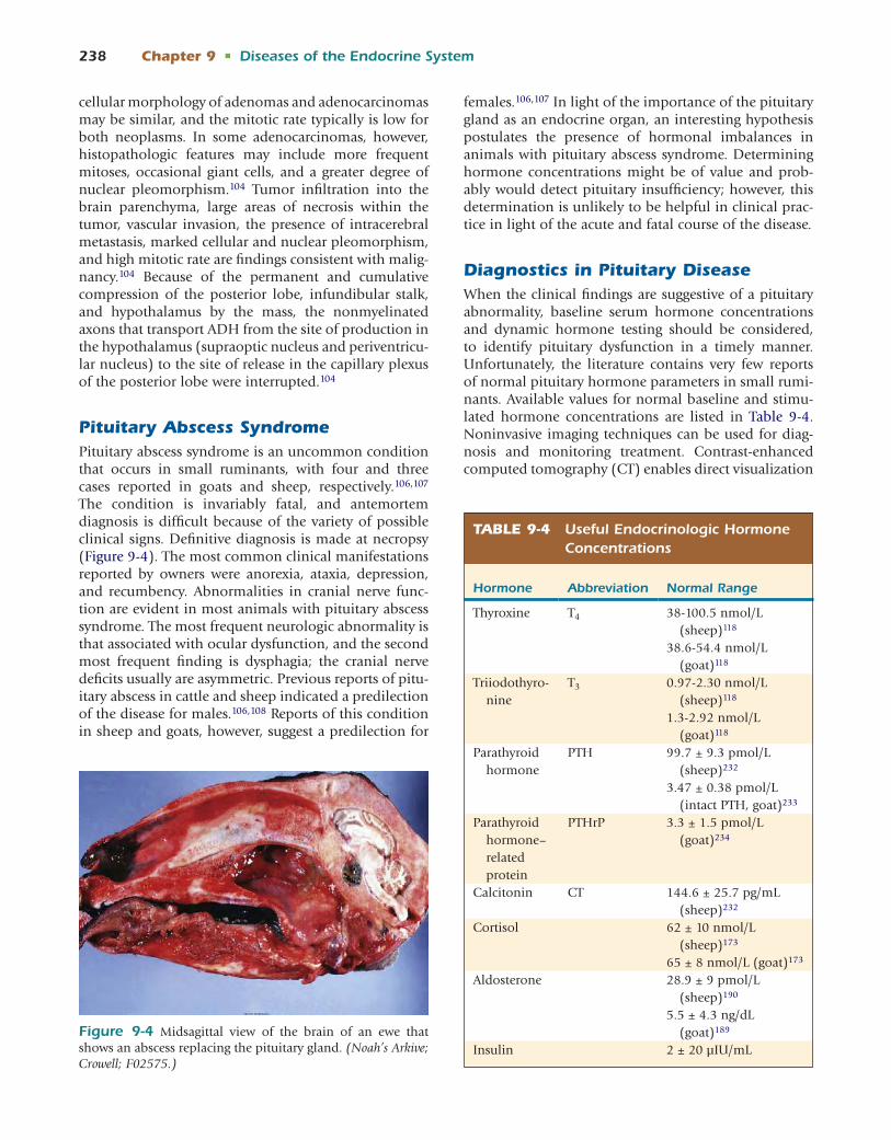
238 Chapter 9 • Diseases of the Endocrine System
cellular morphology of adenomas and adenocarcinomas may be similar, and the mitotic rate typically is low for both neoplasms. In some adenocarcinomas, however, histopathologic features may include more frequent mitoses, occasional giant cells, and a greater degree of nuclear pleomorphism.104 Tumor infiltration into the brain parenchyma, large areas of necrosis within the tumor, vascular invasion, the presence of intracerebral metastasis, marked cellular and nuclear pleomorphism, and high mitotic rate are findings consistent with malig-nancy.104 Because of the permanent and cumulative compression of the posterior lobe, infundibular stalk, and hypothalamus by the mass, the nonmyelinated axons that transport ADH from the site of production in the hypothalamus (supraoptic nucleus and periventricu-lar nucleus) to the site of release in the capillary plexus of the posterior lobe were interrupted.104
Pituitary Abscess SyndromePituitary abscess syndrome is an uncommon condition that occurs in small ruminants, with four and three cases reported in goats and sheep, respectively.106,107 The condition is invariably fatal, and antemortem diagnosis is difficult because of the variety of possible clinical signs. Definitive diagnosis is made at necropsy (Figure 9-4). The most common clinical manifestations reported by owners were anorexia, ataxia, depression, and recumbency. Abnormalities in cranial nerve func-tion are evident in most animals with pituitary abscess syndrome. The most frequent neurologic abnormality is that associated with ocular dysfunction, and the second most frequent finding is dysphagia; the cranial nerve deficits usually are asymmetric. Previous reports of pitu-itary abscess in cattle and sheep indicated a predilection of the disease for males.106,108 Reports of this condition in sheep and goats, however, suggest a predilection for
females.106,107 In light of the importance of the pituitary gland as an endocrine organ, an interesting hypothesis postulates the presence of hormonal imbalances in animals with pituitary abscess syndrome. Determining hormone concentrations might be of value and prob-ably would detect pituitary insufficiency; however, this determination is unlikely to be helpful in clinical prac-tice in light of the acute and fatal course of the disease.
Diagnostics in Pituitary DiseaseWhen the clinical findings are suggestive of a pituitary abnormality, baseline serum hormone concentrations and dynamic hormone testing should be considered, to identify pituitary dysfunction in a timely manner. Unfortunately, the literature contains very few reports of normal pituitary hormone parameters in small rumi-nants. Available values for normal baseline and stimu-lated hormone concentrations are listed in Table 9-4. Noninvasive imaging techniques can be used for diag-nosis and monitoring treatment. Contrast-enhanced computed tomography (CT) enables direct visualization
Figure 9-4 Midsagittal view of the brain of an ewe that shows an abscess replacing the pituitary gland. (Noah’s Arkive; Crowell; F02575.)
TABLE 9-4 Useful Endocrinologic Hormone Concentrations
Hormone Abbreviation Normal Range
Thyroxine T4 38-100.5 nmol/L (sheep)118
38.6-54.4 nmol/L (goat)118
Triiodothyro-nine
T3 0.97-2.30 nmol/L (sheep)118
1.3-2.92 nmol/L (goat)118
Parathyroid hormone
PTH 99.7 ± 9.3 pmol/L (sheep)232
3.47 ± 0.38 pmol/L (intact PTH, goat)233
Parathyroid hormone–related protein
PTHrP 3.3 ± 1.5 pmol/L (goat)234
Calcitonin CT 144.6 ± 25.7 pg/mL (sheep)232
Cortisol 62 ± 10 nmol/L (sheep)173
65 ± 8 nmol/L (goat)173
Aldosterone 28.9 ± 9 pmol/L (sheep)190
5.5 ± 4.3 ng/dL (goat)189
Insulin 2 ± 20 μIU/mL

Chapter 9 • Diseases of the Endocrine System 239
of the pituitary gland,109,110 but magnetic resonance imaging (MRI) may be more sensitive than CT in detec-tion of a hypothalamic-pituitary pathologic process. MRI is considered the method of choice to detect expan-sion of tumors into the suprasellar region and compres-sion of structures adjacent to the pituitary gland.111,112
A specific method to differentiate neurogenic DI from primary nephrogenic DI is the modified water deprivation test, which includes, if necessary, an AVP response test.113
THYROID GLANDStructure and FunctionThe thyroid gland consists of two lobes connected by an isthmus. It lies ventral to the trachea just below the lar-ynx. It originates in the embryo as an evagination of the endodermal epithelium between the first and second pharyngeal pouches; the connection to the epithelium is lost as development progresses, and the incorpora-tion of mesenchymal cells leads to the formation of parafollicular cells. It may remain associated with the thymus in young animals.
The functional unit of the thyroid gland is the folli-cle, which consists of a layer of cuboidal epithelial cells surrounding the colloid. The colloid is a clear, viscous fluid that serves as a reservoir for thyroid hormone in the form of the thyroglobulin-hormone complex. The size of the follicles and the length of their cells vary according to the functional stage of the gland, ranging from an inactive squamous cell to the highly active, tall columnar cell. Interspersed between the follicles are thyroid C cells (parafollicular cells), so called because they are the source of calcitonin, the hormone associ-ated with calcium metabolism. A third type of hor-monal tissue, the parathyroid, is embedded within the thyroid or located in close proximity.114 These tissues and their hormone products are discussed in greater detail later in the chapter under “Calcium Homeostasis and Parathyroid Hormone.”
Thyroid Hormones and the Hypothalamic-Pituitary-Thyroid AxisThyroid-releasing hormone (TRH) is secreted from the paraventricular nucleus of the neurohypophysis and stimulates release of thyroid-stimulating hormone (TSH), or thyrotropin, from the anterior pituitary. This in turn causes secretion of thyroxine (T4) and 3,3′,5- triiodothyronine (T3) from the thyroid gland. The latter is the active form of the hormone; in sheep, T4, T3, and reverse T3 (rT3) account for 90.4%, 8.8%, and 0.7%, respectively, of the secretory product of the thyroid gland.115 T4 can be converted to the more biologically active T3 by 5′-monodeiodinase, which is present in peripheral tissues, especially the liver and
kidney. Alternatively, it can be converted to biologi-cally inert rT3 by 5-monodeiodinase, which also may be found in some tissues. The relative concentrations of these enzymes in different tissues play an integral role in determining local hormone concentration and tissue responsiveness.
Most of the effects of thyroid hormone are mediated through binding to thyroid hormone–responsive tran-scription factors in the nucleus to increase gene expres-sion; however, binding to membrane transporters also may be responsible for mediating some portion of the cellular response. The results of T3 stimulation include increases in heat generation, oxygen utilization, basal metabolic rate, lipid metabolism, cardiac output, neural transmission, glucose availability (glycolysis, gluconeo-genesis, absorption), and protein synthesis and catabo-lism.116-118 Thyroid hormone has been associated with wool growth, weight gain, milk production, and repro-duction, and disorders of the hypothalamic-pituitary- thyroid axis may affect any or all of these. (Note: Hormone levels may fluctuate with illness or nutritional issues and therefore reflect systemic status.)
Regulation of the hypothalamic-pituitary-thyroid axis is dependent on many endogenous and exog-enous factors. Thyroid hormone inhibits the secretion of both TRH and TSH in the hypothalamus and the adenohypophysis (short and long negative feedback loops). Additionally, the neurotransmitters dopamine and somatostatin as well as glucocorticoids may inhibit TSH excretion, whereas α-adrenergic stimulation can act at the pituitary level to increase concentrations of this hormone. Cytokines, growth factors, prostaglandins, and blood concentrations of iodine (often reflecting dietary levels) may act at the level of the thyroid gland to alter expression and excretion. Finally, alterations in the hormone after its excretion can contribute to relative bioavailability and serve as a source of control; differential expression of deiodinases may be affected under changing environmental conditions in a process known as peripheral autoregulation.
HypothyroidismA hypothyroid state may stem from deficits within the gland itself—primary hypothyroidism—or higher up in the axis with TRH or TSH. Alternatively, changes may occur in peripheral deiodination of thyroxine to T3. Decreased 5′-deiodinase activity has been associ-ated with endotoxin insult and expression of cytokines (interleukins and TNF-α) with inflammation or ill-ness.116,119 Results include decreased T3 concentrations, increased rT3 concentrations, and (in severe cases) decreased T4; inflammatory cytokines also can decrease TSH release from the adenohypophysis. The decreased activity of thyroid hormones associated with inflam-mation and illness is known as euthyroid sick syndrome.

240 Chapter 9 • Diseases of the Endocrine System
It has been well characterized in companion animals such as the dog and in other ruminants, but published studies in small ruminants are lacking at present.
Thyroid Hormone and ReproductionThyroid hormone levels play an integral role in the physiology and seasonality of reproduction in small ruminants. Hypothyroidism has been implicated in multiple aspects of reproductive failure in different spe-cies, including altered spermatogenesis, reduced testicu-lar growth, irregular cycling, delayed onset of puberty, abortion, still birth, birth of weak offspring, silent heat, and nymphomania.116-117,120
Thyroid hormones affect seasonality and transition into anestrus. Ewes thyroidectomized during anestrus began cycling with their euthyroid counterparts but failed to return to anestrus after the breeding sea-son.121 Additionally, thyroid hormone supplementation resulted in earlier onset of anestrus.122 Experiments in the Saanen goat suggest that these changes may be mediated by altered seasonal expression of type II deiodinase in the hypothalamus with changing pho-toperiod; because this enzyme catalyzes the formation of active T3 from inactive T4, these changes account for increased bioavailability of active hormone in the mediobasal hypothalamus with longer photoperiod.123 Thus thyroid hormone expression in the central nervous system is greater with increased day length; inhibition of GnRH and LH at this level mediates the transition to anestrus and plays a central role in reproductive physi-ology of the female.
In growing lambs, induction of hypothyroidism by thiourea administration resulted in significant reduction in testicular size, ill-developed seminiferous tubules, and testosterone levels less than 0.02 ng/mL.120 The effects of thyroid hormone deficiency in males are more pronounced in young animals as a consequence of higher numbers of T3 receptors on Sertoli cells, which decrease with age124; because these receptors are virtually gone in the adult animal, these changes are irreversible.
Decreased reproductive efficiency associated with iodine deficiency in adults may be improved with appro-priate treatment. Iodine supplementation with 1 mL of Lipiodol (providing 480 mg of iodine) in animals with low urinary iodine concentrations resulted in signifi-cantly increased thyroid hormone levels and fertility as compared with iodine-deficient control animals.125
Thyroid Hormone and Immune StatusThe role of thyroid hormone in function of the immune system is less clearly elucidated. However, hypothyroid or goitrous animals may show increased susceptibility to infection, which results in decreased production.
Goats with endemic goiter showed significant decreases in indicators of both humoral and cell-mediated immu-nity126; these indices improved with treatment with thyroxine or colloidal iodine.
Thyroid Hormone and Hair FiberThyroid hormone levels appear to be connected to wool fiber characteristics. The exact mechanism of action is unknown, and it is unclear whether thyroid hormones have direct or permissive influence. An association has been reported between wool growth and thyroid hormone levels in sheep. Thyroidectomy resulted in a 60% decrease in wool growth as measured per unit area of skin, primarily related to decrease in length of fiber, ostensibly resulting from a reduced rate of cellular division and number of dividing cells in the cortex.127 Thyroid hormone also appears to be important to the development of hair follicles in lambs in utero.128 Similar effects have been reported on mohair growth in Angora goats, in which hyperthyroidism induced by thyroxine supplementation resulted in increased fiber length and decreased diameter.129 Additionally, supple-mentation with field bean in Angora goat kids resulted in increased thyroid hormone levels, fiber length, and secondary hair follicle activity and decreased fiber diameter.130
Environmental Temperature and Thyroid HormoneA connection between environmental temperature and thyroid hormone expression is well established in both sheep and goats.70-72,131-133 Temperature and hormone levels are negatively correlated such that increased ambient temperature results in decreased thyroid hormone concentrations. Of interest, Dorset sheep (a temperate breed) show a greater decrease in thyroid hormone and responsiveness to TRH with heat stress than that observed in Blackbelly sheep (a tropical breed).134 Also, as mentioned in relation to seasonality of reproduction, photoperiod affects thyroid hormone concentrations. Therefore it may be important to take environmental temperature and breed into consider-ation with investigations of disorders involving the hypothalamic-pituitary-thyroid axis.
Nutrition and the Hypothalamic-Pituitary-Thyroid AxisBecause the hypothalamic-pituitary-thyroid axis responds to changes in the animal’s environment, hormone levels are believed to be a good indicator of metabolic and nutritional status in small ruminants.117 It makes sense that thyroid hormone levels closely reflect nutritional status, because they serve as primary

Chapter 9 • Diseases of the Endocrine System 241
regulators of metabolic rate, and adaptation to changes in nutrient supply are important to survival. A drop in the levels of leptin occurs with decreasing body fat stores; this dip serves as a signal to the paraventricular nucleus to down-regulate TRH expression and alter the glycosylation of TSH to reduce its bioactivity.135 Therefore overall nutritional status plays an impor-tant regulatory role in function of the hypothalamic- pituitary-thyroid axis, especially in the transition from the fed to the starved state.
Selenium is essential to thyroid hormone synthesis and activation, because it is a component of deiodinase and peroxidase enzymes; dietary deficiency can affect hormone levels in addition to its well-known effects on the musculoskeletal system (causing white muscle disease). Lambs suffering from nutritional myode-generation secondary to selenium deficiency showed increased total T4 and decreased T3 levels as compared with those in normal lambs.136 The altered thyroid hormone status may account for some abnormalities or pathologic conditions noted in affected animals (see Chapter 2).
Dietary zinc levels have attracted attention because levels are tied to altered thyroid status in other spe-cies, and supplementation is commonplace in small ruminant husbandry. Experimental zinc supplementa-tion, however, did not result in significant changes in hormone levels or alteration of euthyroid status137 (see Chapter 2).
GoiterGoiter is noninflammatory and non-neoplastic enlarge-ment of the thyroid gland. It usually is detectable on physical exam, and its presence indicates attempted compensation for compromise of the hypothalamic-pituitary-thyroid axis. Whatever the underlying cause
of decreased blood hormone levels, decreased nega-tive feedback leads to enhanced production of both TRH and TSH. Elevated hypothalamic and pituitary hormones then stimulate hyperplasia of the thyroid follicular cells in an effort to increase iodine trapping and hormone excretion. The resulting disease may therefore involve hyperplasia of both the thyroid and the pituitary glands in affected animals.138 Hyperplastic goiter is characterized by increased numbers of follicu-lar cells; this may progress to colloid goiter, which is the later involutionary phase of disease (Figure 9-5, A and B). The gland may compensate early on so that the patient is physiologically normal (simple goiter), but hypothyroidism may eventually manifest clinically if the underlying problem is severe or persistent. Goiter in a euthyroid or hypothyroid animal is more frequently encountered and is classified as nontoxic goiter; glan-dular enlargement associated with hyperthyroidism is classified as toxic goiter.114
Genetic FactorsAn autosomal recessive form of goiter caused by defec-tive thyroglobulin synthesis has been reported in Merino sheep.139 Hypothyroidism secondary to an alteration in thyroglobulin structure of a possibly genetic etiology also has been described in an East Frie-sian milk ram that showed decreased body condition, depressed behavior, and goiter.140 Hereditary congenital goiter linked to defective thyroglobulin also has been described in Dutch, Pygmy, and Nubian goats.141,142
Dietary IodineOwing to its essential role as a component of thy-roxine, iodine intake can have profound effects on hormone levels. Excess or insufficiency may interfere with hormone production. Iodine deficiency is the
A B
Figure 9-5 A, The thyroid gland of a ram showing enlarged follicles filled with colloid and papillary projections into the lumen. B, Thyroid gland of a euthyroid reference sheep. Hematoxylin-eosin staining;.bar = 50 μm.229

242 Chapter 9 • Diseases of the Endocrine System
most common cause of goiter in animals and may be of greater clinical importance in situations in which multiple animals are affected, when the veterinarian is more likely to become involved. Deficiency may be either primary, from inadequate intake of iodine, or secondary, from ingestion of goitrogenic compounds that interfere with iodine uptake or metabolism and, consequently, with thyroid hormone synthesis. Iodine may leach from the soil with rain, and low iodine levels are found in areas with sandy soils or alpine regions. Thiocyanate and goitrin are two of the most common goitrogenic compounds and may be found in numerous plants, including Brassica species (cabbage); legumes (soy beans, lentils, linseed, peas, peanuts, white clover); mustard-like plants (rape, kale); prunes (cherries, apricots); and some grains (sorghum). The goiter that results from nutritional deficiency often is referred to as endemic goiter. Occasionally, insufficient intake by the dam results in manifestation of signs in neonates (Figure 9-6, A and B) (see Chapter 2).
Clinical SignsDisorders of the hypothalamic-pituitary-thyroid axis reflect alterations in its numerous functions throughout the body. Physical findings may include reduced body weight or emaciation, alopecia, facial edema (myx-adema), poor wool growth, thickened skin, weakness, lethargy, decreased production (milk or weight gain), late abortions, weak lambs, decreased reproductive performance, and immunocompromise. The thyroid is palpable as an enlarged mass just caudal to the larynx; it may be possible to palpate a thrill caused by increased blood flow to the enlarged gland. It is important to distinguish pathologic enlargement of the thyroid from the thymus, which can be palpable in young animals, or other swellings, such as cysts or abscesses; however, a large subcutaneous mass in the correct anatomic location in conjunction with suggestive clinical signs is indicative of disease.
DiagnosisClinical suspicion is based on exam findings, a palpably enlarged thyroid gland (goiter), and sometimes sug-gestive hematologic findings. Owing to the effects of thyroid hormone on lipid metabolism, blood chemistry alterations reflecting these changes can be suggestive of disease. Experimentally induced hyperthyroidism in the Nubian goat resulted in decreased serum, liver, and cardiac cholesterol, triglyceride, and phospholipid con-centrations; conversely, experimentally induced hypo-thyroidism resulted in increases in those parameters.143
Definitive diagnosis, with characterization of an individual animal as hypothyroid (or hyperthyroid), requires determination of blood hormone levels. These values may be affected by a variety of environmental or physiologic variables, as outlined previously, and must be interpreted accordingly. As in other species, thorough evaluation of the hypothalamic-pituitary-thyroid axis may require assessment of TSH as well as T4 and T3 levels. The normal reference ranges reported for these values are known to vary according to the lab and assay used, so findings should be interpreted in this light. Normal values reported for total T4 and T3 for sheep range from 38.0 to 100.5 nmol/L and 0.97 to 2.30 nmol/L, respectively.118 For goats the values are 38.6 to 54.4 nmol/L and 1.3 to 2.92 nmol/L, also respectively.118 Determination of thyrotropin levels may be considered as another potential diagnostic tool in the approach to hypothyroidism; measurement of TSH was shown to be useful in diagnosing hypothyroidism in calves, and values differed significantly in those that survived the first 24 hours and in those that did not.144
TSH or TRH response tests have been used diagnosti-cally as described in dogs and horses. A TSH response test has been performed diagnostically in a ram140; thyroxine levels measured 4 and 8 hours after TSH administration failed to double and reach physiologic levels, as would be expected in a normal animal. The results were used to confirm hypothyroidism. Similarly,
A B
Figure 9-6 Congenital goiter in a neonate goat. (Noah’s Arkive: Hedstrom; F03304 and F03305.)

Chapter 9 • Diseases of the Endocrine System 243
TSH administration approximately doubled T4 concen-tration 4 hours after administration in 3 goats; similar increases in thyroxine were obtained by administra-tion of TRH at 1 μg/kg to 25 goats, whereas T3 levels increased an average of 318% in 1 hour. It is recom-mended that a TSH response test be performed when interpretation of thyroid hormone level interpretation is equivocal or additional data are necessary.114
Treatment and PreventionAppropriate dietary iodine or supplementation is the most commonly used therapy, because deficiency is the most common cause of goiter encountered. Dietary iodine requirement for the goat is 0.5 mg/kg dry matter weight for most goats and 0.8 mg/kg dry matter weight for the lactating female145; this may be increased to 2 mg/kg in the presence of goitrogen-containing plants. Pregnant does can be drenched with oral administra-tion of 200 to 300 mg potassium iodide or 2 mL of Lugol’s iodine per week if they are at high risk for iodine deficiency. Additionally, a long-acting injectable formulation has been used, as previously mentioned, to improve reproductive performance in areas with endemic iodine deficiency.125 Oversupplementation should be avoided, because experimental evidence sug-gests that iodine levels exceeding 9.9 mg/kg dry matter weight impair immunoglobulin absorption and passive transfer in the neonate.146 Most diets contain sufficient amounts of iodine; kelp and iodized salts are the most common feed additives to ensure adequate levels. Iodized salts, which contain 0.007% to 0.01% iodine, should be the only salt source provided if they are the only source of iodine. Evidence suggests that use of the iodate form rather than iodide is preferable for supple-mentation of iodine, because it may be biologically more available. For treatment of affected kids, a regimen of either 20 mg of potassium iodine or 3 to 5 drops of Lugol’s solution in the milk daily for 7 days has been used (See Chapter 2).147
CALCIUM HOMEOSTASIS AND PARATHYROID HORMONEThe parathyroid gland is the source of parathyroid hormone, the primary hormone regulator of calcium hemostasis. Embryologically, the external and internal parathyroid glands originate from the third and fourth pharyngeal pouches, respectively; the designation as external or internal refers to their location within or out-side of the capsule, respectively, of the thyroid gland. In sheep, the external gland is associated with the subman-dibular salivary gland and is located bilaterally on the medial surface of the caudodorsal portion. By contrast, this portion is not associated with the salivary tissue in the goat and is found ventral to the wings of the atlas. The internal gland is well demarcated and located
cranial to the thyroid gland along the lateral surface of the common carotid artery in sheep, whereas it is intimately associated with thyroid tissue in the goat.148
Low blood ionized calcium and subsequent decreased binding to calcium receptors on the cell surfaces in parathyroid gland stimulate the release of parathyroid hormone,149 an 84-amino-acid peptide hormone, from the principal cells (sometimes referred to as chief cells) of the parathyroid. Blood calcium levels are raised through the activity of this hormone at the level of bone, kidney, and—indirectly—gastrointestinal tract.
In bone, parathyroid hormone mediates fast and slow changes. It alters the ion equilibrium at the bone surface to raise extracellular fluid calcium concentration and increased permeability to cause efflux of calcium from bone fluid.150 These alterations cause measurable alterations in blood calcium within minutes. Activated osteocytes and osteoblasts also stimulate osteoclast activation, and increased rates of osteolysis also raise calcium levels. Also in the bone, parathyroid hormone induces osteoclast proliferation to further increase bone mineral resorption. The resultant additional increase in calcium takes longer to manifest.
In the kidney, parathyroid hormone increases cal-cium resorption in the distal convoluted tubules while decreasing phosphorus resorption in the proximal convoluted tubules, increasing blood calcium levels, and decreasing blood phosphorus. It also activates 1α-hydroxylase in the kidney, which catalyzes the con-version of inactive vitamin D (25-hydroxycholecalcif-erol) to its active form (1,25-dihydroxycholecalciferol). Vitamin D then facilitates increased active calcium absorption in the intestine and reticulorumen,151 as well as further increasing resorption in the kidney. Ris-ing blood calcium and vitamin D levels in response to these actions inhibit further release of parathyroid hormone by the parathyroid gland (negative feedback control). Normal reported vitamin D levels are 23.8 ± 5.7 ng/mL and 40.7 ± 9.1 ng/mL in goats and sheep, respectively.87
Parathyroid hormone–related peptide (PTHrP) is a protein homologous to the biologically active amino-terminal (N-terminal) fragments of parathyroid hor-mone; it has many of the same actions, with the exception that it does not affect vitamin D levels. It serves a variety of physiologic roles and may be found in the adrenals, parathyroids, thyroids, pituitary gland, and pancreas, as well as nonendocrine tissues such as skin, skeletal muscle, lung, spleen, bones, kidney, reproduc-tive tract, urinary bladder, mammary gland, gastrointes-tinal tract, cardiovascular system, and nervous system.152 PTHrP from the fetal parathyroid glands appears to aid in transplacental calcium transport during pregnancy.153 It also is produced by the mammary gland in response to prolactin, where it has a role in increasing mam-mary blood flow154,155 and affects mineral secretion in

244 Chapter 9 • Diseases of the Endocrine System
milk.152 Nonetheless, it does not serve an endocrine function, and association of hypercalcemia with ele-vated systemic levels of PTHrP (as seen in hypercalcemia of malignancy) represents a pathologic process. A search of the current literature does not yield any reports of hypercalcemia of malignancy; however, in light of its wide distribution in normal tissue, the possibility of such disorders should not be overlooked.
Elevated blood calcium levels are regulated primarily by decreasing parathyroid hormone secretion; how-ever, calcitonin, a 32-amino-acid peptide hormone, is secreted by the parafollicular or C cells of the thyroid in the presence of significant elevations. C cells are located in the basal lamina of the thyroid, surrounding the folli-cles. They detect elevated calcium by sensing binding of the ion to receptors on the cell surface similar to those found in the parathyroid.156 This has the opposite effect of parathyroid hormone on bone, decreasing calcium resorption by decreasing osteoclastic activity and pro-liferation. It also has milder renal and gastrointestinal effects. Reported normal calcitonin levels in males and nonpregnant Marwari sheep range from 90.78 to 105.20 pg/mL, with significant increases in pregnant animals and decreases in drought conditions.157 C cell tumors, with resultant elevated calcitonin levels with hypocal-cemia, have been reported in the bovine in association with chronic excess dietary calcium and vitamin D158 and as an autosomal dominant hereditary disorder.159 In sheep, two cases of C cell hyperplasia and one case of C cell carcinoma were reported in 11 animals with experimentally induced lymphosarcoma.160
Disturbance of the calcium homeostatic mechanisms most frequently is attributable to alterations in para-thyroid hormone, because it is the primary regulator of blood calcium. Hyperparathyroidism may be primary, relating to the gland itself, or secondary, representing a compensatory change to raise blood calcium in the face of other physiologic processes. Regardless of the underlying cause, the clinical manifestations of hyper-parathyroidism are similar and reflect its activity in the various tissues of the body.
Abnormalities of Calcium RegulationPrimary HyperparathyroidismPrimary hyperparathyroidism is related to hyperplasia or neoplasia of the gland and subsequent hypersecretion of the hormonal product. It is most commonly attribut-able to a pituitary adenoma. At best, this condition is rare in small ruminants, and no case reports were found in a search of the existing literature. Nonetheless, it is the third most common endocrine disorder in people161 and is well documented in dogs and cats.162 In dogs, the most common clinical signs are related to urolithia-sis or urinary infections,163 and in one series showed hypercalcemia on bloodwork, whereas phosphate levels
were more variable. Although primary hyperparathy-roidism in people is most common in postmenopausal women,161 primary hyperparathyroidism also has been associated with pregnancy, including one reported case in twin pregnancy.164 In pregnancy-associated cases, recognition of the syndrome is very important, because hypercalcemia can be detrimental to the fetus, and the neonate will require management directed at potential hypocalcemia.
Treatment of primary hyperthyroidism usually is successful and may include surgical removal of the ade-noma or ablation of the abnormal tissue. Despite the current lack of evidence for occurrence of this condition in small ruminants, understanding of its pathogenesis and diagnosis may lead to appropriate recognition and treatment.
Nutritional Secondary HyperparathyroidismNutritional secondary hyperparathyroidism occurs in animals fed a ration low in calcium or with a low calcium-to-phosphorus ratio. Parathyroid hormone secretion is up-regulated in response to elevated phos-phorus and low blood calcium levels. In a study involv-ing growing animals, it was shown that providing up to three times the dietary requirement for phosphorus had no adverse effects on bone mineral content, parathyroid hormone levels, or vitamin D metabolism, provided that calcium requirements were met.165 Nonetheless, calcium-to-phosphorus imbalances can and do occur. High-grain and low-roughage diets and diets high in oxalate-containing compounds or plants are character-ized by low gastrointestinal calcium absorption, which may lead to compensatory increases in parathyroid hormone. Oxalate interferes with calcium absorption in the intestine and is found in high levels in Bermuda grass, dallis grass, foxtail grass, pokeberry, red-rooted pigweed, and sugar beet, to name a few of the possible offending plants.
Additionally, dietary cation-anion differences (DCADs) frequently are intentionally manipulated to induce a mild hyperchloremic metabolic acidosis, thereby increasing tissue responsiveness to parathyroid hormone. Addition of anions to diets and subsequent up-regulation of parathyroid hormone expression before parturition will aid in the prevention of hypo-calcemia.166 In sheep fed different DCAD diets during the last 6 weeks of gestation through day 12 post par-tum, sheep fed the higher cation diet showed higher parathyroid hormone levels, whereas those fed the anionic diet showed greater cortical bone remodeling on electron microscopy.167 Also, supplementation of ammonium chloride is used as a preventive measure to guard against struvite urolithiasis, which is relatively common in male goats. However, supplementation with this salt results in higher parathyroid hormone levels and lower retention of calcium and phosphorus

Chapter 9 • Diseases of the Endocrine System 245
as compared with animals fed a diet supplemented with sodium bicarbonate.168 These diets serve impor-tant roles in preventing potentially serious problems; of note, however, with long-term feeding (most likely years), prolonged elevated parathyroid hormone levels could lead to the same pathologic changes seen with other causes of metabolic bone disease or calcium/phosphorus imbalances (See Chapters 2 and 12).
Renal Secondary HyperparathyroidismThe kidneys play an essential physiologic role in the regulation of body calcium. Hypocalcemia or normo-calcemia and hypophosphatemia are the electrolyte abnormalities most likely to be noted with renal disease, leading to increased parathyroid hormone secretion to counteract the renal loss. The clinical signs associ-ated with elevated hormone expression have not been reported in the small ruminant; the disease is unlikely to progress to this point in these animals owing to eco-nomic limitations on treatment. Although reports of renal failure leading to elevated blood calcium in small ruminants are limited, experimental evidence points to this pathomechanism as a cause of hypercalcemia. In an ovine model of chronic renal failure based on subtotal nephrectomy, one of nine sheep died of hypercalcemia, whereas the others maintained normocalcemia.169
RicketsOwing to the inverse control of phosphate as compared with calcium and the role of vitamin D in regulating gastrointestinal and renal absorption of calcium, abnor-malities in their metabolism may affect overall blood levels of calcium and parathyroid hormone. Rickets can occur in small ruminants as a result of dietary deficien-cies in either phosphorus or vitamin D. Additionally, an inherited autosomal recessive form of rickets has been described in Corriedale sheep.170 In these instances, secondary hyperparathyroidism occurs as a compensa-tory mechanism to maintain blood calcium levels that would otherwise be low.
Alternatively, vitamin D intoxication has been impli-cated in hypercalcemia, hypercalcitonism, and hypo-parathyroidism; this disturbance has been attributed to feeding imbalanced rations as well as toxins, often con-tained in plants. Two lambs with secondary hypercal-cemia showed signs of weakness and inability to stand; the elevated levels of blood calcium were shown to be related to hypervitaminosis D, although the source could not be definitively identified.171 Most calcino-genic plants contain a steroidal glycoside that is cleaved to 1,25-dihydroxyvitamin D3, which then contributes to hypercalcemia, hyperphosphatemia, hypercalcito-nism, osteopetrosis, and calcinosis; some of the most important causative plants include Cestrum diurnum, Solanum malacoxylon, Trisetum flavescens, and Niembergia veitchii.172
Clinical SignsThe clinical manifestation of disease is dependent on the underlying pathologic process at play. In cases of chronic hyperparathyroidism, the most striking clini-cal findings reflect its actions in promoting osteolysis. Long-term stimulation results in the loss of mineral in bone. Subsequent structural deterioration may lead to shifting lameness, angular limb deformities and decreased growth in young animals, loose or missing teeth, and pathologic fractures. Additionally, fibrous tissue may replace lost mineral in bone in the process of fibrous osteodystrophy (osteodystrophia fibrosa). Associated changes are most evident in the flat bones, usually first noted as enlargement of the bones of the skull and face.
Alternatively, hypercalcemia with compensatory hypoparathyroidism is more likely to be associated with osteopetrosis, increased bone density, decreased size of the medullary cavity, and possibly mineraliza-tion of tissues.
DiagnosisDifferentiating among the disorders of calcium metabo-lism requires a thorough history and careful interpre-tation of findings on a blood chemistry profile and electrolyte levels (e.g., calcium and phosphorus). Diag-nosis of hyperparathyroidism may be based directly on measurement of parathyroid hormone levels. Elevated calcium levels on bloodwork lead to suspicion and search for an underlying cause. Normal calcium levels are 11.5 to 12.8 mg/dL for the sheep and 8.9 to 11.9 mg/dL for the goat but may vary in different laboratories.173 A complete parathyroid panel, including parathyroid hormone–related protein, may be considered in cases in which alterations of this peptide are suspected. Alter-natively, one may consider supportive radiographic evi-dence (demineralization), fractional renal clearance of phosphorus, blood calcium levels, and clinical evidence based on history and physical findings. Blood calcium levels may be elevated, within reference ranges, or low, depending on the underlying cause and progression of disease.
Treatment and PreventionThe mainstay of therapy is identification and treatment of the underlying cause of disease. In severe hypercal-cemia, diuresis to increase glomerular filtration and serum calcium concentration may be used as a short-term therapy to moderate potential deleterious effects. Loop diuretics may be indicated to increase urinary calcium excretion.
Feeding an appropriate ration with a balanced calcium-to-phosphorus ratio is essential to prevention of disease. Dicalcium phosphate is the most frequently used supplement to augment dietary calcium, and pro-vision of a mineral salt mixture is highly recommended.

246 Chapter 9 • Diseases of the Endocrine System
A more complete consideration of dietary calcium rec-ommendations is presented in Chapter 2.
ADRENAL GLANDSThe adrenal glands are found bilaterally near the cranial pole of each kidney. Each gland consists of an inner medulla of ectodermal origin and an outer cortex that arises from the intermediate mesoderm. The average size is approximately 2 to 3 cm long by 1 cm wide, and the left gland often is larger than the right.145
The hormone products of the adrenal gland include epinephrine and norepinephrine from the adrenal medulla and aldosterone (zona glomerulosa), gluco-corticoids (zona fasciculata and zona reticularis), and androgens (zona reticularis) from the adrenal cortex.
Regulation of the hypothalamic-pituitary-adrenal axis is a complex process involving the interactions of several different hormones and stimuli. The release of ACTH from the adenohypophysis can be stimulated by CRH, AVP, or oxytocin.174 Of interest, AVP appears to be a stronger stimulus for ACTH secretion than CRH in the sheep.175,176 CRH exerts its effects through activation of adenylate cyclase and increased cyclic AMP (cAMP), whereas the intracellular signaling from AVP relies on activation of phospholipase C through its V1b receptor. AVP has been shown to increase pro-opiomelanocortin mRNA in ovine corticotropes, whereas CRF does not.177 Although the adenohypophysis is able to produce hor-mones, and levels of ACTH, α-melanocyte-stimulating hormone (α-MSH), β-endorphin, and cortisol were actu-ally elevated with hypothalamic-pituitary disconnection in sheep,178 animals with a severed portal system were unable to produce an appropriate stress response in the face of hypoglycemia or audiovisual stressors.179 This observation highlights the importance of these hor-mones in the overall hypothalamic-pituitary-adrenal axis response. Catecholamines also are an integral part of the stress response and serve as a stimulus for ACTH excretion, mostly by increasing levels of AVP and CRF. Both norepinephrine and epinephrine cause release of ACTH and cortisol when injected into the lateral ven-tricle of sheep, and norepinephrine is the more potent of the two.180 Much support also has emerged for a hypothalamic factor that has an inhibitory influence on the expression of ACTH by corticotropes42; thus far, however, no molecule fulfills the profile for the so-called corticotropin-releasing inhibitory factor (CRIF).
ACTH then induces emission of cortisol (95%) and corticosterone (5%) from the adrenal cortex. Gluco-corticoids bind to cytosolic receptors in target tissues to increase gene transcription, increase protein catabo-lism, increase gluconeogenesis, inhibit inflammation through blockade of the arachidonic acid cascade, antagonize peripheral insulin sensitivity, and increase blood glucose. Rising cortisol concentrations serve in
negative feedback at the level of both the pituitary and the brain or hypothalamus and affect both the dura-tion and amplitude of the overall stress response.181 Plasma cortisol increases related to the stress response are depressed in intact males as compared with females. Intact males show decreased blood cortisol elevations as compared to females in response to ACTH adminis-tration.182 The decreased responsiveness appears to be androgen-mediated, because administration of testos-terone or dihydrotestosterone to castrated male goats suppressed the degree to which blood cortisol increased with the stress of transport.183 Furthermore, male cas-trated animals treated with these androgens showed decreased blood cortisol response as compared with control animals consequent to ACTH administration, suggesting that such changes are mediated at the level of the adrenal gland.184 In the study reporting these find-ings, immunohistochemical analysis revealed that 60% of the cells of the zona fasciculata and zona reticularis contained androgen receptors. This evidence suggests that androgens act directly on the adrenal cortex to sup-press secretion of glucocorticoids in response to ACTH administration.
Absence of this androgen-mediated adrenal corti-cal inhibition may account for the higher incidence of adrenal disease seen in castrated male goats. One study found adrenal adenomas in 314 of 2104 castrated males older than 5 years of age.185 By contrast, only 2 of 208 females and none of 188 intact males exhibited this same neoplastic disorder. Adrenal neoplasia resulting in gynecomastia and inappropriate lactation also has been reported in a 6-year-old castrated male Toggenburg goat; associated hormonal changes included elevated estradiol-17β, GH, prolactin, and cortisol, but the goat did not show any clinical signs of Cushing’s-type syndrome.186
Habitual abortion in Angora does is believed to be associated with decreased adrenocortical responsiveness and hypoadrenocorticism.187 Fetal death usually occurs at 90 to 120 days of gestation after a stressful event, and it is more common in multiparous than in primapa-rous animals. The genetic defect predisposing these animals to adrenocortical dysfunction and pregnancy loss is believed to be linked to the mohair coat trait. Affected dams usually exhibit small adrenal glands and low blood cortisol levels; this phenotype results in fetal adrenal hyperplasia and increased fetal cortisol levels, which prematurely induces parturition.188
As with other hormone values, normal values for cortisol levels vary according to the reporting lab and assay applied, and different units of measurement are used according to preference. Therefore it is best to use reference ranges provided by the lab when avail-able. Cortisol values fluctuate with season, with higher levels reported in the autumn. In goats, these changes are more pronounced in females.145 Normal levels

Chapter 9 • Diseases of the Endocrine System 247
for cortisol as determined by radioimmunoassay were reported as 62 ± 10 nmol/L and 65 ± 8 nmol/L in the sheep and goat, respectively.173 Because levels will vary with season, breed, sex, and reproductive status, these variables should be taken into account in the interpre-tation of results. Aldosterone is secreted in response to angiotensin II by the following physiologic sequence: Low blood pressure or low sodium-to-potassium ratio in the blood is detected by the juxtaglomerular appara-tus of the kidney, which then releases renin. The renin cleaves the circulating proenzyme angiotensinogen to angiotensin I, which is then converted to angiotensin II by angiotensin-converting enzyme, found in the high-est concentrations in the lung. Aldosterone stimulates increased sodium retention and potassium and hydro-gen excretion in the renal medullary collecting tubules. Subsequent rise in blood pressure and blood sodium then decreases renin excretion. In goats, reported aldo-sterone levels varied with hydration status, with ranges of 5.5 ± 4.3 ng/dL and 13.9 ± 2.3 ng/dL in hydrated and dehydrated states, respectively.189 A baseline value of 28 ± 9 pmol/L has been reported in sheep.190
The adrenal medulla consists of sympathetic gan-glion cells and chromaffin cells, which secrete the epinephrine and norepinephrine hormone products. Secretion of these catecholamines is stimulated by ace-tylcholine from the sympathetic nervous system or by cortisol from the adrenal cortex. The systemic effects of these hormones are most recognized as part of the “fight-or-flight” response. They include tachycardia, tachypnea, increased metabolic rate, mobilization of fats, sweating, alertness, piloerection, and urination.
Disorders of the adrenal medulla in small ruminants are rare, but they have been reported. An undifferenti-ated ganglioneuroblastoma that presumably arose from the adrenal medulla was described in an 18-month-old Akkaraman sheep191; the diagnosis was based on gross pathologic, histopathologic, and immunohistochem-istry study findings. The tumor was recognized during routine inspection at slaughter, so clinical manifes-tations were not described. Additionally, pheochro-mocytoma has been reported in two does associated with inappropriate lactation, pituitary adenoma, and endometrial hyperplasia.95 A family of does in Finland with pheochromocytomas has also been described; the affected does had a history of nervous attacks, and diagnosis was made at necropsy on the basis of histo-pathologic findings.192
PANCREASThe pancreas of sheep and goat is similar in function to that of other ruminant animals. The pancreas consists of a body and two lobes and is located in the cranio-dorsal abdomen and closely related to the descending duodenum. The pancreas arises from two primordia
that bud from the proximal part of the duodenum. It is a soft, lobulated gland of irregular form and pinkish-yellow in appearance and bears some resemblance to a salivary gland, although it is softer and more loosely structured than most such glands. The pancreas has combined exocrine functions (production and release of digestive enzymes) and endocrine functions (produc-tion and release of insulin, glucagon, somatostatin, and pancreatic polypeptide).193
The portion of the pancreas involved with exocrine functions is by far the larger, and it produces a digestive juice that is discharged into the proximal part of the duodenum through one or two ducts. A single duct that in small ruminants represents the ventral duct opens into the duodenum with the bile duct, usually by means of a common trunk—a feature that makes these species suitable for experimental studies of the effects of divert-ing bile into the pancreatic duct system. The juice con-tains enzymes that break down protein, carbohydrates, and fats.
Some endocrine tissues are incorporated within organs of composite function. The most familiar exam-ple is provided by the endocrine component of the pan-creas, the pancreatic islets, also known as the islets of Langerhans. The endocrine component comprises many hundred (or thousand) islets of varying size unevenly distributed among the predominant exocrine tissue. Several types of islet cells (the exact number is disputed) are recognized; the two most numerous are the alpha and beta cells. The beta cells account for approximately 75% of all cells in the pancreatic islet and produce the hormone insulin. The alpha cells produce the hormone glucagon. Other, less numerous cells produce gastrin and somatostatin.
The beta cells are sensitive to increases in blood glu-cose, such as occur after a meal containing digestible carbohydrates, and they release insulin (2 to 20 μIU/mL) in response to such increases in blood glucose. Insulin lowers blood glucose by stimulating the uptake of glucose by many cells of the body, including skeletal muscle. Insulin also stimulates skeletal muscle and liver cells to synthesize glycogen, the storage form of glucose. Insulin is the major endocrine stimulus for the state of anabolism that exists after a meal is digested and nutri-ents are absorbed. When blood glucose decreases (such as during fasting), the stimulus for insulin secretion is lost, and insulin levels become extremely low.
Glucagon causes liver cells to break down glycogen to release glucose, stimulates adipocytes to release fatty acids, and increases the synthesis of glucose in the liver from substrates other than carbohydrates, such as amino acids. The stimulus for glucagon release is a decrease in blood glucose to levels associated with fast-ing. The alpha cells detect such decreases and respond by secreting glucagon in proportion to the reduction in blood glucose.

248 Chapter 9 • Diseases of the Endocrine System
Diabetes mellitus (DM) has been described as any condition characterized by a permanent elevation of blood glucose and glucosuria.194 Primary DM involves damage to pancreatic beta cells (insulin-producing cells) followed by decreased insulin levels and hyper-glycemia. Secondary DM results from a resistance to the effects of insulin even with normal or elevated plasma insulin levels.195 DM is rare in small ruminants. Only three case reports of spontaneous DM are found in the scientific literature.194-196 Secondary DM was reported in one of these cases,195 and primary DM was noted in another.194 Other case reports describe the features of experimentally induced DM by administration of alloxan197,198 or streptozocin.199-201 Clinical signs of diabetes in small ruminants include weight loss, poor appetite, and lethargy.194,198 Polyuria and polydipsia are invariable findings, with some animals exhibiting two to three times their normal water intake.194 A 90% decrease in milk production has been observed in lac-tating goats with experimentally induced DM.198
Treatment with insulin has been tried in both spon-taneous and experimental DM. In general, a response to treatment is observed within 4 days and consists of a partial relief of clinical signs and normalization of laboratory values.195,198 One goat with primary DM was successfully treated for almost 4 years with twice-daily insulin.194 Of note, however, improper insulin dosage can result in life-threatening hypoglycemia. Recurrence of clinical signs is observed with discontinuation of therapy.
ABERRANT LACTATION, GALACTORRHEA, PRECOCIOUS UDDER: INAPPROPRIATE LACTATION SYNDROMEThe endocrine system normally coordinates develop-ment of the mammary gland with reproductive devel-opment and the demand of the offspring for milk. As described in detail previously,202 three categories of hormones—reproductive, metabolic, and mammary—are involved in mammary gland development and milk production. Ductal morphogenesis is primarily regu-lated by estrogen and GH,203 and the proliferative phase of alveolar morphogenesis requires progesterone and prolactin.204 Hormonal regulation of lactogenesis, the set of processes that lead to the initiation of lactation, is not completely understood, but the candidate hor-mones are progesterone, prolactin or placental lactogen (or both), and possibly GH. The effects of these hor-mones on mammogenesis and lactogenesis often are dependent on one another. For instance, progesterone withdrawal in the presence of high levels of prolactin is necessary for lactogenesis to be complete. Mammogene-sis and lactation have been induced in small ruminants through exogenous administration of estrogen and pro-gesterone,205-207 and a variety of conditions disturbing
the endocrine balances may potentially be accompa-nied by inappropriate mammogenesis or lactation.
Although only a few cases of ILS in small ruminants are described in the scientific literature,95,186,208-216 the anecdotal reports of this problem are seemingly end-less. ILS has been reported in both castrated186 and intact208,210-214,216 male goats as well as in does95,215,217 and ewes.209 Published reports of ILS in male small ruminants outnumber those in females; anecdotally, however, the problem is more common in females, and it appears to be more of a clinical concern in pet does.
Inappropriate development of mammary glands and lactation in bucks is occasionally observed.210 In Eng-land, mammary gland development is seen in fertile bucks from high-producing lines of British Saanens. The phenomenon usually is observed in the summer, and fertility is unaffected216; however, reduced libido and semen quality have been reported in some bucks with gynecomastia.211 Proposed pathogenic factors have included a genetic basis for the syndrome and possibly an increase in production of hormones or an increased sensitivity of mammary tissue to normal hormone levels.
In some instances, gynecomastia in bucks may be the result of a genetic or gonadal sex disorder. One documented case of gynecomastia involved a polled fertile male goat that was determined to be a chromo-somal mosaic with variable deletions of the Y chromo-some (60,XO/60,XY).213 In another report, a buck that yielded up to 300 mL of milk per day had Barr bodies in 45% of neutrophils, suggesting a genetic intersex; however, neither somatic or leukocyte karyotyping was performed.212 Many reports of gynecomastia in small ruminants do not include karyotype. When karyotype is determined, significance of the findings needs to be determined in the context of the tissue(s) tested, because somatic and peripheral (leukocyte) karyotypes from the same animal are not always in agreement. However, the diagnostic benefits of karyotyping in cases of gynecomastia should not be overlooked.
A 6-year-old castrated male goat with a normal karyotype developed gynecomastia and lactation that were ultimately attributed to an adrenal adenoma.186 Hormone concentrations in multiple paired serum samples were compared in the lactating goat and his “normal” castrated twin. Concentrations of estradiol, GH, and IGF-I were consistently higher in the lactating male than in his twin, and concentrations of cortisol and prolactin generally were higher in the lactating male. In comparison with the nonlactating twin, the lactating male also had exaggerated cortisol and thy-roxine responses to ACTH and TSH administration. Eventually, the investigators concluded that high serum concentrations of estradiol probably were responsible (both directly and indirectly) for gynecomastia and lactogenesis. Some reports of gynecomastia in bucks,

Chapter 9 • Diseases of the Endocrine System 249
however, are not associated with increased concentra-tions of sex steroids. For instance, a 3-year-old fertile buck with gynecomastia had a normal karyotype and normal plasma testosterone and estradiol concen-trations but prolactin was significantly increased.218 Although some reports of gynecomastia in bucks do not include hormonal studies, elevated concentrations of prolactin are presumed to be the cause.216,219 For exam-ple, gynecomastia in a fertile buck was associated with hyperplasia of acidophilic cells in the pituitary gland, and those cells normally produce mammotropic hor-mones such as prolactin.219 Other, less common causes of gynecomastia reported include but are not limited to mastitis211 and mammary adenocarcinoma.214
Ewes and does with ILS usually have an enlarged udder with no history of being bred, nursed, or milked. In some animals, the udder enlargement is mainly the result of deposition of adipose tissue. In others, milk is actually produced from one or both glands without preceding parturition. Even with this history, the initial examination in these animals should rule out impending parturition and pseudopregnancy and recent nursing or milking. Also, as in bucks, precocious lactation in does may be a hereditary trait associated with high milk production potential.220 A variety of conditions disturbing the endocrine balances (i.e., pitu-itary, gonad, or adrenal abnormalities and exogenous hormones) could be accompanied by inappropriate mammogenesis or lactation in both male and female small ruminants.
Pseudopregnancy is one possible cause of ILS, and most practitioners believe that affected does should be treated with luteolytic hormones. Natural luteolysis and withdrawal of progesterone may have occurred before ILS developed, and although luteolytic hormones may not result in a cure, they are still recommended to rule out pseudopregnancy. As discussed previously, estrogens affect mammogenesis and lactogenesis and are therefore contraindicated. Consequently, estrogenic feeds, such as legumes (e.g., clover) or moldy grains, should be removed from the diet. In addition, grain and other energy-dense feedstuffs should be temporarily eliminated to discourage milk production.
Pituitary adenomas (which can be prolactin-secreting tumors in other species) have been reported in two does with ILS.95 Although some reports suggest that prolactin levels are elevated in association with these tumors,95,215,221 this may not always be the case. Plasma prolactin concentrations in small ruminants have a circannual rhythm, increasing from low (less than 30 ng/mL) concentrations during the fall and winter to val-ues of approximately 100 ng/mL in the spring. Plasma prolactin concentrations were as high as 400 ng/mL during the summer months.222,223 Dopamine agonists, which are antiprolactin agents, have been suggested for treatment of does with inappropriate lactation.
Bromocriptine mesylate and cabergoline have been suggested as agents for treatment of animals with ILS. Success with use of these drugs to stop inappropriate lactation has been limited,224 however, and although a bromocriptine reduced circulating prolactin concen-trations,225,226 milk production was only temporarily decreased after treatment was discontinued.225,227
Spontaneous lactation also has been reported in does and a ewe in association with ovarian abnormali-ties (Figure 9-7). DeWalque217 described a doe with a granulosa cell tumor that exhibited virilism concurrent with inappropriate lactation. Lofstedt and Williams have reported udder engorgement in a doe 6 days after removal of a granulosa cell tumor.215 In addition, one of the does with ILS described by Miller and associ-ates95 had ovarian follicular cysts. ILS also was reported in a ewe concurrent with an ovarian granulosa cell tumor.209 Transabdominal ultrasonography revealed a multiloculated cystic mass in the caudoventral abdo-men. Granulosa cell tumors may secrete a variety of hor-mones and measurement of abnormal levels of these hormones may aid in diagnosis. Small ruminants with granulosa cell tumors often have elevated serum levels of testosterone and estradiol and variable serum levels of progesterone.209 The production of these hormones frequently is associated with abnormal behavior and physiology (e.g., ILS). Affected animals often exhibit virilism, which may be associated with nymphomania and abnormal estrous cycles. Removal of the affected gonad(s) is the treatment of choice.
The current treatment of choice for inappropriate lactation in pet goats is mastectomy. Although this procedure obviously is curative in preventing further lactation, an underlying problem may remain. Con-sequently, close monitoring is advisable throughout the life of the animal for other conditions known to be associated with inappropriate lactation. Such
Figure 9-7 Reproductive tract of a 20-month-old ewe in which a granulosa cell tumor has replaced the right ovary.209

250 Chapter 9 • Diseases of the Endocrine System
conditions include endometrial hyperplasia and uter-ine fluid retention (which could predispose affected does to pyometra, endometritis, or acute septic metri-tis), pituitary adenomas, pheochromocytoma (which may manifest clinically as nervousness and abnormal behavior), and ovarian granulosa cell tumor (for which the clinical presentation may be virilism). Although the fertility of many of these does and ewes is limited, they should nevertheless be protected from any expo-sure to a buck after mastectomy. Not only is a lack of milk for the offspring a problem, but also the loss of mammary tissue in goats has been associated with an increased incidence of periparturient complications. These reported complications include premature labor, prolonged onset of labor, lack of cervical dilation, and maternal death at parturition.228 Our own observation in one female that had been bred post mastectomy sup-ports this correlation, as she had prolonged labor and poor cervical dilation.
REFERENCES 1. Daniel PM: Anatomy of the hypothalamus and pituitary gland,
J Clin Pathol Suppl (Assoc Clin Pathol) 7:1–7, 1976. 2. Marshall FHA: Sexual periodicity and the causes which deter-
mine it, Phil Trans R Soc Biol Sci Ser B 226:423–456, 1939. 3. Fulton JF, Ranson SW, Frantz AM: The hypothalamus and control
levels of autonomic functions, Association for Research in Nervous and Mental Diseases, Baltimore, Md, 1940, Williams & Wilkins.
4. Harris GW: The function of the pituitary stalk,, Bull Johns Hop-kins Hosp 97:358–375, 1955.
5. Harris GW: Pituitary-hypothalamic mechanisms, AMA Arch Neurol Psychiatry 73:124–126, 1955.
6. Burgus R, et al: [Molecular structure of the hypothalamic hypo-physiotropic TRF factor of ovine origin: mass spectrometry demonstration of the PCA-His-Pro-NH2 sequence], C R Acad Sci Hebd Seances Acad Sci D 269:1870–1873, 1969.
7. Nair RM, et al: Structure of porcine thyrotropin releasing hor-mone, Biochemistry 9:1103–1106, 1970.
8. Wilber JF, Porter JC: Thyrotropin and growth hormone releasing activity in hypophysial portal blood, Endocrinology 87:807–811, 1970.
9. McMahon CD, et al: Neuroregulation of growth hormone secre-tion in domestic animals, Domest Anim Endocrinol 20:65–87, 2001.
10. Ojeda SR, et al: Recent advances in the endocrinology of puberty, Endocr Rev 1:228–257, 1980.
11. Sklar CA, et al: Hormone ontogeny in the ovine fetus. VII. Cir-culating luteinizing hormone and follicle-stimulating hormone in mid- and late gestation, Endocrinology 108:874–880, 1981.
12. Matwijiw I, Thliveris JA, Faiman C: Hypothalamo-pituitary por-tal development in the ovine fetus, Biol Reprod 40:1127–1130, 1989.
13. Levidiotis ML, et al: Hypothalamic-hypophyseal vascular con-nections in the fetal sheep, Neuroendocrinology 49:47–50, 1989.
14. Lauder JM: Hormonal and humoral influences on brain devel-opment, Psychoneuroendocrinology 8:121–155, 1983.
15. Huhtaniemi IT, Warren DW: Ontogeny of pituitary-gonadal interactions: current advances and controversies, Trends Endocri-nol Metab 1:356–362, 1990.
16. McKlveen TL, et al: Assessment of the accuracy of computed tomography for measurement of normal equine pituitary glands, Am J Vet Res 64:1387–1394, 2003.
17. Medvei VC: A history of endocrinology, Boston, 1982, MTP Press.
18. Amar AP, Weiss MH: Pituitary anatomy and physiology, Neuro-surg Clin North Am 14:11, 2003.
19. Cotran R, Kumar V, Collins T: The endocrine system. In Cotran R, Kumar V, Collins T, editors: Robbins Pathologic basis of disease, Philadelphia, 1999, WB Saunders, pp 1121–1169.
20. Goodman HM: Pituitary gland. In Johnson LR, editor: Essen-tial medical physiology, Philadelphia, 1998, Lippincott-Raven, pp 511–520.
21. Mignot M, Skinner DC: Colocalization of GH, TSH and pro-lactin, but not ACTH, with betaLH-immunoreactivity: evidence for pluripotential cells in the ovine pituitary, Cell Tissue Res 319:413–421, 2005.
22. Aron DC, Findling JW, Tyrell JB: Hypothalamus and pituitary. In Greenspan FS, Strewler GJ, editors: Basic and clinical endocrinol-ogy, Stamford, Conn, 1997, Appleton & Lange, pp 95–156.
23. Frawley LS, Boockfor FR: Mammosomatotropes: presence and functions in normal and neoplastic pituitary tissue, Endocr Rev 12:337–355, 1991.
24. Scanlan N, Skinner DC: Estradiol modulation of growth hor-mone secretion in the ewe: no growth hormone-releasing hor-mone neurons and few somatotropes express estradiol receptor alpha, Biol Reprod 66:1267–1273, 2002.
25. Landefeld TD, Suttie JM: Changes in messenger ribonucleic acid concentrations and plasma levels of growth hormone during the ovine estrous cycle and in response to exogenous estradiol, Endocrinology 125:1474–1478, 1989.
26. Malven PV, Haglof SA, Jiang H: Serum concentration of luteiniz-ing hormone, growth hormone, and prolactin in untreated and estradiol-treated ovariectomized ewes after immunoneutraliza-tion of hypothalamic neuropeptide-Y, J Anim Sci 73:2105–2112, 1995.
27. Skinner DC, Caraty A: Prolactin release during the estradiol-induced LH surge in ewes: modulation by progesterone but no evidence for prolactin-releasing peptide involvement, J Endocri-nol 177:453–460, 2003.
28. Anderson ST, et al: Dopaminergic input to the ventromedial hypothalamus facilitates the oestrogen-induced luteinizing hor-mone surge in ewes, Neuroendocrinology 73:91–101, 2001.
29. Arbogast LA, Benjonathan N: The preovulatory prolactin surge—an evaluation of the role of dopamine, Endocrinology 123:2690–2695, 1988.
30. Djahanbakhch O, et al: Changes in plasma levels of prolactin, in relation to those of FSH, oestradiol, androstenedione and progesterone around the preovulatory surge of LH in women, Clin Endocrinology 20:463–472, 1984.
31. Campbell BK, et al: The pattern of ovarian inhibin, estradiol, and androstenedione secretion during the estrous cycle of the ewe, Endocrinology 127:227–235, 1990.
32. Howes KA, et al: Serum luteinizing-hormone, prolactin, and thyrotropin and their pituitary subunit messenger-RNA levels during proestrus in the Syrian hamster, Neuroendocrinology 54:629–634, 1991.
33. Tobin VA, Pompolo S, Clarke IJ: The percentage of pitu-itary gonadotropes with immunoreactive oestradiol receptors increases in the follicular phase of the ovine oestrous cycle, J Neuroendocrinol 13:846–854, 2001.
34. Childs GV, Unabia G, Miller BT: Cytochemical detection of gonadotropin-releasing hormone binding sites on rat pituitary cells with luteinizing hormone, follicle-stimulating hormone, and growth hormone antigens during diestrus up-regulation, Endocrinology 134:1943–1951, 1994.
35. Childs GV, Unabia G, Rougeau D: Cells that express lutein-izing hormone (LH) and follicle-stimulating hormone (FSH) beta-subunit messenger ribonucleic acids during the estrous cycle: the major contributors contain LH beta, FSH beta, and/or growth hormone, Endocrinology 134:990–997, 1994.
36. Childs GV, Miller BT, Miller WL: Differential effects of inhibin on gonadotropin stores and gonadotropin-releasing hormone binding to pituitary cells from cycling female rats, Endocrinology 138:1577–1584, 1997.

Chapter 9 • Diseases of the Endocrine System 251
37. Stefano AV, et al: Colocalization of GnRH binding sites with gonadotropin-, somatotropin-, somatolactin-, and prolactin-expressing pituitary cells of the pejerrey, Odontesthes bonariensis, in vitro, Gen Comp Endocrinol 116:133–139, 1999.
38. Parhar IS, et al: Spatio-temporal expression of gonadotropin-releasing hormone receptor subtypes in gonadotropes, somato-tropes and lactotropes in the cichlid fish, J Neuroendocrinol 14:657–665, 2002.
39. Katoh K, et al: Saturated fatty acids suppress adrenocortico-tropic hormone (ACTH) release from rat anterior pituitary cells in vitro, Comp Biochem Physiol A Mol Integr Physiol 137:357–364, 2004.
40. Senn M, Maier PM, Langhans W: ACTH, cortisol and glucose responses after administration of vasopressin in cattle and sheep, J Comp Physiol B 164:570–578, 1995.
41. Liu JP, et al: The biosynthesis and secretion of adrenocorticotro-pin by the ovine anterior pituitary is predominantly regulated by arginine vasopressin (AVP)—evidence that protein kinase-C mediates the action of AVP, J Biol Chem 265:14136–14142, 1990.
42. Engler D, Redei E, Kola I: The corticotropin-release inhibitory factor hypothesis: a review of the evidence for the existence of inhibitory as well as stimulatory hypophysiotropic regulation of adrenocorticotropin secretion and biosynthesis, Endocr Rev 20:460–500, 1999.
43. Ganong WF: The adrenal medulla and adrenal cortex. In Ganong WF, editor: Review of medical physiology, ed 20, New York, 2001, McGraw-Hill, pp 338–344.
44. Miller WL, Martial JA, Baxter JD: Molecular cloning of DNA complementary to bovine growth hormone mRNA, J Biol Chem 255:7521–7524, 1980.
45. Warwick JM, Wallis OC, Wallis M: Cloning, sequences and expression in Escherichia coli of cDNA for ovine pregrowth hor-mone, Biochim Biophys Acta 1008:247–250, 1989.
46. Karin M, et al: Tissue-specific expression of the growth hormone gene and its control by growth hormone factor-1, Recent Prog Horm Res 46:43–57, 1990.
47. Harvey S, Hull KL: Growth hormone. A paracrine growth factor? Endocrine 7:267–279, 1997.
48. Davis SL, et al: Episodic growth hormone secretory patterns in sheep: relationship to gonadal steroid hormones, Am J Physiol 233:E519–E523, 1977.
49. Yonezawa T, et al: Modulation of growth hormone pulsatility by sex steroids in female goats, Endocrinology 146:2736–2743, 2005.
50. Gahete MD, et al: Understanding the multifactorial control of growth hormone release by somatotropes: lessons from com-parative endocrinology, Ann N Y Acad Sci 1163:137–153, 2009.
51. Goldenberg N, Barkan A: Factors regulating growth hormone secretion in humans, Endocrinol Metab Clin North Am 36:37–55, 2007.
52. Pierce JG, Parsons TF: Glycoprotein hormones: structure and function, Annu Rev Biochem 50:465–495, 1981.
53. Smith JT: Sex steroid control of hypothalamic Kiss1 expression in sheep and rodents: comparative aspects, Peptides 30:94–102, 2009.
54. Pawson AJ, McNeilly AS: The pituitary effects of GnRH, Anim Reprod Sci 88:75–94, 2005.
55. Nicholls MG, et al: Primary aldosteronism. A study in contrasts, Am J Med 59:334–342, 1975.
56. Goffin V, et al: Prolactin: the new biology of an old hormone, Annu Rev Physiol 64:47–67, 2002.
57. Ganong WF: The gonads: development and function of the reproductive system. In Ganong WF, editor: Review of medical physiology, ed 20, New York, 2001, McGraw-Hill, pp 409–410.
58. Picazo RA, et al: Effects of bromocriptine administration dur-ing the follicular phase of the oestrous cycle on prolactin and gonadotrophin secretion and follicular dynamics in Merino monovular ewes, J Reprod Fertil 120:177–186, 2000.
59. Hesselink JW, et al: Serum prolactin concentration in pseudo-pregnant and normally reproducing goats, Vet Rec 137:166–168, 1995.
60. Kennaway DJ, Dunstan EA, Staples LD: Photoperiodic control of the onset of breeding activity and fecundity in ewes, J Reprod Fertil Suppl 34:187–199, 1987.
61. Santiago-Moreno J, et al: Nocturnal variation of prolactin secre-tion in the Mouflon (Ovis gmelini musimon) and domestic sheep (Ovis aries): seasonal changes, Anim Reprod Sci 64:211–219, 2000.
62. Bruckmaier RM, et al: Machine milking of dairy goats during lactation: udder anatomy, milking characteristics, and blood con-centrations of oxytocin and prolactin, J Dairy Res 61:457–466, 1994.
63. Lincoln GA, Clarke IJ: Role of the pituitary gland in the devel-opment of photorefractoriness and generation of long-term changes in prolactin secretion in rams, Biol Reprod 62:432–438, 2000.
64. Hizume T, et al: Involvement of prolactin-releasing peptide in the preovulatory luteinizing hormone and prolactin surges in the rat, Biochem Biophys Res Commun 279:35–39, 2000.
65. Hazlerigg DG, Morgan J, Messager S: Decoding photoperiodic time and melatonin in mammals: what can we learn from the pars tuberalis? J Biol Rhythms 16:326–335, 2001.
66. Lincoln GA: Melatonin entrainment of circannual rhythms, Chronobiol Int 23:301–306, 2006.
67. Walton JS, et al: Changes in concentrations of follicle- stimulating hormone, luteinizing hormone, prolactin and progesterone in plasma of ewes during transition from anestrus to breeding activity, J Endocrinol 75:127–136, 1977.
68. Genuth SM: The hypothalamus and pituitary gland. In Berne RA, Levy MN, Koepen BM, editors: Physiology, St Louis, 1998, Mosby, pp 872–909.
69. Cabello G: Endocrine reactivity (T3, T4, cortisol) during cold exposure in preterm and full-term lambs, Biol Neonate 44:224–233, 1983.
70. Polat H, Dellal G: Changes in serum thyroid hormone levels in Angora goat kids, Tarim Bilimleri Dergisi J Agr Sci 14:70–73, 2008.
71. Rosa HJD, Bryant MJ: Seasonality of reproduction in sheep, Small Rumin Res 48:155–171, 2003.
72. Saber APR, et al: The effect of ambient temperature on thy-roid hormone concentration and histopathological changes of thyroid gland in cattle in Tabriz, Iran, Asian J Anim Vet Adv 4:28–33, 2009.
73. Symonds ME, et al: Influence of rearing temperature on lung development following methimazole treatment of postnatal lambs, Exp Physiol 81:673–683, 1996.
74. Wrutniak C, Cabello G: Influence of tri-iodothyronine or lipid administration on the response of the pituitary-thyroid axis to exposure to cold in the newborn lamb, J Endocrinol 121:361–365, 1989.
75. Boone M, Deen PMT: Physiology and pathophysiology of the vasopressin-regulated renal water reabsorption, Pflugers Arch 456:1005–1024, 2008.
76. Blairwest JR, et al: Acute reduction of plasma vasopressin levels by rehydration in sheep, Am J Physiol 248:R68–R71, 1985.
77. Shaham D, et al: Modulation of plasma arginine vasopres-sin during rehydration in the Bedouin goat, J Comp Physiol B 164:112–117, 1994.
78. Braun JP, Lefebvre HP: Kidney function and damage. In Kaneko JJ, Harvey JW, Bruss M, editors: Clinical biochemistry of domestic animals, Amsterdam/Boston, 2008, Academic Press/Elsevier, pp 485–528.
79. Thomson JR, Anderson DH, Gilmour JS: Neurogenic diabetes insipidus in a sheep, J Comp Pathol 96:119–124, 1986.
80. Zanolari P, et al: Chromophobe adenocarcinoma of the pitu-itary gland in a ram, J Vet Intern Med 18:748–752, 2004.
81. Shiel RE, Pinilla M, Mooney CT: Syndrome of inappropriate antidiuretic hormone secretion associated with congenital hydro-cephalus in a dog, J Am Anim Hosp Assoc 45:249–252, 2009.
82. Fleeman LM: Effects of an oral vasopressin receptor antagonist (OPC-31260) in a dog with syndrome of inappropriate secretion of antidiuretic hormone, Aust Vet J 78:825–830, 2000.

252 Chapter 9 • Diseases of the Endocrine System
83. Decaux G: The syndrome of inappropriate secretion of antidi-uretic hormone (SIADH), Semin Nephrol 29:239–256, 2009.
84. Rundgren M, Fyhrquist F: Transient water diuresis and syndrome of inappropriate antidiuretic hormone secretion (SIADH) induced by forebrain lesions of different location, Acta Physiol Scand 103:421–429, 1978.
85. Christensen NJ, et al: The prostaglandin analogue 9-deoxo-16,16-dimethyl-9-methylene-PGE2 inhibits the antidiuretic effect of vasopressin (AVP) in the conscious sheep, Pflugers Arch 402:360–363, 1984.
86. Hernandez H, et al: Maternal olfaction differentially modulates oxytocin and prolactin release during suckling in goats, Horm Behav 42:232–244, 2002.
87. Bruckmaier RM, et al: Machine milking of dairy goats during lac-tation: udder anatomy, milking characteristics, and blood con-centrations of oxytocin and prolactin, J Dairy Res 61:457–466, 1994.
88. Soloff MS, Fernstrom MA, Fernstrom MJ: Vasopressin and oxyto-cin receptors on plasma membranes from rat mammary gland. Demonstration of vasopressin receptors by stimulation of inosi-tol phosphate formation, and oxytocin receptors by binding of a specific 125I-labeled oxytocin antagonist, d(CH2)5(1)[Tyr(Me)2, Thr4, Tyr-NH2(9)]OVT, Biochem Cell Biol 67:152–162, 1989.
89. Olsson K, et al: Vasopressin increases milk flow and milk fat concentration in the goat, Acta Physiol Scand 177:177–184, 2003.
90. Olsson K, Hogberg M: Plasma vasopressin and oxytocin con-centrations increase simultaneously during suckling in goats, J Dairy Res 76:15–19, 2009.
91. Olson DP, et al: Acidophil adenoma in the pituitary gland of a sheep, Vet Pathol 18:132–135, 1981.
92. Reed SD, Bauer RW: Pituitary acidophil macroadenoma in a Pygmy goat (Capra hircus hircus), J Vet Diagn Invest 21:262–266, 2009.
93. Oda S, et al: Pituitary adenoma with prolactin and growth hor-mone production in a sheep, J Comp Pathol 117:171–175, 1997.
94. Gonzalez L, et al: Prolactinoma in a sheep, J Comp Pathol 111:321–326, 1994.
95. Miller CC, et al: Lactation associated with acidophilic pituitary adenoma, pheochromocytoma, and cystic endometrial hyper-plasia in two goats, J Am Vet Med Assoc 210:378–381, 1997.
96. Ortin A, et al: Coexistence of pituitary adenocarcinoma and intraocular melanoma in a sheep, Vet Rec 159:718–719, 2006.
97. Zanolari P, et al: Chromophobe adenocarcinoma of the pitu-itary gland in a ram, J Vet Intern Med 18:748–752, 2004.
98. Jubb KVF, Kennedy PC: Chapter 5 The endocrine glands, Pathology of domestic animals, New York, 1970, Academic Press, p 432.
99. Capen CC: Tumors of the endocrine glands. In Moulton JE, editor: Tumors in domestic animals, Berkeley, Calif, 1990, University of California Press, pp 553–639.
100. Lipman NS, et al: Prolactin-secreting pituitary adenomas with mammary dysplasia in New Zealand white rabbits, Lab Anim Sci 44:114–120, 1994.
101. Vance ML: Medical progress: hypopituitarism, N Engl J Med 330:1651–1662, 1994.
102. Blondell RD: Hypopituitarism, Am Fam Physician 43:2029–2036, 1991.
103. Powers RD, Winkler JK: Pituitary carcinoma with extracranial metastasis in a cow, Vet Pathol 14:524–526, 1977.
104. Capen CC: Tumors of the endocrine glands. In Meuten DJ, editor: Tumors of domestic animals, Ames, Iowa, 2002, Iowa State Press, pp 620–623.
105. Melmed S: Medical progress: Acromegaly, N Engl J Med 355:2558–2573, 2006.
106. Perdrizet JA, Dinsmore P: Pituitary abscess syndrome, Comp Cont Educ Pract Vet 8:S311–S318, 1986.
107. Lomas ST, Hazell SL: The isolation of Mycoplasma arginini from a pituitary abscess in a goat, Aust Vet J 60:281–282, 1983.
108. Hartley WJ, Kater JC: Diseases of the central nervous system of sheep, Aust Vet J 41:107–111, 1965.
109. Auriemma E, et al: Computed tomography and low-field magnetic resonance imaging of the pituitary gland in dogs with pituitary-dependent hyperadrenocorticism: 11 cases (2001-2003), J Am Vet Med Assoc 235:409–414, 2009.
110. Van der Vlugt-Meijer RH, Meij BP, Voorhout G: Dynamic helical computed tomography of the pituitary gland in healthy dogs, Vet Radiol Ultrasound 48:118–124, 2007.
111. Meij B, Voorhout G, Rijnberk A: Progress in transsphenoidal hypophysectomy for treatment of pituitary-dependent hyperad-renocorticism in dogs and cats, Mol Cell Endocrinol 197:89–96, 2002.
112. van der Vlugt-Meijer RH, Voorhout G, Meij BP: Imaging of the pituitary gland in dogs with pituitary-dependent hyperadreno-corticism, Mol Cell Endocrinol 197:81–87, 2002.
113. Harb MF, et al: Central diabetes insipidus in dogs: 20 cases (1986-1995), J Am Vet Med Assoc 209:1884–1888, 1996.
114. Kaneko JJ: Thyroid function. In Kaneko JJ, Harvey JW, Bruss M, editors: Clinical biochemistry of domestic animals, Amsterdam/Boston, 2008, Academic Press/Elsevier. pp 623-634.
115. Chopra IJ, Sack J, Fisher DA: 3,3′,5′-triiodothyronine (reverse T3) and 3,3′,5′-triiodothyronine (T3) in fetal and adult sheep: studies of metabolic clearance rates, production rates, serum binding, and thyroidal content relative to thyroxine, Endocrinology 97:1080–1088, 1975.
116. Huszenicza G, Kulcsar A, Rudas R: Clinical endocrinology of thyroid gland function in ruminants, Vet Med 47:199–210, 2002.
117. Todini L: Thyroid hormones in small ruminants: effects of endogenous environmental and nutritional factors, Animal 1:997–1008, 2007.
118. Capen CC, Martin SL: The thyroid gland. In Pineda MH, Dooley MP, editors: McDonald’s veterinary endocrinology and reproduction, Ames, Iowa, 2003, Iowa State Press, pp 35–70.
119. Kahl S, Elsasser TH, Blum JW: Effect of endotoxin challenge on hepatic 5′-deiodinase activity in cattle, Domest Anim Endocrinol 18:133–143, 2000.
120. Sokkar SM, et al: Pathological and biochemical studies on experimental hypothyroidism in growing lambs, J Vet Med B Infect Dis Vet Public Health 47:641–652, 2000.
121. Maurenbrecher S, Barrell GK: Suppression of thyroid gland func-tion and its effects on the breeding season of Coopworth ewes, N Z J Agr Res 46:1–7, 2003.
122. O’Callaghan D, et al: Effect of exogenous thyroxine on timing of seasonal reproductive transitions in ewes, Biol Reprod 49:311–315, 1993.
123. Yasuo S, et al: Long-day suppressed expression of type 2 deiodin-ase gene in the mediobasal hypothalamus of the Saanen goat, a short-day breeder: implication for seasonal window of thyroid hormone action on reproductive neuroendocrine axis, Endocri-nology 147:432–440, 2006.
124. Jannini EA, et al: Ontogeny of the nuclear 3,5,3′- triiodothyronine receptor in the rat testis, Endocrinology 126:2521–2526, 1990.
125. Ferri N, et al: Iodine supplementation restores fertility of sheep exposed to iodine deficiency, J Endocrinol Invest 26:1081–1087, 2003.
126. Singh JL, et al: Immune status of goats in endemic goitre and its therapeutic management, Small Rumin Res 63:249–255, 2006.
127. Hynd PI: Follicular determinants of the length and diameter of wool fibers. 2. Comparison of sheep differing in thyroid hor-mone status, Aust J Agr Res 45:1149–1157, 1994.
128. Ferguson KA, et al: The influence of the thyroid on wool follicle development in the lamb, Aust J Biol Sci 9:575–585, 1956.
129. Puchala R, et al: Effects of bovine somatotropin and thyroid hormone status on hormone levels, body weight gain, and mohair fibe-r growth of Angora goats, J Anim Sci 79:2913–2919, 2001.
130. Acuti G, et al: Effects of field bean (Vicia faba L. var. minor) dietary supplementation on plasma thyroid hormones, insulin, insulin-like growth factor-1 concentrations and mohair charac-teristics in growing Angora goat kids, J Anim Physiol Anim Nutr (Berl) , 2008 May 14:[Epub ahead of print].

Chapter 9 • Diseases of the Endocrine System 253
131. Symonds ME, et al: Influence of rearing temperature on lung development following methimazole treatment of postnatal lambs, Exp Physiol 81:673–683, 1996.
132. Wrutniak C, Cabello G: Influence of tri-iodothyronine or lipid administration on the response of the pituitary-thyroid axis to exposure to cold in the newborn lamb, J Endocrinol 121:361–365, 1989.
133. Cabello G: Endocrine reactivity (T3, T4, cortisol) during cold-exposure in preterm and full-term lambs, Biol Neonate 44:224–233, 1983.
134. Ross TT, Goode L, Linnerud AC: Effects of high ambient temper-ature on respiration rate, rectal temperature, fetal development and thyrold gland activity in tropical and temperate breeds of sheep, Theriogenology 24:259–269, 1985.
135. Flier JS, Harris M, Hollenberg AN: Leptin, nutrition, and the thyroid: the why, the wherefore, and the wiring, J Clin Invest 105:859–861, 2000.
136. Dalir-Naghadeh B, Rezaei SA: Assessment of serum thyroid hormone concentrations in lambs with selenium deficiency myopathy, Am J Vet Res 69:659–663, 2008.
137. Kececi T, Keskin E: Zinc supplementation decreases total thyroid hormone concentration in small ruminants, Acta Vet Hung 50:93–100, 2002.
138. Ozmen O, Haligur M: Immunohistochemical observations on TSH secreting cells in pituitary glands of goat kids with congeni-tal goitre, J Vet Med A Physiol Pathol Clin Med 52:454–459, 2005.
139. Rac R, et al: Congenital goitre in Merino sheep due to an inher-ited defect in biosynthesis of thyroid hormone, Res Vet Sci 9:209, 1968.
140. Sipos W, et al: Hypothyroid goitre in a ram: chemical analysis gives indirect evidence for a structurally altered type of ovine thyroglobulin, J Vet Med A Physiol Pathol Clin Med 51:90–96, 2004.
141. Devijlder JJM, et al: Hereditary congenital goiter with thyroglob-ulin deficiency in a breed of goats, Endocrinology 102:1214–1221, 1978.
142. Rijnberk A, et al: Congenital defect in iodothyronine synthesis. Clinical aspects of iodine metabolism in goats with congenital goitre and hypothyroidism, Br Vet J 133:495–503, 1977.
143. Ibrahim RE, et al: The effect of altered thyroid status on lipid metabolism in Nubian goats, Comp Biochem Physiol B 77:507–512, 1984.
144. Guyot H, et al: Thyrotropin in newborn calves as a tool for diag-nosing hypothyroidism, Cattle Pract 15:271–275, 2007.
145. Smith MC, Sherman DM: Goat medicine, Malvern, Penna, 2009, Lea & Febiger, 73–80.
146. Rose MT, Wolf BT, Haresign W: Effect of the level of iodine in the diet of pregnant ewes on the concentration of immu-noglobulin G in the plasma of neonatal lambs following the consumption of colostrum, Br J Nutr 97:315–320, 2007.
147. Bretzlaff K, Haenlein G, Huston E: Common nutritional prob-lems feeding the sick goat. In Naylor JM, Ralston SL, editors: Large animal clinical nutrition, St Louis, 1991, Mosby–Year Book, pp 351–362.
148. Calislar T, Clair LES: Parathyroid gland of domesticated rumi-nants, J Dairy Sci 57:1263–1266, 1974.
149. Tfelt-Hansen J, Brown EM: The calcium-sensing receptor in nor-mal physiology and pathophysiology: a review, Crit Rev Clin Lab Sci 42:35–70, 2005.
150. Talmage RV, Mobley HT: Calcium homeostasis: reassessment of the actions of parathyroid hormone, Gen Comp Endocrinol 156:1–8, 2008.
151. Dua K, et al: Effects of parathyroid hormone and parathyroid hormone-related protein on the rates of absorption of magne-sium, calcium, sodium, potassium and phosphate ions from the reticulorumen of sheep, Exp Physiol 79:401–408, 1994.
152. Thiede MA: Parathyroid hormone-related protein: a regulated calcium-mobilizing product of the mammary gland, J Dairy Sci 77:1952–1963, 1994.
153. Care AD, et al: Stimulation of ovine placental transport of calcium and magnesium by midmolecule fragments of human parathyroid hormone-related protein, Exp Physiol 75:605–608, 1990.
154. Prosser CG, Farr VC, Davis SR: Increased mammary blood flow in the lactating goat induced by parathyroid hormone-related protein, Exp Physiol 79:565–570, 1994.
155. Thompson GE: Parathyroid hormone-related protein and mam-mary blood flow in the sheep, Exp Physiol 78:499–501, 1993.
156. McGehee DS, et al: Mechanism of extracellular Ca2+ receptor-stimulated hormone release from sheep thyroid parafollicular cells, J Physiol (Lond) 502:31–44, 1997.
157. Kataria N, Kataria AK: Serum calcitonin level in Marwari sheep, Indian J Anim Sci 76:802–803, 2006.
158. Seimiya YM, et al: An aged bull with concurrent thyroid C cell carcinoma, adrenal pheochromocytoma and pituitary chromo-phobe adenoma, J Vet Med Sci 71:225–228, 2009.
159. Sponenberg DP, McEntee K: Pheochromocytomas and ultimo-branchial (C-cell) neoplasms in the bull: evidence of autosomal dominant inheritance in the Guernsey breed, Vet Pathol 20:396–400, 1983.
160. Okada H, et al: C cell hyperplasia and carcinoma developing in sheep with experimentally induced lymphosarcoma, J Comp Pathol 105:313–322, 1991.
161. Fraser WD: Hyperparathyroidism, Lancet 374:145–158, 2009. 162. Bonczynski J: Primary hyperparathyroidism in dogs and cats,
Clin Tech Small Anim Pract 22:70–74, 2007. 163. Feldman EC, et al: Pretreatment clinical and laboratory findings
in dogs with primary hyperparathyroidism: 210 cases (1987-2004), J Am Vet Med Assoc 227:756–761, 2005.
164. Haenel LC, Mayfield RK: Primary hyperparathyroidism in a twin pregnancy and review of fetal/maternal calcium homeostasis, Am J Med Sci 319:191–194, 2000.
165. Zahari MW, et al: The effect of high phosphorus intake on cal-cium and phosphorus retention and bone turnover in growing lambs, Exp Physiol 79:175–181, 1994.
166. Goff JP: Calcium, magnesium, and phosphorus. In Smith BP, editor: Large animal internal medicine, St Louis, 2009, Mosby, pp 1369–1380.
167. Espino L, et al: Long-term effects of dietary anion-cation balance on acid-base status and bone morphology in reproducing ewes, J Vet Med A Physiol Pathol Clin Med 50:488–495, 2003.
168. Abu Damir H, et al: The effects of feeding diets containing either NaHCO3 or NH4Cl on indices of bone formation and resorption and on mineral balance in the lamb, Exp Physiol 76:725–732, 1991.
169. Eschbach JW, Adamson JW, Dennis MB: Physiologic studies in normal and uremic sheep: 1. the experimental model, Kidney Int 18:725–731, 1980.
170. Dittmer KE, Thompson KG, Blair HT: Pathology of inherited rickets in Corriedale sheep, J Comp Pathol 141:147–155, 2009.
171. Roberson JR, Swecker WS Jr, Hullender LL: Hypercalcemia and hypervitaminosis D in two lambs, J Am Vet Med Assoc 216:1115–1118, 2000.
172. Mello JRB: Calcinosis—calcinogenic plants, Toxicon 41:1–12, 2003.
173. Kaneko JJ, Harvey JW, Bruss M: Clinical biochemistry of domestic animals, ed 6, Amsterdam/Boston, 2008, Academic Press.
174. Kemppainen RJ, et al: Hypothalamic peptide regulation of acth-secretion from sheep pituitary, Am J Physiol 265:R840–R845, 1993.
175. Katoh K, et al: Responses induced by arginine-vasopressin injection in the plasma concentrations of adrenocorticotropic hormone, cortisol, growth hormone and metabolites around weaning time in goats, J Endocrinol 187:249–256, 2005.
176. Liu JP, et al: The biosynthesis and secretion of adrenocorticotro-pin by the ovine anterior pituitary is predominantly regulated by arginine vasopressin (AVP). Evidence that protein kinase C mediates the action of AVP, J Biol Chem 265:14136–1442, 1990.

254 Chapter 9 • Diseases of the Endocrine System
177. vandePavert SA, et al: Effects of vasopressin and elimination of corticotropin-releasing hormone target cells on pro-opiomel-anocortin mRNA levels and adrenocorticotropin secretion in ovine anterior pituitary cells, J Endocrinol 154:139–147, 1997.
178. Clarke IJ, et al: Elevated plasma levels of pro-opiomelanocortin-derived peptides in sheep following hypothalamo-pituitary disconnection, Neuroendocrinology 44:508–514, 1986.
179. Engler D, et al: Studies of the regulation of the hypothalamic-pituitary-adrenal axis in sheep with hypothalamic-pituitary disconnection. I. Effect of an audiovisual stimulus and insulin-induced hypoglycemia, Neuroendocrinology 48:551–560, 1988.
180. Liu JP, et al: Evidence that the central noradrenergic and adren-ergic pathways activate the hypothalamic-pituitary-adrenal axis in the sheep, Endocrinology 129:200–209, 1991.
181. Smith RF, et al: Identification of stimulatory and inhibitory inputs to the hypothalamic-pituitary-adrenal axis during hypo-glycaemia or transport in ewes, J Neuroendocrinol 15:572–585, 2003.
182. van Lier E, Perez-Clariget R, Forsberg M: Sex differences in corti-sol secretion after administration of an ACTH analogue in sheep during the breeding and non-breeding season, Anim Reprod Sci 79:81–92, 2003.
183. Aoyama M, et al: Effects of androgen on plasma levels of adre-nocorticotropic hormone and cortisol during transportation in goats, J Vet Med Sci 67:1109–1114, 2005.
184. Maejima Y, et al: Adrenocorticotropic hormone-induced secre-tion of cortisol in goats is inhibited by androgen, Anim Sci J 77:87–94, 2006.
185. Altman NH, Streett CS, Terner JY: Castration and its relationship to tumors of adrenal gland in goat, Am J Vet Res 30:583, 1969.
186. Lofstedt RM, Laarveld B, Ihle SL: Adrenal neoplasia causing lactation in a castrated male goat, J Vet Intern Med 8:382–384, 1994.
187. Braun JR: W: Noninfectious prenatal pregnancy loss in the doe. In Youngquist RS, Threlfall WR, editors: Current therapy in large animal theriogenology, 2, Philadelphia, 2007, WB Saunders, pp 585–587.
188. Roberts SJ: Veterinary obstetrics and genital diseases (theriogenol-ogy), ed 3, Woodstock, Vt, 1986, self-published (North Pomfret, Vt, 1986, distributed by David & Charles).
189. Wittenberg C, et al: Effect of dehydration and rapid rehydration on renal function and on plasma renin and aldosterone levels in the black Bedouin goat, Pflugers Arch 406:405–408, 1986.
190. Pradier P, et al: Plasma adrenocorticotrophin, cortisol and aldosterone responses to ovine corticotrophin-releasing factor and vasopressin in sheep, Acta Endocrinol (Copenh) 111:93–100, 1986.
191. Yener Z, Kiran MM: Undifferentiated ganglioneuroblastoma in a sheep, J Comp Pathol 126:216–219, 2002.
192. de Gritz BG: Hereditary caprine phaeochromocytoma, Zentralbl Veterinarmed A 44:313–316, 1997.
193. Harmon DL: Impact of nutrition on pancreatic exocrine and endocrine secretion in ruminants: a review, J Anim Sci 70:1290–1301, 1992.
194. Braun U, et al: [Diabetes mellitus type 1 in a goat], Schweiz Arch Tierheilkd 150:608–612, 2008.
195. Lutz TA, et al: Secondary diabetes mellitus in a Pygmy goat, Vet Rec 135:93, 1994.
196. Mattheeuws D, et al: Diabetes mellitus in two twin male lambs, Vet Q 4:135–138, 1982.
197. Jarrett IG: Alloxan diabetes in a ruminant, Nature 157:441–442, 1946.
198. Nowak J, Dzialosz L: Influence of alloxan diabetes in goats on pro-duction and composition of milk, Acta Physiol Polon 18595, 1967.
199. Stangassinger M, Peruche T, Giesecke D: Diabetes mellitus in dwarf goats: model experiments with streptozocin, Zentralbl Veterinarmed A 29:297–304, 1982.
200. Gelardi NL, et al: Streptozotocin induced gestational diabetes in the sheep, Pediatr Res 45:1662, 1999.
201. Miletich DJ, Hoffman WE, Albrecht RF: Depressed brain metab-olism during streptozotocin-induced diabetes in the awake goat, Fed Proc 40:953, 1981.
202. Neville MC, McFadden TB, Forsyth I: Hormonal regulation of mammary differentiation and milk secretion, J Mammary Gland Biol Neoplasia 7:49–66, 2002.
203. Hovey RC, Trott JF, Vonderhaar BK: Establishing a framework for the functional mammary gland: from endocrinology to mor-phology, J Mammary Gland Biol Neoplasia 7:17–38, 2002.
204. Brisken C: Hormonal control of alveolar development and its implications for breast carcinogenesis, J Mammary Gland Biol Neoplasia 7:39–48, 2002.
205. Fulkerson WJ, McDowell GH: Artificial induction of lactation in ewes: the relative importance of oxytocin and the milking stimulus, Austr J Biol Sci 28:521–524, 1975.
206. Hart IC, Morant SV: Roles of prolactin, growth hormone, insulin and thyroxine in steroid-induced lactation in goats, J Endocrinol 84:343, 1980.
207. Hart IC: Prolactin, growth hormone, insulin and thyroxine: their possible roles in steroid-induced mammary growth and lacta-tion in goat, J Endocrinol 71:P41–P42, 1976.
208. Janett F, et al: [Gynecomastia in a goat buck], Schweiz Arch Tier-heilkd 138:241–244, 1996.
209. Gardner RB, et al: Udder development, lactation and ascites in a ewe with an ovarian granulosa cell tumour, Aust Vet J 83:486–488, 2005.
210. Heidrich HJ, Renk W: Anomalies of lactation, Diseases of the mam-mary glands of domestic animals, Philadelphia, 1967, WB Saun-ders, pp 37–46 (Translated by LW van den Heever).
211. Dafalla EA, et al: A functioning udder in a male goat, Zentralbl Veterinarmed A 37:686–691, 1990.
212. Panchadevi SM, Pandit RV: Milking males: 2 case studies, Indian Vet J 56(7):590–592, 1979:1979.
213. Rieck GW, et al: Gynakomastie bei einem Ziegenbock. II. Zyto-geneticshe Befunde: XO/XY Mosaik mit variablen Deletionen des Y-Chromosomes, Zuchthyg 10:159–168, 1975.
214. Wooldridge AA, et al: Gynecomastia and mammary gland adenocarcinoma in a Nubian buck, Can Vet J 40:663–665, 1999.
215. Lofstedt RM, Williams R: Granulosa cell tumor in a goat, J Am Vet Med Assoc 189:206–208, 1986.
216. AlJassim R, Khamas WA: Gynaecomastia and galactorrhea in a goat buck, Aust Vet J 75:669–670, 1997.
217. DeWalque J: Tumeur ovarienne et masculinization chez une chevre chamoisee des alpas, Ann Med Vet 107:322–328, 1963.
218. Janett F, et al: Gynecomastia in a goat buck, Schweiz Arch Tierhe-ilkd 138:241–244, 1996.
219. Buergelt CD: Color atlas of reproductive pathology of domestic ani-mals, St Louis, 1997, Mosby, p 219.
220. Campbell A: How to handle precocious milkers, Dairy Goat J 39:5, 1961.
221. Davis AJ, et al: Changes in mammary function at the onset of lactation in the goat: correlation with hormonal changes, J Physiol (Lond) 288:33–44, 1979.
222. Hart IC: The relationship between lactation and the release of prolactin and growth hormone in the goat, J Reprod Fertil 39:485–499, 1974.
223. Prandi A, et al: Circannual rhythm of plasma prolactin concen-tration in the goat, Anim Reprod Sci 17:85–94, 1988.
224. Matthews J: Diseases of the goat, ed 2, Oxford, 1999, Blackwell Science.
225. Forsyth IA, Lee PD: Bromocriptine treatment of periparturient goats: long-term suppression of prolactin and lack of effect on lactation, J Dairy Res 60:307–317, 1993.
226. Singh M, Ludri RS: Immediate effect of bromocryptine on plasma hormone concentrations during early lactation in cross-bred goats, Small Rumin Res 31:141–147, 1999.
227. Singh M, Ludri RS: Plasma prolactin, blood metabolites and milk production in bromocryptine-treated crossbred goats, Small Rumin Res 35:255–262, 2000.

Chapter 9 • Diseases of the Endocrine System 255
228. Diamond JM: Mammary gland as an endocrine organ: implica-tions for mastectomy, Nature 295:191–192, 1982.
229. Sipos W, et al: Hypothyroid goitre in a ram: chemical analysis gives indirect evidence for a structurally altered type of ovine thyroglobulin, J Vet Med A Physiol Pathol Clin Med 51:90–96, 2004.
230. Houdeau E, et al: Plasma levels of cortisol and oxytocin, and uterine activity after cervical artificial insemination in the ewe, Reprod Nutr Dev 42:381–392, 2002.
231. Thornton SN, et al: Vasopressin, but not oxytocin, is released in response to water deprivation in conscious goats, J Endocrinol 110:335–340, 1986.
232. Elias E, Shainkin-Kestenbaum R: Hypocalcaemia and serum levels of inorganic phosphorus, magnesium parathyroid and calcitonin hormones in the last month of pregnancy in Awassi fat-tail ewes, Reprod Nutr Dev 30:693–699, 1990.
233. Lippuner K, et al: PTH-related protein is released into the mother’s bloodstream during lactation: evidence for beneficial effects on maternal calcium-phosphate metabolism, J Bone Miner Res 11:1394–1399, 1996.
234. Rong H, et al: Parathyroid hormone-related protein in neonatal and reproductive goats determined by a sensitive time-resolved immunofluorometric assay, Eur J Endocrinol 136:546–551, 1997.

256
Diseases of the Integumentary System
Jerry R. Roberson, A.N. Baird, and D.G. Pugh
C H A P T E R
Dermatologic lesions, which tend to be more common among goats than among sheep, constitute a relatively major reason for examination of small ruminants. Although sheep and goat production has historically been an economic enterprise, an increasing number of owners keep these animals as pets. Diseases of the integumentary system include those that affect skin, hair, and wool. This chapter focuses on both presump-tive and definitive diagnoses and conservative as well as optimal treatments.
ANATOMY AND RELEVANT PHYSIOLOGYA complete discussion of the anatomy and physiology of the skin is beyond the scope of this chapter. This sec-tion reviews the unique anatomy and relevant physiol-ogy that is pertinent to veterinary management of small ruminant diseases and conditions.
The skin functions as a protective barrier to the envi-ronment. It also aids in thermoregulation, acts as a sen-sory organ, and communicates through the secretion of chemicals.1,2 Both sheep and goats have relatively thin skin, with an average thickness of 2.6 mm in sheep and 2.9 mm in goats.1 Hair is important for thermoregula-tion. A short, thick hair coat is best for regulating body temperature during high environmental temperatures, whereas long, fine hair coats are most efficient at low environmental temperatures. Thus shearing sheep when environmental temperatures are still low is not with-out risk. Likewise, failure to complete shearing by the time hot weather arrives may predispose the animals to heat exhaustion. Secondary hairs make up a greater proportion of the hair coat compared with primary or guard hairs in goats and sheep. In Angora goats and sheep, three types of wool are recognized: true wool, Kemp fibers, and hair fibers. True wool fibers are fine and tightly crimped. Kemp fibers are coarse, relatively
short, and poorly crimped. Hair fibers are somewhere in between wool and Kemp fibers in their morphologic characteristics. Guard hairs are undesirable in wool-bearing breeds, because the medulla makes the hair brittle and this hair type does not take up dye well. Small secondary hairs of goats are nonmedullated.
Hair grows in a cycle. The growing period is called anagen and the resting period is called telogen. The hair cycle is controlled by various influences, includ-ing photoperiod, temperature, nutrition, hormones, health status, and genetics, among other factors. Wool follicles constitute an exception in that no established cycle has been documented. Of interest, hair growth is most active during summer months, and during winter months nearly all primary hair follicles and approxi-mately half of the secondary hair follicles are in the telogen phase. During periods of ill health or stress, the anagen phase may be considerably shorter, resulting in “hair break” or “wool break” (also known as telogen defluxion) secondary to growth stoppage. Hairs are more easily lost or pulled out during the telogen phase.
Specialized cells such as the sweat gland allow cooling.The lanolin glands of sheep provide protection from
drying out. The lanolin glands are located near the medial canthus of the eye, between the toes, and caudal to the udder in the inner flank.
The epidermis is composed of five layers—from outermost to innermost, stratum corneum, stratum lucidum, stratum granulosum, stratum spinosum, and stratum basale. The stratum basale produces new cells that continuously move up to replace the sloughing cells of the stratum corneum. The melanocytes of the stratum basale and hair follicles are primarily respon-sible for the color of the hair coat. All melanins arise from a common metabolic pathway that is catalyzed by a copper-containing enzyme. One of the signs of cop-per deficiency, therefore, is a lighter-than-normal color of the hair coat. Besides the obvious physical barrier presented by the epidermis and hair or wool, chemical and microbial barriers to infection also are recognized: The secretion produced by the sweat and sebaceous glands has antimicrobial properties. Included within this secretion are fatty acids, inorganic salts, interferon,
10
We acknowledge and appreciate the original contributions of Drs. David E. Anderson and D. Michael Rings, whose work from the previous edition of this book has been incorporated into this chapter.

Chapter 10 • Diseases of the Integumentary System 257
transferrin, complement, and immunoglobulins. Increased hydration of the skin greatly increases micro-bial populations. Normal skin flora can inhibit colo-nization of other potential pathogens. However, some skin pathogens also may be components of the normal flora. For example, dermatophytes and Staphylococcus aureus can be recovered from clinically normal mam-mals that do not develop clinical disease.
The dermis contains the arrector pili muscles, blood and lymph vessels, and nerves. The dermis functions as the major source of tensile strength and elasticity of the skin. Most of the dermal fibers are collagen. Collagen defects may arise in association with genetic disorders, vitamin C deficiency, iron deficiency, copper deficiency, and beta-aminopropionitrile poisoning.
In both hair and wool, follicles are divided into the innermost medulla, the cortex, and the outermost layer, the cuticle. The color of the hair is due primarily to pig-ment in the cortex. With simple follicle arrangements as in cattle and horses, each hair follicle is accompanied by sebaceous and sweat glands and an arrector pili muscle. Sheep and goats have a compound follicle arrangement, in which hair (or wool) follicles occur in clusters of two to five primary hairs surrounded by smaller secondary hairs. Each primary hair has associated sebaceous and sweat glands and an arrector pili muscle, whereas sec-ondary hairs have associated sebaceous glands only. The wool of sheep becomes finer, with a higher secondary-to-primary hair ratio. In Merinos, known for fine wool, the ratio is 20:1, whereas that for the wool of meat breeds averages 5:1.
In goats, wattles may be present—typically along the ventral neck caudal to the angle of the mandible. Although the function of wattles is unknown, they contain extensive neurovascular structures and cartilage. The presence of wattles is controlled by an autosomal dominant gene.
Sebaceous glands (holocrine glands) produce an oily secretion that keeps the skin soft and pliable and pro-motes retention of moisture, thereby helping to main-tain hydration. This oily substance also covers the hair, yielding a glossy hair coat. In times of stress or illness, the hair coat may appear dull and dry, due in part to inadequate sebaceous gland function. The dull hair coat may be a consequence of involution of the sebaceous glands caused by estrogen and glucocorticoids. Seba-ceous glands increase in size in intact male goats at the beginning of rut. Sebaceous scent glands are located in tissue caudal and medial to the base of the horn in goats. In male goats, these glands produce a pungent odor. Surgical procedures to remove the scent glands of goats (descenting) involve excising the sebaceous glands caudal and medial to the horn base. This procedure is easily done in young buck kids at the time of dehorning. In sheep, scent glands are present rostral and medial to the eye and may produce a pungent odor in rams.
Sweat glands are present in both sheep and goats. Although sweat production increases in response to a hot environment, sweat glands are not thought to constitute an important means of thermoregulation in these species.
APPROACH TO DIAGNOSISThe approach to diagnosis will certainly vary from clini-cian to clinician, depending on past experience, knowl-edge of herd or flock disease history and economics, and availability of diagnostic centers. With experience, many veterinarians will be able to diagnose the disease with reasonable accuracy on the basis of history and reported clinical signs alone. Veterinarian confidence and competence will be enhanced with opportunities for definitive confirmation of this diagnosis early in the practitioner’s career. The diagnosis of skin disease is accomplished in the same way as that for diseases affect-ing other body systems: analysis of complete historical data, including environment and commingling risk assessment; detailed assessment of clinical signs; find-ings on a thorough physical examination; and results of diagnostic testing directed by the most likely possi-bilities in the differential diagnosis. Specific diagnostic tests may be performed when diseases fail to respond to seemingly appropriate therapy or when animals are scheduled for sale or show activities.
Historical data should include the signalment of the animal: species, breed, age, gender, weight, and color. Some breeds have a higher likelihood of developing specific disease conditions (Table 10-1). Therefore breed information is useful to assess for susceptibility. The clinician should note details concerning the origin of the animal and exposure risks. Origin includes whether the animal was born and raised on the farm, purchased by farm contracts, purchased through sale barns, or imported from another state or country. Exposure risks include transportation to another farm; commingling in sales, shows, or fairs; farm tours involving children or livestock owners; and diseases that are endemic to the particular farm. In the last case, the clinician also should note when the last outbreak occurred. Chronologic data are important in making a differential diagnosis. The date of the first observation of clinical signs should be determined, the duration of clinical signs should be evaluated, and details regarding the progression of the disease within the affected animals should be described. The region of the body affected and the spread of dis-ease to other regions of the body also are important. Often the presenting disease state is so severe that the point of origin cannot be determined by physical examination. Assessment of whether the disease has spread from one animal to another within the flock or herd is particularly important. Finally, the veterinarian may assemble a detailed chronology of any treatments
258 Chapter 10 • Diseases of the Integumentary System
applied, the dosage and route used for administration, and the duration of treatment.
Clinical signs are important in the development of a differential diagnosis. They can vary widely and depend on the tissues involved in the disease process. Pos-sibilities in the differential diagnosis are most easily determined early in the course of disease, when the pri-mary lesions are abundant (Table 10-2). As the disease progresses, secondary lesions such as infection, thicken-ing, crusting, and hair loss may overwhelm the primary disease and make assessment of skin disease extremely difficult. Therefore animals with newly emerging dis-ease should be selected for examination.
Erythema refers to reddening of the skin. It is not a disease-specific change but usually indicates the pres-ence of inflammation. Papules are solid masses, small in diameter (less than 1 cm), that are reddened, raised from the surface of the skin, and may be painful to palpation. They are consistent with infection, allergic reaction, and ectoparasites. When the papule is centered on a hair follicle, bacterial or fungal folliculitis and ecto-parasites such as the agent of demodectic mange should be suspected. When papules occur independent from hair follicles, allergic skin reactions and ectoparasites such as scabies mites should be suspected.
Vesicles are similar in size and shape to papules, but these masses are filled with a serous fluid and are fluc-tuant. Vesicle formation may be preceded by a papule.
Vesicles most often are associated with viral skin diseases such as poxvirus infections, contact allergy, and autoim-mune diseases such as pemphigus. Pustules are similar to vesicles but are purulent in nature. Purulent exudate is formed as a result of migration of neutrophils either in response to infection or as part of an autoimmune disease process. Vesicles and pustules are ruptured by abrasion or spontaneous disruption of the overlying membrane. The fluids accumulated on the skin surface form crusts, and the underlying skin becomes thickened in response to the injury. Crusts are firm, adherent amal-gamations of serum, pus, blood, cellular debris, and associated organisms. The presence of crusts indicates an exudative process but is not disease-specific. Microscopic examination of crusts may reveal infectious organisms such as fungi, bacteria, or cells. The term scale simply refers to desquamated stratum corneum and also is not disease-specific. Thickening of the skin (specifically, thick-ening of the stratum corneum) often is referred to as hyperkeratosis. The term orthokeratosis is used to describe hyperkeratosis without the presence of nuclei. Parakera-tosis is hyperkeratosis with nuclei present in the keratin-ized skin. Unfortunately, these findings are consistent with chronic dermatopathy and are not disease-specific. The distribution of lesions may be more important than the actual histopathologic description in this scenario.
Alopecia is hair loss. It may be associated with disease or other stressors, producing a stress-induced telogen phase. This “stress break” in the hair shaft may result in generalized hair loss. Stress alopecia usually is associ-ated with normal skin and normally growing hair else-where on the body. Systemic disease causing prolonged pyrexia also can disrupt normal hair and fiber growth, potentially resulting in easily epilated hair. In sheep, this latter condition is referred to as wool break. Nutri-tional deficiencies in zinc, selenium, and vitamin E may cause hair loss.
Scratching typically is occasioned by the itching or similar discomfort, other than pain, associated with skin disease, termed pruritus. Assessment of the severity of pruritus can aid in the formulation of an accurate differential diagnosis. Severe pruritus typically is asso-ciated with ectoparasitism. Mild pruritus more often is associated with nutritional deficiency, allergic skin disease, bacterial or fungal skin disease, or autoimmune disease. It is a common clinical symptom associated with scrapie that also occurs with pseudorabies virus and rabies virus infections.
Changes in skin and hair pigmentation are uncom-mon in most ruminant diseases. Exceptions include the hair pigment lightening seen in cattle with chronic copper deficiency and molybdenosis and the black wool pigment that develops in blackfaced sheep after skin injury (abrasions, laceration, chronic irritation). The development of dark pigmentation also has been observed in Saanen goats exposed to excessive sunlight.
TABLE 10-1 Breed Predilections for Skin Diseases in Sheep and Goats
DiseaseBreed(s) With Recognized Predilection
Cutaneous asthenia
Border Leicester-Southdown cross sheep
Finnish crossbred sheepMerino sheepNorwegian Dala sheepRomney sheep
Congenito-hereditary photosensitivity
Corriedale sheepSouthdown sheep
Viable hypotrichosis
Dorset sheep
Hereditary goiter Merino sheepSaanen dwarf–cross goat
Epidermolysis bullosa
Scottish blackface sheepSouthdown sheepSuffolk sheep
Scrapie Suffolk sheep
Modified from Scott DW: Large animal dermatology, Philadelphia, 1988, WB Saunders.

Chapter 10 • Diseases of the Integumentary System 259
TABLE 10-2 Typical Distribution of Lesions Associated With Selected Diseases of the Skin
Area Involved Disease Primary Lesion Type
Head and neck Dermatophytosis PapulocrustousDermatophilosis PustulocrustousDemodicosis PapulonodularElaeophoriasis UlcerativeFly bites PapulocrustousActinobacillosis NodularClostridiosis EdematousSarcoptic mange PapulocrustousContagious viral pustular dermatitis PustulocrustousOvine viral ulcerative dermatitis UlcerativeGoat pox PustulocrustousSheep pox PustulocrustousPemphigus foliaceus Vesiculopustular, crustsZinc deficiency CrustsContact dermatitis VariableViral papillomatosis PapulonodularSquamous cell carcinoma Nodular, ulcerative
Ears Dermatophytosis PapulocrustousDermatophilosis PustulocrustousSarcoptic mange PapulocrustousFly bites PapulocrustousPemphigus foliaceus Vesiculopustular, crustsErgotism NecrotizingFescue toxicosis NecrotizingFrostbite NecrotizingPhotodermatitis Edematous, necroulcerativeSquamous cell carcinoma Nodular, ulcerative
Mucocutaneous Contagious viral pustular dermatitis PustulocrustousGoat pox PustulocrustousSheep pox PustulocrustousBluetongue Erythema, edemaZinc deficiency CrustsBullous pemphigus VesiculoulcerativePemphigus foliaceus Vesiculopustular, crustsDermatophytosis PapulocrustousDermatophilosis PustulocrustousSquamous cell carcinoma Nodular, ulcerative
Dorsum Dermatophilosis PustulocrustousFly bites PapulocrustousPsoroptic mange PapulocrustousContact dermatitis Variable
Ventrum Dermatophilosis PustulocrustousFly bites PapulocrustousSarcoptic mange PapulocrustousContact dermatitis VariableGoat pox PustulocrustousSheep pox PustulocrustousContagious viral pustular dermatitis PustulocrustousZinc deficiency CrustsCorynebacterium pseudotuberculosis infection Abscesses
Continued

260 Chapter 10 • Diseases of the Integumentary System
Lesion location can be useful in establishing a dif-ferential diagnosis (see Table 10-2). Regions commonly affected in the early stages of skin disease include the face, ears, feet, udder, and perineal region. Fungal skin infections more commonly occur on the face, neck, and ears, whereas bacterial skin diseases also affect the feet, udder, and perineum. Nutritional deficiencies typically involve all regions to various degrees. Photo-sensitization is more severe in areas that receive little protection by the hair coat and those with slight or no
pigmentation. Ectoparasite lesions are most severe on the feet, face, and ears.
Diagnostic TestsAlthough many skin diseases are diagnosed on the basis of clinical signs and the intuition of an experienced vet-erinarian’s sense of the significance of these and other findings, specific diagnosis requires confirmation by laboratory tests (Table 10-3).
Area Involved Disease Primary Lesion Type
Trunk Dermatophytosis PapulocrustousDermatophilosis PustulocrustousPsoroptic mange PapulocrustousPsorergatic mange Alopecia, pruritusKeds Alopecia, pruritusOvine fleece rot Moist dermatitisPemphigus foliaceus Vesiculopustular, crustsDemodicosis PapulonodularCaprine viral dermatitis PapulonodularScrapie Excoriation, pruritusVitamin A deficiency HyperkeratosisIodine deficiency Alopecia, scalingBiotin, niacin, riboflavin, pantothenic acid
deficiencyAlopecia, scaling, crusts
Vitamin C–responsive dermatosis Alopecia, erythema, purpureaCopper deficiency Depigmentation
Hindquarters Dermatophilosis PustulocrustousChorioptic mange Papulocrustous
Legs and feet Dermatophytosis PapulocrustousDermatophilosis PustulocrustousChorioptic mange PapulocrustousContact dermatitis VariableElaeophoriasis NecroulcerativeClostridiosis EdemaSarcoptic mange PapulocrustousZinc deficiency CrustsVitamin C–responsive dermatosis Alopecia, erythema, purpureaOvine viral ulcerative dermatitis UlcerativePemphigus foliaceus Vesiculopustular, crusts
Tail Psoroptic mange Scales, pruritusSelenosis Alopecia
Coronary band Pemphigus foliaceus Vesiculopustular, crustsBluetongue ErythemaContagious viral pustular dermatitis PustulocrustousErgotism EdemaFescue toxicosis EdemaDermatophilosis PustulocrustousZinc deficiency Crusts
Modified from Scott DW: Large animal dermatology, Philadelphia, 1988, WB Saunders.
TABLE 10-2 Typical Distribution of Lesions Associated With Selected Diseases of the Skin—cont’d

Chapter 10 • Diseases of the Integumentary System 261
Skin ScrapingCytologic evaluation of skin scrapings is easily per-formed under field conditions and may be diagnostic of certain diseases. Observation of bacteria on cyto-logic preparations is not diagnostic because bacteria are ubiquitous on the surface of the skin. Bacteria observed in pustule fluid are more diagnostic. The contents of skin pustules or abscesses may be aspirated and cultured for identification. A direct smear with Gram staining will permit immediate identification of infectious bacteria. The presence of phagocytized bac-teria supports a diagnosis of bacterial infection. In the absence of neutrophils or macrophages, however, such bacteria are likely to be contaminants rather than the cause of disease.
Description of the morphology of groups of bacteria may be helpful. For example, Dermatophilus congolensis is a gram-positive filamentous branching bacterium that forms colonies. Scales and crusts also can be examined under a microscope. Direct examination usually is not rewarding, but softening the material with sodium nitrate solution may allow visualization of ectoparasites or fungal hyphae. These organisms often float to the top of the solution; placing a slide on top of the solution will aid in identification, because mites often are carried with the water adhesion onto the slide.
Skin scrapings can be frustrating to interpret. The scraping should be done firmly and deeply into the skin surface. The presence of blood at the site of scrap-ing indicates that the depth is adequate to collect any infesting ectoparasites. Careful microscopic examina-tion of the debris is useful to identify mites or their eggs. Potassium hydroxide solution may be used to clear the sample for examination.
Microbial CultureBacterial and fungal cultures can be used to determine the presence of pathogenic organisms. Culture results may be challenging to interpret, because some cultured microbes may be part of the normal resident flora of the skin of sheep and goats (Table 10-4). Bacterial cultures may be obtained by aspirating pustules, abscesses, and other nodules. If a skin biopsy is to be performed, mate-rial for bacterial culture may be obtained from a sample of skin tissue. The clinician cleanses the desired sample area with alcohol and obtains a hair sample from the periphery of an active lesion. Cultures for dermatomy-cotic agents must be set up on special media. Fungal cultures may require weeks in a favorable environment before a positive or negative result can be reported.
Impression SmearImpression smears may be of some (albeit limited) value, particularly in the diagnosis of very exudative or very dry lesions. A moist lesion or an area from which a scab has just been removed is selected. A clean glass microscope slide is carefully pressed against the lesion and is allowed to air dry or is fixed. The slide is then suit-ably stained and the cytologic evaluation is performed.
BiopsySkin biopsy is most useful to identify lesions consistent with ectoparasites and allergic and autoimmune dis-ease. Skin biopsy is indicated when a lesion is unusual in appearance or location, has failed to respond to treatment, is suspected to be neoplastic, or is persis-tently ulcerative or exudative. It also can be used to rule out various pathologic conditions in the differential diagnosis. Biopsy specimens should be obtained from primary lesions and ideally should include the junction of normal and abnormal skin. Commercial skin biopsy instruments (with internal diameters of 4 to 8 mm) provide the best-quality samples for pathologists. Areas with minimal skin tension should be chosen. A needle and scalpel blade can be used to harvest a skin sample, or the entire lesion may be submitted if surgical exci-sion has been performed. Full-thickness skin biopsy is recommended to allow examination of all layers of the epidermis and dermis. Sedation or tranquilization of the patient may be required. The clinician may clip the hair surrounding the area of skin biopsy; however, hair emerging from the skin sample is desirable to enhance
TABLE 10-3 Tests Used for Diagnosis of Skin Disease
Cause of Skin Disease Tests Used
Parasites Acetate tapeSkin scrapingFecal flotation
Fungi Potassium hydroxideMineral oil mountWood’s lamp examinationFungal cultureCytology
Bacteria Direct smearBacterial culture
Viruses Viral isolationElectron microscopySerologic studies
Allergy Intradermal skin testsMiscellaneous
pathologic conditions
Histopathologic techniques—examination of biopsy sections, immunofluores-cence tests, antinuclear antibody tests, use of special stains
Modified from Scott DW: Large animal dermatology, Philadelphia, 1988, WB Saunders.

262 Chapter 10 • Diseases of the Integumentary System
the pathologist’s evaluation. Therefore only minimal clipping should be performed, and a razor blade should not be used. A small amount of lidocaine hydrochlo-ride 2% is deposited in the subcutaneous tissue deep within the specimen. This should be done carefully and immediately before biopsy, because the side effects of lidocaine include vascular dilatation and edema, both of which may confuse histologic evaluation.
Many pathologists prefer that skin specimens be pre-served attached to a wooden plank such as a piece of a tongue depressor. Fixatives for skin samples include 10% neutral buffered formalin for routine light micros-copy and glutaraldehyde for electron microscopy. Skin biopsy specimens may be fixed with Michel’s fixative or fresh-frozen without fixative if immunohistochemistry analysis or other such testing is desired. In one study, shrinkage was similar for formalin-fixed and for fresh-frozen specimens (approximately 20%).3 Skin biopsy specimens should be submitted to a veterinary patholo-gist experienced in the interpretation of histopathologic findings in skin. Because skin histology varies dra-matically among species, a pathologist experienced in evaluation of the skin of sheep and goats is preferable. If preferred by the clinician or the owner, the biopsy site can be closed (using a simple interrupted or cruciate suturing pattern) with either absorbable or nonabsorb-able material.
REFERENCES 1. Scott DW: Large animal dermatology, Philadelphia, 1988, WB
Saunders. 2. Smith MC, Sherman DM: Skin, In Goat medicine, ed 2, Ames, Iowa,
2009, Wiley-Blackwell. 3. Steinhagen O, Bredenhann AEJ: The effect of histological process-
ing on sheep skin samples,, S Afr J Anim Sci 17:151, 1987.
TABLE 10-4 Normal Microbial Inhabitants of the Skin in Sheep and Goats
Species Bacteria Fungi
Goat Staphylococcus aureus AspergillusCoagulase-negative
staphylococciMucor
Sheep BacillusEscherichia coliMicrococcusS. aureusS. epidermidisStreptococcus
Modified from Scott DW: Large animal dermatology, Philadelphia, 1988, WB Saunders.
VIRAL DISEASESContagious Ecthyma (Sore Mouth/Orf/Contagious Pustular Dermatitis)Contagious ecthyma—also called “sore mouth,” orf, and contagious pustular dermatitis—is a unique viral skin disease caused by a parapoxvirus. It is seen primar-ily in sheep and goats but also has been reported in other wild and domestic ruminants and in humans. The morbidity in naive herds or flocks will approach 100%, but mortality rates rarely exceed 1%. Death, if it occurs, usually is due not to the infection itself but rather to secondary complications such as pneumonia or starva-tion. The causative virus can persist in the soil for years and has survived in a laboratory environment at room temperature for 20 years.1 A conflicting report indicated that the virus was undetectable in scabs shed naturally from healed lesions.2 Nevertheless, once on the farm, it is considered to be on the farm forever. Outbreaks tend to occur around lambing or kidding time, when newly susceptible offspring are present. Transmission may be through direct contact with clinically affected animals or on fomites contaminated by the clinically affected, or may occur indirectly by contact with virus-contam-inated soil or shed scabs, and some evidence points to the possibility of spread by nonclinical carriers.3
Transmission by nonclinical carriers has been disputed, however: “[Orf virus] does not cause latent infections in animals that recover from clinical disease, but clinically healthy animals that are moved from infected to nonin-fected premises or transported in contaminated vehicles can act as mechanical carriers.”4 The virus typically finds entry through a break in the skin. Thus the disease tends to be most prevalent in young animals, sometimes in association with tooth eruption.5 The incubation period is 3 to 14 days. It is one of the more significant zoonotic skin diseases of sheep and goats and is considered to be extremely painful for affected humans. Veterinar-ians and producers should take precautions to avoid exposure by wearing disposable gloves for treatment or examination in suspected cases.
Clinical SignsThe clinical presentation is relatively unique, with scab-like lesions appearing most often on the lips and muzzle and in the oral cavity (Figure 10-1). Lesions appear as crusty proliferations at mucocutaneous junc-tions, similar to fever blisters. Initial lesions appear as papules, followed by vesicles and pustules and scab formation. Scabs heal over and drop off in 1 to 4 weeks. A typical mild course of the disease in 10- to 21-day-old lambs has been reported, with resolution occurring
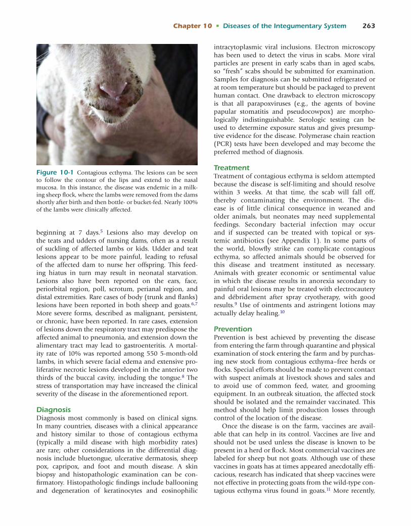
Chapter 10 • Diseases of the Integumentary System 263
beginning at 7 days.5 Lesions also may develop on the teats and udders of nursing dams, often as a result of suckling of affected lambs or kids. Udder and teat lesions appear to be more painful, leading to refusal of the affected dam to nurse her offspring. This feed-ing hiatus in turn may result in neonatal starvation. Lesions also have been reported on the ears, face, periorbital region, poll, scrotum, perianal region, and distal extremities. Rare cases of body (trunk and flanks) lesions have been reported in both sheep and goats.6,7 More severe forms, described as malignant, persistent, or chronic, have been reported. In rare cases, extension of lesions down the respiratory tract may predispose the affected animal to pneumonia, and extension down the alimentary tract may lead to gastroenteritis. A mortal-ity rate of 10% was reported among 550 5-month-old lambs, in which severe facial edema and extensive pro-liferative necrotic lesions developed in the anterior two thirds of the buccal cavity, including the tongue.8 The stress of transportation may have increased the clinical severity of the disease in the aforementioned report.
DiagnosisDiagnosis most commonly is based on clinical signs. In many countries, diseases with a clinical appearance and history similar to those of contagious ecthyma (typically a mild disease with high morbidity rates) are rare; other considerations in the differential diag-nosis include bluetongue, ulcerative dermatosis, sheep pox, capripox, and foot and mouth disease. A skin biopsy and histopathologic examination can be con-firmatory. Histopathologic findings include ballooning and degeneration of keratinocytes and eosinophilic
intracytoplasmic viral inclusions. Electron microscopy has been used to detect the virus in scabs. More viral particles are present in early scabs than in aged scabs, so “fresh” scabs should be submitted for examination. Samples for diagnosis can be submitted refrigerated or at room temperature but should be packaged to prevent human contact. One drawback to electron microscopy is that all parapoxviruses (e.g., the agents of bovine papular stomatitis and pseudocowpox) are morpho-logically indistinguishable. Serologic testing can be used to determine exposure status and gives presump-tive evidence for the disease. Polymerase chain reaction (PCR) tests have been developed and may become the preferred method of diagnosis.
TreatmentTreatment of contagious ecthyma is seldom attempted because the disease is self-limiting and should resolve within 3 weeks. At that time, the scab will fall off, thereby contaminating the environment. The dis-ease is of little clinical consequence in weaned and older animals, but neonates may need supplemental feedings. Secondary bacterial infection may occur and if suspected can be treated with topical or sys-temic antibiotics (see Appendix 1). In some parts of the world, blowfly strike can complicate contagious ecthyma, so affected animals should be observed for this disease and treatment instituted as necessary. Animals with greater economic or sentimental value in which the disease results in anorexia secondary to painful oral lesions may be treated with electrocautery and débridement after spray cryotherapy, with good results.9 Use of ointments and astringent lotions may actually delay healing.10
PreventionPrevention is best achieved by preventing the disease from entering the farm through quarantine and physical examination of stock entering the farm and by purchas-ing new stock from contagious ecthyma–free herds or flocks. Special efforts should be made to prevent contact with suspect animals at livestock shows and sales and to avoid use of common feed, water, and grooming equipment. In an outbreak situation, the affected stock should be isolated and the remainder vaccinated. This method should help limit production losses through control of the location of the disease.
Once the disease is on the farm, vaccines are avail-able that can help in its control. Vaccines are live and should not be used unless the disease is known to be present in a herd or flock. Most commercial vaccines are labeled for sheep but not goats. Although use of these vaccines in goats has at times appeared anecdotally effi-cacious, research has indicated that sheep vaccines were not effective in protecting goats from the wild-type con-tagious ecthyma virus found in goats.11 More recently,
Figure 10-1 Contagious ecthyma. The lesions can be seen to follow the contour of the lips and extend to the nasal mucosa. In this instance, the disease was endemic in a milk-ing sheep flock, where the lambs were removed from the dams shortly after birth and then bottle- or bucket-fed. Nearly 100% of the lambs were clinically affected.

264 Chapter 10 • Diseases of the Integumentary System
a goat strain vaccine for contagious ecthyma was found to be protective against experimental challenge.12
Vaccines typically are placed on scarified skin of the medial thigh. Other sites should be used, such as inside the ear pinna or under the tail, for vaccinating lactating females. Formation of scabs should occur by 3 to 4 days if the vaccination is successful. If vaccination is used, its use should be tailored to optimal management for a particular herd or flock. One approach to vaccine use is to begin with vaccination of all animals that have not been previously exposed and then vaccinate only new naive stock (from new births and new additions) annu-ally. Immunity is reported to be acquired by 3 weeks after vaccination but is not considered to be lifelong.13 Naturally exposed animals that have recovered usually are solidly immune for 2 to 3 years.10 It has been stated that colostrum immunity does not occur, because anti-bodies do not appear to be passed in the colostrum.1,10 A study by Perez, however, demonstrated that lesions did not develop in kids of vaccinated does when chal-lenged before 45 days of age, whereas lesions did appear in kids older than 45 days of age.14 On some farms in which the disease is endemic, producers may choose to simply live with the disease. The disease has a shorter course and is less severe in reinfected animals.
Malignant Contagious EcthymaA persistent form of contagious ecthyma known as malignant contagious ecthyma has been recognized in a limited number of sheep within infected flocks. Pro-liferative lesions develop, especially on the distal legs and feet and, less commonly, on the head. Unlike with ordinary contagious ecthyma, however, the lesions fail to regress and may continually enlarge. Second-ary bacterial infections, fly strike, and hemorrhage are major complications. Although a poxvirus morphologi-cally similar to the contagious ecthyma virus has been identified by electron microscopy in typical lesions, the disease has a different course.
Affected sheep do not pass the infection to com-mingling animals. Preliminary studies of the cellular immune system in affected sheep have failed to demon-strate any deviation from normal.
Ulcerative DermatosisUlcerative dermatosis is a disease of sheep caused by a virus similar to but distinct from the contagious ecthyma parapoxvirus. Infection typically follows a break in the skin such as from shearing injuries and breeding-related injuries. The virus may be spread through physical con-tact during breeding season, perhaps accounting for the highest prevalence of disease in the fall and winter. The incubation period is 2 to 7 days. Healing of lesions occurs in 2 to 6 weeks. In the United States, the disease
is most common in western regions, and the morbidity rate usually is 15% to 20% but may be as high as 60%.
Clinical SignsLesions develop as ulcers, with a thin but very adherent scab. Lesions associated with the initial viral infection may become infected with Fusobacterium necrophorum. Lesions may occur on the face, eyes, lips, and nostrils but also occur in the interdigital space, legs, penis, and vulva. Lesions of the lower limb may lead to septic arthritis. Lesions of the penis may result in phimosis or paraphimosis. The facial and lip lesions are not typi-cally associated with the mucous membranes—a dis-tinction that helps differentiate them from contagious ecthyma lesions.
DiagnosisThe most important clinical entity to rule out is conta-gious ecthyma, but with ulcerative dermatosis, the mor-bidity is lower and the lesions are ulcerative rather than proliferative as in contagious ecthyma. Confirmation is based on histopathologic examination of material from biopsied lesions.
TreatmentThe disease is self-limiting, but use of antibiotics is justi-fied for secondary bacterial infections and severe cases. Otherwise, treatment is symptomatic.
PreventionPrevention is best carried out by examination of breed-ing stock during the breeding season and isolation of affected animals. When the disease is a common occur-rence, efforts should be made to decrease skin trauma by means of reduction in shearing injuries, removing ani-mals from areas that are abrasive to the feet and legs, and using feeds that are nonabrasive. No vaccine is available, and immunity is short-lived (approximately 5 months).
Sheep Pox and Goat PoxThe agents of sheep pox and goat pox are closely related viruses of the Capripox genus in the family Poxviridae. Although the viruses tend to be species-specific, cross-species infection has been reported. Mortality is low in endemic regions but may be high when naive sheep or goats are exposed. Transmission (thought to occur by aerosol and contact with lesions) increases with close contact with infected herds or flocks. These diseases cur-rently are endemic in northern Africa, the Middle East, and southeastern Asia, with occasional outbreaks in southeastern Europe. One case of goat pox in the United States has been reported,16 but a U.S. Department of Agriculture–Animal and Plant Health Inspection Service (USDA-APHIS) website states that neither disease has occurred in the United States. Contagious ecthyma is the

Chapter 10 • Diseases of the Integumentary System 265
primary consideration in the differential diagnosis for sheep pox and goat pox. However, sheep and goat pox lesions tend to occur over the entire skin surface. The two viral agents (of contagious ecthyma and of sheep or goat pox) are easily differentiated by electron micros-copy. A recent review of sheep pox has been published.17
ScrapieA discussion of scrapie is beyond the scope of this chapter (see Chapter 13). As a consequence of the intense associ-ated pruritus, however, scrapie-infected sheep or goats (although pruritus is much less common in goats than in sheep) may present with hair or wool loss secondary to mechanical excoriation. Scrapie does not directly affect the skin; skin lesions are simply a result of the intense pruritus and subsequent aggressive scratching.
BluetongueA discussion of bluetongue is beyond the scope of this chapter (see Chapters 4 and 14). However, skin lesions suggestive of bluetongue include coronitis, ulcerations of the oral mucosa, and muzzle edema. Goats are rela-tively resistant to clinical bluetongue.
Vesicular StomatitisVesicular stomatitis is a viral disease that creates ulcer-ations of the oral mucosa in cattle, swine, and horses. Although both goats and sheep may be experimentally infected with the causative virus, natural clinical disease is quite rare, and no case reports documenting clinical disease are found in the literature.
Sheep and goats never show clinical signs of vesicu-lar stomatitis.18 A 1995 outbreak of the disease in the western United States did not identify a single sheep or goat seropositive for the virus.18 Unpublished reports of vesicular stomatitis in goats, however, indicated that vesicles may occur at the commissures of the lips.19
REFERENCES 1. Scott DW: Large animal dermatology, Philadelphia, 1988, WB
Saunders. 2. Romero-Mercado CH, et al: Virus particles and antigens in experi-
mental orf scabs, Arch Gesamte Virusforsch 40:152, 1973. 3. Nettleton PF, et al: Natural transmission of orf virus from clini-
cally normal ewes to orf-naive sheep, Vet Rec 139:364, 1996. 4. de la Concha-Bermejillo A: Orf/contagious ecthyma. In Haskell
SRR, editor: Blackwell’s Five-minute veterinary consultant, Singapore, 2008, Wiley-Blackwell.
5. McElroy MC, Bassett HF: The development of oral lesions in lambs naturally infected with orf virus, Vet J 174:663, 2007.
6. Coates JW, Hoff S: Contagious ecthyma: an unusual distribution of lesions in goats, Can Vet J 31:209, 1990.
7. Sargison ND, Scott PR, Rhind SM: Unusual outbreak of orf affect-ing the body of sheep associated with plunge dipping, Vet Rec 160:372, 2007.
8. Gumbrell RC, McGregor DA: Outbreak of severe fatal orf in lambs, Vet Rec 141:150, 1997.
9. Meynink SE, Jackson PGG, Platt D: Treatment of intraoral orf lesions in lambs using diathermy and cryosurgery, Vet Rec 121:594, 1987.
10. Blood DC, Radostits OM, Henderson JA, et al: Veterinary medicine, London, 1983, Baillière Tindall.
11. de la Concha-Bermejillo A, Ermel RW, Zhang MZ: Contagious ecthyma (ORF) virulence factors and vaccine failure, Proceedings of the One Hundred and Third Annual Meeting of the United States Animal Health Association, San Diego, Calif, 1999.
12. Musser JMB, et al: Development of a contagious ecthyma vaccine for goats, Am J Vet Res 69:1366, 2008.
13. Mullowney PC: Skin diseases of sheep, Vet Clin North Am Large Anim Pract 6 :131, 1984.
14. Perez JLT: Kids immunity to contagious ecthyma (orf virus), presented at the Goat Diseases and Production 2nd International Collo-quium, Niort, France, June 26 to 29, 1989 (Abstract p 24).
15. Renshaw HW, Dodd AG: Serologic and cross-immunity studies with contagious ecthyma and goat pox virus isolates from the western United States, Arch Virol 56:201, 1978.
16. Animal Health Monitoring and Surveillance: Status of report-able diseases in the United States, USDA-APHIS (website): http://www.aphis.usda.gov/vs/nahss/disease_status.htm#sheep, Accessed November 12, 2008.
17. Bhanuprakash V, et al: The current status of sheep pox disease, Comp Immunol Microbiol Infect Dis 29:27, 2006.
18. Kitching P: Notifiable viral diseases and spongiform encepha-lopathies of cattle, sheep and goats, In Practice 19:51, 1997.
19. Bridges VE, et al: Review of the 1995 vesicular stomatitis outbreak in the western United States, J Am Vet Med Assoc 211:556, 1997.
20. Smith MC, Sherman DM: Skin, Goat medicine, ed 2, Ames, Iowa, 2009, Wiley-Blackwell.
BACTERIAL DISEASESDermatophilosis (Streptothricosis, Lumpy Wool Disease, Rain Scald, Rain Rot)Dermatophilosis (streptothricosis, lumpy wool disease, rain scald, rain rot) is a disease of all ruminants caused by the gram-positive filamentous bacterium Derma-tophilus congolensis. This bacterium appears to be main-tained within herds or flocks by carrier animals. The organism is considered an obligate parasite of ruminant skin and was not thought to survive for very long in
the soil, but later research indicates that it may survive for several months, especially within cast-off crusts.1,2 Predisposing factors for clinical disease include skin damage (as from biting insects or physical abrasion), excessive moisture (hence the common name “rain rot”), and concurrent diseases and stresses that compro-mise the host immune system. The loss of the sebaceous film layer on skin is thought to predispose the animal to development of the disease. Excessively rainy condi-tions without appropriate shelter can lead to dilution of this sebaceous layer, thereby increasing the chance of clinical disease.

266 Chapter 10 • Diseases of the Integumentary System
The incubation period averages 2 weeks. The infec-tive form of the organism is the motile zoospore (Figure 10-2), which germinates, penetrates the epi-dermis, and invades hair or wool follicles. Neutrophils migrate to the affected areas, resulting in accumulation of a serous exudate that seeps to the epidermal surface. The older epidermal skin deteriorates while a new layer of epidermis forms below. This new layer also becomes infected with hyphal branches. Eventually, thick scabs are formed. Early clinical manifestations include small, raised, and circumscribed crusts of epidermal cells and serous exudates with embedded hairs or wool. The dis-ease follows a similar pattern in sheep, but the serous exudates may not be adherent to the epidermis. It also is responsible for “strawberry footrot” of sheep, which appears as dry scabs on the lower legs. Removal of the dry scabs leaves a mass of granulation tissue that has the appearance of a strawberry (hence the name). Although it can be spread from acutely infected animals, out-breaks are rare but have been reported.3 Young goats appear to be more susceptible than adults to develop-ment of clinical disease.3,4 Likewise, young sheep are more susceptible than adult sheep.5
Clinical SignsFollicular and nonfollicular papules and pustules develop and rapidly coalesce and rupture, with consequent mat-ting of groups of hairs or clumps of wool. These are classically described as “paintbrush lesions” in haired ruminants. Lesions may be painful but are not pruritic. In sheep, crusts occurring at the coronary band (i.e., strawberry footrot) also may extend to the carpi or tarsi. Lesions also may be present on other parts of the body. In goat kids, lesions tend to be on the ears and tails; in adults, lesions tend to be on the muzzle, in the dor-sal midline, or on the scrotum or distal legs.4 Lesions have been reported in the ears of kids at 5 days of age.6 Although the condition is relatively rare, it may be fatal in livestock debilitated by other diseases and poor nutrition.
DiagnosisStaining (Gram stain or methylene blue) of direct smears of lesions should reveal branching hyphae with cuboidal packets of coccoid cells arranged in parallel rows (resembling railroad tracks) within the filaments (see Figure 10-2). Skin biopsy and histopathologic examination also may be helpful. Culture can be con-firmatory, although possible subsequent infestation by other bacterial and fungal organisms may complicate the diagnosis in chronic cases.7
TreatmentAgents for topical treatment include iodophors, 2% to 5% lime sulfur, 0.2% copper sulfate, 0.5% zinc sulfate, and 1% potassium aluminum sulfate. These agents may be applied as total body washes, sprays,
or dips for 3 to 5 consecutive days and then weekly until healing has occurred. Systemic antibiotics such as procaine penicillin G (5000-10000 IU/kg twice a day for 4 to 5 days), oxytetracycline (one or two doses of 20 mg/kg during a 72-hour interval), or ceftiofur (2 ml/100 lb once daily for 4 to 5 days) may be effec-tive. The organism is reported to be resistant to poly-myxin B, bacitracin, and sulfonamides. Lesions in kids tend to heal without treatment within 2 to 3 months.8 Dermatophilus is potentially zoonotic to humans.
PreventionIn areas in which the disease is prevalent, removal and disposal of crusts, keeping stock dry (providing shelter from wet weather), providing good-quality nutrition, and control of ectoparasites may limit the number of clinical cases. Vaccines have been studied but do not appear to offer significant protection.5
Fleece Rot (Water Rot, Weather Stain)Fleece rot is an exudative bacterial dermatitis of sheep that is characterized by a greenish-discolored, matted wool. This disease reduces the quality of the wool but is most important as a predisposing factor for fly strike. Although other fleece bacteria may play a significant role in the disease, Pseudomonas aeruginosa is consid-ered the primary etiologic agent.9 The disease was first recognized in Australia in the latter part of the 1800s.5 Bacteria cultured from the skin of affected sheep, when applied to unaffected sheep, resulted in the disease.10
The necessity of moisture for development of dis-ease symptoms was noted in 1929 by Seddon and McGrath.11 Both wool traits and body conformation
10.0 �m
Figure 10-2 Dermatophilus congolensis showing zoospores. A diagnostic slide can be easily and reliably made in field con-ditions by pressing the slide against the underside of the scab, allowing the slide to air dry, and then staining with Wright’s stain. (Courtesy Dr. Mike Fry. University of Tn, Knoxville, Tn)

Chapter 10 • Diseases of the Integumentary System 267
traits are predisposing factors for fleece rot in sheep, and this susceptibility is reportedly heritable.11 The disease appears to be most prevalent in Australia, with reported prevalence rates averaging 24%.5 The disease does not appear to be of clinical or economic significance in the United States but has been reported.12 The reason for the increased susceptibility of young sheep com-pared with older stock is not known, but causative or contributing factors may include maturity of wool and skin characteristics and development of a certain level of immunity after exposure.5 Fleece rot predisposes affected sheep to blowfly strike.
Clinical SignsThe most characteristic clinical sign is the greenish discoloration of wool. A copious serous exudate also may be evident and probably is the attraction for flying insects that leads to associated fly strike. The inflamma-tory reaction can result in a grayish matted wool. Pru-ritus also has been reported, but not all affected sheep demonstrate evidence of this symptom.13 Lesions are most common on the back and withers.
DiagnosisCulture of samples from involved skin and wool is the definitive method to diagnose fleece rot (and would be especially definitive for greenish-discolored wool). Pseudomonas aeruginosa may be found in pure culture. It produces the green pigment pyocyanin.14 Fleece rot may be distinguished from dermatophilosis in that no scab formation is associated with the former.
TreatmentAntibiotics may be helpful, but studies have shown P. aeruginosa to be quite resistant to many antibiotics.15,16 In clinical practice, shearing to allow the involved skin to dry is the most effective means of treatment.
PreventionVaccines are under development, but efficacy to date has been disappointing.5 Some prevention is afforded by producing more resistant sheep by breeding for characteristics of wool and body conformation that are less predisposing to the condition. Perhaps the most practicable control measure is to shear the sheep before the onset of the rainy season.
Malignant Edema (Swelled Head, Bighead)Malignant edema, also called “swelled head” or “big-head,” is a rapidly fatal disease caused by clostridial spe-cies, most commonly Clostridium sordellii, Clostridium novyi, Clostridium septicum, and Clostridium chauvoei, and is seen most commonly in young rams. Although swelled head of bucks (goats) is mentioned in many
textbooks, studies or case reports in goats ar lacking. The organisms typically exist as spores in the soil and appear to occur predominantly in moist soils that are rich in organic matter.17 The organisms usually enter the body through breaks in the skin or mucosa. With development of the requisite anaerobic conditions in body tissues, the organisms proliferate and release sev-eral exotoxins that react locally and systemically. The spores of clostridial organisms are thought to survive in the environment for several years. The disease is most common in Montana in the United States but also occurs in South Africa, South America, and Australia.18
Clinical SignsThe disease usually is sporadic in nature but can occur as outbreaks. The likely cause of a Brazilian outbreak was use of a single common needle to vaccinate a 1000-head flock of sheep with a commercial clostridial vac-cine.17 Several sheep died in that outbreak, all within 1 to 3 days of vaccination. Clinical signs observed before death included severe depression, swelling around the vaccination site, lameness, subcutaneous edema, and crepitation.
The classic epidemiology of bighead in sheep involves butting in rams leading to breaks in the skin that allow bacterial spores access to the bruised subcutaneous tis-sues. Once infection occurs, swelling and edema of the face, head, and neck appear.
DiagnosisAn aseptically collected aspirate of the subcutaneous swelling followed by staining of an impression smear and subsequent anaerobic culture can be definitive. Clostridial organisms stain as large gram-positive rods. Fluorescent antibody testing can differentiate between the different clostridial species.
TreatmentIf treatment is not initiated immediately, the fatality rate will be high. Treatment consists primarily of high doses of penicillin products, which should be administered both locally and systemically. Additional treatment is primarily symptomatic.
PreventionVaccination can be preventive so long as it is admin-istered after the period of passive transfer and before traumatic events. Hygiene also is emphasized, because some cases have been documented to occur after a simple blood draw.19
ActinobacillosisActinobacillus lignieresii, a non–spore-forming, gram-negative rod, causes a pyogranulomatous bacterial infection of the soft tissues of the head in sheep

268 Chapter 10 • Diseases of the Integumentary System
(but has not been documented in goats).20 These bac-teria usually are inoculated into the tissues by grass awns or stemmy forage. A local granulomatous reaction occurs, but these bacteria also may spread to regional lymph nodes or the bloodstream.
Clinical SignsPurulent material may be observed draining from lymph nodes. Extreme enlargement of submaxillary or parotid lymph nodes may lead to difficulty in breath-ing or eating; sheep may die from malnutrition. Nasal exudate may be noted if the infection drains into the nasopharynx.
DiagnosisThe diagnosis is made by performing cytologic evalua-tion with Gram staining of the exudate. The gram-nega-tive rods are filamentous and form sulfur granules in the pus that can be seen without the aid of a microscope.
TreatmentTherapy includes surgical drainage, antibiotics (pro-caine penicillin G, 22,000 to 66,000 units/kg of body weight subcutaneously [SC] every 24 hours for 7 days), and iodine therapy. Sodium iodide (80 mg/kg of body weight intravenously [IV]) can be administered and repeated once or twice at 7-day intervals. Alternatively, organic iodides can be added to the feed (7.5 to 15 g/head/day) for 14 to 21 days.
Staphylococcal Dermatitis (Eye Scab, Impetigo)Staphylococcal dermatitis typically is a nonfatal skin dis-ease of sheep and goats that affects predominantly the head and face or mammary gland. The causative agent is S. aureus, but occasionally other staphylococcal species are involved. The condition appears more commonly during warm seasons of the year. Transmission appears to be by social contact with clinical or nonclinical carriers.21 Spo-radic cases may occur, but outbreaks have been reported.22
Clinical SignsThe facial involvement may manifest as nonpruritic lesions above the upper eyelid that expand to include other parts of the face and, in extreme cases, the lower limbs.22 The skin lesions in one series were character-ized by alopecia, papules, crusts, erosions or ulcers, exudation, erythema, hyperpigmentation, and thicken-ing of the skin.22 Overall, the dermatitis did not appear to adversely affect the health of the affected sheep. S. aureus dermatitis of a ewe has been described as inflam-mation and hyperkeratosis of the skin of the udder and teats, with accompanying pustules.23 Staphylococcus hyicus was determined to be the causative agent in a fatal case of seborrheic dermatitis in an 18-month-old
Pygmy goat.24 The clinical signs in this case were similar to those of greasy pig disease: a generalized seborrheic dermatitis with alopecia, on a background of greasy skin with a pronounced scaly to scabby appearance.
DiagnosisConsiderations in the differential diagnosis should include ectoparasites, zinc deficiency, elaeophoriasis, and mycotic infections. Definitive diagnosis requires culture and exclusion of other possible causes. Histo-pathologic lesions have been well documented.22
TreatmentAntibiotics may be required in severe cases and may hasten resolution in any case. Oxytetracycline and enro-floxacin were minimally effective, whereas a lincomycin-spectinomycin combination was effective in one case report.22 Penicillin has been reported to be efficacious.25 However, facial staphylococcal dermatitis tends to resolve within 2 months.26 Another approach to treatment con-sists of washing the affected skin with an iodophor or chlorhexidine shampoo, followed by drying and then coating it with an antiseptic or antibiotic ointment.20
PreventionFeeding and housing in an environment that lessens the chance of facial injuries and close head to head contact should reduce the incidence of the disease. Fly control may be important in transmission reduction. Isolation of affected animals and avoidance of fomite transmission are important to limit new staphylococcal intramammary infections.
AbscessesAbscesses of the soft tissues are not uncommon in sheep and goats. Abscess formation usually begins with entry of surface bacteria through a wound in the epider-mis. Therefore Staphylococcus spp., Corynebacterium spp., Arcanobacterium pyogenes, and streptococcal bacteria are expected on culture. Noncontagious abscesses may be treated by lancing after infiltration with a local anes-thetic. The interior capsule of the abscess is debrided and flushed with a dilute iodine solution (1%). For large abscesses, roll bandages soaked in dilute iodine solu-tions may be stuffed into the capsule of the abscess, with removal of a portion of the bandage daily over the next 3 to 5 days. Systemic antimicrobial agents are not indi-cated in most cases but may be administered if numer-ous lesions or deeply seeded abscesses are present.
Caseous LymphadenitisCaseous lymphadenitis is a common, contagious, sup-purative bacterial disease of sheep and goats worldwide that most frequently infects the lymph nodes and
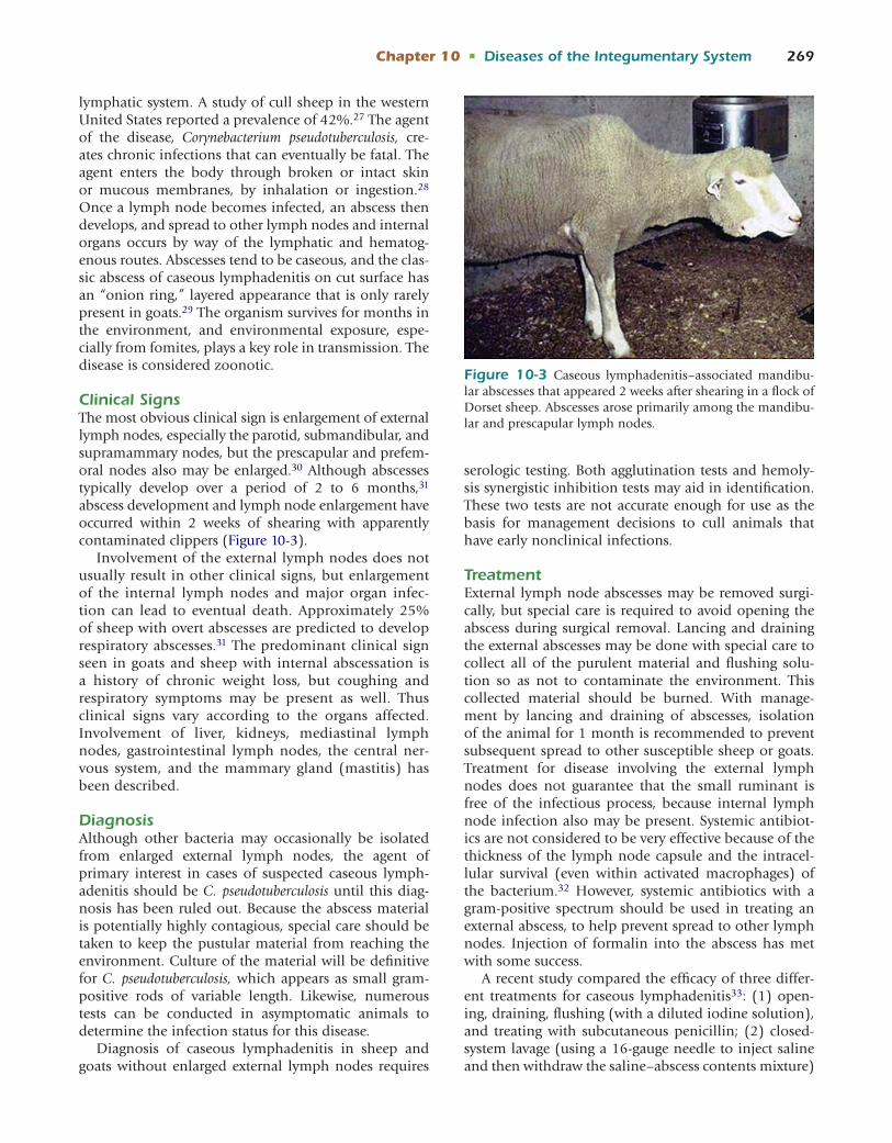
Chapter 10 • Diseases of the Integumentary System 269
lymphatic system. A study of cull sheep in the western United States reported a prevalence of 42%.27 The agent of the disease, Corynebacterium pseudotuberculosis, cre-ates chronic infections that can eventually be fatal. The agent enters the body through broken or intact skin or mucous membranes, by inhalation or ingestion.28 Once a lymph node becomes infected, an abscess then develops, and spread to other lymph nodes and internal organs occurs by way of the lymphatic and hematog-enous routes. Abscesses tend to be caseous, and the clas-sic abscess of caseous lymphadenitis on cut surface has an “onion ring,” layered appearance that is only rarely present in goats.29 The organism survives for months in the environment, and environmental exposure, espe-cially from fomites, plays a key role in transmission. The disease is considered zoonotic.
Clinical SignsThe most obvious clinical sign is enlargement of external lymph nodes, especially the parotid, submandibular, and supramammary nodes, but the prescapular and prefem-oral nodes also may be enlarged.30 Although abscesses typically develop over a period of 2 to 6 months,31 abscess development and lymph node enlargement have occurred within 2 weeks of shearing with apparently contaminated clippers (Figure 10-3).
Involvement of the external lymph nodes does not usually result in other clinical signs, but enlargement of the internal lymph nodes and major organ infec-tion can lead to eventual death. Approximately 25% of sheep with overt abscesses are predicted to develop respiratory abscesses.31 The predominant clinical sign seen in goats and sheep with internal abscessation is a history of chronic weight loss, but coughing and respiratory symptoms may be present as well. Thus clinical signs vary according to the organs affected. Involvement of liver, kidneys, mediastinal lymph nodes, gastrointestinal lymph nodes, the central ner-vous system, and the mammary gland (mastitis) has been described.
DiagnosisAlthough other bacteria may occasionally be isolated from enlarged external lymph nodes, the agent of primary interest in cases of suspected caseous lymph-adenitis should be C. pseudotuberculosis until this diag-nosis has been ruled out. Because the abscess material is potentially highly contagious, special care should be taken to keep the pustular material from reaching the environment. Culture of the material will be definitive for C. pseudotuberculosis, which appears as small gram-positive rods of variable length. Likewise, numerous tests can be conducted in asymptomatic animals to determine the infection status for this disease.
Diagnosis of caseous lymphadenitis in sheep and goats without enlarged external lymph nodes requires
serologic testing. Both agglutination tests and hemoly-sis synergistic inhibition tests may aid in identification. These two tests are not accurate enough for use as the basis for management decisions to cull animals that have early nonclinical infections.
TreatmentExternal lymph node abscesses may be removed surgi-cally, but special care is required to avoid opening the abscess during surgical removal. Lancing and draining the external abscesses may be done with special care to collect all of the purulent material and flushing solu-tion so as not to contaminate the environment. This collected material should be burned. With manage-ment by lancing and draining of abscesses, isolation of the animal for 1 month is recommended to prevent subsequent spread to other susceptible sheep or goats. Treatment for disease involving the external lymph nodes does not guarantee that the small ruminant is free of the infectious process, because internal lymph node infection also may be present. Systemic antibiot-ics are not considered to be very effective because of the thickness of the lymph node capsule and the intracel-lular survival (even within activated macrophages) of the bacterium.32 However, systemic antibiotics with a gram-positive spectrum should be used in treating an external abscess, to help prevent spread to other lymph nodes. Injection of formalin into the abscess has met with some success.
A recent study compared the efficacy of three differ-ent treatments for caseous lymphadenitis33: (1) open-ing, draining, flushing (with a diluted iodine solution), and treating with subcutaneous penicillin; (2) closed-system lavage (using a 16-gauge needle to inject saline and then withdraw the saline–abscess contents mixture)
Figure 10-3 Caseous lymphadenitis–associated mandibu-lar abscesses that appeared 2 weeks after shearing in a flock of Dorset sheep. Abscesses arose primarily among the mandibu-lar and prescapular lymph nodes.

270 Chapter 10 • Diseases of the Integumentary System
and administration of intralesional tulathromcyin; and (3) closed-system lavage and administration of subcu-taneous tulathromycin. All treatment regimens resulted in greater than 80% resolution of the treated lesions. Ultimately, however, the most cost effective treatment is to cull the affected animals.33
PreventionThe best prevention is to maintain a caseous lymph-adenitis–free herd or flock. Any new animals should be tested for the disease and examined for lymph node enlargement before entering the herd or flock. Housing should be maintained free of objects that can cause skin injury. Needles, surgical equipment, tattoo pliers, shears, hoof trimmers, and dipping vats should be cleansed and disinfected after use. In addition, the control of external parasites is considered important because pruritic stock will rub themselves on items that could produce breaks in the skin.20 Eradication of caseous lymphadenitis is possible but difficult in that it requires frequent testing and management of a disease-free group and an infected group. Vaccines are available and can be especially helpful for producers who choose to live with the disease in that vaccination can reduce the incidence of abscesses in a herd or flock.34 Vaccines do not necessarily prevent the disease. Goats vaccinated with sheep caseous lymphadenitis vaccines tend to experience adverse reactions more frequently than do sheep. Anecdotal reports indicate that goat owners are more satisfied with autogenous caseous lymphadenitis vaccines. Milk and colostrum transmission apparently is not important, but removal of neonatal lambs and kids from affected dams should lessen the chances of exposure. Likewise, intrauterine transmission has not been reported.
REFERENCES 1. Roberts DS: Release and survival of Dermatophilus dermatonomus
zoospores, Aust J Agr Res 14:386, 1963. 2. Martinez D, Prior P: Survival of Dermatophilus congolensis in tropi-
cal clay soils submitted to different water potentials, Vet Microbiol 29:135, 1991.
3. Larsen JWA: An outbreak of mycotic dermatitis in goat kids, Aust Vet J 64:160, 1987.
4. Yeruham I, Elad D, Perl S: Dermatophilus in goats in the Judean foothills, Rev Med Vet 154:785, 2003.
5. Norris BJ, Colditz IG, Dixon TJ: Fleece rot and dermatophilosis in sheep, Vet Microbiol 128:217, 2008.
6. Mullowney PC, Baldwin EW: Skin diseases of goats, Vet Clin North Am Large Anim Pract 6:143, 1984.
7. Dalis JS, Kazeem HM: Concurrent infections of a goat with Der-matophilus congolensis and Blastomyces dermatitidis, J Anim Vet Adv 6:773, 2007.
8. Munro R: Caprine dermatophilosis in Fiji, Trop Anim Health Prod 10:221, 1978.
9. Dixon TJ, Mortimer SI, Norris BJ: 16S rRNA gene microbial analy-sis of the skin of fleece rot resistant and susceptible sheep, Aust J Agr Res 58:739, 2007.
10. Stuart TPA: On green-producing chromogenic micro-organisms in wool, J Proc Roy Soc 28:320, 1894.
11. Seddon HR, McGrath TT: Green coloration in wool, Aric Gaz 40:206, 1929.
12. Torell DT, et al: Effects of time of shearing on wool and lamb production, Calif Agr 23:16, 1969.
13. El-Sukhon SN: Isolation and characterization of Pseudomonas aeruginosa from sheep with fleece rot in northern and middle Jordan, Vet Dermatol 13:247, 2002.
14. Chin JC, Watts JE: Dermal and serological response against Pseu-domonas aeruginosa in sheep bred for resistance and susceptibility to fleece-rot, Aust Vet J 68:28, 1991.
15. Bonfiglio G, Carciotto V, Russo G: Antibiotic resistance in Pseudo-monas aeruginosa, an Italian survey, J Antimicrob Agents Chemother 41:307, 1998.
16. Gerceker AA, Gurler B: in vitro activities of various antibiotics, alone and in combination with amikacin against Pseudomonas aeruginosa, J Antimicrob Agents Chemother 36:707, 1995.
17. Costa JLN, et al: Outbreak of malignant oedema in sheep caused by Clostridium sordellii, predisposed by routine vaccination, Vet Rec 160:594, 2007.
18. Mullowney PC: Skin diseases of sheep, Vet Clin North Am Large Anim Pract 6:131, 1984.
19. Morris WE, et al: Malignant oedema associated with blood-sampling in sheep, Aust Vet J 80:280, 2002.
20. Smith MC, Sherman DM: Skin, Goat medicine, ed 2, Ames, Iowa, 2009, Wiley-Blackwell.
21. Watson WA: The carriage of pathogenic staphylococci by sheep, Vet Rec 77:477, 1965.
22. Koutinas AF, et al: Clinical, histopathological and therapeutic considerations in a flock of sheep with facial staphylococcal-associated dermatitis, Vet Dermatol 18:118, 2007.
23. Mavrogianni VS, Fthenakis GC: Clinical, bacteriological, cytologi-cal and pathological features of teat disorders in ewes, J Vet Med A Physiol Pathol Clin 54:219, 2007.
24. Schamber G, Alstad AD: Isolation of Staphylococcus hyicus from seborrheic dermatitis in a pygmy goat, J Vet Diagn Invest 1:276, 1989.
25. Wilson DJ, Scott PR, Sargison ND: Effective treatment of severe facial dermatitis in lambs, Vet Rec 150:45, 2002.
26. Hardy WT, Price DA: Staphylococcal dermatitis of sheep, J Am Vet Med Assoc 119:445, 1951.
27. Stoops SG, Renshaw HW, Thilsted JP: Ovine caseous lymphad-enitis: disease prevalence, lesion distribution, and thoracic mani-festations in a population of mature culled sheep from western United States, Am J Vet Res 45:557, 1984.
28. Seddon HR: A discussion of the method of infection by bacillus of Preisz-Nocard, Aust Vet J 5:49, 1929.
29. Batey RG, Speed CM, Kobes CJ: Prevalence and distribution of caseous lymphadenitis in feral goats, Aust Vet J 63:33, 1986.
30. Maddy KT: Caseous lymphadenitis of sheep, J Am Vet Med Assoc 122:257, 1953.
31. O’Reilly KM, et al: Parameter estimation and simulations of a mathematical model of Corynebacterium pseudotuberculosis trans-mission in sheep, Prev Vet Med 83:242, 2008.
32. Holstad G, Teige J, Larsen HJ: Corynebacterium pseudotuberculosis infection in goats VIII. The effect of vaccination against experi-mental infection, Acta Vet Scand 30:275, 1989.
33. Washburn KE, et al: Comparison of three treatment regimens for sheep and goats with caseous lymphadenitis, J Am Vet Med Assoc 234:1162, 2009.
34. Pugh DG: Caseous lymphadenitis in small ruminants, Proc North Am Vet Conf 11:983, 1997.

Chapter 10 • Diseases of the Integumentary System 271
FUNGAL DISEASESDermatophytoses (Ringworm, Lumpy Wool, Club Lamb Fungus)The primary fungal agent of ringworm in sheep and goats is Trichophyton verrucosum. However, Trichophyton mentagrophytes, Microsporum canis, and other, less com-mon species have been reported to cause ringworm in these species.1,2 Transmission occurs by direct contact with clinically affected animals or indirectly from contaminated fomites (e.g., fences, water or feed equip-ment, grooming equipment). The disease tends to be more common in young animals than in adults, prob-ably because of increased immunity in those previously exposed to the disease agent. Other potential predispos-ing factors include immunosuppression, poor nutri-tion, crowding, high humidity, and other debilitating conditions or diseases.
The zoophilic dermatophytes exist as spores in the environment. The dermatophytes use keratin as a nutri-ent source—hence their predilection for skin.3 Spores typically enter the skin through abrasions. The conidium (the spores) germinates and hyphae appear within the stratum corneum (invasion of living tissue does not occur) and invade the walls of the hair follicles. They then emerge into the follicular canal and grow down-ward between the hair cuticle and the wall of the fol-licle. The hyphal tip penetrates into the hair cortex by dissolving the keratin and by mechanical pressure, and multiplication ensues, with conidia located outside of the hair shaft.3 As the hair grows, the fungal elements are carried out of and above the surface of the skin, where hairs may become broken or fall out. Spread occurs centrifugally from the point of invasion, resulting in the classic ring-shaped lesion. Incubation period from expo-sure to clinical disease is 1 to 6 weeks, and the condition is more commonly seen in the fall and winter months.
Lanolin tends to protect the sheep skin from ring-worm agent invasion. Show lambs are sheared short, and the frequent washing to prepare them for showing tends to remove the protective lanolin, allowing the ringworm agents better access to infect the skin. Thus this disease would be unusual in sheep that are not being prepared for show or sale. Club lamb fungus was first reported in 1989. Although typically considered a disease of show lambs associated with extensive washing and close shearing, herd outbreaks have occurred in com-mercial flocks, in association with recent shearing with possibly contaminated blades.4,5 The disease is conta-gious, which is why sheep with evidence of ringworm are not allowed to participate in fair activities. Lesions take approximately 1 month to become evident. Fungal spores remain viable for years under natural condi-tions. Persons treating or working with the flock or herd should wear protective clothing and gloves, because the disease is zoonotic.
Clinical SignsIn both goats and sheep, lesions affect primarily the ears, head, and neck, but any body surface may be involved (Figure 10-4). Ringworm lesions affecting wooled surfaces were previously considered to be rare, but according to more recent evidence, show lamb fungus probably is as common on the wooled areas as in the more typical locations. Lesions in haired areas consist of circular patches of alopecia, scaling, and crusts, but affected areas also may be uneven and dif-fuse. Lesions in wooled areas may be covered with mat-ted wool and are inflamed and reddened underneath the mat5 (Figure 10-5). Lesions tend to be nonpruritic or only mildly pruritic and usually are not painful. Although lesions typically occur on the face, ears, and neck and shoulder area, they also may be dispersed over the body, including the tailhead region.5
DiagnosisAlthough in most instances the diagnosis is based on the clinical presentation, this assumption may be faulty, because numerous other conditions can have a similar presentation. Considerations in the differential diag-nosis for ringworm in sheep and goats should include external parasites, zinc deficiency, dermatophilosis, staphylococcal dermatitis, and immune-mediated dis-eases. Use of the Wood’s lamp during clinical examina-tion may help with the differential diagnosis. However, Trichophyton species do not fluoresce. Ultimately, iden-tification of the pathogen from fungal culture on Sab-ouraud’s dextrose agar is definitive. Scrapings should be obtained from the periphery of the lesion, because these strictly aerobic fungi die out under the crust in the center of most lesions. The combination of exudate from inflamed epithelial layers, epithelial debris, and fungal hyphae produces the dry crusts that create a more anaerobic environment. Microscopic examination of hairs and keratin from the periphery of an active lesion may reveal ectothrix invasion of hair shafts.6 A 20% potassium hydroxide solution can be used to prepare wet mounts of arthrospores on the hair shafts for micro-scopic examination.
TreatmentThe first step in dealing with a case of ringworm is isolation of affected animals. Care must be taken to eliminate spread of the fungus on the hands of farm personnel or other fomites to herdmates. The disease is self-limiting and usually does not adversely affect the health of the affected small ruminant. Healing takes 4 to 16 weeks with spontaneous recovery. Some researchers have reported healing in as little as 2 weeks, but this rapid clinical course has not been scientifically validated. Many treatments have been suggested, but few have been studied to determine actual efficacy. Most such treatments constitute extralabel use, and

272 Chapter 10 • Diseases of the Integumentary System
meat withdrawal times are rarely known. The following treatment regimens should not be considered definitive but represent options when clients want their stock treated. • Griseofulvin has been suggested for treatment of
infections that are widespread on the body or have become chronic. This antifungal agent becomes incorporated into keratin in the skin and hair, and treated animals remain resistant for a number of weeks after receiving the drug.7 Griseofulvin can be expensive, and slaughter withdrawal time is not specified. One report indicated that lesions regressed soon after treatment with griseofulvin, 7.5 mg/kg once daily for 7 days in feed, with almost complete resolution 20 days later.
• Natamycin has been used with some success but did not cure severe lesions, especially those in wooled areas of the skin.5
• The mouthwash product Listerine has been sug-gested for spot treatment (scrubbed into the lesion) with a brush once a day for 7 days.
• A preparation of 7% iodine mixed with the topical emollient Bag Balm may be applied to lesions once a day for 7 days.
• Use of 10% to 20% sodium iodide given by intrave-nous injection at 1 g/14 kg at weekly intervals has been reported to be effective.1
To stop an outbreak, all exposed animals should be treated. For application of sprays or ointments, treatment should concentrate on the margins of the lesions as the area of the most active growth of the der-matophyte and also should extend beyond the lesion, because dermatophytes may be isolated from normal-appearing skin up to 6 cm beyond.8 These can be
treated with 3% captan or 2% to 5% lime sulfur applied topically daily for 5 days and then weekly for another 3 to 4 weeks. All exposed animals, their environment, and fomites should be treated or, when practical, prop-erly disposed of. Five percent lime sulfur, 5% sodium hypochlorite, 5% formalin, 3% captan, and 3% cresol all have been suggested for environmental treatment.1 Chlorhexidine 0.5% is reportedly very effective for show lamb fungus but is inactivated by soap.4 Exposure to sunlight and treatment with vitamins A and D may has-ten healing of lesions.4,9 As noted by Scott, however, evi-dence is lacking for any benefit of vitamin and mineral preparations in most cases.1 Ultraviolet light has proved helpful in treating ringworm, so exposure to sunlight may be beneficial.3 If animals do not recover within 4 months, the presence of significant immunosuppres-sive or predisposing environmental factors is likely; in such instances, appropriate investigation and interven-tion are required.1
PreventionGeneral preventive measures for the herd or flock include good nutrition, proper health care, and provi-sion of a clean, dry, sunny environment. Vaccines are available in some countries. The disease is zoonotic, so wearing protective gloves is strongly advised. The spores may exist for several years—hence the adage “once on the farm, always on the farm.” Use of antifungal disin-fectants on exposed equipment and housing is highly recommended. Various means are used to determine the infection status of show stock at fairs. By Nebraska regulations, the disease is considered “inactive” if “the affected area is not encrusted and hair/wool has begun growth in the area.”10
Figure 10-4 Typical clinical presentation of show lamb fungus. Lesions may appear on any haired or wooled body surface. (Courtesy Dr. Fred Hopkins, Knoxville, Tennessee.)
Figure 10-5 The first sign of club lamb fungus (ringworm) often is a raised area on which the wool is clumped and feels stiff. The affected area may be covered by a gray-white scab, as seen in this photograph of a closely shorn show lamb. (Cour-tesy Dr. Fred Hopkins, Knoxville, Tennessee.)

Chapter 10 • Diseases of the Integumentary System 273
MycetomaMycetomas are painless granulomatous infections of the skin, subcutaneous tissues, and bones characterized by formation of sinuses through which fungal colonies are discharged in the form of grains.9 Mycetomas also may be formed by bacterial elements or both fungal and bacterial elements.6 Lesions most often occur on the limbs and are slow-growing; they may initially arise at the site of a wound. These lesions cause focal swelling and produce an exudate that contains granules com-posed of microbial organisms coated with host immune elements (e.g., immunoglobulins, fibrin).11 These gran-ules may be red, yellow, or purple. Actinomadura madu-rae and Actinomadura pelletierii have been found in goats with mycetoma, as has Nocardia brasiliensis. Although success rates are unknown, treatment strategies include the use of antimicrobial drugs, surgical excision, and limb amputation, depending on the extent of involve-ment. Mycetomas have not been reported in sheep.
CandidiasisYeast or Candida dermatitis has been diagnosed in goats.11,12 Candida albicans, Candida tropicalis, Candida pseudotropicalis, Candida stellatoidea, Candida parapsilosis, Candida krusei, Candida parakrusei, Candida stellatoidea, Candida guilliermondii, and other yeasts may be iso-lated from lesions. If yeast dermatitis is diagnosed, a compromised immune system or malnutrition must be suspected. Chronic moist conditions resulting in macer-ation of the skin allow the yeast to become established.
Clinical SignsClinical signs include alopecia, scales, crusting, a greasy feel to the skin, and lichenification.
DiagnosisDiagnosis is made by observation of budding yeasts and pseudohyphae on cytologic skin preparations.
Other Fungal ConditionsSeveral other fungi have been isolated from chronic dermatopathy in goats. Peyronella glomerata has been associated with hyperkeratotic lesions of the ears of goats in the United Kingdom.11 Aspergillus spp. can cause clinical disease in animals with compromised immune systems. These fungi also may cause granu-lomatous lesions in the skin. Two cases of Malassezia dermatitis in goats have been reported.13,14 Clinical signs consisted of a seborrhoeic dermatosis and exten-sive alopecia over much of the thorax and abdomen,
with extension to the neck and legs in one case. Pruritus was not evident. Both cases were chronic at presentation (1 month and 5 months), and both of the affected animals had demonstrated weight loss. Findings in the first case included erythema, hyperpig-mentation, mild lichenification, large scales, follicular casts, and a coat that was dull and easily epilated.13 Malassezia pachydermatis was identified by culture of impression smears on Sabouraud’s dextrose agar. Treatment, which consisted of weekly baths using a chlorhexidine-containing shampoo followed by appli-cation of a 0.2% solution of enilconazole for 4 weeks, resulted in complete resolution of the infection.13 In the second case, the animal had to be euthanized, because previous treatment regimens (ivermectin and amitraz) were ineffective and the goat’s condition was poor.14 Findings in this case included diffuse alopecia and thickened skin covered by dense crusts. Malassezia organisms were not recovered from culture media. The identification of Malassezia slooffiae was based on visu-alization of large numbers of yeast and hyphal forms consistent with the genus Malassezia and phylogenetic analyses using PCR.14
REFERENCES 1. Scott DW: Large animal dermatology, Philadelphia, 1988, WB
Saunders. 2. Sharp MW, Lupson GR, Flamank M: Microsporum canis infection
in sheep, Vet Rec 132:388, 1993. 3. Gillespie JH, Timoney JF: Hagan and Bruner’s Infectious diseases of
domestic animals, London, 1981, Cornell University Press. 4. Hopkins FM, Gill W: Ringworm (club lamb fungus) in sheep,
University of Tennessee Institute of Agriculture (website), http://animalscience.ag.utk.edu/sheep/pdf/Ringworm.pdf. Accessed April 17, 2007.
5. Sargison ND, et al: Ringworm caused by Trichophyton verrucosum: an emerging problem in sheep flocks, Vet Rec 150:755, 2002.
6. Smith MC, Sherman DM: Skin, Goat medicine, ed 2, Ames, Iowa, 2009, Wiley-Blackwell.
7. Hiddleston WA: The treatment of bovine ringworm, Vet Rec 92:123, 1973.
8. Knudsen EA: The areal extent of dermatophyte infection, Br J Dermatol 92:413, 1975.
9. Mullowney PC, Baldwin EW: Skin diseases of goats, Vet Clin North Am Large Anim Pract 6:143, 1984.
10. Grotelueschen DM, Sahara RJ: Club lamb fungus disease, Neb-Guide (website), Cooperative Extension, University of Nebraska–Lincoln G92-1075-A, 1992, http://digitalcommons.unl.edu/cgi/viewcontent.cgi?article=1205&context=extensionhist. Accessed October 20, 2008.
11. Scott DW, Smith MC, Manning TO: Caprine dermatology. Part I. Normal skin and bacterial and fungal disorders, Comp Cont Educ Pract Vet 6:S190, 1984.
12. Smith MC: Dermatologic diseases of goats, Vet Clin North Am Large Anim Pract 5:449, 1983.
13. Pin D: Case report: seborrhoeic dermatitis in a goat due to Malas-sezia pachydermatis, Vet Dermatol 15:53, 2004.
14. Uzal FA, et al: Malassezia slooffiae-associated dermatitis in a goat, Vet Dermatol 18:348, 2007.

274 Chapter 10 • Diseases of the Integumentary System
PARASITIC DISEASESIn this section, parasitic diseases are discussed with respect to their importance in causing lesions in the skin and hair.
Lice (Pediculosis)Lice infestation tends to be more common in goats than in sheep, at least in the United States. The litera-ture contains only a few reports of lice infestation of sheep in the United States.1,2 Most reports of sheep lice come from New Zealand, Australia, and Great Britain. On the basis of data from a telephone ques-tionnaire survey, Australian investigators reported that 21% of sheep flocks had lice infestations.3 In a postal survey of Great Britain sheep producers, 10.7% of farmers reported at least one outbreak of infesta-tions by lice in the previous year, but some regions reported up to a 19% prevalence.4 Lice tend to be a greater problem in the winter, when nutrition may be poor and conditions more crowded, and long hair or wool provides a more conducive environ-ment for louse reproduction. However, a Minnesota report of experimentally infested sheep, which were housed out of sunlight and rainfall, noted that louse numbers peaked in late spring.5 Louse activity has been shown to decline significantly in response to higher environmental temperatures. During warm to hot months, only small populations of lice sur-vive, and they typically are found in temperature-stable areas such as inside the ears and between the legs.6
Lice are highly host-specific and spend the complete life cycle on the host. It is not unusual, however, for some species of lice to infest both sheep and goats. Lice reported in goats include the sucking lice Linog-nathus africanus and Linognathus stenopsis and the chewing or biting lice Bovicola (formerly Damalinia) limbata, Bovicola caprae, and Bovicola crassipes. Lice reported in sheep include the chewing louse Bovicola ovis and the sucking lice Linognathus ovillus (body louse) and Linognathus pedalis (sucking foot louse). The biting lice B. ovis and B. caprae may be transferred between sheep and goats.7 Biting or chewing lice feed on epithelial and cutaneous debris, whereas sucking lice feed on blood and tissue fluid. Although lice can survive off the host for a few weeks, most transmission occurs through direct contact or indirectly through contact with equipment or grooming tools. Louse eggs (nits), which may be seen attached to individual hair or wool fibers, hatch within 1 to 2 weeks and develop into adults in 2 to 4 weeks. The foot louse usually is found in circumscribed areas on the feet or limbs but also may be found on abdominal or scrotal areas when large populations develop.8
Clinical SignsLice infestation in goats may manifest as pruritus with rubbing or scratching, weight loss, decreased produc-tion efficiency, and patches of alopecia that give the appearance of a rough, shaggy hair coat (Figures 10-6 and 10-7). In addition to these general signs, seen with most lice infestations, animals with sucking lice infesta-tions may present with anemia and hypoproteinemia, with death in some cases. Heavy infestations probably make affected animals more susceptible to other dis-eases. In sheep, fleece derangement (seen as rubbed or chewed fleece) was found to be a good early indicator of the presence of lice, and pruritus may be evident well before lice can be readily found by direct inspection.9 In the aforementioned study, in sheep experimentally infested with lice, fleece derangement was first evident at 5 weeks after infestation. Lameness may be observed in sheep infested with L. pedalis. The doe pictured in
Figure 10-6 This doe, presented in midsummer in Virginia, was infested with chewing lice. Subcutaneous treatment with ivermectin resulted in dead lice 3 days later.
Figure 10-7 Lice on goats may be visualized more easily by parting the hairs.
Chapter 10 • Diseases of the Integumentary System 275
Figure 10-6, presented in midsummer in Virginia, was infested with chewing lice.
DiagnosisSucking lice are most common around the poll, nose, and eyes; on the neck, brisket, withers, and tail; and in the axillary and inguinal areas. Biting lice are most common in areas of the neck, withers, and tailhead. L. pedalis typically can be found on short-haired skin, especially that of the lower leg and foot. Lice usually are visible to the naked eye, but use of a magnifying glass may help. Viewing of lice collected from these skin regions under a microscope often is sufficient to deter-mine whether the infesting louse is a sucking or biting species. Many cases have been discovered in animals without obvious clinical signs simply by their handler’s direct observation of lice on clothing or perception of movement of lice directly on the body.
TreatmentApplication of an approved insecticide either as a pow-der, dust, dip, spray, or pour-on will help control or eliminate most infestations. It is important to treat the entire herd or flock; otherwise, reinfestation will occur. Of note, few treatments are approved for goats, and special care should be taken to avoid meat and milk residues. Most insecticides are not ovicidal, so treatment needs to be repeated twice at 1- to 2-week intervals. Avermectin injectables at a dose of 200 ug/kg of body weight are useful against sucking lice, but their efficacy against chewing lice is unpredictable. Oral administra-tion of avermectin products is reported to be of limited value. Pour-on ivermectin applied at a rate of 1 mL/22 lb of body weight, along the topline in a narrow strip extending from the withers to the tailhead, may be effective against lice in goats. The efficacy of topically applied products for treatment of lice infestations in sheep and goats requires further study. Development of chemical resistance in lice has been reported when annual treatment for lice in sheep was required by law.10 To obtain greater treatment efficacy, sheep and Angora goats should be shorn before use of externally applied chemicals. Shearing itself can directly remove greater than 50% of louse populations.11 Shearing also allows better contact between the skin and externally applied chemical and allows greater exposure to sunlight. A botanical insecticide, Neem Azal, was found to reduce survival but not eradicate natural infestations of Dama-linia limbata in Angora goats.12
PreventionPrevention of lice infestations through selective breed-ing to obtain resistance is under investigation and has had some promising results.13 Louse resistance to back-line applications with triflumuron and diflubenzuron has been recently reported.14
Melophagus ovinus (Sheep Ked)Sheep keds (Melophagus ovinus) were relatively common until effective pesticides were developed and utilized. Transmission requires direct contact. Sheep are the only definitive host, but other species may occasion-ally be infested. Because sheep keds feed on blood, they may transmit other diseases such as bluetongue. These parasites are unique in that the female produces a single egg that hatches within her uterus, where the larva then develops for 7 to 12 days. Thereafter it enters the pupal stage; the pupal case is attached to the wool, where it will hatch after 2 to 3 weeks. Additional details are available in an extensive review, published relatively recently.15
Clinical SignsThe irritation caused by the biting ked results in pruri-tus with scratching and rubbing, which causes damage to the wool and skin. Severe infestations can lead to weight loss and anemia.
DiagnosisThe sheep ked is a wingless fly and is easily seen with the naked eye. Wool may need to be parted to allow visualization.
TreatmentMost pesticides are effective owing to the sucking nature of this parasite. Treatments should be repeated at 14- to 21-day intervals. A 2007 study reported that both pour-on and subcutaneous ivermectin regimens were 100% effective by 7 days against M. ovinus in long-haired goats.16
PreventionShearing removes a majority of the keds, and when shearing is followed by use of an appropriate pesticide, eradication is possible if the entire flock or herd is treated.
Mange MitesMange is rare in sheep but relatively common in goats.17-19 Mange mites known to infest sheep include Psoroptes communis var. ovis, Sarcoptes scabiei var. ovis, Psorergates ovis, Chorioptes bovis var. ovis, and Demodex ovis. Mange has been essentially eradicated from sheep in the United States, with the exception of demodec-tic mange. However, mange exists in bighorn sheep (caused by Psoroptes spp.), and in settings in which domestic sheep mingle with wild sheep, transmission is possible.20,21 In goats, clinically important forms of mange include sarcoptic mange, demodectic mange (Demodex caprae), psoroptic mange (Psoroptes cuniculi), and chorioptic mange.

276 Chapter 10 • Diseases of the Integumentary System
DiagnosisDiagnosis is made by demonstration of the mites in skin scrapings. A scalpel blade typically is used to obtain material for preparation of a microscope slide with mineral oil (Figure 10-8).
TreatmentTreatment for mange is most easily performed after shearing. Various products have been used, with vari-able success, including coumaphos (0.3% dip), toxa-phene (0.5% dip), lime sulfur (2% dip), and phosmet (0.15% to 0.25% dip).
Psoroptic Mange (Common Sheep Scab)Psoroptic mange (common sheep scab) is a reportable disease in the United States.17-19 The causative mites, Psoroptes ovis and P. cuniculi, have elongated heads and are oval in shape, and their first pair of legs are jointed. These mites are transmitted by direct contact, are host-specific (so they are not zoonotic), have a 2-week life cycle, and can live off the host for as long as 3 weeks. These mites are highly contagious, and the successful transfer of a single ovigerous female to a susceptible sheep is sufficient to establish an infestation.22 Under optimal conditions, the life cycle from egg to egg pro-duction by the adult female takes 11 to 19 days.23,24 In sheep, clinical disease is most severe in the fall and winter. The saliva of the mite causes an intense inflammatory reaction in the skin, with severe pruritus resulting in self-trauma and alopecia. These lesions of P. ovis are distributed primarily along the trunk. The mites infest heavily wooled areas, causing formation of papules with crusting and matting of wool. In goats, P. cuniculi usually infests the ears and may cause alo-pecia, pruritus localized to the ears, and head shaking. Infestation of the ears may be seen in goats as young as 10 days of age. These are nonburrowing mites that appear to congregate, feed, and deposit eggs at the interface of affected and nonaffected skin.25,26 These mites may be observed on the skin surface with a mag-nifying lens. Local application of antilouse medications is curative. Psoroptic mites can be recognized by their round bodies and long-segmented pedicles. Treatments should be applied to all affected and in-contact animals at 5- to 7-day intervals at least twice.
Raillietia Ear MitesRaillietia caprae mites have been isolated from the ears of goats in the United States and other parts of the world.27 A recent study found R. caprae ear mites in 20 of 360 goats at slaughter.28 R. caprae was identified in the ears of 10% of 145 goats from 10 farms in Brazil.29 The youngest animal infested was 8 months of age and
the oldest was 10 years old. Although these mites do not tend to create obvious clinical disease (otitis and neu-rologic signs are possible), they could be mistaken for Psoroptes. Raillietia mites tend to be larger than Psoroptes mites, and their longer legs originate from the anterior half of the body.17
Sarcoptic Mange (Scabies)The agents of sarcoptic mange, Sarcoptes scabiei var. ovis and Sarcoptes scabiei var. caprae, are rare in sheep and goats and are not known to be present in the United States.17-19 Scabies is a reportable disease in the United States and is zoonotic. This parasite prefers to infest the skin around the eyes and ears and causes intense pruritus. The mites are round in head and body and have long, nonjointed stalks for the first pair of legs. These mites burrow through the epidermis, and the female lays eggs in these tunnels. The life cycle of Sar-coptes ranges in duration from 10 to 17 days. The mites most commonly are transmitted by direct contact but can survive in the environment for variable periods. In sheep, excoriations, alopecia, and crusting occur on the face and nonwooled areas but do not spread to the body. Chronic infection causes hyperpigmenta-tion and lichenification of the skin, and affected sheep and goats suffer weight loss and ill thrift because of the discomfort. In goats, sarcoptic mange may affect the entire body, causing alopecia, crusting, pruritus,
Figure 10-8 Skin scraping from a goat infested with Chorioptes caprae. A scalpel blade is used to scrape the skin deeply enough to cause minor abrasion with bleeding. The scraped material falls to a glass slide that has been coated with mineral oil.

Chapter 10 • Diseases of the Integumentary System 277
and subsequent weight loss. Regional lymph nodes may become enlarged because of the severity of skin damage.
In an attempt to identify the organisms, deep skin scrapings should be obtained from the periphery of active lesions, but mites are difficult to find, and diag-nosis often is based on clinical signs and response to therapy. Numerous scrapings may be required to iden-tify these mites, and negative findings do not rule out the infestation. An alternative to direct examination is to mix skin scrapings and crusts with sodium nitrate solution, in a technique similar to fecal flotation.
Treatment consists of ivermectin anthelmintic administration and dips such as 1% lime sulfur. Dips may be required weekly for 4 to 12 weeks before the condition resolves completely. Spontaneous resolution of sarcoptic mange can occur in goats. An antiscabies vaccine failed to protect goats in a 2005 study.30
Psorergates ovis (Sheep Itch Mite)The smallest of the sheep mange mites, P. ovis, has a rounded body with indentations between the attach-ments of the legs.17-19 This mite has a 4- to 5-week life cycle and lives in the epidermis. Alopecia, crusts, and scales are distributed primarily along the trunk (with-ers and sides) of the body. Infested sheep demonstrate signs of severe pruritus, including biting at affected regions. These mites may be observed on the skin sur-face with a magnifying lens. No reports of this mite in the United States have surfaced since the 1950s.31
Chorioptic MangeThe Chorioptes mite (Chorioptes ovis, Chorioptes caprae) has an oval body shape; the first pair of legs are short and unsegmented and have suckers attached to the ends.17-19 Chorioptes is host-specific (no zoonoses), has a 2- to 3-week life cycle, and can live off the host for only a few days. These mites and their associated lesions are limited to the scrotum and distal limbs in sheep and the lower limbs, abdomen, and hind-quarters in goats. Lesions include alopecia, erythema, excoriation, and crusting, associated with pruritus. Infested sheep and goats may be restless, stomp, and chew at their feet because of discomfort. With estab-lished infestations, crusts may be so thick that deep scrapings may be required for adequate sampling; conversely, kids tend to have less chronic infestations, increasing the chance of positive scrapings. Scrotal infestation may cause dermatitis and temporary infer-tility in rams. These mites may be observed on the skin surface with a magnifying lens. Lime sulfur dips usually are curative. Chorioptic mange appears to be the only common mange mite in small ruminants in the United States.
Demodectic MangeDemodectic mange (D. ovis, D. caprae) affects the face, limbs, and back.17-19 D. ovis mites infest hair follicles, causing severe folliculitis often complicated by second-ary pyoderma (evidenced by the presence of pustules or abscesses). This disease is characterized by 2- to 12-mm-diameter nodules in the skin along the face, neck, shoulders, and trunk, although a predilection for the eyelids has been observed. These nodules express a thick exudate, which may be examined microscopically for the cigar-shaped mites.
Diagnosis may require deep skin scraping, which should include follicles bordering active lesions. D. caprae infestation may be the most common mange of goats. Fourteen percent of 118 sheep flocks in an Israeli study were positive for demodectic parasites.32 This study also found a greater proportion of Merino flocks with demodecosis, suggesting greater susceptibil-ity of this breed to infestation by this parasite. Although the exact mode of transmission is not clear, mites are thought to spread among kids and lambs, in which skin lesions may go unnoticed for many months. Spread among adults is not common; therefore isolation of affected animals from kids is prudent, but isolation from adult herd members is not necessary. Severe infestation suggests a compromised immune system. Therefore clinicians and keepers should pay close attention to the nutrition program and general health of affected goats.
Treatment may include weekly dipping with 0.5% malathion, 0.2% trichlorfon, or 0.5% amitraz. Aver-mectins, both oral and pour-on formulations, have been reported to lead to complete healing without scar formation in two clinically affected goats.33
Fly StrikeAlthough no estimates of the incidence of fly strike in the United States are available, fly strike certainly occurs, as a relatively common condition, in the United States. It has been reported as the most prevalent ectoparasite-mediated disease of sheep in the United Kingdom and northern Europe.4 Screwworm (Cochlio-myia hominivorax) has been eradicated from the United States, but continued surveillance for larvae of this fly is prudent. These larvae are 1 to 2 cm long, pink, and tapered. The adult fly is blue-green, with an orange head and three dark longitudinal stripes on the body. Cuta-neous myiasis, caused by the black blowfly (Phormia regina), occurs in sheep in the United States and is most common among breeds that have excessive skin folds, such as Merino sheep. In Australia, the sheep blowfly, Lucilia cuprina, is the major ectoparasite of sheep, caus-ing severe skin damage from myiasis and death from secondary infections.34 However, a variety of fly larvae can infest wounds in which necrotic tissue is present.

278 Chapter 10 • Diseases of the Integumentary System
Skin lesions cause staining of wool and alopecia. Larvae other than those of C. hominivorax feed on necrotic tis-sue and wound secretions.
Clinical SignsAffected animals may or may not show skin irritation, but a foul odor frequently is noted. Death can result from secondary infection and toxemia; thus small ruminants may be sick and depressed. Areas around the tail and perineum that become soiled from diarrhea are especially common locations; however, any wound, such as from dehorning, castration, tail docking, or shearing, will attract flies.
DiagnosisThe clinician should check for maggots in soiled areas (Figure 10-9).
TreatmentTreatment includes routine spraying with various insec-ticides. Application of such agents seems to stimulate the larvae to wiggle out of the sprayed area. It is impera-tive to identify the complete extent of fly-stricken skin, so clipping of the entire area of likely involvement is necessary. In animals with heavy fleece, palpation may be necessary to identify hidden areas of fly strike. Clean-ing, debriding, and drying the wound will certainly help reduce the attraction for the flies. Bandaging the wound also will help so long as the bandage stays dry. After cleaning of the affected area and removal of all visible maggots, follow-up treatment with a systemically administered larvicide (such as ivermectin) is recom-mended to help kill unseen maggots. Severe cases also should be treated with a broad-spectrum antibiotic.
PreventionSurgical procedures should be avoided during fly sea-son, but if procedures must be done, use of fly repel-lants and bandages during the first few postoperative days is recommended. Fly sprays should be used on and around any wounds. Susceptible livestock should be observed at least once a day. A preparation of Bacillus thuringiensis, a nonpathogenic strain that commonly is isolated from wool, when applied in high concentra-tions to the skin, protected treated sheep from fly strike for up to 6 weeks.35 In certain parts of the world, routine dipping or spraying with larvicidal compounds is used for prevention and control.
Elaeophorosis (Sorehead)Elaeophora schneideri is a filarial nematode that has been reported primarily in wildlife species in the west-ern United States, including mule deer and bighorn sheep.36,37 Elaeophorosis (sorehead) is uncommon in sheep and goats. The filariae cause thrombosis of
capillary beds and terminal arteries. Tissue ischemia resulting from vascular injury causes severe lesions that appear similar to those of photosensitization and ulcerative dermatitis. Horse flies (Hybomitra, Tabanus) are intermediate hosts that transmit infective larvae from one host to another. Infective larvae migrate and develop into young adults in the leptomeningeal arteries. If thrombosis occurs at this level, circling, opisthotonos, convulsions, and other neurologic signs or sudden death may occur. Alternatively, the young adults may migrate to the common carotid and maxil-lary arteries, where they develop into mature adults. These adults produce microfilariae that embolize the capillary beds of the face and may cause ischemia or an allergic reaction. Lesions occur primarily on the face but may develop on other areas of the body. They are focal and consistent with vascular compromise and may require months or years to heal completely. The lesions wax and wane with the appearance of new generations of microfilariae. Elaeophorosis should be included in the differential diagnosis for any unilateral lesions of the head. Skin biopsy may reveal the microfilariae either by histopathologic examination or by tissue maceration and harvest of larvae. Avermectin drugs (e.g., ivermectin, 200 μg/kg SC) can kill the microfilariae, but repeated doses may be required. Adult nematodes can be killed by the administration of piperazine salts (50 mg/kg by mouth [PO]) or ivermectin.
Onchocerca Spp. InfestationOnchocerca spp. can parasitize sheep and goats, although relatively few reports of the condition are available. A Finland study reported in 2008 found no instances of
Figure 10-9 Cast removal reveals excessive moisture and myiasis. Both casts and bandages should be used as neces-sary, but periodic observation for lameness and odors can help prevent outbreaks of fly strike. The myiasis in this case primarily involved the casting material, with little damage to the affected limb.

Chapter 10 • Diseases of the Integumentary System 279
Onchocerca infection in sheep.38 Onchocerca adults can live in the connective tissues of sheep and goats, where they induce nodules. Adults produce microfilariae that migrate into the dermis of the ventral abdomen and thorax. Alopecia, erythema, and thickening of the skin develop because of the host’s response to dying larvae.
Other nematodes that have been identified in cases of focal dermatitis include Pelodera strongyloides, Stron-gyloides papillosus, and Parelaphostrongylus tenuis. These nematodes have been associated with dermatitis, but their clinical significance is minimal. Strongyloidiasis is seen on dependent regions of the body; the localized dermatitis is caused by an immune reaction to migrat-ing larvae. P. tenuis infestation of the central nervous system may cause focal regions of hyperesthesia. This disturbance in sensation may lead to self-trauma, observed clinically as excoriations or nonhealing ulcers.
REFERENCES 1. Gray GG, Pence DB: Ectoparasites of sympatric Barbary sheep and
mule deer in the Texas Panhandle, USA, J Med Entomol 16:448, 1979.
2. Boswell E: MSU researchers run their fingers through wool looking for solutions, Montana State University (website), http://www.montana.edu/cpa/news/nwview.php?article=4553, Accessed May 8, 2007.
3. James PJ, Riley MJ: The prevalence of lice on sheep and control practices in South Australia, Aust Vet J 82:563, 2004.
4. Bisdorff B, Milnes A, Wall R: Prevalence and regional distribution of scab, lice and blowfly strike in Great Britain, Vet Rec 158:749, 2006.
5. James PJ, Moon RD, Brown DR: Seasonal dynamics and variation among sheep in densities of the sheep biting louse, Bovicola ovis, Int J Parasitol 28:283, 1998.
6. Scott DW: Large animal dermatology, Philadelphia, 1988, WB Saunders.
7. Hallam GJ: Transmission of Damalinia ovis and Damalinia caprae between sheep and goats, Aust Vet J 62:344, 1985.
8. Livingston CW: Parasitic skin diseases of sheep. In Howard JL, editor: Current Veterinary Therapy Food Animal Practice 2, Philadel-phia, 1986, WB Saunders.
9. James PJ, Bartholomaeus FW, Karlsson LJE: Temporal relation-ship between infestation with lice (Bovicola ovis Schrank) and the development of pruritic behaviours and fleece derangement in sheep, Vet Parasitol 149:251, 2007.
10. Levot GW: Resistance and control of sheep ectoparasites, Int J Parasitol 25:1355, 1995.
11. Heath ACG, Lampkin N, Jowett JH: Evaluation of nonconven-tional treatments for control of the biting louse (Bovicola ovis) on sheep, Med Vet Entomol 9:407, 1995.
12. Habluetzel A, et al: Impact of the botanical insecticide Neem Azal on survival and reproduction of the biting louse Damalinia limbata on angora goats, Vet Parasitol 144:328, 2007.
13. Pfeffer A, et al: Heritability of resistance to infestation with the body louse Bovicola ovis, in Romney sheep bred for differences in resistance or resilience to gastrointestinal nematode parasites, Int J Parasitol 37:1589, 2007.
14. James PJ, Cramp AP, Hook SE: Resistance to insect growth regu-lator insecticides in populations of sheep lice as assessed by a moulting disruption assay, Med Vet Entomol 22:326, 2008.
15. Small RW: A review of Melophagus ovinus (L.), the sheep ked, Vet Parasitol 130:141, 2005.
16. Jafari Shoorijeh S, Noori A: Tamadon A: Comparative efficacy of pour-on and subcutaneous injection of ivermectin on Melophagus ovinus (L.) in Darab ecotype goats of Southern Iran, Vet Parasitol 148:179, 2007.
17. Smith MC, Sherman DM: Skin, Goat medicine, ed 2, Ames, Iowa, 2009, Wiley Blackwell.
18. Mullowney PC: Skin diseases of sheep, Vet Clin North Am Large Anim Pract 6 :131, 1984.
19. Mullowney PC, Baldwin EW: Skin diseases of goats, Vet Clin North Am Large Anim Pract 6:143, 1984.
20. Boyce WM, Weisenberger ME: The rise and fall of psoroptic sca-bies in bighorn sheep in the San Andres Mountains, New Mexico, J Wildl Dis 41:525, 2005.
21. Foreyt WJ: Contact transmission of psoroptic mange from big-horn to stone sheep, J Wildl Dis 33:664, 1997.
22. Van den Broek AH, Huntley JF: Sheep scab: the disease, pathogen-esis and control, J Comp Pathol 128:79, 2003.
23. Downing W: The life history of Psoroptes communis var. ovis with particular reference to latent or suppressed scab. III. The clinical aspect of sheep scab, J Comp Pathol Ther 49:183, 1936.
24. Sweatman GK: On the life history and validity of the species in Psoroptes, a genus of mange mite, Can J Zool 36:906, 1958.
25. Kirkwood AC: History, biology and control of sheep scab, Parasi-tol Today 2:302, 1986.
26 Sargison N: Differential diagnosis and treatment of sheep scab, In Practice 17:3, 1995.
27. Friel J, Greiner EC: Ear mites from domestic goats in Florida, Exp Appl Acarol 4:345, 1988.
28. Jimena ON, et al: Association of Raillietia caprae with the presence of mycoplasmas in the external ear canal of goats, Prev Vet Med 92:150, 2009.
29. Faccini JL, Ribeiro VR: Raillietia caprae (Acari: Raillietidae) and Psoroptes ovis (Acari: Psoroptidae) in the ears of goats in the state of Rio de Janeiro, Southeast Brazil, Rev Bras Parasitol Vet 17:59, 2008.
30. Tarigan S, Huntley JF: Failure to protect goats following vaccina-tion with soluble proteins of Sarcoptes scabiei: evidence for a role for IgE antibody in protection, Vet Parasitol 133:101, 2005.
31. Bell DS, et al: Psorergates ovis: a cause of itchiness in sheep, J Am Vet Med Assoc 120:117, 1952.
32. Yeruham I, Rosen S, Hadani A: Sheep demodecosis (Demodex ovis Railliet, 1895) in Israel, Rev Elev Med Vet Pays Trop 39:363, 1986.
33. Strabel D, et al: The use of avermectins in two goats with demodi-cosis, Schweiz Arch Tierheilkd 145:585, 2003.
34. Young AR, Meeusen EN, Bowles VM: Characterization of ES prod-ucts involved in wound initiation by Lucilia cuprina larvae, Int J Parasitol 26:245, 1996.
35. Heath ACG, et al: Efficacy of native recombinant Cry1B protein against experimentally induced and naturally acquired ovine myiasis (fly strike) in sheep, J Econ Entomol 97:1797, 2004.
36. Boyce W, et al: Elaeophorosis in bighorn sheep in New Mexico, J Wildl Dis 35:786, 1999.
37. McKown RD, Sterner MC, Oates DW: First observation of Elaeophora schneideri Wehr and Dikmans, 1935 (Nematoda: Filari-idae) in mule deer from Nebraska, J Wildl Dis 43:142, 2007.
38. Solismaa M, et al: Filarioid nematodes in cattle, sheep and horses in Finland, Acta Vet Scand 50:20, 2008.

280 Chapter 10 • Diseases of the Integumentary System
AUTOIMMUNE DISEASESPemphigus FoliaceusPemphigus foliaceus is a rarely diagnosed autoimmune skin disease of goats and sheep characterized by wide-spread crusty, pruritic lesions1-3 (Figure 10-10). The dermatopathy has been classified as a type II hypersen-sitivity reaction. Lesions often are noted first on the face or limbs but may be found on the abdomen, perineum, and scrotum as well. The proposed pathophysiologic mechanism is the development of autoantibodies directed against the skin—specifically, the glycocalyx of keratinocytes. Loss of intercellular cohesiveness results in blister formation and acantholysis. The condition has been reported to develop after a dog bite injury. Lesions were present primarily on the face and ears but also on the coronary bands and vulvar area. Lesions continu-ously dripped serum. The administration of corticoste-roids improved healing and decreased serum loss, but the condition never resolved.
DiagnosisA diagnosis of pemphigus foliaceus may be made from findings in skin biopsy specimens obtained from char-acteristic skin lesions. Numerous biopsy samples should be taken to improve the accuracy of the diagnosis. The presence of acantholytic keratinocytes within vesicles is a diagnostic feature of pemphigus. Because acantholysis can be seen in other dermatologic conditions, biopsy specimens should be evaluated by a veterinary patholo-gist with expertise in dermatopathies.
TreatmentTreatment of pemphigus is aimed at diminishing the body’s immune response. Prednisolone (1 mg/kg every 24 hours for 7 days) in conjunction with aurothioglucose (1 mg/kg IM every 24 hours for 7 days) has been reported to be effective in control-ling symptoms, followed by 1 mg/kg of prednisolone every 48 hours. In another caprine case, remission of dermatitis was obtained with injectable dexa-methasone 21-isonicotinate (0.04 mg/kg IM), every 2 months for 1 year.3 Because no improvement was seen in a sheep after 1 week of prednisone treatment at the aforementioned dose, a different regimen was tried: An antibiotic and 0.2 mg/kg triamcinolone were administered by intramuscular injection once every 7 days, with tapering doses for 1 month; this regi-men resulted in clinical improvement.2 A 2-month-old Nigerian Dwarf goat diagnosed with pemphigus foliaceus was successfully treated with subcutane-ous dexamethasone and intramuscular gold sodium thiomalate.4 After treatment for 6 months, the goat was free of clinical signs of pemphigus foliaceus for at least 26 months after discontinuation of therapy. Gold sodium thiomalate is not approved for food
animal species, and appropriate meat or milk with-drawal times are not known.
NUTRITIONAL DISEASESThe subject of nutritional deficiencies and excesses is beyond the scope of this chapter, and these entities are discussed elsewhere in this book (e.g., see Chapter 2). Associated changes specific to the skin or hair, however, are briefly reviewed in this section.
Fescue ToxicityFescue toxicosis is caused by ingestion of tall fes-cue grass (Festuca arundinacea) contaminated with an endophyte (Neotyphodium coenophialum). During winter months, the toxins may cause peripheral vasoconstric-tion, leading in some cases to a gangrenous necrosis of the distal limbs and tail. Of the 35 to 40 million pasture acres in the United States, approximately 80% are infected. Some 8 million acres of fescue grass are not infected with the endophytic fungus, however, and therefore do not contain the ergovaline toxin. Sheep and goats appear to be less sensitive to the toxin than cows. Feeding noninfested fescue and diluting fescue by planting other species of grasses will help reduce the incidence of this condition.
Copper DeficiencyCopper deficiency or molybdenosis decreases wool quality and adversely affects woolcoat color. Wool quality suffers because of decreased crimp and a limp but steely texture. Dark wool loses color intensity until it fades to gray-white. This disease can result from
Figure 10-10 Suspected pemphigus in a Suffolk ewe. Lesions primarily involved the face and ears but also were evident on the coronary bands and perivulvar area.

Chapter 10 • Diseases of the Integumentary System 281
absolute copper deficiency (pasture grass with copper content less than 3 ppm dry matter weight) or excessive molybdenum (pasture grass with molybdenum content more than 10 ppm dry matter weight), sulfur, or iron in the diet. Diagnosis can be made by assessing copper concentrations in the blood or liver. Copper deficiency is diagnosed if the blood copper concentration is less than 0.7 mg/dL or if the liver concentration is less than 80 mg/kg dry matter weight (see Chapter 2).
Iodine DeficiencyIodine deficiency (goiter) of newborn lambs manifests as alopecia, thick scaly skin, weakness, and enlarged thyroid glands.5 Neonatal death, poor reproductive per-formance, and abortion may be recognized as flock or herd problems. Familial goiter occurs in Merino sheep, Dutch goats, and Nubian and Angora goats, among other breeds. Iodine deficiency causes kids to be born hairless or with fine hair. The kids may be weak or still-born and exhibit goiters. Goiter also may be caused by congenital defects or ingestion of goitrogens in the diet. Dietary iodine deficiency is most common in geographic regions with sandy soil and heavy rainfall. Ingestion of large amounts of calcium, cyanogenic glycosides, or cru-ciferous plants also may induce iodine deficiency.
Diagnosis of iodine deficiency can be made by mea-suring protein-bound iodine in serum (the normal value for adult ewes is 2.4 to 4 μg/dL). In herds known to be at risk for iodine deficiency, potassium iodide (250 mg) may be administered at 60 and again at 30 days before lambing (see Chapters 2 and 9). Providing a good-quality iodine-containing trace mineral supplement and removing pregnant animals from pastures containing goitrogenous plants will decrease the incidence of goiter.
Zinc DeficiencyZinc deficiency is associated with parakeratosis and may result in reduced growth rate, wrinkled skin, swollen hocks, and excessive salivation in sheep. Parakeratosis is most pronounced on the face, feet, and scrotum of affected animals. In rams fed a zinc-deficient diet, testicular diameter is smaller and spermatogenesis is impaired. In goats, the most prominent clinical signs include rough hair coat; hair loss on the head, limbs, and scrotum; overgrowth of the dental pad; small testicles; and fissures of the feet. Pruritus may or may not be present. The predominant histologic lesions are hyperkeratosis and parakeratosis.6 Increased calcium and phosphorus intake decreases zinc absorption.
Some goats may have a genetic predisposition to depressed zinc absorption. This tendency is magnified in the face of high calcium (and other mineral) intake.7 Goats with this genetic trait may require lifelong zinc supplementation.8 Diets rich in legumes (high calcium)
and “homemade” high-phosphorus grain supplements (corn-soybean, corn-oats-barley) with no added min-erals also predispose animals fed such diets to zinc deficiency. A biopsy of the affected area followed by examination indicating parakeratosis, coupled with properly collected serum samples demonstrating zinc concentrations less than 0.8 ppm, is diagnostic.6 Blood drawn for zinc analysis should be collected in a special tube that does not have a butyl rubber stopper.
Affected animals benefit from supplementation with a good-quality trace mineral salt offered on a free-choice basis. Adding zinc to the feed or administering zinc sulfate (1 g/day PO) usually is effective.6 If calcium makes up 1.5% of the diet, the zinc sulfate may not be effective, and chelated zinc should be administered or added to a premixed salt supplement.
Response to zinc supplementation should be rapid (within 14 days) (see Chapter 2), although in one report, goats with suspected hereditary malabsorption of zinc required 1 to 3 months for complete resolution.8 Removing legumes and cereal grains from the diet and feeding grass hay and commercially prepared concen-trate feeds (with added zinc) usually are adequate pre-ventive measures.
Vitamin A DeficiencyVitamin A deficiency may cause hair loss and night blindness, overgrowth of hooves, and corneal ulceration in adult goats.5 Deficiency is rare if animals have access to green forage. If dry, brown forage is fed, inclusion of vitamin A in a supplement (mineral mixture) or use of a commercial injectable product will help prevent defi-ciency disease.
PhotosensitizationPhotosensitization is segregated into primary and sec-ondary causes based on disease pathophysiology (Table 10-5). Photosensitization refers to conditions under which photodynamic chemicals accumulate in the skin and become stimulated by sunlight on exposed and unpigmented areas of the skin.7,9-11 These substances damage the capillary beds, resulting in skin necrosis and sloughing. Primary photosensitization refers to ingested photodynamic substances that do not require alteration in the body to cause disease. Primary photosensitiza-tion may occur after ingestion of St. John’s wort, which contains hypericin; aphids containing an unknown photodynamic agent; or lush forage with accumulated phylloerythrin. This condition is most common in late summer and early autumn during periods of rapid pasture growth. Ingestion of alfalfa and other plants, including clover, lucerne, vetch, and oats, has been asso-ciated with photosensitization. The pathophysiologic mechanism is not well understood.

282 Chapter 10 • Diseases of the Integumentary System
Secondary photosensitization occurs when liver damage results in the accumulation of photodynamic substances such as phylloerythrin in the bloodstream. Liver damage may be caused by the ingestion of plants containing pyrrolizidine alkaloids or carbon tetrachlo-ride, Pithomyces chartarum–infected grasses, or blue-green algae (Anacystis cyanea).
Clinical Signs and DiagnosisClinical signs of photosensitization include head shak-ing, restlessness, erythema, and edema of eyelids, muzzle, ears, and tail. Exposed, nonpigmented regions of the skin are characteristically affected. Yellow serum may seep through the skin within 2 days, and pruritus causes self-trauma. The transudate accumulates as a
TABLE 10-5 Causes of Photosensitization in Sheep and Goats
Source Toxin Species Affected
PRIMARY PHOTOSENSITIZATIONPlantsSt. John’s wort Hypericin Any ruminantBuckwheat Fagopyrin, photofagopyrin Any ruminantBishop’s weed Furocoumarins Any ruminantDutchman’s breeches Furocoumarins Any ruminantWild carrot Furocoumarins Any ruminantPerennial ryegrass Perloline Any ruminantBurr trefoil Aphids Any ruminant
ToxinsPhenothiazine Phenothiazine alkaloids Any ruminantThiazides Any ruminantMethylene blue Any ruminantSulfonamides Any ruminantTetracyclines Any ruminant
HEPATOGENOUS PHOTOSENSITIZATIONPlantsRape, kale Any ruminantKleingrass SheepCaltrops Saponins SheepLantana Triterpene Any ruminantRagworts, heliotrope Pyrrolizidine alkaloids Any ruminant
MycotoxinsPithomyces chartarum (pasture grass, especially ryegrass) Sporidesmin Sheep, cattleAnacystis (blue-green algae) Alkaloid Any ruminantPericonia (Bermuda grass) Any ruminantPhomopsis leptostromiformis (lupin) Acid-phenolic compounds Any ruminant
ChemicalsCopper Any ruminantPhosphorus Any ruminantCarbon tetrachloride Any ruminantPhenanthridium Any ruminantBacterial hepatitisViral hepatitisParasitic hepatitisHepatic neoplasia
Modified from Scott DW: Large animal dermatology, Philadelphia, 1988, WB Saunders.
Chapter 10 • Diseases of the Integumentary System 283
crust, and superficial skin sloughing occurs. Secondary bacterial infection is common. Necropsy reveals subcu-taneous edema and sloughing tissue. In cases of second-ary photosensitization, liver disease may be obvious.
TreatmentTreatment for photosensitization is symptomatic and includes the provision of shade, control of secondary infections, treatment of primary disease if liver damage is present, removal of animals from high-risk forage, allowing grazing at night only, maintenance of hydra-tion and access to electrolytes, and administration of nonsteroidal antiinflammatory drugs (NSAIDs) and antibiotics in severe cases. Photosensitization can be prevented by good pasture management and provision of adequate shade.
MYCOTOXINSPithomycotoxicosisPithomycotoxicosis, manifesting primarily as facial eczema, occurs at all ages in sheep, cattle, and, to a lesser extent, goats in Australia, New Zealand, South Africa, and some European and South American countries.12 Pithomyces chartarum is a fungus that produces the myco-toxin sporidesmin; it most often is found in ryegrass. Sporidesmin is a hepatotoxin that causes hepatogenous photosensitization and phylloerythrin accumulation in the bloodstream. Morbidity is highest in summer and fall, especially when rains follow a period of drought.
Clinical SignsClinical signs of pithomycotoxicosis include conjunc-tivitis, keratitis, restlessness, stomping of the feet, and lethargy. Edema of the eyelids and ears may be noted. Ears may become so swollen that they become pendu-lous.13 Erythema and alopecia on the face and around the eyes and ears also have been reported.13 Exudate accumulates on the skin, which then begins to slough. Affected sheep may suffer secondary infections and die in 2 weeks to 2 months.
DiagnosisSporidesmin is a potent hepatotoxin that causes peri-cholangitis and the occlusion of bile ducts. Thus elevated levels of serum gamma-glutamyltransferase (GGT) are suggestive of the disorder. Definitive diag-nosis, however, requires high Pithomyces spore counts and confirmation that the fungal isolate produces sporidesmin.12
TreatmentFeeding zinc sulfate (0.5 to 2 g/head/day) is protective for sheep grazing infected pastures. Applying thiaben-dazole (1 kg per acre) to the pasture has been reported to control the fungus.
StachybotryotoxicosisStachybotryotoxicosis (poisoning by fungi of the genus Stachybotrys) has been reported in sheep. Clinical mani-festations of this fungal mycotoxin include cutaneous necrosis, ulceration, and petechiae, with ulceronecrosis most pronounced in the mucocutaneous junctions. The toxin is a macrocyclic trichothecene that causes bone mar-row suppression, neutropenia, and thrombocytopenia.
ENVIRONMENTAL SKIN DISEASEIntertrigoIntertrigo occurs in areas of skin-to-skin contact; exces-sive motion results in moist dermatitis and inflamma-tion secondary to friction. In ruminants, intertrigo most commonly occurs between the udder and the inner aspect of the thigh. Treatment includes cleansing the region and applying an astringent ointment with the goal of drying the lesion. The disease is self-limiting in most animals, but pain may cause apparent lameness; moreover, the affected skin may have a foul odor, and secondary infection may increase the risk for mastitis.
CallusA callus is formed on areas of the skin that receive chronic mild to moderate abrasion from objects in the environ-ment. The most common locations in sheep and goats are the dorsal aspect of the carpi and the sternum. Other loca-tions include the cranial aspect of the stifle and the caudal aspect of the elbow. Formation of callus constitutes a normal response to wear and tear on the skin, unless the lesions are associated with exudate, swelling, or pain.
HematomaBlunt trauma can result in the formation of a hematoma. Hematomas may develop in exposed, highly vascular tissues such as the ears or on the body proper. Causes of trauma include injury from horned animals, fighting injury, attack by dogs or other predators, equipment-related injury, and entanglement with fences or other objects. Spontaneous bleeding under the skin is rare but may occur if ingestion of toxins causes coagulopathy.
The ultrasonographic appearance may suggest hema-toma, but definitive diagnosis requires needle aspira-tion of blood after aseptic preparation of the overlying skin. Unless the hematoma is enlarging, it is best left to resolve with time.
Cutaneous UlcerationPressure sores (or cutaneous ulcerations) form with prolonged contact of bony prominences with hard sur-faces. They most commonly occur in sheep or goats that

284 Chapter 10 • Diseases of the Integumentary System
habitually rest in lateral recumbency because of mus-culoskeletal or neurologic disease. Pressure sores form as a consequence of prolonged ischemia and cellular injury (pressure necrosis). These lesions can therefore be prevented or their development limited by frequent movement of the animal. Contact with moist surfaces can accelerate the pathophysiologic process, because hyperhydration of the skin weakens its elasticity.
Foreign BodiesForeign bodies can become lodged in the skin from injury or as a normal component of surgery. In a study of skin reaction to suture materials in Borno white goats, researchers found that a prolonged inflammatory phase was associated with nylon and silk but not cotton or stainless steel suture material.13 Stainless steel and nylon sutures produced a moderate amount of granula-tion tissue reaction, cotton suture produced a marked granulation response, and silk produced the smallest amount of granulation. Wounds sutured with cotton or stainless steel healed faster than those sutured with nylon or silk.
Subcutaneous EmphysemaPenetrating wounds or full-thickness lacerations that act as one-way valves can result in subcutaneous emphy-sema. In such instances, air is allowed to enter but cannot freely exit from the subcutaneous tissues (the bellows effect). Subcutaneous emphysema also occurs in sheep and goats with pneumonia, especially after par-turition. The weakened lung parenchyma may rupture into the mediastinum if excessive intrathoracic pressure is applied against a closed glottis (as occurs during par-turition). The air dissects along tissue planes and exits through the thoracic inlet to the subcutaneous spaces.
Clinical SignsSubcutaneous emphysema typically is noted along the neck, dorsal to the shoulder; it may dissect along the back. The condition also may occur with clostridial infections. Often, affected animals are found dead, and subcutaneous emphysema is discovered during necropsy. However, emphysema may be noted on physical exami-nation early in the infectious process. Clostridial disease should be considered in the differential diagnosis if the animal exhibits severe systemic manifestations in the presence of subcutaneous emphysema. The condition also may occur as a complication of a transtracheal wash.
BurnsSkin burns most commonly are found on animals that have been trapped in building fires. Pour-on products containing alcohol are flammable but usually do not
ignite the hair coat and do not continue to burn after the fluid volume is consumed. Burns may be classified by severity and extent of the body surface area involved. Sequelae of burn injuries include secondary infec-tion, especially with Pseudomonas, and hypoproteinemia from protein exudation from the wounds. Severe or extensive burns are more likely to result in fatal infec-tion or protein losses. Smoke inhalation and thermal damage to the lungs also can cause death. The clinician should perform a thorough evaluation of the thorax after the initial injury, with a follow-up examination to look for subsequent changes, because the onset of clini-cal disease may be delayed.
Burns in sheep and goats also are likely to occur as a result of inappropriate heat lamp placement in maternity pens. Lambs and kids stand under the lamps for warmth, which may lead to burns on the dorsum. Pour-on products or irritants such as creosote and strong iodine can cause chemical burns on areas of skin contact.
Clinical SignsDepending on its severity, a burn may produce only superficial scabbing or may result in serum exudation and suppuration with deeper skin layer involvement. Because wool is fire-retardant, the most severe burns on sheep exposed to barn or grass fires are likely to be found around the head and on the limbs, whereas goats are likely to have severe burns over the entire body.
TreatmentEvaluation of the animal’s overall condition is essen-tial with fire-related injury, because smoke inhalation and thermal damage to the respiratory tract may cause death. Treatment is aimed at preventing or controlling secondary infection. Pain management and administra-tion of plasma (if needed to address hypoproteinemia caused by excessive serum exudation from the wounds) are common therapeutic elements.
SunburnSunburn in animals, as in human beings, is caused by skin damage from ultraviolet light. Sunburn is different from photosensitization.6,7 It is more commonly seen in white-faced sheep (especially on the face and ears), particularly those that have been recently shorn, and light-colored goats (especially on the udders, ears, and nose).6 Prolonged sun exposure is associated with devel-opment of tumors (e.g., squamous cell carcinoma).
Clinical SignsClinical signs of sunburn include erythema, swelling, crusting of skin, head shaking, and other behaviors indicative of pruritus. If the udder is affected, animals will resent milking or being nursed.15

Chapter 10 • Diseases of the Integumentary System 285
TreatmentTreatment includes the use of pigmented teat dips and the application of sunburn lotion on the udder (for dairy goats), the provision of adequate shelter, and gradual light exposure for light-pigmented animals. In cases of secondary bacterial infection, the use of topical or systemic antimicrobial agents is warranted.6,7
FrostbiteProlonged exposure to extremely cold temperatures may result in frostbite. Young animals are most prone to frost-bite injury, and the extremities (ears, tail, feet) are most commonly affected. Death from low body core tempera-ture will ensue if treatment is not initiated before vital organs are compromised. Frostbite may occur at a variety of temperatures depending on environmental condi-tions (sunlight, moisture, wind). The crucial temperature threshold for milk-fed neonates has been suggested to be 13° C (55° F).11 The mechanism of injury consists of vasoconstriction, subsequent arterial thrombosis, and ischemic necrosis. After sloughing, damaged ears tend to be rounded with alopecic tips.6 If the surface of the skin is wet, ice crystals can form, accelerating the process.
Frostbite injuries occur in four phases.16,17 Phase one, prefreeze, is characterized by arteriolar constriction, venous dilatation, congestion, and serum transudation. Phase two, freeze-thaw, begins with extracellular ice crystal formation. Phase three, vascular stasis, is characterized by more severe and persistent venous dilatation and arterial spasm, which leads to arteriovenous shunting and tissue hypoxia. Phase four, ischemia, is characterized by nervous tissue damage caused by prolonged local hypoxia.
TreatmentThe therapy for frostbite may result in reperfusion injury. Nevertheless, it should be instituted immediately and continued for at least the first few days after injury. Warming in water of 104° to 106° F is recommended, as is the use of antibiotics and antiinflammatory drugs as needed to control tissue damage. Necrotic tissue should be débrided as needed to facilitate healing and limit secondary bacterial infection.
PreventionAn easily accessible shelter should be provided. For each degree drop in ambient temperature below 0° C, the keeper should offer a 0.5% to 1% increase in feed. Feeding ewes and does in barns keeps them and their lambs and kids out of the cold weather and in a warmer condition.
Wool Slip and Wool BreakGoats naturally shed their coats in spring. By contrast, sheep continuously grow wool and should not shed it. Shorn sheep being housed for winter can, however,
experience complete loss of wool (wool slip).6,15 The affected skin is smooth and free of ectoparasites and shows no signs of disease. No treatment is required, and the wool does grow back.6,15 Wool slip has been associated in some sheep with copper deficiency (low serum copper and cold stress).15 Therefore possible herd deficiencies in dietary copper should be investi-gated. Stressors such as parasitism and systemic disease can cause sheep to undergo cessation of wool growth and can weaken the fiber (wool break) (Figure 10-11). Wool can be lost within days of onset of a systemic stress (anagen defluxion) or within 2 to 3 months after cessation of the stress (telogen defluxion). In either case, the wool does grow back over time. The practi-cal application of this information is in educating the owners of pet sheep that survive a systemic illness—the clinician should warn the neophyte owner of the poten-tial for fiber loss. Anagen defluxion also can occur in goats under stress or as a result of the stress of disease (Figure 10-12).
CONGENITAL DISORDERS OF THE SKINSeveral forms of congenital skin disorders are of clini-cal interest. Because of good identification and culling practices, most such disorders are fairly rare.18
Hepatogenous PhotosensitizationSouthdown lambs have an autosomal recessive trait that can result in hepatogenous photosensitization.18 The defect causes congenital hyperbilirubinemia and subse-quent photosensitization. Corriedale lambs have a pre-sumably inherited condition, similar to Dubin-Johnson syndrome in human beings, characterized by a failure to transfer phylloerythrin and conjugated bilirubin.
Figure 10-11 Appearance of wool break in a Suffolk ewe. This finding signifies a previous illness or serious stress, although the affected animal usually is no longer ill.

286 Chapter 10 • Diseases of the Integumentary System
Epitheliogenesis ImperfectaEpitheliogenesis imperfecta has been diagnosed in numerous breeds of sheep. It is inherited as an autoso-mal recessive genetic defect in cattle. Epithelial defects in the oral cavity (including the tongue and hard palate) are noted at birth. Hoof horn can easily be separated from the underlying laminae.
Collagen Tissue Dysplasia (Ehlers-Danlos Syndrome)Collagen tissue dysplasia, or Ehlers-Danlos syndrome, appears to be a hereditary skin disease of Norwegian sheep. Skin wounds develop rapidly after birth as a consequence of collagen defects.18 Affected lambs die soon after birth because of secondary infection. The underlying genetic defect results in the failure of colla-gen bundles to form in a functional configuration.
Hypotrichosis CongenitaHypotrichosis congenita is a viable hypotrichosis—that is, the disease is not immediately fatal to affected neo-nates. It is hereditary in polled Dorset sheep. Affected lambs have sparse hair fibers, with impaired hair growth most pronounced on the face and limbs.
Epidermolysis BullosaEpidermolysis bullosa is a recessive heritable defect of Weisses Alenschaf sheep and has been diagnosed in Suffolk and South Dorset Down breeds of sheep as well.18,19 Affected animals are born without type VII collagen, so that even minor epidermal abrasion may lead to rapid development of wounds.17 Skin biopsy
reveals separation of the dermal-epidermal junction in the absence of epidermolysis. Sloughing of the hooves and rapid formation of ulcers of the gingiva, hard pal-ate, tongue, and mouth also may be seen.19
Hairy Shaker Disease of LambsBorder disease, or hairy shaker disease, is a congenital condition caused by a pestivirus that may be trans-mitted vertically from the ewe to the fetus in utero. Newborn lambs have domed heads, short limbs, and thick trunks. Viral infection of the fetus before day 80 of gestation may interfere with the development of primary hair follicles, resulting in the formation of “kempy” fibers and long halo fibers in the fleece. Affected lambs appear abnormally hairy and are called “hairy shaker” lambs because in additon to the hairi-ness, tonic-clonic contractions of their skeletal mus-cles, the result of neurologic involvement, cause them to shake. Diagnosis is confirmed by virus isolation or a necropsy finding of hypomyelinogenesis in the central nervous system.
NEOPLASTIC AND RELATED LESIONSNeoplasia occasionally is diagnosed in sheep and goats. As summarized in Table 10-6, some breed predilections have been described for various tumors.
Papillomas (Warts, Fibropapillomatosis)Warts in sheep are caused by species-specific papovavi-ruses. These DNA viruses cause papillomas on the face, legs, and teats that vary in size but may be as large as 4 cm in diameter and 2 cm in height. These lesions are vascular and bleed when disrupted. Secondary bacterial infection may occur with repeated trauma to the lesion. Teat papillomas may predispose to mastitis in sheep.20
Figure 10-12 Anagen defluxion in a Tennessee Fainting goat after surgery to repair a luxation of the hock. The hair was easily pulled away, leaving denuded skin.
TABLE 10-6 Breed Predilections for Skin Tumors
Breed Tumor
Saanen goats Udder papillomatosisAngora goats Squamous cell carcinoma
MelanomaMerino sheep Squamous cell carcinoma
Follicular cystsSuffolk sheep MelanomaNubian goats Wattle cysts
Modified from Scott DW: Large animal dermatology, Philadelphia, 1988, WB Saunders.
Chapter 10 • Diseases of the Integumentary System 287
A cellular immune response eventually clears the lesions, which may require months to regress. Failure of lesions to regress or presence of excessive numbers of lesions suggests compromise of the immune system. A viral cause has not been confirmed in goats. Papillomas on the udder of Saanen goats have been documented; they tend to persist without undergoing the regression typical for viral papillomas.
Squamous Cell CarcinomasSquamous cell carcinomas are most commonly diagnosed in Merino sheep and usually are seen in animals older than 4 years. The peak incidence (12%) was observed in 12-year-old sheep. Tumors occur on the face, ears, and vulva but most commonly involve the ears. Squamous cell carcinoma of the perineum of a Merino ewe and goats has been reported.21,22 The high incidence of this tumor in goats appears to be due to lack of pigmentation at the perineum and the high and short tail of the goats, that exposes the area to intense ultraviolet radiation in the tropics.21 As the tumor grows, the surface may become ulcerated because of tissue necrosis or self-trauma.
Diagnosis is made by histopathologic examination of tissue specimens. Characteristic lesions exhibit acan-thosis, pseudoepitheliomatous hyperplasia, and hyper-keratosis. Inflammation associated with ulceration or secondary bacterial infection is not uncommon. Ultra-violet radiation has been implicated in the pathogen-esis of squamous cell carcinoma; photosensitive sheep are at greatest risk. Lesions in the ear, such as from ear tags, are more prone to mutate into squamous cell carcinoma. Treatment is by surgical excision with wide margins, but early culling is recommended.
MelanomaMelanoma has an unknown incidence in sheep and goats. A survey of the skin in 37,026 sheep and 23,429 goats found only two melanomas, both in goats.23 Another survey indicated an incidence of 0.03% cuta-neous melanoma in goats.24 A case of malignant melanoma, originating at the base of the left horn, was reported in a white 11-year-old Pygora doe.25
HemangiomaHemangioma has been diagnosed in a sheep. The lesion affected the distal rear limb of a ewe. Diagnosis was con-firmed by histopathologic analysis after surgical excision.
DRUG RESIDUE ISSUESPreservation of a wholesome product, free of con-taminants, is paramount to the sheep and goat indus-try.26 Nearly all treatments for skin diseases constitute
extralabel use, because relatively few drugs are approved for use in sheep and goats. Veterinarians must work diligently with industry personnel to identify relevant issues that may affect quality of small ruminant animal products. In the United States, keepers and clinicians should respect the treatment guidelines presented in the Animal Medical Drug Use and Clarification Act (AMDUCA) to avoid residue contamination of meat and milk.
Sheep and goats differ from cattle in both drug dos-ages and drug elimination times. Whenever possible, therefore, drug withdrawal times should be established from research performed specifically on sheep and goats27 (see Appendix 1).
REFERENCES 1. Scott DW: Large animal dermatology, Philadelphia, 1988, WB
Saunders. 2. Brenner DJ, et al: Pemphigus foliaceus in a Barbary sheep (Ammo-
tragus lervia), Vet Rec 165:509, 2009. 3. Pappalardo E, Abramo F, Noli C: Pemphigus foliaceus in a goat,
Vet Dermatol 13:L331, 2002. 4. Cornish J, Highland M: Successful treatment of juvenile pem-
phigus foliaceus in a Nigerian Dwarf goat, J Am Vet Med Assoc 236:674, 2010.
5. Scott DW, Smith MC, Manning TO: Caprine dermatology. Part II. Viral, nutritional, environmental, and congenitohereditary disorders, Comp Cont Educ Pract Vet 6:S473, 1984.
6. Smith MC: Small ruminant dermatology, Proceedings of the 1998 Symposium on Small Ruminants for the Mixed Animal Practitioner Western Veterinary Conference, Las Vegas, Nev, 1998.
7. Linklater KA, Smith MC: Color atlas of diseases and disorders of the sheep and goat, Aylesbury, UK, 1993, Wolfe Publishing.
8. Krametter-Froetscher R, Hauser S, Baumgartner W: Zinc-responsive dermatosis in goats suggestive of hereditary malabsorption: two field cases, Vet Dermatol 16:269, 2005.
9. Smith MC, Sherman DM: Skin, Goat medicine, ed 2, Ames, Iowa, 2009, Wiley-Blackwell.
10. Kimberling CV: Diseases of the skin. In Kimberling CV, editor: Jensen and Swift’s diseases of sheep, ed 3, Philadelphia, 1988, Lea & Febiger.
11. Smith MC: Dermatologic diseases of goats, Vet Clin North Am Large Anim Pract 5:449, 1983.
12. Pinto C, et al: Pithomycotoxicosis (facial eczema) in ruminants in the Azores, Portugal, Vet Rec 157:805, 2005.
13. Smith BL, Towers NR: Mycotoxicoses of grazing animals in New Zealand, N Z Vet J 50:28, 2002.
14. Mohammed A, Rabo JS, Ibrahim AA: Reaction to skin suture materials in Borno white goats, Small Rumin Res 16:191, 1995.
15. Mitchell GBB: Non-parasitic skin diseases in sheep. In Boden E, editor: Sheep and goat practice, London, 1991, Baillière Tindall.
16. Gonzalez-Jimenez E, Blaxter KL: The metabolism and thermal regulation of calves in the first month of life, Br J Nutr 16:199, 1962.
17. Pelton JA, et al: Frostbite in calves, Comp Cont Educ Pract Vet 22:S136, 2000.
18. Basrur PK, Yadav BR: Genetic diseases of sheep and goats, Vet Clin North Am Food Anim Pract 6:779, 1990.
19. Steffen DJ: Congenital skin abnormalities, Vet Clin North Am 9:105, 1993.
20. Mavrogianni VS, et al: Teat disorders predispose ewes to clinical mastitis after challenge with Mannheimia haemolytica, Vet Res 37:89, 2006.
21. Barbosa JD, et al: Perineal squamous cell carcinoma in goats in the State of Pará, Brazil, Pesq Vet Bras 29:421, 2009.

288 Chapter 10 • Diseases of the Integumentary System
22. Bush RD, Toribio J, Windsor PA: The impact of malnutrition and other causes of losses of adult sheep in 12 flocks during drought, Aust Vet J 84:254, 2006.
23. Venkatesan RA, Nandy SC, Santappa M: A note on the incidence of melanoma on goat skin, Indian J Anim Sci 49:154, 1979.
24. Venkatesan RA, et al: Survey of the incidence of various surface defects in goat and sheep skins in Madras, Leather Sci 24:255, 1977.
25. Mavangira V, et al: Malignant melanoma of the horn base in a Pygora goat, J Vet Diagn Invest 20:104, 2008.
26. Bretzlaff K: Special problems of hair goats, Vet Clin North Am Food Anim Pract 6:721, 1990.
27. Fajt VR: Label and extralabel drug use in small ruminants, Vet Clin North Am Food Anim Pract 17:2, 2001.
REMOVAL OF WATTLES, SCENT GLANDS, AND HORNS AND OTHER SKIN PROCEDURESWattlesWattles are skin appendages that are found in the cervical regions of some goats. Although they usually are located in the midneck, they also may be found on the face or ears.1 They are composed of connective tissue, nerves, blood vessels, and smooth muscle, with a cartilaginous core.1 Cysts may be found at the base of some wattles.1 These cysts may be hereditary and either bilateral or unilateral. If swollen, the cysts will be filled with a clear fluid. Wattles may become injured (e.g., caught in feed-ers or fences), detract from the appearance of show ani-mals, make clipping and grooming difficult, or may be chewed or “nursed” by other kids or adults.1,2 For these reasons, some owners may wish to have them removed.
Wattles can be easily removed at the time of castration or disbudding of very young animals. Slight tension can be placed on the wattle before its base is cut with scissors.1,2 If excessive bleeding occurs, pressure should be applied. The skin should heal without further therapy.
DisbuddingSome producers prefer that goats (and occasionally rams) have their horn buds removed during the first 2 weeks of life.2 However, many meat, fiber, and pet goat owners prefer to keep horned animals. Disbudding is more common among dairy goat producers, to reduce fighting-related injuries.
For this procedure, kids can be held or placed in a dehorning box. The animal can be sedated (e.g., with xylazine, 0.05 to 0.2 mg/kg) and local anesthesia of a ring of tissue around the horn achieved with subcuta-neous injection of lidocaine 2%, or general anesthesia can be used. A dehorning box encloses all of the body except the head (see Chapter 1, Figure 1-7). Disbud-ding may be accomplished by cauterization using either a commercial electric dehorning iron (designed for cattle or goats) or an electric cautery unit. Regardless of the method chosen, all hair around the area should be clipped beforehand. If an electric dehorning iron is used for this purpose, Williams2 has recommended allowing it to heat and then making a test application to a pine board. If the iron makes a slightly depressed
black ring on the board, the proper temperature has been achieved.2 The dehorner should be applied in a rocking manner over the horn buds. The area should be burned until a copper color is attained (Figure 10-13). If the hot iron has been correctly applied, the horn “cap” should be easily removed. Williams2 recom-mends burning the horn until destruction of the central core has been achieved. Common mistakes with this method are overzealous application, resulting in heat-induced meningitis, and underheating of the germinal epithelium of the horn, resulting in the regrowth of abnormal horn tissue.1,2 If the germinal horn tissue is not completely destroyed, the “scur” that regrows can be removed later. The calvaria in kids is thin and the cor-nual sinus is small compared with those in calves. Heat-induced malacia of the underlying cerebrum can result in depression, blindness, abscess formation, and death.
Heat-induced meningitis and malacia rarely are reversible. Still, in such cases, immediate treatment with glucocorticosteroids (e.g., dexamethasone sodium phosphate, 1 to 2 mg/kg IV), mannitol (0.25 to 1 mg/kg IV over 5 minutes), and possibly NSAIDs is indicated.
An alternative to cauterization of the horn buds is surgical removal. This can best be accomplished in 2- to 4-day-old kids using a method similar to that described for heat removal. Instead of using a dehorning iron or electric cautery, the clinician makes a circumferential incision through the anesthetized skin and removes the
Figure 10-13 Normal disbudding site 5 days after the pro-cedure was done on a 4-day-old Oberhasli-cross doe.

Chapter 10 • Diseases of the Integumentary System 289
horn bud and germinal tissue. To control hemorrhage, the area can be cauterized or firm digital pressure can be applied.
Caustic paste also is used to remove the horns of young kids. Clipping the hair around the horn buds allows even application of the paste. If caustic paste is used, lanolin or petroleum jelly should be applied around the area, particularly around the eyes. The clini-cian should take care to prevent the caustic ointment from injuring the animal’s eyes or other soft tissues. Use of this method should be relegated to animals kept indoors (out of the rain), kids not nursing their dams, and those not able to rub the caustic ointment onto other kids.
DehorningKids older than 2 to 3 weeks of age, those whose previous dehorning has resulted in the growth of an abnormal horn tissue (scur), and adult goats all are suitable candidates for dehorning. It is recommended that general anesthesia be used for dehorning in adults and particularly males with large horns (see Chapter 18). However, sedation (e.g., with xylazine, 0.05 to 0.2 mg/kg) and local anesthesia of the cornual branches of the lacrimal and supratrochlear nerves also may be effective. Clinicians not familiar with the innervation of the horns in small ruminants (which differs from that in cattle) may consider use of a ring block around both horns, with care taken not to exceed the subtoxic dose of lidocaine. Frequently, sedation with local anesthesia
is chosen when the dehorning is done as a field pro-cedure or when farm economics dictate use of the less expensive approach.
The owner should be forewarned that this procedure usually is a “bloody mess.” The wounds can take 4 to 6 weeks to heal,4 result in secondary sinusitis, leave holes that never completely heal, or possibly result in brain abscesses. The skin around the horns should be clipped and surgically prepared. The clinician makes a circular incision through the skin 2 mm outside of the horn-skin junction. The strip of skin between the two horns should be left intact, to improve healing and shorten healing time. An obstetric wire is then “laid into” the incision. A helper technician holds the head to prevent excessive motion, and the surgeon stands in front of the animal. The cut should be made in a rostral-ventral direction (Figure 10-14, A). Hemorrhage can be controlled by cautery, pressure, or pulling the bleeding vessels with hemostats. If the animal has a small horn base, the surrounding skin can be under-mined and stretched over the opening created by the horn removal.143 Closing the skin over the surgical site allows for a more speedy recovery but is rarely possible in adult males without removing some of the frontal bone, as described later. An antibiotic ointment (triple antibiotic) can be applied and a gauze pad or other absorbable material can be placed over each removal site. The pads can be held in place by tape wrapped around the head or by a piece of orthopedic stocki-nette pulled over the animal’s head with holes cut for the eyes and ears143 (Figure 10-14, B). Animals can be
A B
Figure 10-14 A, Use of obstetric wire to dehorn a 3-month-old Pygmy goat, with an assistant holding the head securely. The goat has been sedated and local anesthetic injected around the horns. B, A stockinette bandage with holes cut for the ears and eyes has been placed on the animal.

290 Chapter 10 • Diseases of the Integumentary System
given antibiotics (penicillin, 20,000 IU/kg twice daily), NSAIDs, tetanus prophylaxis (tetanus antitoxin, 150 to 300 IU), or tetanus toxoid. Fly control measures should be instituted. The bandage should be changed and the area examined every 2 to 4 days or as needed.
Some clinicians prefer not to bandage the dehorning site, for fear of not being able to appreciate postopera-tive wound complications. A recommended alternative management approach is to keep the animal isolated and provided with an above-ground feed source, to avoid contamination of the wound until a scab forms in approximately 48 hours.4 Most practitioners, however, will bandage the dehorning site.
An alternative to use of obstetric wire is application of a small Barnes dehorner to cut or nip off the horn tissue. The cut should be made carefully to avoid injur-ing the thin skull. The area can then be cauterized to control bleeding and destroy all remaining germinal epithelium. This method is not recommended because of the potential for skull damage.
Cosmetic dehorning with primary closure has been described in adult goats.5 The horns are removed with obstetric wire as just described. After the horns are resected and the surrounding skin has been under-mined, rongeurs are used to remove small pieces of the frontal bone (with care taken not to drop bone frag-ments into the sinus) until the skin can be closed over the surgical wound. This surgical technique is associ-ated with minimal change in the shape of the head and allows healing in 10 to 14 days and greatly decreases the aftercare required. No bandage is needed. All of the animals in the original report on use of this technique healed without complications.5 Rather than taking a lot of frontal bone, the clinician can free more skin for closure by making release incisions in the skin between the horns. Although this technique leaves a skin wound, it allows closure of the sinus and quicker healing. In our own practice, we have occasionally used this technique to partially close the dehorning site when complete clo-sure could not be accomplished without major frontal bone revision. Partial closure results in much smaller wounds, which heal more quickly than larger ones. The complete closure with primary healing, however, is the preferred technique.
An alternative method to complete removal of the horn is merely to cut off the horn either at the tip, at its midsection, or as close to level with the skull as possible, depending on the desire of the owner and the animal’s use. This procedure should be done on a sedated or an anesthetized goat. The horn can be cut with obstetric wire or a dehorning saw. The animal should be moni-tored for sinusitis, and the horn will continue to grow.
DescentingBecause of the smell associated with bucks, some own-ers request the removal of these animals’ sebaceous glands. Removal of the glands may decrease the smell, but odor probably will not be completely prevented. In young buck kids, the area behind the horns can be cauterized for this purpose during dehorning. In the adult, the glands are located in thickened, folded skin caudal and medial to the horn base. The gland opening is a hairless area at the base of a skin fold.2 Washing the head will improve visualization of the scent glands. The buck should be anesthetized or heavily sedated, the hair clipped, and the area surgi-cally prepared. The clinician then makes an incision through the skin 1.5 to 2 cm around the gland opening. The incision should be deepened to the periosteum, the area dissected, and the gland identified and removed.2 The clinician should attempt to close the skin defect. In older males, however, the characteristic skin hyper-trophy in this area may make suturing difficult. If the skin is not easily sutured, an antibiotic ointment can be applied and the area allowed to heal by granulation.
REFERENCES 1. Smith MC, Sherman DM: Goat medicine, ed 2, Ames, Iowa, 2009,
Wiley-Blackwell. 2. Williams CSF: Routine sheep and goat procedures, Vet Clin North
Am Food Anim Pract 6:737, 1990. 3. Vitums A: Nerve and arterial supply to the horns of the goat with
reference to the sites of anesthesia for dehorning, J Am Vet Med Assoc 125:284, 1954.
4. Hull BL: Dehorning the adult goat, Vet Clin North Am Food Anim Pract 11:183, 1995.
5. Hague BA, Hooper RN: Cosmetic dehorning in goats, Vet Surg 26:332, 1997.

291
Diseases of the Musculoskeletal System
Laura K. Reilly, A.N. Baird, and D.G. Pugh
C H A P T E R
EXAMINATION OF THE MUSCULOSKELETAL SYSTEMSheep and goats are herd animals, which by their nature prefer living and staying in a group. Therefore any examination of these animals on the farm should include initial observation of the entire group if pos-sible. Flock observation probably is less important in the evaluation of traumatic musculoskeletal conditions than when several animals are affected by infectious diseases, parasitism, nutritional disorders, or improper management. The practitioner should look for potential hazards around feeders and other areas of the environ-ment when the herd has a higher-than-expected inci-dence of fractures or injury. The flock or herd should be observed closely to identify animals that lie down or walk on their knees when their herdmates are mov-ing around. Other clinical problems to look for include difficulty in rising, swollen or enlarged joints, lameness, and abnormal stance.
Examination of an individual animal for musculo-skeletal disorders requires careful, meticulous palpa-tion and close inspection. Problems such as fractures and wounds may be obvious. For detection of more subtle evidence of disease, a thorough and systematic approach is warranted. The clinician should first exam-ine the feet for overgrown hooves, abscesses, interdigi-tal lesions, and exudate, and any foul odor should be noted. The coronary band should be examined for swelling, hyperemia, and proliferative lesions. All limb joints should be evaluated for swelling associated with trauma, septic arthritis, or infectious disease. The clini-cian should flex and extend the animal’s joints through the entire range of motion to detect pain or laxity. In cases of hindlimb lameness, the clinician also should evaluate the patella for laxity, movement, and pain. Any asymmetry associated with swelling or muscle atrophy should be noted. Sciatic or peroneal nerve injury, recognized as a potential sequela of intramus-cular injections, may be associated with lameness and muscle atrophy.
RELATED ANATOMYSheep and goats, like cattle, are members of the Bovidae family. They join several other even-toed species in the order Artiodactyla. Animals in this order share three skeletal characteristics: the talus has distal and proximal trochleae; the calcaneus and the fibula articulate with each other; and the limb axis divides the fused third and fourth metacarpal-metatarsal bones and the associated digits.1 Sheep have short, blunt spinous processes of the cervical vertebrae, whereas those of goats are longer and pointed, with sharp edges. Small ruminants have 7 cervical vertebrae, 13 thoracic vertebrae, 6 or 7 lum-bar vertebrae, 4 sacral vertebrae, and 16 to 18 caudal vertebrae. The presence of 7 cervical vertebrae is a reli-able trait in identification. However, variations are not unusual, such as 12 or 14 thoracic vertebrae or 5 lumbar vertebrae. Occasionally, an unusual transitional vertebra that is difficult to classify is found between the thoracic and lumbar vertebrae.1
When relevant, musculoskeletal differences between sheep and goats, as well as some of the variations from cattle, are noted in the descriptions of the various musculoskeletal disorders presented in this chapter. A thorough review of small ruminant anatomy, how-ever, is beyond the scope of the chapter content.
REFERENCE 1. Getty R, Sisson S: Sisson and Grossman’s The anatomy of domestic
animals, ed 5, Philadelphia, 1975, WB Saunders.
CONGENITAL CONDITIONSMyotonia CongenitaMyotonia congenita is a heritable disorder of goats in which the affected animal experiences tetanic muscle contraction when startled. Occasionally the contraction is severe enough that the goat collapses to the ground. Animals in which this phenomenon is observed have been referred to as “fainting goats.” The disorder is
11

292 Chapter 11 • Diseases of the Musculoskeletal System
inherited as an autosomal dominant trait.1 Some inves-tigators speculate that the variability in clinical signs and intensity of muscle contractions may be related to homozygous versus heterozygous genotype, with homozygosity more likely than heterozygosity to be associated with clinical manifestations.1 The condi-tion closely resembles a form of myotonia congenita in humans, and the animal disease has therefore been used as a research model for the human disease.
The condition is caused by a mutation in the voltage-dependent chloride channel in skeletal muscle that leads to hyperexcitability of the sarcolemma and delayed relaxation of contracted muscle.3 Histochemi-cal and ultrastructural abnormalities have been docu-mented in goats with myotonia congenita.1,2
Hereditary Chondrodysplasia (Spider Lamb Syndrome)Hereditary chondrodysplasia, or spider lamb syndrome, is an inherited musculoskeletal condition that is seen primarily in the Suffolk and Hampshire breeds.4 Clini-cal signs may be present at birth, or affected lambs initially may appear normal, only to have the severe skeletal abnormalities develop by the age of 6 weeks.5 This later presentation may be associated with longer legs with angular deviations, shallower bodies, and nar-rower chests than normal lambs,5 and these animals display the expected radiographic abnormalities associ-ated with this condition at birth. Skeletal abnormalities exhibited by affected lambs vary in severity and type. Chondrodysplasia is evident in the skull, sternum, appendicular skeleton, and vertebrae.
On radiographic evaluation, the dorsal silhouette of the skull may be rounded, the occipital condyles may be elongated (occasionally with cartilage erosion), and thickening of the occipital bone between the con-dyles and the poll may be evident. The sternebrae may be of abnormal size and shape. The sternum often is misaligned, dorsally deviated, and not fused across the midline. The scapula and olecranon usually have more cartilage and less bone distally than normal. Sev-eral islands of ossification near the anconeal process typically can be seen on flexed lateral radiographs of the elbow in animals with this syndrome. The distal physis of the radius is flared, and angular limb deformities are common. The forelimbs generally are more severely affected than the hindlimbs. Erosion of articular carti-lage is common if the lamb survives for a few months. The vertebrae commonly have abnormal and excessive cartilage. Vertebral body abnormalities may contribute to scoliosis or, less commonly, kyphosis.5 On histo-pathologic examination, the typical osseous lesion is manifested as uneven growth cartilage. The pathologic changes are found by the end of the second trimester of gestation.5
Spider lamb syndrome is caused by a mutation in fibroblast growth factor receptor 3 (FGFR3) that leads to excessive skeletal growth.6 Although inheritance ini-tially was considered to follow an autosomal recessive pattern with complete penetrance but variable expres-sion, genetic testing has led to a suggestion of a codomi-nant pattern. Heterozygotes occasionally are affected with spider lamb syndrome but more typically have a phenotype close to normal but with longer bones than in animals without the mutation.6 Carriers were dif-ficult to identify until a DNA test became commercially available. The incidence of spider lamb syndrome has greatly decreased since the test became available.7
ArthrogryposisArthrogryposis, congenital fixation of multiple joints, has been reported to result from infectious, toxic, and genetic causes. Arthrogryposis and hydranencephaly may result from infection with Akabane virus, Cache Valley virus, Border disease virus, and possibly other organisms that affect the developing fetus. Affected animals have severely flexed forelimbs and overex-tended hind limbs. A spiral deviation of the spine also is evident. Neurologic conditions that may be seen with arthrogryposis and hydranencephaly include cer-ebellar hypoplasia, hydrocephalus, micromelia, and hydrocephaly.
Maternal ingestion of lupine (Lupinus spp.) and hem-lock (Conium maculatum) has produced arthrogryposis in offspring. The type and severity of disorders will vary in accordance with stage of gestation, toxin dose, and duration of ingestion.
Inherited arthrogryposis has been reported in Suffolk8 and Corriedale9 sheep. It appears to follow an autosomal recessive pattern, and a site on chromosome 5 has been identified as the likely genetic locus.10
PolydactylyBy definition, polydactyly is a congenital anomaly in which extra digits are present. It is seldom seen in sheep and goats. The condition is certainly heritable in cattle and probably heritable in pigs, where cleft pal-ate may concurrently be seen. Polydactyly is suggested to be heritable in horses. One report of polydactyly in goats describes an affected female that was sired by a male with polydactyly.11 Polydactyly usually has only cosmetic consequences for affected animals but may be associated with serious gait abnormalities in some instances. The practitioner must thoroughly examine animals with gait abnormalities to determine whether the lameness is because of some other anomaly or clinically significant lesion. Radiographs are necessary to assess the anomaly fully and to determine any treat-ment to be rendered.

Chapter 11 • Diseases of the Musculoskeletal System 293
TreatmentTreatment involves surgical removal of the extra digits and primary closure of the skin incision. Removal of some of the digits can be done by sharp excision; how-ever, orthopedic instrumentation sometimes is required to disrupt osseous attachment. Appropriate postopera-tive care should be given after surgical excision.
Patella LuxationAnimals with congenital patella luxation usually are brought for veterinary evaluation shortly after birth, because they tend to crouch on the rear legs when attempting to stand. The patella luxation functionally disrupts the quadriceps apparatus, rendering the animal unable to hold the stifle in extension. The primary con-sideration to be ruled out in the differential diagnosis with this presentation is femoral nerve injury, which also causes failure of the quadriceps apparatus because of lack of strength in the quadriceps muscle, produc-ing the same abnormal stance. Femoral nerve injury is more commonly seen in calves after dystocia than it is in small ruminants.
A diagnosis of patella luxation is readily made by palpating the patella; a luxated patella easily dislocates either medially or laterally. In severely affected animals, the patella remains luxated and is difficult to reduce into its normal position. This manipulation is more easily accomplished with the stifle held in extension.
Standard radiographic views with the addition of a skyline image demonstrate the position of the patella, the depth of the trochlear groove, and other osseous abnormalities that may be present. The skyline view, which allows the best assessment of the trochlear groove, is taken with the stifle flexed and the x-ray beam directed proximally to distally, perpendicular to the tibia. However, the ease of luxation on palpation of the patella is much more important diagnostically than the location of the patella on a single craniocaudal radiograph. The affected patella often is in a normal position on a given radiograph if it is not purposely lux-ated by the examiner before the radiograph is obtained.
Surgery usually is indicated for young animals with congenital patella luxation. Most young animals respond well to imbrication of the fibrous joint capsule and overlying fascia on the side opposite the direction of patella luxation. However, the veterinarian must fully evaluate the limb preoperatively and assess the joint at surgery. Some severe cases may require trochleoplasty or tibial crest osteotomy and relocation. Detailed descrip-tions of the more complex stifle surgeries are available in small animal surgery texts.12
Affected animals should be thoroughly examined for other congenital abnormalities. Specifically, severely affected newborns may not be able to stand and suckle. Therefore failure of passive transfer and associated
illness may become more significant to the health of these animals than even the primary patella luxation. In mild cases of luxation, especially if the condition is unilateral, small ruminants may compensate well enough biomechanically that the condition goes undi-agnosed until they present in adulthood with lameness caused by luxation or degenerative joint disease caused by intermittent luxation. One report of development of patellar luxation as late as the age of 2 years in sheep with common bloodlines suggests some genetic pre-disposition to the condition.13 Adult animals also may exhibit acute lameness as a result of traumatic patella luxation. Surgical treatment of adults tends to be more involved in that orthopedic implants such as screws and wires may be required to secure the patella in the normal position. Older animals also may require wedge trochleoplasty or tibial tuberosity transposition in addi-tion to imbrications and fascial release.13 The prognosis for a return to soundness is not good compared with that in neonates treated for congenital luxation.14,15
Spastic ParesisSpastic paresis has been described in pygmy goats.16 Affected goats suffer constant contraction of the gas-trocnemius muscles in the hind legs. The contraction produces extension of the tibiotarsal joint and arching of the back. Clinical signs are not significantly different from those described in several breeds of cattle.17-19 This condition is suspected to be inherited, but the exact mode of transmission is unknown. No lesions have been noted in the spinal cord, tibial or peroneal nerve, or gastrocnemius muscle. The clinical signs appear to be caused by a defect in the myotactic reflex that results in an overstimulation or relative lack of inhibition of the efferent motor neurons.16
Carpal ContractureCarpal contracture occasionally can manifest as a congenital condition in kids and lambs. Many will respond very quickly to treatment with splints and bandages. Radiographic examination of the limbs is recommended to identify any osseous lesion that may contribute to the flexural deformity. Careful palpation of the limb while attempting to straighten it will fre-quently identify the structures under tension. Tenotomy of the restrictive structure may relieve the flexural deformity (Figures 11-1 and 11-2). Flexural deformities may develop in older animals after an injury that leads to abnormal weight bearing. This secondary flexural deformity often will involve fibrosis of the joint capsule and seldom responds to tenotomy. Some cases will not resolve despite a release incision of the joint capsule and all flexural structures between the skin and joint capsule.

294 Chapter 11 • Diseases of the Musculoskeletal System
REFERENCES 1. Bryant SH, Lipicky RJ, Herzog WH: Variability of myotonia signs
in myotonic goats, Am J Vet Res 29:2371, 1968. 2. McKerrell RE: Myotonia in man and animals: confusing compari-
sons, Equine Vet J 19:266, 1987. 3. Beck CL, Fahlke C, George AL: Molecular basis for decreased
muscle chloride conductance in the myotonic goat, Proc Natl Acad Sci U S A 93:11248–11252, 1996.
4. Rook JS, et al: Diagnosis of hereditary chondrodysplasia (spider lamb syndrome) in sheep, J Am Vet Med Assoc 193:713, 1988.
5. Oberbauer AM, et al: Developmental progression of the spider lamb syndrome, Small Rumin Res 18:179, 1995.
6. Beever JE, et al: A single-base change in the tyrosine kinase II domain of ovine FGFR3 causes hereditary chondrodysplasia in sheep, Anim Genet 37:66–71, 2006.
7. Jolly RD, Blair HT, Johnstone AC: Genetic disorders of sheep in New Zealand: a review and perspective, N Z Vet J 52:52–64, 2004.
8. Doherty ML, Kelly ElP, Healy AM: Congenital arthrogryposis: an inherited limb deformity in pedigree Suffolk lambs, Vet Rec 146:748–753, 2000.
9. Whittington RJ, et al: Congenital hydranencephaly and arthrogry-posis of Corriedale sheep, Aust Vet J 65:124–127, 1988.
10. Murphy AM, et al: Linkage mapping of the locus for inherited ovine arthrogryposis (IOA) to sheep chromosome 5, Mammal Genome 18:43–52, 2007.
11. Al-Ani FK, Hailat NQ, Fathalla MA: Polydactyly in Shami breed goats in Jordan, Small Rumin Res 26:177, 1997.
12. Hulse DA, Shires PK: Textbook of small animal surgery, Philadelphia, 1985, WB Saunders.
13. Shettko DL, Trostle SS: Diagnosis and surgical repair of patellar luxations in a flock of sheep, J Am Vet Med Assoc 216:564, 2000.
14. Baron RJ: Laterally luxating patella in a goat, J Am Vet Med Assoc 191:1471, 1987.
15. Gahlot TK, et al: Correction of patella luxation in goats, Mod Vet Pract (May):418, 1983.
16. Baker J, et al: Spastic paresis in pygmy goats, J Vet Intern Med 3:113, 1989.
17. Leipold HW, et al: Spastic paresis in beef shorthorn cattle, J Am Vet Med Assoc 151:598, 1967.
18. Thomason KJ, Beeman KB: Spastic paresis in Gelbvieh calves: an examination of two cases, Vet Med 82:548, 1987.
19. Harper PAW: Spastic paresis in Brahman crossbred cattle, Aust Vet J 70:456, 1993.
Figure 11-1 Congenital carpal contracture of the left leg in a 4-week-old Pygmy wether. The flexural deformity did not improve with 3 weeks of bandage-splint immobilization in extension (to the degree possible). The ulnaris lateralis was palpably tense on attempts to extend the carpus.
Figure 11-2 The same Pygmy wether as in Figure 11-1, 3 weeks after transection of the ulnaris lateralis and postop-erative extension with a splint bandage. The animal could fully extend the carpus and exhibited normal flexion, with no residual lameness.
TRAUMATIC CONDITIONSPredator AttackSheep and goats are of the stature and disposition to make them susceptible to predators. In the United States, predation accounts for about 37% of sheep and lamb losses, primarily involving attacks from coyotes and dogs.1 Small ruminants seldom survive attacks by wild carnivores. However, veterinarians are sometimes called to treat survivors of attacks by domestic animals or interrupted attacks by wild animals. These survivors
often ultimately die because of either lethal injury to internal organs or physical exhaustion from the chase and the attack. A veterinarian treating animals that survive the initial trauma may face a significant chal-lenge. Although skin wounds are quite obvious after the animal is thoroughly examined and clipped, injuries to deeper structures and serious myopathy are more dif-ficult to assess.
Attacking predators tend to “go for the jugular,” which leads to a concentration of wounds in the head and neck area. The associated injury to the great vessels

Chapter 11 • Diseases of the Musculoskeletal System 295
usually is obvious and often fatal. Tracheal puncture can cause respiratory difficulties leading to subcutane-ous emphysema. Subcutaneous emphysema also can result from the undermining skin wounds alone, mak-ing diagnosis of tracheal perforation difficult in some cases and adding to the difficulty of detecting a tracheal wound. Perforation of the esophagus is common. Esophageal injury may lead to abscess formation and tissue necrosis as a result of contamination of surround-ing tissues by esophageal contents. An abscess may physically impinge on the airway, making swallowing difficult. Neurologic damage from the primary injury or damage caused by abscessation may inhibit normal function of the soft palate.
Tetanus antitoxin should be administered to these animals, as well as broad-spectrum antibiotics (e.g., florfenicol, 20 mg/kg every 48 hours) to combat wound infection and sepsis. Antibiotics with good efficacy against anaerobic bacteria (e.g., penicillin, 20,000 IU/kg twice daily) should be considered in cases in which massive trauma has resulted in some tissue devitaliza-tion. All skin wounds must be thoroughly cleaned of organic debris and foreign material. Establishing drain-age in undermined skin wounds also is important. Some of these wounds lend themselves to débridement and delayed primary closure, whereas others are best managed by allowing healing by second intention. The veterinarian must be conscious of injury to muscle and joints deep beneath these skin wounds. Supportive care in the form of fluids and nonsteroidal antiinflammatory drugs (NSAIDs) (e.g., flunixin meglumine, 1 to 2 mg/kg given intravenously [IV]) is important in treating any myopathy.
FracturesThe hallmark of long bone fracture in small rumi-nants is acute non–weight-bearing lameness. A thor-ough physical examination must be performed to rule out other causes of severe lameness, including septic arthritis, joint luxation, and severe footrot. Clinical assessment should readily detect instability and crepitance on palpation of the fracture site. The exception is an incomplete or greenstick fracture that manifests itself as less severe acute lameness that resolves with time. The clinician should not overlook the possibility that an incomplete fracture may suf-fer a catastrophic breakdown and become unstable, rather than healing. Because of economic constraints, radiographic examination may be impractical. How-ever, whenever possible, radiographic evaluations before and after repair will enhance the success of the procedure.
The most commonly treated fractures occur in the metacarpal and metatarsal bones.22 These fractures usu-ally are treated successfully with casting. Fractures of the
distal half of the metacarpal and metatarsal bones often respond well to use of lower limb casts that incorporate the foot and extend proximally to a point just distal to the carpus or tarsus respectively. Proximal or commi-nuted metacarpal and metatarsal fractures may require full-limb casting with or without transfixation pins to stabilize the fracture properly and prevent collapse.
Many fractures of the carpus or tarsus also respond to treatment with a full-limb cast.23 However, these injuries are often associated with contamination of the joint, and the incidence of septic arthritis is high. Septic arthritis requires more intensive antibiotic therapy, as well as local treatment provided through a window in the cast. One frequent complication with using treat-ment windows in casts is “window edema.” The cast window should be cut out as one piece. Edema can be minimized by securing this piece in the window with tape between treatments. The management of carpal or tarsal fractures with concomitant septic arthritis is difficult. Ankylosis of the joint often results even if suc-cessful fracture healing occurs.23
Radius fractures must be evaluated individually to determine the best mode of treatment. Fractures of the distal radius may respond to a full-limb cast. Proximal radius fractures may heal better with the use of an exter-nal fixator, a transfixation cast, or possibly a modified Thomas splint. Use of splints may be very applicable for neonates, and the splint need stay in place for only 2 to 4 weeks in most instances.24 Some radius fractures may be best treated with internal fixation using plates and screws. Internal fixation is seldom required, however, and often is not economically feasible in small rumi-nants. If a splint is used for a radius fracture, it should extend from the ground or fetlock to the elbow and preferably above it.24
Treatment decisions for tibia fractures are very simi-lar to those for radius fractures. Distal fractures heal well with full-limb casting.25 The fractured tibia responds well to an external fixator or in larger goats (over 60 pounds) a transfixation full-limb cast (Figure 11-3). Frac-tures of the humerus and femur occur less frequently in small ruminants.22 Humerus fractures often heal with stall rest alone. However, the distal limb frequently suf-fers carpal contracture, rendering the animal unsound regardless of fracture healing. Femoral fractures may heal if the limb is taped to the abdomen in a modi-fied Ehmer sling (made of tape placed in figure-eights around the limb). This method is less costly but is still effective in young or lightweight animals.24 Fractures of the humerus and femur frequently heal better with internal fixation using plates and screws or intramed-ullary pins. The mode of internal fixation depends on the complexity of the fracture and the experience of the veterinarian. Financial considerations may dictate the use of intramedullary pins rather than plates and screws when possible.

296 Chapter 11 • Diseases of the Musculoskeletal System
Fractures in other areas such as the scapula and pelvis can be treated much as they are in the dog. Small rumi-nants usually are good orthopedic patients because of their relatively small size and ability to maneuver well on three limbs. Often pelvic or scapula fractures heal if the animal is confined for 3 to 6 weeks.24 The veterinar-ian can form a plan for treating unusual orthopedic injuries in small ruminants by applying principles of small animal orthopedics and considering cost-benefit decision-making processes for food animal medicine.
Mandible fractures may occur in small ruminants that have been kicked by a large animal such as a horse or cow and those that have caught the rostral mandible in a fence or some other object. A kick injury may result in any number of fracture configurations; the veterinar-ian must refer to information on small animal funda-mentals to determine whether plates, wires, or pins are the most appropriate surgical stabilizers. Frequently external fixators can be used to treat mandibular frac-tures. In our own practice, we use cortical bone screws placed in the mandible through stab incisions, leaving the screw to protrude about 2 to 4 cm out of the skin. Then acrylic is made to fit over the screw heads and act as connecting bars of an external fixator. The screws pro-vide better stability in the mandible than that afforded by transcortical pins, and the acrylic allows more liberty in screw placement than that permitted by traditional connecting bars. Rostral fractures may involve mostly teeth and soft tissues but very little bone. They often cause loss of teeth but minimal instability. Therefore the veterinarian may wish to debride the area, institute antibiotic therapy, and recommend appropriate modifi-cations to the animal’s diet. If the mandibular fracture occurs between the incisors and the cheek teeth, it may be stabilized by securing wires from the rostral man-dible to the cheek teeth.26,27 Animals with these types of fractures require nutritional support, provided either
orally or parenterally (see Chapters 2). Many of these animals can be fed a moistened pelleted diet.
Occasionally digit or leg amputation is required to treat septic conditions, fractures, or luxations. Amputa-tion can be done with the animal under general anes-thesia or with use of sedation and local anesthesia (see Chapter 18). For digit amputation, a tourniquet should be applied proximal to the fetlock after the surgical site is prepared in an aseptic manner. A circumferential skin incision is made just proximal to the coronary band. The surgeon may then make two incisions perpen-dicular to the circumferential incision (one dorsal and another palmar or plantar) to create a skin flap that is elevated to allow amputation with Gigli wire. We pre-fer to make one incision over the abaxial aspect of the affected digit perpendicular to the coronary band to cre-ate an inverted T incision. The two flaps of the inverted T can be undermined to allow the passage and crossing of the Gigli wire. The amputation should be completed on an angle at the distal aspect of the proximal phalanx (Figure 11-4), with removal of all of the articular carti-lage and synovial membrane of the proximal interpha-langeal joint; the interdigital ligaments are left intact to provide stability to the fetlock. The corners of the flaps of the inverted T can be trimmed to minimize dead space when the surgical site is closed. The site can be closed completely if the amputation is performed as a treatment for fracture or luxation. However, if infection is present in the form of septic arthritis or osteomyelitis, the clinician should consider the advantages of drainage
Figure 11-4 Intraoperative photograph of claw amputation in a goat. The amputated claw is to the left and the angled cut end of P1 is in the center of the photograph. The skin edges will be trimmed and partially closed to allow drainage.
Figure 11-3 Lateral and dorsoplantar radiographs of a mid-shaft oblique fracture of the tibia in a mixed-breed goat. This is the type of fracture that usually responds well to use of an external fixator or transfixation cast, depending on the weight of the animal.

Chapter 11 • Diseases of the Musculoskeletal System 297
facilitated by partial closure. With either closure, a ban-dage should be placed on the foot to aid in hemostasis before the tourniquet is removed. The bandage should be changed as needed until the incision site has healed. The use of broad-spectrum antibiotics (oxytetracycline 10 mg/kg given IV or intramuscularly [IM] twice daily, or 20 mg/kg once a day every 48 hours) and antiinflam-matory drugs should be considered.
If a limb is to be amputated, the practitioner should first determine postoperative use, living environment, and management of the animal in detailed interviews with the owner. The impact of age and weight on these considerations can then be assessed. Limbs usually are amputated as high as possible (midhumerus, midfe-mur) to prevent trauma to the remaining “stump” of the limb. In our experience, violation of this principle in amputations of the hindlimb of camelids can none-theless lead to good results. The amputation done just below the hock allows a partial limb to aid in rising. This approach does present additional management concerns in that the hock must be protected from trauma by bandages or a protective boot. However, the practitioner should consider this technique when amputating the distal hind limb in small ruminants. Once the location of the amputation has been deter-mined, techniques similar to those used in other small animals can be applied to limb removal in sheep and goats.
CastAs discussed earlier in this section, casting is a primary treatment option for fixation of fractures. The clinician should prepare the limb for cast application by remov-ing any organic debris to ensure that the leg is clean. Cotton or gauze sponges should be placed in the inter-digital space to prevent pinching of the interdigital skin within the cast by the hooves. Orthopedic felt or gauze sponges should be placed over the dewclaws to provide padding; however, holes should be cut to allow the dewclaws to protrude. Without this precaution, pressure from the cast over the dewclaws can cause skin ulcer-ations and may even result in dewclaw sloughing. The clinician then applies a double layer of stockinette to the limb and places a strip of orthopedic felt around the limb where the most proximal part of the cast will end. We prefer to put this proximal felt between two layers of stockinette so the felt is encased in the stockinette when it is rolled down over the felt during application of the cast. However, others place the felt beneath the stocki-nette. Other padding materials may be used accord-ing to preference, but the clinician should remember that the relatively small size of many sheep and goats demands that the cast not be overly heavy or bulky. We believe that no padding beyond the previously mentioned interdigital cotton, orthopedic felt, and stockinette is necessary to prevent skin ulceration under
a properly applied cast. If the wool of heavily wooled animals is not clipped, it may act as excellent padding.24 An exception in which additional padding is useful is for very young animals, which are likely to experience significant growth while in the cast and tend to be more prone than adults to development of cast sores.
Fiberglass casting material has replaced plaster because of its increased strength, lighter weight, and faster drying time. The foot should be included in the cast. The clinician should be careful to apply the cast without wrinkles (which may cause cast sores) and in a timely manner so that all layers bond together as one rather than laminate in several layers. The cast is not as strong if it dries in laminated layers. The solar surface of the cast should be protected from wear in some manner. Methods of protecting this part of the cast include tape alone, a section of tire inner tube and tape, and a walk-ing pad made of hoof acrylic. The particular method chosen is less important than achieving the desired result of preventing exposure of the hoof through a worn cast.
Any animal in a cast must be monitored closely to detect complications as soon as possible. The clinician should consider complications under the cast as the cause of any abnormal clinical signs such as fever, loss of appetite, increased lameness in the cast limb, and swelling proximal to the cast. The cast should be pal-pated daily to determine its fit and check for any areas of increased heat that may indicate the formation of cast sores. However, some areas of the cast (e.g., over wounds or bony protuberances) normally are warmer than other areas of the cast. It is therefore more impor-tant to recognize changes in relative warmth in the same area of the cast from day to day than differences in tem-perature between different areas of the cast. A fiberglass cast applied over stockinette is porous, and exudate from a wound or cast sore will penetrate the cast. If the environment makes fly control difficult, flies may be observed concentrating over these localized areas of the cast before exudate can be seen penetrating the cast. This part of the cast also may have an increased relative temperature before the exudate penetrates it.
Use of transfixation pins adds stability in cases in which cast immobilization alone is not adequate.22 Transfixation pins help immobilize proximal fractures in ways that casting alone does not. Some comminuted distal fractures will collapse unless transfixation pins transfer the weight away from the distal limb to the pins.28 Application of a transfixation cast often requires general anesthesia, although casting of hind limbs can be done with use of sedation and spinal anesthesia. Pin diameter and placement will depend on animal size, bone diameter, and fracture configuration. The trans-fixation pins are placed through stab incisions using aseptic technique. Intraoperative radiographs are help-ful in the placement of the transfixation pins. However,

298 Chapter 11 • Diseases of the Musculoskeletal System
this technique usually is successful even when pin placement is directed by palpation alone. Antimicro-bial ointment, such as “Zipp” ointment or neomycin– polymyxin B–bacitracin, can be applied to the skin at the pin sites, which is then covered with gauze sponges. The limb is then prepared as previously described for cast application. The formula for “Zipp” ointment, which has an antibacterial effect lasting as long as 2 weeks, is given in Box 11-1.
The clinician should cut holes into the stockinette to accommodate the pins and cut the pins so that they protrude about 1 to 1.5 cm beyond the antici-pated thickness of the cast. The cast material should be applied so that the bone pin ends perforate the cast material or the material placed around the pin. When the cast material has set or become hardened, the pin ends should be covered to prevent injury to the contra-lateral limb. Hoof acrylic or cotton and tape can be used to cover the pin ends. As the fracture heals, bone resorp-tion occurs around the pins, causing them to loosen. Neither special instrumentation nor general anesthesia is required for pin removal.
External FixationExternal fixators are preferable to simple casts or trans-fixation casts for stabilization of some fractures of the radius and tibia. Either traditional fixators or modified fixators using cast material to support the transcortical pins work well in small ruminants. Traditional external fixation techniques described for small animals can be used for sheep and goats.29 Standard smooth intramed-ullary pins can be used successfully for this purpose, but our own preference is for positive-profile threaded pins to provide additional stability. A modified fixator designed to treat calf tibia fractures is less technically demanding to apply than a traditional external fixator30 and allows more flexibility in pin placement. We have found this technique to be most useful in tibia fractures but also of value for management of other fractures. The procedure is performed on a surgically prepared animal, under general anesthesia (see Chapter 18), according to aseptic technique. At least two pins must be placed proximal and two pins distal to the fracture site. The pins can be placed through stab incisions from lateral to medial (type II pins) through the skin on each side. One major advantage of this technique is that a single type I pin can be placed from the dorsal aspect. The type I pin passes through one skin surface and both cortices of the bone, but not through the caudal soft tissues and skin. A second type I pin is not required because the cast material itself connects and stabilizes the pins. This inherent stabilization is a major advantage in fractures (either proximal or distal) in which the fragment size does not allow placement of two type II pins. The pins should be incorporated into a cast as described previ-ously for the transfixation cast and the limb treated
with topical antibiotic ointment. This technique incor-porates more padding than that used with a standard cast. Cotton or some other padding should be wrapped around the entire length of the tibia. No stockinette or orthopedic felt is required. Fiberglass cast material should then be placed over the length of the tibia to incorporate the pins, as is done with the transfixation cast. After the cast hardens completely, the caudal quarter to third of the cast can be removed and the pad-ding cut away from the caudal aspect of the limb. This modification allows unencumbered movement of the gastrocnemius. Occasionally the dorsal distal portion of the cast also must be trimmed to allow flexion of the hock. Some patients initially require a splint or ban-dage over the fetlock to ensure the animal bears weight on the solar surface of the foot. Most patients become fully ambulatory in 48 to 72 hours. Treatment of young animals should be tailored to prevent a compensatory tarsal varus of the contralateral limb. This procedure is technically less difficult in that it allows more variation in pin placement than if traditional connecting bars are used. The pin ends should be covered as they are in transfixation casting.30
SplintsSplints can be useful in treating some musculoskeletal conditions in small ruminants. However, the veterinar-ian should be selective in using them. Many practi-tioners are more comfortable using casts and external fixators than applying and monitoring splints. Many of the small ruminants presented to referral centers for malunion or delayed union of fractures have previously been treated with splints. For this reason alone, the ini-tial use if other techniques that achieve more stable frac-ture fixation should be considered. However, splints can be useful in selected cases if the practitioner is skilled at splint management. In emergency situations, a splint can be made of cut polyvinyl chloride (PVC) pipe or other such material.27
A spoon splint, either commercially manufactured or fashioned from cast material, probably is best used to support greenstick fractures of the distal limb. When used in this way, the spoon splint helps prevent
Zinc oxide: 4 partsIodoform: 4 partsMineral oil: 4 parts
BOX 11-1 Zipp Formula*
*Zipp formula can be applied under a cast and may have an antibacterial effect for as long as 2 weeks. If zinc oxide ointment is used, no mineral oil is required, and the ointment can be made of equal parts zinc oxide and iodoform (30 mL of each).

Chapter 11 • Diseases of the Musculoskeletal System 299
catastrophic breakdown of the fracture. However, a more important role may be in preventing the limb contracture that can occur if the carpus is allowed to remain flexed for a prolonged period in a non–weight-bearing animal. With this technique, a padded bandage is placed on the limb and the splint is conformed to the bandage and secured with adhesive tape.
Another type of splint occasionally used in small ruminants is the traction splint, commonly referred to as the Schroeder-Thomas splint (Figure 11-5). This splint usually is made of aluminum rods and consists of a ring that fits in the axillary or inguinal region of the animal with bars on the dorsal and palmar or plantar aspect of the limb joined distally. The shape of the splint varies, as does the way particular parts of the limb are secured to the splint depending on the specific reason the splint is applied. Traction is applied by securing the foot to the distal splint with adhesive tape or by placing wires through the hoof wall. A soft bandage should be placed on the limb, after which the limb is secured strategically to the splint. Usually tape is placed over the entire limb and distal splint.11
REFERENCES 1. US Department of Agriculture: Sheep and lamb predator death
loss in the United States, 2004, Fort Collins, Colo, 2007, USDA: APHIS:VS:CEAH, National Animal Health Monitoring System.
2. Kaneps AJ: Orthopedic conditions of small ruminants, Vet Clin North Am Adv Rumin Orthop 12:211, 1996.
3. Nyack B, Padmore CL, White M: External fixation of carpal and metacarpal fractures in a goat, Bovine Pract 3:23, 1982.
4. Smith MC: Practice tips for small ruminant veterinarians. Proceed-ings of the 69th annual Western Veterinary Conference, Las Vegas, Nev, 1998.
5. Mbiuki SM, Byagagaire SD: Full limb casting: a treatment for tibial fractures in calves and goats, Vet Med 79:243, 1984.
6. Monin T: Tension band repair of equine mandibular fractures, J Eq Med Surg 1:325, 1977.
7. DeBowes RM: Equine fracture repair, Philadelphia, 1996, WB Saunders.
8. Nunamaker DM, et al: A new skeletal fixation device that allows immediate full weight bearing application in the horse, Vet Surg 15:345, 1986.
9. Egger EL, Greenwood KM: Textbook of small animal surgery, Philadelphia, 1985, WB Saunders.
10. St-Jean G, Clem MF, DeBowes RM: Transfixation pinning and casting of tibial fractures in calves: five cases (1985-1989), J Am Vet Med Assoc 198:139, 1991.
11. Arnoczky SP, Blass CE, McCoy L: External coaptation and ban-daging. In Slatter DH, editor: Textbook of small animal surgery, Philadelphia, 1985, WB Saunders.
Figure 11-5 Application of a Schroeder-Thomas splint to the front limb of a yearling mixed-breed goat. Notice the bandage on the limb as well as the padding and initial tape to secure the limb to the splint before it is covered with more tape.
INFECTIOUS CONDITIONSSeptic ArthritisBacterial infections of the joints (septic arthritis) occur most commonly in neonates. However, older sheep and goats sporadically suffer from joint infection as a result of a penetrating injury or spread from adjacent infected tissues, as in the case of footrot. In neonates, septic arthritis is most often a sequela of septicemia and often is a consequence of failure of passive trans-fer.1 Bacteria isolated from lambs include Streptococcus, Escherichia coli, Arcanobacterium pyogenes (formerly Acti-nomyces pyogenes), Erysipelothrix insidiosa (rhusiopathiae), Pasteurella haemolytica, Corynebacterium pseudotuberculo-sis, and Fusobacterium necrophorum. Staphylococcus aureus arthritis is associated with tick pyemia, a disease seen
in lambs 2 to 6 weeks old in areas infested with Ixodes ricinus ticks. Streptococcus dysgalactiae has been reported as a cause of arthritis in dairy goats and was the most common pathogen isolated from arthritic lambs in England and Wales. Other isolates included E. coli, coagulase-positive Staphylococcus, E. rhusiopathiae, and A. pyogenes.2 Coexisting omphalitis was found in 16% of arthritic lambs.
Erysipelothrix polyarthritis is a nonsuppurative con-dition usually seen in 2- to 6-month-old lambs, but it also may be seen in neonates. Outbreaks may affect as many as 40% of the lambs in a flock. Hallmarks of this infection are fever and lameness, with mini-mal swelling of joints. This nonsuppurative polyar-thritis will progress to chronic arthritis if not treated appropriately.1

300 Chapter 11 • Diseases of the Musculoskeletal System
PathogenesisSepticemia often contributes to hematogenous seed-ing of joints with bacteria that localize in the synovial membrane. The resulting synovitis is associated with evidence of joint pain, heat, swelling, and synovial effusion. Progression of the septic arthritis and associ-ated synovitis causes damage to articular cartilage and subchondral bone. As bacteria proliferate, inflammatory cells produce hydrolytic enzymes that destroy bacteria and normal cartilage, resulting in cartilage erosion. In the chronic stages of infection, clinical manifestations include thickening of the synovial tissue, fibrosis of the joint capsule, and signs of degenerative joint disease.
Clinical SignsThe hallmarks of septic arthritis are lameness and warm swelling of the joints. The joints most commonly involved are the carpus, tarsus, and stifle. Any joint may be infected, including the hip, shoulder, or elbow; infec-tion here may be more difficult to diagnose than in the more commonly affected joints. Several joints may be affected, and when one septic joint is discovered, the practitioner should always perform a thorough exami-nation to rule out polyarthritis. Lameness may be severe (non–weight-bearing), leading to chronic recumbency. Affected animals often are febrile and anorexic. Other signs of systemic disease such as omphalitis, meningitis, and uveitis may be evident.
DiagnosisA sterile aspirate of synovial fluid should be obtained and the fluid submitted for culture and cytology. The character of the synovial fluid varies according to the etiology and stage of disease. Synovial fluid from infected joints may be thin and watery (lacking normal viscosity) or thick and cloudy with purulent material. Infected synovial fluid often demonstrates characteristic pleocytosis and neutrophilia (more than 30,000 to 100,000 white blood cells/μL and more than 75% neutrophils), as well as an increased total protein (see Appendix 2, Table 2-9). Not all aspirates from septic joints yield bacteria, but some do. Culture results may yield more definitive results with the use of enhancement media or synovial membrane biopsy, particularly if the animal has previously been treated with antimicrobial agents. Radiography may be used to determine the severity of degenerative changes, although bone changes may not be visible for several days after the onset of disease. Radiographic evaluation may be more important to monitor the progression of septic arthritis during therapy. Ultrasonography also may be useful in evaluating existing soft tissue pathology.
TreatmentThe administration of antimicrobial agents and joint lavage are the mainstays of treatment of septic arthritis. Antimicrobials, which may be administered systemically
or intraarticularly, should be chosen on the basis of an assessment for specific pathogens (gram-positive bacte-ria are more likely) and culture results when available.3
Lavage of the joint with sterile polyionic solution aids in removal of inflammatory products. Light seda-tion of the animal usually is indicated. The skin over the joint should be clipped and surgically prepared, and the clinician should adhere strictly to aseptic technique. To begin the lavage procedure, a needle (16- or 14-gauge) attached to a sterile syringe is inserted into the affected joint at the most obviously distended area, and fluid is aspirated for culture and cytologic analysis. The joint is then distended with an isotonic solution (e.g., saline, lactated Ringer’s). A second needle is placed in the joint on the opposite side of the joint. Between 0.5 to 1 L of fluid should be flushed through the joint. The joint should be distended several times during the lavage by occluding the egress needle. The joint should be flushed daily for 2 to 3 days; the need for subsequent flushing should be based on the presence of pain or swelling and cytologic evaluation of joint fluid. Removing inflam-matory mediators by lavage can improve clinical signs, although such improvement is often temporary. In some instances, accumulation of fibrin within the joint and over the articular cartilage will require drainage and débridement by arthrotomy or arthroscopy. Lavage of these joints may yield clear fluid after treatment, but any improvement is short-lived. Just after lavage, nonir-ritating antibiotics should be instilled into the joint. In general, products for intravenous use are adequate for intraarticular use.
Regional limb perfusion with antibiotics is an adjunctive procedure that may be beneficial in some cases.3 This technique entails instilling small volumes of antimicrobial agents in targeted locations to achieve high concentrations in infected areas. Regional perfu-sion can be accomplished with intramedullary admin-istration of antimicrobial agents but is more easily and commonly performed by intravenous injection distal to a tourniquet. Sheep and goats generally should be sedated before this procedure. The skin over the periph-eral vein is aseptically prepared. The clinician inserts a needle (20- or 21-gauge) into the vein in a proximal direction and infuses the antibiotic of choice (ceftiofur sodium, 1 mg/kg, or potassium or sodium penicillin, 20,000 IU/kg). For repeated administration in chronic conditions, a catheter (22-gauge) can be placed in the vein and the leg wrapped to help maintain catheter patency.4 The prognosis with septic arthritis is guarded, and chronic lameness is a sequela in many cases.
PreventionEnsuring adequate passive transfer in neonates helps prevent septicemia and septic arthritis resulting from hematogenous spread of bacteria to joints. Maintain-ing a clean environment for lambing and kidding and

Chapter 11 • Diseases of the Musculoskeletal System 301
providing appropriate umbilical care also will help pre-vent neonatal septicemia.
Chlamydial PolyarthritisChlamydial polyarthritis is a common contagious dis-ease of feedlot lambs in the United States. The disease is suspected to occur in goats as well.5 The causative agent formerly was considered to be a strain (immuno-type 2) of Chlamydia psittaci but has been reclassified as Chlamydophila pecorum.6,7 Economic losses associated with chlamydial arthritis result from weight loss and treatment costs. Disease occurs in 1- to 8-month-old lambs, with 3- to 5-month-old lambs most commonly affected.8 Outbreaks in feedlots often occur a few weeks after lambs are introduced.8 Morbidity can be as high as 80%, with less than 1% mortality.7
PathogenesisC. pecorum organisms are present in nasal and ocular secretions, feces, and urine of infected animals.8 As many as half the lambs on some farms shed C. pecorum in feces without signs of clinical disease.6
Clinical SignsAffected lambs have fever (with temperatures up to 108° F) and are reluctant to move, often appearing “tucked up” or becoming recumbent. Lameness is apparent in one or more limbs, and affected joints typically are enlarged.5,8 Chlamydial conjunctivitis may occur concurrently.8-10 The course of the disease is approximately 10 to 14 days without treatment. Most lambs recover, but some remain lame.5 Significant nec-ropsy findings include fibrinous exudate in joints and edema of surrounding tissue. The articular cartilage is minimally affected.5,10
DiagnosisJoint fluid may contain fibrin but is not purulent. Elementary inclusion bodies may be seen on Giemsa-stained smears of synovial fluid. Isolation of Chlamydia requires special media and is not routinely performed. The use of DNA-based tests should aid and improve the understanding of the epidemiology of different chla-mydial infections.11
Considerations in the differential diagnosis for chla-mydial polyarthritis include white muscle disease and nutritional osteodystrophy. With these diseases, how-ever, fever and synovial effusion are lacking, and labora-tory testing should help rule out these conditions.
Treatment and PreventionOxytetracycline (20 mg/kg given subcutaneously [SC] or IM every 48 to 72 hours), erythromycin (3 to 5 mg/kg IM three times a day [or twice daily]), and tylosin (20 mg/kg IM twice a day) may be useful.7
Treatment early in the course of disease speeds recovery.5,10 During an outbreak, lame and febrile lambs should be isolated from healthy lambs to minimize the spread of infection. A vaccine is available for chlamydial abortion, but researchers have not determined whether it provides protection against C. pecorum arthritis (see Chapter 8).
Mycoplasmal PolyarthritisMycoplasmal arthritis is a highly fatal disease of goats marked by polyarthritis, septicemia, and mastitis. This disease usually is caused by Mycoplasma mycoides subsp. mycoides Large Colony (MmmLC), recently reclassi-fied as a serovar of Mycoplasma mycoides subsp. capri.12 Other mycoplasmal species (Mycoplasma agalactiae, Mycoplasma capricolum, Mycoplasma putrefaciens) cause similar syndromes.13 This is distinct from the small colony (SC) or bovine biotype of Mmm that causes contagious bovine pleuropneumonia (CBPP), a disease eradicated from the United States in 1892. Sheep may be experimentally infected, and natural infection in sheep is suspected to occur.14
Mycoplasmal arthritis occurs as an epizootic condi-tion in many countries throughout the world. In the United States, most outbreaks are in large goat dairies. Morbidity and mortality rates as high as 90% have been reported in kids.15 M. putrefaciens was responsible for the loss of 700 goats in one California dairy.16
Mmm usually is introduced to a farm by an asymp-tomatic shedder. The bacteria are shed in the colostrum and milk of infected does, and ingestion is thought to be the primary source of infection of kids.14-16 In one outbreak, approximately half of the does were noted to shed Mmm organisms in milk. Some were intermittent asymptomatic shedders, but clinical mastitis ultimately developed in most animals.17 Horizontal transmission was documented among kids housed together and is likely to occur among adults, especially in the milking parlor.18 Illness often follows stress such as from castra-tion, dehorning, concurrent disease, bad weather, and overcrowding.16,17,19
PathogenesisInfection leads to mycoplasmosis with involvement of numerous body systems; fibrinous polyarthritis, pneu-monia, peritonitis, mastitis, conjunctivitis, and peri-carditis are among the more common presentations. If animals recover, the organisms may be shed in ocular and nasal secretions and in milk.20
Clinical SignsKids 3 to 8 weeks old are most susceptible, but animals of any age may be affected. Clinical signs include fever, warm swellings of numerous joints, mastitis, lame-ness, conjunctivitis, weight loss, and pneumonia. Three

302 Chapter 11 • Diseases of the Musculoskeletal System
syndromes have been described in kids. A peracute form results in death in 12 to 24 hours with fever being the only sign. A second group of kids showed signs of brain disease (opisthotonos) and died in 24 to 72 hours. The third syndrome was characterized by fever, warm swollen joints, lameness, recumbency, and pneumonia. Many in this group died within a few days, but some lame kids recovered over a few weeks.15 Adult females may develop acute or peracute mastitis, the latter caus-ing death in 1 to 3 days. Does that recover may have udder fibrosis and may shed Mmm organisms intermit-tently. Arthritis is a less common finding in adults than in kids. Mastitis and severe lameness without fever were observed in an M. putrefaciens outbreak.16
DiagnosisLaboratory work usually shows leukocytosis, neutro-philia, and hyperfibrinogenemia. Peracute cases may exhibit neutropenia with a left shift. Synovial fluid analysis shows an elevated cell count with neutrophilia and fibrin clots. Mycoplasma can be cultured using spe-cial media.
Postmortem findings include suppurative polyarthri-tis, osteomyelitis, fibrinous pleuritis, pneumonia, peri-tonitis, meningoencephalitis, and pericarditis.15,17,19 The joints most commonly affected are the carpus, stifle, tarsus, hip, and elbow. Joint fluid is purulent and contains fibrin, and the joint capsules are thick-ened, with erosions of articular cartilage. Mmm can be cultured from synovial fluid and from many internal sites.17
TreatmentAntibiotic treatment does not eliminate infection in most cases. Some animals appear to improve, only to relapse later. Tylosin is the antibiotic most commonly recommended (10 to 50 mg/kg three times a day), but its efficacy is uncertain.20 Antimicrobial susceptibility may vary with strain, but an in vitro study suggests that tylosin, erythromycin, oxytetracycline, or enrofloxacin may be effective. This application would be an extral-abel use of enrofloxacin, which is prohibited by the U.S. Food and Drug Administration (FDA).21
PreventionEffective preventive measures for kids include the feed-ing of heat-treated colostrum and pasteurized goat milk. Disease in adults can be controlled by identifying carriers by milk culture and either culling carriers or isolating infected animals and milking them after unin-fected animals. Culture of milk samples from individual does and the bulk tank should be performed periodi-cally to identify newly infected animals or intermittent shedders, and colostrum should be cultured at the time of freshening. No vaccine is currently commercially available (see Chapter 15).
OsteomyelitisBone infections usually result from hematogenous spread of organisms or from direct inoculation associ-ated with trauma to soft tissues covering the bone. The soft tissue damage may be from either an acute injury (trauma or surgical incision) or that associated with development of decubitus ulcers in a recumbent ani-mal. Occasionally the tissue damage is incurred during normal recumbency when animals are housed on hard, rough surfaces and is not a sequela of debilitation. The infectious agents include Corynebacterium, A. pyogenes, Rhodococcus equi,22 and E. coli.
Clinical SignsLameness, pain on palpation, and focal swelling are common clinical signs of osteomyelitis. Severe lame-ness may result in recumbency. Infection of vertebrae may produce signs of spinal cord dysfunction.7
DiagnosisRadiographic changes usually do not become evident until the infection has persisted for 10 to 14 days. When present, such changes consist of a combination of lysis and proliferation. Avascular fragments of dead bone and sequestra also may be seen. If the osteomy-elitis is related to a surgical infection, the tissue at the wound site usually dehisces, and the surrounding skin shows signs of inflammation or even vascular compro-mise. The wound exudate may be aspirated for culture. Laboratory tests may reveal leukocytosis, leukopenia, or hyperfibrinogenemia.
Trauma without bone infection must be considered in the differential diagnosis for this condition. Findings in trauma-only cases include soft tissue inflammation but no osseous radiographic changes. The radiographic changes of lysis and proliferation also may resemble those seen in response to neoplasia. Osteomyelitis may predispose affected animals to pathologic fracture if bone lysis becomes severe enough. The distinction must be made between a pathologic fracture related to neoplasia and a fracture that is infected or becoming a proliferative nonunion.
Treatment and PreventionThe prognosis is guarded. Antimicrobial therapy alone is rarely successful because of its poor penetration of infected bone. Surgical débridement of infected tissue is an important component of therapy. Antibiotics, particularly those used based on culture and sensitivity patterns, should be continued for several weeks after surgical débridement. Regional perfusion of antibiotics may be useful in treating osteomyelitis. Amputation is the only possible way to rid the animal of infection in some cases. The possibility of control of infection varies with the cause of the infection. Environmental control probably is the most important mechanism to prevent
Chapter 11 • Diseases of the Musculoskeletal System 303
trauma to the animal. Adherence to aseptic technique when performing any surgery on or near osseous struc-tures decreases surgical infection.
Caprine Arthritis-EncephalitisCaprine arthritis-encephalitis (CAE) is a chronic mul-tisystemic disease of goats caused by a nononcogenic retrovirus. Infection with caprine arthritis-encephalitis virus (CAEV) is widespread, and chronic polyarthritis is the most common clinical manifestation.23 CAEV is closely related to the viruses that cause ovine progres-sive pneumonia (OPP) and maedi-visna,24 and together these are referred to as small ruminant lentiviruses (SRLVs). Phylogenetic analysis has determined four sequence groups, designated A to D, and several sub-types for SRLVs. Some subtypes of these viruses occur in both sheep and goats, and there is evidence of transmis-sion of SRLVs between the species.25
Seroprevalence rates for CAEV in goats in the United States, Canada, and Europe range from 38% to 81%.23,26,27 Seroprevalence in England, Australia, and developing countries usually is less than 10%.28 Clini-cal arthritis is estimated to occur in less than 25% of seropositive animals but it may be more prevalent in some herds.23,27 The prevalence of other clinical syn-dromes is not known. Infection occurs by transmission of fluids that contain infected macrophages from an infected animal to an uninfected animal. The most efficient manner of transmission is from dam to kid by ingestion of colostrum or milk from infected does.29 The presence of antiviral antibodies in colostrum is not protective. Feeding nonpasteurized milk increases the risk of infection.26,27
Horizontal transmission of CAEV also is impor-tant.29,30,31 When uninfected goats are housed with infected goats for long periods, a significant num-ber seroconvert.29 Uninfected does readily seroconvert when milked with infected does, presumably as a result of transfer of the virus during the milking process.29 Venereal transmission is possible, especially if one of the animals exhibits clinical disease.32 Transmission from doe to kid before or during parturition has been documented.30 No evidence supports transmission by an insect vector. Iatrogenic transmission (on dehorning equipment or needles) also is possible. The likelihood of transmission from a contaminated environment is very low.31,32
PathogenesisCAEV is a single-stranded ribonucleic acid (RNA) virus in the Lentivirus family that replicates by forming a reverse transcriptase–dependent deoxyribonucleic acid (DNA) intermediate that may become integrated into the host genome. CAEV infects monocytes and macrophages and induces a persistent (lifelong) infection despite host
antibody production. “Restricted replication” allows the virus to remain latent in the host’s monocytes and undetected by the immune system. Proposed mecha-nisms for persistence include latent infection by a DNA provirus, viral replication that waits for monocytes to differentiate into macrophages in tissue, low levels of neutralizing antibodies, and viral mutation of env genes. The virus localizes in the macrophages of the synovium, lung, central nervous system, and mammary gland. Initially the virus proliferates rapidly and induces a vigorous immune response that limits but does not eliminate the virus. Virus-infected macrophages may be more prone to activation and thereby induce prolif-eration of lymphocytes and macrophages. Lymphocyte proliferation is a hallmark pathologic lesion seen in CAEV infection.
The important target tissues of CAEV include the joints, mammary glands, lungs, and brain. At these tar-get sites, CAEV induces chronic inflammation by invok-ing the host’s immune responses. The virus is capable of making antigenic variants to help it evade the host immune response. CAEV often can be isolated from the synovial fluid and milk of infected animals.23,29 Disease results from inflammation elicited by the reaction of the immune system to the virus. Infected macrophages express viral proteins near major histocompatibility complex (MHC) antigens, which are recognized by T lymphocytes and stimulate cytokine production. Goats usually seroconvert in 2 to 8 weeks, but a long clinical latency (spanning years) is possible.
Clinical SignsCAEV can cause chronic disease in several body systems; however, most infected animals remain asymptomatic. Four clinical syndromes have been described for CAEV-infected goats: arthritis, leukoencephalomyelitis, inter-stitial pneumonia, and mastitis.
Chronic progressive arthritis is seen in goats older than 6 months and usually is characterized by swelling of one or both carpal joints. Arthritis of the hock, stifle, hip, and atlantooccipital joints occurs but usually is not detected clinically. In the initial stages, joint swelling may wax and wane, and lameness is minimal. Some ani-mals experience a sudden onset of lameness. The time course is variable, with some animals exhibiting clinical deterioration over a few years and others remaining stable for several years.23 As the disease progresses, ani-mals become lame or recumbent and debilitated. Effu-sion of the atlantooccipital and supraspinous bursae may be detected. Radiographs of joints initially show soft tissue swelling, and calcification of periarticular structures is evident in more advanced cases. The syno-vial fluid has a decreased protein concentration and an increased cell count with 90% mononuclear cells, pri-marily lymphocytes.23 Postmortem examination usu-ally reveals pathologic changes in numerous joints in

304 Chapter 11 • Diseases of the Musculoskeletal System
addition to the carpus. The joint capsule is thickened, often with periarticular mineralization, but articular cartilage usually is intact. Histopathologic examination shows chronic proliferative synovitis with infiltration by lymphocytes, macrophages, and plasma cells.
DiagnosisNo abnormalities typically are seen on hematologic or blood chemistry tests, except for mild anemia in some cases.23 Routine diagnosis is based on serologic testing, although sensitivity and specificity are not well defined owing to lack of a “gold standard” for infection. The agar gel immunodiffusion (AGID) test is widely used because of its low cost and rapid results. It has good specificity and fair sensitivity. The enzyme-linked immunospecific assay (ELISA) is another test suitable for screening and is more sensitive than the AGID test.33 Polymerase chain reaction (PCR) assays can detect viral proteins in blood, milk, and tissue; however, the low viral load and heterogeneity of virus limit its usefulness in clinical investigations.34 Virus isolation takes 3 to 4 weeks, and sensitivity is poor.
A positive antibody test signifies infection, although animals may remain asymptomatic for years. The time to seroconversion varies and may not occur for months after infection. Therefore false negatives may occur early in the disease process. Intermittent negative results on AGID testing have been reported in seropositive animals.35
TreatmentNo specific treatment exists or is likely to be developed. Affected animals are a source of infection to others and their symptoms worsen over time. Most symptomatic animals are ultimately culled or euthanized because of lameness, recumbency, weight loss, or poor production. Supportive care for affected goats consists of nutritional management and the provision of high-quality, eas-ily digestible, readily accessible feed. Goats with the arthritic form of the disease require frequent proper foot trimming, administration of NSAIDs (phenylbu-tazone, 4 mg/kg, or aspirin, 100 mg/kg by mouth [PO] twice daily), good pasture management, and soft and thick bedding to prevent trauma to the limbs. Treat-ment as described for degenerative joint disease (see further on) may be of benefit.
PreventionAttempts to induce immunity to CAEV with for-malin-inactivated virus in adjuvants have not been successful.36 Vaccines using genetically modified viruses or recombinant plasmids have shown some promise in conferring protection but are still under investigation.34
A program of periodic testing and culling of all sero-positive animals should eradicate the virus from a herd.
This method is not often chosen because of the large numbers of animals likely to be culled from herds with high infection rates.
The following management protocol should sig-nificantly reduce the prevalence of CAEV in a herd by eliminating the transmission of CAEV in colostrum and milk. Kids should be removed from the dam at birth to prevent nursing. They should be removed immediately, because licking of the kid by the doe may allow transmission of CAEV in saliva or respira-tory droplets.31,32 Kids should be isolated from older animals and given colostrum that has been heat-treated at 56° C (133° F) for 1 hour. At this temperature, the virus is inactivated but the immunoglobulins remain intact.32 Kids then are kept isolated and raised on pasteurized (74° C [165° F] for 15 seconds) goat or cow milk or milk replacer. At least every 6 months, kids should be tested for CAEV, and animals that test positive should be culled. Kids fed pasteurized milk are less likely to seroconvert than kids fed unpasteurized milk. However, cases presumed to result from horizon-tal transmission may continue to occur.26,27 Contact transmission of CAEV infection has been demonstrated in goats of all ages, although the exact nature of the contact required for transmission is unknown. Trans-mission during breeding or gestation (transplacental) is unlikely. In a dairy herd, CAEV-infected does should be milked last. New additions should be quarantined and tested within 60 days of arrival. If sheep are housed with goats, they should be tested for OPP and a similar protocol followed owing to the possibility of cross-species infection.
Chemical disinfection of equipment between uses with seropositive and seronegative animals should include the use of phenolic and quaternary ammonium compounds. Complete eradication of CAEV infection in a herd may be impossible without the culling of sero-positive goats. Nevertheless, iatrogenic transmission by needles or instruments can be avoided with adherence to aseptic technique. Segregation of seropositive from seronegative does by a solid wall or a 2-m-wide alley is advisable.32
Ovine Progressive PneumoniaOPP is a chronic disease of sheep caused by a non-oncogenic retrovirus. Predilection sites for this virus include the lung, udder, and, less commonly, joints. The agent of OPP, OPPV, is similar to maedi-visna virus and also is closely related to CAEV, and arthri-tis caused by OPPV in sheep closely resembles that caused by CAEV in goats. These viruses are collectively referred to as small ruminant lentiviruses (SRLVs). Cross-infection with CAEV in sheep and OPPV in goats is uncommon but has been documented.25 Len-tiviruses induce persistent (lifelong) infections and

Chapter 11 • Diseases of the Musculoskeletal System 305
replicate by integrating DNA into the host genome (see Chapters 7, 13, and 16).
Clinical SignsA majority of sheep infected with OPPV are asymptom-atic. Clinically apparent illness, which usually emerges years after infection, may involve one or more body systems. The lungs and udder are the sites most com-monly affected, but chronic arthritis also occurs in association with OPPV infection.37-39 In some sheep, lameness is the chief clinical sign, although other body systems—typically lung or udder—may be concurrently affected.37,38
Because of OPP’s long incubation period, clini-cal signs are observed in adults. Slowly progressive joint swelling, lameness, and weight loss despite good appetite are the typical musculoskeletal manifestations of OPPV infection. The carpi are the joints most com-monly affected; the tarsi are affected less frequently.38-40 Examination of the affected joints reveals firm soft tis-sue swelling.37,38 Radiography may reveal mineraliza-tion of soft tissue and osseous proliferation of adjacent bones.37 Sheep usually die within 1 year of the devel-opment of clinical signs.39 Postmortem examination reveals severe degenerative changes of the joints, with fibrosis of the joint capsule, proliferation of synovial membranes, and erosion of the articular cartilage. His-tology reveals nonsuppurative lymphoid infiltration.39 OPPV frequently can be isolated from the synovial fluid of affected joints.38 The joint pathology is very similar to that reported in goats with CAEV infection.38 Consid-erations in the differential diagnosis include mycoplas-mosis, chlamydial arthritis, and laminitis.
DiagnosisSerologic tests are useful in diagnosing OPPV infection. The AGID test and ELISAs are widely used for OPP diag-nosis because they are quick, inexpensive, very specific, and fairly sensitive. A diagnosis of OPPV infection also may be made by virus isolation or identification of viral nucleic acid, but these methods are costly and rarely useful in clinical case management.
Because OPPV infection is lifelong, the presence of antibodies confirms infection, except in the instance of passive transfer of antibodies to a neonate from a posi-tive dam. (Even in this instance, the lamb is likely to become infected by ingesting colostrum or milk from the infected ewe).41 A majority of infected animals are asymptomatic, so the clinician should rule out other diagnostic possibilities before concluding that clinical signs are caused by OPP. Obviously, a nega-tive test result helps to exclude presence of infection. Reasons for false-negative results include early infection (seroconversion may not take place for months after infection) and sero-reversion, which is seen rarely in advanced stages of the disease.
TreatmentNo specific treatment is available for OPPV infection. Palliative treatment with antiinflammatory drugs may be considered in certain cases; however, affected ani-mals constitute a source of infection to others (see Chapters 7, 13, and 16).
PreventionA surveillance and segregation program as outlined for CAE should reduce the prevalence of OPP in flocks.
Lyme DiseaseLyme disease is a multisystemic infection caused by a spirochete, Borrelia burgdorferi. Ixodes species ticks trans-mit the organism from rodents such as the white-footed mouse (Peromyscus leucopus), the primary reservoir spe-cies in the eastern United States, to larger mammals, including deer, people, cattle, horses, and sheep.
Clinical SignsCommon clinical signs in human beings and dogs include arthritis, skin rash, neuritis, meningitis, and cardiac disease. Arthritis, abortion, poor milk produc-tion, and laminitis have been linked with B. burgdorferi infection in cattle.42 Few cases of Lyme disease have been reported in sheep or goats. Nevertheless, bor-reliosis has been suggested as a cause of arthritis in lambs even when B. burgdorferi could not be isolated.43 A seroprevalence study using sheep from nine farms in Scotland revealed that 40% of 1-year-old ewes were seropositive although no clinical disease was reported. The tick Ixodes ricinus was present on these farms.44 Experimental infection of lambs produced no signs of disease.45
DiagnosisIdeally, diagnosis depends on the identification of B. burgdorferi by culture, PCR assay, or other tech-niques, but the organism is difficult to culture and other techniques are not widely available. Serologic testing often is used to confirm a diagnosis, but the high seropositive rate in the absence of clinical dis-ease is a confounding factor. Frequently, in endemic regions, a clinical diagnosis is made on the basis of clinical signs, elimination of other causes of lameness, and response to treatment.
TreatmentOptimal therapy for Lyme disease in ruminants has not been determined. A typical treatment regimen is a prolonged (2- to 4-week) course of oxytetracycline, ceftiofur, or penicillin. Prevention of the disease cur-rently relies on eliminating the tick with insecticides. A vaccine has been developed for use in dogs, but none is available for large animals.

306 Chapter 11 • Diseases of the Musculoskeletal System
Clostridial Myonecrosis (Blackleg)Clostridial myonecrosis (blackleg) is a highly lethal infection of muscle caused by the anaerobic spore-forming bacterium Clostridium chauvoei. Other clostrid-ial species (chiefly Clostridium septicum and Clostridium novyi) have been isolated from cattle with blackleg, either alone or with C. chauvoei. The disease is most common in cattle, but sheep also may be affected. Goats appear to be less susceptible than sheep to this disease.46
Clostridial myonecrosis is not contagious but often occurs in outbreaks in sheep, because the predispos-ing conditions affect many animals simultaneously. Infection usually is associated with wounds from cas-tration, dehorning, tail docking, shearing, dystocia, or injections.47 Animals of any age, including fetal lambs, may be affected.48 The mortality rate is close to 100%. C. chauvoei is ubiquitous and persistent in the soil and frequently is identified in the gastrointestinal tract. Soil subject to flooding and high rainfall have been linked to outbreaks of disease in cattle.49
PathogenesisIn cattle, most cases of blackleg arise when endogenous clostridial spores that have lodged in tissues after absorption through the gastrointestinal tract begin to proliferate and produce toxins. Affected animals do not usually have an associated break in the skin, although a history of blunt trauma, which presumably creates a hypoxic environment conducive to clostridial growth in the muscle, is not uncommon. By contrast, clostridial myositis in sheep most often develops after contami-nation of a wound by spores from the environment. The vegetative organisms liberate exotoxins that induce severe necrotizing myositis followed by systemic tox-emia and death. Clostridial cardiac myositis has been reported in lambs.47
Clinical SignsClostridial myonecrosis progresses very rapidly, and infected animals often are found moribund or dead. Systemic signs observed early in the disease include fever, anorexia, and depression. Local signs depend on the site of infection. If a wound is infected, severe swell-ing and a malodorous discharge often are evident.48 Blackleg is almost always fatal.
DiagnosisDiagnosis is made based on culture of a clostridial pathogen from wounds or necrotic muscle as well as necropsy findings. Samples for anaerobic culture should be taken quickly, because the normal proliferation of clostridial organisms in tissue after death can confound interpretation of results. Gram staining of material from diseased muscle may show large gram-positive
rods. On gross examination, affected muscle is darker than normal and has a rancid smell. Lesions tend to be deeper and have less associated gas than lesions typically found in cattle.48 When external wounds are involved, edema is evident. Histopathologic examination shows myonecrosis, edema, and neutrophilic inflammation; clostridial organisms usually can be visualized. Identi-fication of C. chauvoei is aided by fluorescent antibody testing and PCR assay, because culture of this organism may be difficult. Considerations in the differential diag-nosis include lightning strike and peracute infections such as anthrax and other clostridial diseases.
TreatmentThe rapid death of most patients precludes treatment. However, if animals are detected by their early signs, high doses of penicillin (44,000 IU/kg IV every 4 to 6 hours) are indicated until the animal’s condition stabilizes. Surgical incision of the skin and fascia over the affected area is thought to be beneficial. Supportive measures include intravenous fluids and NSAIDs.
PreventionVaccination against C. chauvoei, C. novyi, and C. septicum is recommended to reduce losses at lambing and shear-ing time. Ewes should receive two doses, the second being administered 1 month before lambing. Annual boosters before lambing are necessary to protect ewes and neonatal lambs. Some of the literature also recom-mends vaccinating older lambs before shearing. The efficacy of vaccination programs is unknown. Carcasses should be buried deeply or burned to reduce contami-nation of soil.48
SarcocystosisSarcocystis spp. parasites are coccidia that cycle between a carnivorous host and a herbivorous intermediate host. In ruminants, infection is often subclinical, but abortion, failure to thrive, and neuromuscular disease have been reported.50 The development of clinical dis-ease depends on the species of Sarcocystis as well as the dose ingested. Sarcocystis tenella is considered the most pathogenic species for sheep, and Sarcocystis capracanis is most pathogenic in goats.50 The sarcocysts from some species (Sarcocystis gigantea, Sarcocystis medusiformis) are large enough to be seen macroscopically and result in carcass condemnation.
The prevalence of infection in sheep and goats is high, but clinical disease is uncommon. A postmortem survey of range goats in Texas revealed microscopic Sarcocystis organisms in 60% of the animals, with the tongue being the most commonly affected site.50 The presence of working dogs that are fed raw meat is associated with sarcocystosis in a herd. Administration of monensin may predispose to the development of clinical disease.51

Chapter 11 • Diseases of the Musculoskeletal System 307
PathogenesisThe definitive host, a carnivore, becomes infected by eating tissue from an intermediate host that contains sarcocysts. The parasite develops into a sporocyst that is passed in the feces of the definitive host. The inter-mediate herbivore host is infected by consuming con-taminated feed or water. After ingestion, sporozoites penetrate the mucosa of the small intestine and lodge in the endothelial cells of the blood vessels. Damage to the vasculature results in hemorrhage and anemia. The parasites ultimately enter muscle and nerve cells, where they develop into sarcocysts.
Clinical SignsCommon clinical signs in sheep include muscular weakness, ataxia, and flaccid paralysis. Poor growth and anemia also have been reported. Lambs are most sus-ceptible. Sarcocystis infection also has been associated with esophageal dysfunction in sheep.50 Experimental infection of two sheep with coyote-origin Sarcocystis produced fever, anorexia, and anemia; one sheep exhib-ited abnormal behavior. Myositis was found in many sampled tissue sites.51-53
Goats experimentally infected with S. capracanis showed a range of clinical signs. Goats receiving the smallest inoculum remained clinically normal, but goats receiving higher infective doses developed fever, depression, and weakness, and many died after an acute illness of rapid course in the first weeks after infection. On microscopic examination, stages of the parasite were detected in the endothelial cells of arteries in many organs. Myocardial necrosis was observed in many goats. Multifocal necrosis, gliosis, and vasculitis of the central nervous system were noted, and sarcocysts were found in the brain and spinal cord.53
DiagnosisLaboratory findings reported in cattle include a regen-erative anemia, and elevations of the muscle enzymes creatine kinase (CK), aspartate aminotransferase (AST), and lactate dehydrogenase (LDH). Similar results are expected in sheep and goats. Demonstration of a rise in antibody titer after acute illness aids in diagnosis. Devel-opment of PCR tests should improve the ability to diag-nose infections with pathogenic species of Sarcocystis.54 Histopathologic examination of skeletal or cardiac mus-cle reveals the presence of sarcocysts. Considerations in the differential diagnosis include the numerous other causes of fever, anemia, and poor growth.
Treatment and PreventionNo approved treatment exists for sarcocystosis. The use of amprolium (50-100 mg/kg/day) or salinomycin has been reported.51 Carnivores should be kept away from sheep and goats and exposure to uncooked meat or car-casses should be minimized to help control this disease.
However, removing carnivorous guard dogs may increase losses to predators. No vaccine is currently available.
Foot-and-Mouth DiseaseFoot-and-mouth disease (FMD) is a highly contagious viral disease of ruminants and swine characterized by fever and vesicles of the mouth, feet, and teats. Cattle and pigs are most severely affected, but sheep and goats are susceptible. FMD often produces a mild clinical syn-drome in sheep and goats, so these species may be inap-parent sources of the virus during outbreaks.55,56 In the 2001 FMD outbreak in the United Kingdom, movement of apparently normal virus-excreting sheep contributed to widespread dissemination of the disease.57 FMD has significant economic impact resulting from loss of pro-duction and limitations on movement of animals from affected areas. FMD is endemic in Asia, Africa, South America, and parts of Europe. North America, Central America, and Australia are currently free of FMD.58 FMD usually occurs as an outbreak that spreads rapidly. All hoof stock except for horses are susceptible. Morbid-ity is high (close to 100%), although mortality is low. Most deaths are seen in young animals as a result of myocardial necrosis.55,56
FMD is readily spread by direct contact with affected animals; aerosolization of viral particles is another important source of infection. Ruptured vesicles, respi-ratory secretions, saliva, milk, urine, and semen are sources of the virus. FMD also may be spread to new premises by people, animal products, fomites, and even wind currents.59 Most animals stop shedding the virus within a few days of vesicle rupture, but cases of long-term (weeks to years) carriers have been reported.55,56 The virus may persist in the environment for months, and it is not destroyed by common disinfectants. Wild hoof animals are susceptible to FMD and in some instances may act as reservoirs for the virus.
PathogenesisThe FMD virus, an aphthovirus (family Picornaviri-dae), consists of seven serotypes (O, A, C, Asia 1, and SAT 1, 2, and 3) and more than 60 subtypes that vary in virulence and species specificity.7 FMD virus gains access to the animal through the mucosal epithelium, viremia ensues, and the virus localizes to epithelial sites throughout the body. Lesions are most evident in the oral mucosa and feet. Necrotizing myocarditis has been reported to affect primarily young animals. Immunity conferred by infection is fairly short-lived (a few years), and cross-protection against other strains is poor.7
Clinical SignsIn cattle, infection with FMD virus produces fever, vesicles, erosions, and ulcers of the oral mucosa, teats, coronary band, and interdigital area. The lesions seem

308 Chapter 11 • Diseases of the Musculoskeletal System
to be very painful, and typical clinical signs include anorexia, depression, salivation, agalactia, and lame-ness. Associated weight loss, mastitis, and secondary bacterial infections are common.7 Most animals recover within 2 to 3 weeks.
Clinical signs usually are milder in sheep and goats than in cattle; however, severe outbreaks have been reported in sheep. Oral lesions usually are mild and transient, and foot lesions and lameness are the pre-dominant disease manifestations.55,56 If the oral lesions are not detected, FMD may resemble infectious footrot. In the 2001 outbreak in the United Kingdom, lame-ness and fever were the most evident signs in sheep. Vesicles in the interdigital area and on the heel and coronary band required careful inspection to detect, and shallow oral erosions were found in some cases, primarily involving the dental pad, tongue, hard palate, and lips.59
Lesions detected on postmortem examination include vesicles, erosions, and ulcers of the mouth and feet. The udder, pharynx, trachea, esophagus, forestomachs, and intestines also may be affected. The myocardium in neonates often has pale streaks caused by necrosis, an appearance known as “tiger heart.”7
DiagnosisRapid confirmation of the diagnosis is essential because of the far-reaching consequences of this disease. The clinical signs of FMD resemble those of other vesicu-lar diseases such as bluetongue, vesicular stomatitis (which rarely causes disease in small ruminants), and poxvirus infection, as well as infectious footrot. If FMD is suspected, a state veterinarian should be contacted immediately. Development of rapid diagnostic tests will greatly aid diagnosis and management of FMD outbreaks.
TreatmentNo specific treatment for FMD is available. Antiinflam-matory agents and topical dressings may be used to alleviate discomfort.
ControlFMD-free regions maintain their status by restricting the entry of live animals and animal products from endemic areas. Outbreaks in nonendemic areas gener-ally are controlled by quarantine and eradication of affected animals and those with which they have had contact. In endemic regions, vaccination is used to control FMD. The vaccine ideally should contain local strains of virus. The immunity provided by killed vac-cine is short-lived (6 to 8 months), and such prepara-tions confer protection against only a few strains of virus. Cattle usually are the focus of a vaccination pro-gram, but vaccination of sheep and goats in endemic regions is recommended.55,56
DEGENERATIVE JOINT DISEASEDegenerative joint disease is a complex physiologic process that can destroy articular cartilage, with conse-quent crippling of affected animals. Lameness is the most common clinical sign seen in animals with degenerative joint disease. The lameness results from normal destruc-tive processes in the joint overriding the balancing repair processes normally present. This lack of balance in the joint leads to inflammation with ita associated heat, swelling, and pain.
Degenerative joint disease in small ruminants most often is a sequela to infectious arthritis (Figure 11-6). Trauma such as direct injury to a joint also can result in degenerative joint disease. Pet goats and sheep (particularly geriatric animals) tend to develop degenerative joint disease due to the unwillingness to “cull” based on productivity. The condition can be exacerbated by CAE.60 Other joints may be affected as well because of abnormal stresses resulting from aberrant gait or weight-bearing patterns used by the animal to compensate for loss of function from joint damage. As a mixed blessing, small ruminants func-tion well with mild lameness, so degenerative disease often is quite advanced before the affected animal is brought to the attention of a veterinarian. Evalua-tion of the animal early in the process of degenera-tive joint disease, however, may allow the clinician to address the underlying cause directly or at least change management procedures in order to slow the progression of the disease. Some affected animals refuse to walk or have a stiff gait; many often exhibit hoof overgrowth.
Figure 11-6 Dorsopalmar radiograph of the elbow of a yearling sheep with degenerative joint disease secondary to a puncture wound with subsequent septic arthritis. Readily seen are bone lysis and proliferative bone attempting to bridge the elbow joint. The active infection is under control, but the ani-mal is very lame and hesitates to use the limb.

Chapter 11 • Diseases of the Musculoskeletal System 309
TreatmentSeveral dietary supplements and chondroprotective agents are available for use by veterinary practitio-ners today. No scientific studies support the efficacy of these agents in small ruminants, but anecdotal reports suggest some may be beneficial. Injections of a polysulfated glycosaminoglycan (Adequan) (125 mg/week for 4 weeks) have been suggested. Issues of expense and management regarding long-term treat-ment of individual animals must be addressed by the owner before instituting therapy with chondroprotec-tive agents. Administration of NSAIDs (e.g., phenylbu-tazone, 10 mg/kg PO once daily, or aspirin, 100 mg/kg twice a day), provision of proper care, maintenance of overall good body condition scores (2 to 3) in the herd or flock, and avoidance of obesity all are valuable parts of the therapeutic plan.60 Some animals that are severely lame from degenerative joint disease will expe-rience increased comfort when the painful joint fuses over time (i.e., undergoes ankylosis) or after a surgical arthrodesis is performed (Figure 11-7, A and B).
REFERENCES 1. Watkins GH: Arthritis. In Aitken ID, editor: Diseases of sheep,
Ames, Iowa, 2007, Blackwell Publishing Professional. 2. Watkins GH, Sharp MW: Bacteria isolated from arthritis and
omphalitic lesions in lambs in England and Wales, Vet J 156:235, 1998.
3. Trent AM, Plumb D: Treatment of infectious arthritis and osteo-myelitis, Vet Clin North Am Food Anim Pract 7:747, 1991.
4. Navarre CB, et al: Ceftiofur distribution in plasma and joint fluid following regional limb injection in cattle, J Vet Pharmacol Ther 22:13, 1999.
5. Adams DS: Infectious causes of lameness proximal to the foot, Vet Clin North Am Food Anim Pract 5:499, 1983.
6. Everett KDE: Chlamydia and chlamydiates: more than meets the eye, Vet Microbiol 75:109, 2000.
7. Radostits OM, et al: Veterinary medicine, ed 10, Philadelphia, 2007, WB Saunders.
8. Bulgin MS: Diagnosis of lameness in sheep, Comp Contin Educ Pract Vet 8:F122, 1986.
9. Stephenson EH, Storz J, Hopkins JB: Properties and frequency of isolation of Chlamydia from eyes of lambs with conjunctivitis and polyarthritis, Am J Vet Res 35:177, 1974.
10. Cutlip RC, Smith PC, Page LA: Chlamydial polyarthritis of lambs: a review, J Am Vet Med Assoc 161:1213, 1972.
11. Sachse K, et al: Recent developments in the laboratory diagnosis of chlamydial infections, Vet Microbiol 135:2–21, 2009.
12. Manso-Silvan L, et al: Mycoplasma leachii sp. nov. as a new spe-cies designation for Mycoplasma sp. bovine group 7 of Leach, and reclassification of Mycoplasma mycoides subsp. mycoides LC as a serovar of Mycoplasma mycoides subsp, Capr Int J Syst Evol Microbiol 59:1353–1358, 2009.
13. DaMassa AJ, Wakenell PS, Brooks DL: Mycoplasmas of goats and sheep, J Vet Diagn Invest 4:101, 1992.
14. Rosendal S: Experimental infection of goats, sheep, and calves with the large colony type of Mycoplasma mycoides subsp, mycoides, Vet Pathol 18:71, 1981.
15. DaMassa AJ, Brooks DL, Adler HE: Caprine mycoplasmosis: wide-spread infection in goats with Mycoplasma mycoides subsp mycoides (large colony type), Am J Vet Res 44:322, 1983.
16. DaMassa AJ, et al: Caprine mycoplasmosis: an outbreak of mas-titis and arthritis requiring the destruction of 700 goats, Vet Rec 120:409, 1987.
17. East NE, et al: Milkborne outbreak of Mycoplasma mycoides sub-species mycoides infection in a commercial goat dairy, J Am Vet Med Assoc 182:1338, 1983.
A B
Figure 11-7 A, In the same sheep shown in Figure 11-6, an external fixator has been placed to stabilize the elbow, to allow an arthrodesis of the joint in a normal standing position. B, A lateral radiograph of the external fixator shown in A used for arthrodesis of the degenerative elbow. Of note, the connecting rods used with this fixator are radiolucent.

310 Chapter 11 • Diseases of the Musculoskeletal System
18. DaMassa AJ, Brooks DL, Holmberg CA: Induction of mycoplas-mosis in goat kids by oral inoculation with Mycoplasma mycoides subsp mycoides, Am J Vet Res 47:2084, 1986.
19. Rosendal S: Mycoplasma mycoides subspecies mycoides as a cause of polyarthritis in goats, J Am Vet Med Assoc 175:378, 1979.
20. East NE: Mycoplasma mycoides polyarthritis in goats. In Smith BP, editor: Large animal medicine, ed 2, St Louis, 1996, Mosby.
21. Al-Momani, et al: The in vitro effect of six antimicrobials against Mycoplasma putrefaciens, Mycoplasma mycoides subsp. mycoides LC and Mycoplasma capricolum subsp. capricolum isolated from sheep and goats in Jordan, Trop Anim Health Prod 38:1–7, 2006.
22. Davis WP, et al: Disseminated Rhodococcus equi infection in two goats, Vet Pathol 36:336–339, 1999.
23. Crawford TB, Adams DS: Caprine arthritis-encephalitis: clinical features and presence of antibody in selected goat populations, J Am Vet Med Assoc 178:713, 1981.
24. Banks KL, et al: Experimental infection of sheep by caprine arthritis-encephalitis virus and goats by progressive pneumonia virus, Am J Vet Res 44:2307, 1983.
25. Shah C, et al: Direct evidence for natural transmission of small ruminant lentiviruses of subtype A4 from goats to sheep and vice versa, J Virol 78:7518–7522, 2004.
26. Rowe JD, et al: Risk factors associated with the incidence of seroconversion to caprine arthritis-encephalitis virus in goats on California dairies, Am J Vet Res 53:2396, 1992.
27. East NE, et al: Serologic prevalence of caprine arthritis- encephalitis virus in California goat dairies, J Am Vet Med Assoc 190:182, 1987.
28. Adams DS, et al: Global survey of serological evidence of caprine arthritis-encephalitis virus infection, Vet Rec 115:493, 1984.
29. Adams DS, et al: Transmission and control of caprine arthritis-encephalitis virus, Am J Vet Res 44:1670, 1983.
30. East NE, et al: Modes of transmission of caprine arthritis- encephalitis virus infection, Small Rumin Res 10:251, 1993.
31. Blacklaws BA, et al: Transmission of small ruminant lentiviruses, Vet Microbiol 101:199–208, 2004.
32. Rowe JD, East NE: Risk factors for transmission and methods for control of caprine arthritis-encephalitis virus infection, Vet Clin North Am Food Anim Pract 13:35, 1997.
33. Peterhans E, et al: Routes of transmission and consequences of small ruminant lentiviruses (SRLVs) infection and eradication schemes, Vet Res 35:257–274, 2004.
34. Reina R, et al: Prevention strategies against small ruminant lenti-viruses: an update, Vet J 182:31–37, 2009.
35. Hanson J, Hydbring E, Olsson K: A long term study of goats naturally infected with caprine arthritis-encephalitis virus, Acta Vet Scand 37:31, 1996.
36. McGuire TC, et al: Acute arthritis in caprine arthritis-encephalitis virus challenge exposure of vaccinated or persistently infected goats, Am J Vet Res 47:537, 1986.
37. de la Concha-Bermejillo A: Maedi-visna and ovine progressive pneumonia, Vet Clin North Am Food Anim Pract 13:13, 1997.
38. Oliver RE, et al: Ovine progressive pneumonia: pathologic and virologic studies on the naturally occurring disease, Am J Vet Res 42:1554, 1981.
39. Cutlip RC, et al: Arthritis associated with ovine progressive pneu-monia, Am J Vet Res 46:65, 1985.
40. Cutlip RC, et al: Ovine progressive pneumonia (maedi-visna) in sheep, Vet Microbiol 17:237, 1988.
41. Knowles DP: Laboratory diagnostic tests for retrovirus infections of small ruminants, Vet Clin North Am Food Anim Pract 13:1, 1997.
42. Parker JL, White KK: Lyme borreliosis in cattle and horses: a review of the literature, Cornell Vet 82:253, 1992.
43. Fridriksdottir V, Overnes G, Stuen S: Suspected Lyme borreliosis in sheep, Vet Rec 130:323, 1992.
44. Mitchell GBB, Smith IW: Lyme disease in Scotland: results of a serological study in sheep, Vet Rec 133:66, 1993.
45. Stuen S, Fridriksdottir V: Experimental inoculation of sheep with, Borrelia burgdorferi, Vet Rec 129:315, 1991.
46. Guss SB: Management and diseases of dairy goats, Scottsdale, Ariz, 1977, Dairy Goat Journal Publishing.
47. Radostits OM, et al: Veterinary medicine, ed 10, Philadelphia, 2007, WB Saunders.
48. Glastonbury JR, et al: Clostridial myocarditis in lambs, Aust Vet J 65:208, 1988.
49. Useh NM, et al: Relationship between outbreaks of blackleg in cattle and annual rainfall in Zaria, Nigeria, Vet Rec 158:100–101, 2006.
50. Dubey JP, Livingston CW: Sarcocystis capracanis and Toxoplasma gondii infections in range goats from Texas, Am J Vet Res 47:523, 1986.
51. Jeffrey M, Low JC, Uggla A: A myopathy of sheep associated with Sarcocystis infection and monensin administration, Vet Rec 124:422, 1989.
52. Dubey JP, Fayer R, Seesee FM: Sarcocystis in feces of coyotes from Montana: prevalence and experimental transmission to sheep and cattle, J Am Vet Med Assoc 173:1167, 1978.
53. Dubey JP, et al: Sarcocystosis in goats: clinical signs and patho-logic and hematologic findings, J Am Vet Med Assoc 178:683, 1981.
54. Heckeroth AR, Tenter AM: Development and validation of species-specific nested PCRs for diagnosis of acute sarcocystosis in sheep, Int J Parasitol 29:1331–1349, 1999.
55. Sharma SK: Foot and mouth disease in sheep and goats, Vet Res J 4:1, 1981.
56. Barnett PV, Cox SJ: The role of small ruminants in the epidemi-ology and transmission of foot and mouth disease, Vet J 158:6, 1999.
57. Mansley LM, et al: Early dissemination of foot-and-mouth disease virus through sheep marketing in 2001, Vet Rec 153:43–50, 2003.
58. Scott GR: Foot-and-mouth disease. In Sewell MMH, Brocklesby DW, editors: Handbook on animal diseases in the tropics, ed 4, London, 1990, Baillière Tindall.
59. Kitching RP, Hughes GJ: Clinical variation in foot-and-mouth disease: sheep and goats, Rev Sci Tech Off Int Epiz 21:505–512, 2002.
60. Smith ME: Exotic disease of small ruminants. Geriatric medicine for small ruminants. Proceedings of the 69th annual Western Veterinary Conference Las Vegas, 1998, Nev.
METABOLIC AND NUTRITIONAL CONDITIONSNutritional Muscular DystrophyNutritional muscular dystrophy (NMD), also known as white muscle disease, is a disease of all large animals caused by a deficiency of selenium or vitamin E. The disease affects skeletal and cardiac muscle and is most common in young, rapidly growing animals. Selenium
and vitamin E deficiencies also produce syndromes of ill thrift and reproductive losses1 (see Chapter 2).
NMD occurs in selenium-deficient areas throughout the world. It is a significant disease in North America, the United Kingdom, Europe, Australia, and New Zealand. In the United States, the northeast, southeast, and northwest regions are deficient in selenium; the central region (the Midwest) has sufficient selenium in its soil.2 Even within a region the selenium content
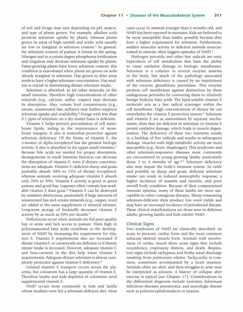
Chapter 11 • Diseases of the Musculoskeletal System 311
of soil and forage may vary depending on pH, season, and type of plants grown. For example, alkaline soils promote selenium uptake by plants, whereas plants grown in areas of high rainfall and acidic soils usually are low or marginal in selenium content.3 In general, the selenium content of pasture is lowest in the spring. Nitrogen and to a certain degree phosphorus fertilization and irrigation may decrease selenium uptake by plants. Faster-growing plants have lower selenium content; this condition is exacerbated when plants are grown on soils already marginal in selenium. Hay grown in drier areas tends to have a higher selenium concentration. Hay anal-ysis is crucial in determining dietary selenium intake.
Selenium is absorbed, as are other minerals, in the small intestine. Therefore high concentrations of other minerals (e.g., calcium, sulfur, copper) may decrease its absorption. Also, certain feed contaminants (e.g., nitrate, unsaturated fats, sulfates) may further suppress selenium uptake and availability.4 Forage with less than 0.1 ppm of selenium on a dry matter basis is deficient.
Vitamin E helps prevent peroxidation of cell mem-brane lipids, aiding in the maintenance of mem-brane integrity. It also is somewhat protective against selenium deficiency. Of the forms of vitamin E, the d-isomer of alpha-tocopherol has the greatest biologic activity. It also is absorbed in the upper small intestine.5 Because bile acids are needed for proper absorption, derangements in small intestine function can decrease the absorption of vitamin E, even if dietary concentra-tions are adequate. Vitamin E–deficient sheep and goats probably absorb 50% to 75% of dietary tocopherol, whereas animals receiving adequate vitamin E absorb only 20% to 30%. Vitamin E activity is good in green pasture and good hay. Legumes often contain less avail-able vitamin E than grass.3 Vitamin E can be destroyed by oxidative destruction, particularly if large amounts of unsaturated fats and certain minerals (e.g., copper, iron) are added to the same supplement or mineral mixture. Long-term storage of feedstuffs decreases vitamin E activity by as much as 50% per month.3
Deficiencies occur when animals are fed poor-quality hay or straw and lack access to pasture. Diets high in polyunsaturated fatty acids contribute to the develop-ment of NMD by increasing the requirement for vita-min E. Vitamin E requirements also are increased if dietary vitamin C or carotenoids are deficient or if dietary nitrate intake is increased. However, adequate vitamin C and beta-carotene in the diet help lower vitamin E requirements. Adequate dietary selenium is almost com-pletely protective against vitamin E deficiency.6
Limited vitamin E transport occurs across the pla-centa, but colostrum has a large quantity of vitamin E. Therefore lambs and kids deprived of colostrum need supplemental vitamin E.
NMD occurs most commonly in kids and lambs whose mothers were fed a selenium-deficient diet. Most
cases occur in animals younger than 6 months old, and NMD has been reported in neonates. Kids are believed to be more susceptible than lambs, possibly because they have a higher requirement for selenium. Furthermore, sudden muscular activity in deficient animals unaccus-tomed to exercise often triggers episodes of NMD.1
Hydrogen peroxide and other free radicals are toxic byproducts of cell metabolism that have the ability to cause oxidative damage to biologic membranes. Selenium is a cofactor in several enzyme systems in the body, but much of the pathology associated with selenium deficiency is caused by an impairment of the enzyme glutathione peroxidase. This enzyme protects cell membranes against destruction by these endogenous peroxides by converting them to relatively benign hydroxy fatty acids. The lipid-soluble vitamin E molecule acts as a free radical scavenger within the cell membrane. High concentrations of dietary fat can overwhelm the vitamin E protection system.6 Selenium and vitamin E act as antioxidants by separate mecha-nisms; diets that are deficient in selenium or vitamin E permit oxidative damage, which leads to muscle degen-eration. The deficiency of these two nutrients results in a buildup of free radicals and results in subsequent damage. Muscles with high metabolic activity are most susceptible (e.g., heart, diaphragm). This syndrome and other selenium-responsive diseases most commonly are encountered in young growing lambs, particularly those 2 to 4 months of age.3,7 Selenium deficiency also may impair the body’s immune system. In cattle and possibly in sheep and goats, deficient selenium intake can result in reduced neutrophilic response, a higher incidence of mastitis and metritis, and poor overall body condition. Because of their compromised immune systems, many of these lambs are more sus-ceptible to other contagious diseases. Sheep consuming selenium-deficient diets produce low wool yields and may have an increased incidence of periodontal disease. These clinical manifestations are those seen in deficient adults; growing lambs and kids exhibit NMD.
Clinical SignsTwo syndromes of NMD are classically described: an acute to peracute cardiac form and the more common subacute skeletal muscle form. Animals with involve-ment of cardiac muscle show acute signs that include recumbency, respiratory distress, and death. Respira-tory signs include tachypnea and frothy nasal discharge resulting from pulmonary edema. Tachycardia is com-mon, sometimes accompanied by a heart murmur. Animals often are alert, and their struggles to arise may be interpreted as seizures. A history of collapse after exercise is typical (see Chapter 17). Considerations in the differential diagnosis include toxicities, fulminant infectious diseases, pneumonia, and neurologic disease such as polioencephalomalacia or tetanus.

312 Chapter 11 • Diseases of the Musculoskeletal System
Animals with skeletal muscle degeneration have a different appearance. These animals have a stiff gait and tremble while standing. Many prefer to remain in sternal recumbency. The muscles may feel firm to pal-pation. Disease manifestations described in this form of NMD include hunched appearance, stiff gait, and overall poor production.3 Lambs and kids continue to weaken and eventually become unable to nurse.7 Many young animals have aspiration pneumonia resulting from dysfunction of the glottis. Some adult animals continue to eat, but others are dysphagic because of involvement of the tongue. Skeletal and cardiac muscle disease may occur concurrently. Careful assessment of flock history and a through physical examination are required to determine the underlying cause of the pneu-monia. Other diseases that may appear similar include enzootic ataxia, polyarthritis, and nutritional osteo-dystrophy. Vitamin E–associated NMD is encountered most commonly in lambs and yearling ewe lambs.3
DiagnosisElevated creatine kinase (CK) is a good indicator of subclinical NMD.3 Marked elevations in CK (10 to 50 times) can occur in NMD. CK has a short half-life (2 to 4 hours), so elevations indicate recent or ongo-ing muscle damage. CK levels return to normal as the animal recovers. AST also is elevated with muscle injury; however, this marker is not specific to muscle disease—hepatic disease also may cause elevations in AST. AST has a longer half-life than CK, and concentrations are elevated for several days after an episode of NMD. Elevations in CK and AST are not specific for NMD, and these enzymes may be elevated in any recumbent animal. However, CK and AST generally occur in much higher serum concentrations in the presence of primary muscle disease such as NMD.
Selenium deficiency can be confirmed by measuring selenium levels in whole blood or tissues. For investiga-tion of flock problems, blood should be collected for selenium analysis in 10% of the flock or at least 7 to 10 ewes or lambs.3 Erythrocyte glutathione peroxidase concentrations are highly correlated with selenium concentration, and determination of activity of this enzyme is a useful diagnostic test. Samples for assay of glutathione peroxidase must be handled with care, however, and many diagnostic laboratories do not offer the test. Testing for serum selenium levels may be of value for flock assays if the diet has been maintained for weeks to months. It is of questionable value in assess-ing individual animals, particularly those that have experienced any dietary changes. Obviously, most sick animals have undergone a diet change, and many have anorexia. Evaluating whole blood selenium is the easi-est and most reliable test. Selenium concentrations in whole blood reflect the selenium level of the diet over the life of a red blood cell.8 More than 95% of blood
selenium is located inside the red blood cell and was placed there when the cell was manufactured. Vitamin E status can be assessed by measuring serum tocopherol. Some specialized laboratories offer a vitamin E assay. This chapter does not provide guidelines for adequate or deficient concentrations because of the variance in techniques and assays among laboratories. Instead, the clinician should inquire about normal values from the laboratory where samples are assayed.
At necropsy, affected muscles are friable and contain pale streaks that correspond with regions of degen-eration and mineralization. The distribution is bilat-erally symmetric. Similar changes are seen in the myocardium if animals had cardiac involvement. His-topathologic examination of muscle shows hyaline degeneration, necrosis, and mineralization. Chronic infections (caused by depressed immune function) and aspiration pneumonia (resulting from compromised glottis-closing ability) also may be encountered.1-3
TreatmentOne injection of a vitamin E or selenium preparation should result in improvement within a few days. The treatment can be repeated in 24 hours. Following the label doses of some commercial products will provide adequate selenium but very little vitamin E, and sup-plementation may be required. If other animals show clinical signs, they also should be treated. The clinician or keeper should avoid exposing the animals to stress or exertion during treatment. Most animals respond to treatment; however, those with cardiac involvement have a poor recovery rate.
PreventionNMD can be prevented by supplementing the diet of susceptible animals with selenium and vitamin E. Sup-plementation of pregnant animals will help reduce dis-ease in newborns, because selenium is transferred across the placenta and also is present in colostrum and milk. Clinicians and keepers should pay careful attention to the proper dosage of selenium to prevent toxicosis in the animals and should adhere to withdrawal periods to limit concentrations in tissues at slaughter. Pasture, hay, and any grain supplements should be assayed to determine the amount of selenium to be added to a supplemental pellet, grain, or mineral mixture.
Selenium and vitamin E supplementation can take many forms. The dietary concentration of selenium should be more than 0.1 to 0.3 mg/kg.1,7 Feed supple-mentation commonly is recommended. In some cir-cumstances, higher levels of selenium are necessary to prevent NMD in lambs. Dietary supplementation appears to be the least expensive, most efficient method of ensuring selenium adequacy. Current regulations in the United States limit selenium supplementation for sheep to 0.7 mg/head/day or 90 ppm in the mineral

Chapter 11 • Diseases of the Musculoskeletal System 313
mixture for free-choice feeding.3 Although the use of free-choice mineral supplementation is an excellent mode of selenium supplementation, formulating a complete diet or providing a dietary supplement of 0.2 ppm of selenium will ensure more consistent min-eral intake.7 Fresh legumes and grasses are good sources of vitamin E.9 Silage, oil seeds, cereal grains, and dry hays tend to be poor sources of vitamin E.5 Therefore diets high in grain content should be supplemented with vitamin E.
Alternatively, selenium and vitamin E can be incor-porated in mineral mixes that are fed free choice to pregnant and lactating ewes. If feedstuffs contain oxi-dizing agents (e.g., copper, iron), fats, or a high content of disulfide bonds (onions), vitamin E potency may be reduced, with resultant deficiency.3 Whenever these dietary factors are encountered, supplemental vitamin E is indicated. Diets high in corn also may be associated with vitamin E deficiency, because a lowered rumen pH reduces vitamin E activity. This condition can be clini-cally significant in the young, growing lamb or kid.
If it is not practical to supplement the diet, monthly injections of a commercial vitamin E–selenium selenite compound may be useful; the injections may need to be repeated more often in lambs.1 Injecting the dam 30 days before birth can help prevent NMD.3,7 Inject-ing lambs with selenium–vitamin E preparations at tail docking (1 mg selenium) and again at weaning (2 mg selenium) may be protective on some farms. In addition to injected supplements, another source of vitamin E should be provided because the amount in commercially available injectable compounds is too low to prevent disease in deficient animals. Access to pasture or good-quality forage should provide adequate levels of vitamin E.
Other options for selenium supplementation are practiced in some regions. A slow-release formulation of selenite can be given by subcutaneous injection. A dose of 1 mg/kg selenium given to ewes 3 weeks before lambing protects lambs for as long as 12 weeks after birth. An intraruminal selenium pellet also is avail-able for sheep. Top-dressing of pasture with sodium sel-enite at a dose of 10 g selenium/hectare is practiced in some countries. This method is safe and prevents NMD for at least 12 months.1 When lambs are bottle-fed, the keeper should ensure an intake of adequate vitamin E in the milk replacer. (See Chapter 2)
Rickets and OsteomalaciaRickets is a disease of young animals caused by a failure of proper cartilage mineralization. Vitamin D deficiency is the most common cause, but rickets may occur as a result of deficiencies in phosphorus and calcium as well. In older animals, the same deficiencies result in abnormal mineralization of osteoid, a condition
known as osteomalacia. An inherited form of rickets has been reported in Corriedale sheep in New Zealand.10
Rickets occurs mostly in rapidly growing animals that have low vitamin D levels because of limited sun exposure. Animals housed indoors, those fed green (uncured) forage, and those living at high latitudes in winter are most prone. Animals that consume a diet low in calcium or phosphorus occasionally develop rickets. Ingestion of some poisonous plants, particularly those containing oxalates (which bind calcium in the intestine); chronic lead, fluoride, or aluminum toxicity; and chronic parasitism can all produce or add to the pathogenesis of rickets.6
PathogenesisThe primary problem is failure of mineralization of cartilage and osteoid, which leads to persistence of carti-lage and irregular osteoid deposition.4 Radiographs will show irregular osteochondral junctions and widened physes. The metaphyses at the costochondral junctions are noticeably affected. In the long bones, the persistent soft tissue in the physis is deformed by weight bearing. In the diaphysis, osteoid is not properly mineralized.11 Long-haired or woolly animals raised in latitudes closer to the earth’s poles, those raised indoors, and those fed milk replacers with inadequate vitamin D concentra-tions may be particularly deficient in vitamin D and thus predisposed to NMD. Twin lambs may be more susceptible to disease in the neonatal period.12
Clinical SignsAffected animals usually are less than 1 year old and exhibit a stiff gait, shifting legs, lameness, and recum-bency. Joints and bones of the distal aspects of the limbs may be enlarged, and enlargements of the ribs at the costochondral junctions (“rachitic rosary”) frequently are seen. Limbs are frequently deformed and may be bowed. Teeth may be mottled and their eruption delayed. Animals may be thin because of failure to graze adequate forage.8 Considerations in the differential diagnosis include NMD and infectious arthritis.
DiagnosisThe blood chemistry panel shows elevations in alkaline phosphatase greater than those seen in normally grow-ing animals. Plasma concentrations of calcium and phosphorus may be low. Serum vitamin D is low but usually is within normal ranges. Radiographic changes include widened growth plates, bowing of long bones, and thinned cortices.11 Radiographic examination of adult animals with osteomalacia will reveal porous bone.
Postmortem examination reveals thickening of growth plates and epiphyseal enlargement of long bones. Rib fractures often are apparent. Normal bone contains an ash–to–organic matter ratio of 3:2, whereas the ratio in rachitic bone is 1:2 to 1:3.

314 Chapter 11 • Diseases of the Musculoskeletal System
Careful investigation of feed content and access to sunlight and determination of vitamin D, calcium, and phosphorus levels will aid in determining the underly-ing cause of rickets.
TreatmentVitamin D3 injections (10,000 to 30,000 IU/kg) may be beneficial if dietary supplementation of calcium and phosphorus occurs concurrently.13 Recovered ani-mals frequently maintain a short stature with limb deformities.
PreventionRickets and osteomalacia can be managed by providing access to sunlight and properly cured forage. Dietary calcium and phosphorus levels should be adjusted if they are low, and a calcium-to-phosphorus ratio of 1:1 to 2:1 should be maintained. Any potentially toxic substances or plants should be removed from the diet.
Osteodystrophia FibrosaOsteodystrophia fibrosa is a metabolic disease of goats and sheep in which bone mineral is resorbed as a result of prolonged hypersecretion of parathyroid hormone. High phosphorus or low calcium levels in the diet frequently contribute to osteodystrophia fibrosa. Clini-cally, this disease is similar to rickets.
Osteodystrophia fibrosa most commonly is seen in animals consuming a high-phosphorus diet. Diets with a high proportion of bran or other cereal grains are often associated with this disease. Cereal grains have an inappropriate calcium-to-phosphorus ratio, and much of the phosphorus in cereal grains is in the form of phytic acid. High phytic acid content can fur-ther depress calcium absorption from the intestine. The dietary calcium–to–phosphorus ratio should be main-tained at 1:1 to 2:1. Many cereal grains or byproduct feeds (bran) have a ratio of 1:6 or greater.
PathogenesisPrimary hyperparathyroidism caused by hyperplasia or neoplasia of the parathyroid gland is extremely rare. Most cases of hyperparathyroidism are sequelae of nutritional or metabolic conditions that produce hypo-calcemia. Diets with low levels of calcium, high levels of phosphorus, or deficient amounts of vitamin D may result in hyperparathyroidism; frequently more than one factor is present. Parathyroid hormone stimulates vitamin D production, which in turn induces resorption of bone in the animal to maintain calcium homeostasis. Renal failure also may result in hyperparathyroidism, but this manifestation is uncommon in sheep and goats. All of the bones of the body are affected, but the bones of the face and mandible are most obviously abnormal.
Clinical SignsBilateral enlargement of the mandible typically is the most obvious sign. The mandible feels soft, and the affected animal may not be able to open its mouth properly. Lameness and stiffness often are observed as a result of pathologic fractures. Affected animals fre-quently are thin because of decreased food intake.
DiagnosisRadiographs show enlargement of the mandible, decreased bone density, and rotation of the cheek teeth with the occlusal surfaces pointed lingually. Fractures of other bones may be apparent.14 Laboratory results may show low calcium or high phosphorus levels, but these tests often fall within the normal range. Postmortem examination shows the mandible to be quite soft and malleable, and histopathologic examination shows a lack of bone mineralization and replacement of bone by an extensive fibrous matrix.
Caseous lymphadenitis commonly causes enlarge-ment of the mandibular region as a result of abscess formation in the submandibular and retropharyngeal lymph nodes. Palpation and radiographs should aid in distinguishing between this inflammatory disorder and osteodystrophia fibrosa.
TreatmentAnimals may recover if placed on a diet with a calcium-to-phosphorus ratio of 1:1 or 2:1. The enlarged man-dible may not improve. Formulation of a ration that ensures a calcium-to-phosphorus ratio of 1:1 or greater should prevent nutritional hyperparathyroidism and osteodystrophia fibrosa. (See Chapter 2)
EpiphysitisEpiphysitis is a condition of rapidly growing animals in which improper ossification of the physes occurs. The etiology is complex, with both genetic and dietary fac-tors believed to play roles. It is seen in young rams being fed to maximize growth and is associated with preg-nancy in approximately 1% of yearling dairy does.15
Clinical SignsClinical signs reported in a pregnant yearling Nubian doe included insidious onset of lameness progressing to recumbency. Enlargement of the carpi, tarsi, and fetlock joints also was observed, as was angular limb deformity. Radiographs revealed delayed maturation of cartilage and overgrowth of new bone. The animal’s gait improved shortly after parturition, but a degree of limb deformity resulting from premature closure of a portion of the physes remained. The cause was attributed to trauma to the physes from the increased weight associated with advanced pregnancy.16 After noting epiphysitis in ani-mals, the clinician or keeper should examine the diet

Chapter 11 • Diseases of the Musculoskeletal System 315
to assess the adequacy of copper and maintain a proper calcium-to-phosphorus ratio of 4:1 to 6:1. Adequate calcium, phosphorus, protein, and energy all should be maintained. Proper hoof trimming, the provision of pain relief (with NSAIDs), and the removal of animals from hard surfaces all may be of benefit.17 (See Chapter 2)
OsteochondrosisOsteochondrosis is a disease of abnormal endochon-dral ossification. It is common in pigs and chickens and occurs in most domestic animals, but reports are rare in small ruminants. Osteochondrosis was observed in a Suffolk flock,18 and it should be considered in the dif-ferential diagnosis for lameness or swelling in animals that have been fed diets high in grain to produce rapid weight gain. Radiographs of limbs from affected and/or symptomatic animals reveal osteochondrosis lesions, as described in other domestic species (See Chapter 2).
REFERENCES 1. Radostits OM, et al: Veterinary medicine, ed 10, Philadelphia, 2007,
Saunders. 2. Edmondson AJ, Norman BB, Suther D: Survey of state veterinar-
ians and state veterinary diagnostic laboratories for selenium deficiency and toxicosis in animals, J Am Vet Med Assoc 202:865, 1993.
3. Maas J, Valberg SJ: Nutritional myodegeneration. In Smith BP, editor: Large animal internal medicine, St Louis, 2009, Mosby.
4. Maxie MG, editor: Jubb, Kennedy and Palmer’s Pathology of domestic animals, ed 5, Philadelphia, 2007, Saunders.
5. Bulgin MS: Diagnosing nutritional difficulties. Proceedings of the 69th annual Western Veterinary Conference, Las Vegas, 1998, Nev.
6. Smart ME Cymbaulk: Trace minerals. In Naylor JM, Ralston SL, editors: Large animal clinical nutrition, St Louis, 1991, Mosby.
7. Naylor JM: Vitamins. In Naylor JM, Ralston SL, editors: Large animal clinical nutrition, St Louis, 1991, Mosby.
8. Ogilvie TH: Musculoskeletal disorders. In Ogilvie TH, editor: Large animal internal medicine, Baltimore, 1998, Williams & Wilkins.
9. Bretzlaff K, Haenlein G, Huston E: Common nutritional prob-lems: feeding the sick goat. In Naylor JM, Ralston SL, editors: Large animal clinical nutrition, St Louis, 1991, Mosby.
10. Thompson KF, et al: An outbreak of rickets in Corriedale sheep: evidence for a genetic aetiology, N Z Vet J 55:137–142, 2007.
11. Goff J: Calcium, magnesium and phosphorus. In Smith BP, editor: Large animal internal medicine, ed 4, St Louis, 2007, Mosby.
12. Van Saun RJ: Vitamin D-responsive rickets in neonatal lambs, Can Vet J 45:841–844, 2004.
13. Bonniwell MA, et al: Rickets associated with vitamin D deficiency in young sheep, Vet Rec 122:386, 1988.
14. Andrews AH, Ingram PL, Longstaffe JA: Osteodystrophia fibrosa in young goats, Vet Rec 112:404, 1983.
15. Guss SB: Management and diseases of dairy goats, Scottsdale, Ariz, 1977, Dairy Goat Journal Publishing.
16. Bulgin MS: Diagnosis of lameness in sheep, Comp Cont Educ Pract Vet 8:F122, 1986.
17. Anderson KL, Adams WM: Epiphysitis and recumbency in a year-ling prepartum goat, J Am Vet Med Assoc 183:226, 1983.
18. Scott CA, Gibbs HA, Thompson H: Osteochondrosis as a cause of lameness in purebred Suffolk lambs, Vet Rec 139:165–167, 1996.
TOXIC CONDITIONSSelenium ToxicitySelenium toxicity may result from grazing pastures with high selenium content or from exogenous administra-tion of selenium by injection or feed supplementation. Acute poisoning may result in death, but chronic over-dose leads to hoof malformation and lameness. The toxic dose for sheep has been reported to be 2.2 mg/kg orally as a single dose or chronic ingestion of 0.25 mg/kg of body weight.1 Sheep are considered more susceptible to selenium toxicosis than cattle. Little information is available about the natural occurrence of selenium toxi-cosis in goats, but the administration of high doses of selenium can result in death.2
Soils in specific regions of North America, Ireland, Australia, and South Africa have high selenium content because of the composition of the underlying rock.1 Soils in areas of low annual rainfall often have an alkaline pH and are more likely to have high selenium levels. Plants extract selenium from the soil, and certain plants are concentrators of selenium. These plants are not highly palatable, but signs of toxicity may develop in animals that graze in these areas if more palatable forage is lacking. Documented cases of naturally occur-ring selenium toxicity are uncommon.3
Selenium poisoning also occurs when incorrect doses of selenium are administered to flocks in an attempt to prevent NMD.4
Organic selenium compounds (i.e., those found in plants) are considered more toxic than inorganic com-pounds such as selenite and selenium dioxide. This reported difference does not always correlate with clinical disease.1
PathogenesisSelenium concentrates in the kidney, liver, and kera-tinized tissue and has a dystrophic effect on skeletal muscle. In toxic concentrations, selenium may displace sulfur in some of the amino acids (methionine, cys-tine), preventing them from forming disulfide bonds and thereby weakening keratin formation. Hoof mate-rial contains high concentrations of methionine and cystine. The mechanism of toxicity has not been deter-mined, but selenium also may interfere with the func-tion of certain enzymes. A high-protein diet is protective against selenium toxicosis in sheep.1
Clinical SignsAcute poisoning may result in dyspnea, tachycardia, fever, depression, and death. White or blood-tinged froth often is observed at the nostrils and mouth.4 Signs of chronic

316 Chapter 11 • Diseases of the Musculoskeletal System
toxicity include poor hair coat, alopecia, ill thrift, abnor-mal appetite, respiratory failure, and lameness. Hoof lesions affecting all feet are seen and include edema of the coronary bands and deformity or separation of hooves. In neonates, hoof abnormalities may be apparent at birth.
DiagnosisDiagnosis is based on identifying toxic levels of selenium in the animal. Selenium levels in blood, urine, and hair all are elevated. Anemia and low hemoglobin levels are characteristic of chronic selenium poisoning. Necropsy findings in chronic selenium poisoning include myopa-thy of skeletal and cardiac muscle and hoof and hair coat abnormalities as described previously. Lesions in many other organs also have been described.
TreatmentNo specific treatment is effective. If possible, the source of excess selenium should be removed.
PreventionSelenium supplementation should be carefully moni-tored to ensure safe dosage. In regions with selenifer-ous soils, supplemental forage can be provided to reduce consumption of selenium-containing plants and increase dietary protein. Rich sources of sulfur-contain-ing amino acids (soybean meal) in the diet are partially protective. Alternate grazing of areas with plants that do not accumulate toxic concentrations of selenium is another option. The addition of 0.01% arsanilic acid or 20 ppm copper to the ration also may be preventive, but these substances are potentially toxic.
Ergot ToxicosisErgot toxicity results from ingestion of alkaloid com-pounds produced by the fungus Claviceps purpurea. This fungus infects cereals and grasses, most commonly rye, wheat, and oats. The seeds of the plants turn dark as they are filled with the fungal sclerotia, and this grossly visible structure is referred to as an ergot. C. purpurea is the fungal species most frequently linked with ergot-ism, but Neotyphodium (Acremonium) coenophialum may cause a similar syndrome.1
This toxicosis occurs in animals grazing ergot-infested pasture or eating grain or hay made from such plants. It is fairly common in cattle, but reports in sheep and goats are rare. In one report of goats and sheep co-grazing a fescue pasture, only goat kids were affected.5 The condition usually occurs after a warm wet season, leading to conditions that favor growth of the fungus.
PathogenesisErgots contain alkaloid compounds and other phar-macologically active compounds known as ergotoxins. The effects of this group of toxins, which includes
ergotamine, ergotoxine, and ergometrine, include con-striction of arterioles and endothelial damage leading to gangrene of the extremities.
Clinical SignsClinical signs of ergotism include swelling, coolness, and hair loss followed by drying and discoloration of the skin of the distal limbs, tail, and ears. A distinct demarcation between normal and gangrenous skin is observed, and affected tissue may slough. Lameness is evident, and animals may remain recumbent. Clinical signs reported in goats include lameness, most often in the hind limbs, with separation of the hoof in the most severe cases.6 Ulceration of the oral, ruminal, and intes-tinal mucosa has been reported in sheep.1
DiagnosisFeed samples should be analyzed for ergot or similar compounds. A primary consideration in the differential diagnosis is thrombosis secondary to sepsis or trauma.
TreatmentNo specific treatment exists for ergot toxicity. Affected animals should be removed from the source of toxin.
PreventionFeed should contain less than 0.1% infected seed-heads.1 Pastures with severe ergot infestations should not be used for grazing or hay.
Fluorosis (Fluorine Poisoning)Chronic fluorine poisoning (fluorosis) occurs after the ingestion of toxic amounts of fluorine compounds by feed or water. The severity of disease depends on the fluorine compound ingested. Sodium fluoride is more toxic than rock phosphate; calcium fluoride and sodium fluorosilicate are much less toxic. Sheep and goats are reported to be less susceptible than cattle.6
Fluorine occurs naturally in rocks, usually in asso-ciation with phosphate. Soils derived from these rocks and water that percolates through these rock for-mations may contain high levels of fluorine. Other sources of fluorine include industrial contamination (as far as 14 km downwind), deep water wells, volca-nic ash, and phosphatic supplements given to combat hypophosphatemia.1
PathogenesisThe mechanism of fluorine toxicity has not been deter-mined. Excess fluorine is deposited in bones and teeth. Bony lesions may develop at any time in the animal, but dental lesions occur only if fluorine levels are high during the formation of the teeth. Urinary excretion of fluorine, accompanied by calcium and phosphorus, leads to mobilization of calcium and phosphorus, with

Chapter 11 • Diseases of the Musculoskeletal System 317
resultant osteomalacia and osteoporosis. Many other sites, including the bone marrow, undergo degenerative changes.1
Clinical SignsAcute fluorine toxicity is marked by gastrointestinal signs, tetany, and death. Chronic ingestion leads to decreased feed consumption and unthriftiness. Dental lesions, which consist of surface pitting and increased wear caused by improper enamel formation, are the first to appear, although initially they may not be noticed. With time, rapid wear and tooth breakage occurs, lead-ing to impaired mastication.7,8
Signs of osteofluorosis include ill thrift, stiffness, and lameness that is most prominent in the hindlimbs. Pathologic fractures, often of the third phalanx (P3), may occur in several animals in the group. The affected bones are painful to palpation and may be enlarged.1
Considerations in the differential diagnosis include other causes of lameness on a herd or flock basis, including hypophosphatemia, vitamin D deficiency, selenium toxicity, and selenium deficiency.
DiagnosisSerum fluorine levels often are elevated in toxicosis (the normal level for cattle is 0.2 mg/dL), but normal levels do not rule out toxicity, because of the storage of fluo-rine in bone. Urinary fluorine often is elevated (16 to 68 mg/kg is normal for cattle). Serum alkaline phosphatase levels usually are elevated.1,7
Radiographic abnormalities include increased bone density, enlarged bones, narrowing of the marrow cav-ity, and spontaneous fractures that heal poorly.
Postmortem examination reveals chalky, brittle bones with diaphyseal exostoses. Histopathologic examination shows abnormal calcification of bone. Hypoplasia of enamel is observed in animals with dental disease. Degenerative changes in many tissues, including bone marrow, are observed. The fluorine content of bones can be measured to confirm the diag-nosis. The mandible and the metacarpals and metatar-sals are considered the most reliable sources of bone for fluorine assay.1
TreatmentKeepers should remove animals from the source of fluorine. Cases of acute toxicity can be treated with aluminum salts (to neutralize hydrofluoric acid in the stomach) and intravenous calcium salts to control tetany. Dental and bone lesions do not usually improve.
Animals should be fed good-quality hay. The addition of calcium carbonate or aluminum sulfate to the diet at 1% of the dry matter intake may be beneficial in decreasing bone flouride content.
PreventionPhosphate feed supplements for cattle should not con-tain more than 0.2% to 0.3% fluorine. The phosphorus-to-fluoride ratio should be maintained at greater than 100:1. Rock phosphate can be a source of fluorine, and deep water wells should be assayed for fluorine levels before use. Careful management of grassland and water in high-fluorine areas may reduce herd or flock losses caused by fluorine toxicosis.9 Some guidelines recom-mend feeding aluminum salts to bind fluorine and reduce accumulation in tissue, but these compounds are unpalatable.1
Plant ToxicityA myopathy of skeletal muscle marked by stiff gait and recumbency was reported to develop in Aus-tralian sheep kept on pasture that included lupine stubble infested with the fungus Phomopsis (specifically, the anamorph Diaporthe toxica).10 Ingestion of Cassia roemeriana (twin-leaf senna) is believed to cause a simi-lar syndrome in cattle and sheep in Texas, New Mexico, and Mexico.11
REFERENCES 1. Radostits OM, et al, editors: Veterinary medicine, ed 10,
Philadelphia, 2007, Saunders. 2. Ahmed KE, et al: Experimental selenium poisoning in Nubian
goats, Vet Hum Toxicol 32:249, 1990. 3. Edmondson AJ, Norman BB, Suther D: Survey of state veterinar-
ians and state veterinary diagnostic laboratories for selenium deficiency and toxicosis in animals, J Am Vet Med Assoc 202:865, 1993.
4. Smith WI, Donovan GA, Rae DO: Selenium toxicosis in a flock of Katahdin hair sheep, Can Vet J 40:192–194, 1999.
5. Hibbs CM, Wolf N: Ergot toxicosis in young goats, Mod Vet Pract 63:126, 1982.
6. Choubisa SL: Some observations on endemic fluorosis in domestic animals in southern Rajasthan (India), Vet Res Comm 23:457, 1999.
7. Botha CJ, et al: Two outbreaks of fluorosis in cattle and sheep, J S Afr Vet Assoc 64:165, 1993.
8. Schultheiss WA, Van Niekerk JC: Suspected chronic fluorosis in a sheep flock, J S Afr Vet Assoc 65:84, 1994.
9. Wang JD, Hong JP, Li JX: Studies on alleviation of industrial fluo-rosis in Baotou goats, Fluoride 28:131, 1995.
10. Allen JG, et al: A lupinosis-associated myopathy in sheep and the effectiveness of treatments to prevent it, Aust Vet J 69:75, 1992.
11. Rowe LD, et al: Experimentally induced Cassia roemeriana poison-ing in cattle and goats, Am J Vet Res 48:992, 1987.

318 Chapter 11 • Diseases of the Musculoskeletal System
NEOPLASIANeoplasia of the musculoskeletal system is extremely rare in sheep and goats. A study of 673 ovine neo-plasms submitted to a veterinary laboratory in South Africa revealed that 21 of them were of connective tissue origin. Types of tumors included chondroma, chondro-sarcoma, fibroma, fibrosarcoma, osteoma, rhabdomyo-sarcoma, leiomyoma, and fibrolipoma.1
Osteosarcoma and pathologic fracture developed in a 9-year-old Toggenburg goat 4 years after a com-minuted humeral fracture had been repaired with an intramedullary pin. The animal also was reported to have pulmonary nodules, but these were not examined histologically.2 Osteoma of the frontal bone with com-promise of the nasal cavity was reported in a sheep.3 Mandibular osteoma was diagnosed in a 10-year-old Toggenburg cross, and osteochondrosarcoma involving a rib and the sternum of a goat also has been described.2,4
A diagnosis of neoplasia is based ultimately on his-topathologic analysis. Bony enlargement, lameness, and radiographic evidence of lysis or proliferation may sug-gest a diagnosis of neoplasia, especially in an older ani-mal. Successful treatment of connective tissue tumors has not been reported.
REFERENCES 1. Bastianello SS: A survey on neoplasia in domestic species over a
40-year period from 1935 to 1974 in the republic of South Africa. II. Tumours occurring in sheep, Onderstepoort J Vet Res 49:205, 1982.
2. Steinberg H, George C: Fracture-associated osteogenic sarcoma and a mandibular osteoma in two goats, J Comp Path 100:453, 1989.
3. Perez V, et al: Osteoma in the skull of a sheep, J Comp Pathol 130:319–322, 2004.
4. Cotchin E: Tumors of farm animals, Vet Rec 40:816, 1960.
TAIL DOCKINGTail removal or “docking” usually is performed during the first 2 weeks of life.1,2 Some lambs sold in niche mar-kets do not have their tails docked, and in some breeds (e.g., Karakul), the tail should be left long, because the fat at the base of the tail is considered a prized com-modity. Still, in most environments in which lambs are kept, long tails can become soiled with loose stool or diarrhea (as a result of high-grain diets, lush pasture, or internal parasites), leading to flystrike or infestation of the wool with maggots. Furthermore, presence of long tails in females appears to depress normal reproductive performance. For these and other reasons, tails usually are removed. If the lamb is less than 24 hours old, the stress associated with tail removal may decrease absorp-tion of colostral antibodies, resulting in the diseases associated with failure or partial failure of passive trans-fer. In general, therefore, lambs should be 2 to 3 days to 2 weeks old at docking. A regimen associated with good results in clinical practice (of D.G. Pugh) is to dock tails at 3 days on alert, healthy animals that are being cared for by their dams. The docking can take place after the lambs and their dams are moved to a single-family unit (jug) or holding area. Placing the new lamb and its dam together helps prevents the ewe from wandering off or abandoning the lamb after the procedure. Anesthesia is seldom required, with the obvious exception of adult or pet animals (on the owner’s request). If anesthesia is to be used, either a sedative or a caudal epidural and ring block will suffice.1 Some studies suggest that a tail ring block of a local anesthetic can reduce the stress associ-ated with tail removal.3 Still, Hooper1 has suggested,
and we agree, that the neonatal lamb responds as much or possibly more to the injection of a local anesthetic as to the surgical removal of the tail without anesthesia.
The tail should be left long enough to cover the anus and may be extended to the dorsal aspect of the vulva on females.2 The woolless distal attachment of the paired caudal skinfolds on the ventral tail surface provide a good landmark for the site of tail removal. Many owners of show or club lambs prefer to remove the tail as close to the body as possible. However, dock-ing too close to the sacrum may result in an increased incidence of rectal and possibly vaginal prolapse.1 The tail can be crushed, cut, or cauterized or removed using a combination of these methods.2 Equipment used for tail removal includes an emasculator, an emasculatome, a hot chisel, a knife, and elastrator bands.
For the docking procedure, tails should first be cleaned of dirt and feces. The lamb should be manu-ally restrained while the clinician determines the exact spot of tail removal; the tail should not be excessively stretched. Leaving some skin proximal to the point of removal provides redundant skin to cover the spi-nal stump.1 Use of a cautery unit (e.g., hot chisel, suture-heated wedge, electric wedge, electric cautery) minimizes hemorrhage. If hemorrhage does occur, the ventral blood vessels can be clamped and sutured if needed. If cautery units are used and the wool is burned, some ewes may reject the lambs.1 Removing wool over the docking site before the procedure and gently washing or cleansing the tail after removal can minimize ewe rejection. Ewe rejection caused by cautery docking is rare, and this method of docking is a very
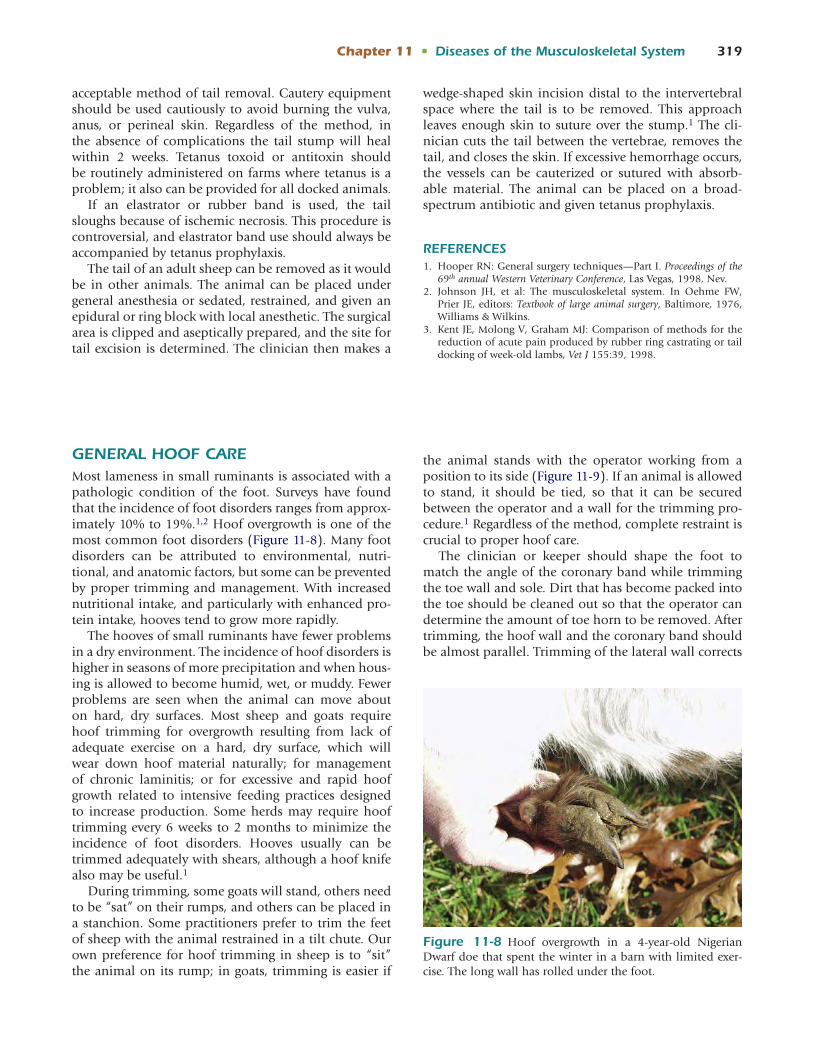
Chapter 11 • Diseases of the Musculoskeletal System 319
GENERAL HOOF CAREMost lameness in small ruminants is associated with a pathologic condition of the foot. Surveys have found that the incidence of foot disorders ranges from approx-imately 10% to 19%.1,2 Hoof overgrowth is one of the most common foot disorders (Figure 11-8). Many foot disorders can be attributed to environmental, nutri-tional, and anatomic factors, but some can be prevented by proper trimming and management. With increased nutritional intake, and particularly with enhanced pro-tein intake, hooves tend to grow more rapidly.
The hooves of small ruminants have fewer problems in a dry environment. The incidence of hoof disorders is higher in seasons of more precipitation and when hous-ing is allowed to become humid, wet, or muddy. Fewer problems are seen when the animal can move about on hard, dry surfaces. Most sheep and goats require hoof trimming for overgrowth resulting from lack of adequate exercise on a hard, dry surface, which will wear down hoof material naturally; for management of chronic laminitis; or for excessive and rapid hoof growth related to intensive feeding practices designed to increase production. Some herds may require hoof trimming every 6 weeks to 2 months to minimize the incidence of foot disorders. Hooves usually can be trimmed adequately with shears, although a hoof knife also may be useful.1
During trimming, some goats will stand, others need to be “sat” on their rumps, and others can be placed in a stanchion. Some practitioners prefer to trim the feet of sheep with the animal restrained in a tilt chute. Our own preference for hoof trimming in sheep is to “sit” the animal on its rump; in goats, trimming is easier if
the animal stands with the operator working from a position to its side (Figure 11-9). If an animal is allowed to stand, it should be tied, so that it can be secured between the operator and a wall for the trimming pro-cedure.1 Regardless of the method, complete restraint is crucial to proper hoof care.
The clinician or keeper should shape the foot to match the angle of the coronary band while trimming the toe wall and sole. Dirt that has become packed into the toe should be cleaned out so that the operator can determine the amount of toe horn to be removed. After trimming, the hoof wall and the coronary band should be almost parallel. Trimming of the lateral wall corrects
acceptable method of tail removal. Cautery equipment should be used cautiously to avoid burning the vulva, anus, or perineal skin. Regardless of the method, in the absence of complications the tail stump will heal within 2 weeks. Tetanus toxoid or antitoxin should be routinely administered on farms where tetanus is a problem; it also can be provided for all docked animals.
If an elastrator or rubber band is used, the tail sloughs because of ischemic necrosis. This procedure is controversial, and elastrator band use should always be accompanied by tetanus prophylaxis.
The tail of an adult sheep can be removed as it would be in other animals. The animal can be placed under general anesthesia or sedated, restrained, and given an epidural or ring block with local anesthetic. The surgical area is clipped and aseptically prepared, and the site for tail excision is determined. The clinician then makes a
wedge-shaped skin incision distal to the intervertebral space where the tail is to be removed. This approach leaves enough skin to suture over the stump.1 The cli-nician cuts the tail between the vertebrae, removes the tail, and closes the skin. If excessive hemorrhage occurs, the vessels can be cauterized or sutured with absorb-able material. The animal can be placed on a broad-spectrum antibiotic and given tetanus prophylaxis.
REFERENCES 1. Hooper RN: General surgery techniques—Part I. Proceedings of the
69th annual Western Veterinary Conference, Las Vegas, 1998, Nev. 2. Johnson JH, et al: The musculoskeletal system. In Oehme FW,
Prier JE, editors: Textbook of large animal surgery, Baltimore, 1976, Williams & Wilkins.
3. Kent JE, Molong V, Graham MJ: Comparison of methods for the reduction of acute pain produced by rubber ring castrating or tail docking of week-old lambs, Vet J 155:39, 1998.
Figure 11-8 Hoof overgrowth in a 4-year-old Nigerian Dwarf doe that spent the winter in a barn with limited exer-cise. The long wall has rolled under the foot.

320 Chapter 11 • Diseases of the Musculoskeletal System
many hoof problems (Figure 11-10). After trimming the toe and lateral wall, the clinician or keeper should cut the inner wall shorter than the outer wall. The rubbery heel should be cut if it is excessively long or overgrown. The outer hoof wall should be left slightly longer than any other hoof structure, because it is a weight-bearing surface. If the hoof is improperly trimmed, the sheep or goat may walk on the toe or side of the foot or on the heel with the toe pointing up. A common cause of foot problems is an inward-turning outer wall, which produces areas that accumulate debris and become infected. The inner wall occasionally may overgrow toward the interdigital cleft, predisposing the animal to interdigital disease. The foot will be better balanced if the operator removes the toe curl by trimming the solar surface of the hoof and keeping it level, rather than ”dubbing” or shortening the toe. In sheep and goat flocks kept on soft pastures or paddocks, placing feeders on rough surfaces helps decrease the amount of trimming needed. Building or stacking rough material (cement or concrete blocks) for goats to play on also may help minimize the need for frequent trimming.
Feeding affects hoof condition and growth. Animals being overfed energy and protein and living on soft ground may be more prone to the development of some abnormalities. As a general rule, a well-balanced feed-ing program with a free-choice mineral salt supplement consisting of calcium, phosphorus, and trace minerals is all that is required. However, some feeding programs may enhance hoof growth and health and are useful in special circumstances.
In other ruminants (cattle), diets that change normal rumen function by increasing the fermentation rate negatively affect hoof health.3 Specifically, the inges-tion of high-energy feeds coupled with inadequate fiber
intake can result in suboptimal hoof health. In rations in which concentrates and roughage are fed separately, the concentrated portion of the diet should be divided into two or more equal feedings each day. This regimen not only promotes overall health but also may help reduce the microflora changes that alter normal rumen fermentation and predispose animals to founder. For-age should always make up more than 30% to 50% of the dry matter content of the ration. Lush, young for-age rarely provides enough effective fiber to optimize rumen fermentation. The feeding of buffers, particularly in high-concentrate diets, may help the rumen resist digestive upsets and thereby prevent subsequent hoof disease. Abnormal rapid hoof growth can occur when abnormal rumen fermentation is induced by the inges-tion of lush, well-fertilized pastures.3
Hoof health also can be affected by certain vita-mins and minerals. The addition of 20 mg of biotin improves short-term healing of hoof and claw lesions and decreases hoof disease in cattle.3 Furthermore, diets that acidify the rumen decrease the microbial synthe-sis of biotin. One recommendation (of D.G.Pugh) is to include biotin (3 to 4 mg/day) in sheep and goat rations for animals with a history of hoof disease. Other vitamins that play major roles in hoof health include vitamins A and E and the vitamin A precursor beta-carotene. Adequate dietary vitamin A and beta-carotene are needed for normal cell replication, epithelial repair, and immune function. Vitamin E maintains cellular integrity and normal immune function. Diets should be fortified with both of these nutrients if hoof problems occur and in cases in which production practices predis-pose to hoof disease (see Chapter 2).
Calcium is the largest mineral component of hooves and is required for normal hoof growth. Dietary cal-cium concentrations should range between 0.6% and
Figure 11-9 Hoof trimming in the doe shown in Figure 11-8, with the animal standing and the foot picked up. The rolled-under lateral hoof wall can be seen on the foot being trimmed.
Figure 11-10 A view of the solar surface of the foot, with one toe trimmed and an overgrown hoof wall in the other.

Chapter 11 • Diseases of the Musculoskeletal System 321
0.8% of the diet, with the calcium-to-phosphorus ratio being maintained between 1:1 and 2:1. Of the trace minerals that appear to affect hoof growth, zinc, copper, and, to a lesser extent, molybdenum and manganese, are most crucial.3 Zinc is required for normal immunity, horn tissue production, vitamin A metabolism, epithe-lial repair, and hoof hardness. Studies in range, dairy, and feedlot cattle have all shown improved hoof health and decreased lameness when zinc is added to the diet, particularly in a chelated form (zinc methionine).3 The use of such minerals also may be of value in improving overall hoof health. In sheep, the administration of oral zinc sulfate (0.5 g daily) to prevent footrot has shown mixed results.4-6 In cases of high legume intake (high calcium), zinc in the chelated form (zinc methionine) may be beneficial. Copper is needed for keratin synthe-sis and normal immune function and as a cofactor for many enzyme systems in the body. Copper deficiency in the body may be primary (inadequate copper in the diet) or conditioned by other dietary factors (exces-sive dietary molybdenum, sulfur, or iron). The dietary copper-to-molybdenum ratio should be maintained at between 4:1 and 6:1 for adequate copper availability.
Excessive nitrogen fertilization and liming of soils may depress copper and selenium uptake by plants. Heavily fertilized forage and roughage harvested after a drought may be sources of nitrates, which are reduced to nitrites by anaerobic microbial metabolism in the rumen. Nitrites can have a direct effect on hoof growth, resulting in abnormal horn tissue in cattle and possibly other ruminants.3
The key to maintaining healthy hoof tissue with respect to nutrition lies in minimizing rumen acido-sis and fortifying the diet with certain nutrients (e.g., biotin, calcium, zinc).
DISEASES OF THE FOOTAs described earlier in the chapter, various diseases encountered in small ruminants may affect the foot as part of the general pathologic process. The diseases dis-cussed in this section are specific to the foot.
Infectious FootrotInfectious footrot is a severe, contagious disease of sheep and, to a lesser extent, goats that leads to sig-nificant economic losses as a result of weight loss, low fleece weight, labor and treatment costs, decreased milk production,7 and premature culling. Many factors contribute to the pathogenesis of the disease, but the primary agent is the anaerobic bacterium Dichelobacter nodosus (Bacteroides nodosus). Previous infection by Fusobacterium necrophorum contributes to the develop-ment of footrot. The presence of both organisms in a large percentage of cases of symptomatic footrot in
sheep gives reason for added consideration of a spe-cific program for management of an outbreak as well as for quarantine of herd additions.8 Corynebacterium (Actinomyces) pyogenes infection may increase the sus-ceptibility of the hoof to the other two bacterial species. One study suggested a number of spirochetes also may be associated with both footrot in sheep and digital der-matitis in cattle.9 Many strains of D. nodosus have been identified, and they can generally be classified as benign or virulent. Virulent strains have a greater keratolytic ability, which is associated with the production of a heat-stable protease.1,2,10-12 A study of 735 D. nodusus isolates from 247 farms in Western Australia found 181 molecular types by pulsed-field gel electrophoresis. Three common clonal groups made up most of the iso-lates and also were identified in cases from other parts of Australia. The molecular type was stable over several years on some farms, whereas it changed within flocks and even within feet on other farms.13
Footrot occurs worldwide wherever periods of warmth and prolonged wetness occur. In many regions the spring and fall are the times when transmission is most likely. If conditions are favorable, a significant por-tion of the flock can be affected. All ages are susceptible, but the severity of disease generally increases with age. Merino sheep are most susceptible to disease, and some breeds (e.g., Gulf Coast native) are more resistant. Some individual animals do not become infected or have less severe forms of the disease, and a genetic basis for resistance is suspected.11,12 Excessive hoof growth and, anecdotally, hoof color (white) may predispose animals to the condition.4
The source of D. nodosus is the feet of infected ani-mals, which transfer the organism to the soil, where it contacts the feet of other sheep.11 The organism can survive only a few days to a few weeks in the environ-ment but can persist for years in carrier sheep and goats. New infections usually are preceded by the introduction of new animals or exposure to ground that has recently been occupied by an infected flock. Management prac-tices that allow the concentration of animals in small areas, irrigated pastures, long grass (which may abrade the interdigital skin), and wet or rainy conditions all predispose to infection.4,11
PathogenesisWet conditions leading to maceration of tissue encour-age infection with F. necrophorum (and occasionally A. pyogenes), which is thought to be necessary for infec-tion by D. nodosus to occur.4 F. necrophorum produces a mild clinical syndrome known as interdigital derma-titis in sheep that usually resolves when the ground becomes drier.11 Interdigital dermatitis may produce severe lameness in goats.
When sheep or goats with interdigital dermatitis are exposed to a benign strain of D. nodosus, the soft horn

322 Chapter 11 • Diseases of the Musculoskeletal System
becomes underrun, but no further pathologic change occurs. This condition is known as benign (or nonprogres-sive) footrot. If sheep come into contact with a virulent strain of D. nodosus, a much more severe disease known as virulent footrot results.
Clinical SignsFootrot usually affects both claws in more than one foot. Benign footrot is characterized by inflammation and necrosis of the interdigital tissue. The soft horn is pale and pitted and may be separated from the skin, but this separation does not involve the hard horn. With benign footrot, often only one or a few animals in a flock are affected. Virulent footrot, by contrast, is marked by severe lameness in numerous animals in the flock, with underrunning of the hard horn beginning near the heel on the axial surface. In severe cases, the entire horn may separate from the underlying tissue. Affected areas produce a malodorous exudate. The ani-mal may carry the affected leg, graze on the knees, or remain recumbent. Other findings may include fever, anorexia, and weight loss. Secondary bacterial infection and flystrike may complicate footrot infection.
Footrot in goats generally is less severe than in sheep, although significant lameness may develop. Interdigital dermatitis is a more prominent sign and underrun-ning of the horn is a less prominent sign than in sheep infected with the same virulent strain of D. nodosus.10,14
DiagnosisThe diagnosis of virulent footrot usually is based on the clinical presentation of interdigital dermatitis and lameness in numerous flock members (virulent foot-rot). Gram’s stain of the interdigital exudate may show the large, curved, gram-negative, barbell-shaped rods characteristic of D. nodosus; however, these organisms may not always be isolated because of their special growth requirements.15 Several tests may be performed to differentiate between benign and virulent strains.11 Serologic testing may aid in identifying carrier animals. Antibody levels are elevated for only a short time and are not always accurate. Vaccination may confound the interpretation of the antibody tests. Footrot is the most common cause of lameness in sheep. Other possibilities in the differential diagnosis include foot abscess, lami-nitis, bluetongue, and FMD disease.
TreatmentThe mainstay of therapy for years has been proper hoof trimming. Although trimming in the face of dis-ease has fallen out of favor with some practitioners because it can increase short-term lameness, others report that appropriate trimming can produce very high cure rates without other forms of therapy.4 Apply-ing antibacterial agents to the foot after trimming further improves cure rates. Topical treatments include
antibiotics (tetracycline) and antiseptics (copper sulfate, zinc sulfate, cetrimide, or 4% to 5% formalin). If only a few animals are affected, these agents may be applied with a spray applicator or brush; bandaging ensures contact of the medication with affected tissue.11
The use of foot baths is a more practical method to treat numerous animals. As a rule, affected animals should be separated from unaffected animals. Both groups of animals are passed through a foot bath and then kept in a dry place for a few hours before being placed on separate clean pastures. If this procedure is repeated several times, a majority of the animals will be cured, and the rest should be culled. A prolonged soak-ing time (1 hour) may be more effective than brief passes through the foot bath, even when soaking is restricted to once every 10 days.16 Copper sulfate (5%), zinc sulfate (10%), and formalin (5%) all have been used in foot baths and seem to have similar efficacy. Zinc sulfate is pre-ferred because it is less hazardous and causes less discom-fort than formalin, does not stain the wool, and carries a reduced risk of toxicity compared with copper sulfate.17
An anionic surfactant, sodium lauryl sulfate, appears to enhance penetration of the zinc sulfate solution.11,16 Dry foot baths (85% powdered limestone, 15% zinc sulfate) also may be beneficial. In designing the foot bath system, the clinician should remember that sheep are capable of jumping long distances and that goats can walk on the thin edge of a small plank. Foot bath receptacles should therefore have solid sides and be at least 21⁄2 to 3 m long. Regardless of the type of foot bath used, trimming the hoofs before the therapy greatly enhances its effectiveness.
Several systemic antibiotics have been shown to be effective in the treatment of footrot. Penicillin (20,000 to 30,000 IU/kg IM twice a day), long-acting oxytetracy-cline (20 mg/kg SC every 72 hours), erythromycin (3 to 5 mg/kg IM twice a day), lincomycin, spectinomycin, and florfenicol (20 mg/kg IM every 48 hours) have been used successfully, especially when conditions are dry. These treatments are not approved in all countries.18,19
Sheep with footrot had a quicker resolution of clinical signs when supplemented with selenium than control sheep treated with saline. Whole blood selenium levels were higher in the clinically normal sheep at the begin-ning of the study than in the sheep with clinical signs.20
A randomized study of treatment of footrot in sheep looking at time to resolution of foot lesions and lame-ness showed no difference with use of NSAIDs, whereas parenteral antibiotics shortened recovery time, and foot trimming soon after diagnosis prolonged the time to resolution of lesions and lameness. It has been sug-gested, therefore, that use of parenteral antibiotics and omission of foot trimming in animals with clinical disease should shorten the time to resolution of clini-cal signs in sheep with footrot.21 Footrot in sheep can be controlled to a degree by antibiotic use. Routine

Chapter 11 • Diseases of the Musculoskeletal System 323
trimming of both diseased and normal feet may exacer-bate the clinical disease either through environmental contamination or an increased susceptibility to disease in recently trimmed feet.22
Vaccination has been shown to shorten the course of disease in flocks. However, a significant number of injec-tion reactions have been reported.11 Although the decision to vaccinate during an outbreak must be carefully consid-ered, the use of currently available serogroup- specific monovalent or bivalent vaccines is recommended.23
PreventionEradication of virulent footrot is possible but often is difficult, especially in areas that are wet most of the year.11 Box 11-2 presents a summary of a footrot preven-tion program. Treating affected animals, culling chronic cases, and isolating new animals are the mainstays of an eradication program. New animals should be segre-gated through a wet season before they are placed with a footrot-free flock. Obviously, any animal showing signs of footrot during quarantine should be culled.
In flocks with endemic footrot, vaccination may be useful in reducing the number and severity of footrot cases, but foot bathing and culling should be continued to complement a good vaccination program targeted at the specific serogroups isolated from diseased animals on the farm. Several types of vaccines are available. Two doses given at least 6 weeks apart, followed by boosters a few weeks before the wet season, may improve effec-tiveness.4 Knowledge of seasonal infection patterns and vaccination status before the predicted increase in clini-cal cases improves vaccination effectiveness.4 Genetic
selection for resistance to footrot should be a primary adjunct to disease control.
Numerous exciting developments in the area of footrot vaccination in recent years have led to vac-cines and strategies that produce an enhanced immune response and consequent better protection from dis-ease. Administration of melatonin with footrot vaccina-tion produced better immune response to the vaccine,24 reportedly through a positive effect on platelet func-tion.25 Melatonin also enhanced the immune response in animals previously vaccinated for footrot when given after vaccination.26
Commercial footrot vaccines that contain as many as nine fimbrial serogroups of D. nodosus will stimulate short-lived and low antibody responses because of anti-genic competition. Vaccines with one or two serogroups will provide better responses for longer time periods. Giving two different bivalent vaccines 3 months apart will produce better immunity without intereference of the serogroups with each other. This approach would be expected to work better to eradicate footrot on farms that are affected by disease caused by several different strains of D. nodosus.27
One study reported the eradication of footrot from two farms in Australia by using farm-specific monova-lent whole cell vaccine in the entire flock for 1 year and culling the few animals that did not respond.23 In another report, an autogenous D. nodosus serogroup B vaccine administered to an entire flock for 2 consecutive years eradicated virulent footrot from the farm (which had seen cases for 10 years), with no other footrot treat-ments given.28 Novel strains within the serogroups of D. nodosus have been identified. Confirmation of an association of such strains with disease could be impor-tant in attempts to use specifically targeted vaccinations to eradicate footrot on a given farm.29
LaminitisLaminitis (inflammation of the dermal and epidermal laminae) is fairly common in sheep and goats. The history often includes consumption of a highly concen-trated or lush forage diet. Laminitis also may be associ-ated with systemic illness such as pneumonia, mastitis, and metritis; in addition, it can occur after parturition.30
Clinical SignsClinical signs of laminitis include lameness and warm feet. Animals move with a stiff gait and prefer recum-bency. In chronic cases, foot deformity, marked by ”turning up” of the toes, occurs. Laminitis is often accompanied by signs of primary gastrointestinal illness such as bloat, diarrhea, and toxemia. Considerations in the differential diagnosis include footrot, CAE, and nutritional conditions that produce lameness, stiff gait, and recumbency.
• Separate infected animals, and when trimming feet, disinfect trimming equipment between animals.
• Move all animals through a 15% zinc sulfate foot bath. When possible, have them stand in the foot bath solution for 30 minutes. Foot baths should be repeated 2 to 4 times at weekly intervals.
• Put both affected and nonaffected sheep in a previously unused (clean) pasture or paddock.
• Cull all severely affected animals and those not responding to treatment.
• Vaccinate with serogroup-specific (mono- or bivalent) vaccines based on farm isolates 8 to 12 weeks before the season when large numbers of footrot cases are anticipated (the disease tends to occur at the same time each year).
• Selectively breed for animals that appear to be less susceptible.
BOX 11-2 Footrot Prevention Program*
*Some or all of these procedures can be used. The main ingredient in any protocol for footrot prevention is vigilance.

324 Chapter 11 • Diseases of the Musculoskeletal System
Treatment and PreventionThe mainstay of treatment is NSAID therapy, such as with phenylbutazone (4-10 mg/kg by mouth [PO] once a day), flunixin meglumine (1 mg/kg once daily), or aspirin (100 mg/kg PO twice a day), as well as treat-ment of the primary disorder. If the inciting cause can be corrected, many animals recover.31 The risk of lami-nitis can be reduced by slowly increasing the amount of grain being fed. Preventing accidental exposure to large amounts of concentrate, ensuring adequate forage intake, and adding rumen buffers to the diet all will help decrease the incidence of laminitis.
Hairy Heel WartCases of unusually severe footrot in sheep in the United Kingdom have been described in the past several years. In affected animals, usually only one digit is involved, but the associated severe undermining of the hoof wall causes pain that precludes any weight bearing. Examina-tion with darkfield microscopy of swabs taken from the affected foot has revealed the presence of spirochetes. The spirochetes isolated were enzymatically and bio-chemically similar to those found in dermal dermatitis (hairy heel wart) cases in cattle.32 This condition may become severe enough that amputation is the treatment of choice. However, topical therapy with tetracycline should be attempted before resorting to amputation. Tetracycline can be placed in a foot bath, injected, or painted onto the lesion.
Interdigital FibromasInterdigital fibromas occasionally occur in small rumi-nants but are much more common in cattle. This hyper-plasia of the interdigital skin may not cause lameness until the lesion is quite large or becomes infected. Some reports speculate that predisposing factors include obesity, footrot, and abnormal hoof conformation.18 Complete surgical excision with the animal under gen-eral anesthesia or with use of sedation and local anes-thesia is the treatment of choice, although cryotherapy, cautery, and topical caustic agents also have been used. After surgery, the foot is bandaged. Healing may be enhanced by securing the toes with wire to prevent spreading and movement of the interdigital skin. Recur-rence of interdigital fibromas is not uncommon.18
REFERENCES 1. Cottom DS, Pinsent PJ: Lameness in the goat, Goat Vet Soc J 9:14,
1988. 2. Chakrabarti A: Incidence of foot disorders in goats in Tripura,
Indian Vet J 74:342, 1997. 3. Greenough PR, Schugel LM, Johnson AB: Illustrated handbook on
cattle lameness, Eden Prairie, Minn, 1996, ZinPro. 4. Morgan K: Footrot. In Boden E, editor: Sheep and goat practice,
London, 1991, Baillière Tindall.
5. Cross RF, Parker CF: Oral administration of zinc sulfate for con-trol of ovine footrot, J Am Vet Med Assoc 178:704, 1981.
6. Cross RF, Parker CF: Zinc sulfate foot bath for control of ovine foot rot, J Am Vet Med Assoc 178:706, 1981.
7. Christodoulopoulos G: Foot lameness in dairy goats, Res Vet Sci 86:281, 2009.
8. Bennett G, et al: Dichelobacter nodosus, Fusobactrium necrophorum and the epidemiology of footrot, Anaerobe 15:173, 2009.
9. Dhawi A, et al: Bovine digital dermatitis and severe virulent ovine foot rot: a common spirochaetal pathogenesis, Vet J 169:232, 2005.
10. Egerton JR: Footrot of cattle, goats, and deer. In Egerton JR, Yong WK, Riffkin GG, editors: Footrot and foot abscess of rumi-nants, Boca Raton, Fla, 1989, CRC Press.
11. Stewart DJ: Footrot in sheep. In Egerton JR, Yong WK, Riffkin GG, editors: Footrot and foot abscess of ruminants, Boca Raton, Fla, 1989, CRC Press.
12. Kimberling CV, Ellis RP: Advances in the control of foot rot in sheep, Vet Clin North Am Food Anim Pract 6:671, 1990.
13. Buller NB, et al: Understanding the molecular epidemiology of the footrot pathogen Dichelobacter nodusus to support control and eradication programs, J Clin Microbiol 48:877, 2010.
14. Ghimire SC, Egerton JR, Dhyngyel OP: Transmission of virulent footrot between sheep and goats, Aust Vet J 77:450, 1999.
15. Rings DM: Ovine contagious foot rot. In Howard JL, Smith RA, editors: Current veterinary therapy 4, food animal practice, Philadelphia, 1999, WB Saunders.
16. Bulgin MS, et al: Comparison of treatment methods for the con-trol of contagious ovine foot rot, J Am Vet Med Assoc 189:194, 1986.
17. Ortolani El, Antonelli AC, de Souza Sarkis JE: Acute sheep poison-ing from a copper sulfate footbath, Vet Hum Toxicol 46:315, 2004.
18. Radostits OM, et al: Veterinary medicine, ed 9, Philadelphia, 2000, WB Saunders.
19. Vandyke S, et al: Treatment of ovine foot rot: use of florfenicol versus oxytetracycline for treatment of ovine foot rot, Sheep Goat Res J 15:54, 1999.
20. Hall JA, et al: Effect of parenteral selenium administration to sheep on prevalence and recovery from footrot, J Vet Intern Med 23:352, 2009.
21. Kaler J, et al: Randomized clinical trial of long-acting oxytetracy-cline, foot trimming, and flunixine meglumine on time to recovery in sheep with footrot, J Vet Intern Med 24:420, 2010.
22. Green LE, et al: Looking after the individual to reduce disease in the flock: a binomial mixed effects model investigating the impact of individual sheep management of footrot and interdigi-tal dermatitis in a prospective longitudinal study on one farm, Prev Vet Med 78:172, 2007.
23. Dhungyel OP, Lehmann DR, Whittington RJ: Pilot trials in Australia on eradication of footrot by flock specific vaccination, Vet Microbiol 132:364, 2008.
24. Ramos A, et al: Evolution of oxidative/nitrosative stress biomark-ers during an open-field vaccination procedure in sheep: effect of melatonin, Vet Immunol Immunopathol 133:16, 2010.
25. Regodon S, et al: Melatonin, as an adjuvant-like agent, enhances platelet responsiveness, J Pineal Res 46:275, 2009.
26. Regodon S, et al: Melatonin enhances the immune response to vaccination against A1 and C strains of, Dichelobacter nodosus, Vaccine 27:1566, 2009.
27. Dhungyel OP, Whittington RJ: Modulation of inter-vaccination interval to avoid antigenic competition in multivalent footrot (Dichelobacter nodosus) vaccines in sheep, Vaccine 28:470, 2009.
28. Gurung RB, et al: The use of an autogenous Dichelobacter nodosus vaccine to eliminate clinical signs of virulent footrot in a sheep flock in Bhutan, Vet J 172:356, 2006.
29. Zhou H, et al: Identification of two new Dichelobacter nodosus strains in Germany, Vet J 184:115, 2010.
30. Guss SB: Management and diseases of dairy goats, Scottsdale, Ariz, 1977, Dairy Goat Journal Publishing.
31. Bulgin MS: Diagnosis of lameness in sheep, Comp Cont Educ Pract Vet 8:F122, 1986.
32. Naylor RD, Martin PK, Jones JR: Isolation of a spirochete from a case of severe virulent ovine footrot, Vet Rec 25:690, 1998.

325
Diseases of the Urinary System
Meredyth Jones, Matt D. Miesner, A.N. Baird, and D.G. Pugh
C H A P T E R
INITIAL EVALUATION OF THE URINARY TRACTHistoryA thorough health and husbandry history is very useful in the assessment of all animals presented for veteri-nary care. The most common urinary tract diseases of sheep and goats are related to management practices, so information about previous management will assist with the diagnosis and may direct recommendations for management modifications that can benefit the entire herd or flock.
For animals with signs referable to the urinary tract, owners should be questioned regarding dietary history, duration and progression of clinical signs, treatments administered, response to therapy, and the quality of the last observed urination. For females, pregnancy status, parturition history, and history of dystocia may provide diagnostic direction. For males, age at castration should be ascertained. It also is important to recognize that the clinical problem first noticed by the owner of an animal with urinary tract disease often is suggestive of gastro-intestinal or reproductive tract disease (e.g., straining, abdominal distention); early identification of such signs at the time of initial consultation is therefore imperative for prompt institution of appropriate veterinary care.
Physical ExaminationOwing to the common nature of urinary tract dis-ease, particularly in male sheep or goats, any ill ani-mal should receive a urinary tract evaluation. This evaluation should begin with a thorough, systematic physical examination, to identify any signs of systemic illness, including mental depression, dehydration, fever, abdominal distention, and rumen hypomotility, noted and used to localize urinary tract disease. During the examination, the animal should be observed for urina-tion behaviors, with classification of these as normal micturition, dysuria, pollakiuria, or polyuria as well as observation for urine scalding.
Palpation of the abdomen of small ruminants gener-ally is easy to perform after determination of abdominal
contour. Palpation of the urinary bladder, ballotte-ment, and succussion can provide characterization of the abdominal contents. In males, the urethra can be indirectly observed as it exits the pelvis and traces the body wall to the external urethral orifice. Pulsations and generalized or focal swellings along this length are suggestive of obstruction, urethral rupture, hematoma, or abscess. The vulvar and prepucial hairs should be examined for the presence of grit, blood, purulent mat-ter or urine, consistent with recent urination.
The penis should be exteriorized and the prepuce and free portion of the penis examined. This part of the evaluation can be accomplished in an unsedated animal by either “sitting” it on its rump or placing it in lateral recumbency with the upper hindlimb pulled forward (see Chapters 1 and 8). Examination of distressed animals may require use of local anes-thesia or sedation. Sedation may be achieved by use of acepromazine (0.05 to 0.1 mg/kg given intravenously [IV] or intramuscularly [IM]) or diazepam (0.1 mg/kg by slow intravenous infusion); alternatively, a lumbosa-cral epidural block with 2% lidocaine (1 mL/7 kg) may be used instead of sedation to relieve discomfort and aid in exteriorization of the penis. The use of xylazine should be avoided in animals with potential obstruc-tion owing to its diuretic effects,1,2 which are associated with increased risk of urinary tract rupture if obstruc-tion is present.
ANCILLARY DIAGNOSTIC TESTINGComplete Blood Count and Serum Biochemistry PanelThe results of a complete blood count (CBC) and a serum chemistry panel can provide assistance in the diagnosis, prognosis, management, and monitoring of diseases involving the urinary tract. Values for blood analysis in animals with urinary tract disease, how-ever, may be within normal limits (as established in reference ranges), depending on disease severity and duration. For this reason, this section focuses on inter-pretation of abnormalities once they are identified (see Appendix 2, Tables 2-1 to 2-3).
12

326 Chapter 12 • Diseases of the Urinary System
Abnormalities noted on the CBC may include ane-mia of chronic inflammation or renal failure, a stress or inflammatory leukogram, and hyperfibrinogenemia. Anemia of chronic inflammation is a nonregenerative anemia characterized by normocytic, normochromic red blood cells, and the anemia typically is mild to moderate in severity.3 The pathomechanisms of this anemia include increased concentrations of inflamma-tory mediators, which reduce red blood cell lifespan and impair bone marrow function. Anemia of chronic renal failure is also normocytic and normochromic, but the anemia may become more severe than anemia of chronic inflammation. The mechanism for this anemia is decreased renal production of erythropoietin in the kidneys.3 The long red blood cell lifespan in ruminants (125 to 160 days)4 precludes the development of ane-mia in acute renal failure (ARF).
Inflammatory diseases are common in sheep and goats, and the leukogram may reflect inflammatory processes primarily affecting the upper urinary tract. Most ruminants have a neutrophil-lymphocyte (N:L) ratio of 1:2, although in adult goats the ratio typically is 1:1. The N:L ratio is a more important consideration than are the actual numbers of each cell type. Sheep and goats have a small circulating pool of neutrophils, so neutropenia typically develops at 24 to 48 hours after the onset of severe inflammation, with a consequent reduction in the N:L ratio. The presence of immature neutrophils (bands) is termed a “left shift” and indi-cates severe inflammation. This is a common finding in the acute phase of severe inflammation but is associated with a poor prognosis if the left shift persists. Increases in the N:L ratio with the presence of bands indicates an inflammatory leukogram. A reversal of the N:L ratio to greater than 2:1, but without the presence of bands, is indicative of a stress leukogram. The stress leukogram occurs as a result of corticosteroid administration or endogenous steroid release, generally from noninflam-matory diseases. Fibrinogen is a positive acute-phase protein, increasing over a period of 2 days after initia-tion of inflammation in ruminants.5,6
The serum biochemistry panel includes determina-tion of renal enzymes as well as electrolytes, which may be altered by renal disease. Blood (serum) urea nitrogen (BUN) is interpreted as a measure of glo-merular filtration rate (GFR), indicating the perfusion and function of the kidneys. BUN is influenced by the protein level of the diet and the ability of the rumen to recycle urea. Creatinine is produced and eliminated in constant amounts in the body and is not influenced by superfluous factors, making it superior for evaluation of renal disease in ruminants. Azotemia, marked by elevations in BUN or creatinine, may be of prerenal, renal, or postrenal causes. Prerenal azotemia is caused by decreased GFR secondary to volume depletion and dehydration. Renal azotemia occurs when greater than
75% of functional nephrons are lost and indicates renal failure. Animals with the prerenal and renal forms of azotemia both may show clinical dehydration, so differentiation is based on urine specific gravity. The production of adequately concentrated urine (see the “Urinalysis” section, next) indicates that enough func-tional nephrons exist to concentrate urine, and the azo-temia is classified as prerenal. The production of dilute urine indicates renal failure, but other causes of urine dilution should be considered, including fluid therapy, diuretic or corticosteroid therapy and hyponatremia or hypokalemia. Further characterization of a renal azote-mia involves the determination of fractional excretion (FE) of electrolytes—namely, sodium. Fractional excre-tion is a sensitive indicator of renal function, reflecting the percentage of an electrolyte that is filtered through the glomerulus and lost in urine. The procedure involves concurrent collection of serum and urine and analysis of each for creatinine and sodium concentration. The following formula is then used to calculate the FE7:
FENa + = ( [Na + ]urine /[Na + ]serum) / ( [Creatinine]urine /[Creatinine]serum) × 100
Normal sheep have an FE of sodium of less than 1%,7 whereas an FE of sodium greater than 1% indi-cates primary renal tubular disease or sodium toxicity. Postrenal azotemia is most commonly caused by uri-nary tract obstruction, which is identified by findings in the history, physical examination, and imaging studies. With prolonged urinary tract obstruction, renal damage may occur, worsening the azotemia.
Hyponatremia and hypochloremia may be pres-ent with renal disease, both as consequences of renal losses and decreased dietary intake. Hyperkalemia, as is seen in monogastrics with renal failure, is not con-sistently seen in ruminants with urinary obstruction or renal disease. Protection from this disturbance may be afforded by aldosterone release in response to hypovo-lemia, thereby preserving sodium.8 This in turn allows potassium to replace sodium as the major cation in the saliva, resulting in sequestration in the gastrointestinal tract. Animals with metabolic acidosis may also show hyperkalemia as potassium is shifted extracellularly. Phosphorus is primarily excreted by ruminants into the saliva, not by the kidney as in other species.9 In lambs, only 3% of total phosphorus excretion occurs through the kidney.9 Therefore conditions that cause a reduced GFR do not necessarily result in increased serum phos-phorus. When phosphorus is elevated, however, it should be considered significant. Mild hypocalcemia also may be noted, particularly in hyperphosphatemic animals as a result of complexing of these two ions. Hypermagnesemia is also associated with decreased GFR. The acid-base status of animals with urinary tract disease is variable and can be partially evaluated by determination of mean total CO2 (TCO2) on serum

Chapter 12 • Diseases of the Urinary System 327
chemistry studies, with a high TCO2 indicating meta-bolic alkalosis and a low value indicating metabolic acidosis (see Appendix Table 2-4).
UrinalysisUrinalysis should be performed in any animal with suspected urinary tract disease or any other systemic disease for which the disease or treatment may impact urinary health. Free-catch urine may be obtained spon-taneously during physical examination, or animals may be encouraged to urinate by occlusion of the nostrils (sheep), placement in a new clean stall, exposure to a new animal, or allowing the animal to lie down for a time and then getting it up. Animals that do not volun-tarily provide a urine sample and have a patent urinary tract may be catheterized, or cystocentesis may be per-formed. Rams, bucks, and castrated males also possess a urethral diverticulum or recess, at the level of the ischial arch, that communicates with the urethra and contains the ducts of the bulbourethral glands.10 This structure readily accepts a urinary catheter, preventing retrograde catheterization of the urinary bladder. Catheterization of males is possible through the use of J-curved human cardiac catheters.11 In ewes and does, a suburethral diverticulum is present below the external urethral ori-fice, which must be bypassed to allow retrograde cath-eterization of the urinary bladder.
After an adequate specimen has been obtained, the urine is subjected to gross examination for color and clarity. Commercial dipsticks are available for bio-chemical testing. In addition, a handheld refractometer can be used for specific gravity determination, with centrifugation at 450g for 3 to 5 minutes, followed by examination of the sediment and supernatant.12
Urine specific gravity is useful for investigating the origin of azotemia and should be determined with a refractometer, rather than by urine dipstick testing, which carries an upper limit of 1.025 to 1.030.11 Urine-concentrating ability is lost before the occurrence of azotemia, so the production of dilute urine in azotemic animals suggests loss of renal function, with a specific gravity greater than 1.025 considered to reflect adequate concentrating ability in ruminants. Urine specific grav-ity should be interpreted carefully and not based on a single sample, as indicated by personal observation (by M.J.) of values from as low as 1.003 in clinically normal goats without added dietary salts.
Urine dipstick testing is now available for biochemical determinations of urine pH, protein, glucose, ketones, occult blood, bilirubin, urobilinogen, nitrites, and urine specific gravity. Urine pH is best determined on a pH meter,13 but urine dipstick measurement can provide a useful indication. In ruminants, the pH normally is alkaline, with urine pH generally between 7.5 and 8.5.14 Sheep and goats commonly experience a paradoxical
aciduria in the presence of metabolic alkalosis associ-ated with abomasal or proximal intestinal obstruction,15 but this also can occur with the significant metabolic and acid-base derangements characteristic of severe urinary tract disease. Any of a variety of pathophysi-ologic mechanisms related to volume, sodium, chlo-ride, and potassium depletion may be responsible for the aciduria.15
Urine normally contains very low quantities of pro-tein, and urine dipstick analysis normally shows no or only trace amounts. However, the normal alkaline urine of sheep and goats influences the protein reaction, lead-ing to falsely elevated protein readings14 of 1+ or 2+. To definitively determine if elevated protein levels exist, the sulfosalicylic acid turbidity test or colorimetric assays should be performed. If proteinuria is determined to be present, postrenal contributions should be consid-ered when urine was obtained as a free-catch specimen. These include cystitis, urethritis, and other exudative processes of the distal urinary tract. Proximal causes of proteinuria include prerenal (e.g., hemoglobin from intravascular hemolysis and myoglobin) and postre-nal (e.g., inflammatory or degenerative glomerular or tubular damage) causes.14 Glomerular protein losses tend to be of greater magnitude and result in significant reductions in blood protein levels. Proteinuria may be present in neonatal lambs and kids until approximately 2 days of age as a result of renal permeability to colos-tral proteins.16
Normally, the urine glucose reaction should be negative. The renal threshold for glucose in ruminants is considered to be 100 to 140 mg/dL,17 although one study reported a renal glucose threshold in goats as low as 81 mg/dL.18 Blood glucose levels above this threshold range will result in glucosuria, with common causes including Clostridium perfringens type D enterotoxemia19 and corticosteroid, xylazine,1 or dextrose administra-tion. Less common causes include stress and renal tubular disease.14
Urine ketone concentrations are useful for detecting excessive fat metabolism, as seen with negative energy balance syndromes, including pregnancy toxemia and starvation (see Chapters 2 and 8). Determination of the urine ketone concentration is the single most use-ful test for diagnosis of pregnancy toxemia in ewes and does (Figure 12-1). Of the three types of ketone bodies produced by the body, urine ketone strips detect aceto-acetate and acetone but not beta hydroxybutyrate, the primary ketone produced.14 False-negative or underesti-mated ketone concentrations may therefore be obtained as a consequence of the volatility of ketone bodies if sample testing is delayed or if beta hydroxybutyrate does not account for a large proportion of the ketone bodies produced in an individual animal.
A positive test for urine occult blood can indicate the presence of hemoglobin, myoglobin, or whole blood

328 Chapter 12 • Diseases of the Urinary System
in the urine sample. Differentiating these can be per-formed in a stepwise fashion, particularly if the urine is visibly pigmented. Red or brown color cannot be relied on to indicate the presence of hemoglobin or myoglo-bin, respectively (Figure 12-2). First, the urine sample should be centrifuged and the sediment examined. If the supernatant loses pigmentation and the sediment is composed primarily of red blood cells, hematuria is present and indicates hemorrhage or an inflammatory condition. If the supernatant remains red or brown and no sediment is produced or or if the sediment does not contain intact red blood cells, hemoglobinuria or myo-globinuria exists. At this time, a blood sample should be drawn, centrifuged in a microhematocrit tube, and observed for evidence of hemolysis, including pink plasma and anemia. If no evidence of hemolysis exists, myoglobinuria is the most likely diagnosis and may be confirmed by clinical examination, history, and eleva-tions of muscle enzymes on a serum chemistry panel. Myoglobin is a much smaller molecule than hemoglo-bin and passes more readily into the urine. It will be present in the urine without being visible in the plasma. Hemoglobin, however, accumulates in the blood and then, upon exceeding the renal threshold, will be fil-tered into the urine. Hemoglobin, if visible in the urine, will therefore be visible in the plasma.
Potential causes of hematuria, hemoglobinuria, and myoglobinuria are summarized in Box 12-1. Diseases that cause purely extravascular or spleen-mediated hemolysis (e.g., anaplasmosis) will not result in hemo-globinuria, which is produced only when intravascular hemolysis exists. Hypophosphatemic hemoglobinuria has been rarely reported in sheep and goats with a feed-ing history that includes Brassica species.20,21 Neonatal isoerythrolysis has been reported in lambs and kids fed cow colostrum,22 but hemoglobinuria does not appear
to be a common clinical finding. Cold water isoeryth-rolysis has been reported in a variety of species after rapid consumption of large amounts of cold water. The condition occurs as a result of fragility of red blood cells from the reduction in plasma osmolality. The red blood
Figure 12-1 Commercial urine dipstick testing of a speci-men from a female goat diagnosed with pregnancy toxemia. The urine ketone concentration is abnormally elevated to approximately 100 mg/dL.
Figure 12-2 Appearance of the urine in a specimen from a sheep with copper toxicity. This brown-colored urine actually contains large amounts of hemoglobin, not myoglobin, as the color may suggest.
Hematuria*CystitisPyelonephritisContamination from reproductive tractBracken fern toxicityNonobstructive urolithiasisTraumaDisseminated intravascular coagulationHemoglobinuriaCopper toxicityWater intoxication/isoerythrolysis (most common in goats)LeptospirosisBacillary hemoglobinuria (Clostridium haemolyticum)Plant toxicity: Brassica, onionPhosphorus deficiencyMyoglobinuriaSevere myodegeneration/myositisProlonged recumbencyTrauma
BOX 12-1 Differential Diagnosis for Red- or Brown-Pigmented Urine
*Indicative of presence of whole blood.

Chapter 12 • Diseases of the Urinary System 329
cells of goats exhibit increased osmotic fragility, making this species the most sensitive to the condition.23
Bilirubinuria (conjugated bilirubin) may be present as a result of hemolytic disease, hepatic insufficiency, or biliary obstruction. Of note, urobilinogen, nitrites, and urine specific gravity, as determined by dipstick testing, are not considered diagnostic in veterinary medicine.12,14
Urinary gamma-glutamyltransferase (GGT) concen-tration, available through reference laboratories, has been shown to be of diagnostic value in nephropathies in sheep and goats owing to presence of the enzyme in proximal tubular cells, where serum concentrations will not be affected.24,25 In two different studies, urine GGT concentrations in normal adult sheep were reported at 5 to 33 U/L (mean, 13.9 U/L)7 and 6.8 to 24.6 U/L (mean, 15.7 U/L)27 respectively. Urinary GGT levels have been shown to increase a mean of 4.5 days after experimental aminoglycoside-induced nephrotoxicosis in sheep.27
Urine sediment examination is performed to iden-tify the presence of cells, bacteria, casts, crystals, or other debris. Cells, typically erythrocytes, leukocytes, and epithelial cells, may originate from any level of the urinary tract. Red and white blood cells degenerate quickly in urine and can only be accurately identified in fresh samples. Large amounts of erythrocytes indicate the presence of hematuria as a cause of red urine or a positive fecal occult blood test; considerations in the differential diagnosis for red or brown urine are listed in Box 12-1. The presence of large numbers of leuko-cytes, particularly neutrophils, indicates the presence of inflammatory exudates, which most commonly originate from the renal pelvis or urinary bladder. If bacteria are noted on urinalysis, it is important to determine if they are contaminants or the cause of uri-nary tract inflammation. The presence of white blood cells, in addition to bacteria, suggests legitimate bacte-rial presence. A cystocentesis sample, along with bacte-rial culture, should be obtained to further clarify this. When accompanied by dysuria or stranguria, this exu-date probably originates from the lower urinary tract, whereas accompanying signs of systemic illness would indicate an origin in the upper urinary tract. Consider-ations in the differential diagnosis for pyuria include contamination of the prepuce or female reproductive tract, pyelonephritis, cystitis, urolithiasis, and neoplasia. Epithelial cells normally are present in low numbers in the urine, so large numbers generally indicate contami-nation at collection, but in such cases the cells should be confirmed to be non-neoplastic.14
Urinary casts are forms of proteins or cells that origi-nate in the kidney. Hyaline casts are protein-only casts and indicate glomerular protein leakage, and the forma-tion of these casts is increased with highly concentrated or acidic urine. Cellular casts may be made up of red or
white blood cells or epithelial cells and indicate hemor-rhage, infection, or tubular sloughing, respectively, all of renal origin. Granular casts and waxy casts are casts that originally were cellular but have been degraded. Casts may be broken down in alkaline urine; findings should therefore be interpreted only for specimens of freshly obtained urine.14
Crystalluria is important in small ruminant urinaly-sis, owing to the commonality of urolithiasis in small ruminants. The most common urolith components include struvite (magnesium ammonium phosphate), apatite (calcium phosphate), calcium carbonate, and silicate. Crystals may be present in clinically normal animals owing to the alkaline urine of ruminants and dietary contributors and should be interpreted in light of other risk factors for urolithiasis to determine case management. Conversely, as encountered in our own experience, the urine of obstructed animals obtained from cystocentesis or cystotomy often is free of crystals (see Appendix 2, Table 2-7).
Ultrasound ExaminationTransabdominal ultrasound imaging is more frequently used than transrectal examination for urinary tract evaluation in small ruminants. The kidneys and urinary bladder are readily evaluated as well as surrounding soft tissue structures, swellings, and the peritoneal cavity. The ureters and urethra may be impossible to iden-tify in normal sheep or goats.28,29 For transabdominal evaluation, a 3.5- or 5-mHz curvilinear or linear probe typically is used, with the left kidney situated in the dorsal region of the right paralumbar fossa and the right kidney visualized dorsally in the 11th and 12th intercos-tal spaces.30 In sheep and goats, the kidney is smooth, lacking the lobulation seen in bovine kidneys29,30 (see Figures 12-3 and 12-4). Reference ranges for the ultraso-nographic evaluation of the urinary tract in sheep have been published.28,29 In ewes weighing between 41 and 89 kg, the mean length, width, and depth of the left kidney were 8.2 cm, 4.4 cm, and 4.0 cm, respectively.29 Rams of the same size range had mean left kidney mea-surements of 8.4 cm in length, 4.7 cm in width, and 4.4 cm in depth, similar to values obtained for the right kidney.28
Abnormalities frequently noted on renal ultrasound examination include hydronephrosis, pyelonephritis, cysts, neoplasms, and perirenal fluid accumulation. Hydronephrosis is evidenced by a dilated collection system filled with anechoic fluid. Pyelonephritis is marked by renal enlargement with dilated renal sinus, containing echogenic debris in varying amounts.31 The ureters also may be dilated.31 Cysts and neoplastic masses may also be noted as hypoechoic fluid-filled or solid masses on the surface of the kidney or within the renal parenchyma. Perirenal fluid accumulation may be

330 Chapter 12 • Diseases of the Urinary System
inflammatory in origin but is more commonly second-ary to urinary tract rupture, in which the fluid will be anechoic.
On ultrasound imaging, the urinary bladder is visu-alized in the right inguinal region or may be examined transrectally. Urinary bladder diameter, wall thickness, mural changes, and intraluminal contents may be evaluated. In female sheep, the diameter of the urinary bladder ranged from 0.3 to 6.9 cm in 96.8% of sheep,
with a mean diameter of 3.6 ± 1.6 cm.29 In rams, uri-nary bladder diameter ranges between 1.8 and 13.2 cm, with a mean of 7.5 ± 2.8 cm. In goats with obstructive urolithiasis, the urinary bladder was distended to 4 to 15 cm (mean 7 cm) and 8 to 12 cm (mean 9.5 cm) in small and large breed goats, respectively,32 so overlap of bladder diameters is observed in goats with patent and those with nonpatent urinary tracts. Wall thick-ness varies in accordance with the degree of bladder filling, with the wall thickness normally decreasing as bladder volume increases.29 Therefore a thick wall in a distended bladder may indicate inflammation or other mural infiltration. The wall of the urinary bladder also should be examined for the presence of nodules or other abnormalities along the interior or exterior of the urinary bladder. Normal urine within the urinary bladder is anechoic, but a common finding is some minor, echogenic debris within the bladder lumen (Figure 12-5). With hematuria, pyuria, or uri-nary calculosis, the ventrum of the urinary bladder may contain hyperechoic material. During transabdominal ultrasound examination, with the probe in contact with the abdominal wall, the operator may shake the probe and abdominal wall vigorously to determine and dem-onstrate the presence of cellular debris, blood clots, or uroliths within the urinary bladder, differentiating this from masses associated with the bladder wall.
Transabdominal ultrasound examination also is use-ful for determining the presence of excess free abdomi-nal fluid. Visual determination of the character of the fluid on ultrasound imaging is the first step in identifi-cation and classification of the fluid type. Anechoic fluid signifies a transudate or modified transudate, as would be seen with urine leakage, whereas fluid with echoic (cells or protein) debris is consistent with inflammatory or exudative processes. For thorough characterization of fluid, abdominocentesis should be performed as described later in this chapter (see also Chapter 5).
CystocentesisNeedle aspiration of urine directly from the urinary blad-der avoids potential contamination of urine by the lower urinary tract, providing superior samples for laboratory evaluation, including bacterial culture, and also may be used in the treatment of obstructive urolithiasis if the urinary bladder is intact.33 With the animal restrained in left lateral recumbency, the urinary bladder should be identified low in the right flank by deep abdomi-nal palpation or transabdominal ultrasonography. The skin surface is clipped and aseptically prepared, and an 18-gauge, 2-3.5 inch needle with syringe attached is inserted perpendicularly through the skin and abdominal wall and quickly thrust into the bladder lumen. The nee-dle is steadied, at least 10 mL of urine is aspirated, and the needle is quickly withdrawn. Quick, sharp insertion
Figure 12-3 Transabdominal ultrasound examination of the right kidney of a healthy ram. This image was taken in the dorsal aspect of the right paralumbar fossa using a 3.5-MHz curvilinear probe.
Figure 12-4 Ultrasound examination of the right kidney from the right paralumbar fossa of a 3-year-old LaMancha cross doe, demonstrating the characteristic echogenic renal pelvis and the nearly anechoic appearance of the medulla compared with the echogenic renal cortex. The corticomedul-lary junction is easily distinguished. This ultrasound image was obtained using a 7-MHz microconvex transducer. Dorsal is to the left of the image. (Courtesy Dr. Karine Pader, Purdue University.)
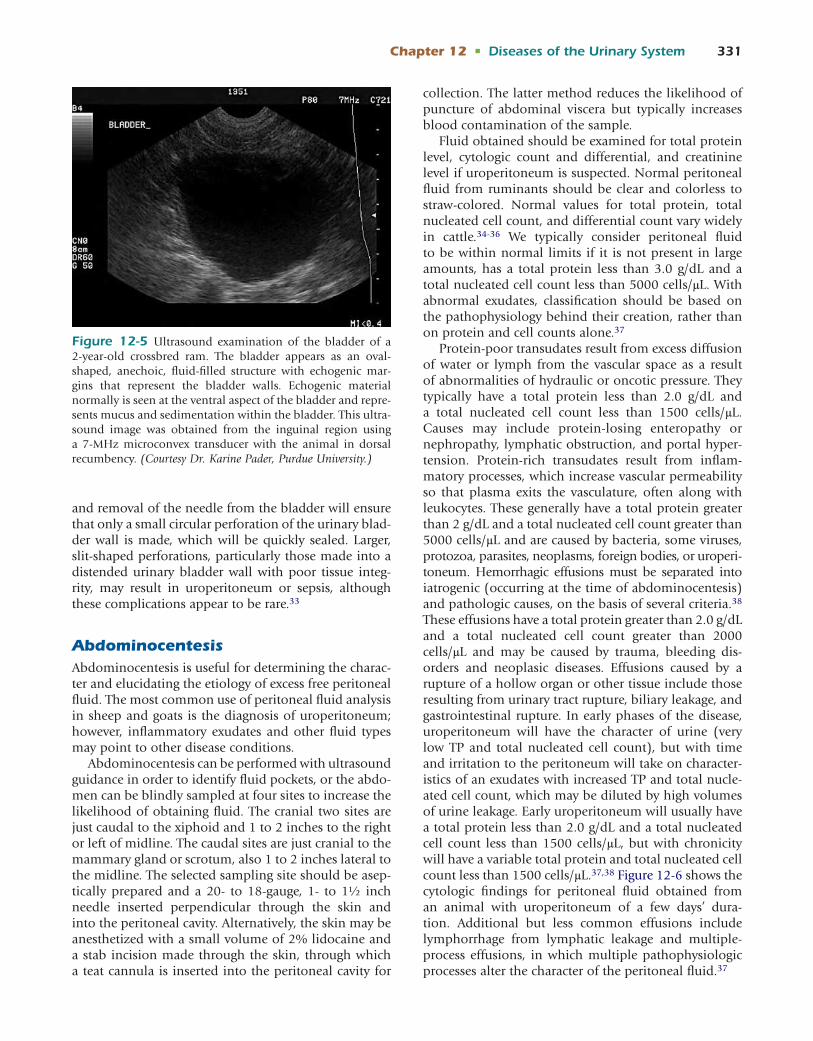
Chapter 12 • Diseases of the Urinary System 331
and removal of the needle from the bladder will ensure that only a small circular perforation of the urinary blad-der wall is made, which will be quickly sealed. Larger, slit-shaped perforations, particularly those made into a distended urinary bladder wall with poor tissue integ-rity, may result in uroperitoneum or sepsis, although these complications appear to be rare.33
AbdominocentesisAbdominocentesis is useful for determining the charac-ter and elucidating the etiology of excess free peritoneal fluid. The most common use of peritoneal fluid analysis in sheep and goats is the diagnosis of uroperitoneum; however, inflammatory exudates and other fluid types may point to other disease conditions.
Abdominocentesis can be performed with ultrasound guidance in order to identify fluid pockets, or the abdo-men can be blindly sampled at four sites to increase the likelihood of obtaining fluid. The cranial two sites are just caudal to the xiphoid and 1 to 2 inches to the right or left of midline. The caudal sites are just cranial to the mammary gland or scrotum, also 1 to 2 inches lateral to the midline. The selected sampling site should be asep-tically prepared and a 20- to 18-gauge, 1- to 1½ inch needle inserted perpendicular through the skin and into the peritoneal cavity. Alternatively, the skin may be anesthetized with a small volume of 2% lidocaine and a stab incision made through the skin, through which a teat cannula is inserted into the peritoneal cavity for
collection. The latter method reduces the likelihood of puncture of abdominal viscera but typically increases blood contamination of the sample.
Fluid obtained should be examined for total protein level, cytologic count and differential, and creatinine level if uroperitoneum is suspected. Normal peritoneal fluid from ruminants should be clear and colorless to straw-colored. Normal values for total protein, total nucleated cell count, and differential count vary widely in cattle.34-36 We typically consider peritoneal fluid to be within normal limits if it is not present in large amounts, has a total protein less than 3.0 g/dL and a total nucleated cell count less than 5000 cells/μL. With abnormal exudates, classification should be based on the pathophysiology behind their creation, rather than on protein and cell counts alone.37
Protein-poor transudates result from excess diffusion of water or lymph from the vascular space as a result of abnormalities of hydraulic or oncotic pressure. They typically have a total protein less than 2.0 g/dL and a total nucleated cell count less than 1500 cells/μL. Causes may include protein-losing enteropathy or nephropathy, lymphatic obstruction, and portal hyper-tension. Protein-rich transudates result from inflam-matory processes, which increase vascular permeability so that plasma exits the vasculature, often along with leukocytes. These generally have a total protein greater than 2 g/dL and a total nucleated cell count greater than 5000 cells/μL and are caused by bacteria, some viruses, protozoa, parasites, neoplasms, foreign bodies, or uroperi-toneum. Hemorrhagic effusions must be separated into iatrogenic (occurring at the time of abdominocentesis) and pathologic causes, on the basis of several criteria.38 These effusions have a total protein greater than 2.0 g/dL and a total nucleated cell count greater than 2000 cells/μL and may be caused by trauma, bleeding dis-orders and neoplasic diseases. Effusions caused by a rupture of a hollow organ or other tissue include those resulting from urinary tract rupture, biliary leakage, and gastrointestinal rupture. In early phases of the disease, uroperitoneum will have the character of urine (very low TP and total nucleated cell count), but with time and irritation to the peritoneum will take on character-istics of an exudates with increased TP and total nucle-ated cell count, which may be diluted by high volumes of urine leakage. Early uroperitoneum will usually have a total protein less than 2.0 g/dL and a total nucleated cell count less than 1500 cells/μL, but with chronicity will have a variable total protein and total nucleated cell count less than 1500 cells/μL.37,38 Figure 12-6 shows the cytologic findings for peritoneal fluid obtained from an animal with uroperitoneum of a few days’ dura-tion. Additional but less common effusions include lymphorrhage from lymphatic leakage and multiple-process effusions, in which multiple pathophysiologic processes alter the character of the peritoneal fluid.37
Figure 12-5 Ultrasound examination of the bladder of a 2-year-old crossbred ram. The bladder appears as an oval-shaped, anechoic, fluid-filled structure with echogenic mar-gins that represent the bladder walls. Echogenic material normally is seen at the ventral aspect of the bladder and repre-sents mucus and sedimentation within the bladder. This ultra-sound image was obtained from the inguinal region using a 7-MHz microconvex transducer with the animal in dorsal recumbency. (Courtesy Dr. Karine Pader, Purdue University.)
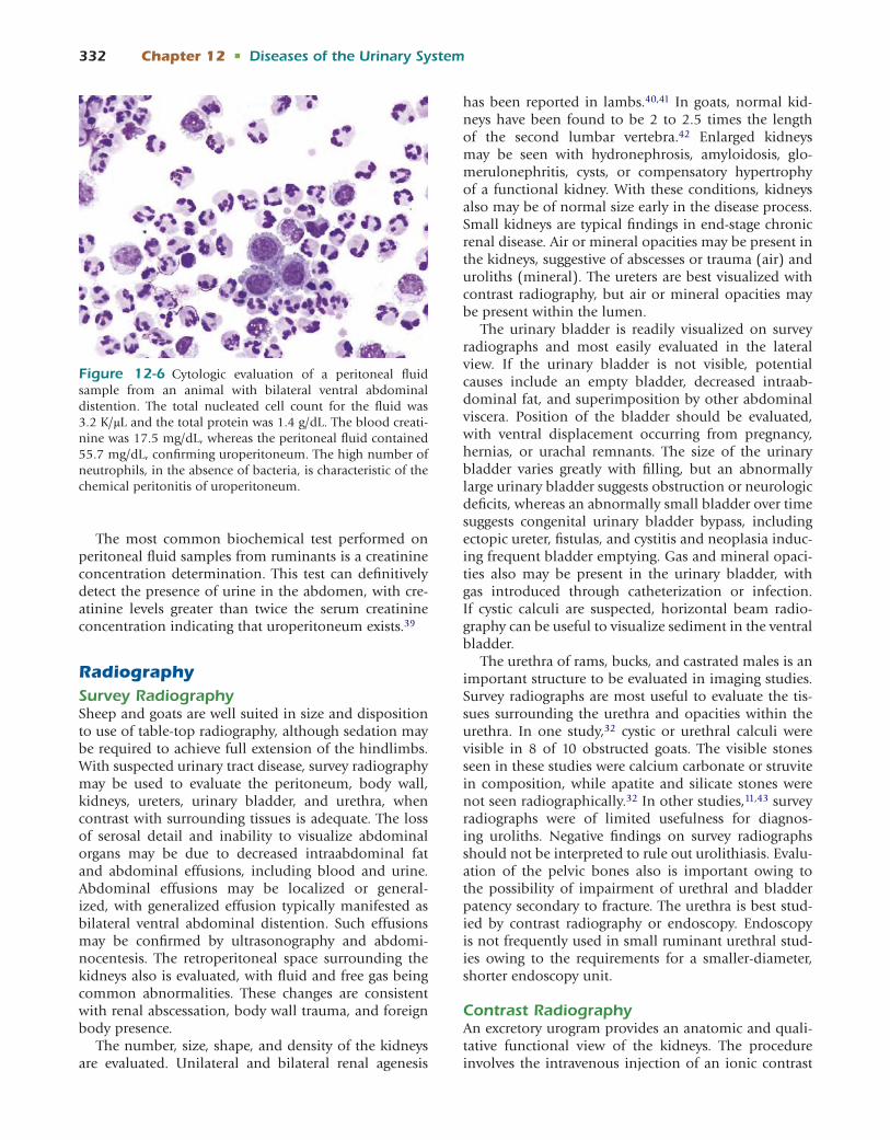
332 Chapter 12 • Diseases of the Urinary System
The most common biochemical test performed on peritoneal fluid samples from ruminants is a creatinine concentration determination. This test can definitively detect the presence of urine in the abdomen, with cre-atinine levels greater than twice the serum creatinine concentration indicating that uroperitoneum exists.39
RadiographySurvey RadiographySheep and goats are well suited in size and disposition to use of table-top radiography, although sedation may be required to achieve full extension of the hindlimbs. With suspected urinary tract disease, survey radiography may be used to evaluate the peritoneum, body wall, kidneys, ureters, urinary bladder, and urethra, when contrast with surrounding tissues is adequate. The loss of serosal detail and inability to visualize abdominal organs may be due to decreased intraabdominal fat and abdominal effusions, including blood and urine. Abdominal effusions may be localized or general-ized, with generalized effusion typically manifested as bilateral ventral abdominal distention. Such effusions may be confirmed by ultrasonography and abdomi-nocentesis. The retroperitoneal space surrounding the kidneys also is evaluated, with fluid and free gas being common abnormalities. These changes are consistent with renal abscessation, body wall trauma, and foreign body presence.
The number, size, shape, and density of the kidneys are evaluated. Unilateral and bilateral renal agenesis
has been reported in lambs.40,41 In goats, normal kid-neys have been found to be 2 to 2.5 times the length of the second lumbar vertebra.42 Enlarged kidneys may be seen with hydronephrosis, amyloidosis, glo-merulonephritis, cysts, or compensatory hypertrophy of a functional kidney. With these conditions, kidneys also may be of normal size early in the disease process. Small kidneys are typical findings in end-stage chronic renal disease. Air or mineral opacities may be present in the kidneys, suggestive of abscesses or trauma (air) and uroliths (mineral). The ureters are best visualized with contrast radiography, but air or mineral opacities may be present within the lumen.
The urinary bladder is readily visualized on survey radiographs and most easily evaluated in the lateral view. If the urinary bladder is not visible, potential causes include an empty bladder, decreased intraab-dominal fat, and superimposition by other abdominal viscera. Position of the bladder should be evaluated, with ventral displacement occurring from pregnancy, hernias, or urachal remnants. The size of the urinary bladder varies greatly with filling, but an abnormally large urinary bladder suggests obstruction or neurologic deficits, whereas an abnormally small bladder over time suggests congenital urinary bladder bypass, including ectopic ureter, fistulas, and cystitis and neoplasia induc-ing frequent bladder emptying. Gas and mineral opaci-ties also may be present in the urinary bladder, with gas introduced through catheterization or infection. If cystic calculi are suspected, horizontal beam radio-graphy can be useful to visualize sediment in the ventral bladder.
The urethra of rams, bucks, and castrated males is an important structure to be evaluated in imaging studies. Survey radiographs are most useful to evaluate the tis-sues surrounding the urethra and opacities within the urethra. In one study,32 cystic or urethral calculi were visible in 8 of 10 obstructed goats. The visible stones seen in these studies were calcium carbonate or struvite in composition, while apatite and silicate stones were not seen radiographically.32 In other studies,11,43 survey radiographs were of limited usefulness for diagnos-ing uroliths. Negative findings on survey radiographs should not be interpreted to rule out urolithiasis. Evalu-ation of the pelvic bones also is important owing to the possibility of impairment of urethral and bladder patency secondary to fracture. The urethra is best stud-ied by contrast radiography or endoscopy. Endoscopy is not frequently used in small ruminant urethral stud-ies owing to the requirements for a smaller-diameter, shorter endoscopy unit.
Contrast RadiographyAn excretory urogram provides an anatomic and quali-tative functional view of the kidneys. The procedure involves the intravenous injection of an ionic contrast
Figure 12-6 Cytologic evaluation of a peritoneal fluid sample from an animal with bilateral ventral abdominal distention. The total nucleated cell count for the fluid was 3.2 K/μL and the total protein was 1.4 g/dL. The blood creati-nine was 17.5 mg/dL, whereas the peritoneal fluid contained 55.7 mg/dL, confirming uroperitoneum. The high number of neutrophils, in the absence of bacteria, is characteristic of the chemical peritonitis of uroperitoneum.

Chapter 12 • Diseases of the Urinary System 333
medium, with sequential radiographs taken up to 40 minutes after injection. Patients undergoing excre-tory urography should be adequately hydrated, because opacification of the kidneys is dependent on glomeru-lar filtration. In normal goats, the kidneys are best visualized if radiographs are taken immediately after injection.42 Delayed or reduced opacification indicates dehydration or inadequate glomerular filtration. In evaluation of ureteral patency, an important consider-ation is that normal peristalsis can appear as a stricture or narrowing of the lumen. Ureteral patency can be altered by uroliths, blood clots, inflammatory exudates, trauma, or stricture. Excretory urography, in some patients, may not provide visualization of the urinary bladder and urethra.43
Contrast cystography and urethrography may be per-formed in normograde or retrograde fashion and with negative (air) or positive (organic iodide) contrast media. Barium should never be used for urinary imag-ing. Indications for such procedures include dysuria, pollakiuria, and chronic hematuria. Cystourethrog-raphy is best performed in a normograde fashion through a cystotomy tube.43 For retrograde studies, catheterization may be performed completely only in the female ruminant but may be partially completed in male ruminants. Occlusion of the distal urethral orifice after partial catheter passage allows for the instillation of contrast media in retrograde fashion. Alternatively, a precurved cardiac catheter may be utilized to bypass the urethral diverticulum.11 For retrograde studies, a ballooned catheter is used and passed into the uri-nary bladder in females, or a nonballooned catheter is passed a few inches into the bladder in males, and con-trast material is instilled. Use of intraluminal contrast material allows for assessment of degree of patency and wall thickness of the urinary bladder and urethra. Mural masses may be visualized and may be caused by cellular or fibrous infiltration. Filling defects of the urinary blad-der may be caused by polyps, air, calculi, blood clots, or inflammatory exudates, and urachal diverticula may be seen.44 Filling defects of the urethra may be caused by air bubbles, calculi (which may be radiopaque or radiolucent), blood clots, neoplasms, inflammation, scar tissue, or extramural compression. Extravasation of contrast material may result from traumatic lacerations, fistulas (urethrorectal and urethrovaginal), and divertic-ula. Fistula between the urethra and corpus spongiosum has been diagnosed by contrast radiography in a goat-sheep after surgery for obstructive urolithiasis.44
Renal BiopsyRenal biopsy is not commonly performed but may provide antemortem diagnosis of metabolic, neoplastic and toxic diseases of the kidney. In cases in which renal abscess is suspected, renal biopsy should be performed
by fine needle aspiration to reduce the risk of localized or generalized peritonitis.
The biopsy procedure should be performed in a well-restrained or adequately sedated animal under ultra-sound guidance. The skin overlying the last 2 or 3 ribs and paralumbar region is aseptically prepared, the skin and body wall are anesthetized with 2% lidocaine, and a stab incision is made for introduction of the biopsy instrument. The ultrasound probe may be placed in a sterile glove filled with ultrasound gel to maintain asep-sis and to locate the target kidney. A 14-gauge biopsy instrument is directed into the kidney parenchyma and a sample obtained. Depending on the testing required, the biopsy specimen should be divided, with both fresh and fixed tissue submitted to a reference labora-tory. Potential complications of renal biopsy include hematuria, hematoma, hemoabdomen, and peritonitis. In a retrospective study in 25 cattle, ultrasound-guided percutaneous renal biopsy resulted in a small subcap-sular hematoma (less than 2 cm in diameter) after the procedure in 6 of the animals, but no gross or occult hematuria.45 Another study using serial laparoscopic biopsies in cattle resulted in microscopic hematuria for 1 to 5 days.46
REFERENCES 1. DeRossi R, Juniqueira AL, Beretta MP: Analgesic and systemic
effects of ketamine, xylazine, and lidocaine after subarachnoid administration in goats, Am J Vet Res 64:51–56, 2003.
2. Thurmon JC, et al: Effects of xylazine hydrochloride on urine in cattle, Aust Vet J 54:178–180, 1978.
3. Waner T, Harrus S: Anemia of inflammatory disease. In Feldman BF, Zinkl JG, Jain NC, editors: Schalm’s Veterinary hematology, ed 5, Philadelphia, 2000, Lippincott Williams & Wilkins, pp 205–209.
4. Kramer JW: Normal hematology of cattle, sheep, and goats. In Feldman BF, Zinkl JG, Jain NC, editors: Schalm’s Veterinary hematology, ed 5, Philadelphia, 2000, Lippincott Williams & Wilkins, pp 1075–1084.
5. Braun U, Stehle C, Ehrensperger F: Clinical findings and treat-ment of listeriosis in 67 sheep and goats, Vet Rec 150:38–42, 2002.
6. de la Concha-Bermejillo A, et al: Severe persistent orf in young goats, J Vet Diagn Invest 15:423–431, 2003.
7. Garry F, et al: Renal excretion of creatinine, electrolytes, protein, and enzymes in healthy sheep, Am J Vet Res 51:414–419, 1990.
8. Mitchell AR, Moss P: Responses to reduced water intake, includ-ing dehydration natriuresis, in sheep excreting sodium predomi-nantly in urine or in faeces, Exp Physiol 80:265–274, 1995.
9. Ammerman CB, et al: Ruminant utilization of inorganic phos-phates, J Anim Sci 16:796–810, 1957.
10. Garrett PD: Urethral recess in male goats, sheep, cattle, and swine, J Am Vet Med Assoc 191:689–691, 1987.
11. Van Weeren PR, Klein WR, Voorhout G: Urolithiasis in small ruminants II. Cysto-urethrography as a new aid in diagnosis, Vet Q 9:79–82, 1987.
12. Osborne CA, Stevens JB: Urinalysis: a clinical guide to compassionate patient care, Shawnee Mission, Kan, 1999, Bayer Corporation.
13. Nappert G, Naylor JM: A comparison of pH determination meth-ods in food animal practice, Can Vet J 42:364–367, 2001.
14. Stockham SL, Scott MA: Urinary system. In Stockham SL, Scott MA, editors: Fundamentals of veterinary clinical pathology, ed 2, Ames, Iowa, 2008, Blackwell, pp 277–336.

334 Chapter 12 • Diseases of the Urinary System
15. Lunn DP, et al: Renal net acid and electrolyte excretion in an experimental model of hypochloremic metabolic alkalosis in sheep, Am J Vet Res 51:1723–1731, 1990.
16. McDougall EI: Proteinuria of newborn suckling ruminants, Biochem J 94:101–105, 1095.
17. Carlson GP: Clinical chemistry tests. In Smith BP, editor: Large animal internal medicine, ed 3, St Louis, 2002, Mosby, pp 389–412.
18. Cutler JT: Studies of the carbohydrate metabolism of the goat, J Biol Chem 106:653–666, 1934.
19. Uzal FA, Kelly WR: Experimental Clostridium perfringens Type D enterotoxemia in goats, Vet Pathol 35:132–140, 1998.
20. Setty DRL, Narayana K: A case of non-febrile haemoglobinuria in a she goat, Indian Vet J 52:149, 1975.
21. Stamp JT, Stewart J: Haemolytic anaemia with jaundice in sheep, J Comp Pathol 63:48–52, 1953.
22. Winter A, Clarkson M: Anaemia in lambs and kids caused by feeding cow colostrum, In Pract 14:283–286, 1992.
23. Perk K, Frie YF, Herz A: Osmotic fragility of red blood cells in young and mature domestic and laboratory animals, Am J Vet Res 25:1241–1248, 1964.
24. Fernandez A, et al: Clinicopathological features in ovine AA amy-loidosis, Res Vet Sci 75:203–208, 2003.
25. Price RG: Urinary enzymes, nephrotoxicity and renal disease, Toxicology 23:99–134, 1982.
26. Garry F, et al: Renal excretion of creatinine, electrolytes, protein, and enzymes in healthy sheep, Am J Vet Res 51:414–419, 1990.
27. Garry F, Chew DJ, Hoffsis GF: Enzymuria as an index of renal damage in sheep with induced aminoglycoside nephrotoxicosis, Am J Vet Res 51:428–432, 1990.
28. Braun U, Schefer U, Fohn J: Urinary tract ultrasonography in normal rams and in rams with obstructive urolithiasis, Can Vet J 33:654–659, 1992.
29. Braun U, Schefer U, Gerber D: Ultrasonography of the urinary tract of female sheep, Am J Vet Res 53:1734–1739, 1992.
30. Hallowell GD: Abdominal ultrasound. Proceedings of the 26th Annual American College of Veterinary Internal Medicine Forum, San Antonio, Tex, 2008.
31. Floeck M: Sonographic application in the diagnosis of pyelone-phritis in cattle, Vet Radiol Ultrasound 48:74–77, 2007.
32. Halland SK, House JK, George LW: Urethroscopy and laser lithro-tripsy for the diagnosis and treatment of obstructive urolithiasis in goats and pot-bellied pigs, J Am Vet Med Assoc 12:1831–1834, 2002.
33. Janke JJ, et al: Use of Walpole’s solution for treatment of goats with urolithiasis: 25 cases (2001-2006), J Am Vet Med Assoc 234:249–252, 2009.
34. Kopcha M, Schulze AE: Peritoneal fluid. II. Abdominocentesis in cattle and interpretation of noneoplastic samples, Comp Cont Educ Pract Vet 13:703–710, 1991.
35. Anderson DE, et al: Comparative analyses of peritoneal fluid from calves and adult cattle, Am J Vet Res 56:973–976, 1995.
36. Wilson AD, Hirsch VM, Osborne AD: Abdominocentesis in cattle: technique and criteria for diagnosis of peritonitis, Can Vet J 26:74–80, 1985.
37. Stockham SL, Scott MA: Cavity effusions. In Stockham SL, Scott MA, editors: Fundamentals of veterinary clinical pathology, ed 2, Ames, Iowa, 2008, Blackwell, pp 831–868.
38. Wilson DG, MacWilliams PS: An evaluation of the clinical patho-logic findings in experimentally induced urinary bladder rupture in pre-ruminant calves, Can J Vet Res 62:140–143, 1998.
39. Sockett DC, et al: Metabolic changes due to experimentally induced rupture of the bovine urinary bladder, Cornell Vet 76:198–212, 1986.
40. Dennis SM: Urogenital defects in sheep, Vet Rec 105:344–347, 1979.
41. Hartley WJ, Kater JC: Perinatal disease conditions of sheep in New Zealand, N Z Vet J 12:49–57, 1964.
42. Cegarra IJ, Lewis RE: Excretory urography in the goat (Capra hircus), Am J Vet Res 38:1129–113, 19772.
43. Palmer JL, et al: Contrast radiography of the lower urinary tract in the management of obstructive urolithiasis in small ruminants and swine, Vet Radiol Ultrasound 39:175–180, 1998.
44. Cruz-Arambulo R de J, et al: What is your diagnosis? Communi-cation between the urethra and the corpus spongiosum, urethral stricture, mild cystitis, and presence of a urachal diverticulum, J Am Vet Med Assoc 222:1211–1212, 2003.
45. Mohamed T, Oikawa S: Efficacy and safety of ultrasound-guided percutaneous biopsy of the right kidney in cattle, J Vet Med Sci 70:175–179, 2008.
46. Naoi M, et al: Laparoscopic-assisted serial biopsy of the bovine kidney, Am J Vet Res 46:699–702, 1985.
DISEASES OF THE KIDNEYSIn clinical practice, kidney disease is not commonly encountered as a primary problem in small ruminants; however, incidental kidney pathology often can be identified at necropsy.1 Kidney disease is described based on duration (acute versus chronic) and the char-acter of renal damage leading to dysfunction (glomeru-lar, tubular, and vascular). The clinician’s challenge is to recognize situations resulting from primary renal disease or risks leading to secondary or induced renal damage. Clinical tendencies with kidney disease are anuria, oliguria, dysuria, abdominal pain, and abnor-mal urinary constituents. Appearance of clinical signs in small ruminants often is synchronized with the multisystem disease processes that lead to renal dam-age. Therefore recognition of risk factors, preemptive case management practices, ancillary diagnostics, and postmortem diagnosis are important in overall disease management and prevention.
General causes of kidney disease are: • Infectious (bacterial, viral, and parasitic) • Toxic (chemical, heavy metals, medications, and
plant origin) • Obstruction and trauma (nephroliths, direct) • Secondary hydronephrosis from ureteral, cystic, and
urethral calculi • Vascular (infarcts, hyperdynamics of sepsis and
toxemia) • Chronic inflammation (glomerulonephritis,
amyloidosis) • Congenital
RENAL FAILURERenal failure occurs when diminished renal function results in persistent metabolic abnormalities such as azotemia as well as the inability to concentrate urine. Renal failure that develops rapidly, within a few hours

Chapter 12 • Diseases of the Urinary System 335
or days, constitutes acute renal failure (ARF) and usually is due to intrinsic (vascular, toxic) causes from systemi-cally absorbed toxins, body origin toxins (myoglo-bin, hemoglobin, urea), administered therapeutics, or dynamic changes in renal blood flow with sepsis, shock, or toxemia. The kidneys receive a large proportion of the circulating blood volume, resulting in high rates of toxin exposure, as well as increased vulnerability to ischemia and reperfusion injury with diseases causing hyperdynamic changes in nutrient blood flow. Toxin exposure is amplified as renal tubules resorb filtered toxins in conjunction with the normal function of urine concentration. Damage to this sensitive portion of the nephron may result in acute tubular necrosis, eventu-ating in loss of urine-concentrating ability associated with increased urinary levels of protein, glucose, and electrolytes. Consequently, the kidneys provide a good postmortem diagnostic sample for toxins, and urinaly-sis can provide objective information about the nature of disease (see Chapter 20). The clinician can assume that a degree of damage is occurring during shock, septicemia, dehydration, or toxemia and should take preemptive steps in preservation and protection of renal function during case management. Changes in blood flow or oxygen delivery to the kidney cause renal insuf-ficiency, potentially leading to acute or chronic renal failure. Dehydration, heat stress, severe rumen bloat, sepsis, and anemia result in physiologic and metabolic changes leading to kidney dysfunction due to decreased cardiac output and renal vasoconstriction and dilation.
Treatment of ARF should include removing any offending toxin or source, promoting diuresis through intravenous fluid administration and diuretic medica-tions, correcting acid-base and electrolyte derangements, and close monitoring of positive or adverse responses to treatment. Intravenous fluids of choice are 0.9% saline and 0.45% saline plus dextrose. Additional intravenous potassium can be substituted if indicated after initial therapy, with care taken to keep the dose below the gen-erally recommended toxicity rate of 0.5 mEq/kg/hour. Diuresis should continue until the patient is producing sufficient volumes of urine. Serum potassium levels should be monitored and parenteral support provided when furosemide (1 mg/kg every 2 to 3 hours to effect) is used. Alternatively, mannitol (1 g/kg per bolus) can be given to provide osmotic diuresis. Additional supportive therapy may include broad-spectrum antimicrobials against susceptible infectious agents, plasma to treat hypoproteinemia, antiinflammatories, and nutritional support by means of parenteral nutrition or rumen transfaunation. Urinalysis and fractional excretion of electrolytes can be measured in conjunction with serum monitoring parameters during intensively managed cases (see earlier section “Ancillary Diagnostic Testing”).
Vasopressors and inotropes can be instituted as adjunct therapy but need to be administered as carefully
calibrated constant-rate infusions (CRIs); ranges of therapeutic efficacy are wide as well as widely debated, and scientific data for use in small ruminants are largely extrapolated from other species including humans. Much of the human literature, however, is based on information gained from sheep models of disease. In a regimen developed in clinical practice based in part on information gathered from other sources,2 persis-tent oliguria or anuria can be treated using intravenous dopamine (2 to 5 μg/kg/minute), dobutamine (5 to 10 μg/kg/minute), and a combination of norepinephrine (0.4 μg/kg/minute) plus dobutamine (5 μg/kg/ minute). Of note, the use of dopamine alone in cases of ARF may not be as beneficial as was once thought, and additional potential adverse effects have been discovered.3,4
Progressive loss of renal function over a period of months or years describes chronic renal failure (CRF). In contrast with ARF, in which nephron repair and compensatory hypertrophy can occur spontaneously and with treatment, chronic disease results in progres-sive, permanent, and irreparable damage and fibrosis to the nephron. CRF may be due to secondary glomeru-lonephritis or tubulointerstitial disease resulting from immune complex deposition secondary to a distant chronic inflammatory process (abscess, pneumonia). Clinical signs may be non-existent or limited to fail-ure to thrive. Polyuria or polydypsia is not a frequent historical complaint but may be detected during hos-pitalization. Antemortem diagnosis is supported by serum markers of azotemia, urine dilution, urinalysis (proteinuria, pyuria), renal biopsy and ultrasound char-acteristics of the kidney. Unique characteristics of serum urea nitrogen and creatinine metabolism in ruminants should be reviewed for accurate interpretation (see ear-lier under “Ancillary Diagnostic Testing”). Ultrasound examination may reveal small, irregular, echodense kidney parenchyma with loss of detail at the corticome-dullary junction, evidence of fluid accumulation (pyelo-nephritis, hydronephrosis), or echodense foci within the renal calices, possibly representing nephroliths. Nonregenerative anemia, hyperkalemia, and other elec-trolyte abnormalities may be detected on blood work. General causes of CRF include a previous episode of ARF with lasting effects and insufficient compensation, pyelonephritis, amyloidosis, and congenital disorders or idiopathic causes. Treatment is supportive to palliative.
REFERENCES 1. Sankarappa EV, Rao PR: Renal lesions in sheep and goats in
Andhra Pradesh, Indian Vet J 59:705–708, 1982. 2. Corley KTT: Inotropes and vasopressors in adults and foals, Vet
Clin North Am Eq Pract 20:77–106, 2004. 3. Trim CM, Moore JN, Clark ES: Renal effects of dopamine infusion
in conscious horses, Equine Vet J Suppl 71:24–28, 1989. 4. Kellum JA, Decker JM: Use of dopamine in acute renal failure: a
meta-analysis, Crit Care Med 29:1526–1531, 2001.

336 Chapter 12 • Diseases of the Urinary System
ACUTE RENAL DISEASESInfectious DiseasesClostridium perfringens Type DDisease syndromes caused by Clostridium perfringens type D are referred to as enterotoxemia, overeating disease, and pulpy kidney disease. The 2001 U.S. Department of Agriculture (USDA)-sponsored National Animal Health Monitoring System (NAHMS) sheep survey revealed that 38.8% of sheep flocks had suspected or confirmed cases of enterotoxemia, with 30.9% confirmed by vet-erinary or laboratory examination, in the previous three years.1 Enterotoxemia most commonly is seen in young, growing animals consuming diets high in rapidly fer-mentable carbohydrates. High milk or starch content allows for excess colonization of the jejunum with C. perfringens type D, which produces alpha and epsilon toxins,2,3 of which epsilon is the more significant in disease. Epsilon toxin is activated in the intestine and is systemically absorbed, resulting in increased capillary permeability from a loss of endothelial integrity,2 and an influx of protein and fluid occurs in the organs and body cavities. Sheep more often experience the systemic form of the disease, characterized by edema throughout the body, including the brain, lungs and kidneys, often resulting in acute death, whereas goats more often are affected by hemorrhagic enterocolitis.4,5 The systemic form most often seen in sheep commonly results in acute death, but live animals may exhibit seizures, blindness, recumbency, dyspnea,6 and other signs con-sistent with fluid accumulation in and around organs.
At necropsy, visceral edema, serosal hemorrhage, and cavitary effusions may be present, but death from C. perfringens type D infection also may result in no gross lesions. The cortices of the kidney may be soft-ened and show subcapsular petechiae.6 Epsilon toxin promotes liver glycolysis, resulting in hyperglycemia and glucosuria, making dipstick evaluation of bladder urine a useful test for investigation of acute death in lambs and kids. After experimental infection in sheep, ileum has been found to be the best sample for isola-tion of epsilon toxin by enzyme-linked immunosorbent assay (ELISA).7 Histopathologic examination of the brain reveals microangiopathy with protein surround-ing the arteries and veins, which is pathognomonic for C. perfringens type D infection,6,8 but no lesions may be seen in the kidneys.8 Negative results on testing for C. perfringens type D by any of these means does not nec-essarily rule out the pathogen as the cause of disease.6,8
Prevention of C. perfringens type D infection is of utmost importance and should include management using vaccination and gradual dietary adaptation. Bac-terin toxoids as well as antitoxins are commercially available. Antitoxins are most useful in outbreak situa-tions, because they provide rapid passive immunity, but preemptive use of bacterin toxoids for prolonged, active
immunity is preferred for protection against systemic disease. It is recommended that ewes be vaccinated using a C. perfringens type D toxoid–containing vaccine 3 to 4 weeks ante partum, which provided passive pro-tection in lambs up to 12 weeks of age.9 No benefit has been seen with vaccination of lambs before the age of 6 weeks,9 so a potential recommendation is to vaccinate lambs at 6 to 10 weeks of age, with a booster vaccina-tion given 1 month later. Although the vaccine is read-ily available, inexpensive, and effective, currently only 48.4% of sheep producers in the United States vaccinate breeding or replacement ewes and 66.9% vaccinate nursing lambs and 44.8% vaccinate feeder lambs after weaning, demonstrating a need for producer education based on available research.9
LeptospirosisSheep and goats may become infected by a number of serovars of Leptospira interrogans, resulting in several clinical syndromes, bacterial clearance, or a subclini-cal carrier state.10 The kidneys may become damaged from leptospirosis through hemolysis and interstitial nephritis.
Infected urine is the primary source of infection, with animals obtaining the bacteria from contaminated water or the urine of herdmates, wildlife, rodents, or other domestic animals.11 In one study of experimental infection of sheep with L. interrogans serovar Pomona, clinical disease occurred 34 days after experimental infection.12 Because the bacteria can penetrate intact mucous membranes and because leptospirosis is con-sidered to be the most widespread zoonosis in the world,13 it should be respected as an occupational hazard for veterinarians, staff, and livestock producers.
Animals presented with leptospirosis may show general malaise, fever, icterus, anemia, azotemia, and hemoglobinuria.14 Hemolytic changes in blood analysis are seen 4 to 8 days after infection.14 Total white blood cell counts are often elevated with a neutrophilia.15 A positive result for hemoglobin may occur 3 to 8 days after infection.14 Urine sediment exam may show cellular or proteinaceous tubular casts. The herd or flock history may reveal reproductive manifestations as well, including infertility, abortions, and stillbirths. On necropsy, the carcass is icteric,14 and the kidneys appear dark red and swollen, with pale foci in the cortices, and the liver often is yellow or copper-colored.16 Histopathologic examination reveals a diffuse acute or chronic interstitial nephritis,15,17 and organisms may be observed. Loss of the brush border is a common feature, and necrotic epithelial cells may be seen within the tubules.12
Diagnosis is based on increasing serologic titers in the acute and convalescent periods, using the micro-scopic agglutination test (MAT),18 complement fixa-tion (CF) test, or ELISA. CF antibodies are short-lived

Chapter 12 • Diseases of the Urinary System 337
(13 to 18 weeks), whereas MAT antibodies can be detected for longer periods after infection.14 On urine, polymerase chain reaction (PCR) assay, darkfield microscopy, or culture may be used. PCR assay is pre-ferred, because darkfield microscopy has yielded false-negative results in infected animals,14 and culture of urine generally is unrewarding owing to difficulties in growth in artificial media and intermittent shedding.19 Histopathologic analysis and immunofluorescent anti-body (IFA) testing20 may identify organisms in renal tissue. One study has determined that, in herd or flock situations, using MAT for herd-level screening, followed by urine PCR assay is suitable for identification of car-rier animals.19
Several serovars are reported in small ruminants including L. interrogans serovars Pomona, Hardjo, Grippotyphosa, Icterohemorhagiae, Canicola, and Bratislava.18-,23 L. interrogans serovar Pomona appears to be the most commonly associated with interstitial nephritis and hepatic centrilobular necrosis.12,17 It also has been shown to cause severe hemolytic anemia in lambs.15,17 In one case in lambs, the kidneys were nega-tive for leptospiral organisms, but a rising titer to the Pomona serovar was observed.15 Ewes administered the hemolysin of L. interrogans serovar Pomona experienced a reduction in hemoglobin levels to 57% of the nor-mal range within 48 hours and had lesions similar to animals infected with the whole organism.16 Necropsy findings included placental separation and autolysis of caruncles and cotyledons in some pregnant ewes16 (see Chapter 20). Pigs are the natural reservoir host of L. interrogans serovar Pomona.
L. interrogans serovar Hardjo is host-adapted to cattle17 and sheep.22,24 It has also been reported in a sheep found acutely dead23 with organisms found in the renal tubular epithelium and tubular lumen. Flock-mates of this animal were seropositive against serovar Hardjo, with small numbers seropositive against other serovars.23 One study comparing the hemolytic proper-ties of three serovars of leptospires found serovar Hardjo to be more hemolytic than serovar Pomona.21 Sheep with renal infections from serovar Hardjo may not harbor the bacterium in their reproductive tracts,24,25 whereas sheep experimentally infected with serovar Hardjo-bovis show renal localization and harbor the bacteria up to 242 days after infection.25 Treatment of leptospirosis consists of intravenous crystalloid fluid therapy combined with blood transfusion in clinically anemic animals. Because of the difficulty in culturing the organism, antimicrobial therapy is based upon anticipated spectrum of coverage. In cattle shedding leptospires in the urine, the following antibiotic regi-mens were shown to clear urinary shedding of organ-isms: oxytetracycline (20 mg/kg IM as a one-time dose), tilmicosin (10 mg/kg SC as a one-time dose), dihydrostreptomycin-penicillin G (25 mg/kg IM once),
or ceftiofur sodium (1-2.2 mg/kg IM once daily for 5 days, or 20 mg/kg IM once daily for 3 days). It should be noted that tilmicosin is toxic to goats and should only be used in sheep.26,27 Vaccination for control of leptospirosis may be useful in reducing urinary shed-ding, but should not be relied upon for protection from disease. Bovine-labeled vaccines are commonly used and suffer from questionable efficacy and duration of immunity even in label species.28-30 In designing man-agement plans for leptospirosis, consideration should be given to biosecurity for new additions, control of access to wild and domestic animals, and the accessibil-ity of potentially contaminated water sources.
Adenovirus InfectionMany serotypes of adenovirus infect sheep worldwide, with varied tissue tropism and unpredictable patho-genicity. Exposure and seroconversion are common and it has been identified in the interstitial vascu-lature of lamb kidneys during routine postmortem examination.31,32 The virus can be isolated from nasal secretions and feces of healthy sheep as well as those with respiratory or gastrointestinal disease. Many infec-tions go unnoticed; however, the virus has been diag-nosed as the cause of sudden death in an outbreak involving several young lambs in the United States.33 Histopathologic analysis in this report revealed a highly cellular, mainly cortical interstitial nephritis, with intra-nuclear inclusion bodies, in all three lambs in which the virus was identified. Evidence of systemic disease also was apparent with hepatic necrosis. Experimen-tal infection with some strains has resulted in similar pathologic findings.34
General supportive therapy may be beneficial; how-ever, clinical observations indicate rapid progression of disease once recognized. Recognition and preventive management practices should be carried out if prob-lems develop. Specific vaccines are not available.
Lamb NephrosisA yet-to-be-determined cause of acute necrotic nephro-sis in young lambs (less than 1 month of age) has been described as a condition seen during the early grazing season, particularly in the United Kingdom and sur-rounding regions.35-38 Occasional sporadic occurrences are documented in various governmental disease sur-veillance reports to date. Associated causes for nephro-sis such as coccidia and other gastrointestinal parasites, Salmonella, pestivirus, and plant, chemical, and gen-tamicin toxicity in young lambs should be ruled out before a “definitive” diagnosis of idiopathic nephrosis is made. Nephrosis and associated staphylococcal skin scald syndrome also have been reported in two approxi-mately 5-month-old Merino lambs.39
A progressive, terminal illness develops over a few days’ time. Lethargy, weakness, and ataxia progress to
338 Chapter 12 • Diseases of the Urinary System
recumbency and death. Diarrhea and dehydration also are common. Serum chemistry and urinalysis findings are consistent with a nephrotic syndrome with azote-mia, hypoalbuminemia, and proteinuria and “inactive” urine sediment. On necropsy the kidneys are large, soft, and pale. Microscopic examination reveals renal tubular degeneration and necrosis affecting predominantly the proximal convoluted tubules and renal cortices. Hyaline to fibrinlike casts may be seen in the distal tubules and collecting ducts with dilation of the distal convoluted tubules. Focal glomerular lesions also may be observed.36
Supportive fluid and electrolyte therapy are indi-cated. Preventive measures aimed at concurrent disease processes should be instituted.
Toxic DiseasesToxic insult to the kidney occurs with a wide variety of substances (bacterial endotoxins, plants, metals, body metabolites) and array of severity. Toxic nephropathy generally occurs due to vascular (ischemia and reperfu-sion) dynamics and direct tubular injury. Glomerular damage may also occur. Endogenous toxicity from hemolytic disease and myopathies may lead to delivery of large amounts of hemoglobin or myoglobin causing renal vasoconstriction and tubular obstruction from protein coagulation.
PlantsNephrotoxic plants are common sources of acute kid-ney disease; tannins (oak), soluble and insoluble oxalate-containing plants (many), vitamin D plants (cestrum, solanum), and sudan or sorghum are potential sources of nephrotoxins in grazing and fed animals.40,41 A list of common nephrotoxic plants is presented in Table 12-1; family, genus and species, and common names are included to help the practitioner in investiga-tions to find more specific information.
Seasonal variation in toxicity may be observed, as seen with Quercus spp. (oak), and ruminants may undergo rumen adaptation to safely graze toxic plants such as those containing oxalates. Herbicides such as the defoliant paraquat, if the treated pasture is grazed soon after application, may cause acute renal tubular necrosis in addition to respiratory symptoms. Pet sheep and goats or those escaping pastures to explore may become exposed to ornamental plants with toxic princi-ples (e.g., lilies, ivy, oleander).42,43 Many of the nephro-toxic plants, particularly those causing oxalate toxicity, are nitrate accumulators as well. Plant-origin nephro-toxins often also exhibit mixed effects on cardiac, gas-trointestinal, respiratory, and hepatic systems. Specific mechanisms of action may not yet be determined in some direct nephrotoxic plants such as Isotropis spp.,44 and the kidneys are susceptible to secondary damage from toxins primarily affecting other organ systems.
Clinical manifestations and pathologic lesions most often are consistent with acute tubular necrosis with exposures to those plants with affinity for inducing nephrotoxicity. Clinical signs usually are acute-onset lethargy, depression, oliguria, and neurologic signs. Anticipated clinicopathologic findings are hyponatre-mia, hypochloremia, hypocalcemia, hyperkalemia, met-abolic alkalosis, azotemia, hyposthenuria, enzymuria, glucosuria, dark tubular casts, and changes in fractional excretion of electrolytes. Gross necropsy usually reveals swollen, pale to dark, edematous kidneys and perire-nal edema. Histopathologic lesions generally feature tubular damage characterized by necrotic epithelial tubular desquamation, hyaline casts with distal tubular obstruction, intact basement membranes, and relatively unaffected glomeruli. Oxalate toxicity may cause urolith and nephrolith formation and polarizing calcium oxa-late crystals may be seen in the tubular lumen.40 Gross and microscopic histopathologic examination reveals generalized calcification of tissues that accompany nephrotoxicity involving vitamin D-containing plants.
Acute toxicity often is treatable, because tubular regeneration is possible. The animals should be removed from the offending toxin.
Treatment and PreventionRumenotomy and evacuation can be considered if history indicates that exposure was less than 12 to 24 hours in duration (see Chapter 5). Fluid and electrolyte replacement therapy and correction of acid-base bal-ance also are indicated (see Chapter 3). Fluids should contain sodium chloride with added potassium and cal-cium. Saturated calcium hydroxide solution given orally may bind unabsorbed soluble oxalates.
The key management strategy is to prevent or mini-mize exposure. Farm personnel should be able to rec-ognize plants with potential to cause disease as well as seasonal variations, allowing more effective graz-ing management, and precautions to avoid exposure should be instituted. Animals that are hungry or starved are more likely to consume dangerous plants when given the opportunity. Ruminants can safely graze many oxalate-contaminated pastures if they are given time to adapt and if access to such pasturage is prevented during rapid growth phases of the plants. Feeding dicalcium phosphate, salt, and supplemental hay can minimize toxicity.
Ethylene GlycolAnother oxalogenic nephrotoxin of potential risk to small ruminants is ethylene glycol (EG). EG is a com-pound found in automotive anti-freeze coolants, brake, transmisison fluid and windshield cleaning fluids as well as a component of many industrial solvents and deter-gents, all of which are commonly found in farm envi-ronments. EG is converted to nephrotoxic metabolites,

Chapter 12 • Diseases of the Urinary System 339
mainly glycolic acid, by the liver which then cause renal damage. Calcium oxalate crystals also form and are deposited in renal tubules causing further kidney dam-age.45 Toxicity is commonly encountered in dogs and cats, but poisoning is rarely reported in farm animals. An early report in a calf was followed by experimental repro-duction in cattle.46 Lethal doses are 5 to 10 mL/kg in
mature cattle and 2 mL/kg in preruminating calves.46,47 The rumen provides some resistance to toxicity through normal oxalate metabolic degradation by microbes but also serves as a resevoir prolonging absorption of EG. Contaminated feedstuffs and byproducts may serve as sources of EG, thus feed analysis may be indicated with compatible clinical and diagnostic findings.48,49
TABLE 12-1 Some Commonly Implicated Nephrotoxic Plants
Plant FamilyCommon Names and Species Toxic Principle (Renal) Comments
Chenopodiaceae Lamb’s quarters (Chenopodium spp.)
Halogeton (H. glomerulatus)
Greasewood (Sarcobatus vermiculatus)
Russian thistle (Salsola spp.)
Mexican fireweed (Kochia scoparia)
Smotherweed (Bassia hyssopifolia)
Soluble sodium and potassium oxalatesPlants often are weeds found in
disturbed alkaline or acidic soils such as seen with overgrazed pastures, along roadsides or railways, in dry watersheds, lakes, floodplains
Plants generally not considered palatable; toxic consumption dose is variable, owing partly to rumen adaptation during gradual introduction but also to variable oxa-late concentration between plants
Green and dried forms of the plants are considered toxic
Often cause mixture of systemic clinical signs
Chenopodium spp. may cause GI signs owing to irritation from terpenes found in the plant oils
Kochia also may cause photosensitivity, toxic hepatitis, and polioencephalomalacia
Commonly accumulate nitrates
Polygonaceae Rhubarb (Rheum rhaponticum)
Beets/sugarbeets (Beta vulgaris)
Dock, orchard sor-rel, Indian tobacco (Rumex spp.)
Beta and Rumex spp. are nitrate accumulators
Oxalidaceae Wood sorrel, oxalis, lady’s sorrel (Oxalis spp.)
Potassium oxalates, very acidic
Amaranthaceae Pigweed (Amaranthus spp.)
May see perirenal edema and nephrosis at necropsy
Nitrate accumulatorsSolanaceae Jessamine/jasmine
(Cestrum diurnum)Nightshades (Solanum
malacoxylon)
Vitamin D–containing plants Cause generalized soft tissue mineralization, including glomerular and interstitial fibrosis Often concomitant GI and/or nervous clinical signs due to toxic alkaloids
Fagaceae Oak (Quercus spp.) Tannins (tannic acids) and pyrogallol from rumen conversion
Direct GI, liver, and kidney toxinsPyrogallol can cause
methemoglobinemia in sheep
All oak species considered toxicProlonged consumption of
immature leaf stages (spring) or acorns (fall)
GI, Gastrointestinal.

340 Chapter 12 • Diseases of the Urinary System
A clinical case of ethylene glycol intoxication has been reported in a pygmy goat with similar clinical signs and progression as reported in other species.50
Central neurologic signs (ataxia, depression, loss of menace), hypersalivation, tachypnea, gastrointes-tinal atony, and rumen bloat typically are described as presenting clinical complaints. Clinical signs are a result of initial degradation of EG to glyoxylic acid by dehydrogenases resulting in central nervous system depression and acidosis. A more commonly occuring differential diagnosis would be polioencephalomalacia. Hemolytic anemia, hemoglobinuria, and ocular signs may occur less commonly. Progressive depression and recumbency follow with oliguric or anuric renal failure, leading to death in 2 to 10 days, depending on exposure rate. Recovery is possible, as described in calves (4 to 5 months of age) with experimental toxicity dosed at 7.5 mL/kg for two consecutive days.47 Azotemia, meta-bolic acidosis, hypocalcemia, and hyperphosphatemia are expected serum chemistry findings. Urinalysis reflects tubular disease with proteinuria and oxalate crystalluria.
Necropsy findings are consistent with oxalate toxico-sis, and histopathologic examination reveals birefrin-gent crystals seen in the renal tubules. Frothy rumen contents, if present, may have a “sweet” smell if intoxi-cation was due to antifreeze consumption. EG poi-soning is differentiated from plant oxalate poisoning through tissue testing. Chemical analysis for ethylene glycol and glycolic acid should be performed on rumen contents, urine, renal tissue and occular fluid.
Aggressive fluid therapy with isotonic sodium chlo-ride and bicarbonate should be instituted early; addi-tional calcium and potassium can be added later on. Intravenous 20% ethanol saturates alcohol dehydro-genase enzymes, preventing glycosylation of EG, thus allowing excretion of unaltered nontoxic EG. Bolus doses of 20% ethanol (5 mL/kg) at 6- to 8-hour inter-vals for 24 to 36 hours, as well as doses of 50 mL/hour, have been recommended. In ruminants, EG lingers in the rumen for days allowing for prolonged absorption. 4-Methylpyrazole, an alcohol dehydrogenase inhibitor, is a reported antidote for EG toxicity that works well in dogs but not cats, and information does not exist about use in ruminants.51 Oral cathartics or activated charcoal is indicated to help prevent absorption of EG. Supple-mental thiamine also may help reduce the toxic effects of glycolic acid.41 Rumenotomy and rumen evacuation could be attempted, but patient stability should be closely assessed (see Chapter 5).
Heavy MetalsMetals (zinc, lead, mercury, cadmium, copper, and arsenic) are potential sources of nephrotoxicity arising from environmental as well as feed sources.52 Copper toxicosis is the most clinically recognized toxicity in small ruminants. Other metals only occasionally cause
or have the potential to cause secondary kidney disease, however the kidneys provide a diagnostic tissue source for evaluating exposure. Cadmium, an environmental contaminant that can cause acute endothelial renal tubular disease, has been reproduced only experimen-tally in sheep, although exposure and accumulation of the toxin in ruminants have been documented.53 Lead poisoning is more commonly recognized as a neurologic disease arising from consumption of bat-teries, lead-base paints, oil, and contaminated water, but lesions indicating chronic kidney degeneration may be discovered at necropsy. Mercury is very unlikely to be a cause of toxicity in sheep or goats, and mercury toxicosis has never been reported. Zinc and arsenic are primarily gastrointestinal toxins but affect cells with high metabolic rates including the kidneys.
Copper ToxicityAcute copper toxicity results from ingestion of high copper feeds, copper salts, pesticides, poultry litter, and other high-copper substances. Acute copper poisoning can occur at copper intakes of 20 to 50 mg/kg in sheep,54 whereas goats are tolerant of copper. Chronic copper toxicity occurs when high levels of copper are ingested over a period of time, but at doses below the acutely toxic level. Sheep are the species most susceptible to chronic copper toxicity, because their liver cells have a high affinity for copper and they excrete copper into the bile at a very low rate, leading to a buildup of liver copper stores over time. The 2001 USDA NAHMS sheep survey reported that 2.9% of flocks incurred at least one case of copper toxicity in the preceding three years.1
One of the most common causes of toxicity in sheep is the accidental feeding of feedstuffs intended for other livestock. Molybdenum reduces the accumulation of copper in the liver. The ratio of copper to molybdenum in the feed is therefore an important factor determining the risk of copper poisoning. Chronic copper toxicity typically involves the ingestion of feeds that have a high copper-to-molybdenum ratio. Any feed that is found to have copper levels greater than 20 ppm is potentially toxic to sheep, whereas a copper-to-molybdenum ratio greater than 10:1 approaches toxicity for sheep.54 Of note, the feeding of monensin to sheep increased hepatic copper and copper retention in the liver relative to find-ings in animals consuming a diet not supplemented with monensin.55 Serum copper levels have been shown to be higher in steers fed monensin or lasalocid.56
Copper is a strong oxidizing agent. It binds to pro-teins in the liver cells and is stored in lysosomes within hepatocytes.57 So long as the copper remains stored in lysosomes, it does not cause tissue damage. Copper can, however, be released spontaneously or at times of stress, such as with shearing, weather extremes, or transport. Chronic copper poisoning is therefore often described as a stress-related disease. When copper

Chapter 12 • Diseases of the Urinary System 341
enters the blood it partitions into red cells, elevating red cell copper levels 15- to 20-fold, whereas plasma copper levels only increase two- to three-fold. It causes oxidative injury to hemoglobin, inducing Heinz body formation and converting it to methemoglobin, which cannot bind O2 or CO2. The sulfhydryl groups of the red blood cell membrane also undergo oxidative change,58 resulting in significant hemolysis and anemia. Finally, this massive release of hemoglobin can result in hemo-globinuric nephrosis and renal failure.
Many animals affected by copper toxicity are simply found dead. Necropsy findings will include icterus and “gun metal blue” kidneys. In the live animal, icterus, red or brown urine, anorexia, pallor, weakness, and recumbency are common signs. Brown blood or pink serum may be noted on blood collection and processing; anemia and, in some cases, evidence of red blood cell regeneration, will be present on blood work. Elevations in creatinine are expected in animals with renal involve-ment. Hepatocellular injury and bile duct occlusion occur with release of the copper. Determination of serum concentrations of the enzymes aspartate transaminase (AST) and GGT may be used to identify animals at risk for copper toxicity and death; levels have been shown to rise above normal at least 9 weeks before appearance of clinical signs in some animals (see Chapter 2).59
Animals presenting alive ill exhibit anorexia, weak-ness, icterus, and hemoglobinuria.60 Once these clini-cal signs are recognized, the current feed for the flock should be withdrawn pending testing for both copper and molybdenum. Because copper may be stored in the liver for up to 18 months, it is common to find that the current feed is not the source. Fresh samples of liver and kidney obtained at necropsy should be submitted to a diagnostic laboratory for assay of copper levels. Serum copper levels are unreliable in live animals owing to the primary storage in liver. Elevation of serum copper to greater than 2.0 ppm is diagnostic, but if the levels are below this value, copper toxicity cannot be excluded, because the elevation in serum copper concentration often is transient. Liver copper levels should also be interpreted with caution, because the release of copper from the liver during the disease process can signifi-cantly reduce liver copper concentrations.
Treatment is complicated by economic restrictions and antidote availability. Methylene blue (4 to 10 mg/kg by slow intravenous infusion, given to effect) is impor-tant in controlling the acute methemoglobinemia. Doses up to 15 mg/kg have been shown to be safe in sheep.61 Response is typically rapid with a noticeable effect expected within 15 minutes. The low end of the dose range may be repeated if additional doses are required. Methylene blue is a potential carcinogen, and because of the lack of residue studies that accounts for bound methylene blue in tissues, a slaughter with-holding of 180 days has been recommended by the
U.S. Food and Drug Administration (FDA) in any spe-cies.62 Free methylene blue is not readily retained in the body and is virtually completely eliminated by 14 days, this being the current recommendation for withhold-ing in cattle suggested by FARAD.62 Sodium thiosulfate (1000 mg per animal) is administered orally once daily for 3 weeks. This usually comes in an injectable form, which is administered orally. This drug is considered by FARAD to not be a concern for slaughter, but it is recommended to impose a slaughter withdrawal of 24 hours.62 Dpenicillamine (26 mg/kg orally twice daily for 6 days) is a heavy metal chelator and increases cop-per excretion in urine. The recommended slaughter withdrawal is 21 days.62 Ammonium tetrathiomolybdate (1.7 mg/kg IV every other day for three treatments) decreases absorption of copper and increases removal from liver.63 A 10-day slaughter withdrawal period is recommended, along with a 5-day milk withholding period.62 Vitamin C (500 mg SC) may also be useful in treating copper toxicity as ascorbic acid counters red blood cell oxidative damage, as may zinc and vitamin E supplementation.64,65 Supportive treatments, including blood transfusions and aggressive intravenous fluid ther-apy should be considered as indicated by clinical and economic parameters. In addressing individual ill ani-mals, it also is important to consider flock management. Sodium thiosulfate, at the recommended listed dosage, should be administered to all at-risk animals daily for 3 weeks to facilitate copper removal from the liver.
Overlap has been observed between adequate and toxic copper levels in the serum of sheep and goats, making serum an inadequate sample for definitive diagnosis.54 Liver copper levels in goats above 180 ppm wet weight and above 100 ppm wet weight in sheep are considered high, with >250 in sheep and >230 in goats considered toxic.54 Liver and kidney levels of copper should be accompanied by histopathologic analysis in order to document organ damage and failure due to heavy metal toxicosis.
Antibiotic ToxicitySome antibiotics have the potential to cause acute kidney disease, particularly when used in dehydrated animals or during episodes of altered renal perfusion such as shock. Owing to concerns about violative resi-dues with aminoglycoside usage in farm animals, ami-noglycoside (gentamicin, neomycin) usage has greatly diminished. Gentamicin concentrations are cumulative in the renal tubules, potentially leading to cell death secondary to mitochondrial oxidation or other mecha-nisms.40,66 In addition to cardiac effects, tetracyclines can cause nephrotoxicity when given at high doses or to dehydrated cattle, although similar instances have not been documented in small ruminants.67,68 Sul-fonamides have the potential to cause renal disease from consequent deposition of precipitates in the renal

342 Chapter 12 • Diseases of the Urinary System
tubules, resulting in decreased blood flow and urine concentration.
Judicious use of antibiotics, patient status recogni-tion, risk factors, and prompt detection of adverse affects should eliminate severe consequences of anti-biotic toxicity.66 Clinical signs of toxicity are consistent with tubular nephrosis, and crystalluria may be seen with sulfonamide toxicity. Discontinuing potentially nephrotoxic substances, administering adequate intra-venous fluids, and promoting diuresis can reverse toxic insult and result in recovery.
REFERENCES 1. US Department of Agriculture: Sheep 2001. Part II: Reference of
Sheep Health in the United States, 2001, Fort Collins, Colo, 2001, USDA:APHIS:VS, CEAH, National Animal Health Monitoring System.
2. Niilo L: Clostridium perfringens in animal disease: a review of cur-rent knowledge, Can Vet J 21:141–148, 1980.
3. Gardner DE: Pathology of Clostridium perfringens type D entero-toxemia. II. Structural and ultrastructural alterations in the tissues of lambs and mice, J Comp Pathol 83:509–524, 1973.
4. Fernandez Miyakawa ME, Uzal FA: The early effects of Clostridium perfringens type D epsilon toxin in ligated intestinal loops of goats and sheep, Vet Res Commun 27:231–241, 2003.
5. Blackwell TE, et al: Differences in signs and lesions in sheep and goats with enterotoxemia induced by intraduodenal infusion of Clostridium perfringens type D, Am J Vet Res 52:1147–1152, 1991.
6. Uzal FA, et al: The pathology of peracute experimental Clostridium perfringens type D enterotoxemia in sheep, J Vet Diagn Invest 16:403–411, 2004.
7. Layana JE, Miyakawa MEF, Uzal FA: Evaluation of different fluids for detection of Clostridium perfringens type D epsilon toxin in sheep with experimental enterotoxemia, Anaerobe 12:204–206, 2006.
8. Uzal FA, Songer JG: Diagnosis of Clostridium perfringens intestinal infections in sheep and goats, J Vet Diagn Invest 20:253–265, 2008.
9. de la Rosa C, Hogue De, Thonney ML: Vaccine schedules to raise antibody concentrations against E toxin of Clostridium perfringens in ewes and their triplet lambs, J Anim Sci 75:2328–2334, 1997.
10. Ellis GR, et al: Seroprevalence to Leptospira interrogans serovar hardjo in Merino stud rams in South Australia, Aust Vet J 71:203–206, 1994.
11. Heath SE, Johnson R: Leptospirosis, J Am Vet Med Assoc 205:1250–1254, 1994.
12. Marshall RB: Ultrastructural changes in renal tubules of sheep fol-lowing experimental infection with Leptospira interrogans serotype pomona, J Med Microbiol 7:505–508, 1974.
13. Adler B, de la Pena Moctezuma A: Leptospira and leptospirosis, Vet Microbiol 140:287–296, 2010.
14. Hodges RT: Some observations on experimental Leptospira sero-type pomona infection in sheep, N Z Vet J 22:151–154, 1974.
15. Smith BP, Armstrong JM: Fatal hemolytic anemia attributed to leptospirosis in lambs, J Am Vet Med Assoc 167:739–741, 1975.
16. Sleight SD, Langham RF: The effects of Leptospira pomona hemo-lysin on pregnant ewes, cows, and sows, J Infect Dis 111:63–77, 1962.
17. Vermunt JJ, et al: Observations on three outbreaks of Leptospira interrogans serovar pomona infection in lambs, N Z Vet J 42:133–136, 1994.
18. Harrington R: Leptospiral antibodies in serum from cattle, swine, horses, deer, sheep and goats: 1973-1974, Am J Vet Res 36:1367–1370, 1975.
19. Lilenbaum W, et al: Identification of Leptospira spp. carriers among seroreactive goats and sheep by polymerase chain reac-tion, Res Vet Sci 87:16–19, 2009.
20. Kingscote B: Leptospirosis in sheep in western Canada, Can Vet J 26:164–168, 1985.
21. Prusty PK, Srivastava SK: Haemolytic properties of different Leptospira strains, Indian J Anim Sci 61:129–134, 1991.
22. Hathaway SC, Marshall RB: Experimental infection of sheep with Leptospira interrogans: serovars hardjo and balcanica, N Z Vet J 27:197, 1979.
23. Mitchell G, Leonard J: Leptospira interrogans serovar hardjo infec-tion in a sheep, Aust Vet J 62(10):346–347, 1985.
24. Cerri D, et al: Leptospira interrogans serovar hardjo in the kidneys and genital tracts of naturally infected sheep, Microbiologica 19:175–178, 1996.
25. Farina R, et al: Leptospira interrogans in the genital tract of sheep. Research on ewes and rams experimentally infected with serovar hardjo (hardjobovis), Microbiologica 19:235–242, 1996.
26. Webb AI, et al: Drugs approved for small ruminants, J Am Vet Med Assoc 224:520–523, 2004.
27. Adverse reaction to tilmicosin use in goats: Large Anim Vet Rep 7:95, 1996.
28. Bolin CA, et al: Effect of vaccination with a pentavalent lepto-spiral vaccine on Leptospira interrogans serovar hardjo type hardjo-bovis infection of pregnant cattle, Am J Vet Res 50:161–165, 1989.
29. Bolin CA, Zuerner RL, Tureba G: Effect of vaccination with a pentavalent leptospiral vaccine containing Leptospira interrrogans serovar hardjo type hardjo-bovis on type hardjo-bovis infection in cattle, Am J Vet Res 50:2004–2008, 1989.
30. Bolin CA, et al: Effect of vaccination with a monovalent Leptospira interrogans serovar hardjo type hardjo-bovis vaccine on type hardjo-bovis infection in cattle, Am J Vet Res 52:1639–1643, 1991.
31. Lehmkuhl HD, Cutlip RC, Brogden KA: Seroepidemiologic survey for adenovirus infection in lambs, Am J Vet Res 54:1277–1279, 1993.
32. Finnie JW, Swift JG: Adenovirus infection in ovine kidney and liver, Aust Vet J 68:184, 1991.
33. Debey BM, et al: Ovine adenovirus serotype 7–associated mortal-ity in lambs in the United States, Vet Pathol 38:644–648, 2001.
34. Belak S, et al: Isolation of a pathogenic strain of ovine adenovirus type 5 and a comparison of its pathogenicity with that of another strain of the same serotype, J Comp Pathol 90:169–176, 1980.
35. Angus KW, et al: Acute nephropathy in young lambs, Vet Rec 124:9–14, 1989.
36. Angus KW, Hodgson JC: Recognition and management of nephrosis in lambs, In Pract 12:3–5, 1990.
37. Angus KW: Nephropathy in young lambs, Vet Rec 126:525–528, 1990.
38. Benson JA, Williams BM: Acute renal failure in lambs, Br Vet J 130:475–481, 1974.
39. Yeruham I, et al: A generalized staphylococcal scalded skin-like disease in lambs, J Vet Med B 46:635–640, 1999.
40. Plumlee KH, editor: Clinical veterinary toxicology, St Louis, 2004, Mosby.
41. Radostits OM, et al: Diseases of the kidney. In Radostits OM, et al, editors: Veterinary medicine: a textbook of the diseases of cattle, sheep, pigs, goats and horses, ed 9, Philadelphia, 2000, Saunders.
42. Barbosa RR, Fontenele-Neto JD, Soto-Blanco B: Toxicity in goats caused by oleander (Nerium oleander), Res Vet Sci 85:279–281, 2008.
43. Wisløff H, et al: Narthecium ossifragum (L.) Huds. causes kidney damage in goats: morphologic and functional effects, Vet Pathol 40:317–327, 2003.
44. Cooper TB, Huxtable CR, Vogel P: The nephrotoxicity of Isotropis forrestii in sheep, Aust Vet J 63:178–182, 1986.
45. McMartin K: Are calcium oxalate crystals involved in the mech-anism of acute renal failure in ethylene glycol poisoning? Clin Toxicol 47:859–869, 2009.
46. Crowell WA, et al: Ethylene glycol toxicosis in cattle, Cornell Vet 69:272–279, 1979.
47. Singh DP, Kumar M, Varshney KC: Pathological changes in experimental ethylene glycol intoxication in cow calves: clinico-haematological and biochemical changes, Indian J Anim Sci 64:1361–1363, 1994.

Chapter 12 • Diseases of the Urinary System 343
48. Barigye R, et al: Ethylene glycol toxicosis in adult beef cattle fed contaminated feeds, Can Vet J 49:1018–1020, 2008.
49. Church AS, Witting MD: Laboratory testing in ethanol, metha-nol, ethylene glycol, and isopropanol toxicities, J Emerg Med 15:687–692, 1997.
50. Boermans HJ, Ruegg PL, Leach M: Ethylene glycol toxicosis in a pygmy goat, J Am Vet Med Assoc 193:694–696, 1988.
51. Dial SM, Thrall MA, Hamar DW: Comparison of ethanol and 4-methylpyrazole as treatments for ethylene glycol in cats, Am J Vet Res 55:1771–1782, 1994.
52. Ammerman CB, et al: Contaminating elements in mineral supplements and their potential toxicity: a review, J Anim Sci 44:485–508, 1977.
53. Stoev SD, et al: Experimental cadmium poisoning in sheep, Exp Toxicol Pathol 55:309–314, 2003.
54. Puls R: Mineral levels of animal health, ed 2, Clearbrook, BC, 1994, Sherpa International.
55. van Ryssen JB: Effect of monensin and its metabolites in broiler litter on sheep consuming broiler litter, J S Afr Vet Assoc 62:94–99, 1991.
56. Starnes SR, et al: Influence of monensin and lasalocid on mineral metabolism and ruminal urease activity in steers, J Nutr 114:518–525, 1984.
57. Nederbragt H, van den Ingh TSGAM, Wensvoort P: Pathobiology of copper toxicity, Vet Q :6179–6185, 1984.
58. Hochstein P, Kumar KS, Forman SJ: Mechanisms of copper toxic-ity in red cells, Prog Clin Biol Res 21:669–686, 1978.
59. Lewis NJ, Fallah AH, Connor ML: Copper toxicity in confinement- housed ram lambs, Can Vet J 38:496–498, 1997.
60. Roubies N, et al: A retrospective study of chronic copper poison-ing in 79 sheep flocks in Greece (1987-2007), J Vet Pharmacol Ther 31:181–183, 2008.
61. Burrows GE: Methylene blue: effects and disposition in sheep, J Vet Pharmacol Ther 7:225–231, 1984.
62. Haskell SRR, et al: Antidotes used in food animal practice, J Am Vet Med Assoc 226:884–887, 2005.
63. Humphries WR, et al: Use of ammonium tetrathiomolybdate in the treatment of copper poisoning in sheep, Vet Rec 119:596–598, 1986.
64. Gaetke LM, Chow CK: Copper toxicity, oxidative stress and anti-oxidant nutrients, Toxicology 189:147–163, 2003.
65. Schilsky ML, et al: Hepatocellular copper toxicity and its attenua-tion by zinc, J Clin Invest 84:1562–1568, 1989.
66. Garry F, Chew DJ, Hoffsis GF: Urinary indices and renal function in sheep with induced aminoglycoside nephrotoxicosis, Am J Vet Res 51:420–427, 1990.
67. Lairmore MD, Alexander AF, Powers BE: Oxytetracycline asso-ciated nephrotoxicosis in feedlot calves, J Am Vet Med Assoc 185:793–795, 1984.
68. Vaala WE, Ehnen SJ, Divers TJ: Acute renal failure associated with administration of excessive amounts of tetracycline in a cow, J Am Vet Med Assoc 191:1601–1603, 1987.
CHRONIC RENAL DISEASESSystemic DiseaseAcute and chronic respiratory, gastrointestinal, and der-mal diseases are common and have the potential to lead to direct renal pathology and dysfunction through seed-ing infection and abscess formation within the kidney or embolic showering. Often lesions seen in the kidneys secondary to a primary disease are subclinical. Sheep and goats are susceptible to infection with Corynebacterium pseudotuberculosis leading to chronic systemic abscess formation including the kidney. Staphylococcus spp., Streptococcus spp., Salmonella, E. coli, Chlamydia, Klepsiella spp., as well as other environmental contaminants have the ability to cause embolic disease, renal infarcts, and abscesses. Conidiobolomycosis, a fungal disease seen in tropical regions, primarily affects the upper airway or ethmoids but also can disseminate to the kidney.1 Chronic intravenous catheterization is a risk factor for renal infarction and kidney dysfunction. Evaluation of indwelling catheter effects on kidneys in sheep revealed immune-mediated glomerulonephritis, however clinical disease was not aparent.2 Renal lipomatosis was reported as more of an incidental finding in a 5-month-old lamb with severe coccidiosis as the primary problem.3
PyelonephritisUrinary tract infections (UTIs) may cause chronic kid-ney disease in small ruminants but is a less commonly reported condition than in cattle. As with other species, UTI involving the kidney results most commonly from
ascending infection of pathogenic bacteria. Bacterial infections may arise from normal inhabitants of the genitourinary epithelium, gastrointestinal tract, or the environment. Infection may ascend from the urinary bladder to cause unilateral or bilateral disease of the ureters and kidneys. Pyelonephritis of the left kidney is more common, with the increased incidence thought to be due to a shorter ureteral distance from the bladder, although bilateral disease may be present. Ascending infection originating from the lower urinary tract is more likely in females owing to the shorter anatomic urethra and often is reported with a history of decreased frequency of urination and with postparturient diseases. Dehydration, spinal disease, and anatomic anomalies may result in production of lower volumes of urine and decrease in urine flow. Down animals that are unable to rise often urinate infrequently, if at all without assis-tance, and are in closer, prolonged proximity to envi-ronmental contaminants. Other origins for ascending infection may be from an infected urachus in neonates, indwelling transabdominal cystostomy tubes and cystic-cutaneous marsupialization for urethral obstruction, or introduced through urethral catheterization and obstetric manipulation. Hematogenous spread is pos-sible, and inflammatory urinary diseases or trauma may increase the likelihood of establishing infection. Inflammation within the urinary tract from trauma or urinary calculi increases the risk for a UTI, although Escherichia coli and Corynebacterium renale can establish primary infections in normal mucosa.
The most commonly isolated organisms in pyelo-nephritis cases are E. coli and C. renale. E. coli is more
344 Chapter 12 • Diseases of the Urinary System
commonly considered an environmental or opportu-nistic pathogen, whereas C. renale is commonly con-sidered the agent of “infectious” pyelonephritis. Both bacterial species can normally reside subclinically in urogenital epithelium, although poor on-farm environ-mental hygiene increases the frequency of clinical dis-ease. C. renale organisms are continually disseminated into the environment from infected animals and may survive there for up to 2 months.4 Other Corynebacterium species, coliforms, and Arcanobacterium pyogenes also are capable of causing disease. Some recognized pathogens have been isolated from the male genital tract, Corynebacterium spp. in bulls and Eubacterium suis (formerly Corynebacterium) in boars, which makes venereal transmission possible.5,6 Because C. renale and E. coli can be part of the normal flora in healthy ani-mals, bacterial fimbria attachments, urine pH, and other factors enhance the chances of establishing clini-cal infection in individual animals.
Ill thrift, fever, and vague colic signs may accom-pany a diagnosis of pyelonephritis. Other features may include a history of straining to urinate or pus in the urine. Ancillary diagnostic studies should include urinalysis for evidence of hematuria, leukouria, bacte-riuria, and proteinuria. Isosthenuria with an alkaline pH is expected. It is important to collect urine voided at the end of the micturition period, because debris may have settled to the ventral bladder and be voided only late in the urination process. Transabdominal palpation should be performed in attempt to determine whether it elicits pain in the kidneys. Percutaneous ultrasound imaging may be diagnostic.7
Promoting diuresis is an important adjunctive ther-apy in flushing the urinary tract. Intravenous fluids may be used initially or in severe cases. Encouraging water intake or delivering water by orogastric tube should be performed at a minimum. Providing salt and feed-ing ammonium chloride (at a maximum daily dose of 200 mg/kg) will encourage water consumption. Ammo-nium chloride has the added benefit of urine acidifica-tion, which may prevent adhesion of some organisms.
Antibiotic selection should be based on culture and sensitivity testing; however, penicillin is the most common agent of choice for initial treatment. Over-whelming infection may result in a lack of therapeutic response despite bacterial susceptibility. Long-term antibiotic administration extending several weeks may be required in some cases. Limited studies have sug-gested relapse rates in cattle of nearly 10% and overall mortality or culling rates of one third of clinical cases.8
Confirming bilateral disease can be important for prognosis, and extent of renal function is more difficult to determine in ruminants. Measurement of urine spe-cific gravity, assessment of azotemia response to fluid therapy, and physical exam findings should be com-bined to determine response to therapy. Nephrectomy
may be an option in select cases, and both kidneys can be carefully evaluated with palpation and ultrasound imaging during the exploratory examination.9
AmyloidosisAmyloidosis is a systemic disease associated with deposition of insoluble extracellular hyaline protein throughout bodily organs. Insoluble β-pleated sheets of amyloid fibrils are generated with partial degradation of circulating precursor proteins and are then deposited in multiple tissues. Many forms of amyloid can develop and cause disease in all species. The proteinaceous complex can develop as a result of chronic inflam-matory disease, when the condition is called reactive or secondary amyloidosis or AA amyloidosis, or it may be immunologically derived from lymphoid origin neoplasia (namely, myeloma), when the condition is termed AL amyloidosis. In addition, genetic development is recognized in some species and breeds. Cases of amy-loidosis in small ruminants due to myeloma or genetic susceptibility, disregarding scrapie, were not located in the veterinary literature. Only one case of systemic AL amyloidosis in a cow can be located in the literature.10 Chronic suppurative, inflammatory, and neoplastic con-ditions, which are commonly regarded as risk factors for development of reactive amyloidosis, often occur in small ruminants; however, clinical cases of amyloidosis are infrequently reported.11,12 Subclinical amyloidosis may be an incidental finding in ruminant species.13 Amyloidosis can be induced experimentally and is more common in hyperimmunized animals for product development or research, in which it usually manifests as a more systemic process.14
Amyloidosis is a systemic disease that commonly affects kidney function as a chronic noninflammatory glomerular disease associated with a history of chronic weight loss and decreasing appetite or anorexia. Clini-cal presentation usually is that of nephrotic syndrome and hypoalbuminemia characterized by edema, asci-tes, pleural and pericardial effusion, dyspnea, exercise intolerance, and possibly diarrhea. A recent case report described severe chemosis as the presenting complaint in a goat.15 Multiorgan dysfunction, including the hepatic, hematopoietic, and gastrointestinal systems, with associated clinical signs, are expected in addition to renal signs.
Clinical serum chemistries may reveal hyperka-lemia, hyperphosphatemia, and elevated BUN with normal serum creatinine levels.16 Normal to decreased serum total protein may be found, along with hypo-albuminemia, hyperglobulinemia, and a decreased albumin-to-globulin ratio. Proteinuria is a consistent urinalysis finding, with an elevated urine protein-to-creatinine ratio. Urine concentration is inconsistent, and sedimentation characteristics of epithelial cells

Chapter 12 • Diseases of the Urinary System 345
and cellular casts are variable. Unless secondary infec-tion is present, however, the cellular sediment is not “active.” Enzymuria and elevated serum amyloid mark-ers also may support the diagnosis. Ultrasonography may reveal renal enlargement and perirenal edema with hyperechogenic parenchyma. Renal biopsy can be performed as an additional antemortem test, with variable results.
Necropsy findings are somewhat dependent on the severity of amyloidosis and include normal-size to grossly enlarged kidneys, with a diffusely pale appear-ance or pale miliary foci on the surface and throughout the parenchyma.17 Microscopic changes with amyloid deposition include extracellular hematoxylin and eosin (H&E)-positive tissue staining mostly affecting the glomerulus. Congo red staining confirms the presence of amyloid.
Treatment is supportive and symptomatic including diagnosis and treatment of potential inciting causes. Prognosis generally is regarded as poor to grave. Steroids and nonsteroidal antiinflammatories can be admin-istered, and several sources indicate use of dimethyl sulfoxide (DMSO) may be efficacious for dissolving amyloid protein. Use of DMSO in food production ani-mals is extra-label and will require careful judgement by the clinician.
GlomerulonephritisGlomerular inflammation and specific pathologic pro-cesses may occur in both mature and young animals, for various reasons. As opposed to amyloidosis, the glomerular damage is inflammatory, with deposition of immune-mediated components of antigen, immu-noglobulin, and complement, and can occur spon-taneously in sheep and goats.18 In older animals, the inciting cause usually is a nonlocalized chronic inflam-matory process, often pneumonia or abscesses. Immune complex deposition affects the glomerular capillary basement membranes, causing thickening and over-growth leading to both clinical and subclinical renal disease. Proliferative glomerulonephritis is a common incidental finding at necropsy and often is of no clinical significance in sheep and goats. In sheep suffering from pregnancy toxemia, toxic and vascular damage to the glomeruli may occur and result in edema and epithelial and endothelial cell destruction.
A well-described spontaneous glomerulonephritis, spontaneous mesangiocapillary glomerulonephritis (MCGN), occurs in purebred and crossbred Finnish Landrace lambs.19 Literature reports and research regarding this condition within the past few decades are lacking. MCGN has a genetic association with C3 complement deficiency with a recessive mode of inheritance, affect-ing lambs younger than 4 months of age.20 Although the disorder initially was presumed to be isolated to
Scotland, several cases were diagnosed in northern Alberta in the mid-1980s.21 This form of glomerulone-phritis is considered terminal, with lambs developing clinical signs of lethargy, abdominal pain, and renal failure within weeks after parturition.
With the exception of MCGN, glomerulonephritis is not rapidly progressive in sheep and goats. A relatively nonspecific clinical presentation of failure to thrive is noted and may be confused with that in more com-mon conditions such as metabolic disease or gastro-intestinal parasitism. Historical evidence of disease (e.g., pregnancy toxemia, abscesses, pneumonia), in addition to concurrent clinical signs of active infection or inflammation, should be considered in determining the etiology of the condition. Depending on the degree of glomerular disease leading to hypoproteinemia, pleural and peritoneal effusion may develop. The hall-mark of glomerular disease is proteinuria. Renal biopsy can be definitive. Gross and microscopic examination at necropsy reveals pale, contracted kidneys with glomeru-lar fibrosis, interstitial thickening, tubular fibrosis, and capillary occlusion.
Treatment for the inciting cause, if apparent, is indicated. Immunosuppressive and chronic steroidal therapy may suppress glomerular inflammation. Long-term prognosis is poor.
Mesangiocapillary GlomerulonephritisMesangiocapillary glomerulonephritis is an immune-mediated renal disease characterized by immunoglobu-lin deposition in the glomerular capillary walls and a third component of complement deficiency in affected lambs.22 This congenital,23 heritable condition is best described as recessive, although the mode of inheri-tance is complex,20 and colostral intake has not been shown to play a primary role in disease development.24 The disease has been described primarily in Finnish Landrace lambs younger than 4 months of age but has been documented in crossbred lambs from “low-risk” Finnish Landrace ewes and Dorset rams.25 Clinical signs include isolation, anorexia, conjunctival edema, and cerebral neurologic signs, and acute death has been reported.21 The kidneys are grossly enlarged, and affected animals become uremic21 and hypoalbuminemic. The diagnosis is based on histopathologic examination of the kidney, which demonstrates crescent lesions of neutrophilic infiltration, fibrosis, and glomerular hypertrophy.22 Ultrastructural studies have shown this condition to be similar to human mesangiocapillary glomerulonephritis type I.22 Edema, IgG deposition, and cellular infiltration also have been seen in the choroid plexus of affected lambs.26 Treatment generally is not attempted in affected lambs, but alternate-day prednisone therapy has been shown to ameliorate the disease in children.27

346 Chapter 12 • Diseases of the Urinary System
Renal AbscessesAbscessation of the kidneys typically is the result of hematogenous spread, and abscesses often are revealed in other organs of the body. Around the world, the most common cause of abscesses in sheep and goats is C. pseudotuberculosis, the cause of caseous lymphad-enitis. Abscesses may be superficial, limited to the subcutaneous tissues and superficial lymph nodes, or visceral, primarily affecting the lungs, mediastinal lymph nodes, and other organs, including the kid-neys.28 Corynebacterium shares a family with Rhodococcus, Mycobacterium, and Nocardia and is associated with chronic, pyogranulomatous inflammation; infection should be considered lifelong, because viable bacteria may be cultured from older abscesses for several years after infection.28 Sheep tend to have more frequent and more severe visceral manifestations of caseous lymph-adenitis than those typical in goats.28 The purulent material within the abscess contains large numbers of the bacteria and is extremely contagious, with transmis-sion generally occurring by respiratory, integumentary, oral, and other routes. The bacteria invade local lymph nodes and disseminate, producing thickly encapsulated pyogranulomas throughout the lymphatic system and visceral organs.
Other bacteria have been reported to cause abscesses in sheep and goats and should be considered potential pathogens in formulating a differential diagnosis for caseous lymphadenitis. Mycobacterium bovis has been demonstrated in an infected sheep flock, with abscesses throughout the body, including the kidneys.29 An aviru-lent strain of Rhodococcus equi has caused disseminated abscesses in goats, but renal involvement did not spe-cifically exist in these cases.30 Staphylococcus spp., Streptococcus spp., Salmonella spp., and Chlamydia spp. also have been associated with renal abscesses in lambs.31
Burkholderia (Pseudomonas) pseudomallei has been cultured from abscesses in a Boer doe in South Africa involving the mammary gland and the cortex of one kidney.32 This bacterium inhabits soil and water in endemic areas, occurring in Asia, Africa, Australia, and Central and South America.
Diagnosis of renal abscesses is most readily made on ultrasonographic examination of the kidneys and retroperitoneal space, where fluid pockets or gas accu-mulation may be encountered. Fine needle aspiration of suspected abscesses is recommended over needle biopsy to minimize peritoneal contamination. Culture of abscesses provides etiologic diagnosis, along with antimicrobial sensitivities. In the case of C. pseudotuberculosis, cultured bacteria frequently demonstrate sensitivity to several antimicrobials in vitro; however, in vivo performance is poor, probably because of the thick encapsulation of the abscesses and intracellular activity of the organism.28 A course of rifampin and
oxytetracycline has been shown to result in clinical reso-lution of caseous lymphadenitis abscesses in sheep, but it was not determined if animals remained infected.33 Serologic tests for hemagglutination inhibition as well as ELISA and other serologic tests are available, as is PCR assay.28
Surgical removal of abscesses in their entirety has been performed and, if renal involvement is unilateral, nephrectomy may be performed, relying on compensa-tory changes in the remaining kidney,34 although it is likely that other visceral manifestations exist. Animals affected with caseous lymphadenitis should be consid-ered lifelong, systemic carriers, serving as a potential source of infection to other animals and people, mak-ing culling often the most appropriate management decision.
REFERENCES 1. Silva SMMS, et al: Conidiobolomycosis in sheep in Brazil,
Vet Pathol 44:314–319, 2007. 2. Rao VP, et al: Renal infarction and immune-mediated glomeru-
lonephritis in sheep (Ovis aries) chronically implanted with indwelling catheters, J Am Assoc Lab Anim Sci 45:14–19, 2006.
3. Wobeser G, Haas S: Renal lipomatosis in a lamb, Can Vet J 45:863–864, 2004.
4. Hayashi A, Yangawa R, Kida H: Survival of Corynebacterium renale, Corynebacterium pilosum and Corynebacterium cystitidis in soil, Vet Microbiol 10:381–386, 1985.
5. Pijoan C, Lastra A, Leman A: Isolation of Corynebacterium suis from the prepuce of boars, J Am Vet Med Assoc 183:428–429, 1983.
6. Tubbs RC: Cystitis, pyelonephritis, and miscellaneous diseases of swine. In Howard JL, Smith RA, editors: Current veterinary therapy: food animal practice, ed 4, Philadelphia, 1999, WB Saunders, pp 631–632.
7. Floeck M: Sonographic application in the diagnosis of pyelone-phritis in cattle, Vet Radiol Ultrasound 48:74–77, 2007.
8. Markusfeld O, et al: Observations of bovine pyelonephritis, Br Vet J 145:573–579, 1989.
9. Miesner MD, Anderson DE: Unilateral nephrectomy of cattle, Vet Clin N Am Food Anim Pract 24:499–500, 2008.
10. Taniyama H, et al: Systemic kappaAL amyloidosis associated with bovine leukocyte adhesion deficiency, Vet Pathol 37:98–100, 2000.
11. Rings DM, Garry FB: Amyloidosis associated with paratuberculo-sis in a sheep, Comp Cont Educfor the practicing vet Food Animal suppl 10:381–385, 1988.
12. Thaum VL, Bunn CM: Amyloidosis in an angora goat, Aust Vet J 69:40–41, 1992.
13. Woolf A, Kradel DC: Mortality in captive bighorn sheep—clini-cal, hematological, and pathological observations, J Wildlife Dis 9:12–17, 1973.
14. Biescas E, et al: AA amyloidosis induced in sheep principally affects the gastrointestinal tract, J Comp Pathol 140:238–246, 2009.
15. Stummer P, et al: Severe chemosis caused by nephrotic syndrome in a goat: a case report, Vet J 175:141–143, 2008.
16. Fernández A, et al: Clinicopathological features of ovine AA amy-loidosis, Res Vet Sci 75:203–208, 2003.
17. Ménsua C, et al: Pathology of AA amyloidosis in domestic sheep and goats, Vet Pathol 40:71–80, 2003.
18. Lerner RA, Dixon FJ, Lee S: Spontaneous glomerulonephritis in sheep. II. Studies on natural history, occurrence in other species, and pathogenesis, Am J Pathol 53:501–512, 1968.
Chapter 12 • Diseases of the Urinary System 347
19. Angus KW, et al: Mesangiocapillary glomerulonephritis in lambs. I. Clinical and biochemical findings in a Finnish Landrace flock, J Comp Pathol 84:309–317, 1974.
20. Young GB, et al: Genetic aspects of mesangiocapillary glomerulo-nephritis in Finnish sheep, Br Vet J 137:368–373, 1981.
21. Frelier PF, et al: Spontaneous mesangiocapillary nephritis in Finn cross lambs from Alberta, Can J Comp Med 48:215–218, 1984.
22. Frelier PF, Armstrong DL, Pritchard J: Ovine mesangiocapillary glomerulonephritis type I and crescent formation, Vet Pathol 27:26–34, 1990.
23. Angus KW, et al: Mesangiocapillary glomerulonephritis in lambs: the ultrastructure and immunopathology of diffuse glomerulonephritis in newly born Finnish Landrace lambs, J Pathol 131:65–74, 1980.
24. Angus KW, et al: The role of colostrum in glomerulonephritis of Finnish Landrace lambs: effect of cross-fostering newly born Finn-ish Landrace and Cheviot lambs, Res Vet Sci 19:222–224, 1975.
25. Angus KW, Gardiner AC: Mesangiocapillary glomerulonephritis in Dorset-Finnish Landrace cross lambs, Vet Rec 105:471, 1979.
26. Morgan KT, Gardiner AC, Angus KW: A choroid plexus lesion in lambs with mesangiocapillary glomerulonephritis, J Comp Pathol 87:15–25, 1977.
27. Tarshish P, et al: Treatment of mesangiocapillary glomerulo-nephritis with alternate-day prednisone—a report of the Inter-national Study of Kidney Disease in Children, Pediatr Nephrol 6:123–130, 1992.
28. Baird GJ, Fontaine MC: Corynebacterium pseudotuberculosis and its role in ovine caseous lymphadenitis, J Comp Pathol 137:179–210, 2007.
29. Cordes DO, et al: Observations on tuberculosis caused by Mycobacterium bovis in sheep, N Z Vet J 29:60–62, 1981.
30. Davis WP, et al: Disseminated Rhodococcus equi infection in two goats, Vet Pathol 36:336–339, 1999.
31. Angus KW, Hodgson JC: Renal ultrastructure in lamb nephrosis, J Comp Pathol 103:241–245, 1990.
32. Van der Lugt JJ, Henton MM: Melioidosis in a goat, J S Afr Vet Assoc 66:71–73, 1995.
33. Senturk S, Temizel M: Clinical efficacy of rifampin SV combined with oxytetracycline in the treatment of caseous lymphadenitis in sheep, Vet Rec 159:216–217, 2006.
34. Ziada G, Youseif H, Khalil M: Compensatory changes in the function of the remaining kidney immediately after unilateral nephrectomy in sheep, Tohoku J Exp Med 219:165–168, 2009.
MISCELLANEOUS CAUSES OF RENAL DISEASEParasites Affecting the KidneysProtozoa, cestodes, trematodes and nematodes have been shown to infect or have effects on the kidneys of sheep and goats. Disseminated disease with Toxoplasma gondii has been documented in a 2-year-old goat, in which examination of the kidneys showed white streaks in the cortex and a necrotizing glomerulonephritis.1 Toxoplasma brachyzoides were present in the kidney, and other mani-festations of systemic involvement included cystitis and respiratory and gastrointestinal lesions.1 A 4-year-old pregnant goat exhibited encephalitis and abortion and was found to have hepatic and renal organisms con-sistent with Toxoplasma spp.2 Encephalitozoon (Nosema) cuniculi has been shown to infect sheep3 and goats.4 Renal tubular cells contain the organism and cause a chronic interstitial nephritis.3,4 Congenital sarcocystosis has been documented in a stillborn goat, with Sarcocyst organisms present in the kidneys.5 Of particular concern with many protozoal parasites is their zoonotic potential. Control of such parasites should include limitation of contact of flocks and herds with cats and rabbits.
Infertile cysts and cysticerci of the cestodes Echinococcus granulosus and Taenia hydatigena have been found in the kidneys of sheep,6 with mesangial and membra-noproliferative glomerulonephritis a common change noted with hydatidosis in sheep.7 An acute nephrosis believed to be caused by Nematodirus battus has been shown to affect lambs, primarily those younger than 1 month of age, but the exact etiology of this condition is unknown.8 Fasciola hepatica also causes anemia and hemosiderin deposition in the proximal renal tubules in lambs.9 Control of these systemic manifestations of parasitic infections includes the control of carnivores,
limiting stocking density, and regular monitoring for parasitism and appropriate application of parasiticides, with owner education regarding food safety issues.
Cloisonné KidneyCloisonné kidney previously was termed “caprine” cloi-sonne kidney, because it originally was described exclu-sively in male white Angora goats in Texas,10,11 but the condition has since been reported in sheep12,13 as well. On gross examination, the kidneys are of normal size but appear brown, with this discoloration extend-ing throughout the renal cortex.10 Histopathologic examination will reveal a thickened, brown-pigmented proximal tubular basement membrane.11 This pigment has been characterized as containing a glycolytic group, inorganic material, amino acids,14 and ferritin,15 with repeated intravascular hemolysis proposed as a caus-ative mechanism.15 The condition initially was believed to have a restricted geographic distribution10 but has been described in North America and Eurasia.11-14 The condition generally is subclinical, frequently an inci-dental finding on postmortem surveys10,12-14 or survey renal biopsies,11,15 but may be of clinical significance in some animals.12 Management and treatment principles for cloisonné kidney have not been proposed.
Congenital Anomalies of the Kidneys and UretersA number of congenital anomalies have been reported to involve the kidneys and ureters, with most of the reports involving lambs. Reported anomalies include unilateral and bilateral renal agenesis, cystic and polycystic kid-neys, hydronephrosis, lobulated kidney, and renal dys-genesis in fetal and neonatal lambs.16-21 Polycystic renal

348 Chapter 12 • Diseases of the Urinary System
disease in lambs is believed to be an autosomal recessive trait.16 Renal agenesis has been reported in young goats where the kidneys were pale and slightly small, with cysts, fibrosis, cellular infiltrates, and oxalate crystals were present in the renal tubules of one goat.22 In one case of cystic renal dysplasia of lambs, 30% of one ram’s offspring were affected, and the condition was associ-ated with abortions and stillbirths.20 Ectopic ureter has been reported in a goat examined for a ventricular septal defect.23 Animals diagnosed with a congenital anomaly frequently have concurrent anomalies,17,19,23 which are important considerations in case management.
Neoplasia of the KidneysMulticentric lymphosarcoma has been reported to involve the kidney of sheep experimentally infected with the bovine leukemia virus (BLV).24 With this experiment, sheep developed tumors at a much higher rate (34.7%)24 than is reported in cattle. Goats appear to be more resistant to the development of lympho-sarcoma as a consequence of BLV infection, but renal involvement has been noted. Nephroblastoma has been reported in an aborted lamb fetus.25
REFERENCES 1. Mehdi NA, Kazacos KR, Carlton WW: Fatal disseminated toxo-
plasmosis in a goat, J Am Vet Med Assoc 183:115–117, 1983. 2. Hartley WJ, Seaman JT: Suspected Toxoplasma infection in an
adult goat, Vet Pathol 19:210–212, 1982. 3. Pang VF, Shadduck JA: Susceptibility of cats, sheep and swine
to a rabbit isolate of, Encephalitozoon cuniculi, Am J Vet Res 46:1071–1077, 1985.
4. Khanna RS, Iyer PK: A case of Nosema cuniculi infection in a goat, Indian J Med Res 59:993–995, 1971.
5. Mackie JT, Dubey JP: Congenital sarcocystosis in a Saanen goat, J Parasitol 82:350–351, 1996.
6. Oryan A, Moghaddar N, Gaur SNS: Metacestodes of sheep with special reference to their epidemiological status, pathogenesis and economic implications in Fars Province, Iran, Vet Parasitol 51:231–240, 1994.
7. Lizardo-Daudt HM: Albano Edelweiss MI: Ultrastructural altera-tions in glomerulonephropathy associated with hydatidosis in sheep, Bol Chil Parasitol 53:52–57, 1998.
8. Angus KW: Nephropathy in young lambs, Vet Rec 126:525–528, 1990.
9. Pullan NB, Sewell MM, Hammond JA: Studies in the pathogenic-ity of massive infections of Fasciola hepatica L. in lambs, Br Vet J 126:543–558, 1970.
10. Light FW: Pigmented thickening of the basement membranes of the renal tubules of the goat (“cloisonné kidney, Lab Invest 8:228–238, 1960.
11. Altman NH, Grossman IW, Jernigan NB: Caprine cloisonné renal lesion. Clinicopathological observations, Cornell Vet 60:83–90, 1970.
12. Oryan A, Razavi M, Maleki M: Observations on cloisonné kidney in sheep, N Z Vet J 41:210, 1993.
13. Hatipoglu F, Erer H: Lesions of cloisonné kidney in sheep: report on four cases, Rev Med Vet 152:313–315, 2001.
14. Thompson SW, Bogdon TR, Yost DH: Some histochemical stud-ies of “cloisonné kidney” in the male Angora goat, Am J Vet Res 22:757–763, 1961.
15. Grossman IW, Altman NH: Caprine cloisonné renal lesion. Ultra-structure of the thickened proximal convoluted tubular basement membrane, Arch Pathol 88:609–612, 1969.
16. Johnstone AC, et al: Congenital polycystic kidney disease in lambs, N Z Vet J 53:307–314, 2005.
17. Krotek K, et al: Congenital cystic disease of the liver, pancreas and kidney in a Nubian goat (Capra hircus), Vet Pathol 33:708–710, 1996.
18. Newman SJ, et al: Congenital cystic disease of the liver and kidney in a pygmy goat, J Vet Diagn Invest 12:374–378, 2000.
19. Dennis SM: Urogenital defects in sheep, Vet Rec 105:344–347, 1979.
20. Jones TO, et al: A vertically transmitted cystic renal dysplasia of lambs, Vet Rec 127:421–424, 1990.
21. O’Toole D, et al: Pathology of renal dysplasia and bladder aplasia-hypoplasia in a flock of sheep, J Vet Diagn Invest 5:591–602, 1993.
22. Gomez-Villamandos JC, et al: Possible renal dysplasia in two related, juvenile goats, Small Rumin Res 13:311–314, 1994.
23. Scarratt WK, Lombard CW, Buergelt CD: Ventricular septal defects in two goats, Cornell Vet 74:136–145, 1984.
24. Olson C, Baumgartener LE: Pathology of lymphosarcoma in sheep induced with bovine leukemia virus, Cancer Res 36:2365–2373, 1976.
25. Raperto F, Damiano S: Nephroblastoma in an ovine foetus, Zentralbl Vet Med 28:504, 1981.
DISEASES OF THE URINARY BLADDERCystitisInflammation of the urinary bladder is a common con-dition, primarily affecting females, acquired through ascending infection. This commonly occurs post par-tum as a result of contamination from the genital tract or iatrogenic from fetal manipulation. Cystitis also may result from the presence of uroliths, as an ascending infection from an infected urachus in neonates, and from bladder atony resulting from neurologic disorders. Animals experiencing prolonged recumbency may not urinate frequently, predisposing them to the devel-opment of urinary tract infections (UTIs). The most
common etiologic organisms are Corynebacterium renale, Escherichia coli, staphylococci, and streptococci. Animals with cystitis often are pollakiuric and stranguric1 and may have blood clots or purulent debris on the vulvar or preputial hairs. Urinalysis will reveal hematuria or pyuria, with red blood cells, neutrophils, and, in some cases, bacteria visible in microscopic sediment. Ultra-sound examination of the urinary bladder may reveal a thickened wall, hyperechoic urine, and blood clots or purulent debris on the bladder floor. Horizontal beam radiography is useful for demonstrating sediment in the bladder, and survey and contrast radiography can be used to demonstrate bladder wall thickening.2 Endos-copy1 is useful for visualizing the interior of the urinary

Chapter 12 • Diseases of the Urinary System 349
bladder to rule out other likely diagnoses, including urolithiasis and enzootic hematuria. Urine obtained as a midstream, free-catch sample or by cystocentesis may be submitted for bacterial culture and sensitivity testing. Cystocentesis provides the preferred samples for culture, because contamination is minimized. The presence or absence of renal involvement in animals with UTIs should be determined. Animals with renal involvement or pyelonephritis often will be systemically ill, febrile, or azotemic and will require more aggressive treatment.
Consideration should be given to the administration of antimicrobials or antiinflammatory drugs,1 promo-tion of diuresis, and urinary acidification in the man-agement of animals with UTIs. The antimicrobial of choice should be an appropriate broad-spectrum agent selected on the basis on culture and sensitivity testing and should be excreted through urine. The beta-lactam antibiotics ceftiofur and penicillin are most commonly utilized. Sulfonamide and tetracycline products may also be used. For lower UTIs, a treatment duration of 7 to 10 days generally is recommended, followed by repeat urinalysis to determine the ongoing need for treatment. Antiinflammatories may be given in the first 2 to 3 days of therapy to provide relief of discomfort but should be used with caution if renal involvement is suspected. In selecting an antimicrobial and antiin-flammatory protocol for individual veterinary patients in the United States, the Minor Use Minor Species approved drug lists and the Food Animal Residue Avoidance Database (FARAD) should be consulted for drug approval and appropriate withdrawals.3,4 Encour-aging the frequent voiding of urine may be achieved with fluid therapy and salt consumption for frequent bacterial removal from the bladder.
An indwelling urinary catheter provides a consis-tent outlet for urine and the opportunity for bladder lavage. Catheters must be placed and maintained in a hygienic manner to avoid further contamination of the urinary tract. Urine acidification is particularly useful for infections with C. renale, because this organism pos-sesses pili that adhere to uroepithelium in an alkaline environment.
Urinary IncontinenceUrinary incontinence arises primarily from neurologic disorders and is of clinical concern owing to urine scalding and risk for UTI from urine retention or inad-equate urethral sphincter activity. On examination of affected animals, the urinary bladder lesion should be classified as upper motor neuron (UMN) or lower motor neuron (LMN). Animals affected with UMN lesions (spinal segment L4 to S2) exhibit a distended bladder that is difficult to manually express. The uri-nary bladder affected by a LMN lesion (spinal segment S2 to caudal) also will be distended but will be easily
expressed, and the animal may dribble urine. Other signs of UMN lesions include ataxia and decreased tail tone and perineal reflexes.
Causes of urinary incontinence include trauma to lumbosacral spinal cord segments, detrusor atony sec-ondary to urinary tract obstruction, and a variety of diseases affecting the spinal cord. Urinary incontinence has been reported as a clinical sign in a case series of enzootic ataxia in goat kids.5 Enzootic ataxia is a disease of animals in the first few months of life, characterized by low tissue copper levels and wallerian degeneration and dysmyelinogenesis of the cervical and thoracic spinal cord segments.6 Urinary incontinence has been reported in a heifer affected with central nervous sys-tem migration of Parelaphostrongylus tenuis,7 which also occurs in goats.8 West Nile virus has been reported to cause encephalomyelitis in a ewe, whose clinical signs included a distended urinary bladder and urinary incontinence.9 Urinary incontinence is well recognized as a result of axonal degeneration and demyelination in horses consuming Sorghum spp. plants but was not recognized in lambs affected by central nervous system lesions after grazing Sorghum pastures.10 Lesions in lambs differ from those in horses in distribution and severity, because the occurance of axonal degenera-tion and demyelination was not statistically significant in lambs. One study in lambs evaluated tail docking and its effects on health using urine staining as one parameter.11 No significant difference in urine staining was found between docked and undocked lambs, but tail docking is believed to be a risk factor for urinary incontinence in dogs.12 Urinary incontinence also may result from primary urinary tract disease, including cys-titis, ectopic ureter, and hypospadias, which have been reported in goats and sheep and may result in urinary incontinence.13-15
Treatment of urinary incontinence has not been specifically described in small ruminants and should focus on management of the primary disease, adequate nursing care, and prevention of potential sequelae, including hydronephrosis and UTI. Urinary catheteriza-tion may be performed in females and cystocentesis in males to provide relief of the bladder distention and to ensure urine outflow until neurologic recovery occurs. Animals with detrusor atony may be treated with cho-linergic drugs such as bethanechol (0.04 to 0.08 mg/kg SC three times a day) to stimulate detrusor activity, but should not be used in animals with urethral obstruction or increased urethral tone.
Congenital Anomalies of the Urinary BladderA heritable, congenital hypoplasia or aplasia of the urinary bladder, along with renal dysplasia, has been reported in Sufffolk lambs; this condition was fatal

350 Chapter 12 • Diseases of the Urinary System
within the first 5 days of life.16 Patent urachus has been reported in lambs,14,15 with other concurrent anoma-lies, including atresia ani and vaginalis. These cases occurred in the absence of omphalitis,17 the most com-mon cause of patent urachus in ruminant neonates.
Neoplasia of the Urinary BladderLeiomyoma, a smooth muscle tumor, has been reported to occur with hepatocellular carcinoma and phaeochromocytoma in a 12-year-old male goat.18 Multicentric lymphosarcoma involving the kidneys, ureter, and urinary bladder wall has been reported in sheep experimentally infected with bovine leukemia virus.19 Renal and cardiac metastases of a goat with Jaagsiekte disease or pulmonary adenomatosis have been reported.20
REFERENCES 1. Halland SK, House JK, George LW: Urethroscopy and laser lithro-
tripsy for the diagnosis and treatment of obstructive urolithiasis in goats and pot-bellied pigs, J Am Vet Med Assoc 12:1831–1834, 2002.
2. Cruz-Arambulo RdeJ, et al: What is your diagnosis? Communica-tion between the urethra and the corpus spongiosum, urethral stricture, mild cystitis, and presence of a urachal diverticulum, J Am Vet Med Assoc 222:1211–1212, 2003.
3. Food Animal Residue Avoidance Database (website): http://www.farad.org. Accessed February 17, 2009.
4. The Minor Use Animal Drug Program (website): http://www.nrsp-7.org/mumsrx. Accessed February 17, 2009.
5. Wouda W, Borst GH, Gruys E: Delayed swayback in goat kids, a study of 23 cases, Vet Q 8:45–56, 1986.
6. O’Sullivan BM: Enzootic ataxia in goat kids, Aust Vet J 53:455–456, 1977.
7. Duncan RB, Patton S: Naturally occurring cerebral parela-phostrongylosis in a heifer, J Vet Diagn Invest 10:287–291, 1998.
8. Kopcha M, et al: Cerebrospinal nematodiasis in a goat herd, J Am Vet Med Assoc 194:1439–144, 19892.
9. Tyler JW, et al: West Nile Virus encephalomyelitis in a sheep, J Vet Intern Med 17:242–244, 2003.
10. Bradley GA, et al: Neuroaxonal degeneration in sheep grazing Sorghum pastures, J Vet Diagn Invest 7:229–236, 1995.
11. French NP, Wall R, Morgan KL: Lamb tail docking: a controlled field study of the effects of tail amputation on health and produc-tivity, Vet Rec 134:463–467, 1994.
12. Holt PE, Thrusfield MV: Association in bitches between breed, size, neutering and docking, and acquired urinary incontinence due to incompetence of the urethral sphincter mechanism, Vet Rec 133:177–180, 1993.
13. Scarratt WK, Lombard CW, Buergelt CD: Ventricular septal defects in two goats, Cornell Vet 74:136–145, 1984.
14. Dennis SM: Hypospadias in Merino lambs, Vet Rec 105:94–96, 1979.
15. Hartley WJ, Kater JC: Perinatal disease conditions of sheep in New Zealand, N Z Vet J 12:49–57, 1964.
16. O’Toole D, et al: Pathology of renal dysplasia and bladder aplasia-hypoplasia in a flock of sheep, J Vet Diagn Invest 5:591–602, 1993.
17. Dennis SM: Patent urachus in a neonatal lamb, Cornell Vet 59:581–584, 1969.
18. Lairmore MD, Knight AP, DeMartini JC: Three primary neo-plasms in a goat: hepatocellular carcinoma, phaeochromocytoma and leiomyoma, J Comp Pathol 97:267–271, 1987.
19. Olson C, Baumgartener LE: Pathology of lymphosarcoma in sheep induced with bovine leukemia virus, Cancer Res 36:2365–2373, 1976:1976.
20. Al-Dubaib MA: Renal and cardiac metastases of Jaagsiekte-like tumour in a goat, Small Rumin Res 58:75–78, 2005.
DISEASES OF THE URETHRA AND RELATED CONDITIONSThe anatomy of the distal urinary tract in male rumi-nants differs significantly from that in males of other species. The penis is sigmoid in its configuration,1 with two major bends occurring between the urinary bladder and the distal glans penis. The glans penis of the small ruminant also has a vermiform appendage, or urethral process, which is an extension of the urethra 2 to 4 cm beyond the distal end of the penis.1 It has a narrowed diameter in comparison with the more proximal por-tions of the urethra.
Obstructive UrolithiasisObstructive urolithiasis is the single most common urinary tract disease of small ruminants, with signifi-cant economic consequences. The 2001 USDA NAHMS sheep study reported that 20% of surveyed sheep opera-tions had at least one case of urinary calculi in the pre-vious 3 years.2 Males are most commonly affected, but uroliths may form in females as well.
Uroliths are solid crystalline formations in the urine that are composed of organic matrix and organic and inorganic crystalloids that precipitate in supersaturated urine.3 Factors affecting urine supersaturation include the rate of renal excretion of crystalloids, total body water balance, urine pH, and the presence or absence of crystallization inhibitors.3 Metaplasia of uroepithelium, occurring as a result of vitamin A deficiency, may con-tribute cells and protein for nuclear formation.4 Suture, tissue debris, blood clots, or bacteria also may serve as nuclear components initiating urolith formation.3 Infection, however, is considered to be a minor factor in urolith formation in ruminants.
Urolithiasis is a multifactorial disease with diet, urine pH, and body water balance playing significant roles. Struvite (magnesium ammonium phosphate) and apatite (calcium phosphate) may be commonly seen in animals fed high-grain diets, while animals consuming legumes are predisposed to calcium carbonate uroliths. Silicate stones may be found in animals grazing silica-ceous plants and soils in the western United States and Canada. Calcium oxalate stones may be associated with oxalate-containing plants. A significant factor in

Chapter 12 • Diseases of the Urinary System 351
availability of urolith components and their binding ability is urine pH.5 Struvite, apatite and calcium car-bonate uroliths are known to precipitate in alkaline urine.3,5 Struvite crystallization occurs only at a pH range of 7.2 to 8.4, whereas apatite stones develop at a urine pH of 6.5 to 7.5.6 Calcium carbonate stones also tend to form in alkaline urine, but pH may have little or no effect on silicate or calcium oxalate uroliths. Total body water balance plays an important role in calculogenesis by its effects on urine volume and con-centration. Such effects may be apparent in winter and during times of other systemic illness, when animals consume decreased volumes of water, thereby reducing urine output.
Uroliths may obstruct urine flow anywhere from the renal pelvis to the distal urethra, although the most common sites of obstruction are at the distal sigmoid flexure and in the vermiform appendage in sheep and goats (Figure 12-7, A and B). Obstruction at these sites may result in rupture of either the urethra or the urinary bladder.
Although hematuria may be noted, urolithiasis with-out obstruction rarely results in clinical disease. Animals presenting with clinical disease related to urolithiasis are often obstructed and signs are dependent upon the degree of obstruction, location of the obstruction, and the duration of disease. Uroliths may not completely obstruct urine flow, yet the clinical presentation may be one of incomplete or even intermittent obstruction. Initial incomplete obstruction often becomes complete over time as a consequence of inflammation of dam-aged urethral mucosa. Clinical signs of urinary obstruc-tion may range from nonspecific inappetance and lethargy to overt colic. Restlessness and persistent strain-ing, repetitive posturing to urinate, and vocalization are common. Swelling around the prepuce or bilateral
ventral abdominal distention may be noted with rup-ture of the urethra or urinary bladder, respectively.
Clinical pathology findings are related to the dura-tion of obstruction and sequelae, such as uroabdomen and hydronephrosis. In a retrospective study of goats with urolithiasis, the most common abnormalities were azotemia and hypophosphatemia.7 Animals also may have slight decreases in sodium and chloride, and eleva-tions in potassium and a metabolic alkalosis.7 Unlike monogastric species, the azotemia is often mild or may not be present early in the disease as ruminants have the ability to more effectively manage uremia. In addition, ruminants often maintain adequate phosphorus and potassium homeostasis through salivary secretions,8,9 without experiencing the large increases of these ana-lytes as seen in obstructed monogastric animals.
The principles of management for obstructive uro-lithiasis include establishing a patent route of urine excretion, providing analgesia, correcting fluid deficits and electrolyte derangements, decreasing inflamma-tion, and preventing infection. The presence of the urethral diverticulum prevents passage of a urinary catheter retrograde from the urethral orifice to the uri-nary bladder.1 Retrograde catheterization or retropul-sion of uroliths is not recommended, because further trauma or puncture of the urethra is possible. Attempts at retropulsion of uroliths may result in overdistention of the urinary bladder as the stone is diverted into the diverticulum, allowing fluid to pass into the bladder, followed by the urolith’s falling back into the urethra.
Occasionally, removal of the vermiform appendage (Figure 12-8) in male sheep and goats establishes a patent urethra; however, inflammation in the proximal urethra from passage of the uroliths may still prevent normal urination. Uroliths tend to occur in multiples in the urinary bladder, and in most animals with
A B
Figure 12-7 A, The pelvic urethra from an 8-month-old male goat with urinary calculi. B, The urethral process of this buck was occluded by a urinary calculus. This was a postmortem finding. (Courtesy Dr. John Roberts, ThompsonBishopSparks Alabama State Diagnostic Laboratory, Auburn, Alabama.)

352 Chapter 12 • Diseases of the Urinary System
obstruction initially relieved by amputation of the ver-miform appendage, recurrence with subsequent stone passage is typical. Relief of urinary obstruction often requires surgical intervention.
The systemic health of the patient is an important consideration in selecting drugs to facilitate treatment. Acepromazine (0.05 to 0.1 mg/kg IV or IM) has been utilized in the medical management of urolithiasis.10,11 Unproven rationales for use of acepromazine have been to relax urethral tone through α-antagonistic effects on smooth muscle and relaxation of the retractor penis muscle. Acepromazine also may suppress the anxiety associated with the inability to urinate. Caution is indicated with use of phenothiazine tranquilizers in patients that may already be hypotensive and hypo-thermic. Diazepam (0.1 mg/kg by slow intravenous infusion) also may be used for urethral relaxation and as an anxiolytic. Xylazine (0.05 to 0.1 mg/kg IV or IM) or other α2-agonists may be used in attempt to restrain the patient for examination of the penis and have excel-lent analgesic properties in ruminants. Caution should be exercised with use of xylazine before relief of the obstruction, because it promotes diuresis,12 as well as enhancing hypotension. Lumbosacral epidural blocks using 2% lidocaine (1 mL/7 kg) may be used in place of sedation to relieve discomfort and aid in exteriorization of the penis (see Chapter 18).
Fluid therapy should be instituted as indicated by the clinical examination. After relief of the obstruction, diuresis is important to replace dehydration, reduce azotemia and flush the urinary tract. Normal saline (0.9% NaCl) is a good choice for intravenous fluid therapy, although additional electrolytes and acid-base abnormalities should be considered. If obstruction has been present for longer than 36 to 48 hours or if the animal has a ruptured bladder, potassium is likely to be
elevated, and electrolyte panels are very helpful in guid-ing the correction of electrolyte and acid-base abnor-malities. Potassium levels may be used as a marker for determining the degree of intervention, and high levels exert inhibitory effects on the heart, causing bradycar-dia. If the potassium levels are high, dextrose may be added to make a 2.5% to 5% solution (50 to 100 mL of 50% solution per liter of fluid) or insulin may be utilized to move potassium intracellularly, protecting the heart. The addition of 20 mL of 23% calcium boro-gluconate per liter of fluid can improve cardiac contrac-tility, and atropine in a dose of 0.04 mg/kg can be used in bradycardic patients. Sodium bicarbonate can be used to correct acidosis and decrease hyperkalemia but should not be mixed with calcium-containing fluids. Nonsteroidal antiinflammatory drugs should be admin-istered to decrease inflammation, thereby helping to prevent urethral stricture formation, but should be used with caution until adequate renal perfusion is attained. Broad-spectrum antibiotic therapy should be instituted to prevent or treat infection resulting from devitalized or inflamed urinary tissues or cavitational accumula-tion of urine. Beta-lactam antimicrobials (penicillins and cephalosporins) may be chosen, because they have good spectrum of activity and are excreted in the urine.
Of the many methods for relieving urethral obstruc-tion from urolithiasis, those with practical application include vermiform appendage amputation, urethrot-omy, urethrostomy, cystotomy, tube cystostomy, and urinary bladder marsupialization. Other methods including prepubic urethrostomy, extrapelvic and ure-throprepucial anastamosis, buccal mucosal urethral grafting, and laser lithotripsy, are described much less commonly.13-16 Relieving the obstruction by retrograde urinary catheterization is highly unlikely to be achieved in ruminants and pigs, owing to presence of the urethral diverticulum at the ischial arch of the penis.17 On the occasions when an obstruction is cleared by retrograde catheterization, the relief is temporary, and some surgi-cal treatment will be required to resolve the condition. In addition, dynamic and physiologic healing charac-teristics of the ruminant urethra are associated with a strong likelihood of luminal stricture formation as a result of trauma from calculi, attempted catheteriza-tion, or surgery (i.e., urethrotomy). Procedures such as urethrotomy and perineal urethrostomy are considered palliative or salvage treatments, because stricture forma-tion at the surgical site within months is likely, resulting in reobstruction.18 Surgery should, however, be consid-ered the treatment of choice for long-term survival when the urethra has ruptured and significant damage to the distal portion of the penis and surrounding tissues has occurred as a result of urine accumulation. Perineal ure-throstomy is not a viable option for maintaining intact breeding males. Tube cystostomy is a viable option for curative (long-term) relief of urethral obstruction,
Figure 12-8 Removal of the vermiform appendage in a goat wether without clinical obstruction. A scalpel blade or sharp scissors may be used.

Chapter 12 • Diseases of the Urinary System 353
as well as for maintaining functional breeding males. Short- and long-term prognoses, complication rates, and reobstruction rates for each procedure have been recently reviewed.19
Vermiform Appendage AmputationOne of the first procedures to attempt relief of urethral obstruction is to visualize the vermiform appendage (urethral process) for evidence of lodged calculi. This is a narrow structure at the terminal urethra that is prone to calculi obstruction (see Figure 12-7, B). The patient is restrained in a sitting position while the penis is extended and visualized. Visualization of the penis may not be possible without general anesthesia in very young males, because diffuse preputial-penile attach-ments are still present before the effects of testosterone and maturity allow release. Sponge forceps may be used to extend the penis while the animal is under general anesthesia, which will allow the clinician to then care-fully free the distal portion of the penis from the pre-puce. Amputation of the vermiform is done with either a pair of Mayo scissors or a scalpel blade. Amputation usually is performed even if calculi are not visualized. Hemorrhage is expected but not profuse and may con-tinue for some time (hours) owing to the effects of urine to delay coagulation (see Figure 12-8).
Urethrotomy, Urethrostomy, and Penile Transection and Transposition (With or Without Penile Amputation)Urethrotomy and urolith removal can be attempted when stones are located by palpation, radiographs, or ultrasound. The distal sigmoid flexure is another com-mon site where uroliths may lodge. A urethral incision may be made directly over the stone or in healthy ure-thra adjacent to it followed by urolith removal. Sutur-ing the urethra is recommended by most investigators, but allowing healing of the urethrotomy site by second intention also is acceptable and much less technically challenging. Stricture formation is a high-risk complica-tion of urethral surgery in small ruminants regardless of the specific technique employed.
Urethrostomy can be performed in different ways to allow for a prolonged or permanent stoma for urinary diversion. The most commonly performed technique often is referred to as a perineal urethrostomy. A com-bination of local and epidural anesthesia is provided and an incision is made on midline in the perineum somewhere between the ischial arch and dorsal to the sigmoid flexure, which is just dorsal to the scrotum. Our own preference is to incise the skin and subcutaneous tissue in as distal a location as possible, because the dis-sected penis will then be more mobile for urethrostomy with less tension. The distal urethrostomy also pro-vides extra tissue proximally for surgical reconstruction should stricture develop. Alternatively, the approach
can be at the level of the ischial arch, which may have the benefit of easier urinary bladder catheterization because the urethral diverticulum can be bypassed. Once the penis is dissected free from the subcutaneous tissue, the urethra can be incised longitudinally and a stoma sutured to the skin.
Alternatively, the penis can be transected and dorsal segment repositioned. Amputation of the penis pro-vides a simple approach to relieving urethral obstruc-tions. However, this procedure may not be cosmetically appealing, and strictures often appear within months after surgery. The surgery can be accomplished either with the animal under general anesthesia or with use of an epidural block. Many clinicians prefer to perform this surgery after administering epidural anesthesia to a sedated animal in sternal recumbency with the hindlimbs off the end of the table. A midline incision is made in the perineum dorsal to the sigmoid flexure at the point where the perineum turns ventrocranially. Careful sharp dissection is performed to expose the penis. The distal sigmoid flexure is identified and pulled to the incision site. If significant urine damage to the preputial tissues from urine leakage is noted, the entire distal penis often can be extracted from the wound with moderate pressure. The penis is avulsed from its prepu-tial attachment.
A point on the penis 4 to 7 cm distal to the dorsal edge of the skin incision is chosen for the amputation site. The dorsal penile vessels are ligated dorsal to this point, and the retractor penis muscles are ligated and transected as far proximally as possible. The distal part of the penis is very difficult to remove (and removal is not recommended) if the surrounding tissues are nor-mal. If the distal penis is not removed, the dorsal penile vessels should be reflected off the penis and left intact. The penis is transected as far distally as the perineal skin incision will allow. A wedge-shaped piece of tunica albuginea and the underlying cavernous tissue of the corpus cavernosum penis (CCP) are removed to allow a better closure of the CCP, thereby minimizing risk of hemorrhage with sexual stimulation of an intact male.
The transected CCP is closed with a simple continu-ous or continuous mattress pattern using 2-0 absorb-able suture in the tunica albuginea surrounding the CCP. The urethra and tunica albuginea of the corpus spongiosum penis (CSP) are split longitudinally for 2 to 3 cm in order to spatulate the new urethral open-ing. The urethral mucosa is sutured to the tunica albu-ginea down each side and at the transected end of the penis with a continuous pattern using 3-0 absorbable material. This suture line seals the CSP and lessens hem-orrhage during urination. A suture can be placed into the tunica albuginea at the mucosal closure near the transected end of the penis, around the dorsal aspect of the penis (opposite the urethra) and into the tunica albuginea again near the mucosa of the opposite side.

354 Chapter 12 • Diseases of the Urinary System
The suture is then tied on the dorsal aspect of the penis. This suture creates a bigger opening of the spatulated urethra. The penile stump is secured to the skin with a mattress suture. A “bite” is taken through the skin where the penile stump will exit the incision. The next “bite” is into the tunica albuginea of the CCP and then the skin on the opposite side. The second half of the mattress suture is placed through the skin as is normally done ventral to the first “bite.” This suture will secure the penis in place, as well as directing the transected end of the penis out of the skin incision. The remainder of the skin incision is closed in a routine fashion of the clini-cian’s choosing. Castration at the same time as for the penile amputation is prudent.20
Tube Cystostomy
AnesthesiaSurgical success in dealing with urinary obstruction largely depends on duration of disease, and correc-tion of fluid and electrolyte derangements before or during surgery. Avoiding hypotensive drugs and rapid replacement of fluid volume probably are of primary importance. The electrolyte abnormalities to correct are hypochloremia, hyponatremia, and hyperkalemia. The severity of these electrolyte changes varies with duration and if the bladder has ruptured.
Potassium levels can be variable in ruminants, even with ruptured bladder. A ruptured bladder may quickly lead to hyperkalemic and acidotic in many species, but ruminants manage pH and electrolyte derangements better through salivary metabolism. Small ruminants (especially sheep), however, seem to be affected more often with the hyperkalemic acidosis syndrome, as seen in small animals and foals. This association probably reflects the duration of obstruction before recognition. Several anesthetic and preanesthetic protocols can be used to combat these life-threatening changes.
Tube cystostomy can be successfully performed in field situations with percutaneous introduction of the catheter. A method for percutaneous tube cystostomy and vesicular irrigation has been described.21 Risks with this procedure include bowel perforation and increased risk of peritonitis.22 A disadvantage of this technique is that it does not allow normograde urethral flushing or removing stones through a cystotomy. Therefore the tube probably will need to be left in the bladder longer before resolution of the condition is achieved. Guafen-esin (5%) with 1 mg/mL of ketamine added is adequate for intubation and also could be used to maintain a surgical plane of anesthesia.23 For surgical induction or intubation, the dose of the “double drip” mixture is approximately 0.75 to 1 mL per pound of body weight. The onset of anesthesia is slow, and the drug should be administered slowly and to effect. In patients to be intubated, the use of a stylet to guide the endotracheal
tube through the larynx and selection of a long laryn-geal blade will facilitate the procedure.
General anesthesia is not essential for success in performing a tube cystostomy; however, it provides the surgeon more time to flush the bladder and attempt normograde catheterization for hydropulsion of stones from the urethra. Induction with “dou-ble drip” (guafenesin and ketamine) tracheal intuba-tion with maintenance on a small animal anesthesia machine is easily performed in small ruminants. Alter-natives to general anesthesia are a lidocaine epidural technique and local anesthesia with xylazine-ketamine sedation. Care should be taken with use of lidocaine in goats—the toxic dose is only 5 to 10 mg/kg. Xyla-zine also should be used with caution owing to its hypotensive and diuretic effects. The metabolic and electrolyte imbalances should be considered (hypona-tremia, hypochloremia, and possibly hyperkalemia) and either monitored or empirically treated. Hyperka-lemic animals can experience significant adverse car-diovascular effects with xylazine, which also sensitizes the heart to catecholamine-induced tachyarrhythmias (see Chapter 18).
ApproachLaparotomy procedures are performed with the patient in dorsal recumbency. The abdomen is clipped and prepared for abdominal surgery. The recommended approach is paramedian, which avoids the penis. The incision should be approximately 15 cm long (anterior to posterior) with the posterior extent of the incision ending just anterior to the teat. Care is taken to avoid the caudal superficial epigastric vessels.
CystotomyThe tip of the distended bladder is easily exteriorized through the body wall incision and packed off with moistened towels. Two stay sutures are placed in the bladder wall to maintain the bladder at the incision once it is decompressed. A sharp stab incision is made with a scalpel blade between the stay sutures, with care taken to avoid abdominal contamination with urine and calculi. Suction is very helpful in limiting contami-nation if available. The bladder incision is enlarged ade-quately to allow intraluminal palpation of the trigone of the bladder for stones. The bladder is then lavaged with saline to remove any stones, blood clots, and debris. A small spoon or scoop often is useful for removing stones (Figure 12-9). Next, the normograde passage of a polypropylene urinary catheter is attempted to flush stones from the urethra. It may be difficult to pass the catheter in many cases, but unsuccessful attempts do not predict failure to relieve urethral calculi. One technique that aids in placing a catheter in a normo-grade fashion is to place a finger in the trigone area and push the catheter under the finger into the urethra.

Chapter 12 • Diseases of the Urinary System 355
A syringe casing also can be used to fill the trigone. This technique more easily directs the catheter into the ure-thra, whereas it may curl in the trigone otherwise. Pro-longed attempts at normograde catheterization should be avoided, to prevent excessive trauma to the urethral mucosa.
Tube CystostomyThe tube cystostomy involves placing a Foley cath-eter through the abdominal wall and into the bladder to allow urine flow to bypass the urethra while the obstruction resolves and the urethral mucosa heals. The size of the tube should be large enough to permit free flow of urine and to allow passage of small blood clots without becoming obstructed. A stab incision is made in a caudal paramedian location contralateral to the laparotomy incision. The tip of the catheter is then passed through the abdominal wall. It is easiest to pass a hemostat from interior (peritoneum) to exterior (skin) and pull the catheter through. The Foley is then pleated through the omentum several times before being inserted into the bladder through a stab incision approximately 2 cm lateral to the cystotomy incision (Figure 12-10). The bulb is then filled with approxi-mately 10 mL of saline. Some surgeons will then place a pursestring suture around the insertion of the catheter into the bladder. The cystotomy incision is then closed in a one- or two-layer inverting pattern. Finally, the laparotomy incision itself is closed. The portion of the Foley catheter exiting the abdomen should be anchored to the skin using a “finger trap” suture where it exits the body wall and as needed cranially with interrupted sutures, to prevent the tube from dragging on the ground (Figure 12-11).
Postoperative CareAn Elizabethan-type collar can be placed on the patient to prevent chewing or dislodging the Foley. The Foley catheter should be tested for urethral patency 3 to 4 days after surgery, as follows: Obstruct the catheter by clamping it off, and monitor for urination or evidence of patient discomfort indicating persistent obstruction. If the animal becomes uncomfortable, the clamp is removed to allow urine drainage through the Foley. This process is repeated until the patient is able to urinate comfortably with the Foley occluded for 48 hours, at which time the Foley is removed. The Foley is removed
Figure 12-9 Uroliths removed through a cystotomy from a 2-year-old castrated Pygmy goat with obstructive urolithiasis. Calcium carbonate stones tend to be smooth and yellow-white to golden and resemble BB shot. Struvite crystals tend to be “sand-like” in appearance.
Figure 12-10 Intraoperative photograph showing a dis-tended, inflamed bladder in a 50-pound Pygmy wether with urethral obstruction. A Foley catheter has been placed through the abdominal wall, pleated through part of the omentum, and then secured into the decompressed yet still inflamed bladder with a purse string suture.
Figure 12-11 An immediate postoperative photograph of the Pygmy wether, showing the left paramedian abdominal incision closed with a continuous suture pattern and the Foley catheter exiting the body wall in the right paramedian area.

356 Chapter 12 • Diseases of the Urinary System
by simply cutting any retention sutures, deflating the bulb, and pulling the catheter out of the bladder and withdrawing it through the skin. The surgeon should be cognizant that the clamp occluding urine flow through the Foley also prevents saline flow from the bulb, so it is important to unclamp the catheter before attempting to decompress the bulb. Regardless of when the animal urinates normally, the Foley should be left in place at least 7 days to allow formation of a fibrous tract around the catheter from the bladder to the body wall. This will prevent urine from diffusely filling the abdomen while the catheter site in the bladder heals. Urine leakage from the insertion site should cease with initial healing. A healing period of approximately 14 days is typical before the patient is able to urinate, even if, as reported in some instances, urethral calculi were present after surgery. However, tube retention may be necessary for even longer. The success rate for long-term cure (beyond 1 year) was approximately 70% in one retrospective study.19
Potential complications from urethral obstruction are hydronephrosis, cystitis, pyelonephritis, atonic blad-der from overdistention, urethral stricture due to trauma from the calculi, failure to pass the obstruction, and erectile dysfunction in breeding males secondary to damage to the CCP.19,24 The potential complications should be discussed with the client before initiation of treatment.
Urinary Bladder MarsupializationUrinary bladder marsupialization provides direct drain-age of urine from the bladder. A paramedian approach is made similar to tube cystostomy. When an empty or contracted bladder is anticipated, the selected approach should be more caudal than for a tube cystostomy. The bladder is localized and the apex is sutured to the body wall and skin at a 3- to 5-cm paramedian inci-sion site contralateral to the laparotomy incision. The seromuscular layer of the bladder is secured to the external rectus sheath; then the bladder is opened and the mucosa is sutured to the skin. Interrupted or short continuous segments of absorbable suture are used to create this stoma for permanent drainage of urine (Figure 12-12). Problems may arise with localized or ascending UTIs and urine scald, and obstruction of the stoma is possible owing to bladder mucosal prolifera-tion and prolapse.
Nonsurgical TherapyThe use of Walpole’s solution (sodium acetate and gla-cial acetic acid) has been published as an alternative therapy for obstructed cases with an intact urinary blad-der in which surgery is not elected.25 The procedure involves sedation of the animal and performance of ultrasound-guided cystocentesis to withdraw urine, then infusing 50 mL of Walpole’s solution in to the bladder.
The solution is allowed to remain in the bladder for 2 minutes, followed by withdrawal of urine and pH test-ing. This procedure is repeated until the urine pH is 4 to 5; sufficient urine must remain in the bladder to main-tain the cystocentesis needle in place. Reportedly, 80% of obstructions are relieved in the short term with use of this method, but approximately 30% of those animals subsequently experienced reobstruction after discharge.25
Walpole’s solution also can be used to dissolve stones in cases treated by tube cystostomy. It is infused into the bladder through the Foley catheter, which is then occluded to retain the solution in the bladder. This is frequently done twice a day, with the catheter remain-ing occluded for up to 30 minutes so long as the animal is comfortable. A more aggressive approach with the pH testing as just described also may be used.
Once the obstruction is relieved, dietary and man-agement modifications should be instituted to prevent reoccurrence in the individual animal and in the herd. Risk factors addressed in preventive strategies include high dietary phosphorus relative to calcium, high dietary magnesium, and low fiber content of rations, as well as low urine output and alkaline urine. Additional factors including selective grazing and castration timing may be addressed as well.
Dietary and Medical Management and PreventionAn elevated level of phosphorus in the diet, with a calcium-to-phosphorus ratio less than 2:1 increases the excretion of phosphorus in the urine and provides an
Figure 12-12 Immediate postoperative photograph of a 2-year-old castrated Pygmy goat with obstructive urolithiasis. A bladder marsupialization has been performed with the animal under general anesthesia in dorsal recumbency. For orientation, the preputial orifice is to the left and the rudi-mentary teats are to the right. The skin at the left paramedian laparotomy site has been closed with a simple continuous suture pattern. The bladder mucosa has been sutured to the skin edge to the right of midline in a simple interrupted pattern.

Chapter 12 • Diseases of the Urinary System 357
ion to bind to organic matrix.26 Increasing the level of calcium in the diet markedly decreases the incidence of urolithiasis, probably owing to competition with phos-phorus for intestinal absorption and matrix binding.26 Phosphorus should not make up greater than 0.6% of the total ration,5 and it is recommended that a 2.5:1 or 2:1 calcium-to-phosphorus ratio be maintained, with the use of calcium salts if necessary.26
Calcium oversupplementation should be avoided, because increased calcium excretion in the urine may contribute to calcium-containing uroliths. High phos-phorus levels are present in grains, particularly sor-ghum, wheat, corn, milo, and oats. A reduction in phosphorus excretion into the urine also is desirable. Excessive dietary levels of phosphorus may saturate the salivary pathway of excretion,9 causing the excess to be excreted in the urine. Urine phosphorus excretion is greater in animals fed pelleted rations as compared with meal-type rations,27 owing to a decrease in saliva pro-duction, and therefore a pathway for excess phosphorus excretion. Increases in the roughage component of diets are important from this standpoint as they increase the amount of saliva that must be produced for proper mastication. Particularly in the case of struvite stones, but also with apatite stones, an increase in magnesium excretion into the urine is contributory to crystalliza-tion. It is recommended that magnesium not exceed 0.6% of the total ration of ruminants.5
Increasing water intake and urine volume is an important preventive measure for urolithiasis. The provision of adequate palatable water at desirable temperatures according to the ambient environment is desirable.5 Ruminants demonstrate a reduction in water intake for grain feeding over roughage feeding. Additionally, the feeding of intermittent meals may cause shunting of body water into the rumen owing to increased osmotic pull from generated volatile fatty acids, resulting in a decrease in urine output. This pos-sibility has led to the recommendation that ruminants be fed ad libitum to maintain urine output.5
Increasing forage versus grain in the diet of animals at risk for urolithiasis has many benefits. Grain results in increased magnesium, phosphorus, and peptides in the urine, and forage consumption encourages saliva production for phosphorus excretion, potentially reduces magnesium uptake, reduces overall grain con-sumption, and increases water intake. Legumes and their hays should be avoided, because they contain high levels of calcium and are associated with calcium carbonate urolithiasis (see Chapter 2).
The role of urine pH in urolithiasis is well docu-mented; urine pH goals of 5.5 to 6.5 are recommended, based on the solubilities of the common stone compo-sitions. Owing to their ability to alter acid-base balance and body water balance, salts have been widely used and recommended for the prevention of urolithiasis.
Anionic salts containing primarily chlorides have been popular and used extensively, because they reduce urine pH, increase urine output, and ultimately help prevent urolithiasis. Sodium chloride (1% to 4%), calcium chloride (1% to 2%), and ammonium chlo-ride (0.5% to 2%) traditionally have been added as percentages of rations to increase water intake and produce acidic urine, with inconsistent results. The tra-ditional addition of these salts as a simple percentage of the diet without consideration for the components of the total ration commonly leads to inconsistent and unsuccessful maintenance of a low urinary pH. Dietary cation-anion difference (DCAD) is a concept based on the strong ion difference theory and the effects on the body of dietary concentrations of the major physi-ologic cations and anions, represented by the formula ([Na+] + [K+]) - ([Cl-] + [S]) = mEq/kg of feed. With increased anions in the diet, the feed has a more nega-tive DCAD, which produces a metabolic acidosis and aciduria in the animal. Few controlled studies for target DCAD levels currently exist, but a DCAD of 0 mEq/kg appears to achieve urine pH of less than 6.5 in both intact and castrated goats.28,29 To accurately assess the efficacy of salts in the diet, whether DCAD is balanced or not, owners should be encouraged to periodically assess urine pH at home or on site.
Castration is significantly associated with the devel-opment of obstructive urolithiasis,7 and early castration is thought to reduce the positive influence of testoster-one on urethral diameter, as well as diminishing normal preputial-to-penile attachments that are present in the neonate. Delaying castration in pet animals may serve to increase urethral diameter, as well as increasing the ability to extend the penis for examination. A 2.5-fold increase in cross-sectional urethral diameter at the level of the distal sigmoid flexure was noted when castra-tion of lambs was delayed from 2 weeks to 3 months of age.30 When castration was delayed to 5 months, an 3.5-fold increase in urethral diameter was seen.30 Other considerations may include prophylactic removal of the vermiform appendage in young animals and limiting the grazing of males on silicaceous pastures.
Ulcerative Posthitis and VulvovaginitisUlcerative posthitis, also known as enzootic balano-posthitis, pizzle rot, and sheath rot, is an infectious disease of the external genitalia of male small rumi-nants, with lesions also occurring in females. The primary etiologic agent is Corynebacterium renale, a normal inhabitant of the skin and external genitalia of small ruminants. Other organisms, including parapox-viruses, C. pilosum, C. cystitidis,31 Acholeplasma oculi,32 caprine herpesvirus 1,33 orf virus (parapoxvirus),34 Mycoplasma ovine- caprine serogroup 11,35 M. capricolum

358 Chapter 12 • Diseases of the Urinary System
ssp. capricolum,36 M. mycoides ssp. mycoides,36 and Ureaplasma spp.,37,38 all have been demonstrated to cause ulcerative or granular posthitis or vulvovaginitis in sheep and goats.
Risk factors for infection include high-protein diets, legume diets,31 thick fiber, and wet conditions; in addition, several of these organisms are transmitted venereally. C. renale proliferates on genital mucosa in the presence of urea, which increases in concentration in the urine of animals fed high-protein diets, legume pasture, and nonprotein nitrogen (NPN) diets.39 It then acts to hydrolyze urea to ammonia, resulting in necrosis of the surrounding tissue.31 The incidence is increased in Merino and Angora animals as a consequence of their dense fiber coats.40 Symptomatic or asymptom-atic carriers may spread large numbers of the bacteria venereally.
Clinical findings in ulcerative posthitis in rams, bucks, and wethers include moist ulcers and thin, brown, malodorous scabs at the mucocutaneous junc-tion of the prepuce.40 The infection may become inter-nalized, with diffuse swelling of the prepuce, and the presence of necrotic tissue and exudate. Eventually, fibrinous or fibrous adhesions may form between the penis and prepuce, and stenosis of the preputial orifice may result. In some cases, the prepucial orifice may be reduced to a pinhole-sized stoma.40 The lesions are quite painful and may be associated with dysuria, vocalization during urination, and a stilted gait; chronic weight loss also may be noted. In does and ewes, ulcer-ative lesions of the perineum and vulva, with general-ized vulvar swelling, are seen. Dysuria also may be a feature as a result of infection and inflammation of the urethral orifice, and long-standing cases may be associ-ated with fibrosis and contracture of the vulva. Lesions of herpesvirus infection include hyperemia of the penis, ulcerative lesions of the prepuce, discrete punctate areas of epithelial desquamation of the prepuce, and pete-chiae and ecchymoses.31 Other clinical syndromes are known to be caused by the agents of posthitis and vul-vovaginitis, including abortions with caprine herpesvi-rus 1, keratoconjunctivitis with Acholeplasma oculi,32 and inflammation of the entire female reproductive tract, polyarthritis, pneumonia, and mastitis from infection with Mycoplasma spp.35,37 Outbreaks may occur with up to 95% incidence when nutritional factors contribute.31
Diagnosis of this condition usually is based on lesion characteristics of preputial or vulvar hyperemia, scabs, nodules, and proliferative masses34,35 and dietary information. Histopathologic examination, bacterial culture, and PCR assay can provide a definitive diagno-sis, which assists with planning control and prevention programs. Serologic testing may show high titers to caprine herpesvirus 1, which is antigenically related to bovine herpesvirus (the agent of infectious bovine rhi-notracheitis [IBR]).41 The serum chemistry panel may
reveal increased BUN, creatinine, and potassium levels if urinary outflow is obstructed.
TreatmentMild lesions may resolve spontaneously.31 Medical treatment involves reducing the protein or NPN levels in the diet to less than 16%, which alone may effect a cure with no further treatment in mild cases. Shearing fiber away from the external genitalia to allow air flow and irrigation of the sheath followed by application of nonirritating antiseptic or antibiotic solutions are useful measures. Iodine solutions should be avoided because they promote formation of adhesions and production of granulation tissue. Systemic use of penicillin or oxy-tetracycline should be initiated in internalized cases and continued until lesions are dry and inflammation is reduced. Surgical management may involve lesion débridement, or as a salvage procedure, 2- to 4-cm inci-sions may be made through the ventral skin into the prepuce to allow drainage and lavage. In attempting to retain animals for breeding, preputial resection to allow urine flow and prevent adhesions may be tried. After treatment, patients should be monitored closely to ensure patency of the urinary tract. At 3 months after a large outbreak, 33% of animals were found to have residual posthitis, and scarring was reported to develop after lesion resolution in some cases.31
Control and PreventionControl and prevention of ulcerative posthitis should involve isolation of affected animals and a reduction in dietary protein to less than 16%. Supplementation with grass hay may limit intake of high protein feeds and legume pastures. Shearing animals at times of high protein intake, the inclusion of the urinary acidifier ammonium chloride, or addition of chlortetracycline to the feed may reduce disease incidence. Shearing of the entire ventrum has been shown to reduce lesion incidence, whereas shearing only 3 inches around the prepucial orifice was only minimally effective.42 The fiber of affected animals should be burned, and the bacterium is environmentally resistant in exudate. The lesion material from wethers was able to induce lesions on other ewes, wethers, and steers but produced only minimal lesions in heifers.42 Venereal transfer from rams to ewes readily occurs, and flies may play a role in mechanical transmission.
The prognosis for recovery depends largely on the severity of signs when treatment is initiated. Without a reduction in dietary protein, it is unlikely that any treatment or preventive regime will be successful. If the disease is recognized before development of fibrosis, the prognosis for a full recovery may be good with appropriate medical and dietary management. Potential sequelae in males include loss of breeding soundness due to adhesion of penis to prepuce, scarring of the

Chapter 12 • Diseases of the Urinary System 359
prepucial orifice, urethritis, and urethral obstruction. In females, subsequent problems may include urine scalding, loss of breeding soundness due to impaired vulvar conformation, and fibrosis of the vulva that may be severe enough to cause dystocia (see Chapter 2 and 8).
Congenital Anomalies of the UrethraHypospadias appears to be the most common congeni-tal anomaly of the urethra reported in lambs,43,44 and the urethral exposure may range from a small opening located on the ventral aspect of the glans to exposure along the full length of the urethra.45 Lambs with hypo-spadias frequently have concurrent anomalies, includ-ing cleft scrotum and atresia ani.43
Another condition of ventral urethral dilatation has been reported in an intersex goat wherein the urethra was not associated with the rudimentary penis.46 Con-genital narrowing of the urethral process with subse-quent formation of a urethral diverticulum has been documented and repaired surgically in a goat kid.47 Congenital urethral diverticulum and surgical correc-tion has been reported in male kids.48,49
REFERENCES 1. Dyce KM, Sack WO, Wensing CJG: The pelvis and reproductive
organs of male ruminants. In Dyce KM, Sack WO, Wensig CJG, editors: Textbook of veterinary anatomy, ed 3, Philadelphia, 2002, Saunders, pp 713–722.
2. US Department of Agriculture: Sheep 2001. Part II: Reference of Sheep Health in the United States, 2001, Fort Collins, Colo, 2001, USDA: APHIS:VS, CEAH, National Animal Health Monitoring System.
3. Osborne CA, et al: Struvite urolithiasis in animals and man: formation, detection and dissolution, Adv Vet Sci Comp Med 29:1–45, 1985.
4. Packett LV, Coburn SP: Urine proteins in nutritionally induced ovine urolithiasis, Am J Vet Res 26:10, 1965.
5. Hay L: Prevention and treatment of urolithiasis in sheep, In Pract 12:87–91, 1990.
6. Elliot JS, et al: Mineralogical studies of urine: the relationship of apa-tite, brushite and struvite to urinary pH, J Urol 80:269–271, 1958.
7. George JW, Hird DW, George LW: Serum biochemical abnormali-ties in goats with uroliths: 107 cases (1992-2003), J Am Vet Med Assoc 230:101–106, 2007.
8. Mitchell AR, Moss P: Responses to reduced water intake, includ-ing dehydration natriuresis, in sheep excreting sodium predomi-nantly in urine or in feces, Exp Physiol 80:265–274, 1995.
9. Ammerman CB, et al: Ruminant utilization of inorganic phos-phates, J Anim Sci 16:796–810, 1957.
10. Oehme FW, Tillmann H: Diagnosis and treatment of ruminant urolithiasis, J Am Vet Med Assoc 147:1331–1339, 1965.
11. Van Metre DC, et al: Obstructive urolithiasis: medical treatment and urethral surgery, Comp Cont Educ Pract Vet 18:317–328, 1996.
12. DeRossi R, Juniqueira AL, Beretta MP: Analgesic and systemic effects of ketamine, xylazine, and lidocaine after subarachnoid administration in goats, Am J Vet Res 64:51–56, 2003.
13. Mann FA, et al: Permanent urinary diversion in two Vietnamese pot-bellied pigs by extrapelvic urethral or urethropreputial anas-tomosis, J Am Vet Med Assoc 205:1157–1160, 1994.
14. Stone WC, et al: Prepubic urethrostomy for relief of urethral obstruction in a sheep and a goat, J Am Vet Med Assoc 210:939–941, 1997.
15. Gill MS, Sod GA: Buccal mucosal graft urethroplasty for reversal of a perineal urethrostomy in a goat wether, Vet Surg 33:382–385, 2004.
16. Halland SK, House JK, George LW: Urethroscopy and laser lithro-tripsy for the diagnosis and treatment of obstructive urolithiasis in goats and pot-bellied pigs, J Am Vet Med Assoc 12:1831–1834, 2002:2002.
17. Garrett PD: Urethral recess in male goats, sheep, cattle and swine, J Am Vet Med Assoc 220:1831–1834, 1987.
18. Haven ML, et al: Surgical management of urolithiasis in small ruminants, Cornell Vet 83:47–55, 1993.
19. Ewoldt JM, Jones ML, Miesner MD: Surgery of obstructive urolithiasis in ruminants, Vet Clin N Am Food Animal Pract 24:455–465, 2008.
20. May KA, et al: Urinary bladder marsupialization for treatment of obstructive urolithiasis in male goats, Vet Surg 2:583–588, 1998.
21. Streeter RN, Washburn KE, McCauley CT: Percutaneous tube cystotomy and vesicular irrigation for treatment of obstructive urolithiasis in a goat, J Am Vet Med Assoc 221:546–549, 2002.
22. Fortier LA, et al: Caprine obstructive urolithiasis: requirement for 2nd surgical intervention and mortality after percutaneous tube cystotomy, surgical tube cystotomy, or urinary bladder marsupi-alization, Vet Surg 33:661–667, 2004.
23. Abrahamsen EJ: Ruminant field anesthesia. In Anderson DE, Rings DM, editors: Current veterinary therapy: food animal practice, ed 5, St Louis, 2009, Saunders Elsevier, p 558.
24. Todhunter P, Baird AN, Wolfe DF: Erection failure as a sequela to obstructive urolithiasis in a male goat, J Am Vet Med Assoc 209:650–652, 1996.
25. Janke JJ, et al: Use of Walpole’s solution for treatment of goats with urolithiasis: 25 cases (2001-2006), J Am Vet Med Assoc 234:249–252, 2009.
26. Hoar DW, Emerick RJ, Embry LB: Potassium, phosphorus and calcium interrelationships influencing feedlot performance and phosphatic urolithiasis in lambs, J Anim Sci 30:597–600, 1970.
27. Sockett DC, et al: Metabolic changes due to experimentally induced rupture of the bovine urinary bladder, Cornell Vet 76:198–212, 1986.
28. Stratton-Phelps M, House JK: Effect of a commercial anion dietary supplement on acid-base balance, urine volume, and uri-nary ion excretion in male goats fed oat or grass hay diets, Am J Vet Res 65:1391–1397, 2004.
29. Jones M, Streeter RN, Goad CL: Use of dietary cation anion differ-ence for control of urolithiasis risk factors in goats, Am J Vet Res 70:149–155, 2009.
30. Bani Ismail ZA, et al: Effects of castration on penile and urethral development in Awassi lambs, Bulg J Vet Med 10:29–34, 2007.
31. Loste A, et al: High prevalence of ulcerative posthitis in Rasa Aragonesa rams associated with a legume-rich diet, J Vet Med A 52:176–179, 2005.
32. Sharma CJ, Gupta PP, Banga HS: Pathogenicity of Acholeplasma oculi for female genital tract of goats, Acta Vet Brno 60:289–295, 1991.
33. Tarigan S, Webb RF, Kirkland D: Caprine herpesvirus from bala-noposthitis, Aust Vet J 64:321, 1987.
34. de la Concha-Bermejillo A, et al: Severe persistent orf in young goats, J Vet Diagn Invest 15:423–431, 2003.
35. Kumar D, et al: Granular vulvovaginitis (GVV) in sheep experi-mentally induced with Mycoplasma ovine/caprine serogroup 11, Acta Vet Brno 61:241–249, 1992.
36. Bergonier D, Berthelot X, Poumarat F: Contagious agalactia of small ruminants: current knowledge concerning epidemiology, diag-nosis and control, Rev Sci Tech 16:848–873, 1997.
37. DaMassa AJ, Wakenell PS, Brooks DL: Mycoplasmas of goats and sheep, J Vet Diagn Invest 4:101–113, 1992.
38. Doig PA, Ruhnke HL: Isolation of Ureaplasma from sheep with granular vulvitis, Vet Rec 100:179–180, 1977.
39. McMillian KR, Southcott WH: Aetiological factors in ovine pos-thitis, Aust Vet J 49:405–408, 1973.
40. Shelton M, Livingston CW Jr: Posthitis in Angora wethers, J Am Vet Med Assoc 167:154–155, 1975.

360 Chapter 12 • Diseases of the Urinary System
41. Whetstone CA, Evermann JF: Characterization of bovine her-pesviruses isolated from six sheep and four goats by restriction endonuclease analysis and radioimmunoprecipitation, Am J Vet Res 49:781–785, 1988.
42. Southcott WH: Epidemiology and control of ovine posthitis and vulvitis, Aust Vet J 41:225–234, 1965.
43. Dennis SM: Hypospadias in Merino lambs, Vet Rec 105:94–96, 1979.
44. Hartley WJ, Kater JC: Perinatal disease conditions of sheep in New Zealand, N Z Vet J 12:49–57, 1964.
45. Dennis SM: Urogenital defects in sheep, Vet Rec 105:344–347, 1979.
46. Karras S, Modransky P, Welker B: Surgical correction of urethral dilatation in an intersex goat, J Am Vet Med Assoc 201:1584–1586, 1992.
47. Temizoylu MD: Penile urethral diverticulum in a kid, Ankara Univ Vet Fak Derg 52:185–187, 2005.
48. Nair NR, Tiwari SK: Congenital urethral anomaly in a kid and its surgical reconstruction, Indian Vet J 66:762–763, 1989.
49. Gahlot TK, et al: Congenital urethral diverticulum in a male goat (Caprahircus)—surgical management, Indian Vet Surg 3:95–97, 1982.
361
Diseases of the Neurologic System
Thomas Passler, Paul H. Walz, and D.G. Pugh
C H A P T E R
EXAMINATION OF THE NEUROLOGIC SYSTEMThe central nervous system (CNS) is a complex tis-sue, and clinical signs of neurologic disease depend on the location of the disease process within the ner-vous system. Diseases of diverse etiologic origins can produce similar or identical neurologic signs in sheep and goats. In addition, accurate diagnosis can be chal-lenging because many systemic diseases encountered in small ruminant practice can manifest with clinical signs referable to the nervous system. Specific examples are hypocalcemia, hypoglycemia, pregnancy toxemia, grain overload, hepatoencephalopathy, and severe endotox-emia. Therefore the objectives in the management of the sheep or goat with a clinical problem that may be related to the nervous system are (1) to verify that the underlying disorder is truly of neurologic origin and (2) to localize the lesion to a certain segment or segments of the nervous system (neuroanatomic localization). Of note, clinical signs associated with nervous system disease usually reflect the location of the pathologic process within the nervous system, rather than the spe-cific cause of disease. However, determining the cause of the pathologic changes and the extent of the lesion are important for prognosis and for estimating costs associated with treatment. Finally, obtaining an accu-rate diagnosis is important because some neurologic diseases carry herd health implications or are zoonotic, so preventive measures are important for limiting or avoiding disease in at-risk populations.
Complete Neurologic ExaminationAssessment of Chief ComplaintObtaining information on signalment and history and performing a thorough physical examination including assessment of the nervous system constitute the com-plete neurologic examination. Information on signal-ment is important, because disease susceptibility may be linked to age, species, breed, and sex. For example, Suffolk sheep older than 2 years of age are more likely to be affected by scrapie than younger animals, or animals
of different breeds. The signalment often is ascertained by simple observation, but specific details such as production status and exact age are more accurately determined through client interview. Some diseases capable of causing neurologic signs are specific to sheep or goats, especially those diseases with an infectious or genetic basis. In general, young animals with neurologic problems are more likely to have congenital, inherited, or infectious disorders, whereas older animals are more likely to be affected by neoplastic and degenerative dis-eases. Knowledge of common neurologic diseases, in either individual animals or groups, related to gender or a particular breed or age can greatly assist the clinician in developing a list of entities to consider in the differ-ential diagnosis. Many sheep and goats are production animals, so the expenses associated with evaluation and treatment must be weighed against the prognosis for future productivity. Because sheep and goats are flock and herd animals, the interests of the population also must be considered.
The clinical history is an important step in the diagnosis of neurologic disease. Information related to onset, duration, and progression of the chief com-plaint can assist with an etiologic diagnosis (What is the cause?) after the anatomic diagnosis (Where is the lesion?) has been made. In collecting historical informa-tion, it is important to determine the nature of the first clinical signs but also to define the relationship between the severity of clinical signs with respect to time (as on a sign-time graph). Some neurologic diseases occur acutely, with all clinical signs apparent within hours. Traumatic, toxic, infectious, and metabolic diseases can manifest with this pattern, whereas degenerative, neoplastic, or some viral disorders may develop more slowly, requiring days to weeks before the full comple-ment of clinical signs becomes apparent. In addition to specific information related to the chief complaint, information on diet, housing, gestational status, and vaccination and deworming regimens should be part of the information gathered from the client. In interview-ing clients for historical information, ambiguous or leading questions should be carefully avoided, because the information thus obtained may be inaccurate.
13
362 Chapter 13 • Diseases of the Neurologic System
A thorough physical examination should be performed in conjunction with every neurologic examination. The nervous system is integrated within many other body systems, and diseases of the cardiovascular, respiratory, musculoskeletal, and endocrine and metabolic systems can manifest with clinical signs similar to those observed with nervous system disease. Before being subjected to the stress of handling, the animal should be observed in its normal surroundings to assess behavior, mental status, gait, tremors, and head, neck and limb postures.
MentationEvaluation of mentation can assist the clinician in differentiating intracranial from extracranial disease processes. As described previously, some systemic dis-eases will result in depression without nervous system pathology. During the period of initial observation, the animal’s mental status and behavior can be assessed, but this must be done when the animal is not stimu-lated. For animals to be alert and oriented, the cerebral cortex and the ascending reticular activating system must be functioning properly. The ascending reticular activating system makes up the major portion of the brain stem parenchyma and is responsible for arousal and sleep-wake transitions in animals. Consequently, disorders involving the ascending reticular activating system can cause somnolence. The ascending reticular activating system is composed of several neuronal cir-cuits connecting the brain stem to the cortex. External stimuli, such as light, touch, sound, smell, and tempera-ture, help to maintain consciousness. An animal should appear as sensitive to its environment as its herdmates. If removed from its usual environment, the normal animal will be alert and cautious of new situations and aware of the examiner. The animal should follow the examiner’s movement with its head, eyes, and ears. All animals should avoid painful stimuli. Abnormal mentation in ruminants can be placed into one of the following categories: (1) excitement, mania, or hyperes-thesia; (2) seizures; (3) depression; (4) aimless circling, stupor, and coma; (5) abnormal vocalization; and (6) blindness.1 Stupor is characterized as a condition of unresponsiveness to environmental stimulation such as light and sound, with rentention of response to painful stimuli. By contrast, a comatose animal is nonrespon-sive to either environmental or painful stimulation.
Behavioral changes may be difficult to assess if the animal’s environment has been changed. Alterations in behavior include aggression, vocalization, compulsive activities such as circling or walking or gazing, yawn-ing, head pressing, and increased or abnormal sexual activity. The neuroanatomic localization of behavioral disorders may be difficult because the components of the limbic system—hypothalamus, hippocampus, amygdala, and portions of the cerebral cortex—all are associated with complex behavior.
Gait and PostureTo properly assess gait and posture, the goat or sheep should be allowed to move freely within an enclosed area. A pet or tame animal can be walked at a slow pace by the client using a halter. Observations on forelimb gait are made as the animal is walked toward the exam-iner, and observations on hindlimb gait should be made as the animal is walked away from the examiner. Gait is defined as a regularly repeating series of leg movements during walking or running. Goats and sheep walk by first flexing the hindlimb on one side and then the forelimb on the same side. This process is then repeated for the opposite side. Animals integrate multiple neural processes in order to walk. The cerebrum initiates vol-untary locomotion and adjusts movements according to learned functions. The cerebellum contributes to the coordination of movement. The vestibular system maintains balance and helps anticipate alterations in the animal’s center of gravity so that it can compensate appropriately. Spinal cord reflexes are responsible for maintaining the limbs in extension, supporting the ani-mal’s weight, and initiating stepping motion. The orga-nization of stepping motion is performed at the brain stem in the reticular formation.
Diseases of the nervous system, muscles, bones, joints, and associated connective tissues can affect gait. When associated with conditions originating in the nervous system, gait abnormalities can result from lesions within the cerebellum, brain stem, spinal cord, or peripheral nerves. Ataxia is a term used to describe an abnormal gait characterized by incoordination, but without spasticity, weakness, or involuntary move-ments. Ataxia can be classified by the quality of signs observed and the pathway involved; the three types are vestibular ataxia, cerebellar ataxia, and proprioceptive ataxia. In general, vestibular ataxia is associated with a head tilt, while cerebellar ataxia is characterized by hypermetria and no proprioceptive deficits. Propriocep-tive ataxia also is referred to as spinal ataxia because it is associated with spinal cord lesions and is character-ized by abnormal proprioception, weakness, and lack of head tilt, circling, or other intracranial signs.
Posture typically is evaluated with the animal at rest in a comfortable position and unrestrained. Head, neck, trunk, and limb posture should be assessed and abnor-malities identified. Head tilt, rotation of the neck and thoracic, and wide-based stance are examples of abnor-mal head, neck, trunk, and limb posture, respectively. A “base-wide” stance can be caused by lesions within the vestibular system, cerebellum, or spinal cord. The inverse posture, or “base-narrow” stance, can result from muscle weakness due to peripheral nerve disease, abnor-malities of neuromuscular junctions, or disorders of the skeletal muscles. Spasticity is a condition of increased tone of skeletal muscles producing abnormal limb posture. Abnormal distribution of weight to one side

Chapter 13 • Diseases of the Neurologic System 363
should be noted, because this finding can indicate either weakness of ipsilateral extensor muscles from a periph-eral nerve disorder or increased tone of contralateral extensor muscles, which would indicate a UMN lesion.2
Assessment of Cranial NervesTwelve pairs of cranial nerves, labeled I to XII, are described as follows: cranial nerve I (CN I), the olfac-tory nerve,; CN II, the optic nerve; CN III, the ocu-lomotor nerve; CN IV, the trochlear nerve; CN V, the trigeminal nerve; CN VI, the abducent (or abducens) nerve; CN VII, the facial nerve; CN VIII, the vestibulo-cochlear nerve; CN XI, the glossopharyngeal nerve; CN X, the vagal nerve; CN XI, the spinal accessory nerve; and CN XII, the hypoglossal nerve. Clinical evaluation of CN I (olfactory nerve) and CN XI (spinal accessory nerve) cannot be reliably performed in sheep and goats. Because the intact sense of smell is important for nutri-tional intake, CN I is assumed to be intact in sheep and goats that are eating. CN XI (spinal accessory nerve) has axonal inputs from cervical spinal nerves and inner-vates specific muscles of the neck. Damage to CN XI is rare because the nerve is protected throughout much of its course by the muscles it innervates, and injury to the spinal canal or base of the skull is accompanied by neurologic deficits that can mask clinical signs of CN XI damage.
Abnormalities in the function of CNs result from localized lesions involving neuronal cell bodies within the brain or the specific nerves themselves. With the exception of CN I and CN II which are located within the cerebral cortex, all CNs arise from the brain stem. Knowledge of the location of CN nuclei assists the clinician in neuroanatomic localization of lesions. The presence of neurologic deficits involving CNs III to XII in a sheep or goat that is severely depressed or somno-lent would indicate that the responsible lesion is most likely to be within the brain stem.
Cranial Nerve II (Optic Nerve): Menace Response. Vision is the function of CN II. The nerve transmits sensory information from electrochemical receptors in the retina to the visual cortex in the occipi-tal lobe of the cerebrum. The visual pathway (Figure 13-1) is an afferent pathway and consists of an extrapa-renchymal portion (retinas, optic nerves, optic chiasm) and an intraparenchymal portion (optic tracts, lateral geniculate nucleus in the thalamus, optic radiations, visual cortices). In ruminants, 90% of optic nerve fibers cross at the chiasm to enter the contralateral optic tract; this arrangement has important implications in evaluat-ing lesions of the visual pathways. During assessment of the chief complaint, the client may report that the animal appears blind, but an important consideration is that depressed or somnolent animals or animals with loss of balance due to cerebellar or vestibular disease can stumble and appear blind.
The clinical assessments of visual ability include the menace response test and the obstacle test. Eliciting the menace response, which also is referred to as the blink response or eye preservation response, is the easiest method for evaluation of vision impairment; however, the obstacle test, if performed accurately, is superior for assessing vision in ruminants. For this test, objects are placed in the path of the animal, and then its ability to negotiate around the objects is assessed. Degrees of blindness can occur with ocular disease, and the sim-plest obstacle test is to determine if the blind animal moves toward the lighted opening when placed in a dark environment.
The menace response evaluates the entire visual pathway, CN VII, and the cerebellum. This test assesses a response, rather than a reflex, because the response involves the cerebrum and thus is a learned response. The presence of a response or its magnitude paral-lels the maturity of the cerebellum—thus a reduced response is detected in kids and lambs less than a week of age. A diminished or absent menace response also can be observed in animals that are severely depressed or have cerebellar disease or CN VII lesions. The nor-mal response is characterized by an eyelid blink, ocular retraction, and head aversion as the examiner rapidly moves a finger toward the eye from a rostral direc-tion. It is important to limit air movement toward the eye and not to touch the eye or adnexa, because such maneuvers may result in a response in animals with intact facial sensation (CN V). Alternatively, the exam-iner can drop an object into the animal’s visual field from above, which should elicit the menace response from the animal. This method is considered imprecise, with a stimulus that is too slow, for adequate evalua-tion of this reflex in ruminants. When a visual deficit is observed during the menace response or obstacle test, pupillary light reflexes are tested to assist in localizing
Left and right eyes
Optic nerves (CN II)
Optic chiasm
Optic tracts
Lateral geniculatebody
Optic radiations
Pretectual nucleus
Visual cortex
Oculomotornerves(CN III)
Figure 13-1 Schematic of the simplified pathways of vision and pupillary light reflex (see text for details).

364 Chapter 13 • Diseases of the Neurologic System
the lesion and in characterizing the blindness as central or peripheral.
Cranial Nerve III (Oculomotor Nerve): Pupil-lary Light Reflex. The oculomotor nerve contains parasympathetic fibers responsible for constriction of the pupils and motor fibers which influence move-ment of the eye. The sympathetic nervous system is responsible for pupil dilation, so as a result of such stimulation, stressed and frightened animals can have dilated pupils. The pupillary light reflex assesses CN II and CN III. The afferent pathway for pupillary constric-tion during light stimulation follows a similar pathway as the afferent pathway for vision (optic nerve, optic chiasm, and optic tract); however, before reaching the synapse in the lateral geniculate nucleus in the thala-mus, nerve fibers associated with the pupillary light reflex diverge from those of the optic tract and synapse on the pretectal nucleus, which sends a majority of its neurons to the contralateral oculomotor nucleus (see Figure 13-1). This forms the basis for the direct pupil-lary light reflex. The pretectal nucleus also sends some of its neurons to the ipsilateral nucleus of CN III; this neuroanatomic arrangement forms the basis for the indirect or consensual pupillary light reflex. Before performing the pupillary light reflex test, the examiner should assess the pupils for size at rest and symmetry and check the eyes for the presence of primary ocular disease. The normal sheep or goat will often have large pupil diameters owing to sympathetic stimulation from fear. Ideally, animals are moved to a dimly lit location so that external light does not influence the examination. Pupils that are very small are considered miotic, and dilated pupils are mydriatic. Occasionally, inequality in pupil size may be observed, but if the size difference is not extremely pronounced, this may be a normal finding for the animal. Severe asymmetry is termed anisocoria. To assess for irregularity in pupil size, the clinician can move the animal from a dark to a bright area. Although a sympathetic lesion will prevent the affected pupil from dilating in the dark, lesions of CN III (parasympathetic nerve) will prevent the pupil from constricting in bright light. To assess the pupillary light reflex, a strong light source should be used to overcome sympathetic pupil dilation. The light beam is directed into one eye in a nasotemporal direction toward the temporal region of the retina. The direct response should be constriction of the examined pupil, and the opposite eye also should constrict as a result of the consensual pupillary reflex, although this is difficult to assess by a single examiner. If the intrapa-renchymal visual pathways are affected by a neurologic disorder (central or cortical blindness), the menace response will be absent on the side contralateral to the lesion, but the pupillary light reflexes will be intact. With involvement of the extraparenchymal visual path-way (retina, optic nerve, optic chiasm), blindness on
the side of the lesion is characteristic, and the pupillary light reflexes will be abnormal.
Cranial Nerves III (Oculomotor Nerve), IV (Trochlear Nerve), and VI (Abducent Nerve): Movement of Eye. CNs III, IV, and VI are responsible for conjugate eye movements through innervations of the somatic extraocular muscles, and these nerves are examined as a functional unit. CN III provides a majority of innervations for eye movements because it is responsible for function of the dorsal, ventral, and medial rectus muscles; the ventral oblique muscle; and the levator palpebrae muscle. The trochlear nerve (CN IV) is responsible for innervation to the dorsal oblique muscle of the eye; the abducent nerve (CN VI) is responsible for innervations to the lateral rectus and retractor bulbi muscles of the eye.
The clinician should first examine the eye position in relationship to the head at rest and note if strabismus (abnormal position of the eyeball) exists. Strabismus can be the result of damage to the nerves or the muscles they innervate. Further evaluation of the motor func-tion of CNs III, IV, and VI can be performed by moving the animal’s head. Sheep and goats should drop the eyes as the head is lifted. When the nose is elevated, the eyes tend to maintain a horizontal axis, and ventral strabismus becomes apparent. Slow, lateral (horizontal) motion of the head should cause the animal’s eye to try to remain focused straight ahead, with the result that the eye moves slowly in the opposite direction of head movement. However, as the head continues to turn, vestibular influences will then move the eye quickly in the same direction. This movement pattern is referred to as physiologic nystagmus, or normal inducible vestibular nystagmus, and indicates normal function of extraocu-lar muscles, the vestibular system, and CNs III, IV, and VI and their connections in the medial longitudinal fasciculus. Clinical assessment for lesions affecting CNs III, IV, and VI can be performed by moving the animal’s head and observing the ocular position. Cerebellar and vestibular diseases also produce nystagmus, but the strabismus changes whenever the head and neck are moved. With paralysis of CN III, IV, or VI, the strabis-mus should be present with all positions of the head. Lesions involving CN III can result in ipsilateral ventro-lateral strabismus and mydriasis, usually without vision loss in either eye. In addition, CN III is responsible for innervation of the levator palpebrae muscle, but ptosis (eyelid droop) occurring as a result of CN III lesions is not commonly observed in sheep or goats, because the frontalis muscle can lift the upper eyelid. Lesions in CN IV can result in ipsilateral, contralateral, or bilateral dorsomedial strabismus. Bilateral dorsomedial strabis-mus occurs in several diffuse encephalopathies in sheep and goats such as polioencephalomalacia (PEM) and listeriosis, but whether this abnormality is the result of a true bilateral lesion involving the CN VI nucleus is

Chapter 13 • Diseases of the Neurologic System 365
unclear.2 Lesions involving CN VI result in ipsilateral medial strabismus with a more forward positioning of the eye. In addition, failure to retract the globe may be noted during assessment of the palpebral reflex. This is not entirely specific to CN VI, because eyeball retraction also may require function of all extraocular muscles, including those innervated by CN III and CN IV.
CN V (Trigeminal Nerve): Corneal and Pal-pebral Reflexes. The large CN V contains motor nerve fibers that innervate the muscles of mastication and acquires sensory information from most parts of the head. The nerve is divided into three branches: ophthalmic, maxillary, and mandibular. All three branches have sensory nerves, but only the mandibu-lar branch contains motor nerve fibers. The mandibu-lar nerve innervates the masseter, temporal, rostral digastric, pterygoid, and mylohyoid muscles. The masticatory muscles should be palpated for symmetry and atrophy. Bilateral loss of motor function of the mandibular nerve is rare but would result in muscle atrophy of the temporal and masseter muscles, a flac-cid and lowered jaw, inability to chew, and excessive drooling, which can result in bicarbonate loss. The animal’s tongue may protrude from the mouth as a result of fatigue, but the tongue can be withdrawn to appropriate stimulation. Unilateral lesions can cause asymmetric muscle atrophy and a slightly lowered position of the jaw. This may not result in dysphagia, but abnormal wear of teeth and dental problems may be evident.
Function of the sensory branches is tested by cor-neal and palpebral reflexes (Figures 13-2 and 13-3) and assessing sensation across multiple areas of the face. In the palpebral and corneal reflexes, CN V is the afferent (sensory) portion, whereas CN VII is the effer-ent (motor) portion of the reflex. The corneal reflex is performed by slowly advancing a finger or cotton swab toward the animal’s eye and placing it directly on the cornea. The palpebral reflex is performed by touch-ing a finger on periococular skin without the animal’s visualizing the finger. The corneal reflex and touching the medial canthus of the eye for the palpebral reflex assess the ophthalmic branch of CN V, which innervates the eye and surrounding skin and is responsible for the maintenance of corneal epithelium. The maxillary branch of CN V can be assessed by touching the lateral canthus of the eye during elicitation of the palpebral reflex. The normal reflex response with intact CN V, CN VI, and CN VII is closure of the lid, retraction of the eye, and aversion of the head, respectively. The mandibular branch of CN V can be assessed by touching the ear base and observing for closure of the lid. A deficient palpe-bral reflex with a normal menace response suggests a lesion in the trigeminal nerve or ganglion. Loss of CN V innervation to the corneal epithelium can result in neurotropic or exposure keratitis, because the affected
animal cannot sense corneal dryness or the presence of ocular foreign bodies.
Damage to any branch of the trigeminal nerve results in sensory losses to the areas it innervates. Deficits in CN V function will manifest as the ipsilateral loss of sensation over the face and affected animals will not reflexly blink or twitch the face. This is a subcortical reflex and does not require conscious input. The con-sciously mediated, coordinated movement of the head away from the noxious stimuli is assessed by stimulat-ing the nasal septum. The examiner applies stimulation using a finger or cotton swab to the inner (medial) surface of the nasal septum (Figure 13-4). The response in a normal animal is blinking and facial twitching; the head is pulled away in response to a painful stimulus. This response requires conscious recognition of the noxious stimulus by way of CN V maxillary nerve to
Figure 13-2 The corneal reflex is assessed by gently plac-ing a finger or the loosened fibers of a cotton tip applicator directly on the cornea.
Figure 13-3 The palpebral reflex is tested by touching the periorbital skin with the examiner’s finger while keeping the animal from seeing the approaching finger.

366 Chapter 13 • Diseases of the Neurologic System
the contralateral parietal cortex. This determination is important, because an animal with a CN V maxillary branch abnormality will have neither sensation nor conscious recognition of pain, whereas a sheep or goat with a contralateral cerebral cortical lesion will have normal sensation but no conscious recognition of the painful stimuli.
Cranial Nerve VII (Facial Nerve): Facial Expres-sion, Other Brain Stem Function. The facial nerve (CN VII) is predominantly a motor nerve, providing innervations to muscles responsible for facial expres-sion, but CN VII also contains parasympathetic nerve fibers that provide innervations to the lacrimal gland, and mandibular and submandibular salivary glands. The CN VII neurons supplying innervations to the muscles of facial expression are located within the brain stem. Assessment of CN VII motor function is performed through the menace response test and elicit-ing the corneal and palpebral reflexes as discussed pre-viously. Symmetry and posture of the eyelids, ears, and lips are important to evaluate; abnormal findings can provide initial evidence of CN VII dysfunction. Goats and sheep of breeds with erect ears should hold them upright, whereas those with pendulous ears should be able to move the base of the ear canal to follow external stimuli. In goats and sheep with compromised CN VII motor function, eyelid droop (ptosis), lack of ear move-ment, ear droop, and deviation of the nasal filtrum can be observed. CN VII is most easily assessed by the men-ace response and palpebral reflex. Simultaneous loss of the menace response and the palpebral reflex, character-ized by a failure to blink rapidly and completely, sug-gests a lesion in CN VII innervations to the orbicularis oculi muscle.3 The animal’s vision will be intact when
lesions are limited to CN VII. CN VII dysfunction results in protrusion of the tongue on the affected side of the mouth, and the animal may drool. Feedstuff often is found packed into the cheek pouch on the affected side. Damage to CN VII can be localized according to the clinical signs.
Lesions in the brain stem can result in a number of discrete or diffuse clinical signs in affected ani-mals. Listeriosis in sheep and goats can cause discrete lesions throughout the brain stem, which may result in abnormal function of the ipsilateral facial muscles. An additional manifestation of CN VII dysfunction is the presence of neurotropic (exposure) keratitis and corneal ulceration, because affected animals cannot blink to distribute the tear film. Because of close proximity of CN VIII and CN VII nuclei in the brain stem as well as proximity of the CN’s in the periphery, vestibular signs often accompany those of facial nerve palsy.
CN VIII (Vestibulocochlear Nerve): Head Tilt and Other Reflections of Vestibular Function. The cranial nerve VIII has two main divisions: vestibular and cochlear. The vestibular division is responsible for maintaining the position of the head and other structures relative to gravity; the cochlear division func-tions in hearing.4 The objective assessment of hearing loss in large animals is difficult and requires the use of electrodiagnostic testing (brain stem auditory evoked response). Animals that have bilateral hearing losses may be easier to assess because they do not respond to loud environmental noises.
The vestibular part of CN VIII supplies the major input to the vestibular system and is evaluated by observing the animal’s head and body position. Clini-cal signs of vestibular dysfunction of CN VIII include a head tilt, abnormal nystagmus, ataxia, staggering, and positional strabismus. With vestibular dysfunction, many of these signs will be observed at presentation without being elicited. The presence of a head tilt is best assessed with the examiner looking face on at the animal’s head. A head tilt is an abnormal posture when sustained and can be visualized as ventral deviation of one ear compared with the opposite, or as deviation of an imaginary line drawn across the eyes from the normal horizontal plane. The head tilt is continuously directed toward the side of the lesion whenever CN VIII or the vestibulocochlear nucleus is affected. Animals that are recumbent with vestibular disease will tend to lie on the side of the lesion.
Cranial Nerves IX (Glossopharyngeal Nerve) and X (Vagus Nerve): Laryngeal and Pharyn-geal Function. The glossopharyngeal nerve, or CN IX, carries motor and sensory fibers to and from the rostral pharynx, palate, larynx, and tongue. The glos-sopharyngeal nerve also contains a parasympathetic component that innervates the parotid and zygomatic salivary glands. The vagus nerve, or CN X, provides
Figure 13-4 Assessment of the conscious recognition of a noxious stimulus by way of the maxillary branch of cranial nerve V to the contralateral parietal cortex. The animal dem-onstrates the appropriate response of blinking and attempting to withdraw the head from the stimulus.

Chapter 13 • Diseases of the Neurologic System 367
motor innervation to the pharynx, larynx, palate, and striated muscles of the esophagus by the recurrent laryngeal nerve. The parasympathetic branch of CN X arises from the vagal nucleus in the medulla and innervates the abdominal and thoracic viscera, with the exception of the pelvic viscera. Damage to CN IX and CN X results in clinical signs related to laryngeal and pharyngeal function. Affected animals have difficulty swallowing and may drool from an inability to swallow saliva. Choke also may be observed. The gag reflex can be used to assess normal function. In normal animals, placing a tongue depressor in the back of the mouth elicits the gag reflex, in which the caudal portion of the tongue pushes the tongue depressor forward. The clini-cian should always wear gloves when examining a small ruminant with suspected CN IX or X disease, because oropharyngeal paralysis is common in rabid animals. Inspiratory stertor may be heard as a result of unilateral or bilateral paresis of the pharynx and larynx. Animals with pharyngeal paralysis can regurgitate food through the nose. In sheep and goats, disease of CNs IX and X is rare.
Cranial Nerve XII (Hypoglossal Nerve): Tongue Function. The hypoglossal nerve, or CN XII, is the motor pathway to the muscles of the tongue, allowing its protrusion and retraction. Animals with damage to CN XII often have a history of difficulty apprehending and masticating food. The tongue should be examined for atrophy and tone. Normal animals will attempt to retract the tongue with strength when it is pulled by the examiner. A palatable substance or loose salt can be placed on the animal’s lips, and with normal CN XII function, the animal will lick the substance off the area. Unilateral damage to CN XII causes deviation or pro-trusion of the tongue toward the affected side, because the tongue will be pushed by the intact muscles on that side.
Postural ReactionsPostural reactions complement the evaluation of gait. Postural reactions are easily examined in sheep and goats and include the following: hopping, wheelbar-rowing, hemi-standing, hemi-walking, placing, and proprioception. Testing for the hopping postural reac-tion is performed by lifting three limbs off the ground while walking the animal forward. Each of the front limbs should be evaluated for the hopping postural reaction; a normal animal will lift and place the limb as it would with normal locomotion. Wheelbarrowing is similar, but only the two hindlimbs are lifted off the ground as the animal is walked forward. Hemi-standing and hemi-walking are similar postural reactions that are assessed by lifting the ipsilateral front limbs and hindlimbs while the animal is observed at rest and during locomotion, respectively. Placing is assessed by lifting the goat or sheep and advancing it to the edge of
a table; normal animals lift the front legs to place them on the table. Proprioception reflects the animal’s ability to consciously recognize an abnormal limb posture. To test for the proprioception reaction, the standing ani-mal’s distal limb is flexed at the fetlock joint, resulting in weight-bearing at the dorsum of the digit. A normal proprioceptive reaction will quickly result in correction of the abnormal weight-bearing.
Spinal ReflexesFive spinal reflexes should be evaluated in sheep and goats with suspected neurologic disease: the extensor reflex of the front limb, the panniculus reflex, the patel-lar reflex, the perineal reflex, and the withdrawal reflexes of the forelimbs and hindlimbs. The spinal reflexes are best examined with the animal in lateral recumbency, with the side to be evaluated in the upper position. Spinal reflexes involve a local reflex arc that includes a stretch or touch receptor, an afferent peripheral nerve that relays information to the spinal cord gray matter, spinal cord interneurons that can stimulate or inhibit other neurons, an efferent motor neuron that exits the spinal cord, and a muscle. Spinal reflexes do not require conscious or voluntary input for normal function.
Assessment of spinal reflexes tests the integrity of the lower motor neuron (LMN) but also can provide some information on influences of the upper motor neurons (UMNs) on the LMN (Table 13-1). The UMNs are a group of neurons that do not physically exit the nervous system and provide stimulatory or inhibitory influences to the LMN. The LMNs are composed of the peripheral nerves and the effector organs (primarily skeletal muscles). Several responses can be observed when spinal reflexes are tested. A normal response can be observed, which indicates normal sensory and motor components of the reflex arc. An exaggerated response often is observed with UMN pathway abnormalities. A diminished or absent response indicates LMN disease in either its sensory or motor components. In addition to diminished responses, animals with LMN disease exhibit muscle atrophy, hyporeflexia or areflexia, hypo-tonia or atonia, and paresis.
Testing the extensor reflex of the front limb assesses the radial nerve. The radial nerve is responsible for weight-bearing of the front limb and innervates the triceps muscle group. With the animal in lateral recum-bency, the extensor reflex is assessed by placing a hand under the foot of the animal and pushing the limb gently toward the animal until extensor tone is noted. The normal reflex is for the animal to “push back” with its leg. Animals with LMN disease display decreased or absent resistance, and those with UMN disease may exhibit increased tone of the triceps muscle.
Testing the patellar reflex evaluates motor and sen-sory components of the femoral nerve. The femoral nerve innervates the quadriceps muscles, which are

368 Chapter 13 • Diseases of the Neurologic System
responsible for extension of the stifle and weight-bear-ing in the hindlimb. The patellar reflex is a tendinous reflex and is elicited by lightly tapping the patellar ten-don with a reflex hammer while observing an extension of the stifle. Patellar reflex testing is a subjective assess-ment, and clinicians should be as consistent as possible in technique. To begin, the limb should be in relaxed flexion with the patellar tendon just barely tightened. The tendon is palpated, and then, while the examiner’s fingers are kept on the tendon, the limb is flexed until the tendon feels tight. To raise tension in the tendon, the clinician can place a hand under the foot while extending the digits. The tapping on the tendon is done with a pendulum motion. The reflex cannot be elicited if the limb is tense, but by tapping the tendon rhythmi-cally, the animal relaxes over time. The strength of the patellar reflex is proportional to the force applied to the tendon. The plexor (hammer) used for examination of large dogs is adequate for testing the reflex of small ruminants. The patellar reflex combined with the pro-prioceptive reaction is used to determine the integrity of the LMN. With LMN lesions, deficits exist in conscious
proprioception and patellar reflexes, whereas deficits of conscious proprioception in animals with intact patel-lar reflexes indicate lesions in the UMN.
Withdrawal reflexes also are referred to as flexion reflexes, and testing is performed by applying a nox-ious stimulus to the medial or lateral digits of the front limbs and hindlimbs. A hemostat often is used to apply the stimulus. In the front limb, the withdrawal reflex evaluates the axillary, median, and ulnar nerves. In the hindlimb, the reflex evaluates the sciatic nerve on the lateral part of the limb and the femoral nerve on the medial part of the limb. A normal response is the flexion of the limb fully away from the stimulus.
Testing the perineal reflex is performed by pinch-ing the skin around the anus. The perineal reflex tests the afferent pudendal nerve, whereas the efferent nerve fibers are part of the caudal nerves. The normal response is anal sphincter contraction and downward contraction of the tail. During the reflex test, the tail should not be manipulated, because this may cause contraction of the anus.
The panniculus reflex or cutaneous trunci reflex also relies on a reflex arc. This test is performed by applying stimuli to both sides of the body starting caudally at the wing of the ileum to the cranial thoracic area (at the T2 level). The stimulus usually is applied with the tip of a ballpoint pen or a hemostat. The sensory fibers from the skin enter the dorsal root of the spinal cord and then ascend to the C8 and T1 segments, where the efferent limb of the reflex is the motor neurons of the lateral thoracic nerve. A normal reflex is flinching of the skin. If twitching of the skin occurs at the level of the wing of the ileum, then the afferent limb is intact in its entirety. However, a transection of the spinal cord caudal to T1 may result in a decreased or absent cutaneous response in the area caudal to the transection.
PainWhereas spinal reflexes test the LMN, assessments of conscious proprioception, voluntary motor functions, superficial pain sensation, and deep pain sensation are used to test UMNs. With compromise to the spinal cord, conscious proprioception is the first deficit observed, followed in order by voluntary motor function, superfi-cial pain sensation, and deep pain sensation. Superficial pain sensation can be assessed by applying a noxious stimulus over a dermatome or cutaneous zone, which is an area of skin on the animal’s body surface that is innervated by a single nerve. A two-step pinch technique is recommended to test superficial pain sensation. First, a small area of skin is lightly tented using a hemo-stat. After a slight pause, a second, sharp skin pinch is applied. Intact superficial pain sensation is present if a reflex withdrawal occurs, and the UMN is intact if the animal demonstrates conscious recognition of the pain through an aversion response, vocalization,
TABLE 13-1 Summary of Lower and Upper Motor Neuron Signs
Parameter
Lower Motor Neuron Segmental Signs
Upper Motor Neuron Long Tract Signs
Motor function
Paralysis—loss of muscle power, flaccidity
Paresis to paralysis—loss of voluntary movements
Reflexes Hyporeflexia to areflexia
Normal to hyperre-flexia (especially myotatic reflexes)
Muscle atrophy
Early and severe: neurologic; contracture after several weeks
Late and mild: disuse
Muscle tone Decreased Normal to increasedElectromyo-
graphic changes
Abnormal poten-tials (fibrilla-tion, positive sharp waves) after 5 to 7 days
No changes
Associated sensory signs
Anesthesia of innervated area, paresthesia or hyperesthesia of adjacent areas
Decreased pro-prioception; decreased percep-tion of superficial and deep pain
From Oliver JE Jr, Lorenz MD: Handbook of veterinary neurologic diagnosis, Philadelphia, 1983, WB Saunders.

Chapter 13 • Diseases of the Neurologic System 369
or both. Deep pain sensation is determined by placing a large hemostat or needle-holders across the digit just above the coronary band, and progressively pinching to stimulate the periosteum. As with the superficial pain sensation, a positive response is conscious recognition of the stimulus as evidenced by aversion, vocalization, or both. The assessment of deep pain sensation is important for prognosis for the recumbent small rumi-nant with neurologic disease, because deep pain is the last function to be lost with a severe spinal cord lesion.
Localization of Neurologic LesionsDuring the complete neurologic examination, abnor-malities of nervous system function should be identi-fied, characterized, and recorded. Some abnormalities in nervous system function can be readily ascribed to specific segments of the nervous system, whereas for others, the origin of dysfunction is more difficult to identify. Determining the neuroanatomic location of lesions or abnormalities within the nervous system is important with respect to management and prognosis for the sheep or goat with neurologic disease. For sheep and goats with suspected neurologic disease, clinical signs or specific pathologic processes should be ascribed to four functional areas of neuroanatomy: (1) the
cerebrum, (2) cerebellum, (3) brain stem and cranial nerves, and (4) the spinal cord and peripheral nerves (Table 13-2). If the location of a lesion is not readily apparent after the complete neurologic examination, repeating all or specific portions of the neurologic examination can reveal subtle abnormalities missed earlier.
Cerebral DiseaseNervous system disorders involving the cerebrum can be variable in severity and frequently are characterized by alterations in mental acuity, behavioral changes, seizures, and blindness. Diffuse or symmetric cerebral disease often does not affect the gait on flat surfaces, but gait can appear abnormal on ascending or descending slopes. Likewise, postural and proprioceptive reflexes are normal with diffuse cerebral disorders unless the affected animal is moved across slopes. In most animals with diffuse cerebral disease, spinal reflexes are normal. Of note, metabolic abnormalities are considered the most common cause of symmetric cerebral disease in ruminants.1 Dehydration and acid-base and electro-lyte abnormalities often result in depression in small ruminants.
With unilateral lesions located within the cerebrum, a majority of clinical signs will be observed contralateral
TABLE 13-2 Association of Neurologic Signs With Functional Deficits of Clinically Relevant Neuroanatomic Locations
Neurologic Sign/Disorder Cerebral Diseases
Cerebellar Diseases
Diseases of Brain Stem and Cranial Nerves
Diseases of Spinal Cord and Peripheral Nerves
Mentation Abnormal Normal Abnormal or normal NormalGait Normal Abnormal Abnormal or normal AbnormalPosture Normal Abnormal Abnormal or normal AbnormalSpinal reflexes Normal Abnormal or normal Abnormal or normal Abnormal or normalDisorders
discussed within chapter
• Bacterial meningitis • Clostridium enterotoxemia • Lentiviral encephalitis • Louping-ill • Polioencephalomalacia • Thiamine deficiency • Sulfur toxicosis • Lead toxicosis • Sodium toxicosis • Pseudorabies • Rabies • Scrapie • Urea toxicity • West Nile virus
encephalitis
• Grass staggers • Listeriosis • Otitis
• Botulism • Cerebrospinal
nematodiasis • Enzootic ataxia • Organophosphate
toxicity • Spastic paresis • Spinal cord trauma • Tetanus • Tick paralysis
Modified from Constable PD: Clinical examination of the ruminant nervous system, Vet Clin North Am Food Anim Pract 20:185, 2004.

370 Chapter 13 • Diseases of the Neurologic System
to the lesion, with the exception of circling and head turn, which usually are in the direction of the lesion. Specific examples of clinical signs associated with asymmetric cerebral disease are contralateral hemipa-resis, circling with the head turned toward the side of the lesion, contralateral facial sensation deficits, and presence of a contralateral menace deficit with normal palpebral reflexes and normal pupillary light reflexes.
Cerebellar DiseaseThe cerebellum functions by providing input data to motor areas of the cerebral cortex and brain stem, thus clinical signs of cerebellar dysfunction reflect failure of smooth, coordinated movements of the head, body, and limbs. Clinical cerebellar disease with lack of coor-dinating input results in ataxia, truncal sway, dysmetria, intention tremors, pupillary abnormalities, and vestibu-lar signs. Sheep and goats with clinical cerebellar disease are characterized by normal mentation and abnormal gait; however, depression and stupor can be observed in kids and lambs with congenital cerebellar disease as a result of failure to nurse with subsequent dehydration and hypoglycemia (metabolic disease). In sheep and goats with cerebellar ataxia, strength is preserved in the front limbs and hindlimbs, and proprioceptive defi-cits are not observed. Strength of the limbs is assessed by applying pressure on the withers and the pelvis. Although diffuse cerebellar disease, which is more com-mon in large domestic animals/livestock, causes ataxia of all four limbs, unilateral cerebellar lesions result in ipsilateral ataxia of front and rear limbs.
Brain Stem and Cranial Nerve DiseasesSheep and goats with brain stem lesions may have normal or abnormal mentation. Because the ascend-ing reticular activating system in the rostral brain stem frequently is affected by diseases of the brain stem, profound depression and stupor are common. Cranial nerve deficits in a small ruminant with abnormal men-tation characterized as depression and stupor suggest that the lesion is localized to the brain stem. During the complete neurologic examination, the evaluation of cranial nerve function should receive much atten-tion. Clinical signs associated with brain stem disease include cranial nerve deficits (as described previously), ipsilateral hemiparesis, coma, and stupor (with abnor-malities of the ascending reticular activating system). In addition, respiratory and cardiovascular abnormalities may be noted, because the respiratory center is located in the caudal brain stem.
Spinal Cord and Peripheral Nerve DiseasesClinical signs of spinal cord disease depend on the loca-tion of the disease within the spinal cord. In examining a small ruminant with suspected spinal cord or periph-eral nerve disease, differentiating LMN disease from
UMN disease is useful. As described previously, the UMNs are a group of neurons that do not physically exit the nervous system and provide stimulatory or inhibi-tory influences to the LMNs. The LMNs are composed of the peripheral nerves and the effector organs (primarily skeletal muscles). Clinical signs of UMN disease include proprioceptive deficits, weakness, and paralysis, but exaggerated reflexes and increased extensor tone also are observed. Lesions of the LMN result in clinical signs consisting of paresis or paralysis combined with absent or diminished spinal reflexes.
Peripheral nerve injuries are uncommon in sheep and goats but result in clinical signs referable to LMN disease, including paresis, paralysis, diminished or absent spinal reflexes, and muscle atrophy. The radial nerve and brachial plexus can be traumatized in the forelimb. In the hindlimb, the sciatic, peroneal, femo-ral, or obturator nerve can become damaged.
Radial nerve paralysis can result from fractures of ribs or the humerus or from avulsion of the brachial plexus, and the severity of clinical signs often is dependent on the location of traumatic injury. Damage to the radial nerve causes a lack of innervations to the triceps muscle group and the inability to extend the elbow joint. The more distal the injury on the limb, the less the ani-mal’s gait is affected. Trauma to the distal radial nerve results in paralysis of the extensor muscles of the carpus and digits, and knuckling of the lower limb. A loss of sensory innervation to the dorsum of the leg below the elbow also may be evident. The animals may have scuffed or abnormally shaped hooves, as well as abra-sions on the front of the fetlocks.
The femoral nerve provides innervations to the quadri-ceps muscle group. Animals with femoral nerve paraly-sis drag or carry the affected rear limb while hopping on the unaffected leg. Injury to this nerve can occur as a result of extreme extension of the hind limb or as a result of injection sites that have become infected. Sensation of medial skin surfaces often is preserved through the saphenous branch of the femoral nerve, because this branch separates from the rest of the femo-ral nerve proximally at the level of the iliopsoas muscle.
Obturator nerve paralysis occurs as a result of extreme abduction of the rear limb or as a lambing or kidding injury. When the nerve is damaged, the animal is unable to adduct its rear limb. Unilateral involvement of the obturator nerve is associated with gait abnormalities less frequently than is bilateral involvement. Radio-graphs of the pelvis are warranted to rule out pelvic fracture.
Sciatic (ischiadic) nerve paralysis can occur after pelvic or lumbosacral fractures. The nerve arises from spinal cord segments L6 to S2 and travels in the vertebral canal before its fibers exit. The sciatic nerve innervates the muscles that extend the hip and flex the stifle before dividing into the peroneal and tibial branches. The

Chapter 13 • Diseases of the Neurologic System 371
tibial nerve provides motor innervations to the gastroc-nemius muscle, whereas the peroneal provides motor innervations to the extensors of the digits. The tibial and peroneal nerves also collect sensory input from the distal portion of the hindlimb. Whereas lumbosacral fractures usually cause bilateral hindlimb paresis or paralysis, damage to the proximal sciatic nerve, which can be a consequence of acetabular and femoral frac-tures, results in dysfunction of flexor muscles only. The extensor muscles of the stifle remain functional, allow-ing the animal to bear weight but not flex the stifle. The animal will exhibit a dropped hock, and the limb will be knuckled over. On testing, the animal’s flexor response is greatly inhibited, and with pinching of the medial claw, the animal flexes its hip without flexing the rest of the limb. This differential response occurs because the medial side of the limb still has intact sen-sory innervation through the saphenous branch of the femoral nerve. Many of these injuries resolve over time, but a poor prognosis is associated with the complete loss of deep pain.
The peroneal nerve supplies the muscles that flex the hock and extend the digits and provides cutaneous sensory innervation to the dorsal aspect of the foot and cranial surface of the hock and tibia. Improperly administered injections may injure this nerve, result-ing in knuckling onto the dorsum of the fetlock and overextension of the hock. Animals appear to be able to compensate fairly well with this type of injury by extenuated flexing of the hip and extension of the stifle at walk. The flexor response is depressed when the dor-sum of the fetlock is stimulated; however, if the sole of the hoof is stimulated, the animal flexes its leg but keeps the hock fixed.
Ancillary TestsAs noted previously, the objectives of the neurologic examination are to verify that the sheep or goat has disease of nervous system origin and to determine the anatomic location of the lesion within the nervous sys-tem. Once the neuroanatomic location has been identi-fied, additional diagnostic testing can be performed to identify a specific causative disorder or contributing fac-tor. A precise etiologic diagnosis is important because individual sheep and goats typically are members of herd or flock, and elucidation of the etiology will allow implementation of preventive strategies aimed at reduc-ing the potential for disease in other at-risk animals. A number of diagnostic tests are available for the neruologic workup of a small ruminant, but owing to cost and availability of testing equipment, only a few tests can be routinely used in practice settings for the diagnosis of neurologic diseases in small ruminants. Complete blood count and serum biochemistry profile, cerebrospinal fluid (CSF) analysis, and routine imaging
studies such as survey radiographs can be used during the diagnostic evaluation of sheep and goats with sus-pected nervous system disease.5,6
Complete Blood Count and Serum Biochemistry PanelBecause metabolic disorders are the most frequent cause of symmetric cerebral dysfunction, the complete blood count and serum biochemistry panel should be per-formed to evaluate for the presence of hypocalcemia, hypoglycemia, acid-base disorders, electrolyte abnormal-ities, and inflammatory conditions. Anomalies observed during routine blood workup may be primary or second-ary to the nervous system disorder.6 (see Appendix 2)
Cerebrospinal Fluid AnalysisCSF is located within the subarachnoid space; therefore diseases involving the CNS can lead to alterations in the normal composition of the CSF. The CSF can be col-lected from the atlantooccipital space but is more easily obtained at the lumbosacral site. General anesthesia or heavy sedation is required for atlantooccipital CSF col-lection, and anesthesia for neurologically impaired ani-mals often is contraindicated. Positioning and restraint are critically important for successful collection of CSF (Figure 13-5, A). Sampling can be performed in the standing sheep or goat provided that restraint is suf-ficient to prevent lateral motion, or the animal can be sedated. If recumbent, most sheep or goats can be manually restrained in sternal recumbency. Ideally, the animal is positioned such that the hips are flexed and the pelvic limbs extended alongside the abdomen, with the pelvis kept straight and level. The skin over the lumbosacral space should be clipped and aseptically prepared. A palpable indentation should be felt at the lumbosacral space, and this site should be infiltrated with 2% lidocaine (0.5 mL administered subcutane-ously [SC]) (Figure 13-5, B). A final scrub should be applied, and the clinician should don sterile gloves.
For lambs and kids weighing less than 30 kg, a 20- or 21-gauge, 1-inch needle can be used; an 18- or 20-gauge, 1.5-inch needle can be used for adult sheep and goats. A disposable needle or a stylet-type spinal needle can be used. The needle should be inserted on midline halfway between the last palpable lumbar dorsal spinous process and the first palpable sacral dorsal spinous pro-cess. The needle should be placed perpendicular to the spine from the lateral view and straight up and down as viewed from the back of the animal. If bone is encoun-tered, the needle should be redirected either cranially or caudally. The needle is advanced until a slight “pop” is felt as the needle passes through both the interarcuate ligament and the subarachnoid membrane. The animal may move or jump slightly when the needle punctures the dura mater, or the tail and anus may reflexively con-tract. The clinician can periodically remove the stylet

372 Chapter 13 • Diseases of the Neurologic System
to check for presence of CSF in the hub of the needle. Approximately 1 mL of fluid/5 kg of body weight can be safely removed, but only 1 to 2 mL is necessary for cytologic evaluation. Gently and slowly aspirating CSF or allowing it to flow freely from the needle pre-vents excessive movement and blood contamination (Figure 13-5, C). The CSF samples should be placed in ethylenediamine tetraacetic acid (EDTA) for cytologic analysis and in a serum separator tube for culture. For biochemical analysis, CSF should be placed in serum separator or lithium heparin tubes. Cytologic evalua-tion of CSF should be performed rapidly, ideally within 60 minutes of collection. If this is not possible, the CSF can be mixed with an equal volume of 40% ethanol to preserve the cells.
Once collected, CSF can be evaluated for gross appearance, cytology, protein concentration, biochemi-cal composition, and presence of bacteria. Normal CSF is clear and colorless. Red discoloration indicates
presence of blood in the CSF, and the hemorrhage may be iatrogenic (Figure 13-6, A) or the result of the col-lection or from previous hemorrhage within the CSF. In general, blood from a previous hemorrhage is evenly mixed with the CSF and often does not clot, as opposed to iatrogenic hemorrhage during collection, in which the red discoloration may lessen as additional fluid is collected and the CSF will clot. Xanthochromia is orange or yellow discoloration of the CSF, and this find-ing can be observed for up to 10 days after occurrence of bleeding within the CSF. Turbid CSF usually indicates a high white blood cell count, as can occur with bacte-rial meningitis. The total nucleated cell and differential counts should be performed to assist with an etiologic diagnosis. Normal CSF contains less than 10 nucleated cells/μL, with a majority of cells being mononuclear (see Appendix II , Table F). Bacterial infections of the nervous system usually are characterized by a neutro-philic pleocytosis (Figure 13-6, B), with the exception of
A B
C
Figure 13-5 Collection of cerebrospinal fluid. A, Correct positioning of an animal for collection of cerebrospinal fluid from the lumbosacral space. The animal is placed in sternal recumbency, and both pelvic limbs are pulled cranially to arch the spinal column. B, The lumbosacral space is identified at the intersection of a line connecting the caudal aspects of the tuber coxae with the vertebral midline (between L6 and S1). C, Cerebrospinal fluid is collected by free catch or aspiration using a 5- to 10-mL syringe and immediately placed into tubes for analysis.

Chapter 13 • Diseases of the Neurologic System 373
listeriosis in small ruminants where mononuclear pleo-cytosis (Figure 13-6, C) is usually present. Mononuclear pleocytosis can also be observed with viral encephaliti-des and PEM. Cerebrospinal nematodiasis, secondary to aberrant migration of nematode parasites, often results in marked elevations of eosinophils, which may be the predominant CSF leukocyte in affected animals (Figure 13-6, D). Normal protein concentrations in CSF are considerably lower than in blood. CSF protein concen-tration of healthy sheep is less than 40 mg of protein/dL and that of healthy goats less than 15 mg of protein/dL. CSF glucose content generally is low compared with that in the peripheral blood. Glucose concentrations normally are 80% of the value in peripheral blood, and
decreased concentrations are detected in animals with bacterial meningoencephalitis as a consequence of bac-terial glucose consumption.
Medical ImagingAfter a lesion has been localized within the CNS, plain survey films may be helpful to identify luxations of the vertebral column, osteomyelitis, or fractures of the pel-vis. Survey radiographs of the skull can be used to diag-nose fractures or assess involvement of the tympanic bulla in cases of otitis. Radiographic techniques used in medium to large dogs are applicable in sheep and goats. For UMN disease of the forebrain, brain stem, or cerebellum, several diagnostic imaging procedures
A B
C D
Figure 13-6 A, Iatrogenic blood contamination during collection of a cerebrospinal fluid (CSF) sample. This can be differenti-ated from histopathologic evidence of disease by the presence of red and white blood cells in ratios similar to those in blood samples and the presence of thrombocytes. Similar findings are detected in cases of traumatic injury occurring within 30 min-utes of CSF collection. B-D, CSF cytology for different neurologic diseases. B, Findings in a goat with streptococcal meningitis. A majority of nucleated cells are neutrophils that show degenerative changes. The identification of many small cocci in pairs or short chains suggests streptococcal meningitis. C, Findings in a goat with listeriosis include a mixed-cell mononuclear pleocytosis, with a predominance of mononuclear cells, small lymphocytes, and presence of neutrophils. D, Findings in a goat with cerebrospi-nal nematodiasis include predominance of eosinophils, which is seen in cases of aberrant spinal migration by Parelaphostrongylus tenuis. (Courtesy Dr. Elizabeth Spangler, Auburn, Alabama.)

374 Chapter 13 • Diseases of the Neurologic System
can be performed. The structural integrity of the UMN anatomy can be evaluated by the use of computed tomography (CT) and magnetic resonance imaging (MRI). Myelography can be used to identify compres-sive or expansive lesions in the spinal cord. Electromy-ography also can be used to determine whether specific neurons are responsible for neuromuscular disease by assessing the electrical activity of the muscle after a neuron is stimulated. Electroencephalography can be used to assess the electrical activity in various parts of the brain. It is used primarily in cases of presumed neu-rologic disease manifested by seizures, narcolepsy, and encephalopathy.
REFERENCES 1. Constable PD: Clinical examination of the ruminant nervous sys-
tem, Vet Clin North Am Food Anim Pract 20:185, 2004. 2. Mayhew IG: Neurologic evaluation. In Mayhew IG, editor: Large
animal neurology, Ames, Iowa, 2009, Blackwell Publishing. 3. Bagley RS: Fundamentals of veterinary clinical neurology, Ames, Iowa,
2005, Blackwell Publishing Professional. 4. Parent J: Clinical examination. In Anderson DE, Rings DM, editors:
Current veterinary therapy: food animal practice, St Louis, 2009, Saunders Elsevier, pp 274–278.
5. Francoz D: Ancillary tests. In Anderson DE, Rings DM, editors: Current veterinary therapy: food animal practice, St Louis, 2009, Saun-ders Elsevier, pp 279–283.
6. Scott PR: Diagnostic techniques and clinicopathologic findings in ruminant neurologic disease, Vet Clin North Am Food Anim Pract 20:215, 2004.
CEREBRAL DISEASESBacterial Meningitis and EncephalitisEtiology and PathophysiologyIn meningitis, one or more of the three layers (dura mater, arachnoid, and pia mater) covering the CNS are inflamed, and involvement of adjacent structures (CNS or spinal cord) is common. Meningitis and meningoencephalitis may result from many different disorders but principally follow extension of local processes or hematogenous dissemination.1 Infec-tions extending into meninges and nervous tissues may be caused by surgical procedures such as dehorn-ing and tail docking, thermal osteonecrosis after cauterization for dehorning, sinusitis, otitis interna, and skull fractures. Bacteremia associated with pneu-monia, omphalophlebitis, mastitis, endocarditis, or other septic processes also may cause meningoen-cephalitis. Hematogenous infection is especially com-mon in neonatal septicemia arising with failure of passive transfer. Any of several bacterial pathogens may be involved in the disease. In neonatal menin-goencephalitis, Escherichia coli, Pasteurella multocida, Streptococcus spp., Staphylococcus spp., and Arcanobac-terium pyogenes have been reported and invasion of the CNS may depend on various virulence factors.1 Goat kids affected by polyarthritis and pleuropneumoni-tis caused by Mycoplasma mycoides ssp. mycoides, may develop meningoencephalitis.2 Pseudomonas aerugi-nosa may cause septicemia and meningitis in goats secondary to mastitis.3 In the CSF, host immune defense mechanisms provide only limited protection because antibody and complement concentrations of CSF are low.1,3 Bacterial and inflammatory insults may potentially lead to congestion and infarction of arachnoidal or subependymal veins, decreased CSF absorption, intraventricular hypertension, and necro-sis of nerve cells.1,3
Clinical SignsAffected animals often are severely lethargic and depressed but may be hyperexcitable. In cases asso-ciated with neonatal septicemia, diarrhea and dys-thermia are common. Clinical examination reveals hyperesthesia, a stiff and extended neck, and pain induced by movement of the head and neck. Passive manipulation of the neck may result in sudden tonic extension and rigidity of the limbs.3 Loss of cranial nerve functions may be recognizable as nystagmus, strabismus, and facial palsy.1,3 Progression of the dis-ease leads to decreased sensory functions, propulsive walking, seizures, and coma.
DiagnosisMeningitis should be suspected on the basis of clini-cal signs, especially in neonates with indications of failure of passive transfer and sepsis. To differentiate the disease from metabolic abnormalities, serum elec-trolytes and glucose should be evaluated. Confirmation of meningitis is based on CSF analysis or postmortem examination. Marked increases in protein concentra-tion, total leukocyte count, and proportion of neu-trophils in CSF samples are characteristic of bacterial meningitis. CSF glucose concentration is below that in serum, reflecting bacterial consumption of glucose in the CSF.1 Xanthochromia and free or intracellular bacteria also may be present. Bacteriologic culture and sensitivity testing should be attempted if therapy is intended.
TreatmentCase-fatality rates in farm animals with bacterial men-ingitis are high, owing in part to late recognition of the disease. Therapy is based on aggressive and prolonged administration of antibiotics, supplemented with anti-inflammatory drugs, and anticonvulsive therapy as

Chapter 13 • Diseases of the Neurologic System 375
needed. The choice of antibiotic should be guided by an initial Gram stain of CSF or by culture and sensi-tivity testing. Preferably, antibiotic therapy should be administered by the intravenous route to attain maxi-mum peak blood and CSF concentrations.3 The use of a third-generation cephalosporin (e.g., ceftiofur, 1 to 5 mg/kg given intravenously [IV] one to three times daily) has been recommended, and a combination of antibi-otics may be used (e.g., a β-lactam plus a trimethoprim-sulfonamide agent).1 These recommended regimens are empirical, and other intravenous antibiotics may be used. The choice of antiinflammatory agent has not been evaluated in farm animals, but nonsteroidal antiinflammatory drugs are thought to be beneficial. Seizures should be controlled using diazepam (0.01 to 0.2 mg/kg every 30 minutes).1 In neonates with failure of passive transfer, plasma should be administered (15 to 25 mL/kg IV).
PreventionTimely administration of adequate colostrum is the most important method of preventing bacterial men-ingitis in neonates. Proper sanitation should be pro-vided during surgical procedures, and dehorning of goats should be performed with attention to hygiene, analgesia, and limited thermal cauterization. When pre-disposing conditions are recognized, early therapy may prevent extension to the nervous system.
REFERENCES 1. Fecteau G, George LW: Bacterial meningitis and encephalitis in
ruminants, Vet Clin North Am Food Anim Pract 20:363, 2004. 2. Bajmocy E, et al: Disease caused by Mycoplasma mycoides subspecies
mycoides LC in Hungarian goat herds, Acta Vet Hung 48:277, 2000. 3. Berthelin-Baker C, George LW: Meningitis (suppurative meningi-
tis, bacterial meningitis). In Smith BP, editor: Large animal internal medicine, 4th ed, St Louis, 2009, Mosby Elsevier, pp 998–1002.
Clostridium perfringens EnterotoxemiaEtiology and PathophysiologyClostridium perfringens types C and D (and possibly type E) are important pathogens of sheep and goats that cause infection manifesting primarily as enteric disease and peracute death, with associated pathologic changes in the nervous system. The organisms are ubiq-uitous in the environment and feces of farm animals. C. perfringens type C produces α and β toxins, which are not degraded in young animals (less than 10 days of age), owing to low intestinal concentrations of pro-teolytic enzymes. The β toxin induces ion-conductive channels in membranes of excitable cells, leading to irreversible depolarization and neurologic disease.1 Disease associated with C. perfringens type D usually is seen in young animals consuming overly plentiful diets, allowing clostridial overgrowth and produc-tion of α and ε toxins in the small intestine; however, neonatal lambs in unvaccinated flocks also may be affected.2 Activation of ε toxin from its precursor is induced by enteric proteases. The active toxin causes disruption of tight junctions of vascular endothelial cells and vasogenic edema in different organs, includ-ing brain, lungs, and kidneys.3 The systemic effects of ε toxin may be facilitated by intestinal activity of β2 toxins4 (see Chapters 12 and 16).
Clinical SignsThe duration of disease is limited to a few hours, and clinical signs preceding death may not be observed. Abdominal discomfort and signs of colic, such as teeth-grinding and vocalization, may be noted. Hemorrhagic
diarrhea may be present in some cases and is regularly observed in goats with type D enterotoxemia.5 Neuro-logic signs include depression, tetany, opisthotonos, convulsions, and coma.5,6 In type D enterotoxemia, focal encephalomalacia may develop, which is charac-terized by aimless wandering, blindness, and walking into inanimate objects.5
DiagnosisThe short duration of clinical disease precludes use of most antemortem diagnostic modalities. Identification of large numbers of clostridia in fecal smears may be suggestive. At postmortem examination, the presence of enteritis with numerous clostridia in the upper small intestine and, in cases of type D enterotoxemia, edema-tous tissues supports the diagnosis. Toxin assays may be performed using polymerase chain reaction (PCR) techniques, enzyme-linked immunoassay (ELISA), or mouse inoculation assays.
TreatmentAffected animals usually die before systemic antibiotics (affording coverage of the gram-positive spectrum) and fluid therapy could have any effect. Administration of C and D antitoxin probably is more beneficial to prevent disease in at-risk herdmates than as a treatment of mori-bund animals.6
PreventionRoutine vaccination of sheep and goats against C. per-fringens C and D is paramount. Regular boosters should be administered to all breeding stock. Administration of C and D antitoxin shortly after birth should provide

376 Chapter 13 • Diseases of the Neurologic System
protection to lambs and kids of unvaccinated dams for approximately 2 weeks.6
REFERENCES 1. Shatursky O, et al: Clostridium perfringens beta-toxin forms poten-
tial-dependent, cation-selective channels in lipid bilayers, Infect Immun 68:5546, 2000.
2. Scholes SF, et al: Clostridium perfringens type D enterotoxaemia in neonatal lambs, Vet Rec 160:811, 2007.
3. Watson PJ, Scholes SF: Clostridium perfringens type D epsilon intoxication in one-day-old calves, Vet Rec 164:816, 2009.
4. Uzal FA, et al: Ulcerative enterocolitis in two goats associated with enterotoxin- and beta2 toxin-positive Clostridium perfringens type D, J Vet Diagn Invest 20:668, 2008.
5. Songer JG: Clostridial enteric diseases of domestic animals, Clin Microbiol Rev 9:216, 1996.
6. Rings DM: Clostridial disease associated with neurologic signs: tetanus, botulism, and enterotoxemia, Vet Clin North Am Food Anim Pract 20:379, 2004.
Lentiviral Encephalitis: Caprine Arthritis-Encephalitis and Maedi-VisnaEtiology and PathophysiologyIn small ruminants, lentiviral leukoencephalomyeli-tis is caused by caprine arthritis-encephalitis virus (CAEV) in goats and either maedi visna virus (MVV) or, less commonly, ovine progressive pneumonia virus (OPPV) in sheep. In North America, OPPV usually is associated with respiratory disease; however, neuro-logic disease in sheep also has been described.1 Small ruminant lentiviruses (SLRVs) belong to the genus Lentivirus, family Retroviridae, and have a world-wide distribution, although prevalence varies widely among herds.2 While differing in certain clinical aspects, SLRVs share many virologic, epidemiologic, and pathophysiologic features. The nomenclature may indicate SLRVs to be species-specific, but natural infection of sheep with CAEV and goats with MVV is possible, and crossing of species barriers is not uncommon.3,4
The SRLVs are enveloped RNA viruses that, on infection of host cells, transcribe their genome into a double-stranded DNA that is inserted into the host’s genome, resulting in life-long infection.2 Monocytes and macrophages are the primary cells infected by SRLVs, and invasion of target organs is believed to be in macrophages.5,6 Transmission of SRLVs is primar-ily horizontal. Infection of lambs or kids by ingestion of colostrum and milk from infected dams is a main route of transmission. Horizontal transmission by the respiratory route may cause infections in older animals. In utero infections are possible, but their relative importance is unclear.2,6 Inflammatory changes associated with SRLV infection may occur in the CNS, lungs, udder, joints, lymph nodes, and blood vessels, potentially resulting in progressive dysfunction of the affected organ(s).
Clinical SignsClinical signs associated with SLRV infection are slowly progressive and initially nonspecific. Neurologic dis-ease (leukoencephalomyelitis) is a relatively rare form
of infection in comparison with respiratory disease in adult sheep and arthritis in adult goats.1,7 In sheep, clinical signs usually occur in animals older than 1 to 3 years of age; however, clinical disease has been reported in younger sheep.2,7,8 Initial signs include weight loss, hindlimb weakness, and abnormal stance with progression to ascending paresis and paralysis. Death occurs several months after initial signs are noticed and may be preceded by neurologic signs localized to the head, such as lip twitching, nystagmus, and blindness.
The clinical course of neurologic disease is more rapid in goats than in sheep. Affected goats usually are 1 to 5 months of age; however, adult goats may be affected.6 Clinical signs include a short, choppy gait and unilateral or bilateral posterior paresis and ataxia. Initially, the mentation appears to be unaf-fected, and animals eat, drink, or nurse normally. Pro-gression of the disease leads to tetraparesis, and head tilt, circling, torticollis, opisthotonos, and blindness may develop.
DiagnosisNeurologic disorders associated with SRLV infection are relatively rare, and other conditions should be consid-ered in the workup. Presence of interstitial pneumonia may suggest SRLV infection, but clinical signs of respi-ratory disease often are absent. CSF analysis may reveal increased protein concentrations and mononuclear pleocytosis. Identification of SRLV antibodies is per-formed by AGID or ELISA techniques. The presence of antibodies in serum implies infection but not disease causation. PCR techniques may be used to detect SLRV infections before antibodies are produced.9 Postmortem diagnosis is based on identification of a nonsuppurative demyelinating encephalomyelitis and lymphocytic infil-tration of the CNS.
TreatmentEffective therapies to slow or halt the disease do not exist, and affected animals should be humanely eutha-nized (see Chapter 16).

Chapter 13 • Diseases of the Neurologic System 377
PreventionSuccessful vaccination strategies suitable for field con-ditions are not available.10 Eradication of SRLVs from a herd is possible with use of test-and-slaughter pro-grams, which may not be feasible in herds with high prevalence rates. Control measures should be chosen according to herd prevalence of infection.10 In herds with mixed populations of sheep and goats, cross-spe-cific transmission is possible, and measures for control of both CAEV and MVV-OPPV must be instituted.11
Prevention is based on identification of seropositive animals and segregation of infected from uninfected animals, removal of infected animals, and continued serologic testing. Prevention of perinatal transmission is achieved by separation of lambs and kids from their dam immediately after birth, and by cleaning uterine and placental fluids off newborns.2 Because consumption of colostrum and milk from infected dams is an important method of transmission, these products should be har-vested only from uninfected dams. Alternatively, heating of colostrum to 56° C (130 F) for 60 minutes decreases the load of infectious virus and appears to decrease immunoglobin concentrations only minimally.12,13 Use of pasteurized milk or milk replacers is recommended in kid- and lamb-raising protocols. Iatrogenic transmis-sion by needles, tattooing equipment, and other surgical instruments must be prevented through use of disposable instruments or sterilization2 (see Chapters 11 and 16).
REFERENCES 1. Biescas E, et al: Ovine lentivirus-associated leucomyelitis in
naturally infected North American sheep, J Comp Pathol 132:107, 2005.
2. Callan RJ, Van Metre DC: Viral diseases of the ruminant nervous system, Vet Clin North Am Food Anim Pract 20:327, 2004.
3. Glaria I, et al: Phylogenetic analysis of SRLV sequences from an arthritic sheep outbreak demonstrates the introduction of CAEV-like viruses among Spanish sheep, Vet Microbiol 138:156, 2009.
4. Grego E, et al: Serological characterization of the new genotype E of small ruminant lentivirus in Roccaverano goat flocks, Vet Res Commun 1 33(Suppl 1):137, 2009.
5. Pepin M, et al: Maedi-visna virus infection in sheep: a review, Vet Res 29:341, 1998.
6. Radostits OM, et al: Diseases associated with viruses and chla-mydia—II. In Radostits OM, et al: Veterinary medicine: a textbook of the diseases of cattle, horses, sheep, pigs, and goats, ed 10, St Louis, 2007, Saunders Elsevier.
7. Benavides J, et al: Diagnosis of clinical cases of the nervous form of maedi-visna in 4- and 6-month-old lambs, Vet J 174:655, 2007.
8. Benavides J, et al: Diagnosis of the nervous form of maedi-visna infection with a high frequency in sheep in Castilla y Leon, Spain, Vet Rec 158:230, 2006.
9. de Andres D, et al: Diagnostic tests for small ruminant lentivi-ruses, Vet Microbiol 107:49, 2005.
10. Reina R, et al: Prevention strategies against small ruminant lenti-viruses: an update, Vet J 182:31, 2009.
11. Brulisauer F, et al: Risk factors for the infection of Swiss goat herds with small ruminant lentivirus: a case-control study, Vet Rec 157:229, 2005.
12. Adams DS, et al: Transmission and control of caprine arthritis-encephalitis virus, Am J Vet Res 44:1670, 1983.
13. Trujillo AJ, et al: Effect of heat and high-pressure treatments on microbiological quality and immunoglobulin G stability of cap-rine colostrum, J Dairy Sci 90:833, 2007.
Louping-Ill (Ovine Encephalomyelitis)Etiology and PathophysiologyLouping-ill is a tickborne viral encephalomyelitis of sheep that also affects red grouse (Lagopus lagopus scotica) and occasionally other species, including humans. The disease is caused by louping-ill virus (LIV), a member of the genus Flavivirus in the family Flaviviridae. Louping-ill virus is closely related to other tickborne flaviviruses, which cause similar disease but demonstrate antigenic and geographic variation, including Turkish sheep encephalitis virus, Spanish sheep encephalitis virus, and Greek goat encephalitis virus.1 Louping-ill occurs in moorland pastures in upland United Kingdom, Ireland, and Norway, where the tick vector Ixodes ricinus is sup-ported by a ground-layer microclimate of high humid-ity. I. ricinus is a three-host tick that becomes infected with LIV by feeding on viremic hosts, which is present mainly in the spring but also in the early fall, when lambs have lost colostral immunity.2,3 After infection of the tick, LIV survives in salivary glands, and infection of a new susceptible host occurs when the tick feeds dur-ing its next life cycle in the following season. Although
maintained primarily by the sheep-tick cycle, LIV also is amplified by red grouse and hare, in which high levels of viremia sustain transmission. After ticks transmit the pathogen to sheep, replication of LIV occurs in lymphatic tissues, followed by viremia and invasion of the CNS after 6 to 20 days. Viral replication in the CNS results in nonsuppurative inflammation, neuronal degeneration, and associated clinical signs.4
Clinical SignsSheep that have acquired protective antibody levels through previous infection, vaccination, or ingestion of colostrum are protected from infection, and clinical signs are most common in lambs older than 3 months of age and yearlings.2,3 Initially, pyrexia, anorexia, con-stipation, and depression are observed. Progression of the disease leads to generalized muscle tremors and rigidity, hyperesthesia, ataxia, hypermetria, and a stiff, bounding gait. Severely affected animals develop cere-bral disease and exhibit head-pressing, recumbency, convulsions, and coma, with progression to death in some cases.2

378 Chapter 13 • Diseases of the Neurologic System
DiagnosisWhen neurologic signs develop, viremia has ceased, and virus isolation from blood will be unsuccessful. High levels of specific antibodies develop in CSF of affected animals. CSF should be handled with caution owing to the zoonotic potential of LIV.5,6 Detection of hemagglutinating IgM antibodies, which occur early in the disease course, may be used diagnostically.5 No gross lesions are observed on postmortem examina-tion. Histopathologic changes include perivascular cuffing of mononuclear cells and presence of neutro-phils in the meninges, brain, and spinal cord. Neural degeneration is most evident in cerebellar Purkinje cells. Virus isolation, immunohistochemistry, or PCR techniques are used to demonstrate LIV in tissues2 (see Chapter 20).
TreatmentNo specific therapy is available, and affected animals should receive supportive care.
PreventionLIV may cause disease in humans, and veterinarians, shepherds, and abattoir workers are at increased risk
for infection.7 Infection of susceptible sheep may be prevented by vaccination and acaricide treatments8 (see Chapter 16).
REFERENCES 1. Grard G, et al: Genetic characterization of tick-borne flaviviruses:
new insights into evolution, pathogenetic determinants and tax-onomy, Virology 361:80, 2007.
2. Callan RJ, Van Metre DC: Viral diseases of the ruminant nervous system, Vet Clin North Am Food Anim Pract 20:327, 2004.
3. Laurenson MK, et al: The role of lambs in louping-ill virus ampli-fication,, Parasitology 120:97, 2000.
4. Doherty PP, Smith W, Reid HW: Louping-ill encephalomyelitis in the sheep. V. Histopathogenesis of the fatal disease, J Comp Pathol 82:337, 1972.
5. Reid HW, Doherty PC: Experimental louping-ill in sheep and lambs. I. Viraemia and the antibody response, J Comp Pathol 81:291, 1971.
6. Radostits OM, et al: Diseases associated with viruses and chlamydia—II. In Radostits OM, editor: Veterinary medicine: a textbook of the diseases of cattle, horses, sheep, pigs, and goats, ed 10, St Louis, 2007, Saunders Elsevier.
7. Davidson MM, Williams H, Macleod JA: Louping ill in man: a forgotten disease, J Infect 23:241, 1991.
8. Laurenson MK, et al: Prevalence, spatial distribution and the effect of control measures on louping-ill virus in the Forest of Bowland, Lancashire, Epidemiol Infect 135:963, 2007.
PolioencephalomalaciaThe histopathologic changes in the cerebral gray matter referred to as polioencephalomalacia (PEM) may result from diverse etiologic disorders. All appear to involve a disruption of the cerebral energy metabolism, resulting in intracellular sodium and water accumulation. These pathophysiologic changes cause edema, swelling, and subsequent pressure necrosis of cerebral neurons, which have a limited capacity to expand within the bony cal-varia.1 In sheep and goats, PEM may result from thia-mine deficiency, sulfur toxicosis, sodium toxicosis and water deprivation, or lead toxicosis. Regardless of the pathophysiologic mechanism, clinical signs are similar in most cases of PEM, and further diagnostic testing may be warranted before initiation of specific therapies.
Thiamine DeficiencyEtiology and PathophysiologySheep and goats rely on thiamine (vitamin B1) produc-tion by ruminal microorganisms for adequate amounts of this vitamin, and sufficient quantities are produced by the healthy rumen microflora. Thiamine phosphate serves as a cofactor for transketolase, the rate-limiting enzyme of the glycolytic pathway (pentose phosphate pathway) that provides most ATP to the brain. In thia-mine deficiency, reduced availability of ATP results in
dysfunction of neuronal Na+,K+-ATPases with conse-quent intracellular sodium and water accumulation leading to PEM. In sheep, ruminal thiamine synthesis is estimated to be approximately 1.5 to 3 mg/day,2 which implies that little excess to daily requirements (2.9 mg for a 75-kg pregnant ewe) exists.3 Thiamine require-ments are altered by certain physiologic and environ-mental conditions, such as pregnancy, lactation, and availability of pasture.3 As is true for all water-soluble vitamins, long-term storage of thiamine is impossible, and any disruption of the ruminal fauna may quickly lead to a state of deficiency. Subclinical thiamine deficiency may result from increased thiaminase pro-duction when dietary changes are made without prior adaptation.4
PEM secondary to thiamine deficiency is associated most commonly with ruminal acidosis.5 Under acidic conditions, a decrease in the number of thiamine- producing bacteria is exacerbated by increasing num-bers of bacteria that produce thiamine destroying “thiaminases.” The activity of bacterial thiaminase type II is enhanced in acidic conditions such as rumen acidosis. This pathophysiologic mechanism is most frequently identified in lambs and kids on low-roughage, high-concentrate diets, such as in feedlots or in preparation for shows, but may occur in small ruminants of any age. In preruminants, the feeding of low-quality milk

Chapter 13 • Diseases of the Neurologic System 379
replacers with insufficient thiamine content also may cause PEM. (see Chapter 5)
In addition to thiaminase type II, bacterial thiami-nase type I, cofactors such as common medications (promazines, levamisole, benzimidazoles, and oth-ers), and products of fermentation are necessary, may be of important in reducing available thiamine in the rumen.1 Plant-derived thiaminases are produced by bracken fern (Pteridium aquilinum), horsetail (Equise-tum arvense), Nardoo fern (Marsilea drummondii), and prostrate pigweed (Amaranthus blitoides) and have been implicated in experimental and natural cases of thia-mine deficiency–associated PEM.6-9 Administration of thiamine analogues, such as the commonly used anti-coccidial drug amprolium, may cause PEM by competi-tive inhibition.10
Clinical SignsTypical clinical signs of PEM are bilaterally symmetric, develop rapidly, and may progress. The onset of clinical signs may be peracute, and the rapid development of severe clinical manifestations warrants a poor progno-sis.1 Affected sheep often are found wandering aimlessly but become recumbent and display central blind-ness (absent menace response and intact PLR), opis-thotonos, muscle tremors, extensor rigidity, periodic tonic-clonic convulsions, and nystagmus (Figure 13-7). Affected animals may walk along their confinement’s enclosure and into inanimate objects (head-pressing). Behavioral changes, including depression, stupor, coma, and hyperexcitability, and excessive chewing may be observed. Dorsomedial strabismus, although difficult to assess in presence of opisthotonos, has been suggested to be typical of PEM caused by thiamine deficiency. In the absence of nystagmus, an ophthalmoscopic exami-nation may reveal papilledema as a result of increased
intracranial pressure. Normal rumen function may be present initially, but inappetence and underlying ruminal acidosis result in rumen atony and diarrhea. In addition to neurologic disease, thiamine deficiency and the presence of thiaminases in feces have been associ-ated with poor growth and development in lambs.11
DiagnosisCommonly, signalment, history, and clinical signs con-stitute the rationale for initiation of therapy. A rapid response to thiamine therapy suggests the correct diag-nosis. Complete blood count and serum chemistry find-ings usually are unrewarding but may serve to rule out other conditions. CSF analysis may show mild increases in protein concentrations and mononuclear cell count, and CSF pressure is increased.
Specific diagnostic tests include measurements of blood thiamine concentrations, erythrocyte transke-tolase activity, and ruminal or fecal thiaminase con-centrations, but these tests are not widely available. Erythrocyte transketolase activity may be determined using the thiamine diphosphate (TPP) effect, which measures the increase in transketolase activity when TTP is added to the sample in excess. A TPP effect above 70% to 80% is detected in animals affected by PEM.12 Sheep affected by acute ruminal acidosis had a mean TTP effect of 109% ± 28%, compared with 22.2% ± 3.7% in normal sheep5 (see Chapter 5).
On postmortem examination, evidence of diffuse cerebral edema is present. Cerebral gyri are flattened and widened and the brain surface may be yellowish-discolored. The cerebellum may be dislocated caudally into the foramen magnum. Under ultraviolet light, lesions may autofluoresce, indicating necrosis and engulfment of necrotic tissues by lipophages.12 Histo-pathologic examination reveals bilateral cortical lami-nar necrosis, edema, and presence of phagocytic cells.
TreatmentTherapy consists of immediate thiamine replacement, and a common recommendation is to give the initial dose (10 mg/kg) by intravenous injection.1 Because adverse reactions including sudden death may occur with intravenous administration of thiamine, the injec-tion should be given slowly. Thiamine hydrochloride therapy should be continued for several days with 10 mg/kg administered every 3 to 6 hours by the subcu-taneous or intramuscular route. Improvement often is noticed in 6 to 24 hours, and the frequency of thiamine administration can be slowly reduced. In severely defi-cient animals, treatment of cerebral edema using man-nitol 20% (1 to 2 g/kg IV), followed by dexamethasone 3 hours later (1 mg/kg IV), has been recommended.13 Oral administration of thiamine hydrochloride (1 g) has been recommended when deficiencies are caused by thiaminases, or in herd outbreak situations, in which
Figure 13-7 A goat with thiamine-responsive polioen-cephalomalacia, displaying opisthotonos, incoordination, recumbency, and central blindness.

380 Chapter 13 • Diseases of the Neurologic System
other animals are likely to be affected. Oral thiamine administrations also may be of benefit in cases of acute ruminal acidosis.5
PreventionAdequate provision of good-quality roughage (1.5 kg/100 kg), allowing slow adaptation to dietary changes, prevention of ruminal acidosis, and avoidance of phy-tothiaminases are cornerstones of preventing thia-mine deficiency. Diets for animals at risk for thiamine deficiency may be supplemented with oral thiamine. Although protective levels of oral thiamine are not well established, rates of 3 to 30 mg of thiamine per kg of feed have been recommended.12
Sulfur ToxicosisPathophysiologyHigh dietary intake of sulfur or sulfates has been asso-ciated with PEM in ruminants without thiamine defi-ciency. Potential sources for sulfur include elemental sulfur, feed intake limiters such as gypsym (calcium sulfate), urinary acidifiers such as ammonium sulfate, cruciferous crops, Kochia scoparia (burningbush, “poor man’s alfalfa”), and molasses.1 High levels of sulfates also may be found in some water sources, such as new wells.
A seasonal occurrence of sulfur toxicosis may be observed in the summer months, when concentrations of sulfates in water sources may be elevated, coincid-ing with increased water intakes during high ambient temperatures. Although not completely understood, the pathophysiology of sulfur-induced PEM is likely to involve sulfides produced from ingested sulfur com-pounds in the rumen. Sulfides normally are incorpo-rated into bacterial de novo synthesis of amino acids, eructated as hydrogen sulfide (H2S), or absorbed across the rumen wall. Sulfides are detoxified by hepatic oxida-tion but may reach the brain when large amounts are intestinally absorbed, overwhelming hepatic capacities. Alternatively, sulfides may circumvent hepatic detoxi-fication when inhaled as eructated H2S. Sulfides are thought to inhibit the cellular electron transport chain, thereby reducing neuronal ATP availability and result-ing in PEM.14
Clinical SignsClinical signs of bilaterally symmetric cerebral disease are principally those described for thiamine deficiency–associated PEM. A larger percentage of at-risk animals may develop clinical PEM due to sulfur toxicosis, com-pared with thiamine deficiency–induced PEM.15,16 In sheep with PEM due to sulfur toxicosis, reported clini-cal signs included depression, central blindness, and head-pressing, but not hyperesthesia, nystagmus, or opisthotonos.16 Furthermore, no evidence of acidosis
or scouring was noted in affected lambs.15 The ruminal contents of affected animals may smell of rotten eggs (H2S).15
DiagnosisSulfur toxicosis may be suspected in cases of PEM that do not respond rapidly to thiamine therapy.15,16 In suspected cases, all sources of feed and water should be tested for sulfur content. Reported cases of PEM have been associated with sulfur contents of 0.43% of a diet fed ad libitum.16 Dietary sulfur content of greater than 4000 ppm and water sulfur content of greater than 1000 ppm are suggestive of sulfur toxicosis.1
The assessment of H2S in the ruminal gas cap may be useful to detect sulfur toxicosis. Although ruminal H2S concentrations above 1000 ppm are considered diagnostic, ruminal H2S concentrations may decline rapidly owing to anorexia in affected animals. Com-paring the ruminal H2S concentrations of affected ani-mals with those of unaffected herdmates may be more informative.
On postmortem examination, cerebrocortical necro-sis similar to thiamine deficiency–induced PEM is seen. In sulfur toxicosis, lesions may be distributed multifo-cally rather than in the laminar pattern seen in the PEM of thiamine deficiency. Severe involvement of the rostral neuroaxis, thalamus, and midbrain without lesions in cerebellum and hippocampus may be observed at nec-ropsy in animals affected by sulfur toxicosis.17
TreatmentSpecific therapies do not exist, and treatment as described for thiamine deficiency is recommended.
PreventionPrevention relies on testing for and avoidance of feed and water sources with high sulfur content. Other mea-sures to reduce the incidence of PEM include allowing free access to a good-quality trace mineral salt, moni-toring for the possibility of sulfur-related changes in animals fed a diet with calcium sulfate as a component, and providing free access to good-quality forage (see Chapter 2).
Lead ToxicosisEtiology and PathophysiologyLead poisoning is among the most frequent intoxica-tions of ruminants, although the disease appears to be more common in cattle than in sheep and goats.18,19 In recent years, various new sources for lead have been eliminated in many countries, but reports of environ-mental contamination continue to be of concern.19 Concomitant pollution with other heavy metals may occur in areas or situations in which lead contami-nation is detected.19 Sources for lead include lead

Chapter 13 • Diseases of the Neurologic System 381
arsenical insecticides and herbicides, lead-acid batteries, lead-containing paints, gasoline, crankcase oil, shotgun pellets, and discharges from smelting plants.1,19
The pathophysiology of lead poisoning is affected by various factors. The route and chronicity of exposure influence the type and severity of clinical signs. Young animals are more susceptible to lead toxicosis as a conse-quence of higher rates of intestinal absorption.20 Metallic or sulfide lead compounds are intestinally absorbed less efficiently than are lead acetate, phosphate, carbonate, and hydroxide salts; however, chronic toxicities may result from entrapment of metallic lead in the reticu-lum. Once absorbed, most lead is irreversibly bound to erythrocyte proteins. When aged erythrocytes are removed from the bloodstream, lead is again released and deposited in bone and in smaller quantities, in kidneys and liver. Lead adversely affects many biologic processes and enzyme systems. Lead damages capillary endothelial cells and interferes with mitochondrial functions and neuronal ATPases, resulting in dysfunc-tion of the cerebral energy metabolism and neurologic disease. Gastrointestinal damage results from caustic actions of the ingested lead salts. Interference with enzymes of heme synthesis, such as δ-aminolevulinic acid dehydratase and ferrochelatase, as well as other red cell proteins, results in a shortened erythrocyte lifespan, anemia, and increases in blood δ-aminolevulinic acid and porphyrin concentrations. Lead readily crosses the placental barrier and accumulates in fetal tissues, espe-cially bones.
Clinical SignsIn cases of acute toxicosis, clinical signs of neurologic disease and gastrointestinal irritation predominate. Affected animals show bilaterally symmetric cerebral disease with weakness, ataxia, either dullness or excit-ability, cortical blindness, muscle tremors, and skin twitching. Clinical signs of intestinal irritation include abdominal pain, anorexia, and scant feces, followed by foul-smelling diarrhea. Subacute and chronic expo-sures, such as those caused by industrial pollutants, may result in emaciation, anorexia, weakness, and anemia.19 In chronically exposed sheep, infertility and abor-tions have been reported.21 Young lambs grazing lead- contaminated pastures may develop stiffness, lameness, and hindlimb paralysis as the result of osteoporosis and weakness of vertebral bones that results in spinal compression. Posterior paresis in lambs with high tissue levels of lead but without evidence of osteoporosis also has been described.22
DiagnosisThe presence of fetid diarrhea may suggest lead toxi-cosis as the cause for clinical manifestations of PEM. Measurement of lead concentrations in whole blood samples is the standard method of diagnosis, and levels
up to 0.3 ppm are considered the maximum normal concentration.23 Because blood lead concentrations may vary significantly by laboratory and test assay, the interpretation of test results as well as the choice of blood tubes for sample collection should be discussed with the laboratory. Blood lead concentrations do not reflect the length of exposure or tissue concentrations. In some chronic cases, blood lead concentrations may be normal. In such cases, submission of blood and urine samples after initiation of therapy with calcium disodium EDTA has been recommended.23 The activity of erythrocyte δ-aminolevulinic acid dehydratase has been demonstrated to be a sensitive indicator of lead poisoning in sheep.24 In chronically exposed animals, a normocytic, normochromic anemia may be present. Basophilic stippling and other pathologic changes in red cells may be detected, but they are neither specific nor sensitive diagnostic tools.24 For postmortem diag-nostic testing, liver, kidney, fetuses and feed samples should be submitted either fresh or fixed in formalin.
TreatmentCalcium disodium EDTA is used for chelation therapy. This agent removes lead from osseous but not soft tis-sues. Slow intravenous administration of EDTA for 3 to 5 days (70 to 110 mg/kg IV, once daily) followed by 2 days without treatment and then 3 to 5 days of additional administration once daily has been recom-mended. An alternative treatment regimen uses 110 mg/kg of EDTA twice daily for 2 days, followed by 2 days without treatment, after which 2 more days of twice-daily treatment are administered.23 At present, no com-mercial preparations containing Ca disodium EDTA are available. Although the use of compounded products is allowed with restrictions, their use in food animals requires that no violative residues occur. Administration of adjunctive thiamine therapy (20 mg/kg or 500 mg/head SC once daily) enhances the success of chelation therapy and reduces lead deposition in soft tissues.25,26 Oral administration of magnesium sulfate enhances fecal lead excretion by formation of insoluble salts. Further therapies may include supportive fluid and nutritional care and the control of seizures. Nutritional (oral or total parenteral nutrition) and fluid support are required. If convulsions occur, they can be controlled with diazepam (0.5 to 2 mg/kg IV).
Sodium Toxicosis and Water Deprivation (Salt Poisoning)Etiology and PathophysiologySodium toxicosis may result either from ingestion of excessive amounts of sodium chloride in feed or water or from normal sodium chloride intake during restricted water access. In small ruminants, the disease and its associated clinical signs are likely to be the

382 Chapter 13 • Diseases of the Neurologic System
consequence of water intoxication rather than over-consumption of salt, because sheep and goats tolerate fairly high dietary salt concentrations.22 High saline concentrations are found in certain water sources (e.g. artesian wells), in oral electrolyte solutions, or when salt is used to limit feed intake. Animals with-out previous access to salt may consume excessive amounts when salt becomes available. Limited access to water may be a factor in bottle-fed neonates and transported animals or may become an issue when water troughs malfunction or freeze. Regardless of the underlying cause, the resulting hypernatremia causes fluid shift from extracellular spaces and, subse-quently, increases in CSF sodium concentrations. As a protective response to prevent neuronal water loss, increased concentrations of electrolytes and idiogenic osmoles are maintained within the CNS. If hyper-natremic animals are allowed unrestricted access to water, or are rapidly rehydrated, cerebral edema and PEM will result as fluids follow the osmotic gradient into the CNS.
Clinical SignsHigh dietary salt concentrations may result in reduced feed intake, depression, and gastroenteritis. The neu-rologic form of the disorder is associated with clinical signs of PEM, including ataxia, central blindness, behav-ioral changes, nystagmus, opisthotonos, coma, and death. Brown-discolored serum and urine may result from accompanying intravascular hemolysis.1
DiagnosisHistorical overexposure to salt or water deprivation and presence of hemolysis may be suggestive of salt poisoning. Hypernatremia may be present but may be masked by water intake before serum chemistry analy-sis. Sodium concentrations of CSF in excess of those of serum are diagnostic for salt poisoning.
TreatmentThe type and route of administration of fluid therapy will depend on the number of animals affected and the severity of clinical signs. Oral and intravenous fluids should be isotonic or hypertonic to prevent the development of cerebral edema. Affected animals and herdmates that are still able to drink should be pro-vided with restricted access to water four to six times a day until free-choice drinking water can be offered after 3 to 4 days. For initial therapy, drinking water should be made isotonic by addition of 9 g of sodium chloride/L. More severely depressed animals should be given fluids by oral or intravenous route. Intrave-nous fluids should be isotonic or hypertonic, and a slow correction of the serum sodium concentration is
desirable, especially in chronically affected animals. Although higher rates of correction may be tolerated, a reduction in the serum sodium concentration by 1 mEq/L/hour has been recommended.27 Cerebral edema may be treated by administration of intrave-nous mannitol 20% (1 to 2 g/kg IV) and possibly dexamethasone.
PreventionThe provision of free-choice water of high quality to animals of all ages and the proper preparation of milk replacers and electrolyte solutions are important in pre-venting salt poisoning.
REFERENCES 1. Cebra CK, Cebra ML: Altered mentation caused by polioencepha-
lomalacia, hypernatremia, and lead poisoning, Vet Clin North Am Food Anim Pract 20:287, 2004.
2. Breves G, et al: Thiamin balance in the gastrointestinal tract of sheep, J Anim Sci 51:1177, 1980.
3. Rammell CG, Hill JH: A review of thiamine deficiency and its diagnosis, especially in ruminants, N Z Vet J 34:202, 1986.
4. Ramos JJ, et al: Faecal thiaminase, plasma lactate and pyruvate concentrations and erythrocyte transketolase activity changes in apparently normal replacement ewes after the initiation to the pasture, Res Vet Sci 80:11, 2006.
5. Karapinar T, et al: Severe thiamine deficiency in sheep with acute ruminal lactic acidosis, J Vet Intern Med 22:662, 2008.
6. Ramos JJ, et al: Polioencephalomalacia in adult sheep grazing pastures with prostrate pigweed, Can Vet J 46:59, 2005.
7. Pritchard D, Eggleston GW, Macadam JF: Nardoo fern and polio-encephalomalacia, Aust Vet J 54:204, 1978.
8. Bakker HJ, et al: Experimental induction of ovine polioencepha-lomalacia, Vet Rec 107:464, 1980.
9. Meyer P: Thiaminase activities and thiamine content of Pteridium aquilinum, Equisetum ramosissimum, Malva parviflora, Pennisetum clandestinum and, Medicago sativa, Onderstepoort J Vet Res 56:145, 1989.
10. Spicer EM, Horton BJ: Biochemistry of natural and amprolium-induced polioencephalomalacia in sheep, Aust Vet J 57:230, 1981.
11. Thomas KW, Griffiths FR: Natural establishment of thiaminase activity in the alimentary tract of newborn lambs and effects on thiamine status and growth rates, Aust Vet J 64:207, 1987.
12. Radostits OM, et al: Specific diseases of uncertain etiology. In Radostits OM, et al: Veterinary medicine: a textbook of the diseases of cattle, horses, sheep, pigs and goats, ed 10, St Louis, 2007, Saunders Elsevier.
13. Radostits OM, et al: Diseases of the nervous system. In Radostits OM, et al: Veterinary medicine: a textbook of the diseases of cattle, horses, sheep, pigs and goats, ed 10, St Louis, 2007, Saunders Elsevier.
14. Dorman DC, et al: Cytochrome oxidase inhibition induced by acute hydrogen sulfide inhalation: correlation with tissue sulfide concentrations in the rat brain, liver, lung, and nasal epithelium, Toxicol Sci 65:18, 2002.
15. Bulgin MS, Lincoln SD, Mather G: Elemental sulfur toxicosis in a flock of sheep, J Am Vet Med Assoc 208:1063, 1996.
16. Low JC, et al: Sulphur-induced polioencephalomalacia in lambs, Vet Rec 138:327, 1996.
17. Jeffrey M, et al: Polioencephalomalacia associated with the inges-tion of ammonium sulphate by sheep and cattle, Vet Rec 134:343, 1994.
18. Hoff B, Boermans HJ, Baird JD: Retrospective study of toxic metal analyses requested at a veterinary diagnostic toxicology labora-tory in Ontario (1990-1995), Can Vet J 39:39, 1998.

Chapter 13 • Diseases of the Neurologic System 383
19. Liu ZP: Lead poisoning combined with cadmium in sheep and horses in the vicinity of non-ferrous metal smelters, Sci Total Environ 309:117, 2003.
20. Ammerman CB, et al: Contaminating elements in mineral sup-plements and thier potential toxicity: a review, J Anim Sci 44:485, 1977.
21. Sharma RM, Buck WB: Effects of chronic lead exposure on preg-nant sheep and their progeny, Vet Toxicol 18:186, 1976.
22. Radostits OM, et al: Diseases associated with inorganic and farm chemicals. In Radostits OM, et al: Veterinary medicine: a textbook of the diseases of cattle, horses, sheep, pigs, and goats, ed 10, St Louis, 2007, Saunders Elsevier.
23. Loneragan GH, Gould DH: Lead poisoning. In Smith BP, editor: Large animal internal medicine: diseases of horses, cattle, sheep, and goats, St Louis, 2002, Mosby.
24. Rolton CE, Horton BJ, Pass DA: Evaluation of tests for the diag-nosis of lead exposure in sheep, Aust Vet J 54:393, 1978.
25. Bratton GR, et al: Thiamin as treatment of lead poisoning in ruminants, Mod Vet Pract 62:441, 1981.
26. Maiti SK, Swarup D, Chandra SV: Therapeutic potential of thia-mine hydrochloride in experimental chronic lead intoxication in goats, Res Vet Sci 48:377, 1990.
27. Moritz ML, Ayus JC: Preventing neurological complications from dysnatremias in children, Pediatr Nephrol 20:1687, 2005.
Pseudorabies (Aujeszky’s Disease, Mad Itch)Etiology and PathophysiologyPseudorabies, also called Aujeszky’s disease and “mad itch,” is caused by suid herpesvirus 1 (SHV1), an alpha-herpesvirus of swine. Other farm animal species, includ-ing sheep and goats, may develop clinical disease but are considered dead-end hosts.1 Although severe neurologic disease occasionally may occur in piglets, low case-fatality rates are seen in older pigs, which serve as the reservoir for SHV1. Pseudorabies encephalomyelitis in sheep or goats has been reported only rarely, but infec-tion may be acquired through the bite of an infected pig, from exposure of open wounds or mucous membranes, iatrogenically, and by the airborne route. The virus is neurotropic and spreads centripetally from peripheral neurons to the CNS, resulting in progressive encephalo-myelitis. The incubation period and duration of clinical signs are short, and death occurs within 2 to 7 days.1
Clinical SignsIn infected small ruminants, paresthesia and intense local pruritus at the site of inoculation are common.1,2 Pyrexia and self-mutilation result in abrasions, swelling, and alopecia.
Other clinical signs include fever, ataxia, circling, excitement, and aggression, although affected animals often are depressed. Death is preceded by recumbency, convulsions, vocalization, and other signs of severe cerebral disease.2
DiagnosisIn sheep and goats that have contact with pigs and show clinical signs of severe pruritus and cerebral dis-ease, pseudorabies should be suspected. At postmortem
examination, superficial traumatic injury is noted, and signs of meningoencephalitis including histopathologic evidence of nonsuppurative inflammation are pres-ent. Affected ruminants die before the development of serum antibodies, and serologic testing is not useful. Diagnostic confirmation is made by identification of SHV1 on virus isolation, immunohistochemistry stud-ies, or PCR assay.
TreatmentEffective therapeutic measures are not available, and affected animals should be humanely euthanized. Pseudorabies is a reportable disease, and regulatory authorities should be alerted when suspicious clinical signs are noticed.
PreventionPrevention of contact with swine or secretions from swine protects small ruminants from pseudorabies. Contaminated barns and paddocks can be sanitized with quaternary ammonium or phenol-containing compounds.3
REFERENCES 1. Callan RJ, Van Metre DC: Viral diseases of the ruminant nervous
system, Vet Clin North Am Food Anim Pract 20:327, 2004. 2. Schlipf JW: Pseudorabies (mad itch; Aujeszky’s disease). In Smith
BP, editor: Large animal internal medicine: diseases of horses, cattle, sheep, and goats, St Louis, 2002, Mosby.
3. Brown TT Jr: Laboratory evaluation of selected disinfectants as virucidal agents against porcine parvovirus, pseudorabies virus, and transmissible gastroenteritis virus, Am J Vet Res 42:1033, 1981.

384 Chapter 13 • Diseases of the Neurologic System
RabiesEtiology and PathophysiologyInfections with rabies virus result in fatal nonsuppura-tive encephalomyelitis in all farm animal species and humans. With the exception of a few countries, the dis-ease has a worldwide distribution. In 2008, 6841 cases were detected in animals and 2 cases in people in the United States.1 In the same year, 12 small ruminants were reported as being seropositive for rabies virus.1
The virus belongs to the genus Lyssavirus in the family Rhabdoviridae. Several viral strains that are adapted to particular host species have been identi-fied. In farm animals, the disease is associated with spillover infections from affected wildlife populations, which include the skunk (south central to north central regions of the United States and California), raccoon (the southeastern United States and the East Coast), fox (southern United States and Alaska), and mongoose (Puerto Rico).1 Bat-associated rabies variants also are recognized; although a public health concern, such strains are of limited importance in sheep and goats. Maintenance of the virus within wildlife is influenced by migration, expansion, and density of populations. Natural barriers such as mountain ranges and rivers contain the disease geographically.2,3
Transmission of rabies virus occurs principally through the bite of an affected animal, in which the virus is present in saliva. After infection, the virus replicates in muscle cells and subsequently spreads toward the CNS by centripetal axoplasmic flow. The virus replicates in the CNS gray matter and, by way of parasympathetic nerves, invades several tissues including salivary glands.4 Shedding of rabies virus may occur before clinical signs are noticeable. The length of the incubation period may be from 2 weeks to several months and depends on the proximity of the bite wound to the CNS.
Clinical SignsRabies should be a consideration in the differential diagnosis for any animal with neurologic disease, because clinical signs may be quite variable. Although the disease usually affects individual animals, several sheep may have been bitten and infected. Early clini-cal signs may include depression, ataxia, and anorexia. Proprioceptive deficits, hyperesthesia, muscle twitching, and ascending paralysis may develop as the disease pro-gresses. Pharyngeal paralysis results in inability to swal-low, stertorous breathing, and accumulation of frothy saliva around the oral cavity. Behavioral abnormalities, such as aggression toward handlers and inanimate objects and sexual hyperactivity, may be intermittent. The disease is rapidly progressive, and affected ani-mals become recumbent within 3 to 5 days, followed by coma and death within 10 days of onset of clinical illness.5
DiagnosisWhenever rabies is suspected, efforts must be focused on preventing the exposure of personnel handling the animal, and diagnostic samples must be appropriately labeled before submission. Antemortem diagnostic tests aid in ruling out other diseases but are not avail-able for rabies testing. Collection of CSF should be avoided in all suspected rabies cases, because of the potential for exposure to high viral numbers with this zoonotic disease. The diagnosis of rabies is based on examination of brain sections by histopathologic analysis and fluorescent antibody test (FAT) performed on fresh tissues. When fresh tissues are not available, an immunoperoxidase test can be used on formalin-fixed samples. Tissues should be submitted cooled, but not frozen. The entire brain or head of the animal in suspected cases should be submitted intact. Methods of euthanasia that traumatize brain tissue (i.e., cap-tive bolt or gunshot) should be avoided. Classically, diagnosis has been based on histopathologic detection of Negri bodies within the hippocampus, medulla oblongata, and cerebellum, but false-negative and false-positive findings are not uncommon. The FAT is a highly accurate test and detects viral antigen in thala-mus, pons, and medulla, among other regions (see Chapter 20).
TreatmentRabies is uniformly fatal, and effective treatments are not available.
PreventionIn endemic areas, vaccination of sheep and goats with a killed vaccine may be appropriate, and vaccines labeled for sheep are available. Postexposure pro-phylaxis in sheep and goats depends on the animal’s vaccination history and the value of the animal and should be discussed with regulatory authorities. After all bite wounds have been cleaned and disinfected, exposed vaccinated animals should be revaccinated immediately and observed for 45 days in quarantine. Unvaccinated animals may be culled immediately or vaccinated and observed for at least 90 days to 6 months, with further booster vaccination during quarantine.2
REFERENCES 1. Blanton JD, et al: Rabies surveillance in the United States during
2008, J Am Vet Med Assoc 235:676, 2009. 2. Callan RJ, Van Metre DC: Viral diseases of the ruminant nervous
system, Vet Clin North Am Food Anim Pract 20:327, 2004. 3. Brookes SM, et al: Susceptibility of sheep to European bat lys-
savirus type-1 and -2 infection: a clinical pathogenesis study, Vet Microbiol 125:210, 2007.
4. Jackson AC: Update on rabies, Curr Opin Neurol 15:327, 2002. 5. Bowen RA: Rabies. In Smith BP, editor: Large animal internal medi-
cine: diseases of horses, cattle, sheep, and goats, St Louis, 2002, Mosby.

Chapter 13 • Diseases of the Neurologic System 385
ScrapieEtiology and PathophysiologyScrapie is the earliest known member of the transmissible spongiform encephalopathies (TSEs) and is characterized as a uniformly fatal, progressive neurodegenerative disease of sheep and, less frequently, goats. The disease is classi-fied as “classical scrapie” or “atypical scrapie” according to immunobiochemical differences and variations in the localization of neuropathologic lesions. Both clas-sical and atypical forms of scrapie have been reported in sheep and goats.1 In addition, bovine spongiform encephalopathy (BSE) has been reported to occur natu-rally in two goats.2,3 Scrapie is endemic in most coun-tries but appears to have been eliminated from Australia and New Zealand by extensive slaughter campaigns. Prevalence rates of 0.1% to 0.3% have been reported, but accurate estimates are difficult to obtain. Bias from testing sensitivity, underreporting of fallen animals, and type of studied population (e.g. fallen, culled, or adult slaughtered sheep) has been well documented.4-7 Since institution of a mandatory surveillance program in the European Union in 2002, larger numbers of cases of classical and atypical scrapie have been discovered, of which many were in a preclinical stage. From 2002 to 2008, scrapie was detected in 15,034 sheep and 3,292 goats in EU countries.1
The causative agent of scrapie is believed to be a transmissible, self-replicating protein that lacks nucleic acid—a prion (proteinaceous infectious particle). How-ever, other hypothesized agents, including bacteria and small virions, may be involved in the pathophysiol-ogy of TSEs and the production of the characteristic abnormal protein found in affected animals.8,9 The infectious agent is highly resistant to many methods of disinfection and can be recovered from fresh-frozen or formaldehyde-fixed tissues.10 Common to all TSEs is the misfolding and accumulation of PrPSc, an abnormal isoform of a host-encoded glycoprotein that is found in high concentrations in lymphoreticular and nervous tissues. Accumulation of PrPSc in nervous tissues is associated with the development of clinical signs. Trans-mission of the agent is not fully understood; however, infected animals contaminate pastures by shedding from the placenta and fetal fluids and possibly by other routes.11 Shedding of the pathogen occurs during the extended incubation period, and transmission is likely to occur only by the horizontal mode after ingestion of the organism. Young sheep appear to be most suscep-tible to infection.12
Susceptibility and length of incubation time are strongly associated with polymorphisms of the prion protein gene. In sheep, polymorphisms at codons 136, 154, and 171 (A136V/T, R154H/L, and Q171R/H/K, respectively) are linked to susceptibility or resistance to classical scrapie. Although various polymorphisms
exist, the ARR/ARR genotype is most resistant to scra-pie and therefore recommended for breeding. Highest risk for classical scrapie is found in sheep with geno-types VRQ/VRQ, VRQ/ARQ, VRQ/ARH, and VRQ/AHQ.13 By a less complex mechanism than in other breeds, the increased susceptibility of Suffolk sheep is associated with homozygosity for glutamine at codon 171. Genotype ARR/ARR does not protect from atypi-cal scrapie, in which polymorphisms of codons 141 and 154 are of importance for disease susceptibility.13 In goats, susceptibility to scrapie also is associated with PrP gene polymorphisms; however, these are only incompletely understood. Codons 142, 154, 211, and 222 appear to be influential for classical scrapie in goats; codon 154 also was related to atypical caprine scrapie.1,14
After ingestion, the agent crosses the intestinal mucosa and invades gut-associated lymphoid tissues, where it replicates before invasion of the nervous sys-tem. The agent reaches the CNS by way of the enteric nervous system, followed by sympathetic and parasym-pathetic fibers.11 Initial infection of the brain is local-ized to the diencephalon and medulla oblongata, with subsequent replication in other areas of the brain. A noninflammatory, vacuolar (spongiform) degeneration of the gray matter with presence of PrPSc in scrapie-associated fibrils follows and results in clinical signs.
Clinical SignsScrapie is a progressive and protracted neurologic and dermatologic disease characterized by intense wasting, pruritus, behavioral changes, and gait abnormalities. The clinical course may last from 2 to 12 months but in sheep is commonly approximately 6 months and in goats 2 to 24 weeks in duration.15 Affected animals usually are between 2 and 5 years of age but may be older. Initial clinical signs are subtle and noticed inter-mittently. Behavioral changes such as aggression toward people and objects, fixed gaze, charging dogs or gates, and failure to respond to herding are noted. Affected animals begin rubbing and biting, especially on the tail head, rump, thighs, and dorsum, and ataxia and weight loss become apparent. As clinical signs worsen, pruritus becomes more persistent and leads to wool loss and self-inflicted trauma, which may manifest as aural hematomas or facial swelling, with secondary bacterial infection in some instances (Figure 13-8). Application of pressure to affected areas may provoke a “scratch reflex,” during which the animal nibbles on the distal extremities, smacks and licks its lips, or per-forms rhythmic head movements. Gait abnormalities begin as hindlimb ataxia and are progressive (Figure 13-9). Poor postural reactions, exaggerated gaits, hyper-metria, “bunny-hopping,” and falling can be observed. Affected animals separate from the herd and become severely anorectic, which may predispose the females

386 Chapter 13 • Diseases of the Neurologic System
to pregnancy toxemia. Other clinical signs may include nystagmus, inability to swallow, dysphonia, blindness, and vomiting. Severely affected animals are emaciated and weak and become recumbent, with convulsions and hyperextension of the limbs.10,15
DiagnosisScrapie is a reportable disease, and early contact with regulatory authorities limits the potential for mis-diagnosis and errors in the enforcement of control and eradication strategies. Clinical signs of scrapie are variable and nonspecific and may go unnoticed in early cases. Antemortem diagnostic tests for scra-pie rely on the detection of PrPSc in lymphoid tis-sues. Immunohistochemistry techniques are used on biopsied specimens from tonsils, pharyngeal lymph nodes, or lymphoid follicles in the third eyelid.16 Detection of PrP plaques in lymphoid tissues enables
the identification of preclinical disease, before involve-ment of the nervous system. Tonsillary biopsy material is a better sample for analysis but is difficult to collect. Third eyelid specimens are easier to obtain but may not contain sufficient amounts of lymphoid tissue, especially in early cases, when PrPSc is less evenly distributed.16 For collection of third eyelid biopsy material, animals are sedated and topical analgesia is applied. Visualization of the lymphoid tissue may be enhanced by adding histamine to the topical analgesic. After eversion of the third eyelid, lymphoid specimens are excised using a pair of scissors.10 Postmortem diagnosis is based on histopathologic identification of degenerative changes in the CNS gray matter. Submit-ted samples must be of good quality, and euthanasia of affected animals at the diagnostic laboratory may be advisable. Distribution of lesions varies by geno-type of the infected animal and the infecting scrapie agent.16 In classical scrapie, typical PrPSc distribution is observed in the medulla oblongata and the peripheral lymphoreticular system, but in atypical scrapie, PrPSc accumulates preferentially in cerebellar cortex. Identi-fication of PrPSc is performed by immunohistochem-istry, Western blot, ELISA, or confirmation-dependent immunoassay16 (see Chapter 20).
TreatmentAn effective treatment is not available. Early diagnosis and removal of affected animals are paramount in pre-venting disease transmission and should be undertaken in collaboration with regulatory authorities.
PreventionScrapie prevention is based on removal of affected and high-risk animals from herds, biosecurity, and selec-tive breeding for resistant animals (ARR genotype). When affected small ruminants are identified, regula-tory authorities may require removal and testing of the entire flock or of affected and high-risk animals (e.g., sheep born in the same year as those affected, offspring of affected sheep, lambs born in the year affected ewe gave birth).10 In the United States, the Voluntary Scrapie Flock Certification Program identifies and monitors enrolled flocks and, if scrapie is not detected, assigns the certified status. Certified herds avoid trade restrictions that are otherwise impeding the U.S. sheep industry. National breeding efforts to perpetuate resistant geno-types and increased testing strategies are used in scrapie eradication efforts in countries of the European Union. In the United Kingdom, where widespread testing of breeding sheep has been performed since 2001, geno-typic selection has effectively altered the national PrP genotype while having little influence on performance traits or increased inbreeding.17 Although genetic selec-tion for resistance is utilized for the prevention of clas-sical scrapie in sheep, breeding for resistance against
Figure 13-9 Hunched posture and wool loss in a Suffolk sheep with clinical scrapie. (Courtesy Dr. Michelle L. Crocheck, Ames, Iowa.)
Figure 13-8 Patchy wool loss in a Southdown sheep with clinical scrapie. (Courtesy Dr. Michelle L. Crocheck, Ames, Iowa.)

Chapter 13 • Diseases of the Neurologic System 387
atypical scrapie and scrapie resistance in goats still proves challenging.
REFERENCES 1. Vaccari G, et al: State-of-the-art review of goat TSE in the Euro-
pean Union, with special emphasis on PRNP genetics and epide-miology, Vet Res 40:48, 2009.
2. Eloit M, et al: BSE agent signatures in a goat, Vet Rec 156:523, 2005.
3. Jeffrey M, et al: Immunohistochemical features of PrP(d) accu-mulation in natural and experimental goat transmissible spongi-form encephalopathies, J Comp Pathol 134:171, 2006.
4. Morignat E, et al: Estimates of the prevalence of transmissible spongiform encephalopathies in sheep and goats in France in 2002, Vet Rec 158:683, 2006.
5. Gubbins S, McIntyre KM: Prevalence of sheep infected with clas-sical scrapie in Great Britain, 1993-2007, Epidemiol Infect 137:787, 2009.
6. Gubbins S: Prevalence of sheep infected with classical scrapie in Great Britain: integrating multiple sources of surveillance data for 2002, J R Soc Interface 5:1343, 2008.
7. APHIS Veterinary Services: Scrapie, 2004, U.S. Depart-ment of Agriculture (website), http://www.aphis.usda.gov/animal_health/animal_diseases/scrapie/downloads/fs_ ahscrapie.pdf. Accessed: September 2, 2009.
8. Bastian FO, et al: Spiroplasma spp. from transmissible spongiform encephalopathy brains or ticks induce spongiform encephalopa-thy in ruminants, J Med Microbiol 56:1235, 2007.
9. Manuelidis L, et al: Cells infected with scrapie and Creutzfeldt-Jakob disease agents produce intracellular 25-nm virus-like par-ticles, Proc Natl Acad Sci U S A 104:2007, 1965.
10. Tyler JW, Middleton JR: Transmissible spongiform encephalopa-thies in ruminants, Vet Clin North Am Food Anim Pract 20:303, 2004.
11. van Keulen LJ, Bossers A, van Zijderveld F: TSE pathogenesis in cattle and sheep, Vet Res 39:24, 2008.
12. Hamir AN, et al: Experimental oral transmission of United States origin scrapie to neonatal sheep, J Vet Diagn Invest 21:64, 2009.
13. Goldmann W: PrP genetics in ruminant transmissible spongi-form encephalopathies, Vet Res 39:30, 2008.
14. Colussi S, et al: Histidine at codon 154 of the prion protein gene is a risk factor for Nor98 scrapie in goats, J Gen Virol 89:3173, 2008.
15. Radostits OM, et al: Diseases associated with prions. In Radostits OM, et al: Veterinary medicine: a textbook of the diseases of cattle, horses, sheep, pigs, and goats, ed 10, St Louis, 2007, Saunders Elsevier.
16. Gavier-Widen D, et al: Diagnosis of transmissible spongiform encephalopathies in animals: a review, J Vet Diagn Invest 17:509, 2005.
17. Dawson M, Moore RC, Bishop SC: Progress and limits of PrP gene selection policy, Vet Res 39:25, 2008.
Urea (Ammonia) ToxicityEtiology and PathophysiologySheep and goats fed urea as a source of nonprotein nitrogen, or those accidentally exposed to large quanti-ties of urea as a result of mixing errors or contamina-tion of water sources, may develop toxicity.1,2 In the rumen, urea is catabolized to ammonia and assimilated into microbial proteins, provided that adequate carbo-hydrates are available. If this process is overwhelmed, ammonia is absorbed from the rumen and detoxified by the liver. Absorption of ammonia exceeding the liver capacity results in clinical signs. Risk factors for development of toxicity include lack of adaptation, high rumen pH (associated with poor-quality forages), concurrent feeding of soybean meal (high in urease), and the lack of readily fermentable carbohydrates in the diet.3
Clinical SignsAffected animals may display signs of severe abdomi-nal pain, become bloated, regurgitate, and froth at the mouth. Muscle tremors, hyperesthesia, incoordination, weakness, ataxia, violent struggling, convulsion, and recumbency may be observed.4 In severe toxicity, death results from cardiac or respiratory failure.
DiagnosisHistory of exposure and clinical signs may suggest urea toxicity. Serum chemistry analysis may show marked increases in glucose and potassium concentrations,
elevated aspartate aminotransferase (AST) and ala-nine aminotransferase (ALT) activity, and decreased sodium concentrations.4,5 The ruminal pH is markedly increased at approximately 7.5 to 8, and ammonia con-centrations are elevated in rumen, blood, and ocular samples.
TreatmentInfusion of a 5% acetic acid (vinegar, 0.5 to 1 L) into the rumen followed by cold water (1/2 - 1L) reduces the degradation of urea to ammonia and decreases its absorption. This treatment may have to be repeated. In valuable animals, intravenous fluid therapy and a rumenotomy to remove rumen contents may be consid-ered (see Chapter 5).
PreventionRations containing urea should be introduced slowly and should never contain more than 3% of the chem-ical. The adaptation to urea feeding is lost rapidly (in 1 to 3 days), and animals must be reintroduced to such rations when urea feeding is interrupted (see Chapter 2).
REFERENCES 1. Ortolani EL, Mori CS, Rodrigues Filho JA: Ammonia toxicity from
urea in a Brazilian dairy goat flock, Vet Hum Toxicol 42:87, 2000. 2. Campagnolo ER, Kasten S, Banerjee M: Accidental ammonia expo-
sure to county fair show livestock due to contaminated drinking water, Vet Hum Toxicol 44:282, 2002.

388 Chapter 13 • Diseases of the Neurologic System
3. Radostits OM, et al: Diseases associated with farm chemicals. In Radostits OM, et al: Veterinary medicine: a textbook of the diseases of cattle, horses, sheep, pigs, and goats, ed 10, St Louis, 2007, Saunders Elsevier.
4. Edjtehadi M, Szabuniewicz M, Emmanuel B: Acute urea toxicity in sheep, Can J Comp Med 42:63, 1978.
5. Roy A, et al: Subchronic toxicity study of urea molasses mineral block in kids, Vet Res Commun 33:183, 2009.
West Nile Virus EncephalitisEtiology and PathophysiologyWest Nile virus (WNV) is a flavivirus in the family Fla-viviridae with wide geographic distribution. After its introduction in 1999, the virus has become endemic in the United States. Although different studies have dem-onstrated antibodies in sheep, disease caused by WNV appears to be rare in small ruminants.1,2 Maintenance of WNV in a population relies on an endemic cycle that involves mosquitoes (Culex spp.) and various species of birds. When a susceptible bird is bitten by an infected mosquito during the vector season (July to October), an amplifying viremia develops that allows transmission of WNV to another mosquito. Large warm-blooded ani-mals appear to be dead-end hosts that do not develop viremia of sufficient degree for transmission of the virus to uninfected mosquitoes. WNV has caused sporadic cases of fatal meningoencephalomyelitis in sheep.3,4
Clinical SignsThe disease typically affects only individual animals within a herd.4 The interval from appearance of initial clinical signs to death has been reported to be as short as 8 hours to longer than 1 week.4,5 Presenting clinical signs include depression, ataxia, and teeth-grinding. Muscle fasciculations and spasms are typical signs of WNV infection in horses and also have been described in sheep. Affected animals become recumbent and may exhibit convulsions preceding death.
DiagnosisAttempts at detection of WNV in blood samples may be unrewarding owing to low viral titers in many instances. Hyperfibrinogenemia and increased muscle enzymes secondary to fasciculation and recumbency may be present in blood samples. On CSF analysis, increased protein concentrations and mononuclear pleocytosis may be detected. Definitive diagnosis is
based on comparison of acute and convalescent sera or on findings at postmortem examination. Histo-pathologic examination may reveal nonsuppurative encephalitis with lymphocytic, plasmacytic perivascular inflammation. The virus may be detected in nervous tis-sue by virus isolation, PCR, or immunohistochemistry techniques.
TreatmentEffective therapies in small ruminants have not been reported, and treatment is supportive. In addition to flu-ids, nutritional support, and deep bedding, antiinflamma-tory therapy has been recommended.6 However, studies evaluating the efficacy of steroidal or nonsteroidal antiin-flammatory drugs, or of oral vitamin E supplementation for its antioxidant properties, have not been published.
PreventionCurrently, an approved WNV vaccine for use in rumi-nants is not available, but four such vaccines are approved for use in horses. Prevention of mosquito bites by application of repellants, housing animals indoors at dusk and dawn, and use of fans to limit indoor invasion and feeding of mosquitoes has been recommended.6
REFERENCES 1. Filipe AR, de Andrade HR: Arboviruses in the Iberian Peninsula,
Acta Virol 34:582, 1990. 2. Ozkul A, et al: Serological evidence of West Nile virus (WNV) in
mammalian species in Turkey, Epidemiol Infect 134:826, 2006. 3. Kecskemeti S, et al: Encephalitis due to West Nile virus in a sheep,
Vet Rec 161:568, 2007. 4. Yaeger M, et al: West Nile virus meningoencephalitis in a Suri
alpaca and Suffolk ewe, J Vet Diagn Invest 16:64, 2004. 5. Tyler JW, et al: West Nile virus encephalomyelitis in a sheep, J Vet
Intern Med 17:242, 2003. 6. Callan RJ, Van Metre DC: Viral diseases of the ruminant nervous
system, Vet Clin North Am Food Anim Pract 20:327, 2004.
CEREBELLAR DISEASESGrass StaggersGrass staggers refers to any of the various plant-associated tremor syndromes caused by the ingestion of endophytic or intrinsic toxins of grasses. Because these toxins confer
protection from parasites and overgrazing (as plant-defensive compounds), infested grasses often have a selective advantage over noninfested grasses.1 Although toxic principles vary among grasses, the clinical charac-teristics of grass-associated tremor syndromes are similar.

Chapter 13 • Diseases of the Neurologic System 389
Etiology and PathophysiologyAnnual Ryegrass (Lolium rigidum). Toxicity is
caused by corynetoxin produced by Clavibacter toxicus, which may infect seed galls of annual ryegrass infested with the nematode Anguina agrostis. The disease has been reported in Oregon but is a problem principally in Australia and South Africa.1,2 Although clinically similar to perennial ryegrass staggers, toxicosis associated with annual ryegrass causes severe pathologic changes in the brain and may be lethal.1
Bermuda Grass (Cynodon dactylon). The toxic principle of offending Bermuda grass pastures is uncer-tain but has been proposed to be intrinsic alkaloids or mycotoxins.3 Production of tremorgenic indole-diter-penoid mycotoxins by Claviceps cynodontis on ergotized Bermuda grass samples has been described, and the identified toxin was similar to that of Claviceps paspali.4
Canary Grass (Phalaris spp.). Tryptamine alka-loids present in certain species of canary grass inhibit the breakdown of serotonin by monoamine oxidase, increasing the responses to excitatory stimuli in specific brain and spinal cord nuclei.5 Two forms of disease exist in canary grass intoxication: acute cardiovascular disease and neurologic disease. The neurologic form occurs with prolonged exposure to the toxin.6 Only weaned animals are affected in an outbreak, and mor-bidity rates of up to 80% and mortality rates of 20% have been reported.7
Dallis Grass (Paspalum spp.). Infestation of grasses of the genus Paspalum, including Dallis grass, water couch grass, and Argentine bahia grass, with the ergot fungus C. paspali results in production of trem-orgenic mycotoxins (paspalitrems).The fungus infects seed heads, and large amounts of toxin are found in the reddish-brown to black sclerotia.6
Perennial Ryegrass (Lolium perenne). Infesta-tion with Acremonium loliae and production of tremor-genic mycotoxins (lolitrems) occur mainly in closely grazed pastures at ambient temperatures over 23° C (73.4° F), and the concentration of toxin varies season-ally.6 Fungal growth is most active in basal leaf sheaths, and lolitrem B levels of 1800 to 2000 ppb are consid-ered to represent the threshold for disease onset.8
Clinical SignsFindings are similar for any of the tremorgenic grass staggers, regardless of the grass species involved. Clini-cal signs, including muscle tremors, stiffness, and ataxia, commonly appear in several animals of an affected herd. Affected animals also exhibit a spastic,
hypermetric gait and are prone to falling. Excitement or external stimuli will exacerbate clinical signs and may produce intention tremors of the head.3 In addi-tion to ataxia and tremors, ingestion of phalaris grass may result in signs of cardiovascular disease including arrhythmia, dyspnea, cyanosis, and sudden death.
DiagnosisAntemortem diagnosis is based on identification of the typical clinical signs in animals grazing contaminated pastures and on detection of the toxin in plant or seed heads. In some cases of canary grass staggers, severe pathologic lesions may be detected in lungs, heart, kid-neys, and CNS.
TreatmentAnimals should be quietly removed from offending pastures and provided alternate feed sources. Although severely affected animals may have to be euthanized, spontaneous recovery from grass staggers is possible but may take several months.
PreventionGrasses in which the toxic principle resides in the seed head (e.g., annual ryegrass, Bermuda grass, Dallis grass) can be mowed before seed head development. Mowing and raking seed heads or intermittent grazing also has been recommended.6 Pastures may have to be burned or sprayed, tilled, and reseeded.6 Toxins produced by claviceps fungi in Bermuda and Dallis grass survive dry-ing and remain toxic for years.
REFERENCES 1. Cheeke PR: Endogenous toxins and mycotoxins in forage grasses
and their effects on livestock, J Anim Sci 73:909, 1995. 2. Galloway JH: Grass seed nematode poisoning in livestock, J Am Vet
Med Assoc 139:1212, 1961. 3. Scarratt WK: Cerebellar disease and disease characterized by dys-
metria or tremors, Vet Clin North Am Food Anim Pract 20:275, 2004. 4. Uhlig S, et al: Indole-diterpenes and ergot alkaloids in Cynodon
dactylon (Bermuda grass) infected with Claviceps cynodontis from an outbreak of tremors in cattle, J Agr Food Chem 57:11112, 2009.
5. Bourke CA, Carrigan MJ, Dixon RJ: The pathogenesis of the ner-vous syndrome of Phalaris aquatica toxicity in sheep, Aust Vet J 67:356, 1990.
6. George LW: Grass staggers. In Smith BP, editor: Large animal inter-nal medicine: diseases of horses, cattle, sheep, and goats, St Louis, 2002, Mosby.
7. Simpson BH, Jolly RD, Thomas SH: Phalaris arundinacea as a cause of deaths and incoordination in sheep, N Z Vet J 17:240, 1969.
8. Tor-Agbidye J, Blythe LL, Craig AM: Correlation of endophyte toxins (ergovaline and lolitrem B) with clinical disease: fescue foot and perennial ryegrass staggers, Vet Hum Toxicol 43:140, 2001.

390 Chapter 13 • Diseases of the Neurologic System
DISEASES OF BRAIN STEM AND CRANIAL NERVESListeriosisEtiology and PathophysiologyInfections with Listeria monocytogenes are common in small ruminants worldwide, but the disease most often is encountered in temperate climates. In addi-tion to focal encephalitis, which is the most common disease form in ruminants, L. monocytogenes may cause septicemia, abortions, mastitis, and ophthalmitis, which usually manifest as separate disease entities.1,2 The gram-positive, facultatively intracellular bacteria are ubiquitous in the environment of farm animals and may be shed by healthy carrier animals. L. mono-cytogenes is shed in feces, tears, nasal secretions, and uterine fluid of sick and apparently healthy small ruminants. Shedding in milk is of concern because the organism is an important foodborne zoonotic pathogen.
L. monocytogenes survives for month to years in soil, feces, and contaminated feed and is able to grow at broad ranges of pH and temperature, including refrig-eration temperatures.3 Listeriosis typically is associated with feeding of improperly fermented silage with pH greater than 5.5, but sources of infection also include other spoiled forages, such as the bottom of round bales and rotten vegetation (e.g., grass clippings) (see Chap-ter 2). Goats consuming woody browse also may be at increased risk for the disease.4
Listeriosis occurs throughout the year, but higher rates of morbidity are reported during the winter,5 which may be associated with silage feeding and crowded winter housing. After ingestion, L. monocyto-genes may penetrate/cross mucosal surfaces, resulting in bacteremia, septicemia, and infection of placenta and fetus. Although the bacteria may breach the blood-brain barrier within infected leukocytes or by direct invasion of endothial cells, invasion of the CNS in ruminants is thought to occur most commonly by centripetal migration within axons of cranial nerves.6 In small ruminants, axonal migration within the trigeminal nerve has been demonstrated, and other cranial nerves may serve as ports of entry.6,7 Abrasions in the oral cavity associated with hard feedstuffs and the replacement of teeth appear to allow entrance of the bacteria into cranial nerve rootlets, but invasion through intestinal and conjuctival mucosae also may be possible.1,6 Once in the CNS, L. monocytogenes induces formation of microabscesses, focal neuronal necrosis, and neuronophagia. Clinical signs depend on the location of lesions. Commonly, the brain stem and nuclei and roots of cranial nerves are affected. Lesions also may extend into more rostral regions of the brain, including the cerebellum, midbrain, and thalamus.6
Clinical SignsClinical signs usually are unilateral and reflect the loss of function of multiple cranial nerves. Affected animals are anorectic and depressed, which may be the result of metabolic abnormalities, meningitis, and involvement of rostral brain regions. Listeriosis is an acute disease that, in the absence of therapy, rapidly progresses. Fever is observed only in early stages of the disease, and affected animals often are dehydrated and have decreased rumen motility. The impaired function of cra-nial nerves results in facial hypalgesia, dropped jaw, and dysphagia (CN V); drooped lips and ears, nasal devia-tion, and ptosis with secondary exposure keratitis (CN VII); head tilt, circling, and nystagmus (CN VIII) (Figure 13-10); pharyngeal paresis, dysphagia, and upper respi-ratory stertors (CNs IX and X); and unilateral tongue paresis and dysphagia (CN XII). Involvement of other areas of the nervous system may lead to development of additional clinical signs, and spinal cord deficits may result in limb paresis or paralysis.8 The case-fatality rate in untreated small ruminants approaches 100%. Recumbency, torticollis, and opisthotonos are observed in moribund animals.1,9
DiagnosisListeriosis is among the most common CNS diseases of small ruminants and should be suspected when clinical signs of unilateral brain stem disease are present. Clinical signs are not pathognomonic for listeriosis, and specific antemortem tests are not available. Clinicopath-ologic findings reflect metabolic derangements associ-ated with dehydration (azotemia, increased hematocrit, and total protein concentration) and metabolic acidosis, which results from loss of salivary bicarbonate in dys-phagic animals.10 Identification of elevated protein con-centrations and nucleated cell counts in CSF is useful in
Figure 13-10 Severe head tilt in a goat with listeriosis. Other clinical signs in this animal included circling, nystag-mus, ataxia, and inability to blink.

Chapter 13 • Diseases of the Neurologic System 391
diagnosing listeriosis. Mononuclear cells often predomi-nate in CSF of affected animals, but neutrophils (see Fig-ure 13-6, C) also are elevated. L. monocytogenes is rarely identified in CSF, and microbiologic culture or PCR assay of CSF samples is unrewarding.11 On postmortem examination, severe gross pathologic lesions usually are absent, but CNS microabscessation and neuronal necrosis are characteristic histopathologic findings. The bacteria may be identified using fluorescent antibody testing or immunohistochemistry techniques.12
TreatmentTreatment of listeriosis includes antibiotic, antiin-flammatory, and supportive therapies. These interven-tions must be initiated early in the disease progress, because the treatment of severely ill and recumbent animals is rarely successful.10 Commonly used anti-biotics include oxytetracycline (5 to 10 mg/kg IV, slowly, twice a day), penicillin (22,000 to 44,000 IU/kg given intramuscularly [IM] as procaine penicillin twice a day or IV as potassium penicillin two to four times a day), and florfenicol (20 mg/kg IM every 48 hours). Cephalosporins have been reported to be inef-fective for treatment of listeriosis, and aminoglycosides such as gentamicin, although apparently effective, are discouraged for use in food animals.10 Nonsteroidal antiinflammatory drugs (e.g., flunixin meglumine, 1.1 to 2.2 mg/kg IV once or twice daily) and fluid therapy to correct dehydration and acid-base abnormalities are recommended. In cases of conjunctivitis or keratitis, broad-spectrum ophthalmic antibiotics (tetracycline) and ophthalmic atropine may be indicated. Supportive care involves provision of good bedding and main-taining animals in sternal recumbency. Enteral force-feeding of alfalfa slurries, moist feeds, green browse, improvement of ruminal microflora by transfaunation, and administration of vitamin B complex may be ben-eficial. Recumbent animals should be turned often, supported in sternal recumbency, and given adequate supportive care. Animals that are treated before becom-ing recumbent have a fair to good prognosis for recov-ery if appropriate antibiotic and supportive therapy are
provided. Therapy appears to be less effective in sheep than in goats.
PreventionProper storage and handling of feedstuffs to prevent growth of L. monocytogenes are important in preventing exposures. Removal of improperly fermented silages and rotten forages will decrease exposure to the organism. Because many healthy ruminants, including wildlife, shed L. monocytogenes, fecal contamination of feed sources should be prevented. Adequate animal hygiene, preven-tion of overcrowding, and providing access to pasture have been documented to decrease the risk of listeriosis.5
REFERENCES 1. Morin DE: Brainstem and cranial nerve abnormalities: listeriosis,
otitis media/interna, and pituitary abscess syndrome, Vet Clin North Am Food Anim Pract 20:243, 2004.
2. Walker JK, Morgan JH: Ovine ophthalmitis associated with, Liste-ria monocytogenes, Vet Rec 132:636, 1993.
3. Sanders BD, Wiedmann M: Ecology of Listeria species and Listeria monocytogenes in the natural environment. In Ryser ET, Marth EH, editors: Listeria, listeriosis, and food safety, Boca Raton, Fla, 2007, CRC Press.
4. Johnso GC, et al: Epidemiologic evaluation of encephalitic liste-riosis in goats, J Am Vet Med Assoc 208:1695, 1996.
5. Nightingale KK, et al: Evaluation of farm management practices as risk factors for clinical listeriosis and fecal shedding of Listeria monocytogenes in ruminants, J Am Vet Med Assoc 227:1808, 2005.
6. Oevermann A, et al: Neuropathogenesis of naturally occurring encephalitis caused by Listeria monocytogenes in ruminants, Brain Pathol 20:378, 2010.
7. Otter A, Blakemore WF: Observation on the presence of Listeria monocytogenes in axons, Acta Microbiol Hung 36:125, 1989.
8. Seaman JT, et al: An outbreak of listerial myelitis in sheep, Aust Vet J 67:142, 1990.
9. Vazquez-Boland JA, et al: Epidemiologic investigation of a silage-associated epizootic of ovine listeric encephalitis, using a new Listeria-selective enumeration medium and phage typing, Am J Vet Res 53:368, 1992.
10. Braun U, Stehle C, Ehrensperger F: Clinical findings and treat-ment of listeriosis in 67 sheep and goats, Vet Rec 150:38, 2002.
11. Peters M, et al: Studies of the detection of Listeria monocytogenes by culture and PCR in cerebrospinal fluid samples from ruminants with listeric encephalitis, Zentralbl Veterinarmed B 42:84, 1995.
12. Loeb E: Encephalitic listeriosis in ruminants: immunohistochem-istry as a diagnostic tool, J Vet Med A Physiol Pathol Clin Med 51:453, 2004.
Otitis Media and InternaEtiology and PathophysiologyInfections of the ear are common in ruminants, and involvement of the middle and inner ear, or both, may be associated with clinical signs of vestibular disease and loss of facial nerve function. Invasion of the middle ear by pathogens occurs by three routes: (1) invasion of pathogens through the auditory tube, which extends from the nasopharynx to the middle ear; (2) extension
of an infection from the external ear canal through the tympanic membrane; and (3) the hematogenous spread secondary to bacteremia. The last route is believed to be least common, because bacteremia are rarely identi-fied in cases of otitis media.1 Otitis media or interna is believed to occur most commonly by invasion of respiratory bacterial pathogens through the auditory tube, and young animals such as feedlot lambs are at increased risk for infection.2,3 After infection of the

392 Chapter 13 • Diseases of the Neurologic System
middle ear, local inflammation and accumulation of exudate result in clinical signs of facial nerve dysfunc-tion. Otitis media may remain localized or extend through the tympanic membrane into the external ear canal. Extension of otitis media into the inner ear also is possible, resulting in otits interna and clinical signs of vestibular disease. Various bacteria have been isolated from sheep and goats affected by otitis. Com-monly, mixed bacterial populations are present, and the composition of bacterial populations depends on the route of infection. Reported bacterial pathogens include Mannheimia hemolytica, Pasteurella multocida, Pseudo-monas aeruginosa, Neisseria catarrhalis, staphylococci (specifically, coagulase-positive, hemolytic, and mixed hemolytic Staphylococcus spp.), hemolytic streptococci, and coliform bacteria.2-4 Several species of Mycoplasma, some of which are pathogenic, have been recovered from the external ear canal of goats; however, their importance in otitis media and interna is uncertain.5-7 A role for ear mites in the transmission of Mycoplasma spp. has been suggested, and the presence of mycoplas-mas in ear-swab samples was associated with the pres-ence of ear mites.8,9 Ear mites occur in all age groups and in animals of both sexes, but breeds with depen-dent ears and younger animals may be at increased risk.10,11 In goats, the ear mite species Psoroptes cunniculi and Raillieta caprae are commonly detected, but infesta-tions usually are subclinical.10,12 Signs of otitis externa, including increased amount of cerumen, drainage of purulent material, and head-shaking, are present in a small percentage of infested animals.10,11 Infestation of sheep with P. ovis, which is endemic in many herds, results in clinical sheep scab, which is characterized by exuberant yellowish scab on the ears and body, intense pruritus, wool loss, and secondary lesions (eg, micro-bial dermatitis).13
Clinical SignsOtitis externa may be subclinical. Clinical signs, when present, include excessive head shaking, ear twitch-ing, and scratching. Excessive production of cerumen and formation of casts at the base of the ear are com-mon. In addition, scabs and infected crusty lesions may be noted. In more severe cases, aural hematomas and abscesses resulting in thickening and distortion of the pinnae (cauliflower ears) may be present.14 Animals affected by uncomplicated otitis media usu-ally are alert and appetent, which may assist in ruling out listeriosis.1 Visible discharge from the external ear canal may be noted but is a less common finding in sheep than in cattle. Loss of facial nerve function results in ear droop, the most common clinical sign in affected animals.1 Ptosis, lip droop, and exposure keratitis on the side of the lesion, with deviation of lips and nostril to the opposite side, also may be observed. Vestibulocochlear nerve dysfunction may
follow extension of the infection into the inner ear and is characterized by head tilt, incoordination, fall-ing to the affected side, and occasionally nystagmus. Circling to the same side as the head tilt is observed, and the tightness of circles may aid in differentiation from central vestibular disease, in which the circling diameter is smaller.15
DiagnosisIn addition to the physical and neurologic examina-tions, an otoscopic examination of the ear canal for presence of foreign bodies, severity of inflammation, and integrity of the tympanic membrane should be performed. Visualization of the tympanic membrane often is difficult, and endoscopy is a useful adjunct in evaluating the external ear canal. The presence of ear mites can support a diagnosis of otitis media, but this finding may be incidental. Mites may be local-ized deep in the ear canal at the tympanic membrane, which may complicate their recovery by swabbing or irrigation. Bacterial culture and sensitivity testing of exudate from the external ear should include testing for Mycoplasma spp., and although results are often unrewarding, such evaluation may aid in cases of bacterial resistance. Ancillary diagnostic studies such as a complete blood count, serum chemistry studies, and CSF analysis uncommonly assist in the diagnosis of otitis media or interna, except when meningitis or secondary systemic illness is present. Radiographic thickening and loss of definition of the temporal bulla and sclerosis of the petrous temporal bone may be visualized. Injection of contrast media into the ear canal may improve visualization of the ear canal and aid in assessment of the integrity of the tympanic membrane.1,16
TreatmentIn cases of otitis externa, therapy is based on removal of the inciting cause and treatment of inflammation and secondary infections. Solutions used to flush and cleanse the ear must be chosen according to the integ-rity of the tympanic membrane. Warm physiologic saline is the safest solution to use, but microbicidal solutions may be applied (Table 13-3). Irrigation, cleansing, and subsequent drying of the ear canal should be repeated before topical treatments are administered. Ear mite infestations can be successfully treated using parenteral administration of ivermec-tin (0.2 mg/kg in a single dose) or topical solutions containing rotenone or fenthion.17 In cases of otitis media, extended systemic antibiotic and antiinflamma-tory therapy may be successful, but chronic cases may not respond. Surgical lateral ear resection facilitates access to the horizontal ear canal in animals that do not respond to medical therapy and in which adequate drainage cannot be achieved.

Chapter 13 • Diseases of the Neurologic System 393
PreventionPrevention is based on decreasing metabolic and envi-ronmental stress in animals at high risk, such as feedlot lambs, and on early treatment of respiratory dis-ease. Parenteral administration of a macrocylic lactone anthelmintic repeated at 2- to 3-week intervals is useful in controlling ear mite infestations.1,17
REFERENCES 1. Morin DE: Brainstem and cranial nerve abnormalities: listeriosis,
otitis media/interna, and pituitary abscess syndrome, Vet Clin North Am Food Anim Pract 20:243, 2004.
2. Macleod NS, Wiener G, Barlow RM: Factors involved in middle ear infection (otitis media) in lambs, Vet Rec 91:360, 1972.
3. Jensen R, et al: Middle ear infection in feedlot lambs, J Am Vet Med Assoc 181:805, 1982.
4. Davies IH, Done SH: Necrotic dermatitis and otitis media associ-ated with Pseudomonas aeruginosa in sheep following dipping, Vet Rec 132:460, 1993.
5. Tardy F, et al: Mycoplasma mycoides subsp. mycoides biotype large colony isolates from healthy and diseased goats: prevalence and typing, Vet Microbiol 121:268, 2007.
6. Gil MC, et al: Isolation of mycoplasmas from the external ear canal of goats affected with contagious agalactia, Vet J 158:152, 1999.
7. DaMassa AJ: The ear canal as a culture site for demonstration of mycoplasmas in clinically normal goats, Aust Vet J 67:267, 1990.
8. Jimena ON, et al: Association of Raillietia caprae with the presence of mycoplasmas in the external ear canal of goats, Prev Vet Med 92:150, 2009.
9. Cottew GS, Yeats FR: Mycoplasmas and mites in the ears of clini-cally normal goats, Aust Vet J 59:77, 1982.
10. Friel J, Greiner EC: Ear mites from domestic goats in Florida, Exp Appl Acarol 4:345, 1988.
11. Faccini JL, Ribeiro VR: Raillietia caprae (Acari: Raillietidae) and Psoroptes ovis (Acari: Psoroptidae) in the ears of goats in the state of Rio de Janeiro, Southeast Brazil, Rev Bras Parasitol Vet 17:59, 2008.
12. Williams JF, Williams CS: Psoroptic ear mites in dairy goats, J Am Vet Med Assoc 173:1582, 1978.
13. van den Broek AH, Huntley JF: Sheep scab: the disease, pathogen-esis and control, J Comp Pathol 128:79, 2003.
14. Morgan KL: Parasitic otitis in sheep associated with Psoroptes infestation: a clinical and epidemiological study, Vet Rec 130:530, 1992.
15. Constable PD: Clinical examination of the ruminant nervous system, Vet Clin North Am Food Anim Pract 20:185, 2004.
16. Koenig JB, et al: Otitis media in a llama, J Am Vet Med Assoc 218:1619, 2001.
17. George LW: Diseases presenting principally with brainstem and cranial nerve dysfunction. In Smith BP, editor: Large animal inter-nal medicine: diseases of horses, cattle, sheep, and goats, St Louis, 2002, Mosby.
TABLE 13-3 Bactericidal Solutions for Flushing the External Auditory Meatus
Solution Concentration Toxicity Susceptible Organisms Resistant Bacteria
Chlorhexidine 2%, 0.05% Ototoxic Gram-negative and gram-positive bacteria; fungi
Pseudomonas
Povidone-iodine
0.1% to 1%; smaller concentrations are more effective
Ototoxic Gram-negative and gram-positive bacteria; fungi
Gram-negative bacteria
Acetic acid 1:1, 1:2, or 1:3 dilution of a 5% solution
Ototoxic Pseudomonas, Staphylococcus, Streptococcus, Escherichia coli, and Proteus
Very few bacteria are resistant at 5% concentration, but it is irritating to mucosa
DISEASES OF SPINAL CORD AND PERIPHERAL NERVESBotulismEtiology and PathophysiologyBotulism is caused by toxins produced by the gram- positive, spore-forming, anaerobic bacterium Clostridium botulinum. Seven antigenically distinct types of toxin are recognized, designated A to G. The distribution of these toxin types varies geographically, and small ruminants most commonly are affected by types C and D. Toxin production occurs in warm, moist, anaerobic environments such as those provided by spoiled for-ages (forage botulism) and animal carcasses (carrion-associated botulism). Contamination of feedstuff with
carrion, feeding of poultry litter, and factors that pro-mote pica (e.g., hypophosphatemic animals chewing on bones) increase the risk for toxin ingestion.1-3 In sheep and goats, botulism is almost always associated with ingestion of preformed toxin, although wound contamination or toxic-infectious botulism (seen with proliferation of C. botulinum in the intestinal tract) also is possible.1 Because the degradation of ingested toxin by the ruminal microflora provides a degree of protection, young animals are at a higher risk than that found in adults. After intestinal absorption, the toxin is distributed to cholinergic nerve terminals by the blood-stream. Similar to tetanus neurotoxin, botulinum toxin attaches to nerve cell walls by binding to gangliosides whereupon it is translocated into the cell. The toxic

394 Chapter 13 • Diseases of the Neurologic System
effects and flaccid paralysis are caused by inhibition of synaptic acetylcholine release at the motor endplate and parasympathetic nerve endings.
Clinical SignsAfter a variable incubation period, generalized muscular weakness, reluctance to move, and a stumbling gait are noted. Initial weakness and ataxia are more prominent in the rear limbs.1 As the disease progresses, animals become recumbent with flaccid paralysis. Weakness of the neck musculature results in low head carriage and head bobbing. Protrusion of the tongue, dysphagia, and drooling may be observed. Rumen hypomotility, rumen tympany, regurgitation, and bladder distention may occur. Death results from respiratory paralysis and may occur without premonitory signs.
DiagnosisSpecific hematologic, serum chemistry, or postmortem findings are lacking, and the diagnosis often relies on the clinical signs observed. Definitive diagnosis is based on identification of toxin in feed, gastrointestinal con-tents, or liver. Botulinum toxin can be identified using mouse bioassay or ELISA techniques, but detection may not be successful in all affected animals.4
TreatmentExtended nursing care is the mainstay of therapy and seeks to support the animal until toxin has degraded and new nerve synapses have formed. Such care includes fluid and nutritional support and, in severe cases, pro-longed mechanical ventilation. Repeated bladder cath-eterizations and gas removal in cases of rumen tympany
also may be necessary. Drugs that deplete acetylcholine from the neuromuscular junction, such as neostigmine, or that enhance muscular weakness, such as penicillin or oxytetracycline, should be avoided.5 When available, polyvalent antisera may be beneficial if administered early in the disease.
PreventionAdequate feed hygiene and removal of contaminated feeds are important factors in prevention. Other mea-sures include proper fermentation of silage, rodent control, and the removal of carcasses. Nutritional deficiencies that lead to pica should be corrected. Vac-cination is practiced in endemic areas of the world; in the United States, however, vaccines are available only for horses (type B toxoid) and mink (type C bacterin-toxoid).1
REFERENCES 1. Rings DM: Clostridial disease associated with neurologic signs:
tetanus, botulism, and enterotoxemia, Vet Clin North Am Food Anim Pract 20:379, 2004.
2. Otter A, et al: Risk of botulism in cattle and sheep arising from contact with broiler litter, Vet Rec 159:186, 2006.
3. van der Lugt JJ, Henton MM, Steyn BG: Type C botulism in sheep associated with the feeding of poultry litter, J S Afr Vet Assoc 67:3, 1996.
4. Radostits OM, et al, editors: Specific diseases of uncertain etiology. In Radostits OM, et al: Veterinary medicine: a textbook of the diseases of cattle, horses, sheep, pigs, and goats, ed 10, St Louis, 2007, Saunders Elsevier.
5. Radostits OM, et al, editors: Diseases associated with bacteria—II. In Radostits OM, et al: Veterinary medicine: a textbook of the diseases of cattle, horses, sheep, pigs and goats, ed 10, St Louis, 2007, Saunders Elsevier.
Cerebrospinal Nematodiasis (Meningeal Worm)Etiology and PathophysiologyCerebrospinal nematodiasis results from aberrant migration of nematode larvae within the spinal cord and occurs in many ruminant species, including sheep and goats. Parelaphostrongylus tenuis is endemic in white-tailed deer and causes nematodiasis in North America.1-3 In white-tailed deer, adult parasites reside in the subarachnoid space, where they produce eggs, which are removed from the CNS through venous sinuses. First-stage larvae hatch and enter the lungs and trachea, whence they are coughed up, swallowed, and passed in the feces. Larvae then enter snails or slugs, their inter-mediate hosts, where they develop into the infectious third stage and are protected from environmental con-ditions. Deer accidentally ingest the intermediate host when feeding. Larvae migrate from the abomasum and
migrate to the dorsal horn of the spinal cord where they mature to adulthood and enter the subarachnoid space, completing the life cycle.4 In sheep and goats, the life cycle is not completed, and larvae migrate aberrantly in the spinal cord, inciting inflammation. Although uncommon, the aberrant larval migration may extend further rostrally, causing brain involvement and more severe disease.3,5 In Europe and New Zealand, cerebro-spinal nematodiasis is associated with infestation by the nematode Elaphostrongylus cervi,6-8 whereas in Asia, the filarial nematode Setaria digitata may be the etiologic agent of this disease in sheep and goats.9-11
Clinical SignsInfestation with P. tenuis occurs most commonly from late summer to winter.1 Clinical signs depend on the number of infesting larvae and their migration pat-tern. Unilateral to bilateral hindlimb paresis and ataxia

Chapter 13 • Diseases of the Neurologic System 395
are most common. Affected animals refuse to rise and exhibit neurologic deficits of UMN disease. Neuro-logic deficits may progress, and generalized ataxia and recumbency may develop. Affected animals are bright and alert and maintain a good appetite unless larvae have migrated into the brain. Brain involvement is uncommon but may result in depression, blindness, and death.
DiagnosisDefinitive diagnosis can be made only by identification of migrating larvae in the spinal cord during postmor-tem examination. Histopathologic changes include demyelination, axonal degeneration, and presence of larval sections and leukocytes. Antemortem diagnosis is based on clinical signs and CSF analysis. In a major-ity of affected animals, eosinophilic pleocytosis with 7% to 97% eosinophils may be detected, but monocytes may predominate in some cases.1,3 Other CSF abnor-malities include increased concentrations of proteins, erythrocytes, and leukocytes (see Figure 13-6, D). Serum chemistry findings and the complete blood count usu-ally are normal, but muscle enzyme concentrations may be increased.
TreatmentAlthough spontaneous recovery has been reported, cerebrospinal nematodiasis usually is a progressive dis-order. Many treated animals have residual neurologic deficits, and full recovery is slow. Treatment protocols for this disease include administration of anthelmintics, antiinflammatory drug therapy, and supportive care. Fenbendazole (15 to 50 mg/kg by mouth [PO] once a day for 5 days) may be administered alone or in com-bination with macrocyclic lactone anthelmintics and appears to provide good clinical efficacy. Disagreement exists over the effectiveness of ivermectin (200 to 400 μg/kg SC for 5 days), because this anthelmintic does not cross the intact blood-brain barrier, but the integrity of the blood-brain barrier is disrupted in cerebrospi-nal nematodiasis, so ivermectin should be efficacious. Alternatively, moxidectin, a milbemycin with enhanced lipid solubility and potential to cross the blood-brain barrier, may be used. Nonsteroidal antiinflammatory drugs (e.g., flunixin meglumine, 1.1 mg/kg IV for 3 to 5 days) or glucocorticoids, in nonpregnant animals,
aid in the suppression of inflammation that is believed to be central in the pathophysiology of cerebrospinal nematodiasis. Vitamin E given as an antioxidant, physi-cal therapy, and supportive care may promote recovery and improve outcome.
PreventionRemoval of susceptible animals from moist, low-lying areas that support the intermediate hosts of P. tenuis or fencing off these areas may decrease exposure. The use of molluscicides is regulated and may be impractical in larger production operations. Geese and ducks have been used to reduce snail populations. Reduction of contact with white-tailed deer, which shed P. tenuis lar-vae in their feces, also is difficult. Preventive deworming programs using avermectins every 4 to 6 weeks are com-monly used in at-risk New World camelids; these agents disrupt larval migration from the abomasum to the spi-nal cord. Although apparently effective, such programs may be too costly in many sheep and goat populations and probably promote anthelmintic resistance of other nematode parasites.
REFERENCES 1. Kopcha M, et al: Cerebrospinal nematodiasis in a goat herd, J Am
Vet Med Assoc 194:1439, 1989. 2. Guthery FS, Beasom SL, Jones L: Cerebrospinal nematodiasis
caused by Parelaphostrongylus tenuis in Angora goats in Texas, J Wildl Dis 15:37, 1979.
3. Mayhew IG, et al: Naturally occurring cerebrospinal parela-phostronglylosis, Cornell Vet 66:56, 1976.
4. Nagy DW: Parelaphostrongylus tenuis and other parasitic diseases of the ruminant nervous system, Vet Clin North Am Food Anim Pract 20:393, 2004.
5. Alden C, et al: Cerebrospinal nematodiasis in sheep, J Am Vet Med Assoc 166:784, 1975.
6. Sironi G, et al: [Case report of Elaphostrongylus cervi in a goat in north Italy], Parassitologia 48:437, 2006.
7. Pusterla N, et al: Elaphostrongylus cervi infection in a Swiss goat, Vet Rec 148:382, 2001.
8. Handeland K, Slettbakk T: Epidemiological aspects of cerebrospi-nal elaphostrongylosis in small ruminants in northern Norway, Zentralbl Veterinarmed B 42:110, 1995.
9. El-Azazy OM, Ahmed YF: Patent infection with Setaria digitata in goats in Saudi Arabia, Vet Parasitol 82:161, 1999.
10. Shirasaka S, et al: Efficacy of ivermectin against Setaria microfi-lariae in calves and cerebrospinal setariosis in sheep and goats, J Vet Med Sci 56:1213, 1994.
11. Bush DL: Lumbar paralysis of ovine species in Japan reportedly caused by, Setaria digitata, J Am Vet Med Assoc 118:388, 1951.
Enzootic Ataxia (Swayback)Etiology and PathophysiologyEnzootic ataxia results from copper deficiency in unweaned lambs and kids. Two types of the disease are recognized: a congenital form and a delayed-onset
form.1 The congenital form affects neonates born to dams on diets with very low copper content. The delayed form is characterized by slower progression and later onset of clinical signs. Copper is a cofactor for many biologic processes. Deficiencies during the
396 Chapter 13 • Diseases of the Neurologic System
perinatal period may cause abnormal mitochondrial function and cytochrome c oxidase activity in the cerebral white matter and spinal cord, which results in oxidative degeneration and demyelination.1 Second-ary copper deficiencies may develop when the copper metabolism is disturbed by excess molybdenum, iron, cadmium, or sulfate (see Chapter 2).
Clinical SignsNeonates with congenital swayback may be stillborn or may be weak and unable to stand and nurse. Affected animals show spastic tetraparalysis and die in the first week of life.
Delayed-onset enzootic ataxia may affect lambs and kids at 2 to 4 months of age. The disease begins as pelvic limb ataxia, which is less severe than in the congenital form. With progression of the disease, ataxia and pare-sis will involve all limbs, resulting in recumbency and death in most affected animals.
DiagnosisMeasurements of copper concentrations in diet, plasma, and liver biopsy samples are useful to confirm clinical suspicion of the disorder. Neonatal values differ from those in adults, complicating the evaluation. A plasma copper level of 4.5 to 9 μmol/L has been proposed as a marginal concentration in sheep.2 Hepatic copper concentrations below 0.35 to 0.5 ppm are considered
deficient. Liver samples are preferred over blood sam-ples; however, 0.3 g of tissues is required for analysis.3
TreatmentAffected animals may be supplemented with copper given either orally or parenterally. Many of the CNS changes appear to be irreversible, and supplementation of copper may have little effect.
PreventionPrevention is based on dietary supplementation in deficient areas and maintaining proper ratios of copper to interacting minerals. Increasing the dietary copper to 5 to 15 ppm and maintaining a copper-to-molyb-denum ratio of 6:1 in pregnant females usually are protective. Copper sulfate (35 mg/head twice a week) has been advocated to prevent swayback in lambs3 (see Chapter 2).
REFERENCES 1. Divers TJ: Acquired spinal cord and peripheral nerve disease, Vet
Clin North Am Food Anim Pract 20:231, 2004. 2. Laven R, Smith S: Copper deficiency in sheep: an assessment
of relationship between concentrations of copper in serum and plasma, N Z Vet J 56:334, 2008.
3. Maas J, Smith BP: Copper deficiency in ruminants. In Smith BP, editor: Large animal internal medicine: diseases of horses, cattle, sheep, and goats, St Louis, 2002, Mosby.
Organophosphate PolyneuropathyEtiology and PathophysiologyOrganophosphate toxicity may result from exposure to insecticides for crop use or overdosing of medicinal insecticides and anthelmintics of the organophosphate or carbamate chemicals. These chemicals bind with and inhibit acetylcholinesterase, resulting in accumula-tion of acetylcholine in tissues.1 In addition to typical clinical signs of acute toxicosis (as embodied in the acronym SLUD: salivation, lacrimation, urination, and defecation), delayed neurotoxicity may occur. Axo-nal degeneration and secondary demyelination result from an interaction between the toxic chemical and the esterase, leading to a phosphorylated neuropathy target esterase.2,3 Some sheep appear to have a familial predisposition.4
Clinical SignsClinical signs appear 8 to 90 days after exposure. Poste-rior incoordination, weakness, and loss of propriocep-tive ability are signs of neuropathy. Animals become recumbent and lose tail, rectal, and bladder function. Additional signs include anorexia, depression, ruminal stasis, and diarrhea.2
DiagnosisHistory of exposure and clinical signs may suggest the disease. On postmortem examination, histopathologic evidence of degeneration in peripheral nerves and spi-nal cord is apparent.
TreatmentIn contrast with cases of acute intoxication, no treat-ments are available for delayed-onset neurotoxicity. In young animals, peripheral nerve lesions may regress after removal of the offending chemical. Acute cases may be treated with high doses of atropine (0.2 to 0.4 mg/kg IV) or pralidoxime (2-pyridine aldoxime methyl chloride [2-PAM]) (20 mg/kg).
PreventionUse of organophosphates in accordance with label instructions aids in prevention of intoxication and con-sequent neurologic disease.
REFERENCES 1. Scarratt WK: Cerebellar disease and disease characterized by dys-
metria or tremors, Vet Clin North Am Food Anim Pract 20:275, 2004.

Chapter 13 • Diseases of the Neurologic System 397
2. Radostits OM, et al: Diseases associated with farm chemicals. In Radostits OM, et al, editors: Veterinary medicine: a textbook of the diseases of cattle, horses, sheep, pigs, and goats, ed 10, St Louis, 2007, Saunders Elsevier.
3. Lotti M, Moretto A: Organophosphate-induced delayed polyneu-ropathy, Toxicol Rev 24:37, 2005.
4. Williams JE, Dade AW, Benne R: Posterior paralysis associated with anthelmintic treatment of sheep, J Am Vet Med Assoc 169:1307, 1976.
Spastic ParesisEtiology and PathophysiologySpastic paresis is a sporadic disease of goats and has been reported in different breeds including Pygmy and Saanen.1,2 The condition is characterized by progressive and intermittent or continuous contrac-tion of the gastrocnemius muscles of one or both hindlimbs. Spastic paresis appears to be inherited, although the mode of inheritance and pathophysi-ology are currently poorly understood and other etiologic mechanisms and disorders are under dis-cussion.3 The exaggerated tone of the gastrocnemius muscle is believed to be a consequence of overstimula-tion of myotatic reflexes in the muscle spindle of the affected muscles.4
Clinical SignsSpastic paresis affects goats between 1 and 3 years of age and results in progressive clinical signs. Contraction of the gastrocnemius muscles results in extension of the tibiotarsal joint, weight shifting to the front limbs, and apparent lameness. The pelvic limbs may barely touch the ground or may be extended behind the animal. Clinical signs are abolished or reduced when the animal lies down.
DiagnosisThe diagnosis is based on identifying typical clinical signs and severe contracture of the gastrocnemius mus-cle. Other diseases, such as CAE, that may cause similar clinical signs must be ruled out. Epidural injection of diluted procaine solution briefly abolishes clinical signs of spastic paresis and may be useful for exclusion of other potential causative disorders.1,4
TreatmentDifferent surgical techniques including tenectomy, partial tibial neurectomy, and desafferentiation have been used in cattle and may be successful in goats. Alternatively, medical treatment with lithium (0.2 mEq/kg) or tryptophan poten-tiated by manganese and especially copper (20 to 50 mg/kg per day of tryptophan) may be effective in early cases.3
REFERENCES 1. Baker J, et al: Spastic paresis in pygmy goats, J Vet Intern Med 3:113,
1989. 2. Kral E, Hlousek A: Spasticka pareza panevnich konceti nukozla,
Veterinarstvi 23:425, 1973. 3. Ledoux M: Bovine spastic paresis: etiological hypotheses, Med
Hypotheses 57:573, 2001. 4. De Ley G, De Moor A: Bovine spastic paralysis—results of selective
gamma-efferent suppression with dilute procaine, Vet Sci Comm 3:289, 1980.
Spinal Trauma, Abscesses, and TumorsEtiology and PathophysiologyVarious clinical entities may be associated with impaired function of the spinal cord, either from external compression or from damage as a result of injuries. Space-occupying lesions may be neoplastic, infectious, or inflammatory. Neoplastic tissue com-pressing the spinal cord may originate in the CNS (e.g., meningioma), other organs, or may be systemic (e.g., lymphosarcoma).1,2 Abscesses of the spinal cord usually result from vertebral osteomyelitis following hematogenous infection as a sequela of bacteremia.3 Septicemia in neonates, rumenitis secondary to aci-dosis, pneumonia, and sepsis developing at injection sites are common causes of bacteremia. Fractures of the spinal cord may be traumatic or pathologic and may result from nutritional imbalances of calcium
and phosphorus. Traumatic injuries to the spinal cord commonly are inflicted by other animals (predators, horses, and donkeys), are incurred in motor vehicle accidents, or result from high-velocity impact with inanimate objects. Identifying the cause of an injury may be challenging.
Clinical SignsClinical signs are dependent on the location of the lesion in relation to spinal nerve roots and the degree of damage. In addition to paresis or paralysis of the extremities, affected animals may display signs of pain associated with movement of the spinal column. The head and neck may be held in extension, and a stiff gait may be noted. Spinal abscesses that extend through the dura mater cause clinical signs of septic meningitis.3

398 Chapter 13 • Diseases of the Neurologic System
DiagnosisIn addition to clinical signs and palpation of the ver-tebral column, radiographs are useful in obtaining a diagnosis of demineralization, spinal cord abscessation, osteomyelitis, or fracture. Osteomyelitis is characterized by a random pattern of hyperlucency and increased bone density in the affected vertebrae.3 If indications of bone demineralization are present, calcium and phosphorus contents of feed and serum parathyroid hormone concentrations should be evaluated.4 Myelog-raphy and advanced imaging techniques (CT, MRI) may be required to visualize lesions from soft tissue com-pression and internal concussion.4 Although the com-plete blood count and serum chemistry findings may not reflect diagnostic alterations, analysis of CSF often is useful. In cases of trauma, xanthochromia and eleva-tions in total protein and mononuclear cells may be observed. Although localized abscesses that do not infil-trate the meninges may change the CSF composition only marginally, protein elevations and neutrophilic pleocytosis have been reported.5 Cytologic examination of CSF also may suggest the presence of neoplasia.
TreatmentTherapy is based on the causative disorder and may involve administration of antibiotics and antiinflamma-tory drugs and surgical stabilization or decompression. Antimicrobial drugs must be given for several weeks to treat vertebral osteomyelitis, and recurrence rates are high. Regimens for antiinflammatory therapy have not been formally evaluated, and agents should be selected according to the specific underlying disorder.
REFERENCES 1. Gygi M, et al: [Paraparesis in a dwarf goat: clarification by means
of magnetic resonance imaging], Schweiz Arch Tierheilkd 146:523, 2004.
2. Watt NJ, Scott PR: Cervical spine meningioma causing acute-onset quadriplegia in an aged sheep, Vet Rec 136:543, 1995.
3. George LW: Spinal abscesses, spinal tumors. In Smith BP, editor: Large animal internal medicine: diseases of horses, cattle, sheep, and goats, St Louis, 2002, Mosby.
4. Divers TJ: Acquired spinal cord and peripheral nerve disease, Vet Clin North Am Food Anim Pract 20:231, 2004.
5. Scott PR, Penny CD, Murray LD: A field study of eight ovine verte-bral body abscess cases, N Z Vet J 39:105, 1991.
TetanusEtiology and PathophysiologyTetanus is caused by toxins of the gram-positive, anaero-bic, spore-forming bacterium Clostridium tetani. The organism is ubiquitous in soil and in the intestinal tract of herbivores. Under aerobic conditions, C. tetani produces tenacious spores that have a drumstick-like appearance and can remain viable in soil for years. Contamination of and entrance into tissues usually are the consequence of penetrating wounds or surgi-cal procedures (tail docking, castration, or shearing). Although usually sporadic, outbreaks of tetanus have been reported as a result of contaminated vaccines or injectable dewormers, or after ear-tagging of unvacci-nated sheep.1,2 Tetanus occurs when suitable anaerobic conditions develop within tissues, allowing spores to enter the vegetative state and begin production and release of toxins.3 The necrotizing toxin tetanolysin enhances anaerobic conditions and proliferation of the bacteria. The tetanus neurotoxin (tetanospasmin) affects the nervous system and is responsible for clinical signs. After receptor-specific binding of tetanospasmin to cell wall gangliosides on target motor neurons at the site of infection, the neurotoxin is translocated into the neuronal cytoplasm. Following retrograde axonal flow, tetanospasmin crosses into inhibitory interneurons of the spinal cord and blocks their function. Additionally, effects on the brain stem and midbrain may occur with further axonal or circulatory transport. Disruption of
the synaptic membrane transport protein synaptobrevin prevents release of glycine and gamma-aminobutyric acid (GABA), blocking the inhibitory effect of affected interneurons.4
Clinical SignsThe incubation period varies with the time of develop-ment of an anaerobic tissue environment, and a wound may not be visible by the time clinical signs are present. Early clinical signs include changes in gait, such as stiff-ness or apparent lameness. The disease is progressive, and generalized stiffness with involvement of head, neck, extremities, and tail (“pump handle tail”) ensues (Figure 13-11). The animal may be in a “sawhorse” stance or recumbent. Trismus (lockjaw), erect ears, retraction of lips (“sardonic grin”), and third eyelid prolapse are observed on examination of the animal’s head. Tetanus involving laryngeal and pharyngeal muscles decreases the ability to swallow, and drooling of saliva, regurgita-tion, bloat, and aspiration pneumonia may develop. External stimuli may result in accentuated symptoms and tetanic convulsions.3 In animals that recover, clini-cal signs may last for weeks, because toxin binding is irreversible. Death from respiratory paralysis is common.
DiagnosisClinical signs are typical of tetanus and suggest the diagnosis. A definitive diagnosis is based on identifica-tion of C. tetani in infected wounds. Blood work reflects

Chapter 13 • Diseases of the Neurologic System 399
dehydration (azotemia), loss of salivary bicarbonate (acidosis), and stress (hyperglycemia, stress leukogram) and reveals increased muscle enzymes.
TreatmentAnimals affected by tetanus should be handled in a calm and quiet fashion. Treatment is aimed at elimi-nating the infection, neutralizing unbound toxins, relieving muscle spasms, and provision of nursing care. Identification and débridement of the infected wound will remove the anaerobic and necrotic tissues, thereby exposing the bacteria to oxygen. Penicillins are the antibiotic of choice and are given at high doses and fre-quency (22,000 to 44,000 IU/kg IM or SC as procaine penicillin or IV as potassium penicillin two to four times a day). The administration of tetanus antitoxin (1500 to 15000 IU SC for 3 to 5 days) neutralizes only unbound toxin and may not be effective in advanced
cases. Sedation and muscle relaxation can be achieved using acepromazine (0.05 to 0.1 mg/kg IM twice daily), diazepam (0.5 mg/kg IV), or xylazine (0.02 to 0.05 mg/kg IV) until clinical improvement has been obtained. Providing soft, deep bedding and ensuring a quiet, dark environment are important supportive mea-sures. Ruminal tympany and anorexia may be alleviated by a rumenotomy to allow escape of free gas as well as enteral feeding (see Chapter 5).
PreventionImmunization against tetanus is efficacious and cost-effective. Vaccines containing tetanus toxoid often include other clostridial bacterins such as C. perfrin-gens C and D (CD/T). Lambs and kids benefit from improved colostral immunity when dams are vacci-nated with tetanus toxoid during pregnancy.5 Lambs and kids should be vaccinated at 2 to 3 months of age, followed by a booster vaccination 3 weeks later, and then revaccinated annually. Adequate hygiene should be maintained during predisposing surgical procedures, and nonvaccinated animals should receive tetanus anti-toxin (150 to 200 IU) before surgery, which provides protection for 2 to 3 weeks.
REFERENCES 1. Driemeier D, et al: Outbreaks of tetanus in beef cattle and sheep
in Brazil associated with disophenol injection, J Vet Med A Physiol Pathol Clin Med 54:333, 2007.
2. Aslani MR, et al: Outbreak of tetanus in lambs, Vet Rec 142:518, 1998.
3. Rings DM: Clostridial disease associated with neurologic signs: tetanus, botulism, and enterotoxemia, Vet Clin North Am Food Anim Pract 20:379, 2004.
4. Cook TM, Protheroe RT, Handel JM: Tetanus: a review of the litera-ture, Br J Anaesth 87:477, 2001.
5. Reynolds GE, Griffin JFT: Humoral immunity in the ewe. 3. The influence of adjuvants and immunisation regimes on Immune reactivity in the breeding ewe and her progeny, Vet Immunol Immu-nopathol 25:167, 1990.
Figure 13-11 Severe clinical signs of tetanus, including recumbency and generalized stiffness of the head, neck, and extremities, in a goat that sustained traumatic injury to the hindlimb.
Tick ParalysisEtiology and PathophysiologyTick paralysis is a rapidly progressing, ascending LMN paralysis affecting many species, including sheep and goats. In North America, the disease mainly occurs west of the Rocky Mountains, despite broader distribution of the causative ticks, Dermacentor spp.1 Globally, various tick species have been reported to cause tick paralysis.2 The disease occurs during periods of highest tick activ-ity—in North America from April to June.1 After attach-ment to the host by a female tick, a salivary neurotoxin that impairs the acetylcholine release at the motor end plate and causes neuromuscular blockade is secreted.3,4
Clinical SignsClinical signs are observed approximately 1 week after ticks begin feeding on infested animals.5 Initially, pelvic limb weakness and ataxia may be present, but flaccid quadriplegia rapidly develops.1,5 Affected animals show typical LMN deficits, are recumbent, and exhibit dimin-ished spinal and withdrawal reflexes. Menace response and corneal and palpebral reflexes may be absent.5
DiagnosisThe rapid development of flaccid paralysis and the pres-ence of feeding ticks help to differentiate the disease from cerebrospinal nematodiasis and botulism.

400 Chapter 13 • Diseases of the Neurologic System
TreatmentRemoval of all ticks is curative, and clinical signs resolve within 24 hours. Animals must be examined carefully and possibly shorn to detect all ticks. Application of acaricides may be of benefit but should not replace manual removal of ticks.1
PreventionIn high-risk areas, use of acaricides, such as pyre-thrins or avermectins, will aid in prevention of the disease.
REFERENCES 1. Nagy DW: Parelaphostrongylus tenuis and other parasitic diseases of
the ruminant nervous system, Vet Clin North Am Food Anim Pract 20:393, 2004.
2. Radostits OM, et al: Diseases associated with arthropod parasites. In Radostits OM, et al, editors: Veterinary medicine: A textbook of the diseases of cattle, horses, sheep, pigs, and goats, ed 10, St Louis, 2007, Saunders Elsevier.
3. Shelton GD: Disorders of neuromuscular transmission, Semin Vet Med Surg (Small Anim) 4:126, 1989.
4. Krishnan AV, et al: Conduction block and impaired axonal func-tion in tick paralysis, Muscle Nerve 40:358, 2009.
5. Schofield LN, Saunders JR: An incidental case of tick paralysis in a Holstein calf exposed to Dermacentor andersoni, Can Vet J 33:190, 1992.
CONGENITAL AND PERINATAL NEUROLOGIC DISEASESA high degree of differentiation and complexity, together with a long duration of development, makes the CNS prone to congenital disorders. Type and sever-ity of congenital defect will depend on the gestational age and, consequently, stage of fetal brain development at the time of exposure to a teratogen or pathogen. Developmental dysfunction may result from hereditary, environmental, or infectious disorders, and a combina-tion of lesions within and outside of the CNS may be detected in affected animals. Elucidating the cause of a congenital defect helps to prevent exposure of other susceptible animals to a pathogen or environmental stressor and will permit exclusion of carriers of inher-itable conditions from breeding programs. Affected stillborn fetuses should be evaluated thoroughly and submitted for postmortem examination. In neonates displaying neurologic deficits, presence of common neonatal disorders such as hypoglycemia, meningitis, hypoxia, and hypothermia should be ruled out when congenital problems are suspected.1
Before the discussion of specific etiologic disorders, common congenital disorders affecting the cerebrum (hydrocephalus and hydrancephaly) and cerebellum (hypoplasia and abiotrophy) are briefly described under separate headings, although they may be encountered in combination. Various factors, including maternal hyperthermia, plant toxins (e.g., false hellebore [Vera-tum californicum], hemlock [Conium maculatum]), and medications (e.g., certain benzimidazoles), may have teratogenic effects that injure the developing fetal CNS1,2 and are discussed in Chapter 8.
Hydrocephalus and HydranencephalyAccumulation of excessive fluid in the ventricular system of the cranium may be a consequence of infectious, inherited, environmental, nutritional, or
neoplastic conditions.1 The disorder can be classified as either normotensive (hydranencephaly) or hypertensive hydrocephalus, depending on the underlying patho-physiology. Both conditions may develop as congeni-tal defects in sheep and goats, with hydranencephaly occurring more commonly.3
In hydranencephaly, fetal cerebral tissues fail to develop or undergo necrosis as a result of viral infection or cerebrovascular insults. The cerebral hemispheres and basal ganglia are nearly completely replaced by CSF, surrounded by a thin layer of cerebrum. Affected fetuses do not develop cranial enlargement because of the nor-motensive character of hydranencephaly. Hypertensive hydrocephalus is caused by stenosis of the ventricular system that prevents absorption of CSF and increased intracranial pressures. Developmental anatomic mal-formations or alterations resulting from inflammation may lead to stenosis and hypertension. The resulting compression causes ischemia and necrosis of cerebral hemispheres and enlargement and deformation of the calvaria.
Cerebellar Hypoplasia and AbiotrophyCerebellar dysfunction and associated clinical signs in neonates and young animals may arise through either of two distinct pathogenic mechanisms. Cerebel-lar hypoplasia refers to an arrested development of fetal cerebellar tissue, commonly caused by viral infection of the fetus in utero. The severity of cerebellar dysfunc-tion depends on the gestational age and duration of the developmental arrest and may range from aplasia to hypoplasia.1 By contrast, cerebellar abiotrophy is observed in animals in the postnatal period and describes the premature degeneration of formed cerebellar tissues and especially Purkinje cells.1 Affected animals may appear healthy at birth but develop clinical signs in the first months of life.

Chapter 13 • Diseases of the Neurologic System 401
Etiologic Agents of InfectionAkabane Virus. Akabane virus, an orthobunyavi-
rus in the family Bunyaviridae, causes clinical disease in ruminants in Africa, Japan, Israel, Korea, and Aus-tralia. Transmission of the virus is by arthropod vec-tors, including midges (Culicoides spp.) and mosquitos (Aedes and Culex). In Australia, Culicoides brevitarsis is considered the major vector, and occurrence of disease is closely linked to vector distribution.4 Disease in postnatal animals is rare, and infection in nonpregnant animals produces protective immunity. Infection of pregnant, susceptible animals without sufficient immu-nity (e.g., vector-virus spread to novel locations) results in viremia.4
Transplacental invasion of the developing fetus results in a persistent infection of fetal membranes, with subsequent spread to fetal tissues.5 Sheep fetuses infected at a gestational age of 30 to 36 days are sus-ceptible to damage of nervous tissues, because rapid development of the CNS occurs at this time and fetuses are not yet immunocompetent. Viral activity is greatest in the CNS and skeletal muscles, resulting in nonsup-purative encephalomyelitis and polymyositis. Necrosis of subventricular zones of the developing cerebrum prevents migration of neuroblasts and results in accu-mulation of CSF (hydranencephaly). Arthogryposis appears to be a consequence of polymyositis and the neurotropic failure of muscle development, which may result in joint contracture.
Cache Valley Virus. Cache Valley virus (CVV) is a member of the viral family Bunyaviridae that is endemic in North America, and subtypes of CVV have been detected in Central and South America.6,7 Transmission of CVV is by arthropods, especially mosquitoes, in which the virus replicates and persists. The presence of antibodies in mammalian hosts is correlated with the distribution of mosquito vectors, and infections are most common in late summer and fall. Seropositive animals and their fetuses are pro-tected from clinical disease, which is most common in fetuses of seronegative ewes.7 Seronegative status is typical in animals living in areas either previously unaffected by infected mosquitoes or with diminished vector populations after repeated years of drought and winter frosts.7 In warm and wet years, when mosqui-toes thrive, infection of seronegative ewes may result in transplacental infection of fetuses. Although infections occurring between 27 and 35 days of gestation result in fetal death, fetuses infected between 36 and 45 days of gestation may display a wide range of abnormalities including hydranencephaly, skeletal muscle abnormal-ities, arthrogryposis, hydrocephalus, microcephalus, porencephaly, cerebellar hypoplasia, and deformities of the spinal cord. Older fetuses are protected from congenital abnormalities and may seroconvert8,9 (see Chapter 8).
Bluetongue Virus. Infections with bluetongue virus (BTV), a orbivirus in the family Reoviridae, are associated with vascular injury and systemic disease in a variety of ruminants, and among domestic spe-cies, sheep are considered particularly susceptible. At least 24 serovars of BTV exist worldwide, with infec-tions reported in all continents except Antarctica.10 Transmission of BTV between mammalian hosts is by biting insects of the genus Culicoides, which serve as vectors and are able to introduce BTV into new geo-graphic areas when climatic conditions are permissive. Various factors determine the virulence of an infect-ing BTV isolate, including nutritional and immune statuses, environmental stresses, and breed of sheep (with more severe disease seen in European fine-wool sheep).10 Transplacental transmission and congenital defects are not caused by all BTV isolates but have been associated only with attenuated live vaccine strains and, more recently, BTV serotype 8.11,12 Infections with these strains in pregnant ewes at the time of development of the fetal CNS may result in congenital defects. Neuro-nal and glial precursor cells that reside at subepidermal regions before migrating into the developing cerebrum are susceptible to BTV infection and undergo necrotiz-ing cytolysis.10 Fetal lambs infected between days 55 and 60 develop hydranencephaly and retinal dysplasia (blindness), whereas infections at days 70 to 80 result in porencephaly and cerebral cysts without ocular defects. Other congenital lesions in affected fetuses include brachygnathia and arthrogryposis. After day 100 of gestation, fetuses may develop meningoencephalitis without destructive lesions13 (see Chapters 8 and 16).
Pestivirus Infections. Border disease virus and bovine viral diarrhea virus are members of the genus Pestivirus, family Flaviviridae, and both infect sheep and goats.14,15 Pestivirus infections in nonpregnant or postparturition animals often are mild, but infection of susceptible pregnant ewes and does results in substan-tial clinical disease. In addition to pregnancy loss by fetal resorption or abortion, transplacental infection of the developing fetus may result in stillbirths, congeni-tal anomalies, and persistently infected offspring. Fetal infections before development of immunocompetence (approximately 60 days) may result in tolerance to the virus and persistent infection. Persistently infected lambs often are weak, “poor doers,” chronically ill but may appear normal. Persistent infections are rare in goats. Affected lambs (so-called hairy shaker lambs) may be bright and alert but exhibit cerebellar deficits (intention tremors, ataxia, limb tremors, and inability to stand) that worsen when attempting to nurse. In addition, fleece changes are apparent including coarse, hairy appearance and abnormal pigmentation. More severe neurologic signs occur in fetuses infected at midgestation, including hydrancephaly, cerebellar hypoplasia, porencephaly, and ocular defects.15

402 Chapter 13 • Diseases of the Neurologic System
Heritable Diseases and Plants Associated with Neurologic DisordersTable 13-4 summarizes features of heritable dis-eases of small ruminants that have significant neu-rologic manifestations.16-34 Plants associated with
neurologic disorders are listed in Table 13-5 (see also Chapter 8).
TABLE 13-4 Congenital and Perinatal Neurologic Diseases of Sheep and Goats with Heritable Etiology
Condition Breeds Inheritance Clinical FindingsAdditional Information Reference(s)
Cerebellar abiotrophy
Sheep: Charol-lais, Merino, Wiltshire
Suspected autosomal recessive
Cerebellar dysfunction in lambs beginning at 1 to 4 months of age, tremors, equilibrium disturbances
Severe loss of cerebellar Purkinje cells, prolif-eration of Bergmann glial cells
16-18
Cerebellar corti-cal atrophy (daft lamb disease 1)
Sheep: Cor-riedale, Dry-dale, Welsh Mountain
Suspected autosomal recessive
Weak lambs, inability to stand, wide-based stance
Severe loss of cerebel-lar Purkinje cells, cell loss and gliosis of the granular layer
17, 19
Star-gazing lambs (daft lamb disease 2)
Sheep: Border Leicester, Coopworth
Suspected autosomal recessive
Clinically similar to cer-ebellar cortical atro-phy: newborn lambs with dorsal arching of the neck
No loss of Purkinje cells or reactive changes, histopathologic changes in neck muscles and nerves
20, 21
Dandy-Walker syndrome
Sheep: Suffolk Probably autosomal recessive
Usually stillborn with enlarged domed skull; dystocia common
Hydrocephalus, agen-esis or hypoplasia of cerebellar vermis
22
GM1 gangliosido-sis (lysosomal beta-d-galactosidase deficiency)
Sheep: “Coop-worth Romney,” Suffolk, Suf-folk crosses
Autosomal recessive
Normal at birth; at 4-6 months: rapidly pro-gressive ataxia, recum-bency, blindness
Evidence of intraneu-ronal lipid stor-age, deficiency of β-galactosidase in leukocytes
23
Holoprosen-cephaly
Sheep: Border Leicester
Autosomal recessive
Facial abnormalities, inability to stand, depression, blindness
Lack of longitudinal cerebral fissure, fusion of cerebral hemispheres, and single lateral ventricle
24
Mucopolysac-charidosis IIID (lysosomal N-acetylglucos-amine 6- sulfatase deficiency)
Goats: Nubian Autosomal recessive
Neurologic deficiencies at birth, clinical sign of cerebral disease
Accumulation of lyso-somal heparan sulfate glycosaminoglycan in central nervous sys-tem and other organs, leading to cytoplas-mic vacuolation
25, 26
Neuraxonal dystrophy
Sheep: Coop-worth, Merino, Perendale, Romney, South Suffolk
Likely autosomal recessive
Onset of sign varies by breed, progressive ataxia, recumbency, cerebellar signs
Spheroidal swellings of axons in spinal cord and peripheral nerves
17, 27

Chapter 13 • Diseases of the Neurologic System 403
TABLE 13-5 Plants Associated With Neurologic Diseases*
Disease Category Plant Clinical Signs and Symptoms
Paralysis Astragalus, Oxytropis—locoweed Emaciation, proprioceptive deficits, staggering, paralysisDelphinium—larkspur Rapid onset, “nervous” behavior, muscle twitching, paralysis, death
Seizures or central nervous system stimulation
Apocynum—Indian hemp Convulsions, weakness, comaAsclepias—milkweed Convulsions, coma, deathCicuta—water hemlock Rapid onset, extremely toxic, convulsions, muscle spasms, grinding
teeth, coma, deathConium—poison hemlock Trembling, incoordination, respiratory paralysisCorydalis—fitweed Rapid onset, ataxia, seizures, twitching facial muscles, chewing
movementsDelphinium—larkspur Excitability, staggering, vomiting, convulsionsLupinus—lupines Nervousness, convulsions, coma
TABLE 13-4 Congenital and Perinatal Neurologic Diseases of Sheep and Goats with Heritable Etiology—cont’d
Condition Breeds Inheritance Clinical FindingsAdditional Information Reference(s)
Neuronal ceroid-lipofuscinosis
Sheep: Ram-boulliet, South Hampshire
Autosomal recessive
Progressive signs from 7-10 months of age, blindness, circling, proprioceptive defi-cits, reduced cognition
Accumulation of lysosomal ceroid-lipofuscin in neurons of the brain, spinal cord, eye, and dorsal root ganglia
28, 29
Spina bifida Sheep: Icelan-dic sheep
Autosomal recessive
Paralysis of hindlimbs, arthrogryposis, tail defect hairless slit in lumbar area
Failure of closure of dorsal arches of lumbar and sacral vertebrae
30, 31
Spongiform leukoencepha-lopathy
Sheep: Romney Suspected hereditary
Posterior paralysis in 2- to 3-month-old lambs developing to flaccid paralysis
Diagnosis: histopatho-logic evidence of spongy vacuolation of brain and spinal cord
32
Thalamic cerebel-lar neuropathy
Sheep: Merino Suspected hereditary
Onset at 2 years or older, clinical signs of cerebellar and spinal cord dysfunc-tion, ataxia, tremors, hypermetria
Swelling and degen-eration of neurons in cerebellum, thala-mus, and spinal cord
33
β-Mannosidosis (lysosomal β-mannosidase deficiency)
Goats: Nubian Autosomal recessive
Recumbency and inability to stand in neonates, carpal contraction, hindlimb extension, excessive gingival tissue, thick-ened skin, intention tremors, deafness, nys-tagmus, domed head
Vacuolation and demyelinization of neurons, increases in urine mannose and N-acetylglucos-amine, reduced β-mannosidase in plasma in homozygotes and heterozygotes
1, 34
Continued

404 Chapter 13 • Diseases of the Neurologic System
REFERENCES 1. Washburn KE, Streeter RN: Congenital defects of the ruminant
nervous system, Vet Clin North Am Food Anim Pract 20:413, 2004. 2. Graham JM Jr, Edwards MJ, Edwards MJ: Teratogen update: ges-
tational effects of maternal hyperthermia due to febrile illnesses and resultant patterns of defects in humans, Teratology 58:209, 1998.
3. Mayhew IG: Disorders of behavior and personality (including diffuse CNS disorders). In Mayhew IG, editor: Large animal neurol-ogy: a handbook for veterinary clinicians, Philadelphia, 1989, Lea & Febiger.
4. Murray MD: Akabane epizootics in New South Wales: evidence for long-distance dispersal of the biting midge Culicoides brevitarsis, Aust Vet J 64:305, 1987.
5. Parsonson IM, et al: Transmission of Akabane virus from the ewe to the early fetus (32 to 53 days), J Comp Pathol 99:215, 1988.
6. Calisher CH, et al: Cross-neutralization tests among Cache Valley virus isolates revealing the existence of multiple subtypes, Am J Trop Med Hyg 39:202, 1988.
7. de la Concha-Bermejillo A: Cache Valley virus is a cause of fetal malformation and pregnancy loss in sheep, Small Rumin Res 49:1, 2003.
8. Chung SI, et al: Congenital malformations in sheep resulting from in utero inoculation of Cache Valley virus, Am J Vet Res 51:1645, 1990.
9. Edwards JF, et al: Ovine arthrogryposis and central nervous sys-tem malformations associated with in utero Cache Valley virus infection: spontaneous disease, Vet Pathol 26:33, 1989.
10. Maclachlan NJ, et al: The pathology and pathogenesis of blue-tongue,, J Comp Pathol 141:1, 2009.
11. Worwa G, et al: Experimental transplacental infection of sheep with bluetongue virus serotype 8, Vet Rec 164:499, 2009.
12. Flanagan M, Johnson SJ: The effects of vaccination of Merino ewes with an attenuated Australian bluetongue virus serotype 23 at different stages of gestation, Aust Vet J 72:455, 1995.
13. Machen MR, et al: Diseases of the neurologic system. In Pugh DG, editor: Sheep and goat medicine, Philadelphia, 2002, WB Saunders.
14. Passler T, Walz PH: Bovine viral diarrhea virus infections in heter-ologous species, Anim Health Res Rev 11:191, 2010.
15. Nettleton PF, et al: Border disease of sheep and goats, Vet Res 29:327, 1998.
16. Milne EM, Schock A: Cerebellar abiotrophy in a pedigree Charol-lais sheep flock, Vet Rec 143:224, 1998.
17. Jolly RD, Blair HT, Johnstone AC: Genetic disorders of sheep in New Zealand: a review and perspective, N Z Vet J 52:52, 2004.
18. Harper PA, et al: Cerebellar abiotrophy and segmental axonopa-thy: two syndromes of progressive ataxia of Merino sheep, Aust Vet J 63:18, 1986.
19. Innes JR: MacNaughton: Inherited cortical cerebellar atrophy in Corriedale lambs in Canada identical with “daft lamb” disease in Britain, Cornell Vet 40:127, 1950.
TABLE 13-5 Plants Associated With Neurologic Diseases*—cont’d
Disease Category Plant Clinical Signs and Symptoms
Central nervous system stimula-tion and depres-sion or mixed central nervous effects
Aesculus—buckeye, horse chestnut
Vomiting, ataxia, trembling, convulsions, hyperesthesia excitement or depression
Datura—jimson weed Ataxia, tremors, hallucinations, mydriasis, tachycardia, tachypneaEupatorium—white snakeroot Trembling in the muzzle and legs after exercise, weakness, difficulty
breathingHaplopappus—rayless goldenrod Depression, stiff gait, trembling, weakness, recumbency, coma,
deathKalmia, Rhododendron—moun-
tain laurel, rhododendron, azaleas
Convulsions, vomiting, weakness, paralysis, death
Leucothoe—fetterbush Incoordination, vomiting, weakness, spasm, coma, deathLupinus—lupines Nervousness, depression, twitching, convulsions, deathRicinus—castor bean Diarrhea, dullness, weakness, trembling, incoordinationSolanaceae—ground cherry,
nightshade, horsenettle, soda apple
Depression, mydriasis, bradycardia, incoordination
Veratrum—false hellebore Vomiting, arrhythmias, weakness, convulsions, comaZigadenus—death camus Weakness, staggering, convulsions, coma, excess salivation
Depression or weakness
Halogeton Rapid and shallow breathing, comaHelenium—sneezeweed,
bitterweedDepression, weakness, chronic vomiting
Hymenoxys—rubberweed Depression, weakness, bloat, green nasal dischargeOxytenia—copperweed Depression, weakness, comaSarcobatus—greasewood Dullness, nasal discharge, drooling, weaknessTetradymia—horsebrush Depression, weakness, swelling around head, peeling skin
*Cyanogenetic plants such as Triglochin (arrowgrass) and Prunus (wild cherry), as well as plants that contain nitrates, may cause signs that mimic neurologic deficits. Treatment of animals that have ingested any of these toxic plants should include oral charcoal (0.5 kg PO) and diazepam (0.25 to 0.5 mg/kg) to control seizures, maintenance of hydration status, and nutritional support.

Chapter 13 • Diseases of the Neurologic System 405
20. Terlecki S, et al: A congenital disease of lambs clinically similar to ‘inherited cerebellar cortical atrophy’ (daft lamb disease), Br Vet J 134:299, 1978.
21. Bradley R, Terlecki S: Muscle lesions in hereditary “daft lamb” disease of Border Leicester sheep, J Pathol 123:225, 1977.
22. Pritchard GC, et al: Multiple cases of Dandy-Walker malforma-tion in three sheep flocks, Vet Rec 135:163, 1994.
23. Murnane RD, et al: Clinical and clinicopathologic characteristics of ovine GM-1 gangliosidosis, J Vet Intern Med 8:221, 1994.
24. Roth IJ, et al: Holoprosencephaly in Border Leicester lambs, Aust Vet J 64:271, 1987.
25. Jones MZ, et al: Caprine mucopolysaccharidosis IIID: fetal and neonatal brain and liver glycosaminoglycan and morphological perturbations, J Mol Neurosci 24:277, 2004.
26. Thompson JN, et al: N-acetylglucosamine 6-sulphatase deficiency in a Nubian goat: a model of Sanfilippo syndrome type D (muco-polysaccharidosis IIID), J Inherit Metab Dis 15:760, 1992.
27. Radostits OM, et al: Diseases associated with the inheritance of undesirable characteristics. In Radostits OM, et al: Veterinary medi-cine: a textbook of the diseases of cattle, horses, sheep, pigs, and goats, ed 10, St Louis, 2007, Saunders Elsevier.
28. Edwards JF, et al: Juvenile-onset neuronal ceroid-lipofuscinosis in Rambouillet sheep, Vet Pathol 31:48, 1994.
29. Jolly RD, et al: Ovine ceroid-lipofuscinosis: a model of Batten’s disease, Neuropathol Appl Neurobiol 6:195, 1980.
30. Davies IH: Spina bifida in lambs, Vet Rec 132:90, 1993. 31. Adalsteinsson S, Basrur PK: Inheritance of spina bifida in Icelan-
dic lambs, J Hered 75:378, 1984. 32. Manktelow BW, Hartley WJ, Gill JM: A presumed inherited spon-
giform leucoencephalomyelopathy of Romney lambs in New Zealand, N Z Vet J 45:199, 1997.
33. Bourke CA, Carrigan MJ, Dent CH: Chronic locomotor dysfunc-tion, associated with a thalamic-cerebellar neuropathy, in Austra-lian merino sheep, Aust Vet J 70:232, 1993.
34. Jones MZ, Dawson G: Caprine beta-mannosidosis. Inherited defi-ciency of beta-d-mannosidase, J Biol Chem 256:5185, 1981.

406
Diseases of the EyeMelanie J. Boileau and Margi A. Gilmour
C H A P T E R
OCULAR ANATOMYAn understanding of the normal anatomy of the eye is essential for clinical recognition and accurate identifica-tion of ocular abnormalities likely to be encountered in veterinary practices that provide care for small rumi-nants. In these species eyes have retained essentially the same basic components and embryologic development over the course of evolution. Variations are additive to the basic design and have emerged largely because of ecologic factors such as light intensity and duration and feeding habits.1 Goats and sheep are arrhyth-mic ruminants—they are equally active diurnally and nocturnally.
AdnexaOrbitSheep and goats have an enclosed orbit, typical of most grazing animals. In both species, the bony fossa of the orbit comprises lacrimal, zygomatic, frontal, sphenoid, and palatine bones. In addition, sheep have a maxillary bone and goats have an ethmoid bone that forms part of the orbit. The size, shape, and position of the orbit are closely associated with visual activity and feeding behavior.1 In general, prey species such as sheep and goats have eyes that are located more laterally on the skull and have mostly monocular vision.1 The orbit contains the globe, extraocular muscles, the orbital lacrimal gland, and fat. The globe is surrounded by three fascial layers of the orbit: the periorbita, which is the most external layer; the superficial muscular fascial layer, which encloses the lacrimal gland and the levator palpebrae superioris muscle; and the deep muscular fascia that sheaths the extraocular muscles and optic nerve.2 The orbital fat fills the orbital dead space and provides a cushion that protects the globe and extra-ocular muscles.
Extraocular MusclesSeven extraocular muscles suspend the globe and move the eye. The four rectus muscles are the dorsal, ventral, medial, and lateral recti; they move the globe in the
direction indicated by their names. The dorsal oblique muscle inserts on the dorsolateral aspect of the globe and rotates the dorsal aspect of the globe medially and ventrally. The ventral oblique muscle inserts on the ventrolateral aspect of the globe and rotates the globe medially and dorsally. The retractor bulbi muscle inserts posterior to the equator of the globe, forming an almost complete cone around the optic nerve. This muscle retracts the globe for additional protection. The oculo-motor nerve (cranial nerve III) innervates the dorsal, ven-tral, and medial recti. The dorsal oblique is innervated by the trochlear nerve (cranial nerve IV), and the lateral rectus and retractor bulbi muscles are innervated by the abducens nerve (cranial nerve VI) (see Chapter 13).
Eyelids and ConjunctivaThe superior and inferior palpebrae—the eyelids—are two musculofibrous folds of thin skin continuous with the facial skin. The superior eyelid is more mobile than the inferior eyelid. The opening formed by the free edges of the eyelids is the palpebral fissure.1-3 The medial angle of the palpebral fissure is the medial can-thus; the lateral angle is the lateral canthus. Histologic analysis shows that the eyelids have four tissue layers: the skin, the orbicularis oculi muscle, the tarsus and stromal layer, and the palpebral conjunctiva. The pal-pebral skin, containing small tubular and sebaceous glands, is thin and elastic and is covered by a dense coat of short hairs. The superior palpebrae has a row of cilia, and vibrissae are present a short distance from the supe-rior and inferior palpebral margins in goats and sheep. The superior palpebral skin receives sensory innerva-tion from the ophthalmic branch of the trigeminal nerve (cranial nerve V), and the inferior palpebral skin is innervated by the maxillary branch of the trigeminal nerve. The orbicularis oculi muscle encircles the entire palpebral fissure and functions to close this opening. It receives motor innervation by the palpebral branch of the facial nerve (cranial nerve VII). The superior eyelid is elevated by the levator palpebrae superioris muscle, which receives motor innervation from the oculomo-tor nerve (cranial nerve III). At the margins of both eyelids are the tarsal gland openings. The tarsal glands
14

Chapter 14 • Diseases of the Eye 407
are sebaceous glands that produce the lipid component of the precorneal tear film. These glands open onto the edge of both eyelids through small openings arranged longitudinally. The palpebral conjunctiva is the mucous membrane that lines the inner aspect of the eyelids. The conjunctival epithelium has numerous goblet cells that contribute to the mucin layer of the precorneal tear film. The palpebral conjunctiva continues onto the globe as the bulbar conjunctiva, where it meets and is continu-ous with the corneal epithelium. The palpebral, bulbar, and nictitans conjunctivae are named according to their anatomic locations, but they are continuous.
The nictitating membrane (nictitans)—the third eyelid—is located ventromedially between the inferior eyelid and the globe. It is completely lined by con-junctiva and contains a T-shaped cartilaginous plate with a gland (the gland of the third eyelid) at its base. The horizontal part of the T lies at the free edge of the nictitating membrane. The gland surrounds the stem of the cartilage on the membrane’s posterior aspect. The nictitating membrane moves passively over the eye in a dorsolateral direction when the globe is retracted by contraction of the retractor bulbi muscle with displace-ment of the orbital fat.1
Lacrimal and Nasolacrimal SystemsThe lacrimal system consists of the orbital lacrimal gland, the gland of the third eyelid, the accessory glands of Krause and Wolfring, the glands of Zeis, the tarsal glands, and the nasolacrimal duct system.1-3 The lacrimal gland lies in the dorsolateral wall of the orbit. The lacrimal gland is innervated by the lacrimal nerve (branch of the ophthalmic branch of the trigeminal nerve) and parasympathetic fibers from the facial nerve nucleus and, to a lesser extent, by sympathetic nerve fibers.4 Two large and four to five small excretory ducts originate from the central surface of the lacrimal gland in both sheep and goats.5 The lacrimal fluid drains into the dorsal fornix of the conjunctival sac and mixes with the secretions of the accessory glands.3 The tarsal glands and, to a small extent, the glands of Zeis pro-duce the outer lipid layer of the precorneal tear film. The orbital lacrimal gland and the gland of the third eyelid produce more than 90% of the middle aqueous component of the precorneal tear film, with a minor contribution from the accessory glands of Krause and Wolfring. The inner mucin layer is produced by the conjunctival goblet cells.1 The three layers of the pre-corneal tear film are continuously spread across the eye’s surface by the eyelids and nictitating membrane during blinking. Unlike cattle, sheep and goats have lysozyme, an antibacterial enzyme, in their tears.6 Excess tear film pools in the lacrimal lake at the medial canthus. Mechanical pumping action draws the tear fluid into the superior and inferior lacrimal puncta
located on the palpebral conjunctiva, just inside the edge of the eyelid and medial to the last tarsal gland.1 The superior and inferior lacrimal puncta continue as the superior and inferior canaliculi. The canaliculi coalesce at the nasolacrimal sac located in the lacrimal fossa of the lacrimal bone.1 The lacrimal sac empties into the nasolacrimal duct, which initially continues rostrally through the osseous lacrimal canal and the osseous lacrimal groove of the maxilla. It then paral-lels the mucous membrane of the middle meatus and opens on the nasal mucous membrane at the junction of pigmented and nonpigmented skin.5
GlobeThe globe (bulbus oculi) is nearly spherical in shape. The average anterior-to-posterior axis of the globe in sheep is 26.85 mm1 and in goats is 24.24 mm.7 The globe is composed of three tunics, or coats: the fibrous, vascular, and nervous tunics. The external fibrous tunic is made up of dense collagenous connective tissue that resists the eye’s internal pressure and gives the globe its round shape. The fibrous tunic is composed of the cornea and sclera, which coalesce at the corneoscleral junction or limbus. The middle vascular tunic is com-posed of the uvea, which includes the iris, ciliary body, and choroid. The inner nervous tunic includes the retina and optic nerve. The three tunics surround the clear intraocular media: the aqueous humor, lens, and vitreous humor.
Fibrous TunicCorneaThe cornea is transparent and avascular and makes up 20% of the fibrous tunic. It is composed of dense col-lagenous connective tissue arranged in a regular lamel-lar pattern. This lamellar pattern, combined with the physiologic pump of the endothelium, maintains the cornea’s transparency and deturgescence.
The cornea is the most powerful refractive surface of the eye. In sheep and goats, the shape of the cornea is elliptical, with its horizontal diameter greater than its vertical diameter. In sheep, the average width of the cornea has been reported as 22.4 mm8 and 27 mm4; and the average height as 15.4 mm8 and 19 mm.4 The sheep cornea is thickest at its center (0.8 to 2.0 mm) and thinnest at its edge (0.3 to 0.5 mm), as determined on postmortem measurements.1 The cornea is inner-vated by the long ciliary nerves, which derive from the ophthalmic branch of the trigeminal nerve.
In domestic animals the cornea has four layers: the epithelium, stroma, Descemet’s membrane, and endothelium. The epithelium covers the outermost corneal surface and is continuous with the conjunc-tival epithelium. The stromal layer makes up 90% of

408 Chapter 14 • Diseases of the Eye
the cornea and is composed of a lamellar arrangement of collagen fibrils with scattered keratocytes. After a deep corneal injury, keratocytes can differentiate into fibroblasts and contribute to scar formation.1 Descemet’s membrane is a clear, acellular membrane on the posterior aspect of the stroma. On clinical probing, it is the deepest layer visible before corneal perforation occurs. The endothelium is a single layer of cells lining Descemet’s membrane. The endothe-lium has an active pump mechanism responsible for corneal deturgescence.9 Endothelial cell loss or injury results in corneal edema from imbibing of aqueous by the stroma.
ScleraThe sclera makes up the posterior 80% of the fibrous tunic and serves as the primary tissue for support and protection of the intraocular structures. The sclera also is composed of collagen fibrils; however, they are irregularly arranged, and the scleral epithelium is thicker than the corneal epithelium. Scleral thickness at the entry point of the optic nerve in the sheep is 1.0 to 1.2 mm. The sclera thins at the equator to 0.25 to 0.30 mm and thickens at the corneoscleral junction to 0.4 to 0.5 mm.1
Vascular Tunic (Tunica Vasculosa Oculi)The vascular tunic is composed of the iris, ciliary body, and choroid. These structures are highly vascularized and usually are pigmented.
IrisThe iris is the smallest component of the uvea. It is a muscular diaphragm suspended between the cornea and the lens. It is attached to the sclera at its periphery by the pectinate ligaments and to the ciliary body. The iris divides the space between the cornea and the lens into the anterior and posterior chambers of the anterior segment. Its central aspect has an aperture, the pupil, that changes in size to adjust the amount of light enter-ing the eye and reaching the retina. The iris sphincter muscle lies concentrically near the pupillary margin, receives parasympathetic innervation, and functions to cause constriction of the pupil (miosis). The iris dilator muscle has fibers arranged radially from the sphincter to the ciliary border, receives sympathetic innervation, and functions to cause dilation of the pupil (mydriasis). The pupil is oval in a horizontal plane in sheep and goats and has several round, variably sized black masses at the superior and inferior aspects of the pupillary border called granula iridica. The granula iridica are extensions of the posterior pigmented epithelium of the iris. They enhance the effect of pupillary constriction. Iris color depends on the density of the pigmentation (melanin) in the iris stroma.1
Ciliary BodyThe ciliary body is the middle portion of the uvea that joins the choroid (posterior uvea) to the peripheral iris (anterior uvea). It consists of an anterior section (pars plicata) and a posterior section (pars plana). The pars plicata consists of radial folds called ciliary processes that are thick with shallow valleys between them in herbivores.10 Zonular fibers insert on the ciliary processes and lens equator to hold the lens in place. The ciliary processes also have well-developed capillary beds and specialized epithelium with an active transport mecha-nism that produces the major portion of the aqueous humor.1,9 Aside from the ciliary processes, the ciliary muscles make up the main part of the ciliary body. This musculature is poorly developed in ungulates, account-ing for their very limited accommodative ability. Evolu-tion has allowed herbivores to develop large corneas, horizontally oval-shaped pupils, and large anterior chambers for better night vision and good motion detection. However, these evolutionary changes also have led to the loss of ciliary musculature development.
ChoroidThe choroid is a dense network of blood vessels and pigmented stroma between the retina and the sclera. The choroid supplies nutrition to the posterior layers of the retina. The total choroidal blood supply far exceeds the need for retinal nutrition, and it also may serve as a heat exchange mechanism to prevent the retina from overheating. Within the inner stromal layer of the supe-rior portion of the choroid lies the specialized, highly reflective tapetum. In ungulates, the tapetum is fibrous and composed of regularly arranged collagen fibers and occasional fibrocytes. Herbivores are born with mature eyes and well-developed tapeta.
Neural TunicThe neural tunic includes the retina and optic nerve, both derivatives of the forebrain. The retina and optic nerve are the only portions of the brain that can be seen on a physical examination, so observations of these structures can provide clinical information about the animal’s physical status. The retinal blood vessels and, to a small extent, the vitreous provide nutrition for the inner layers of the retina. The inner layer of the choroid (choriocapillaris) provides the outer layers of the retina with nutrients. The retinal metabolic rate is one of the highest in the body, and therefore if either the retinal or choroidal vasculature is even marginally compromised, the retina can become ischemic.1
The retina has 10 layers, the outermost of which is the retinal pigmented epithelium (RPE), and nine inner histologic layers, known as the sensory retina, which include the clinically important photoreceptors and ganglion cells. The RPE is a single layer of cells between
Chapter 14 • Diseases of the Eye 409
the sensory retina and the choriocapillaris. It is non-pigmented in the superior half of the fundus, allowing exposure of the tapetum to light. The RPE has tight interepithelial junctions that form the major portion of the blood-retina barrier, and it removes photoreceptor metabolic waste products.
The photoreceptors include the rods and cones. Rods function in dim light vision. Cones function in bright light and are involved with color recognition and visual acuity. The retina in all ungulates is primarily composed of rods. The rod-to-cone ratio for sheep is 30:1 to 40:1.11 No rod-to-cone ratio has been reported for goats. Both sheep and goats have two types of cones, allowing these animals dichromatic color vision.1 When stimulated by light, the photoreceptors initiate the electrical impulse through the other layers of the sensory retina eventually reaching the ganglion cells. The axons of the ganglion cells make up the optic nerve (cranial nerve II), which then carries the impulse to the optic tracts, optic radia-tion, and finally the visual cortex of the brain. The area centralis of the retina is the area of maximal cone den-sity, and the visual streak is the area of maximal gan-glion cell density. The central retina of sheep is similar to that of other mammals, with an area centralis and a single visual streak. Goats have an area centralis and two visual streaks—a horizontal streak and a vertical streak.12 Sheep and goat retinas have a holangiotic vas-cular pattern with very prominent vessels, as is typical of ruminants. The term holangiotic means that all quad-rants of the retina are vascularized with vessels extend-ing from the optic nerve to the periphery. Sheep retinas have three or four pairs of vessels (artery and vein) in the dorsal, ventral, ventronasal, and ventrotemporal quadrants; and an additional five to eight arterioles and venules radiating from the nasal and temporal portion of the optic disk. Paired vessels may wrap around each other, especially the larger, dorsal vessels. Occasionally the superior arteriole and venule wrap around each other. Goat retinas have three to six arteries (one to three dorsal and two or three ventral) and two or three retinal veins. The dorsal (tapetal) fundus appears more vascularized than the nontapetal fundus owing to the more numerous vessels branching across the tapetum.13
The tapetal fundus is triangular, can be yellow to bluish-purple, and is stippled with the stars of Winslow (end-on choroidal capillaries). The dorsomedial tapetal fundus has more pigment than the other sections. The nontapetal fundus is pigmented owing to presence of pigmentation in the RPE cells in this region. In sheep, the optic disk is located within the nontapetal fundus just ventral to the tapetal-nontapetal junction; in goats, it usu-ally is located in the tapetal fundus just above the tapetal-nontapetal junction. Sheep have a kidney bean–shaped optic disk; goats have a rounder optic disk surrounded by a ring of pigment.14 The small dark depression in the center of the optic disc is the physiologic cup or pit.
The optic nerve is located ventrolateral to the poste-rior pole of the globe. It is myelinated in all species; in sheep and goats the myelin is maintained as the fibers enter the globe through the sclera. Herbivores, including sheep and goats, exhibit more than 80% decussation at the optic chiasm to form the optic tracts. Each optic tract is composed of pupillary and visual fibers. The pupillary fibers travel to the pretectal nucleus to control the pupil-lary light reflex (described in detail further on), whereas the visual fibers travel to the lateral geniculate nucleus and then to the visual cortex for visual perception.
Clear Intraocular MediaAqueousAqueous humor is the optically clear fluid within the anterior chamber (between the cornea and the iris) and posterior chamber (between the iris and the lens). Aqueous is produced by the nonpigmented epithelium of the ciliary processes and flows into the posterior chamber, through the pupil, and into the anterior chamber. From the anterior chamber, aqueous can exit the globe by either the conventional or unconventional pathway into the scleral venous circulation. The major portion of aqueous follows the conventional pathway through the iridocorneal trabecular meshwork to the scleral veins. In the unconventional (uveoscleral) path-way, the aqueous diffuses across the iris and ciliary body into the suprachoroidal space (between the choroid and the sclera) and into the scleral veins. The percentage of outflow through the uveoscleral pathway has been determined for many species but not for cattle, sheep, or goats. The continuous production and outflow of aqueous maintain the normal intraocular pressure of the globe. The average normal intraocular pressure in goats has been reported at 7.9 to 11.8 mm Hg in Pygmy goats15 and 13.9 mm Hg in Angora goats.16 The aque-ous humor provides glucose, oxygen, amino acids, and electrolytes for nutrition of the avascular cornea and lens and also removes their metabolic waste products.
LensThe lens further focuses light entering the eye to allow for sharp focus of visualized images. The lens is a transparent, biconvex, almost spherical structure. It is held in position by the zonular ligaments that arise from the ciliary body processes. The lens rests against the iris anteriorly and in the patellar fossa of the vitreous posteriorly. Herbivorous animals have a marginally functional accommodative mechanism and therefore have poor near vision.17
The lens is transparent and avascular and receives the major part of its nutrients from the aqueous humor. The lens grows throughout life at a slow, regulated rate by means of continued division and differentiation of the lens epithelial cells into lens fiber cells. The newest fiber cells are located peripherally and the oldest become

410 Chapter 14 • Diseases of the Eye
the most centralized and compressed lens fibers. As the animal ages, these centrally compressed fibers become a distinct area of nuclear sclerosis that is always bilaterally symmetrical and homogeneous; these changes do not affect vision. The average diameter of the sheep lens is between 14.5 and 15.53 mm; it weighs approximately 2.3 g.1
VitreousThe vitreous also refracts light that enters the eye and passes through the lens to focus light on the retina. The vitreous is gel-like and lies posterior to the lens and anterior to the retina. The vitreous is 98% water that is suspended in collagen fibers and glycosaminoglycan matrix. The vitreous body physically holds the retina against the choroid. Unlike with the aqueous humor, no continuous turnover of the vitreous occurs.
REFERENCES 1. Samuelson DA: Ophthalmic anatomy. In Gelatt KN, editor: Veteri-
nary ophthalmology, ed 4, Ames, Iowa, 2007, Blackwell Publishing. 2. Sisson S, Grossman JD: The sense organs and common integu-
ment. In Grossman JD, editor: The anatomy of domestic animals, ed 4, Philadelphia, 1953, WB Saunders.
3. Dyce KM, Sack WO, Wensing CJG: The sense organs. In Dyce KM, Sack WO, Wensing CJG, editors: Textbook of veterinary anatomy, ed 2, Philadelphia, 1996, WB Saunders.
4. Prince JH, Diesem CD, Eglitis I, Ruskell GL: Anatomy and histology of the eye and orbit in domestic animals, Springfield, 1960, Charles C Thomas.
5. Sinha RD, Calhoun ML: A gross, histologic, and histochemical study of the lacrimal apparatus of sheep and goats, Am J Vet Res 27(121):1633, 1996.
6. Brightman AH, Wachsstock RS, Erskine R: Lysozyme concentra-tions in the tears of cattle, goats and sheep, Am J Vet Res 52(1):9, 1991.
7. Ribeiro AP, Miguel LS, Juliana PR, et al: Ocular biometry in a colony of Saanen goats with different ages. In Proceedings 39th Annu Meet Am Coll Vet Ophthalmol 45, 2008, Boston, MA.
8. Martin CL, Anderson BG: Ocular anatomy. In Gelatt KN, editor: Veterinary ophthalmology, ed 1, Philadelphia, 1991, Lea & Febiger.
9. Gum GG, Gelatt KN, Esson DW: Physiology of the eye. In Gelatt KN, editor: Veterinary ophthalmology, ed 4, Ames, Iowa, 2007, Blackwell Publishing.
10. Duke-Elder S: The eyes of mammals in the eye of evolution, St Louis, 1958, Mosby.
11. Braekevelt CR: Retinal photoreceptor fine structure in the domes-tic sheep, Acta Anat 116(3):265, 1983.
12. Gonzalez-Soriano J, et al: A quantitative study of ganglion cells in the goat retina, Anat Histol Embryol 26(1):39, 1997.
13. Galan A, Martín-Suárez EM, Granados MM, et al: Comparative fluorescein angiography of the normal sheep and goat ocular fundi, Vet Ophthalmol 9(1):7–15, 2006.
14. Whittaker CJG, Gelatt KN, Wilkie DA: Food animal ophthal-mology. In Gelatt KN, editor: Veterinary ophthalmology, ed 3, Philadelphia, 1999, Williams & Wilkins.
15. Broadwater JJ, Schorling JJ, Herring IP, et al: Ophthalmic exami-nation findings in adult pygmy goats (Capra hicus), Vet Ophthal-mol 10(5):269, 2007.
16. Whelan NC, Thompson D: Normal ophthalmic diagnostic test values in Angora goats. Procedings 39th Annu Meet Am Coll Vet Ophthalmol 44, , 2008, Boston, MA.
17. Ofri R: Optics and physiology of vision. In Gelatt KN, editor: Veterinary ophthalmology, ed 4, Ames, Iowa, 2007, Blackwell Publishing.
OPHTHALMIC EXAMINATION TECHNIQUES FOR SMALL RUMINANTSPreliminary ConsiderationsAlthough special instrumentation and proper restraint are necessary, routine eye examination is not excessively time-consuming and promotes not only familiarity with variations of normal eye appearance but also an appreciation of the variety of ocular lesions encoun-tered in different small ruminant species and breeds. An ophthalmic examination is strongly indicated for all sheep and goats exhibiting obvious or primary ocular or periorbital symptoms. It also should be included as a key component of a routine physical examination and whenever systemic disease is present,1 because the over-all appearance of the eyes may reflect the general condi-tion of the animal. Information obtained from an eye examination can be helpful in differentiating among many systemic diseases, leading to a more precise diag-nosis and institution of a tailored treatment regimen.
It is imperative to document historical findings, clini-cal signs and interpretation, final diagnosis, treatment administered, and progression of the condition. Record keeping is invaluable, especially in the management of
complicated eye diseases or with patients unresponsive to treatment. Unfortunately, such scenarios are not always anticipated, so every case should be well docu-mented from the very beginning.2 Special forms are helpful in organizing the examination process, allowing efficient recording of all observations in a user-friendly manner (Figure 14-1). Drawing and labeling a diagram of the lesions will facilitate objective evaluation of prog-ress over time.
Getting the HistoryA thorough ophthalmic history begins with ascertaining the animal’s signalment including age, breed, gender, pregnancy status, and presenting ophthalmic problem. Owners and caretakers should be interviewed about initial signs and symptoms such as presence of ocular pain (blepharospasm, apparent photophobia or epiph-ora); nature of ocular discharge, if present; change in color or size of the eye(s); visual status in bright versus dim light; and behavioral changes (e.g., isolation from the rest of the flock). Other factors to identify include the duration of the ocular condition, its progression (improving, same, or worsening), and whether it is

Chapter 14 • Diseases of the Eye 411
worse on one side or the same on both sides. Investigate if related (in case of hereditary conditions) and unre-lated herd mates also are affected with similar clinical signs. Inquire information about treatment adminis-tered and whether it changed the appearance, pain or vision status of the affected eye(s). Next, ask about the environment in which the sheep or goats are housed (e.g., indoor (bedding type, air quality), outdoor (pas-ture, dry lot, stocking density), exposure to temperature extremes or potential hazards, present and previous diet, vaccination and deworming status including prod-ucts used and dates, recent or past diseases diagnosed in the flock, existence of previous eye problems, medi-cal therapy administered, response to treatment, and
intended use of the animal. Ocular changes can be clues to an animal’s health status, so questions concerning the animal’s ophthalmic problems should be accompa-nied by inquiries about the animal’s physical condition. The examiner should avoid asking so-called leading questions that may induce the owner to overinterpret observed clinical signs.
Initial Vision AssessmentAfter taking the history, the examiner should observe the animal’s movements in a small area before begin-ning the ophthalmic examination. The animal should be encouraged to maneuver around obstacles in bright
Right eye
Orbit/globe position
Normal Abnormal Not examined
Eyelids/nictitans
Normal Abnormal Not examined
Conjunctiva
Normal Abnormal Not examined
Cornea
Normal Abnormal Not examined
Anterior chamber/iris/pupil
Normal Abnormal Not examined
Lens
Normal Abnormal Not examined
Fundus
Normal Abnormal Not examined
Left eye
Orbit/globe position
Normal Abnormal Not examined
Eyelids/nictitans
Normal Abnormal Not examined
Conjunctiva
Normal Abnormal Not examined
Cornea
Normal Abnormal Not examined
Anterior chamber/iris/pupil
Normal Abnormal Not examined
Lens
Normal Abnormal Not examined
Fundus
Normal Abnormal Not examined
Figure 14-1 Ophthalmology examination form.

412 Chapter 14 • Diseases of the Eye
and dim light. Because sheep and goats have laterally placed eyes, unilateral blindness is less likely to be com-pensated for by the contralateral eye. An animal often turns its head in an attempt to see in front of it when visual acuity is compromised on one side. If the exam-iner still harbors doubts concerning vision, each eye can be covered individually for better assessment.
During initial inspection and before restraint, the head carriage, appearance and symmetry of the face, the eyes and periocular region should be first examined from a distance in normal ambient light for obvious gross abnormalities. More specifically, periocular swell-ing or alopecia, palpebral fissure size, ocular or nasal discharge or dryness, redness or other color changes, corneal clarity and moistness, and size and posi-tion (enophthalmia, exophthalmia, strabismus) of the globes in their respective orbit should be noted.1,3 In most cases, enophthalmia is associated with moderate to severe dehydration, but sunken-appearing eyeballs also may reflect loss of periorbital fat in animals with overall poor body condition. During this initial assess-ment, the animal’s temperament and requisite methods of restraint also are determined.1
Instruments and Supplies for Ophthalmic ExaminationOphthalmologic instruments and supplies should be placed in a portable box or carry-on tote for easy access on field calls. In a hospital setting, the ophthalmic box should be located in a designated area of the examina-tion room, readily available for use. Instruments and supplies recommended for small ruminant ophthalmic examination are listed in Box 14-1.
Restraint for Eye ExaminationThe ophthalmic examination ideally is conducted in dim ambient light, preferably in a darkened room or stall. If this is not feasible, a blanket or dark cloth can be used to cover the animal’s head during examina-tion to evaluate the ocular condition.3 In most small ruminants, this evaluation can be performed using manual restraint of the animal in a standing posture; in sheep, “sitting” the animal on its rump is an alterna-tive means of restraint. Depending on the species, size, and temperament of the animal, placing it in a chute, stand, or crate and application of halter can contribute to optimal immobilization. Use of a simple rope halter helps limit head movement and allows for safe and expedient completion of the task. If the eye is painful, topical proparacaine 0.5% can be used to eliminate superficial (corneal and conjunctival) pain, thereby facilitating the examination. Swabbing for subsequent culture and Schirmer tear testing, if indicated, should be performed before instillation of a topical anesthetic.
An auriculopalpebral nerve block may be required for examination of patients exhibiting severe blepharo-spasm not alleviated by topical anesthetic.
Chemical restraint usually is not necessary but may be needed in noncooperative patients or in animals that must be completely immobilized for more delicate pro-cedures. In such instances, xylaxine (0.05 to 0.1 mg/kg IV, IM), or butorphanol (0.05 to 0.1 mg/kg IV) alone or combined with diazepam (0.05 to 0.2 mg/kg IV) can be administered. Higher dosage or combined sedation is likely to lead to recumbency. This outcome should be anticipated and prepared for ahead of time. It is prudent to have tolazoline (1 to 2 mg/kg IM) or yohimbine (0.125 mg/kg IV) available for reversing the effects of xylazine if necessary. Assessment of the neu-rophthalmic reflexes (palpebral, corneal, menace) must be performed before auriculopalpebral nerve block and chemical sedation, because the effects of these tech-niques may interfere with the animal’s true response.
Assessment of Neurophthalmic ReflexesBefore touching the head, the examiner should assess the eyes for symmetry in size and position, note the presence of abnormal ocular discharge, observe the eye-lids as they pass over the ocular surface, and record any
INSTRUMENTSDirect ophthalmoscope with cobalt blue filter (Welch-
Allyn) with Finnoff transilluminator head attachment20-, 28-, or 30-diopter indirect ophthalmoscopy lensDressing forceps with serrated tips, 6-inch, or Graefe fixa-
tion forceps (nonlocking)Lacrimal cannula, 22- or 23-gaugeKimura spatula or cytobrush or microbrush for preparing
cytology specimensTonometer (Tonopen or Tonovet)
SUPPLIESEyewashFluorescein-impregnated paper stripsSchirmer tear test stripsTropicamide 1%Proparacaine 0.5%Culturettes (regular tip and mini tip)3.5F flexible polypropylene catheterGlass slides for preparing cytology specimensLidocaine 2%1- and 6-mL syringes22- and 25-gauge, 1-inch needlesGauze pads (2 × 2- or 4 × 4-inch)
BOX 14-1 Recommended Instruments and
Supplies for Ophthalmologic Examination

Chapter 14 • Diseases of the Eye 413
rubbing, blepharospasm, or other abnormalities. The menace response can be used to evaluate the optic nerve (cranial nerve II) and facial nerve (cranial nerve VII) for presence of vision and ability to blink, respectively. This is an acquired response and therefore may not occur in normal lambs and goat kids younger than 2 weeks of age. In this age group, vision can be better evaluated by observing the animal’s ability to maneuver around obstacles in an enclosed area. After checking for the menace response, the palpebral reflex should be tested to confirm the presence of the ability to blink and to assess the completeness of the blink. The palpebral reflex test is performed by touching the skin around the eye. This test assesses the trigeminal nerve (cranial nerve V) and facial nerve (cranial nerve VII).
Both pupils should be assessed for size, shape, and symmetry under both light and dark conditions with-out direct stimulation. Shining a focal bright light source into one eye allows assessment of the pupillary light reflex (PLR). After the response from the stimu-lated eye is observed, the contralateral eye should be quickly assessed for the consensual pupillary response. The consensual pupillary response is slower and more incomplete than that in the stimulated eye because of unequal crossover of the optic nerve fibers at the optic chiasm. The PLR is a subcortical response that requires normal function of the retina, optic nerve (cranial nerve II), midbrain, oculomotor nerve (cranial nerve III), and iris sphincter muscle. Cortically blind animals have a normal PLR. The dazzle response assesses the visual pathway between the optic nerve and the midbrain. A very bright light source directed toward the eye usu-ally causes a bilateral blink or turning of the head away from the light stimulus. This is a subcortical response that reaches the rostral colliculus and also stimulates the facial nucleus to cause the blink reflex.
Detailed Ophthalmic EvaluationAbnormalities of the orbit can be assessed by palpation of the bones of the orbital rim for fractures and asym-metry or by skull radiography. Difficulty in retropulsing the globe (with the eyelids closed) may indicate a ret-robulbar space-occupying mass or other orbital disease. Difficulty or pain on opening of mouth may indicate inflammatory orbital disease. Retrobulbar neoplasia usually does not cause pain on opening of the mouth. The involved orbit or globe should always be compared with the contralateral side. The eyelids should be evalu-ated for entropion or ectropion, complete closure of the palpebral fissures, increased wetness or ocular discharge on the hair adjacent to the eyelid margins, and disti-chiasis or trichiasis.
The nictitans can be examined by pulling the lower eyelid down and retropulsing the globe. The nictitans will sweep across the corneal surface. To examine the
bulbar aspect of the nictitans, after application of topi-cal anesthetic (proparacaine 0.5% or lidocaine 2%) over the surface of the nictitans and cornea, the nictitans is elevated by retropulsing the globe, and the margin of the nictitans is grasped with Graefe or dressing forceps and pulled up and then out.
The patency of the nasolacrimal apparatus can be assessed by determining whether fluorescein dye passes from the lacrimal lake to the nares after it is placed on the globe and flushed with saline solution. If fluores-cein dye is not evident at one or both nares, the exam-iner can use a 22- or 23-gauge cannula attached to a 6-mL syringe filled with sterile saline solution to flush the nasolacrimal ducts in an orthograde direction. This procedure is performed by first applying topical anes-thetic (0.5% proparacaine) to the globe and puncta. The distal blunt end of the cannula is inserted into the superior puncta, and saline solution is injected until fluid is seen exiting the inferior puncta. The cannula is then inserted into the inferior puncta, and again, saline solution is gently injected until fluid is seen exiting the distal naris in this case.
The conjunctiva should not be hyperemic, thickened, or edematous (indicating chemosis). Examination for hemorrhage, foreign bodies (especially beneath the nic-titating membrane), and lymphoid follicle hyperplasia is indicated. Samples from the conjunctiva for culture and sensitivity testing, cytologic analysis, immuno-fluorescent antibody (IFA) testing, polymerase chain reaction (PCR) assay, and biopsy can be obtained in physically restrained animals after the application of topical anesthetic solution. Fluorescein dye should not be applied before sample collection for IFA testing because it may cause a false-positive result.4
The cornea is examined with a focal light source for clarity. A bluish hue is indicative of edema, white opaci-ties may indicate scarring, a yellow-white color is often associated with white blood cell infiltrate, and red is consistent with neovascularization. Corneal edema can result from injury to the superficial corneal epithelium or corneal endothelium. Corneal ulcers manifest with focal corneal edema and uptake of dye on fluorescein staining. Any pathologic process involving the cornea warrants use of this staining procedure for visualization, because fluo-rescein is a hydrophilic dye that binds to exposed corneal stroma but not epithelium or Descemet’s membrane.
The slit beam of a direct ophthalmoscope can be used to assess the depth of a corneal ulcer by how deeply the beam is projected on the ulcer. If the ulcer is deep and fluorescein dye uptake is not evident, a descemetocele is likely. With a perforated corneal ulcer, aqueous humor may be seen draining from the perforation, or the iris or fibrin may occlude the perforation. Such ulcers should not be manipulated, and minimal diagnostic testing should be performed, because surgical intervention is the treatment of choice.

414 Chapter 14 • Diseases of the Eye
The anterior chamber is evaluated for clarity and depth. Damage to the blood-aqueous barrier allows protein and cells into the aqueous humor, creating turbidity or the Tyndall effect (aqueous flare). The slit beam or the smallest circle on a direct ophthalmoscope can be used to identify aqueous flare. The beam of light is focused directly on the cornea, which is then observed at 90 degrees to the direction of the beam as it passes through the anterior chamber. The light should not be visible passing through the anterior chamber. Aqueous flare is seen when protein and cells absorb light and the light beam is visible passing through the aqueous humor. A shallow anterior chamber can be caused by a perforating corneal injury, anterior lens subluxation, intumescent cataract, iris mass, or iris bombé, charac-terized by a 360-degree posterior synechia (resulting in complete adherence of the pupil to the lens), with the peripheral iris bowing forward. A deep anterior chamber can be caused by buphthalmia (enlargement of the globe from glaucoma), posterior lens luxation, or hypermature resorbing cataract.5
The iris is examined for abnormal color and thick-ness, and the pupil is examined for abnormal shape (dyscoria), miosis, or mydriasis inconsistent with the level of ambient light. Dyscoria can result from syn-echiae or a mass caudal to or within the iris. Pupil size should be examined in both bright and dim light, and the examiner should determine direct and consensual PLRs. The color and thickness of the iris should be com-pared with those on the contralateral side; increased iridal thickness may be obvious in the presence of cel-lular infiltrate and with anterior uveitis. The granula iridica should be examined for size and symmetry, because severe acute or chronic uveitis can cause them to atrophy.
Intraocular pressure (IOP) in most species is between 15 and 25 mm Hg. The average IOP reported in Cor-riedale sheep was 10.61 ± 1.4 mm Hg (range, 9 to 13 mm Hg) as measured using a Perkins applanation tonometer.6 The average intraocular pressure in caprine species has been reported at 7.9 to 11.8 mm Hg (range, 6 to 14 mm Hg) using a Tonovet rebound tonometer and 10.8 mm Hg (range, 8 to 14 mm Hg) using a Tono-pen applanation tonometer in Pygmy goats.7 In Angora goats, mean IOP was reported at 13.9 mm Hg (range, 8 to 20 mm Hg) using a Tonopen applanation tonometer.8 The most common tonometers used in veterinary medicine are the Tonopen applanation tonometer and the Tonovet rebound tonometer. Both are easy to use in sheep and goats. With the Tonopen, topical anes-thesia is required before the examiner gently touches the cornea several times to obtain a computer-averaged pressure reading. The Tonovet does not require topi-cal anesthesia but has a more precise requirement for head position so that the tonometer probe is parallel to the ground. The probe assesses IOP at the corneal
surface several times for a computer-averaged pressure reading, but the examiner does not have to touch the cornea directly. Proper technique is critical for accurate IOP readings. The examiner must avoid any pressure on the globe by placing a finger on the dorsal orbital rim and on the ventral orbital rim to stretch the eyelids open. The person restraining the animal must avoid holding the neck, because any pressure on the jugular veins will increase IOP. A high reading is consistent with glaucoma. The lens, vitreous, and fundus are best evaluated through a dilated pupil. The pupil should be dilated with a short-acting topical parasympatholytic such as 1% tropicamide. Time to effect for tropicamide is 10 to 20 minutes, and the effect lasts between 4 and 8 hours.5 The lens should be evaluated for position and clarity. Nuclear sclerosis is a normal aging change that does not preclude evaluation of the fundus but must be differentiated from cataract. It will appear as a bilater-ally symmetric, homogeneous, slight grayness to the center (nucleus) of the lens.
A fundic examination can be performed by either direct or indirect ophthalmoscopy. Direct ophthalmos-copy is performed at a distance of 2 to 3 cm from the patient’s eye. The large circle is used when the pupil is dilated, and the smaller circles are used when the pupil is not dilated. The instrument is set at either 0 or the red (negative diopter) 2 to begin the examination. The numbers are sequentially changed to bring a lesion into focus depending on its location. If a lesion is posterior to the plane of the fundus, it will be in focus at a more negative diopter setting; a lesion anterior to the plane of the fundus will be in focus at a more positive (green) diopter setting. The disadvantages of direct ophthal-moscopy include the small field of view (approximately 2% of the entire fundus), difficulty in examining the peripheral fundus, and limited ability to see through any opacification of the clear media. The advantages of direct ophthalmoscopy include greater magnifica-tion and the ability to alter the dioptric strength of the ophthalmoscope.5
Indirect ophthalmoscopy requires use of a focal light source, such as the Finnoff transilluminator, to be held adjacent to one of the examiner’s eyes and an indirect lens (held at arm’s length) positioned 2 to 4 cm in front of the patient’s eye after the tapetal reflex has been identified. A relatively inexpensive, 20- to 30-diopter, ophthalmic indirect lens is effective. The image seen is virtual but is inverted and reversed. The advantages of indirect ophthalmoscopy include a larger field of view (approximately 40% of the entire fundus depending on the strength of the lens), which allows the peripheral fundus to be examined more completely; stereopsis (use of both of the examiner’s eyes) for depth perception; and the ability to see through mild to moderate opacifi-cation of the clear ocular media. Disadvantages include the need for a relatively dilated pupil. Examination in a

Chapter 14 • Diseases of the Eye 415
darkened room along with use of a dimmed light source often allows fundic evaluation without dilation in her-bivores through their horizontally oval pupils. Another limitation of indirect ophthalmoscopy is that it requires practice for the examiner to become proficient with the technique. In examining the fundus by indirect oph-thalmoscopy, movement of the examiner’s head should follow in the direction that is to be visualized within the fundus. The examiner should have a pattern for examin-ing the fundus beginning with the optic nerve, dividing the fundus into quadrants, and examining each one by evaluating the vessels and color of the tapetal and non-tapetal fundi (Figures 14-2 and 14-3).
Auriculopalpebral Nerve BlockThe auriculopalpebral nerve is a branch of the facial nerve and provides motor function to the eyelids. It can be palpated along the dorsal margin of the zygomatic arch. Local anesthesia of the auriculopalpebral nerve results in flaccid eyelids, which facilitates manipula-tions of the lid and examination of the cornea and con-junctiva, especially in a painful eye. When combined with topical anesthesia (0.5% proparacaine), local anes-thesia helps with removal of foreign bodies, cytology, flushing the lacrimal puncta, and subconjunctival injec-tions. The auriculopalpebral nerve block is performed by first palpating the nerve over the zygomatic arch and then tenting the skin over the nerve to insert a 22- or 25-gauge needle to the hub. A 3-mL syringe with 2% lidocaine is then attached, and 2 to 3 mL of lidocaine is infiltrated subcutaneously around the nerve.9 Local anesthesia of the eyelids may be accomplished with up to 4 to 5 mL of anesthetic infiltrated into each eyelid quadrant. In small ruminants, the amount of lidocaine 2% used for local anesthetic purposes should remain below the toxic dose (5 mg/kg).
SPECIAL DIAGNOSTIC PROCEDURESCorneoconjunctival Bacterial CultureFor investigation of suspected bacterial infection of the eyes, specimens obtained by swab of the conjunc-tiva, cornea, or third eyelid may be cultured to isolate and identify the microorganism(s). Aerobic bacterial cultures are valuable for the diagnosis of infectious keratoconjunctivitis in small ruminants. Antibiotic sus-ceptibility testing of bacteria isolated should be used as a guide for therapy.
Reports describing the normal conjunctival flora of sheep and goats are scarce. In clinically normal sheep, 60% of eye swabs were negative for bacterial growth.10 In sheep, the most commonly isolated bac-teria were similar to Branhamella ovis (previously Neis-seria ovis) and were recovered in low numbers. Other frequently isolated organisms were Micrococcus and Streptococcus species. Less common isolates included Corynebacterium, Acinetobacter, Bacillus, Staphylococcus, Pseudomonas, Moraxella, Escherichia coli, and Pasteu-rella.1,10,11 Moraxella bovis is not a cause of infectious keratoconjunctivitis in goats.11 However, conflicting reports indicate the possible pathogenicity of Moraxella in ovine keratoconjunctivitis.12,13
TechniqueAfter gentle retraction of the upper and lower eyelids, the affected conjunctiva or cornea selected area is swabbed using a standard tip or micro tip bacterial culturette (e.g., Copan Transystem swab or BBL CultureSwab, respec-tively, both made by Copan Diagnostics, Inc., Corona, California). Care should be taken to avoid contamina-tion from the palpebral margins. The sample should
Figure 14-2 Normal fundus of a sheep. (Courtesy Dr. Paige Evans, Leesburg, Virginia/Annapolis, Maryland.)
Figure 14-3 Normal fundus of a goat. (Courtesy Dr. John Mould, Marlbrook, Leominster, United Kingdom.)

416 Chapter 14 • Diseases of the Eye
immediately be placed in the transport tube or plated onto an appropriate growth medium. Corneoconjuncti-val swabbing ideally should be performed before appli-cation of topical anesthetic or fluorescein stain to the eyes. Topical anesthetic solutions have been reported to have antibacterial effects.1 Once collected, the sample is submitted to a veterinary microbiology laboratory, with care taken to specify appropriate culture conditions (especially with Mycoplasma spp.) for the presumptive clinical diagnosis. Anaerobic bacteria and fungi are rare ocular pathogens in sheep and goats.11 Fungal cultures usually can be obtained using standard culturettes; however, scraping the tissue with a sterile Kimura cytol-ogy spatula and placing the sample directly onto fungal culture media may provide even better results.11
Corneoconjunctival CytologyConjunctival cytologic evaluation is valuable for the diagnosis of infectious keratoconjunctivitis. Corneal cytologic examination is useful in characterizing cel-lular infiltrates such as for bacteria, fungi, and type of inflammatory cells. Cell samples obtained from con-junctival scraping also can be submitted for PCR test-ing. Before the procedure is undertaken, the cornea and conjunctiva should be anesthetized with topical 0.5% proparacaine; an auriculopalpebral nerve block may be beneficial. If conjunctival follicles are present, these areas should be avoided to obtain a more representative sample. Cytologic samples are obtained either by gently scraping the tissue with a spatula or by using a cyto-brush (e.g., Care Express Products, Inc., Cary, Illinois) or regular-size Microbrush (Microbrush International, www.microbrush.com), which is simply rolled over the affected tissue. The cytobrush is inexpensive and very easy to use and creates slides with evenly distributed cells with little crush artifact. Cytologic samples should be very gently rolled or smeared onto a slide and allowed to dry somewhat for 1 or 2 minutes before staining with Diff-Quick or Wright’s or Gram’s stain.11,13,14 At least one slide should always be left unstained for submis-sion to a veterinary clinical pathologist if required. Viral, mycoplasmal, or chlamydial organisms are poorly identified by Gram staining.11
Healthy conjunctiva is characterized cytologically by numerous epithelial cells, occasional lymphocytes, and rare neutrophils. Intracytoplasmic melanin gran-ules may be observed in dark-faced breeds and can be mistaken for bacteria or chlamydial elementary bod-ies. Goblet cells are more common in animals with subacute to chronic keratoconjunctivitis but can be a normal finding in corneal or conjunctival cytologic preparations. Neutrophils are the predominant cell type seen in acute conjunctivitis, especially of bacterial or viral origin. A few mononuclear cells, multiple bacteria, and degenerating epithelial cells also should be present.
Lymphocytes and plasma cells are more typical to chronic infection. Presence of small numbers of eosino-phils in otherwise normal sheep probably indicates a local reaction to environmental irritants.15
Nasolacrimal FlushingSigns of nasolacrimal duct obstruction may include moderate to severe epiphora (wet face appearance) that persists for days to weeks or accumulation of mucus or purulent material, in the absence of obvious ocular lesions or mild conjunctivitis, especially medially. Pre-sumptive diagnosis is made on the basis of delayed or absent passage of fluorescein stain from the lacrimal puncta to the nasal punctum. For normograde flushing of the nasolacrimal duct, after application of topical anesthetic (0.5% proparacaine or 2% lidocaine) a 5-mL syringe is filled with buffered saline or eyewash solu-tion, and a nasolacrimal canula or blunted, smooth 20- or 22-gauge stainless steel hypodermic needle is used to canulate the dorsal and ventral lacrimal puncta, near the medial canthus.1 Fluorescein stain can be added to the irrigating solution to aid visualization of the fluid pas-sage. In larger patients, a small canine urinary catheter or 3.5F flexible polypropylene catheter (e.g., Tom Cat Catheter, Sherwood Medical Industries, Inc., St. Louis, Missouri) may be used for normograde and retrograde flushing. In ruminants, the nasal orifice of the nasolacri-mal duct is located caudolateral to the alar fold.
Imperforate lacrimal puncta, congenital atresia of the nasal puncta, or agenesis of the distal nasolacrimal duct should be suspected in young animals presented with bilateral nasolacrimal duct obstruction. Surgical intervention and management should follow published guidelines for small animals.3
Imaging TechniquesRadiographyPlain radiographs of the skull in dorsoventral, lateral, anterior-posterior, and oblique views may reveal disease processes in and around the orbit, paranasal sinuses, tympanic bulla, and maxillary teeth.3 Radiographic examination of the small ruminant bony orbit can be technically difficult and may or may not yield diagnos-tic images. However, it can be helpful to identify frac-ture, osteomyelitis with or without bony sequestrum, soft tissue swelling, and radiopaque foreign bodies in the ocular or periocular area.16
Contrast radiography can be performed after plain radiographs have been obtained. This technique is indicated mainly for performance of dacryocystorhi-nography, in which a contrast agent is injected into the nasolacrimal system to achieve radiographic visu-alization in a patient that is heavily sedated or, ideally, under general anesthesia. This study may be useful for

Chapter 14 • Diseases of the Eye 417
evaluation of patients experiencing chronic or recur-rent dacryocystitis, and for localization of foreign body (typically a grass awn), soft tissue mass, or stricture within the nasolacrimal system. Two to eight mL of contrast medium should be slowly infused through the lacrimal puncta. Lateral and dorsoventral radio-graphs are sufficient for evaluation of the condition. If a partial obstruction is present, the ipsilateral nasal punctum should be occluded during the infusion, with release of occlusion just before the radiographs are exposed.
Ultrasound ExaminationUltrasonography is particularly useful to examine intra-ocular structures that are not visible on the ophthal-mic examination and to view the retrobulbar orbital space. Intraocular tumors, some foreign bodies, retinal detachment, and retrobulbar masses or abscesses can be detected using this modality. Ultrasound guidance also can be used for fine needle aspiration of orbital and ocular lesions.3 A small, 7.5- or 10-MHz probe in B-mode is recommended because it provides a two-dimensional cross section of the eye.3
The patient is best examined under physical restraint only, after administration of topical anesthetic and, if deemed necessary, an auriculopalpebral nerve block. The ultrasound probe with sterile coupling gel is applied directly to the cornea or to the shaved eyelids. Findings from the normal and the abnormal eye should be compared.
Computed Tomography and Magnetic Resonance ImagingComputed tomography (CT) and magnetic resonance imaging (MRI) are noninvasive cross-sectional imaging techniques typically available only in referral centers; however, these modalities provide the most detailed localization of orbital and periorbital lesions. CT can facilitate precise placement of the hypodermic needle or biopsy instrument in a space-occupying mass, to obtain specimens for cytologic analysis or histopathologic confirmation. Patients undergoing CT or MRI must be kept under general anesthesia for maximal diagnostic yield and safety.
Retrobulbar Needle AspirationRetrobulbar aspiration is indicated in patients showing progressive exophthalmia. For differentiation of space-occupying mass in the retrobulbar space, aspirated material is submitted for cytologic examination and bacterial culture. The material required and landmarks for needle insertion are similar to those described for the Peterson eye block. Use of ultrasound imaging to guide needle placement usually will yield better diag-nostic results.
TREATMENT TECHNIQUESCleaning the Eyes and Periocular TissuesProper cleaning of the eyes often is done inadequately, usually owing to a lack of a suitable eyewash bottle.2 A 6- or 8-ounce plastic goosenecked bottle that pro-duces a sizable stream of buffered saline to the eye is an inexpensive choice for this purpose. Commercially available eyewash squirt-type bottles also can be used. In all cases, the examiner must avoid aiming the stream at the cornea and holding the bottle tip too close to the eye surface, because either may lead to damage of the cornea if the animal moves suddenly. Likewise, the tip should not touch any ocular or periocular tissues, tears, mucus, exudate, or blood owing to the risk of aspirat-ing contaminated fluids back into the eyewash bottle. Mucoid or purulent ocular discharge can be wiped off from the conjunctiva or lid margins with a dry gauze pad or lightly moistened cotton ball. Care must be taken to avoid abrasion of the cornea during this process.
In preparing periocular tissues before surgery (e.g., tarsorrhaphy, eyelid laceration repair, third eyelid flap), it is crucial to ensure protection of and avoid irritation or damage to the eyes. Application of sterile artificial tear ointment helps protect the cornea. Hair removal near the eyes requires particular care and should be done using sharp clipper blades. Repeated lavage using eyewash solution or buffered sterile saline is warranted after clip-ping to ensure that all hair particles are removed from the eye surface, conjunctival sac, and bulbar conjunctiva of the third eyelid. Povidone-iodine solution (10% solu-tion in sterile saline) is an acceptable detergent for peri-ocular skin preparation. Chlorexidine solutions or scrubs are very irritating to the eye and should be avoided.
Topical MedicationsAdministration of topical eye medications in small ruminants is relatively easy to accomplish when proper restraint has been obtained but can be challenging in a noncooperative patient. In most cases, ophthalmic oint-ments and solutions can be applied to the eye surface directly from the tube or bottle. An alternative method of application is by use of a clean gloved fingertip to smear a ½- to 1-inch strip of ointment across the medial canthus or one of the lid margins.2 Use of ophthalmic ointments should be avoided if a deep corneal ulcer or corneal perforation is suspected, because such prepara-tions are very irritating to intraocular structures. Liquid medications are administered using one hand below the mandible to direct the patient’s nose upward while the thumb holds the lower lid open. The upper eyelid is retracted with the back of the second hand as it rests on the patient’s head while holding the medication at a distance of 2 to 4 cm from the eye surface. A single drop

418 Chapter 14 • Diseases of the Eye
is then instilled. The purpose of placing the hand on the animal’s head is to allow for simultaneous movement with the animal’s head and decrease the likelihood of contamination or iatrogenically induced ocular trauma from contact of the bottle with the patient’s eye.
The choice of topical eye medication formulation should be based on the specific ocular disease present, product availability, patient demeanor, and ease of treat-ment for the caretaker. Several ophthalmic medications, either solutions or ointments, are commercially available. Topical ointments have a contact time with the eye of no more than 20 minutes, and solutions even less. Oint-ments are appropriate for eyelid, conjunctival, and cor-neal disease with no threat of perforation; however, many owners find ointments more difficult to administer, and their use requires a closer proximity to the eye, risking accidental contact with ocular structures. Solutions can be used for eyelid, nasolacrimal, conjunctival, corneal, and anterior intraocular disease. Systemic therapy is required for posterior intraocular disease (chorioreti-nitis), retrobulbar or orbital disease, severe blepharitis, and severe dacryocystitis. Frequency of topical treatment depends on the disease and severity of disease being treated. A septic corneal ulcer should be treated every 2 to 4 hours; however, prophylactic treatment of a superfi-cial, noninfected corneal ulcer can be every 6 to 8 hours. Anticollagenase drugs for melting corneal ulcers usually are recommended every 1 to 2 hours. In a mild case of uveitis, one dose of atropine may provide mydriasis for several days, whereas in a severe case of uveitis atropine may be required every 6 hours.
Powdered preparations, sprays, insecticides, mastitis preparations, or injectable antibiotics are, in most cases, very irritating to the eyes because of either the carrier type or an acidic or alkaline pH and should be avoided.
Subpalpebral Ocular Lavage SystemFor safe and reliable delivery of ophthalmic solutions, a single-hole subpalpebral ocular lavage system (SPL) can be a valuable means of providing topical ocular therapy, especially in a patient with severe ocular disease requir-ing medication as often as every hour,2 or with a very painful eye in which direct manipulation and treatment of the eye can be expected to be difficult. The lavage apparatus is commercially available (Eye Lavage Kit, Mila International, Inc., Erlanger, Kentucky) or can be easily fabricated from polyethylene tubing. Homemade fabrication of an SPL entails cutting a 90-cm-long piece of #190 polyethylene tubing and creating a footplate flange by warming the tube extremity over a match flame until it is softened and then pressing it gently against the flat surface of a scalpel blade.17
The patient can be sedated before placement of an SPL, if necessary. An auriculopalpebral nerve block as previously described will ease the procedure. The skin at
the exit point of the needle tip, in the area of the dorsal to dorsolateral orbital rim, is aseptically prepared and locally anesthetized by infiltrating 2 mL of 2% lidocaine subcutaneously at the site the needle will exit. Using a 22-gauge needle hub (obtained by breaking off the needle while leaving the hub attached), 2 mL of 2% lidocaine is sprayed into and across the dorsal conjunc-tival fornix. The lavage tubing is secured into a hubless needle. The needle-tubing unit is held in one sterilely gloved hand; the needle tip securely positioned along the veterinarian’s index finger. Before insertion in the palpebral fissure, the veterinarian’s hand is turned such that the index fingernail lies between the cornea and the needle.17 The operator’s opposite hand should lift the upper eyelid away from the cornea. The finger and needle are then pushed dorsolaterally through the pal-pebral conjunctiva of the upper eyelid until the needle is touching the inside of the orbital rim. Once properly positioned, the needle is fully inserted through the upper eyelid, just rostral to the orbital rim, and the lid is released. The tubing is gently pulled through until the footplate reaches its destination in the dorsal conjuncti-val fornix. An alternative placement in the lower, medial conjunctival fornix between the eyelid and nictitans has been described in the horse.18
Waterproof “butterfly” tape is placed around the tube and sutured to the skin at the lavage exit point from the upper eyelid, to prevent retrograde movement of tub-ing and footplate and potential damage to the cornea. A 20-gauge, 2-inch intravenous catheter is carefully inserted in the extremity of the polyethylene tubing and then occluded with an injection cap. To avoid tubing damage, the stylet should be retracted 2 to 3 mm from the catheter tip during placement. Additional butterfly suture units are placed along the length of the tubing as needed. To prevent breaks, the remaining free portion of the tubing and catheter is folded (not bent), taped along a wooden tongue depressor or similar light plastic device, and secured to the animal’s halter or dorsum. A regular halter must be left in place until the ocular condi-tion is improved enough to warrant removal of the SPL.
Once the SPL is in place, the dead space in the tub-ing is measured by slowly injecting eyewash through the system and measuring the amount injected as soon as it is seen coming out the eye. This is the volume of air that should be used to push medication through the tubing. Excessive air pushed through the tubing blows on the cornea, which is uncomfortable for the patient. For each treatment, 0.1 mL of ophthalmic solution is administered through the injection cap of the subpal-pebral lavage system.
Medications should be given individually as combin-ing some drugs cause precipitates in the tubing19 and risks diluting the effectiveness of medications. Minor lid swelling can be expected within 48 hours after SPL placement. The swelling must be differentiated with

Chapter 14 • Diseases of the Eye 419
migration of the footplate subconjunctivally which occurs more commonly with small home-made foot-plate. Tearing in the lavage tubing as well as loss of the injection cap also can occur. A tubular stockinette can be placed and fitted around the head of the patient to protect the tubing from getting caught on objects and subsequently torn. Improper placement of the lavage system or loosening of the butterfly sutures can result in corneal ulceration as a consequence of rubbing of the footplate on the cornea. It is therefore essential to check for and correct such technical problems: loose tubing should be tightened and an improperly placed SPL must be removed promptly.
Subconjunctival InjectionsSubconjunctival injections with antibiotics are meant to provide a drug deposit or an initial high drug concentra-tion locally. They frequently are administered by everting a lid and injecting beneath the palpebral conjunctiva. Depending on the volume injected, a small portion of the drug will leak from the needle tract onto the surface of the eye and mix with the tear film. Local antibiotic concentration may be high initially but drug persistence over time is expected to be short-lived. Owing to the specifics of eyelid vasculature and lymphatics, the major portion of the injected drug is quickly absorbed into the systemic blood circulation, where it is unavailable for treating the affected globe. Therefore subconjunctival injections are not recommended for treatment of infec-tious intraocular diseases in small ruminants.
An alternative and potentially more effective tech-nique consists of injection of nonirritating antibiotics (e.g., procaine penicillin G) under the dorsal or dor-solateral bulbar conjunctiva, using a 25-gauge needle (Figure 14-4). Before injection, proper restraint (physi-cal or chemical) combined with an auriculopalpebral
nerve block is desirable. A few drops of topical oph-thalmic anesthetic (e.g., proparacaine) are instilled to improve patient compliance. A total volume of 0.5 mL should be sufficient in most sheep and goats. Care must be taken to avoid penetrating the globe. Slight hemor-rhage at the injection site may occur, and the blood will resorb over a 7- to 10-day period. Subconjunctival injec-tion of antibiotics should not be substituted for topical antibiotic administration.20
Tarsorrhaphy and Third Eyelid FlapWhen protection and support of the cornea is indicated (e.g., with severe corneal ulcer or facial nerve paralysis with subsequent exposure keratitis), a temporary tarsor-rhaphy or third eyelid (nictitans) flap procedure can be performed with the use of light sedation, auriculopalpe-bral nerve block, and topical anesthesia.
TarsorrhaphyThe technique of tarsorrhaphy, using horizontal mat-tress sutures, is useful in cases requiring short-term corneal protection, for a few days to a few weeks at most.18 Nonabsorbable suture, 3-0 or 4-0, is first placed through a stent (made from a rubber band, intravenous drip set tubing, button). A partial-thickness bite of the lower eyelid is then made, with the needle entering 3 to 4 mm below the lower lid margin and exiting beneath the lashes, through the center of the hairless portion of the lid margin (Figure 14-5). While the cornea is pro-tected, the suture is continued through the opposite lid margin center and exits the skin in a similar fashion. A second stent is placed, the needle direction is reversed, and the mattress suture is completed by taking a final bite through the lower eyelid stent. Care must be taken to achieve apposition of the lid margins and to avoid inversion (entropion), which could lead to further dam-age of the cornea. Depending on the size of the palpe-bral fissure, approximately 5 to 7 horizontal mattress sutures may be necessary to complete the tarsorrhaphy. The lid margins are carefully apposed; then each hori-zontal mattress is secured in place with either regular knots or bow-type knots. The use of bow-type knots is preferred because it allows examination of the affected eye at regular intervals by untying the bows.
Third Eyelid FlapA third eyelid flap may be used in selected cases of ulcerative keratitis as a temporary ophthalmic bandage. The flap technique is advantageous in that it provides slightly more intimate contact with the cornea than that afforded by a tarsorrhaphy. After administration of a topical anesthetic combined with a local block over the dorsolateral aspect of the upper eyelid, the nictitans is sutured to the dorsolateral fornix of the upper lid with one or two simple interrupted horizontal mattress
Figure 14-4 Injection of procaine penicillin under the dor-sal bulbar conjunctiva.

420 Chapter 14 • Diseases of the Eye
sutures, with bites spaced accordingly. The preferred suture material is 2-0 or 3-0 nonabsorbable monofila-ment. If the flap is to be left in place for several weeks, the sutures should be first placed through stent tubing and tied with bow-type knots, similarly to that just described for tarsorrhaphy, to allow follow-up exami-nation of the cornea and intraocular structures. Nonab-sorbable sutures should be removed as soon as ocular disease has resolved.21
In brief, the sutures are preplaced using a curved cutting needle directed through the skin and conjunc-tiva of the upper eyelid, 1 cm from the lid margin. The upper lid is grasped and pulled away from the globe as the needle is directed out through the palpebral fissure, guarding the cornea. With the third eyelid extended, a partial-thickness horizontal mattress-like bite is taken through the palpebral or front surface of the nictitans, 2 to 3 mm from its free edge. A partial-thickness bite in the cartilage is acceptable so long as it remains on the palpebral surface of the third eyelid. The sutures should not be placed around the base of the T-shaped cartilage or on its caudal or bulbar surface.17 The suture is completed by passing it back through the palpebral conjunctiva and skin so that the final bite is 2 to 4 mm from the first. An additional suture is preplaced in a similar fashion. Before tying is completed, the sutures are pulled in unison to allow the nictitating membrane to sit as deeply as possible in the dorsal conjunctival fornix, avoiding corneal injury by the suture material.
Tarsorrhaphy and creation of a third eyelid flap are procedures with the primary goal of supporting the globe. They should be avoided in cases of melting cor-neal ulcers, ulcers deeper than three quarters of the cor-neal stromal thickness, and infected ulcers. Institution of an appropriate primary therapeutic regimen must pre-cede their implementation, and the regimen should con-tinue as needed on the basis of follow-up evaluations.
Housing and Feeding RecommendationsSheep and goats experiencing ocular diseases, regard-less of the nature of the condition, must be housed in shaded areas, away from prevailing winds and dusty
environments. Offered feedstuff, more specifically for-ages, should be fed on the ground. Animals eating hay from an elevated feed trough may further worsen their ocular condition, especially when a deep or melting corneal ulcer is present.
REFERENCES 1. Whitley DR, Moore CP: Ocular diagnostic and therapeutic tech-
niques in food animals, Vet Clin North Am Large Anim Pract 6:553, 1984.
2. Moore CP: Diseases of the eye. In Howard JL, editor: Current veterinary therapy 3: food animal practice, Philadelphia, 1993, WB Saunders.
3. Maggs DJ, Miller PE, Ofri R, editors: Slatter’s Fundamentals of vet-erinary ophthalmology, ed 4, St Louis, 2008, Saunders.
4. da Silva-Curieal JMA, et al: Topical fluorescein dye: effects on immunofluorescent antibody test for feline herpesvirus kerato-conjunctivitis, Prog Vet Comp Ophthalmol 1:99, 1991.
5. Strubbe DT, Gelatt KN: Ophthalmic examination and diagnostic procedures. In Gelatt KN, editor: Veterinary ophthalmology, ed 3, Philadelphia, 1999, Williams & Wilkins.
6. Gerometta R, et al: Steroid-induced ocular hypertension in nor-mal sheep, Invest Ophthalmol Vis Sci 50:669, 2009.
7. Broadwater JJ, et al: Ophthalmic examination findings in adult pygmy goats (Capra hicus), Vet Ophthalmol 10:269, 2007.
8. Whelan NC, Thompson D: Normal ophthalmic diagnostic test values in Angora goats, October 15-18 Proceedings of the 39th Annual Meeting of the American College of Veterinary Ophthalmolo-gists, Boston 44, 2008.
9. Skarda RT: Local and regional anesthetic techniques: rumi-nants and swine. In Thurmon JC, Tranquilli WJ, Benson GJ, editors: Veterinary anesthesia, ed 3, Baltimore, 1996, Williams & Wilkins.
10. Spradbrow PB: The bacterial flora of the ovine conjunctival sac, Aust Vet J 44:117, 1968.
11. Ramsey DT: Surface ocular microbiology in food and fiber-producing animals. In Howard JL, Smith RA, editors: Current veterinary therapy: food animal practice, ed 4, Philadelphia, 1999, WB Saunders.
12. Baker JR, Faull WB, Ward WR: Conjunctivitis and keratitis in sheep associated with Moraxella (Haemophilus) organisms, Vet Rec 77:402, 1965.
13. Wood DR, Watson WA, Hunter D: Conjunctivitis in sheep, Vet Rec 77:551, 1965.
14. Pickett JP: Ophthalmic examination techniques for food animals. In Howard JL, Smith RA, editors: Current veterinary therapy: food animal practice, ed 4, Philadelphia, 1999, WB Saunders.
15. Dagnall GJR: Use of exfoliative cytology in the diagnosis of ovine keratoconjunctivitis, Vet Rec 135:127, 1994.
16. Turner LM, Whitley RD, Hager D: Management of ocular trauma in horses. Part 2: Orbit, eyelids, uvea, lens, retina, and optic nerve, Mod Vet Pract 67:341, 1986.
A B C
Stent
Cornea
Stent
Stent
Meibomianglandopenings
Figure 14-5 Placement of temporary tarsorrhaphy sutures and stents.

Chapter 14 • Diseases of the Eye 421
PATHOLOGIC CONDITIONS OF THE EYELID, THIRD EYELID, AND NASOLACRIMAL DUCTEntropionEntropion is an inward rolling of the eyelid margin that causes a contact irritation of the cornea and con-junctiva by the eyelashes and periocular hair. Entro-pion has been reported to be the most frequent ocular disease of neonatal lambs.1 If entropion is congenital (or primary), usually only the lower eyelid is affected and the condition is bilateral.2-4 Congenital entropion is considered to be a genetic defect in sheep; however, the mode of inheritance is not clear.5 Acquired (or secondary) entropion may result from trauma, severe dehydration, loss of retrobulbar fat secondary to ema-ciation or old age, microphthalmia, phthisis bulbi, or painful corneal or conjunctival conditions that cause contraction of the retractor bulbi muscle and blepharo-spasm (Figure 14-6).
Clinical SignsBesides eyelid inversion, clinical manifestations of entropion ordinarily are observed in lambs during the first few days to weeks of life and may include blepharospasm, photophobia, eye rubbing, and kera-toconjunctivitis. Epiphora may be present initially, but ocular discharge becomes mucopurulent as secondary bacterial keratoconjunctivitis develops.1-4 Secondary or acquired entropion usually is unilateral and may involve either the upper or lower eyelid. Animals of any age may be affected.3,4
TreatmentInitial treatment of entropion generally is conserva-tive and involves the administration of topical anti-biotic ointments and nonsurgical attempts to evert the affected eyelid(s). Antibiotic ointments should be applied at least every 8 to 12 hours, especially in the early course of the disease. Topical 1% atropine is indi-cated if severe ocular pain and ciliary spasm are present and should be administered every 12 hours until the pupil is dilated, after which the frequency of adminis-tration may be reduced.3
Nonsurgical eversion of the affected eyelid may be attempted using a variety of methods, ideally instituted
within 48 hours of birth for best outcome.4 For eco-nomic reasons, entropion in lambs has been treated with subcutaneous injection of benzathine or procaine penicillin to physically alter lid alignment. The injected drug acts as a local irritant that often causes sufficient fibrosis to correct the problem. Approximately 1 to 2 mL of penicillin (sufficient to evert the eyelid) is injected in a linear fashion just parallel to the affected eyelid margin.1-3,6,7 Another method for producing local irritation involves application of a hemostat on the skin just below and parallel to the eyelid margin for a period of 30 seconds.1,3,4,8
Placement of two or three vertical mattress sutures of 2-0 or 3-0 nylon or surgical skin staples (Figure 14-7) is preferable to injection or local irritation tech-niques.1-4,6-9 Skin staples, currently in favor, have the advantage of being easy to apply as well as less trau-matic and irritating, with persistence in the tissues for longer than sutures.9 In most cases entropion is effec-tively treated with these nonsurgical methods. Surgical correction should be considered only when temporary eversion techniques have not been successful.7 Because the condition generally progresses and improves over time, it is strongly recommended to delay permanent surgical correction of entropion to animals of at least 4 to 6 months of age7 or ideally, until facial maturity is reached.9
17. Irby NL: Surgical diseases of the eye in farm animals. In Fubini SL, Ducharme NG, editors: Farm animal surgery, St Louis, 2004, Saunders.
18. Giuliano EA, et al: Inferomedial placement of a single-entry subpalpebral lavage tube for treatment of equine eye disease, Vet Ophthalmol 3:153, 2000.
19. Davis JL: The use of antifungals, Compend Equine 3:128, 2008.
20. Ramsey DT: Ophthalmic therapeutics. In Howard JL, Smith RA, editors: Current veterinary therapy, food animal practice, ed 4, Phila-delphia, 1999, WB Saunders.
21. Moore CP, Whitley RD: Ophthalmologic diseases of small domestic ruminants, Vet Clin North Am Large Anim Pract 6:641, 1984.
Figure 14-6 Entropion of the lower eyelid in an adult goat. (Courtesy Dr. Ralph Hamor, Champaign, Illinois.)

422 Chapter 14 • Diseases of the Eye
Surgical correction of entropion, also referred to as the Hotz-Celcus procedure (Figure 14-8), is best undertaken with the animal anesthetized and placed in lateral recumbency. After surgical preparation of the area, a crescent-shaped flap of skin is removed from the affected eyelid using a No. 15 scalpel blade. The flap of skin to be removed is first incised 1 to 2 mm distal to the eyelid margin (e.g., at the haired-nonhaired bor-der) and is made 3 to 4 mm wider than the affected area. Placement of a sterile tongue depressor, Jaeger lid plate, or scalpel handle underneath the eyelid in the conjunctival fornix facilitates incision and dissection
of the eyelid. A second incision that arcs between the ends of the first incision is made, creating the crescent-shaped skin flap. The skin defect is closed in one layer with a series of small, closely spaced simple interrupted sutures placed perpendicular to the eyelid margins. Closure should begin in the middle of the incision or widest point of the resected tissue and proceed to the edges, to ensure even skin tension across the suture line. Soft, monofilament (polybutester or nylon), fine (3-0 to 4-0) suture material is recommended. The clini-cian should take care to tie knots away from the cornea, to prevent irritation. Sutures should be removed in 10 to 14 days. Topical antibiotics should be continued for several days after surgery or until any corneal ulcers have healed. Systemic administration of a nonsteroidal antiinflammatory agent also is indicated to alleviate pain and reduce soft tissue swelling in the postopera-tive period.
PreventionCongenital entropion is suspected to be a heritable trait, and affected animals should not be used for breeding purposes.2,3,7,8 The genetic factors resulting in congeni-tal entropion are unknown, but the trait is believed to be multifactorial, involving more than simple autoso-mal recessive inheritance.2,4,10 Dusty or windy condi-tions may contribute to the development of entropion in genetically predisposed animals.7 From 4% to 80% of a flock may be affected with congenital entropion, and usually several lambs are affected in the new lamb crop.11 By contrast, acquired or secondary entropion normally affects only single animals.4
A B
C D
Figure 14-8 Hotz-Celcus procedure for correction of entropion.
A
B
C
Figure 14-7 Nonsurgical correction of entropion in lambs using temporary “tacking” sutures or staples.

Chapter 14 • Diseases of the Eye 423
EctropionThe most common causes of ectropion include iat-rogenic overcorrection of entropion and trauma.1,3,4 Ectropion is relatively rare in sheep and goats.3,4 A congenital upper eyelid eversion has been described in Piebald sheep.12
Clinical SignsClinical signs of ectropion include drooping of the affected eyelid, epiphora, and exposure keratoconjuncti-vitis.4 Some sheep display mild drooping of the eyelids as a normal conformational variation. Mildly affected animals may be predisposed to development of conjunc-tivitis, but most have no clinical signs of ocular disease.1
TreatmentIf surgical correction is necessary, a V-to-Y blepharo-plasty or eyelid shortening procedure is recommended, using techniques as described for other species.1,3,9
Eyelid TraumaTraumatic injury to the eyelids may result in defects that require surgical removal or reconstruction, especially when involving the lid margins. Minor and major inju-ries necessitate cleaning and débridement and primary closure (apposition) of the tissue (Figure 14-9). Prompt treatment is essential to avoid distortion, scarring, infection, and loss of lid function.13 Preservation of the eyelid margin, if at all possible, is paramount, because loss or distortion of this margin will result in perma-nent hair irritation to the cornea or conjunctiva in the affected area.
Surgical preparation must be gentle to prevent fur-ther edema. Tissue débridement must be minimal, with freshening of the wound margins (e.g., scraping the laceration edges with a scalpel blade until they are cleaned of obvious debris and exudates), and any bleeding is noted.9 A two-layer closure with small (3-0 to 4-0) suture is recommended. The first repair is in the subcutaneous layer just above the conjunctiva, with care taken not to penetrate the conjunctiva, using soft, absorbable, braided suture in a simple continuous pat-tern. For the second layer, the skin edges are apposed with nonabsorbable sutures in a simple interrupted pattern. A figure-eight suture is ideal at the eyelid margin to provide apposition across the eyelid while keeping the knot away from the lid margin.13 Topical and systemic antibiotics should be used perioperatively. Standard wound hygiene and application of fly repel-lent are desirable measures. Duration of post operative treatment (typically 5 to 7 days in most patients) will vary on a case-by-case basis. Systemic administration of nonsteroidal antiinflammatory drugs and application of warm compress postoperatively may minimize pain
and swelling.9 To prevent self-trauma, an Elizabethan collar may be indicated.
Bacterial BlepharitisBacterial blepharitis may be caused by Dermatophilus con-golensis, Actinobacillus lignieresii, and Clostridium novyi.3,4 A. lignieresii infections are characterized by pyogranulo-matous nodules with draining tracts. Impression smears of exudate reveal clumps of filamentous gram-negative bacteria and inflammatory cells.3 “Bighead” (C. novyi infection) usually occurs in rams as a consequence of head trauma during fighting and butting. Affected animals exhibit extensive facial and cervical swelling that may involve the eyelids.3,4 Blepharoedema may be severe enough to obliterate the palpebral fissure, lead-ing to blindness, or may cause exposure keratitis when the animal is unable to close the eyelids fully.4
TreatmentPrevention of C. novyi infection through proper immu-nization and selective isolation of breeding rams is the most reasonable approach. However, when the disease
B
A
Figure 14-9 Lower eyelid laceration in a pygmy goat before (A) and after (B) surgical repair. (Courtesy Dr. Ralph Hamor, Champaign, Illinois.)

424 Chapter 14 • Diseases of the Eye
is diagnosed, systemic antibiotics (e.g., penicillin), anti-inflammatory therapy, and supportive treatment for the eyes such as application of topical antibiotic ointments are warranted.4 (see Chapter 16)
Viral BlepharitisInflammation of the upper and lower eyelids can be associated with numerous pathogens, several of which cause systemic diseases that are likely to affect other body systems. Viral causes of blepharitis in small ruminants include parapoxvirus infections such as contagious ecthyma (orf, sore mouth) and ulcerative dermatosis (lip and leg ulcer), sheep pox and goat pox (capripoxiviruses), and bluetongue, caused by an orbi-virus transmitted primarily through the bite of Cullicoi-des midges (gnats).3,4
Clinical SignsLesions of contagious ecthyma are characterized by vesicles or pustules that rapidly progress to proliferative, coalescing, scab-like crusts of the face, especially at the mucocutaneous junctions of the mouth and nose and, less frequently, the eyelids.14 Owners and clinicians should wear gloves when treating animals exhibiting signs of orf, because of its zoonotic potential. Similarly, ulcerative dermatosis virus may cause encrusted but nonproliferative ulcerations of the eyelids of sheep.
Sheep pox and goat pox are mostly exotic to North America, although goat pox has been reported in parts of the United States.15 The disease affects animals of all ages and causes pyrexia, anorexia, conjunctivitis, rhinitis, and skin lesions on the eyelids. Morbidity and mortality rates are high. The lid lesions first appear as circular (1 to 3 cm) hyperemic maculas, progress to raised firm papules, and eventually become delicate scabs that can be easily removed.4
Clinical manifestations of bluetongue are more likely to be observed in ovine than in caprine species and include blepharitis, blepharoedema, blepharospasm, and conjunctivitis,3,4 accompanied by pyrexia, oral ulcerations, cyanotic tongue, and abortion in pregnant females. Other ocular lesions associated with this dis-ease include hyperemia and eczema of the adnexal skin and retinal dysplasia.3
TreatmentNo antiviral drugs have been developed for treatment of viruses of veterinary ophthalmic importance.9 There-fore treatment of viral blepharitis in small ruminants is largely symptomatic and involves frequent clean-ing of the eyelid margins followed by application of ophthalmic antibiotics to prevent secondary bacterial infection.3 Use of attenuated vaccines is the preferred control measure for sheep pox and goat pox because of the long-lasting immunity.16,17 Providing adequate
nutrition and maintaining hydration are essential, especially for systemically ill patients. Along those lines, antiinflammatory agents may be beneficial. Affected animals should be isolated, and contamination of oph-thalmic ointment tubes must be avoided.3
Fungal BlepharitisDermatophytosis or ringworm is due to skin infection caused by Microsporum and Trichophyton species. It is uncommon in goats and rare in sheep.
Clinical SignsClinical manifestation of dermatophytosis includes facial alopecia and crusting that may affect the eye-lids. The lesions often are circular. Pruritus is variable in severity but usually is mild. Young goats are most commonly affected, and cases are most likely to occur during late winter and early spring.3 Diagnosis may be based on fungal culture, fungal growth on dermato-phyte test media (i.e., a DTM plate), and microscopic examination of affected hairs after application of potas-sium hydroxide (KOH). For fungal cultures, broken hairs at the periphery of the lesions constitute the sample of choice.15 Dermatophytes are potentially zoo-notic, and people should wear gloves when examining and treating infected animals.
TreatmentFungal blepharitis usually is self-limiting, and most ani-mals recover in the spring with improved pasture condi-tions. Symptomatic therapy may involve the application of topical antifungal ointments or shampoos.3
Parasitic BlepharitisA variety of ectoparasite infestations can lead to blepha-ritis either directly through the effects of the parasite itself or indirectly, from the pruritus and consequent self-induced trauma. Chorioptic, psoroptic, or sarcoptic mange may cause intense pruritus but rarely involves only the facial area.3 Diagnosis is based on clinical signs and confirmed by microscopic examination of skin scrapings. Other external parasites that may cause blepharitis include sheep keds, biting and sucking lice, and ticks.3,4
The nematode Elaeophora schneideri may be the source of infection by aberrant migrating larvae in sheep and goats living in the Western states. Elaeo-phoriasis, or “sorehead,” occurs in the high mountain regions of the western United States, where small rumi-nants are pastured near deer, the definitive host.1,7 The horse fly is the intermediate host.4 Clinical signs of E. schneideri infection in small ruminants and elk include facial swelling, blepharospasm, keratoconjunc-tivitis, alopecia, ulceration, and encrusted lesions of the

Chapter 14 • Diseases of the Eye 425
face.1,6 Sorehead most commonly affects adult sheep during the fall and winter.1,7 Diagnosis is based on consistent presence of clinical signs in animals residing in endemic areas and demonstration of E. schneideri microfilaria in skin or conjunctival biopsy specimens.
TreatmentMite infestations can be treated by ivermectin admin-istration (0.2 mg/kg SC at 14-day intervals for two to four treatments),15 pour-on eprinomectin, insecticide dips, and local application of insecticides as necessary.3 Reported effective treatments for E. schneideri infection include piperazine (50 mg/kg PO) and diethylcar-bamazine (100 mg/kg PO). The efficacy of ivermectin against this parasite is unknown.7
Miscellaneous Causes of BlepharitisOther causes of blepharitis and blepharoedema include photosensitization, solar dermatitis, contact hypersensi-tivity dermatitis, and cutaneous myiasis.3,6
NeoplasiaNeoplasms reported to affect the eyelids of sheep and goats include squamous cell carcinoma, fibroma, fibro-sarcoma, melanoma, and papillomatosis.3,6,18 Involve-ment of peripheral adjacent lymph nodes or other ocular structures should be determined at time of evalu-ation. Papillomavirus infection has been documented to cause eyelid warts.3,6 In immunocompetent animals, warts generally are fairly benign and self-limiting.
TreatmentPapillomas that interfere with eyelid function or irritate the cornea should be removed.3 Treatment selection for benign and malignant eyelid neoplasms is determined by the invasiveness of the tumor and may involve surgical debulking followed by cryotherapy or local radiofrequency hyperthermia, “block” surgical removal, or CO2 laser excision. Surgical debulking with adjunct cryotherapy or hyperthermia is a simple, effective, rapid, and affordable approach for use in small ruminants.
Before surgical debulking, proper restraint and local infiltration of 2% lidocaine is recommended. A No. 15 or No. 10 scalpel blade is used to shave as much of the raised portion of abnormal tissue as possible, ideally to the level of the normal skin underneath. If present, significant hemorrhage should be controlled appropri-ately. In performing cryotherapy, liquid nitrogen is used to freeze the tissues to −30° C at least twice and up to three times, with complete thawing between freeze cycles.18 A nitrous oxide cryotherapy unit with probe is preferred for close contact with small or peduncu-lated lesions (less than 5 mm in diameter); however, a spray delivery system with liquid nitrogen may be used
with larger, broad-based lesions (greater than 5 mm). Extreme care must be taken to avoid freezing normal ocular and periocular tissues. A piece of Styrofoam cup, shaped according to the area to be protected, works well for this purpose. In performing radiofrequency hyper-thermia, the surface probes of the unit are used to heat the tissues to 50° C for a period of 30 seconds.18,19 In horses and cattle with ocular squamous cell carcinoma, the latter technique is not recommended for large tumors (5 cm in diameter or greater) with deep eyelid penetration.20 Follow-up evaluation is recommended, and retreatment may be necessary in some cases.
Surgical RemovalSelection of the most appropriate surgical approach for en bloc (“block”) resection of eyelid neoplasm is based on its size, location, and nature. Surgical resection of small tumors or masses can be performed through an elliptical or tent-shaped incision, as described else-where.21 For removal of larger tumors, a wedge resec-tion, H- or V-to-Y blepharoplasty may be indicated.9,22 Topical antibiotic ointments should be administered for several days after surgery (see Chapter 10).
No matter which technique is used for treatment of eyelid neoplasms, as much of the tumor as possible should be removed, and margins of the excised tissue should be examined histologically.
Third Eyelid DiseasesAbnormalities of the nictitating membrane are uncom-mon. Lymphoid follicles of the third eyelid may be very prominent in cases of infectious conjunctivitis, especially if the condition is caused by chlamydial organisms. Carcinomas and adenocarcinomas affect-ing the third eyelid may be treated by local excision or, preferably, removal of the entire nictitating membrane.3
Third eyelid amputation is simple, requiring proper head restraint and use of an auriculopalpebral nerve block coupled with topical anesthetic. In brief, the third eyelid is grasped and fully exteriorized. One or two curved hemostats are clamped across the base of the nictitans, proximal to the lesion. Using a scalpel blade or a sharp pair of scissors, the tissue distal to the hemo-stat is resected along the clamp. The hemostatic clamps are left in place for at least 2 to 3 minutes for proper hemostasis. If clamps are not used, slight hemorrhage can be anticipated but poses no problem. Microscopic examination of the lesion and its surgical margin is valuable in confirming the diagnosis and establishing a prognosis.22
In cases of tetanus (Clostridium tetani infection), the third eyelid of affected small ruminants often is pro-lapsed. Also reported in association with this condition is mild bilateral exophthalmia secondary to retraction of the eyelids.

426 Chapter 14 • Diseases of the Eye
Nasolacrimal Duct DiseaseDisease of the nasolacrimal duct of sheep most com-monly is caused by larvae of the nasal bot (Oestrus ovis). Normally the larvae of the nasal botfly mature in the frontal and nasal sinuses until they are sneezed out to complete their life cycle. Occasionally, larvae may aber-rantly migrate up the nasolacrimal duct and enter the conjunctival sac, causing local inflammation.
Clinical SignsClinical signs of ocular or nasolacrimal infection include epiphora, mucoid or mucopurulent ocular discharge, and conjunctivitis. Affected animals also may exhibit frenzied behavior to avoid adult botflies, stomp, or sneeze and may have a nasal discharge. Finding the larvae in the conjunctival sac is diagnostic.
TreatmentTreatment involves mechanical removal of visible lar-vae, flushing of the nasolacrimal ducts, and administra-tion of ivermectin (0.2 mg/kg SC). Nasal bot infections are most effectively treated in the fall, when larvae are smaller.3,7
REFERENCES 1. Moore CP, Wallace LM: Selected eye diseases of sheep and goats.
In Howard JL, editor: Current veterinary therapy 3: food animal practice, Philadelphia, 1993, WB Saunders.
2. Miller TR, Gelatt KN: Food animal ophthalmology. In Gelatt KN, editor: Veterinary ophthalmology, ed 2, Philadelphia, 1991, Lea & Febiger.
3. Moore CP, Whitley RD: Ophthalmic diseases of small domestic ruminants, Vet Clin North Am Large Anim Pract 6:641, 1984.
4. Wyman M: Eye diseases of sheep and goats, Vet Clin North Am Large Anim Pract 5:657, 1983.
5. Leipold HW: Congenital ocular defects in food-producing ani-mals, Vet Clin North Am Large Anim Pract 6:589, 1984.
6. Lavach JD: Disorders of the eyelids, conjunctivae, and nasolac-rimal system. In Lavach JD, editor: Large animal ophthalmology, St Louis, 1990, Mosby.
7. Pickett JP: Selected eye diseases of food and fiber-producing animals. In Howard JL, Smith RA, editors: Current veterinary therapy, food animal practice, ed 4, Philadelphia, 1999, WB Saunders.
8. Rook JS, Cortese V: Repair of entropion in the lamb, Vet Med Small Anim Clin 76:571, 1981.
9. Maggs DJ, Miller PE, Ofri R, editors: Slatter’s Fundamentals of vet-erinary ophthalmology, ed 4, St Louis, 2008, Saunders Elsevier.
10. Taylor M, Catchpole J: Incidence of entropion in lambs from two ewe flocks put to the same rams, Vet Rec 118:361, 1986.
11. Gelatt KN: Congenital entropion in a Hampshire lamb, Vet Med Small Anim Clin 65:761, 1970.
12. Littlejohn AI: A defect of the upper eyelid in a flock of piebald sheep, Vet Rec 85:189, 1969.
13. Turner LM, Whitley RD, Hager D: Management of ocular trauma in horses. Part 2: orbit, eyelids, uvea, lens, retina and optic nerve, Mod Vet Pract 67:341, 1986.
14. Smith MC, Sherman DM: Skin. In Smith MC, Sherman DM, edi-tors: Goat medicine, Philadelphia, 1994, Lea & Febiger.
15. White SD: Diseases of the skin. In Smith BP, editor: Large animal internal medicine, ed 4, St Louis, 2009, Mosby Elsevier.
16. Rao TV, Bandyopadhyay SK: A comprehensive review of goat pox and sheep pox and their diagnosis, Anim Health Res Rev 1:127, 2000.
17. Bhanuprakash V, et al: The current status of sheep pox disease, Comp Immunol Microbiol Infect Dis 29:27, 2006.
18. Maggs DJ: Diseases of the eye. In Smith BP, editor: Large animal internal medicine, ed 4, St Louis, 2009, Mosby Elsevier.
19. Kainer RA, Stringer JM, Lueker DC: Hyperthermia for treatment of ocular squamous cell tumors in cattle, J Am Vet Med Assoc 176:356, 1980.
20. Grier RL, et al: Treatment of bovine and equine ocular squamous cell carcinoma by radiofrequency hyperthermia, J Am Vet Med Assoc 177:55, 1980.
21. Irby NL: Surgical diseases of the eye in farm animals. In Fubini SL, Ducharme NG, editors: Farm animal surgery, St Louis, 2004, Saunders.
22. Welker B: Ocular surgery, Vet Clin North Am Food Anim Pract 11:149, 1995.
PATHOLOGIC CONDITIONS OF THE CONJUNCTIVA AND CORNEAGeneral ConsiderationsConjunctivitis is a clinical sign that may be associated with systemic diseases or conditions affecting only the ocular or periocular tissues. Therefore assessment of general health status combined with meticulous eye examination is necessary for accurate diagnosis. Clini-cal signs of a conjunctival pathologic process include ocular discharge (typically mucoid), conjunctival hyper-emia, edema (e.g., chemosis) swelling, subconjunctival emphysema, pruritus, and hemorrhage.1
Conjunctival TraumaGoats and sheep have large, prominent eyes that may be traumatized by fencing, feeders, and coarse forage during browsing. Dusty or windy conditions also may cause an irritant conjunctivitis.
Clinical SignsAnimals with conjunctival trauma often have conjuncti-val hemorrhage and swelling. If the globe appears soft (as in decreased IOP) or the anterior chamber is shallow or flat, a scleral or corneal laceration is likely. Subconjuncti-val hemorrhage often is an incidental finding in neonates and is caused by minor trauma during parturition.

Chapter 14 • Diseases of the Eye 427
TreatmentIn simple, uncomplicated cases, conjunctival irritation or minor traumatic injury tends to heal rapidly, within 24 to 48 hours.1 Treatment with topical broad-spectrum antibiotic ointments (e.g., neomycin–polymyxin B–bac-itracin ointment) can be given to prevent secondary bacterial infections and to provide corneal lubrication.2 More severe lacerations should be flushed with buffered saline to ensure removal of foreign bodies, sutured with 6-0 or 7-0 polyglactin 910 (Vicryl) or polydioxanone (i.e., PDS) under a magnifying loupe, and treated with topical antibiotics.1
Corneal TraumaCorneal trauma may be due to penetrating foreign bod-ies, lacerations from sharp objects in the environment, or abrasions causing ulcerative keratitis. Regardless of the type of trauma, prognosis should be established as indicated by depth of the laceration, involvement of the lens, and extension of the laceration beyond the limbus.1
TreatmentSmall, superficial foreign bodies such as particles of plant material that are adherent to or superficially embedded in the cornea must be removed to limit pain and to prevent secondary bacterial infection and scar formation. Adhered foreign bodies are best removed with flushing, with a fine but forceful stream of saline or eyewash solution directed at the cornea after appli-cation of a topical anesthestic (0.5% proparacaine).1 Embedded foreign bodies usually are easily removed using cotton-tipped applicators or ophthalmic forceps. After foreign body removal and with nonpenetrating corneal lacerations, broad-spectrum topical antibiotics and 1% atropine should be administered for several days, as with treatment for a simple corneal ulcer.2
Deep corneal ulcerations warrant bacterial culture and cytologic study to direct specific antimicrobial therapy. Small, sealed (with fibrin or iris) corneal perforations can heal with medical management and result in a visual eye so long as a pupil is maintained. Antibiotic and atropine solutions should be used rather than ointment, because ointment in the anterior chamber can exacer-bate the anterior uveitis. The wound should be allowed to vascularize and form scar tissue, and when healing is complete, the blood vessels will regress on their own.
For animals with large perforating wounds, loss of the pupil, rupture of the lens, or corneal lacerations extending past the limbus into the sclera, the prog-nosis is poor, and referral to an ophthalmologist for eye enucleation is recommended. Chemical injuries to the cornea and conjunctiva may be caused by direct contact with insecticide dips or sprays, shampoos, or disinfectants. In the case of chemical injury, immediate lavage with large volumes of isotonic saline or ordinary
tap water is essential to flush the conjunctival sac and dilute the offending agent. After lavage, the eyes should be treated with topical antibiotics and atropine. Topical anticollagenase preparations such as acetylcysteine 8% to 10% may be beneficial to control ongoing keratoma-lacia. Systemic antiinflammatory agents are indicated to control secondary uveitis.2
Infectious KeratoconjunctivitisInfectious keratoconjunctivitis (IKC), also known as “pinkeye,” is recognized worldwide as a common con-tagious disease affecting the eyes of domestic and wild sheep and goats.3,4 Although numerous agents have been associated with this condition, only two, Myco-plasma conjunctivae and Chlamydophila pecorum, are con-sidered to be the primary etiologic pathogens.5-7
Mycoplasma KeratoconjunctivitisMycoplasma species (M. conjunctivae, Mycoplasma mycoi-des subsp. mycoides, Mycoplasma capricolum, Mycoplasma agalactiae, and Mycoplasma arginini) are important and frequent pathogens causing IKC in small ruminants. Within this group, M. conjunctivae appears to be the most commonly isolated pathogen. Other clinical syn-dromes associated with mycoplasmal infection in small ruminants include pleuropneumonia, arthritis, mastitis, and cellulitis.8-10
Clinical SignsClassic clinical signs of ocular mycoplasmal infections are conjunctival hyperemia, photophobia, blepharo-spasm, and epiphora, which may become mucopuru-lent.6,10-16 Corneal neovascularization and opacification beginning at the limbus and progressing centrally may occur in some cases.6,12-16 Occasionally, accompanying conjunctival follicles may be noted. In severely affected animals, findings may include anterior uveitis,17 cor-neal ulcers, and temporary or persistent blindness consequent to intense corneal opacity.3,10-14,18 In one study, 9 clinically affected bighorn sheep were followed over time and found to be blind for approximately 38 days on average.3 Three regained eyesight after being blind for 44 days. Six did not improve and eventu-ally died after a period of 36 days. Clinical signs of M. conjunctivae infection are more common and severe in adult sheep than in lambs.6,14 Milder cases are usual in goats.19 The condition may be bilateral or unilateral. Apparent healthy carriers of M. conjunctivae have been described in ovine species.20
DiagnosisDiagnosis of mycoplasmal infection is based on clinical signs, IFA staining of conjunctival scrapings, and positive culture of lacrimal secretions, blood,

428 Chapter 14 • Diseases of the Eye
or milk.6,13 Conjunctival scrapings stained with Giemsa-type stains may reveal basophilic, coccobacil-lary organisms within the cytoplasm of the epithe-lial cells.6,13,14 Conjunctival neutrophils, lymphocytes, and plasma cells, as well as necrotic epithelium, also may be observed.13 Culture results are more likely to be positive when swabs are obtained soon after the onset of clinical signs14 and placed immediately into Mycoplasma transport medium.6 M. conjunctivae also can be detected rapidly with a high level of sensitivity using real-time polymerase chain reaction (rt-PCR) techniques on conjunctival swab samples.21 An indi-rect serum antibody enzyme-linked immunosorbent assay (ELISA) has been developed to identify infected sheep herds.22
TreatmentMany cases of IKC are self-limiting, and the condi-tion may resolve completely within a few weeks without treatment.10,12,16 However, the immunity to Mycoplasma-associated keratoconjunctivitis is poor, and relapses in individual animals and recurrence of outbreaks in flocks are common.8 The disease can be spread to other flocks by clinically healthy sheep, because the organism can persist in the ocular and nasal secretions for 3 and up to 6 months after appar-ent clinical recovery from the disease.6,20 Therefore both systemic and topical antibiotic therapies have been recommended, and antibiotics may speed the recovery of affected animals.16,23 Systemic tylosin, oxytetracycline (10 to 20 mg/kg intramuscularly [IM] or subcutaneously [SC]), chlortetracycline (80 mg/head/day in the feed), and streptomycin have been found to be effective in vitro against isolates taken from sheep.23 Topical tetracycline alone (applied at least once a day for 5 or 6 consecutive days)14 or combined with polymyxin B16 also is reported to be effective, especially when clinical signs are restricted to the conjunctiva. Subconjunctival administration of oxytetracycline is not recommended because it may cause a severe local inflammatory reaction. If anterior uveitis also is present, atropine sulfate ointments are indicated. A single intramuscular injection of oxy-tetracycline in lambs experimentally infected with M. conjunctivae was successful in improving clinical eye scores; however, it did not clear the bacteria from the ocular secretion.24 In the face of an outbreak of IKC, the prophylactic use of long-acting oxytetracy-cline (20 mg/kg every 72 hours IM or SC) may prevent the appearance of clinical signs in other members of the herd or flock.14
PreventionAffected animals should be isolated to prevent the spread of disease.13 Recently affected herds or flocks often have a history of introduction of new members
that were subclinical carriers of M. conjunctivae.6,14,16,18 The infection is spread by direct contact with infective ocular secretions, fomites, and face flies.13,14,18 Some researchers recommend isolation and prophylactic treatment of new animals before they are added to the herd or flock13 (Chapters 7 and 16).
Chlamydophila KeratoconjunctivitisChlamydophila abortus and Chlamydophila pecorum are two species of the genus Chlamydophila (formerly Chla-mydia), which causes diseases in sheep and goats. C. abortus has affinity for the reproductive tract and represents an important cause of abortion and orchitis in small ruminants.7 C. pecorum commonly is isolated from the digestive tract of ruminants and has been determined to be the cause of keratoconjunctivitis and polyarthritis outbreaks, predominantly in sheep flocks.7,13,18
Clinical SignsInitial ocular manifestations of Chlamydophila infec-tion include epiphora, conjunctival hyperemia, and chemosis (Figure 14-10). As the condition progresses, ocular discharge becomes mucoid to purulent, inflam-mation spreads from sclera onto the cornea resulting in corneal neovascularization, corneal ulceration can occur but is uncommon,25,26 and follicle formation in the conjunctiva becomes prominent.13,18,25,27-29 Lymphoid follicles appear initially as small, discrete, pale, elevated areas in the conjunctiva that gradually enlarge and coalesce to form pink to red folds in the
Figure 14-10 Chlamydophila keratoconjunctivitis in a goat. (Courtesy Dr. Gwendolyn Lynch, Leesburg, Virginia.)

Chapter 14 • Diseases of the Eye 429
lower conjunctival fornix. They can protrude as much as 8 to 10 mm to fill the conjunctival fornix and become confluent with the follicles on the surface of the nictitating membrane.25 Most keratoconjunctivitis cases (approximately 80%) show bilateral and sym-metrical lesions.13,18,25,27 Clinical signs are generally more severe in lambs compared to adults. Despite high morbidity associated (as many as 90% of lamb crop may become infected),13,18 mortality rate is gen-erally low. In case of keratoconjunctivitis outbreak, approximately 25% of affected animals are expected to develop polyarthritis.27 Owing to their similarity in clinical signs, infection caused by Chlamydophila in small ruminants can be indistinguishable from that caused by Mycoplasma conjunctivae.18
DiagnosisEarly in the disease, microscopic examination of con-junctival scrapings reveal numerous neutrophils with some lymphocytes and plasma cells.25 Intracytoplasmic chlamydophilic inclusions are basophilic (on Giemsa staining), usually juxtanuclear, and may be found in approximately a third of eye scrapings.6,13,19,25,27 Their presence also can be confirmed by IFA testing.7 The organisms can be cultured from conjunctival scrapings or blood sampled from sheep with keratoconjunctivitis and polyarthritis.25,27,30 Organisms are more likely to be found using culture or cytologic methods early in the disease process.14,29 PCR technology also can be used for final identification.
Serologic studies such as the complement fixation test (CFT) also have been used to diagnose Chla-mydophila infection in small ruminants.6,13,14,18,25,27,28 A four-fold or greater rise between titers in acute and convalescent serum samples (taken 2 weeks apart) iden-tified by CFT may confirm the diagnosis.6
Treatment and PreventionTreatment of Chlamydophila infection is as described for mycoplasmal infections. Long-acting systemic and topi-cal tetracycline is reportedly effective.6,13,18,19 Parenteral antibiotics should be considered for lambs with Chla-mydophila-induced keratoconjunctivitis, particularly because of the possibility of polyarthritis as a sequela.6 Intramuscular administration of tylosin represents another alternative to tetracycline regimens.31 To reduce incidence and severity of clinical disease associated with Chlamydophila outbreaks, a recommended regimen is administration of 150 to 200 mg of tetracycline/head/day in the feed.7 In uncomplicated cases, the disease usually is self-limiting, and recovery can be expected within 2 to 3 weeks.13,25,29
Transmission is by direct contact with infective secretions, especially in animals closely confined,7 and indirectly, by insects.13,25 Recovered animals may shed C. pecorum in tears and nasal secretions for several
months after the resolution of clinical signs.6,13 In 8- to 10-month-old lambs a degree of resistance appears to develop.25 Although some workers14,29 state that sheep may be carriers for C. pecorum, others report that these organisms cannot be cultured from the eyes of clinically normal animals.18
Branhamella ovis KeratoconjunctivitisBranhamella ovis (previously Neisseria ovis) may cause bilateral conjunctivitis, epiphora, injected scleral blood vessels, photophobia, and corneal neovascular-ization in sheep and goats.13,32 In one report, affected goats also developed 0.5- to 1-mm raised transpar-ent conjunctival follicles.33 Only a small number of affected sheep develop keratitis.13 The role of B. ovis as a primary pathogen is unclear, and the bacteria may be primarily opportunistic with infections by other organisms such as M. conjunctivae and C. peco-rum6,14,18,34,35 or may contribute to the severity of the disease.36 Animals of all ages are susceptible, with those younger than 1 year of age most commonly affected.35
DiagnosisA predominance of gram-negative diplococci on Gram staining of conjunctival scrapings is suggestive of B. ovis infection; however, bacterial culture should be used to confirm the diagnosis.32 B. ovis may be isolated from the eyes of both diseased and unaffected sheep and goats6,12,14,18,37,38 but has been isolated more frequently in affected sheep.12,14
TreatmentSuccessful treatment of B. ovis infection in goats involves parenteral administration of tylosin for 5 days combined with topical application of bacitracin–neomycin–poly-myxin B ointment. Injection of procaine penicillin G under the bulbar conjunctiva was successful in treating affected goats.33 Animals without any corneal involve-ment recover within 48 hours.32
Colesiota conjunctivae ConjunctivitisColesiota conjunctivae is a member of the family Chla-mydiaceae, and like C. pecorum, it has been reported to cause infectious keratoconjunctivitis in sheep, but doc-umentation is sparse. C. conjunctivae formerly was pre-sumed to be a rickettsia-like organism on the basis of its morphologic appearance.13,29,39 However, this clas-sification probably was based on misidentification of epithelial inclusions of C. pecorum as rickettsial organ-isms.13,29 The bacterium has not been cultured and has been identified only in conjunctival scrapings.13
430 Chapter 14 • Diseases of the Eye
Acholeplasma oculi ConjunctivitisClinical signs of Acholeplasma oculi infection in sheep and goats include conjunctivitis, keratitis, blepharo-spasm, epiphora, and pannus.40,41 One report isolated A. oculi together with M. conjunctivae from cultured eye swabs obtained from a flock of ewes in England.40 The significance of the concurrent isolations was not stated.
Listeria monocytogenes ConjunctivitisCompared with the encephalitic form, ocular infections associated with L. monocytogenes in sheep and goats appear to be of rare occurrence. Conjunctivitis and ante-rior uveitis due to L. monocytogenes infection have been described in sheep fed baled silage.42
Clinical SignsReported clinical signs included blepharospasm, cor-neal edema, hypopyon, miosis, and catarrhal conjuncti-vitis. Bacterial culture of conjunctival swabs was used to confirm the diagnosis.42
Treatment and PreventionThe response to topical ophthalmic oxytetracycline alone appeared to be poor, but when it was combined with parenteral administration of ampicillin, resolution was obtained within 2 weeks of therapy, with no residual cor-neal scarring.42 The condition may be prevented by reduc-ing eye contact with silage through feeding in troughs.26
General Considerations in Management of Bacterial KeratoconjunctivitisTreatment OptionsMost of the bacterial ocular pathogens of sheep and goats are susceptible to tetracycline. Combination ther-apy with long-acting injectable tetracycline (20 mg/kg IM or SC every 72 hours) and an ophthalmic preparation of tetracycline (every 6 to 8 hours) usually is effective for most bacterial infections.43 Topical ointments generally are preferred over solutions because of their prolonged contact time and because less drug is likely to be lost in ocular secretions. Subconjunctival injection of antibiot-ics may be used to achieve initial high concentrations of the therapeutic agent but should not be considered a substitute for application of topical antibiotics.43 Third eyelid flaps may be used in some cases of ulcerative keratitis as a temporary ophthalmic bandage. In the presence of overt ulcerative keratitis and anterior uveitis, atropine sulfate ointments are recommended. Steroids are contraindicated in cases of corneal ulceration and should be avoided until corneal reepithelialization has occurred.19
Removal of ocular discharge is important to pre-vent blepharitis, periocular dermatitis, and formation of adhesions between upper and lower lids, and to improve penetration and bioavailability of ophthalmic medications.1 Removal of exudates or crusts with moist-ened cotton or gauze, flushing with commercial eye cleaning solutions, and “warm packing” of the eyelids not only will improve the patient’s comfort but also represent useful and inexpensive add-on therapies for most conjunctival diseases.
Control and PreventionIn epizootic cases of suspected bacterial keratoconjunc-tivitis, several control measures have been suggested to reduce the spread of disease.44 Infected animals and ani-mals in direct contact with them should be completely isolated from the rest of the herd or flock for at least 2 weeks26 or longer (3 to 4 weeks) if possible. While in quarantine, clinically affected and exposed animals should be treated and carefully monitored on a daily basis for clinical signs of ocular disease, respectively.
Exposure to environmental irritants such as flies, dust, pollen, and wind should be avoided. Pastures should be mowed to eliminate long-stemmed or rough weeds and grasses. Contaminated stall bedding should be removed, and water troughs and feeders should be cleaned and disinfected. Affected animals may have visual deficits and should be either confined or kept near readily accessible feed and water sources.
Viral KeratoconjunctivitisSevere conjunctivitis, keratoconus, corneal opacifica-tion, and blindness were reported in two goats naturally affected with infectious bovine rhinotracheitis virus.45 The virus was isolated from the nasal secretions of both goats. The goats exhibited clinical signs of respiratory tract disease before ocular involvement was noted, and both recovered. Bluetongue virus infection may cause conjunc-tivitis, blepharitis, and blepharoedema in sheep.19
Mycotic KeratitisMycotic keratitis is rare in ruminants.19 If fungal ele-ments are cultured or observed in conjunctival scrapings, their presence as possible environmental contaminants should be carefully considered. Saprophytic fungi such as Aspergillus and Mucor spp. have been isolated from both diseased and unaffected eyes in sheep and goats.16,25
TreatmentTreatment involves the application of topical antifun-gal agents, such as natamycin, miconazole, itracon-azole–dimethyl sulfoxide (DMSO), ketoconazole, or fluconazole. Use of topical miconazole usually is the most cost-effective regimen, and application every 4 to

Chapter 14 • Diseases of the Eye 431
6 hours is recommended. Superficial keratectomy can be used for both therapeutic and diagnostic purposes.
Parasitic KeratitisThelazia californiensis, the eye worm, causes widespread infection in many warm-blooded animals in North America. The nematodes are small, 7 to 18 mm long, and are found in the conjunctival fornix, particularly behind the third eyelid.19 Infrequently, they may invade the nasolacrimal system. Of interest, development of ocular clinical signs is uncommon.46,47 Such signs, if present, may include epiphora, conjunctivitis, and subtle blepharospasm in mild cases. With more severe infesta-tion, corneal edema, ulceration, and neovascularization may occur.47 Oestrus ovis larvae can aberrantly migrate into the conjunctiva or nasolacrimal duct of sheep, caus-ing epiphora and conjunctivitis.46 Keratoconjunctivitis, blepharospasm, and blepharoedema may occur in small ruminants as manifestations of E. schneideri infection.46
TreatmentTreatment of Thelezia californiensis infestation can involve manual removal of worms after the application of topical anesthestics47,48 or organophosphate (echo-thiophate iodide)44 and generous irrigation of the con-junctival sac. In cattle, both ivermectin (0.2 mg/kg SC)48 and doramectin (0.2 mg/kg SC)49 have greater than 99% efficacy against eye worms. Levamisole (5 mg/kg PO) also may be efficacious.44 Face flies and other Musca spe-cies are intermediate hosts for the parasite, so fly control is essential for reducing the prevalence of infections. Therapy for O. ovis and E. schneideri is described further on under “Pathologic Conditions of the Eyelid.”
Exposure KeratitisLocoweed (Astragalus and Oxytropis species) produces paralysis of the palpebral nerve and secondary expo-sure-related keratoconjunctivitis sicca from an inabil-ity to blink normally.50 Locoweed toxicity also causes marked cytoplasmic vacuolization of the lacrimal gland secretory epithelium. The resultant decrease in tear production further contributes to keratoconjunctivitis sicca, leading to a “dull-eyed” appearance in some affected animals.51 Infectious disease such as listeriosis or extension of otitis media or interna should be ruled out as a possible cause of facial nerve paralysis or pare-sis, especially in animals with unilateral deficits.52
Conjunctival Manifestations of Systemic DiseasesThe conjunctival membranes are very accessible to physical inspection during an examination and may provide diagnostic clues to many ongoing systemic
diseases. Extreme pallor of the conjunctiva may be used for clinical identification of anemic sheep and goats sec-ondary to Haemonchus contortus infestation. The severity of the anemia can be scored according to the FAMACHA eye color chart53 (see Chapters 6 and 16).
Yellow coloration of the conjunctiva and sclera (e.g., icterus) accompanied by weakness possibly progressing to death, in sheep and goats may be due to hemolytic diseases such as copper toxicity, eperythrozoonosis (Mycoplasma ovis), anaplasmosis (Anaplasma ovis), lep-tospirosis (Leptospira interrogans serovar Pomona or Icterohaemorrhagiae), bacillary hemoglobinuria (Clos-tridium haemolyticum), and piroplasmosis (Babesia ovis), the last being exotic to the Americas. Icterus also may be a feature of liver diseases such as pyrrolizidine alkaloid toxicity, liver fluke insfestation, and infectious hepatitis (e.g., black disease).
Hemorrhages in the conjunctiva are most likely to be traumatic in origin, especially if unilateral. Petechiae or ecchymoses may be observed in patients suffering from gram-negative sepsis (as in neonates), severe thrombo-cytopenia, or warfarin toxicity.54
Miscellaneous Disorders of the EyeDermoids (choristomas) are ectopic patches of epi-dermal tissue and can be found on the conjunctiva, limbus, and cornea. They rarely occur in sheep and goats.46 Dermoids affecting the conjunctiva or palpebral mucosae often are easily removed by sharp dissection with use of topical or regional anesthesia. Corneal der-moids can be surgically removed through a superficial lamellar keratectomy. Regeneration can occur if the entire lesion is not removed.55 Referral to an ophthal-mologist for patients showing regrowth of the lesion is recommended.
REFERENCES 1. Maggs DJ, Miller PE, Ofri R, editors: Slatter’s Fundamentals of vet-
erinary ophthalmology, ed 4, St Louis, 2008, Saunders Elsevier. 2. Whitley RD: Ocular trauma. In Smith BP, editor: Large animal
internal medicine, ed 2, St Louis, 1996, Mosby Elsevier. 3. Jansen BD, et al: Infectious keratoconjunctivitis in bighorn sheep,
Silver Bell Mountains, Arizona, USA, J Wildl Dis 42:407, 2006. 4. Jones GE: Infectious keratoconjunctivitis. In Martin WB, Ait-
ken ID, editors: Diseases of sheep, ed 2, Oxford, 1991, Blackwell Scientific.
5. Trotter SL, et al: Epidemic caprine keratoconjunctivitis: experi-mentally induced disease with a pure culture of Mycoplasma conjunctivae, Infect Immun 18:816, 1977.
6. Hosie BO: Infectious keratoconjunctivitis in sheep and goats, Vet Ann 29:93, 1989.
7. Neitfeld JC: Chlamydial infections in small ruminants, Vet Clin North Am Food Anim Pract 17:301, 2001.
8. Jones GE: Ovine keratoconjunctivitis. In Martin WB, Aitken ID, editors: Diseases of sheep, Oxford, 1983, Blackwell Scientific.
9. Rodriguez JL, et al: High mortality in goats associated with the isolation of a strain of Mycoplasma mycoides subsp, Mycoides (large colony type), J Vet Med B 42:587, 1995.
432 Chapter 14 • Diseases of the Eye
10. McCauley EH, Surman PG, Anderson DR: Isolation of Mycoplasma from goats during an epizootic of keratoconjunctivitis, Am J Vet Res 32:861, 1971.
11. Jones GE, et al: Mycoplasmas and ovine keratoconjunctivitis, Vet Rec 99:137, 1976.
12. Egwu GO, et al: Ovine infectious keratoconjunctivitis: a micro-biological study of clinically unaffected and affected sheep’s eyes with special reference to Mycoplasma conjunctivae, Vet Rec 125:253, 1989.
13. Moore CP, Wallace LM: Selected eye diseases of sheep and goats. In Howard JL, editor: Current veterinary therapy 3: food animal practice, Philadelphia, 1993, WB Saunders.
14. Greig A: Ovine keratoconjunctivitis, In Pract 11:110, 1989. 15. Dagnall GJR: Experimental infection of the conjunctival sac of
lambs with, Mycoplasma conjunctivae, Br Vet J 149:429, 1993. 16. Baas EJ, et al: Epidemic caprine keratoconjunctivitis: recovery
of Mycoplasma conjunctivae and its possible role in pathogenesis, Infect Immun 18:806, 1977.
17. Whitley RD, Albert RA: Clinical uveitis and polyarthritis associ-ated with Mycoplasma species in a young goat, Vet Rec 115:217, 1984.
18. Ramsey DT: Surface ocular microbiology in food and fiber-producing animals. In Howard JL, Smith RA, editors: Current veterinary therapy: food animal practice, ed 4, Philadelphia, 1999, WB Saunders.
19. Wyman M: Eye diseases of sheep and goats, Vet Clin North Am Large Anim Pract 5:657, 1983.
20. Janovsky M, et al: Mycoplasma conjunctivae infection is self-maintained in the Swiss domestic sheep population, Vet Microbiol 83:11, 2001.
21. Vilei EM, et al: Validation and diagnostic efficacy of a TaqMan real-time PCR for the detection of Mycoplasma conjunctivae in the eyes of infected caprinae, J Microbiol Methods 70:384, 2007.
22. Belloy L, et al: Detection of specific Mycoplasma conjunctivae antibodies in the sera of sheep with infectious keratoconjunctivi-tis, Vet Res 32:155, 2001.
23. Egwu GO: in vitro antibiotic sensitivity of Mycoplasma conjunctivae and some bacterial species causing ovine infectious kerato-con-junctivitis, Small Rumin Res 7:85, 1992.
24. Hosie BD, Greig A: Role of oxytetracycline dehydrate in the treat-ment of mycoplasma-associated ovine keratoconjunctivitis in lambs, Br Vet J 151:83, 1995.
25. Hopkins JB, et al: Conjunctivitis associated with chlamydial poly-arthritis in lambs, J Am Vet Med Assoc 163:1157, 1973.
26. Hosie BD: Ocular diseases. In Martin WB, Aitken ID, editors: Diseases of sheep, ed 3, Oxford, 2000, Blackwell Science.
27. Stephenson EH, Storz J, Hopkins JB: Properties and frequency of isolation of chlamydiae from eyes of lambs with conjunctivitis and polyarthritis, Am J Vet Res 35:177, 1974.
28. Wilsmore AJ, Dagnall GJR, Woodland RM: Experimental con-junctival infection of lambs with a strain of Chlamydia psittaci isolated from the eyes of a sheep naturally affected with kerato-conjunctivitis, Vet Rec 127:229, 1990.
29. Cello RM: Ocular infections in animals with PLT (Bedsonia) group agents, Am J Ophthalmol 63(5):1270, 1967.
30. Storz J, et al: Isolation of psittacosis agents from follicular con-junctivitis of sheep, Proc Soc Exp Biol Med 125:857, 1967.
31. Smith MC, Sherman DM: Ocular system. In Smith MC, Sherman DM, editors: Goat medicine, Philadelphia, 1994, Lea & Febiger.
32. Bulgin MS, Dubose DA: Pinkeye associated with Branhamella ovis infection in dairy goats, Vet Med Small Anim Clin 77:1791, 1982.
33. Bankemper KW, et al: Keratoconjunctivitis associated with Neisseria ovis infection in a herd of goats, J Vet Diagn Invest 2:76, 1990.
34. Fatima CTNI, Mutalib AR: Shah Majid MS: Cross-sectional study of the clinical and microbiological status of eyes of sheep during an infectious keratoconjunctivitis outbreak, Trop Anim Health Prod 26:257, 1994.
35. Naglic T, et al: Epidemiological and microbiological study of an outbreak of infectious keratoconjunctivitis in sheep, Vet Rec 147:72, 2000.
36. Dagnall GJR: The role of Branhamella ovis, Mycoplasma conjunc-tivae, and Chlamydia psittaci in conjunctivitis of sheep, Br Vet J 150:65, 1994.
37. Dagnall GJR: Use of exfoliative cytology in the diagnosis of ovine keratoconjunctivitis, Vet Rec 135:127, 1994.
38. Dagnall GJR: An investigation of colonization of the conjuncti-val sac of sheep by bacteria and mycoplasmas, Epidemiol Infect 112:561, 1994.
39. König CDW: Keratoconjunctivitis infectious ovis (KIO), “pink eye” or “zere oogjes” (a survey), Vet Q 5:127, 1983.
40. Arbuckle JBR, Bonson MD: The isolation of Acholeplasma oculi from an outbreak of ovine keratoconjunctivitis, Vet Rec 106:15, 1979.
41. Al-Aubaidi JM, et al: Identification and characterization of Achole-plasma oculi spec. nov. from the eyes of goats with keratoconjunc-tivitis, Cornell Vet 63:117, 1973.
42. Walker JK, Morgan JH: Ovine ophthalmitis associated with Liste-ria monocytogenes, Vet Rec 132:636, 1993.
43. Ramsey DT: Ophthalmic therapeutics. In Howard JL, Smith RA, editors: Current veterinary therapy: food animal practice, ed 4, Phila-delphia, 1999, WB Saunders.
44. Moore CP, Whitley RD: Ophthalmic diseases of small domestic ruminants, Vet Clin North Am Large Anim Pract 6:641, 1984.
45. Mohanty SB, et al: Natural infection with infectious bovine rhino-tracheitis virus in goats, J Am Vet Med Assoc 160:879, 1972.
46. Pickett JP: Selected eye diseases of food and fiber-producing ani-mals. In Howard JL, Smith RA, editors: Current veterinary therapy: food animal practice, ed 4, Philadelphia, 1999, WB Saunders.
47. English RV, Nasisse MP: Ocular parasites. In Smith BP, editor: Large animal internal medicine, ed 2, St Louis, 1996, Mosby.
48. Soll MD, et al: The efficacy of ivermectin against Thelazia rhodesii (Desmarest, 1828) in the eyes of cattle, Vet Parasitol 42:67, 1992.
49. Kennedy MJ, Phillips FE: Efficacy of doramectin against eyeworms (Thelazia spp.) in naturally and experimentally infected cattle, Vet Parasitol 49:61, 1993.
50. Pickett JP: Selected eye diseases of food and fiber-producing ani-mals. In Howard JL, Smith RA, editors: Current veterinary therapy: food animal practice, ed 4, Philadelphia, 1999, WB Saunders.
51. Van Kampen KR: Ophthalmic lesions in locoweed poisoning of cattle, sheep, and horses, Am J Vet Res 32:1293, 1971.
52. Cooper J, Walker RD: Listeriosis, Vet Clin North Am Food Anim Pract 14:113, 1998.
53. Kaplan RM, et al: Validation of the FAMACHA eye color chart for detecting clinical anemia in sheep and goats on farms in the southern United States, Vet Parasitol 123:105, 2004.
54. Rebhun WC: Ocular manifestations of systemic diseases in cattle, Vet Clin North Am Large Anim Pract 6:623, 1984.
55. Miller TR, Gelatt KN: Food animal ophthalmology. In Gelatt KN, editor: Veterinary ophthalmology, ed 2, Philadelphia, 1991, WB Saunders.

Chapter 14 • Diseases of the Eye 433
PATHOLOGIC CONDITIONS OF THE UVEAL TRACT AND LENSUveitisClinical SignsClinical manifestation of uveitis may include miosis, pho-tophobia, iris hyperemia and edema, aqueous flare, hypo-pyon, hyphema, blindness, and fibrin deposition within the anterior chamber.1-5 Aqueous flare is due to break-down of the blood-aqueous barrier with increased perme-ability of vessels in the iris and ciliary body, allowing small proteins to pass in the aqueous humor.6 Hypopyon is due to accumulation of white blood cells, leading to formation of a white layer in the anterior chamber of the eye.
Uveitis is a frequent clinical sign of bacteremia or septicemia and often is observed in neonates with failure of passive transfer.7 The organism usually gains entrance through the umbilicus or the gastrointestinal tract if enteritis is present. A thorough physical exami-nation is indicated to reveal the original foci of infec-tion and its extension in other organs (joints, meninges, lungs) over time.7
Mycoplasma spp. often cause septicemia and sys-temic disease (pneumonia, mastitis, polyarthritis) in both neonates and adult animals. The bacterium has been isolated from goats with keratoconjunctivitis and associated iritis.8 In young goats, Mycoplasma infection may result in development of bilateral uveitis with miosis, aqueous flare, and fibrin deposition in the anterior chamber accompanied by polyarthritis.9 L. monocytogenes infections can result in septicemia in 4- to 6-month-old feedlot lambs fed silage-based rations. The affected animals may exhibit clinical signs of uveitis, conjunctivitis, and endophthalmitis, as well as cranial nerve deficits.2,5,10 E. schneideri infection (“sorehead”) can cause uveitis; however, blepharitis and conjuncti-vitis are the primary ocular clinical manifestations of infection with this bloodborne parasite.5
The intracellular protozoan Toxoplasma gondii, typi-cally associated with abortion or stillbirth after ingestion of sporulated oocysts shed by cats,11 is an uncommon cause of anterior uveitis in sheep and goats.10 In sheep, ocular toxoplasmosis most frequently involves the iris, ciliary body, and retina, and a nonsuppurative irido-cyclitis often is present.12 Sheep infected with Trypano-soma brucei (an exotic species in the United States) can develop keratoconjunctivitis and panuveitis, including chorioretinitis and optic neuritis.13
Diagnosis and TreatmentThe goals of uveitis treatment are to suppress the intraocular inflammation, prevent synechia formation by maintaining a dilated pupil, and identify and treat the underlying cause. In the absence of corneal ulcer, systemic nonsteroidal antiinflammatory drugs and topi-cal corticosteroids (e.g., prednisolone acetate 1% or
dexamethasone 0.1%) can be used to suppress the intraocular inflammation. Topical atropine is used to prevent synechia formation, as well as to decrease pain by paralyzing ciliary muscle spasm, and to help stabi-lize the blood-aqueous barrier. Appropriate topical and systemic antibiotics are indicated based on the under-lying cause. In septicemic neonates, results of blood culture and sensitivity testing are valuable to determine appropriate antimicrobial agents. Correction of total or partial failure of passive transfer and use of support-ive therapy (e.g., intravenous fluids) also are strongly recommended.7 Culture of ocular secretions or blood may identify pathogenic Mycoplasma or Listeria species. Mycoplasmas usually are susceptible to tetracycline, erythromycin, and tylosin.1,5 Penicillin or tetracycline5 generally is effective against L. monocytogenes.
Reported effective treatments for E. schneideri infec-tion include piperazine (50 mg/kg PO) and diethylcar-bamazine (100 mg/kg PO).10 Systemic pyrimethamine and sulfadiazine in combination with topical 10% sulfacetamide, atropine, and steroid ointments have been recommended for treatment of ocular toxoplas-mosis.5 Management of small ruminant flocks and herds should aim to reduce the likelihood of cat fecal contamination of pasture, feedstuff, and water source.11
Diseases of the LensCataracts are the most common lens abnormality of sheep and goats. In a majority of cases, cataracts are congenitally acquired.10 Any opacity in the lens or its capsule is a cata-ract, except for nuclear sclerosis, which is an aging change that results from compression of the oldest lens fibers. Cataracts are described by their appearance, location, and size. The smallest cataracts (involving less than 5% of the total lens) are incipient. Immature cataracts can be sub-divided into early immature (6% to 50% lens coverage) and late immature (51% to 99% lens coverage). Mature cataracts involve the entire lens. Hypermature cataracts are characterized by lens fiber liquefaction, wrinkling of the lens capsule, and development of dense plaques on the anterior and posterior aspects of the capsule.14
Cataracts with an autosomal dominant inheritance have been described in New Zealand Romney sheep.15 These cataracts were bilateral, and a majority of them developed in animals between 2 to 4 months of age; however, some lambs were affected at birth. Congenital, nonprogressive nuclear cataracts that do not interfere with vision have been observed in sheep and goats.3 Incipient cataracts and confirmed diabetes mellitus have been reported in twin male lambs.16 Cataracts also may occur as sequelae to ocular trauma and severe uveitis.3,10 Regardless of the primary etiologic disorder, any uveitis can potentially cause cataracts and therefore should be treated promptly. Septicemic infection such as that caused by M. agalactiae can result in uveitis-induced

434 Chapter 14 • Diseases of the Eye
GlaucomaGlaucoma in sheep and goats usually results from severe anterior uveitis secondary to severe ulcerative ker-atitis, ocular trauma, septicemia, and other pathologic processes.1,2 The pathomechanism in glaucoma is a decreased outflow of aqueous humor, which may result from formation of extensive anterior (Figure 14-11) or posterior synechiae or from filtration angle obstruction with inflammatory cells or fibrin.1
Clinical SignsClinical signs of glaucoma include congestion of conjunc-tival and episcleral blood vessels, corneal edema, buph-thalmos, blindness, exposure keratitis, lens luxation,
cataracts.10 Intraocular E. schneideri infection may cause posterior synechia and cataract formation.17 No treat-ment for cataracts is available except surgery. Animals with cataract secondary to uveitis generally are not good surgical candidates. The client should be referred to an ophthalmologist if he or she wishes to explore the pos-sibility of cataract extraction.5
Miscellaneous Conditions Affecting the LensPersistence of the hyaloid artery may be an incidental find-ing during ophthalmoscopic examination of sheep and goats.18 In the embryo, the hyaloid artery supplies blood to the lens and normally atrophies after birth. However, as many as 30% of sheep between 1 and 3 years of age and approximately 40% of goats may have unilateral or bilateral persistent hyaloid arteries. The remnant of the hyaloid artery appears as a tight linear structure extending from the posterior lens capsule to the optic disk.
Persistent pupillary membranes have been reported in sheep.3 Remnants of the embryonic pupillary membrane appear as pigmented strands of iris tissue extending from the iris collarette to the anterior lens capsule or corneal endothelium. In mild cases, such remnants may appear only as small pigmented foci over the anterior lens cap-sule. Focal opacities may be present in the cornea or on the anterior lens capsule in areas where the persistent pupillary membranes adhere. An essential iris atrophy has been reported in Shropshire sheep.19 Affected sheep are born normal but develop ocular lesions by 1 to 1½ years of age. Lesions are bilateral but not symmetric and include partial- or full-thickness holes in the iris stroma and absent or rudimentary corpora nigra (granula iri-daca). Pupils are pear-shaped and respond poorly to both light and the administration of topical tropicamide. No treatment is needed for persistent hyaloid remnants, persistent pupillary membranes, or iris atrophy.
REFERENCES 1. Moore CP, Wallace LM: Selected eye diseases of sheep and goats.
In Howard JL, editor: Current veterinary therapy 3: food animal practice, Philadelphia, 1993, WB Saunders.
2. Moore CP, Whitley RD: Ophthalmic diseases of small domestic ruminants, Vet Clin North Am Large Anim Pract 6:641, 1984.
3. Lavach JD: Lens, uvea, and glaucoma. In Lavach JD, editor: Large animal ophthalmology, St Louis, 1990, Mosby.
4. Whitley RD, Albert RA: Clinical uveitis and polyarthritis associ-ated with Mycoplasma species in a young goat, Vet Rec 115:217, 1984.
5. Wyman M: Eye diseases of sheep and goats, Vet Clin North Am Large Anim Pract 5:657, 1983.
6. Maggs DJ, Miller PE, Ofri R, editors: Slatter’s Fundamentals of vet-erinary ophthalmology, ed 4, St Louis, 2008, Saunders Elsevier.
7. Rebhun WC: Ocular manifestations of systemic diseases in cattle, Vet Clin North Am Large Anim Pract 6:623, 1984.
8. McCauley EH, Surman PG, Anderson DR: Isolation of Mycoplasma from goats during an epizootic of keratoconjunctivitis, Am J Vet Res 32:861, 1971.
9. Whitley RD, Albert RA: Clinical uveitis and polyarthritis associated with Mycoplasma species in a young goat, Vet Rec 115:217, 1984.
10. Pickett JP: Selected eye diseases of food and fiber-producing ani-mals. In Howard JL, Smith RA, editors: Current veterinary therapy: food animal practice, ed 4, Philadelphia, 1999, WB Saunders.
11. Buxton D, et al: Ovine toxoplasmosis: transmission, clinical out-come and control, Parassitologia 49:219, 2007.
12. Piper RC, Cole CR, Shadduck JA: Natural and experimental ocular toxoplasmosis in animals, Am J Ophthalmol 69:662, 1970.
13. Ikede BO: Ocular lesions in sheep infected with Trypanosoma brucei, J Comp Pathol 84:203, 1974.
14. Colitz CMH, et al: Histologic and immunohistochemical charac-terization of lens capsular plaques in dogs with cataracts, Am J Vet Res 61:139, 1999.
15. Brooks HV, et al: An inherited cataract in New Zealand Romney sheep, N Z Vet J 30:113, 1982.
16. Mattheeuws D, et al: Diabetes mellitus in two twin male lambs, Vet Q 4:135, 1982.
17. Abdelbaki YZ, Davis RW: Ophthalmoscopic findings in elaeopho-rosis of domestic sheep, Vet Med Small Anim Clin 67:69, 1972.
18. Rubin LF: Fundus of ox, sheep, and other ruminants and pig. In Rubin LF, editor: Atlas of veterinary ophthalmoscopy, Philadelphia, 1974, Lea & Febiger.
19. Aguirre G, Greene B, Gross S: Essential iris atrophy in sheep, Proc Am Coll Vet Ophthalmol 12:84, 1981.
Figure 14-11 Anterior synechiae in a sheep. (Courtesy Dr. Ralph Hamor, Champaign, Illinois.)

Chapter 14 • Diseases of the Eye 435
dilated and nonresponsive pupil, and cataract.2 Intra-ocular pressure (IOP) should be determined in every red eye with an intact cornea and sclera, particularly if anterior uveitis or diffuse corneal edema is present.3 Although normal IOP values in sheep with glaucoma have been noted in limited reports, one study reported an average IOP of 10.61 mm Hg measured with an applanation Perkins tonometer.4 In two series of goats, reported average IOPs were 7.9 and 11.8 mm Hg using the rebound Tonovet tonometer and 10.8 and 13.9 mm Hg using the applanation Tonopen tonometer.5,6 IOP should be measured with use of only minimal physical restraint and topical anesthesia (the Tonovet requires no topical anesthetic). Sedation with xylazine or ketamine can decrease or increase, respectively, the IOP.7
TreatmentIdentification of the underlying cause of glaucoma is crucial.3 Because glaucoma is almost exclusively sec-ondary to inflammation in ruminants, antiinflamma-tory treatment such as with systemic dexamethasone (0.1 mg/kg IV) or flunixin meglumine (1.1 mg/kg IV), or topical dexamethasone 0.1% or prednisolone acetate 1.0% every 4 to 6 hours, can be used in the absence of any ophthalmic or systemic contraindication.3 Specific glaucoma medications such as topical carbonic anhy-drase inhibitors (e.g., dorzolamide, brinzolamide) and beta-adrenergic blockers (e.g., timolol 0.5%) can be used every 8 to 12 hours; however, their efficacy in ruminants is not known. Prostaglandin analogues (latanoprost,
travoprost) probably should be avoided owing to the potential for exacerbating the anterior uveitis.
Eyes that are nonresponsive to medical therapy and are still visual may benefit from diode laser cycloabla-tion, which selectively destroys ciliary body epithelial cells, thereby decreasing aqueous humor production. Permanently blind and buphthalmic eyes should be enucleated, especially if exposure keratitis is present.1,2 A silicone intraorbital prosthesis may be implanted in some cases if a cosmetic appearance is desired.1 Owing to the difficulty in managing glaucoma, referral to a veterinary ophthalmologist for treatment options is strongly recommended.
REFERENCES 1. Moore CP, Whitley RD: Ophthalmic diseases of small domestic
ruminants, Vet Clin North Am Large Anim Pract 6:641, 1984. 2. Lavach JD: Lens, uvea, and glaucoma. In Lavach JD, editor: Large
animal ophthalmology, St Louis, 1990, Mosby. 3. Maggs DJ, Miller PE, Ofri R, editors: Slatter’s Fundamentals of veteri-
nary ophthalmology, ed 4, St Louis, 2008, Saunders Elsevier. 4. Gerometta R, et al: Steroid-induced ocular hypertension in normal
sheep, Invest Ophthalmol Vis Sci 50:669, 2009. 5. Broadwater JJ, et al: Ophthalmic examination findings in adult
pygmy goats (Capra hicus), Vet Ophthalmol 10:269–273, 2007. 6. Whelan NC, Thompson D: Normal ophthalmic diagnostic test val-
ues in Angora goats, October 15-18 Proceedings of the 39th Annual Meeting of the American College of Veterinary Ophthalmologists, Boston 44, 2008.
7. Seleim MA, Makady FM, Ahmed IH: Intraocular pressure after xylazine and xylazine-ketamine injection in goats, Assiut Vet Med J 24:225, 1990.
PATHOLOGIC CONDITIONS OF THE RETINAInfectious Conditions and Related DisordersMany infectious organisms and septicemic conditions can cause retinitis or retinal changes. Septic neonates and feedlot lambs with listeriosis may develop cho-rioretinitis.1 In sheep, toxoplasmosis frequently causes focal retinal necrosis and anterior uveitis .2
E. schneideri infections in sheep can result in retinal disease. An ophthalmoscopic examination can greatly aid in the identification of this parasite.3 Reported oph-thalmoscopic changes include chorioretinal atrophy with proliferation of tapetal pigment, attenuation of retinal vasculature, and optic nerve atrophy.3 The optic disks of affected animals may have a hazy outline and appear edematous and pale gray. In contrast with elk, affected sheep do not become blind.3
A necrotizing retinopathy and retinal dysplasia occur in lambs if their dams are naturally infected with blue-tongue virus or if a modified live vaccine is adminis-tered during the first half of gestation.4 Lambs born to infected or vaccinated ewes have visual impairment and central nervous system defects such as hydrocephalus and cerebellar hypoplasia.1 Goats are more resistant to bluetongue virus than sheep.5 Modified live blue-tongue vaccine should not be administered to pregnant ewes, particularly during the first half of gestation6 (see Chapter 8).
Scrapie has been shown to be a rare cause of blind-ness.7 In one report, affected sheep lacked a menace reflex and walked into objects, although they main-tained a normal PLR to bright light.7 Ophthalmos-copy revealed multifocal oval-shaped areas of retinal detachment scattered throughout the tapetal fundus; these lesions ranged in size from one-fourth to three-fourths the area of the optic disk. On histologic

436 Chapter 14 • Diseases of the Eye
examination, the lesions were caused by an accumula-tion of lipid pigment between the retinal pigmented epithelium and photoreceptors in the retina. Finding such lesions in association with chronic weight loss, poor body condition, neurologic signs, wool loss, and pruritus is suggestive of this diagnosis. The presence of prion protein PrPSc can be confirmed in clinical and preclinical ovine cases of scrapie using immunohisto-chemistry staining of formalin-fixed third eyelid8,9 or rectoanal mucosa10 biopsy specimens (see Chapters 13 and 16).
Plant ToxicityIf it is chronically grazed, bracken fern (Pteridium aquilinum) causes a progressive retinal degeneration in sheep colloquially called “bright blindness.”11-13 A majority of affected sheep are noted to be blind between September and November,11 several months after they begin to graze bracken fern.13 Most sheep are affected between 3 and 4 years of age.11 The earliest detectable clinical sign of bright blindness is an increased hyperreflectivity from the tapetum lucidum.11 Affected sheep are permanently blind and very alert. The pupils are dilated and rounded, and the PLR is poor.11,13 Ophthalmoscopic examination may reveal attenuated and narrowed retinal blood vessels that appear more widely separated than normal. In advanced cases, the nontapetal fundus is pale with small cracks and gray foci. The optic disk may appear pale or have a gray-pink hue.11,13 Choroidal vessels may be visible in some areas of the fundus.13 Ptaqui-loside, the glycoside present in bracken fern, has been identified as the toxic principal responsible for bright blindness in sheep; however, the exact mechanism of action is unknown.14 Possible explanations include disturbance in blood circulation secondary to narrow-ing of the blood vessels14 and decreased retinal lactate dehydrogenase activity.12 Platelet and leukocyte counts also are significantly lower in affected sheep.12 Micro-scopic lesions are limited to the retina and are charac-terized by a complete destruction of the outer nuclear layer and photoreceptors.11
Locoweed (Astragalus and Oxytropis spp.) toxicity causes marked cytoplasmic vacuolization of the reti-nal ganglion and bipolar cells, which may result in visual deficits.15 Blindgrass (Stypandra imbricata) toxicity occurs in sheep and goats of Western Australia. Ocular clinical signs include blindness (no PLR, mydriasis) caused by lesions in the photoreceptor layer of the retina, optic nerve, and optic tracts.16 In southwest Africa, ruminants grazing on Helichrysum argyrosphaerum may develop paresis or paralysis and blindness char-acterized by bilateral mydriasis and papilledema on fundus exam.17 Cataracts may be observed in affected sheep. The toxic principle of H. argyrosphaerum is
unknown, but ingestion leads to retinal degeneration and demyelination of the optic nerve.17
Inherited Retinal DegenerationCeroid lipofuscinosis (Batten’s disease) is an inherited lysosomal storage disease that causes blindness, ataxia, and tremors in South Hampshire sheep.18 Blindness occurs by two mechanisms. Early loss of vision results from atrophy of the cerebral cortex. A concurrent retinal dystrophy also occurs in the rod and cone outer seg-ments as the retinal cells accumulate ceroid lipofuscin pigment.19,20 Affected animals exhibit abnormalities on the electroretinogram.19,20 Retinal degeneration charac-terized as a rod-cone dysplasia has been reported in a 4-month-old Toggenburg doe.21 Clinical signs of blind-ness in this animal became apparent after weaning and included bumping into objects, decreased weight gain, horizontal nystagmus, and poor PLRs.
Vitamin A DeficiencyThe retina requires a constant supply of vitamin A to maintain vision, bone growth, reproduction, and immune response.22 Phototransduction depends on vitamin A, and progressive retinal degeneration results from a dietary deficiency.23 Because vitamin A (retinol) is a component of rhodopsin, the visual photopigment of rods, severe deficiency causes impaired rod func-tion, which is manifested clinically as nyctalopia or night blindness in many domestic species.24 In young animals, vitamin A deficiency induces bony remod-eling, narrowing of the optic canal, and thickening of the dura mater, which in turn causes an ischemic necrosis of the optic nerves.25,26 Remodeling of the optic canal does not occur in skeletally mature animals, and associated blindness probably is caused by retinal degeneration.25,26
Ruminants are efficient at converting dietary beta-carotene into vitamin A if they have access to succulent plants on pasture or good-quality green forage sources. The vitamin content decreases, however, as forages mature during the hay-making process, and the vita-min is essentially depleted from foodstuffs after several years of storage.22,27 Diet components that are naturally low in vitamin A include cereal grains, beet pulp, and cottonseed hulls.27 Furthermore, vitamin A and beta-carotene can be degraded in substantial amounts when high-grain diets are fed.22 Sheep and goats are protected from short-term deprivation (e.g., for several weeks) of vitamin A by their ability to store the vitamin in the liver; however, it is estimated that the intake required to initiate hepatic accumulation is at least three times the minimum daily intake.28 Vitamin A uptake may be impaired in cases of severe endoparasitism, owing to consequent damage to the intestinal wall.22

Chapter 14 • Diseases of the Eye 437
Clinical SignsClinical signs of vitamin A deficiency may not become apparent for at least 3 months in goats29 and 200 days in sheep30 if they previously have been grazing good-quality pasture. Under the same conditions of dietary vitamin A deficiency, males apparently are more suscep-tible than females to the development of clinical signs of deficiency.25 Nyctalopia is a consistent clinical sign of vitamin A depletion in sheep and goats, along with anorexia and poor body condition.31,32 Severely affected animals may be completely blind with dilated and unresponsive pupils.25,32 Ophthalmoscopy reveals pap-illedema (pale optic disk with an inverted-heart appear-ance), papillary and peripapillary retinal hemorrhages, and depigmentation of the nontapetal retina.25,33 Deter-mination of vitamin A levels in plasma and feed is the most direct method of diagnosing the dietary deficiency.
TreatmentNyctalopia is reversible with vitamin A replacement. The minimum recommended daily dose of vitamin A for both growing lambs and 60-kg replacement ewes is 50 IU/kg.22 Pregnant and lactating ewes require 100 IU/kg and 150 IU/kg of vitamin A, respectively.22 The upper safe dietary limit for sheep and goats has been sug-gested to be 20,000 IU/kg for a maximum duration of 4 weeks.22 In affected cattle, the recommended treat-ment regimen consists of parenteral injection of 440 IU/kg of vitamin A once daily for 3 to 4 days and then 6000 IU/kg every 50 to 60 days until the diet has been enriched.27 Animals with severe and complete blindness caused by damage to the retina and optic nerves will not regain their vision despite therapy.25 Allowing free access to green forages or parenter-ally administering a commercially available vitamin A product usually is preventive in areas in which dry, brown hay is fed for extended periods. In such feeding conditions, inclusion of vitamin A in a feed or mineral supplement is warranted.
REFERENCES 1. Pickett JP: Selected eye diseases of food and fiber-producing ani-
mals. In Howard JL, Smith RA, editors: Current veterinary therapy: food animal practice, ed 4, Philadelphia, 1999, WB Saunders.
2. Piper RC, Cole CR, Shadduck JA: Natural and experimental ocular toxoplasmosis in animals, Am J Ophthalmol 69:662, 1970.
3. Abdelbaki YZ, Davis RW: Ophthalmoscopic findings in elaeopho-rosis of domestic sheep, Vet Med Small Anim Clin 67:69, 1972.
4. Silverstein AM, et al: An experimental, virus-induced retinal dys-plasia in the fetal lamb, Am J Ophthalmol 72:22, 1971.
5. Wyman M: Eye diseases of sheep and goats, Vet Clin North Am Large Anim Pract 5:657, 1983.
6. Moore CP, Wallace LM: Selected eye diseases of sheep and goats. In Howard JL, editor: Current veterinary therapy 3: food animal practice, Philadelphia, 1993, WB Saunders.
7. Barnett KC, Palmer AC: Retinopathy in sheep affected with natu-ral scrapie, Res Vet Sci 12:383, 1971.
8. O’Rourke KI, et al: Preclinical diagnosis of scrapie by immuno-histochemistry of third eyelid lymphoid tissue, J Clin Microbiol 38:3254, 2000.
9. O’Rourke KI, et al: Active surveillance for scrapie by third eyelid biopsy and genetic susceptibility testing of flocks of sheep in Wyoming, Clin Diagn Lab Immunol 9:966, 2002.
10. Dennis MM, et al: Evaluation of immunohistochemical detection of prion protein in rectoanal mucosa-associated lymphoid tissue for diagnosis of scrapie in sheep, Am J Vet Res 70:63, 2009.
11. Watson WA, Barnett KC, Terlecki S: Progressive retinal degen-eration (bright blindness) in sheep: a review, Vet Rec 91:665, 1972.
12. Watson WA, et al: Experimentally produced progressive retinal degeneration (bright blindness) in sheep, Br Vet J 128:457, 1972.
13. Watson WA, Barlow RM, Barnett KC: Bright blindness—a condi-tion prevalent in Yorkshire hill sheep, Vet Rec 77:1060, 1965.
14. Hirono I, et al: Reproduction of progressive retinal degeneration (bright blindness) in sheep by administration of ptaquiloside contained in braken fern, J Vet Med Sci 55:979, 1993.
15. Van Kampen KR, James LF: Ophthalmic lesions in locoweed poi-soning of cattle, sheep, and horses, Am J Vet Res 32:1293, 1971.
16. Main DC, et al: Stypandra imbricate (“blindgrass”) toxicosis in goats and sheep—clinical and pathologic findings in 4 field cases, Aust Vet J 57:132, 1981.
17. Basson PA, et al: Blindness and encephalopathy caused by Helichrysum argyrosphaerum DC (Compositae) in sheep and cattle, Onderstepoort J Vet Res 42:135, 1975.
18. Jolly RD, West DM: Blindness in South Hampshire sheep: a neu-ronal ceroid-lipofuscinosis, N Z Vet J 24:123, 1976.
19. Mayhew IG, et al: Ceroid-lipofuscinosis (Batten’s disease): patho-genesis of blindness in the ovine model, Neuropathol Appl Neuro-biol 11:273, 1985.
20. Graydon RJ, Jolly RD: Ceroid-lipofuscinosis (Batten’s disease) sequential electrophysiologic and pathologic changes in the retina of the ovine model, Invest Ophthalmol Vis Sci 25:294, 1984.
21. Buyukmichi N: Retinal degeneration in a goat, J Am Vet Med Assoc 177:351, 1980.
22. Commonwealth Scientific and Industrial Research Organisation (CSIRO): Vitamins, Nutrient requirements of domesticated ruminants, Melbourne, 2007, CSIRO Publishing, pp 173–185.
23. Ofri R: Optics and physiology of vision. In Gelatt KN, editor: Veter-inary ophthalmology, ed 3, Philadelphia, 1999, Williams & Wilkins.
24. Maggs DJ, Miller PE, Ofri R, editors: Slatter’s Fundamentals of vet-erinary ophthalmology, ed 4, St Louis, 2008, Saunders Elsevier.
25. Paulsen ME, et al: Blindness and sexual dimorphism associated with vitamin A deficiency in feedlot cattle, J Am Vet Med Assoc 194:933, 1989.
26. Hayes KC, Nielsen SW, Eaton HD: Pathogenesis of the optic nerve lesion in vitamin A-deficient calves, Arch Ophthalmol 80:777, 1968.
27. Cebra CK, Loneragan G, Gould D: Vitamin A deficiency. In Smith BP, editor: Large animal internal medicine, ed 4, St Louis, 2009, Mosby Elsevier.
28. Guilbert HR, Miller RF, Hughes EH: The minimum vitamin A and carotene requirement of cattle, sheep, and swine, J Nutr 13:543, 1937.
29. National Research Council: Nutrient requirements. In National Research Council, editor: Nutrient requirements of goats: Angora, dairy, and meat goats in temperate and tropical countries, Washing-ton, DC, 1981, National Academy Press.
30. National Research Council: Nutrient requirements and signs of defi-ciency. In National Research Council, editor: Nutrient requirements of sheep, ed 6, Washington, DC, 1985, National Academy Press.
30. Schmidt H: Vitamin A deficiencies in ruminants, Am J Vet Res 2:373, 1941.
31. Eveleth DF, Bolin DW, Goldsby AI: Experimental avitaminosis A in sheep, Am J Vet Res 10:250, 1949.
32. Divers TJ, et al: Blindness and convulsions associated with vita-min A deficiency in feedlot steers, J Am Vet Med Assoc 189:1579, 1986.
438 Chapter 14 • Diseases of the Eye
BLINDNESSApparent blindness has been described in severely ill or septicemic animals in association with depression or systemic disease. Evaluation of vision is difficult in neonatal animals, because they normally lack a menace response for several days after birth. Blindness can be caused by severe hypoglycemia in kids and lambs or by neurologic diseases such as hydrocephalus, intracra-nial neoplasia, and any encephalitis, including caprine arthritis-encephalitis, ovine progressive encephalomy-elitis (maedi-visna), scrapie, cerebral abscesses, bacterial meningitis, coenuriasis, toxoplasmosis, and aberrant parasite migration (e.g., by larvae of Parelaphostrongylus tenuis).1-3 L. monocytogenes infections may cause blind-ness as a sequela of septicemia, which generally causes severe endophthalmitis or, less commonly, meningo-encephalitis.3,4 Other clinical signs of listeriosis are optic neuritis, amaurosis, decreased PLRs, head tilt, and unilateral cranial nerve deficits.3 Pituitary abscesses or neoplasia may lead to blindness if the optic chiasm is compressed. Overeating disease (Clostridium perfringens1 syndrome characterized by ophthalmoscopically type D infection)5 and hepatic encephalopathy secondary to acute or chronic liver failure may cause blindness and neurologic signs. Diseases causing retinitis or retinal degeneration (e.g., E. schneideri infection or bluetongue; intoxication with bracken fern, locoweed, or blindgrass; vitamin A deficiency) may lead to visual impairment. Pregnancy toxemia and ketosis may produce clinically apparent blindness as a consequence of cerebral energy deprivation and swelling.6 Lightning strike, trauma, and improper use of debudding irons may damage the cere-bral cortex, with resultant blindness.2,4,7
Central blindness is a clinical syndrome characterized by ophthalmoscopically normal eyes, absence of a men-ace response, and normal PLR bilaterally.2 Causes of central blindness in sheep and goats include thiamine
deficiency, sulfur toxicosis, lead poisoning, and sodium toxicosis or water deprivation.8
Ascertaining the animal’s signalment, obtaining a complete history, and performing a thorough physical, neurologic, and ophthalmologic examination are war-ranted to localize the lesion and identify the most likely disorders causing the visual impairment. Diagnostic workup for blindness in small ruminants may include but is not limited to blood glucose level determination (neonates), urinalysis (pregnant doe or ewe), complete blood count, serum chemistry panel, cerebrospinal fluid analysis (for sodium content), cytologic studies and bacterial culture, testing for a significant response to parenteral administration of thiamine (within 12 to 24 hours), determination of blood lead levels, and com-puted tomography evaluation. Appropriate treatment should be based on the final diagnosis.
REFERENCES 1. Lavach JD: Ophthalmoscopic anatomy and disorders of the optic
nerve, retina, and choroid. In Lavach JD, editor: Large animal oph-thalmology, St Louis, 1990, Mosby.
2. Collins BK: Neuro-ophthalmology in food animals. In Howard JL, editor: Current veterinary therapy, food animal practice, ed 3, Phila-delphia, 1993, WB Saunders.
3. Miller PE: Neurogenic vision loss. In Howard JL, Smith RA, editors: Current veterinary therapy, food animal practice, ed 4, Philadelphia, 1999, WB Saunders.
4. Moore CP, Whitley RD: Ophthalmic diseases of small domestic ruminants, Vet Clin North Am Large Anim Pract 6(3):641, 1984.
5. Uzal FA, Songer JG: Diagnosis of Clostridium perfringens intes-tinal infections in sheep and goats, J of Vet Diagn Invest 20:253, 2008.
6. Smith MC: Polioencephalomalacia in goats, J Am Vet Med Assoc 174(12):1328, 1979.
7. Smith MC, Sherman DM: Ocular system. In Smith MC, Sherman DM, editors: Goat medicine, Philadelphia, 1994, Lea and Febiger.
8. Cebra CK, Cebra ML: Altered mentation caused by polioencepha-lomalacia, hypernatremia, and lead poisoning, Vet Clin North Am Food Anim Pract 20:287, 2004.
PATHOLOGIC CONDITIONS OF THE ORBITExophthalmosExophthalmos can be caused by retrobulbar abscesses or neoplasia, especially lymphoma and squamous cell carcinoma (Figure 14-12). Chronic nasal discharge, inspiratory dyspnea, and diminished airflow from the nostrils commonly are encountered in sheep with advanced nasal adenocarcinoma or squamous cell car-cinoma; however, exophthalmia and facial asymmetry
also may be noted.1,2 Orbital cellulitis is rare but may result from periocular puncture wounds, migration of plant awns from the oral cavity, and caseous lymphade-nitis–associated abscesses.3
Examination with magnifying loupe, ultrasonogra-phy, radiography, computed tomography, endoscopy of the upper airways, and cytologic analysis of fine needle aspiration samples or histopathologic examination of tissue biopsy specimens may be appropriate to con-firm the cause of exophthalmia. Appropriate treatment should be selected on the basis of the final diagnosis.

Chapter 14 • Diseases of the Eye 439
CyclopiaCyclopia in fetal lambs, a developmental anomaly char-acterized by the presence of only one orbit, has been associated with the consumption of Veratrum califor-nicum (skunk cabbage, corn lily, or false hellebore) by ewes on day 14 of gestation.4,5 The plant grows in the mountain ranges of the western United States. Other congenital defects attributed to grazing V. californicum include anophthalmia, shortening or absence of the maxillary and nasal bones, cebocephalus (“monkey face”), hydrocephalus, and harelip.4,5 The incidence of congenital deformities may range from 1% to 25%
of lambs in flocks grazing pastures contaminated with skunk cabbage.5 Occasionally only one lamb of twins is affected.4,5 Because no treatment is available for affected offspring, prevention is warranted.
Miscellaneous Ophthalmic ProblemsCongenital microphthalmia (small eye), along with other ocular defects such as aphakia, aniridia, and optic nerve hypoplasia, may be inherited as an autosomal recessive trait in Texel sheep.6 Several ocular abnormali-ties may occur in lambs born to ewes grazing selenifer-ous pastures, including microphthalmia.7,8 In addition to microphthalmia, conjunctival cysts, aphakia or dis-placement of the lens, aniridia or rudimentary iris, and lack of a division between the cornea and sclera have been reported. Some 75% of affected lambs die at birth.
REFERENCES 1. Rings DM, Rojko J: Naturally occurring nasal obstructions in 11
sheep, Cornell Vet 75:269, 1985. 2. Johnson R, et al: Nasal squamous cell carcinoma in a sheep, Mod
Vet Pract 63:897, 1982. 3. Pickett JP: Selected eye diseases of food and fiber-producing ani-
mals. In Howard JL, Smith RA, editors: Current veterinary therapy: food animal practice, ed 4, Philadelphia, 1999, WB Saunders.
4. Binns W, et al: Chronologic evaluation of teratogenicity in sheep fed Veratrum californicum, J Am Vet Med Assoc 147:839, 1965.
5. Binns W, et al: A congenital cyclopian-type malformation in lambs induced by maternal ingestion of a range plant, Veratrum californi-cum, Am J Vet Res 24:1164, 1963.
6. Moore CP, Wallace LM: Selected eye diseases of sheep and goats. In Howard JL, editor: Current veterinary therapy 3: food animal practice, Philadelphia, 1993, WB Saunders.
7. Rosenfeld I, Beath OA: Congenital malformations of eyes of sheep, J Agr Res 75:93, 1947.
8. Wyman M: Eye diseases of sheep and goats, Vet Clin North Am Large Anim Pract 5:657, 1983.
Figure 14-12 Severe exophthalmia, keratitis, and conjunc-tivitis secondary to a retrobulbar mass extending into the right nasal cavity and decreasing airflow at the ipsilateral nostril. Although not confirmed on histopathologic examination, the mass was suspected to be neoplastic in origin.
ENUCLEATIONEnucleation is indicated for any permanently blind or painful eye such as from severe perforating ocular trauma with disruption or loss of ocular contents, glaucoma, intra-ocular neoplasia, severe panophthalmitis, or congenital defects that result in exposure keratitis. Exenteration may be necessary in cases of severe ocular infections extending outside the globe or for orbital neoplasia. This procedure involves removal of the entire globe, adnexa, and orbital tissue including extraocular muscles and fat. The contra-lateral eye should be carefully evaluated to ensure normal vision before enucleation or exenteration is performed. If keratitis or endophthalmitis is present, systemic and topi-cal antibiotics should be administered preoperatively.
Retrobulbar AnesthesiaGeneral anesthesia or sedation with local retrobulbar anesthesia may be used for enucleation in sheep and goats.1 Analgesia of the eye and orbit and immobiliza-tion of the globe can be achieved using either a Peter-son eye block or a four-point retrobulbar technique.1 Retrobulbar anesthesia desensitizes the optic nerve, extraocular muscles, and sensory portions of the eye and adnexa innervated by the oculomotor (CN III), trochlear (CN IV), maxillary and ophthalmic branches of the trigeminal nerve (CN V), and abducens nerve (CN VI). Corneal analgesia, mydriasis, and maneuver-ability of the globe into proptosis are criteria indicative of a satisfactory block.

440 Chapter 14 • Diseases of the Eye
Peterson Eye Block TechniqueWith the site aseptically prepared with povidone-iodine solution, 2 to 3 mL of anesthetic (2% lidocaine) is injected subcutaneously, half way between the lateral canthus of the eye and the base of the horn, in the pos-terior angle of the junction of the supraorbital process and the zygomatic arch, using an 18- or 16-gauge, ¾-inch needle2 (Figure 14-13, A). Next, a slightly curved 3- to 4-inch 18- or 20-gauge needle is inserted through the skin opening and advanced medially, with the concavity of the needle directed caudally along the zygomatic arch and slightly ventrally. If the needle con-tacts the coronoid process of the mandible, the needle tip is “walked off” the anterior border until it can be advanced freely, with the goal of reaching the medial floor of the orbit at a 2- to 3-inch depth. After retracting the needle 2 to 3 mm away from the orbital bone, and ensuring that the tip is not located in the ophthalmic artery nearby (through detection of nonproductive negative pressure), approximately 7 to 8 mL of 2% lido-caine is injected slowly behind the globe.
Four-Point Block TechniqueAlternatively, a four-point retrobulbar injection tech-nique using a 2.5-inch, 20-gauge, slightly curved needle may be used for retrobulbar anesthesia. Landmarks for the injections are the dorsal, ventral, medial, and lateral edges of the bony orbit (Figure 14-13, B). Topi-cal ophthalmic anesthetic (0.5% proparacaine) should be applied before injection to desensitize the cornea and conjunctiva. The surgeon’s index finger should be used to deflect the globe and protect it from the needle as each injection is administered. The wall of the bony orbit is palpated for orientation, and the needle is inserted from the conjunctival fornix until it encounters the apex of the orbit. Approximately 1 to 2 mL of local
anesthetic (2% lidocaine) is injected at each site as the needle is advanced.
Possible complications related to the local anesthetic techniques just described include development of retro-bulbar hematoma and trauma to the globe.3 Although rare, seizure activity resulting from inadvertent injection of lidocaine into the meningeal reflection of the optic nerve has been reported. The convulsions typically are short-lived and self-limiting but may be fatal.3 These complications can be avoided through proper needle placement and adequate patient restraint.
Preoperative ConsiderationsEnucleation may be performed using either the sub-conjunctival or the transpalpebral technique. The transpalpebral approach is indicated in cases of ocular infection or neoplasia. Because enucleation commonly is performed to remove a severely infected or ruptured globe, the transpalpebral approach most often is indi-cated, to reduce surgical contamination.
After induction of general anesthesia or sedation combined with retrobulbar and local anesthesia (lido-caine should be infiltrated circumferentially 1 to 1.5 cm from the upper and lower eyelid margins), the patient is placed in lateral recumbency with the eye to be enucleated toward the surgeon. The affected orbit should be lavaged with a 1:50 dilution of povidone-iodine solution in isotonic saline. Before clipping and aseptic preparation of the surgical field, the affected eyelids should be sutured closed using a monofilament, nonabsorbable suture material in a simple continuous pattern. A tail of suture should be left at each end of the incision to facilitate manipulation of the eye during surgery. Preoperative administration of antibiotics and an antiinflammatory drug is recommended.
A
Z
S
C
B
Figure 14-13 Retrobulbar anesthesia.

Chapter 14 • Diseases of the Eye 441
Transpalpebral Enucleation TechniqueA circumferential incision 5 to 6 cm away from the eyelid margin is made through the skin using a No. 15 or No. 10 scalpel blade. Sharp dissection is continued through the eyelid muscle to the level of the conjunc-tiva. The medial and lateral canthal ligaments are transected; the medial ligament is significantly larger and is tightly adherent to the medial orbit. Once the ligaments are cut, the eyelids will be completely free. Sharp dissection is used to incise through the soft tis-sue down to the level of the sclera, just posterior to the limbus. Once the sclera is visualized, the remaining periorbital soft tissue can be cut using blunt and sharp dissection around the circumference of the globe stay-ing just posterior to the limbus. This will completely free the globe. Any visible extraocular muscles are now incised at their insertion on the sclera. The optic nerve and associated vessels may be cut with scissors or clamped with a curved hemostat and transected. Ligation is optional. Excessive traction on the globe should be avoided, because it may damage the optic nerve or optic chiasm or induce an acute reduction in heart rate (oculocardiac reflex).4 The orbit should be gently lavaged several times with sterile saline solution ideally containing a broad-spectrum antibiotic. Hem-orrhage can be controlled by packing the orbit with sterile gauze. Care must be taken to remove all gauze pads before closure of the subcutaneous tissues, which is accomplished using absorbable suture (2-0 to 3-0) in a simple continuous pattern.
Closure of the orbital soft tissues should be attempted, to reduce postoperative swelling and sink-ing of the eyelids; however, this aspect of the procedure often is difficult in sheep or goats because of the rather deep and wide bony orbit in these animals. The skin is closed in a simple interrupted pattern using nonabsorb-able suture (0-0 to 2-0) appropriate for the size of the patient. A pressure bandage may be applied for the first 24 hours to further control hemorrhage and decrease postoperative swelling. Systemic antibiotics and antiin-flammatory drugs should be continued for 5 to 7 days after surgery.
All enucleated eyes should be submitted for histo-pathologic examination to determine the exact cause of ocular disease. It is advisable to isolate the patient from other herdmates for the first 2 weeks after enucle-ation, to prevent trauma or suture failure from head butting. Skin sutures can be removed 10 to 14 days after surgery.
REFERENCES 1. Skarda RT: Local and regional anesthetic techniques. In Thurmon
JC, Tranquilli WJ, Benson GJ, editors: Veterinary anesthesia, ed 3, Baltimore, 1996, Williams & Wilkins, ruminants and swine.
2. Irby NL: Surgical diseases of the eye in farm animals. In Fubini SL, Ducharme NG, editors: Farm animal surgery, Philadelphia, 2004, Saunders.
3. Welker B: Ocular surgery, Vet Clin North Am Food Anim Pract 11:149, 1995.
4. Maggs DJ, Miller PE, Ofri R, editors: Slatter’s Fundamentals of veteri-nary ophthalmology, ed 4, St Louis, 2008, Saunders.

442
Diseases of the Mammary Gland
Paul J. Plummer and Cassandra Plummer
C H A P T E R
Growth in the small ruminant dairy industry is driv-ing the need for more sheep and goat–specific research and tailored application of milk quality principles and practices to small ruminant production settings. Intra-mammary infections (IMIs) in dairy operations result in significant economic losses to the producer. Mammary health is critical to young stock performance in food and fiber herds and to overall well-being in small ruminants kept as pets.
NORMAL ANATOMY OF THE MAMMARY GLANDThe mammary structure of the sheep and goat consists of two functionally and anatomically separate glands (halves), each with one teat. Each half is supported by a medial and a lateral suspensory ligament; in turn, these ligaments branch off as secondary laminae that enter into and support the gland tissue. The medial suspen-sory ligaments are adhered together and run on midline from the prepubic symphysial tendon to the abdomi-nal tunic. The intramammary groove is formed where the medial suspensory ligaments meet the skin of the ventral udder. The elastic medial ligament should hold the udder high and tight to the abdominal wall above the level of the hocks; heritability of the medial sus-pensory ligament conformation is 0.33,1 and breeding programs should specifically select against a pendulous udder. The lateral suspensory ligaments run deep to the skin and superficial to the mammary neurovascular and lymphatic structures. The draining supramam-mary (superficial inguinal) lymph node is located at the dorsocaudal aspect of each gland. The main arte-rial supply to the udder is from the external pudendal arteries, which emerge from the inguinal rings and can be readily identified by their tortuous path. The paired external pudendal veins, paired branching subcutane-ous abdominal veins, and paired perineal veins drain the udder and the supramammary lymph nodes. The gland is innervated by the genitofemoral nerve with superficial contributions from the lumbar cutaneous nerves (cranially) and from the mammary branch of the pudendal nerve (caudally).
Each half of the udder consists of multiple gland lobes that drain into six to nine milk ducts.2 These ducts coalesce to form a gland cistern, which in turn drains into the teat cistern. Furstenberg’s ring, an annu-lar venous structure, forms the demarcation between udder and teat. As in cattle, the teat wall consists of five layers: mucosa, vascular connective tissue, circular and longitudinal muscular layers, and epithelium. A 0.5- to 1.0-cm streak canal at the distal end of the teat connects the teat cistern to the teat orifice and is identified proxi-mately by the rosette of Furstenberg. The streak canal is an important anatomic and physiologic barrier to the udder with its keratin-producing squamous epithelial cell lining and muscular teat sphincter.
PRODUCTION AND COMPONENT BENCHMARKSSpecies and operation type significantly influence vari-ous aspects of lactation. Dairy goats typically are milked for a 305-day lactation with a 60-day dry period, in a regimen similar to that for dairy cattle. It is common for kids to be hand-reared on pasteurized milk or milk replacer. Dairy sheep have a much steeper lactation curve of approximately 5 months, similar to that for sheep and goats in meat and fiber operations. A com-mon practice in dairy sheep operations is to dam-raise the lambs for the first 1 to 2 months and then switch over to machine milking for commercial milk produc-tion, the suckling and suckling-to-milking periods offer some unique challenges with regard to mastitis control. Nutrition, mastitis, and reproduction are the major factors influencing production in all herd types (see Chapters 2 and 8).
Milk volume and composition differ among the dairy species. Compared with milk from goats and cows, sheep milk is highest in fat (7.62%), proteins (6.21%), caseins, and other solids.3 As a result, rennet coagula-tion time is shortened and curd firmness is improved during cheese production. However, dairy sheep produce less milk volume per lactation period, and genetic potential differs significantly between European and U.S. lines. As a rough average, the East Fresian breed
15

Chapter 15 • Diseases of the Mammary Gland 443
produces approximately 1000 lb of milk per lactation, and the Lacaune lait breed, evaluated on an as-milked basis (excluding milk used to dam-rear the lambs), pro-duces around 650 lb per lactation.
Goat milk frequently is prescribed for milk- intolerant children and adults; 40% of people who cannot digest cow milk will be able to tolerate goat milk.3 Digest-ibility is facilitated by smaller fat globules and a higher proportion of short-chain fatty acids, such as the appropriately named caproic, caprylic, and capric acids.3 Dairy goats produce more milk volume than that typical for sheep, with component percentages falling between those of cattle and sheep; production volume and components are significantly influenced by breed, herd, and individual genetic potential. Miniature breeds produce a smaller volume of milk with higher fat and protein content. The average milk production in Wisconsin herds for 2008 was 1288 lb/doe, with the top four herds averaging 1510 lb/doe.4 By comparison, the 2009 “honor roll” members of the California Dairy Herd Improvement Association (DHIA) produced as much as 2717 and 603 lb/doe for standard and minia-ture breed dairy goats, respectively.5
In the standard European breeds, a 3.8% fat content is typical.3 Among the top California operations, fat contents as high as 4.9% and 6.9% were recorded for standard and miniature breeds.5 In one high-volume operation, however, the result of a butterfat test was very low at 2.7%. As in cow milk, milkfat in goat milk can be suppressed by highly fermentable diets (with a carbohydrate-to-forage ratio greater than 2:1), subacute ruminal acidosis, and heat stress. Increasing dietary for-age and offering free-choice buffers (e.g., bicarbonate) will help raise measured butterfat content. In the stan-dard European breeds, protein averages 2.9%.3 Among the top California operations protein ranged from 2.61% to 3.96% and 3.91% to 5.0% in the standard and miniature breeds.5
SOMATIC CELLSIn small ruminants, increased somatic cell counts (SCCs) are associated with increased parity, days in milk, stressors, and onset of estrus, as well as with infec-tion. The contribution of these factors is compounded in a seasonally producing herd. In a survey of 71 U.S. goat dairies, 65% did not meet grade A standards of 1 million cells/mL near the end of their lactation cycle,6 and many had difficulty meeting grade B standards of 1.5 million cells/mL. Apocrine milk production in small ruminants complicates SCC determination because some testing methods will miscount normal DNA-free cytoplasmic droplets; goats produce 10 times more cytoplasmic droplets than sheep.7 The Levowitz-Weber stain used for cattle SCC determinations does not adequately differentiate between leukocytes and
cytoplasmic droplets.7 Direct microscopic counts using the pyronin Y methyl green stain is specified by the U.S. reference standard for small ruminant milk, but stain-ing methods and technician competency may vary by laboratory.7,8 With Fossomatic techniques, counts are in good agreement with the reference standard.6,8
Normal somatic cell populations differ dramatically between the species. Neutrophils are the most com-mon leukocyte in both the infected and uninfected caprine mammary gland, making up 74% to 80% of the cell population in late lactation.9 By comparison, the noninfected ovine mammary gland cell population is comparable to that in cattle, being largely composed of macrophages (45% to 85%), with fewer neutrophils (10% to 35%), lymphocytes (10% to 17%), and epi-thelial cells (2% to 3%); neutrophil numbers increase during infection and are highly correlated with SCC.7,9
The degree to which infection directly correlates with SCC is controversial. Some workers suggest that IMI status is the major variable factoring into SCC.9,10 In one goat dairy, however, although SCC increased with IMI prevalence, 90% of SCC variability relates to fac-tors other than mastitis.11 In both species, higher SCCs in early and midlactation are more likely to indicate infection than equivalent counts in late lactation,2,9 and repeated tests, or comparative samples between udder halves, are more informative than single test points.9 SCC also is moderately heritable, estimated at approximately 0.11 to 0.15 in the larger sheep breed databases. French Lacaune breeders are trying to reduce SCC through selective breeding.9
BACTERIAL PATHOGENSBacterial pathogens responsible for clinical and sub-clinical mastitis in small ruminants are well character-ized. Sporadic cases of clinical mastitis most frequently are caused by Staphylococcus aureus, coagulase-negative Staphylococcus spp., Arcanobacterium pyogenes, Coryne-bacterium, Pasteurella spp., and Pseudomonas spp.9 Out-breaks of clinical mastitis most frequently involve S. aureus, Streptococcus spp. (S. uberis, S. agalactiae, and S. suis), and opportunists such as Aspergillus, Pseudomonas, Burkholderia, and Serratia.9
Numerous studies have identified coagulase-negative Staphylococcus spp. as by far the most important cause of subclinical mastitis in both the ewe (78%) and doe (71%). S. epidermidis and S. caprae are isolated most frequently, although other species are commonly iden-tified.8,9 Shedding of coagulase-negative staphylococci often is cyclic, in inverse proportion to SCC elevation, and may be missed on single culture. From 60% to 80% of cultured strains of coagulase-negative staphylococci are hemolytic; hemolytic strains, and S. epidermidis as a species, tend to cause very high elevations in SCC, whereas other coagulase-negative staphylococcal species

444 Chapter 15 • Diseases of the Mammary Gland
may not be obviously associated with an elevated SCC.9 S. aureus is the second most frequently isolated sub-clinical mastitis agent in the ewe (4%) and doe (8%), whereas Streptococcus spp. and Corynebacterium are less frequently identified.9 Unlike in dairy cattle, gram-negative bacteria are infrequent causes of mastitis in the ewe (3%) and doe (8%).9 Although rarely involved in mastitis, Listeria and Salmonella spp. are worth mention-ing owing to their zoonotic potential; Listeria can be shed from clinically normal udders.2
FUNCTIONAL ABNORMALITIES AND THERAPIESCongenital AbnormalitiesSupernumerary TeatsThe normal conformation of the udder in both sheep and goats includes the presence of two teats, one on each half of the udder; however, some animals may be identified with three to six teats. Dairy breed orga-nizations (for both goats and sheep) often identify supernumerary teats as a serious disqualification in both sexes and prohibit the surgical removal and sub-sequent registration of purebred animals with extra teats. In meat animals, less emphasis is placed on teat conformation, and many meat breed animals have supernumerary teats. In some instances, breeders have even advocated the selection of meat breed replace-ment animals that have four “clean” teats (i.e., fully and separately developed) as a means of increasing productivity.12
As with many conditions in goats and sheep, a vari-ety of lay terms have emerged to describe specific super-numerary teat conformations. A clean teat is a single normally shaped teat with a single teat orifice located at the end of the teat. Teats are then further classified as functional or nonfunctional, on the basis of the presence of a single teat orifice and the ability to produce and excrete milk from that teat. Nonfunctional teats have the potential to interfere significantly with nursing if associated with a separate milk gland that is not drained by the primary teat; in our experience, presence of such teats can lead to udder asymmetry with a slightly increased risk of mastitis. Cluster teats are multiple teats in close proximity to each other but remaining distinctly independent (i.e., not split or bifid). In some instances, two teats will be fused for some or all of their length. These fused teats may have either one or two teat orifices. If the fused portion accounts for less than 50% of the teat length, the teats are referred to as split, whereas teats that are fused for the entire length often are referred to as fishtailed. Both of these teat types have been referred to as bifid teats. In rare instances, more than two teats may be fused together; such large, con-glomerate teats are referred to as “Christmas tree” teats because of their multibranched appearance. Animals
with supernumerary teats also may have supernu-merary mammary glands—a condition referred to as hypermastia.
As mentioned earlier, most dairy breed organizations prohibit the registration of animals with more than two clean functional teats. The breed registry requirements for meat breeds vary widely by registry. The American Boer Goat Association (ABGA) allows registration of animals with up to two functional teats per side (for a total of four) and also allows split teats (in which 50% or more of the teat is separate), with clean teats being preferable. The ABGA considers cluster teats and fishtail teats to be disqualifications.
Bifid (split or fishtailed) teats, when present, some-times can be associated with and drain two distinct and noncommunicating portions of the mammary gland. In such instances, a thin membranous division along the full length of the teat cistern may be visualized with ultrasonography of the distal teat. For this exami-nation, a 7.5-MHz or higher-resolution probe will provide images of reasonable quality. Use of a probe standoff will facilitate better-quality images; in field situations, submerging the teat in a plastic container of water will suffice for this purpose. For this technique, a small plastic flat-sided storage container is filled with water and then lifted up to the ventral portion of the udder, with the teat submerged in the middle of the container. Coupling gel can then be applied to the probe and the side of the container, and the teat is imaged in the middle of the water bath through the side of the container. In the presence of a membranous division of the teat cistern, a distinct variable-thickness hyperechoic division will be seen extending down the length of the teat cistern dividing the two teats. This finding is of greatest clinical significance when one of the teat cisterns lacks a teat orifice. In such cases, this portion of the gland cannot be emptied of milk and may remain swollen and painful until the gland atrophies, which can take prolonged periods in high-producing animals.
From a production standpoint, the presence of super-numerary teats poses significant management prob-lems. Most obviously, because the milking machine claws have only two teat cups, the presence of more than two functional teats constitutes a practical prob-lem in getting the animals milked. Furthermore, the presence of a split or bifid teat precludes proper place-ment of the inflation and renders milking by mechani-cal means impossible on that teat. These issues are less important in meat production operations; nevertheless, supernumerary teats can complicate initial attempts at nursing by newborn lambs or kids, with cluster teats in particular presenting significant challenges for offspring trying to latch on to a functional teat.
Although detailed studies of the inheritance of super-numerary teats in small ruminants are not available,

Chapter 15 • Diseases of the Mammary Gland 445
a genetic mode of inheritance has been recognized. Consequently, attention to the teat structure in breed-ing males and females should be of high priority, and animals with unacceptable teat conformation should be culled. Surgical correction of supernumerary teats, especially those classified as a disqualification, does not address the genetic inheritance of this condition and only prolongs and increases the prevalence of this defect in the breeding population.
Weeping Teats and Teat Wall CystIn some animals selected for high milk production, milk-secreting tissue may be present in the wall of the teat. Three outcomes are possible relative to the milk produced by such tissue: (1) In some instances, the milk passes through local pores into the teat cistern, with no clinical evidence of presence of this tissue. (2) Alternatively, the milk can pass through skin pores in the external epithelial surface of the teat and be released onto the skin surface, resulting in a “ weeping teat.” Because the muscular orifice typical of the teat streak canal is absent, this tissue may be prone to development of retrograde bacterial infections and localized mastitis. Clinically, animals with weeping teats are easily identified by the presence of milk on the lateral external surface, particularly at the time of milking. Owners of affected animals also may report that during hand-milking, their hands become wet with milk. Apart from the aesthetic downside of these lesions and the very occasional associated mastitis, they generally do not pose significant health problems for affected animals. The use of silver nitrate sticks to cauterize these weeping pores has been reported2; however, this procedure may potentially lead to the development of a teat cyst, as described further on. (3) Finally, if no porous passage exists for the milk to move out of the teat wall, a teat wall cyst will develop to contain the accumulating milk. In such cases, the cyst can be readily identified clinically by detection of a focal fluctuant swelling in the teat wall. Teat wall cysts may be as small as a couple of millimeters in diameter up to 1 to 2 cm in diameter. Ultrasono-graphic evaluation of the teat (as just described for bifid teats) will readily identify a hypoechoic fluid-filled structure located in the teat wall. Aspiration of the cyst, performed using aseptic technique, will con-firm the diagnosis.2 In some instances, presence of the cyst may lead to difficulty in placing the teat cup on the teat; however, this problem generally is of limited importance. Perhaps more significant is the occasional teat cyst that results in deformation of the mucosal wall of the teat cistern, with consequent functional outflow obstruction of milk through the teat canal. In such cases, ultrasonography-guided aspiration of the cyst may restore milk flow, and surgical resection of the teat cyst can be performed if warranted.
Poor Suspensory Ligament SupportThe mammary gland is supported by three primary attachments: the two lateral suspensory ligaments and the medial suspensory ligament located between the two halves of the udder and oriented in the axis parallel to the animal’s body. These three suspensory ligaments provide the support necessary to hold the udder up tight against the body wall, where it is less likely to be injured. In cases where these suspensory ligaments do not provide sufficient support, the udder will be carried in a more pendulous fashion, with excessive movement and swinging during locomotion. One commonly used rule of thumb is that ideally the udder should be held above the level of the hock in lactating animals.
Poor support of the udder contributes to a variety of potential problems for both the doe or ewe and her offspring. Excessively low carriage of the udder often makes it difficult for newborn lambs or kids to find the teats, because by nature they tend to look up at the base of the udder. Furthermore, as the lambs and kids grow older, a normal nursing posture becomes impos-sible when teats are close to the ground. In the doe, poor udder support predisposes the animal to injury, bruising, and mastitis. Pendulous udders can experi-ence significant trauma associated with swinging while the animal runs, or more directly when either the doe or ewe or one of her penmates steps on a portion of the udder or teat. Pendulous udders also are more prone to damage during dog attacks or from barbed wire or horns of other animals. With regard to mastitis, the low carriage of the mammary system exposes it to more fecal and environmental contamination from the bed-ding and predisposes affected animals to some forms of mastitis, including coliform mastitis.
Poor udder conformation generally is considered to be an inherited genetic defect and should be negatively selected for in breeding programs, for the health of both the does or ewes and their offspring. Commer-cially available nylon mesh udder supports are available when warranted. Alternatively, a mastectomy provides a long-term solution if the animal is being kept as a pet. Because these animals should not be bred, the absence of an udder will not be of significant concern with regard to raising offspring.
Uneven or Asymmetric UdderAsymmetric udders or uneven udders can occur as both a congenital and an acquired condition. In rare instances, the suspensory ligaments of the udder are attached in an asymmetric fashion, which results in a “twisted” appearance of the udder in relation to the main body axis. In dairy goats, it is relatively common for does to have an asymmetric udder associated with uneven milk production. This situation may be pres-ent from the time of parturition or may develop over the course of the lactation period. In some cases this

446 Chapter 15 • Diseases of the Mammary Gland
finding may be associated with a subclinical infection with coagulase-negative staphylococci on the side with less milk production, so milk culture of each half per-formed separately is suggested. If the condition occurs as a herd-level problem, a thorough evaluation of the milking system should be conducted in addition to individual animal milk cultures. Milking system clean-ing and disinfection practices should be evaluated, as well as milking claw design and placement during milk-ing. In our own practice, we have observed a herd-level problem with asymmetric udders associated with place-ment of milking claws from the side, resulting in differ-ential milking rates from the halves and, consequently, differential milk production.
Physiologic AbnormalitiesAgalactiaAgalactia is the absence of milk production in an ani-mal that should be producing milk. The two most common causes of this condition are systemic disease and mastitis. In animals with severe systemic disease and decreased feed intake, milk production will drop dramatically and, in some cases, will cease altogether. A good physical exam often will identify the specific systemic disease, and treatment should be focused on correction of the underlying issue. If the duration and extent of the systemic disease are limited in duration and severity, the animal may return to some level of milk production for the remainder of the lactation. If, however, the insult is severe, milk production may not be salvageable for that lactation. A syndrome known as “contagious agalactia” associated with mastitis may be caused by any of several members of the Mycoplasma genus. Many of the organisms considered in the etiol-ogy of this syndrome are not routinely found in the United States and are considered the agents of foreign animal diseases; however, Mycoplasma spp. have been identified as a significant cause of mastitis in some U.S. operations and can be associated with decreased or absent milk production. In clinical practice, the pres-ence of agalactia with evidence of abnormal mammary gland secretions or texture should result in inclusion of mastitis (due to any of the organisms discussed later on) on the differential diagnosis list.
Udder EdemaUdder edema is a common finding in recently fresh animals, especially primiparous does and ewes. Care-ful evaluation of an enlarged mammary gland is indi-cated to differentiate between mastitis and edema. In mastitis, the gland often will be enlarged, may be either very warm or very cold to the touch, and may be painful and typically expresses milk with an abnormal-looking texture or color or with an odor. In edema of other causes, the clinical presentation also will include
mammary gland swelling but the milk will be normal. Considerations in the differential diagnosis for udder edema should include trauma, hypoproteinemia, recent parturition (fresh doe or ewe), and dependent edema. A less obvious but nonetheless important possibility is hypoproteinemia associated with intestinal parasitism. In one herd, udder edema was the first clinical sign of hypoproteinemia and resolved after appropriate ther-apy for the parasitic infection (see Chapter 6).
In most cases, udder edema resolves without treat-ment in recently fresh animals. Correction of the pri-mary cause of hypoproteinemia generally will result in clinical resolution when this is the cause of udder edema. Diuretics such as furosemide can be used if the udder edema poses a significant risk for trauma or is seen to impede locomotion.
Precocious UdderPrecocious udder can occur in all small ruminant spe-cies but most commonly is observed in nulliparous dairy goats. In these animals, the mammary gland development occurs before breeding or is excessive for the stage of gestation in bred animals. The presence of a precocious udder generally is not cause for concern; however, the udder should be evaluated for texture, heat, and pain, which may be indicative of a mastitis. If heat and pain suggest the presence of an infection, expression of some secretions for bacterial culture often is possible, but the benefit of this procedure must be weighed against the decreased mammary gland protec-tion associated with removing the keratin plug from the teat streak canal. Precocious udders may be asym-metric but should be soft and pliable on palpation. If an infection is confirmed, antimicrobial therapy may be indicated. Although intramammary infusion of an appropriate drug for mastitis therapy may be consid-ered, the very small size of the streak canal may preclude infusion without significant trauma. In such cases, sys-temic therapy with an antimicrobial with good volume of distribution may be a more effective option. In either case, emphasis on appropriate drug withdrawal proto-cols is important, because most producers are not accus-tomed to scenarios involving antimicrobial-associated milk withdrawals at the time of parturition.
GynecomastiaGynecomastia refers to the abnormal development of a mammary system and milk secretion in a male. Three different causes have been identified in small rumi-nants, particularly goats. In two published reports, the animals had evidence of sex chromosome abnormali-ties, one with Y chromosome deletions and the other with sex chromatin in the neutrophils.13,14 Gynecomas-tia also has been reported to occur as the consequence of a familial predisposition associated with high milk production in the maternal line. It is speculated that the

Chapter 15 • Diseases of the Mammary Gland 447
affected animals may have higher baseline production of prolactogenic hormones that lead to the abnormal mammary gland development.15 Similarly, animals with endocrine imbalances associated with adrenal tumors may exhibit gynecomastia.16 Finally, excessive mechanical stimulation of the teats associated with simulated milking or nursing appears to be sufficient to elicit mammary gland development with secretion of small volumes of milk.15
In many cases of short-term gynecomastia, the fertil-ity of the buck may not be affected; nevertheless, a full breeding soundness exam is always warranted. When the mammary gland is excessively large, it may interfere with normal cooling of the testicles, with the potential for decrease in or loss of fertility.15 With abnormalities involving the sex chromosomes, the affected animal generally is infertile.
Obstructions to FlowBlind HalfSevere damage to the mammary gland associated with mastitis (bacterial or viral) or trauma may result in fibrosis of the secretory tissue and the loss of function in one or both halves of the mammary gland. In such cases the mammary gland typically appears atrophied and no milk can be expressed from the gland. The situ-ation may resolve spontaneously at the time of the next parturition or may be present for the remainder of the animal’s life. Anecdotally, some practitioners also have utilized chemical (chlorhexidine or iodine) means of “killing” one half of an udder in cases of chronic non-treatable mastitis; however, no published reports have evaluated the safety of this procedure, and the potential exists for adulterated milk from the untreated gland. In naturally occurring cases of “blind halves,” no therapy is required, and the prognosis for returning to normal full production is moderate to guarded.
Hard MilkerIn some animals, the small size of the streak canal in the teat severely limits flow of milk through the orifice; animals with this condition are routinely referred to as “hard milkers.” This problem may be the result of genetic inheritance of small streak canals or due to trauma or irritation associated with teat end lesions. In severe cases, any of several types of teat knife or bistoury can be used to expand the streak canal open-ing. The instrument is placed through the streak canal and then removed in such a fashion as to cut the internal portion of the streak canal while minimally cutting the external portion of the canal. This proce-dure should be performed while the udder is full, to assist in assessing the teat opening size. A second or third cut may need to be performed in severe cases. After surgery the teat ideally should be milked every
20 minutes for 2 hours and then every hour until the next day. Owners should be warned that milking will become more difficult over the next 2 to 3 days owing to swelling, but the surgery should not be repeated until at least 1 week later, when a true assessment of the success can be determined. This procedure does carry significant risk of inducing a mastitis or chronic teat leakage if the surgeon is overly aggressive. In our own experience, some does with small but milkable teat orifices tend to have lower somatic cell counts; however, no controlled studies have been performed to determine the role of teat canal size in relation to mastitis and SCC.
Teat Spider and LactolithsAnother consideration in the differential diagnosis with animals that are difficult to milk is the presence of a so-called teat spider or one or more lactoliths. Unlike with tight streak canals, these conditions result in difficult milking as a consequence of partial or intermittent blockage of the canal from abnormal tissue or by calcified concretions. Tissue-associated blockage often is secondary to formation of a mass on a pedunculated stalk that allows its free movement—the teat spider. A concretion termed a lactolith may form within the teat cistern, starting from a particulate nidus or teat gargot, and grow to the point that it can occlude flow through the teat canal. Typically, block-age occurs at the top of the streak canal by a ball valve mechanism.
In cases of blockage palpation of the teat often will reveal a firm pea-size mass that may be movable in the teat cistern. Ultrasound examination can be performed as described in the section “Diagnostic and Therapeu-tic Procedures” and can reveal the presence of a tissue mass extending from the mucosa surface of the teat cistern. With this type of lesion, two basic forms of therapy have been used: Various forms of teat knives can be introduced through the streak canal and used to macerate the teat spider so that it can be removed in smaller portions.17 Alternatively, a surgical thelotomy may be performed to remove the mass. Anesthetic block is obtained with local infiltration of lidocaine in a circumferential pattern at the base of the teat. For the procedure, a 3- to 4-cm-long incision is made paral-lel to the length of the teat. A teat cannula should be passed through the streak canal and used to protect the mucosa of the opposite side of the teat cistern during entry. The mucosa surrounding the lesion should be undermined and its edges apposed with monofilament suture17 to prevent excessive granulation tissue from developing and occluding the teat cistern. The submu-cosa and intermediate layer are closed in a continu-ous horizontal pattern using resorbable monofilament suture, and the skin is closed with simple interrupted sutures.

448 Chapter 15 • Diseases of the Mammary Gland
Common Surgeries of the Teat and UdderTeat Laceration RepairThe first step in repair of any teat laceration is to consider the prognosis for return to function. Several factors influence the prognosis, including laceration severity (partial-thickness versus full-thickness), lac-eration site, direction of laceration (parallel versus per-pendicular to teat axis), and involvement of complex anatomic structures (streak canal or annular ring). With a full-thickness laceration that penetrates either the teat cistern or gland cistern, the risk of mastitis or elevated SCC is significant. An additional risk with these lesions is the potential for postoperative development of teat fistulas. The likelihood of successful laceration repair generally increases as the laceration moves closer to the base of the teat and when the laceration is oriented parallel to the teat axis.
Preoperatively the animal can be sedated if neces-sary, and a ring block with 2% lidocaine is performed around the base of the teat, with care taken to avoid the circumferential vein and the teat and gland cistern. If the laceration is full-thickness, some clinicians also place a tourniquet at the base of the teat to minimize interference with surgical visualization by milk from the teat. If necessary, the wound should be surgically debrided, with preservation of as much tissue as pos-sible. Full-thickness lacerations should be closed in three layers.17 First the submucosa is closed using a con-tinuous horizontal pattern that does not penetrate the mucosa, followed by closure of the intermediate layer using a similar pattern, best accomplished with 4-0 monofilament synthetic resorbable suture introduced by a swaged-on taper needle. Finally the skin is closed using 4-0 or 3-0 monofilament suture in a simple inter-rupted pattern. Postoperatively, the patient should not be subjected to mechanical milking or hand-milking for at least 10 days. Instead, the milk should be passively removed from the teat cistern using a teat cannula. Intramammary antibiotics should be given every other day during this time, and the mammary gland should be closely monitored for signs of mastitis.
Teat Fistula RepairTeat fistulas that develop after teat lacerations increase the risk for mastitis in that gland of the udder. Because they do not have a streak canal or sphincter, they are open to retrograde movement of bacteria and often are associated with elevated SCCs or mastitis. When surgical correction of the fistula is warranted, the lesions should be allowed to heal until the fistula is well demarcated and easily visible. The teat should be anesthetized as described previously; then an elliptical incision is made along an axis parallel to that of the teat and around the perimeter of the fistula. Care is taken to
minimize the width of the elliptical tissue removed so as to retain as much normal skin as possible for closure. The incision is closed as described for the full-thickness teat laceration.
MastectomyRadical mastectomy is a treatment for mammary con-ditions such as gangrenous mastitis not responsive to medical treatment, precocious udder that exhibits inappropriate lactation, or other localized mammary disease. Goats with gangrenous mastitis present with clinical signs of a discolored (dark) udder that is cold, painful, and swollen. The milk usually is blood-tinged. Most animals are affected at 10 to 15 days after kidding. Medical treatment is seldom successful, and chronic mastitis frequently is the end result.18 Mastectomy has proved to be a safe and effective treatment to allow good quality of life in pet animals or in genetically valu-able animals to be used as embryo donors, or in natural dams of offspring to be hand-raised.19
A radical mastectomy is performed with the animal in dorsal recumbency under general anesthesia (Figure 15-1). This positioning allows access to more skin for closure with minimal tension. Some veterinary sur-geons prefer an elliptical skin incision. The inverted cloverleaf skin incision, however, allows dissection of the skin away from the mammary tissue and identifica-tion of the vasculature to allow ligation of the vessels to prevent hemorrhage (Figure 15-2). The arterial blood supply to the mammary gland arises from the external pudendal and perineal arteries. The blood drains from the gland by way of the external pudendal and perineal veins as well as the large subcutaneous abdominal vein. The mammary tissue can be bluntly dissected off the
Figure 15-1 Three lines of the four that make up the inverted cloverleaf skin incision for mastectomy in a 4-year-old Pygmy doe. This view is from the rear, with the animal in dorsal recumbency. The teats are being held adjacent to each other by an assistant’s gloved hand. (Courtesy Dr. A.N. Baird, Purdue University.)

Chapter 15 • Diseases of the Mammary Gland 449
external rectus sheath by fanning of the operator’s hand under the glandular tissue. The skin closure is then done in an X shape, with latex drains placed subcutane-ously exiting away from the incision line (Figure 15-3). The dissection leaves abundant dead space, which should be ablated as much as possible by tacking the subcutaneous tissue to the external rectus sheath with absorbable sutures.
Partial mastectomy may be performed in the case of unilateral disease. The partial mastectomy is done through an elliptical incision around the teat of the affected gland. Partial mastectomy is technically more difficult to perform because of collateral circulation and different dissection required. Care must be taken in the dissection not to compromise the gland to be left intact.20
An alternative to radical mastectomy in does with gangrenous mastitis is ligation of the mammary vas-culature in conjunction with the amputation of the teat. This surgical approach allows drainage of the glandular discharge and ultimately avascular necrosis of the udder. When compared with a traditional radical mastectomy, this method was described as quicker to perform, less expensive, and less stressful to the goat.21 However, the sloughing udder may not be cosmetically pleasing to the owner.
Ligation of the External Pudendal ArteryThe patient with severe gangrenous mastitis may be an unsatisfactory anesthetic risk. In such cases, one option is to ligate the external pudendal artery and vein as they exit the inguinal canal. This procedure can be performed with use of mild sedation and local anesthesia with the animal in lateral recumbency. After ligation of these ves-sels, the absorption of toxins from the mammary gland is limited, and the mammary gland will atrophy because the external pudendal artery is the primary vascular sup-ply for the mammary gland of sheep and goats.17
For this procedure, the animal is placed in lateral recumbency, and the area external to the inguinal canal is infiltrated with 2% lidocaine. A skin incision is made over the region, and blunt dissection is used to identify the inguinal canal with both the external pudendal artery and vein exiting it. Each vessel is triple-ligated and incised, leaving two ligatures on the cardiac side. The dead space is minimized using several layers of subcutaneous sutures and the skin is closed in a routine fashion. The clinician also may consider teat amputa-tion after this surgery, in order to facilitate drainage of the mammary gland.17
REFERENCES 1. Wiggans GR, Hubbard SM: Genetic evaluation of yield and type
traits of dairy goats in the United States, J Dairy Sci 84(Suppl 1):E69–E73, 2001.
2. Smith M, Sherman D: Mammary system. In Smith M, Sherman D, editors: Goat medicine, Ames, Iowa, 2009, Wiley-Blackwell.
3. Jandal JM: Comparative aspects of goat and sheep milk, Small Rumin Res 22:177–185, 1996.
4. Dietman P, Tranel L: The Wisconsin goat dairy profitability project: 2007 and 2008 results for a select group of Wisconsin goat dairies: A collaborative project of the Wisconsin Department of Agriculture, Trade and Consumer Protection, Wisconsin Technical College System, University of Wisconsin-Extension, Iowa State University Extension, and Southwest Badger Resource Conservation and Development Council Madison, Wisc, 2009, Wisconsin Depart-ment of Agriculture, Trade and Consumer Protection.
5. California Dairy Herd Improvement Association: 2009 honor roll (website): http://caldairygoats.com/cdhiagoats.htm. Accessed February 1, 2011.
6. Droke EA, Paape MJ, Di Carlo AL: Prevalence of high somatic cell counts in bulk tank goat milk, J Dairy Sci 76:1035–1039, 1993.
7. Paape MJ, et al: Milk somatic cells and lactation in small rumi-nants, J Dairy Sci 84(Suppl 1):E237–E244, 2001.
8. Contreras A, et al: Mastitis in small ruminants, Small Rumin Res 68:145–153, 2007.
Figure 15-2 The external pudendal vein near the right inguinal ring, in the doe shown in Figure 15-1. In this caudal view, the skin has been dissected to the left and the mam-mary tissue is to the right. (Courtesy Dr. A.N. Baird, Purdue University.)
Figure 15-3 X-shaped skin closure after mastectomy in the doe in photo 4, with a latex drain in place. The view is from the right side of the doe with her rear to the right. (Courtesy Dr. A.N. Baird, Purdue University.)

450 Chapter 15 • Diseases of the Mammary Gland
MASTITIS: DIAGNOSTIC APPROACH AND TECHNIQUESHerd Milk Quality InvestigationMastitis is an “economic, hygienic, and legal” prob-lem for producers.1 Although the incidence of small ruminant clinical mastitis typically is less than 5% per year,1,2 problem herds may have clinical mastitis rates of 30% to 50%. The prevalence of subclinical mastitis in the average herd is very high, especially during late lactation, when chronic infections are at their highest prevalence.1 Mastitis in small ruminants, especially the goat, often persists through the lactation and dry periods, and re-infection is common. Self-cure rates for subclinical mastitis during the dry period are 35% to 67% in the ewe and 20% to 60% in the doe.1 New infec-tions are associated with the first third of lactation, the start of machine milking, and the suckling-to-milking
transition.1 Mastitis control programs should focus on hygiene, the milking system and process, dry-off protocols, and culling. Culling often is the best recom-mendation for animals with clinical mastitis and for those with subclinical disease that do not respond to dry therapy1,3 (Figure 15-4).
Prevention and ControlHygieneSkin flora and IMIs are the main reservoir for staphy-lococcal and streptococcal pathogens. Infections are spread and established during milking or nursing.1 Although supporting evidence for their use is minimal, udder hygiene practices common to cattle dairies are encouraged in sheep and goat dairy operations.3 The dry, pelleted form of small ruminant feces facilitates good udder hygiene scores; this is especially important
9. Bergonier D, et al: Mastitis of dairy small ruminants, Vet Res 34:689–716, 2003.
10. Poutrel B, et al: Control of intramammary infections in goats: impact on somatic cell counts, J Anim Sci 75:566–570, 1997.
11. Wilson DJ, Stewart KN, Sears PM: Effects of stage of lactation, production, parity and season on somatic cell counts in infected and uninfected dairy goats, Small Rumin Res 16:165–169, 1995.
12. Mauldin J: Is “two teats” the best answer? Jack & Anita Mauldin’s Boer goats (website): http://www.jackmauldin.com/management/two_teat_question.htm. Accessed December 10, 2010.
13. Panchadevi SM, Pandit RV: Milking males—two case studies, Indian Vet J 56:590–592, 1979.
14. Rieck GW, et al: Gynakomastie bei einem Ziegenbock. II. Zytoge-neticsche Befunde: XO/XY. Mosaik mit variablen Deletionen des Y-Chromosoms, Zuchthyg 10:159–168, 1975.
15. Wooldridge A, et al: Gynecomastic and mammary gland adeno-carcinoma in a Nubian buck, Can Vet J 40:663–665, 1999.
16. Lofstedt R, Laarveld B, Ihle S: Adrenal neoplasia causing lactation in a castrated male goat, J Vet Intern Med 8:382–384, 1994.
17. Fubini S, Ducharme N, editors: Farm animal surgery, St Louis, 2004, Saunders.
18. Peer FU, Bhattacharyya HK: Studies on caprine gangrenous mas-titis, Indian J Small Rumin 13:92–94, 2007.
19. Cable CS, Peery K, Fubini SL: Radical mastectomy in 20 rumi-nants, Vet Surg 33:263–266, 2004.
20. Youssef HA: Mastectomy as a radical treatment for some preva-lent udder affections in goats in Al-Gasseem, Assuit Vet Med J 41:181–193, 1999.
21. El-Maghraby HM: Comparison of two surgical techniques for mastectomy of goats, Small Rumin Res 40:215–221, 2001.
Clinical mastitis in a small ruminant
Is the milk normal: NoIs the gland normal: No
Is the animal normal: Yes
Day 1:Submit milk culture
Monitor every 6-8 hoursNo therapy if maintaining
Day 2:Re-evaluate severity,Initiate IMM treatment
based on preliminary culture
Day 2:Re-evaluate severity,Initiate IMM treatment
based on preliminary culture
Day 2:Continue therapy,
reevaluate IMM choicebased on culture
Day 1:Submit milk culture,No therapy initiated,
Monitor carefully
Day 1:Submit milk cultureInitiate fluid therapy,systemic antibiotics,anti-inflammatories,IMM therapy (target
S. aureus when in doubt)
Is the milk normal: NoIs the gland normal: NoIs the animal normal: No
Is the milk normal: NoIs the gland normal: YesIs the animal normal: Yes
Figure 15-4 Flow chart for clinical mastitis in a small ruminant.

Chapter 15 • Diseases of the Mammary Gland 451
at milking, when the teats must be clean and dry. Teat-dipping is recommended, especially “post-dipping,” which can reduce the incidence of new IMI by 30% to 40% and also improve bulk tank SCCs4; in goat herds, use of individual or single use towels also can reduce IMI rates.5 These effects are most obvious during early lactation and in herds with high levels of IMI. If the producer is unwilling to adopt routine teat-dipping, strategic application of teat dips during the high-risk periods is a reasonable compromise.3,4,5 Teat dip solu-tions should be clean and changed regularly, with use of potable water to avoid contamination with Pseudomonas and Serratia.1 Intramammary infusions should be applied with good aseptic technique and minimal can-nula insertion. Splitting or sharing antibiotic infusion tubes between teats is not recommended, but if it is done, the healthy half should be infused first.1,3 Any teat injury (e.g., lacerations, frostbite, orf infections, teat end hyperkeratosis) increases the opportunity for pathogen colonization.
Farm hygiene can have a direct effect on udder health. Bedding areas should be kept clean and dry to prevent coliform invasion. Moldy feed and bed-ding may introduce fungal pathogens.1 Strict attention should be paid to stocking density; poor air ventilation and increased humidity associated with overcrowding will result in high airborne bacterial counts and a more favorable cutaneous environment for pathogens.
Milking ProcessesMilking practices and the milking system may have a critical impact on udder health by causing mechanical insult or by providing bacterial reservoirs in dirty equip-ment. Producers are advised to implement a milking order whereby primiparous and nonmastitic animals are milked first. This strategy will decrease major (clini-cal) mastitis rates among first-lactation animals and decrease minor (subclinical) mastitis rates among mul-tiparous animals.1 Milking insult cumulatively increases end-of-lactation bulk tank SCCs.1
Milking practices that should be avoided include overmilking and undermilking, claw removal under vacuum, and vigorous udder massage or stripping. Overmilking will promote teat end hyperkeratosis and subsequent bacterial colonization; undermilk-ing can increase udder sensitivity to bacterial patho-gens.1 In dairy cattle, milkout and milk letdown are directly influenced by the teat prep process and timing. In sheep or goats, however, milk letdown is not closely dependent on the teat preparation process, because most of the milk (more than 50% in dairy sheep and 80% in goats) is stored in the gland cistern, rather than the gland alveoli.3,6 Impact events such as claw removal under vacuum, vigorous udder massage, and machine stripping transmit mastitis through retrograde entry of infected milk and surface bacteria into the teat.1
Machine milking systems should optimize equip-ment to production levels, teat conformation, and operation size. Ideal small ruminant milking systems have not been fully characterized, and it is likely that current recommendations will continue to evolve. Insti-tution of a program of annual system inspection and maintenance is a reasonable and often-overlooked step. Detailed information on how to perform a system inspection is published by the National Mastitis Coun-cil (NMC) as well as other sources. Inflations should be inspected and replaced before visible wear caused by time, milking, and sanitation processes. Suggested replacement frequencies include every 1000 to 1500 milkings and annually (for rubber inflations) and on alternating years (for silicone inflations)1 and as often as every 60 days.3 The milking system should be cleaned twice daily by a clean-in-place (CIP) system using appropriate detergents, sanitizers, and water tempera-tures. A blacklight exam can identify milkstone or pro-tein deposits missed by the cleaning program; further information on CIP processes is published by the NMC.
Most milking system mechanical recommendations are based on data from dairy cattle. Milk lines should be constructed with sufficient size and slope to avoid milk slugging. Low-line systems that permit lower and more consistent teat end vacuum are preferred in small ruminant dairies.3,5 Low teat end vacuum pressures of 11.5 to 12 mm Hg are appropriate for small ruminant low-line systems3; slightly higher teat end vacuum pres-sure is necessary in high-line systems. Unnecessarily high vacuum pressure will cause teat end hyperkeratosis and increased SCCs.1 System vacuum requirements are similar to those for cattle dairies. Line system require-ments can be calculated by summing a base of 30 cubic feet per minute (CFM) plus 1.5 CFM/milking unit and 3 to 4 CFM in reserve; bucket systems require a base of 10 CFM plus 1 CFM/unit. Current recommendations for pulsation rate are 70 to 100 pulsations/minute, with milk-to-rest ratios ranging from 50:50 to 70:303; these are at the higher end of cattle-specific recommendations.
Dry-OffThe dry period permits udder involution and colostrum development before the next lactation cycle. If well managed, it is an excellent opportunity to improve udder health and cure existing IMIs. Institution of dry-off should be prompted by decreased milk production or increased bacterial or somatic cell concentration, or should coincide with the next kidding date. Generally, it is better for udder health to abruptly cease milking than to gradually decrease milking frequency. This approach is facilitated by decreasing the doe’s or ewe’s nutritional plane several days in advance of the dry date.
Dry therapy is used to cure existing infections or to prevent new infections in the close post-dry period; the former is more important in sheep and goats.22

452 Chapter 15 • Diseases of the Mammary Gland
Animals with IMIs that persist through the dry period despite appropriate treatment should be culled.1 Intra-mammary dry treatments significantly improve mas-titis cure rates in the ewe (655 to 95.8%) and doe (50% to 92.5%) in comparison with untreated control animals1,7; coagulase-negative staphylococcal infec-tions are more responsive to dry therapy than are S. aureus infections.8 In herds with a low prevalence of subclinical mastitis (30% to 40% or less), selective treatment of only infected animals or halves is a rea-sonable approach; otherwise, all animals should be dry-treated.1,2,7,8 In Mycoplasma-free herds, goats with multiple individual somatic cell count (iSCC)s greater than 2 million cells/mL probably are infected with S. aureus (with sensitivity of 100% and specificity of 74%) and should be dry-treated or culled.12 Unless steps are taken to prevent new IMI in the following lac-tation period, dry-treated animals will lose their udder health advantage by 75 to 100 days in milk (DIM).4,8
Parenteral dry therapy is an option in meat and fiber herds. It is not advised in dairy operations, however, because antibiotics that achieve effective concentra-tions in the gland are associated with prolonged milk residues. Limited evidence suggests that two or more injections of parenteral medication may be needed to achieve mastitis cure rates above those in control ani-mals.1 Several studies show a positive effect of extralabel tilmicosin on udder texture, bacterial shedding, and preweaned lamb performance when administered to ewes1,9; tilmicosin should not be used in goats owing to reports of adverse events.
Clinical trials are unavailable for several other drugs that theoretically should be effective in the mammary gland. Tulathromycin (2.5 mg/kg SC in a one-time dose) and florfenicol (20 to 25 mg/kg IV or IM twice a day) both are legal for extralabel drug use under the Animal Medicinal Drug Use Clarification Act (AMDUCA) in the United States. Tiamulin (25 mg/kg IM twice daily) is not available in an injectable form in the United States, and the labeled swine feed and water additives are not legal for extralabel use. Other drugs that are not labeled or recommended for food animal use in the United States include the aminoglycosides tobramyacine and apramycin, along with two fluoroquinolones (enro-floxacin and norfloxacin) that cannot be legally used extralabel in the United States.1
Three nonantibiotic dry period interventions are teat sealants, mastitis vaccination, and vitamin E and selenium supplementation. Vitamin E and selenium supplementation may decrease SCC in the following lactation.1,3 Teat sealants are available in either external (e.g.. Stronghold) or internal (Orbaseal) formulations. Research in dairy cattle demonstrates that appropriately applied teat sealants help prevent new IMIs during the critical dry-off and precalving weeks.10 Although sheep and goats are less susceptible to acquiring new
infections during the dry period, these products may help in certain herds for which the environment is less than optimal. External sealants may require multiple applications—once at dry-off and repeated applica-tions from 10 days before parturition until kidding or lambing. The internal sealant should last through the dry period and will need to be stripped out at the first milking.10 Early milk from animals treated with inter-nal sealants will appear clotty and mastitic but will not cause a positive reaction on CMT. External sealants may be the better choice in herds that dam-rear the young, unless personnel will be available at parturition to vigorously strip out the sealant material before nursing can begin.
Staphylococcal and coliform mastitis vaccines are available. Several S. aureus vaccinations have been used in small ruminants with variable success. They are most effective in decreasing the severity but not the frequency of mastitis and may be of value if gangrenous mastitis prevention is the goal, rather than reduction in herd infection prevalence and decreased SCCs.3 The coli-form J5 cattle vaccine originally was modeled in goats. This vaccine did reduce shedding and clinical signs in experimental infection.3 In general, however, coliform mastitis is not a significant problem in small ruminant herds, and when counts are elevated, hygienic practices should be evaluated first.
Elevated Bulk Tank Bacterial CountsElevated bulk tank bacterial counts indicate poor milk quality and create an economic crisis for the producer when in excess of legal limits (U.S. Pasteurized Milk Ordinance PMO Grade A, more than 100,000 colony-forming units [CFUs]/mL; PMO Grade B, more than 300,000 CFUs/mL). The first step is to determine the bacterial source: mastitis, dirty udders, dirty equipment, or poor milk cooling. The standard panel of bulk tank bacteriologic tests includes the standard plate count (SPC), preliminary incubation (or pre-incubation) count (PIC), lab-pasteurized count (LPC), and coliform count (Table 15-1). The SPC regulatory test is a measure of the total bacteria in the milk flora and generally is nonsensitive to source. Unlike with infections in cattle, very high elevations in SPCs do occur with high herd prevalence of staphylococcal IMI. Clinical laboratories used to reading cattle milk cultures may report growth of coagulase-negative staphylococci as “nonsignificant”; in small ruminants, culture of these organisms is very significant. The PIC is performed on milk that has been incubated at 55° F for 18 hours before plating and counting; this test selects for psychrotrophic bacteria, and the PIC is an indicator of on-farm sanitation and milk cooling problems. The LPC is performed on milk that has been subject to pasteurization temperatures and times in the lab before plating and counting; this

Chapter 15 • Diseases of the Mammary Gland 453
test selects for thermoduric bacteria, and an increased LPC is an indicator of poorly cleaned milking equip-ment, biofilm development, and occasionally dirty udders. The coliform count is performed by plating milk on selective media and is an indicator of dirty udders (low levels), poor equipment sanitation (high levels), or possibly coliform mastitis (rare in the small ruminant). It is important that the bulk tank sample was correctly obtained; a sanitized dipper should be used to draw a 2-ounce sample from the top of a tank that has been thoroughly agitated for 10 minutes.
The results of these tests will direct the remainder of the diagnostic investigation (see Figure 15-4). Scores indicative of mastitis necessitate follow-up individual cultures in order to isolate the pathogen(s) and identify infected animals. Improved mastitis controls, selective therapy, and removal of individual animals from the main milking string may be necessary. Milk-ing procedures and udder cleanliness scores should be examined if dirty animals are implicated. Problems with equipment sanitation require a thorough inspec-tion of the entire milking and CIP system. Any gasket or rubber components should be closely inspected, the CIP process and chemicals should be reviewed, and the cleaning water should be tested. The farm’s milking equipment supplier can be a welcome asset in this investigation. If inadequate cooling or holding temperatures are suspected, submersible temperature data loggers such as the HOBO (Onset, Bourne, Mas-sachusetts) can track milk temperatures between milk shipments.
Elevated Bulk Tank Somatic Cell CountsThe bulk tank SCC often is ignored until a regulatory violation occurs. Such problems often arise late in the lactation cycle, when making substantial changes can be very difficult. Although nonpathologic increases in SCC are unavoidable, especially in goats, it is pos-sible to influence SCC by controlling subclinical mas-titis. Focusing on the annual average bulk tank SCC will help control for lactation-stage confounding fac-tors and identify herd-level IMI. A strong correlation (r2 = 0.845) between the annual average bulk tank SCC and persistent subclinical mastitis has been docu-mented in ewes. Each 100,000 cell/mL–step increase in average bulk tank SCC equals a 2.5% increase in flock IMI prevalence (e.g., 250,000 cells/mL = 16% preva-lence; 1 million cells/mL = 35% prevalence).1 Although interpretation of SCC in goats is more complex, a sur-vey of 155 French goat dairies demonstrated a similar association: bulk tank SCC of 750,000 cells/mL = 30% (±12%) prevalence; 1 million cells/mL = 39% (±8%) prevalence; and 1.5 million cells/mL = 51% (±8%) prevalence.1 For these reasons elevations in bulk tank SCC should be treated as an udder health problem until proven otherwise.
In cattle, high bulk tank SCCs typically are broken down into “cow versus herd” and “new versus chronic” classification categories. High individual SCCs are tal-lied, and a 15% threshold is used to separate out herd issues from a few very high-level shedders. If it is a
TABLE 15-1 Standard Bulk Tank Bacteriologic Test Panel: Significance of Results
Procedure/Result(cfu/mL) Mastitis Dirty Udder Dirty Equipment Poor Cooling
SPC >10,000 Possible Possible Possible PossibleSPC >100,000 Possible (especially in
small ruminants)Unlikely Possible (more likely
in cattle)Possible (more likely in
cattle)LPC >200-300 Unlikely Possible Possible, more likely Unlikely (can occur under
certain circumstances)PIC high vs. SPC
(>3-4 × SPC or >50,000)
Unlikely Possible Possible, more likely Possible, more likely
SPC high/no increase in PIC
Possible Unlikely (can occur under certain conditions)
Possible Unlikely (can occur under certain conditions
Coliform count high (>25-50)
Possible (rare, especially in small ruminants)
Possible Possible Unlikely (can occur under certain conditions)
LPC, Lab-pasteurized count; PIC, preliminary incubation count; SPC, standard plate count.Modified from Murphy S: Raw milk bacterial tests—standard plate, preliminary incubation, lab pasteurization and coliform counts. Sources and causes of high bacteria counts, Ithaca, NY, 2004, Quality Milk Production Services, p 704.

454 Chapter 15 • Diseases of the Mammary Gland
herd problem, then the percentage of new infections (10% threshold) is used to classify chronicity; new problems often are due to lapses in milking technique or hygiene (Schukken Y: personal communication, 2009). Although this approach has not been validated in small ruminants, the theory should translate. Most often, chronic, herd-level IMI is observed in small ruminants.
Individual SCCs and production records should be obtained to identify the heaviest contributors to the tank and likely candidates for individual cultures. Whole-herd CMT testing is a cheaper but less informa-tive option. Bulk tank aerobic and Mycoplasma cultures should be performed. If these tests support subclini-cal mastitis, individual culture specimens should be obtained in all animals with the top 50% SCCs; ideally, all animals would be tested. Milking processes and dry therapy should be closely reviewed, as discussed earlier. Nonphysiologic, noninfectious causes of elevated indi-vidual or bulk tank SCCs include feeding the Guatema-lan avocado leaf (20 g of fresh leaf/kg of body weight) and very recent intramammary infusions.3
Milk Quality Crisis InterventionBecause a majority of bulk tank SCC and bacte-rial violations occur in late lactation, one rapid but temporary solution is to identify and remove the highest- contributing animals through milk diversion, treatment, or dry-off. These animals can be identified by calculating and ranking the contribution to the tank for each herd member; current individual SCC or quan-titative bacterial counts and production volumes are needed. The per-animal contribution to the tank can be estimated by multiplying the individual SCC or bacte-rial counts by production volume after first converting to matching units. This “Band-Aid” approach will allow the producer to maintain the maximum possible pro-duction while quickly meeting regulatory standards. If the underlying disease issues are not addressed, a rapid return to elevated herd counts can be expected. In one 138-head goat dairy, the bulk tank bacterial counts were reduced from 1 million CFUs/mL to as low as 6000 CFUs/mL simply by removing 13 animals from the main milking string/line.
CONSIDERATIONS IN RESPONSIBLE ANTIBIOTIC THERAPIES AND RESIDUE AVOIDANCEImportant considerations in medicating dairy animals include bioavailability in the udder and residue avoid-ance. Route of administration (systemic or intramam-mary) and drug type will influence both of these factors. Generally, an antibiotic that easily crosses into mam-mary tissue after systemic administration will persist in
the milk for an extended period. Because very few drugs are labeled for small ruminants in the United States, proper extralabel use under AMDUCA guidelines should be followed. Drugs prohibited from extralabel use (e.g., enrofloxacin [Baytril], phenylbutazone, chlorampheni-col, metronidazole) should not be used in sheep and goats in the United States. Future restrictions on extra-label cephalosporin use are currently under review. Although gentamicin is legally allowed, the American Association of Small Ruminant Practitioners supports a voluntary ban on use of this drug in ruminants because of extremely long tissue withdrawal times.
One significant challenge with extralabel drug use is calculating an appropriate withdrawal period. Several studies have documented increased milk residues from cattle intramammary products used in goats,1,3 and goats that are dry less than 2 months are at increased risk for dry-therapy residues.3 General recommenda-tions include at least doubling the label withdrawal period.3 The European Union requires a 7-day with-drawal period for all extralabel lactating intramammary therapy regimens and a 14-day withdrawal for nonlac-tating intramammary therapy regimens.1 Clearance of systemic penicillin is highly variable in the goat, and residue testing should be performed before the milk is returned to the tank.3 The Food Animal Residue Avoid-ance Databank (FARAD) (available at www.farad.org) can be consulted for specific pharmacokinetic and residue concerns. More information on AMDUCA and extralabel drug use is available on the American Vet-erinary Medical Association website (www.avma.org/reference/amduca/amduca1.asp).
DIAGNOSTIC AND THERAPEUTIC PROCEDURESCalifornia Mastitis TestThe California mastitis test (CMT) is widely used in the United States as a rapid “animal-side” assay that can be used in conjunction with clinical signs to identify mas-titis. The basis for this test is lysis of somatic cells by the CMT reagent to precipitate the DNA and proteins con-tained in the cells. Consequently, the development of a change in viscosity of the reagent when it is added to milk is directly related to the relative number of somatic cells. On the basis of the viscosity change, the sample can be semiquantitatively scored to allow for sample comparison and to facilitate communication of the severity. In the United States, the scale in common use ranks the samples from “trace” to “+++.” Concurrent to evaluating the change in viscosity, the CMT reagent also contains a pH indicator that will turn from blue to yel-low in acidic milk.
Owing to the higher SCCs in dairy goats relative to dairy cattle and the seasonal variability in SCCs in the fall, the interpretation of CMT results in goats is more

Chapter 15 • Diseases of the Mammary Gland 455
complicated than in cows. With clinical mastitis, the test will clearly show evidence of a change and provides additional support for a diagnosis. The more compli-cated situation arises in trying to interpret “trace” or “+” reactions in animals that have no clinical signs of mastitis. For this reason the CMT may be best used to evaluate trends in animals or to compare the results for one half of the udder with those for the other half. Demonstration of a clear difference in the test results between halves of the udder would provide good sup-port for an increased SCC in one side and may require further diagnostic investigation including but not lim-ited to a more thorough physical exam, milk culture, and SCC. The CMT also may provide a reasonably low-cost screening test to evaluate each of the animals in a herd with high bulk tank SCCs or bacterial counts. Of note, however, if an animal with a very high SCC contributes only a small amount of milk to the bulk tank, its contribution may involve a lower total number of cells than that contributed by a very heavy milker shedding moderate levels of cells or bacteria. Therefore the one downside of using the CMT as a screening test for bulk tank problems is that it does not allow for specific quantitation and subsequent “percent contribu-tion” calculations, which provide the most useful data in these situations.
To perform the CMT, equal amounts of milk and CMT reagent are added to individual wells of the CMT paddle and swirled while scoring. The paddles are made of white plastic that allow for easy visualization
of “stringing” with even small changes in viscosity. The results should be read quickly and will change with pro-longed incubation. The paddle should be rinsed with clean water between uses (Figure 15-5).
Somatic Cell Count TestingMonthly measurement of individual animal SCCs is a sensitive and easy method of identifying subclinical mastitis cases in dairy herds. Such testing often is per-formed as part of a monthly DHIA testing plan; how-ever, very few producers use the data to fullest potential. The results are reported back as either an actual count or a linear score that is logarithmically derived from the actual count. Perhaps one of the easiest ways to utilize these data is to monitor for animals that have a linear score above a preselected “trigger score” or have had a significant jump in linear scores from the previous test point. These animals should be identified and “pulled” for further evaluation by physical exam and milk cul-ture. Use of a CMT in these animals may help identify if one half is worse than the other, to focus diagnostic test-ing, with expected cost savings if separate milk cultures are to be performed for each half. Application of these techniques to herd-level problems is discussed in the earlier section “Diagnostic and Therapeutic Procedures.”
One important consideration with use of SCC testing is that some automated SCC methods developed for cattle are not valid when applied in dairy goats. Unlike cattle, goats produce milk by apocrine secretion, which
Biopsy- Viral mastitis
- Tumor
Ultrasound- Characterize
parenchymal andstructural
abnormalities- Guided biopsy
Qualitative culture(each half)
- Full speciation andsensitivity not
usually necessary- Pathogenpredictive ofprognosis
CMT/SCC(each half)
-CMT excellentnegative predictive
value- Differences
between halvessignificant
Milk examination:Clots and flakesThin and watery
BloodyDecreased production
Clinical history:Uneven gait
Unthrifty kids/lambsSystemic illnessCAE/OPP status
Udder abnormalities:Asymmetrical udder
Abnormal textureErythema/edema
Abscesses
Mastitis
Figure 15-5 Flow chart for individual animal mastitis investigation.

456 Chapter 15 • Diseases of the Mammary Gland
results in the release of a large quantity of non- nucleated cellular debris in the milk. If this debris is enumerated by the automated cell counter as a true somatic cell, it will result in a falsely elevated count. For this reason, SCCs in dairy goats are most accurate when performed with a dye procedure that monitors for nuclear stain-ing. The Pasteurized Milk Ordinance requires that such a technique, the pyocyanin green assay, be used for all regulatory purposes associated with dairy goat milk.
Milk Culture and Antibiotic Susceptibility TestingMilk cultures provide a cheap and cost-effective means of confirming a clinical mastitis, driving therapy, and determining the potential sources for infection. Cultures should be obtained before the initiation of antimicro-bial therapy and should be collected in a sterile fashion. The teat should be thoroughly disinfected with teat dip and then cleaned with isopropyl alcohol. Care should be taken to prevent recontamination of the sample or of the teat by the collector’s hands. A sterile milk vial should be used for sample collection; after the cap is removed, the tube should be held close to horizontal to prevent contaminates from falling into the tube during collection. An important consideration is whether the
sample should be collected as a composite sample of the two halves or as a “half” sample with independent samples taken from each side. If one half of the udder is clearly more affected then the other, an independent sample of the effected side probably is warranted. In instances in which no clear differences are observed between the udder halves, the decision may be more difficult. Individual samples ideally should be collected from each half, although culture cost must be weighed against the potential added benefit. It is clear that in many cases, the results of the halves are not the same, often with one side being culture-negative and the other culture-positive. Recognition of such differences will help focus intramammary therapy to the affected side, with some drug cost savings realized from not having to treat both sides (Table 15-2).
After collection, the milk samples should be rap-idly cooled to minimize overgrowth of contaminates and then sent to the laboratory for testing. If the sam-ples will not be inoculated within 24 hours, freezing the samples until processing may be considered. The effect of freezing on bacterial recoverability has been evaluated and apparently is negligible, especially with S. aureus, which may be more readily identified after freezing—an effect probably mediated by cellular rup-ture and release of intracellular organisms. Also of
TABLE 15-2 Treatment Recommendations Based on Milk Culture Results in Small Ruminants
Bacterial Culture Result Treatment Recommendations
No growth If not sick: No antibiotics; monitor for disease progression
If sick (fever, off feed, dehydrated): Antiinflammatories, IV or oral fluids if warranted for dehydration; monitor often for progression of disease
Coagulase-negative staphylocci
If not sick: IMM lactating antibiotics; milk last; record culture result in record; monitor closely for recurrence
If sick (fever, off feed, dehydrated): IMM lactating antibiotics, fluid therapy if warranted; monitor often for progression of disease
Staphylococcus aureus If not sick: Immediately segregate animal and milk last; consider culling; extended IMM therapy is an option but need to monitor culture status; monitor for disease progression
If sick (fever, off feed, dehydrated): Treat aggressively (rapid deterioration is possible): IV fluid therapy, IMM lactating therapy with drug effective against staphylococci and NSAIDs; consider teat amputation, pudendal artery ligation, or mastectomy in genetically valuable animals if systemic illness progresses; monitor often for progression of disease
Coliform If not sick: IMM antibiotics with high CFU count (consider use of IMM preparation with adequate coliform activity, possibly ceftiofur hydrochloride IMM preparation); monitor low CFU counts; monitor for disease progression
If sick (fever, off feed, dehydrated): Treat aggressively (possible rapid deterioration): IV or oral fluid therapy, systemic antibiotics with good gram-negative activity, IMM antibiotics with high CFU count, NSAIDs; monitor for progression of disease
CFU, Colony-forming unit; IMM, intramammary; IV, intravenous; NSAIDs, nonsteroidal antiinflammatory drugs.

Chapter 15 • Diseases of the Mammary Gland 457
clinical importance is identification of likely suspects among possible etiologic pathogens, because this con-sideration may have significant implications for sample submission. For instance, if involvement of Mycoplasma spp. is suspected, this possibility needs to be noted on the submission form and a separate Mycoplasma culture requested in addition to the standard aerobic culture. In cases in which the clinician is familiar with the herd and knows what microorganisms are common in the herd, rapid culture screening that does not speciate the organisms may be sufficient to drive clinical decision-making to maximize cost savings. In such cases, for example, knowing that the organism is gram-positive and looks like either a “strep” or a “staph” may be all that is needed. On-farm culture systems that use com-binations of selective media have been developed to allow producers to perform their own milk cultures but do require some training and supervision to be fully effective.
Milk cultures ideally should be collected and submit-ted in all cases of clinical mastitis and when significant changes in SCC are observed during monthly testing. Recent data suggest that many animals with subclinical mastitis may freshen, so routine screening of recently fresh animals in herds in which significant subclinical mastitis is present may be considered. Farm records should be kept with the culture information, to permit trend evaluation and identification of common organ-isms, which can be used to drive therapy decisions dur-ing the wait for culture results.
In most instances, treatment decisions have already been made before the results of antibiotic sensitivity testing, which often takes 48 hours after sample submis-sion, become available. Determining sensitivity patterns may be helpful in driving treatment decisions on future cases, particularly when common trends in the iso-lated organisms are observed. An additional important consideration is that only a limited number of drugs have mammary-specific minimum inhibitory concen-tration (MIC) cutoff points for testing; thus reported sensitivity findings need to be interpreted in light of the drug distribution and locally attainable concentrations. A point worthy of emphasis is that no drugs are cur-rently labeled for use in mastitis of sheep or goats—hence all such regimens constitute extralabel drug use and must follow the guidelines provided by AMDUCA.
When herd-level problems are identified, the use of bulk tank milk cultures also may be worthwhile. These cultures provide a herd-level view of potential patho-gens and of the relative extent of mastitis issues in the herd. Bulk tank milk culture samples should be col-lected only after the tank has been agitated for a mini-mum of 5 minutes and should be taken from the top of the tank using a sterile dipper.11 Samples should not be collected at the outlet of the tank. The primary bulk tank culture method used is the SPC, which enumerates
culturable bacteria per mL of bulk tank milk. It also pro-vides some speciation and an estimate of relative num-bers of different bacterial classes, which can assist in identifying what pathogens are present in the herd and what organisms predominate in the sample. Knowledge of the most prevalent organisms can help identify the likely source of the problem, because the organisms that cause subclinical mastitis (commonly coagulase-negative staphylococci) typically do not overlap with the organisms commonly associated with poor sanita-tion of the milking system.
Two additional bulk tank milk culture techniques, the LPC and the PIC, can be used for further evaluation of the types of organisms present. For the LPC, the bulk tank milk sample is subjected to a simulated pasteuriza-tion process. This test is used to identify thermophilic organisms (those that survive and replicate in hot conditions), which often are associated with improper sanitation and cleaning of the milking system or pipe-line. Elevations in LPC should trigger further evalua-tion of the milking system, replacement and cleaning of pipeline gaskets, and evaluation of the cleaning protocols. The second test is the PIC, which allows a moderate temperature incubation period before an SPC is performed. This test identifies organisms that may be associated with milk spoilage as well as organisms that will overgrow if the bulk tank is not cooling appropri-ately and rapidly. Bulk tank cultures can also be used to assess the herd status of contagious organisms such as S. aureus and Mycoplasma. As with standard milk culture, the laboratory needs to be made aware if Mycoplasma culture is needed at the time of sample submission.
Ultrasound ExaminationMammary gland ultrasonography is most helpful in identifying lesions associated with the teat cistern, streak canal, or teat wall.12 Details of its use for this purpose are provided in the sections discussing those topics. Ultrasonography also can be utilized to evalu-ate focal swellings of the mammary gland associated with abscessation of the supramammary lymph nodes. The procedure is best performed with a 7.5- to 10-MHz linear or curvilinear probe and a latex standoff. Use of a standoff permits higher-resolution imaging of superfi-cial structures, particularly of the teat. Alternatively, the teat can be submerged into a plastic container holding water and the probe used to image it through the con-tainer wall, in essence creating an inexpensive standoff (Figure 15-6).
BiopsyIn cases in which other diagnostic modalities fail to provide sufficient evidence of a specific cause mas-titis, mammary gland biopsy may be considered.

458 Chapter 15 • Diseases of the Mammary Gland
This procedure should not be used to evaluate routine mastitis cases and should instead be reserved for com-plex cases in which definitive diagnosis both is neces-sary and will drive treatment decisions. Complications of udder biopsies include iatrogenic mastitis, produc-tion of blood tinged milk, and udder edema.
Biopsy is best performed using a 16- to 18-gauge spring-loaded biopsy needle. In our experience, the automated feature of these biopsy needles allows more reliable collection and results in higher-quality biopsy
specimens. Generally, the skin is prepared using sterile technique and the needle introduced through the skin into the area of interest. Of note, with some styles of instruments, the biopsy tray will extend 1 to 2 cm past the needle with deployment. The biopsy instrument should therefore not be advanced too deep to miss the desired biopsy area. Ultrasound-assisted or -guided biopsy allows for sampling a target area when localized lesions are present. The biopsied material can be used for bacterial culture, viral isolation, and histopathologic analysis when needed. In one report, the biopsy was per-formed by passing a Tru-Cut needle through the streak canal and up into the mammary gland parenchyma.13
MASTITIS PATHOGENSClinical MastitisAlthough clinical mastitis constitutes a small percentage of mastitis cases in small ruminants, usually less than 5%, it frequently is the form of mastitis that the pro-ducer is most aware of.1 Clinical signs of mastitis include hard and swollen glands, enlarged supramam mary lymph nodes, and possibly fever. Milk from affected glands may have an “off” color, contain flakes or clots, or be thinner or thicker than normal. Lameness or abnormal gait may be observed in some animals as a consequence of pain in the affected gland. Clinical mas-titis usually is limited to sporadic cases, but occasional herd outbreaks have been observed.14-16 Even with treat-ment, clinical mastitis can become subclinical mastitis in many cases.
A number of different organisms have been impli-cated in small ruminant clinical mastitis. The most common cause of clinical mastitis is S. aureus.1 Other organisms that have been implicated include coagulase-negative staphylococci, Enterobacteria spp., Mannheimia haemolytica, Pseudomonas spp., Arcanobacterium pyogenes, Streptococcus spp., Bacillus spp., mycoplasmas, and fun-gal organisms.1
Coliform MastitisAlthough coliforms are very common in clinical mas-titis in dairy cattle, these organisms are not a common cause of clinical mastitis in small ruminants. Coliforms account for between 1.4% and 14.2% of reported cases.17-19 Coliforms, mainly Escherichia coli and Klebsi-ella, have been isolated in cases of small ruminant clini-cal mastitis. Both organisms are gram-negative rods and form large gray or yellow, moist colonies. The relatively lower incidence of coliform mastitis in small ruminants probably is due to the difference in fecal consistency between small ruminants and cattle. The drier feces of small ruminants contribute to less fecal contamination of the udder. Coliform mastitis is most common in periparturient does.
A
R UDDER
CISTERN
TEAT
B
Figure 15-6 A, Ultrasound image of the right mammary gland obtained from a 3-year-old LaMancha cross goat, dem-onstrating the normal appearance of the udder in a lactating goat. The cistern has an echogenic appearance in which the anechoic milk accumulates and is directed toward the teat. The milk usually is seen to swirl during real-time examination. This image was obtained using a 7-MHz microconvex trans-ducer. Dorsal is to the left. B, Ultrasound image of the mam-mary gland of a 4-year-old Pygmy doe with chronic mastitis of 1 year’s duration. Fibrin strands of adhesions, in response to the chronic inflammation, appear as hyperechoic lines throughout the gland cistern. The normal gland cistern should appear as a cavity containing anechoic milk. This image was obtained using a 7-MHz microconvex transducer. The adhe-sions were confirmed by gross examination after mastectomy in this goat. (Courtesy Dr. Debra Baird, Purdue University.)

Chapter 15 • Diseases of the Mammary Gland 459
Clinical SignsClinical signs of coliform mastitis include fever, ele-vated heart rate, swelling, and heat and pain in the affected gland.20 Although coliforms are not the pre-dominant species associated with this disorder, they have been isolated in clinical cases.19 Coliforms can cause an endotoxin release that leads to severe systemic illness in the affected animal. Many of the clinical signs of coliform mastitis are associated with release of lipopolysaccharides and the systemic response to these endotoxins.
TreatmentTreatment of coliform mastitis must be aimed at elimi-nation of the organism as well as supportive care of the patient. Intramammary and systemic antibiotics may be indicated. Controversy exists regarding the benefit of antibiotic therapy in cases of coliform masti-tis. It is believed that the bacteria are cleared from the udder very quickly and that a majority of the clinical signs constitute a reaction to the endotoxin release; intramammary antibiotics may therefore be of little benefit.21 Some research has shown a benefit from sys-temic antibiotics. Systemic antibiotics may be helpful in cases with possible septicemia. Therefore treatment with systemic antibiotics should be considered in cases in which the animal is systemically ill. Support-ive care includes administration of antiinflammatory agents, such as nonsteroidal antiinflammatory drugs (NSAIDs), and intravenous fluid support (see Chapter 3). It is important to evaluate the hydration status of the patient when NSAID dosages are determined. Dehydra-tion increases the potential for nephrotoxic effects of NSAIDs. Until the hydration status is corrected, the dose of NSAIDs should be reduced, to decrease the chance of damage to the kidneys.
PreventionColiform mastitis is an environmental disease, so prevention strategies should be aimed at the envi-ronment. Care should be taken to provide dry, clean bedding, and teats should be dried thoroughly after milking. Efforts also should be made to prevent teat end injuries as well, because teat injuries may predis-pose affected animals to the development of coliform mastitis.
Bluebag (Gangrenous Mastitis)Bluebag is a form of acute mastitis characterized by ischemic necrosis of the udder causing discoloration of the udder. The most common bacterium isolated in gangrenous mastitis is S. aureus.19,22 M. haemolytica, Clostridium spp., and the coliforms also have been iso-lated in cases of gangrenous mastitis.19 In one study, S. aureus was isolated in 60% of cases.21 Gangrenous
mastitis typically is seen during lactation but occa-sionally appears during the last week of gestation as well.
Clinical SignsClinical signs of gangrenous mastitis begin with change in the teat or udder floor becoming cool and edema-tous. The affected animal also may become lame. Animals with gangrenous mastitis often will develop a fever and have a decreased appetite as well. Eventually the udder progresses in appearance from a reddish to a blue discoloration, and the secretions become watery and red. Occasionally gas bubbles may be present as well. In some cases, death may occur within 24 hours of onset of clinical signs. If the animal survives the ini-tial stage of infection, a demarcation line will form on the udder, and the affected portion of the udder will slough. Supramammary lymph nodes also will become enlarged, edematous, and hemorrhagic. Histopatho-logic exam of the affected tissues reveals proliferation of the connective tissue and thrombosis and necrosis of groups of lobules.22
TreatmentTreatment of gangrenous mastitis varies depending on the severity of the infection. Early cases can be treated with antiinflammatory agents, systemic antibiotics, and fluid support. As cases progress and a larger portion of the udder becomes necrotic, surgical removal of the affected udder may be required. Surgical removal can be accomplished through a surgical mastectomy or by vas-cular ligation and teat amputation.23 The case-fatality rate is high with gangrenous mastitis, especially in cases left untreated.
Staphylococcus aureus MastitisS. aureus is the most common cause of clinical mastitis in small ruminants, accounting for 11% to 65.3% of the cases.17-19 This organism is a gram-positive coccus that occurs in clumps or pairs. It forms large colonies that are surrounded by a zone of incomplete hemolysis and up to 2 mm of complete hemolysis (Figure 15-7). Most but not all isolates exhibit such double-zone hemolysis.
Clinical SignsThe clinical presentation in S. aureus mastitis ranges from severe gangrenous mastitis to subclinical mas-titis. Acute infections manifest with a swollen, hot, and painful udder half accompanied by systemic ill-ness. Chronic infections are associated with decreased production accompanied by induration and abscess formation within the udder.3 Subclinical infections are extremely difficult to treat and should be considered contagious.

460 Chapter 15 • Diseases of the Mammary Gland
ControlS. aureus is thought to be transmitted primarily through milking. The organism resides in microabcesses in chronically infected animals, which then serve as a source of infection for other members of the herd or flock. S. aureus mastitis can be very difficult to cure, and all culture-positive animals should either be culled or milked last to prevent spread to flock- or herdmates. S. aureus is shed intermittently, so a single negative culture does not mean that an animal is truly clear of the organism. Before an animal can be returned to the main milking string, negative results on serial cul-tures and persistently low SCCs must be documented. S. aureus milk should be pasteurized before it is fed to kids or lambs, because diarrhea, pneumonia, and even death have been reported in kids and lambs consuming infected milk.3
Mannheimia MastitisM. haemolytica is a common cause of mastitis in sheep and occasionally has been isolated from goat’s milk. This organism is a gram-negative bipolar rod that forms medium, gray-tinged, transparent colonies on blood agar. Hemolysis also can be seen on blood agar. M. haemolytica probably is transmitted by suckling kids or lambs, where it often is found as part of the normal flora of the upper respiratory tract.24,25 Clinical signs can mimic those of S. aureus mastitis, so this infection should be a consider-ation in the differential diagnosis for bluebag.
Pseudomonas MastitisPseudomonas is a gram-negative rod that forms granular and dry-appearing colonies of a variety of colors. The source of Pseudomonas may be contaminated water or
teat dips, old pitted inflations on the milking machine, and wet bedding. Case presentations range from sub-clinical to gangrenous mastitis.16,26 Affected animals show clinical signs of systemic disease such as inap-petence, fever, and depression, in addition to a firm, swollen, painful udder. Culling of infected and carrier animals is recommended; however, aggressive therapy may be successful.27 When this organism is cultured from a mastitis specimen, careful attention should be paid to the water in the parlor and the teat dip as a pos-sible source.
Arcanobacterium pyogenes InfectionArcanobacterium pyogenes is a small gram-negative rod that grows slowly on blood agar and forms very small “peach fuzz” colonies. A. pyogenes infections are associ-ated with multiple abscesses in the udder. It is believed that such wounds predispose the affected animal to entry of the organism. A. pyogenes infections are more severe in nonlactating animals than in lactating ani-mals. With chronic infection, culling is advised. If no evidence of spread to any other organs is found, ampu-tation of the affected teat or gland can be performed.3
Other Species Associated With Clinical MastitisAdditional species that have been isolated in clinical mastitis cases include Streptococcus spp., Micrococcus spp., Corynebacterium spp., and Bacillus spp.18,27,28
Mycoplasma MastitisMycoplasma mastitis frequently is suspected when signs of clinical mastitis appear but repeated bacterial cultures are negative. Mycoplasma also should be considered as the infecting organism in mastitis associated with arthritis, pneumonia, or conjunctivitis in the herd.29 Several different species of Mycoplasma cause mastitis in sheep and goats. These species vary in their geographic distribution and clinical signs of disease.
Mycoplasma agalactiaeMycoplasma agalactiae is the etiologic agent associated with the specific disease entity contagious mastitis. At present, M. agalactiae infection is rare in the United States but commonly is found in Mediteranean coun-tries, Europe, Middle East, and South Africa. In the United States, M. agalactiae mastitis is a reportable disease. Clinical signs of contagious mastitis include septicemia with localization in the udder, joints, or eyes. The organism is shed in the milk, urine, feces, and ocular and nasal discharge for months, which can be
Figure 15-7 Characteristic double-zone hemolysis of Staphylococcus aureus. Black arrow shows complete hemolysis zone; white arrow shows incomplete hemolysis zone.

Chapter 15 • Diseases of the Mammary Gland 461
a source of infection for other animals in the flock or herd.1 Transmission of M. agalactiae is through inges-tion or inhalation. Environmental contamination can occur and can be a source of infection as well.
Mycoplasma mycoides subsp. mycoides (Large Colony)Mycoplasma mycoides subsp. mycoides (Large Colony) has been identified in cases of mastitis in the United States, Israel, and Europe.14,19,29-33 This species is associated with respiratory disease as well.29 It also has been clas-sified by some workers as a cause of contagious aga-lactia. The disease associated with the organism occurs frequently in Europe.
Mycoplasma putrifaciensMycoplasma putrifaciens has been associated with out-breaks of mastitis, agalactia, abortion, and arthritis in California, Europe, and the Middle East.3,20 M. putri-faciens also has been identified in cases of subclinical mastitis characterized by fibrosis or palpable inflam-mation within the udder with no visible changes in the milk. This organism does not always cause fever in affected animals.
Other MycoplasmasSeveral other Mycoplasma species have been described in association with mastitis. Mycoplasma mycoides subsp. capri and M. mycoides subsp. capricolum have both been implicated in cases of mastitis in goats in France. Experi-mental infections with M. mycoides subsp. capricolum resulted in severe clinical mastitis in does, manifesting with thick yellowish secretions, increased somatic cells, agalactia, and enlarged lymph nodes. Pneumonia, poly-arthritis, and keratoconjunctivitis also were observed in the nursing kids. M. arginini has been associated with purulent mastitis in does in India but usually is consid-ered nonpathogenic.
Clinical SignsClinical signs of Mycoplasma mastitis develop within 5 to 7 days of infection. Affected animals usually are in early lactation. Early signs of Mycoplasma mastitis seen during the septicemia stage include decreased appetite and depression. Some animals also will be unwilling to follow the herd. The septicemic stage is followed by development of purulent mastitis and agalactia. The secretions initially are watery but quickly become thick and lumpy. A very rapid decrease in milk production with progression to agalactia within 2 to 3 days may be seen. Affected udders may return to production in subsequent lactations. Mycoplasma does not always affect both halves of the udder. In cases in which young
animals are ingesting the affected milk, pneumonia and polyarthritis may develop in the young stock. Mortality rates can reach up to 20% if the disease is left untreated. Carrier animals can be found in herds or flocks and can act as a source of infection to the rest of the herd or flock. Several Mycoplasma species have been cultured from the external ear canals of goats and sheep and may be a reservoir for the organism in carrier animals.34 In addition, ear mites have been suggested as a potential vector for spread between animals, because large num-bers of mycoplasmas have been isolated from ear mites, which easily pass between animals.34 Mycoplasma mas-titis also can occur in conjunction with outbreaks of respiratory disease, arthritis, or abortion.30,35 Death can be seen during the acute stage of the disease.
DiagnosisMycoplasma mastitis can be suspected when blood agar cultures are negative in face of a clinical mastitis out-break or when SCCs are elevated with no cause. Special cultures must be performed to diagnose Mycoplasma, so Mycoplasma cultures must be specifically requested from the diagnostic lab. In addition to milk samples, joint fluid, ocular swabs, ear swabs, blood, liver, spleen, feces, and urine can all be potential samples for culture. Polymerase chain reaction (PCR) assay is available and may speed diagnosis of Mycoplasma infection. Serologic testing using a commercially available enzyme-linked immunosorbent assay (ELISA) is available in Europe but is not widely available or utilized in the United States at this time.
Histopathologic examination reveals marked inter-stitial inflammation with mononuclear leukocytes seen around acini and ducts. Additionally, mononuclear cells and desquamated epithelial cells also may be seen within the ducts. Immunohistochemistry or Giemsa staining may identify the organism.
TreatmentTreatment of Mycoplasma mastitis generally is inef-fective. Antibiotics that typically are effective against mycoplasmas can be tried but may induce carrier status in affected animals, and their use entails very extended milk withdrawal periods. Slaughter or culling of affected animals is recommended unless Mycoplasma is endemic in the herd or flock. In such situations, anti-Mycoplasma treatment is recommended for all animals in the herd or flock.
ControlMycoplasma usually is introduced into the herd or flock through a carrier animal that has subclinical disease. If available, serologic testing may be used to determine the herd status.36 In vaccinated herds, such testing is unable to differentiate between infected and vaccinated animals.36 In dairy herds, bulk tank cultures can be a

462 Chapter 15 • Diseases of the Mammary Gland
starting point to determine if Mycoplasma is present in the herd or flock. Outbreaks may occur months to years after the introduction of a carrier animal. This time lag reflects the potential for intermittent shedding of mycoplasmas. Stress can trigger shedding of the organ-ism. Reported risk factors for M. agalactiae infections have included introduction of outside rams, improper cleaning of milking equipment, and leaving the young animals on the dams.37
In herds or flocks in which Mycoplasma is present, any affected animals should be either culled or segregated.14 The decision to cull versus segregation should be based on the prevalence of the organism within the herd or flock. In addition, the use of common udder towels should be avoided, and individual single-use towels should be instituted. With M. agalactiae infection, envi-ronmental contamination is an important transmis-sion factor. M. agalactiae is shed in urine and feces, so it is important to remove bedding and disinfect stalls. Because Mycoplasma organisms lack a cell wall, they appear to be susceptible to most routine disinfectants.
Several vaccinations have been developed and could potentially be used but are not commercially available at this time.38 The vaccines appear to protect against clinical disease, but carrier states can still develop despite vaccination. Therefore vaccination should be used only as part of a complete prevention program. Vaccination may complicate the interpretation of sero-logic test findings, because such tests are unable to differentiate between vaccinated animals and infected animals.
Fungal MastitisAlthough uncommon, fungal mastitis does occur and usually is the result of prolonged antibiotic use. A variety of organisms have been implicated, including Candida albicans, Aspergillus fumigatus, Aspergillus terreus, Cryptococcus albidus, Cryptococcus neoformans, Yersinia pseudotuberculosis, Nocardia spp., Rhodotorula glutinis, and Geotrichum candidum. Clinical signs of fungal mas-titis include purulent mammary secretions, induration of the affected gland, fever, and weight loss.39 Generally treatment is not recommended owing to the lack of approved drugs for use in food-producing species.
Subclinical MastitisSubclinical mastitis is a significant cause of elevated SCC and decreased production levels in small rumi-nants. Subclinical disease accounts for a majority of mastitis cases in a flock or herd and is a common cause of high bacterial counts or SCCs. Identification of animals affected by subclinical mastitis is much more difficult than recognition of those with clinical masti-tis. In subclinical mastitis, few outward signs emerge
to indicate presence of a problem. Occasionally the affected milk may have a slightly “off” color and may contain clots or blood, but frequently the affected milk may be completely normal in appearance. Some pro-ducers will note a decrease in production levels for an animal subsequently found to have subclinical mastitis. Detection of subclinical mastitis may require some additional testing such as with CMT or by SCC.
Bacterial Subclinical MastitisThe most common cause of subclinical mastitis in most herds or flocks will be bacterial in origin. Coag-ulase-negative staphylococci have been implicated as the leading cause of subclinical mastitis, with preva-lence rates of 71% and 78%, respectively, in goats and sheep.1 The second most common reported cause of subclinical mastitis is S. aureus, with reported preva-lence rates of 8% in goats and 4% in sheep.1 Subclini-cal S. aureus infections may start as clinical mastitis, which subsequently progresses to chronic, subclinical mastitis.
Coagulase-Negative StaphylococciA variety of species have been implicated in caus-ing subclinical mastitis, including S. epidermidis, S. caprae, Staphylococcus haemolyticus, Staphylococcus simu-lans, Staphylococcus lugdunensis, Staphylococcus chromo-genes, and Staphylococcus warneri.40-44 S. epidermidis and S. caprae are the most common isolates (Figure 15-8). These subclinical infections tend to persist through the lactation cycle and also are more common in older does and with later lactation. Coagulase-negative staphylococci commonly are found on the skin or in the environment. An ongoing debate concerns the clini-cal significance of infections due to coagulase-negative
Figure 15-8 Typical appearance of nonhemolytic staphylo-cocci. Often these are coagulase-negative staphylococci.

Chapter 15 • Diseases of the Mammary Gland 463
staphylococci.41,44 Overall, the economic importance is unclear, because these infections do not cause severe illness or major production losses. A high prevalence of these infections is seen in many dairy goat herds.
Coagulase-Positive StaphylococciS. aureus is the most common coagulase-positive staph-ylococcal isolate in subclinical mastitis. Many of these subclinical cases started as clinical mastitis, which did not resolve completely because the organism was not fully eradicated from the udder. Chronic S. aureus mastitis can be very difficult to clear, and any culture- positive animals either should be culled from the milking herd or should be milked last to decrease the potential to spread the organism to other animals in the herd. Only after multiple negative cultures and a low SCC have been obtained should an animal be returned to the main milking string.
Streptococcus spp.Streptococci also have been isolated in cases of subclini-cal mastitis. Prevalence rates range between 1.1% and 6.8% of subclinical mastitis cases.18,19,27,28 With the exception of Streptococcus agalactiae, these organisms are environmental contaminants and should be treated as such.
Retroviral MastitisThe caprine and ovine retroviruses that are the agents of caprine arthritis-encephalitis (CAE) and ovine pro-gressive pneumonia (OPP), respectively, both can be the cause of subclinical mastitis. Although mastitis may not be the primary clinical sign observed with each of these infections, the mastitis caused by these viruses can significantly affect the productivity of the doe or ewe.
Retroviral mastitis commonly is referred to as “hard udder” or “hard bag.” It is an interstitial mastitis that frequently is recognized at the time of parturition. The primary clinical manifestation in interstitial mastitis is a firm udder with loose overlying skin. No edema in the skin, heat, or erythema is noted. At the start of lactation, the affected animal may produce little to no milk, but milk production may gradually increase over the first couple of weeks after parturition. Any milk that is produced will be normal in appearance but will have significantly elevated cell counts. Evidence of systemic illness is lacking in affected animals. Supramammary lymph nodes also may be enlarged. Firmness also may be noted in the udder of does or ewes that are milking normally. In addition, affected animals may show signs of arthritis or respiratory problems.
Diagnosis of retroviral mastitis includes a physical exam to rule out other potential causes such as metritis,
udder edema, or teat obstruction. Biopsy of the affected udder also can be done ante mortem but frequently is done at necropsy. Histopathologic changes that may be observed include an accumulation of mononuclear cells (lymphocytes, macrophages, and plasmacytes) in the parenchyma and around the ducts. Occasionally these cells will be organized into lymphoid follicles. The cel-lular infiltrations can compress ducts or protrude into ducts. Lobular atrophy and prominent corpora amyla-cea also have been reported. CAE or OPP testing can be done on either the herd level or in individual animals. Testing options include ELISA, agar gel immunodif-fusion (AGID) testing, and PCR testing. Additionally, bacterial and mycoplasmal cultures should be done to rule out bacterial mastitis.
Unfortunately, no treatment is available for CAE or OPP. Therefore culling of affected animals is rec-ommended. Cortisone injections can be given 2 days before parturition to decrease clinical signs and make the animals more comfortable. Control of CAE and OPP is aimed at eradicating the viral infection within the herd or flock. CAE and OPP prevention programs include removal of kids or lambs at birth and feeding heat-treated colostrum and pasteurized milk or milk replacer. Biannual testing of the herd or flock should be done, and all seropositive animals culled. Another option is to dam-raise kids or lambs on known CAE- or OPP-negative animals (see Chapter 16).
Zoonotic Pathogens of Raw MilkBecause of today’s growing interest in raw milk prod-ucts, the veterinarian should be aware of the potential risks associated with raw milk consumption. At pres-ent, raw milk sales are allowed in 29 states, and efforts are ongoing in several other states to legalize the sale of raw milk. Proponents of raw milk availability cite higher nutritional qualities, increased nutritional ben-efits, and better taste as reason for consumption of raw milk. Little research has been able to document improved nutritional values and benefits of raw milk. Opponents of raw milk cite the public health implica-tions for requiring pasteurization. Since the implemen-tation of the Pasteurized Milk Ordinance, a majority of reported milk-associated foodborne illnesses have been associated with consumption of raw milk products. Between 2000 and 2008, 12 outbreaks associated with consumption of raw unpasteurized milk were reported, compared with only 2 documented outbreaks associ-ated with pasteurized milk consumption.45 Especially in consideration of the quantity of raw versus pasteur-ized milk consumed, these data show the significantly higher rate of foodborne illnesses associated with raw milk consumption.
A number of foodborne pathogens have been identi-fied and isolated from raw milk. The most commonly

464 Chapter 15 • Diseases of the Mammary Gland
identified organisms that are studied further for clinical significance are Campylobacter jejuni, Shiga toxin– producing E. coli, Listeria monocytogenes, and salmonellae. Several surveys have been done to evalu-ate the incidence of these organisms in bulk tank milk from cattle dairies. A summary of the reported findings found that incidence of these organisms varies sig-nificantly between surveys.45 Several studies also have looked directly at the incidence of these organisms in goat or sheep milk; isolates have included Campylobacter jejuni, Listeria monocytogenes, Salmonella, and E. coli.46-51 In these reports, the most common isolate is S. aureus, identified in from 7% to 43% of samples culturing posi-tive for S. aureus.47-50,52 In addition to these foodborne pathogens, several other potentially zoonotic organisms can be found in unpasteurized milk and milk products, including Coxiella burnetii, Mycobacterium avium subsp. paratuberculosis, Brucella melitensis, Mycobacterium tuber-culosis, and Mycobacterium bovis, all of which do not show up on routine milk culture and require additional diagnostic testing.53 In investigations of potential food-borne pathogens in raw milk, it is important to consider the source of such organisms. Although several of these organisms are shed directly into the milk, other organ-isms find their way into the milk through fecal contami-nation of the product at the time of or after milking. Depending on the organism involved, detection and control methods will vary depending on the potential source of the organism. An important point is that sev-eral of these organisms are shed intermittently, so nega-tive bulk tank cultures may not reflect the microbiologic reality, and occurrence of a single fecal contamination event can infect a tank.
Home pasteurization can be performed to reduce the incidence of foodborne pathogens in milk or milk products used for home consumption. As specified in the Pasteurized Milk Ordinance, pasteurization can be performed by heating the milk to a temperature of 161° F for 15 seconds. Several other combinations of heat and time, as set forth in the Pasteurized Milk Ordi-nance, also are effective.
REFERENCES 1. Bergonier D, et al: Mastitis of dairy small ruminants, Vet Res
34:689–716, 2003. 2. Contreras A, et al: Mastitis in small ruminants, Small Rumin Res
68:145–153, 2007. 3. Smith M, Sherman D: Mammary system. In Smith M, Sherman D,
editors: Goat medicine, Ames, Iowa, 2009, Wiley-Blackwell. 4. Paape MJ, et al: Milk somatic cells and lactation in small rumi-
nants, J Dairy Sci 84(Suppl 1):E237–E244, 2001. 5. East NE, Birnie EF, Farver TB: Risk factors associated with mastitis
in dairy goats, Am J Vet Res 48:776–779, 1987. 6. Caja G, Such X, Roval M: Udder morphology and machine milking
ability in dairy sheep. Proceedings of the 6th Great Lakes Dairy Sheep Symposium, November 2-4, 2000, Guelph, Ontario, Canada Madison, Wisc, 2001, University of Wisconsin–Madison Extension.,.
7. Fox LK, Hancock DD, Horner DD: Selective intramammary antibiotic therapy during the nonlactating period in goats, Small Rumin Res 9:313–318, 1992.
8. Poutrel B, et al: Control of intramammary infections in goats: impact on somatic cell counts, J Anim Sci 75:566–570, 1997.
9. Croft A, et al: The effect of tilmicosin administered to ewes prior to lambing on incidence of clinical mastitis and subsequent lamb performance, Can Vet J 41:306–311, 2000.
10. Godden S: Use of external and internal teat sealants to prevent new intramammary infections during the dry period, University of Wisconsin Extension, editor (website): dysci.wisc.edu/uwex/brochures/brochures/GoddenTreatSealants.pdf. Accessed January 25, 2009.
11. Murphy S: Raw milk bacterial tests—standard plate, preliminary incu-bation, lab pasteurization and coliform counts. Sources and causes of high bacteria counts, Ithaca, NY, 2004, Quality Milk Production Services, p 704.
12. Franz S, Floek M, Hofmann-Parisot M: Ultrasonography of the bovine udder and teat, Vet Clin North Am Food Anim Pract 25:669–685, 2009.
13. Shulaw B: Ewes that don’t milk: Part 2, Ohio State Uni-versity Extension OARDC: OSU Sheep Team (website): http://sheep.osu.edu/2009/04/01/ewes-that-dont-milk-part-2. Accessed May 10, 2010.
14. Kinde H, et al: Mycoplasma infection in a commercial goat dairy caused by Mycoplasma agalactiae and Mycoplasma mycoides subsp. mycoides (caprine biotype), J Vet Diagn Invest 6:423–427, 1994.
15. Yeruham I, et al: Investigation and control of mastitis outbreaks caused by Pseudomonas aeruginosa in a sheep flock and a goat herd, Berl Munch Tierarztl Wochenschr 118:220–223, 2005.
16. Sela S, et al: Phenotypic and genotypic characterization of Pseudo-monas aeruginosa strains isolated from mastitis outbreaks in dairy herds, J Dairy Res 74:425–429, 2007.
17. Suarez VH, et al: Effect of infectious status and parity on somatic cell count and California mastitis test in pampinta dairy ewes, J Vet Med B Infect Dis Vet Public Health 49:230–234, 2002.
18. Lafi SQ, et al: Epidemiological studies of clinical and subclinical ovine mastitis in Awassi sheep in northern Jordan, Prev Vet Med 33:171–181, 1998.
19. Mork T, et al: Clinical mastitis in ewes: bacteriology, epidemiol-ogy and clinical features, Acta Vet Scand 49:23, 2007.
20. Dhondt G, Burvenich C, Peeters G: Mammary blood flow during experimental Escherichia coli endotoxin induced mastitis in goats and cows, J Dairy Res 44:433–440, 1977.
21. Hogan J, Smith L: Coliform mastitis, Vet Res 34:507–519, 2003. 22. Abu-Samra MT, et al: Studies on gangrenous mastitis in goats,
Cornell Vet 78:281–300, 1988. 23. El-Maghraby H: Comparison of two surgical techniques for mas-
tectomy of goats, Small Rumin Res 40:215–221, 2001. 24. Scott MJ, Jones JE: The carriage of Pasteurella haemolytica in sheep
and its transfer between ewes and lambs in relation to mastitis, J Comp Pathol 118:359–363, 1998.
25. Gougoulis DA, et al: Effects of lamb sucking on the bacterial flora of teat duct and mammary gland of ewes, Reprod Domest Anim 43:22–26, 2008.
26. Leitner G, Krifucks O: Pseudomonas aeruginosa mastitis outbreaks in sheep and goat flocks: antibody production and vaccination in a mouse model, Vet Immunol Immunopathol 119:198–203, 2007.
27. Hall SM, Rycroft AN: Causative organisms and somatic cell counts in subclinical intramammary infections in milking goats in the UK, Vet Rec 160:19–22, 2007.
28. Ndegwa EN, Mulei CM, Munyua SJ: Prevalence of microorgan-isms associated with udder infections in dairy goats on small-scale farms in Kenya, J S Afr Vet Assoc 72:97–98, 2001.
29. Rodriguez JL, et al: High mortality in goats associated with the isolation of a strain of Mycoplasma mycoides subsp. mycoides (Large Colony type), Zentralbl Veterinarmed B 42:587–593, 1995.
30. DaMassa AJ, Brooks DL, Adler HE: Caprine mycoplasmosis: wide-spread infection in goats with Mycoplasma mycoides subsp mycoides (large-colony type), Am J Vet Res 44:322–325, 1983.

Chapter 15 • Diseases of the Mammary Gland 465
31. Bar-Moshe B, Rapapport E: Observations on Mycoplasma mycoi-des subsp. mycoides infection in Saanen goats, Isr J Med Sci 17:537–539, 1981.
32. East NE, et al: Milkborne outbreak of Mycoplasma mycoides sub-species mycoides infection in a commercial goat dairy, J Am Vet Med Assoc 182:1338–1341, 1983.
33. Blikslager AT, Anderson KL: Mycoplasma mycoides subspecies mycoides as the cause of a subauricular abscess and mastitis in a goat, J Am Vet Med Assoc 201:1404–1406, 1992.
34. DaMassa AJ, Brooks DL: The external ear canal of goats and other animals as a mycoplasma habitat, Small Rumin Res 4:85–93, 1991.
35. DaMassa AJ: Recovery of Mycoplasma agalactiae from mastitic goat milk, J Am Vet Med Assoc 183:548–549, 1983.
36. Corrales JC, Esnal A: Contagious agalactia in small ruminants, Small Rumin Res 68:154–166, 2007.
37. Al-Momani W, et al: The in vitro effect of six antimicrobials against Mycoplasma putrefaciens, Mycoplasma mycoides subsp. mycoi-des LC and Mycoplasma capricolum subsp. capricolum isolated from sheep and goats in Jordan, Trop Anim Health Prod 38:1–7, 2006.
38. de la Fe C, et al: Field trial of two dual vaccines against Myco-plasma agalactiae and Mycoplasma mycoides subsp. mycoides (large colony type) in goats, Vaccine 25:2340–2345, 2007.
39. Jensen HE, Espinosa de los Monteros A, Carrasco L: Caprine mastitis due to aspergillosis and zygomycosis: a pathological and immunohistochemical study, J Comp Pathol 114:183–191, 1996.
40. Valle J, et al: Staphylococci isolated from healthy goats, Zentralbl Veterinarmed B 38:81–89, 1991.
41. Deinhofer M, Pernthaner A: Staphylococcus spp. as mastitis-related pathogens in goat milk, Vet Microbiol 43:161–166, 1995.
42. Ariznabarreta A, Gonzalo C, San Primitivo F: Microbiological quality and somatic cell count of ewe milk with special reference to staphylococci, J Dairy Sci 85:1370–1375, 2002.
43. Moroni P, et al: Subclinical mastitis and antimicrobial suscep-tibility of Staphylococcus caprae and Staphylococcus epidermidis isolated from two Italian goat herds, J Dairy Sci 88:1694–1704, 2005.
44. Leitner G, Merin G: Changes in milk composition as affected by subclinical mastitis in goats, J Dairy Sci 87:1719–1726, 2004.
45. Oliver SP, et al: Food safety hazards associated with consumption of raw milk, Foodborne Pathog Dis 6:793–806, 2009.
46. Harris N, Kimball T: Campylobacter jejuni enteritis associated with raw goat’s milk, Am J Epidemiol 126:179–186, 1987.
47. Little C, De Louvois J: Health risk associated with unpasterurized goats’ and ewes’ milk on retail sale in England and Wales. A PHLS Dairy Products Working Group Study, Epidemiol Infect 122:403–408, 1999.
48. Foschino R, Invernizzi A: Microbial composition, including the incidence of pathogens, in goat milk from the Bergamo region of Italy during a lactation year, J Dairy Res 69:213–225, 2002.
49. Muehlher JE, et al: Microbiological quality of raw goat’s and ewe’s bulk-tank milk in Switzerland, J Dairy Sci 86:3849–3856, 2003.
50. Almeida G, et al: Microbiological characterization of randomly selected Portuguese raw milk cheeses with reference to food safety, J Food Prot 70:1710–1716, 2007.
51. Desenclos JC, et al: Large outbreak of Salmonella enterica serotype paratyphi B infection caused by a goats’ milk cheese, France, 1993: a case finding and epidemiological study BMJ 312:91–94, 1996.
52. D’Amico DJ, Groves E, Donnelly CW: Low incidence of food-borne pathogens of concern in raw milk utilized for farmstead cheese production, J Food Prot 71:1580–1589, 2008.
53. Lejeune JT, Rajala-Schultz PJ: Food safety: unpasteurized milk: a continued public health threat, Clin Infect Dis 48:93–100, 2009.

466
Diseases of the Hematologic, Immunologic, and Lymphatic Systems (Multisystem Diseases)
Christopher Cebra and Margaret Cebra
C H A P T E R
BASIC HEMATOLOGYAn adequate volume of blood for hematologic and biochemical analysis is best obtained from the jugu-lar vein of sheep and goats. The animal should be restrained in a standing position (for goats or sheep) or tipped up (for sheep only) with the head turned away from the jugular vein to be used. Ideally the animal should be restrained by someone other than the operator who will collect the blood, although with sheep, the same person may be able both to provide the necessary restraint and to collect blood if the ani-mal is tipped up or a halter is used (Chapter 1; see Figure 1-5, A and B). The animal should be at rest and handled as gently as possible to minimize stress. The operator parts or clips the wool or hair to visualize the jugular vein and then uses the hand not holding the needle to apply digital pressure proximally just above the thoracic inlet to block blood movement through the vein. The vessel may take a second or more to distend after pressure is applied. The opera-tor may then use the needle-bearing hand to “strum” the vessel, causing the blood to oscillate. If in doubt about whether the distended vessel is the jugular vein, the operator can release the hand placing pressure on the vessel and observe whether the distended vessel disappears; if it does, the distended vessel probably was the jugular vein. Also advisable is to avoid vessels that pulsate, because these probably are the carotid arteries. The area should be cleaned with alcohol or other disinfectant, water, or a clean, dry gauze sponge. An 18- or 20-gauge, 1- to 1.5-inch needle usually is adequate to collect blood from an adult sheep or goat, whereas a 22-gauge needle may be used in a neonate (see Figure 1-5, A and B). The skin of adults or males may be thicker and more difficult to penetrate with the needle. A syringe or evacuated tube attached to a Vacu-tainer (Becton Dickinson Inc., Rutherford, New Jersey) can be used to collect blood. The needle should be plunged through the skin into the vein at an approxi-mate 30-degree angle. The blood should not come out of the vessel in pulsatile waves; observation of such pulsatility is suggestive of an arterial stick.
After aseptically obtaining an adequate volume of blood, the operator removes the needle and releases the pressure on the vessel near the thoracic inlet. Pressure should be applied to the site of puncture for a minute or more to prevent extravascular leakage of blood and hematoma formation. The blood should be carefully transferred to a vial containing the appropriate antico-agulant to prevent red blood cell (RBC) rupture. Goat erythrocytes are small and particularly prone to hemo-lysis. To minimize this problem, goat blood should be collected with a needle and syringe, not a Vacutainer. White blood cell (WBC) differential distribution, indi-vidual blood cell staining characteristics, and morphol-ogy may be assessed by microscopic examination of a stained blood film. The differential distribution pro-vides more information than total WBC count, because inflammatory conditions in sheep and goats often result in a shift in neutrophil populations toward more degen-erate, toxic, or immature forms without changing the overall WBC count.1 The preferred anticoagulant for a complete blood count (CBC) is ethylenediaminetetraac-etate (EDTA), and tubes should be filled to capacity to ensure the proper blood-to-anticoagulant ratio. Blood samples should be processed as soon as possible after collection. If a delay is anticipated, the blood sample should be refrigerated (at 4° C), and an air-dried blood smear should be made because prolonged contact of blood with EDTA causes changes in WBC morphology and the separation of some RBC parasites. Blood can be refrigerated for 24 hours and still yield an accurate CBC.
A reference range for hematologic data for sheep and goats is presented in Table 16-1 (see also Appendix Tables 2-1 and 2-2). Goats tend to have a low mean corpuscular volume (MCV) because of their small eryth-rocytes. Sheep and goats younger than 6 months of age tend to have lower hematocrit, RBC count, hemoglobin, and plasma protein concentrations, as well as a higher total WBC count. Neonates often have a high hema-tocrit at birth that decreases with colostral ingestion. Lactating animals may have decreased hematocrits, RBC counts, and hemoglobin concentrations. Animals grazing at high altitude (mountain goats and bighorrn
16

Chapter 16 • Diseases of the Hematologic, Immunologic, and Lymphatic Systems 467
sheep) tend to have increased RBC counts, hematocrits, and hemoglobin concentrations.
ADDITIONAL HEMATOLOGIC ASSESSMENTS FOR ANEMIA AND OTHER DISEASESBone Marrow Aspiration and BiopsyBone marrow aspirates and core biopsy samples taken from sites of active erythropoiesis can be useful to evaluate erythrocyte production and determine the cause of anemia and other hemogram abnormalities. The sites of biopsy in sheep and goats include the ster-nebra, femur, and ileum. The procedure should be done with use of chemical sedation or with the animal under general anesthesia (see Chapter 18). The area over the biopsy site is clipped and surgically prepared; the operator should wear sterile gloves to maintain asepsis. Aspirates can be obtained by inserting a sterile needle attached to a 3- or 6-mL syringe containing one or two drops of EDTA through the bone and into the bone marrow. Drawing back on the syringe plunger several times may aid in the procurement of an acceptable sam-ple; such a sample may consist of as little as 0.5 mL of bone marrow. If the sample is going to be processed
immediately, no anticoagulant is required. Core sam-ples are obtained using a Jamshidi or Westerman-Jensen biopsy needle. The skin is incised with a scalpel and the biopsy needle is inserted into the bone and turned several times to obtain the specimen. More than one site may be used. The operator then closes the skin with sutures or staples.
Biopsy samples are preserved by placing them in 10% neutral buffered formalin solution. Impression smears can be made from these samples by gently rolling them on a clean glass slide before placing them in the forma-lin solution. Information obtained from bone marrow samples includes subjective data regarding cell density, megakaryocyte numbers, abnormal cells, maturation patterns of RBCs and WBCs, and the ratio of erythroid to myeloid cells. Prussian blue stain can be used on bone marrow to demonstrate iron stores.
Bone marrow aspiration and biopsy are painful and invasive procedures. Therefore animals should receive prophylactic antibiotics and appropriate antiinflamma-tory drugs.
Blood CulturesBlood cultures can be useful in diagnosing bacteremia in an intermittently or persistently febrile sheep or goat or an animal with numerous sites of organ infection. Ideally the sample should be obtained before institu-tion of antimicrobial therapy. If this is impossible, however, antimicrobial therapy should be discontinued 48 to 72 hours before sampling. Samples should be taken before and during febrile episodes. The jugular vein is most commonly used to obtain a blood culture. As described previously, the skin over the jugular vein should be clipped and surgically prepared. The person collecting the blood sample should wear sterile gloves and use a sterile needle and syringe. Blood samples should be placed immediately in a blood culture flask. The chances of obtaining a positive culture from bac-teremic animals increases with the size of the sample up to about 30 mL, but adding more than the recom-mended amount to any one culture vial may overwhelm the capacity of the specialized antibiotic-absorbing res-ins within the flasks. The clinician should change the needle on the sample syringe after collecting the blood and before putting the sample in the culture medium. Samples should be refrigerated until they can be sent to a diagnostic laboratory, where aerobic and sometimes anaerobic cultures are performed.
The Famacha System of Assessing for AnemiaAs an alternative to hematologic testing, comparing conjunctival color against swatches on a standard-ized FAMACHA chart has been used as a rapid and
TABLE 16-1 Normal Hematologic Parameters For Sheep And Goats
Parameter (Units)Adult Sheep Adult Goat
Hematocrit (%) 27-45 22-36Hemoglobin (g/dL) 9-15.8 8-12Red blood cell count
(×106/μL)9-17.5 8-17
Mean corpuscular volume (fL)
28-40 15-26
Mean corpuscular hemoglobin concentration (g/dL)
31-34 29-35
Platelet count (×105/μL) 2.4-7.0 2.8-6.4Total white blood cell
count (/μL)4000-12,000 4000-13,000
Segmented neutrophils (/μL)
1500-9000 1400-8000
Band neutrophils (/μL) 0 0Lymphocytes (/μL) 2000-9000 2000-9000Monocytes (/μL) 0-600 0-500Eosinophils (/μL) 0-1000 0-900Basophils (/μL) 0-300 0-100Total plasma
protein (g/dL)6.2-7.5 6.0-7.5
Fibrinogen (mg/dL) 100-600 100-500

468 Chapter 16 • Diseases of the Hematologic, Immunologic, and Lymphatic Systems
inexpensive assessment for anemia in whole flocks, pri-marily to evaluate the impact of Haemonchus contortus and other blood-sucking parasites.2,3 Results from a number of trials have yielded fair to good sensitivity for packed cell volume and Haemonchus load in both sheep and goats. As with body condition scoring systems, it is essential to calibrate assessors to ensure consistency in using this system.4 Also, some breeds “read” differently on the cards, and use of an electronic color analyzer, although more expensive and less field-friendly, may detect anemia earlier5 (see Chapter 6).
CHANGES IN THE HEMOGRAMThe most common and significant abnormality on the hemogram is anemia. In sheep and goats, anemia occurs most commonly after blood loss and as a result of toxin- or parasite-induced hemolysis or chronic disease. Blood loss usually is covert and commonly is caused by gastrointestinal or external parasites. Overt blood loss usually is caused by major trauma such as that from dog bites or severe lacerations of other cause or is a complication of castration or dehorning. CBC values appear normal immediately after acute blood loss. However, after a few hours of fluid redistribution, anemia and hypoproteinemia are evident. Evidence of red cell regeneration (macrocytosis, reticulocytosis, nucleated red cells) should appear within a day or two of the blood loss.
Hemolysis occurs most commonly after ingestion of toxic plants, RBC parasitism, intravenous injection of hypotonic or hypertonic agents, contact with bacte-rial toxins, water intoxication, or immune-mediated destruction of opsonized erythrocytes. Ingested toxins include sulfur compounds from onions and Brassica plants (kale, rapeseed [the source of canola oil]),6-9 nitrates, nitrites, and copper.10-13 Except for that caused by copper, hemolysis usually occurs within a day or two after ingestion. Copper toxicosis can occur after acute overingestion but more commonly is seen in animals that are chronically overfed copper and suffer some stressful event. Goats are more tolerant of excess copper than sheep, and certain breeds of sheep, particularly the Suffolk, are highly sensitive to copper toxicosis (see Chapters 2, 5, and 12).
Hemolytic bacterial toxins include those from Clos-tridium perfringens type A, Clostridium haemolyticum, and Leptospira interrogans.14,15 Intraerythrocytic parasites include Anaplasma spp., Mycoplasma (Eperythrozoon) ovis, and Babesia spp.16-20 Immune-mediated RBC destruc-tion is very uncommon except with parasitemia or the administration of certain drugs (penicillin) or bovine colostrum to small ruminant neonates.21 Rapid reduc-tion of plasma osmolality can lead to osmotic lysis of erythrocytes. This can occur locally as a sequela to rapid intravenous injection of hypotonic substances or after
ingestion of a large quantity of water after a period of water deprivation and dehydration (water intoxication). Selenium and copper deficiency also have been associ-ated with Heinz body anemia.22
Parasite infestation, opsonization, and ingestion of toxic plants typically are the cause of extravascular hemolysis. Subsequent removal of damaged erythro-cytes by cells of the reticuloendothelial system results in anemia, pallor, weakness, depression, icterus, and dark urine. Bacterial toxins, changes in plasma osmolal-ity, and copper toxicosis cause intravascular hemolysis, resulting in the additional signs of hemoglobinemia and hemoglobinuria. Other disease manifestations such as fever, neurologic abnormalities, and sudden death may be associated with specific diseases. Signs of regeneration should be seen on the hemogram 1 to 2 days after the onset of hemolysis.
Anemia that is not related to the loss or destruction of erythrocytes usually results from a lack of erythrocyte production. By definition, these anemias are nonre-generative. Although mild forms may exist in pregnant sheep and goats and animals deficient in vital minerals (e.g., iron, selenium, copper, zinc), the most common cause of nonregenerative anemia is chronic disease. Under such conditions, iron is sequestered in an unus-able form in the bone marrow; staining a marrow sam-ple with Prussian blue stain will reveal large iron stores, differentiating this disease from iron deficiency anemia. The causes of anemia of chronic disease are numerous and include infectious conditions (e.g., pneumonia, footrot, caseous lymphadenitis), malnutrition, and environmental stressors.1
TREATMENT OF ANEMIAIn most instances, anemia does not require treatment. Unless loss of RBC mass is rapid and severe, the affected animal usually is able to compensate for the decreased oxygen-carrying capacity by decreasing activity. Of importance in this regard is that anemia often first becomes apparent to the manager of a sheep or goat flock when animals appear overly stressed or die during movement or handling.
If possible, the cause of the anemia should be addressed. Interventions can involve strategies to con-trol internal and external parasites, changes in the diet, and treatment of infectious diseases. Maintaining adequate hydration is essential in animals with intra-vascular hemolysis to avoid hemoglobin-induced renal tubular damage. Specialty compounds such as molyb-denum salts (e.g., ammonium molybdate) plus sulfur or penicillamine for copper toxicosis13 and methylene blue (15 mg/kg in a 4% solution in 5% dextrose or nor-mal saline intravenously) for nitrate toxicity usually are too expensive or difficult for application on a flockwide basis but may be useful in valuable individual animals.

Chapter 16 • Diseases of the Hematologic, Immunologic, and Lymphatic Systems 469
Veterinarians should be aware that methylene blue is no longer approved for use in food-producing animals (see Chapter 12).
Animals with severe acute blood loss or hemolysis may benefit from a whole blood transfusion. Because transfusion reactions are rare and strong erythrocyte antigens have not been identified in sheep and goats, almost any donor of the same species is acceptable for a first transfusion. Cross-matching can be done to ensure compatibility, which becomes more important if the animal receives more than one transfusion. Blood should be withdrawn aseptically from the donor and collected by a bleeding trocar into an open flask or by a catheter into a special collection bag. Blood should be mixed at a 7.5:1 ratio with acid-citrate dextrose, or 9:1 with 2% sodium citrate, or another suitable anti-coagulant, and administered through a filtered blood administration set. If the jugular vein is not accessible, blood may be infused into the peritoneal cavity, but the slower absorption from that site makes it less effective for treating acute blood loss. The first 15 to 30 minutes of administration should be slow. If no reaction is seen (fever, tenesmus, tachypnea, tachycardia, shaking), the rate may be increased. Transfused erythrocytes may sur-vive only a few days, so it is important to address the original cause of the anemia.1
CHANGES IN THE LEUKOGRAMPeripheral WBCs include granulocytes (neutrophils, eosinophils, and basophils) and mononuclear cells (lymphocytes and monocytes). Immature forms of neutrophils and lymphocytes may be seen during severe inflammatory diseases. Abnormalities of the neutrophil line usually constitute the best cellular evidence of inflammation in small ruminants, and inflammation is almost always a sequela of infection. An increase in neu-trophil numbers and their proportional contribution to the total WBC count usually is seen in mild gram- positive, subacute, or chronic bacterial infections. Ani-mals with more severe disease may exhibit high or normal counts, but a greater proportion of the neutrophils will display toxic changes or be immature forms (band cells, metamyelocytes, or myelocytes). In severe, acute inflam-mation and many diseases caused by gram-negative bacteria, a temporary reduction in neutrophil numbers is observed, often with a concurrent shift toward more toxic or immature forms. If the animal survives the peracute disease, neutropenia should resolve over 3 to 4 days, mediated first by an increase in immature cells and later by a mature neutrophilic response. Another important cause of increased total and relative neutro-phil counts is stress (or glucocorticoid administration), which inhibits neutrophil margination and extravasa-tion, thereby increasing the number of these cells in the midstream blood.
Increases in eosinophil counts usually are related to exposure to eukaryotic parasites. Decreases are rarely of clinical significance and may be seen as part of the stress response. Idiopathic allergic-type reac-tions also are indicators of a pathologic process but are very rare. Increases in basophils are rarely clini-cally significant.
Increases in lymphocyte counts often reflect chronic inflammatory disease such as that seen with internal abscesses. In rare cases, lymphocytosis may feature abnormal, blast-type cells, indicating a lymphoprolif-erative neoplasm. Lymphopenia is an important part of the stress response; nevertheless, the clinician must keep in mind that many diseases stimulate a stress response. Therefore lymphopenia and neutrophilia may represent either stress or inflammation, and an examination of neutrophil morphology and determi-nation of plasma fibrinogen concentrations may be useful in distinguishing between the two situations. A high fibrinogen concentration, toxic changes, and high counts of immature neutrophils indicate inflam-mation under those circumstances. Blood monocyte counts also may indicate stress or chronic inflam-mation. The difficulties in interpreting individual cell count abnormalities highlight the importance of obtaining a differential WBC count and description of cellular morphology in assessing sick sheep and goats.
Leukogram abnormalities rarely lead directly to spe-cific treatment. It is far more common and useful to use the information from the leukogram to develop a plan to treat the disease responsible for the abnormality.
ASSESSMENT OF THE LYMPHATIC SYSTEMPalpation of external lymph nodes is part of a thor-ough physical examination. Lymph nodes that can be identified by this means in normal sheep and goats include the submandibular, prescapular, and prefemo-ral nodes. None of these should be prominent or pain-ful on palpation. Additional nodes that may be palpated occasionally in normal animals include the parotid, retropharyngeal, supramammary, perirectal, and pop-liteal nodes. Internal lymph nodes that may be identi-fied during specialized diagnostic procedures include the mediastinal, mesenteric, and other abdominal nodes.
Enlargement of lymph nodes may be focal, mul-tifocal, or generalized. Identification of a single enlarged superficial node does not always rule out a multifocal or generalized disorder, because the status of the internal nodes often cannot be determined. Enlargement generally indicates either inflammation or neoplasia. Inflammatory enlargement typically is related to an associated disease with an infectious

470 Chapter 16 • Diseases of the Hematologic, Immunologic, and Lymphatic Systems
component. Small ruminants are particularly sensi-tive to lymph node–based infections (e.g., caseous lymphadenitis), so the search often does not extend beyond aspirating or draining the lymph node itself. Neoplastic enlargement almost always results from lymphosarcoma.
DISEASE OF THE LYMPHATIC SYSTEMLymphosarcomaPathogenesisNeoplastic transformation of a member of the lym-phocyte cell line leads to unregulated clonal expansion of that cell. The cause of transformation is usually unknown; in rare cases, especially in flock outbreaks in sheep, it can be linked to exposure to the bovine leukemia virus, which has occurred experimentally and as a result of the administration of whole blood Ana-plasma vaccines. Whether the bovine leukemia virus can induce lymphosarcoma in goats is still unclear. Proliferation of lymphocytes leads to mass lesions and infiltration of viscera. These changes cause physi-cal obstruction (to breathing, blood flow, urination, defecation), ulceration of mucosal surfaces (blood loss, bacterial invasion), immune system dysfunction, organ failure, and generalized malaise and cachexia. Tissues masses may be internal or visible on external examination.
Clinical SignsSigns vary according to the site of the masses. Slowly progressive weight loss is the most common finding. In some cases, expansile masses are noted; at first they usually are presumed to be caseous lymphadenitis abscesses. Most masses form at the sites of internal or external lymph nodes. Leukemia is rare. The most common abnormalities are those of chronic disease and cachexia and include nonregenerative anemia and hypoalbuminemia. Bone marrow examination may reveal clonal expansion of lymphoid precursor cells.
DiagnosisLesions seen at necropsy include homogeneous white to tan masses that bulge on the cut surface. They may be small or large. Less commonly, diffuse paleness of the reticuloendothelial organs is noted. Microscopic exami-nation of these tissues reveals infiltrates of abnormal cells of the lymphocyte line.
PreventionAvoiding exposure to the bovine leukemia virus and restricting the use of instruments to one animal between cleaning procedures may help prevent the spread of lymphosarcoma. In most animals, however, this neo-plasm appears to develop spontaneously.
Failure of Passive TransferPathogenesisLambs and kids are born with functional lymphocytes that are capable of producing endogenous immuno-globulin. These cells develop the ability to respond to foreign antigens in the fetus at approximately 80 days of gestation. Because of a lack of in utero exposure, how-ever, basal concentrations of immunoglobulin are low at birth. These cells therefore respond too sluggishly to new challenges to provide an adequate defense against acute infection for approximately the first 6 weeks of life. Additionally, as with other ruminants, no trans-placental passage of immunoglobulin to fetal sheep and goats occurs. Lambs and kids depend on intestinal absorption of ingested colostral antibodies to provide a ready supply of immunoglobulin and allow opsoniza-tion of pathogens for the first months of life.
Adequate passive transfer requires delivery of a suf-ficient quantity of good-quality colostrum into the gastrointestinal tract, as well as adequate absorption of antibodies from the colostrum into the blood. In general, meeting this requirement is left to chance: The quality of the colostrum, amount ingested, and ade-quacy of absorption are rarely monitored. Problems in colostral quality can arise with young, sick, undernour-ished, and poorly vaccinated dams. Problems in avail-ability can arise with antepartum leakage or nursing by another lamb or kid. Problems in ingestion can arise with weak or sick neonates, competition with other lambs or kids, and separation of the neonate from the dam. Problems in absorption can arise with weakness, sickness, hypothermia, hypoxemia, dehydration, previ-ous exposure of the gut to protein, delay in ingestion, and other factors that affect gut function in the neonate. Sheep and goats are especially prone to many of these causes of failure of passive transfer because of their tendency to give birth to multiple offspring per gesta-tion; the earliest-nursing, most vigorous offspring may ingest more than their share of colostrum.
As extrapolated from equine research, a finding of 800 mg/dL of immunoglobulin in the plasma of a 1-day-old lamb or kid is considered indicative of adequate passive transfer. No research has been done to show that this particular concentration is significantly protective in small ruminants, and probably of greater importance than any bulk amount is absorption of immunoglobulin against specific opportunistic and primary pathogens. Moreover, this amount should be considered minimally acceptable—most healthy small ruminant neonates achieve immunoglobulin concen-trations that are 50% to 200% higher.
In addition to immunoglobulin, colostrum also contains large quantities of fat-soluble vitamins that do not cross the placenta. The most important of these are vitamins A, D, and E, which are important

Chapter 16 • Diseases of the Hematologic, Immunologic, and Lymphatic Systems 471
in bone development and the immune or inflamma-tory response. Neonates that have not ingested enough colostrum are likely to be deficient in these vitamins.
DiagnosisA diagnosis of failure of passive transfer can be deduced from the history if the neonate is known not to have ingested colostrum in the first day of life or the dam is known not to have produced colostrum. Owners occasionally evaluate lambs or kids for ade-quate intake by picking up the animal and holding it at ear level, while carefully cradling the head and neck, and then shaking the abdomen in order to hear milk in the abomasum. A presumptive diagnosis can be made if the neonate shows signs of undernourishment or sepsis in the first few days after birth. A definitive diag-nosis can be made by direct laboratory measurement (radioimmunoassay) of immunoglobulin concentra-tions. Numerous semiquantitative methods of estimat-ing immunoglobulin are available, including various agglutination (glutaraldehyde) and precipitate assays (sodium sulfate) and measurement of blood protein fractions (Chapter 8; see Table 8-6). Measurement of total protein in a well-hydrated animal by means of a hand-held refractometer may be used as a quick screen. A total protein of 5.5 to 6 mg/dL or greater usually is suggestive of successful transfer of colostral antibodies in a normally hydrated neonate. These methods may be relied on to give an overall flock assessment of ade-quacy of passive transfer, but they are rarely accurate enough to provide definitive information on individual animals23 (see Chapter 8).
TreatmentFailure of passive transfer is not in itself pathologic, but it greatly increases the neonate’s susceptibility to infectious diseases. The amount of colostrum absorbed across the gut decreases with time, especially in animals that have been ingesting other proteins (e.g., the casein in milk); it also decreases with illnesses that decrease gastrointestinal function. Sufficient immunoglobulin likely cannot be absorbed more than 24 hours after birth. Therefore oral colostrum is the best treatment in the immediate postpartum period in still-healthy neonates. Same-species colostrum is best: Hemolysis has been reported in lambs receiving cattle colostrum. To make up for complete failure, approximately 5% of the neonate’s body weight by volume of colostrum (or around 1.25 g of immunoglobulin/kg of body weight) should be administered on two separate occasions, 4 to 12 hours apart. Colostral substitutes generated from slaughterhouse blood are becoming available, but their absorption and efficacy remain largely untested. After the window for immunoglobulin absorption has closed, plasma administered by the intravenous or intra-peritoneal route is the best way to raise the neonate’s
blood immunoglobulin concentrations. Adult donor plasma contains approximately 2.5 to 3.5 g of immu-noglobulin/dL, so a volume equivalent to 10% of body weight is necessary to achieve similar concentrations as with normal passive transfer. If plasma is used instead of colostrum, administration of vitamins A, D, and E also may be beneficial.
If colostrum and plasma are unavailable or cost-prohibitive, “closing” the gut as quickly as possible with milk, maintaining high standards of hygiene, and possibly administering prophylactic antibiotics offer the greatest prospects for preventing infectious disease. Vaccination of the neonate or the administration of antitoxin hyperimmune serum should not be consid-ered protective but may be of value.
PreventionEnsuring colostral quality is best done through good nutrition, health care, and vaccination of dam (see Chapters 2 and 19). Administration of vaccines 6 weeks before parturition, followed in 2 weeks with a booster, provides the highest quantity of protective immuno-globulin in the colostrum. Antepartum leakage is rarely the problem in small ruminants that it is in horses and cattle. However, in a flock or herd environment, still-pregnant dams may steal babies from other sheep or goats. To prevent such theft and the resultant loss of colostrum by the “adopted” neonate, owners may choose to keep pregnant animals separate from those that have already delivered. If complete separation is not possible, the dam and her offspring should be allowed to bond with each other in a private pen (“jug” or “crate”) for at least 24 hours before being placed back with the flock. Clipping excessive wool or mohair from around the perineal area and udder before lamb-ing or kidding, expressing the teats to ensure they are not plugged, and having extra colostrum available when pregnant females are placed in jugs or crates are other good preventive measures.
Neonatal SepsisPathogenesisSepsis is the condition resulting from systemic bacterial infections or toxemia. Most systemic bacterial infections are caused by opportunistic infections in immunocom-promised animals or the overwhelming of a competent immune system with massive challenge. Rarely, small numbers of aggressive primary pathogens are the cause. The most common cause of immune dysfunction in neonates is failure of passive transfer. Less common causes include nutritional deficiencies (notably in sele-nium, copper, or vitamin E), stress, and other illnesses.
Bacteria enter the body through the gastrointestinal or respiratory tract or through a break in the skin (e.g., umbilicus, castration site, docked tail, wound). The role

472 Chapter 16 • Diseases of the Hematologic, Immunologic, and Lymphatic Systems
of the umbilicus is usually overemphasized over the other, more common routes. Bacteria proliferate locally and either enter the circulation or produce toxins that enter the circulation. After entering the bloodstream, bacteria seed various body sites, including the lungs, kidneys, liver, central nervous system, joints, umbili-cus, lymphoid tissue, and body cavities. Toxins tend to damage blood cells, vascular endothelium, and various organ tissues. Overwhelming bacteremia or toxemia usually is fatal; less severe disease is associated with localization of the bacteria to one or more sites of chronic infection such as the umbilicus, lymph nodes, organ abscesses, and joints. The greater the immune responsiveness of the animal, the more likely it is to prevent invasion and clear the infection.
The major opportunistic causes of neonatal sepsis include most Escherichia coli, Streptococcus, Actinomy-ces (Arcanobacter), and other organisms (often gram-negative enteric bacteria). Most of these organisms are normal inhabitants of the ruminant gastrointestinal tract or soil and therefore are likely to be found in the highest concentrations around areas on farms/ranches with the poorest hygiene. The major primary causes of neonatal sepsis include some strains of E. coli, Salmo-nella dublin or Salmonella typhimurium, and Erysipelothrix rhusiopathiae. These organisms may be associated with illness in adults and outbreaks in neonates despite good nutrition and hygiene and adequate passive transfer.
Clinical SignsThe clinical signs of acute sepsis are the same as those of shock. Fever is present in the most acute phase but is transient, and the absence of fever should not be taken as evidence against the presence of bacterial infection. Hypothermia often is present in advanced cases. Other common clinical signs include obtundation, anorexia with a weak suckle reflex, cold extremities, dry or tacky mucous membranes with purple-blue discolor-ation (buccal) or enlarged and engorged blood vessels (sclerae), tachycardia, prolonged tenting skin, and poor filling of the jugular veins. Diarrhea, swollen joints, tachypnea, and specific neurologic abnormalities may be present if the pertinent organs are affected.
DiagnosisA presumptive diagnosis can be made by observation of the previously described clinical signs in a neonate. Other disorders that can produce these signs include hypothermia, hypoxemia, congenital cardiovascular or nervous system anomalies, and starvation. These other disorders often coexist with sepsis as predispos-ing factors or complications of the infectious disorder. Clinicopathologic data that support a diagnosis of sepsis include evidence of failure of passive transfer, hyperfibrinogenemia, and left shift of the leukogram,
particularly when neutropenia, toxic changes to neutro-phils, or myelocytes and metamyelocytes are present. Serum biochemical analysis may be helpful in assess-ing overall condition.23 Definitive diagnosis of sepsis is achieved by isolating the bacteria. Blood culture or postmortem internal organ culture is the best diagnostic tool for acute, untreated bacteremia. Findings in aspi-rates of abscesses or infected joints are more accurate if sepsis is long-standing, particularly if the animal has been treated with antibiotics.
TreatmentTreatment for neonatal sepsis is most rewarding when initiated early in the infection. Although cost and residues are always a concern when treating young food animals, their small size combined with a long time interval until slaughter or milk production gives the clinician greater leeway in choosing appropriate antimicrobial agents. Because both gram-positive and gram-negative infections are possible, broad-spectrum coverage through single antibiotics or combinations of them is preferred (ceftiofur, 1 to 2.2 mg/kg given intra-muscularly [IM] once or twice a day). Treatment should be continued for at least 5 days to ensure clearance of the infective organism. Nonsteroidal antiinflammatory drugs (NSAIDs) also are beneficial in their antipyretic, antiinflammatory, and anti-endotoxin effects (e.g., flu-nixin meglumine, 1 to 2 mg/kg given intravenously [IV] or IM [of note, although flunixin meglumine is labeled for intramuscular use, myositis has been reported in some species when the drug is administered by this route]).
If the neonate is severely dehydrated or obtunded, fluid treatment should be initiated. Because septic neo-nates often are deficient in immunoglobulin as a result of failure of passive transfer or consumption of antibod-ies fighting the infection, plasma transfusions often are beneficial. Plasma should come from the same species; whole blood can be used in an emergency situation. Adverse reactions are rare.
PreventionMethods to improve colostral quality and passive transfer are helpful in preventing sepsis. Decreasing overcrowding, separating neonates from most adult stock (except their dams), and decreasing fecal and soil contamination of facilities will decrease the amount of bacterial challenge. Sanitizing common equipment and minimizing contamination of tail docking and castra-tion sites also are important. Dipping the umbilicus is of questionable importance and probably is unneces-sary in well-managed, clean flocks. The procedure is harmless, however, and provides an opportunity to examine the newborn, so current recommendations include dipping the umbilicus with dilute iodine or chlorhexidine solutions.23

Chapter 16 • Diseases of the Hematologic, Immunologic, and Lymphatic Systems 473
Uncomplicated Neonatal DiarrheaPathogenesisUncomplicated diarrhea may be caused by viral, bacte-rial, or protozoal pathogens. These organisms differ from the agents of complicated diarrhea in that they do not invade beyond the gut wall or result in systemic toxemia (see Chapter 5).
The net result of such an infection is that a large volume of isotonic intestinal fluid is lost into the bowel with the unabsorbed ingesta. If enough fluid and elec-trolytes are lost, dehydration and metabolic acidosis induce systemic clinical signs similar to those seen in complicated diarrhea. In goats, this clinical entity is one component of the floppy kid syndrome.
Clinical SignsProfuse, watery diarrhea without fever is the hallmark clinical sign. With severe dehydration or acidosis, affected lambs and kids become weak and dull and lack appetite. Mucous membranes become tacky, and skin tenting times are prolonged. Shock signs may develop. Physical assessment often has to take the place of clini-copathologic analysis in lambs and kids.
Mild, nonclinically complicated diarrhea is character-ized by profuse diarrhea with minimal systemic signs. The affected animal is bright and alert, with minimal skin tenting, and can stand and eat readily, with a strong suckle reflex. It is less than 5% dehydrated, with a blood pH of 7.35 to 7.50 and a bicarbonate deficit of 0 mEq/L (see also Chapter 3).
Moderate uncomplicated diarrhea is characterized by profuse diarrhea in a dull but responsive animal. Skin tenting is prolonged, but eye luster is normal. The affected sheep or goat is able to stand and eat, but eats slowly and has a weak suckle reflex. The head typi-cally is held down. It is 5% to 7% dehydrated, with a blood pH of 7.10 to 7.25 and a bicarbonate deficit of 5 mEq/L.
Severe uncomplicated diarrhea is characterized by profuse diarrhea. The affected sheep or goat is dull and minimally responsive, with a very long skin tent time and dull, sunken eyes. It can stand only with assistance and prefers to stay in sternal recumbency with its head up. The animal eats very slowly, if at all, and has a minimal suckle reflex. It is 8% to 10% dehydrated, with a blood pH of 6.90 to 7.10 and a bicarbonate deficit of 10 mEq/L.
Very severe uncomplicated diarrhea is characterized by profuse diarrhea and profound weakness. The ani-mal’s skin remains tented for more than 1 minute, and its eyes are very sunken and dull. It is nonresponsive with no suckle response. It is unable to maintain sternal recumbency lying on its side instead. The animal is 10% to 12% dehydrated, with a blood pH of 6.8 to 7.0 and a bicarbonate deficiency of 15 to 20 mEq/L.
Clinical PathologyThe leukogram should be normal or show abnormali-ties compatible with stress. Serum biochemical or blood gas analysis may reveal evidence of intestinal electrolyte loss (hyponatremia, hypochloremia, metabolic acido-sis) and dehydration (hyperalbuminemia, azotemia).
DiagnosisA presumptive diagnosis may be based on the charac-teristic clinical signs and exclusion of causes of com-plicated diarrhea (“scours”). Response to conservative treatment also is supportive of this diagnosis. Identifi-cation of the specific causative agent is less important than proper treatment of infected animals. To iden-tify the causative agent, feces should be examined by electron microscopy to identify viruses, by culture to determine a bacterial cause, and by light or fluorescence microscopy after sugar flotation, acid-fasting, auramine, or fluorescent antibody staining for Cryptosporidium parvum.
TreatmentThe immediate goals of treatment are rehydration, replacement of lost electrolytes, and restoration of acid-base balance. Less immediate goals are provision of nutrition and replacement of ongoing losses. The aggressiveness of treatment is dictated by the severity of the condition, as well as economic considerations.24
1. Rehydration: Calculate the percent dehydration and use to calculate fluid requirement.
Example: 10% dehydration in a 3-kg lamb: 0.1 × 3 kg × 1 kg/L = 0.3 L, or 300 mL. 2. Replace lost electrolytes: Sodium, chloride, bicarbon-
ate, and potassium are lost roughly in proportion to extracellular fluid; replace in roughly the same proportions (except for providing more bicarbonate and less chloride; see next step). Replace with fluid that is similar in composition to extracellular fluid.
3. Restore the acid-base balance: Estimate bicarbonate deficit by blood gas analysis (24 mEq, as measured) or physical assessment. Then calculate the whole body deficit.
Example: Assessment suggests a deficit of 16 mEq/L bicarbonate in a 3-kg, comatose lamb with prolonged skin tenting (0.5 is the multiplier for extracellular fluid in a neonate): 0.5 × (16 mEq/L) × 3 kg × 1 kg/L = 24 mEq bicarbonate.
Therefore the immediate goal is to provide 300 mL of fluid and 24 mEq of bicarbonate to this lamb in a formulation that resembles normal extracellular fluid (ECF). Fluids can be given by various routes:
Oral • Advantages: Oral fluids are inexpensive (nonsterile)
and easy to give. They are less likely to cause fatal arrhythmias or neurologic disease than intravenous fluids.

474 Chapter 16 • Diseases of the Hematologic, Immunologic, and Lymphatic Systems
• Disadvantages: An animal receives a maximum of its gastric volume (5% of body weight), and good gas-tric motility is required. Oral fluids may not be well absorbed by a damaged gut. Absorption also is slow.Intravenous
• Advantage: This method allows rapid correction of all deficits, even in moribund animals.
• Disadvantages: It is expensive (sterile), requires venous access, and can rapidly lead to overcorrection.Subcutaneous
• Advantages: This method does not require venous access or good gut motility.
• Disadvantages: It is expensive (sterile), and the fluids may not be well absorbed in very dehydrated ani-mals. Absorption is not as quick as by intravenous administration. Animals should be given only hypo-tonic or isotonic fluids.Intraperitoneal
• Advantages: This method does not require venous access or gut motility. Fluids are absorbed quickly by this route.
• Disadvantages: It is expensive (sterile) and can cause peritonitis. Isotonic fluids are best used in this route. Only a limited volume can be given.In general, lambs and kids with good appetites (espe-
cially those being fed by bottle) and those that have recently become inappetent (including those being fed by tube or bottle) may be treated with oral fluids, but animals with poor appetite coupled with severe dehy-dration should receive intravenous or subcutaneous flu-ids. Subcutaneous fluids are most useful as an adjunct: Another 300 or 400 mL or so can be given to a neonate that has already received an intravenous bolus, to pro-vide a prolonged effect. If oral fluids have not produced an improvement within 2 to 4 hours, intravenous treat-ment should be strongly considered.
Many good commercial oral fluids are available. These contain electrolytes (sodium similar to plasma), an alkalinizing agent (bicarbonate, propionate, acetate, citrate; most good ones have approximately 80 mEq/L of base), and glucose or glycine to slow gastric emptying and aid in sodium absorption. The amount of carbohy-drates varies, being higher in “high-energy” solutions. Less carbohydrate is needed in less severely affected animals because they are less likely to have severe hypo-natremia. Fluids to be avoided include medicated milk replacers and unbuffered saline solutions.
Intravenous treatment should be provided with a sterile commercial product. Such preparations typically contain 25 to 30 mEq/L of base. Additional sodium bicarbonate solution or powder can be added (12 mEq of bicarbonate/g of powder, or 1 mEq/mL of 8.4% solu-tion) to replace the base deficit. The bicarbonate deficit should be replaced gradually, over 4 hours, to avoid the development of neurologic abnormalities.
After deficits are replaced, the following continued treatments and adjuncts may be considered: 1. Continued administration of fluids (oral rather
than intravenous if possible) to replace ongoing losses (see Chapter 3):
• A volume equal to 5% of the body weight per feeding can be given orally; the number of feed-ings can be increased from two (normal) to three to six a day. After 24 hours, less hypertonic fluids can be used.
• Intravenous fluids can be continued at twice the maintenance fluid rate until appetite is restored.
• More bicarbonate may be necessary. 2. Consideration of addition of milk to the treatment
regimen: • Milk has more energy but fewer electrolytes per
unit of fluid. • Milk can be used for up to half of the feedings:
Lambs fed milk lose less weight with scours.Free water helps prevent hypernatremia.
• Milk should not be mixed with electrolytes because they inhibit curd formation (although acetate is allegedly safe).
• Milk may exacerbate diarrhea in the early stage:Large intestine fermentation leads to osmotic
diarrhea and further fluid loss. Withholding milk for 24 hours may help, but longer delays lead to cachexia.
• Milk is a good potassium source.
Other Causes of Weakness and Depression in NeonatesRuling out infectious causes of depression and weakness is difficult, and clinicians often do well to assume that an infectious disease is contributing to clinical signs when making treatment decisions. However, a number of noninfectious systemic disturbances also can depress neurologic and muscular function. Successful treatment often requires identification and correction of each of these disturbances. Among the more common abnor-malities leading to depression in neonates are hypox-emia, metabolic or respiratory acidosis, hypothermia, hyperthermia, hypoglycemia, dehydration, azotemia, and some electrolyte imbalances.
Hypothermia and hyperthermia can easily be diag-nosed by measuring body temperature with a rectal thermometer. Hypothermia is far more common and can result from weakness, shock, and environmental stress. Cold, windy weather or tube feeding with cold milk replacer or fluids can lead to a rapid drop in core body temperature, especially in neonates that are small or weak or have been inadequately licked off or were rejected by their dams. Strong, vigorous neonates usually are protected by heat produced during mus-cular activity and are able to seek food and shelter.
Chapter 16 • Diseases of the Hematologic, Immunologic, and Lymphatic Systems 475
Clinical signs appear when the rectal temperature drops to 98° F (36.7° C) or below. Protection from wind and cold such as with an individual ewe jug or pen, heat lamps (positioned far enough away so as not to burn the neonate), hot water bottles, blankets, and administration of warm fluids is helpful in treating and preventing hypothermia. Shearing the ewe before lambing is of value because it forces the ewe to seek shelter. If this management technique is used, care should be taken to avoid inducing severe hypothermia in the dam.
Environmental hyperthermia is much less common than fever in neonates. Therefore treatment for infec-tious diseases in young animals with high temperatures usually is warranted. Providing cool shelter with good ventilation, minimizing stressful events, ensuring ade-quate fluid intake, and shearing the adults are the best defenses against environmental heat stress.
Hypoglycemia also is easy to diagnose with the aid of an inexpensive, portable glucose meter. Lambs and kids typically develop hypoglycemia under the same circum-stances as those leading to hypothermia. Administering 50 mL/kg of dextrose (approximately 3.5 fl oz/lb, or 5% of body weight) in warm milk replacer or 1 mL/kg of 50% dextrose, by either the intravenous or oral route (diluted to 5% dextrose), should provide ample energy to correct hypoglycemia. Intravenous administration may be necessary if gut motility is absent. Follow-up treatment may be necessary if the neonate does not regain its appetite.
Except during severe conditions, normal lambs and kids should be able to maintain normal body core temperature. They should therefore be examined for an underlying disorder if they exhibit signs of hypothermia or hyperthermia. Clinicians and owners should not assume that warming and feeding a cold, weak neonate will always correct the problem.
Hypoxemia is much more difficult to diagnose. Por-table blood gas meters for arterial analysis and radiog-raphy units for thoracic imaging are available but are still not in common use in small ruminant practice. For those reasons, hypoxemia usually is underdiagnosed. Hypoxemia can result from prematurity or dysmaturity, infection, depression or weakness (decreased venti-lation), meconium aspiration, bullous emphysema, hernias, and other thoracic fluid or tissue masses. It is likely to be a contributing factor in illness and death in most weak neonates younger than 3 days of age. Such animals benefit from the provision of supplemental oxygen, either through a nasal insufflation tube or by oxygen tent. In addition to its direct effect on general well-being and behavior/attitude, hypoxemia at birth leads to poor gut function and subsequent poor colos-tral absorption. Many animals that exhibit failure of passive transfer and subsequent sepsis had a previous bout of hypoxemia.
Azotemia, metabolic acidosis, and electrolyte imbal-ances are difficult to diagnose without clinicopatho-logic analysis. Therefore these problems are best treated in animals showing signs of dehydration with the administration of a balanced, physiologic electrolyte solution. Metabolic acidosis usually is accompanied by either obvious evidence of bicarbonate loss (diarrhea) or severe dehydration. However, neither of these condi-tions is present with floppy kid syndrome. This descrip-tive title is applied to muscle weakness, anorexia, and depression in kids observed in the first 2 weeks of life. By its strictest definition, floppy kid syndrome refers to metabolic acidosis with a high anion gap without dehy-dration or any known cause in young kids that were normal at birth. A variety of disorders and conditions have been proposed as the cause of metabolic acidosis without dehydration, including intestinal fermentation of milk in well-fed kids with subsequent absorption of volatile fatty acids, transient neonatal renal tubular aci-dosis, and lactic acidosis secondary to toxic impairment of cardiovascular function. Overgrowth of C. perfringens type A often is suggested as a source of the toxin. With a high anion gap, a pathologic condition that leads to overproduction of an organic acid is more likely than one that leads to bicarbonate loss. The disease can occur in individual animals or in outbreaks; although parity of the dam and number of offspring have not been associated with this metabolic disturbance, aggressively feeding kids are more likely to suffer from milk fermen-tation or clostridial overgrowth. An infectious etiol-ogy appears to be more likely in herds displaying an increased incidence of this metabolic disturbaces as the kidding season progresses. The disease also is reported to be more common in meat goats than in dairy goats. The prevalence can vary tremendously from year to year in a single flock or region. A similar disease has been reported in calves and llama crias, and lambs also are likely to be susceptible under the right conditions.
Because blood gas analysis and exclusion of other diseases often are impractical, the term floppy kid syn-drome frequently is used by owners to refer to any kid that is weak and does not have an overt, organ-specific sign (e.g., diarrhea). Different pathologic processes are grouped together by their common clinical endpoint (as with “thin ewe syndrome”), and the veterinarian is charged with determining the etiology in a specific flock. Most possible causes are found in the previous list of conditions that cause weakness and depression in neonates. Among these entities, sepsis and hypoxemia are the most important items and therefore also must be considered important causes of possible floppy kid syndrome. Treatment and prevention of floppy kid syn-drome currently follow the same lines as for treatment and prevention of neonatal sepsis or enteritis. Sponta-neous recovery of animals with floppy kid syndrome may occur. However, in valuable kids, quick assessment

476 Chapter 16 • Diseases of the Hematologic, Immunologic, and Lymphatic Systems
of blood chemistry and base deficits will allow requisite correction of electrolyte and blood pH abnormalities with 1.3% sodium bicarbonate.25
REFERENCES 1. Morris DD: Anemia. In Smith BP, editor: Large animal internal
medicine, ed 2, St Louis, 1996, Mosby. 2. Vatta AF, et al: Testing for clinical anaemia caused by Haemonchus
spp. in goats farmed under resource-poor conditions in South Africa using an eye colour chart developed for sheep, Vet Parasitol 99:1–14, 2001.
3. Kaplan RM, et al: Validation of the FAMACHA eye color chart for detecting clinical anemia in sheep and goats on farms in the southern United States, Vet Parasitol 123:105–120, 2004.
4. Reynecke DP, et al: Validation of the FAMACHA eye colour chart using sensitivity/specificity analysis on two South African sheep farms, Vet Parasitol (online postprint article), doi:10.1016/ j.vetpar.2009.08.023: http://hdl.handle.net/2263/11638. Accessed November 6, 2009.
5. Moors E, Gauly M: Is the FAMACHA chart suitable for every breed? Correlations between FAMACHA scores and different traits of mucosa colour in naturally parasite infected sheep breeds, Vet Parasitol 166:108–111, 2009.
6. Selim HM, et al: Rumen bacteria are involved in the onset of onion-induced hemolytic anemia in sheep, J Vet Med Sci 61:369–374, 1999.
7. McPhail DB, Sibbald AM: The role of free radicals in Brassica-induced anaemia of sheep: an ESR spin trapping study, Free Radic Res Commun 16:277–284, 1992.
8. Smith RH: Kale poisoning: the Brassica anaemia factor, Vet Rec 107:12–15, 1980.
9. Van Kampen KR, James LF, Johnson AE: Hemolytic anemia in sheep fed wild onion (Allium validum), J Am Vet Med Assoc 156:328–332, 1970.
10. Maiorka PC, et al: Copper toxicosis in sheep: a case report, Vet Hum Toxicol 40:99–100, 1998.
11. Soli NE, Froslie A: Chronic copper poisoning in sheep. I. The relationship of methaemoglobinemia to Heinz body formation and haemolysis during the terminal crisis, Acta Pharmacol Toxicol (Copenh) 40:169–177, 1977.
12. Todd JR: Chronic copper toxicity of ruminants, Proc Nutr Soc 28:189–198, 1969.
13. Hidiroglou M, Heaney DP, Hartin KE: Copper poisoning in a flock of sheep. copper excretion patterns after treatment with molybde-num and sulfur or penicillamine, Can Vet J 25:377–382, 1984.
14. Decker MJ, Freeman MJ, Morter RL: Evaluation of mechanisms of leptospiral hemolytic anemia, Am J Vet Res 31:873–878, 1970.
15. Smith BP, Armstrong JM: Fatal hemolytic anemia attributed to leptospirosis in lambs, J Am Vet Med Assoc 167:739–741, 1975.
16. Overås J: Studies on Eperythrozoon ovis infection in sheep, Acta Vet Scand Suppl 28(Suppl 28):1, 1969.
17. Sutton RH: Eperythrozoon ovis—a blood parasite of sheep, N Z Vet J 18:156–164, 1974.
18. Sutton RH, Jolly RD: Experimental Eperythrozoon ovis infection of sheep, N Z Vet J 21:160–166, 1973.
19. Neimark H, Hoff B, Ganter M: Mycoplasma ovis comb. nov. (formerly Eperythrozoon ovis), an epierythrocytic agent of haemolytic anaemia in sheep and goats, Int J Syst Evol Microbiol 54:365–371, 2004.
20. Hornok S, et al: Molecular characterization of two different strains of haemotropic mycoplasmas from a sheep flock with fatal haemolytic anaemia and concomitant Anaplasma ovis infection, Vet Microbiol 136:372–377, 2009.
21. Nappert G, et al: Bovine colostrum as a cause of hemolytic ane-mia in a lamb, Can Vet J 36:104–105, 1995.
22. Suttle NF, et al: Heinz body anaemia in lambs with deficiencies of copper or selenium, Br J Nutr 58:539–548, 1987.
23. Koterba AM, House JK: Neonatal infection. In Smith BP, editor: Large animal internal medicine, ed 2, St Louis, 1996, Mosby.
24. Naylor JM: Neonatal ruminant diarrhea. In Smith BP, editor: Large animal internal medicine, ed 2, St Louis, 1996, Mosby.
25. Rowe JD, East NE: Floppy kid syndrome (metabolic acidosis with-out dehydration in kids), Proceedings of the 1998 Symposium on the Health and Disease of Small Ruminants Western Veterinary Conference, Las Vegas, Nev, 1998.
DISEASE CAUSED BY TISSUE-INVADING CLOSTRIDIATissue-invading clostridia are large, straight, gram-positive rods that are 3 to 10 μm in length. C. per-fringens and C. haemolytica are smaller bacteria, and Clostridium novyi, Clostridium chauvoei, and Clostridium septicum are larger. The bacteria grow best under anaerobic conditions and produce waste gases. Clos-tridia bear spores, which may be the only viable form in soil. Identification of these spores within bacteria on microscopic examination is useful to identify clostridia. Spores in C. perfringens are central and do not affect the shape, whereas most other species have the spore toward one end and appear slightly club-shaped.
Clostridia cause infectious, noncontagious disease. The bacteria inhabit the intestinal tract and are present in the feces of many healthy animals. Small numbers of organisms in their dormant spore form also may reside in tissues such as liver and skeletal muscle. They can be
isolated from soil, where most are thought to have short life spans. Soil concentrations are highest in locations recently contaminated with ruminant feces, especially crowded, overused facilities such as feedlots and lamb-ing sheds. Environmental contaminations are associated with cool, damp times of the year such as late winter and spring.
The concentration of organisms and their toxins found in the feces, gut contents, and internal organs of most adult ruminants usually is small. Competi-tion and peristalsis prevent overgrowth in the gut and aerobic conditions prevent overgrowth in other tissues in live animals. However, rapid overgrowth and tissue invasion ensue after death, making rapid postmortem examination essential to ascertain whether clostridial organisms are responsible for the death.
Pathogenic clostridial organisms all produce heat-labile protein exotoxins. Most make a variety of tox-ins, and the relative contribution of each toxin to the disease state is not known. The major exotoxins of C. perfringens are alpha, a phospholipase that lyses
Chapter 16 • Diseases of the Hematologic, Immunologic, and Lymphatic Systems 477
mammalian cells; beta, a trypsin-labile necrotizing toxin; epsilon, a trypsin-activated necrotizing toxin; and iota, another trypsin-activated necrotizing toxin. Toxin production is used to classify C. perfringens organisms according to type. All five types of C. perfringens make alpha toxin. Types B and C also make beta toxin (with B making epsilon toxin as well), type D makes epsilon toxin, and type E makes iota toxin. Because the necro-tizing toxins cause more prominent lesions than alpha toxin, they are used to characterize diseases caused by C. perfringens infection with types other than A. Other tissue-invasive clostridial organisms make toxins simi-lar to those produced by C. perfringens, in addition to various other necrotizing and hemolyzing toxins. In many instances these toxins can be chemically altered to produce antigenic toxoids.1-4
Enteric InfectionsPathogenesisEnteric clostridial organisms are thought to prolifer-ate under conditions of decreased peristalsis and poor ruminal and abomasal function. Weather and handling stresses, feed changes, and an overabundance of high-energy feeds are thought to promote overgrowth. Milk, bakery products, and cereal grains are the most com-mon high-energy feeds associated with outbreaks. Toxin production occurs with overgrowth and precipitates dis-ease. Other enteric infections that disrupt the mucosal border may increase systemic absorption of toxins.
C. perfringens type A occurs worldwide and is the most common type of this species isolated from soil. It causes “yellow lamb disease” in younger animals, a condition reported much more commonly in sheep than in goats. Risk factors for infection have not been established. This disease occurs most commonly in lambs 2 to 6 months old. Under favorable conditions, the organisms prolifer-ate and cause a corresponding increase in alpha toxin production. The alpha toxin causes minor gastrointesti-nal lesions and is absorbed across the gut wall to cause hemolysis and vasculitis. The clinical course usually is less than 24 hours (see Chapter 5).
Clinical SignsThe clinical signs include weakness, depression, fever, icterus, anemia, hemoglobinuria, tachypnea, and ter-minal recumbency. Laboratory evaluation reveals evi-dence of intravascular hemolysis. Necropsy reveals evidence of hemolysis, hyperemic intestines, and mul-tifocal internal petechial hemorrhages. Positive diagno-sis is based on identification of the alpha toxin and the absence of other toxins on testing by newer enzyme-linked immunosorbent assays (ELISAs) or older live animal assays. Morbidity in a flock is lower than for many of the other enteric clostridial diseases, but the mortality rate is very high.
Adult animals also are susceptible to hemolytic dis-ease and vasculitis caused by C. perfringens type A infec-tion. The organism has been isolated from sites distant from the infection, including muscle and the mammary gland. Fatal abomasitis and rumenitis in neonates and juveniles also have been blamed on C. perfringens type A, but the rapid postmortem proliferation of the organ-ism makes substantiation of this claim difficult.
Clinical PathologyThe most characteristic clinicopathologic change is neu-trophilic leukocytosis with a left shift. Other evidence of systemic toxemia (metabolic acidosis, azotemia, increases in liver and muscle enzymes) also may be seen.
TreatmentAdministration of antibiotics such as penicillin and Clostridium antitoxin is the mainstay of treatment, although animals may die acutely before therapies can be instituted.
PreventionA conditionally licensed toxoid against the clostridial alpha toxin is available for cattle in the United States. Prevention efforts should focus on environmental hygiene and avoiding gut conditions favorable for pro-liferation of the organism. Because this type appears to survive better in soil than other types, preventing inges-tion of soil may be important in preventing disease.
Clostridium perfringens Type B and C DiseaseC. perfringens types B and C cause very similar diseases called lamb dysentery and hemorrhagic enterotoxemia, respectively. Both lambs and kids can be affected. With both diseases, the beta toxin is an important patho-physiologic factor, and inactivation of this toxin after maturation of pancreatic trypsinogen secretion effec-tively limits the susceptible population to neonatal animals. Older animals may become susceptible as a result of overwhelming infection or trypsin inhibition by some soy and sweet potato products. The reported geographic range of both neonatal diseases is limited (type B to the United Kingdom and South Africa and type C to the United Kingdom and North America), even though infection with C. perfringens type C appears to occur worldwide.
The diseases initially affect lambs and kids younger than 3 days of age, with illness occasionally occurring in older lambs. Because of management practices in this age group and age-related vulnerability, fecal contami-nation of teats, hands, and equipment that enter the mouths of the neonates (orogastric tubes, nipples) is a major cause of infection. Severely affected animals or

478 Chapter 16 • Diseases of the Hematologic, Immunologic, and Lymphatic Systems
those at the beginning of an outbreak usually are found dead. Less acutely affected animals expel yellow, fluid feces that may contain brown flecks of blood and show splinting of the abdomen, especially when handled, along with signs of colic and feed refusal. The clinical course usually is short, and the disease is almost always fatal. Terminal convulsions and coma occasionally are noted, especially in outbreaks in the United States. Postmortem examination reveals small hemorrhagic ulcers in the small intestine with type B infection and diffuse reddening with hemorrhage and necrosis of the abomasum and the entire intestine with type C infec-tion. Animals that die very rapidly may exhibit minimal or no gross abnormalities of the intestine.
C. perfringens type C in older sheep causes the disease known as “struck.” Temporary suppression of pancre-atic trypsin production may be important in pathogen-esis. Affected animals usually are found dead or with signs of toxemia. Specific antemortem signs of gastro-intestinal disease are rare. Postmortem changes include neutrophilic leukocytosis with a left shift. Additional evidence of systemic toxemia (metabolic acidosis, azo-temia, increases in liver and muscle enzymes) also may be seen.
DiagnosisDiagnosis of these diseases is made by identification of characteristic lesions and positive toxin assays. Because the beta toxin is very labile, negative toxin assays are less significant than negative assays for presence of other tissue-invading clostridia.
TreatmentIf the infection is identified early in the disease course, antibiotics such as penicillins and Clostridium antitoxin may be of benefit. Fluids and antiinflammatory agents may be indicated as well. Usually the condition is not recognized until the animal is dead or dying.
PreventionA beta toxoid is available in the United States and other countries. It usually is packaged with an epsilon toxoid. The best protection is achieved by vaccinating pregnant dams twice, with the second dose administered approxi-mately 3 to 4 weeks before lambing or kidding. Juve-niles also should be vaccinated twice, starting around weaning time. Adults should receive an annual booster.
Clostridium perfringens Type D DiseaseC. perfringens type D produces epsilon toxin, which is responsible for increasing gut permeability and widespread tissue damage. The organism proliferates best and produces the most toxin in the duodenum under conditions of excess fermentable starch, such as
occurs after overingestion of high-energy feeds (milk, grain, lush pasture). Overindulgence can be a primary condition or may reflect failure of the ruminal flora to adjust to an abrupt feed change such as increasing the grain portion of the ration or moving a flock onto an ungrazed, lush pasture. In addition to providing substrate for the organisms, these diets of rapidly fermentable feedstuffs may decrease peristalsis, allow-ing the toxin to accumulate. Because of the need for trypsin cleavage of the protoxin, animals less than 2 weeks of age are rarely affected. The disease occurs worldwide.
Natural disease caused by C. perfringens type D differs between sheep and goats, possibly because of a differ-ence in relative local and systemic actions of the toxin, although experimental models have demonstrated that both species develop similar lesions. Sheep tend to develop enterotoxemia. Peracutely affected sheep may die before or shortly after illness is noted and may exhibit no postmortem lesions. Acutely affected sheep develop tachypnea, tachycardia, fever, colic signs such as lateral recumbency and splinting of the abdomen, anorexia, and neurologic abnormalities that begin as dullness and progress to seizures or coma. Yellow, watery diarrhea may be evident in terminal stages of disease and is a more prominent sign in subacutely or chronically affected lambs. Postmortem lesions include subendocardial hemorrhage around the mitral valve and pericardial effusion (see Chapter 20). The disease is more common in ewe lambs; in single, rapidly growing lambs 3 to 8 weeks of age; and in feedlot lambs 2 to 3 weeks after they enter the lot. Tail docking, castration, and other management interventions are thought to decrease the incidence of this disease by temporarily decreasing appetite. The disease also affects unvacci-nated adult sheep, even without any history of stressors or feed changes.
Goats tend to develop more severe enteritis but exhibit fewer and less severe neurologic and systemic signs. Postmortem findings include pseudomembra-nous enterocolitis with mucosal ulceration, as well as fibrin, blood clots, and watery contents in the bowel lumen. Evidence of systemic toxemia, including mul-tifocal petechial and ecchymotic hemorrhage, protein-aceous exudates in body cavities, pulmonary edema, and cerebral malacia with perivascular cuffing, is seen after both natural and experimental infections but is less common and less pronounced than lesions seen in sheep.
Clinical PathologyCharacteristic clinicopathologic changes include neu-trophilic leukocytosis with a left shift. Additional evidence of systemic toxemia (metabolic acidosis, azo-temia, increases in liver and muscle enzymes) also may be seen.

Chapter 16 • Diseases of the Hematologic, Immunologic, and Lymphatic Systems 479
TreatmentAs with infections with types B and C, if the disease is identified early in the disease course, antibiotics such as penicillin products and Clostridium antitoxin may be of benefit. Fluids and antiinflammatory agents may be indicated as well. Usually the condition is not recog-nized until the animal is dead or dying.
PreventionVaccination of pregnant ewes with two doses of tox-oid, with the second dose given 3 to 4 weeks before lambing, and adequate ingestion of colostrum are the best methods of protecting newborn lambs. Vaccina-tion of the lamb itself before it is 6 weeks old provides minimal protection. Lambs should be vaccinated twice around weaning or at entrance to a feedlot. Males and adult females that are not part of the breeding program may be vaccinated annually. Vaccination has been shown to protect goats from experimental disease, but clinical evidence suggests that well-vaccinated goats are still susceptible to developing clostridial enteri-tis. The toxoids may not protect against local action of the toxins in the goat, which appears to play a greater role in their disease than it does in the sheep1-4 (see Chapter 5).
NONENTERIC CLOSTRIDIAL INFECTIONSC. novyi—the agent of black disease (necrotic hepatitis) and “bighead”—is found worldwide in the soil, feces, and gastrointestinal tracts of healthy ruminants. The organism also can be found in the liver of some healthy ruminants. It is a very large, straight, rod-shaped organ-ism with terminal oval spores. Two types exist: type A secretes alpha, gamma, and epsilon toxins and is one of the organisms responsible for bighead and a minor contributor to malignant edema (see later section on C. septicum); type B secretes alpha and beta toxins and is responsible for black disease. The temporal and geo-graphic distributions of black disease resemble those of fascioliasis, with the highest incidence of disease in milder, moister months in many countries. Black dis-ease is less common in sheep than in cattle and also is rare in goats.5-6
BigheadPathogenesis and Clinical SignsFecal and soil contamination of wounds received dur-ing head-butting leads to proliferation of C. novyi type A in damaged head and neck tissues. Accumulation of secreted toxins leads to swelling, edema, serohem-orrhagic exudates, and local tissue necrosis. Wounds appear and smell gangrenous. Systemic toxemia may affect internal organs, leading to the death of the ani-mal. Clostridium sordelli causes identical disease.
DiagnosisLaboratory analysis may reveal an increase in enzymes of muscle or liver origin as well as neutrophilic leu-kocytosis with many immature and toxic neutrophils. Postmortem findings include local necrosis around the injury site. Diagnosis usually is made by identifying characteristic lesions.
TreatmentWound management (disinfection, débridement) and administration of antibiotic products (e.g., penicillin G sodium, 20,000 IU/kg IV every 6 hours) are important treatment considerations.
PreventionRam management may aid in prevention of head-butting wounds. Annual vaccination with multivalent clostridial toxoids also may be helpful. In flocks with a high prevalence of this disorder, a booster vaccine given to rams 1 month before the breeding season may pro-vide additional protection.6
Black DiseasePathogenesisSporulated organisms within Kupffer cells and spores of the organism present in the liver are thought to pro-liferate and begin secreting toxins when migrating fluke larvae create adequate anaerobic conditions. Infective organisms also may be brought into the liver by the flukes. Necrotizing toxins cause local hepatic necrosis and systemic toxemia.
Clinical SignsAffected sheep are debilitated, fail to keep up with the flock, and exhibit generalized weakness, recumbency, separation, and anorexia. Tachypnea and tachycardia may be seen; fever occurs early in the disease. The patchy subcutaneous hemorrhagic edema that gives this disease its name is less likely to develop in sheep than in cattle and therefore is rarely noticed before the animal dies. The clinical course usually lasts less than 1 day, and the disease is uniformly fatal. The rapidity of the course often prevents producers from noticing any abnormalities before the death of the animal.
DiagnosisThe most characteristic clinicopathologic change is neutrophilic leukocytosis with a left shift. Additional evidence of systemic toxemia (metabolic acidosis, azotemia, increases in liver and muscle enzymes) also may be seen. Postmortem findings include mul-tifocal hepatic necrosis, straw-colored body cavity exudates, and patches of subcutaneous hemorrhagic edema.

480 Chapter 16 • Diseases of the Hematologic, Immunologic, and Lymphatic Systems
Treatment and PreventionAlthough black disease is rarely identified before the animal dies, it is best treated with flukicides (e.g., clor-sulon, 7 mg/kg PO) and antibiotics (e.g., penicillin G sodium, 20,000 to 40,000 IU/kg IV every 6 hours). Sup-portive care, including nutritional support, fluids, and stress reduction, may be beneficial. Efforts to control fluke infestation constitute the most effective approach to prevention of this disease (see Chapter 6). Annual administration of multivalent clostridial vaccines also may be helpful. In flocks at high risk for develop-ing this disorder, a booster vaccine given 1 month before expected fluke exposure may provide additional protection.5,6
Malignant Edema and BraxyC. septicum is the most important cause of malignant edema. Other tissue-invasive clostridia can cause this disease, however, and mixed infections are common. The pathogenesis of infection is often similar to that seen with bighead and blackleg: soil or fecal clostridial invasion of a contaminated wound. Activation of dor-mant bacteria in damaged tissue similar to that seen in clostridial necrotic hepatitis also occurs. In both cases, bacterial toxins precipitate local tissue necrosis and systemic toxemia. The alpha, beta, gamma, and delta toxins are lecithinase, deoxyribonuclease, hyaluroni-dase, and hemolysin, respectively. Commonly affected sites include castration, dehorning, and injection sites; the umbilicus; and the postpartum uterus. Invasion through the lining of the abomasum causes braxy. Factors that promote braxy have not been identified, although ingestion of frozen feedstuffs in yearlings has been implicated. Both forms of the disease have world-wide distribution and are described more commonly in sheep than in goats.
Clinical SignsMalignant edema is characterized by local or regional pain, edematous swelling, fever, and signs of shock. Evidence of subcutaneous gas production is less com-mon in this infection than in blackleg. Uterine infec-tion may cause a fetid vaginal discharge. Death occurs within hours to a few days after onset of clinical signs. Braxy usually causes death before any abnormalities are noted. On rare occasions, signs of depression, weakness, and colic may be seen.
DiagnosisCharacteristic clinicopathologic changes include neu-trophilic leukocytosis with a left shift. A decrease in WBC and RBC counts also is possible because of the leukocidal and hemolytic effects of the toxins. Addi-tional evidence of systemic toxemia (metabolic acido-sis, azotemia, increases in liver and muscle enzymes)
also may be seen. Postmortem changes with malignant edema include dark red, swollen muscle filled with hemorrhagic, proteinaceous exudate and little or no gas. With braxy, the abomasal wall is hemorrhagic and necrotic. Both diseases are associated with rapid post-mortem decomposition of the carcass.
Treatment and PreventionWound management and antibiotics are important in treating braxy. Ancillary treatments such as fluids, anti-inflammatory agents (e.g., flunixin meglumine, 2 mg/kg IV), nutritional support, and blood transfusions may be necessary. Maintenance of good hygiene during invasive procedures such as castration, obstetric manipulation, shearing, tail docking, and administering injections is helpful in preventing malignant edema. Multivalent clostridial toxoids may provide some protection and should be given annually to animals at risk for the disorder.7,8
BlacklegC. chauvoei is the most important cause of blackleg. As with braxy, several other strains of tissue-invasive clostridia can cause this disease, however, and mixed infections are common. The pathogenesis of infec-tion often is similar to that seen with bighead and malignant edema: soil or fecal clostridial invasion of a contaminated wound. Tail docking, castration, and shearing wounds appear to be especially important sites of infection in sheep. Activation of dormant bacteria in damaged tissue, similar to that seen in clostridial necrotic hepatitis, also occurs, and the original cause of tissue damage such as a wound is not always evident. In some cases, bacterial proliferation appears to occur in a site distant from the original wound (as with fetal infections after shearing of a ewe). Bacterial toxins cause local tissue necrosis and systemic toxemia. This bacte-rium produces toxins similar to those produced by C. septicum, and the disease is worldwide in distribu-tion. C. chauvoei also causes severe gangrenous mastitis in postparturient ewes.
Clinical SignsBlackleg is characterized by local to regional painful, edematous swelling; fever; and signs of shock. Focal signs such as lameness, udder swelling, and subcutane-ous swelling are more commonly seen with this disease than with malignant edema. Evidence of subcutaneous gas production is common. Uterine and mammary infections may cause fetid vaginal and mammary dis-charge, resepectively. Death often occurs within hours to a few days after onset of clinical signs, but the focal nature of this disorder gives affected animals a better prognosis than for those affected by other nonintestinal clostridial disorders.

Chapter 16 • Diseases of the Hematologic, Immunologic, and Lymphatic Systems 481
DiagnosisCharacteristic changes on clinicopathologic analysis include neutrophilic leukocytosis with a left shift. A decrease in WBC and RBC counts also is pos-sible because of the leukocidal and hemolytic effects of the toxins. Additional evidence of systemic toxemia— metabolic acidosis, azotemia, increases in liver and muscle enzymes—also may be seen. Postmortem changes with blackleg most commonly consist of focal, crepitant red, brown, or black areas of myonecrosis. Other regions such as the fetus, tailhead, or mammary gland also may be affected. Degenerative changes can occur in internal organs, especially if postmortem evalu-ation is delayed. Diagnosis is made by isolating the organism. Fluorescent antibody tests are available to differentiate C. chauvoei from C. septicum.
Treatment and PreventionWound management, administration of antibiotics (e.g., penicillin G sodium, 20,000 to 40,000 IU/kg IV every 6 hours), and supportive care (nutritional sup-port, fluids, antiinflammatory agents) are important. Maintaining excellent hygiene during invasive proce-dures such as castration, obstetric manipulation, shear-ing, tail docking, and administering injections is helpful in preventing blackleg. Multivalent clostridial toxoids may provide some protection and should be given annually to animals at risk for this disease.7
Red Water DiseaseC. haemolyticum is the etiologic agent associated with red water disease. The organism appears to be related closely to C. novyi and may be referred to as type D of that species. The major difference between the two organisms is that C. novyi type B produces a phage-associated alpha toxin in addition to the beta toxin produced by both species. C. haemolyticum is similar to other clostridial species in its life cycle and appears to thrive on alkaline pastures with standing water. It colo-nizes the livers of healthy animals and proliferates after liver damage, including damage caused by migrating flukes or incurred in liver biopsy. The beta toxin causes intravascular hemolysis and damages the capillary endothelium. Anemia, hepatic infarction, hemorrhagic enteritis, and hemoglobin-induced tubular nephritis are the primary results of such damage. The disease is seen worldwide and is more commonly reported in sheep than in goats. Seasonality varies with the life cycle of the flukes (see Chapter 5).
Clinical SignsAffected animals appear weak and depressed and pro-duce red urine and feces. Heart and respiratory rates are high and become much higher with any sort of effort or stress. Fever and icterus are seen early and late,
respectively, in the course of the disease. Death occurs within hours to a few days after onset of clinical signs.
DiagnosisHematologic evaluation reveals evidence of intravascu-lar hemolysis, including severe anemia, hemoglobine-mia, and hemoglobinuria. Mature neutrophilia with a degenerative left shift (immature forms of neutrophils and toxic changes) often is present. Serum biochemi-cal evaluation may reveal evidence of organ failure and shock. The most characteristic postmortem finding is a large pale, necrotic center in the liver that results from bacterial proliferation and regional infarction of the portal vein. Microscopic evaluation reveals numerous clostridia-like organisms within the necrotic region. Hemorrhagic polyserositis with blood-tinged body cav-ity exudates also is a common finding. A presumptive diagnosis can be made based on identification of the characteristic lesions. Positive diagnosis is made by iso-lating the organism.
Treatment and PreventionTreatment includes the administration of antibiotics (e.g., penicillin G sodium, 20,000 to 40,000 IU/kg IV every 6 hours) and flukicides (e.g., clorsulon, 7 mg/kg PO) and the provision of supportive care (nutritional support and administration of antiinflammatory agents, blood transfusions, and fluids). Efforts to control liver flukes and prevent other causes of liver damage are most important. Polyvalent clostridial toxoids may provide some protection. In addition to the annual booster, a second or biannual booster vaccination given 1 month before fluke exposure may provide additional protec-tion to flocks at high risk for developing this disorder.9
Noninvasive Clostridia–Associated DiseaseBoth tetanus and botulism are important diseases in small ruminant medicine. These two diseases are cov-ered in Chapter 13.
REFERENCES 1. Hagan WA, Bruner DW, Timoney JF: Clostridium perfringens. In
Hagan WA, Bruner DW, Timoney JF, editors: Hagan and Bruner’s microbiology and infectious diseases of domestic animals, ed 8, Ithaca, NY, 1988, Comstock Publishing.
2. Blackwell TE: Clinical signs, treatments, and postmortem lesions in dairy goats with enterotoxemia: 13 cases (1979-1982), J Am Vet Med Assoc 200:214, 1992.
3. Uzal FA, Kelly FA: Enterotoxemia in goats, Vet Res Comm 20:481, 1996.
4. Uzal FA, Kelly FA: Experimental Clostridium perfringens type D enterotoxemia in goats, Vet Pathol 35:142, 1998.
5. Hagan WA, Bruner DW, Timoney JF: Clostridium novyi. In Hagan WA, Bruner DW, Timoney JF, editors: Hagan and Bruner’s microbiol-ogy and infectious diseases of domestic animals, ed 8, Ithaca, NY, 1988, Comstock Publishing.

482 Chapter 16 • Diseases of the Hematologic, Immunologic, and Lymphatic Systems
6. Hamid ME, et al: First report of infectious necrotic hepatitis (black disease) among Nubian goats in Sudan, Rev Elev Med Vet Pays Trop 44:273, 1991.
7. Hagan WA, Bruner DW, Timoney JF: Clostridium septicum. In Hagan WA, Bruner DW, Timoney JF, editors: Hagan and Bruner’s microbiology and infectious diseases of domestic animals, ed 8, Ithaca, NY, 1988, Comstock Publishing.
8. Eustis SL, Bergeland ME: Suppurative abomasitis associated with Clostridium septicum infection, J Am Vet Med Assoc 178:732, 1981.
9. Hagan WA, Bruner DW, Timoney JF: Clostridium haemolyticum. In Hagan WA, Bruner DW, Timoney JF, editors: Hagan and Bruner’s microbiology and infectious diseases of domestic animals, ed 8, Ithaca, NY, 1988, Comstock Publishing.
JUVENILE AND ADULT SEPSIS
PathophysiologyOlder animals generally are more resistant to sepsis than neonates because they have larger amounts of circulating antibodies. However, this resistance can be overwhelmed by aggressive bacteria, or loss of immune function can allow invasion by opportunistic bacte-ria. Malnutrition, parasitism, transport, overcrowding, other diseases, extreme weather conditions, and other stressors are the major causes of immune suppression.
Clinical SignsSepsis may produce peracute, acute, or chronic disease signs. Peracute signs include fever, injected mucous membranes (including the sclera), tachycardia, tachy-pnea, dyspnea, swollen joints, lameness, splinting of the abdomen, weakness, depression, anorexia, recum-bency, seizures, and coma, with sudden death in some cases. Acute signs are similar, except that they typically persist for a longer period and therefore are more likely to be noticed. Chronic signs usually result from the par-tial clearance of infection after an acute episode, which may be clinical or inapparent.
IMPORTANT BACTERIAL CAUSES OF SEPSISActinobacillus seminis is a gram-negative bacillus or coc-cobacillus that primarily affects the male and female reproductive tracts. Infection causes posthitis, epididy-mitis, and orchitis in rams and metritis and abortion in ewes. Other sites of infection (e.g., sheath, joints), including rare occurrences of chronic sepsis, also are possible. Serologic tests are much more useful for iden-tifying infected flocks than infected individual animals within flocks. Definitive diagnosis depends on bacte-riologic culture of the organism and its differentiation from Brucella ovis. The bacillus is common in sheep in some parts of the world but is uncommon in North American sheep and goats.1
Arcanobacterium (Actinomyces) pyogenes is best known as an abscess-forming bacterium because of the forma-tion of thick pus and the fibrinous response elicited by infection with the organism. It occasionally also causes
sepsis. Its association with chronic sepsis lends credence to the belief that Arcanobacterium often is a second-ary invader that colonizes tissues damaged by another bacterium (see Chapter 10).
Bacillus anthracis is a large, gram-positive, anaero-bic bacillus that causes anthrax. It forms spores under aerobic conditions (such as on culture plates) but rarely does so when oxygen tensions are low, as in carcasses. The organism affects most mammals, with herbivores being most susceptible. It usually is carried from one area to another by shedding or dying animals and also can multiply in alkaline, nitrogenous soils. Periods of heat and intermittent flooding promote overgrowth of the organism. B. anthracis spores may be inhaled or ingested; in rare instances, the bacillus itself may be spread by biting flies. After local replication the organ-ism gains access to the blood, where it multiplies read-ily. Large numbers of the organism colonize the spleen.
B. anthracis secretes a holotoxin made of edema fac-tor (EF), protective antigen (PA), and lethal factor (LF). This toxin impairs phagocytosis, increases capillary per-meability, and inhibits clotting. Splenic engorgement, generalized edema, circulatory shock, and bleeding diathesis are the most common lesions and signs of anthrax. Generalized infection should be considered uniformly fatal. Death may occur before or within hours of initial recognition that the animal is sick. Prophylactic antibiotic treatment of healthy animals (e.g., with oxytetracycline, 10 mg/kg IV once a day) may decrease spread and mortality during outbreaks. The disease is reportable in many localities. Local forms of anthrax also occur, most commonly through transmis-sion by a skin wound or fly bite. Presenting manifesta-tions include local heat, pain, swelling, and necrosis, often followed by the generalized syndrome.2
Borrelia burgdorferi is thought to be spread to rumi-nants from its mouse host by Ixodes ticks. The condi-tion is zoonotic, causing Lyme disease in people. The organism is thought to be responsible for fever, weight loss, and chronic septic arthritis in some ruminants, as suggested by the finding that these animals are sero-positive and occasionally respond to tetracycline anti-biotics. Abundant evidence for this etiologic theory is lacking, and it is likely that some of those animals have mycoplasmal or other infections. A similar organism,

Chapter 16 • Diseases of the Hematologic, Immunologic, and Lymphatic Systems 483
possibly Borrelia theileri, is responsible for rare cases of bacteremia and fever3 (see Chapter 11).
ZOONOTIC INFECTIONSBrucella melitensis is more common in goats than in sheep. Swine, cattle, and other ruminants are common hosts. The disease is zoonotic. Infection usually causes inapparent mammary gland infection and abortions. Diagnosis is made by serologic studies, culture, or agglutination tests4 (see Chapter 8).
Chlamydophila psittaci (formerly Chlamydia psittaci) has a life cycle that is not particularly well understood. It is an obligate intracellular parasite that spreads from cell to cell in the form of elementary bodies. It colo-nizes epithelial membranes, including the intestinal mucosa, where it may persist within a flock. Transmis-sion between animals may occur through direct contact (ocular secretions, abortions), by fecal-oral passage of infective elementary bodies, and possibly through insect bites, birds, and breeding (venereal spread). Polyarthritis, conjunctivitis, pneumonia, orchitis, epi-didymitis, and middle- to late-pregnancy abortion are the most common disease manifestations; the different clinical syndromes may be caused by different strains. Chlamydophilic diseases are more commonly reported in sheep than in goats. Diagnosis often is difficult. Elementary bodies may be seen on histopathologic examination of affected tissues, including the placenta. Serologic and cytopathologic assays also exist. Vaccines are available to prevent Chlamydophila-induced abortion in sheep5 (see Chapter 8).
Coxiella burnetii is a rickettsial organism that is an important cause of abortion in sheep and goats and also causes zoonotic disease. It is spread between animals by ticks and also possibly by inhalation of aerosolized particles or contaminated dust. In addition to abortion, newly infected sheep and goats occasionally exhibit mild, transient fevers. C. burnetii is far more important as the cause of Q fever in people, who become infected after inhaling particles, handling contaminated animals, or coming into contact with contaminated body fluids (uterine fluid, milk) from infected animals. Results of vaccination trials in animals have been equivocal in terms of both preventing abortion and limiting shed-ding of the organism. Currently no vaccine is available in the United States6 (see Chapter 8).
E. rhusiopathiae has many hosts (including domestic swine and wild rodents). Disease caused by this patho-gen is more common in sheep than in goats. E. rhusio-pathiae appears to be plentiful in some environments and this zoonotic disease can cause a wound infection. The organism appears to enter through skin breaks such as from tail docking, castration, and shearing. Chronic septic arthritis is the most common manifestation. Diagnosis is by culture.7
Francisella (Pasteurella) tularensis infection is more common in sheep than in goats. The organism has many hosts, of which the most important are wild rabbits and rodents. It can contaminate water sources. It is zoonotic, causing tularemia, or rabbit fever. Transmission to sheep usually is through the bite of arthropods (ticks, flies) that have previously fed on an infected wild mammal. Acute or chronic sepsis may result, with more widespread and severe disease occur-ring in sheep with poor immune function. Healthy sheep are thought to be resistant to infection. Granu-lomatous splenitis and hepatitis are seen at necropsy8 (see Chapter 5).
Leptospira interrogans InfectionPathogenesisLeptospires are spirochete bacteria that live in moist environments. Their survival time outside of hosts usu-ally is short, so their most important reservoirs are the kidneys of infected animals, especially rodents. Infected animals shed the organisms through urine and most other body fluids. Organisms enter new hosts through mucous membranes and skin breaks and cause bactere-mia. Clinical signs of sepsis range from severe, especially in neonates, to inapparent. Intravascular hemolysis may result from the action of hemolytic toxins or agglutinat-ing antibodies.9,10 Animals that survive the acute stage localize the infection in sites such as the kidneys, eyes, and fetoplacental unit. Abortion may occur a month or more after acute signs first become evident; renal shed-ding may occur for several months. Leptospirosis is zoo-notic, causing flulike signs and encephalitis in people. Because sheep and goats are not commonly infected, they are less likely to be sources of infection than are other domestic and wild species.
Clinical SignsAcute leptospirosis causes signs of sepsis, including fever, depression, dyspnea, exercise intolerance, and weakness, with death in some cases. Additionally, many affected animals show signs of intravascular hemolysis such as anemia, icterus, and hemoglobinuria.
DiagnosisEvidence of intravascular hemolysis such as anemia, hyperbilirubinemia, hemoglobinuria, and hemoglobi-nemia is specific to this disease. In chronic infection, nonspecific inflammatory changes and azotemia may be seen. Animals dying in the acute hemolytic stage are likely to have dark, discolored urine, bladder, and kid-neys. Spirochetes can be identified on darkfield micros-copy of fresh urine or plasma from infected animals and may be cultured with special techniques. In animals with less severe infection, a rise in antibody titers can be used to support a diagnosis of leptospirosis.11

484 Chapter 16 • Diseases of the Hematologic, Immunologic, and Lymphatic Systems
PreventionNumerous vaccines are available for sheep. Because protection is serotype-specific, it is important to vac-cinate against common serotypes in the area. Leptospira pomona is the most consistent isolate from sheep and goats. Vaccination immunity is thought to be short-lived; boosters should be given at least twice a year in endemic areas11 (see Chapter 8).
Listeria monocytogenes InfectionPathogenesisL. monocytogenes causes disease with similar frequency in sheep and in goats. The organism is a common soil and fecal contaminant, especially if pH is greater than 5.0. It also proliferates in silage that is not properly acidified and in rotting, woody debris (see Chapter 13). Risk of exposure depends on the feed and envi-ronment of the animals. Environmental and fecal con-tamination are more common pathogenic factors than improper silage preparation in small ruminants overall, because most sheep and goats throughout the world are never fed silage. Infection almost always results from ingestion.
Listeria may invade the body through the gastro-intestinal tract. The most common form of listerial infection is sepsis, especially in animals younger than 1 year old. The organism appears to be cleared quickly from the blood but causes persistent problems result-ing from localized infections. Hepatitis, abortion, and nervous system disease are the most common manifestations. Listeria may have a predilection for the central nervous system, because many affected animals have clinical signs and necropsy lesions compatible with diffuse meningoencephalitis or spinal myelitis. Animals with the latter lesions appear bright but have hindlimb paresis or tetraparesis. A better-known form of listeriosis in ruminants is a specialized brain stem disease.12
Clinical SignsThe most common signs are those of sepsis, with a majority of affected animals also exhibiting neurologic abnormalities. Animals with the brain stem form of the disease display signs, mainly unilateral or occasionally bilateral cranial nerve deficits, that differ according to the nerve nuclei affected.
DiagnosisDiagnosis is made by isolating the organism from lesions, body fluids, or feeds (silage). A presumptive diagnosis of the brain stem form of the disease can be made by histopathologic identification of the microab-scesses. Immunohistochemistry or polymerase chain reaction (PCR) techniques can be used to confirm the diagnosis.
PreventionNo vaccine is available, so efforts to avoid infection include general cleanliness, providing adequate passive transfer of immunoglobulin to neonates, properly fer-menting silage, and removing rotting, woody vegetation from pastures12 (see Chapters 8 and 13).
Nonhemotropic Mycoplasmal DiseasesPathogenesisMycoplasmas are very small, simple bacteria that para-sitize cells of higher species. They are common inhab-itants of mucous membranes and can have either a commensal or a pathogenic relationship with the host. Transmission between animals probably is through direct or indirect contact with infected body fluids from infected animals, inhalation of respiratory droplets, and arthropod vectors. Common sites for superficial infec-tion include the ocular membranes, lung, mammary gland, and female reproductive tract. The organisms also can enter the blood and cause septicemia, abor-tion, pleuritis, and polyarthritis. Flare-ups often occur during times of crowding and during lambing or kid-ding, when neonates can spread the organisms from the mother’s mouth to her udder and in turn become infected by ingesting contaminated milk.
The most important Mycoplasma species in sheep and goats in the United States are Mycoplasma conjunc-tivae, Mycoplasma capricolum, and the less pathogenic Mycoplasma ovipneumoniae. They are most commonly associated with keratoconjunctivitis, acute or chronic sepsis, and pneumonia, respectively. M. conjunctivae and C. psittaci are the most common causes of pinkeye in North American small ruminants. Mycoplasmas are thought to inhibit tracheal ciliary function and thus have a role similar to that of viruses in “shipping fever pneu-monia” in facilitating lower respiratory tract invasion by Pasteurella and Mannheimia. Many of the major patho-genic serotypes found in other countries (some of which cause severe pleuropneumonia without the participation of another bacterial pathogen), including Mycoplasma mycoides subsp. mycoides, Mycoplasma mycoides subsp. capri, Mycoplasma agalactiae, and strain F38, are not found in or have been eradicated from North America.
Clinical SignsKeratoconjunctivitis, mastitis, exudative vulvovaginitis, fever, cough, dyspnea, exercise intolerance, abortion, lameness, swollen joints, neonatal death, and depres-sion all may be seen with Mycoplasma infections (see Chapters 7, 14, and 15).
DiagnosisNo specific clinicopathologic changes occur with these diseases. Mycoplasma infection should be suspected in sheep and goats with severe exudative pleuropneumonia

Chapter 16 • Diseases of the Hematologic, Immunologic, and Lymphatic Systems 485
in some parts of the world. Mycoplasmas can be identified by bacteriologic culture or staining of exu-dates. Examiners must take care in interpreting positive cultures from body surfaces, because nonpathogenic Mycoplasma spp. are common.
PreventionVaccines against mycoplasmal infections are available in some parts of the world, but not in the United States. Providing fly control, preventing stress and overcrowd-ing, and isolating sick animals from healthy ones may help prevent the spread of disease13 (see Chapters 7, 14, and 15).
Fusobacterium InfectionsFusobacterium necrophorum causes or is associated with a variety of diseases in sheep and is likely to cause many similar diseases in goats. It is best known as a cause of footrot and hepatic abscesses and also appears to be important in lip-leg ulceration. It is an enteric gram-negative anaerobe and as such can cause gram-negative sepsis after entrance of the bacterium or its toxins into the circulation.
F. necrophorum has a poor ability to invade healthy tissue. However, it readily colonizes regions dam-aged by trauma, persistent moisture, and infection. In addition to endotoxin, the bacterium produces leukocidal and cytolytic toxins that form zones of necrosis around bacterial colonies. This tissue necro-sis, as well as the foul-smelling waste gases produced by the bacteria, are characteristic of necrobacillosis, or F. necrophorum infection. Clinical signs include necrotic, fetid lesions, usually of the mouth or feet, that can cause ingestion or lameness problems. Efforts to maintain good hygiene are helpful in preventing fecal contamination. Additionally, preventing trauma to foot and mouth tissues through good surface choices and proper pasture drainage is important14 (see Chapter 11).
PASTEURELLA AND PASTEURELLA-LIKE INFECTIONSPasteurella multocida InfectionPathogenesisPasteurella multocida is a small, gram-negative, bipo-lar, ovoid rod that inhabits the pharynx of healthy ruminants, similar to other Pasteurella species and Mannheimia (formerly Pasteurella) haemolytica. It can survive in soil and water for a various period after con-tamination with ruminant nasal secretions. Healthy ruminants shed P. multocida much more frequently than M. haemolytica. Disease occurs when bacteria colonize the lower respiratory tract or enter the bloodstream.
Risk factors for pulmonary and systemic infection include viral or mycoplasmal respiratory diseases, tem-perature extremes, respiratory tract irritants, transport, overcrowding, changes to higher-energy feeds, and han-dling stress. These factors are thought both to increase bacterial replication in the airway and to suppress mechanisms to clear the infection. Pasteurellosis is a major problem in feedlot sheep but is less common in small breeding or hobby flocks. Pasteurellosis also is a significant disease in certain wild small ruminants such as bighorn sheep.
Direct spread of the organism between animals occurs with nasal contact, and indirect spread occurs after contact with infected nasal secretions. The organ-ism persists in the environment for longer periods during warm, moist weather. Pasteurella produces a polysaccharide capsule that inhibits phagocytosis and an endotoxin that contributes to clinical signs. Unlike M. haemolytica, P. multocida does not appear to produce a leukotoxin that has a direct lytic effect on host cells and leads to extensive secondary damage resulting from the release of proteolytic enzymes from lysed neutro-phils. The major disease caused by P. multocida is pneu-monia. However, Pasteurella organisms also are capable of entering the blood to cause septicemia in neonates and hemorrhagic septicemia in adults. Occasionally focal infections such as septic arthritis and mastitis are encountered.
Clinical SignsClinical signs of pneumonic and septicemic pasteurello-sis include bilateral purulent nasal discharge, coughing, diarrhea, anorexia, and high fever. The disease course can be short with septicemic pasteurellosis and usually is more insidious with P. multocida pneumonia. Pasteu-rella mastitis is characterized by bluebag, or gangrene of the udder.
DiagnosisInflammatory changes on the leukogram and hyper-fibrinogenemia are the most frequent abnormalities. With severe disease and in the septicemic form, imma-ture neutrophils may predominate over mature cells. Inflammation of the intestine and abomasum also may be seen. Hemorrhage and fibrin usually are absent or less prominent than in pneumonia caused by M. hae-molytica. Samples for bacteriologic culture usually are obtained post mortem. Blood or tracheal fluid may be obtained for culture before death if the value of the ani-mal warrants such testing.
PreventionVaccines are available for other species for control of pneumonic pasteurellosis, although they are of question able efficacy with this infection in sheep and goats.15 (see Chapters 7 and 8).

486 Chapter 16 • Diseases of the Hematologic, Immunologic, and Lymphatic Systems
Pasteurella haemolyticaM. haemolytica (P. haemolytica biotype A) is a gram-negative rod that is a common commensal inhabitant of the tonsils of young animals. With age, it is gradu-ally replaced by P. (formerly Bibersteinia) trehalosi, as seen in older animals. Disease is described much more frequently in sheep than in goats and occurs when the organism gains access to the lower respiratory tract.
Clinical Signs and DiagnosisThe most common clinical presentation is that of an enzootic pneumonia, which is seen in young lambs and their dams. Hemorrhagic bronchopneumonia is the major lesion, and respiratory signs predominate. Gangrenous mastitis (bluebag) is seen in some infected dams, presumably after they have been nursed by infected offspring. Factors that promote respiratory dis-ease, including viral infections, airborne irritants, high stocking density, and stress, are thought to predispose the animal to invasion of the lower airway by these bacteria.
PreventionVaccines are available for control of pneumonic pasteu-rellosis in other species, but they are of questionable efficacy with this infection in sheep and goats16 (see Chapter 7).
Pasteurella trehalosi InfectionP. trehalosi (P. haemolytica biotype T, Bibersteinia treha-losi) is a gram-negative rod that gradually replaces M. haemolytica as the major commensal inhabitant of the tonsils. Disease is described much more frequently in sheep than in goats and occurs when the organism gains access to the lung or blood. Replication occurs in the lung, with consequent systemic toxemia or bac-teremia. Hemorrhagic pneumonia, necrotic hepatitis, erosions of the tonsils and gastrointestinal tract, and hemorrhagic serositis are seen. The case-fatality rate is high. Studies have not determined whether vaccines against M. haemolytica provide any protection against P. trehalosi infection (see Chapter 7).
YersiniosisPathogenesisYersiniae are gram-negative bacteria. Yersinia enteroco-litica and Yersinia pseudotuberculosis both have many mammalian and avian hosts, including humans, and cause clostridial enteritis–like disease in goats. Rodent and bird hosts may be important reservoir populations for infections in domestic animals. Kids younger than 6 months of age develop enteritis, bacteremia, and diar-rhea that is watery but not bloody. Severe toxemia and
sudden death can occur. Older kids and flocks with chronic exposure tend to have less severe acute disease. Instead, chronic diarrhea and weight loss are seen, usually in association with formation of gut wall and abdominal abscesses. Sheep are rarely affected.
Clinical SignsSigns of enteritis or sepsis predominate in acute disease, whereas signs of wasting are more common in chronic disease.
DiagnosisEvidence of acute or chronic inflammation is provided by blood work. Characteristic necropsy lesions include numerous microabscesses in the gut wall and mesen-teric lymph nodes, as well as additional evidence of enteritis or sepsis. Culture of lesions and demonstration of a rising antibody titer are diagnostic.
PreventionAvoiding exposure to sources and maintaining overall flock health are helpful in preventing losses caused by yersiniosis.
Gram-Negative SepsisPathogenesisGram-negative bacteria and their toxins gain access to the blood from a site of proliferation or destruction. The most important toxin is the endotoxin consisting of a group of lipopolysaccharide molecules that reside within the wall of the bacteria. Bacteria or endotox-ins incite a systemic inflammatory response, chiefly through activation of host macrophages and stimula-tion of host cytokine release. These cytokines cause inflammation, produce leukocyte recruitment, increase capillary permeability, induce fever through stimula-tion of the hypothalamus, and have regional or diffuse vasomotor effects.
Because the ruminant gut has a plentiful popula-tion of gram-negative bacteria, it is implicated as the source of most cases of gram-negative sepsis. Grain overload causes a die-off of the normal gram-negative ruminal flora, ulcerative enteric disease allows invasion of bacteria or absorption of their toxins, and ingestion of pathogens provides a suitable place for proliferation and a route for invasion of the body. Gram-negative sepsis caused by opportunistic organisms is best recog-nized in the immunocompromised neonate but also can be seen in stressed or immunocompromised sheep or goats of all ages. In such cases, a predisposing cause or source of overwhelming challenge should be sought. E. coli commonly is found in fecal material, Klebsi-ella pneumoniae is found in feces and wood products, F. necrophorum lives in the gastrointestinal tract and in soil and invades through compromised gastric mucosa

Chapter 16 • Diseases of the Hematologic, Immunologic, and Lymphatic Systems 487
or footrot lesions, and Pseudomonas aeruginosa is found in water and wash solutions.
Primary pathogens are relatively more common in adults. Although some coliform bacteria may fit into this category, by far the most important genus is Salmonella. Sources of Salmonella infection are numer-ous and include carrier animals of the same species, cattle, rodents, birds, other animals, and possibly feed-stuffs. Only one serotype of Salmonella is specifically adapted to sheep (S. abortus ovis), and it is not found in North America. No strain is known to be host-adapted to goats. Therefore all infections in sheep and goats have the potential to spread to and from other species, including humans. Serotypes of Salmonella that have caused important infections in sheep or goats include Salmonella typhimurium, Salmonella dublin, and Salmo-nella montevideo. Most of these infections lead to bacte-remia with mild systemic signs, followed by abortion. S. dublin and S. typhimurium are more likely to cause illness in adult animals, manfesting as fibrinonecrotic enteritis.
Clinical SignsAffected animals can exhibit a wide range of clinical signs, from mild depression with a low-grade fever to shock. Common disease manifestations include fever, tachycardia, tachypnea, depression with slow or absent eating and drinking, weakness or recumbency, and injection or cyanosis of mucous membranes. Organ-specific signs may betray the source or at least the pri-mary location of the infection. Fetid discharge may be seen with metritis or abortion; dyspnea and abnormal lung sounds may be noted with pulmonary infection; and bloat, ruminal atony, abdominal distention, and diarrhea may occur with gastrointestinal infections.
DiagnosisThe most common abnormality identified on a CBC with peracute gram-negative sepsis is panleukopenia. Over the course of several days, this condition may resolve, first through an increase in immature neu-trophils and later through an increase in mature neu-trophils and restoration of lymphocyte counts. Very immature cells, severe toxic changes, and persistence of neutropenia suggest a poor prognosis. Changes in serum biochemistry values often reflect the severity of the condition; that is, the more normal the blood work, the less severe the disease. The greater the evidence of shock or tissue damage, the worse the prognosis. Meta-bolic acidosis with a large anion gap and azotemia sug-gest advanced disease. Necropsy findings will include diffuse evidence of inflammation, including pulmo-nary congestion, and polyserositis with body cavity exudates. Hemorrhagic pneumonia or fibrinonecrotic enteritis may be seen and reflect the source of bacterial invasion. In all cases, diagnosis is best confirmed by
bacteriologic culture of body tissues or fluids. In the live animal, culture of blood, feces, or tracheal fluid yields the best results. When numerous animals are infected, environmental samples (including feed, water, and bed-ding) should be tested for the presence of the bacteria. However, bacteriologic culture of aborted fetuses or pla-centas frequently yields heavy growth of the organism.
PreventionMaintaining overall good health and hygiene is the best means of preventing gram-negative sepsis. Anti- endotoxin bacterins are available for cattle in the United States, but their use in small ruminants has been too limited to permit assessment of their efficacy.17 During a flock outbreak, the use of autogenous bacterin may help prevent the spread of disease on a farm.
TREATMENT FOR SEPSIS (ADULT AND JUVENILE)Bacterial organisms are rarely identified before impor-tant treatment decisions must be made. Therefore treat-ment should follow general principles of supportive therapy and the use of broad spectrum antimicrobials. Antimicrobial drugs are the cornerstone of treatment. In meat- or milk-producing small ruminants, the vet-erinarian must be careful to use drugs within label directions or have a rational plan for extralabel drug use (see Appendix 1). The issue of extralabel drug use is especially important in goats, because very few pharma-ceutical products have been licensed for this species in North America. Cost and convenience of treatment also may dictate the drugs to be used.
Unless a particular organism (as with clostridiosis or anaplasmosis) is strongly suspected, he or she should use a single antibiotic or combination of antimicro-bial drugs to provide a broad spectrum of coverage. Penicillins, macrolides, tetracyclines, and cephalospo-rins all provide reasonably effective coverage against gram-positive pathogens, but among these drugs, only the newer cephalosporins are reasonably effec-tive against many systemic and enteric gram- negative pathogens. The gram-negative pathogens of the respi-ratory tract often are sensitive to other classes of anti-biotics. Macrolides and tetracyclines also are effective against Mycoplasma spp. and rickettsial organisms (see Appendix 1).
NSAIDs almost always are beneficial in severe infec-tious conditions because of their antiinflammatory, anti-pyretic, and anti-endotoxin effects. They are likely to be more effective than corticosteroids because they provide benefits without suppressing the immune response. All such drug use should be considered extralabel and insti-tuted accordingly (see Appendix 1). Specific antisera for some of the clostridial diseases are available and may be of benefit if given before widespread tissue necrosis

488 Chapter 16 • Diseases of the Hematologic, Immunologic, and Lymphatic Systems
has occurred. Severely compromised animals should be treated with fluids for shock (see Chapter 3).
OTHER CAUSES OF DISEASEInfection with Common Abscess-Forming BacteriaPathophysiologyAbscess-forming bacteria usually are able to survive phagocytosis and thereby avoid destruction by cells of the immune system. Alternatively, they invoke such an inflammatory response that the host body “walls off” the entire region with fibrous tissue. Abscesses may occur locally, frequently after a wound infection, or at numerous or distant sites from the point of infection. For abscesses to occur at multiple or distant sites, the organism must travel either by way of the blood or within leukocytes. Therefore disease characterized by multifocal or internal abscesses usually results from a low-grade, transient event of bacteremia.
The best-known and most important abscess-forming bacterium in small ruminants is Corynebacterium pseu-dotuberculosis, the gram-positive, facultative anaerobic coccobacillus that causes caseous lymphadenitis. Infec-tion usually is maintained in a flock/herd by infected animals that spread the organism to others through purulent material draining from open abscesses. The organism is very hardy, so infection can occur through direct contact or indirect contact with contaminated common instruments and facilities. Infection usually is introduced into a flock through acquisition of an infected animal, although it also can occur when a naive flock is moved into a contaminated area. Horses, cattle, and people also are minor hosts. Infection is thought to occur after ingestion, inhalation, or wound contamina-tion. Except for lower respiratory tract invasion, a surface break is thought to be necessary. Contaminated shears, tail docking knives, and emasculators readily spread the organisms through a flock. Abscesses can form at the site of invasion or, more commonly, the local lymph node.
Clinical SignsClinical signs of external abscesses include surface swell-ings and draining lesions. Drainage may be intermittent and usually consists of thick, yellow-white purulent mate-rial. Internal abscesses are more difficult to diagnose. Tho-racic masses may cause inspiratory dyspnea or occlude venous return to the heart. Abdominal lesions may cause tenesmus, stranguria, and occasionally colic. The most common sign of internal abscesses is weight loss with or without intermittent fever. Common external sites include the submandibular or retromandibular space, prein-guinal, prefemoral, and supramammary nodes. Head and neck lesions are more common in goats, whereas sheep show a more even distribution of cranial and cau-dal lesions, presumably as a result of shearing wounds.
External infections rarely cause clinical illness beyond the draining abscess, although some degree of cachexia may be present. More important are internal infections.18
DiagnosisDiagnosis often is made by identification of the char-acteristic lesions with their thick, nonmalodorous pus. Bacteriologic culture provides a more specific answer, which may be important for flock management. Sero-logic tests have been developed to identify carrier animals and may be useful if a management goal is to eliminate infection from the flock.
TreatmentTreatment often is unrewarding: antibiotic sensitivity profiles do not reflect the degree of protection afforded the organisms within the abscesses. Long-term treat-ment with antibiotics and drainage of any compromis-ing masses may lead to some degree of resolution, but internal abscesses are likely to persist.
PreventionPrevention through the use of vaccines has been attempted. Vaccines appear to reduce the severity of the disease but do not completely prevent infection. More-over, use of live attenuated bacterins leads to de facto infection of all vaccinated animals and therefore should not be used in virus-naive flocks18 (see Chapter 11).
Other abscess-forming bacteria are most important as agents of diseases that should be considered in the differential diagnosis for caseous lymphadenitis. A. (Actinomyces, Corynebacterium) pyogenes is another wound contaminant that affects focal areas or regional external lymph nodes. It also commonly colonizes damaged internal tissues such as lungs with pathophysi-ologic changes secondary to pneumonia, post-acidotic livers, and injured or damaged feet and heart valves. It is thought to be ubiquitous and poorly invasive in ruminants and therefore does not have the same flock significance as for C. pseudotuberculosis. Outbreaks of this infection often reflect suboptimal management of the affected flock. F. necrophorum is similar to A. pyogenes in clinical manifestations of infection and often co-infects with this pathogen. Infections with F. necropho-rum generally are more necrotizing and associated with more serious systemic signs of acute illness, including death. F. necrophorum also produces fetid pus, whereas A. pyogenes usually does not. Rhodococcus equi is a rare cause of pulmonary abscesses in sheep.
Numerous small, coalescent, nodular skin abscesses may result from Pseudomonas pseudomallei infection (melioidosis). Infection usually occurs after the sheep or goat is bitten by an insect that previously fed on an infected rodent. This organism is found in many sub-tropical regions, including the Caribbean, but is not reported in North America.

Chapter 16 • Diseases of the Hematologic, Immunologic, and Lymphatic Systems 489
Mycobacterial DiseasePathogenesisMycobacteria are small, aerobic, straight or curved pleomorphic rods with thick lipid cell walls. They can be stained with acid-fast stains and usually are gram-positive. The bacteria live within infected animals of many mammalian species and survive for several years in warm, moist environments. Infection occurs after ingestion or inhalation. An identifying characteristic of the mechanism of infection by mycobacteria is the organism’s ability to survive within macrophages by preventing fusion of phagosomes and lysosomes. The organisms are carried to local lymphatic vessels or lymph nodes, where they form granulomas. As they enlarge, granulomas may develop necrotic or mineral-ized centers surrounded by macrophages and giant cells. Disease can be local, regional, or generalized, depending on the distance the organism is carried from the original site of infection. Granulomatous pneumo-nia, enterocolitis, and lymphadenitis are the most com-mon local and regional forms of the disease.
Organisms from ruptured granulomas may be spread in contaminated respiratory secretions and feces. Myco-bacterial infections of all types are uncommon in North American sheep and goats, and these species are con-sidered to be relatively resistant to infection. Mycobac-terium bovis is the most common organism associated with ovine tuberculosis in other countries, but Myco-bacterium avium is more common in the United States. The most common mycobacterial infection is Johne’s disease (paratuberculosis), whose causative organism has been reclassified as M. avium subsp. paratuberculosis. M. tuberculosis in small ruminants is rare in the United States.
Mycobacterial infections are reportable in most parts of the United States. Some debate is ongoing about human susceptibility to M. avium subsp. paratuberculo-sis; the other organisms are known to be pathogenic in people.
Clinical SignsThe most common clinical sign is emaciation. Diar-rhea may be seen in terminal stages of disease in both tuberculosis and paratuberculosis. The disease course is insidious, with signs becoming more apparent over sev-eral weeks to months. Respiratory signs may be noted, especially with infection by M. bovis or M. avium.
DiagnosisReports of clinicopathologic abnormalities are rare. Hypoalbuminemia and hypoproteinemia are likely to be common with chronic enterocolitis caused by either tuberculosis or paratuberculosis.
The most common necropsy lesions seen with tuber-culosis are nodular lesions of the lung, liver, lymph
nodes, spleen, and intestines. Histopathologic evalua-tion reveals the nodules to be granulomas with giant cells and acid-fast organisms. Frequently the center of the lesion is necrotic and mineralized. Intestinal lesions appear to be more common than pulmonary lesions in goats.
The lesions of paratuberculosis are centered on the ileocecocolic junction and the adjacent mesentery. The regions may appear normal or be notably thickened. Thickening of bowel or nodular infiltrates of lung or liver may be detected ante mortem using imag-ing modalities, such as ultrasonography or computed tomography. Postmortem diagnosis is made by identify-ing characteristic lesions and culturing the organisms.
Antemortem diagnosis of tuberculosis is best accom-plished by observing the reaction to intradermal injec-tion of tuberculin with or without comparative injection of purified protein derivatives of M. bovis and M. avium. All tuberculosis testing should be done in accordance with local regulations. Antemortem diagnosis of Johne’s disease can be achieved by fecal culture of the organ-ism, but this test takes several weeks to months to complete and is far less reliable in sheep or goats than in cattle, with a sensitivity as low as 0.08.19 Serologic tests (e.g., ELISA) appear to be sensitive and specific for Johne’s disease, with both values higher for small ruminants with clinical disease than for those with pre-clinical infection, and sensitivity higher in goats than in sheep.20 The AGID test also is used, with false-negative results typical in early infection or advanced disease. Fecal or milk PCR assay can be used on pooled samples for flock identification and to type the organism.21
PreventionTuberculosis should not be endemic in flocks in the United States, because seropositive animals are quar-antined or destroyed. Therefore preventing exposure to wild ruminants and other possible sources is crucial. Except in goat flocks raised for the production of milk that is to be sold unpasteurized, testing is uncommon, so affected animals usually are not identified until they develop overt disease.
Paratuberculosis is much more common and may be maintained in flocks by carrier animals. No effective treatment is available for either disease, nor should any be encouraged, because efforts should be concentrated on eliminating infection from the flock or herd.22
REFERENCES 1. Hagan WA, Bruner DW, Timoney JF: Actinobacillus seminis. In
Hagan WA, Bruner DW, Timoney JF, editors: Hagan and Bruner’s microbiology and infectious diseases of domestic animals, ed 8, Ithaca, NY, 1988, Comstock Publishing.
2. Hagan WA, Bruner DW, Timoney JF: Bacillus anthracis. In Hagan WA, Bruner DW, Timoney JF, editors: Hagan and Bruner’s micro-biology and infectious diseases of domestic animals, ed 8, Ithaca, NY, 1988, Comstock Publishing.

490 Chapter 16 • Diseases of the Hematologic, Immunologic, and Lymphatic Systems
3. Hagan WA, Bruner DW, Timoney JF: Borrelia burgdorferi. In Hagan WA, Bruner DW, Timoney JF, editors: Hagan and Bruner’s microbiology and infectious diseases of domestic animals, ed 8, Ithaca, NY, 1988, Comstock Publishing.
4. Hagan WA, Bruner DW, Timoney JF: Brucella melitensis. In Hagan WA, Bruner DW, Timoney JF, editors: Hagan and Bruner’s microbiology and infectious diseases of domestic animals, ed 8, Ithaca, NY, 1988, Comstock Publishing.
5. Hagan WA, Bruner DW, Timoney JF: Chlamydia psittaci. In Hagan WA, Bruner DW, Timoney JF, editors: Hagan and Bruner’s microbiology and infectious diseases of domestic animals, ed 8, Ithaca, NY, 1988, Comstock Publishing.
6. Hagan WA, Bruner DW, Timoney JF: Coxiella burnetii. In Hagan WA, Bruner DW, Timoney JF, editors: Hagan and Bruner’s micro-biology and infectious diseases of domestic animals, ed 8, Ithaca, NY, 1988, Comstock Publishing.
7. Hagan WA, Bruner DW, Timoney JF: Erysipelas rhusiopathiae. In Hagan WA, Bruner DW, Timoney JF, editors: Hagan and Bruner’s microbiology and infectious diseases of domestic animals, ed 8, Ithaca, NY, 1988, Comstock Publishing.
8. Hagan WA, Bruner DW, Timoney JF: Francisella tularensis. In Hagan WA, Bruner DW, Timoney JF, editors: Hagan and Bruner’s microbiology and infectious diseases of domestic animals, ed 8, Ithaca, NY, 1988, Comstock Publishing.
9. Decker MJ, Freeman MJ, Morter RL: Evaluation of mechanisms of leptospiral hemolytic anemia, Am J Vet Res 31:873–878, 1970.
10. Smith BP, Armstrong JM: Fatal hemolytic anemia attributed to leptospirosis in lambs, J Am Vet Med Assoc 167:739–741, 1975.
11. Hagan WA, Bruner DW, Timoney JF: Leptospira interrogans. In Hagan WA, Bruner DW, Timoney JF, editors: Hagan and Bruner’s microbiology and infectious diseases of domestic animals, ed 8, Ithaca, NY, 1988, Comstock Publishing.
12. Hagan WA, Bruner DW, Timoney JF: Listeria monocytogenes. In Hagan WA, Bruner DW, Timoney JF, editors: Hagan and Bruner’s microbiology and infectious diseases of domestic animals, ed 8, Ithaca, NY, 1988, Comstock Publishing.
13. Hagan WA, Bruner DW, Timoney JF: The genera Mycoplasma and Ureaplasma. In Hagan WA, Bruner DW, Timoney JF, editors: Hagan and Bruner’s microbiology and infectious diseases of domestic animals, ed 8, Ithaca, NY, 1988, Comstock Publishing.
14. Hagan WA, Bruner DW, Timoney JF: Fusobacterium necropho-rum. In Hagan WA, Bruner DW, Timoney JF, editors: Hagan and Bruner’s microbiology and infectious diseases of domestic animals, ed 8, Ithaca, NY, 1988, Comstock Publishing.
15. Hagan WA, Bruner DW, Timoney JF: Pasteurella multocida. In Hagan WA, Bruner DW, Timoney JF, editors: Hagan and Bruner’s microbiology and infectious diseases of domestic animals, ed 8, Ithaca, NY, 1988, Comstock Publishing.
16. Hagan WA, Bruner DW, Timoney JF: Pasteurella haemolytica. In Hagan WA, Bruner DW, Timoney JF, editors: Hagan and Bruner’s microbiology and infectious diseases of domestic animals, ed 8, Ithaca, NY, 1988, Comstock Publishing.
17. Hagan WA, Bruner DW, Timoney JF: The genus. In Hagan WA, Bruner DW, Timoney JF, editors: Hagan and Bruner’s microbiology and infectious diseases of domestic animals, ed 8, Salmonella Ithaca, NY, 1988, Comstock Publishing.
18. Hagan WA, Bruner DW, Timoney JF: Corynebacterium pseudotu-berculosis. In Hagan WA, Bruner DW, Timoney JF, editors: Hagan and Bruner’s microbiology and infectious diseases of domestic animals, ed 8, Ithaca, NY, 1988, Comstock Publishing.
19. Kostoulas P, et al: Bayesian estimation of sensitivity and specificity of serum ELISA and faecal culture for diagnosis of paratuberculo-sis in Greek dairy sheep and goats, Prev Vet Med 76:56–73, 2006.
20. Nielsen SS, Toft N: Ante mortem diagnosis of paratuberculosis: a review of accuracies of ELISA, interferon-gamma assay and faecal culture techniques, Vet Microbiol 129:217–235, 2008.
21. Whittington RJ, et al: Rapid detection of Mycobacterium paratubercu-losis in clinical samples from ruminants and in spiked environmen-tal samples by modified BACTEC 12B radiometric culture and direct confirmation by IS900 PCR, J Clin Microbiol 36:701–707, 1998.
22. Hagan WA, Bruner DW, Timoney JF: The genus Mycobacterium. In Hagan WA, Bruner DW, Timoney JF, editors: Hagan and Bruner’s microbiology and infectious diseases of domestic animals, ed 8, Ithaca, NY, 1988, Comstock Publishing.
BLOOD AND TISSUE PARASITIC DISEASESAnaplasma ovis, Mycoplasma ovis, and Babesia spp. InfectionsPathogenesisA. ovis and M. (formerly Eperythrozoon) ovis are small bacteria that lack cell walls and parasitize erythro-cytes.1-5 These and similar organisms have undergone recent reclassification subsequent to molecular analy-sis. Other species of hemotropic mycoplasmas may affect sheep as well.1 The organisms are spread from animal to animal by insect or mechanical vectors. Known arthropod vectors for A. ovis include ticks and horseflies; other biting flies may be more important with M. ovis infection. Hypodermic needles and equip-ment used for tail docking, castration, or horn disbud-ding may be important in iatrogenic transmission. After being introduced into a naive host, the organ-isms proliferate, and the number of red cells infected increases rapidly until an effective immune response begins 1 to 2 weeks later. A similar proliferation of
organisms may occur in chronically infected animals after temporary immune suppression. The humoral and cellular immune responses against A. ovis lead to opsonization of parasitized erythrocytes and their removal by cells of the reticuloendothelial system; M. ovis infection is thought to cause more intravas-cular hemolysis. The result in both cases is hemolytic anemia.
The protozoal parasites Babesia ovis and Babesia motasi have similar life cycles and cause similar dis-eases, but parasitic infections with these species have been eradicated and are reportable in the United States. Babesia spp. affecting small ruminants generally are less pathogenic than their bovine counterparts.
Animals that survive the acute hemolytic crisis reduce the parasites to low numbers but rarely clear the infec-tion completely; they then serve as sources of infection for other animals. Sheep and goats are susceptible to infection by either organism; goats generally appear to be more resistant to the development of severe parasit-emia and clinical signs.

Chapter 16 • Diseases of the Hematologic, Immunologic, and Lymphatic Systems 491
Clinical SignsAnimals may have fever after acute infection and during the hemolytic period. Other signs present during the hemolytic crisis may include weakness, mucous mem-brane pallor, and dark urine. Urine discoloration results from increased amounts of bilirubin in most cases, although hemoglobinuria may be seen in some sheep with M. ovis infection. Icterus usually is seen only after the acute hemolytic crisis. Clinical signs are exacerbated during times of stress, and infection often is first noted when the animals are moved or handled. Chronically infected animals may appear clinically normal, may have recrudescence of infection after stress, or may display signs of ill-thrift such as poor body condition and poor-quality fleece. Babesiosis occasionally causes concurrent central neurologic abnormalities.
DiagnosisThe major clinical laboratory finding is regenerative anemia with detection of the intraerythrocytic bodies. Chronically infected sheep often exhibit high counts of nucleated erythrocytes. Because M. ovis consumes glucose, hypoglycemia and metabolic acidosis may be detected, especially in blood samples that are not processed imme-diately. Diagnosis is by identification of the organisms on blood smears. Special stains are available to make the organisms more visible. Postmortem lesions include pal-lor or icterus of membranes and splenomegaly. Some evi-dence of vasculitis, including edema or exudates in body tissues or cavities, may be seen with M. ovis infection.
TreatmentMycoplasma and Anaplasma are sensitive to tetracycline antibiotics (oxytetracycline, 10 mg/kg IV once daily). Babesiosis is more difficult to treat. Drugs effective against Babesia include diminazene, pentamidine, and imidocarb dipropionate (1.2 mg/kg IM, repeated in 10 days). Supportive care for all blood parasite infections includes whole blood transfusions, nutritional support, and administration of fluids.
PreventionPrevention in most cases involves maintaining low lev-els of parasites, rather than eliminating them entirely. This strategy ensures continual stimulation of the immune response, whereas eradication often leaves the animal susceptible to another bout of acute infection. Vector control also is important.6,7
Anaplasmataceae Infection of White Blood CellsPathogenesisMember species of the family Anaplasmataceae have recently undergone reclassification. Two of these organ-isms, Ehrlichia ovis and Anaplasma (formerly Ehrlichia or
Cytoecetes) phagocytophila, infect ovine WBCs, causing fever, immune suppression, and some organ damage. Their major importance in sheep lies in the fact that they cause abortion if infection occurs during late preg-nancy and that they act as facilitators of other infectious diseases. Anaplasma is spread by Ixodes ticks and causes tickborne or pasture fever. The incidence of disease is seasonal with the life cycle of the tick. A number of other species can become clinically infected, including humans. The organism infects granulocytes and some monocytes, leading to severe persistent neutropenia and acute lymphopenia. Fever occurs 1 to 2 weeks after infection and lasts as long as 2 weeks, with occasional relapses. Chronic infection is common. Spleen, lung, liver, and kidney tissue may show some damage sec-ondary to immune destruction of infected cells, but organ-specific signs usually are the result of secondary infection. Establishing A. phagocytophilia as a contribu-tor to flock illness often requires looking beyond the obvious clinical signs.
E. ovis causes fever (benign ehrlichiosis) 1 to 2 weeks after infection. Because of this organism’s predilection for mononuclear cells, the degree of immune suppres-sion and subsequent importance of this disease are much less than for E. phagocytophila infection.
DiagnosisSpecific diagnosis is best made by identifying darkly stained bodies at the periphery of granulocytic cells, as well as occasional large bodies deep within the cyto-plasm of some cells. Stained bodies also can be seen on the periphery of mononuclear cells from a blood smear during the acute febrile stage or in tissues dur-ing chronic infection. Lymphadenopathy and spleno-megaly may be seen.
Both of these infections affect sheep and goats (A. phagocytophilia also affects many other ruminants), but neither has been reported in North America. A. phagocytophilia is found in northwestern Europe, including the United Kingdom, Scandinavia, and India, and E. ovis is found mainly in the countries bordering the Indian Ocean.
Treatment and PreventionTreatment and prevention efforts should focus on reducing vectors and bacterial counts during vector season.8,9 Both organisms also are susceptible to treat-ment with tetracycline.
Sarcocystis spp. and Neospora caninum InfectionsPathogenesisSarcocystis is a protozoal parasite that has a two-host life cycle. Sexual reproduction occurs in the bowel of a carnivore (mainly dogs and wild canids) after the

492 Chapter 16 • Diseases of the Hematologic, Immunologic, and Lymphatic Systems
carnivore ingests cysts in the muscles of sheep or goats. Sporocysts are passed in the carnivore’s feces and later ingested by a sheep or goat. The sporocysts hatch in the ruminant gut and invade the vascular endothelium during three phases of asexual reproduction. After the third phase (approximately 8 to 10 weeks after inges-tion), merozoites enter the ruminant’s muscle tissue and encyst. Clinical signs are uncommon but can occur during the stages of reproduction and muscle invasion of the host. Neospora caninum has a similar life cycle and causes similar disease, except that it appears more likely to cause abortion and affect the central nervous system.
Clinical SignsMost infections are asymptomatic. However, if a large number of sporocysts are ingested, tissue damage may occur during the intestinal, vascular, and muscle stages of the Sarcocystis life cycle. Fever, lameness or a stiff gait, reluctance to move, and diarrhea may be noted. Central neurologic abnormalities (blindness, changes in menta-tion, seizures) may develop if the organisms invade the brain or interrupt blood flow to it. Abortion can occur as early as 4 weeks after ingestion. With severe chronic infections, emaciation and anorexia are noted.
DiagnosisThe most characteristic abnormality is an increase in muscle enzyme activity (creatine kinase [CK], aspartate aminotransferase [AST]) in the blood. Anemia is com-mon and may result from extravascular hemolysis. Cere-brospinal fluid may show mild mononuclear pleocytosis or may appear normal. On necropsy, muscles may display pale streaks or macroscopic cysts throughout. Other evi-dence of vasculitis includes hemorrhagic serosal surfaces, body cavity fluids, and lymphadenopathy. Microscopic or ultrastructural examination of affected tissues should reveal the presence of organisms. Specific antibody tests are available and do not cross-react with Toxoplasma gondii antibodies. Blood antibody titers often peak around the onset of clinical signs and should be markedly higher than baseline values. Antibody preparations also are avail-able for identification of organisms in tissue preparations.
TreatmentSheep infected with Sarcocystis species can be treated with salinomycin (200 ppm in complete feed), monen-sin (0.5 to 1 mg/kg PO), or amprolium (25 to 40 mg/kg PO). Drugs such as sulfadiazine or trimethoprim (25 to 44 mg/kg IM once a day), pyrimethamine (0.5 to 1 mg/kg PO once a day), and clindamycin have shown some success in treating Neospora infections.
PreventionPreventing contamination of feedstuffs with the feces of infected carnivores and preventing ingestion of raw meat by on-farm carnivores are most important, but
these measures may not be possible in flocks handled with dogs or those living on range land. Anticoc-cidial drugs appear to decrease the chance of clinical disease.10
Toxoplasma gondii InfectionPathogenesisT. gondii is a protozoal parasite with a life cycle very similar to that of Sarcocystis, except that the definitive host is the cat and a wider range of mammalian and avian species, including humans, appear to be capable of acting as intermediate hosts. Sporocysts are infective a few days after passage in cat feces, and most ruminants are infected by eating feed contaminated with cat feces. People can become infected by ingesting raw meat or milk from infected animals.
Abortion, stillbirth, and neonatal death are the most common forms of clinical disease in sheep and goats, and Toxoplasma should be considered one of the most common causes of perinatal losses in small ruminants. Abortion usually occurs during the final month of pregnancy. Fever, vasculitis-induced disease, and neurologic disease are less common manifestations.
Clinical SignsBeyond abortion, clinical disease is rare in adults and resembles systemic sarcocystosis. Clinical signs include fever, dyspnea, depression, and anorexia. Neurologic signs are more common than with Sarcocystis infection, especially in lambs and kids infected in utero.
DiagnosisNo specific laboratory abnormalities are associated with toxoplasmosis. Nodular lesions similar to sarcocysts may be seen in various tissues, including the brain. Aborted or stillborn fetuses may appear normal except for histologic lesions in the brain, liver, or lung, but more commonly fetuses are macerated. The placenta usually is abnormal, with gross and microscopic evi-dence of necrosis of the cotyledons. Microscopic iden-tification of the organism in body tissues is the most common means of diagnosis. Serologic tests also are available.
Treatment and PreventionDrugs similar to those used to treat Neospora are effec-tive against Toxoplasma. Preventing contamination of feeds with cat feces and preventing ingestion of infective carcasses of dead animals by cats are the most impor-tant ways of stemming the spread of this organism. Both methods are likely to be difficult in most small ruminant operations. Direct spread from one sheep or goat to another is rare. Anticoccidial drugs may have some prophylactic effect.11 (see Chapter 6).

Chapter 16 • Diseases of the Hematologic, Immunologic, and Lymphatic Systems 493
REFERENCES 1. Hornok S, et al: Molecular characterization of two different
strains of haemotropic mycoplasmas from a sheep flock with fatal haemolytic anaemia and concomitant Anaplasma ovis infection, Vet Microbiol 136:372–377, 2009.
2. Neimark H, Hoff B, Ganter M: Mycoplasma ovis comb. nov. (for-merly Eperythrozoon ovis), an epierythrocytic agent of haemolytic anaemia in sheep and goats, Int J Syst Evol Microbiol 542:365–371, 2004.
3. Sutton RH, Jolly RD: Experimental Eperythrozoon ovis infection of sheep, N Z Vet J 21:160–166, 1973.
4. Sutton RH: Eperythrozoon ovis—a blood parasite of sheep, N Z Vet J 18:156–164, 1970.
5. Overås J: Studies on Eperythrozoon ovis infection in sheep, Acta Vet Scand Suppl 28(Suppl 28):1, 1969.
6. Hagan WA, Bruner DW, Timoney JF: Anaplasma ovis. In Hagan WA, Bruner DW, Timoney JF, editors: Hagan and Bruner’s micro-biology and infectious diseases of domestic animals, ed 8, Ithaca, NY, 1988, Comstock Publishing.
7. Hagan WA, Bruner DW, Timoney JF: Eperythrozoon ovis. In Hagan WA, Bruner DW, Timoney JF, editors: Hagan and Bruner’s microbiology and infectious diseases of domestic animals, ed 8, Ithaca, NY, 1988, Comstock Publishing.
8. Hagan WA, Bruner DW, Timoney JF: Ehrlichia phagocytophilia. In Hagan WA, Bruner DW, Timoney JF, editors: Hagan and Bruner’s microbiology and infectious diseases of domestic animals, ed 8, Ithaca, NY, 1988, Comstock Publishing.
9. Lepidi H, et al: Comparative pathology, and immunohistology associated with clinical illness after Ehrlichia phagocytophilia–group infections, Am J Trop Med Hyg 62:29, 2000.
10. Dubey JP: Sarcocystosis. In Howard JL, editor: Current veterinary therapy, ed 3, Philadelphia, 1993, WB Saunders.
11. Dubey JP: Toxoplasmosis. In Howard JL, editor: Current veterinary therapy, ed 3, Philadelphia, 1993, WB Saunders.
ACUTE VIRAL DISEASESBluetongueBluetongue is an acute viral disease of domestic and wild ruminants caused by a ribonucleic acid (RNA) virus in the genus Orbivirus, family Reovirus; it is trans-mitted by the insect vector Culicoides varipennis (gnat)1,2 in North America and by other Culicoides spp. in other regions and countries. Six of the 24 serotypes of the virus are found in the United States. Of the domestic ruminants, sheep are most severely affected. Goats and cattle rarely develop acute disease (see Chapter 8).
Clinical SignsBluetongue disease has two different manifestations—reproductive problems and acute vasculitis of several organ systems. With vasculitis, a spiking fever often pre-cedes depression, anorexia, and rapid weight loss. Leu-kopenia is present. Affected animals may exhibit edema of the lips, tongue, throat, ears, and brisket. Other signs include excessive salivation and hyperemia or cyanosis of the oral mucosa, including the tongue (hence the name bluetongue). A common finding is a profuse serous nasal discharge that soon becomes mucopurulent, with crusting and excoriations apparent around the nose and muzzle. Oral lesions progress to petechial hemorrhages, erosions, and ulcers. Pulmonary edema often is severe, and pneumonia may develop. Skin lesions can pro-gress to localized dermatitis. Affected sheep may exhibit stiffness or lameness because of muscular changes and laminitis. Cyanosis or hemorrhagic changes of the skin of the coronet can extend into the horny tissue. After recovery, a definite ridge in the horn of the hoof may be present for many months. In severe cases, the hoof sloughs. Mortality varies widely. In Africa, the virus is much more virulent than in the United States, and mor-tality rates range from 2% to 30%.
The reproductive or teratogenic form of the disease varies greatly with strain, host, and environmental fac-tors. Teratogenic effects include abortions, stillbirths, and weak, live “dummy lambs.” Congenital defects may include hydranencephaly.
DiagnosisIn parts of the world in which the disease is common, the diagnosis is usually based on clinical signs alone. The virus can be isolated from blood, semen, or tissues (spleen and brain from aborted fetuses). Viral isolation from blood obtained during the viremic, febrile state is the most definitive means of diagnosis. Serologic evalu-ation involves two types of viral antigen groups, P7 and P2. The former is found in all bluetongue viruses, and the latter determines the serotype. Sera commonly are tested with complement fixation, agar gel immunodif-fusion (AGID), or one of several ELISAs. A competitive ELISA is considered the best serologic test for detecting group antibodies to bluetongue virus. A direct fluo-rescent antibody test is available. PCR-based tests for bluetongue are now available and are extremely sensi-tive and specific. They can be useful for distinguishing serotypes.
Other clinicopathologic signs that aid in diagnosis include leukopenia during the early febrile stage of the disease and an increase in serum CK corresponding to the latter phase of muscle stiffness and lameness.
TreatmentTreatment is nonspecific and consists of supportive and nursing care. Because of the reluctance of animals to eat, they should be fed a gruel of alfalfa pellets by stomach tube or encouraged to eat soft feeds and green grass. Broad-spectrum antimicrobials (e.g., oxytetracycline, 5 mg/kg IM once or twice daily) often are used to treat

494 Chapter 16 • Diseases of the Hematologic, Immunologic, and Lymphatic Systems
secondary pneumonia and dermatitis. Animals should be kept on soft bedding with good footing. Water and shade should be readily available. NSAIDs (e.g., flunixin meglumine, 1.1 to 2 mg/kg IV) are commonly used.
PreventionThe Culicoides vector is difficult to eliminate, so ani-mals should be kept indoors during periods of peak gnat activity (dusk and early evening). Owners should attempt to eliminate gnat breeding grounds such as around overflowing watering troughs and shallow septic systems and should limit animal exposure to gnats with the use of repellent sprays.
Modified live vaccines based on local strains and serotypes are available in some parts of the world. Some cross-protection among serotypes does occur. The vaccine should be administered at least 2 weeks before breeding season to prevent teratogenic effects. Vaccinated breeding rams may be at a slight risk for decreased fertility. Lambs can be vaccinated in the face of an outbreak. Pregnant animals cannot be vaccinated with modified live vaccines.
Sheep that have recovered from an attack of blue-tongue are solidly resistant for months to infection by the same viral strain and to some other viral types. Active immunity in sheep requires both humoral and cellular immunity.1,2
Peste des Petits Ruminants (Pseudorinderpest)EtiologyPeste des petits ruminants (PPR) is an acute or peracute, febrile, often fatal disease of ruminants caused by a virus in the genus Morbillivirus, family Paramyxoviridae. Sheep are less susceptible than goats and white-tailed deer. Cattle are infected only subclinically, and some wild ungulates as well as camels appear to suffer the occasional epizootic. The virus is serologically related to the virus that causes Rinderpest. Geographically, the virus is found throughout Northern Africa, the Middle East, and adjacent regions of Asia, with possible move-ment into southern Africa and Europe reported.
PathogenesisThe main route of infection is respiratory, and PPR is spread by airborne droplets. All secretions and excre-tions of infected animals are contagious throughout the course of the disease, but no carrier state exists. The virus targets lymphoid tissue. Lymphocytes are destroyed in germinal centers in lymph nodes, Peyer’s patches, tonsils, splenic corpuscles, and cecal lym-phoid tissue. Immunosuppression results from lym-phoid destruction. Lymphocytes are partially replaced by plasma cells, macrophages, an eosinophilic acellular matrix, and occasionally neutrophils. The epithelial
lining of the mouth and digestive tract is highly vulner-able to the PPR virus. With the loss of the alimentary tract mucosa, weight loss and diarrhea become severe. The incubation period usually is 2 to 6 days, with up to 10 days possible.
Clinical SignsThe clinical disease produced by PPR virus in sheep and goats closely resembles that of Rinderpest, but the course is much more rapid. With the acute form, sheep and goats typically display an abrupt rise in temperature to 104° to 106° F (40° to 41° C). Within a few days, infected animals develop nasal and lacrimal discharge, depression, thirst, anorexia, and leukopenia. Con-gestion of the conjunctival and other mucous mem-branes occurs, followed by production of serous and mucopurulent exudates. Sheep and goats develop oral erosions with necrotic foci, which results in excessive salivation. Diarrhea, which may be profuse but rarely is hemorrhagic, develops later (within 2 to 3 days) and is accompanied by abdominal pain, tachypnea, ema-ciation, and severe dehydration. Bronchopneumonia, particularly that caused by Pasteurella spp., may be a terminal sequela. Death usually occurs 5 to 10 days after the onset of fever. Pregnant sheep or goats with PPR may abort.
DiagnosisA presumptive diagnosis of PPR can be made on the basis of clinical, pathologic, and epizootiologic find-ings. The diagnosis can be confirmed by isolating the virus from blood or tissues including lymph nodes, tonsils, spleen, and lung. Immunocapture ELISA or PCR assay may be used to detect infection several days before the development of clinical disease.3 Most serologic tests (complement fixation test or AGID test) cannot differentiate between PPR and Rinderpest. Character-istic postmortem findings include necrotic stomatitis, which generally is confined to the inside of the lower lip and adjacent gum, the cheeks near the lip commis-sures, and the ventral surface of the free portion of the tongue. Abomasal erosions often are present. In the small intestine, Peyer’s patches are markedly affected, particularly in the first portion of the duodenum and terminal ileum. The large intestine may be severely affected. Lesions occurring near the ileocecal valve, at the cecocolic junction, and in the rectum often are described as “zebra stripes,” which represent areas of congestion along the folds of the mucosa.
Treatment and PreventionPPR virus infection has no specific treatment. Mortal-ity can be reduced by supportive care, including the administration of antimicrobial and antiinflammatory agents, as well as nutritional support. In the United States, state and federal veterinarians should be notified

Chapter 16 • Diseases of the Hematologic, Immunologic, and Lymphatic Systems 495
if PPR is suspected. Methods used to eradicate Rinder-pest are useful in the eradication and control of PPR. All sick sheep and goats and those exposed should be slaughtered and their carcasses disposed of by burning, burying, or rendering. The premises should be decon-taminated and the area quarantined. Sheep and goats can be protected against PPR by immunization with Rinderpest vaccines or by the simultaneous administra-tion of PPR hyperimmune bovine serum and virulent PPR virus vaccine.4,5
Louping-IllPathogenesisLouping-ill is caused by a togavirus related to the other arthropod-borne encephalitis viruses. It mainly affects lambs; occasionally also affects grouse, goats, and cattle; and infrequently affects pigs, deer, rodents, and humans. Currently, it is thought to occur only in and near Scotland and Ireland, although a second focus of a related viral disease in Eastern Europe is suspected.6 Transmission is most common during tick season, and Ixodes ricinus is thought to be the most important infective host. Co-infection of sheep with Cytoecetes (formerly Ehrlichia) phagocytophilia is common and may contribute to central nervous system infection.
Many sheep clear the infection after a few days of fever and viremia, but others develop severe, fatal viral encephalitis. The virus is shed in many secretions, including milk, which is an important source of infec-tion for other animals (and humans). The severity of the disease depends on herd immunity, because previ-ous exposure gives long-lasting immunity. Colostrum from immune females is protective for the neonate. High antibody titers also appear to shorten the dura-tion and level of viremia, thereby preventing invasion of the central nervous system. Virus-naive flocks may have fatality rates as high as 60%.
Clinical SignsHigh biphasic fever, anorexia, and depression are seen in most infected sheep. Lambs may die quickly before illness is noted. Some sheep also develop central neuro-logic abnormalities, including hyperexcitability, muscle tremors, and rigidity. Abnormal coordination and mus-cle activity may cause affected animals to move with a bounding gait (hence the name louping-ill).
DiagnosisNo characteristic gross lesions are associated with this condition. Microscopic examination of the brain of animals that exhibited neurologic signs reveals evidence of viral meningoencephalitis. Diagnosis is made by his-tory (based on location, signs, and time of year), the identification of characteristic lesions, virus isolation, or fluorescent antibody staining of fresh brain tissue.
A demonstrated increase in specific antibody titers in survivors strongly suggests the presence of this infection.
PreventionVaccines are available in endemic areas to control infec-tion. Vector control during tick season also is important. Lambing season should also be timed for maximal colostral antibody protection at the time of exposure to ticks.7,8
Foot-and-Mouth Disease and Vesicular StomatitisPathogenesisFoot-and-mouth disease is caused by a highly conta-gious picornavirus and has been eradicated from the United States. Vesicular stomatitis is caused by a rhab-dovirus and is intermittently eradicated from the United States. Both diseases, which are nearly indistinguishable from each other, are zoonotic and reportable. Foot-and-mouth disease has a broad host range that includes most hoof stock (including pigs but not horses) and several other mammalian species. Vesicular stomatitis also affects many species of hoof stock, including both pigs and horses. Sheep and goats are relatively less sus-ceptible than cattle, particularly to vesicular stomatitis.
The viruses are spread by aerosol and mechanical vectors and primarily colonize skin or mucous mem-branes. Milking machines, flies, birds, and humans all may be important mechanical vectors. Vesicular stomatitis tends to remain at the site of infection, and colonization is facilitated by damage to the skin. Oral mucous membranes, coronary bands and interdigital skin, and teat end skin are common sites of lesions. Vesicular stomatitis outbreaks in the United States tend to occur in the summer or fall and end with the first killing frost.
Viremia plays more of a role with foot-and-mouth disease. The virus is present in most body tissues and fluids in infected animals and can be transmitted through milk, meat, bone, and hide products, as well as in semen and on equipment that pierces the skin, and by biting arthropods. It also tends to spread through the circulation from the site of infection to other suscep-tible tissues, including the sites of vesicular stomatitis, as well as to the nasal cavity, mammary glandular epi-thelium, and ruminal pillars. A rare “malignant” form of foot-and-mouth disease also causes fatal myocarditis.
The basic lesion for both diseases is the vesicle that forms and quickly ruptures approximately 2 to 14 days after infection. Ruptured vesicles leave deep erosions on the skin or mucous membranes and appear to be pain-ful. Tissue damage and inflammation often are com-pounded by secondary bacterial infection, which can cause greater morbidity and mortality than the original viral infection. Morbidity is related to feed refusal,
496 Chapter 16 • Diseases of the Hematologic, Immunologic, and Lymphatic Systems
increased recumbency, and secondary infections of the mouth, udder, and feet.
Clinical SignsSheep and goats usually develop minor lesions, if any, and are more important in many outbreaks as transport or multiplying hosts than as primary clinical cases. However, identification of the following lesions should raise suspicion for presence of this disorder: In the worst cases, vesicles, erosions, and ulcers are seen at target sites. They may appear mildly inflamed and erythematous; if they are infected, they may appear severely inflamed with hemorrhage and necrosis. Other signs vary according to the location and severity of the lesions. Lingual and buccal lesions cause salivation, dysphagia, and feed refusal. Foot lesions, which are the most common clinical manifestation in small rumi-nants, cause lameness and recumbency. Teat lesions cause reluctance to be milked or nursed and a drop in production. Fever also may occur early in the disease, when vesicles are most apparent. The fever then usually abates and vesicles are replaced by erosions or ulcers. Abortion may occur, especially with foot-and-mouth disease, and probably is related to the fever, rather than to fetal infection.
Except for the malignant form of foot-and-mouth disease and infection complicated by severe second-ary infection, the disease usually is self-limiting; most animals recover within 2 to 3 weeks. Shedding of the virus causing vesicular stomatitis is thought to subside soon after healing of lesions. Foot-and-mouth disease virus may be shed for as long as 6 months, and all body secretions and tissues should be considered contagious, including milk, semen, meat, and offal. Both viruses have zoonotic potential and cause a disease in humans that resembles mild influenza. The diseases are self-limiting, but people can shed the viruses in sufficient quantities to infect other animals.
DiagnosisNo characteristic clinicopathologic changes are reported for either virus. Gross lesions resemble those seen before death and include vesicular, erosive, and ulcer-ative lesions of the mouth, feet, and teat ends; foot-and-mouth disease also causes lesions of the mammary gland and ruminal epithelium. Microscopic findings include hydropic degeneration of cells of the stratum spinosum of the epidermis without inclusion bodies. Secondary bacterial infection may lead to deeper ulcers and complicate identification of the viral etiology of these lesions. “Tiger heart” striping of the myocardium may be seen with the malignant form of foot-and-mouth disease.
A presumptive diagnosis may be made by identi-fying characteristic lesions during a season and in a locality at risk for one of these infections. In North
America, bluetongue should be a primary consideration in the differential diagnosis for ulcerative oral lesions in sheep. A confirmed diagnosis of foot-and-mouth disease is achieved by a combination of virus isolation (from vesicles), immunohistochemistry staining, and serologic testing. Identifying the source of infection also is very important. Diagnosis of vesicular stomatitis is achieved by complement fixation testing or fluorescent antibody staining of virus in vesicular fluid or detection of a rise in antibody titers. Flocks with either of these diseases in the United States are subject to quarantine and possible destruction (especially for foot-and-mouth disease).
PreventionMeticulous personal hygiene and avoidance of contact with new animals are important during outbreaks to prevent spread between flocks. Vaccines against foot-and-mouth disease are available in many parts of the world, but not in the United States. Most nations slaughter or quarantine affected animals. Vaccines against vesicular stomatitis are available and are most commonly used if the risk of outbreak is high, but vac-cination does not prevent infection or shedding. Good hoof and teat care and provision of soft feeds may help prevent spread of the virus by providing a healthy, intact barrier against invasion.9-11
Contagious Ecthyma (Orf, Sore Mouth)Contagious ecthyma (orf, sore mouth) is caused by a parapoxvirus that has zoonotic potential. Animals of all ages can be affected, but clinical disease generally is seen only in young nursing animals. Characteristic papules or pustules on the lips, nose, and udder may last 2 to 4 weeks (see Chapters 4 and 10).
Sheep Pox and Goat PoxPathogenesisSheep pox and goat pox are caused by two closely related poxviruses. Some strains are infective to both sheep and goats; most are species-specific. These dis-eases are maintained in populations by infected ani-mals, and transmission occurs by aerosol or direct or indirect contact. Flies may play an important role as mechanical vectors in some flocks. Viruses remain infec-tive in the environment for as long as 6 months.
After infection, viremia and inflammation of the oral, nasal, and ocular mucous membranes occur. Erythematous papular pox lesions appear a few days later. Mild infections are characterized by lesions con-centrated in the nonwooled or hairless regions of the skin. Severe infections produce lesions throughout the oral cavity, respiratory tract, and peritoneal cavity.

Chapter 16 • Diseases of the Hematologic, Immunologic, and Lymphatic Systems 497
Secondary infection is common with the severe form, and mortality is high. Severity varies according to strain pathogenicity, breed susceptibility, and immune sta-tus. If the affected animal survives, lesions heal in 3 to 4 weeks. Both diseases have been eradicated from the United States and are reportable. People can acquire mild disease on exposure to these viruses.
Clinical SignsFever, inappetence, conjunctivitis, and upper respiratory signs are noted in the initial stages. Pox lesions are vis-ible shortly thereafter. Secondary infection can lead to a variety of clinical signs indicative of more serious disease including respiratory involvement, sepsis, and shock.
DiagnosisCharacteristic pox lesions are highly suggestive of this disease. Microscopic analysis reveals eosinophilic intra-cytoplasmic inclusion bodies, acantholysis, and pustule formation within the epidermis and occasionally the dermis. Viral particles may be seen on ultrastructural examination.
Gross and microscopic lesions are characteristic with the severe form, but mild disease may produce less spe-cific lesions that are difficult to differentiate from the oral proliferative or ulcerative changes associated with other viral diseases. Virus can be isolated from blood or tissues (mainly skin) during the acute viremic stage and identified by antibody staining of material from more chronic lesions. Serologic tests are available to detect rising antibody titers in convalescent animals.
Treatment and PreventionNo specific treatment is available for sheep pox or goat pox. Antibacterial drugs may be useful to treat second-ary infection. Judicious use of insecticides and confine-ment of affected animals may prevent spread. Vaccines are available in some countries, but not in the United States. Infected flocks are placed under quarantine or destroyed in regions in which the diseases are not
endemic. These viruses are difficult to eradicate from flocks because of their environmental persistence and the constant supply of susceptible hosts.12,13
REFERENCES 1. Hagan WA, Bruner DW, Timoney JF: Bluetongue. In Hagan WA,
Bruner DW, Timoney JF, editors: Hagan and Bruner’s microbiology and infectious diseases of domestic animals, ed 8, Ithaca, NY, 1988, Comstock Publishing.
2. Walton TE: Bluetongue in sheep. In Howard JL, editor: Current veterinary therapy, ed 3, Philadelphia, 1993, WB Saunders.
3. Couacy-Hymann E, et al: The early detection of peste-des-petits-ruminants (PPR) virus antigens and nucleic acid from experi-mentally infected goats using RT-PCR and immunocapture ELISA techniques, Res Vet Sci 87:332–335, 2009.
4. Hagan WA, Bruner DW, Timoney JF: Peste des petits ruminants. In Hagan WA, Bruner DW, Timoney JF, editors: Hagan and Bruner’s microbiology and infectious diseases of domestic animals, ed 8, Ithaca, NY, 1988, Comstock Publishing.
5. Commission on Foreign Animal Disease: Pest of small ruminants. In Commission on Foreign Animal Disease, editor: Foreign animal diseases, Richmond, Va, 1984, US Animal Health Association.
6. Hubálek Z, et al: Antigenic similarity of central European enceph-alitis and louping-ill viruses, Acta Virol 39:251–256, 1995.
7. Hagan WA, et al: Hagan and Bruner’s microbiology and infectious diseases of domestic animals, ed 8, Ithaca, NY, 1988, Comstock Publishing.
8. Commission on Foreign Animal Disease: Louping ill of sheep. In Commission on Foreign Animal Disease, editor: Foreign animal diseases, Richmond, Va, 1984, US Animal Health Association.
9. Hagan WA, Bruner DW, Timoney JF: Foot and mouth disease. In Hagan WA, Bruner DW, Timoney JF, editors: Hagan and Bruner’s microbiology and infectious diseases of domestic animals, ed 8, Ithaca, NY, 1988, Comstock Publishing.
10. Hagan WA, Bruner DW, Timoney JF: Vesicular stomatitis. In Hagan WA, Bruner DW, Timoney JF, editors: Hagan and Bruner’s microbiology and infectious diseases of domestic animals, ed 8, Ithaca, NY, 1988, Comstock Publishing.
11. Commission on Foreign Animal Disease: Foot and mouth disease. In Commission on Foreign Animal Disease, editor: Foreign animal diseases, Richmond, Va, 1984, US Animal Health Association.
12. Hagan WA, Bruner DW, Timoney JF: The genus. Capripoxvirus In Hagan WA, Bruner DW, Timoney JF, editors: Hagan and Bruner’s microbiology and infectious diseases of domestic animals, ed 8, Ithaca, NY, 1988, Comstock Publishing.
13. Commission on Foreign Animal Disease: Sheep and goat pox. In Commission on Foreign Animal Disease Foreign animal diseases, Richmond, Va, 1984, US Animal Health Association.
CHRONIC VIRAL DISEASESCaprine Arthritis-Encephalitis Virus InfectionCaprine arthritis-encephalitis virus (CAEV) is an envel-oped, single-stranded RNA virus in the subfamily Len-tivirinae. Similar to other retroviruses, CAEV integrates into the host chromosomal deoxyribonucleic acid (DNA) before replicating. The virus is able to remain latent or undergo sporadic bouts of productive viral replication. CAEV is closely related to ovine lentiviruses, and recombination events have been recorded.1
Clinical SignsClinical disease may be evident in only 10% of goats from a CAEV- infected herd at any given time. As many as 85% of seropositive goats may be clinically normal. CAEV produces four clinical syndromes: encephalomy-elitis, arthritis, interstitial pneumonia, and indurative mastitis. The pattern of disease usually varies with age. Arthritis generally is seen in sexually mature goats, whereas encephalomyelitis generally is seen in kids 2 to 4 months old. Interstitial pneumonia and indura-tive mastitis are more common in adult goats. Some

498 Chapter 16 • Diseases of the Hematologic, Immunologic, and Lymphatic Systems
affected animals suffer from a wasting disorder char-acterized by poor body condition and rough hair coat.
DiagnosisA presumptive diagnosis of CAEV can be made on the basis of history and clinical signs suggestive of one or more of the syndromes. In general, ELISAs are better for detecting disease in an individual animal because the sensitivity of these tests is higher than that of the AGID, whereas the AGID is better for herd screening that requires high specificity. With the AGID test, false-negative results may be obtained in goats that have not yet seroconverted to recent infection. Individual goats may take months or years to seroconvert or may never do so. Parturition or advanced stages of disease also may be associated with a false-negative result. False-positive results may be obtained in goats younger than 90 days of age that have colostral antibodies. For this reason it often is suggested that kids be at least 6 months old before they are first tested. PCR testing has a high specificity and sensitivity and can detect infec-tion within a day of exposure. Other, less commonly used tests include a Western blot to detect antibodies and a Northern blot to look for mitochondrial RNA (mtRNA). Because of the limitations in interpreting serologic results, CAEV-induced disease can be defini-tively diagnosed only by identification of characteristic lesions in biopsy specimens or by postmortem viral isolation.
TreatmentNo specific treatments are available for any of the syn-dromes associated with CAEV, although chemothera-peutics currently used for acquired immunodeficiency syndrome (AIDS) may be useful (zidovudine [AZT], interferons IFN-α and IFN-γ, interleukin-2, and antiviral agents). Young goats suffering from encephalomyelitis may benefit from physical therapy if they are recum-bent, and bottle feeding may help maintain hydration and caloric intake. Antibiotics may be beneficial for goats affected with interstitial pneumonia or mastitis if secondary bacterial infection is present. Generally the prognosis is poor for the encephalitic form and guarded for the other forms.
PreventionPrevention of CAE is crucial because infection is life-long. Infected colostrum and milk are the most impor-tant sources of infection. Newborn kids should be prevented from ingesting colostrum from infected does and should instead be fed pasteurized goat’s milk (heated to 56° C or 132.8° F for 60 minutes) or milk from CAEV-negative goats. All goats in a herd should undergo serologic testing twice yearly; seropositive goats should be segregated or culled to prevent direct contact between infected and uninfected animals. When
no seropositive animals remain after two successive test-ing periods, the herd is considered to be free of CAEV.2,3 Because of recent evidence supporting the transmission of retroviruses between goats and sheep, all control measures apply to contact with sheep as well.4
Ovine Progressive Pneumonia Virus InfectionOvine progressive pneumonia (OPP) is an ultimately fatal retroviral illness that causes chronic, progressive, debilitating inflammatory disease of the lungs (in the United States) and central nervous system (in other parts of the world). It also is called maedi-visna (in Icelandic, “shortness of breath–wasting”). The virus is a member of the Lentivirinae subfamily of retroviruses and is closely related to CAEV. Recombination between OPP and CAE viruses has been observed.1 OPP affects primarily sheep and rarely goats5,6 and has been identi-fied worldwide, except in Australia and New Zealand. The disease has a long incubation period and a pro-tracted clinical course.
PathogenesisOnly sheep (or the rare goat) older than 2 years are affected by ovine progressive pneumonia virus (OPPV). The virus is spread by direct contact, probably with respiratory and salivary secretions, and by excretion in the milk and colostrum.7 Transplacental transfer is of minor importance. Virus is shed by both clinical and asymptomatic animals. Infection is established in the monocyte and macrophage cell line and spread by these cells to the lungs, lymph nodes, choroid plexus, spleen, bone marrow, mammary gland, and kidneys. Similar to CAEV, OPPV evades the cellular and humoral immune system of the host by incorporation of its provirus in host DNA, low-grade replication of virus only when monocytes differentiate into macrophages (restricted replication), and production of antigenic variants that are not neutralized by existing antibodies. Continual antigenic stimulation of the host by low-grade repli-cation of OPPV results in chronic inflammation and resultant lymphoid proliferation in various target tis-sues. The virus may prevent B lymphocytes from dif-ferentiating into plasma cells in lymph nodes and may thereby impair immunoregulation. Seroconversion occurs within 2 to 3 weeks after infection.
Clinical SignsIn the United States, serologic surveys reveal infection rates of between 30% and 67%, but rarely is more than 5% of a flock lost to OPPV. Icelandic, Texel, Border Leicester, and Finnish Landrace appear to be susceptible sheep breeds. Goats also are susceptible. More resis-tant sheep breeds include Rambouillet, Suffolk, and Columbia.

Chapter 16 • Diseases of the Hematologic, Immunologic, and Lymphatic Systems 499
Various clinical syndromes are associated with OPPV and include wasting (thin ewe syndrome), dyspnea (occasionally with a dry cough), pneumonia, mastitis (“hard bag”), posterior paresis, arthritis, and vascu-litis. In North America, pneumonia and indurative aseptic mastitis are common sequelae of infection. Co- infection with the Jaagsiekte virus (the cause of pulmo-nary adenomatosis) worsens respiratory signs.
Visna, the neurologic form, is more common in goats. Over the course of up to a year, subtle signs such as a head tilt or hindlimb weakness progress to gross incoordination, whole body tremors, and rarely, more profound cranial nerve tract signs.
DiagnosisA presumptive diagnosis can be made on the basis of clinical signs, poor response to treatment, characteris-tic postmortem findings, and serologic testing. Defini-tive diagnosis requires PCR assay or isolation of the virus from WBCs (buffy coat of whole blood sample) or tissues. Less expensive and more rapid serologic tests include AGID, ELISA, and an indirect immuno-fluorescence test. The AGID test frequently is used as a flock screening test, but the ELISA is more sensitive on an individual basis and can detect antibodies earlier in the course of the disease.8 As with CAEV, false-negative and false-positive results are possible (see the section on CAEV, which discusses instances in which each occur).
Characteristic postmortem lesions include general-ized wasting and firm, noncollapsing lung or firm, mottled mammary glands, both with regional lymph-adenopathy. Microscopic evaluation of those tissues reveals interstitial nonseptic, mononuclear cell infil-trates, although these infiltrations may be complicated by secondary infections. Microscopic evaluation of nervous tissue reveals evdience of meningoleukoencephalitis.
TreatmentNo effective treatment is available for OPPV infection. Supportive therapy that includes appropriate husbandry and control of secondary infection with antibiotics may prolong life for a few weeks or months, but the disease ultimately is fatal. Because of the poor prognosis and risk of exposure of naive animals to clinical disease, long-term treatment is not recommended.
PreventionThe only known method of preventing OPPV infection in a flock is to prevent exposure to the virus. Manage-ment practices that help decrease the incidence of horizontal transmission include disinfection of milking equipment, dehorning instruments, and tail docking and castration tools before use and between animals. Contaminated feed and water also are potential sources of infection, and water and feed should not be shared
between infected and uninfected animals. Serologic testing and separation or culling of seropositive animals may help reduce infection. Although OPPV can readily be isolated from ewe colostrum, colostral transmission of OPPV has not been definitively established. However, many prevention guidelines recommend that offspring from infected dams be separated from the dam before they nurse and then be fed cow colostrum and artifi-cially reared. Quarantine and serologic testing of flock additions before placing them with the current flock and purchase of sheep only from OPPV-free flocks are important to prevent the introduction of new infec-tions. Because of the potential cross-species spread, all precautions taken for sheep also apply to contact with goats. Serologic testing should be performed at least annually in a flock until two consecutive negative test results are obtained.9,10
ScrapieAnother member of the “slow virus infection” group of diseases of small ruminants is scrapie (see Chapter 13). It is an afebrile, chronic, progressive degenerative disorder of the central nervous system of sheep and occasionally of goats. The causative agent is poorly char-acterized and is postulated to consist of protein fibrils (scrapie-associated fibrils).
Sheep (and goats and mouflon to a lesser degree) are the natural hosts for scrapie. Clinical signs often do not appear until animals are 2 years of age, and animals as old as 5 years may exhibit clinical disease. Both vertical and horizontal transmission have been demonstrated experimentally in sheep and goats. The abnormal scrapie protein has been identified in the milk,11,12 urine,13 and seminal plasma14 of sheep up to 20 months before the development of clinical signs. Also, more recent evidence from deer with chronic wasting disease, a similar disorder, suggests that infec-tive prions are excreted in the saliva15 and feces16 well before the development of clinical signs. These rev-elations may help explain horizontal transmission of infection.
Clinical SignsThe onset of scrapie is insidious. Initially, sheep show subtle changes in behavior such as mild apprehension, staring or fixed gaze, failure to respond to herding dogs, and boldness around people. Several months later, exercise intolerance with development of a clumsy, unsteady gait and floppy ears is noted. Later, affected sheep develop itchy skin that causes them to rub them-selves excessively against firm, immobile objects (hence the name scrapie). These efforts at relieving the pruritus lead to excoriations and wool damage. A general decline in body condition and coordination becomes apparent as well.

500 Chapter 16 • Diseases of the Hematologic, Immunologic, and Lymphatic Systems
DiagnosisHistologically the only consistent lesions are degenera-tive changes in the central nervous system consisting of bilaterally symmetric vacuolation of the neurons in the brainstem and spinal cord with accompanying spongy degeneration.17 As a preclinical test, immunohisto-chemistry staining may be performed in lymphoid tis-sue from the tonsils, third eyelid, or rectoanal mucosa,18 but none of these methods is fool-proof.
Border Disease Virus InfectionBorder disease virus (BDV) is in the genus Pestivirus, family Flaviviridae, which also includes the two types of bovine viral diarrhea (BVD) virus and classical swine fever (hog cholera) virus. The virus is a helical, enveloped, noncytopathic RNA virus. It rarely causes disease in adults and is most important as a cause of in utero infection of lambs and kids. The condition gets its name from the fact that it was first reported in sheep along the Welsh border of the United Kingdom. Other names such as “hairy shaker disease” and “fuzzy lamb disease” refer to some of the clinical signs seen in affected newborns. An important point is that although BDV is genetically distinct from the two types of BVD virus, sheep and goats also are susceptible to some strains of BVD virus19-21; the designation border disease may therefore refer to a condition caused by either BDV or BVD virus.
PathogenesisHorizontal transmission of BDV occurs through contact with secretions and excretions of body fluids and tissues from infected animals. The virus crosses intact mucous membranes and can spread rapidly through a flock. The major reservoir is the persistently infected sheep or goat. These animals, which usually are asymptomatic, congenitally infected, and often seronegative, shed large quantities of virus. They may be residents of a flock with an ongoing problem or brought in as replacement ani-mals to a virus-naive flock. Some cross-infection from other species is possible, particularly from cattle.
Adult, immunocompetent sheep rarely show any signs of acute infection. However, if a pregnant ewe or doe is infected, the virus may be transmitted vertically to the embryo or fetus. Depending on the stage of ges-tation, embryonic or fetal infection may have different outcomes ranging from embryonic reabsorption to normal birth. These infections are the most important aspect of border disease.
The major organ system targeted by BDV is the fetal central nervous system. The hallmark lesion is hypo-myelination, or degeneration of oligodendroglial cells. Three factors contribute to this lesion. The first is direct viral damage. The second is virus-induced inhibition of the thyroid gland that causes decreased secretion of
thyroid hormones. In the absence of these hormones, a resultant lowered concentration of a specific nucleotide in the central nervous system also contributes to the hypomyelination. The third factor is altered immune function. The virus causes the host to produce a virus-specific delayed hypersensitivity reaction that causes inflammation in the central nervous system. It also causes immunosuppression. Death often results from opportunistic pathogen–related conditions such as parasitism, diarrhea, and bronchopneumonia.
Clinical SignsClinical signs depend on the time during gestation when the fetus or embryo is exposed to the virus. Clinical signs also may vary in severity from animal to animal, because different fetuses develop compe-tent immune systems at different times. If the fetus or embryo is exposed to the virus within 45 days of conception, it dies and is resorbed or aborted. These losses are not usually noticed by the flock manager. The principal manifestation in the flock is a large number of open ewes and a small lamb crop. Infection of the fetus between days 45 and 80 of gestation results in damage to rapidly growing systems such as the skin and nervous, lymphoid, thyroid, and skeletal systems. Congenital malformations are seen at birth: The lambs have abnormal fleece (hairy rather than woolly in con-sistency), small stature, domed heads, shortened legs, and dark pigmentation of the skin, particularly on the dorsal aspect of the neck. A characteristic feature of this disease is the occurrence of tonic-clonic tremors (hence the name “hairy shaker”) in the awake state, which may prevent standing or suckling. Most affected lambs die within a few days of birth. If they survive, the hair changes disappear in 9 to 12 weeks and the central ner-vous system signs resolve by 20 weeks. Goats infected at this time have similar symptoms except that they rarely exhibit hair coat changes. If kids are infected before day 80 of gestation and are still viable, they may become persistently infected and immunologically compro-mised. They are small at birth and exhibit generalized weakness.
Typical outbreaks of border disease cause abortions and birth of weak lambs in the first year as the virus rapidly spreads throughout a susceptible flock, and then insignificant losses in the succeeding years as adult sheep acquire immunity. However, if new naive ewes are introduced to the flock, substantial losses may occur in essential perpetuity.
DiagnosisBorder disease viral antigens can be demonstrated in abomasum, pancreas, kidney, thyroid, skin, and testicle tissues from aborted fetuses and persistently infected ani-mals using fluorescent antibody tests.22 However, immu-nohistochemistry staining on ear notch samples is not

Chapter 16 • Diseases of the Hematologic, Immunologic, and Lymphatic Systems 501
considered as reliable for detecting persistently infected small ruminants as it is for cattle. The virus can be iso-lated, or viral antigen detected by ELISA, from serum, heparinized whole blood, or tissue taken from brain, spi-nal cord, spleen, and bone marrow from affected lambs. Whole blood is better than serum if colostral antibodies are likely to be high; serum is an adequate sample in neonates and juveniles that have not suckled.
Antibodies to the virus may be quantified by serum neutralization, AGID, and complement fixation with hyperimmune BVD antiserum. Serologic tests are use-ful to detect exposure in late-gestation (after day 80) neonates and unvaccinated animals, but results may be confounded by presence of colostral antibodies in suckling neonates, previous exposure, and vaccination in older animals. Any measurable titer in a presuckling neonate indicates in utero exposure, whereas a serum neutralization titer of 1:20 to 1:320 suggests infection in adults. The presence of specific antibodies in the cere-brospinal fluid suggests border disease virus infection. Negative results on presuckling serologic tests do not rule out exposure, because persistently infected lambs tend to be immunotolerant to BDV and therefore are born without an antibody titer. These animals may sub-sequently develop a titer that is indistinguishable from that of a normal animal. Although persistently infected animals do not respond immunologically to the strain of the virus they carry, they may respond to other strains of the virus, including vaccine strains.
As with BVD, PCR assays are gaining popularity for the detection of BDV in fluids and tissue samples. These assays appear to be superior to other techniques, except in autolyzed tissues.23 Real-time PCR techniques also may be used to differentiate BDV from BVD virus and to type isolates.24
Gross postmortem findings include hydranenceph-aly, porencephaly, microcephaly, cerebellar hypoplasia, abnormal rib curvature, brachygnathia, doming of the frontal bones of the skull, narrowed distance between the orbits, shortened crown-to-rump length, shortened diaphyseal length, retention of secondary hair fibers, and abnormal skin pigmentation. The major histopath-ologic changes include hypomyelination and hyper-cellularity of the white matter. Glial cells appear normal.
TreatmentNo treatment is available for border disease infection. Supportive care may include assistance in nursing and standing for affected lambs, provision of good bedding and solid footing, and treatment of secondary opportu-nistic infections.
PreventionControl is achieved primarily by eliminating persis-tently infected carrier animals from the flock and preventing the addition of new carrier animals. This
approach is easiest in a closed flock but especially dif-ficult in small ruminant flocks because of the frequent desire to import new genetics. To identify carriers, virus isolation must be performed in every animal in the flock; carrier animals must be culled. Additionally, all unborn animals must be considered potential carriers and should be tested at birth. After two lamb or kid crops are born without any positive animals, the flock is likely to be free of border disease. An alternative solu-tion in hobby flocks is to arrest breeding activity until all animals have been shown to be free of infection. New animals should be quarantined and tested before admission to the flock. Herd screening with the ear skin biopsy test using fluorescent antibody staining to detect virus is less expensive and more convenient than the whole blood virus isolation test.
The role of vaccination in preventing infection is still unclear. No vaccine against border disease virus is available, but some reports suggest that BVD vaccines (inactivated or killed products) for cattle may be helpful for sheep at risk. However, these vaccines have proven to be more effective at preventing clinical disease in vaccinated animals than in preventing in utero infec-tion because they do not prevent transient viremia. Vaccination decreases viremia and fetal infection but does not eliminate them. Therefore vaccines play a role in decreasing economic loss, but a vaccination program does not replace culling of carrier animals as the major method of control.25,26
REFERENCES 1. Pisoni G, et al: Demonstration of coinfection with and recom-
bination by caprine arthritis-encephalitis virus and maedi-visna virus in naturally infected goats, J Virol 81:4948–4955, 2007.
2. Hagan WA, Bruner DW, Timoney JF: Caprine arthritis-encepha-litis. In Hagan WA, Bruner DW, Timoney JF, editors: Hagan and Bruner’s microbiology and infectious diseases of domestic animals, ed 8, Ithaca, NY, 1988, Comstock Publishing.
3. Phelps SL, Smith MC: Caprine arthritis-encephalitis virus infec-tion, J Am Vet Med Assoc 203:1663–1666, 1993.
4. Gjerset B, et al: Impact of natural sheep-goat transmission on detection and control of small ruminant lentivirus group C infec-tions, Vet Microbiol 135:231–238, 2009.
5. Pisoni G, Quasso A, Moroni P: Phylogenetic analysis of small-ruminant lentivirus subtype B1 in mixed flocks: evidence for natural transmission from goats to sheep, Virology 339:147–152, 2005.
6. Shah C, et al: Direct evidence for natural transmission of small-ruminant lentiviruses of subtype A4 from goats to sheep and vice versa, J Virol 78:7518–7522, 2004.
7. Peterhans E, et al: Routes of transmission and consequences of small ruminant lentiviruses (SRLVs) infection and eradication schemes, Vet Res 35:257–274, 2004.
8. De Andres D, et al: Diagnostic tests for small ruminant lentivi-ruses, Vet Microbiol 107:49–62, 2005.
9. Hagan WA, Bruner DW, Timoney JF: Maedi-visna. In Hagan WA, Bruner DW, Timoney JF, editors: Hagan and Bruner’s microbiology and infectious diseases of domestic animals, ed 8, Ithaca, NY, 1988, Comstock Publishing.
10. Cutlip RC: Maedi-visna. In Howard JL, editor: Current veterinary therapy, ed 3, Philadelphia, 1993, WB Saunders.

502 Chapter 16 • Diseases of the Hematologic, Immunologic, and Lymphatic Systems
11. Horiuchi M, et al: A cellular form of prion protein (PrPC) exists in many non-neuronal tissues of sheep, J Gen Virol 76:2583–2587, 1995.
12. Maddison BC, et al: Prions are secreted in milk from clinically normal scrapie-exposed sheep, J Virol 83:8293–8296, 2009.
13. Andrievskaia O, et al: Prion protein in sheep urine, J Vet Diagn Invest 20:141–146, 2008.
14. Ecroyd H, et al: Compartmentalization of prion isoforms within the reproductive tract of the ram, Biol Reprod 71:993–1001, 2004.
15. Mathiason CK, et al: Infectious prions in the saliva and blood of deer with chronic wasting disease, Science 314:133–136, 2006.
16. Tamgüney G, et al: Asymptomatic deer excrete infectious prions in faeces, Nature 461:529–532, 2009.
17. Hagan WA, Bruner DW, Timoney JF: Scrapie. In Hagan WA, Bruner DW, Timoney JF, editors: Hagan and Bruner’s microbiology and infectious diseases of domestic animals, ed 8, Ithaca, NY, 1988, Comstock Publishing.
18. Dennis MM, et al: Evaluation of immunohistochemical detection of prion protein in rectoanal mucosa-associated lymphoid tissue for diagnosis of scrapie in sheep, Am J Vet Res 70:63–72, 2009.
19. Juliá S, et al: First report of BVDV circulation in sheep in Argen-tina, Prev Vet Med 90:274–277, 2009.
20. Løken T: Pestivirus infections in ruminants in Norway, Rev Sci Tech 11:895–899, 1992.
21. Hewicker-Trautwein M, et al: [Immunohistochemical studies on organ tropism of different biotypes of BVD virus in experi-mentally infected sheep fetuses], Dtsch Tierarztl Wochenschr 104:436–439, 1997.
22. Terpstra C: Diagnosis of border disease by direct immunofluores-cence, Vet Sci Comm I:75–77, 1977.
23. García-Pérez AL, et al: Detection of border disease virus in fetuses, stillbirths, and newborn lambs from natural and experimental infections, J Vet Diagn Invest 21:331–337, 2009.
24. Vilbek S, Paton DJ: A RT-PCR assay for the rapid recognition of border disease virus, Vet Res 31:437–445, 2000.
25. George LW: Diseases of the nervous system. In Smith BP, editor: Large animal internal medicine, ed 2, St Louis, 1996, Mosby.
26. Radostits OM, Gay CC, Blood DC, editors: Veterinary medicine, ed 9, Philadelphia, 2000, WB Saunders.
503
Diseases of the Cardiovascular System
Christopher Cebra and Margaret Cebra
C H A P T E R
EXAMINATION OF THE CARDIOVASCULAR SYSTEMAuscultation of the HeartThe most basic method of assessing cardiac health is thoracic auscultation. For this procedure, the clinician places a stethoscope against the chest wall in the axil-lary region and then assesses heart rate, rhythm, and strength and listens for any abnormal sounds. The axillary region, the ventral third of the chest between the second rib and the fourth or fifth rib, has rela-tively little fleece or hair cover, allowing good contact between the bell of the stethoscope and the skin. The clinician should listen to the heart from both sides of the chest at two or three intercostal spaces on each side. Two important considerations in this examination are (1) the need to push the bell under the elbow for assessing the cranial aspects of the heart and (2) the risk of too-ventral placement of the bell, overlying the sternum rather than the thorax, thereby preventing effective auscultation.
Normal heart rate varies with the age of the sheep or goat. Neonatal lambs and kids frequently exhibit rates of 120 to 140 beats/minute, whereas adults of both spe-cies often have rates between 66 and 80 beats/minute. Juveniles typically attain an adult rate by 3 months of age. Rates can be increased in stressed or excited ani-mals; such animals should be given time to acclimate to restraint before the clinician assesses their heart rate. Other reasons for tachycardia include anxiety, hypovo-lemia, venous pooling of blood, arterial hypotension, tachyarrhythmia, and poor cardiac function. Causes of bradycardia include lesions affecting the vagus nerve, bradyarrhythmia, and late-stage shock.
Normal heart rhythm is regular. The most common rhythm anomaly is sinus arrhythmia, in which the heart rate speeds with inspiration and slows with expiration. Reports of other dysrhythmias in sheep and goats are uncommon, but a reasonable presumption is that atrial and ventricular fibrillation, atrioventricular block of variable degree, premature and escape beats, and patho-logic tachyarrhythmias occur under conditions similar to those described in other species.
Assessment of the strength of cardiac contractions often is subjective. Sounds are louder on the left side than the right side, and amplitude varies inversely with the body condition of the animal. Both S1 (closure of the atrioventricular valves or onset of ventricular sys-tole) and S2 (closure of the semilunar valves or onset of ventricular diastole) should be audible.1
Peripheral PulsesAssessment of peripheral pulse strength and syn-chronicity with cardiac contractions is the simplest way of evaluating the effectiveness of cardiac output. Peripheral arteries can be difficult to find in sheep and goats, especially adult animals. The largest of these vessels are the femoral artery (medial thigh) and the brachial artery (proximal medial foreleg). The facial artery (ventrolateral mandible) and the carotid artery (ventrolateral neck) also can be used for pulse assess-ment in some animals. Weak or absent pulses are consistent with hypotension and poor cardiac output. Exuberant pulses are consistent with hyperdynamic shock or regurgitation of blood from the aorta into the heart (aortic valve insufficiency) or lung (patent ductus arteriosus with left-to-right shunting). Pulses usually are assessed manually because of the difficulty of plac-ing any sort of manometric device on a conscious sheep or goat.1
Venous Filling, Pulses, and PressuresMonitoring jugular vein filling and pulses allows the operator to assess right heart function and blood vol-ume. Sheep and goats with hypovolemia may have small jugular veins that are not visible or palpable even after manual occlusion for several minutes. By con-trast, sheep and goats with right heart failure or restric-tive pericardial disease may have large jugular veins that are visible or palpable without being occluded and have positive pressures. Pulses in the jugular vein result from backflow of blood during right atrial or ventricular systole. No valve is present to prevent regur-gitation during right atrial systole; “weak” pulses that
17

504 Chapter 17 • Diseases of the Cardiovascular System
disappear when the head is elevated or do not extend above the level of the heart base are common and nonpathologic. Pulses that extend further up the neck even when the head is elevated most commonly are the result of tricuspid valve insufficiency. Such pulses coincide with right ventricular systole and are caused by regurgitation of ventricular blood through the incompetent valve. Tricuspid insufficiency can occur with right heart failure (and jugular distention) or as a separate entity.
Monitoring venous pressures requires a manom-eter. The most common form of monitoring involves inserting a fluid-filled line into the jugular vein. The line is attached to a pressure transducer and measuring instrument. Many electrocardiographs also have the capability of measuring pressures. The venous line may be left in the jugular vein or advanced into the central veins and heart. Pressures for the jugular and central veins usually range from negative to as high as 5 cm H2O. Positive pressures are the result of hypervolemia (caused by excess fluid administration or renal dys-function), restrictive pericardial disease, and cardiac dysfunction. If venous hypertension becomes severe, especially over a long period, edema develops.1
Mucous Membrane AssessmentMucous membranes can be assessed for color, appear-ance of vessels, hydration, and capillary refill time. The most commonly evaluated mucosae are the buccal, conjunctival, scleral, and vaginal mucous membranes.
Normal mucous membranes are pale pink to pale red, although the high frequency of dark-pigmented membranes in some breeds of sheep and goats some-times makes this assessment difficult. Overly pale mem-branes can be attributed to anemia or hypoperfusion; of note, however, ruminant membranes tend to be paler than those in many monogastric species because of their smaller erythrocytes and keratinized membranes. Anemic ruminant membranes often are white, rather than pale pink, and scleral vessels become very small. A semiquantitative color comparison system (FAMA-CHA) for estimating anemia is available (see Chapters 6 and 16). In some animals, anemia can be differentiated from hypoperfusion by observing capillary refill time after slight digital pressure is applied to the buccal or vaginal membranes. In normal animals, color returns in 1.5 to 2 seconds. A shorter refill time is seen in hyperdynamic shock (which often is accompanied by a reddening of the membranes), and a longer time is seen in hypoperfusion.
Change of the normal membrane color toward a purple or blue hue is indicative of cyanosis. Cyanosis results from poorly oxygenated hemoglobin and can be seen with poor central oxygenation (right-to-left cardiac shunting, pulmonary disease), nonfunctional
hemoglobin (methemoglobinemia or sulfhemoglobi-nemia), and local vascular stasis (hypodynamic shock, poor cardiac output, hypothermia). With poor central oxygenation, purple-blue discoloration of all mucous membranes and possibly of nonpigmented skin is char-acteristic, whereas with vascular stasis, only certain areas such as the gingival margins may be affected. Scleral vessels often become engorged, tortuous, and purple during vascular stasis. Approximately one third to one half of blood hemoglobin must be deoxygenated for membranes to become cyanotic; therefore cyanosis usu-ally occurs only when blood oxygen partial pressures (local or central) are already very low.
Accumulation of bilirubin leads to yellow discolor-ation of the mucous membranes—a condition termed jaundice or icterus. Icterus can develop as a result of intravascular or extravascular hemolysis, decreased hepatic uptake of bilirubin, and decreased biliary excre-tion; the hemolytic causes are most common in sheep and goats.
Hydration can be assessed by observation of the moistness or tackiness of the mucous membranes. This is a subjective determination that is improved by prac-tice on normal animals. A loss of body water suggests that fluids should be part of the treatment protocol. However, dehydration becomes clinically apparent only when body fluid loss exceeds 5% of total body weight (see Chapters 3 and 16).
Blood Gas AnalysisAnalysis of blood gases can provide valuable informa-tion about animals with hypoperfusion. Metabolic aci-dosis in sheep and goats without diarrhea, ketonemia, or grain overload often results from lactic acid produc-tion by underperfused tissues. Venous blood for analysis can be obtained from any accessible peripheral vein; the blood specimen should be collected anaerobically and stored in a heparinized container. A determination of whether underperfusion is attributable to inadequate blood oxygen content (pulmonary gas exchange) or inadequate tissue blood flow (blood volume and pres-sure) requires arterial blood gas analysis. Arterial blood most commonly is collected from the brachial, femoral, or medial saphenous artery, but these arteries are poorly accessible in vigorous animals. Clinicians must avoid unnecessary stress when restraining sick animals for blood collection. The auricular arteries and peripheral limb arteries can be used in anesthetized patients. Inad-equate arterial blood oxygen content suggests right-to-left cardiac shunting (as in right-to-left patent ductus arteriosus or septal defect with or without abnormali-ties of the great vessels) or pulmonary disease. Differen-tiation among the causes of inadequate arterial blood oxygen content requires an extensive cardiopulmonary examination.1

Chapter 17 • Diseases of the Cardiovascular System 505
ElectrocardiogramElectrocardiographic evaluation is most useful for sheep and goats with cardiac dysrhythmias. The most com-mon technique uses the base-apex lead: The positive electrode (LA) is placed over the cardiac apex in the left fifth intercostal space at the level of the elbow, the nega-tive electrode (RA) is placed in the right jugular furrow at the height of the base of the heart, and the ground (LL) is placed on the dorsal spine or another site distant from the heart. Topical application of alcohol improves skin contact, and clipping of fleece may be necessary if the complexes are small (Figure 15-1). Presence of behaviors such as panting and muscle tremors often leads to baseline interference in adult sheep.
The electrocardiogram (ECG) should reveal a dis-tinct P wave (atrial depolarization), QRS complex (ventricular depolarization), and T wave (ventricular repolarization). The R component (negative deflection after a positive deflection) of the QRS complex usually is the most prominent, and the Q component (negative deflection before the first positive deflection) usually is absent. The T wave can be either positive or negative and may vary on a single strip.
The ECG should be evaluated for regular appear-ance of P waves and QRS complexes; regular P-P, R-R, and P-R intervals; presence of P waves and QRS com-plexes that are identical in appearance; and presence of T waves of normal amplitude. The Q-T interval varies inversely with heart rate. An absence of P waves indi-cates atrial fibrillation or ascension of a ventricular or supraventricular pacemaker. The absence of QRS com-plexes indicates atrioventricular block.1
EchocardiographyEchocardiography is a diagnostic ultrasound imaging modality that is safe, noninvasive, and convenient to perform in a standing animal; however, it is seldom used in small ruminants because of the expense. Nev-ertheless, it is extremely useful in sheep and goats to confirm presence of intracardiac and pericardial dis-eases, including valvular endocarditis, pericarditis and pericardial effusions, cardiomyopathy, congestive heart failure, and congenital heart defects. Echocardiography can be used to assess heart chamber size, valve motion, wall thickness of various structural components, blood flow, and intracardiac hemodynamics.
Echocardiography is performed in three basic modes: 1. M-mode echocardiography, used to evaluate wall
thickness, heart chamber diameters, and valve motion 2. Dimensional echocardiography, used to evaluate
anatomic relationships between cardiac structures and to define their movement relative to each other
3. Doppler echocardiography, used to evaluate blood flow direction, turbulence, and velocity
With these ultrasound techniques, pressure gradi-ents can be estimated within the heart and great ves-sels such as the pulmonary artery and aorta. The two main Doppler modes are pulsed wave and continuous wave. Another modality in common use, Color flow Doppler, converts the Doppler signals to an arbitrarily chosen color scale for semiquantitative evaluation of the direction, velocity, and turbulence of blood. The use of color flow Doppler is restricted mainly to specialty practices and referral institutions, because significant expertise and experience are required to interpret the findings, and the equipment is cost-prohibitive for many smaller practices.
Echocardiography can be performed in a standing or laterally recumbent small ruminant. The best loca-tion for placement of the transducer is over the right third intercostal space at the level of the elbow; a high- frequency transducer that fits well in the intercostal space should be selected. The same spot on the left side can be used if the entire heart cannot be visualized from the right side. The examiner may choose to clip the fleece of a sheep or the hair of a goat to improve image resolution before applying the coupling gel.
Performing echocardiography involves systematic interrogation (examination) of cardiac structures to determine chamber size, myocardial function, valve appearance and motion, and aorta and pulmonary artery blood flow and assessment for the presence of abnormalities within or around the heart. All car-diac structures should be imaged on both long- and short-axis views.1
Other Imaging ModalitiesThoracic radiographs are used diagnostically by many private practitioners with access to portable radio-graphic equipment or a stationary small animal x-ray machine. Most sheep and goats can be examined using the same radiographic techniques as for large dogs. Radiographic studies are most helpful if the practitioner suspects that heart failure, valvular lesions, or abnormal extracardiac or intracardiac communication may be causing chamber or great vessel enlargement or dilata-tion. Some pulmonary disorders also can be visualized.
Lateral thoracic radiographs are easiest to obtain in sheep and goats, and the imaging may be performed with use of physical restraint only. Ventrodorsal views may require use of sedation and are of limited useful-ness in deep-chested small ruminants. Dirt and foreign bodies in the fleece of sheep can create artifacts on the radiographic film, so the fiber should be examined before the radiograph is taken.1
Cross-sectional imaging techniques such as com-puted tomography (CT) and magnetic resonance imag-ing (MRI) have seen limited use in sheep and goats because of expense and availability, but their use will

506 Chapter 17 • Diseases of the Cardiovascular System
increase as more practices invest in this type of equip-ment. Both of these modalities are useful for identifying subtle morphologic defects that might be obscured by overlying tissue on conventional radiographic examina-tion, such as vascular anomalies or other cardiac mal-formations. Sheep and goats are relatively easy to image, because they fit on equipment designed for people or larger small animals.
CONGENITAL CARDIAC DISEASECongenital cardiac defects are abnormalities of car-diac structure or function that are present at birth. Proposed etiologic factors include maternal viral infec-tions leading to fetal infection or metabolic dysfunc-tion, fetal anoxia from placental insufficiency, use of pharmacologic agents in pregnant dams, exposure to toxins, nutritional deficiencies in early pregnancy, and heredity. The most common defect in sheep and goats is a ventricular septal defect (VSD).2,3 Other reported defects include atrial or ventricular hypoplasia, cardio-megaly, patent ductus arteriosus (PDA), atrial septal defect, valve anomalies, tetralogy of Fallot, and abnor-malities associated with partial duplication of the head or body.
PathogenesisA VSD is an opening, primarily in the membranous portion of the ventricular septum, that separates the right and left ventricles. The defect is suspected to be the result of failure of the ventricular septum to fuse during gestation. Blood flows through the hole from the left ventricle to the right ventricle and right ven-tricular outflow tract after birth. This shunting increases the blood flow to the pulmonary circulation and the venous return to the left atrium and ventricle, causing volume overloading in the left heart. Eventually left heart failure may lead to the backup of blood through the lung into the right heart, with consequent right heart failure. If the defect is large and a great deal of the blood is shunted, right-sided congestive heart failure may develop first. This defect is thought to be inherited as a simple autosomal recessive trait in Southdown sheep and possibly in Saanen goats, and to be a spo-radic occurrence in other breeds.
Rarely a large VSD may cause such severe right ventricular hypertrophy that deoxygenated blood is pumped into the left side of the heart and out the aorta. This “reverse VSD,” or so-called Eisenmenger syndrome, results in hypoxemia.
Tetralogy of Fallot is a more complex abnormality that includes a VSD as well as pulmonic stenosis, an overriding aorta, and right ventricular hypertrophy. The result is an increase in deoxygenated blood entering the systemic circulation, which severely decreases the oxygen content of arterial blood.
PDA and atrial septal defects are uncommon and often are transient. Both result in recirculation of oxy-genated blood through the lung, without diminishment of the oxygen content of arterial blood. Unless a large volume of blood is recirculated, these lesions often do not cause clinical disease. In some instances, either defect may be detected and then resolve spontaneously over the first months of life.1
Clinical SignsThe major clinical signs associated with all congenital heart defects include anorexia, reduced growth rate, exercise intolerance, lethargy, and weakness. Other signs such as dyspnea and cyanosis at rest or with exercise may suggest a specific defect. Signs of congestive heart failure may predominate. Tachycardia and a heart mur-mur may be present.
The most common murmur associated with a VSD is a pansystolic murmur heard best on the right side over the tricuspid valve. Often it can be heard on both sides of the thorax. A PDA murmur also is often heard on both sides of the thorax, but it is loudest in the left third or fourth intercostal space at the level of the shoulder. The murmur is described as high-pitched and continuous throughout systole and diastole. Its intensity increases with increased heart rate, exercise, and excite-ment. A tetralogy of Fallot may cause both a VSD mur-mur and a murmur heard best over the pulmonic valve.
DiagnosisIdentification of a murmur in a young sheep or goat in conjunction with signs of failure to thrive is highly suggestive of congenital heart disease. Echocardiogra-phy is the diagnostic method of choice for noninvasive identification of cardiac anomalies and assessment of the hemodynamic significance of the shunt. Two-dimensional echocardiography can be used to image the VSD directly and to measure the size of the defect. Color flow Doppler may be useful in observing the jet of regurgitant blood. A PDA and tetralogy of Fallot may be difficult to visualize with echocardiography. Clini-copathologic findings usually are unremarkable with VSDs and PDAs, but tetralogy of Fallot may be associ-ated with an increase in packed cell volume and hemo-globin concentration (polycythemia). Radiography may be used to diagnose congenital heart defects. Cardio-megaly, decreased pulmonary vascularity, or presence of an overriding aorta may be noted as evidence of such defects. Cardiac catheterization can be used to provide supplemental information about congenital cardiac defects when applicable.
Treatment and PrognosisNo treatment to correct congenital heart defects is eco-nomically feasible in small ruminants. Prostaglandin inhibitors have been used successfully in humans to

Chapter 17 • Diseases of the Cardiovascular System 507
close PDAs, although the efficacy of these agents has not been evaluated in sheep or goats. In the absence of clinical signs, animals with defects can live productive lives, but the prognosis is poor for animals with signs of congestive heart failure.
PreventionBecause the role of inheritance in congenital heart defects is unclear, affected animals generally should not be bred.1
ACQUIRED CARDIAC DISEASESHeartwater Disease (Cowdriosis)Heartwater disease, or cowdriosis, is an acute, tick-borne septicemic disease caused by the rickettsial organism Ehrlichia (formerly Cowdria) ruminantium. All ruminants are susceptible, particularly Merino sheep and Angora goats. Birds, tortoises, and various mammals also can serve as hosts. The disease is not contagious and is transmitted by ticks of the genus Amblyomma, particu-larly Amblyomma hebraeum (the bont tick) and Ambly-omma variegatum. The rickettsial organism is found in the intestinal epithelial cells of its vector. The host tick requires three blood meals to complete its life cycle; only the third host must be a large mammal. Infected ewes and does develop ovarian infections and pass E. ruminantium vertically to their offspring. Transcolos-tral transmission has been described as well.
The disease is largely confined to areas in which ticks of the genus Amblyomma are prevalent, including sub-Saharan Africa, Madagascar, some islands in the Indian Ocean and the Caribbean Sea, and Europe. Reports have described the recovery of infected ticks from imported tortoises in Florida, and uninfected vector ticks from people and birds in the United States.4-6 Some North American Amblyomma ticks have been shown to be competent vectors, but the disease has never established itself in the United States. Heartwater disease severely impairs ruminant health and husbandry in disease-endemic areas.
PathogenesisE. ruminantium multiplies initially in macrophages and neutrophils close to the site of infection. On rupture of these cells, the organism is released into the circula-tion, where it invades the vessel walls, particularly the capillary endothelial cells of the brain. Vasculitis leads to effusion in various sites, including the pericardial sac (“heartwater”).5,6
Clinical SignsThe incubation period in sheep and goats varies, ranging between 14 and 17 days. Depending on the susceptibility of the animal (Angora goats are exqui-sitely sensitive; lambs younger than 8 days and kids
younger than 6 weeks are inherently resistant to E. ruminantium) and the virulence of the organism, three different clinical forms of heartwater disease have been identified. Peracute cowdriosis is relatively rare and occurs most commonly in naive exotic breeds of ruminants in a heartwater-endemic area. The clinical presentation may be one of sudden death with no premonitory signs or fever and convulsions. Occasion-ally severe diarrhea may be seen. The acute form is the most common. Presenting manifestations typically include pyrexia of sudden onset (with temperatures as high as 107° F) followed by anorexia, depression, and respiratory distress, with resultant rapid breath-ing and cyanosis. Clinical signs may develop in a few days and include chewing movements, twitching of the eyelids, protrusion of the tongue, behavior changes, circling and high-stepping gait, wide-based stance, and muscle fasciculations. Hyperesthesia, nystagmus, froth-ing at the mouth, recumbency, seizures, and coma can occur in terminal stages of the disease. Death usually occurs within 1 week of onset of clinical signs. A mild or subacute form (heartwater fever) is seen in some indig-enous breeds of sheep with high natural resistance to the disease. It is more common in older animals. This form is characterized by a transient fever. Animals with heartwater fever may serve as a source of infection for others, because the rickettsial organisms do not clear for as long as 223 days in sheep and 8 days in goats.4-6
DiagnosisHeartwater disease can be definitively diagnosed by identification of the tick vector and microscopic dem-onstration of E. ruminantium in histologic sections of brain cortex stained with Giemsa stain. Brain biopsy specimens have been experimentally obtained from goats for antemortem diagnosis. The organism also can be found in the intimal layer of large blood ves-sels and in sections of kidney glomeruli and lymph nodes. A common biologic test to confirm the diagno-sis involves inoculation of fresh blood from an animal with suspected infection into susceptible sheep. An indirect immunofluorescence test also is available.
TreatmentEarly in the disease course, oxytetracycline (6 to 10 mg/kg given intravenously [IV] every 12 hours for 3 to 4 days) may be helpful. Long-acting tetracyclines also are effective but must be used early in the disease course. Treatment is less effective in the later, neurologic stages of the disease.
PreventionTick control in pastures is the mainstay of cowdriosis prevention. Control is difficult to achieve, because Amblyomma ticks have developed acaricide resistance and because ticks feed off many hosts and have a high
508 Chapter 17 • Diseases of the Cardiovascular System
rate of reproduction. Complete elimination of tick infestation may not be possible in some localities and in any case is not desirable, because exposure to low lev-els of the organism is effective in developing immunity. In some parts of South Africa, goat herds are given oxy-tetracycline every 14 days during the summer months. Controlled infection followed by antibiotic administra-tion has been tried as a means of immunizing small ruminants. Such a method is effective in preventing dis-ease caused by a homologous strain of E. ruminantium but has no effect against a heterologous strain.
An attenuated live strain of E. ruminantium is avail-able as a vaccine, but it has not been tested in field con-ditions. Immunity after disease is not lifelong; time to emergence of susceptibility to reinfection varies, rang-ing between 6 and 58 months. Mortality rates in sheep range between 6% and 80%, depending on the breed (Persian or Africander sheep versus Merino sheep). Mortality rates for Angora goats can exceed 90%.4-6
Nutritional Myodegeneration (White Muscle Disease)Nutritional myodegeneration, or white muscle disease, produces two distinct syndromes: a cardiac form and a skeletal form (see Chapters 2 and 11). Both forms of the disease affect young, rapidly growing farm animals, but the cardiac form typically affects neonates in the first week of life. It is caused primarily by a dietary defi-ciency of selenium or vitamin E or both. Animals are most often affected if their dams consumed selenium-deficient diets during gestation.7,8
PathogenesisDeficiencies of selenium and vitamin E result in the destruction of cell membranes and proteins, impair-ing cellular integrity. Both nutrients are biologic anti-oxidants. In their absence, cell damage results from the presence of free radicals and peroxides liberated during normal cellular metabolism. Many animals deficient in selenium or vitamin E, or both, exhibit no evidence of nutritional myodegeneration, and sometimes both nutrients must be deficient to cause clinical signs.7,8
Clinical SignsClinical signs of both the cardiac and skeletal syn-dromes range from peracute to subacute. White mucle disease often results in severe debilitation or sudden death. Small ruminants suffering from the cardiac form often exhibit referred respiratory signs, because the car-diac, diaphragm, and intercostal muscles are affected. Respiratory signs include dyspnea, tachypnea, and foamy or blood-tinged nasal discharge; related mani-festations include profound weakness, recumbency, and sudden death. Auscultation often reveals cardiac murmurs and irregular rapid heartbeats. The clinical
course often is short, with death often occurring within 24 hours. Small ruminants that survive the acute phase may fail to thrive because of residual cardiac damage and may exhibit signs of skeletal muscle involvement later in life.
DiagnosisDefinitive antemortem diagnosis of white muscle dis-ease is based on measurement of deficient whole blood levels of selenium and plasma levels of vitamin E. Plasma selenium concentrations are indicative of the current diet or recent injections, whereas whole blood concentrations (or glutathione peroxidase analysis) also include selenium incorporated into intracellular sele-noenzymes over the previous several months. Therefore plasma is useful only if the diet has not changed.
Tissue samples of liver can be used to evaluate body stores of selenium. Ration analysis may help support a diagnosis. Necropsy lesions include white streaks in muscle fibers and pale areas that represent areas of acute coagulative necrosis or chronic fibrosis and calci-fication. Nonspecific clinicopathologic findings that are suggestive of white muscle disease include significant elevations in creatine kinase (CK), aspartate amino-transferase (AST), and lactate dehydrogenase (LDH). Evidence of dehydration and myoglobinuria may be present.
TreatmentThe cardiac form of white muscle disease carries a poor prognosis despite appropriate treatment. Injectable vitamin E and selenium preparations should be given parenterally, and appropriate supportive care should be provided. The vitamin E content of combination supplements is insufficient to correct vitamin E defi-ciency. Both oral and injectable vitamin E products are available.7,8
PreventionPrevention is aimed at proper supplementation of the dam either by salt mix or by total ration supplementa-tion (0.1 to 0.3 ppm selenium in the diet). During late gestation, injectable vitamin E and selenium may be necessary7,8 (see Chapters 2 and 9).
Vegetative EndocarditisAcquired endocarditis is most common in adult small ruminants and is caused by infection, degenerative changes, inflammation, trauma, and valvular insuf-ficiency caused by idiopathic dysfunction of one or more of the four heart valves. As a result of infectious, neoplastic, or inflammatory changes, vegetative lesions may form on the cardiac valves; this is the pathomecha-nism for vegetative valvular endocarditis. Chronic active infections such as foot or liver abscesses, rumenitis, and

Chapter 17 • Diseases of the Cardiovascular System 509
omphalophlebitis can lead to sustained or recurrent bacteremia that predisposes the animal to development of bacterial endocarditis. Common bacterial isolates include Arcanobacter (Actinomyces) pyogenes and alpha-hemolytic streptococci.9
PathogenesisVegetative lesions on the valves interfere with normal blood flow and cause cardiac dysfunction. Dysfunction can result from leakage of blood caused by the inability of the valve to close properly or from interference with the ejection of blood by an obstructed orifice. The veg-etative lesions also fragment easily, resulting in embolic showers that can create abscesses in distant sites such as the lungs, kidney, and joints.
Valve incompetence results in volume overload of the recipient heart chamber. Over time, the increased end-diastolic volume of this chamber leads to dilata-tion with mild elevations in end-diastolic pressure. Compensatory hypertrophy may result. Eventually the contractility of the chamber is impaired, lead-ing to further elevations in end-diastolic pressure and reduced compliance. Depending on the valve involved, other sequelae may include pulmonary venous hypertension and left-sided heart failure (with aortic and mitral regurgitation) or elevated central venous pressure and right-sided heart failure (with tricuspid regurgitation).
Clinical SignsAffected animals initially show no clinical signs but have an audible heart murmur, with the point of maxi-mum intensity (PMI) over the affected heart valve in the direction of abnormal blood flow. The intensity of the murmur is not associated with the severity of the lesion. With chronic endocarditis, disease manifestations may include cyclic or intermittent fever, weight loss, exercise intolerance, anorexia, and signs of congestive heart failure (tachycardia, respiratory distress, cough, jugular venous distention, subcutaneous edema, and ascites).10 In cattle, diarrhea, decreased milk production, and lameness have been reported; these signs also may be encountered in small ruminants.
DiagnosisThe best method of diagnosing vegetative endocarditis is with a complete echocardiographic examination. Two-dimensional echocardiography is best for detect-ing valve lesions and dysfunction and measuring ven-tricular function. Lesions must be at least 2 to 3 mm in diameter to be visible. M-mode ultrasonography may help detect chamber enlargement and a decrease in left ventricular shortening fraction. Color flow, pulse wave, or continuous wave Doppler ultrasound evalua-tion may be useful to help quantify the severity of valve regurgitation.
In the absence of echocardiographic data, a presump-tive diagnosis can be based on thoracic auscultation. Systolic heart murmurs over the left or right heart apex or diastolic murmurs over the left base are suggestive of valve incompetence.
Bacteriologic culturing of blood samples taken dur-ing febrile episodes and preferably before antibiotic administration may help determine the etiology of bac-terial endocarditis. Other nonspecific clinicopathologic findings include nonregenerative anemia, neutrophilia with or without a left shift, hyperglobulinemia, and hyperfibrinogenemia. Radiographic changes include generalized or focal cardiac enlargement and dissem-inated pneumonia. The ECG may indicate cardiac arrhythmias secondary to chamber enlargement or underlying myocardial disease. Nonspecific tests such as cardiac catheterization for pressure measurements and nuclear angiocardiography may reveal myocardial dysfunction and cardiac enlargement, respectively, but may be cost-prohibitive.
TreatmentTreatment and prognosis depend on the cause, dura-tion, and severity of the valve lesion. When valve incompetence is present, the prognosis often is guarded to poor. Degenerative lesions may be asymptomatic and often exhibit slow progression, contributing to the guarded prognosis. If no abnormalities are detected or minimal regurgitation is present on echocardiography, the prognosis is fair to good.
Bacterial endocarditis should be treated with long-term administration of broad-spectrum antibiotics. Treatment may last for weeks to months. Bactericidal antibiotics are chosen on the basis of sensitivity patterns and the ability of the drug to penetrate tissue. Neverthe-less, bacterial endocarditis has a guarded to grave prog-nosis even with long-term antibiotic administration.9
Plant CardiotoxicityImportant plants that contain cardioactive glycosides (cardenolides) capable of poisoning livestock in North America include oleander (Nerium oleander), foxglove (Digitalis purpurea), Indian hemp or dogbane (Apocynum cannabinum), lily-of-the-valley (Convallaria majalis), laurels (Kalmia species), milkweed (Asclepias species), azalea (Rhododendron species), and ornamental suc-culents (Cotyledon orbiculata). Plant-derived drugs that affect the autonomic nervous system and cardiac func-tion include atropine derived from Atropa belladonna and Datura species, muscarine derived from Amanita muscaria, ephedrine derived from Ephedra species, ergot-amine derived from Claviceps purpurea, and nicotine derived from Nicotiana species. Gossypol is the toxic polyphenolic aldehyde from cottonseed. It particularly affects preruminant sheep and goats and causes necrosis

510 Chapter 17 • Diseases of the Cardiovascular System
of heart muscle, liver, and kidneys, as well as decreased reproductive function. Alkaloids found in yews (Taxus species) and false hellebore (Veratrum species) have both direct and indirect cardiac effects. Locoweed (Astragalus spp. and Oxytropis spp.) ingestion induces an increase in vascular resistance, which has been linked to fetal heart hypertrophy and potentially failure and eventual abortion when ingestion occurs during pregnancy.11
A number of different cardiotoxic plants have been identified outside of North America. Such plants include Urginea sanguinea Shinz12; Galenia africana,13 which is primarily hepatotoxic; and Tylecodon species,14,15 which are mainly neurotoxic, in southern Africa.
Specific antidotes rarely are available for plant tox-ins. Therefore prevention is the most effective cure. In general, most toxic plants are unpalatable and grow in overgrazed pastures. The plants may be masked in hay, silage, or grain. Most plant toxicoses can be prevented by providing adequate amounts of feed that is free of toxic plants and maintaining appropriate grazing man-agement practices, weed control programs, and harvest techniques for feeds. Goats are more inquisitive than sheep and therefore are more susceptible to plant toxic-ity through oral exploration of their environment.16,17
PathogenesisThe principal agents responsible for producing cardio-toxic effects in plants are cardioactive glycosides; many varieties exist. The concentration of glycosides within the plant varies with season, stage of maturity, part of the plant, and environmental conditions. Cardiac glycosides block cellular Na+,K+-ATPase, leading to sodium accumulation in excitable cells such as those found in nervous tissue and the myocardium. The result is increased myocardial contraction and altered heart rhythm. These glycosides also are potent gastrointesti-nal irritants.
Gossypol has a number of toxic effects, and it is dif-ficult to say which are more pathogenic: It binds iron, inhibits a number of enzymes, and binds calmodulin, leading to anemia and damage to most internal organs.
As noted previously, agents found in plants that affect the autonomic nervous system also may affect car-diac function. The alkaloids in yews depress myocardial conduction by blocking sodium movement through membranes. The result is any of various arrhythmias, ranging from those associated with decreased chrono-tropic and inotropic effects to ventricular tachycardia or fibrillation. Nicotinic-acting alkaloids, such as those specific to Nicotiana, cause toxicity by stimulating gan-glions and then blocking them, leading to paralysis. Sheep are less susceptible to these effects than cattle. Tropane alkaloids such as belladonna contain atropine, which blocks acetylcholine at muscarinic nerve syn-apses. The cardiac effect is tachycardia.
Clinical SignsMany of the clinical signs of acute cardiac glycoside tox-icosis in sheep and goats are the direct result of hypoxia caused by the inability of the heart to pump blood, as well as gastrointestinal irritation. They may include nausea, abdominal pain, anorexia, increased salivation, catarrhal or hemorrhagic diarrhea, coma, and convul-sions, with sudden death in some cases. A variety of cardiac conduction abnormalities are seen with this toxicosis, including bradycardia, tachycardia later in the disease course, dropped beats, heart block, and atrial or ventricular arrhythmias. Clinical manifestations appear 4 to 12 hours after ingestion of the plant. If death does not occur, signs may persist for 2 or 3 days. Relapses may occur in ruminant species because of continued release of the glycoside from the rumen. Cardiac toxins often cause death before any clinical signs are noted.
Yew poisoning causes both cardiac and nervous sys-tem signs. Cardiac signs include bradycardia and cardiac arrest. Nervous system signs include depression, trem-bling, dyspnea, collapse, and sudden death. Subacute toxicity causes gastroenteritis and diarrhea.
Belladonna toxicosis causes gastrointestinal atony, anorexia, rapid heart and respiratory rates, diarrhea, excess urination, vision impairment, and delirium. Death is uncommon. With consumption of nicotine-containing plants, ataxia, weakness, central nervous system stimulation, tremors, and bloating are charac-teristic findings, and death may result from respiratory paralysis.16,17
Overingestion of gossypol can lead to dyspnea and rapid death. Affected animals have a faster ST seg-ment and heightened T wave on electrocardiogram.18 Dyspnea also is evident in animals with more chronic exposure, as are weight loss, weakness, and potentially other signs of liver or congestive heart failure.
DiagnosisDefinitive diagnosis of cardiotoxic plant ingestion requires a demonstration of the presence of cardiotoxic plants in the animal’s pasture and rumen contents. Clinical signs are suggestive of ingestion of cardiotoxic plants but are not pathognomonic. Findings on cardiac auscultation and electrocardiographic examination may suggest rhythm and rate disturbances. With oleander poisoning, bradycardia or tachycardia, AV block, depres-sion of S-T segments, ventricular premature beats, and ventricular fibrillation have been described.19 Necropsy lesions also are nonspecific and include hemorrhagic gastroenteritis and pale mottling of the heart with congestion and hemorrhage. Histopathologic evidence may include myocardial degeneration and necrosis and similar lesions in the liver and kidney. A human serum radioimmunoassay is available to assess expo-sure to digoxin or ouabain, but such tests are often host- and glycoside-specific and may not be practical for

Chapter 17 • Diseases of the Cardiovascular System 511
evaluating suspected animal poisoning. Cross-reactivity with oleander glycosides also is possible with this test. Alkaloids can be identified by mass spectral chemistry in samples taken from poisoned animals.
Abortion is the major sign of fetal locoweed intoxi-cation. Ultrasonographic evaluation of affected lambs may reveal tachycardia and fibrillation in early stages of poisoning, followed by bradycardia, and eventually asystole.11 Other abnormalities include malformation of the cotyledons and accumulation of fluid in body cavities. At postmortem examination, fetal hearts are found to be enlarged with right ventricular dilatation.
TreatmentElimination of continued exposure to the poisonous plant is an important part of treatment and prevention. A rumenotomy may be required to permit lavage of the rumen and removal of any remaining cardiotoxic plants before significant systemic absorption occurs. Trans-faunation may help to reestablish rumen motility (see Chapter 5). Supportive care should include provision of fluids, minimization of stress, and administration of activated charcoal (2 g/kg), as well as beta-adrenergic blocking agents and antidysrhythmic drugs to treat car-diac dysrhythmias. Fab antibodies against cardiac gly-cosides have been used experimentally to treat digitalis and oleander toxicosis.16,17
PreventionPrevention involves vigilant pasture care and examina-tion of all feedstuffs. Animals typically do not consume these plants if other palatable, readily available food is present.
Ionophore ToxicityIonophores include monensin, lasalocid, salinomycin, maduramicin, and narasin. Some are approved for use as coccidiostats and others are used to improve feed effi-ciency in sheep, goats, chickens, and cattle. Ionophore toxicity varies considerably among species, with horses being the most sensitive. Toxicoses commonly occur when sensitive species consume ionophore-containing feed formulated for another species and when errors are made in mixing.20,21 Exposure to poultry litter from ionophore-fed chickens has been another source.22
Ionophore toxicity is potentiated by concurrent administration of various antibiotics, including eryth-romycin, chloramphenicol, and sulfonamides. Even at therapeutic doses, monensin can potentiate selenium toxicity.20
PathogenesisExcessive ionophore ingestion causes preferential trans-port of specific ions, which results in altered ionic gradients and disturbed cellular physiology. Damage to
the myocardium leads to fibrosis and resultant reduced performance or congestive heart failure. As dilated cardiomyopathy develops, changes in cellular metabo-lism occur, leading to ECG abnormalities and reduced cardiac output.20 Compensation for reduced cardiac output includes activation of the renin-angiotensin-aldosterone system and increased arterial resistance. The result is increased ventricular preload (venous return) and afterload (arterial resistance), leading to pulmonary edema and further reduction in cardiac contractility. The ventricle dilates, further reducing cardiac output, and signs of heart failure appear.
Clinical SignsSigns in sheep acutely poisoned with monensin include lethargy, stiffness, muscular weakness, stilted gait, tachy-cardia, tachypnea, mild to moderate dyspnea, mild mucoid diarrhea, and recumbency. Sudden death may occur after stress or exercise. Cardiac auscultation may reveal an arrhythmia, and pulmonary auscultation may reveal evidence of pulmonary edema. Signs of con-gestive heart failure such as jugular venous distention, peripheral edema, and evidence of circulatory collapse may be present. Cardiac murmurs may be noted and associated with ventricular dilatation. Signs in goats may be similar. Dairy goats may exhibit decreased milk production.20
DiagnosisDiagnosis is made by analyzing suspected feed for inappropriate ionophore concentrations. A presump-tive diagnosis can be based on identification of clinical signs and characteristic necropsy lesions and clinico-pathologic findings, including evidence of skeletal and cardiac muscle injury, kidney damage, and increased erythrocyte fragility. Findings indicative of erythrocyte fragility include elevated concentrations of alkaline phosphatase, indirect bilirubin, blood urea nitrogen, CK, creatinine, LDH, and AST. Reductions in calcium and potassium concentrations and urine pH may occur. Abnormal values for the myocardial isoenzymes CK and LDH are highly suggestive of monensin toxicosis in conjunction with historical information consis-tent with this diagnosis. An ECG may demonstrate sinus tachycardia and other cardiac dysrhythmias. Echocardiography may yield normal findings or show evidence of dilated cardiomyopathy with increased ventricular chamber size, decreased thickness of the interventricular septum and left ventricular free wall, and decreased myocardial function. Increased end-diastolic dimensions of the left and right ventricles may be apparent, as well as increased left arterial size and an increased left atrial–to–aortic root dimension ratio. Cardiac catheterization may reveal elevated intracar-diac pressures consistent with dilated cardiomyopathy. Postmortem findings include microscopic evidence of

512 Chapter 17 • Diseases of the Cardiovascular System
fibrosis, degeneration of the myocardium, and myo-cardial necrosis. The heart may be grossly dilated, pale, and pale-streaked as well. Myopathy of skeletal muscles, characterized by paleness, mottling, streaking, and edema on gross examination, may be apparent. Evidence of congestive heart failure may be reflected in hydrothorax, hydropericardium, and passive conges-tion of the liver and lungs.20-22
Major considerations in the differential diagnosis include white muscle disease and infectious causes of weakness, tachypnea, and dyspnea. When multiple ani-mals are affected, other feed-based toxicity and carbohy-drate overload should be considered as well.
TreatmentNo specific antidote or tested treatment is available for animals that have recently ingested ionophores. General management consists of administration of activated charcoal; provision of intravenous fluids to correct electrolyte imbalances, preserve renal function, and minimize shock (but not in amounts large enough to exacerbate congestive heart failure); specific treat-ment for dysrhythmias (e.g., with digoxin, quinidine, vasodilators, or diuretics); and stall rest. The prognosis for animals with dilated cardiomyopathy is poor.
PreventionPrevention involves careful mixing of feeds containing ionophores, caution in feeding practices to preclude giving animals feed formulated for other species, and avoiding contact with litter from ionophore-fed poultry. Proper concentrations of selenium in ionophore-treated feeds are important to prevent selenium toxicosis.
CysticercosisPathogenesisTaenia ovis is a tapeworm that has its egg-producing adult phase within the gut of domestic dogs or, less commonly, other canids. Sheep or goats act as the inter-mediate host, ingesting eggs on pasture or in hay con-taminated with dog feces. Larvae hatch in the animal’s gut and migrate to form cysts in the heart and other muscles. Cysts mature 7 to 10 weeks after ingestion and then rapidly degenerate. Degeneration may precipitate sudden death, gait or chewing abnormalities, or ill-thrift, depending on which muscles are affected. Sud-den death is the most common clinical presentation. This disorder does not appear to be zoonotic.
DiagnosisHistory of exposure to infected dogs is helpful. Ante-mortem electrocardiographic changes, including sinus tachycardia and arrhythmia, atrial fibrillation or disso-ciation, a pathologic Q deflection, decreased amplitude of the R wave, and inversion of the T wave, have been
identified in sheep with heart lesions.23 The disease most commonly has been detected on postmortem examination or at slaughter, when multiple white, 3 to 10 mm long ovoid lesions are found in the heart, diaphragm, and various skeletal muscles. Presence of these internal lesions is referred to as sheep measles or bladderworm infection. The lesions appear cystic the weeks after infection and then become more caseous with degeneration. In older animals, calcified nodules may be found.
PreventionThis disorder can be a major cause of small ruminant death and carcass condemnation in regions in which it is endemic. No effective treatment is available for infected sheep, so efforts must be directed at reducing infection in dogs and transmission from dogs to sheep or goats. Strategies for prevention aim to minimize exposure of the herd or flock and should include treat-ing dogs with effective anti-tapeworm agents, keeping infected dogs out of pastures as much as possible, and limiting exposure of dogs to contaminated meat or carcasses.
PERICARDIAL DISEASEPericarditisPericarditis is pericardial inflammation that results in fluid or exudate accumulation between the visceral and parietal layers of the pericardium. Causes in large animals include trauma induced by penetration of an ingested foreign object (hardware disease), external wounds, hematogenous spread of infection (septice-mia), extension of infection originating from the pul-monary cavity, and specific viral or neoplastic causes. This condition is rare in sheep and goats.
PathogenesisPericarditis is classified as primarily effusive or con-strictive, or a combination of both. Inflammation between the parietal and visceral layers of the pericar-dium results in fluid accumulation. Over time, pericar-ditis results in decreased cardiac distensibility, which causes increased ventricular end-diastolic pressure. This in turn impairs the ability of the heart to fill during diastole. As a result, atrial pressures rise and venous return to the heart is reduced, impairing the perfu-sion of the myocardium. Reduced perfusion causes a depression in ventricular contractility, stroke volume, and ultimately cardiac output. Arterial pressures and renal blood flow also decrease. The heart compensates initially through vasoconstriction, increased heart rate, and sodium retention, increasing vascular volume to maintain cardiac output.
Traumatic reticulopericarditis results from penetra-tion of the reticular wall, diaphragm, and pericardial

Chapter 17 • Diseases of the Cardiovascular System 513
sac by a sharp metal object and ensuing septic inflam-mation. The foreign body is pushed through the cranial wall of the reticulum during reticular contractions or episodes of increased abdominal pressure such as par-turition. (The pericardium and reticulum are in close proximity.) The foreign object allows bacteria from the reticulum to enter the pericardial sac. Cattle often are said to have “splashy” heart sounds, referred to collec-tively as a “washing machine murmur,” from gas and fluid accumulation in the pericardium. Acute, subacute, or chronic fibrinopurulent pericarditis results. After the pericardial sac becomes inflamed, the pathogenesis is similar to that for pericarditis from other causes. Because of their grazing habits, sheep are only rarely affected by traumatic reticulopericarditis. Goats may be slightly more prone to development of the condition because of their inquisitive nature and eating habits. Nontraumatic pericarditis usually results from sepsis.
Clinical SignsThe most consistent clinical signs of pericarditis on aus-cultation include muffled heart sounds, tachycardia, and dampened or absent lung sounds in the ventral thorax. Amplitude of heart sounds may alternate between loud and quiet beats (bigeminy). Dorsal lung sounds may be more pronounced. Nonspecific clinical signs include fever, anorexia, depression, and weight loss. Signs of congestive heart failure may be apparent and include distended or pulsatile jugular veins, tachypnea or dys-pnea, and exercise intolerance. Signs of chest pain such as abducted elbows and grunting or breath-holding on palpation and auscultation may be noted. Clinical signs depend on the speed of development of pericarditis and the volume of fluid accumulation.
DiagnosisDefinitive diagnosis of pericarditis is made by pericardi-ocentesis and echocardiography. The former technique enhances evaluation of effusion and allows aspiration of fluid samples for bacterial and fungal culture. It should be performed using echocardiography to guide aspiration. Echocardiography can be useful to evaluate cardiac function and visualize the site and extent of fluid and gas accumulation. A common finding is an echo-free space surrounding the right and left ventricu-lar free walls and between the descending aorta and left ventricular posterior wall. If cardiac tamponade is pres-ent, right ventricular diastolic collapse and right atrial collapse will be apparent. Electrocardiographic findings associated with pericarditis include decreased ampli-tude of the QRS complexes, electrical alternans, and S-T segment elevation or slurring. A right axis deviation may be noted in the standard limb leads. Clinicopatho-logic changes are nonspecific and may include evidence of hemoconcentration, mild anemia, leukocytosis with an absolute neutrophilia or lymphopenia, and
hyperfibrinogenemia. Hypoalbuminemia and hyper-globulinemia may be present. Frequently mild eleva-tions in liver enzymes, creatinine, bilirubin, and serum urea nitrogen are seen. Elevations in the myocardial isoenzymes AST, CK, and LDH occur. Radiography of the thorax or reticulum and diaphragm may be helpful, particularly for diagnosing traumatic reticulopericardi-tis in ruminants. A metallic foreign body may be seen in the cranial reticulum or caudal thorax. Apparent cardiomegaly may be seen with pericarditis and is the result of pericardial effusion obscuring the cardiac sil-houette. An obscured vena cava and diaphragm, dorsal displacement of the trachea, and interstitial pneumonia may be noted.
TreatmentTreatment of traumatic reticulopericarditis often is unrewarding and not economically feasible in most instances. When instituted, it frequently is directed toward salvage and short-term survival. Removal of a foreign body by rumenotomy may prevent further com-plications but rarely is curative. Pericardial drainage can be performed repeatedly by means of pericardiocente-sis, fifth rib resection, lavage, or pericardiectomy. How-ever, often heart function does not return to normal after these procedures are performed. Thoracotomy and pericardiectomy performed using a split rib technique may be effective in treating ruminants with traumatic, restrictive pericarditis. Treatment with long-term anti-biotics is required. The prognosis with treatment of nontraumatic pericarditis is guarded. Indwelling chest tubes placed in the pericardial sac can enable drainage, lavage, and local infusion of antibiotics. These proce-dures can be performed once or twice a day. Treatment with systemic broad-spectrum antibiotics is indicated. Nonsteroidal antiinflammatory drugs (NSAIDs) are useful adjuncts, as are corticosteroids in the absence of evidence of bacterial involvement. Diuretics may be helpful in the short term to relieve some of the symp-toms of congestive heart disease. Fluids may help com-bat dehydration and reverse signs of shock.
PreventionIn flocks in which this condition has been a problem, small magnets can be administered before pregnancy to help prevent metallic foreign bodies from perforating the pericardial sac. Because of the rare nature of this condition, however, their routine use is not warranted.
VASCULAR DISEASESAfrican TrypanosomiasisPeople and animals can become infected with try-panosome protozoa. The trypanosomes can complete their developmental cycle only in tsetse flies (Glossina species). Trypanosomes multiply in blood, tissues, and
514 Chapter 17 • Diseases of the Cardiovascular System
body fluids of their vertebrate hosts and are transmitted between vertebrate hosts in the saliva of blood-sucking flies as they feed. The trypanosome species that are known to infect goats and sheep include Trypanosoma congolense, Trypanosoma vivax, Trypanosoma brucei subsp. brucei, Trypanosoma evansi, and Trypanosoma simiae. The first three are moderately pathogenic to these small ruminants, whereas the latter two are only mildly pathogenic.24
PathogenesisAfter entering through the skin, trypanosomes reach the bloodstream by way of the lymphatic system. The parasites multiply, and the prepatent period lasts for 10 to 14 days after infection. The infection is character-ized by periods of parasitemia followed by the absence of parasites. This pattern of infection occurs because of antigenic variation: Trypanosomes vary the antigenic nature of their glycoprotein surface coat to evade the host’s immune system. This immune system–evasive maneuver prolongs infection and is responsible for chronic disease. Some trypanosomes tend to invade extravascular spaces such as the ocular aqueous humor and cerebrospinal fluid. The pathogenicity of trypano-somes varies with the different host species. Trypano-somes may produce a hemolysin early in the course of the disease that causes anemia in the host. Later, increased phagocytic activity results in massive eryth-rocyte destruction. This may occur in the absence of parasitemia.24
Clinical SignsThe clinical signs are variable and nonspecific and depend on the speed of onset of anemia and the degree of organ impairment. Entire herds may be affected. All aspects of production are impaired—fertility, birth weight, lactation, weaning weight, growth, and survival. Trypanosomiasis may predispose the animal to devel-opment of other diseases that mask the underlying trypanosome infection.
Trypanosomiasis may be acute, subacute, or chronic, with the last being the most common. Acute disease often causes abortion. Dairy goats may show a sudden drop in milk production. Depression, anorexia, and a stiff gait may be present. Physical examination reveals tachycardia, tachypnea, and a slight fever. Hyperemic mucous membranes and excessive lacrimation may be noted. Affected animals often become recumbent and anorexic and die within 1 to 3 weeks of onset of clinical signs. If the animal survives, progression to the subacute phase, characterized by listlessness, weight loss, enlargement of superficial lymph nodes, and a dull, dry hair coat, may occur. In such cases, auscul-tation findings are similar to those in other forms of acute cardiac disease, as well as pale mucous mem-branes and a pronounced jugular pulse. The animal
may linger for several weeks or months, or the chronic form of the disease may develop. Affected animals show ill-thrift—presence of dull and dry hair coat, inelastic skin, lethargy, emaciation, peripheral lymph-adenopathy, pale mucous membranes, and exercise and stress intolerance. Death may occur many months or even years after infection and usually results from congestive heart failure. Subclinical trypanosomiasis causes acute episodes when animals are stressed by inadequate nutrition, increased production demands, or concurrent disease.24
DiagnosisDiagnosis is difficult because the parasitemia is inter-mittent, clinical signs are nonspecific, and infection is not always synonymous with disease. A PCR assay is gaining acceptance as the most sensitive diagnostic modality, but not all infected animals exhibit clinical disease.25 Although a tentative diagnosis of pathologic trypanosomiasis can be made on the basis of clini-cal signs, presence of appropriate vectors, and history of trypanocide use in the herd, a definitive diagnosis requires identification of trypanosomes on a fresh blood smear, a Giemsa-stained blood smear, or less commonly, a lymph smear. Examination of the buffy coat of centrifuged blood with darkfield phase-contrast spore illumination is the most sensitive direct micro-scopic method and is useful when parasite numbers are low. Pathogenic trypanosomes must be distinguished from more ubiquitous, nonpathogenic species par-ticularly common in cattle, such as Trypanosoma theileri. Repeated blood sampling in individual animals often is necessary, because as noted, parasitemia is intermittent. The diagnosis is supported by evidence of anemia on a complete blood count.
Indirect diagnostic methods include an indirect fluorescent antibody test (IFA) and the enzyme-linked immunosorbent assay (ELISA). These tests are less help-ful for diagnosis of a single clinical case but are useful in assessment for herd infection. Both T. congolense and T. brucei readily infect rats and mice, and detection of these pathogens can be used to diagnose the infection indirectly.24
TreatmentTreatment consists of the use of trypanocidal agents and supportive care. Animals with acute, subacute, and sub-clinical disease respond better to treatment than those with chronic disease because of the irreversible damage to hematopoiesis associated with chronic infection. With most trypanocides, the therapeutic index is low and varies with the host species. Trypanocide efficacy also varies with the species of trypanosome present; resistance to agents is common. Some trypanocides are irritating to the skin and may cause severe inflammation at the injection site.

Chapter 17 • Diseases of the Cardiovascular System 515
In sheep and goats with T. brucei infection, the trypanocide of choice is diminazene aceturate, which should be used at a higher dosage rate (7 mg/kg given intramuscularly [IM] or subcutaneously [SC]) than that recommended for cattle. Protection after trypanocide use usually lasts 2 to 4 months, depending on the sea-son. Animals must be rested before and after treatment. Supportive care consists of providing fluids, an environ-ment conducive to rest, good nutrition, and possibly blood transfusions.
PreventionVector control, stress and nutrition management, and selection of trypanosome-tolerant breeds of sheep and goats all help control or prevent trypanosomiasis. No vaccine is available. Animals can be treated with insec-ticides (pyrethroids) to prevent bites by tsetse flies and other flies. Control is accomplished by strategic use of trypanocides during the peak season. Continued para-sitologic and clinical surveillance is essential to deter-mine the efficacy of control measures.24
ShockPathogenesisShock is the result of many varied pathologic processes and, regrettably, often is the condition for which veteri-nary attention is sought. Among the numerous impor-tant causes are sepsis, localized bacterial infections, myocarditis, dehydration, electrolyte and acid-base disturbances, and cardiovascular anomalies. The patho-genic mechanism in shock is inadequate tissue perfu-sion and organ dysfunction, which can result from inadequate vascular tone or integrity, poor cardiac out-put, and pooling of blood within capacitance vessels.
Shock begins with a hormone-mediated hyperdy-namic phase that is characterized by bounding pulses, tachycardia with loud heartbeats, and decreased capil-lary filling times of mucous membranes. This phase is transient and often goes unnoticed, but it is quickly followed by the hypodynamic phase of circulatory failure.
Clinical SignsMost clinical abnormalities reflect insufficient perfusion of organs or the inability of the cardiovascular system to maintain perfusion. Common signs include weakness, obtundation, cold extremities, tachycardia with a weak pulse, decreased urination and defecation, cyanotic or pale mucous membranes, and shallow breathing. Other signs are possible, depending on the inciting cause.
DiagnosisMost of the clinicopathologic changes reflect the incit-ing cause of shock, whereas others reflect the changes that occur with shock. The latter category includes
organic acidosis, azotemia, and stress hyperglycemia. Diagnosis is by recognition of characteristic clinical signs and clinicopathologic abnormalities.
TreatmentRestoration of organ perfusion and function is the main goal of treatment. This is best accomplished through administration of intravenous fluids. A shock dose of approximately 8% of the animal’s body weight should be given as an initial bolus. Any isotonic fluid is adequate in most emergency situations because of the positive effect such fluids have on vascular volume. Polyionic, pH-balanced fluids are the most useful. Con-tinued therapy depends on the animal’s response to the initial bolus. Oral and subcutaneous fluids are unlikely to restore adequate cardiovascular function in animals in shock (see Chapters 3 and 16).
Vasoactive drugs may be helpful to increase blood pressure, but these agents should be used in conjunc-tion with fluids if possible. Corticosteroids may be beneficial for nonseptic shock because of their antiin-flammatory and membrane-stabilizing effects. Because most cases of shock in small ruminants have an infec-tious origin, the usefulness of these drugs is limited. Other treatments for primary disease processes, such as administration of antimicrobial drugs, antitoxins, or antiinflammatory drugs, may be indicated in specific cases.
REFERENCES 1. Reef VB, McGuirk SM: Congenital cardiovascular disease.
In Smith BP, editor: Large animal internal medicine, ed 2, St Louis, 1996, Mosby.
2. Saperstein G, Leipold HW, Dennis SM: Congenital defects of sheep, J Am Vet Med Assoc 167:314–322, 1975.
3. Dennis SM, Leipold HW: Congenital cardiac defects in lambs, Am J Vet Res 29:2337–2340, 1968.
4. Hagan WA, Bruner DW, Timoney JH: Heartwater disease. In Hagan WA, Bruner DW, Timoney JF, editors: Hagan and Bruner’s microbiology and infectious diseases of domestic animals, ed 8, Ithaca, NY, 1988, Comstock Publishing.
5. Commission on Foreign Animal Diseases: Heartwater. In Com-mission on Foreign Animal Diseases, editors: Foreign animal diseases, Richmond, Va, 1984, US Animal Health Association.
6. Oberem PT: Heartwater. In Howard JL, editor: Current veterinary therapy, ed 3, Philadelphia, 1993, WB Saunders.
7. Welker B: Nutritional myodegeneration. In Howard JL, editor: Current veterinary therapy, ed 3, Philadelphia, 1993, WB Saunders.
8. Maas J, et al: Nutritional myodegeneration. In Smith BP, editor: Large animal internal medicine, ed 2, St Louis, 1996, Mosby.
9. Reef VB, McGuirk SM: Valvular heart disease. In Smith BP, editor: Large animal internal medicine, ed 2, St Louis, 1996, Mosby.
10. Scott PR, Sargison ND: Extensive ascites associated with vegetative endocarditis and Sarcocystis myositis in a shearling ram, Vet Rec 149:240–241, 2001.
11. Bunch TD, Panter KE, James LF: Ultrasound studies of the effects of certain poisonous plants on uterine function and fetal devel-opment in livestock, J Anim Sci 70:1639–1643, 1992.
12. Joubert JP, Schultz RA: The treatment of Urginea sanguinea Schinz poisoning in sheep with activated charcoal and potassium chlo-ride, J S Afr Vet Assoc 53:25–28, 1982.

516 Chapter 17 • Diseases of the Cardiovascular System
13. Van Der Lugt JJ, et al: Galenia africana L. poisoning in sheep and goats: hepatic and cardiac changes, Onderstepoort J Vet Res, 59:323–233.
14. Botha CJ, et al: Krimpsiekte, associated with thalamic lesions, induced by the neurotoxic cardiac glycoside, cotyledoside, iso-lated from Tylecodon wallichii (Harv.) Toelken subsp, wallichii, Onderstepoort J Vet Res 64:189–194, 1997.
15. Botha CJ, Kellerman TS, Schultz RA, et al: Krimpsiekte in a sheep following a single dose of Tylecodon ventricosus (Burm. f.) Toelken and the isolation of tyledoside D from this plant species, Onder-stepoort J Vet Res 65:17–23, 1998.
16. Fowler ME: Cardiotoxic plants. In Howard JL, editor: Current vet-erinary therapy, ed 3, Philadelphia, 1993, WB Saunders.
17. Galey FD: Glycosides. In Smith BP, editor: Large animal internal medicine, ed 2, St Louis, 1996, Mosby.
18. Morgan S, et al: Clinical, clinicopathologic, pathologic, and toxi-cologic alterations associated with gossypol toxicosis in feeder lambs, Am J Vet Res 49:493–499, 1988.
19. Aslani MR, Movassaghi AR, Mohri M, et al: Clinical and patholog-ical aspects of experimental oleander (Nerium oleander) toxicosis in sheep, Vet Res Commun 28:609–616, 2004.
20. McCoy CP: Ionophores: monensin, lasalocid, salinomycin, and narasin. In Howard JL, editor: Current veterinary therapy, ed 3, Philadelphia, 1993, WB Saunders.
21. Jones A: Monensin toxicosis in 2 sheep flocks, Can Vet J 42:135–136, 2001.
22. Bastianello SS, et al: Cardiomyopathy of ruminants induced by the litter of poultry fed on rations containing the ionophore antibiotic, maduramicin. II. Macropathology and histopathology, Onderstepoort J Vet Res 62:5–18, 1995.
23. Kostov I, Georgieva D: Changes in the ECG telemetry of lambs infected with, Cysticercus ovis, Vet Med Nauki 22:56–61, 1985.
24. Commission on Foreign Animal DiseasesCommission on For-eign Animal Diseases, : African trypanosomiasis. Commission on Foreign Animal Diseases, editor: Foreign animal diseases, Richmond, Va, 1984, US Animal Health Association.
25. Bengaly Z, et al: Validation of a polymerase chain reaction assay for monitoring the therapeutic efficacy of diminazene aceturate in trypanosome-infected sheep, Vet Parasitol 96:101–113, 2001.

517
Anesthetic Management
Hui-Chu Lin, Fred Caldwell, and D.G. Pugh
C H A P T E R
The veterinarian with cattle anesthetic experience will find both anatomic and physiologic similarity with sheep and goats. Physical restraint and local anesthetic techniques are most commonly used to achieve immo-bility and analgesia for sheep and goats. Pain percep-tion in these animals is no different from that in other species, so analgesia for prevention and easing of pain is an important component of specific veterinary care and general management practices, just as in other animals. Occasionally, general anesthesia is required for surgi-cal intervention. In such instances, balanced anesthetic techniques should be used to provide narcosis, anal-gesia, and muscle relaxation, thereby minimizing the stress response induced by surgery and anesthesia.
At present, only one anesthetic drug has been approved for use in goats (ophthalmic proparacaine) and only one for use in sheep (thiopental sodium). Extralabel use of drugs is permitted only when animal health is threatened or death may result without treat-ment. Although issues of violative residues should be considered, use of anesthetics is strictly short term, and animals that have been given such agents are unlikely to be marketed immediately after surgery. Furthermore, anesthetics are administered either intravenously (IV) or by inhalation, and the drugs commonly used today tend to be potent enough that low doses are required to produce general anesthesia. Thus problems with anes-thetic drug residues appear to be very rare.1 Table 18-1 summarizes meat and milk withdrawal intervals recom-mended by the Food Animal Residual Avoidance Data-bank (FARAD) for some of the analgesics, tranquilizers, and injectable anesthetics typically used in an extralabel manner for sheep and goats.2,3 Clinicians should con-sult FARAD recommendations whenever using unap-proved drugs, because withdrawal times are subject to change.
PREANESTHETIC PREPARATIONDomestic ruminants have a multicompartmental stom-ach with a large rumen that does not empty completely4 and are therefore susceptible to certain complications associated with recumbency and anesthesia. Tympany,
bloat, regurgitation, and aspiration pneumonia are common problems that should be anticipated and addressed with the proper precautions. When possible, adult animals should be fasted for 12 to 24 hours and water withheld for 8 to 12 hours before induction of anesthesia. The fasting of neonates is not recommended because of the potential for hypoglycemia in this age group.4 In emergency situations, fasting may not be possible, and precautions should be taken to avoid aspiration of gastric fluid and ingesta. Effective mea-sures include endotracheal intubation and positioning of the animal’s head so that the throat latch area is elevated relative to the mouth and thoracic inlet, which prevents pooling of saliva and ruminal contents in the oral cavity.
Venipuncture and catheterization of the jugular vein usually are performed before induction of anesthesia. A 16-gauge indwelling catheter is appropriate for adult sheep and goats; an 18-gauge catheter is suitable for younger animals. The technique for catheterization in sheep and goats is similar to that used in calves (Figure 18-1).
Intubation is more difficult to accomplish in sheep and goats than in many other animals, for several rea-sons: The mouth does not open widely, the interman-dibular space is narrow, and the laryngeal opening is distant beyond the thick base of the tongue.4 Intubation should be prompt and performed with the animal in sternal recumbency immediately after the induction of anesthesia. Intubation is best accomplished if an assis-tant pulls the mouth open by means of a loop of gauze placed around the upper jaw and a second loop around the lower jaw and tongue (Figure 18-2). At the same time, the assistant should hyperextend the animal’s neck. If the larynx cannot be visualized, the neck should be extended further.5 A long (25- to 35-cm) laryngo-scope blade can be used to suppress the tongue base and epiglottis and enable visualization of the larynx. The next step is placement of a “guide tube” (preferably a 10F, 22-inch-long polyethylene canine urethral cath-eter that is three times the length of the endotracheal tube), over which the endotracheal tube is slipped into place. This method makes endotracheal intubation
18

518 Chapter 18 • Anesthetic Management
much easier to achieve than with other methods (see Figure 18-2). A cuffed endotracheal tube should be used to prevent regurgitation and aspiration of ruminal con-tents, and the animal should be maintained in sternal recumbency until the cuff is inflated.
PREANESTHETICSPreanesthetic tranquilization or sedation is rarely needed in small ruminants. In larger or more vigorous animals, however, the use of a tranquilizer or sedative may minimize the stress caused by forceful restraint, ease the induction process, and decrease the dose requirement for anesthetic, thereby potentially prevent-ing disastrous hypotension (Table 18-2).
Phenothiazine DerivativeAcepromazine maleate produces mild tranquilization without analgesia. This drug has minimal effects on heart rate and respiratory function. Its use may result
in hypotension and increase the risk of regurgitation.6,7 When administering acepromazine, the clinician should avoid using the coccygeal vein for intravenous injection because of the close proximity of the coccygeal artery.6 Prolapse of the penis with the potential for traumatic injury sometimes occurs in sheep and goats after the use of this agent. Furthermore, acepromazine is contraindi-cated in debilitated or hypovolemic animals.6
α2-Adrenergic Agonists and Antagonistsα2-Adrenergic AgonistsXylazine hydrochloride probably is the most popular α2-adrenergic agonist in large animal practice today. Ruminants are very sensitive to the effects of xylazine, with goats appearing to be more sensitive than sheep.7 Xylazine is a potent sedative, analgesic, and muscle relaxant that frequently is used as a preanesthetic or anesthetic adjunct in ruminants. Xylazine alone pro-duces dose-dependent effects ranging from standing
TABLE 18-1 FARAD Recommended Withdrawal Intervals for Sheep and Goats with Single and Multiple Doses of Anesthetic Drugs*
Drug DoseMeat Withdrawal Interval (days)
Milk Withdrawal Interval (hours)
Acepromazine Up to 0.13 mg/kg IV 7 48Aspirin Typical use
100 mg/kg PO11
2424
Butorphanol 0.02-0.05 mg/kg SC or IV 2 72Detomidine Up to 0.08 mg/kg IM, IV
0.05-0.08 mg/kg IV37
7272
DMSO Not specified 4 96Guaifenesin Up to 100 mg/kg IV 3 48Flunixin meglumine 0.5-1 mg/kg IV 10 72Ketamine Up to 2 mg/kg IV; 10 mg/kg IM 3 48Ketoprofen Up to 3.3 mg/kg IV once daily for 3 days 7 24Lidocaine with epinephrine Infiltration, epidural 1 24Phenylbutazone 5 mg/kg every other day 6-8 monthsUltra-short-acting barbiturates Thiamylal: up to 5.5 mg/kg
Thiopental: up to 9.4 mg/kg1 24
Tolazoline 2-4 mg/kg IV 30 NAXylazine 0.016-0.1 mg/kg IV
0.05-0.3 mg/kg IM 0.3-2.0 mg/kg
105
12072
Yohimbine Up to 0.3 mg/kg IV 7 72
*Whenever using unapproved pharmacologics in animals intended for meat or milk production, the clinician should check with federal authorities con-cerning proper withdrawal times.DMSO, Dimethyl sulfoxide.Data from Craigmill AL, Rangel-Lugo M, Riviere JE: Extralable use of tranquilizers and general anesthetics, J Am Vet Med Assoc 211:302, 1997; Fajt VR: Label and extralabel drug use in small ruminants, Vet Clin North Am Food Anim Pract 17:403, 2001; George LW: Pain control in food animals. In Steffey EP, editor: Recent advances in anesthetic management of large domestic animals, Ithaca, NY, 2003, International Veterinary Informa-tion Service; and Damian P, Craigmill AL, Reviere JE: Extralabel use of nonsteroidal anti-inflammatory drugs, J Am Vet Med Assoc 211:860, 1997.
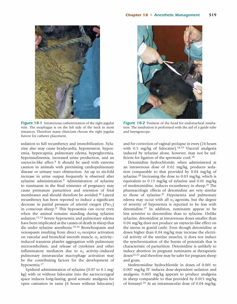
Chapter 18 • Anesthetic Management 519
sedation to full recumbency and immobilization. Xyla-zine also may cause bradycardia, hypotension, hypox-emia, hypercapnia, pulmonary edema, hyperglycemia, hypoinsulinemia, increased urine production, and an oxytocin-like effect.8 It should be used with extreme caution in animals with preexisting cardiopulmonary disease or urinary tract obstruction. An up to six-fold increase in urine output frequently is observed after xylazine administration.9 Administration of xylazine to ruminants in the final trimester of pregnancy may cause premature parturition and retention of fetal membranes and should therefore be avoided.10 Lateral recumbency has been reported to induce a significant decrease in partial pressure of arterial oxygen (Pao2) in conscious sheep.11 This hypoxemia can occur even when the animal remains standing during xylazine sedation.12,13 Severe hypoxemia and pulmonary edema have been implicated as the causes of death in sheep that die under xylazine anesthesia.14-16 Bronchospasm and venospasm resulting from direct α2-receptor activation on vascular and bronchial smooth muscle, α2 activity–induced transient platelet aggregation with pulmonary microembolism, and release of cytokines and other inflammatory mediators due to α2 activity–induced pulmonary intravascular macrophage activation may be the contributing factors for the development of hypoxemia.17
Epidural administration of xylazine (0.07 to 0.1 mg/kg) with or without lidocaine into the sacrococcygeal space induces long-lasting, good somatic analgesia for open castration in rams (8 hours without lidocaine)
and for correction of vaginal prolapse in ewes (24 hours with 0.5 mg/kg of lidocaine).18,19 Visceral analgesia induced by xylazine alone, however, may not be suf-ficient for ligation of the spermatic cord.18
Detomidine hydrochloride, when administered at an intravenous dose of 0.02 mg/kg, produces seda-tion comparable to that provided by 0.04 mg/kg of xylazine.20 Increasing the dose to 0.03 mg/kg, which is equivalent to 0.15 mg/kg of xylazine and 0.01 mg/kg of medetomidine, induces recumbency in sheep.21 The pharmacologic effects of detomidine are very similar to those of xylazine.21 Hypoxemia and pulmonary edema may occur with all α2-agonists, but the degree of severity of hypoxemia is reported to be less with detomidine.17 In addition, ruminants appear to be less sensitive to detomidine than to xylazine. Unlike xylazine, detomidine at intravenous doses smaller than 0.04 mg/kg does not produce an oxytocin-like effect on the uterus in gravid cattle. Even though detomidine at doses higher than 0.04 mg/kg may increase the electri-cal activity of the uterine muscles, it does not induce the synchronization of the bursts of potentials that is characteristic of parturition. Detomidine is unlikely to induce abortion in pregnant ruminants at therapeutic doses22,23 and therefore may be safer for pregnant sheep and goats.
Medetomidine hydrochloride in doses of 0.001 to 0.007 mg/kg IV induces dose-dependent sedation and analgesia: 0.005 mg/kg appears to produce analgesia in sheep comparable to that provided by 0.015 mg/kg of fentanyl.24 At an intramuscular dose of 0.04 mg/kg,
Figure 18-2 Position of the head for endotracheal intuba-tion. The intubation is performed with the aid of a guide tube and laryngoscope.
Figure 18-1 Intravenous catheterization of the right jugular vein. The esophagus is on the left side of the neck in most instances. Therefore many clinicians choose the right jugular furrow for catheter placement.

520 Chapter 18 • Anesthetic Management
medetomidine induces recumbency for 58 minutes, as well as good analgesia and marked muscle relaxation for 30 to 45 minutes. Sheep usually recover within 1.5 to 2 hours after regaining the righting reflex.25 When medetomidine was administered intramuscularly (IM) as a preanesthetic at either of two doses (0.005 or 0.01 mg/kg) 30 minutes before induction with propo-fol and maintenance with isoflurane, medetomidine at either dose induced decreases in heart rate and respiratory rate. Mean arterial blood pressure values were significantly higher in sheep receiving a higher dose of medetomidine than in those receiving a lower dose or no preanesthetic medetomidine. In general, the administration of medetomidine reduced the dose requirement of propofol for induction and isoflurane anesthesia during surgery.26 In sheep anesthetized with medetomidine (0.02 mg/kg IV) and ketamine (2 mg/kg IV) and breathing room air, Pao2, arterial pH, and arterial oxygen (O2) saturation decrease and the partial pressure of arterial carbon dioxide (Paco2) increases sig-nificantly. Supplementation of 100% O2 may improve Pao2 and hemoglobin saturation.27
α2-Adrenergic AntagonistsThe pharmacologic effects induced by xylazine, detomi-dine, or medetomidine can be effectively antagonized by an α2-adrenergic antagonist such as yohimbine, tolazoline, or atipamezole (Table 18-3). These antago-nists can be used to shorten recovery time and prevent the significant adverse effects sometimes seen with agonists, especially accidental overdose. However, these drugs are not without risks: The death of a sheep after administration of a large dose of yohimbine (0.8 mg/kg IV) has been reported.28 For administration of an antagonist, slow injection is recommended to avoid sudden awareness of pain and excitement on the part of the animal. Rapid injection of tolazoline has been reported to cause significant cardiac stimulation, tachy-cardia, increased cardiac output, vasodilation, coronary vasodilation, and gastrointestinal distress.29 Ruminants and camelids apparently are more sensitive to tolazo-line than other species, and death has been reported in several animals after its use.30,31 When adminis-tered alone to Holstein calves, intravenous tolazoline caused coughing, increased frequency of defecation,
TABLE 18-2 Doses of Preanesthetics Commonly Used in Sheep and Goats
Preanesthetics Dosage for Sheep Dosage for Goats
Atropine 0.066 mg/kg IV0.005-0.01 mg/kg SC or IM; 0.02-0.04 mg/kg IV
0.066 mg/kg IV0.005-0.01 mg/kg SC or IM; 0.02-0.04 mg/kg IV
Glycopyrrolate 0.002-0.005 mg/kg IV; 0.005-0.01 mg/kg IM0.01-0.02 mg/kg SC or IM; 0.005-0.01 mg/kg IV
0.002-0.005 mg/kg IV; 0.005-0.01 mg/kg IM0.01-0.02 mg/kg SC or IM; 0.005-0.01 mg/kg IV
Acepromazine Less than 50 kg: 0.to 0.2 mg/kg IVMore than 50 kg: 0.05-0.1 mg/kg IV or IM0.05-0.1 mg/kg SC or IM; 0.025-0.05 mg/kg IV0.04-0.09 mg/kg IM; 0.01-0.02 mg/kg IV
Less than 50 kg: 0.1-0.2 mg/kg IVMore than 50 kg: 0.05-0.1 mg/kg IV or IM0.05-0.1 mg/kg SC or IM; 0.025-0.05 mg/kg IV0.04-0.09 mg/kg IM; 0.01-0.02 mg/kg IV
Chloral hydrate 30-60 mg/kg IV 30-60 mg/kg IVDetomidine 0.001-0.007 mg/kg IV for sedation; 0.04 mg/kg IM,
recumbency for 45-60 minutes0.05-0.08 mg/kg IV
0.001-0.007 mg/kg IV, sedation; 0.04 mg/kg IM, recumbency for 45-60 minutes
0.05-0.08 mg/kg IVDiazepam 0.25-0.5 mg/kg IV slowly
0.2-1 mg/kg SC or IM; 0.2-0.4 mg/kg IV0.25-0.5 mg/kg IV slowly0.2-1 mg/kg SC or IM; 0.2-0.4 mg/kg IV
Medetomidine 0.001-0.007 mg/kg IV for sedation; 0.04 mg/kg IM, recumbency for 58 minutes
0.001-0.007 mg/kg IV, sedation; 0.04 mg/kg IM, recumbency for 58 minutes
Midazolam 0.1-0.5 mg/kg SC, IM, or IV 0.1-0.5 mg/kg SC, IM, or IVXylazine 0.01-0.02 mg/kg IV, standing sedation for
30-60 minutes0.1-0.2 mg/kg IV, or 0.2-0.3 mg/kg IM, recumbency
for 60 minutes0.1-0.3 mg/kg IV or SC0.1-0.3 mg/kg SC or IM; 0.025-0.1 mg/kg IV≤0.1 mg/kg IV; 0.1-0.6 mg/kg IM
0.01-0.02 mg/kg IV, standing sedation for 30-60 minutes
0.05-0.11 mg/kg IV, or 0.11-0.22 mg/kg IM, recumbency for 60 minutes
0. 1-0.3 mg/kg IV or SC0.1-0.3 mg/kg SC or IM; 0.05-0.1 mg/kg IV≤0.1 mg/kg IV; 0.1-0.6 mg/kg IM
Data from Blaze CA, Glowaski MM, editors: Veterinary anesthesia drug quick reference, St Louis, 2004, Elsevier Saunders; Muir WW, et al, editors: Handbook of veterinary anesthesia, St Louis, 2007, Mosby Elsevier; Cornick-Seahorn JL, editor: The practical veterinarian: veterinary anesthesia, Boston, 2001, Buttorworth Heinemann.

Chapter 18 • Anesthetic Management 521
and a mild increase in breathing effort at a dose of 1.5 mg/kg. At higher doses (2 to 10 mg/kg IV), adverse effects such as bright red conjunctival mucous mem-branes, coughing, nasal discharge, salivation, increased breathing effort (labored breathing), CNS depression, signs of abdominal pain, straining, head pressing, restlessness, and increased frequency of defecation to severe diarrhea were observed. Nonetheless, all calves in the study recovered uneventfully.31 Therefore lower doses at 0.5 to 1.5 mg/kg IV are now recommended in ruminants including sheep and goats. When atipa-mezole (0.1 mg/kg IV) was administered to six goats to antagonize medetomidine (0.02 mg/kg IV)-induced sedation and recumbency, all goats stood within 86 ± 24 seconds. Four goats developed piloerection, and all six appeared to be agitated and vocalized.32 Nev-ertheless, the undesirable effects of α2-antagonists are extremely rare in healthy animals when the drugs are administered by slow intravenous injection and at appropriate dosages.
BenzodiazepinesBenzodiazepines such as diazepam and midazolam are classified as minor tranquilizers and are useful for their effective anxiolytic, anticonvulsant, and central muscle relaxant effects. Diazepam and midazolam produce minimal cardiovascular depression. Thus they can be used as alternative agents to induce sedation in small ruminants when adverse effects such as hypoxia, lung edema, or increased airway pressure commonly associ-ated with the administration of xylazine or other α2-agonists are particularly undesirable.
Diazepam has a mild sedative-hypnotic effect and produces decreased anxiety along with muscle relax-ation. Diazepam often is used for its anxiolytic effect in high-risk animals because of its minimal cardiovascular and pulmonary effects at therapeutic doses. It also can be used in combination with ketamine to improve muscle relaxation during anesthesia.33
In goats, intramuscular midazolam (0.6 mg/kg) induced ~20 minutes of sedation. Hypnosis with
TABLE 18-3 Doses of Antagonists Commonly Used in Sheep and Goats
Drug Dosage for Sheep Dosage for Goats
α2-ADRENERGIC ANTAGONISTAtipamezole 0.05 mg/kg IV
0.125-0.2 mg/kg slow IV0.05 mg/kg IV0.125-0.2 mg/kg slow IV
Tolazoline 2 mg/kg slow IV0.5-1.5 mg/kg slow, IV
2 mg/kg slow IV0.5-1.5 mg/kg slow, IV
YohimbineYohimbineYohimbineYohimbine
0.125-0.22 mg/kg slow IV0.1-0.3 mg/kg IV1, IV0.3-0.5 mg/kg IM
0.125-0.22 mg/kg slow IV0.1-0.3 mg/kg IV0.3-0.5 mg/kg IM
BENZODIAZEPINE ANTAGONISTFlumazenil 0.1-1 mg/kg IV 0.1-1 mg/kg IV
NEUROMUSCULAR BLOCKING DRUG ANTAGONISTEdrophonium 0.5 mg/kg IV 0.5 mg/kg IVNeostigmine 0.02-0.04 mg/kg IV 0.02-0.04 mg/kg IVPyridostigmine 0.2 mg/kg IV 0.2 mg/kg IV
NONSPECIFIC ANTAGONISTDoxapramDoxapram
5-10 mg/kg IV0.2-0.4 mg/kg IV
5-10 mg/kg IV
OPIATE ANTAGONISTNalmefene 0.25-30 μg/kg IV 0.25-30 μg/kg IVNaloxone 0.01-0.02 mg/kg IV, titrate to effect 0.01-0.02 mg/kg IV, titrate to effectNaltrexone 0.05-0.1 mg/kg SC 0.05-0.1 mg/kg SC
Data from Blaze CA, Glowaski MM, editors: Veterinary anesthesia drug quick reference, St Louis, 2004, Elsevier Saunders; Muir WW, et al, editors: Handbook of veterinary anesthesia, St Louis, 2007, Mosby Elsevier; and Cornick-Seahorn JL, editor: The practical veterinarian: veterinary anesthe-sia, Boston, 2001, Butterworth Heinemann.

522 Chapter 18 • Anesthetic Management
recumbency for 10 to 20 minutes occurred with intrave-nous administration of midazolam at 0.6 and 1.2 mg/kg. Increasing the dose to 1.2 mg/kg increased the degree of reflex suppression and the animals appeared to be in a light plane of anesthesia, as evidenced by the lack of response to mechanical stimulation applied by tail base clamp.34
ANESTHETICS (Table 18-4)Injectable AnestheticsThiopental sodium can be used to induce anesthesia in sheep and goats; the depth of anesthesia and muscle relaxation is sufficient for endotracheal intubation. Additional incremental doses may be administered to prolong anesthesia.4 A guaifenesin-thiopental mixture can be administered to effect, to induce and main-tain short-term anesthesia. The final concentration of the mixture is 5% (50 mg/mL) guaifenesin and 0.2% (2 mg/mL) thiopental.35 Thiopental causes minimal cardiovascular depression. A moderate tachycardia, slight decrease in mean arterial blood pressure, and short-lived respiratory depression usually occur imme-diately after rapid induction of anesthesia with thiopen-tal. Transient apnea is not uncommon during induction with thiopental, and spontaneous breathing returns within several minutes. With prolonged apnea, the animal should be intubated and ventilated until spon-taneous breathing resumes.36 Recovery from thiopental anesthesia relies mainly on redistribution of the drug from the brain to the peripheral tissues. Administra-tion of a large dose or prolonged infusion may result in extremely prolonged recovery. Therefore maintenance of anesthesia with thiopental is not recommended if the surgical procedure will require more than 1 hour.36
Ketamine hydrochloride, a dissociative derivative, probably is the most commonly used injectable anes-thetic in sheep and goats. Acepromazine, diazepam, xylazine, and medetomidine can be used in combina-tion with ketamine to enhance the degree of analgesia and muscle relaxation during anesthesia. Unlike other conventional anesthetics, ketamine does not depress cardiovascular function; instead, heart rate and arterial blood pressure increase during ketamine anesthesia as a result of central sympathetic stimulation. A mixture of ketamine (1 mg/mL) and guaifenesin can be used to maintain short-term anesthesia.37 A combination of guaifenesin (50 mg/mL), ketamine (1 to 2 mg/mL), and xylazine (0.1 mg/mL) (GKX), often referred to as “triple drip,” can be used for both induction and maintenance of anesthesia.38
Telazol (Pfizer Animal Health, New York) is a pro-prietary combination of tiletamine (dissociative) and zolazepam (benzodiazepine) in a 1:1 (weight-weight) ratio. Compared with ketamine, Telazol produces bet-ter muscle relaxation, more profound analgesia, and
longer-lasting effects. In ruminants the induction of anes-thesia after Telazol administration is rapid and smooth, and the recovery usually is gradual and prolonged.16 Similar to ketamine, this drug causes cardiovascular stimulation rather than depression.37 Hypoventilation and hypothermia may occur during Telazol-induced anesthesia. Assisted or controlled ventilation with O2 supplementation may be required in cases of severe hypoventilation and hypoxemia. Animals should be placed in sternal recumbency with support throughout the recovery period.16
Propofol is a unique short-acting anesthetic. Struc-turally, this drug does not relate to any of the injectable anesthetics currently available in veterinary practice. Propofol is only slightly water-soluble and is formu-lated as an emulsion containing 10 mg of propofol, 100 mg of soybean oil, 22.5 mg of glycerol, and 12 mg/mL of egg lecithin in sterile glass ampules. Because this emulsion contains no preservative, after the ampule is opened, the contents should be used or discarded within 8 hours.37 A single dose of propofol (2 mg/kg) induces approximately 10 minutes of anesthesia, with complete recovery occurring in 20 to 30 minutes.4,39 Propofol is best used for induction before inhalation anesthesia; it also can be given as a continuous infusion to maintain short-term anesthesia.40-42 In goats, a com-bination of detomidine, butorphanol, and propofol for induction and continuous intravenous infusion of propofol for maintenance provides adequate anesthesia for castration or ovariectomy.43
A comparative study was performed to evaluate use of propofol (3 mg/kg IV), thiopental (8 mg/kg IV), and ketamine (10 mg/kg IV) as induction agents before halothane anesthesia in goats. The result of this study indicated that propofol was superior to thiopental or ketamine as an induction agent owing to rapid and uneventful recovery from its effects. Time to standing with propofol after 30 minutes of halothane anesthe-sia was 18 ± 2.4 minutes, as opposed to 43.9 ± 7.3 minutes with thiopental and 76.9 ± 10.3 minutes with ketamine.44
Total intravenous anesthesia (TIVA) with ketamine and propofol infusion has been used to maintain immobilization and anesthesia in goats undergoing magnetic resonance imaging (MRI) procedure. These goats were sedated with midazolam (0.4 mg/kg IV), and anesthesia was induced with intravenous propofol (1 mg/kg) and ketamine (3 mg/kg) and maintained with constant infusion rates of propofol (0.3 mg/kg/minute) and ketamine (0.03 mg/kg/minute) with or without sevoflurane. Goats anesthetized with propofol and ketamine exhibited significant decreases in respira-tory rates and increases in arterial blood pressure values. As supported by our own experience, overall, anesthesia with ketamine and propofol infusion is practical and safe for animals undergoing MRI procedures.45

Chapter 18 • Anesthetic Management 523
TABLE 18-4 Doses of General Anesthetics Commonly Used in Sheep and Goats
Anesthetic Agent(s) Dosage for Sheep Dosage for Goats
Guaifenesin 30-90 mg/kg IV; when muscle relaxed, followed by ketamine 1.1 mg/kg IV
30-90 mg/kg IV; when muscle relaxed, followed by ketamine 1.1 mg/kg IV
Atracurium (neuromuscular blocking drug)
0.2 IV mg/kg for initial dose; 0.1 IV mg/kg for repeat dose
0.005/mg/kg/hour IV (infusion)
0.2 IV mg/kg for initial dose; 0.1 IV mg/kg for repeat dose
0.005/mg/kg/hour IV (infusion)Etomidate 0.5-1 mg/kg IV 5-10 mg/kg IVKetamine 22 mg/kg SC or IM; 2-4 mg/kg IV 11 mg/kg SC or IM; 2-4 mg/kg IVAcepromazineKetamine
0.55 mg/kg IV2.2 mg/kg IV
N/A
DiazepamKetamineDiazepamKetamineDiazepamKetamine
0.11 mg/kg IV4.4 mg/kg IV0.25-0.5 mg/kg IV4-7.5 mg/kg IV0.28 mg/kg IV5.5 mg/kg IV
0.11 mg/kg IV4.4 mg/kg IV
0.28 mg/kg IV5.5 mg/kg IV
Guaifenesin (5%) plus ketamine (0.1%)
Induction: 2 mL/kg IV, 50-75% calculated dose first
Maintenance: 2.2 mL/kg/hour or to effect
Induction: 2 mL/kg IV, 50-75% calculated dose first
Maintenance: 2.2 mL/kg/hour or to effect
MedetomidineKetamineMedetomidineKetamine
0.02 mg/kg IV2 mg/kg IV0.02 mg/kg IV0.5-1 mg/kg IV
0.02 mg/kg IV2 mg/kg IV0.02 mg/kg IV0.5-1 mg/kg IV
XylazineKetamineXylazineKetamine
0.22 mg/kg IM; wait 10 minutes10-15 mg/kg IM0.03-0.2 mg/kg IV or IM5 mg/kg IV
0.22 mg/kg IM; wait 10 minutes11 mg/kg IM0.03-0.2 mg/kg IV or IM5 mg/kg IV
Guaifenesin (5%), xylazine (0.01%), and ketamine (0.1-0.2%)
Guaifenesin (5%), xylazine (0.005%), and ketamine (0.1-0.2%)
Induction: 0.67-1.1 mL/kgMaintenance: 2.2 mL/kg/hour to effectInduction: 0.5-1 mL/kgMaintenance: 1.5-2 mL/kg/hour
Induction: 0.67-1.1 mL/kgMaintenance: 2.2 mL/kg/hour to effectInduction: 0.5-1 mL/kgMaintenance: 1.5-2 mL/kg/hour
Pancuronium (neuromuscular blocking drug)
0.005 mg/kg IV 0.005 mg/kg IV
Propofol Induction: 3-4 mg/kg IV or 4-8 mg/kg IVMaintenance: 18-40 mg/kg/hour IV
(infusion) or to effect4-6 mg/kg IV
Preanesthetic: detomidine 0.01 mg/kg IM plus butorphanol 0.1 mg/kg IM
Induction: 3-5 mg/kg IMMaintenance: 31 mg/kg/hour4-6 mg/kg IV
DiazepamPropofol
0.28 mg/kg IV4-6 mg/kg IV
0.28 mg/kg IV4-6 mg/kg IV
Thiopental 10-16 mg/kg IV 10-16 mg/kg IVGuaifenesin (5%) plus thiopental
(0.2%)Induction: 2 mL/kg IV, 50-75%
calculated dose firstMaintenance: 2.2 mL/kg/hour or to
effect
Induction: 2 mL/kg IV, 50-75% calculated dose first
Maintenance: 2 mL/kg/hour or to effect
Tiletamine plus zolazepam (Telazol) 2-6 mg/kg IM; 1-4 mg/kg IV5.5 mg/kg IV, anesthesia for 100
minutes5.5 mg/kg IV with butorphanol 0.1
mg/kg IV, anesthesia for 100 minutes
2-6 mg/kg IM; 1-4 mg/kg IV5.5 mg/kg IV, anesthesia for 100 minutes5.5 mg/kg IV with butorphanol 0.1 mg/
kg IV, anesthesia for 100 minutes
Continued

524 Chapter 18 • Anesthetic Management
Inhalation AnestheticsInhalation anesthetics require expensive and specialized equipment for delivery to the patient. However, these agents allow veterinarians to perform complicated and prolonged surgery. Either halothane or isoflurane can be used effectively and safely in sheep and goats. Mask induction may not be a wise choice in healthy adults but can be used in smaller or debilitated animals. Use of a small animal anesthesia machine with a double carbon dioxide (CO2)-absorbent canister usually is adequate for most sheep and goats. The clinician should be aware of a rare condition called halothane-induced hepatitis, an acute, massive liver necrosis that sometimes occurs after halothane anesthesia in healthy goats, especially after prolonged exposure.46,47 Clinical signs, including depression, inappetence, salivation, teeth grinding, head pressing, and icterus, usually appear within 24 hours. Serum concentrations of aspartate transaminase, bili-rubin, alkaline phosphatase, and creatinine and blood urea nitrogen are significantly increased from normal ranges. Death usually occurs within 4 days, and histo-pathologic examination reveals centrilobular necrosis. Necrosis of the proximal renal tubules, abomasal ulcer-ation, and hepatic encephalopathy have been observed in some cases.46,47 Severe hypotension, hypoxemia, and hepatic hypoxia may encourage the reductive metabo-lism of halothane, leading to the production of toxic free radicals.48 Therefore maintaining adequate car-diovascular function and oxygenation through careful monitoring and supportive therapies is key to a success-ful anesthetic procedure.
Isoflurane and sevoflurane have become popular inhalation anesthetics in recent years. Both anesthet-ics have lower potency (halothane: 0.96%; isoflurane: 1.29%; sevoflurane: 2.33%)49 and lower lipid solubility compared with halothane (halothane: 2.36; isoflurane: 1.41; sevoflurane: 0.69).50 Thus the induction of anes-thesia and recovery usually occur more rapidly with these agents than with halothane. Hepatitis associated with halothane is unlikely to develop with isoflurane and sevoflurane owing to the fact that elimination of these two anesthetics involves very little hepatic
metabolism (halothane: 20%; isoflurane: 0.25%; sevo-flurane: 3% to 5%).50 Isoflurane and sevoflurane have similar cardiovascular and pulmonary effects, which include vasodilation and dose-dependent decreases in arterial blood pressures, respiratory rate, tidal volume, and minute ventilation. Decreases in arterial blood pressure during halothane anesthesia have been shown to be the result of depression of myocardial contractil-ity and the subsequent decrease in cardiac output. In contrast with halothane, the vasodilating effect of iso-flurane and sevoflurane is believed to be the primary contributing factor in the decreases in arterial blood pressure.49
Local AnestheticsLocal anesthetics produce their effects by blocking the propagation of action potentials along nerve axons in a reversible manner. These anesthetics can be injected into the tissue at the surgical site to produce local anes-thesia, or they can be administered in the perineural area of major nerves to produce regional anesthesia (see Chapter 8). In small ruminants, many surgical proce-dures are performed safely and painlessly with the use of local or regional anesthesia.
All local anesthetics have similar physical properties and molecular structures. Most of them are weakly basic tertiary amines with a hydrophilic end, a lipophilic end, and an intermediate hydrocarbon chain. They are generally available as acid solutions of the water-soluble salts. The acid salt is neutralized in the tissue, liberating the base, which then penetrates the cell membrane and interrupts the propagation of the action potential. This mechanism of action means that a local anesthetic is less effective in inflamed tissue with lower pH, because less liberation of the basic form of the drug occurs under these conditions.51 Local anesthetics are classi-fied as either ester-link or amide-link drugs, depending on the intermediate chain structure. Inactivation of ester-link local anesthetics (e.g., procaine, tetracaine) depends on hydrolysis by cholinesterase enzymes in the plasma and to a lesser extent in the liver. Metabolism
TABLE 18-4 Doses of General Anesthetics Commonly Used in Sheep and Goats—cont’d
Anesthetic Agent(s) Dosage for Sheep Dosage for Goats
XylazineTelazol
0.05-0.1 mg/kg IV or IM2-4 mg/kg IV or IM
0.05-0.1 mg/kg IV or IM2-4 mg/kg IV or IM
Vecuronium (neuromuscular blocking drug)
0.005 mg/kg IV 0.005 mg/kg IV
Data from Blaze CA, Glowaski MM, editors: Veterinary anesthesia drug quick reference, St Louis, 2004, Elsevier Saunders; Muir WW, et al, editors, Handbook of veterinary anesthesia, St Louis, 2007. Mosby Elsevier; and Cornick-Seahorn JL, editor: The practical veterinarian: veterinary anesthe-sia, Boston, 2001, Butterworth Heinemann.

Chapter 18 • Anesthetic Management 525
of amide-link local anesthetics (e.g., lidocaine, bupi-vacaine, mepivacaine) relies on microsomal enzymes located primarily in the liver.51
Lidocaine probably is the most popular local anes-thetic used and may produce anesthesia for 0.75 to 2 hours. Because of its ability to induce vasoconstric-tion in the tissue around the injected area, epineph-rine decreases systemic absorption of concurrently administered local anesthetics. Therefore epinephrine (1:200,000 to 1:50,000) at concentrations of 5 to 20 μg/mL can be incorporated with or added to lidocaine solution to prolong the duration of local anesthesia.52 Mepivacaine (5 mg/kg), with effect duration of 1.5 to 3 hours, and bupivacaine (2 mg/kg), with effect duration of 4 to 8 hours, can be used for procedures that require a longer duration of local anesthesia.51,53 Administration of a large single dose or repeated small doses of local anesthetics can result in toxicity in sheep and goats, especially in neonatal and young patients. Clinical signs of toxicity include nystagmus, muscle fascicula-tion, CNS stimulation progressing to opisthotonos and convulsions, hypotension, respiratory arrest, and circu-latory collapse, with death in some cases.51 The maxi-mum calculated safe dose of lidocaine was reported to be 13 mg/kg in one study.54 In another study, accumu-lated intravenous doses of 5.8 mg/kg, 18 mg/kg, and 42 mg/kg induced signs of toxicity in adult, neonatal, and fetal sheep, respectively.55 Intravenous infusion of mepivacaine in sheep induced convulsions at doses of 7.5 to 7.9 mg/kg and cardiovascular collapse at doses as high as 52 to 69 mg/kg.56 Bupivacaine is approximately four times more potent than lidocaine, so a 0.5% solu-tion produces the same degree of neuronal blockade as that achieved with a 2% lidocaine solution.57
Ewing58 suggests using a maximum of 6 mg/kg of lidocaine or mepivacaine and 2 mg/kg of bupivacaine in small ruminants. With this maximum safe dose in mind, the clinician should dilute lidocaine and mepiva-caine solutions to 1% and 0.5%, respectively, to prevent overdosage when using these drugs in lambs and kids.58 Diazepam (0.1 mg/kg IV) or thiopental (5 mg/kg IV) should be administered if seizure activity or convul-sions caused by accidental overdose persist longer than 1 to 2 minutes.57,59
PERIOPERATIVE MANAGEMENT AND RECOVERYMonitoring During AnesthesiaAnimals should be monitored continuously throughout anesthesia. Peripheral pulses should be palpable, and an electrocardiogram, if available, should be used at all times. Recently, the use of capnograms to evaluate end-tidal CO2 and pulse oximeters to assess arterial O2 saturation has became part of routine monitoring to ensure adequate ventilation and gas exchange. Samples
for measurement of end-tidal CO2 are collected directly at the connecting point between the breathing system (Y piece) of the anesthesia machine and the end of the endotracheal tube; the partial pressure of CO2 is deter-mined by infrared absorptiometry. Normal end-tidal CO2 should be closely related to alveolar CO2, if the anesthetized patient is healthy with no preexisting dif-fusion disturbance in the lung tissues.
Arterial hemoglobin O2 saturation is measured by pulse oximetry with the sensor clip placed on the lin-gual artery in the tongue or on the auricular artery in an ear. Normal arterial hemoglobin O2 saturation should always be close to 98% to 100%. Indirect arterial blood pressures can be measured by an oscillometric blood pressure machine with an inflatable pressure cuff placed on the tail or mid thigh and over the coccygeal or dorsal metatarsal artery, respectively. Normal values for heart rate, respiratory rate, arterial blood pressures and vari-ous arterial blood gases are listed in Table 18-5.
A balanced electrolyte solution (5 to 10 mL/kg/hour) should be administered by the intravenous route for supportive hydration during anesthesia. Administration of 5% dextrose solution is required to prevent hypo-glycemia in neonates and young animals. A circulating warm-water blanket can be used to maintain body tem-perature during anesthesia and recovery.
Sheep and goats usually recover from anesthesia gradually and smoothly. Emergence delirium and pre-mature attempts to stand seldom occur in these ani-mals. They should be placed in sternal recumbency
TABLE 18-5 Normal Vital Signs and Values for Anesthetized Sheep and Goats
Vital Sign or ValueValues for Sheep and Goats
Heart rate (beats/min) 80-150Respiratory rate (breaths/min) 20-40Systolic arterial pressure (mm Hg) 80-120Mean arterial pressure (mm Hg) 75-100Diastolic arterial pressure (mm Hg) 60-80Partial pressure of arterial carbon
dioxide (Paco2) (mm Hg)28-36
Partial pressure of arterial oxygen (Pao2) (mm Hg)
72-90
Arterial pH 7.48-7.58
Data from Riebold TW, Geiser DR, Goble DO: Clinical techniques for food animals anesthesia. In Riebold TW, Geiser DR, Goble DO, editors: Large animal anesthesia: principles and techniques, ed 2, Ames, Iowa, 1995, Iowa State University Press; and Alon E, et al: Effects of propofol and thiopental on maternal and fetal cardiovas-cular and acid-base variables in the pregnant ewes, Anesthesiology 78:562, 1993.

526 Chapter 18 • Anesthetic Management
with support, if necessary, during the recovery period. If regurgitation occurred during anesthesia, the oral cavity and pharynx should be lavaged to prevent aspiration of ruminal materials and subsequent aspiration pneumo-nia. The endotracheal tube should be left in place until the animal regains its chewing and coughing reflexes. This tube should be removed with the cuff inflated.
Influences of Pathophysiologic Alterations on AnesthesiaObstructive urolithiasis is the inability to void urine normally secondary to obstruction of the urinary out-flow tract by calculi (see Chapter 12). This condition occurs most frequently in young castrated goats. In cases of urethral obstruction without ruptured bladder, perineal urethrostomy can be performed with use of a local infiltration technique or epidural analgesia. Gen-eral anesthesia is induced for bladder repair, bladder marsupialization, penile urethrostomy, and tube cys-totomy.60,61 Xylazine, detomidine, medetomidine, and other α2-agonists are contraindicated in obstructive uro-lithiasis, because their potent diuretic effects can result in bladder rupture before the obstruction can be relieved and the bladder emptied.8 Most of the routine anes-thetic regimens can be safely used in cases of obstructive urolithiasis and ruptured bladder. However, anesthetic dosage adjustments may be necessary depending on the animal’s physical condition, particularly when blood work reveals increased blood urea nitrogen and serum creatinine levels or hypoproteinemia.60
As discussed in various chapters in this book, case-ous lymphadenitis in sheep and goats is a chronic contagious disease caused by Corynebacterium pseudo-tuberculosis. Airway obstruction may result from occlu-sion of the pharynx by the enlarged pharyngeal lymph nodes. In some cases, respiratory embarrassment is severe enough to cause respiratory distress. Treatment of affected animals involves either draining or surgi-cally removing the abscessed nodes. Depending on the size and invasiveness of the abscess, general anesthesia sometimes is required to ensure immobilization so that vital tissues and structures (e.g., carotid artery, vagus nerve, esophagus) are not compromised during dissec-tion.62 Animals with respiratory embarrassment should be handled with great caution and with as little stress as possible. Stress and excitement may worsen the severity of respiratory embarrassment, resulting in severe hypox-emia and death. Rapid induction of anesthesia and intubation to maintain the airway is indicated. Tracheal intubation may be difficult because the larynx may be obscured by the swollen pharyngeal tissues. Therefore the skin over the upper third of the trachea should be clipped and surgically prepared before the induc-tion of anesthesia. Tracheostomy can be performed to ensure a patent airway should difficulty in endotracheal
intubation occur. The tracheotomy and placement of a cuffed tracheostomy tube also can be performed with use of local anesthesia before the induction of general anesthesia. Anesthesia can be induced with either intravenous injectable anesthetics or inhalation anesthetics administered through the tracheostomy tube. The tracheostomy tube should be left in place for 48 to 72 hours after surgery if possible. If removal of the tracheostomy tube is necessary after completion of the surgery, the clinician should assess airway patency rostral to the tracheostomy tube by deflating the cuff and ensuring good airflow before removing the tube.61
Cesarean section sometimes is performed as an elective surgical procedure to save valuable fetuses and relieve dystocia in sheep and goats. The selection of suitable anesthetics and techniques is vital to the survival of both dam and fetus. Pregnancy induces several physiologic changes that can affect the response to anesthesia. Because of hormonal changes, minute ventilation increases and the requirement for inhala-tion anesthetics decreases (by 25% for halothane, 32% for methoxyflurane, and 40% for isoflurane). Induc-tion of anesthesia is therefore more rapid than it is in nonpregnant patients.61 Heart rate and cardiac output increase because of the increased cardiac work required by pregnancy and at parturition. Consequently, cardiac reserves decrease, and pulmonary congestion and heart failure may occur in animals that are exhausted or prone to shock after a prolonged and difficult delivery; animals with preexisting cardiovascular disease also are at risk.61 Furthermore, venous engorgement result-ing from increased intraabdominal pressure from the enlarged uterus reduces the space available for local anesthetic administration by epidural injection, with resultant cranial migration of the anesthetic solution. This migration may result in sympathetic blockade, profound maternal hypotension, and reduction of uteroplacental perfusion. Therefore the dose of local anesthetic administered to produce adequate epidural anesthesia should be reduced to one-third to one-half that used in nonpregnant animals.61 Drugs adminis-tered to induce or enhance uterine contraction during parturition (e.g., oxytocin) cause peripheral vasodila-tion and hypotension when administered in large or repeated doses, with the potential for decreased uter-ine perfusion to the uterus/placenta, and subsequent decreased fetal viability. Fluid with balanced electrolytes should be administered to animals with severe hypo-tension before induction and during anesthesia.61
Either general anesthesia or local anesthesia is suit-able for cesarean section. Selection of the anesthetic regimen should be based on maternal and fetal safety considerations. Most anesthetics have physicochemi-cal properties that allow them to cross the blood-brain barrier; these properties also permit them to cross the placenta, with resultant depression of the fetal CNS.

Chapter 18 • Anesthetic Management 527
The only exceptions are neuromuscular blocking agents such as succinylcholine, pancuronium, and atracurium. These drugs cannot cross the placenta as easily as other anesthetics because of their quaternary molecular struc-ture. To enhance fetal viability, the intended incision site should be clipped and aseptically prepared before induction, the period between induction of anesthesia and removal of the fetus should be as short as possible, and the anesthetic concentration should be kept to a minimum. For these reasons, in addition to economic concerns, local anesthesia often is chosen for this pro-cedure. Local infiltration of anesthetic along the inci-sion site and lumbosacral epidural or subarachnoid anesthesia are the two most commonly used techniques for cesarean section in most food animals. Procaine and tetracaine, which are esters of paraaminobenzoic acid, do not accumulate in the dam and fetus. Lido-caine, mepivacaine, bupivacaine, and etidocaine are amide-link local anesthetics that depend on hepatic microsomal enzymes for inactivation of drug effects.51 Blood concentrations of these drugs decline slowly after absorption from the injection site. They can accumulate in the fetus, causing fetal depression when large doses are administered.61 (See Chapter 8)
Preanesthetic tranquilizers or sedatives usually are reserved for use in excited or unruly animals. General anesthetics with low lipid solubility, short duration of action, and minimal cardiovascular depression are preferred. Low doses of thiopental, a combination of xylazine and ketamine, or “triple drip” can be used for induction; anesthesia is maintained with triple drip intravenous infusion to effect or with an inhalational anesthetic agent (halothane or isoflurane). Tracheal intubation should be performed in these animals even with use of injectable anesthesia techniques, to prevent possible aspiration pneumonia, and administration of supplemental O2 is recommended. Neonatal respira-tory or CNS depression caused by anesthetic effects can be reversed by the administration of doxapram or an α2-antagonist immediately after delivery.
PERIOPERATIVE ANESTHETIC COMPLICATIONSRegurgitation and Aspiration PneumoniaRegurgitation can occur during both light (active regur-gitation) and deep (passive regurgitation) anesthe-sia in sheep and goats despite preoperative fasting and withholding of water. Active regurgitation is a reflexive, protective mechanism directed at rejection of inhaled material from the pharynx, upper airway, and upper digestive tract. Passive regurgitation occurs when the esophageal muscles and transluminal pres-sure gradients relax as a result of the anesthetic effects.
Aspiration of acidic stomach or gastric contents may result in aspiration pneumonia, which is character-ized by reflex airway closure, bronchospasm, dyspnea, hypoxemia, and cyanosis. Pulmonary hemorrhage or edema may result from destruction of type II alveolar cells and pulmonary capillary lining cells after aspira-tion of large amounts of particulate matter or extremely acidic material. In severe cases, the animal dies before an endotracheal tube can be placed to protect the airway. Therefore preoperative withholding of feed and endotracheal intubation with the cuff adequately inflated are recommended for prevention of aspiration pneumonia in all ruminants that will undergo an anes-thetic procedure.63 Placing a sandbag or some other rolled padding beneath the animal’s neck to elevate the occiput and avoiding vigorous manipulation of the rumen and other internal abdominal organs during surgery helps minimize the occurrence of aspiration pneumonia. If regurgitation occurs before intubation can be completed, the clinician should either quickly lower the animal’s head or place the endotracheal tube in the esophagus and inflate the cuff to allow ruminal contents to flow out of the mouth while another tube is placed in the trachea.4 Intravenous aminophylline (2 to 4 mg/kg given over a 5-minute period or 11 mg/kg delivered over a 20-minute period) or other broncho-dilators along with 100% O2 can be administered to relieve bronchospasm at the time of aspiration. If the animal survives the initial insult, corticosteroids and broad-spectrum antibiotics are indicated for the treat-ment of pneumonia.4,35
Ruminal TympanyRuminal tympany sometimes develops during anesthe-sia as a result of the animal’s inability to eructate, lead-ing to accumulation of gas produced by fermentation of ingesta (see Chapter 5). Pressure increases within the abdomen and on the diaphragm, resulting in reduced functional residual capacity of the lungs and impaired ventilation. Placing the animal in sternal recumbency immediately after anesthesia helps eliminate the accu-mulated gas in the rumen. However, decompression of the rumen may need to be performed while the animal is still anesthetized and the surgery is in progress. Pas-sage of a stomach tube or insertion of a 12-gauge needle through the abdominal wall allows outflow of the accu-mulated gas, reducing the pressure in the abdomen and on the diaphragm and thereby improving ventilation. Most anesthetics, particularly α2-agonists and opioid agonists, decrease gastrointestinal motility; an antago-nist can be administered to treat the resultant ruminal tympany if necessary. Preoperative fasting reduces the amount of fermentable ingesta in the rumen, thereby decreasing the chance for development of perioperative ruminal tympany.63

528 Chapter 18 • Anesthetic Management
HypoventilationIn conscious sheep, lateral recumbency alone can lead to significant hypoxemia.11 During anesthesia, severe hypoventilation and hypoxemia (characterized by a significant increase in Paco2 and decrease in Pao2) may result from the abnormal surgical body position combined with the respiratory depressant effect of the anesthetic drugs. Ventilation should be assisted or con-trolled by squeezing the rebreathing bag with a positive pressure of 20 to 25 cm H2O if an inhalation anesthetic is used. Supplemental O2 also can be provided to sedated and recumbent animals through an orotracheal or endotracheal tube by using an O2 demand valve, particularly if xylazine is part of the anesthetic regimen. Occasionally, apnea may occur and persist throughout anesthesia. The anesthetist should ensure that the ani-mal is under an adequate plane of anesthesia before instituting any drug treatment. A respiratory stimulant (doxapram, 0.1 to 0.5 mg/kg IV) can be administered to initiate respiration. If the depression persists, doxapram can be administered by continuous intravenous infu-sion (5 to 10 μg/kg/minute).64
Cardiovascular CollapseDuring anesthesia, prolonged decreases in pulse pres-sure, hypotension (mean arterial pressure less than 60 to 75 mm Hg), increases in capillary refill time, pale mucous membranes, and bradycardia (heart rate less than 70 to 80 beats/minute) or tachycardia (heart rate greater than 150 beats/minute) can lead to car-diovascular collapse. Causes of this perioperative col-lapse include significant endotoxin-induced peripheral vasodilation, severe hypovolemia resulting from dehy-dration or blood loss, and deep anesthesia result-ing in profound myocardial depression. Treatment of impending cardiovascular failure should begin with correction of the causative disease status, rapid admin-istration of supportive fluid (90 mL/kg), and reduction in anesthetic dosage or even cessation of anesthesia.64 Additional symptomatic treatment includes vasoactive drugs (e.g., dopamine, phenylephrine, ephedrine) for hypotension, inotropic drugs (e.g., dobutamine) for myocardial depression, chronotropic drugs (e.g., atro-pine, glycopyrrolate) for bradycardia, and antiarrhyth-mic drugs (e.g., lidocaine) for ventricular arrhythmias such as ventricular tachycardia or premature ventricular contractions. Dobutamine (1 to 5 μg/kg/minute) is a β1 agonist that increases cardiac output and arterial blood pressure by increasing myocardial contractility when administered at a low dosage. Dobutamine seldom increases heart rate, except in the presence of reduced total blood volume.65 Dopamine is a dose-dependent, dopaminergic, α- and β-agonist. When administered at an intravenous infusion rate of 1 to 2 μg/kg/minute,
dopamine increases renal perfusion by stimulating dopaminergic receptors. Increasing the infusion rate to 2 to 10 μg/kg/minute causes stimulation of β1 receptors and a resultant increase in heart rate. Vasoconstriction and the subsequent increase in arterial blood pressure mediated by stimulation of α1-receptors are not evident until the infusion rate is greater than 10 μg/kg/minute.65 Phenylephrine (2 to 4 mg/kg given as an intravenous bolus or 0.2 to 0.4 μg/kg/minute by intravenous infu-sion) and ephedrine (22 to 66 μg/kg IV) both have been used to treat hypotension in anesthetized animals.66 Ephedrine appears to have a longer duration of action (30 to 60 minutes) than dobutamine or dopamine, which may be undesirable if tachycardia occurs after ephedrine administration.66
Prolonged untreated cardiovascular collapse may result in cardiac arrest and death. Cardiopulmonary resuscitation (CPR) should follow the ABC technique, whereby the airway is opened by endotracheal intuba-tion, controlled breathing is initiated by squeezing an Ambu bag or using the rebreathing bag on the anesthe-sia machine (12 to 20 breaths/minute), and artificial circulation is established by cardiac compression (80 to 100 compressions/minute). After CPR has been insti-tuted, an intravenous catheter should be placed if one is not already present. If the attempts at intravenous catheterization are unsuccessful, emergency drugs may be administered intratracheally through the endotra-cheal tube at 2 to 2.5 times the intravenous dose after dilution with sterile water or saline to a volume of 5 to 10 mL. Absorption of the drugs from the lung is sometimes significant enough to be more effective than intramuscular, subcutaneous, or intravenous adminis-tration through a peripheral vein.64 Emergency drugs and products frequently used during CPR include 100% O2, balanced electrolyte solutions, atropine, lidocaine, and epinephrine. Depending on the animal’s condition, the vasoactive drugs mentioned previously can be used in conjunction with emergency drugs.
Electrical defibrillation is the most effective treatment for conversion of ventricular fibrillation, but it is not practical in field situations. Epinephrine (10 μg/kg IV) usually is the drug of choice for treatment of ventricular fibrillation. Epinephrine induces peripheral vasocon-striction, increases arterial diastolic blood pressure and intracranial and coronary blood flow, helps convert fine to coarse ventricular fibrillation, and produces a positive inotropic effect by stimulating α- and β-adrenoceptors. Potential side effects of epinephrine include increased myocardial and cerebral O2 demand, postresuscitation arrhythmia, and tachycardia. Lidocaine (0.5 to 2 mg/kg IV) may be used to treat the postresuscitation ventricu-lar arrhythmia.64 Chemical defibrillation with intrave-nous potassium chloride (1 mg/kg) and acetylcholine (6 mg/kg), followed by administration of 10% calcium chloride (1 mL/10 kg), is recommended for treatment

Chapter 18 • Anesthetic Management 529
of ventricular fibrillation. Although it is ineffective in defibrillation, this technique usually converts fibrilla-tion to asystole.64 A normal sinus rhythm is actually more easily initiated from asystole.
The best treatment for perioperative complications is prevention, which requires a devoted and vigilant anes-thetist. Careful preanesthetic evaluation and prepara-tion and proper use of anesthetic regimens can prevent most anesthetic-related complications; close monitor-ing allows recognition of potentially dangerous situa-tions with rapid institution of corrective measures at an early stage. In this regard, an important concept is that “there are no safe anesthetic or anesthetic techniques—only safe anesthetists.”
PERIOPERATIVE PAIN MANAGEMENTIncreased awareness in the general public of ani-mal suffering has resulted in the improvement of pain management in livestock species in recent years. Although veterinarians often struggle to establish a bal-ance between financial considerations for the owner and the ethics and legalities of pain management for animals, the focus of veterinary anesthesiology should always be on restoration of normal physiology, preven-tion of the initiation of pain, preemption of the pain cascade, and attack of the pain at various levels along the cascade.67 Pain can originate from preexisting dis-ease processes, traumatic injury, or surgery. Pain would be easy to treat if it were always of iatrogenic origin, such as that associated with surgery, but prediction or prevention of the initiation of pain is impossible when it is caused by naturally occurring disease.
The rule of thumb for pain management is that less analgesic is needed to relieve pain if the analge-sics are administered before the initiation of the pain (preemptive analgesia). In addition, use of preemp-tive analgesia techniques will decrease the amount of anesthetics required to maintain surgical anesthesia. Multimodal therapy using combination or sequential administration of analgesics or analgesic techniques that act through different pain mechanisms or pathways is highly recommended. Such multimodal therapy has the benefit of maximizing analgesic effects of each drug as a consequence of synergism of the drugs and, at the same time, reducing individual drug dose and subse-quently minimizing the side effects of each drug. Opi-oids, μ2 agonists, nonsteroidal antiinflammatory drugs (NSAIDs), and ketamine have been administered alone or in combination to produce multimodal (balanced) analgesia. For example, injection of a local anesthetic or administration of an NSAID combined with low doses of an opioid or α2-agonist, an NSAID with an opioid, an NSAID with an α2-agonist, and an opioid with an α2-agonist (neuroleptanalgesia) are the most commonly used multimodal regimens for intra- and post-operative
pain management.68 Administration of analgesics or local anesthetics to provide pain relief should be con-sidered in the context of overall case management to optimize the patient’s quality of life and to restore normal function. In ruminants, this aim must take into consideration the issue of drug residues and standards established by the Animal Medical Drug Use and Clari-fication Act (AMDUCA).69
SYSTEMIC PAIN MANAGEMENT (Table 18-6)Opioid AnalgesicsOpioid analgesics such as morphine, meperidine, fen-tanyl, buprenorphine, and butorphanol have been used effectively for pain management in sheep and goats. These drugs bind either to μ, κ, or δ opiate receptors located on neuronal cell membranes. Binding of an opioid to these receptors then triggers cellular changes that hyperpolarize the cell membrane and inhibit spinal pain transmission. Activation of the μ receptor results in depletion of intraneuronal substance P, which reduces overall inflammation and neural pain transmission. Most opioids used in veterinary practice are classified as either pure or partial μ receptor agonists. Side effects including increased gastric and intestinal emptying time and tachycardia sometimes occur with high doses of opioids. Of interest, hyperexcitability, commonly observed in other species, is an uncommon effect in ruminants.70
Morphine has poor analgesic properties in sheep and goats. Good analgesia was achieved in only one third of the animals receiving morphine.70 With these species, morphine should be administered parenterally, rather than orally, because the drug is inactivated by the ruminal microflora. Doses of 0.05 to 0.1 mg/kg, admin-istered by either the intravenous or subcutaneous route, every 4 to 6 hours have been recommended. However, superior analgesia has been reported with doses as high as 10 mg/kg in goats.70
Meperidine hydrochloride is a synthetic opioid that has an analgesic potency of only 10% to 50% that of morphine. Meperidine produces mild sedation and analgesia. Its administration can be associated with his-tamine release.71 In yearling goats, meperidine (10 mg/kg IM) can be used as a preanesthetic given 10 minutes before induction of anesthesia with thiopental. After intubation this combination provides 20 minutes of surgical anesthesia with complete recovery occurring in 90 minutes.72
Fentanyl is a pure μ agonist similar to morphine, with a potency that is approximately 75 to 100 times that of morphine. Fentanyl can be administered paren-terally or transdermally. When administered parenter-ally, fentanyl induces analgesia within 5 minutes, which lasts for 20 minutes. Intravenous administration of

530 Chapter 18 • Anesthetic Management
fentanyl has been associated with abnormal behaviors such as pica, stall pacing, nystagmus, hyperexcitability, and ataxia.70 Transdermal patches are available in doses of 0.025, 0.05, 0.075, and 0.1 mg/hour. A 0.05 mg/hour patch is an appropriate dose for a 30- to 50-kg goat. Onset of analgesia is observed at 18 to 24 hours after placement, and each patch lasts approximately 3 days. Therefore a new patch should be placed 48 hours after the first one. Owing to slow absorption of the drug from the transdermal patch, another fast-acting analgesic should be given to provide pain relief until the fentanyl effect has been realized.67 A 3-day meat withdrawal time is recommended when fentanyl is administered parenterally. However, rumenosalivary recycling may prolong fentanyl’s effect, posing a problem in establish-ing withdrawal times.73
Tramadol is a synthetic analog of codeine and mor-phine.74 This agent produces analgesia by its action through central opiate, adrenergic, and serotoninergic receptors.75 Tramadol offers some advantages over other opioids: The drug is not a controlled or schedule drug, which makes its use less complicated; it also is associated with less potential for respiratory depression, less poten-tial for abuse in people,74,76 and less CNS excitation in horses.77 It has been shown that Tramadol is effective in treating moderate to moderately severe postoperative pain in people and dogs.78,79 Tramadol has low affin-ity for opiate μ receptors, and its dose requirement to produce the same degree of analgesia for moderate pain as that obtained with morphine is higher in a 10:1 ratio.80 However, for more severe pain, tramadol at the same dose ratio is less effective than morphine.81 Orally administered tramadol is well absorbed in humans, dogs, and cats, with bioavailabilities of 70%, 65%, and 93%, respectively.82-84 In goats, lower oral absorption with a bioavailability of 36.9% was reported after a single dose of 2 mg/kg.85 A large volume-to-surface ratio, a constantly high content of solid matter, and a complex microflora and microfauna in the rumen were suggested to be the contributing factors for the lower oral absorption of tramadol in goats compared with that in dogs and cats.85 Nonetheless, a higher gastric pH of 6.8 in the rumen, as opposed to pH of 1.0 to 2.0 in the monogastric stomach, in the presence of a pKa value of 9.41 for tramadol, resulting in greater percentage of nonionized tramadol in plasma, and the enterohepatic recycling of ruminants increase the absorption of the drug were suggested to be the primary factors respon-sible for the high plasma tramadol concentration of 542.9 ± 219.5 ng/mL in goats.85 In people, plasma con-centrations of 100 to 150 ng/mL are the recommended minimum effective concentrations for relieving mild to moderate pain.78,82 The elimination half-life after oral administration was 2.67 ± 0.54 hours in goats, which is shorter than in people76 but longer than in horses.77 Intravenous administration of tramadol at 2 mg/kg to
TABLE 18-6 Doses of Analgesics and Non-steroidal Antiinflammatory Drugs (NSAIDs) for Sheep and Goats
DrugDosage for Sheep and Goats
Duration (hours)
OPIOIDSFentanyl PatchFentanyl
Injectable
5 mg or 10 mg/70 kg transdermal
1-6 μg/kg IV; 1-5 μg/kg/hour IV during anesthesia
72-96 (3-4 days)
Meperidine 3.3-4.4 mg/kg SC or IM10 mg/kg IM for goats
Morphine 0.1-0.5 mg/kg IM0.5-1 mg/kg IV0.05-0.1 mg/kg epidural
41224
Buprenorphine 0.005-0.01 mg/kg SC0.005-0.1 mg/kg IV or IM0.005 mg/kg IM
68-1212
Butorphanol 0.05-0.5 mg/kg IV or IM0.05 mg/kg SC0.05-0.5 mg/kg IM for
sedation; 0.4 mg/kg IV for sedation and ataxia
2-46
AcepromazineButorphanolGlycopyrrolate
0.05 mg/kg IM0.01 mg/kg IM0.01 mg/kg IM
DiazepamButorphanol
0.1-0.5 mg/kg IM or IV0.01 mg/kg IM
MidazolamButorphanol
0.05-0.025 mg/kg IM or IV
0.01 mg/kg IMXylazineButorphanolXylazineButorphanol
0.1-0.2 mg/kg IV0.01-0.02 mg/kg IV0.02 mg/kg IM0.05-0.07 mg/kg IM
NSAIDSAspirin 50-100 mg/kg PO
100 mg/kg PO12-2412
Diclofenac 1 mg/kg IV or IMFlunixin
meglumine1-2.2 mg/kg PO, 1 - 2.5 mg/kg SC, 1 mg/kg IV
24
12Ketoprofen 2 mg/kg IV
3 mg/kg IV or IM1224
Carprofen 2-2 mg/kg PO, SC, IV4 mg/kg SC
24
Phenylbutazone 5 mg/kg PO10 mg/kg PO
2424
Data from Anderson DA, Muir WW: Pain management in cattle, North Am Vet Clin Food Anim Pract 21:623, 2005; and Wegner K: Anesthesia and analgesia. In SRR Hankell, editor: Blackwell’s five-minute veterinary consult, Ames, Iowa, 2008, Wiley-Blackwell.

Chapter 18 • Anesthetic Management 531
the goats did not result in a plasma concentration that provided effective pain relief, because the plasma con-centration rapidly declined below minimum effective concentrations.85 Apparently, with oral administration in goats, not only is drug delivery easier physiologically, but the analgesic effect provided is better and more intense than with the intravenous route. Tramadol is capable of crossing the placenta and appears in the fetal circulation, and low concentrations of parent drug and its active metabolite (M1) also have been detected in breast milk within 16 hours after administration.76 However, no milk or meat withdrawal time for tramadol has been established in goats.
Buprenorphine is classified as a partial μ agonist, with an analgesic potency 25 times that of morphine. Buprenorphine is poorly absorbed from the gastroin-testinal tract. Its effect is weakly reversed by an opiate antagonist because of buprenorphine’s high affinity and low specificity for the μ receptors. Onset of analge-sia occurs in 45 minutes with a duration of 240 minutes after intramuscular administration of buprenorphine (0.005 to 0.01 mg/kg). Propulsive walking, rapid and frequent head movements, chewing, and hyper-sensitivity to auditory and visual stimuli have been observed in sheep receiving buprenorphine.86 In goats, intramuscular buprenorphine at 0.01 mg/kg given every 6 hours after orthopedic surgery provided satis-factory analgesia. No signs of central nervous system (CNS) effects as described in sheep were observed in these goats (Edmondson M: personal communication, 2010).
Butorphanol is a κ agonist and μ antagonist with an analgesic potency approximately three to five times that of morphine. Butorphanol has a unique “ceiling effect”—that is, after effective action has been attained, further increases in dose do not increase or enhance the degree of desired pharmacologic effect.87 Butorphanol may cause slight CNS stimulation, especially when used in animals that are not in pain. Twitching of the facial muscles, lips, and head may occur.88 Butorphanol is the most frequently used opioid in ruminants at recom-mended doses of 0.02 to 0.05 mg/kg IV or SC every 4 to 6 hours.67 Butorphanol can be given alone in sheep and goats to produce light sedation. No behavioral effects were seen with butorphanol given at 0.05 mg/kg IV in sheep, but ataxia was observed at 0.4 mg/kg IV, and excitement occurred at 0.1 to 0.2 mg/kg IV.7,88,89 Butorphanol frequently is used in combination with a sedative or tranquilizer to produce good standing seda-tion for minor surgery and diagnostic procedures. It also can be administered postoperatively for pain relief. In sheep and goats, xylazine and butorphanol can be administered simultaneously to produce deep sedation and recumbency for as long as 60 minutes.4 In cows, the residues of butorphanol can be detected in milk 36 hours after administration. Therefore, withdrawal
times of 4 days and 72 hours are recommended for meat and milk, respectively.70
Nonsteroidal Antiinflammatory DrugsNonsteroidal antiinflammatory drugs (NSAIDs) are used for their analgesic, antipyretic, and antiinflammatory effects mediated through inhibition of cyclooxygenase (COX), lipoxygenase, and thromboxane enzymes. COX acts on arachidonic acid to release prostaglandins and other mediators of inflammation, so COX inhibitors such as NSAIDs prevent production of these mediators. Additional evidence indicates that NSAIDs may pro-duce analgesia by central inhibition of pain response involving α2 and μ receptors. Different NSAIDs appear to have differential activity according to their affinity for different receptors; for example, flunixin meglumine is an excellent visceral analgesic and phenylbutazone is very effective at relieving musculoskeletal pain. Flunixin meglumine, ketoprofen, and phenylbutazone are non-specific COX inhibitors, whereas etodolac and carpro-fen are selective COX-2 inhibitors. Clinical observation suggests that specific COX-2 inhibitors are not effective analgesics in ruminants.67,90 All NSAIDs have good oral bioavailability, allowing oral administration—an easy and effective route for treatment. However, significant differences in clearance between animal species and age groups are recognized. Also, some of the NSAIDs have narrow margins of safety such that the therapeutic index often is relatively close to the toxic index. Therefore extrapolation of drug dosing regimens from other spe-cies is extremely dangerous and not recommended.73
Aspirin, although not approved for use in food ani-mals by the U.S. Food and Drug Administration (FDA), has been administered orally at 100 mg/kg twice daily for treatment of fever and minor joint or muscle pain. The Food Animal Residual Avoidance Databank (FARAD) suggests a milk and meat withdrawal time of 24 hours.70
Flunixin meglumine (Banamine) is a COX-1 inhibi-tor. It is approved for use in beef and lactating dairy cattle for fever and inflammation associated with respi-ratory disease, endotoxemia, and acute bovine mastitis. The drug is approved only for intravenous administra-tion at a dose of 1.1 mg/kg twice daily or 2.2 mg/kg once a day to produce analgesia for a duration of 6 to 12 hours. The dose can be repeated for up to 3 days. The recommended meat withdrawal time is 4 days in the United States and 6 days in Canada, and the milk withdrawal time is 36 hours.67,73
Phenylbutazone has been clinically proved to be more effective than flunixin meglumine in relieving pain associated with musculoskeletal injuries and chronic osteoarthritis. Phenylbutazone is a drug associated with a high level of regulatory concern owing to lack of pre-dictable withdrawal times. The drug is highly protein bound, with a very long t½ in cattle (30 to 80 hours)

532 Chapter 18 • Anesthetic Management
and sheep and goats (15 to 20 hours) compared with that in other large animal species. When administered to cattle at a loading dose of 24 mg/kg followed by a single daily dose of 12 mg/kg, the drug was still detectable in milk 82 hours after administration. At this time, the use of phenylbutazone in dairy cattle older than 20 months of age is strictly prohibited and its use in other milk- and meat-producing animals is strongly discouraged owing to concerns regarding consumption by people.91 Phen-ylbutazone is believed to be a carcinogen in humans, in whom it has been reported to induce blood dyscra-sias (e.g., aplastic anemia, leukopenia, agranulocytosis, thrombocytopenia), with death in some cases. Several reports have described an idiosyncratic serum sickness–type hypersensitivity reaction resulting from exposure to food residue of the drug; however, no threshold con-centration has been established at this time.92 Extralabel use of phenylbutazone is believed not to be justifiable under AMDUCA, because other effective NSAIDs, such aspirin and flunixin meglumine, are available and approved for use in food animals.91 It is recommended that phenylbutazone be restricted to use in animals with severe chronic disease conditions that are valuable beef breeding stock for which slaughter is not an option but temporary relief of pain is necessary for embryo or semen collection, to be followed by euthanasia. Meat withdrawal time of a minimum of 45 days for the first dose of phenylbutazone, with another 5 days added to each additional day of therapy beyond the first, is rec-ommended, with a duration of up to 6 to 8 months if needed.67,73 Although no NSAIDs are approved for use in small ruminants, flunixin meglumine should be used preferentially over phenylbutazone because it is labeled for use in food animals.
Ketoprofen is not approved for use in food-producing animals by the FDA in the United States. Ketoprofen has a short plasma t½ (30 minutes) and a small volume of distribution (0.2 L/kg). At peak plasma concentrations, the concentration of ketoprofen in milk was below the test sensitivity level, suggesting that the drug can be used safely in milk-producing ruminants with a short and predictable milk withdrawal time. The recommended meat withdrawal time is 4 to 7 days. Some clinicians believe that ketoprofen does not have any advantage over flunixin meglumine.73 However, if the criteria for extralabel use can be met, ketoprofen can be admin-istered at 3.3 mg/kg once a day. Ketoprofen (3 mg/kg PO) has been shown to reduce pain-related behaviors in 4- to 8-week calves when administered before and at 2 and 7 hours after hot iron dehorning.67,70
Carprofen has greater potency and lower ulcero-genicity when compared with phenylbutazone and aspirin. However, the effect of carprofen on both COX-1 and COX-2 enzymes is considered to be very weak. With intravenous administration of this agent to sheep at doses of 0.7 and 4 mg/kg, therapeutic plasma
concentrations of carprofen were maintained for at least 72 hours.70 Measurable amounts of carprofen were detected in the milk of mastitis cattle after a single intravenous dose of 0.7 mg/kg.70 Similar to ketoprofen, carprofen is believed not to have any advantage over flunixin meglumine, and its use in food-producing ani-mals is not recommended owing to its prolonged clear-ance time and detectable milk distribution.70
Diclofenac, a more recently introduced NSAID, is an effective analgesic for post-traumatic pain, postoperative wound hyperalgesia, pain associated with movement and swelling, and joint pain resulting from lameness in horses.93-95 Diclofenac also has been reported to be effective in the treatment of acute aseptic arthritis and myositis in cattle and buffalos96,97 and in relieving pain in lambs due to castration.98,99 In sheep, diclofenac has been proved to be very effective against Brucella spp. and agents of schistosomiasis when used in combination with streptomycin, rifampicin, or tetracycline.100,101 The elimination t½ values for diclofenac after its intravenous and intramuscular administration in a dose of 1 mg/kg to sheep were 2.84 ± 1.94 hours and 2.12 ± 1.60 hours, respectively. Therefore twice- and thrice-daily doses will be ideal to maintain an effective plasma concentration.102
α2-Adrenergic Agonistsα2-Adrenergic agonists and opioids often are used in combination to provide good analgesia. The effects of these two drugs are synergistic, so greater analgesia can be achieved at lower doses of both drugs. More detailed information is presented earlier under “Preanesthetics.”
KetamineKetamine, at subanesthetic doses, is effective in pre-venting or minimizing pain by blocking the NMDA receptors. Ketamine has been used in combination with opioids or NSAIDs at 0.25 to 0.5 mg/kg IM every 6 to 8 hours to provide pain relief for goats suffering severe pain from burn injury, polyarthritis, or osteomyelitis.53
Drug Combinations for Pain ManagementSimilar to the technique of using different classes of anesthetics to produce balanced anesthesia (i.e., unconsciousness, analgesia, and muscle relaxation), combining different classes of analgesics can prevent pain transmission at multiple levels and, at the same time, minimize the side effects of each drug by reduc-ing the required dose of each, because of the synergistic effects of the agents when used together. For example, xylazine (0.02 mg/kg IV) plus butorphanol (0.05 mg/kg IV) or detomidine (0.01 mg/kg IV) plus butorphanol (0.05 mg/kg) has been used to provide good standing

Chapter 18 • Anesthetic Management 533
sedation in cows.103 Another combination used to pro-duce good standing sedation is intramuscular xylazine (0.02 to 0.05 mg/kg), butorphanol (0.01 to 0.025 mg/kg), and ketamine (0.04 to 0.1 mg/kg). For procedures associated with more intense pain such as castration, higher recommended doses of each drug can be admin-istered IV to induce good, appropriate analgesia.67
Constant-Rate InfusionSeveral advantages of administering an analgesic or analgesic combination by constant-rate infusion (CRI) are recognized: With this technique, low doses of analgesic(s) can be used to maintain steady-state plasma concentrations; the constant infusion avoids peak-and-trough fluctuation of the drug effect; and, most impor-tant, continuous pain relief is afforded to the patient suffering severe and occasionally chronic pain.
Lidocaine is a commonly used local anesthetic in veterinary medicine. The use of systemically adminis-tered lidocaine for the treatment of postoperative ileus after colic surgery in horses has increased dramatically. In such cases, lidocaine is believed to provide beneficial effects including analgesia achieved through its local anesthetic effect, prevention of intestinal adhesions and ileus by promoting gastrointestinal movement (proki-netic), and minimizing reperfusion injury of ischemic areas by providing an antiinflammatory effect.104,105 In horses, lidocaine CRI has been proved to provide good long-term analgesia after laparotomy at a loading dose of 1.3 mg/kg followed by an infusion of 0.05 mg/kg/minute.105 Concomitant CRI administration of low doses of lidocaine (0.05 mg/kg/minute) and detomi-dine (0.1 mg/kg IV) every 4 hours markedly reduced peritoneal pain in food animals.70
In a study reported by Grant and colleagues,106 sheep weighing 45 to 60 kg were sedated with xylazine (5 mg IM) and given a continuous intravenous infu-sion of xylazine (2 mg/hour) for 90 minutes. A steady-state analgesic effect, tested by leg lifting response to an electrical stimulus, was obtained 10 minutes after the start of infusion and lasting until the end of the study (90 min). Ketamine produces intense analge-sia by blocking N-methyl-d-aspartate (NMDA) and stimulating opiate receptors. Studies also indicated that ketamine may have a potent antiinflammatory effect mediated by suppressing cytokines and neutrophil che-motaxis.67 Ketamine is an effective analgesic for estab-lished pain, will provide analgesia at microdoses, and can be used for long-term pain relief. Ketamine can be used for CRI at 0.4 to 1.2 mg/kg/hour.67
Butorphanol has been proved to be an excellent vis-ceral analgesic in ruminants. In adult cattle, long-term analgesia can be maintained by CRI of low doses of ket-amine for which 10 to 20 mg is added to 5 L of balanced electrolyte solution and administered at a rate of 1 or
2 L/hour.67 Currently, no report on the use of butor-phanol CRI in sheep and goats has been published. In the aforementioned regimen, however, the amount of butorphanol (10 to 20 mg) in 5 L of balanced electro-lyte solution results in a butorphanol concentration of 0.002 to 0.004 mg/mL. If this solution is given at a rate of 1 or 2 L/hour to a small ruminant weighing 60 kg, the dose of butorphanol administered for an hour will be 2 to 4 mg and the dosage given will be 0.03 to 0.06 mg/kg; thus it would still be within the recommended dosage range for small ruminants.
SPECIFIC NERVE BLOCKS (Table 18-7)Many of the local anesthetic techniques for specific sur-gical procedures described in this book are covered as part of the relevant surgical description. In this chapter, however, separate descriptions are provided for three commonly used local and regional anesthetic tech-niques: caudal epidural block, lumbosacral epidural block, and local anesthesia for dehorning. In our own practice, we perform caudal epidural anesthesia rou-tinely and lumbosacral epidural anesthesia occasion-ally but rarely use local anesthetic techniques for horn removal. For removing large horns from adult goats or rams, our preference is for general anesthesia. On occa-sion, however, local anesthesia for horn removal is war-ranted, so it is included here.
Caudal Epidural BlockIf properly performed, caudal epidural anesthesia desensitizes the perineum, vulva, vagina, and rectum. To locate the injection site, the clinician grasps the ani-mal’s tail and locates the most cranial movable inter-vertebral space. Localization of this space is enhanced by “pumping” the tail. In sheep and goats, the injection site is either in the interspace between the fifth sacral and first coccygeal vertebrae or between the first coccy-geal and second coccygeal vertebrae. The area over the site is clipped and aseptically prepared. The needle (18- to 20-gauge, 4-cm) is placed into the correct interspace at a 90-degree angle to the skin. The needle (without an attached syringe) is pushed ventrally through the inter-arcuate ligament toward the floor of the neural canal.107 A slight vacuum should be noted on entering the epidu-ral space. If blood is visualized in the hub or flows from the needle, the needle should be withdrawn because it has been inappropriately placed into a venous sinus. As a general rule, lidocaine 2% will achieve adequate anal-gesia at a dose of 1 mL/45 kg of body weight, so a total dose of 1 to 2 mL is effective for most sheep and goats. The onset of analgesia occurs within 1 to 5 minutes, and the duration is approximately 1 hour.4
Bupivacaine, xylazine (0.05 to 0.07 mg/kg, diluted with saline or lidocaine to required epidural volume),

534 Chapter 18 • Anesthetic Management
detomidine (0.04 mg/kg), and medetomidine (0.015 mg/kg) have been administered for caudal epidural block to provide longer duration of analgesia of 4 hours, 4 to 5.5 hours, 3 to 4 hours, and 7 hours, respectively.67 In sheep, epidural injection of fentanyl (0.0015 mg/kg) did not produce analgesia.108 With addition of fentanyl to xylazine (0.2 mg/kg) administered into the caudal epidural space, fentanyl was able to shorten the time of onset (4.5 ± 0.5 minutes versus 10 ± 1.1 minutes) and prolong the duration of analgesia (315 ± 6 minutes versus 96 ± 6 minutes) as compared with use of xylazine alone. Cardiopulmonary functions were not affected sig-nificantly by caudal epidural administration of fentanyl with xylazine, and the relevant values were maintained within baseline ranges.108 CNS depressant effects such as sedation, ataxia, and sometimes recumbency may occur after caudal epidural or lumbosacral intrathecal injection of an α2-agonist (e.g., xylazine, detomidine, medetomidine). This CNS effect is believed to result from (1) absorption of the drug from the epidural space into the systemic circulation, (2) cranial migration of the drug, or (3) a local anesthetic effect of the drug, par-ticularly with xylazine. Intravenous administration of an α2-antagonist, atipamezole (0.005 mg/kg), reversed the CNS effects induced by epidural xylazine but did not impair the epidural analgesia.109
Lumbosacral Epidural BlockIf properly performed, a lumbosacral epidural block provides analgesia caudal to the diaphragm (abdominal wall caudal to the umbilicus, inguinal region, flank, and perineal area).2 The site for injection can be palpated in
some sheep and goats as a soft spot at the intersection of the dorsal midline with an imaginary line drawn between the cranial borders of the two iliac wings4 (Figure 18-3). In sheep and goats, this lumbosacral space is located just caudal to the spinous process of the last lumbar vertebra. The skin over the space between the last lumbar vertebra and the first sacral vertebra is clipped and aseptically pre-pared. A needle (18- to 20-gauge, 4- to 9-cm) is advanced through the skin at a 90-degree angle. Occasionally a slight cranialward movement of the needle is required. If cerebrospinal fluid or blood is encountered, the needle is in the subarachnoid space, so it should be withdrawn and insertion reattempted. A slight vacuum should be noted on entering the epidural space. If local anesthetic is injected into the subarachnoid space, rear limb paralysis occurs within 3 to 5 minutes.107,110 After passing through the epidural space, the needle “pops” through the inter-arcuate ligament.110 The drug can then be injected slowly without resistance. Use of a 2% lidocaine hydrochloride solution in a dosage of 0.3 to 0.5 mL/10 kg of body weight provides adequate analgesia for most surgical procedures.110 The onset of analgesia and rear limb paralysis occurs after 5 to 15 minutes; duration of these effects is for 1 to 2 hours.107,110
Lumbosacral injection of xylazine (0.05 mg/kg) with lidocaine (2 mg/kg) or buprenorphine (0.005 mg/kg) with lidocaine (2 mg/kg) was used to produce intrathe-cal analgesia for pain management in goats after stifle surgery. The result of the study showed that intrathecal buprenorphine with lidocaine produced more pro-found and longer-lasting analgesia with less sedation and hemodynamic and respiratory impairment than xylazine with lidocaine. Heart rate and arterial blood
TABLE 18-7 Doses of Local Anesthetics and Drugs Used for Epidural Blockade
Drug Dosage for Sheep Dosage for Goats
Lidocaine 0.05 mg/kg by infiltration1 mL of 2%/50 kg by epidural instillation0.22 mg/kg by epidural instillation1 mg/kg by intrapleural instillation
0.05 mg/kg by infiltration1 mL of 2%/50 kg by epidural instillation0.22 mg/kg by epidural instillation1 mg/kg by intrapleural instillation
Bupivacaine 0.05 mg/kg by infiltration0.05 mg/kg by epidural instillation1 mg/kg by intrapleural instillation
0.05 mg/kg by infiltration0.05 mg/kg by epidural instillation1 mg/kg by intrapleural instillation
Medetomidine 0.02 mg/kg by epidural instillation, 5 mL with sterile water
0.02 mg/kg by epidural instillation, 5 mL with sterile water
Xylazine 0.03 mg/kg by epidural instillation0.15 mg/kg by epidural instillation, 5 mL
with sterile water
0.03 mg/kg by epidural instillation0.15 mg/kg by epidural instillation, 5 mL with
sterile waterMorphine (preservative-free) 0.1-0.2 mg/kg by epidural instillation 0.1-0.2 mg/kg by epidural instillation
Data from Blaze CA, Glowaski MM, editors: Veterinary anesthesia drug quick reference, St Louis, Elsevier Saunders, 2004; Muir WW, et al, editors, Handbook of veterinary anesthesia, St Louis, 2007 Mosby Elsevier; and Cornick-Seahorn JL, editor: The practical veterinarian: veterinary anesthesia, Boston, 2001, Butterworth Heinemann.

Chapter 18 • Anesthetic Management 535
pressure values were significantly lower whereas Paco2 was significantly higher in goats receiving xylazine with lidocaine. Both combinations produced satisfactory pain relief for these goats after stifle surgery. However, the duration of analgesia for buprenorphine with lidocaine was at least 6 hours, which was approximately twice as long as that for xylazine with lidocaine.111 In another study, morphine (0.1 mg/kg, preservative-free) and bupi-vacaine (1.5 mg/kg) administered into the lumbosacral epidural space were compared for analgesic effects after abdominal surgery. In goats, morphine produced 22 hours of analgesia, but a duration of only 3 hours was observed with bupivacaine.112 Goats receiving morphine stood within 59 ± 9 minutes after surgery, whereas those receiving bupivacaine remained recumbent for 285 ± 49 minutes.112 Although prolonged recumbency gener-ally is not associated with long-term adverse effects in goats, a lumbosacral epidural block with morphine is the preferred anesthetic technique when faster recovery to standing with longer pain relief is desired.113
Clinical Use of Caudal or Lumbosacral Epidural AnalgesiaCastration, regardless of the method used, has been shown to cause acute pain. It is therefore better to per-form castration in animals at a young age. Rubber ring
castration is believed to result in chronic pain, whereas Burdizzo castration is proved to be the least painful procedure as evidenced by the stress/increrased corti-sol output response. Caudal epidural lidocaine (1 mL/ 90 kg) combined with intratesticular or intrascrotal lidocaine and a systemic NSAID is recommended as the approach of greatest clinical benefit, with conse-quent production advantages.70 Tail docking in lambs is a commonly performed procedure. Heat docking at a young age was not associated with behavioral changes or a cortisol response in control lambs. Ring block with lidocaine or bupivacaine is recommended when a rub-ber ring plus Burdizzo clamp technique is applied.
Caudal epidural anesthesia produced by lidocaine during dystocia may aid in the delivery of a live fetus. However, lidocaine will not block sensation in the cranial vagina and cervix and thus may not provide sufficient analgesia to reduce straining and adverse and potentially harmful behaviors. Butorphanol (0.04 mg/kg IV) can be added with caudal epidural lidocaine to supplement pain relief. Acepromazine (0.02 mg/kg IV) has been used to provide sedation to the dam. When used alone, acepromazine does not have an analgesic effect, but when used with an opioid such as butor-phanol, acepromazine will enhance the analgesic effect of the opioid. Administration of xylazine or other α2-agonists either systemically or by epidural instillation is not a good choice when the goal is to deliver a live fetus. α2-Agonists can cause reduction in uterine blood flow and oxygen delivery to the fetus. Nevertheless, xylazine (0.04 mg/kg IV) provides good sedation and analgesia when the procedure is performed to remove a dead fetus.67
Horn AnesthesiaBecause dehorning in adult goats and rams is a very bloody surgical procedure, general anesthesia probably should be used; however, some clinicians prefer local anesthesia. The administration of local anesthesia for dehorning sheep (rams) is similar technically to that used in cattle.114,115 In the ram, local anesthetic (1 to 2 mL of 2% lidocaine) is injected at a depth of 1 to 2 cm just beneath the zygomatic ridge toward the horn. Another protocol entails the application of a 3- to 4-cm line block 1 or 2 cm from the base of the horn. The sub-cutaneous line of injection should be placed perpen-dicular to an imaginary line drawn along the shortest distance from the horn base to the eye. A third option is to place a ring block around one or both horns and adjacent areas by infiltrating the tissue with a local anesthetic. In clinical practice (specifically, of D.G.P.), the ring block has yielded the most reproducible results. Because of the potential for lidocaine toxicity, the 2% lidocaine should be mixed with an equal volume of sterile saline.116
Figure 18-3 Lumbosacral anesthesia. The injection site is located on the dorsal midline, behind the most caudal lumbar vertebra and midway between the dorsal aspects of the iliac wings. The clinician’s thumbs are pressing on the iliac wings. The index finger indicates the site to be injected. The clinician locates the site, places the needle at an angle of approximately 90 degrees to the skin, and injects the anesthetic agent without resistance.

536 Chapter 18 • Anesthetic Management
In the goat, the horn and immediately surround-ing skin can be anesthetized by “blocking” the corneal branch of the lacrimal nerve. This nerve is anesthe-tized by injecting a local anesthetic (1 to 2 mL of 2% lidocaine), at a depth of 1.5 to 2 cm, adjacent to the caudal ridge of the base of the supraorbital process. The cornual branch of the intratrochlear nerve, which is located dorsomedial to the eye and adjacent to the mar-gin of the orbit (Figure 18-4), can be palpated in some animals. Local anesthetic (1 to 2 mL of 2% lidocaine) can be administered there approximately 1 cm deep. By anesthetizing both of these sites, good horn analgesia is obtained.116
Dehorning is a frequently performed procedure in food animals. Lidocaine alone can provide 90 minutes to 2 hours of local anesthesia when used for corneal nerve block and ring block around the base of the horn. Bupivacaine with its longer-lasting effect will provide 4 hours of pain relief. Xylazine sometimes is adminis-tered in combination with lidocaine, which prolongs the usual duration of analgesia for this agent to 3 hours. Adding systemic administration of an NSAID-like keto-profen to the dehorning protocol, along with local anesthetic with or without xylazine, has been shown to greatly improve animal comfort with consequent pro-duction advantages.70
Intraarticular AnalgesiaIn sheep, proper pain relief can be obtained after stifle arthrotomy by means of intraarticular analgesia. Intraarticular injection of lidocaine (40 mg; 2 mL) before incision and bupivacaine (10 mg; 2 mL) after closure, in addition to phenylbutazone (1 g PO once daily for 5 days) and transdermal fentanyl (15 mg), ini-tiated 24 hours before surgery, was reported to result in apparently excellent analgesia. Although this study did
not evaluate the total duration of analgesia induced by the combination, a similar study in dogs117 suggested an analgesic duration of approximately 24 hours.118
REFERENCES 1. Papich MG: Drug residue considerations for anesthetics and
adjunctive drugs in food-producing animals, Vet Clin North Am Food Anim Pract 12:693, 1996.
2. Craigmill AL, Rangel-Lugo M, Riviere JE: Extralabel use of tran-quilizers and general anesthetics, J Am Vet Med Assoc 211:302, 1997.
3. Fajt VR: Label and extralabel drug use in small ruminants, Vet Clin North Am Food Anim Pract 17:403, 2001.
4. Riebold TW: Ruminants. In Thurmon JC, Tranquilli WJ, Benson GJ, editors: Lumb & Jones’ veterinary anesthesia, ed 3, Baltimore, 1996, Lea & Febiger.
5. Short CE: Preanesthetic medications in ruminants and swine, Vet Clin North Am Food Anim Pract 2:553, 1986.
6. Gross ME, Booth NH: Tranquilizers, α2-adrenergic agonists, and related agents. In Adams HR, editor: Veterinary pharmacology and therapeutics, ed 7, Ames, Iowa, 1995, Iowa State University Press.
7. Taylor PM: Anesthesia in sheep and goats, In Pract 13:31, 1991. 8. Greene SA, Thurmon JC: Xylazine—a review of its pharmacol-
ogy and use in veterinary medicine, J Vet Pharmacol Ther 11:295, 1988.
9. Thurmon JC, et al: Effects of xylazine hydrochloride on urine in cattle, Aust Vet J 54:178, 1978.
10. Rosenberger G, Hempel E, Baumeister M: Contributions to the effect and applicability of Rompun in cattle, Vet Med Rev 2:137, 1996.
11. Mitchell B, Williams JT: Respiratory function changes in sheep associated with lying in lateral recumbency and with sedation by xylazine, Proc Assoc Vet Anaesth Great Br Ire 6:30, 1976-1977.
12. Doherty TJ, et al: Antagonism of xylazine-induced sedation by idazoxan in calves, Can J Vet Res 51:244, 1987.
13. Waterman AE, Nolan A, Livingston A: Influence of idazoxan on the respiratory blood gas changes in conscious sheep, Vet Rec 121:105, 1987.
14. Hsu WH, Schaffer DD, Hanson CE: Effects of tolazoline and yohimbine on xylazine-induced central nervous system depres-sion, bradycardia, and tachypnea in sheep, J Am Vet Med Assoc 190:423, 1987.
15. Hsu WH, et al: Effects of idazoxan, tolazoline, and yohimbine on xylazine-induced respiratory changes and central nervous system depression in ewes, Am J Vet Res 50:1570, 1989.
16. Lin HC, et al: Telazol and xylazine anesthesia in sheep, Cornell Vet 83:117, 1993.
17. Kästner SBR: α2-agonists in sheep: a review, Vet Anaesth Analg 33:79, 2006.
18. Scott PR, et al: Assessment of xylazine hydrochloride epidural analgesia for open castration of rams, Theriogenology 42:1029, 1994.
19. Gessert ME, Scott PR: Combined xylazine and lidocaine caudal epidural analgesia injection in the treatment of ewes with pre-parturient vaginal or cervico-vaginal prolapse, Agr Pract 16:15, 1995.
20. Ruckebusch Y, Allal C: Depression of reticuloruminal motor functions through the stimulation of α2-adrenoceptors, Vet Phar-macol Ther 10:1, 1987.
21. Celly CS, et al: Comparative cardiopulmonary effects of four α-2 adrenoceptor agonists in sheep, Vet Surg 22:545, 1993.
22. Pyrörälä E, et al: Detomidine to pregnant cows, Nord Vet Med 38:237, 1986.
23. Jedruch J, Gajewski Z: The effect of detomidine hydrochloride (Domosedan) on the electrical activity of the uterus in cows, Acta Vet Scand 82:189, 1986.
24. Muge DK, et al: Analgesic effects of medetomidine in sheep, Vet Rec 135:43, 1994.
A B
Figure 18-4 The location of the cornual branch of the intratrochlear nerve (A) and the cornual branch of the lacrimal nerve (B). Local anesthetic can be infiltrated into these two areas to obtain good analgesia of the horn and surrounding skin.
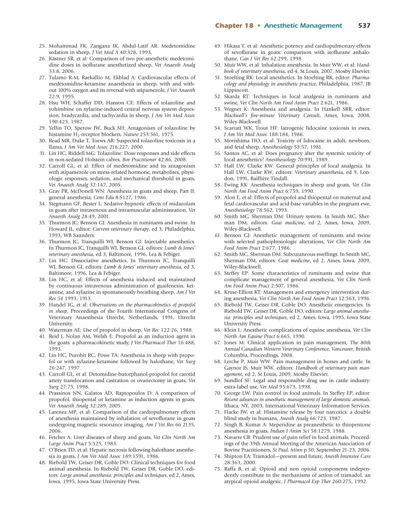
Chapter 18 • Anesthetic Management 537
25. Mohammad FK, Zangana IK, Abdul-Latif AR: Medetomidine sedation in sheep, J Vet Med A 40:328, 1993.
26. Kästner SR, et al: Comparison of two pre-anesthetic medetomi-dine doses in isoflurane anesthetized sheep, Vet Anaesth Analg 33:8, 2006.
27. Tulamo R-M, Raekallio M, Ekblad A: Cardiovascular effects of medetomidine-ketamine anaesthesia in sheep, with and with-out 100% oxygen and its reversal with atipamezole, J Vet Anaesth 22:9, 1995.
28. Hsu WH, Schaffer DD, Hanson CE: Effects of tolazoline and yohimbine on xylazine-induced central nervous system depres-sion, bradycardia, and tachycardia in sheep, J Am Vet Med Assoc 190:423, 1987.
29. Yellin TO, Sperow JW, Buck SH: Antagonism of tolazoline by histamine H2-receptor blockers, Nature 253:561, 1975.
30. Read MR, Duke T, Toews AR: Suspected tolazoline toxicosis in a llama, J Am Vet Med Assoc 216:227, 2000.
31. Lin HC, Riddell MG: Tolazoline: Dose responses and side effects in non-sedated Holstein calves, Bov Practitioner 42:86, 2008.
32. Carroll GL, et al: Effect of medetomidine and its antagonism with atipamezole on stress-related hormone, metabolites, physi-ologic responses, sedation, and mechanical threshold in goats, Vet Anaesth Analg 32:147, 2005.
33. Gray PR, McDonell WN: Anesthesia in goats and sheep, Part II: general anesthesia, Cont Edu 8:S127, 1986.
34. Stegmann GF, Bester L: Sedative-hypnotic effects of midazolam in goats after intravenous and intramuscular administration, Vet Anaesth Analg 28:49, 2001.
35. Thurmon JC, Benson GJ: Anesthesia in ruminants and swine. In Howard JL, editor: Current veterinary therapy, ed 3, Philadelphia, 1993, WB Saunders.
36. Thurmon JC, Tranquilli WJ, Benson GJ: Injectable anesthetics. In Thurmon JC, Tranquilli WJ, Benson GJ, editors: Lumb & Jones’ veterinary anesthesia, ed 3, Baltimore, 1996, Lea & Febiger.
37. Lin HC: Dissociative anesthetics. In Thurmon JC, Tranquilli WJ, Benson GJ, editors: Lumb & Jones’ veterinary anesthesia, ed 3, Baltimore, 1996, Lea & Febiger.
38. Lin HC, et al: Effects of anesthesia induced and maintained by continuous intravenous administration of guaifenesin, ket-amine, and xylazine in spontaneously breathing sheep, Am J Vet Res 54 1993, 1913.
39. Handel IG, et al: Observations on the pharmacokinetics of propofol in sheep, Proceedings of the Fourth International Congress of Veterinary Anaesthesia Utrecht, Netherlands, 1991, Utrecht University.
40. Waterman AE: Use of propofol in sheep, Vet Rec 122:26, 1988. 41. Reid J, Nolan AM, Welsh E: Propofol as an induction agent in
the goats: a pharmacokinetic study, J Vet Pharmacol Ther 16:488, 1993.
42. Lin HC, Purohit RC, Powe TA: Anesthesia in sheep with propo-fol or with xylazine-ketamine followed by halothane, Vet Surg 26:247, 1997.
43. Carroll GL, et al: Detomidine-butorphanol-propofol for carotid artery translocation and castration or ovariectomy in goats, Vet Surg 27:75, 1998.
44. Prassinos NN, Galatos AD, Raptopoulos D: A comparison of propofol, thiopental or ketamine as induction agents in goats, Vet Anaesth Analg 32:289, 2005.
45. Larenza MP, et al: Comparison of the cardiopulmonary effects of anesthesia maintained by inhalation of sevoflurane in goats undergoing magnetic resonance imaging, Am J Vet Res 66 2135, 2006.
46. Fetcher A: Liver diseases of sheep and goats, Vet Clin North Am Large Anim Pract 5:525, 1983.
47. O’Brien TD, et al: Hepatic necrosis following halothane anesthe-sia in goats, J Am Vet Med Assoc 189:1591, 1986.
48. Riebold TW, Geiser DR, Goble DO: Clinical techniques for food animal anesthesia. In Riebold TW, Geiser DR, Goble DO, edi-tors: Large animal anesthesia: principles and techniques, ed 2, Ames, Iowa, 1995, Iowa State University Press.
49. Hikasa Y, et al: Anesthetic potency and cardiopulmonary effects of sevoflurane in goats: comparison with isoflurane anhalo-thane, Can J Vet Res 62:299, 1998.
50. Muir WW, et al: Inhalation anesthesia. In Muir WW, et al: Hand-book of veterinary anesthesia, ed 4, St Louis, 2007, Mosby Elsevier.
51. Stoelting RK: Local anesthetics. In Stoelting RK, editor: Pharma-cology and physiology in anesthetic practice, Philadelphia, 1987, JB Lippincott.
52. Skarda RT: Techniques in local analgesia in ruminants and swine, Vet Clin North Am Food Anim Pract 2:621, 1986.
53. Wegner K: Anesthesia and analgesia. In Hankell SRR, editor: Blackwell’s five-minute Veterinary Consult, Ames, Iowa, 2008, Wiley-Blackwell.
54. Scarratt WK, Trout HF: Iatrogenic lidocaine toxicosis in ewes, J Am Vet Med Assoc 188:184, 1986.
55. Morishima HO, et al: Toxicity of lidocaine in adult, newborn, and fetal sheep, Anesthesiology 55:57, 1981.
56. Santos AC, et al: Does pregnancy alter the systemic toxicity of local anesthetics? Anesthesiology 70:991, 1989.
57. Hall LW, Clarke KW: General principles of local analgesia. In Hall LW, Clarke KW, editors: Veterinary anaesthesia, ed 9, Lon-don, 1991, Baillière Tindall.
58. Ewing KK: Anesthesia techniques in sheep and goats, Vet Clin North Am Food Anim Pract 6:759, 1990.
59. Alon E, et al: Effects of propofol and thiopental on maternal and fetal cardiovascular and acid-base variables in the pregnant ewe, Anesthesiology 78:562, 1993.
60. Smith MC, Sherman DM: Urinary system. In Smith MC, Sher-man DM, editors: Goat medicine, ed 2, Ames, Iowa, 2009, Wiley-Blackwell.
61. Benson GJ: Anesthetic management of ruminants and swine with selected pathophysiologic alterations, Vet Clin North Am Food Anim Pract 2:677, 1986.
62. Smith MC, Sherman DM: Subcutaneous swellings. In Smith MC, Sherman DM, editors: Goat medicine, ed 2, Ames, Iowa, 2009, Wiley-Blackwell.
63. Steffey EP: Some characteristics of ruminants and swine that complicate management of general anesthesia, Vet Clin North Am Food Anim Pract 2:507, 1986.
64. Kruse-Elliott KT: Management and emergency intervention dur-ing anesthesia, Vet Clin North Am Food Anim Pract 12:563, 1996.
65. Riebold TW, Geiser DR, Goble DO: Anesthetic emergencies. In Riebold TW, Geiser DR, Goble DO, editors: Large animal anesthe-sia: principles and techniques, ed 2, Ames, Iowa, 1995, Iowa State University Press.
66. Klein L: Anesthetic complications of equine anesthesia, Vet Clin North Am Equine Pract 6:665, 1990.
67. Jones M: Clinical application in pain management, The 80th Annual Canadian Western Veterinary Conference, Vancouver, British Columbia, Proceedings, 2008.
68. Lerche P, Muir WW: Pain management in horses and cattle. In Gaynor JS, Muir WW, editors: Handbook of veterinary pain man-agement, ed 2, St Louis, 2009, Mosby Elsevier.
69. Sundlof SF: Legal and responsible drug use in cattle industry: extra-label use, Vet Med 93:673, 1998.
70. George LW: Pain control in food animals. In Steffey EP, editor: Recent advances in anesthetic management of large domestic animals, Ithaca, NY, 2003, International Veterinary Information Services.
71. Flacke JW, et al: Histamine release by four narcotics: a double blind study in humans, Anesth Analg 66:723, 1987.
72. Singh B, Kumar A: Meperidine as preanesthetic to thiopentone anesthesia in goats, Indian J Anim Sci 58:1279, 1988.
73. Navarre CB: Prudent use of pain relief in food animals, Proceed-ings of the 39th Annual Meeting of the American Association of Bovine Practitioners, St Paul, Minn p 50, September 21-23, 2006.
74. Shipton EA: Tramadol—present and future, Anesth Intensive Care 28:363, 2000.
75. Raffa B, et al: Opioid and non opioid components indepen-dently contribute to the mechanisms of action of tramadol, an atypical opioid analgesic, J Pharmacol Exp Ther 260:275, 1992.

538 Chapter 18 • Anesthetic Management
76. Scott LJ, Perry CM: Tramadol: a review of its use in perioperative pain, Drugs 60:139, 2000.
77. Shilo Y, et al: Pharmacokinetics of tramadol in horses after intra-venous, intramuscular and oral administration, J Vet Pharmacol Ther 31:60, 2007.
78. Lehmann KA, Schroeder-Bark U, Horrichs-Haermeyer G: Post-operative patient-controlled analgesia with tramadol: analgesic efficacy and minimum effective concentrations, Clin J Pain 6:212, 1990.
79. Mastrocinque S, Fantoni DT: A comparison of preoperative tra-madol and morphine for the control of early postoperative pain in canine ovariohysterectomy, Vet Anaesth Analg 30:220, 2003.
80. Hopkinsins D, et al: Comparison of tramadol and morphine via subcutaneous PCA following major orthopaedic surgery, Can J Anaesth 45:435, 1998.
81. Houmes RJM, et al: Efficacy and safety of tramadol versus mor-phine for moderate and severe postoperative pain with special regard to respiratory depression, Anesth Analg 74:510, 1992.
82. Malonne H, et al: Pharmacokinetic evaluation of a new oral sustained release dosage form of Tramadol, Br J Clin Pharmacol 57:270, 2004.
83. Kukanich B, Papich MG: Pharmacokinetics of tramadol and the metabolite O-desmethyltramadol in dogs, J Vet Pharmacol Ther 27:239, 2004.
84. Pypendop BH, Ilkiew JE: Pharmacokinetics of tramadol, and its metabolite O-desmethyl-tramadol in cats, J Vet Pharmacol Ther 31:52, 2007.
85. De Sousa AB, et al: Pharmacokinetics of tramadol and O- desmethyltramadol in goats after intravenous and oral adminis-tration, J Vet Pharmacol Ther 31:45, 2007.
86. Nolan A, Livingston A, Waterman AE: Investigation of the antinociceptive activity of buprenorphine in sheep, Br J Pharm 92:527, 1987.
87. Murphy MR, Hugg CC Jr: “Ceiling effect” of butorphanol (Stadol) as an anesthetic supplement [abstract], Anesthesiology 55:261, 1981.
88. Waterman AE, Livingston A, Amin A: Analgesic activity and respiratory effects of butorphanol in sheep, Res Vet Sci 51:19, 1991.
89. O’Hair KC, et al: Cardiopulmonary effects of nalbuphine hydro-chloride and butorphanol tartrate in sheep, Lab Anim Sci 38:58, 1988.
90. Anderson DE: Pain management: treatment, management, and prevention, Proceedings of the North American Veterinary Confer-ence, Orlando, Fla, 2007.
91. Davis JL, et al: Update on drugs prohibited from extralabel use in food animals, J Am Vet Med Assoc 235:528, 2009.
92. New animal drugs, phenylbutazone, extralabel animal drug use, order of prohibition, Fed Regist 68:9528, 2003.
93. Ramesh N, et al: A study on toxicity of diclofenac in dogs, Indian Vet J 79:668, 2002.
94. Bertone JJ, et al: Clinical field trial to evaluate the efficacy of topi-cally applied diclofenac liposomal cream for the relief of joint lame-ness in horses, Proceedings of the 48th Annual Convention of the American Association of Equine Practitioners, Lexington, Ky, 2002, AAEP, p 190.
95. Singh NK, et al: Evaluation of epidural xylazine and ketamine for the management of posttraumatic pain in goats, Indian J Vet Surg 22:73, 2001.
96. Mahajan DN, Ali MS, Singh B: Therapeutic efficacy of diclofenac sodium in clinical and experimental cases of arthritis and myo-sitis in cattle and buffaloes, Indian J Vet Surg 22:73, 2001.
97. Gupta AK, et al: Evaluation of homogenous synovial in the treat-ment of acute aseptic arthritis in horses: gross and histopatho-logical studies, Centaur 18:6, 2001.
98. Graham MJ, Kent JE, Molony V: Effects of four analgesic treat-ments on the behavioral and cortisol responses of 3-week-old lambs to tail docking, Vet J 153:87, 1997.
99. Molony V, et al: Reduction in pain suffered by lambs at castra-tion, Vet J 153:205, 1997.
100. Farag MM, Salama MA, Abou-Basha L: Experimental murine schistosomiasis: reduced hepatic morbidity after pre- and/or post-infection treatment with ibuprofen or diclofenac sodium, Ann Trop Med Parasitol 89:497, 1995.
101. Munoz-Criado S, Munoz-Bellido JL, Garcia-Rodriguez JA: in vivo activity of nonsteroidal anti-inflammatory agents, phe-nothiazines, and antidepressants against Brucella species, Eur J Clin Microbiol Infact Dis 15:418, 1996.
102. Altaher AY, et al: Pharmacokinetics of diclofenac in sheep following intravenous and intramuscular administration, Vet Anaesth Analg 33:241, 2006.
103. Lin HC, Riddell MG: Preliminary study of the effects of xylazine or detomidine with or without butorphanol for standing seda-tion in dairy cattle, Vet Therapeutics 4:285–291, 2003.
104. Cook VL, Blikslager AT: Use of systemically administered lido-caine in horses with gastrointestinal tract disease, J Am Vet Med Assoc 232:1144–1148, 2008.
105. Mudge MC: Review of the analgesic, prokinetic, and anti-inflamma-tory uses of IV lidocaine, Proceedings of the 53rd Annual Con-vention of the American Association of Equine Practitioners, Orlando, Fla, December 1-5, 2007 Lexington, Ky, 2007, AAEP, pp 245-248.
106. Grant C, Summersides GE, Kuchel TR: A xylazine infusion regi-men to provide analgesia in sheep, Lab Anim 35:277–281, 2001.
107. Purohit RC: Anesthesia. In Wolfe DF, Moll HD, editors: Large animal urogenital surgery, Baltimore, 1998, Williams & Wilkins.
108. Aminkov BY, Dinev D, Pascalev M: The anti-nociceptive and car-diopulmonary effects of extradural fentanyl-xylazine in sheep, Vet Anaesth Analg 29:126–132, 2002.
109. Haerdi-Landerer MC, Schlegel U, Neiger-Aeschbacher G: The analgesic effects of intrathecal xylazine and detomidine in sheep and their antagonism with systemic atipamezole,, Vet Anaesth Analg 32:297–307, 2005.
110. Hooper RN: General surgical techniques for small ruminants: Part II, Proceedings of the Small Ruminants for the Mixed Animal Practitioner Western Veterinary Conference, 1998, Las Vegas, Nev.
111. Staffier F, et al: A comparison of subarachnoid buprenorphine or xylazine as an adjunct to lidocaine for analgesia in goats, Vet Anaesth Analg 36:502–511, 2009.
112. Pablo LS: Epidural morphine in goats after hindlimb orthopedic surgery, Vet Surg 22:307–310, 1993.
113. Hendrickson DA, Kruse-Elliott KT, Broadstone RV: A compari-son of epidural saline, morphine or bupivacaine for pain relief after abdominal surgery in goats, Vet Surg 25:83–87, 1996.
114. George AN: A note on the anatomy of the horns of sheep, Br Vet J 111:391, 1955.
115. Greenough PR, Johnson L: The integumentary system: skin, hoof, claw, and appendages. In Oehme FW, Prier JE, editors: Textbook of large animal surgery, Baltimore, 1974, Williams & Wilkins.
116. Hooper RN: General surgical techniques for small ruminants: Part I, Small Ruminants for the Mixed Animal Practitioner Western Veterinary Conference, 1998, Las Vegas, Nev.
117. Sammarco JL, et al: Post-operative analgesia for stifle surgery: a comparison of intra-articular bupivacaine, morphine, or saline, Vet Surg 25:59–69, 1996.
118. Shafford HL, Hellyer PW, Turner S: Intra-articular lidocaine plus bupivacaine in sheep undergoing stifle arthrotomy, Vet Anaesth Analg 31:20–26, 2004.

539
Flock and Herd Health
Patty Scharko, Jason Johnson, Seyedmehdi Mobini, and D.G. Pugh
C H A P T E R
SHEEP FLOCK HEALTHPatty Scharko and D.G. Pugh
DEFINITION OF FLOCK HEALTHThe aim of a flock health program is to improve the overall health and welfare of the sheep in the flock, decreasing losses from disease, increasing productiv-ity, and maximizing the profitability of the flock. The use and analysis of production, health, and financial records are excellent ways to measure the success of the program. Regardless of the approach used, flock health programs should include a written component that addresses all aspects of the production unit, time of year, vaccination, nutrition, reproduction, and parasite control, as well as overall productivity.
Flock health programs are developed for multiple applications. They may be used by an individual pro-ducer or incorporated into initiatives for the control and eradication of specific diseases through either vol-untary or mandatory cooperation with state and federal animal health officials. Programs used by individual operators generally are planned annual approaches that address the following: • Prevention of common disease conditions through the
use of appropriate vaccination schedules (Table 19-1) • Management strategies that minimize risk factors for
disease occurrence • Provision of appropriate levels of nutrition for the
stage of production • Assessment of metabolic status and productivity
In order to provide correct advice, the veterinarian must have a sound knowledge base regarding the sheep industry and the individual production system. The cli-ent and the veterinarian should work together in order to outline all pertinent details of a record-keeping system.
Veterinary visits usually are scheduled in accordance with timing of major production events (e.g., before breeding; in midpregnancy; before, during, and after lambing; in midsummer). During the first visit, the vet-erinarian assigns a body condition score (BCS) to the ewe flock, which is imperative for making nutritional
19
TABLE 19-1 Basic Vaccination Program for Sheep
Timing Immunization/Scheduled Management
Practice(s)/Intervention(s)
PREGNANT EWES • Midgestation In endemic areas, vaccinate ewe
lambs or previously vaccinated animals against Campylobacter and Chlamydia (Chlamydophila) abortion.
• Last month of pregnancy
Vaccinate for Clostridium species (C. perfringens type C and D, C. novyi, C. sordelli, C. chauvoei, C. septicum, C. tetani)
• 2 to 4 weeks after birth
Repeat Chlamydophila and Campylobacter vaccinations for previously unvaccinated animals and give yearly booster to other ewes
LAMBS • 1 to 2 months • Repeat immuni-
zations in 3 to 4 weeks
Immunize lambs from immunized dams for Clostridium species (C. perfringens types C and D, C. novyi, C. sordelli, C. chauvoei, C. septicum, C. tetani)
• 1 to 3 weeks • Repeat immu-
nizations twice at 3- to 4-week intervals
Immunize lambs from nonim-munized dams for Clostridium species (C. perfringens types C and D, C. novyi, C. sordelli, C. chauvoei, C. septicum, C. tetani)
RAMS AND YEARLINGS • As for ewes Vaccinate at the same time as for
ewes, with an emphasis on Clostridium species.
Vaccines against rabies should be given in endemic areas.

540 Chapter 19 • Flock and Herd Health
and feeding management recommendations. Evalua-tion of the rams should include both a general physical examination and fertility assessment (see Chapter 8). A written record should indicate the ram’s BCS and weight, problems identified on the physical examina-tion, and an action plan to address any problems that are crucial to breeding. The midpregnancy visit focuses primarily on ultrasound pregnancy determination and fetal counting and aging. On the basis of these results, the veterinarian can develop a plan for feeding ewes appropriately and economically as appropriate for fetal numbers and stage of pregnancy (see Chapter 2). This visit can be crucial in preventing metabolic disease in late pregnancy. The visit, examination, and herd/flock recommendations for the ewe in late pregnancy entails a review of the nutrition of the late-gestation ewe so that she can perform up to her genetic potential at lambing.
Properly fed ewes produce maximal amounts of colostrum and milk, give birth to thrifty lambs with
minimal difficulty, and demonstrate excellent mother-ing abilities. The final 2 months of pregnancy are crucial to successful lamb growth and survivability. During this visit, the veterinarian can review strategies to prevent disease such as vaccinating against clostridial infections 4 weeks before lambing and providing a clean environ-ment. The management plan at lambing time, including the layout and use of facilities, also should be reviewed. This plan should include education of farm person-nel to recognize the need for intervention for lambing problems (Tables 19-2 and 19-3).
Minimum standards and target production param-eters should be set for morbidity, mortality, culling, and growth rates (Table 19-4). A quality assurance program also should be designed for the flock. To date, the sheep-packing industry has not required producers to participate in flock quality assurance programs, but such programs exist and will become more common in the future as a result of consumer demand. Quality
TABLE 19-2 Generic Management Calendar for Spring Kidding and Lambing
January Evaluate range and forage conditions; monitor body condition of does and ewes and supplement if necessary; ensure adequate intake of minerals, salt, and water; vaccinate during the final month of gestation for clos-tridial disease and any other endemic diseases.
February Begin supplemental feeding of pregnant females and consider prebirthing shearing; begin birthing; check teats for milk and identify lambs and kids; ensure ingestion of adequate colostrum by lambs and kids; institute pre- and postbirthing strategic deworming; maintain an ionophore in feed or mineral mixture before and after birthing to decrease coccidial contamination of pasture.
March Separate singles from twins; confine and feed females with their lambs and kids as needed; feed does and ewes to maintain milk production; continue strategic deworming program.
April Continue to feed a supplement to lactating does and ewes; monitor for parasites with FAMACHA scoring and “smart drenching.”
May Wean small, stunted lambs and kids; discontinue supplemental feeding of does and ewes; monitor internal parasites (with FAMACHA and fecal egg counts using McMaster technique).
June Continue parasite control program with FAMACHA monitoring.July Monitor internal parasites; watch for signs of heat stress; wean lambs and kids.August Continue parasite control program; continue weaning lambs and kids; supplement replacement does, ewes,
bucks, and rams; select replacement males and females; identify and cull unsound and inferior animals; perform breeding soundness evaluation in males. Criteria for culling include the following:
• Poor or slow growth • Barren status in females (missed one season) • Unsound teats or udders (too big or too small) • Poor dentition • Structural defects (feet, leg, or back abnormalities) • Testicles that are small or soft or have other abnormalities (so that animal fails a breeding soundness evaluation) • Unthriftiness (caused by old age or chronic disease)
September Begin supplemental feeding of females and males on fresh green pasture with ½ lb feed/head/day for 2-3 weeks before and after males are placed with females; continue parasite control program.
October Begin breeding; maintain good male-to-female ratio, depending on pasture size and conditions; continue supplemental feeding of females for 2-3 weeks after start of breeding season.
November Evaluate range and forage conditions; determine females’ body condition and plan winter supplemental and feeding program; control internal and external parasites; remove some of males’ feed to regain body condi-tion; determine pregnancy status and number of fetuses.
December Evaluate body condition of does and supplement feed if needed; monitor internal and external parasites.
Chapter 19 • Flock and Herd Health 541
assurance programs educate producers in good pro-duction practices and encourage cooperation between practitioners and area producers. Table 19-2 presents a sample management calendar for spring lambing.
BIOSECURITY ASPECTS OF FLOCK HEALTH PROGRAMSBiosecurity programs are a critical element in the con-trol of infectious disease. Such programs include man-agement steps to reduce the likelihood of introduction of a new disease from an external source.
Another benefit of a biosecurity program is that it has the potential to reduce the spread of infectious disease already present in the flock. Scientifically sound
approaches regarding the introduction of sheep into a flock are required to prevent the introduction of new diseases into the resident sheep population. Practi-cal biosecurity principles include purchasing animals directly from the farm of origin, transporting them in clean and disinfected transport vehicles, housing them in true isolation facilities while following practical isolation practices for 1 month, and regularly observ-ing for conditions such as pruritus, lameness, external lumps, and unexplained weight loss. In addition, iso-lated sheep should be integrated into the flock’s regu-lar vaccination program, dewormed with an effective anthelmintic, and have their feet trimmed and visually inspected for footrot.
Even before performing a visual examination and obtaining the history of the flock of origin, the producer or the clinician may wish to use a foot bath (15 minutes in a 10% zinc sulfate solution) for footrot control. Dur-ing isolation, the sheep may be serologically tested for diseases of concern to the buyer. If the sheep were tested for various diseases before purchase, the buyer should request the official test forms and results to check the results and test dates. Incoming sheep should be thor-oughly examined for obvious clinical manifestations of contagious or infectious diseases such as contagious ecthyma (“soremouth” or orf), keratoconjunctivitis (“pinkeye”), external parasites, caseous lymphadenitis, scrapie, footrot, strawberry footrot, ulcerative dermato-sis, and other contagious diseases of concern.
BARNYARD BIOSECURITYProducers should be educated regarding the following practices and principles as key to an efficacious biosecu-rity program for farm animals: • Do not haul disease home or to the market. Table
19-5 lists some of the diseases that should “stay home” on the farm until the animal fully recovers.
• Isolate new animals for 2 to 4 weeks. Have no contact between new animals and the resident farm herd or flock animals during this time. Prevent introduction onto farm by keeping a closed herd or flock, and purchase animals from known sources (Table 19-6).
• Restrict access to the farm: post signs for vehicle and foot traffic control; keep a visitor log; don’t track disease in—wear different shoes, clothes, and hat to livestock auction market or public area and change before working with livestock.
• Provide good nutrition (water, feed, and minerals) and management plan (including vaccination, deworming, and the like) to maintain a healthy herd or flock.
• Report animals with unusual illness or those that are not responding to treatment to a local veterinar-ian, the state veterinarian, or the U.S. Department of Agriculture (USDA) Veterinary Services. Early detec-tion may save animal lives.
TABLE 19-3 Seasonal Veterinary Management
Period in Seasonal Cycle Veterinary Visit Assessments
Prebreeding BCS in ewesNutrition and feeding recommenda-
tions for flushingRam breeding soundness exam
Midpregnancy Ultrasound pregnancy exam: fetal counting and aging
BCSPrelambing Vaccination for clostridial diseases
Review of nutritionLambing management planReview of intervention criteriaBCS
Midsummer BCSFAMACHA scoring and “smart
drenching”
BCS, Body condition score.
TABLE 19-4 Production Targets
Production Parameter Target
PREGNANCYEwes More than 95%Ewe lambs More than 75%Visible abortion Less than 5%
LAMBINGEwes More than 90%Ewe lambs More than 70%Stillbirths Less than 2%Weaning More than 95%

542 Chapter 19 • Flock and Herd Health
Vaccination ProgramsThrough regular and correct usage, vaccines are designed to reduce the incidence and severity of a specific disease. Few, if any, vaccines are 100% efficacious in preventing disease. When vaccines are used properly, however, in most instances their beneficial effects will far outweigh their drawbacks. Area veterinarians can provide advice on prevalent diseases that should be used regionally, because farms in different areas have different needs. The local veterinarian should work with the producer to design a farm-specific vaccination program for ewes, young lambs, market or feedlot lambs, replacement breeding stock, and rams. Knowledge of prevalent dis-eases in the area and of diagnostic laboratory data is the best basis for developing a vaccination program. Vaccination should proceed according to label direc-tions, because the timing of doses is crucial to optimal protection.
Clostridial diseases are the only universal group of diseases for which all sheep should receive vaccinations. Decisions regarding the inclusion of other vaccines in an individual flock health program should be based on knowledge of prevalent diseases in the area and the needs of the particular flock. In areas in which a disease
is known to occur but no vaccine is available, risk fac-tors for that disease should be controlled with proper management. Sheep that are frequently exhibited (as in, FFA, 4H, or breed shows) are at greater risk for contract-ing contagious and infectious diseases. For this reason, show sheep usually should be vaccinated against more diseases than is usual for sheep in a closed flock.
Other diseases that have an available vaccine labeled for use in sheep in the United States include some infec-tions associated with abortion such as those caused by Campylobacter spp. and Chlamydia (Chlamydophila) psit-taci; multisystem diseases such as caseous lymphadeni-tis, musculoskeletal diseases such as footrot, neurologic diseases such as rabies, and integumentary diseases such as contagious ecthyma (soremouth). If past history indi-cates that the flock is at risk for a disease, such vaccines can be included in an immunization program (see Table 19-1), which can be modified for the individual farm.
Internal Parasite Control ProgramsThe epidemiology of pathogenic sheep nematodes and protozoal species depends on the climate of the region; therefore internal parasite control must be
TABLE 19-5 Diseases That Need to Stay Home on the Farm Until Animal Recovers
Disease Biosecurity* Zoonosis† Comment(s)
Contagious ecthyma (soremouth, orf) Yes Yes Scabs on outside of mouth; wear glovesCaseous lymphadenitis (“cheesy gland”) Yes Yes—not common Abscesses in head, neck, or bodyKeratoconjunctivitis (“pinkeye”) and/or
conjunctivitisYes No
Ringworm/club lamb fungus Yes Yes Wear glovesFootrot (not foot scald) Yes No Bottom of sole/foot usually deformed;
does not include overgrown toes; foot scald affects only between the toes
*Biosecurity: set of preventive measures to protect livestock from disease and keep animals healthy. The transmission of infectious agents will be mini-mized and disease prevention will be maximized with attention to biosecurity strategies for these five clinical entities.†Zoonosis: infectious disease that can be transmitted from animals to humans. These are public health risks.
TABLE 19-6 Diseases Without Clinical Manifestations That Can Cause Abortion*
Disease Biosecurity Zoonosis Comments
Toxoplasmosis Keep cats away from feed Yes Abortions; pregnant women should use caution around kidding or lambing
Chlamydiosis (Chlamydophilosis) Yes Yes AbortionCampylobacteriosis Yes Rare Abortion—rareQ fever Goats and sheep can be
carriersYes Usually with handling birthing fluids and
placenta, drinking unpasteurized milk
*Animals can be inapparent carriers of these diseases.

Chapter 19 • Flock and Herd Health 543
tailored to the region of the country in which the flock resides. Parasite control programs also must take into account whether the flock is confined, pastured, or rotationally grazed. Successful programs implement regular monitoring of the efficacy of anthelmintics, sheep-friendly handling equipment for anthelmintic delivery such as well-designed pens and chutes, and use of automatic syringes or drench guns. Other com-ponents of a parasite control program include the use of multiple strategies to minimize the buildup of nematode eggs on pasture; this aim can be achieved by deworming ewes with larvicidal doses during win-ter housing, at 4 weeks after spring turnout, and at 3 weeks into lambing. Control also is enhanced by the use of management practices that reduce reliance on chemical anthelmintics, such as FAMACHA “Smart Drenching,” grazing clean ground with weaned lambs, “vacuuming” nematode eggs by grazing the previous year’s sheep pastures with cows or horses, and select-ing for and breeding nematode-resistant sheep (see Chapter 6).
When the flock must graze close to the ground and nutritional input is marginal, nematode infestation may escalate, typically with development of clinical parasitism in stressed sheep. The veterinarian and the producer should create an annual calendar that details the entire flock health program. The producer should record details about any procedures performed in the production groups. Box 19-1 summarizes recommenda-tions to improve parasite control. Table 6-1 shows some commonly used anthelmintics and coccidiostats useful in parasite control for sheep and goats.
External Parasite Control ProgramsKeds and biting lice are the prevalent external parasites of sheep. Many sheep flocks have not introduced these parasites. In these flocks, all newly purchased sheep should be treated prophylactically while they are in isolation to prevent parasite introduction. In flocks in which either parasite is endemic, all sheep on the property should be treated at the same time, so that one group does not serve as a reservoir for reintroduction. Treated sheep should be kept out of any contaminated buildings for 2 weeks, because buildings also can serve as reservoirs during this time. Further treatments should not be necessary after the whole flock is properly treated. The flock should be monitored for ectoparasite infesta-tion after reintroduction from carrier sheep or after improperly applied whole flock treatment. An external parasite control program is described in Chapter 6.
Flock Health MonitoringProgram records should contain a flock inventory divided into production groups. Important information gained from the records can include: • Ram-to-ewe ratios at breeding • Pregnancy rates • Lambing percentages • Ewe and ram mortality numbers and reasons • Pre- and postweaning lamb mortality rates and
reasons • Ram morbidity and treatment outcomes (especially
those that pertain to breeding use)
1. Make certain that the anthelmintic or combination of anthelmintics used on the farm actually works (kills at least 90% of the viable worms). Check for resistance with fecal egg counts before and after deworming.
2. Utilize FAMACHA “smart drenching” in the spring and continue to assess every 2-4 weeks until the hazard of Haemonchus infestation no longer remains in cold weather.
3. Use strategic deworming. Deworm the flock while the parasites are in hypobiosis and are being transmitted at low levels (i.e., the winter). This strategy reduces the frequency of exposure to deworming products.
4. Employ pre- and postbirthing deworming starting 1 month before birthing at 2- to 4-week intervals and ending 2-4 weeks after the final lamb or kid is born.
5. Tactical dewormings (based on increased levels of parasite eggs or 10-14 days after rainfall) enhance the effectiveness of a parasite program.
6. Graze above 4 inches; use “clean” or safe pastures when possible (aftermath of crops, annual forage such as chicory); utilize rotational grazing or cograzing with cattle or horses. (Beware: Permanent pastures promote parasites.)
7. Deworm new animals and place them in a nonpasture environment such as a dry lot or barn after treatment for as long as 72 hours before moving them to a safe pasture. Check fecal egg count 10-14 days after treat-ment for fecal egg shedding
8. Rotate anthelmintics yearly if effective drugs are available.
9. Do not underdose. Determine dose for the heaviest animal in a production group.
10. Identify and select individual animals resistant to inter-nal parasites for flock/herd retention and breeding.
BOX 19-1 Summary of Recommendations for Parasite Control*
*More in-depth recommendations are available in Chapter 6.

544 Chapter 19 • Flock and Herd Health
The average BCS of the ewe flock should be recorded at breeding, lambing, and weaning (see Chapter 2). If target scores are not achieved, the producer and the veterinarian need to determine why and make appro-priate management changes. If the correct changes are implemented, future scores should improve, with con-sequently enhanced production.
If mortality rates exceed targets, necropsy should be performed in representative sheep to determine cause of death. Gross findings can be further investigated with ancillary laboratory-based tests.
Culling and Disposal PracticesCulling practices should be based on genetics, produc-tivity, poor fertility, substandard growth, parasite and disease susceptibility, and disease (e.g., footrot, caseous lymphadenitis, scrapie, ovine progressive pneumonia). Not all sick or thin animals survive to the point of cull-ing. Each operation must have a plan for carcass dis-posal. Carcass disposal procedures used should be legal in the area and state, environmentally friendly for the size of the animal and number of carcasses, and practi-cal for the producer. Many states legally permit sheep composting (see Box 20-2). A sheep of any size will turn into compost if the procedure is done correctly. With some diseases (e.g., scrapie, foot-and-mouth disease), carcasses may by law require incineration.
Neonatal CareA “strip, sip, clip, and dip” strategy for management of the newborn lamb is sound advice for beginning and advanced clinicians and owners alike: The producer can strip the wax plug from the teat so that the lamb can sip colostrum, as well as clipping and dipping the umbilicus in 7% iodine or another antiseptic or astringent (e.g., solutions of chlorhexidine or povidone-iodine). Owners and practitioners should be able to recognize normal maternal and neonatal behavior, as well as normal-appearing and functioning mammary glands. The recognition of normal traits and conditions allows abnormalities to be detected and dealt with appropriately.
Equipment and facilities should be prepared and organized before the start of lambing. Records to guide improvements in management should be kept and used. Regardless of whether animals are raised in con-finement or on pasture, periparturient ewes should be grouped together according to expected lambing dates and fetal numbers, if available. These groupings enable tailored levels of feeding, which are economically jus-tifiable, minimize the occurrence of metabolic disease, and prevent fetal under- or overfeeding. The result is the birth of viable lambs. When lamb losses occur, postmortem examination by the local veterinarian or
veterinary diagnostic center in representative cases is recommended. Management changes should be based on the necropsy findings (see Chapter 20).
Shearing ManagementShearing is a highly stressful experience for sheep. Accordingly, the shearing setup should be designed so that handling stress is minimized. Many preventive health procedures often are performed at shearing, such as prelambing vaccination against clostridial dis-eases, especially those caused by C. perfringens types C and D and C. tetani. Ewes should be vaccinated against colibacillosis (in situations in which such vac-cines are used) approximately 30 days before lambing on farms with a history of previous cases of neonatal lamb diarrhea known or suspected to be caused by pathogenic Escherichia coli. Other procedures fre-quently performed at shearing include sorting out noncompetitive sheep or those with abscesses or mas-titis, foot trimming, and deworming. With all such procedures, the well-being of both the sheep and the shearers must be considered. Freshly trimmed feet can lacerate a shearer if the sheep kicks, for example. The owner should be encouraged to be present at shearing and to accept the shearers’ observations as valuable professional input. Prelambing shearing of ewes may decrease the incidence of pregnancy toxemia, encour-age the ewe to seek shelter on cold days (with the resultant birth of lambs in warmer, dry environment), decrease the maintenance requirements of the ewe (because of less fleece weight), and enhance the likeli-hood that newborns will nurse the udder instead of the ewes’ wool.
Most small flock facilities lack a permanent shearing setup. Therefore many shearers perform the procedure on sheets of plywood or pieces of indoor-outdoor car-pet that they carry in their trucks and use every day on numerous farms. Such surfaces may readily transmit bacteria, fungi, and even viruses from other farms. Owners should therefore provide their own shearing surfaces.
Foot CarePart of the annual care of sheep should include assess-ment of the condition and length of their hooves. Depending on the terrain and rainfall in the area, some flocks do not require annual hoof trimming, but most flocks in the midwestern and eastern regions of the United States will need regular trimming. The timing of this procedure is not important so long as hoof over-growth, which predisposes affected animals to other foot problems such as foot scald, toe abscesses, and footrot, is not allowed to occur. Many farm flocks trim the ewes’ feet around lambing time while they are in

Chapter 19 • Flock and Herd Health 545
confinement and being handled regularly. Many range flocks only require foot trimming of a few individual animals. These sheep wear their feet down adequately on dry, rough terrain, and some breeds are predisposed to slower foot growth. Rams should have their feet trimmed 4 to 8 weeks before breeding. Trimming in the week before the start of breeding is contraindicated in case overzealous trimming causes temporary lameness. A generic footrot prevention program is presented in Box 11-2 (see also Chapter 11).
Facility Design and FunctionSheep housing should be designed and constructed for proper ventilation and efficient manure handling.
Water Availability and DesignWater is an essential nutrient for sheep of all ages. It should be of good quality and readily available. Cleanliness, taste, impurities, and temperature all affect consumption. Key times during which the qual-ity of the water supply has a direct influence on the productivity or health of the animal are the lambing and finishing phases, late pregnancy, and lactation (see Chapter 2).
Role of Management in Maintaining HealthThe level of management determines the success and sustainability of a farming operation. Excellent manag-ers make most decisions on a timely basis and correctly relative to the care of their flock. Accordingly, the flock responds by meeting target production goals. In today’s economic environment, sound management decisions are based on a combination of recorded data and obser-vational subjective findings.
Specific Diseases Introduced by Carrier SheepSheep can carry and introduce into a flock a num-ber of diseases that are not visible to the naked eye. These disease conditions include footrot, chlamydiosis, campylobacteriosis, Q fever, and anthelmintic-resistant nematode infection. In general, tests for these diseases do not exist or are not financially feasible for whole flocks. Therefore all animals introduced into the flock should undergo a complete physical examination and be quarantined from the rest of the flock for 21 to 30 days, to minimize the introduction of new diseases.
All new additions should be quarantined in a dry lot (without any grass) or on concrete and aggressively dewormed with a product from each of the three classes of anthelmintics. After 14 days, a fecal egg count is
performed, and sheep with negative fecal counts can then be added to previously grazed pastures (not onto clean or safe pastures).
GOAT HERD HEALTHJason Johnson, Seyedmehdi Mobini, and D.G. Pugh
HERD HEALTH MANAGEMENTHerd health management and preventive medicine programs are designed to minimize potential adverse effects of predictable problems and to protect against unexpected ones. The goal of a herd health program is to improve the goat herd’s productivity through general husbandry, nutritional management, parasite control, vaccination, and environmental management. An understanding of various management practices and common disease problems is necessary to achieve this goal. Ultimate success lies in the positive results that will emerge from implementation of a herd health pro-gram, but it will take effective communication between practitioner and owner. Effective communication can be achieved by conducting regular herd visits, producing newsletters, providing website links, or other resources to farms. Information exchanged should include a written herd health plan for each specific farm and regular assessment of herd production. The practitioner should keep owners informed on any new technolo-gies in goat production, emerging disease information, current treatment recommendations and preventative medicine. For the typical busy practitioner, this is a lofty goal; however, with existing computer programs, the practitioner is encouraged to maintain a “template,” which may be adjusted on the basis of individual farm nuances.
Because of their remarkable adaptability, goats are maintained over a more diverse range of production systems than any other domestic livestock species. Goat production can be divided into dairy, fiber, meat, pet, and show production. The largest body of information about goat herd health management and preventive medicine is for dairy and fiber-producing goats. How-ever, the meat goat industry continues to expand, and this text will include current herd health recommenda-tions therein. Herd health management programs must be developed on an individual basis, and these pro-grams vary according to the herd size, its purpose, and the owner’s production goals. Overall, the practitioner should keep in mind a few target areas responsible for economic loss in goat production and make efforts to address these respective areas (Box 19-2). For the pur-pose of organizing goat herd health information into a simple, usable, and easily remembered format, the herd can be divided into four groups: bred does and dry does, kidding does and newborn kids, kids and wean-lings, and bucks.

546 Chapter 19 • Flock and Herd Health
Bred Does and Dry DoesThe major disease problems in this group are preg-nancy toxemia, abortion, and pseudopregnancy. Goats that are over-conditioned during this period are at risk of pregnancy toxemia, vaginal prolapse, and dystocia. The primary objective for these animals is proper nutritional management, avoidance of obesity in early gestation, and provision of adequate nutrients to support the rapid growth of the fetus in the final trimester.
Pregnancy toxemia can be avoided by proper dietary management. First, care should be taken to prevent obesity in late lactation does by decreasing fed grain. These does in early gestation (late lactation) can be maintained on high quality forages until around the last trimester, when supplemental feeding should begin. During the dry period, does should receive a daily ration of hay with at least 10% protein and grain supplementation at a rate of 1 to 2 lb/head. Grain supplementation can be increased if environmental conditions are excessively cold and wet. Owners should aim for does to freshen with a body condition score of 3.5 to 3.75. If pregnancy toxemia is detected in a herd, then urine ketone testing is warranted (see Chapters 2 and 5).
As reviewed in other chapters, many infectious and noninfectious causes of abortions in goats are recog-nized. If abortions do occur, the clinician should strive for a diagnosis. Does that have aborted should be isolated, and if possible, areas in which the abortion occurred disinfected. Aborted fetuses and placentas that are not submitted to a laboratory should be incen-erated or buried. Sound biosecurity measures are the mainstay of preventing infectious abortions. All new incoming animals, including bucks, should undergo a period of quarantine. No new animals should be introduced into the bred doeling or dry doe groups. In herds in which Chlamydia (Chlamydophila) or Cam-pylobacter abortions have occurred, all does should be vaccinated before the breeding season. Management principles for bred does and doelings are outlined in Table 19-7.
Kidding Does and Newborn KidsKidding should be a well-anticipated event and not an unexpected surprise. Where possible the pregnant doe must be given adequate exercise until the time for kid-ding. Does should be placed in a clean, warm, dry, well-ventilated area as parturition nears. Dairy goat udders can be clipped 2 weeks before the due date. Preparation for routine processing of kids at birth, including navel dipping, colostrum feeding, and establishing a protocol for responding to parturition-related emergencies, will reduce neonatal losses. To this end, equipment lists should be in place for all farm personnel, and work schedules should address the possibility of attending the does during parturition. Induction of parturition using prostaglandins (e.g., PGF2α, 2.5 to 20 mg IM) is one man-agement option to ensure that does are attended at birth.
The major obstacles to survival of newborn kids are hypothermia, hypoglycemia, and infectious diseases secondary to delayed or inadequate colostral intake. Other diseases that are seen in this production group are septicemia, pneumonia, omphalophlebitis, and diarrhea. Proper facilities and management techniques can decrease the incidence and severity of these diseases.
1. Suboptimal reproductive performance 2. Periparturient diseases of does 3. Neonatal mortality 4. Diseases of weanlings 5. Nutritional mismanagement of pregnant and lactating
does 6. Parasitism 7. Lack of sound biosecurity program
BOX 19-2 Major Causes of Economic Loss in Goat Herds in North America
TABLE 19-7 Management of Bred Does and Dry Does
Management Practice(s)
Scheduled Management Intervention(s)
Maintain proper nutrition.
Maintain proper BCS: Scores of 2.5 to 3.5 (on a 5-point system).
Ensure adequate dry period.
Introduce no new animals into bred does production group.
Perform pregnancy diagnosis 45-60 days after breeding.
Trim feet as needed.Provide a clean, dry,
draft-free area for maternity pens, or a well-drained, clean pasture with shelter.
Vaccinate with Clostridium perfringens types C and D and tetani toxoid 5-7 weeks before parturition.
Vaccinate for Chlamydia or Campylobacter (in previously diagnosed herds) 1 month before breeding season.
Deworm 2-3 weeks before kidding (if applicable).
Monitor urinary ketone levels in herds with history of pregnancy toxemia.
In deficient areas, administer vitamin E and selenium to does 30 to 45 days before kidding.
Ensure adequate selenium intake with a free-choice mineral mixture.
If herd has history of infec-tious abortions, consider adding ionophores/tetracy-clines to feed.

Chapter 19 • Flock and Herd Health 547
The doe and neonate should be placed in an area that is warm, dry, and protected from wind. Environmental pathogen load can be decreased by regular changing of bedding. The newborn kid should receive an adequate amount of high-quality colostrum. Failure of passive transfer of maternal antibodies to newborn kids leads to an increased incidence of disease and death. On farms that practice dam-rearing of kids (meat and fiber), visual assessment of colostral intake is indicated; if adequacy of intake is questionable, neonate immunoglobulin lev-els should be tested. Hand-feeding of colostrum is the most definitive way to ensure adequate intake.
Caprine arthritis-encephalitis virus (CAEV), which is transmitted from infected does to kids primarily by colostrum and milk, is a major concern in dairy goats. Basically, implementation of a CAEV control program targets the newborn dairy kids; however, multipronged control measures that may maximize protection also are available. Kids should be dried and separated from their dams immediately after birth. To help prevent the spread of CAEV, colostrum can be heat-treated at 56° C (133° F) for 60 minutes before being fed. Does that have the mammary form of CAEV disease may have normal-size udders that are firm on palpation and may exhibit hypogalactia or agalactia (see Chapters 13 and 16).
Newborn kids and their mothers should be properly identified, and records should be updated accordingly. The newborns should be inspected for the presence of any congenital abnormalities. Kids born to does that were not vaccinated for Clostridium perfringens types C and D and tetanus should receive an antitoxin during their first week of life. Udder conformation, milk pro-duction, and milk quality should be critically assessed during this period in milking does. Standardized milk-ing procedures, with emphasis on the use of good-qual-ity equipment, good udder hygiene, and milk records, should be in place. Adequate trough space should be provided for milking does. Bucks should be separated from lactating does to prevent development of a “buck odor” to the milk. A summary of management practices for this production group is presented in Table 19-8.
Kids and WeanlingsWeaning may occur at 6 to 12 weeks in dairy goats or orphaned kids, depending on the feeding and manage-ment system. Meat goats usually are weaned at 4 to 6 months. Common diseases encountered during the weaning period include pneumonia, coccidiosis, and gastrointestinal nematode parasites. Enterotoxemia and neurologic CAE disease also can be seen in this produc-tion group.
The diseases in this category are intimately connected to management practices. Incidence of respiratory dis-ease may be reduced by providing proper ventilation, maintaining an ambient temperature between 6° C and
27° C (43° to 81° F), and preventing overcrowding. Attempts to control coccidiosis should concentrate on reducing the infective number of oocysts in the environ-ment. Prompt and regular removal of soiled bedding, decreasing stocking densities, and the use of movable hutches can be expected to decrease the effective num-ber of oocysts. The disease may still show up on a farm, however, and addition of a coccidiostat to the feed may be necessary. Common dosages are listed in Table 6-1. Under intense production conditions, it may be pru-dent to begin coccidiostats around 1 to 4 months of age (see Chapter 6).
On initial release onto summer grazing, weanlings may incur heavy internal parasite burdens. Depending
TABLE 19-8 Management of Kidding Does and Newborn Kids
Management Practice(s)
Scheduled Management Intervention(s)
Provide proper protec-tion from elements for periparturient does and neonates: • Dry area • Out of wind • Warm area • Well-ventilated
Decrease environ-mental pathogen load:
• Provide clean bedding.
• Change bedding regularly.
• Clean housing regularly. Clean dam’s teats.
Examine and palpate udders of parturient does.
Assess colostrum intake in dam-reared kids: • Visually • IgG testsAccurately identify all
newborn kids.Examine each kid for
congenital defects.Introduce hay and feed
within first week of life.
Ensure adequate intake of high-quality colostrum within 4 hours of birth.
Implement CAEV control program measures: • Feed pasteurized
colostrums/milk. • Dry and separate kid from
dam immediately after birth.
• Tape teats of CAEV-positive does. Dip navel with iodine.
Administer Clostridium per-fringens type C and D and tetanus antitoxin within first week of life to kids born to unvaccinated does, with booster vaccination within 3 to 4 weeks.
In deficient areas, administer vitamin E and selenium injections to kids within first 2 weeks of life.
Perform horn disbudding of dairy kids at 2-14 days of age.
Inspect doelings for extra teats; remove within 7 days of age.
Castrate bucks at 4-14 days of age.
IgG, Immunoglobulin G.

548 Chapter 19 • Flock and Herd Health
on the particular parasite management approach of the farm, some investigators advocate deworming before turnout. Release of weanlings onto “new” pastures that have been without livestock for certain minimum periods may decrease initial infective worm burdens. Parasite monitoring strategies may include the use of FAMACHA coupled with composite fecal egg counts (see Chapters 6 and 16).
Hay and concentrates should be offered early dur-ing the life of the kid, before weaning, in an attempt to decrease anorexic weaning stress. Depending on genetics, nutrition levels, and management practices, spring-born kids may be of sufficient body weight (70% adult body weight) for entry into the autumn breed-ing doeling group. In milking herds, where year-round kidding is desirable, reproductive manipulation will be warranted, as covered in depth in Chapters 2 and 8.
Vaccines for C. perfringens types C and D and teta-nus toxoid should be administered at the age of 1 to 2 months in kids whose dams were vaccinated during pregnancy. No respiratory disease vaccines exist for weanling goats.
In meat goat finishing programs in which young bucks are fed high-concentrate feed, conditions such as bloat, urinary calculi, and enterotoxemia are fre-quently encountered. As confirmed by our own experience, urinary calculi may be diagnosed in male Boer kids as young as 2 to 3 months of age. Provid-ing a continuous supply of clean, fresh water and increasing the concentration of salt in the ration to a maximum of 4% are helpful strategies to prevent calculi. Prophylactic use of urinary acidifiers also has been advocated. The continuous administration of ammonium chloride at a level of 1% to 2% of the diet is recommended in castrated and intact bucks. Polioencephalomalacia can occur under intensive management conditions in goats of any age; however, the stress and anorexia associated with weaning pre-dispose kids and weanlings to development of this disease. Weaned meat goat kids consuming rations containing high levels of concentrates or grains also are highly susceptible. Causes of polioencephaloma-lacia include thiamine deficiency, inhibition of thia-mine activity, and sulfate contamination of water or feed. Increasing the roughage and decreasing the grain portion of the diet, decreasing sulfate ingestion, and possibly including thiamine in the diet all are good therapeutic interventions. An outline of management practices for this production group is presented in Table 19-9.
BucksBucks are too often neglected and omitted from specific consideration in herd health management practices. Urolithiasis probably is the most important disease
condition in bucks. Although formation of urinary calculi is more common in castrated males, calculi also have been observed in intact males (especially meat goats); this problem can necessitate the bucks’ destruc-tion as breeding animals. Providing fresh water, increas-ing salt concentration in the diet to 2% to 4%, and using urine acidifiers are as important in the herd health management of bucks as they are in the management of kids and weanlings.
Bucks become very aggressive during the breeding season and can injure workers and one another. They urinate on their faces and forelimbs during breeding season, which can cause severe urine scald, secondary bacterial dermatitis, and stench.
TABLE 19-9 Management of Kids and Weanlings
Management Practice(s)
Scheduled Management Intervention(s)
Introduce hay and concentrates early in life.
Provide proper hous-ing to decrease respira-tory disease: • Ensure good
ventilation. • Maintain comfort-
able temperature. • Prevent
overcrowding.Ensure adequate fenc-
ing is in place before turnout.
Separate doelings from intact bucks before 3 or 4 months of age to prevent unwanted breeding.
Examine external geni-talia for abnormali-ties such as intersex.
Administer Clostridium per-fringens type C and D and tetanus vaccines to kids born to vaccinated dams by 1 to 2 months of age.
Implement control measures to aid in controlling coccidiosis: • Regularly change bedding. • Consider using movable
housing. • Provide coccidiostats in feed
or water.Implement parasite control strategy: • Administer anthelmintic
before turnout. • Turn out onto “new”
pastures. • Perform regular FAMACHA
evaluations and fecal egg counts.
Institute measures to prevent urinary calculi in finishing meat animals: • Maintain a 2:1 dietary
calcium-to-phosphorus ratio. • Provide high levels of salt
(up to 4%) and 1% to 2% ammonium chloride in diet.
• Allow for sufficient trace mineral intake.
• Ensure ready access to plen-tiful, clean drinking water.

Chapter 19 • Flock and Herd Health 549
Bucks should be in good body condition before onset of the breeding season. Regular foot trim-ming, deworming, and health checks should not be neglected in bucks. Before the breeding season, the buck should undergo a breeding soundness evalua-tion, including semen evaluation, and libido testing (Table 19-10).
Vaccination ProtocolEnterotoxemia and TetanusA minimal vaccination program for goats includes vac-cinations against C. perfringens types C and D (entero-toxemia) and Clostridium tetani. Some multivalent clostridial vaccines, including those against blackleg, malignant edema, and bacillary hemoglobinuria, are used in goats. These are unusual diseases in goats, and vaccination to prevent them usually is not eco-nomically justified, but many vaccines are combined to contain seven or eight bacterins. Vaccination site considerations include the potential for development of blemishes, lameness, and site reactions that require trimming at slaughter or cause poor performance in the show ring. Subcutaneous injection in the caudolateral
neck region or behind the elbow often is preferred. Injection over the ribs is an alternative for adult ani-mals. All animals should be vaccinated initially with clostridial and tetanus toxoid products and receive a second vaccination in 3 to 4 weeks. Pregnant does should receive their annual vaccination 1 month before parturition. Bucks, yearlings, and other adults should receive annual boosters at the same time, to streamline animal handling. Animals that are fed large amounts of concentrate may need to receive a booster vaccination every 6 months. This booster program can be accompanied by vaccination of does a month before breeding.
Kids from immunized dams should be vaccinated at 1 to 2 months and given a booster 3 to 4 weeks later. Kids from nonimmunized dams should be vaccinated at 1 to 3 weeks and given a booster 3 to 4 weeks later. A C. perfringens type C and D antitoxin is available for treatment in cases of failure of pas-sive transfer of colostral antibodies and for use in endemic areas. Similarly, C. tetani antitoxin is avail-able and should be used before even routine surgical procedures (e.g., castration, dehorning) and parturi-tion, if the previous vaccination status of the animal is unclear. The dosage of the tetanus antitoxin for young kids is 150 to 250 U and 400 to 750 U for adult goats (Box 19-3).
TABLE 19-10 Management of Bucks
Management Practice(s)Scheduled Management Intervention(s)
Allow adequate oppor-tunity/conditions for exercise.
Ensure adequate BCS (3-3.5) before breeding season.
Perform regular foot trims.Perform breeding sound-
ness exam 1-2 months before breeding season.
Apply petroleum jelly to areas of urine scald.
Vaccinate bucks at same time as for does with Clostridium perfringens type C and D and tetanus toxoid.
Monitor fecal egg counts and administer anthelmintics.
Institute measures to pre-vent urinary calculi:
• Maintain a 2:1 dietary calcium-to-phosphorus ratio
• Provide high levels of salt (up to 4%) and 1% to 2% ammonium chloride in diet.
• Allow for sufficient trace mineral intake.
• Ensure ready access to plentiful clean drinking water.
BCS, Body condition score.
PREGNANT DOES • Vaccinate does during last month of pregnancy for
Clostridium perfringens types C and D and C. tetani.KIDS
• Immunize kids from immunized dams at 1-2 months of age for Clostridium perfringens types C and D and C. tetani; repeat immunization in 3-4 weeks.
• Immunize kids from nonimmunized dams at 1-3 weeks of age for Clostridium perfringens type C and D and C. tetani; repeat immunization twice at 3- to 4-week intervals.
BUCKS AND YEARLINGS • Immunize bucks and yearlings at the same time preg-
nant does are vaccinated, with emphasis on Clostridium species.
• In endemic areas, vaccines for rabies and leptospirosis may be of value.
BREEDING DOES • Vaccinate breeding does for Chlamydia (Chlamydophila)
and Campylobacter before breeding, and repeat in midgestation.
BOX 19-3 Basic Vaccination Program for Goats

550 Chapter 19 • Flock and Herd Health
Contagious Ecthyma (Soremouth, Orf, Contagious Pustular Dermatitis)In general, goats should be vaccinated to protect against contagious ecthyma (soremouth, orf, contagious pus-tular dermatitis) only if the disease has been identified in the herd. Vaccine preparations contain live virus and therefore serve as a means of introduction of the virus onto a farm. The virus, once introduced, may reside in the environment from year to year. With herds that have been exposed to vaccine virus or through natural infection, the producer may consider implementing a vaccine protocol. Vaccination and isolation of unaf-fected animals during an outbreak may shorten disease duration. Vaccination should be coupled with disinfec-tion of working areas, pens, and fomites with detergent.
Additionally, for farms that regularly show or acquire new animals, vaccination may be considered to prevent epidemic disease. The initial program of vaccination for contagious ecthyma should include all animals in the herd; thereafter, yearly vaccination of new kids (6 to 8 weeks of age) and new goats is recommended. The preferred site for vaccination is inside the ear pinna, in the axilla, or beneath the tail. The skin is lightly scari-fied, and the virus suspension is applied. Scabs usually appear at the vaccination site within 1 to 3 days; the absence of scabs may indicate unsuccessful vaccination or previous exposure to virus. Vaccination should be timed for 6 to 8 weeks before showing the animal, to ensure that scabs disappear. Commercially available or autogenous vaccine may be used.
Of note, contagious ecthyma is a zoonotic disease and may be transmitted by scabs, vaccines, or active lesions. Gloves should be worn for handling ill animals or vaccine. Vaccines can cause severe lesions in people who incur accidental self-injection.
Caseous LymphadenitisCaseous lymphadenitis is a common disease of goats that is caused by Corynebacterium pseudotuberculosis. Infection with this pathogen causes abscess formation in lymph nodes and organs, resulting in poor produc-tion, weight loss, and death. The organism may remain viable for months in the environment, and fomites also spread disease. A vaccine usually is available singly or in combination with C. perfringens type C and D and C. tetani vaccines for sheep. The vaccine itself can cause severe local or systemic reactions in infected goats and may interfere with some forms of serologic testing. Under natural conditions, the ability of the vaccine to prevent spread of disease is questionable. Autogenous or commercial vaccines may reduce the number and severity of lesions in individual animals (see Chapter 10). An isolation protocol for all incoming animals, whether bought at or returning from show, should be in place to prevent entrance of the organism onto the farm. Effective identification and rapid culling of
ill animals constitute the mainstay of treatment in affected herds.
Other VaccinesVaccines to Control AbortionNo routine vaccination programs are recommended for control of abortion in goats. The most common organ-isms associated with abortions in goats in the United States are Chlamydia (Chlamydophila) and Toxoplasma. Of note, Campylobacter, which is one of the most common causes of abortion in sheep, is rarely implicated in abor-tions in North American goats (see Chapter 8).
No United States–approved goat vaccines for Chlamydia(Chlamydophila) are available; however, sheep vaccines may be used in problem herds. The vaccine may cause adverse tissue reactions. Initially, all breeding adults and replacement yearlings should be vaccinated 60 days and 30 days before the breeding season. In successive years, all adults should be vaccinated 2 to 4 weeks before the start of the breeding season. A com-bined vaccine against Chlamydia (Chlamydophila) and Campylobacter for sheep can be used in the rare event that Campylobacter infection is a problem in a particular goat herd. The efficiency of these vaccines is question-able, because several biotypes of these two diseases exist, and vaccines against one biotype may not be cross-protective
Toxoplasma gondii causes abortions in goats around the world. Producers should concentrate on minimiz-ing herd exposure to cat feces, where the infective oocyst is shed. Vaccines have been available in New Zealand and Europe to aid in control of the disease. Listeria monocytogenes may cause abortion in goats, but this manifestation of the disease is not common. Serial vac-cination with live virus provided protection to a cohort of pregnant does challenged with virus. No commer-cially available vaccine exists in the United States (see Chapter 8).
Cattle leptospirosis vaccines can be used in goats that are kept near cattle or hogs or housed in an area where confirmed Leptospira-induced abortion is a problem in other species.
Miscellaneous Vaccine NotesFootrot, caused by Dichelobacter nodosus, among other organisms, occurs during warm, wet months of the year. Some investigators suggest vaccination of goats with Footvax, a sheep-labeled product, to control footrot in endemic goat herds. Vaccines should be given 30 days apart and then again yearly, preferably before the start of the wet season. Owners should be informed that injection site reactions are relatively common. Current control and prevention measures for this disease are covered in Chapter 11.

Chapter 19 • Flock and Herd Health 551
Keratoconjunctivitis is caused by different organisms in caprine and in bovine species, although both forms of the disease share the name “pinkeye” and are associ-ated with similar clinical signs. Moraxella bovis vaccines are not warranted in goats (see Chapter 14).
No rabies vaccine is approved for use in goats in the United States. Vaccination of valuable breeding stock or pets with a killed vaccine approved for sheep is advis-able in endemic areas (see Chapter 13).
Some cattle respiratory disease vaccines are occa-sionally used by goat producers and veterinarians. The efficacy of these vaccines and their value in reducing “respiratory disease complex” for goats are questionable (see Chapter 7).
Herd Nutritional Management PrinciplesPresented next is an overview of basic nutrition prin-ciples; more in-depth coverage of this topic is provided in Chapter 2 on nutrition.
On the basis of body size, goats have larger nutri-tional requirements compared with other ruminants. They have an almost unique browsing and foraging ability and can utilize a wide variety of grass, legume, weed, and browse species as food. Goats seem to prefer approximately 50% browse, 20% grasses and legumes, and 20% forage in their diets.
Good-quality grass forage plus minimal grain feed-ing, with a trace mineral and salt supplement and fresh water, generally is sufficient to meet most nutritional requirements of goats, particularly those kept as pets. However, pregnant does should receive supplemental feed 1 month before and after kidding. Does in late pregnancy and early lactation and breeding bucks also may benefit from the feeding of concentrates, par-ticularly if forage quality and quantity are suboptimal. However, excessive grain feeding can result in health problems. The likely economic benefit of feeding grain to some meat goats, versus the risk of feed-induced health problems, is not sufficient to warrant this prac-tice. Dairy goats with high genetic potential for milk production benefit from relatively high levels of grain feeding during early lactation. Goats should be grouped according to age and production levels.
Flushing, or increasing the feed intake of does 3 to 4 weeks before and during the breeding season, increases the ovulation rate, particularly in animals of moderate body condition, and has been described in sheep. Flushing can be accomplished by providing free-access high-quality pasture, supplement grain feeding, or creep grazing pasture. Over- or under-conditioning can predispose females to pregnancy toxemia. Free access to legumes should be limited during the final trimester of gestation (particularly in dairy goats) to prevent excessive calcium intake, which
can predispose animals on such diets to milk fever. Pregnant animals in selenium-deficient areas should receive dietary supplementation or vitamin E and sele-nium injections during the final months of pregnancy. A complete, loose, trace mineral–salt preparation containing selenium should be offered free choice year-round to all goats. Salt or mineral blocks designed for cattle may not be suitable for goats, particularly in deficient areas or for growing or lactating goats (see Chapter 2).
Proper feeding of colostrum is crucial to the health and survival of the kid. Properly treated, slow-pas-teurized colostrum should be used in dairy herds for CAEV control. If goat colostrum is not available, heat-activated cattle colostrum can be used. A creep feeding starter ration and good-quality hay should be provided to kids from the first week of life to promote rumen development. Adult bucks and wethers should be fed a ration similar to one that is appropriate for nonpregnant, nonlactating does. Bucks should be on urinary calculi prevention programs. Body condition scoring can be used to monitor the long-term energy intake of goats. Because of their curiosity and brows-ing habits, goats are exposed to toxic plants but are not likely to eat fatal quantities of any given plant. When feed supply is limited, however, they are at risk for intake-related toxicity. Also, confined goats may be more likely to consume anything within their reach, including toxic plants
PRODUCTION MANAGEMENTIn dairy goat operations, the monitoring of milk and udder health is an important aspect of production man-agement. All animals should possess accurate identifi-cation indicators. Dairy goats usually are identified by neck tags or ear tattoos. Plastic ear tags are more com-monly used in meat goats. Complete animal records are important for both dairy and fiber-producing goat operations. Records can be kept on individual animals with use of a card file or computer system. Dairy Herd Improvement Association (DHIA) records should be kept for dairy goats, both for individual animals and for the herd as a whole. Multiple record-keeping models that allow assessment of herd productivity are available for use by the practicing veterinarian.
Unproductive animals should be identified and culled. Does that fail to conceive, fail to carry their kids to birth, or fail to raise kids to weaning and those that produce kids with genetically undesirable traits should be culled. Bucks that fail the breeding soundness exam, do not get does bred during an acceptable time period, or lack libido should be culled. Herd improvement can be accomplished by focusing on increasing kidding percentage, growth rate, and milk yield, based on farm purpose (see Chapter 15).
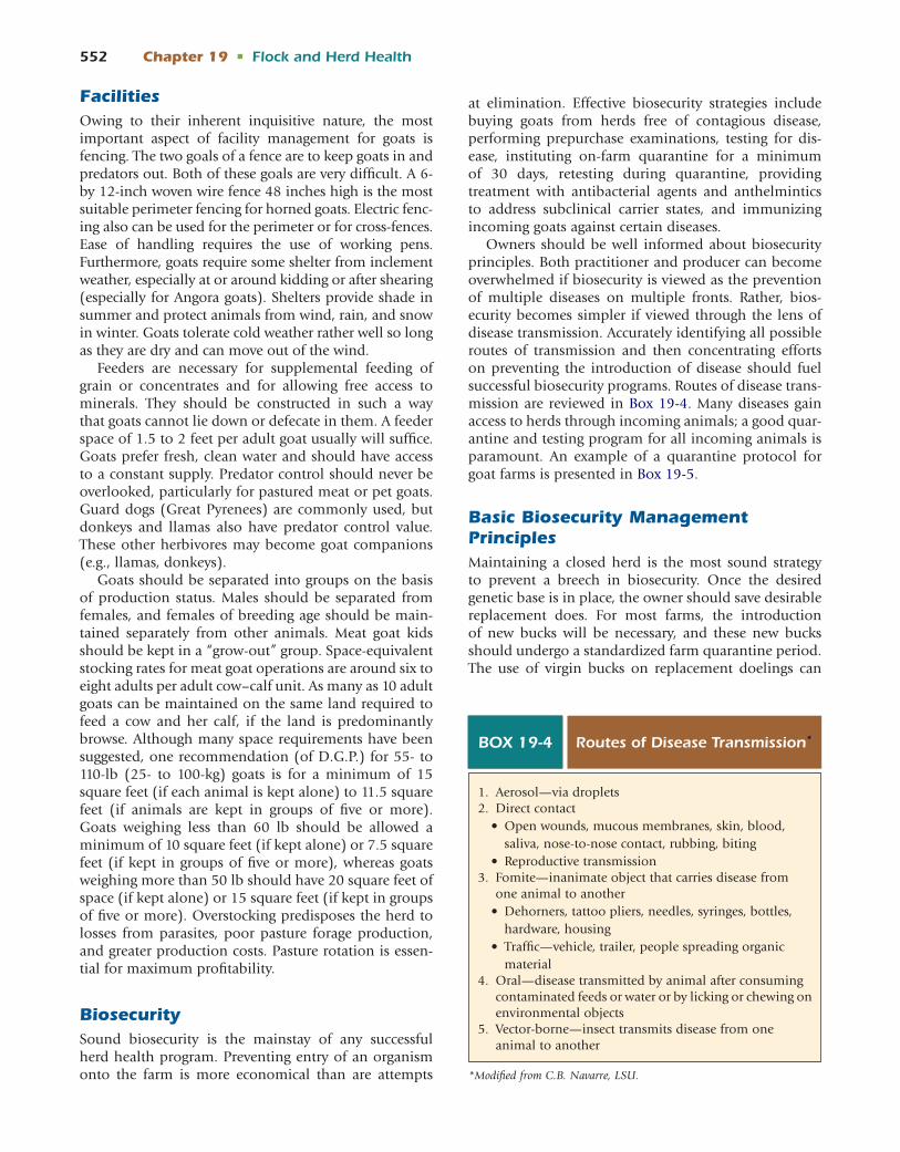
552 Chapter 19 • Flock and Herd Health
FacilitiesOwing to their inherent inquisitive nature, the most important aspect of facility management for goats is fencing. The two goals of a fence are to keep goats in and predators out. Both of these goals are very difficult. A 6- by 12-inch woven wire fence 48 inches high is the most suitable perimeter fencing for horned goats. Electric fenc-ing also can be used for the perimeter or for cross-fences. Ease of handling requires the use of working pens. Furthermore, goats require some shelter from inclement weather, especially at or around kidding or after shearing (especially for Angora goats). Shelters provide shade in summer and protect animals from wind, rain, and snow in winter. Goats tolerate cold weather rather well so long as they are dry and can move out of the wind.
Feeders are necessary for supplemental feeding of grain or concentrates and for allowing free access to minerals. They should be constructed in such a way that goats cannot lie down or defecate in them. A feeder space of 1.5 to 2 feet per adult goat usually will suffice. Goats prefer fresh, clean water and should have access to a constant supply. Predator control should never be overlooked, particularly for pastured meat or pet goats. Guard dogs (Great Pyrenees) are commonly used, but donkeys and llamas also have predator control value. These other herbivores may become goat companions (e.g., llamas, donkeys).
Goats should be separated into groups on the basis of production status. Males should be separated from females, and females of breeding age should be main-tained separately from other animals. Meat goat kids should be kept in a “grow-out” group. Space-equivalent stocking rates for meat goat operations are around six to eight adults per adult cow–calf unit. As many as 10 adult goats can be maintained on the same land required to feed a cow and her calf, if the land is predominantly browse. Although many space requirements have been suggested, one recommendation (of D.G.P.) for 55- to 110-lb (25- to 100-kg) goats is for a minimum of 15 square feet (if each animal is kept alone) to 11.5 square feet (if animals are kept in groups of five or more). Goats weighing less than 60 lb should be allowed a minimum of 10 square feet (if kept alone) or 7.5 square feet (if kept in groups of five or more), whereas goats weighing more than 50 lb should have 20 square feet of space (if kept alone) or 15 square feet (if kept in groups of five or more). Overstocking predisposes the herd to losses from parasites, poor pasture forage production, and greater production costs. Pasture rotation is essen-tial for maximum profitability.
BiosecuritySound biosecurity is the mainstay of any successful herd health program. Preventing entry of an organism onto the farm is more economical than are attempts
at elimination. Effective biosecurity strategies include buying goats from herds free of contagious disease, performing prepurchase examinations, testing for dis-ease, instituting on-farm quarantine for a minimum of 30 days, retesting during quarantine, providing treatment with antibacterial agents and anthelmintics to address subclinical carrier states, and immunizing incoming goats against certain diseases.
Owners should be well informed about biosecurity principles. Both practitioner and producer can become overwhelmed if biosecurity is viewed as the prevention of multiple diseases on multiple fronts. Rather, bios-ecurity becomes simpler if viewed through the lens of disease transmission. Accurately identifying all possible routes of transmission and then concentrating efforts on preventing the introduction of disease should fuel successful biosecurity programs. Routes of disease trans-mission are reviewed in Box 19-4. Many diseases gain access to herds through incoming animals; a good quar-antine and testing program for all incoming animals is paramount. An example of a quarantine protocol for goat farms is presented in Box 19-5.
Basic Biosecurity Management PrinciplesMaintaining a closed herd is the most sound strategy to prevent a breech in biosecurity. Once the desired genetic base is in place, the owner should save desirable replacement does. For most farms, the introduction of new bucks will be necessary, and these new bucks should undergo a standardized farm quarantine period. The use of virgin bucks on replacement doelings can
1. Aerosol—via droplets 2. Direct contact • Open wounds, mucous membranes, skin, blood,
saliva, nose-to-nose contact, rubbing, biting • Reproductive transmission 3. Fomite—inanimate object that carries disease from
one animal to another • Dehorners, tattoo pliers, needles, syringes, bottles,
hardware, housing • Traffic—vehicle, trailer, people spreading organic
material 4. Oral—disease transmitted by animal after consuming
contaminated feeds or water or by licking or chewing on environmental objects
5. Vector-borne—insect transmits disease from one animal to another
BOX 19-4 Routes of Disease Transmission*
*Modified from C.B. Navarre, LSU.

Chapter 19 • Flock and Herd Health 553
augment this goal. All animals in the herd should have permanent identification in order to keep accurate records in case of disease outbreak. The owner should document all off-farm travel of individual animals. Any new animals or animals returning to the farm from show or breeding should undergo quarantine (as outlined in Box 19-5). Owners should recognize that bucks that are bought, leased, or borrowed can serve as a means of introduction of caseous lymphadenitis, CAE, or Johne’s disease into a herd.
The veterinarian should encourage the producer and all farm personnel to practice good sanitation prin-ciples. Clothing should be changed in between working with groups of separate animals. Rubber boots, dispos-able boots, and soap and brushes should be available for all people who enter the farm. The farm should have well-maintained fences, locks on gates, and a registry for visitors. Excessive vehicular traffic should be discouraged, and proper paving or gravel should be in place in heavy traffic areas. Rodents, cats, and wildlife can harbor diseases that are transmissible to goats. Measures to decrease food available to these ani-mals should be in place. Some workers have advocated spaying and neutering the cats on the farm to control numbers. The veterinarian should seek a diagnosis in all herd deaths by performing a complete necropsy examination or by submitting the whole animal to a diagnostic laboratory. Aborted fetuses, placentas, and carcasses should always be buried or incinerated to prevent disease propagation.
Considerable effort should be made to educate new goat owners on the value of merely observing individ-ual animals from a distance. The owner should be com-fortably familiar with normal breathing, rumination, ambulation, body posture, and social interaction in goats. Early identification and quarantine of ill animals may thwart an epidemic disease in a herd.
REPRODUCTIVE MANAGEMENTEfficient reproductive management is essential to the profitability of a goat enterprise. Reproductive manage-ment of goats in the United States has focused primar-ily on milk and fiber production systems. However, the introduction of exotic meat goat breeds (South African Boer, New Zealand Kiko) is encouraging a greater emphasis on reproductive management to increase the number of offspring born and feed conversion rates. In milking herds, the production of out-of-season kids and milk is desirable to take advantage of market premiums.
As a general rule, does are seasonally polyestrous in North America and are simply polyestrous the closer to the equator they are located. Breed exceptions to this rule are recognized, and some goats in North America, especially in the southern United States, can and will breed year round. The typical doe in a temperate region will cycle from August to March, with the most fertile heats in the October-to-December time frame. This tim-ing results in kidding in January through March. The transition periods are approximately 2 months before and after the breeding season. The “deepest” anestrous period is April.
A goal of the dairy goat herd producer typically is kidding on a year-round basis in order to meet supply and demand. This approach will require manipulation of the reproductive cycle of the doe, which is covered in depth in Chapter 8. A controlled kidding program will be appealing to the meat goat producer in order to manage pastures and forages and to optimize use of personnel and furthermore meet niche market meat prices at a particular point in time in the year. A tight kidding season will enable the producer to keep a watchful eye on newborns and weanlings as the sea-son progresses. This timing also will promote maxi-mum utilization of spring grasses at turnout, thereby positively contributing to overall herd health in a cyclic fashion.
Goals for reproductive efficiency in meat goats include high fertility rate (greater than 90%), optimal litter size (1.5 to 2 kids), high rate of survival to wean-ing (more than 90%), and kidding intervals of less than 12 months. Establishing the time of mating marks the commencement of the annual production cycle. It can be calculated on the basis of the desired date of parturi-tion in a given region to take advantage of good spring pasture. The bucks should be left with does for 32 days
1. Quarantine area should be located away from herd. 2. Quarantine and sick pens should not be the same pen. 3. Quarantine all animals arriving on farm: • Show • Animals returning from breeding • New acquisitions 4. Maintain quarantine period of 2-4 weeks. 5. Observe quarantined animals daily for any signs of
disease: • “Pinkeye” • Orf • Caprine arthritis-encephalitis (CAE) • Footrot 6. Perform appropriate tests on quarantined animals: • Anthelmintic resistance testing • CAE • Caseous lymphadenitis • Johne’s disease 7. Administer anthelmintics to quarantined animals. 8. Inspect and treat for external parasites.
BOX 19-5 Quarantine Principles

554 Chapter 19 • Flock and Herd Health
(equivalent to one and a half reproductive cycles), resulting in birth of kids within 1 month. This short kidding period should produce a uniform kid crop and can be expected to streamline weaning and other herd health management practices. After 32 days with the does, the bucks’ job is finished for the year. Pregnancy should be confirmed between 45 and 60 days after breeding.
Controlled accelerated kidding programs (three kid crops every 2 years) require out-of-season breeding. Proper nutrition, intensive management practices, early weaning, and hormonal manipulation are required for such programs to be successful. If enhanced production is the aim of the breeding program, at least 20% of the breeding females should be replaced annually. The kid-ding percentage can be increased by retaining female replacements that are twins and the daughters of twins. Selection of highly fertile males also contributes to the attainment of this goal. As a consequence of breed variation, the age at onset of puberty will vary, but most does will show signs of estrus at approximately 6 to 8 months of age. The does should not enter the breed-ing program until they reach 60% to 70% of their adult body weight.
In selecting a buck, the ultimate goal of the par-ticular herd should be kept in mind. Criteria such as milk production, twinning rate, and muscling are just a few of the parameters that should be examined for potential sires. Although still more commonly used in dairy breeds, artificial insemination is becoming a more popular technology in today’s goat herds. All bucks should be in good body condition before turnout into the breeding herd. Each buck should undergo a thor-ough physical exam, scrotal circumference determina-tion, and breeding soundness evaluation well before the breeding season (see Chapter 8).
Parasite Control StrategiesThis section summarizes current recommendations for parasite control strategies for goats. More detailed information is provided in Chapter 6. Regular-interval deworming is no longer practical in goat herds. Fur-thermore, no parasite control program is broadly appli-cable across many herds. Rather, the practitioner needs to develop an integrated approach to parasite control guided by the specifics of host, parasite, and farm interactions.
Gastrointestinal nematodes are the most serious problem affecting goat production, inducing substan-tial economic losses worldwide. Haemonchus contortus is the most important such nematode affecting meat and fiber goats. In dairy goats, several other gastroin-testinal nematodes such as Ostertagia, Trichostrongylus, and Nematodirus species may be significant in certain geographic areas.
As with any disease process, host, parasite, and envi-ronmental interactions are important to consider in developing a parasite control strategy. The Haemonchus nematode thrives in warm, moist conditions. The sur-vival time of the infective stage is short during hot sum-mers (30 to 60 days) and prolonged during the winter (4 to 8 months). Haemonchus has the ability to undergo hypobiosis, or become metabolically inactive, within the host during adverse weather conditions and then emerge when favorable environmental conditions are forthcoming. This dormancy results in better parasite survival and increased transmission from spring to fall. For example, with the arrival of spring, a rise in fecal egg counts is seen in periparturient does. Implementa-tion of a control program for gastrointestinal parasites in goats requires a working knowledge of the important parts of their life cycle.
AnthelminticsThe only dewormers currently approved for use in goats are morantel, thiabendazole, fenbendazole, and pheno-thiazine. Morantel and thiabendazole are not currently commercially available, and widespread resistance to fenbendazole has emerged. Therefore it has become commonplace to use cattle-based dewormers in extral-abel fashion in goats. A summary of current dewormers is provided in Table 6-1 and Appendix 1. The practitio-ner should consult FARAD regulations regularly for any changes at http://www.farad.org.
A broad generalization for the administration of anthelmintics to goats is to double (1.5 to 2 times) the cattle or sheep dose, although exceptions exist, as noted throughout this book. The benzimidazoles are broad-spectrum dewormers, all formulated as a white liquid, and include oxfendazole, febantel, fenben-dazole, and albendazole. These agents possess good adulticidal nemotocidal activity, with a lesser effect on larvae. As a group, they generally are safe for goats and have a wide margin of safety, although albenda-zole is not recommended for use in pregnant goats up to 45 days of gestation owing to possible teratogenic activity.
The macrocyclic lactones are broad-spectrum dewormers, all formulated as a clear liquid, either (1) avermectins, including ivermectin, doramectin, and eprinomectin, or (2) milbemycins, such as moxidectin. These agents possess both larval and adulticidal activity. As a group, macrocyclic lactones have a wide margin of safety and exhibit persistence of efficacy in the body. However, resistance to this group is quite widespread. The current recommendations are to administer mox-idectin subcutaneously to goats based on cattle dose. All other drugs are administered orally and at 1.5 to 2 times the cattle dose. This class of dewormers has little efficacy if delivered topically.

Chapter 19 • Flock and Herd Health 555
The cholinergic agonists, or membrane depolariz-ers, include (1) imidazothiazoles, such as levamisole, and (2) tetrahydropyrimidines, including pyrantel and morantel. These dewormers possess nematocidal activ-ity similar to the benzimidazoles. Levamisole has a narrow margin of safety and should be administered at 1.5 times the cattle dosage. Morantel is not commer-cially available at this time.
Approved products for dairy goats include morantel, thiabendazole, and fenbendazole. Dewormers should be used until proved ineffective; alternatively, some inves-tigators recommend switching classes on a yearly basis.
Integrated Parasite Management ControlBefore changing or rotating deworming chemicals, the practitioner should set a goal of controlling, rather than eliminating, parasites within the goat herd. A concerted combination of management and treatment is neces-sary to achieve control. An integrated parasite control program should be designed for implementation of strategies on multiple fronts, as outlined in Box 19-6. Parasite control management strategies should focus on grazing practices, but the overall aim should be to decrease stocking rates and lessen pasture contamina-tion with infective larvae.
The degree of infestation varies with the manage-ment system in use (Box 19-7). Goats kept in dry lots (e.g., dairy goats) may have minimal problems with gastrointestinal nematodes. Meat goats grazing land with abundant brush and tall weeds suffer little expo-sure to infective parasitic larvae. These browsing goats develop little immunity and are highly susceptible to infection if they then graze infested pastures. Parasite infestation may be insignificant in dry years and in cer-tain rangeland areas. Parasites are a continual problem in moist, temperate areas. Special care should be taken during times of drought, in inclement weather, and with lack of adequate forage (or pasture), because these situations may increase animal concentration, thereby encouraging parasitism. When feeding is necessary, the feed bunks should be designed to allow the animals to eat comfortably but not allow them to get in or defecate in the trough. Obviously, feed should never be placed on the ground. Goats flock together and tend to bed down in the same place nightly; this behavior pattern promotes development of a heavy parasite population in concentrated areas.
Assessment of the fecal egg count is a valuable tool for parasite control programs in goats. A McMaster count of 500 to 1000 parasite eggs per gram of feces (EPG) in the spring or early summer or more than 2000 in the fall suggests serious infection. Fecal samples are
1. Quarantine new animals: • Place new animals in dry lot. • Perform fecal egg count (number of parasite eggs per
gram [EPG]). • Deworm with double-dose broad-spectrum anthelmin-
tics from two different classes.*
• Perform follow-up EPG count 14 days after initial treatment.
• Release new animals onto “dirty” pasture. 2. Administration of anthelmintics: • Weigh individual animals or base weight on heaviest
animal. • Administer anthelmintic over base of tongue. • If using avermectins or benzimedazoles, withhold feed
24 hours before administration of anthelmintic. 3. Work with owner to determine anthelmintic resistance
profile of herd. • Fecal egg count reduction test (FECRT)† • Larval development assay (LDA)‡
4. Adjust anthelmintic regimen used on farm on the basis of FECRT and LDA results.
5. Treat selected animals only: • Evaluate individual animals based on FAMACHA:§
✓ Administer anthelmintic only to animals that score 3, 4, or 5.
✓ Train producers in this methodology. • In grazing dairy goat herds: ✓ Administer anthelmintic to first-lactation does and
multiparous does before turnout. 6. If documented resistance in herd: • Consider administration of two different anthelmintics
at once. • Perform regular FECRT to assess for effectiveness of
chosen anthelmintic(s). 7. Develop and maintain management approach that
complements parasite control.¶
BOX 19-6 Steps for Implementation of Integrated Parasite Control Strategy for Goat Herds
*Some authors advocate regimen of agents from all three classes (Kaplan, 2009)†See Box 6-1 for instructions on how to perform FECRT.‡DrenchRite: information available at http://www.scsrpc.org, or contact Dr. Ray Kaplan at the the University of Georgia’s Veterinary Diagnostic Laboratory.§Information is available at http://www.scsrpc.org; FAMACHA cards also are available from Dr. Kaplan.¶See Box 19-7 for management measures that complement parasite control strategies.

556 Chapter 19 • Flock and Herd Health
collected on the day of deworming and 10 to 14 days later. McMaster flotation testing (see Chapter 6) is performed on both samples, and the EPG counts are compared. If the drop in EPG value is less than 90%, anthelmintic resistance is present, and the animals should be switched to another class of dewormer. Regu-lar egg counts help the veterinarian determine when animals are to be dewormed.
Anthelmintic ResistanceAll classes of anthelmintics have been reported to dem-onstrate decreasing or inconsistent effectiveness against various parasites. Parasite resistance to two or more major classes of anthelmintics is a growing concern as well. An apparent correlation has been noted between the number of times a worm population has been exposed to a deworming agent and the emergence of anthelmintic resistance. Multiple factors have contrib-uted to parasite resistance. Anthelmintic bioavailability varies between sheep and goats, and when these agents are administered to goats in doses based on sheep parameters, insufficient drug levels are attained to pro-duce a parasite kill.
Underdosing the amount of anthelmintic delivered to the goat produces suboptimal results. The parasites “see” the dewormers, but quantities or levels are insuf-ficient to kill them. These treatment failures over time contribute to development of anthelmintic resistance and survival of resistant parasite subpopulations, as the worms that survive reproduce and populate the envi-ronment. Finally, emerging evidence indicates that cer-tain nematodes may be able to develop intrinsic genetic resistance (see Chapter 6).
CoccidiosisIn addition to Haemonchus infestation, as mentioned previously, coccidiosis can be a major problem in recently weaned meat and dairy kids. Kids grazing on rangeland pastures seem to be less often affected by coccidiosis, but in confined quarters they are very sus-ceptible to infestation. Management efforts to control coccidiosis should be aimed at decreasing environmen-tal moisture and decreasing stocking densities. Housing in movable hutches that allow the sunlight to penetrate within is associated with decreased oocyst survival. In herds with a recognized coccidial problem or those in which intensive production practices are used, coccidio-stats can be provided in the feed or water. Dosages are listed in Table 6-1.
RECOMMENDED READINGBretzlaf K: Production medicine and health programs for goats.
In Howard J, editor: Current veterinary therapy in food animal practice, Philadelphia, 1993, WB Saunders.
Craig TM: Helminth parasites of the ruminant gastrointestinal tract. In Anderson DE, Rings RM, editors: Current veterinary therapy: food animal practice, ed 5, St Louis, 2009, Saunders.
Kaplan RM, et al: Anthelmintic treatment in the era of resistance. In Anderson DE, Rings RM, editors: Current veterinary therapy: food animal practice, ed 5, St Louis, 2009, Saunders, pp 470–478.
Mobini S: Infectious causes of abortion. In Youngquist RS, Threlfall WR, editors: Current therapy in large animal theriogenology, St Louis, 2007, Saunders, pp 575–584.
Olcott B: Production medicine and health problems in goats. In Howard JL, Smith BJ, editors: Current veterinary therapy in food animal practice, Philadelphia, 1999, WB Saunders.
Scott P: Health and production management in sheep flocks. In Radostits OM, editor: Herd health: food animal production medicine, Philadelphia, 2001, Saunders.
1. Zero grazing • Ensure good-quality forage/feed. • Place forage/feed in bunks off ground. • Monitor fecal egg count reduction test (FECRT). 2. Seasonal pasturing • Practice pasture rotation. • Ideally, allow pastures 1 full year of rest. • Maintain high forage height. • Prevent overstocking. • Provide areas of browse around fences. • Administer anthelmintics to pregnant doe 30 days
before kidding. • Administer anthelmintics to weanlings before turnout.
• Release animals onto “clean pastures.” • Monitor FAMACHA. • Monitor FECRT. 3. Continuous grazing • Provide ample browse areas. • Decrease stocking densities. • Pasture rotation every 30 days may decrease larva
numbers on pasture. • Consider mixed grazing to decrease larva numbers on
pasture. • Monitor FAMACHA. • Monitor FECRT.
BOX 19-7 Herd/Flock Management Systems and Parasite Control Strategies

557
NecropsyJohn F. Roberts
C H A P T E R
A systematic and carefully performed necropsy proce-dure will save time and money, and can be expected to result in a definitive diagnosis in the greatest possible proportion of cases. The potential risk for zoonotic diseases should be considered before initiation of the postmortem examination. If rabies or another report-able disease is strongly suspected as the cause of death, the carcass should be transported to a governmental diagnostic laboratory.
Collection of a complete standard set of formalin-fixed samples and fresh or frozen tissues and body fluids, along with appropriate culture specimens, for submission to the clinical testing laboratory is most likely to result in a definitive diagnosis. Providing lim-ited tissue samples as suggested by a tentative gross diagnosis or based on owner-reported clinical symp-toms may result in a missed opportunity for identifi-cation of an important problem. Although necropsies performed by veterinary clinicians may be refered to as “field necropsies,” it is not recommended to conduct the procedure in a pasture or similar setting. The small size of most sheep and goats allows for transfer of the entire carcass to a suitable working area for conducting the examination.
Of note, in sheep with thick wool and goats with dense hair, autolysis occurs rapidly after death, even in winter. A carcass frozen shortly after the animal’s demise and necropsied when freshly thawed is prefer-able to an autolyzed carcass.
DESCRIPTION AND INTERPRETATION OF GROSS OBSERVATIONSLesions encountered on gross examination can be described in terms of the following characteristics1: • Location • Color • Size and weight • Shape • Consistency • Number or percent involvement • Content • Odor
Weights of individual organs may aid diagnosis. For example, liver weight may vary depending on the presence or absence of toxic hepatopathy, and thyroid weight may be increased in cases of goiter. Volume of gastrointestinal content or fluid in compartments also may provide reliable objective data. Many common veterinary diagnoses, such as trauma, lightning strike, and certain types of bloat, can be made only by gross examination (Figure 20-1, A and B). In such instances, submission of tissues for histopathologic examination and culture to a diagnostic laboratory, without appro-priate history and descriptive gross observations, may result in no diagnosis.
Table 20-1 lists commonly encountered systemic pathologic processes along with their characteristic necropsy lesions and possible causes.2-8 Table 20-2 lists gastrointestinal diseases that may require gross exami-nation for diagnosis.6, 8-13 Table 20-3 lists “pseudole-sions” that may be confused with pathologic changes4 (Figure 20-2). Astute clinical observation and field necropsy constitute the first line of defense against exotic or reportable livestock diseases in the United States. Table 20-4 lists necropsy lesions that may be observed in goats and sheep infected with reportable agents.14-32
NECROPSY PREPARATION AND EQUIPMENTIf removal of the spinal cord is not elected, a skilled practitioner with advance preparation and an assistant can conduct a complete necropsy in less than 1 hour. To maximize the operator’s physical comfort, the exami-nation is performed with the carcass on a table of proper height, under controlled environmental temperature and with sufficient lighting. Necessary components such as water, access to cold storage, scales, prearranged sample containers, recording forms, necropsy tools, and digital camera must be easily accessible before the procedure is started.
Necropsy tools and supplies include the following: saw, loppers, knife and sharpener, scalpel, forceps, scissors, hemostat, rongeur, clippers, ruler, camera
20

558 Chapter 20 • Necropsy
A B
Figure 20-1 A, Singed hair in a linear pattern (arrows) on a goat caused by lightning strike. This is an unusual case in that singe marks often are not obvious. B, Close examination demonstrates curled burnt hairs.
TABLE 20-1 Selected Systemic Pathologic Processes: Necropsy Findings and Potential Causes
Pathologic Process Possible Lesions Cause(s)
Cardiogenic shock Pulmonary congestion and edema, jugular vein distention, large liver with “nutmeg” pattern, generalized edema, anasarca, ascites, submandibular edema, hydropericardium, hydrothorax, hydroperitoneum, heart lesions (cardiomegaly, myocarditis, hydatid cysts, myocardial abscess, hemorrhage/pallor, white streaks in myocardium, morphologic defect, myocardial mineralization)2
Hypoxia, ventricular tachycardia, and other arrhythmias, cardiomyopathy, congenital defect, hypertension, endocarditis (chronic infections, long vascular blood catheter use), bacterial or viral infection, nutritional deficiency (selenium, copper), cardiotoxic substance or plant (e.g., vitamin D, rhododendron/azalea), monensin toxicity, electrocution or lightning strike, obstruction of blood flow, fluid overload3
Obstruction of blood flow
Pericardial fluid or exudate accumulation, thrombus in vena cava or lung
Thrombosis, congenital (aortic stenosis), cardiac tamponade (traumatic reticulopericarditis, ruptured coronary artery), neoplasia, bloat (abdominal or rumen distention), liver failure
Hypovolemic shock Pale or swollen kidney, contracted spleen Hemorrhage, fluid loss (dehydration)Hemorrhage Extravasation of blood, pallor of tissues
(lungs, mucous membranes), watery/thin blood, observation of blood in body cavity, contracted spleen
Enteric hemorrhage, predator attack, ruptured viscera (spleen, uterus, abomasa), ruptured liver from dystocia, ruptured uterine or ovarian artery, abomasal ulceration, rupture of aorta, ruptured aneurysm4
Fluid loss (dehydration)
Sunken eyes, reduced skin elasticity Diarrhea, burn injury, acute intestinal obstruction, electrolyte imbalance, acid-base imbalance5
Maldistributive shock Reduced circulating blood, volume pooling in capillaries, congested spleen, pulmonary edema
Endotoxemia, septicemia (gram-positive or - negative bacteria septicemia), rapid reduction of body fluid (removal of ascitic fluid),10 neurologic dysfunction, anaphylactic shock, neurogenic shock, septic shock2,5

Chapter 20 • Necropsy 559
TABLE 20-1 Selected Systemic Pathologic Processes: Necropsy Findings and Potential Causes—cont’d
Pathologic Process Possible Lesions Cause(s)
Anaphylactic shock Pulmonary congestion, mucus secretion in bronchioles, pulmonary edema, emphysema edema in gut wall5
Repeated administration of antibiotic, vaccine, or biologic breakdown products of arthropods2
Neurologic shock Nonspecific gross lesions that are similar to cardiogenic shock
Trauma, electrocution, lightening strike, fear and stress2
Septic shock Petechiae, injected sclera, ecchymoses, large swollen lymph nodes, splenomegaly, fibrinopurulent exudates in pericardial space or joint spaces or adhered to meninges5
Bacteremia
Endotoxemia (toxic or septic shock)
Lesions may be same as for toxemia5 Lipopolysaccharide cell wall of gram-negative bacteria
Toxemia (toxic shock) Pale or enhanced liver parenchyma, enlarged kidneys, enlarged adrenal glands, hemorrhage of myocardium, coagulopathy, increased vascular permeability, congestion, distended intestines, presence of glucose in urine6
Bacterial toxins (enterotoxemia), tetanus, inorganic toxin, plant toxins, nitrate toxicity, snake bite, xenobiotic toxicity (e.g., from tilmicosin),4 urea toxicity
Edema Fluid accumulation in tissues, pleural cavity, or abdominal cavity5 or in subcutis or ventrum
Congestive heart failure, obstructed venous return, endotoxemia, anaphylactic shock, vasculitis, obstruction of lymph flow5
Hypoxia Nasal discharge, pulmonary changes, cyanosis Anemia, cardiac failure, pneumonia, rhinitis, pulmonary edema, infectious disease, toxicity (cyanide anemia, nitrate or carbon monoxide toxicity), metabolic disease (pregnancy toxemia), neoplasia, pneumothorax, tracheal collapse, allergic pneumonitis, hyperthermia, trauma (diaphragmatic hernia, tight collar)4
Hypothermia Pulmonary congestion, generalized congestion
Cold stress, exposure to rain or wind; may occur after shearing4
Hyperthermia Congested tissues, petechial hemorrhages in mucous membranes, subcutis; hyperemia of skin
Infection, high environmental temperature
Liver failure Altered liver size (small or large), altered liver texture (firm or friable), ecchymoses, enhanced lobular pattern
Hepatitis, hepatopathy
Renal dysfunction Urine obstruction, swollen pale kidneys, dilated ureter or calyces
Urolithaisis, glomerulonephritis, nephritis, nephropathy,
Coagulopathy Petechial or ecchymotic hemorrhage, subcutaneous hemorrhage, pooling of blood in spaces or lumina of organs
Disseminated intravascular coagulation, reduction in vitamin K–dependent clotting (toxic plant ingestion, rodenticide toxicity), snake envenomation, fungal toxins (Aspergillus, Fusarium), liver flukes (Fasciola hepatica), bacterial infection7
Emaciation Decreased subcutaneous, pericardial adipose tissue, muscle atrophy, poor hair or wool quality, oral disease, changes in liver parenchyma
Dental disease, protein-energy malnutrition,5 micronutrient deficiency, gastric foreign body, chronic enteritis, chronic liver disease, infectious (prion disease, retroviral infection, chronic septicemia, chronic parasitism), plant toxicities, neoplasia, amyloidosis8

560 Chapter 20 • Necropsy
TABLE 20-2 Digestive Tract Diseases: Necropsy Findings and Potential Causes (see Chapter 5)
Digestive Disease Possible Lesions Cause(s)
Esophageal choke Distended esophagus with thinned or hemorrhagic wall
Inadequate mastication, large/firm food item, foreign body, injury from stomach tube, injury to tissue surrounding esophagus
Rumen acidosis Distended rumen with porridge-like content and “fermented” odor, rumen pH <5.5,9,10 rumen epithelium hyperemic and sloughs readily,11 dehydration (sunken eyes), chronic (scarred rumen lining, liver abscesses)11; urine pH 5.0-6.0; packed cell volume >35%6,9
Rapid rumen fermentation with ingestion of highly digestible carbohydrates (corn, oats, wheat, barley, breads, fruits, beets, potatoes, others)9
Rumen distention, impaction
Distended rumen, esophageal “bloat line,” edema in hindquarters, dry scant feces
Protein-energy or micronutrient malnutrition,10 ingestion of foreign body (plastic bags),8 dehy-dration, legume, ingestion of high-fiber/low-digestibility diet, sand ingestion, consumption of horse feed9
Frothy bloat Esophageal “bloat line,” flattened liver, dia-phragm rupture; edema in hindquarters, hindquarters blanched11
Legume consumption (alfalfa), lush cereal-grain pastures, high-grain diets
Free gas bloat Rumen distended with gas, esophageal “bloat line”
Inhibition of eructation (rumen malfunction, esophageal obstruction, newly introduced grain diet, neurologic function impaired, hypo-calcemia, endotoxemia, pain, peritonitis, some pharmaceuticals (e.g., xylazine)9,12
Traumatic reticuloperitonitis
Peritonitis, pericarditis, draining tracts from chest cavity
Ingestion of wires, needles
Abomasal impaction Abomasum enlarged with rumen-type con-tents that are dry and doughy
Poor-quality roughage, foreign body obstruc-tion (phytobezoar, trichophytobezoar), protein-energy malnutrition, abomasal atony, emptying defect in Suffolk and Dorset sheep, dysautonomia9,11
Abomasal bloat in juveniles10
Distended abomasa with milk content Associated with large quantities of milk replacer diet,10 consumption of hay mixed with feces13
Abomasal rupture6 Feed content in peritoneal space, tear in greater curvature of abomasa accompanied by hemorrhage in abomasal wall
Abomasal impaction, abomasal bloat, abomasal atony
Abomasal torsion6 Hemorrhage and congestion at the site of the torsion
Gas accumulation and displacement10
Intestinal volvulus Hemorrhage, gas distention, mesenteric rent (tear)
Gas accumulation in intestine
Intussusception Intestinal telescoping most commonly observed at ileocecal valve
Associated with intestinal mass in adults, enteri-tis in young, Oesophagostomum in sheep9
Intestinal ileus Dilated intestine with watery content Multiple-system diseaseIntestinal atresia, Failure of intestinal segment to form, first
week of lifeCongenital defect
Cecal volvulus/torsion of root of mesentery
Distention of forestomachs, abomasa, and intestine
Gas accumulation and displacement9,10
Rectal prolapse Protrusion of rectal tissues Diarrhea in lambs and kids; dietary imbalance, urolithiasis, grazing lush pasture; secondary to chronic coughing, short tail docking, growth implants, rabies, atresia ani9
Atresia ani The rectum is not patent Congenital defect

Chapter 20 • Necropsy 561
(preferably digital), polyethylene cutting board, nec-ropsy form, indelible markers, pencil, pen, formalin fixation container, tissue cassettes, fecal container, clean Petri dish, syringe, needles, commercial culture kits with swabs and collection tubes (Culturettes), general-purpose swabs, blood tubes, string, and plas-tic specimen bags (Whirl-Pac). Box 20-1 lists types of blood collection tubes that are indicated for different testing procedures. Consistent use of a necropsy check-list form will save time, reduce omissions, facilitate communication, and serve as a written legal record. Use of a sharp stainless steel knife and polyethylene cutting surface will reduce risk of injury and ease the creation of appropriate-width tissue samples for fixation.
An overall “clean” necropsy procedure with reduced spillage of ruminal/intestinal contents and blood will enhance visualization of gross lesions, reduce exposure to potential pathogens, decrease attraction of flies, and allow for fast clean-up. Utilization of personal protec-tive equipment (gloves, apron, and mask) will limit exposure to zoonotic pathogens or toxic substances. Special training and personal protective gear such as N95 particulate respirators, nonpermeable (e.g., Tyvek) coveralls, nitrile or vinyl gloves, and face shields are recommended if an exotic disease or easily contracted zoonosis is suspected.
HISTORY AND CLINICAL INFORMATIONNecropsy samples submitted to a clinical testing labo-ratory are best interpreted by diagnosticians or expe-rienced veterinary pathologists if patient and herd or flock history, clinicopathologic data, and a differential diagnosis list are provided. A review of farm manage-ment practices and clinical data is recorded on the necropsy submission form or provided as attachments. History may include information such as pasture con-ditions, types of forage, stocking rate, supplemental feeding, hay and grain storage practices, weather infor-mation, and epidemiology of past disease outbreaks. The veterinarian has the responsibility to inform the laboratory if a potentially zoonotic disease (e.g., rabies) is suspected.
Digital Photography of SpecimensDigital photography of fresh necropsy specimens pro-vides a one-time opportunity to collect information that will facilitate communication among the owner, veterinarian, plant specialist, toxicologist, epidemi-ologist, and pathologist. Photographs may facilitate
TABLE 20-3 Pseudolesions Observed at Necropsy
Pseudolesion Necropsy FindingsBird damage Loss of eyes, damage to anusEuthanasia barbiturate Soft, discolored dark tissue
at injection siteGunshot euthanasia
(owner may not inform vet concerning method of euthanasia)
Hemorrhage and fractures, bullets
Liver mortis-postmortem Settling of blood in depen-dent parts of body4
Digestive overflow-postmortem
Rumen contents in bronchi, nasal cavity4
Rumen distention after death (“postmortem bloat”)
Rectal or vaginal prolapse
Terminal lesions associ-ated with death process
Tracheal froth from cardiac hemorrhages
Congenital melanosis Dark pigmentation of meninges, other tissue4 (see Figure 20-2)
Normal placenta features mistaken for lesions
Necrotic membrane tip, hippomanes, amniotic plaques (see Figures 20-17 and 20-18)
Autolysis Rumen mucosal sloughing, postmortem GI rupture
Blackening of tissues adja-cent to intestines4
Discoloration of tissues adja-cent to gallbladder
Diaphragm rupture (postmortem)
Free gas in rumen combined with autolysis; mimics rup-ture from bloat or trauma
GI, Gastrointestinal.
Figure 20-2 Melanosis of the meninges is an example of a “pseudolesion,” as observed in a 2-year-old black-faced mixed-breed ewe. (Courtesy Dr. William Castleman, Gainesville, Florida.)

562 Chapter 20 • Necropsy
TABLE 20-4 Reportable Diseases of Worldwide Significance or Foreign to the United States: Endemic Distribution and Necropsy Lesions
Disease* with Causative Pathogen When Known
Endemic Distribution Necropsy Findings
PRION DISEASEScrapieOIE list B
North America, Europe, Asia, Australia14
Emaciation, loss of wool or hair, traumatic skin lesions caused by rubbing14
VIRAL DISEASESFoot-and-mouth diseaseAphthovirus (family
Picornaviridae)OIE list A, USDA select agent
Africa, Asia, Europe, South America, previously in North America15
High morbidity, low mortality; usually affects younger ani-mals; lesions in goats and sheep are mild or nonapparent
Small vesicles or erosions on the dental pad, lips, gums, or tongue; vesicle or erosion on the coronary band or in the interdigital space; decreased milk in mammary gland; gray or yellow streaking in the myocardium; abortion is possible16
Vesicular stomatitisVesiculovirus (family
Rhabdoviridae)OIE list A, USDA select agent
South America, Central America, North America15
Less frequent in sheep and goats, high morbidity, rarely causes death; vesicles on tongue, gums, lips, coronary bands, and possibly teats; secondary bacterial mastitis16
RabiesOIE list BGenus Lyssavirus (family
Rhabdoviridae)
Worldwide except some Pacific islands, Australia17
No gross lesions
Peste du petits ruminantsMorbillivirus (family
Paramyxoviridae)OIE list A, USDA select agent
Africa, Middle East, Indian subcontinent15
Emaciation; inflammatory lesions in GI tract from oral cavity to colon; dehydration, fecal staining; stomatitis involving inside of lower lip and adjacent gum, cheeks near commissures and free portion of tongue; lesions possible on hard palate, phar-ynx, upper third of esophagus, rumen, reticulum, omasum, abomasa; Peyer’s patch necrosis, congestion at ileocecal valve and cecocolic junction, “zebra stripe” congestion in colon
Respiratory lesions: erosion and petechiae of nasal mucosa turbinates, larynx, and trachea; bronchopneumonia
Enlarged lymph nodes, conjunctivitis, vulvovaginitis16
RinderpestMorbillivirus (family
Paramyxoviridae)OIE list A, USDA select agent
Africa, Middle East, Asia, previously Europe and South America15
Emaciation, dehydrationDigestive tract lesions: small gray foci on oral mucosa that
coalesce, gray necrotic epithelium that sloughs and leaves red erosion; erosions on gums, lips, hard and soft palate, cheeks, and base of tongue; erosion of esophagus/omasum, congestion and edema of abomasum; Peyer’s patch necro-sis, fibrinous exudates on intestinal mucosa, edema and/or luminal blood in cecum and colon; congestion of colonic ridges—“tiger” or zebra striping, swollen or edematous lymph nodes16
Sheep pox and goat poxGenus Capripoxvirus (family
Poxviridae)OIE list A, USDA select agent
Africa, Middle East, Asia17 1- to 3-cm-diameter macules on skin, particularly in groin, axilla, perineum; papules on mucosal membranes of nose, mouth, mammary glands, vulva, or prepuce; nasal discharge from rhinitis; mastitis, multiple necrotic foci of mucous membranes, enlarged lymph nodes; papules on mucosal surface of abomasum, rumen, large intestine, tongue, hard and soft palate, trachea, and esophagus; pale areas on surface of kidney, liver, and testicle; hemorrhagic enteritis, multifocal firm areas in lungs, abortion possible16,17

Chapter 20 • Necropsy 563
TABLE 20-4 Reportable Diseases of Worldwide Significance or Foreign to the United States: Endemic Distribution and Necropsy Lesions—cont’d
Ovine encephalomyelitis (louping ill)
Genus Flavivirus (family Flaviviridae)
Scotland, England, Ireland17
No characteristic gross lesions17
Rift Valley feverGenus Plebovirus (family
Bunyaviridae)OIE list A, USDA select agent
Africa, Middle East15,16 Hepatic necrosis; enlarged friable, soft, red to yellow-brown liver; petechial to ecchymotic hemorrhage; patchy conges-tion, small gray-white 1- to 2-mm foci in liver parenchyma, petechiae and ecchymoses in mucosa of abomasa, dark chocolate-brown abomasal contents; edema and hemor-rhage in wall of gallbladder and hepatic lymph nodes16
Nairobi sheep diseaseGenus Nairovirus (family
Bunyaviridae)OIE list B
Africa16 Hemorrhagic and catarrhal gastroenteritis, soiling of hind-quarters with mixture of blood and feces; hemorrhages or congestion in longitudinal folds of the abomasum distal ileum, ileocecal valve, cecum, and colon; congestion or hemorrhage in the cecum and colon appearing as longi-tudinal striations of the mucosa (“zebra striping”); colon contents may be watery and blood-tinged, with hemor-rhages in serosa of colon, submucosa of gallbladder, epicar-dium, and endocardium; nasal discharge, prominent lymph nodes, enlarged spleen; abortion is possible with numerous hemorrhages in tissues, edema, and hemorrhage of fetal membranes16
Akabane virus infectionGenus Bunyavirus (family
Bunyaviridae)USDA select agent
Africa, Asia, Australia15 Encephalitis with no gross lesions, stillborn, weak or aborted fetus, atrophy of skeletal muscle, tendon contraction, arthrogryposis, hydranencephaly, torticollis, scoliosis, brachygnathism15
Wesselsbron diseaseFamily TogoviridaeZoonotic risk
Africa18 Liver in fetus and newborn is enlarged and orange-brown, with multifocal pinpoint white areas; icterus often present; hydranencephaly, microcephaly, arthrogryposis, hydrops amnion in ewes16,18
BluetongueGenus Orbivirus (family
Reoviridae)OIE list A, USDA select agent
Worldwide in mostly tropical and subtropical climates, Europe, North America15
Vascular injury, consumption coagulopathy, generalized edema, hyperemia, hemorrhages, erosions, ulceration of upper gastrointestinal tract (oral cavity, esophagus, fore-stomachs), subintimal hemorrhages in pulmonary artery, pulmonary edema, pleural effusion, pericardial effusion, edema in facsial planes, necrosis of papillary muscle in left ventricle; embryonic or fetal death, fetus: cavitating encephalopathy, hydranencephaly16
Borna diseaseGenus Bornavirus (order
Mononegavirales)17
Europe No characteristic gross lesions; possible leptomeningeal hyper-emia, brain edema16
Caprine arthritis-encephalitis (CAE)
Nononcogenic retrovirus (subfamily Lentivirinae)17
OIE list B
Worldwide Emaciation, chronic polysynovitis, degenerative joint disease, hyperplasia of synovium, enlarged lymph node, diffusely firm lungs (interstitial pneumonia), mastitis17
Ovine progressive pneumo-nia (maedi-visna)
Retrovirus (subfamily Lentivirinae)17
OIE list B
Africa, North America, South America, Europe, Asia17
Noncollapsing heavy lungs, diffuse thickening of lungsEnlarged bronchial and mediastinal lymph node, thickened
firm mammary gland, thickened synovial membrane, arteritis17
Continued

564 Chapter 20 • Necropsy
TABLE 20-4 Reportable Diseases of Worldwide Significance or Foreign to the United States: Endemic Distribution and Necropsy Lesions—cont’d
Ovine pulmonary adenocarcinoma (Jaagsiekte, pulmonary adenomatosis)
Beta retrovirus (family Retroviridae)
OIE list B
Africa, North America, South America, Europe, Asia17
Solid raised foci in lungs, pulmonary neoplasia in anterior-ventral regions and diaphragmatic lobes, excessive froth in bronchi, enlarged bronchial and mediastinal lymph nodes with occasional metastasis, secondary pneumonia, pulmonary abscess, pleuritis17
Aujeszky’s disease (pseudorabies)
Porcine herpesvirus-1 (family Herpesviridae)
OIE list B
North America, South America, Europe17
No characteristic gross lesions, in purities cases damage to skin, subcutaneous edema, pulmonary congestion and edema, hydropericardium, endocardial hemorrhages17
BACTERIAL DISEASESAnthraxBacillus anthracisOIE list B, USDA select agent
Worldwide Absence of rigor mortis, rapid gaseous decomposition; dark tarry nonclotting blood at orifices, widespread ecchymoses, blood-stained fluid in body cavities, hemorrhagic enteritis, splenomegaly with “blackberry jam” consistency19
Burkholderia pseudomallei infection
USDA select agent
Asia, Australia, Middle east, Africa, Caribbean, South America (tropical regions)20
Thick exudates from eyes and nose, multiple abscesses in subcutis associated with lymph nodes and abscesses in many organs (lungs, spleen, liver), abscess may contain green-tinged pus, aortic lesions, joint effusions, cloudy meninges20
Heartwater (cowdriosis)Ehrlichia (Cowdria)
ruminantiumOIE list B, USDA select agent
Africa Hydropericardium, pericardial fluid that is reddish to straw-colored, decreased packed cell volume, orange-yellow serum, hydrothorax, pulmonary edema, ascites, edema of lymph nodes, mediastinal edema, froth in the trachea, subendocardial petechial hemorrhages, subtle swelling of the brain, partial brain herniation, possible splenomegaly16
Contagious caprine pleuropneumonia
Mycoplasma capricolum subsp. capripneumoniae
OIE list B, USDA select agent
Africa, Indian subcontinent
Lesions usually confined to the thoracic cavity; 1- to 2-cm yellow nodules in lungs, consolidation of lung lobes (hepatization), pleuritis with involvement of one or both lungs, straw-colored pleural fluid16
Contagious agalactiaMycoplasma agalactiae,
Mycoplasma capricolum subsp. capricolum, Mycoplasma putrefaciens, Mycoplasma mycoides subsp. mycoides Large Colony type16
OIE list B
Mortality is lowMastitis with edema between lobule leading to fibrosis,
polyarthritis, keratoconjunctivitis to corneal ulcerationAcute disease may be associated with congestion of muscles,
spleen, and liver, and with periarticular edema, particularly in carpal joints, and hyperemia of synovial membranes and joint cavities, as well as abortion16
BrucellosisBrucella abortus, Brucella
melitensis, Brucella ovisOIE list B, USDA select agent
Worldwide Metritis, orchitis, epididymitis, synovitis, abortion– placentitis (placental edema, grayish-white areas of necrosis on placenta, brownish-red exudate on chorionic surface, with progression to leathery plaques on chorionic surface), inflammation of multiple fetal organs, serohemorrhagic fluid in multiple body cavities, fetal pneumonia21,22
Q fever–induced abortionCoxiella burnetiiOIE list B
Worldwide Abortion–intercotyledonary necrotizing placentitis, minimal lesions in fetus; fetus may be nonautolyzed16

Chapter 20 • Necropsy 565
TABLE 20-4 Reportable Diseases of Worldwide Significance or Foreign to the United States: Endemic Distribution and Necropsy Lesions—cont’d
TuberculosisMycobacterium bovisMycobacterium bovis subsp.
caprae
Worldwide Often subclinical in adult animalsGranulomas affecting any lymph nodes (bronchial,
retropharyngeal, mediastinal nodes most common); bronchopneumonia, intestinal ulceration, diarrhea, enlargement of lymph nodes associated with gastrointestinal tract; with generalized form, miliary granulomas in many organs; mastitis, metritis23
Johne’s diseaseMycobacterium avium subsp.
paratuberculosisOIE list B
North and South America, Asia, Europe, Africa, Australia24
Thickening and corrugation of intestinal mucosa, most pronounced in distal jejunum and ileum; mesenteric lymphatic vessels may be thickened and cordlike, with enlargement of ileocecal and mesenteric lymph nodes; in advanced cases, edema of abomasal wall, fluid accumulation in abdominal and pericardial cavities24
TularemiaFrancisella tularensisHHS select agent
North America, Europe, North America, Europe, Asia21
Large numbers of ticks may be present on skin; multifocal dark red subcutaneous areas of congestion, necrosis of tissue associated with tick attachment; enlarged and congested lymph nodes, pulmonary edema21,25
Salmonella-induced abortionSalmonella enterica subsp.
enterica serovar AbortusovisOIE list B
Worldwide; most common in Europe, Asia
Metritis, enlarged darkened uterus with foul-smelling retained placenta, peritonitis, pneumonia in lambs, abortion, placentitis; placental cotyledons may be pale and edematous; fetal pneumonia; rapid autolysis, with development of a “cooked” appearance to the fetus, with bruising and severe edema of subcutaneous tissues21
Ovine enzootic abortion (OEA)
Chlamydophila abortusOIE list B
Worldwide Pneumonia in lambs, hepatitis, bloody vaginal discharge in females, abortion, regional to generalized thickening of allantochorion, brownish exudates on chorionic surface, opacity of the intercotyledonary spaces and white-gray foci on the chorionic surface of cotyledons; fetus may have white spots on liver26,27
LeptospirosisOIE list B
Worldwide Jaundice, hemoglobinuria, polyarthritis, abortion, jaundice of fetal subcutaneous tissues, petechial hemorrhage on surface of serosal membranes, enlarged kidney; fetus may be autolyzed28
PARASITESBabesia-induced abortionBabesia ovis, Babesia motasi,
Babesia crassa
North America, South America, Africa, Asia, Australia16
Intravascular hemolysis, dark-red discoloration of urine, icteric mucous membranes, watery blood, swollen spleen, swollen orange-red liver, dark red or black kidneys16
TrypanosomiasisTrypanosoma congolense,
Trypanosoma brucei, Trypanosoma Vivax
Africa Anemia lesions, pallor of mucous membranes, serous atrophy of fat, pulmonary edema, subcutaneous edema, ascites, hydrothorax, enlarged lymph nodes and spleen; also may produce atrophic lymph nodes16
Echinococcosis/hydatidosisOIE list B
Worldwide Hydatid cyst in various organs; lungs and liver commonly affected; cyst usually less than 10 cm in diameter but can grow larger29
ARTHROPOD PESTScrewworm myiasis South America, Mexico Difficult to confirm at necropsy; tissue destruction of open
wounds with larvae (navel, tick bites, castration sites, barbed wire sores, mouth sours); vacated larval cases will be evident unless death was recent; postmortem confirmation needed16
New World: Cochliomyia hominivorax
OIE list B
Continued

566 Chapter 20 • Necropsy
publication of an interesting case or can be used as evidence for legal purposes. (Note: Photographs are best made early in the necropsy, at an angle perpendicu-lar to the structure or area of interest on the subject’s body, and should depict tissues that are free of excess blood and digesta.) The images in the camera should be checked between shots to ensure proper exposure and focus. Removal of tissue and arrangement on a suitable background for photography may facilitate visualization of the lesions. Preservation of tissues in Klotz solution and maintenance in a refrigerated envi-ronment will maintain organ color if photography must be postponed for 24 hours.
Collection of Blood, Urine, Fecal, Fluid, and Fresh Tissue Samples: Clinicopathologic Analysis, Bacterial Culture, and Virology, Molecular Biology, and Toxicology StudiesWhen animals are to be euthanized immediately before necropsy, appropriate tubes should be used for blood collection while the animal is alive33 (Table 20-5). Fresh blood can be used for serum chemistry studies, complete blood count (CBC), serologic testing, and element analysis. If delayed laboratory analysis of blood is anticipated, blood smears should be made
STOPPER COLOR ADDITIVE INDICATIONRed None or clot activator Serum: most biochemical assays, serologic studies for
infectious diseasesGreen Lithium or sodium heparin Plasma: erythrocyte enzymes (GSH-Px, SOD, transketolase),
most components of biochemical analysis, endocrinologic studies, hematocrit, hemoglobin
Purple/lavender EDTA Whole blood: hematology, leadRoyal blue-plastic None or heparin Trace element, especially zinc, because zinc analysis affected
by rubber stopperBlood should be drawn from animal with Vacutainer needle
Blue Buffered sodium citrate Whole blood: coagulation studies (fibrinogen, prothrombin time)
Gray Potassium oxalate/sodium fluoride Whole blood: glucose determination
BOX 20-1 Blood Tubes for Venous Collection and Indications
EDTA, ethylenediaminetetraacetic acid; GSH-Px, glutathione peroxidase; SOD, superoxide dismutase.
TABLE 20-4 Reportable Diseases of Worldwide Significance or Foreign to the United States: Endemic Distribution and Necropsy Lesions—cont’d
Old World: Chrysomyia bezziana
OIE list B
Africa, Asia, Difficult to confirm at necropsy; tissue destruction of open wounds with larvae; presence of vacated larval cases16
Psoroptes ovis North America, Europe Loss of fleece, scabs, emaciation30
Amblyomma hebraeumSouth African bont tick
Africa31 Present on skin; vector of heartwater
Amblyomma variegatumTropical bont tick
Africa, Caribbean32 Present on skin; vector of heartwater, Nairobi sheep disease, and C. burnetii32
Ixodes ricinus Europe Present on skin; vector of louping ill, tick-borne fever in ruminants17
*OIE (Office International des Epizooties, The World Organization for Animal Health) lists specify diseases requiring possible intervention by governmental veterinary offices: list A, transmissible diseases with potential for very serious spread causing widespread socioeconomic or public health consequences and affecting international trade; list B, transmissible diseases with socioeconomical significance and/or public health importance and significance in international trade.A USDA select agent or HHS select agent is an agent deemed to be a threat to public, animal, or plant health and is regulated under the Public Health Security and Bioterrorism Preparedness and Response Act of 2002, United States federal law. The law is implemented and enforced by the U.S. Department of Agriculture (USDA) and the U.S. Department of Health and Human Services (HHS).

Chapter 20 • Necropsy 567
TABLE 20-5 Tissue Collection During Necropsy With Subject in Left Lateral Recumbency
Tissue Location Fresh Fixed Comments
Skin X Sample vesicles, macules, erosions from mucocutaneous junctions, lesions from infection or trauma
Cervical lymph node X X May be used for culture or PCR assayMammary gland X Culture may be indicated in cases of mastitis; both lobes may be sampled and
labeled for comparisonExternal genitalia X Sample vesicles, viral lesions such as papillomasBone marrow X X Use cassette to place sample in formalin; obtain fresh sample for cytologic
analysis if animal was euthanizedJoint capsule X Optional—may be important in CAE diagnosis, culture may be indicated in
neonatal septicemia.Bone X Optional—may be important in nutritional disease, vitamin D deficiencySkeletal muscle X Sample dark or pale or hemorrhagic lesions, (clostridial myositis, selenium
deficiency, trauma verification)Tongue X Sample vesicles or lesions on the mucosal surfaceOral mucosa X Sample vesicles on gingival soft or hard palateTonsil X Located in a pouch adjacent to the glottisThyroid X X Weight, mineral analysis of fresh tissue may be indicated for iodine deficiencyParathyroid X Difficult to locateTrachea X Sample lesions (viral or bacterial tracheitis)Esophagus X Sample “bloat line”Lung X X Sampling of multiple sites may be indicated if lesion is observed
Fresh tissue may be used for culture, virology or PCR assayMediastinal lymph node X X Culture or virology or PCR assay may be indicatedPericardium X May be important for diagnosis of clostridial myositis or traumatic
reticulopericarditisPleura XCervicothoracic ganglia X Optional—used to diagnosis disease of autonomic nervous systemAdrenal gland X X Frozen sample may be needed for herpesvirus diagnosisCeliacomesenteric
gangliaX Optional—used to diagnosis disease of autonomic nervous system (abomasal
emptying defect)Pancreas X Easy to confuse with adipose tissueLiver X X Fresh tissue is important for toxicology and mineral analysis; at least 100 g
may be requiredCulture, virology studies, or PCR assay may be indicated on fresh tissue
Gallbladder XSpleen X X Culture, virology studies, or PCR assay may be indicated on fresh tissueRumen X Sample with mucosa intactReticulum X Sample with mucosa intactOmasum X Sample with mucosa intactAbomasum X Sample with mucosa intactDuodenum X Observe location of bile papillae and sample if indicatedJejunum X X Multiple fresh samples should be saved “tied off” for bacterial culture
Parasitology or virology studies or PCR assay may be indicated on fresh tissueMesenteric lymph node X X Culture, virology studies, or PCR assay may be indicated on fresh tissueIleum X Tissue should including gut-associated lymphoid tissue (GALT) (i.e., Peyer’s
patches) should be sampledIleocecal valve Important site of parasite identificationCecum X X Culture or parasitology or virology studies may be indicated on fresh tissueSpiral colon X X Culture or parasitology or virology studies may be indicated on fresh tissueDescending colon XRectum X
Continued

568 Chapter 20 • Necropsy
at time of blood collection.33 Bone marrow for cyto-logic study also can be collected while the animal is alive or shortly after euthanasia. Depending on the degree of autolysis, blood collected hours to days after death and refrigerated may still be useful for serologic testing. Serum should be separated as soon as possible after clotting. Fresh urine may be cultured or submitted for clinical pathology tests. Glucosuria detected by dipstick testing at necropsy may indicate enterotoxemia, and ketonuria may support a diagnosis of pregnancy toxemia.11 Urinalysis and urine cyto-logic studies should be done immediately, because bacteria rapidly overgrow urine flora, which may alter interpretation. Use of ethylenediaminetetraacetic acid (EDTA) containers for urine specimens will slow down bacterial overgrowth.33 Feces obtained post mortem can be used for fecal flotation testing and may provide data that can be applied to herd health programs (see Chapter 6).
Collection of most fresh tissue for culture is best accomplished shortly after the carcass is opened and before organ removal. Both aqueous (anterior cham-ber) and vitreous eye fluids (posterior chamber) can be collected in separate tubes and cooled. Lungs, peri-cardial fluid, abomasal contents (fetus), liver, kidney, and spleen are best sampled for bacterial culture early in the necropsy procedure. Tubular organs (intestine, gallbladder, and urinary bladder) can be sampled after the viscera are removed. Culture swabs with media are useful for sampling abscesses and fluid-filled compart-ments, and in situations in which transport of samples to the laboratory may be delayed. Sections of intestine
submitted for culture should be tied off at both free ends to contain contents and improve viability of anaerobic organisms (Figure 20-3).
Fresh tissues for toxicology and virology studies and molecular analysis can be collected at a later stage in the necropsy procedure after organs are removed. Although some aspects of molecular analysis can be conducted on formalin-fixed tissues, the clinician should be aware that many laboratories do not accept formalin-fixed tissues for polymerase chain reaction (PCR) assay.
TABLE 20-5 Tissue Collection During Necropsy With Subject in Left Lateral Recumbency—cont’d
Tissue Location Fresh Fixed Comments
Urinary bladder X Culture may be indicatedKidney X X Important tissue for toxicology; at least 100 g may be required; culture or
virology studies may be indicatedVagina/cervix XUterus X X Culture may be indicated for metritis casesOvary X The ovary is best sectioned longitudinally and saved whole for histopatho-
logic examination; both ovaries should be saved if ovarian histopathologic analysis is required
Brain X X See sections on rabies, louping ill, and scrapieEyes X Eyes may be fixed whole after adipose tissue and muscle are removed from the
globe; Bouin’s solution or 10% buffered formalin can be usedEars X Optional—ears are often overlooked and culture may be indicated for Myco-
plasma diagnosis or other otitisNasal mucosa X X Culture, virology studies, or PCR assay may be indicated on fresh tissueSpinal cord X X Multiple specimens may be taken and labeled with clothespin-type tags or
placed in cassettes to designate region of spinal cord or relationship to disk space
Figure 20-3 Preparation of intestinal specimens. Intestinal sections are first tied off and then removed and submitted with lumen contents intact for culture.

Chapter 20 • Necropsy 569
DNA extraction from formalin-fixed paraffin-embedded tissues requires modification of the usual procedure, and sensitivity is decreased the longer tissues are held “wet” in formalin.34 Fresh tissue samples collected for virology and PCR testing need not exceed 20 g, whereas 100 or more g each of fresh liver, kidney, and stomach contents usually is required for routine toxicology assays. Fresh thyroid gland can be submitted for analy-sis of iodine in cases of goiter (see Chapters 2 and 9). Collection of stomach contents may be crucial for iden-tification of toxic plants, insecticides, and other toxins. All tissues should be collected, shipped, and stored in individually labeled containers or plastic bags. Tis-sue from different organs should not be commingled. Rumen fluid content usually is not diagnostic for nitrate or nitrite analysis, so a hay sample or plasma, serum, or ocular fluid must be submitted for this purpose. If grain or hay is analyzed for nitrate or mycotoxins, several liters may be required.
Collection of Brain Tissue for Diagnosis of Rabies, Louping Ill, and ScrapieIf the clinical history indicates a strong possibility of rabies virus infection, the head or entire carcass should be submitted to the appropriate governmen-tal diagnostic facility. Depending on the laboratory, more than half of the brain, as fresh tissue, divided in a sagittal plane or the entire brain preserved fresh or frozen may be required for rabies diagnosis by standard fluorescent antibody technique. Fresh samples of brain and any other tissues from the animal with suspected disease should not be further processed by diagnostic personnel until negative rabies status is confirmed. If
circumstances dictate that the fresh brain or entire head must be held longer than 2 days in refrigeration, the samples are best frozen.
If rabies is low on the list of diagnostic possibilities, the brain can be sagittally sectioned (slightly off center). The larger half of the brain should be stored frozen for potential processing by the rabies lab. The smaller, sagit-tally sectioned portion can be fixed whole in formalin. Before this smaller half is immersed in formalin, small fresh tissue samples are collected for freezing and bac-terial culture. Culturettes may be useful for culture of the meninges and abscesses. If the clinician discovers evidence of rabies on histopathologic examination, the larger half of the brain can be recovered from the freezer and shipped to the diagnostic laboratory for confirma-tion. In countries with louping ill, a similar procedure can be followed.33
If scrapie is suspected, the smaller fixed section of brain is examined to detect characteristic microscopic changes and is subjected to immunohistochemistry staining for detection of scrapie prion protein (PrPSc). If clinical data strongly suggest scrapie or the necropsy is conducted in compliance with a scrapie surveillance program, the obex area of the brain stem should be fixed whole. The obex is a Y-shaped area as observed from the dorsal aspect of the brain stem, where the fourth ventricle narrows to form the central canal of the spinal cord. The obex is located under the cerebellum. The caudal half of the cerebellum must be detached by means of a complete transverse cut through the middle of the cerebellum before the obex can be observed (Figure 20-4). With ovine necropsy conducted in com-pliance with scrapie eradication programs, other spe-cific tissue collection requirements may be dictated in a protocol (see Chapter 13).
Obex
A
Obex
Obex cut
Cerebellar cut
B
Figure 20-4 Sheep brain. A, Fresh brain with cerebellum lifted to reveal location of obex. B, The same brain fixed and sectioned in dorsal plane to demonstrate the location of the obex for removal during scrapie surveillance. The obex is a Y-shaped area observed on the dorsal brain stem where the fourth ventricle narrows to become the central canal of the spinal cord. The obex is located under the cerebellum. The cerebellum must be cut to facilitate visualization of this structure. Scrapie protocols usually dictate that the obex sample be submitted fixed in a complete transverse section (“obex cut”).
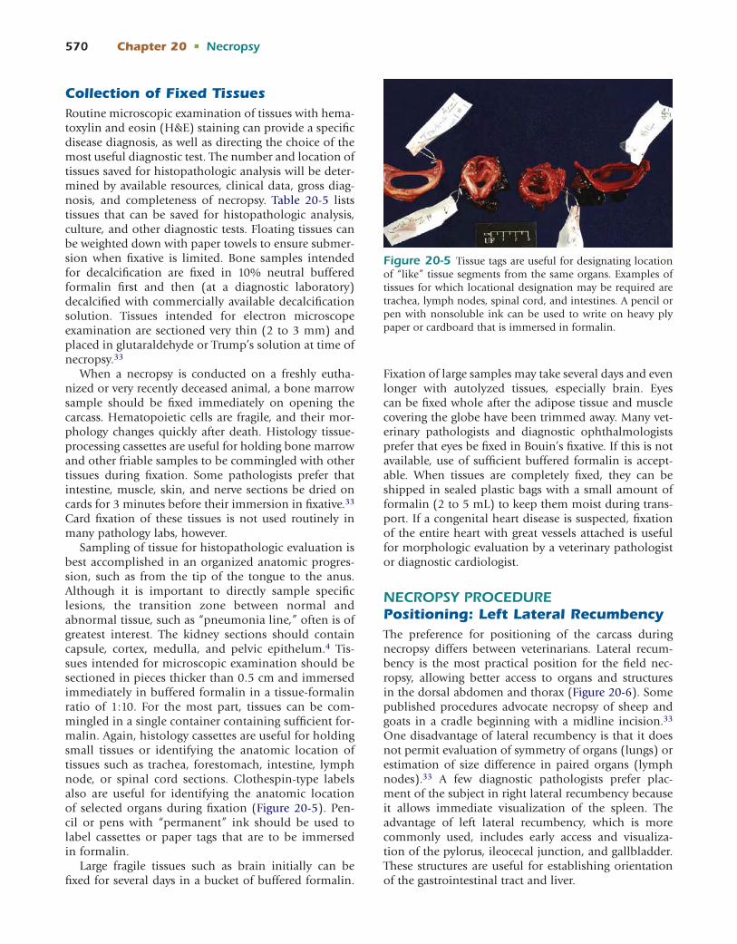
570 Chapter 20 • Necropsy
Collection of Fixed TissuesRoutine microscopic examination of tissues with hema-toxylin and eosin (H&E) staining can provide a specific disease diagnosis, as well as directing the choice of the most useful diagnostic test. The number and location of tissues saved for histopathologic analysis will be deter-mined by available resources, clinical data, gross diag-nosis, and completeness of necropsy. Table 20-5 lists tissues that can be saved for histopathologic analysis, culture, and other diagnostic tests. Floating tissues can be weighted down with paper towels to ensure submer-sion when fixative is limited. Bone samples intended for decalcification are fixed in 10% neutral buffered formalin first and then (at a diagnostic laboratory) decalcified with commercially available decalcification solution. Tissues intended for electron microscope examination are sectioned very thin (2 to 3 mm) and placed in glutaraldehyde or Trump’s solution at time of necropsy.33
When a necropsy is conducted on a freshly eutha-nized or very recently deceased animal, a bone marrow sample should be fixed immediately on opening the carcass. Hematopoietic cells are fragile, and their mor-phology changes quickly after death. Histology tissue-processing cassettes are useful for holding bone marrow and other friable samples to be commingled with other tissues during fixation. Some pathologists prefer that intestine, muscle, skin, and nerve sections be dried on cards for 3 minutes before their immersion in fixative.33 Card fixation of these tissues is not used routinely in many pathology labs, however.
Sampling of tissue for histopathologic evaluation is best accomplished in an organized anatomic progres-sion, such as from the tip of the tongue to the anus. Although it is important to directly sample specific lesions, the transition zone between normal and abnormal tissue, such as “pneumonia line,” often is of greatest interest. The kidney sections should contain capsule, cortex, medulla, and pelvic epithelum.4 Tis-sues intended for microscopic examination should be sectioned in pieces thicker than 0.5 cm and immersed immediately in buffered formalin in a tissue-formalin ratio of 1:10. For the most part, tissues can be com-mingled in a single container containing sufficient for-malin. Again, histology cassettes are useful for holding small tissues or identifying the anatomic location of tissues such as trachea, forestomach, intestine, lymph node, or spinal cord sections. Clothespin-type labels also are useful for identifying the anatomic location of selected organs during fixation (Figure 20-5). Pen-cil or pens with “permanent” ink should be used to label cassettes or paper tags that are to be immersed in formalin.
Large fragile tissues such as brain initially can be fixed for several days in a bucket of buffered formalin.
Fixation of large samples may take several days and even longer with autolyzed tissues, especially brain. Eyes can be fixed whole after the adipose tissue and muscle covering the globe have been trimmed away. Many vet-erinary pathologists and diagnostic ophthalmologists prefer that eyes be fixed in Bouin’s fixative. If this is not available, use of sufficient buffered formalin is accept-able. When tissues are completely fixed, they can be shipped in sealed plastic bags with a small amount of formalin (2 to 5 mL) to keep them moist during trans-port. If a congenital heart disease is suspected, fixation of the entire heart with great vessels attached is useful for morphologic evaluation by a veterinary pathologist or diagnostic cardiologist.
NECROPSY PROCEDUREPositioning: Left Lateral RecumbencyThe preference for positioning of the carcass during necropsy differs between veterinarians. Lateral recum-bency is the most practical position for the field nec-ropsy, allowing better access to organs and structures in the dorsal abdomen and thorax (Figure 20-6). Some published procedures advocate necropsy of sheep and goats in a cradle beginning with a midline incision.33 One disadvantage of lateral recumbency is that it does not permit evaluation of symmetry of organs (lungs) or estimation of size difference in paired organs (lymph nodes).33 A few diagnostic pathologists prefer plac-ment of the subject in right lateral recumbency because it allows immediate visualization of the spleen. The advantage of left lateral recumbency, which is more commonly used, includes early access and visualiza-tion of the pylorus, ileocecal junction, and gallbladder. These structures are useful for establishing orientation of the gastrointestinal tract and liver.
Figure 20-5 Tissue tags are useful for designating location of “like” tissue segments from the same organs. Examples of tissues for which locational designation may be required are trachea, lymph nodes, spinal cord, and intestines. A pencil or pen with nonsoluble ink can be used to write on heavy ply paper or cardboard that is immersed in formalin.

Chapter 20 • Necropsy 571
External Examination: Skin, Hooves, Body OrificesThe carcass is weighed and measured before the post-mortem exam is begun. A condition score can be assigned to the carcass (see Chapter 2, Fig 2-1). Deter-mination of crown-rump length and observations of developmental characteristics are useful for investiga-tions of abortion and neonatal death. The external characteristics of the carcass on both right and left aspects of the body—orifices (nostrils, ears, mouth, anus, vulva, external urogenital opening, and teat canals), umbilicus, hair or wool, skin, and hooves—are noted. Mucous membranes are examined for anemia or jaundice (icterus). If the necropsy is to be conducted with the subject in left lateral recumbency, the “down side” (the left external and muscular skeletal aspect) should be examined before the carcass is placed in final necropsy position. Mammary glands and external genitalia are evaluated for symmetry and consistency. Lymph nodes are palpated before the carcass is opened. (Note: It is essential to sample and culture skin lesions at this time.)
External Examination: Musculoskeletal SystemMuscle and bones are examined before the carcass is opened. The limbs are flexed if possible and evalu-ated for fractures or joint disease. Rigor mortis begins approximately 3 hours after death, peaks at 12 hours, and declines over days, depending on environmental conditions. If the carcass is in rigor mortis and was not moved at time of death, the positioning of the limbs
and head is noted: If the animal died in a posture of opisthotonos and has external evidence of a wound, for example, a diagnosis of tetanus may be suspected. In addition, joint spaces in the lower extremities should be opened and examined, and joint fluid and other tis-sue collected for clinical pathology analysis and culture, if indicated.
Skinning the Carcass: Right Subcutis—Adipose Tissue, Lymph Nodes, MuscleAlthough knife positioning should be comfortable for the operator, “blade-up” dissection of skin will reduce dulling of the cutting instrument and decrease contamination of the necropsy field. If the forelimbs are reflected backward, the brachial plexus is excised directly adjacent to the limb. The free nerves of the brachial plexus nerves can be clamped to a hemostat for orientation purposes during location of cervico-thoracic ganglia. When the inguinal skin and muscle are excised, aspects of the right hip, including inguinal and mammary lymph nodes, pelvic plexus, hip joint, and articular surfaces, are examined. The color and consistency of subcutaneous adipose tissue should be recorded. Tissue should be examined for bruising or hemorrhage
Opening of the Abdominal and Thoracic Cavities and Abdominal Cavity ExaminationWithout puncturing the rumen, the musculature and parietal peritoneum of the abdominal wall are com-pletely excised along the caudal border of the rib cage. Rupture of the rumen will make recognition of lesions in the abdominal cavity difficult. The ventral abdominal musculature is excised off center and right of midline to create a “bowl” for retention of abdominal viscera within the carcass. The umbilicus, fetal vasculature (umbilical arteries and veins), and urachus are left intact in fetuses and neonates (Figure 20-7).
Quantity and consistency of abdominal fluid should be recorded. The intact diaphragm is penetrated with the point of a knife, before the ribs and sternum are cut, to verify negative pressure in the thoracic cavity. The dia-phragm muscle is excised along its insertion point on the right rib cage, leaving the intact diaphragm in place as an anatomic partition. This landmark may be use-ful for identifying the origin of free fluids (abdominal versus thoracic effusions) and in assessing for the pres-ence of a diaphragmatic hernia. The dorsal and ventral attachments of the right rib cage are removed with lop-pers and a knife, with care taken not to excise vessels, brachial nerves, and the trachea at the thoracic inlet. The pleural surface of the right rib cage is examined for fractures or adhesions before proceeding.
Figure 20-6 Positioning for necropsy in a goat. A midline incision has been made with the subject in left lateral recum-bency. Arrow identifies the abomasal pylorus. Hemostats are clamped onto the stump of the brachial plexus nerves, provid-ing a landmark for identification of cervicothoracic ganglia.

572 Chapter 20 • Necropsy
With the right aspect of the thoracic and peritoneal cavity exposed, the operator should pause and resist handling the organs. This is the last opportunity to observe and photograph the “in vivo” position of gastrointestinal tract and the location and volume of compartmental fluids. At this time, gross observations and digital images of organs before manipulation are best recorded. Lung, liver, spleen, and other organs or effusions for which bacterial culture may be appropriate are sampled with use of sterile forceps, scissors, needles, or syringes as required. After these tasks are completed, the small intestine can be flipped over the vertebral musculature to allow observation of the spleen and left aspect of the peritoneal cavity.
Thoracic Cavity ExaminationBefore removal of heart and lungs, organs and struc-tures of the thoracic cavity should be examined in place. The pericardial space is examined for fluid volume and presence of exudate. Pericardial or thoracic fluid is col-lected for culture, serologic testing (as for leptospirosis in fetuses), or clinicopathologic analysis (Chapter 8). Fresh postmortem heart blood should be collected for serologic studies. The mediastinum, lymph nodes, and thoracic duct are examined.
Throat and Neck ExaminationThe skin of the right medial aspect of the neck should be excised down to the mandibular symphysis and carefully skinned off the vasculature, esophagus, and trachea. The right cervical and mandibular lymph nodes are best observed at this time; findings may be crucial for the diagnosis of caseous lymphadenitis (caused by Corynebacterium pseudotuberculosis). Individual lymph
nodes, either subcutaneous or associated with viscera, can be labeled with a tag or placed in a histology cas-sette at time of fixation. This is now the best opportu-nity to observe and photograph lesions or artifact such as hemorrhage from jugular injection or bleeding from neck trauma. The thyroids can be located and weighed if goiter is suspected (see Chapter 9).
Removal of the “Pluck” (Tongue, Esophagus, Trachea, Lungs, and Heart)The cervicothoracic ganglia can be located and removed before removal of the “pluck” (tongue, trachea, lungs, and heart). Ganglia are best located by means of blunt dissection performed with an appreciation of their anatomic location in relation to the first rib and nerves of the brachial plexus. The tongue is removed by cut-ting along the medial aspect of the mandibular rami while the tongue and associated muscle are reflected back. Excising joints of the hyoid apparatus and tis-sues of dorsal pharynx will free up the larynx, trachea, and esophagus for removal. The complete tongue with attached trachea and esophagus including thyroids and jugular veins and arteries is dissected away from the musculature of the neck.
The organs of the thoracic cavity (heart, lungs, dorsal aorta, and esophagus) are removed by incising against the thoracic vertebrae and making a complete transverse section though the esophagus and caudal vena cava just proximal to the diaphragm. Organs of the “pluck” can be hosed off before examination and sampling for histopathologic evaluation. Symmetry of paired organs such as lungs and thyroids is now evaluated. Lungs are palpated before proceeding. The esophagus can be opened in a cranial to caudal direc-tion and examined for a “bloat line.” A bloat line is a faint demarcation between cranial, congested tissue and pale caudal tissue caused by abdominal disten-tion and blocked return of blood from the head and cranial neck. The trachea should be opened longitudi-nally from the larynx, with the incision continued into each bronchial branch. The lumen of the trachea may contain excess mucus associated with shock, blood, or lungworms.
To facilitate examination of the pulmonary artery, the heart should be opened while it is still attached to the lungs. The right and left sides of the heart can be opened using an elliptical incision to preserve the architecture of the heart while allowing examination of valves, compartments, and great vessels. The heart should be examined in a systematic order that follows the flow of blood—cranial or caudal vena cava > right atrium > right AV valve (tricuspid) > right ventricle > pulmonary valve > pulmonary artery > ductus arteriosus (if indi-cated) > pulmonary vessels in lungs > pulmonary vein
uauv
Figure 20-7 Paramidline sagittal excision to open the abdomen of a goat fetus of 15 weeks gestation. The internal umbilicus is formed at the junction of the umbilical arteries (ua), urachus, and vein (uv).

Chapter 20 • Necropsy 573
> left atrium > left AV valve (mitral valve) > left ventricle > aortic valve > aorta (see Chapter 17). The pulmonary artery is excised longitudinally and followed into each lobe of the lung to check for thromboemboli. In young animals, closure status of the ductus arteriosus should be determined before the heart is removed from the lungs.
Removal of Abdominal Organs: Adrenal Gland, Pancreas, Spleen, Liver, Forestomachs, Abomasum, Small Intestine, Cecum, Spiral Colon, Descending Colon, MesenteryThe adrenal glands and celiac-mesenteric ganglia are examined before removal of the abdominal viscera (Figure 20-8). These ganglia can be used for micro-scopic diagnosis of abomasal emptying defect caused by dysautonomia35 (see Chapter 5). Perirenal adipose tissue is evaluated to assess body condition. At this time, renal arteries can be excised and the kidneys can be dissected away, leaving the ureter attached. The kid-neys are flipped over the vertebral musculature, to be examined later. The gallbladder and patency of the bile duct are evaluated before organ removal by opening the duodenum at the location of the bile papilla and squeezing the gallbladder (Figure 20-9). The lumen of the gastrointestinal tract is not further opened before removal of the viscera.
The gastrointestinal tract with attached liver and spleen can now be removed by dissecting between the dorsal aorta and the vertebrae. The genitourinary tract with attached kidneys can remain in the abdomen at this time. The entire gastrointestinal tract is now arranged for orientation before culture, collection of contents, and sampling of tissue for histopathologic evaluation. The ileocecal junction and pylorus are best observed from the right-sided aspect of the mesentery (Figure 20-10). The spiral loop of the colon is best examined from the left aspect of the mesentery. The last centrifugal coils of the spiral loop of the colon are difficult to differentiate from small intestine but can be identified by the presence of fecal balls. Palpating or running a knife over mucosa of tissues intended for histopathologic examination is to be avoided, because such maneuvers destroy the superficial mucosa. The jejunal lymph node and lymphatics supplying the intestine are now examined (Figure 20-11). Dis-tended lymphatic vessels can be best visualized in anorexic animals; such distention may reflect uptake of intestinal materials, lymphatic edema, or Johne’s disease (caused by Mycobacterium avium subsp. paratu-berculosis) (see Chapter 5).
Careful examination of the gastrointestinal tract requires a systematic approach. Some pathologists pre-fer to examine the forestomachs last, because spillage
of rumen contents often obscures visualization of the intestine. The rumen often is overlooked at necropsy, but detection of ruminal lesions or incriminating content may be crucial for determination of cause of death. Consistency of rumen contents and distention of the rumen are important for diagnosis of bloat secondary to grain overload or legume consumption. Identification of feedstuffs in rumen, abomasum, and intestine may be crucial for achieving a diagnosis. At least 500 mL of rumen content is needed for toxico-logic analysis. The pH of rumen contents is determined at the time of necropsy. A pH value less than 5.5 indi-cates acidosis. The contents of the rumen, abomasum, and intestine may be sifted in shallow trays to identify plant material, seeds, parasites, inorganic debris, or toxic substances (Figure 20-12, A). A stream of gentle free-flowing water can be directed to the mucosal surface of forestomachs, abomasum, and intestine to
Right ad
Pan
Right kid
cm ganglia
Figure 20-8 Location of celiacomesenteric ganglia in the right aspect of the dorsal abdomen. The celiomesenteric gan-glia (cm ganglia) lies under the right adrenal gland (right ad) near pancreas (pan) and right kidney (right kid).
ddn
ddn
Gall bl
Bile ductddn papillae
Figure 20-9 Checking the bile duct for patency. The duo-denum (ddn) is excised to reveal the duodenal papillae (ddn papillae). Compression of the gallbladder (gall bl) forces yellow-green bile through the bile duct and out the papillae opening.

574 Chapter 20 • Necropsy
reveal parasites (Figure 20-12, B). The ileocecal junc-tion is a good landmark for use in examination of the intestine (see Chapter 6). Sections of ileum and cecum are useful for histopathologic evaluation because they contain gut-associated lymphoid tissue (GALT) (i.e., Peyer’s patches). Examination of the entire mucosal surface of the small intestine is time-consuming in sheep and goats. If a gastrointestinal disease is low on the differential diagnosis list, the intestine can be spot-checked for mucosal changes. Organ weights (liver and spleen) and volume of lumen contents from the gastro-intestinal tract (sand, digesta) may be useful to validate disease processes.
Urogenital TractThe ureters of the reflected kidneys are now exam-ined to determine patency of the lower urinary tract (with evidence of hydronephrosis in cases of lower tract obstruction) or to detect pylonephritis (caused by Corynebacterium renale). The capsule of the kidney is removed to help facilitate visualization of infarcts. The bony pelvis can be opened by cutting through the ischium and pubis using loppers or a saw. This maneu-ver allows dissection of the entire genitourinary tract system from kidneys to the external opening. In cases of severe bladder dilation, it is important to evaluate the entire penile urethra for identification of urinary calculi. Obstructions may be present in the penile viliform process of bucks and rams (see Chapter 12, Figures 12-7 and 12-8).dc
icjce
lccslc
jln
Pylorus
Rumen
Figure 20-10 Right aspect of the gastrointestinal tract with structures arranged for examination. The cecum (ce), ileocecal junction (icj), descending colon (dc), and pylorus are anatomic landmarks for examination of the intestines. In right orientation, the last centrifugal coils of the spiral loop of the colon (lccslc) and jejunal lymph nodes (jln) are observed. This goat had distention of the cecum.
Afferent lymph vessel
Figure 20-11 A jejunal lymph node demonstrating affer-ent lymph vessels containing white substance likely taken up from the intestine.
A
B
Figure 20-12 A, Abomasal contents sifted on a dispos-able tray to reveal numerous Haemonchus contortus organisms (barber’s pole or wire worms). B, The abomasal mucosa with an H. contortus organism visible.5 Multifocal hemorrhage can be seen on on the mucosal surface of the abomasum.

Chapter 20 • Necropsy 575
Head and Brain Removal and Examination of Pituitary, Eyes, Ears, Oral Cavity, Teeth, and Nasal CavityWith the subject in dorsal recumbency and the head extended off the table edge, the atlantooccipital joint is excised for decapitation. Cerebrospinal fluid can be collected at this time. Skin, hair, and muscle should be removed from the cranium, and anatomic land-marks located for making saw cuts to open the cal-varia. Horned sheep or goats can be dehorned post mortem by a single sagittal cut that opens the frontal sinus and removes both horns (Figure 20-13). In polled or dehorned animals, the calvaria can be cleanly entered with a series of five saw cuts. The orbital bone
around the eye serves as a locator for transverse cut 1. The supraorbital foramen is used to determine location of sagittal cuts 2 and 3. The occipital-parietal suture and the occipital condyles are used to locate oblique cuts 4 and 5 (Figure 20-14, A and B). After removal of the cal-varia and examination and biopsy (for culture) of the meninges, the skull is inverted and tapped to remove the brain with help of gravity. Care is taken not to damage the fragile brain during manipulations to free it from the calvaria, accomplished by clipping cranial nerve attachments one by one.
If rabies is suspected, the disease-specific protocol should be followed. If the obex is to be removed for scrapie surveillance, the brain should not be sagittally sectioned. The entire fixed obex, which should be located on the dorsal aspect of the brain stem under the cerebellum (see Figure 20-4), is required as a sample. After removal of the mandible, the external ear canal and tympanic bulla serve as a locator for a complete transverse saw cut through the head that will expose both internal ears and tympanic cavity for examination and sampling for culture. The pituitary is separated from the brain by connective tissue and lies in the hypophyseal fossa (sella turcica) (Figure 20-15). The skull should now be completely sectioned in a sagittal plane to examine the nasal cavity (Figure 20-16).
Left Subcutis and Spinal Cord RemovalThe carcass should now be flipped and the subcutis and muscle of the left side examined. Finally, the spinal canal can be entered with a Stryker autopsy saw, ron-geur, or small Barnes dehorner. Removal of the spinal cord is time-consuming and usually is optional.
Horn cut
Location of orbit
Figure 20-13 Sagitally sectioned goat skull demonstrating landmarks for removal of horns and position of the calvaria in regard to the orbital rim. A single dorsal-plane “horn cut” can be made through the frontal sinus to remove both horns.
Cut 1Cut 5
Cut 3
Cut 2Cut 4
A
Cuts 2 and 3Cut 1
Cuts 4 and 5
Ear cutB
Figure 20-14 Sheep skull (ewe): A, medial aspects; B, dorsal aspects. White lines indicate location of saw cuts required for entry into the calvaria for brain removal and the final “ear cut” needed to gain access to the ear and tympanic bulla. Cut 1 is transverse, directly behind the orbit of the eye. Cuts 2 and 3 are in a sagit-tal plane perpendicular to cut 1 and are angled slightly medially. Cuts 2 and 3 are started just medial to the supraorbital foramen. Cuts 4 and 5 continue from the junction of cuts 2 and 3 at the parietal-occipital fissure and continue obliquely through the occipital condyle. The “ear cut” is a complete transverse section of the skull at the external acoustic meatus and tympanic bulla.

576 Chapter 20 • Necropsy
Necropsy of the Fetus and Neonate, Including Stillborn, and PlacentaThe procedure for fetal or neonate necropsy is similar to that in an adult animal. In addition, care should be taken not to excise the structures of the internal umbi-licus when the abdominal cavity is opened. The buoy-ancy of the lungs in formalin is observed to determine if the neonate took a breath of air. Fetal abomasal con-tents are extracted with a sterile needle and syringe and
submitted for culture. In absence of fetal membranes, this bacterial population may reflect what was growing in amniotic fluid before occurrence of abortion.
Amniotic plaques on the fetal surface of the umbili-cal cord and amnion are normal (Figure 20-17). The placenta should be cleansed gently with water if coated by debris and then arranged in the best position for a complete evaluation. The integrity and length of the umbilical cord should be noted. Complete transverse sections of the umbilical cord should be obtained for histopathologic examination. Although the surface of the chorion is likely to be covered with debris, coty-ledons should be sampled and submitted for culture and frozen for PCR assay. Multiple sites should be sampled for histopathologic examination from the chorioallantois, including both horns and body. The intercotyledonary areas should be thin and transparent in the normal placenta. The nongravid horns normally may have yellowish, necrotic, membrane-covered tips (Figure 20-18).
Packaging and Shipment of Diagnostic SamplesPackaging and shipment of samples can be time-con-suming and complex. International, national, and state laws vary in regard to shipment of diagnostic speci-mens. The United States Animal Health Association’s Foreign Animal Diseases (“The Gray Book”), available online (www.aphis.usda.gov/emergency_response/downloads/nahems/fad), presents detailed information on shipping requirements.
Specimens should be mailed in leak-proof containers at appropriate temperature and labeled with a water-proof marker. Fresh tissues should be packaged individu-ally, although formalin-fixed tissues can be combined.
ec
tcpt
Figure 20-15 Caudal aspect of head with brain removed and complete transverse section to reveal the ear canal (ec) and tympanic cavity (tc). A membrane on the floor of the cranial cavity must be excised to remove the pituitary (pt) concealed in the hypophyseal fossa (sella turcica).
pt
nac
Figure 20-16 Enzootic nasal tumor in a 3-year-old ram. Sagittal section of the head before brain removal demon-strates position of nasal turbinates, sinuses, brain, pituitary (pt), and a nasal adenocarcinoma (nac). The nasal tumor deforms the cribriform plate. (Courtesy Dr. Lisa Farina and Dr. Jason Kimbro, Gainesville, Florida.)
Figure 20-17 Normal fetus and umbilical cord with amni-otic plaques. Amniotic plaques are normal incidental findings consisting of multifocal accumulations of layered keratin on the fetal side of the amnion. (Courtesy Dr. Jose Ramos-Vara, West Lafayette, Indiana.)

Chapter 20 • Necropsy 577
The submission sheet or necropsy form should be placed in a separate plastic bag to prevent its destruc-tion from accidental spillage. Specimens should not be shipped on Friday before the weekend or on the day before a holiday. With prolonged time in transit, thawing or warming of specimens may occur if they are retained in a warm mailroom for a day or longer.
Clean-up and Disposal of RemainsLaws concerning the disposal of ruminant remains may vary between countries, states, and municipalities. A plan for disposal of remains should consider the geo-graphic location of digestion or incineration facilities. Methods of carcass disposal include burial, landfill, incineration or combustion, rendering, composting (see Box 20-2), and alkaline hydrolysis (chemical digestion).7 The remains of animals suspected of having scrapie or zoonotic diseases should be burned, incinerated, bur-ied, or chemically digested. The United States Animal Health Association’s “Gray Book,” available online as noted earlier, provides detailed information on spe-cific chemical products needed to inactivate individual pathogens.
ACKNOWLEDGMENTSWe thank Dr. K.B. Poonacha and Dr. Lenn R. Harrison, both of Lexington, Kentucky; Dr. Ralph Giles, George-town, Kentucky; and Dr. Claus Buergelt and the Univer-sity of Florida, Gainesville, Florida, for their instruction on necropsy technique. We also thank Mr. Helm Roberts, Lexington, Kentucky, for computer imaging
assistance and Mrs. Dorothea Dillman and Mr. Frank Dillman, both of Notasulga, Alabama, for necropsy support.
REFERENCES 1. King JM: The necropsy book, Ithaca, NY, 1989, Cornell University/
Arnold Printing Corp, NewYork State College of Veterinary Medicine.
2. Moisier DA: Vascular disorders and thrombosis. In McGavin MD, Zachary JF, editors: Pathologic basis of veterinary disease, ed 4, St Louis, 2007, Mosby Elsevier.
3. Smith MC, Sherman DM: Cardiovascular system. In Smith MC, Sherman DM, editors: Goat medicine, ed 2, Ames, Iowa, 2009, Wiley-Blackwell.
4. Smith MC, Sherman DM: Sudden death. In Smith MC, Sherman DM, editors: Goat medicine, ed 2, Ames, Iowa, 2009, Wiley-Blackwell.
5. Radostits OM, et al: General systemic states. In Radostits OM, et al: Veterinary medicine, ed 10, Philadelphia, 2007, Saunders Elsevier.
6. Smith MC, Sherman DM: Digestive system. In Smith MC, Sherman DM, editors: Goat medicine, ed 2, Ames, Iowa, 2009, Wiley-Blackwell.
nt
cd
ica
Figure 20-18 The maternal side of a normal placenta demonstrating cotyledons (cd) and intercotyledonary areas (ica) of the chorioallantois and a necrotic tip of the chorioallantoic membrane from the nongravid horn (nt). The intercotyledonary area of the normal chorioallantois should be thin and transpar-ent. The necrotic tip of the horn is a nonpathogenic finding, or “pseudolesion.” (Courtesy Dr. John F. Edwards, College Station, Texas.)
• A 5- to 10-square-foot enclosure or container makes a good composter. The sides (e.g., corrugated wire, fine wire mesh) should have enough holes to allow maximal airflow during the composting process.
• The composter should be placed separate from the flock but in an area where water is easily attainable to keep the composted material moist. All runoff should be kept out of the flock’s water supply.
• The carcass, aborted material, or offal should be placed in the composter so that 1 foot of sawdust separates it from the ground and the sides. The carcass should then be covered with 1 to 1.5 feet of sawdust. Approximately 1 cubic foot of sawdust is required for every 10 lb of carcass weight. Green or freshly cut sawdust is superior for composting.
• Water should be added to attain a water–to–dry matter ratio between 50:50 and 60:40. During very rainy seasons, the composter should be covered to prevent excessive water accumulation.
• A 2- to 4-foot-long thermometer should be placed into the stack and the temperature monitored. The temperature should be maintained at 130° F for 1 week to destroy most pathogenic bacteria. If the composter’s temperature drops below 100° F, water should be added and the material aerated (i.e., stirred, forked).
• The composted material can be safely placed on the pasture after 21 to 30 weeks. However, in the case of animals dying of scrapie, the owner should notify local, state, or federal authorities and follow recommended guidelines for carcass disposal (e.g., incineration).
Modified from Estienne MJ: Disposing of dead goats, Practical Goat Farming Seminar, 1998, Salisbury, Md: http://www.sheepandgoat.com/compost.html. Accessed March 12, 2011.
BOX 20-2 Sheep or Goat Composting

578 Chapter 20 • Necropsy
7. Smith MC, Sherman DM: Blood, lymph, and Immune systems. In Smith MC, Sherman DM, editors: Goat medicine, ed 2, Ames, Iowa, 2009, Wiley-Blackwell.
8. Smith MC, Sherman DM: Wasting diseases. In Smith MC, Sherman DM, editors: goat Medicine, ed 2, Ames, Iowa, 2009, Wiley-Blackwell.
9. Navarre CB, Pugh DG: Disease of the gastrointestinal system. In Pugh DG, editor: Sheep and goat medicine, Philadelphia, 2002, Saunders Elsevier.
10. Radostits OM, et al: Disease of the alimentary tract I. In Radostits OM, et al: Veterinary medicine, ed 10, Philadelphia, 2007, Saunders Elsevier.
11. Johnson DD, Libal MC: Necropsy of sheep and goats, Vet Clin North Am Food Anim Pract 2:129–147, 1986.
12. Machen MR, et al: Disease of the neurologic system. In Pugh DG, editor: Sheep and goat medicine, Philadelphia, 2002, Saunders Elsevier.
13. Lutnaes B, Simensen E: An epidemiological study of abomasal bloat in young lambs, Prevent Vet Med 1:335, 1983.
14. Radostits OM, et al: Disease associated with prions. In Radostits OM, et al: Veterinary medicine, ed 10, Philadelphia, 2007, Saunders Elsevier.
15. Radostits OM, et al: Diseases associated with viruses and Chlamydia I. In Radostits OM, et al: Veterinary medicine, ed 10, Philadelphia, 2007, Saunders Elsevier.
16. Foreign animal diseases 2008, ed 7, St Joseph, Mo, 2008, United States Animal Health Association.
17. Radostits OM, et al: Diseases associated with viruses and Chlamydia II. In Radostits OM, et al: Veterinary medicine, ed 10, Philadelphia, 2007, Saunders Elsevier.
18. Swaneoel R, Coetzer JAW: Wesselsbron disease. In Coetzer JAW, Tustin RC, editors: Infectious Diseases of Livestock, ed 2, Cape Town, 2004, Oxford University Press.
19. Radostits OM, et al: Diseases associated with bacteria I. In Radostits OM, et al: Veterinary medicine, ed 10, Philadelphia, 2007, Saunders Elsevier.
20. Radostits OM, et al: Diseases associated with bacteria V. In Radostits OM, et al: Veterinary medicine, ed 10, Philadelphia, 2007, Saunders Elsevier.
21. Radostits OM, et al: Diseases associated with bacteria III. In Radostits OM, et al: Veterinary medicine, ed 10, Philadelphia, 2007, Saunders Elsevier.
22. Godfroid J, et al: Brucella melitensis infection. In Coetzer JAW, Tustin RC, editors: Infectious diseases of livestock, ed 2, Cape Town, 2004, Oxford University Press.
23. Radostits OM, et al: Diseases associated with bacteria IV. In Radostits OM, et al: Veterinary medicine, ed 10, Philadelphia, 2007, Saunders Elsevier.
24. Buergelt CD, et al: Paratuberculosis. In Coetzer JAW, Tustin RC, editors: Infectious diseases of livestock, ed 2, Cape Town, 2004, Oxford University Press.
25. O’Toole DO, et al: Tularemia in range sheep: an overlooked syn-drome? J Vet Diagn Invest 20:508–513, 2008.
26. Navarro JA, et al: Kinetics of infection and effects on the placenta of Chlamydophila abortus in experimentally infected pregnant ewes, Vet Pathol 41:498–505, 2004.
27. Moeller RB: Causes of caprine abortion: diagnostic assessment of 211 cases (1991-1998) J Vet Diagn Invest 13:265–270, 2001.
28. Ellis WA, et al: Leptospirosis as a cause of reproductive failure, Vet Clin North Am Food Animl Pract 10:463–478, 1994.
29. Cullen JM: Liver, biliary system, and exocrine pancreas. In McGavin MD, Zachary JF, editors: Pathologic basis of veterinary disease, ed 4, St Louis, 2007, Mosby Elsevier.
30. Radostits OM, et al: Diseases associated with arthropod parasites. In Radostits OM, et al: Veterinary medicine, ed 10, Philadelphia, 2007, Saunders Elsevier.
31. Norval RAI, Horak IG: Vectors: ticks. In Coetzer JAW, Tustin RC, editors: Infectious diseases of livestock, ed 2, Cape Town, 2004, Oxford University Press.
32. Radostits OM, et al: Diseases associated with Rickettsiales. In Radostits OM, et al: Veterinary medicine, ed 10, Philadelphia, 2007, Saunders Elsevier.
33. Howie F: Necropsy and sampling techniques. In Aitken ID, editor: Diseases of sheep, ed 4, Oxford, 2007, Blackwell Publishing.
34. Nieves Ortega JA, et al: Evaluation of Chlamydophila abortus DNA extraction protocols for polymerase chain reaction diagnosis in paraffin-embedded tissues, J Vet Diagn Invest 19:421, 2007.
35. Pruden SJ, et al: Abomasal emptying defect of sheep may be an acquired form of dysautonomia, Vet Pathol 41:164, 2004.

579
Commonly Used Drugs in Sheep and Goats: Suggested Dosages
Virginia R. Fajt, and D.G. Pugh
A P P E N D I X
GENERAL PRINCIPLESSome of the drugs and uses listed in this appendix may be illegal, unavailable, or extralabel in the United States or other countries. It is the responsibility of attending veterinarians to be familiar with the laws governing drugs in their practice areas. The clinician should therefore be cognizant of and take specific steps and help educate pro-ducers in order to reduce drug residues in food animals.
If a dose is provided only for sheep and not for goats, unless the drug appears to be contraindicated for or toxic to goats, the sheep dose usually can be extrapo-lated for use in goats, and vice versa (as recommended by D.G.P.). Some of the anesthetic dosages presented here are the same as those given in Chapter 18 and are referenced as such. After reviewing Chapter 18, where dosages for anesthetics appear different, the clinician then compare the two, and decide the best dosage for the situation based on referenced materials.
AN EVIDENCE-BASED APPROACHIn an effort to provide an evidence-based approach and to characterize confidence levels for the dosages listed in this appendix, notations have been made to indicate the quality and type of evidence presented to support the dosage recommendation. The categories used in the evi-dence notations are defined at the end of this section; specific designations are given in brackets after each dosage in the table that follows.
As an example, a dosage that has been demonstrated to be effective in at least one good-quality, large, random-ized controlled trial (RCT) would be assigned the desig-nation A-1, and a dosage for which moderate evidence based on expert opinion is available would have the des-ignation B-5. In this appendix, only drug dosages with A- or B-quality evidence are listed, but the type of evi-dence varies by drug and species, with a range of A-1 to
B-6, with the highest level of evidence available noted in the designation (in other words, lower levels of evidence also may be available to support that particular dosage).
All evidence notations derive from an unpublished analysis of the data (by V.R.F.) and have not been sub-jected to peer review. For some dosages, the cited refer-ences do not constitute the only evidence but rather are provided as examples of relevant published data.
Evidence Quality A Good evidence to support a recommendation for use B Moderate evidence to support a recommendation
for use C Insufficient evidence to support a recommendation
for use D Moderate evidence to support a recommendation
against use E Good evidence to support a recommendation
against use
Evidence Type 1 Species-specific evidence from at least one large
RCT or multiple small RCTs 2 Species-specific evidence from a small RCT, disease
models, large case studies, pharmacokinetic studies using surrogate end points, or evidence from well-designed trials in a different species that is consid-ered appropriate for comparison
3 Dramatic results from either well-designed, species-specific trials without controls or small case studies
4 Pharmacokinetic studies without surrogate end points
5 In vitro studies 6 Opinions of respected authorities based on clinical
experience or reports of expert committees
1

580 Appendix 1 • Commonly Used Drugs in Sheep and Goats: Suggested Dosages
Drug Sheep Goats
Acepromazine maleate (see Chapter 18)
0.05-0.10 mg/kg IM [A-1]1-3
0.05-0.10 mg/kg IM [A-1]1-3
0.2 mg/kg IM for tetany [B-6]4
Acetic acid (5% solution)
0.5-1.0 L/head PO for ammonia toxicosis [B-3]5-7
0.5-1.0 L/head PO for ammonia toxicosis [B-3]5-7
Albendazole 7.5 mg/kg PO for flukes and nematodes [A-1]8,9
10 mg/kg PO for flukes [A-1]8
10 mg/kg PO for cestodes [A-2]10 10 mg/kg PO for cestodes [A-3]10,11
Ammonium chloride 0.5-1.0% of diet for prevention of urinary calculi [A-1]12-19
0.5-1.0% of diet for prevention of urinary calculi [A-1]12-19
Ammonium molybdate
100 mg/head/day PO in combination with sodium sulfate to increase elimination of copper [A-1]20-22
300 mg/head/day PO in combination with sodium thiosulfate to increase elimination of copper [A-2]23
Ammonium tetrathiomolybdate
3.4 mg/kg IV once daily for 4 days [B-3]24
Amoxicillin– clavulanic acid
20 mg/kg (amoxicillin component) IV two or three times a day [B-4]25,26
20 mg/kg (amoxicillin component) IV two or three times a day [B-4]25,26
7 mg/kg IM for prevention of pneumonia [B-2]27
200 mg amoxicillin plus 50 mg clavulanate per quarter IMM [B-4]28
200 mg amoxicillin plus 50 mg clavulanate per quarter IMM [B-4]29
Amoxicillin trihydrate
10 mg/kg IM two or three times a day [B-4]30,31
10 mg/kg IM two or three times a day [B-4]30,31
Ampicillin sodium 10 mg/kg IV three or four times a day [B-4]30,32
10 mg/kg IV three or four times a day [B-4]30,32
Ampicillin-sulbactam 13.3 mg/kg (ampicillin component) IM once or twice a day [B-4]33
13.3 mg/kg (ampicillin component) IM once or twice a day [B-4]33
Ampicillin trihydrate 10 mg/kg IM [B-4]34 10 mg/kg IM [B-4]34
Amprolium 50 mg/kg PO for 5 days for treatment [A-2]35 100 mg/kg PO for 5 days for treatment [A-2]35
55 mg/kg PO twice a day for 21 days for treatment [A-2]36
50 mg/kg PO for 21 days for treatment [A-2]37
15 mg/kg in feed for prevention [A-2]38
Aspirin 100 mg/kg PO twice a day [B-4]39-41 100 mg/kg PO twice a day [B-4]39-41
Atipamezole (see Chapter 18)
0.1-0.2 mg/kg IV slowly [A-2]42,43 0.1-0.2 mg/kg IV slowly [A-2]42,43
0.005 μg/kg IV slowly after intrathecal or subarachnoid alpha2-agonists [B-2]44,45
0.005 μg/kg IV slowly after intrathecal or subarachnoid alpha2-agonists [B-2]44,45
Atropine (see Chapter 18)
0.05-0.2 mg/kg IV to prevent bradycardia during anesthesia [A-2]46
0.05-0.2 mg/kg IV to prevent bradycardia during anesthesia [A-2]46
0.15-0.5 mg/kg IV for organophosphate tox-icity [A-3]47-49 (Some recommendations are to give one half to one third of the dose IV and the rest IM or SC, but published data on the efficacy of this approach are lacking)
0.15-0.5 mg/kg IV for organophosphate toxicity [A-3]47-49 (Some recommendations are to give one half to one third of the dose IV and the rest IM or SC, but published data on the efficacy of this approach are lacking)
Azithromycin 20 mg/kg IV or IM [B-4]50,51 20 mg/kg IV or IM [B-4]50,51
Buprenorphine 0.01 mg/kg IM every 6 hours [B-3]52,53
0.5 mg/kg SC [B-3]54
6 μg/kg IV [B-3]55
Butorphanol 0.2-0.5 mg/kg IM or SC for sedation and analgesia [A-2]3,54,56,57
0.2-0.5 mg/kg IM for sedation and analgesia [A-2]3,56,57
0.1 mg/kg IV for reducing stress response [B-3]58

Appendix 1 • Commonly Used Drugs in Sheep and Goats: Suggested Dosages 581
Drug Sheep Goats
Calcium gluconate 11 mg/kg IV (approximately 1 g/200 lb) for hypocalcemia [A-3]59
11 mg/kg IV (approximately 1 g/200 lb) for hypocalcemia [A-3]59
Calcium borogluconate
50-100 mL of 20% solution IV or SC for hypocalcemia [A-3]60-62
50-100 mL of 20% solution IV or SC for hypocalcemia [A-3]60-62
Carprofen 4 mg/kg SC or IM [A-1]53,63-65 4 mg/kg SC or IM [B-2]53,63-65
Cefquinome 1 mg/kg IM once daily [B-4]66,67 1 mg/kg IM once daily [B-4]66,67
Ceftiofur sodium 1.0-2.2 mg/kg IM once daily [A-1 for respiratory disease; B-4 for all other indications]68-71
1.0-2.2 mg/kg IM once daily [A-1 for respiratory disease; B-4 for all other indications]68-71
Cefuroxime 250 mg IMM every 12 hours for three doses [B-3]72
250 mg IMM every 12 hours for three doses [B-3]73
Cephapirin benzathine
300-mg dry cow syringe once IMM for dry does with mastitis [B-3]74
Charcoal (activated) 500 g in 4 L of fluid [A-2]75 500 g in 4 L of fluid [A-2]75
Chloral hydrate 100-150 mg/kg IV reported in calves [B-3]76,77
Chlortetracycline 80 mg/head/day to reduce the incidence of abortion caused by susceptible Campylobacter fetus strains [A-1]78
Clopidogrel 6 mg/kg IV loading dose on day 1, 3 mg/kg daily after loading [B-3]79
7 mg/kg loading dose on day 1, 3.4 mg/kg daily after loading [B-3]79
Cloprostenol 100-125 μg IM at 9- to 11-days apart for estrus synchronization and for early pregnancy termination [A-1]80-84
100-125 μg IM at 9- to 11-day intervals for estrus synchronization and for pregnancy termination [A-1]80-83,85,86
100 μg IM followed by 50 μg IM 10 hours later for induction of parturition [B-2]87
Clorsulon 7-21 mg/kg PO for flukes [A-2]88-90 7-15 mg/kg PO for flukes [A-2]91,92
Clostantel 10 mg/kg PO for flukes and Oestrus ovis [A-1]93-97
Danofloxacin 6 mg/kg SC or IV once daily for pneumonia [B-2]98; for other bacterial diseases [B-4]99
6 mg/kg SC or IV once daily for pleuropneumonia [B-2]100; for other bacterial disease [B-4]99
Decoquinate 0.5 mg/kg in feed for at least 28 days for prevention of coccidiosis [A-1]101,102
0.5 mg/kg in feed for at least 28 days for prevention of coccidiosis [A-1]101
2 mg/kg in feed during pregnancy to prevent abortion and decrease lamb mortality caused by Toxoplasma gondii [A-1]102,103
Detomidine (see Chapter 18)
0.01 mg/kg intrathecally [B-3]44 0.01-0.04 mg/kg IM [A-2]104-106
0.01-0.02 mg/kg IM [A-2]105,107,108
Dexamethasone 15 mg IM once for pregnancy termination [A-3]109,110
10 mg IM once for induction of parturition [A-2]111
0.05-0.44 mg/kg IM as an antiinflammatory [A-2]112-114
0.44 mg/kg IV once as an antiinflammatory [A-2]115
(Higher doses or more than one dose may result in immunosuppression116)
Dexamethasone sodium phosphate
5-6 mg/kg IV for shock [B-3]117,118 5-6 mg/kg IV for shock [B-3]117,118
Dextrose (glucose) 4-10 g IV for pregnancy toxemia [B-3]119,120
10 mL/kg of 20% solution intraperitoneally for weak lambs [B-3]121
4-10 g IV for pregnancy toxemia [B-3]119,120
Diazepam (see Chapter 18)
0.3-0.4 mg/kg IV [A-2]52,122-124 0.3-0.4 mg/kg IV [A-2]52,122-124
1 mg/kg IV for tetany [A-3]125 1 mg/kg IV for tetany [A-3]125
0.06 mg/kg IV to stimulate appetite [B-2]126
Diclazuril 1 mg/kg PO [A-2]127-129
Continued

582 Appendix 1 • Commonly Used Drugs in Sheep and Goats: Suggested Dosages
Drug Sheep Goats
Dinoprost ( prostaglandin F2α)
15 mg IM twice, 10 days apart, for estrus synchronization [A-2]130,131
5-10 mg IM once for induction of parturition, treatment of hydrometra, and luteolysis and estrus synchronization [A-2]132-134
10 mg/kg to terminate early pregnancy [A-2]135
Dopamine 5-20 μg/kg/minute IV to increase blood pressure [B-3]136-138
5-20 μg/kg/minute IV to increase blood pressure [B-3]136-138
Doramectin 200 μg/kg IM for gastrointestinal parasites [A-2]139
200 μg/kg IM for Oestris ovis infection [A-2]139
300 μg/kg IM for Psoroptes and gastrointestinal nematodes [A-2]140
400 μg/kg PO [A-2]141
Doxapram 5.5 mg/kg IV [B-2]142
EDTA (calcium EDTA)
100-110 mg/kg IV for lead poisoning for 4 days [B-2]143-145
Enrofloxacin 5 mg/kg IV, IM, or SC once daily B-4]146,147 5-7.5 mg/kg SC once daily [B-4]147,148
Epinephrine 0.01 mg/kg IV, IM, or SC [B-3]149
Eprinomectin 0.5 mg/kg topically for gastrointestinal nematodes and Oestris ovis infection [A-2]150,151
0.5 mg/kg topically [A-2]152
Erythromycin 10 mg/kg IM once or twice a day [B-2]153-156 10 mg/kg IM once or twice a day [B-2]153-156
Estradiol cypionate 0.2 mg/kg IM after GnRH for induction of estrus [B-2]157
Febantel 5-12 mg/kg PO [A-2]158-160 5 mg/kg PO [A-2]161
Fenbendazole 5 mg/kg PO [A-2]162 5 mg/kg PO [A-1]162
(Anecdotal reports suggest that this dosage may not be clinically effective and that 10-20 mg/kg PO may be required to control nematode parasites in sheep and goats)
Fentanyl transdermal patch on clean shaved skin
2 μg/kg/hour [B-2]52
Fenprostalene 0.5 mg IM for pregnancy termination [B-3]163
Florfenicol 20-30 mg/kg IM [B-4]164
40 mg/kg SC [B-4]165
20 mg/kg IM [B-4]166,167
Flumazenil 20 μg/kg IV to reverse benzodiazepine effects [B-3]168
Flunixin meglumine 1 mg/kg IV [B-2]169
2.5 mg/kg SC or IM [A-1]65,72
2.5 mg/kg IM [A-1]73
Follicle-stimulating hormone
See Chapter 8 on reproduction See Chapter 8 on reproduction
Furosemide 0.5-1.0 mg/kg IV or PO for heart failure or diuresis [B-2]170-172
0.5-1 mg/kg IV or PO for heart failure or diuresis [B-2]170-172
Glycopyrrolate 0.01 mg/kg IV [B-2]46 0.01 mg/kg IV [B-2]46
Griseofulvin 7.5 mg/kg PO for 7 days [B-3]173
Guaifenesin 50 mg/kg IV [B-3]174
Guaifenesin- ketamine-xylazine (see Chapter 18)
1.2 mL/kg/hour for induction, 2.6 mL/kg/hour for maintenance (guaifenesin, 50 mg/mL; ketamine, 1 mg/mL; xylazine, 0.1 mg/mL) [B-3]175

Appendix 1 • Commonly Used Drugs in Sheep and Goats: Suggested Dosages 583
Drug Sheep Goats
Heparin 200 U/kg bolus for anticoagulation [B-2]79 350-400 U/kg bolus for anticoagulation [B-2]79
Hyaluronate sodium 20 mg IA weekly for 5 weeks [B-2]176,177
Hypertonic saline (7%)
4 mL/kg IV over 5-10 minutes [B-2]178-180 4 mL/kg IV over 5-10 minutes [B-2]178-180
Ibuprofen 12.5-15 mg/kg IV [B-2]181,182 14-25 mg/kg IV or 50 mg/kg PO [B-3]181-183
Imidocarb 1.2 mg/kg IM twice, separated by 10-14 days [B-2]184
Insulin 0.4 IU/kg SC of repository insulin for pregnancy toxemia [B-3]120
Ivermectin 200 μg/kg PO [A-1]185 200 μg/kg PO [A-1]185
(Anecdotal reports suggest that this dosage may be clinically ineffective in sheep and goats, and that more than 300 μg/kg may be needed for nematode parasite control)
(Anecdotal reports suggest that this dosage may be clinically ineffective in sheep and goats, and that more than 300 μg/kg may be needed for nematode parasite control)
Ketamine (see Chapter 18)
1 mg/kg epidural [A-2]174 for analgesia 4-5 mg/kg IV in combination with midazolam [B-2]186,187
4-7.5 mg/kg IV [A-2]122,124 10 mg/kg IV [A-1]188
0.03 mg/kg/minute constant-rate infusion for anesthesia [B-2]189
Ketoprofen 3 mg/kg IV [A-2]107,190,191 3 mg/kg IV [A-2]107,190,191
Lasalocid 15-70 mg/head/day (20-30 g/ton in feed) for prevention of coccidiosis [A-1]192,193
Levamisole 8 mg/kg PO [A-1]194,195
(Anecdotal reports suggest that this dosage may be clinically ineffective for nematode parasite control in sheep and goats, and that 12 mg/kg PO may be needed)
8 mg/kg PO [A-2]194,195
Lidocaine (see Chapter 18)
4 mg/kg epidural [A-3]122
0.5-0.6 mg/kg caudal epidural [B-3]196
3 mg/kg IV as antiarrhythmic [B-2]197
Lincomycin hydrochloride
10 mg/kg IM once daily B-3]198 10 mg/kg IM once daily [B-3]198
Lincomycin- spectinomycin
5 mg/kg lincomycin plus 10 mg/kg spectinomycin for footrot and Ureaplasma infection [B-3]199,200; for dermatophilosis [B-2]201
Magnesium 200-400 mg/kg IV or SC for treatment of hypomagnesemic tetany [A-2]202,203
Mannitol 0.3-2 mg/kg IV over 5-10 minutes [B-3]204-206
0.3-2 mg/kg IV over 5-10 minutes [B-3]204-206
Mebendazole 15 mg/kg PO [A-2]207-212 15 mg/kg PO [A-2]207-212
Medetomidine (see Chapter 18)
0.005-0.020 mg/kg IV [A-2]42,213 0.005-0.020 mg/kg IV [A-2]42,213
Melengesterol acetate 0.125 twice a day or 0.25-0.3 mg once daily PO for 7-10 days for estrus synchroniza-tion [A-1]214-217
Methocarbamol 22 mg/kg IV for tetany [B-6]4 22 mg/kg IV for tetany [B-6]4
Methohexitone (methohexital)
3-5 mg/kg IV [B-3]218,219
Methylene blue 2-15 mg/kg IV (depending on severity) for treatment of nitrate toxicity [A-3]220-225
2-4 mg/kg IV (depending on severity) for treatment of nitrate toxicity [A-3]220-225
Metoclopramide 0.5 mg/kg IM or IV [B-3]226,227 0.5 mg/kg IM or IV [B-3]226,227
Continued

584 Appendix 1 • Commonly Used Drugs in Sheep and Goats: Suggested Dosages
Drug Sheep Goats
Midazolam 0.3-0.4 mg/kg IV for induction and analgesia [B-2]3,168,228
0.3-0.6 mg/kg IV for induction [A-2]3,186,187,228
Mineral oil 0.5-1.0 L PO for treatment of bloat effective in cattle [B-3]229,230
Monensin sodium 15 mg/head/day throughout gestation to prevent abortion and to improve subpar lamb birth weights associated with Toxoplasma gondii [A-1]231,232
11-22 ppm for coccidiosis control [A-1]193,233
Morantel tartrate 1.0 lb of medicated ration (0.44 g of morantel)/45 kg body weight [A-2]234
10 mg/kg PO [A-2]235
1.0 lb of medicated ration (0.44 g of morantel)/45 kg body weight [A-1]234
10 mg/kg PO [A-1]235
Moxidectin 200-500 μg/kg PO or SC [A-1]236
300 μg/kg SC for persistent activity against Psoroptes [B-2]237
200 μg/kg SC [B-2]238,239
400 μg/kg PO [B-3]141
Nandrolone 1-1.5 mg/kg/week for adjunctive therapy of anemia [B-6]240,241
1-1.5 mg/kg/week for adjunctive therapy of anemia [B-6]240,241
Neomycin soluble powder
22 mg/kg PO twice a day in water or milk replacer for a maximum of 14 days for treatment and control of colibacillosis caused by E. coli susceptible to neomycin [A-1]242,243
22 mg/kg PO twice a day in water or milk replacer for a maximum of 14 days for treatment and control of colibacillosis caused by E. coli susceptible to neomycin [A-1]242,243
Neostigmine methylsulfate
0.02-0.03 mg/kg SC [A-1]244 0.02-0.03 mg/kg SC [A-2]244
Netobimin 7.5 mg/kg PO for adult nematodes [B-2]245
20 mg/kg PO for L4 larvae or flukes [A-2]245-247
10 mg/kg PO for 2 days or 7.5 mg/kg PO for 3 days for gastrointestinal and lung nematodes [B-2]248
Niclosamide 75 mg/kg for cestodes [A-1]249 75 mg/kg for cestodes [B-3]11,249
Nitroxynil 10 mg/kg SC for flukes [B-2]93,250
Norgestomet 6 mg implant
One-half implant (3 mg) for estrus synchronization [B-2]251,252
One-half implant (3 mg) for estrus synchronization [B-2]251,252
Norgestomet– estradiol valerate
1.5 mg norgestomet plus 0.5 mg estradiol [B-3]253
0.375 mg norgestomet plus 0.625 mg estradiol [B-3]251
Oxfendazole 5 mg/kg PO for nonresistant nematodes and cestodes [A-2]254-257
10 mg/kg PO [A-2]209,258
30 mg/kg PO once a week for several weeks for cystic echinococcosis [B-1]259,260
5 mg/kg PO for nonresistant nematodes and cestodes [A-2]254-257
10 mg/kg PO [A-2]209,258
Oxyclozanide 15 mg/kg PO for flukes [A-1]93,250
Oxytetracycline, injectable
10 mg/kg IV or IM once daily for 7-10 days for listeriosis [B-3]261
Oxytetracycline (in feed)
22 mg/kg once daily PO for 7-14 days [A-1 for E. coli enteritis and Pasteurella multocida pneumonia; B-4 for other indications]262
22 mg/kg once daily PO for 7-14 days [A-1 for E. coli enteritis and Pasteurella multocida pneumonia; B-4 for other indications]262
100-150 mg/head/day before breeding to prevent chlamydial abortion [B-6]263
400-500 mg/head/day for an outbreak of chlamydial abortion [B-6]263
100-150 mg/head/day before breeding to prevent chlamydial abortion [B-6]263
400-500 mg/head/day for an outbreak of chlamydial abortion [B-6]263
Oxytetracycline (in water)
22 mg/kg once daily PO for up-14 days [A-1 for E. coli enteritis and bacterial pneumonia; B-4 for other indications]264
22 mg/kg once daily PO for up to 14 days [A-2 for E. coli enteritis and bacterial pneumonia; B-4 for other indications]264

Appendix 1 • Commonly Used Drugs in Sheep and Goats: Suggested Dosages 585
Drug Sheep Goats
Oxytetracycline (long-acting)
20 mg/kg IM once for prevention of Chlamydia-induced abortions [B-2]265-267; for treatment of Pasteurella pneumonia [B-2]268; twice 7 days apart for treatment of footrot [B-2]269
20 mg/kg IM every 72 hours [B-4]270,271
20 mg/kg IM every 72 hours [B-3]270-272
Oxytocin 20-50 IU IV, IM, or SC for obstetric use and retained placenta [A-1]273,274
20-50 IU IV, IM, or SC for obstetric use and retained placenta [A-2]273,275
Penicillamine 50 mg/kg/day PO to increase elimination of copper [A-2]20,276,277
50 mg/kg/day PO to increase elimination of copper [B-3]23
Penicillin G sodium or potassium
20,000-40,000 IU/kg IV every 4-6 hours [B-4]278,279
20,000-40,000 IU/kg IV every 4-6 hours [B-4]278,279
Penicillin G procaine 10,000 IU/kg IM once daily [B-3]280-282 10,000 IU/kg IM once daily [B-3]280-282
50,000 IU/kg SC once daily for 7-14 days for listeriosis [B-3]283
50,000 IU/kg SC once daily for 7-14 days for listeriosis [B-3]283
15,000 IU/kg IM every 8 hours for 7-10 days for listeriosis or Clostridium haemolyticum infection [B-3]261,284
15,000 IU/kg IM every 8 hours for 7-10 days for listeriosis [B-3]261
Penicillin-novobiocin dry cow therapy
1⁄2 syringe of 200,000 IU penicillin plus 200 mg novobiocin [B-2]285
Pentobarbital (pentobarbitone)
10-30 mg/kg IV [B-3]219,286
6-75 mg/kg IV to control tetany or seizures [B-3]287,288
Phenylbutazone 4 mg/kg IV or PO [B-2]289-291 4-10 mg/kg IV or PO [B-2]291,292
Poloxalene 3 g/50 kg in feed to prevent bloat [B-2]293
Praziquantel 60 mg/kg PO for Schistosoma bovis infection [B-2]294
PMSG (pregnant mare serum gonadotropin, equine chorionic gonadotropin [eCG])
400-500 IU at the time of progestin removal before breeding and for superovulation protocols [A-2]295-299
200-400 IU 2 days before progestin removal for estrus synchronization [A-3]300-302
1000-1500 IU for superovulation [A-2]303,304 750-1250 IU for superovulation [A-2]305,306
Prednisolone 1 mg/kg IM every 12 hours for immunosuppression until remission, then 1 mg/kg IM every 48 hours [B-3]307
Propofol 2.0-6.0 mg/kg IV for induction [A-2]3,189,213 2.0-6.0 mg/kg IV for induction [A-2]3,189,213
0.3 mg/kg/minute IV for constant-rate infusion [B-3]189
0.3 mg/kg/minute IV for constant-rate infusion [B-3]189
Propylene glycol 60-100 mL PO [B-3]308,309
15-30 mL twice a day PO for pregnancy toxemia [B-6]310
60-100 mL PO [B-3]308,309
15-30 mL twice a day PO for pregnancy toxemia [B-6]310
Pyrantel 25 mg/kg PO [A-1]209,311-314 20-40 mg/kg PO [B-2]315
Salinomycin 0.5-2.0 mg/kg PO in cattle [B-2]316
10-30 ppm in feed [B-2]317,318
Sodium bicarbonate 2-3 L of isotonic solution (1.3%, 156 mmol/L) IV for pregnancy toxemia (in 60- to 70-kg ewe) [B-3]319
0.5-1.0 L of 5% solution for ruminal acidosis [B-3]320
23-63 mL of 5% solution IV for floppy kid syndrome [B-3]321
Sodium iodide 3 g IV at weekly intervals [B-3]322
70 mg/kg IV every 7-10 days [B-6]323
Continued

586 Appendix 1 • Commonly Used Drugs in Sheep and Goats: Suggested Dosages
Drug Sheep Goats
Sodium nitrite 20 mg/kg IV for cyanide poisoning in combination with sodium thiosulfate [B-2]324-326
Sodium propionate 12.5 g PO for pregnancy toxemia [B-3]120
Sodium sulfate 1 g/head/day PO in combination with ammonium molybdate to increase elimination of copper [A-1]20-22
Sodium thiosulfate 500 mg/kg IV for cyanide poisoning in combination with sodium nitrite [B-2]324-326
300 mg/head/day in combination with ammonium molybdate to increase elimination of copper [A-2]23
Stanozolol 25-50 mg IM weekly for adjunctive therapy of anemia [B-6]240,241
25-50 mg IM weekly for adjunctive therapy of anemia [B-6]240,241
Sulfadiazine 100 mg/kg IV [B-4]327
Sulfadimethoxine 50 mg/kg PO for 5 days to reduce coccidial oocyst shedding [B-2]128
Sulfamethazine 200 mg/kg PO for several days to reduce coccidial oocyst shedding [B-2]328
100 mg/kg IV [B-4]328
200 mg/kg PO loading dose, then 100 mg/kg PO for 4 more days [B-3]
Sulfamethazine-pyrimethamine
165 mg/kg sulfamethazine IM plus 2 mg/kg pyrimethamine IP on day 1, 83 mg/kg sulfamethazine IM plus 1 mg/kg pyrimet-hemine IP on days 2 and 3 [B-2]329
Sulfaquinoxaline 1 tsp of 24% powder/125 lb body weight in drinking water for 3-5 days (approximately 125 mg/kg) approved in cattle [B-2]330,331
1 tsp of 24% powder/125 lb body weight in drinking water for 3-5 days (approximately 125 mg/kg) approved in cattle [B-2]330,331
Testosterone propionate
25 mg IM three times a week for adjunctive therapy of anemia [B-6]240
25 mg IM three times a week for adjunctive therapy of anemia [B-6]240
Thiabendazole 44 mg/kg PO [A-2]332 44 mg/kg PO [A-2]332
(Anecdotal reports suggest that this drug may only rarely be clinically effective for sheep and goats)
Thiamine 10 mg/kg IV or 25 mg/kg twice a day or 75 mg/kg SC daily as an adjunct for lead poisoning [B-3]143,333,334
10 mg/kg IV or 25 mg/kg twice a day or 75 mg/kg SC daily as an adjunct for lead poisoning [B-3]143,333,334
10 mg/kg IV or SC for polioencephalomalacia [B-3]335,336
10 mg/kg IV or SC for polioencephalomalacia [B-3]335,336
Thiopental (thiopentone) sodium (see Chapter 18)
13-20 mg/kg IV [B-3]337,338 6-8 mg/kg IV [B-2]174,188
Tiletamine-zolazepam (Telazol) (see Chapter 18)
1.1-5.5 mg/kg IV [B-3]57,339,340 1.1-5.5 mg/kg IV [B-3]57,339,340
Tilmicosin 10 mg/kg SC for bacterial pneumonia [A-1]341,342
10 mg/kg SC for mastitis [B-2]343
(Anecdotal reports suggest that the use of this drug may result in death in some goats)
Tolazoline (see Chapter 18)
2 mg/kg IV [A-2]286,344 2.2 mg/kg IV [A-2]345
Toltrazuril 20 mg/kg PO once [B-2]346
Triclabendazole 10-20 mg/kg PO for flukes [A-1]347-351 5 mg/kg PO for flukes [B-2]347
Trimethoprim- sulfonamide
30 mg/kg IM in preruminant lambs once daily [B-4]352
20 mg/kg IM in preruminant goats once daily [B-4]353

Appendix 1 • Commonly Used Drugs in Sheep and Goats: Suggested Dosages 587
Drug Sheep Goats
Tulathromycin 2.5 mg/kg SC [B-2]354-356 2.5 mg/kg SC [B-2]354-356
Tylosin 20 mg/kg IM every 12-24 hours [B-3]357,358 20 mg/kg IM [B-3]359
Vitamin B12 (cyanocobalamin)
0.05-0.2 mg IM or 2 mg SC for deficiency [A-2]360-364
Vitamin K1 (phylloquinone)
1.1 mg/kg IM or 2.2 mg/kg or IV reported in cattle [B-2]365-367
1.1 mg/kg IM or 2.2 mg/kg IV reported in cattle [B-2]365-367
Xylazine (see Chapter 18)
0.05-0.22 mg/kg IV or IM [A-2]108,122,368-370 0.05-0.22 mg/kg IV or IM [A-2]186,371,372
Yohimbine (see Chapter 18)
0.2-0.25 mg/kg IV [A-2]45,344,373,374 0.2-0.25 mg/kg IV [A-2]45,373,374
EDTA, Ethylenediaminetetraacetic acid; GnRH, gonadotropin-releasing hormone; IA, intraarticularly; IM, intramuscularly; IMM, by intramammary infusion; IP, intraperitoneally; IU, international unit; IV, intravenously; PO, per os (orally); SC, subcutaneously.
REFERENCES 1. Acepromazine maleate, Atravet 10 mg injectable. Guelph, Canada:
Wyeth Animal Health, Accessed December 1, 2009. 2. Doherty TJ, Rohrbach BW, Geiser DR: Effect of acepromazine
and butorphanol on isoflurane minimum alveolar concentra-tion in goats, J Vet Pharmacol Ther 25:65–67, 2002.
3. Dzikiti TB, et al: Sedative and cardiopulmonary effects of acepromazine, midazolam, butorphanol, acepromazine- butorphanol and midazolam-butorphanol on propofol anaes-thesia in goats, J S Afr Vet Assoc 80:10–16, 2009.
4. Smith MC, Sherman DM: Nervous system. In Smith MC, Sherman DM, editors: Goat medicine, ed 2, Ames, Iowa, 2009, Wiley-Blackwell.
5. Halliburton JC: Nonprotein nitrogen-induced ammonia toxi-cosis in ruminants. In Howard JL, Smith RA, editors: Current veterinary therapy 4: food animal practice, Philadelphia, 1999, WB Saunders, pp 242–243.
6. Word JD, et al: Urea toxicity studies in the bovine, J Anim Sci 29:786–791, 1969.
7. Ortolani EL, Mori CS, Rodrigues Filho JA: Ammonia toxic-ity from urea in a Brazilian dairy goat flock, Vet Hum Toxicol 42:87–89, 2000.
8. U.S. Food and Drug Administration Center for Veterinary Medi-cine (website): Code of Federal Regulations; Part 520 oral dosage form new animal drugs; Section 520.45a; albendazole suspension. Accessed November 2, 2009.
9. Moreno L, et al: Dose-dependent activity of albendazole against benzimidazole-resistant nematodes in sheep: relationship between pharmacokinetics and efficacy, Exp Parasitol 106:150–157, 2004.
10. Morris DL, et al: Comparison of albendazole and praziquantel therapy of Echinococcus granulosus in naturally infected sheep, Vet Parasitol 36:83–90, 1990.
11. Smith MC, Sherman DM: Digestive system. In Smith MC, Sherman DM, editors: Goat medicine, ed 2, Ames, Iowa, 2009, Wiley-Blackwell.
12. Romanowski RD: Biochemistry of urolith formation, J Am Vet Med Assoc 147:1324–1326, 1965.
13. Manning RA, Blaney BJ: Epidemiological aspects of urolithiasis in domestic animals in Queensland, Aust Vet J 63:423–424, 1986.
14. Stewart SR, Emerick RJ, Pritchard RH: Effects of dietary ammo-nium chloride and variations in calcium to phosphorus ratio on silica urolithiasis in sheep, J Anim Sci 69:2225–2229, 1991.
15. Stewart SR, Emerick RJ, Pritchard RH: High dietary calcium to phosphorus ratio and alkali-forming potential as factors pro-moting silica urolithiasis in sheep, J Anim Sci 68:498–503, 1990.
16. Bushman DH, Emerick RJ, Embry LB: Effect of various chlorides and calcium carbonate on calcium, phosphorus, sodium, potas-sium and chloride balance and their relationship to urinary calculi in lambs, J Anim Sci 27:490–496, 1968.
17. Jones ML, Streeter RN, Goad CL: Use of dietary cation anion dif-ference for control of urolithiasis risk factors in goats, Am J Vet Res 70:149–155, 2009.
18. Crookshank HR, et al: Effect of chemical and enzymatic agents on the formation of urinary calculi in fattening steers, J Anim Sci 19:595–600, 1960.
19. Stratton-Phelps M, House JK: Effect of a commercial anion dietary supplement on acid-base balance, urine volume, and urinary ion excretion in male goats fed oat or grass hay diets, Am J Vet Res 65:1391–1397, 2004.
20. Hidiroglou M, Heaney DP, Hartin KE: Copper poisoning in a flock of sheep. Copper excretion patterns after treatment with molybdenum and sulfur or penicillamine, Can Vet J 25: 377–382, 1984.
21. Kupper J, et al: [Treatment of chronic copper poisoning in dairy sheep with oral ammonium molybdate and sodium sulphate], Schweiz Arch Tierheilkd 147:219–224, 2005.
22. Ross DB: The effect of oral ammonium molybdate and sodium sulphate given to lambs with high liver copper concentrations, Res Vet Sci 11:295–297, 1970.
23. Cornish J, et al: Copper toxicosis in a dairy goat herd, J Am Vet Med Assoc 231:586–589, 2007.
24. Ortolani EL, Antonelli AC, de Souza Sarkis JE: Acute sheep poisoning from a copper sulfate footbath, Vet Hum Toxicol 46:315–318, 2004.
25. Escudero E, Carceles CM, Vicente S: Pharmacokinetics of amoxi-cillin/clavulanic acid combination and of both drugs alone after intravenous administration to goats, Br Vet J 152:551–559, 1996.
26. Carceles CM, et al: Pharmacokinetics of amoxicillin/clavulanic acid combination after intravenous and oral administration in goats, Vet Q 17:134–138, 1995.
27. Gilmour NJ, et al: Treatment of experimental pasteurellosis in lambs with clavulanic acid and amoxycillin, Vet Rec 126:311, 1990.
28. Buswell JF, Barber DM: Antibiotic persistence and tolerance in the lactating sheep following a course of intramammary therapy, Br Vet J 145:552–557.
29. Buswell JF, Knight CH, Barber DM: Antibiotic persistence and tolerance in the lactating goat following intramammary therapy, Vet Rec 125:301–303, 1989.
30. Fernandez C, et al: Pharmacokinetics of sodium and trihy-drate amoxicillin in sheep after intravenous and intramuscular administration, J Vet Pharmacol Ther 30:263–266, 2007.
31. Elsheikh HA, et al: Pharmacokinetics of amoxicillin trihydrate in Desert sheep and Nubian goats, Vet Res Commun 23:507–514, 1999.
32. Craigmill AL, Pass MA, Wetzlich S: Comparative pharmacoki-netics of amoxicillin administered intravenously to sheep and goats, J Vet Pharmacol Ther 15:72–77, 1992.

588 Appendix 1 • Commonly Used Drugs in Sheep and Goats: Suggested Dosages
33. Escudero E, et al: Pharmacokinetics of an ampicillin-sulbactam combination after intravenous and intramuscular administra-tion to sheep, Can J Vet Res 63:25–30, 1999.
34. Elsheikh HA, Osman IA, Ali BH: Comparative pharmacokinet-ics of ampicillin trihydrate, gentamicin sulphate and oxytetra-cycline hydrochloride in Nubian goats and desert sheep, J Vet Pharmacol Ther 20:262–266, 1997.
35. Horak IG, Raymond SM, Louw JP: The use of amprolium in the treatment of coccidiosis in domestic ruminants, J S Afr Vet Med Assoc 40:293–299, 1969.
36. Hammond DM: Amprolium for control of experimental coc-cidiosis in lambs, Cornell Vet 57:611–623, 1967.
37. Fitzsimmons WM: Amprolium as a coccidiostat for goats, Vet Rec 80:24–26, 1967.
38. Talmon P, et al: [Coccidiosis in lambs: observations in the preventive use of an amprolium-containing medicated feed], Tijdschr Diergeneeskd 114:611–617, 1989.
39. Ali BH: Comparative pharmacokinetics of salicylate in camels, sheep and goats, Eur J Drug Metab Pharmacokinet 28:125–128, 2003.
40. Coetzee JF, et al: Attenuation of acute plasma cortisol response in calves following intravenous sodium salicylate administra-tion before castration, J Vet Pharmacol Ther 30:305–313, 2007.
41. Gingerich DA, Baggot JD, Yeary RA: Pharmacokinetics and dos-age of aspirin in cattle, J Am Vet Med Assoc 167:945–948, 1975.
42. Carroll GL, et al: Effect of medetomidine and its antagonism with atipamezole on stress-related hormones, metabolites, physiologic responses, sedation, and mechanical threshold in goats, Vet Anaesth Analg 32:147–157, 2005.
43. Ranheim B, et al: Medetomidine and atipamezole in sheep: disposition and clinical effects, J Vet Pharmacol Ther 23:401–404, 2000.
44. Christina Haerdi-Landerer M, Schlegel U, Neiger-Aeschbacher G: The analgesic effects of intrathecal xylazine and detomidine in sheep and their antagonism with systemic atipamezole, Vet Anaesth Analg 32:297–307, 2005.
45. Kinjavdekar P, et al: Influence of yohimbine and atipamezole on haemodynamics and ECG after lumbosacral subarachnoid administration of medetomidine in goats, J Vet Med A Physiol Pathol Clin Med 50:424–431, 2003.
46. Pablo LS, Webb AI, McNicholas WT Jr: The effects of atropine and glycopyrrolate on heart rates in conscious mature goats, Vet Surg 24:531–534, 1995.
47. Bakima M, et al: Respiratory and pulmonary haemodynamic changes during experimental organophosphate poisoning in goats, Vet Res Commun 13:127–133, 1989.
48. Boermans HJ, et al: Terbufos poisoning in a dairy herd, Can Vet J 25:335–338, 1984.
49. Younger RL, Wright FC: Acute coumaphos toxicosis in cattle: antidotal therapy with pralidoxime chloride and atropine, and related alterations of blood and serum enzymatic activities, Am J Vet Res 32:1053–1063, 1971.
50. Carceles CM, et al: Pharmacokinetics of azithromycin after i.v. and i.m. administration to sheep, J Vet Pharmacol Ther 28:475–479, 2005.
51. Carceles CM, et al: Pharmacokinetics of azithromycin after intravenous and intramuscular administration to goats, J Vet Pharmacol Ther 28:51–55, 2005.
52. Ahern BJ, et al: Comparison of the analgesic properties of trans-dermally administered fentanyl and intramuscularly admin-istered buprenorphine during and following experimental orthopedic surgery in sheep, Am J Vet Res 70:418–422, 2009.
53. Schauvliege S, et al: Refined anaesthesia for implantation of engineered experimental aortic valves in the pulmonary artery using a right heart bypass in sheep, Lab Anim 40:341–352, 2006.
54. O’Hair KC, et al: Cardiopulmonary effects of nalbuphine hydrochloride and butorphanol tartrate in sheep, Lab Anim Sci 38:58–61, 1988.
55. Nolan A, Livingston A, Waterman AE: Investigation of the anti-nociceptive activity of buprenorphine in sheep, Br J Pharmacol 92:527–533, 1987.
56. Waterman AE, Livingston A, Amin A: Analgesic activity and respiratory effects of butorphanol in sheep, Res Vet Sci 51:19–23, 1991.
57. Howard BW, et al: The cardiovascular response of sheep to tiletamine-zolazepam and butorphanol tartrate anesthesia, Vet Surg 19:461–467, 1990.
58. Carroll GL, et al: Stress-related hormonal and metabolic responses to restraint, with and without butorphanol administration, in pre-conditioned goats, Lab Anim Sci 48:387–390, 1998.
59. Doze JG, Donders R, van der Kolk JH: Effects of intravenous administration of two volumes of calcium solution on plasma ionized calcium concentration and recovery from naturally occurring hypocalcemia in lactating dairy cows, Am J Vet Res 69:1346–1350, 2008.
60. Farningham DA: Formation of calcium complexes by boroglu-conate in vitro and during calcium borogluconate infusion in sheep, Res Vet Sci 39:70–74, 1985.
61. Melendez P, et al: Metabolic responses of transition Holstein cows fed anionic salts and supplemented at calving with calcium and energy, J Dairy Sci 85:1085–1092, 2002.
62. Elias E, Shainkin-Kestenbaum R: Hypocalcaemia and serum levels of inorganic phosphorus, magnesium parathyroid and calcitonin hormones in the last month of pregnancy in Awassi fat-tail ewes, Reprod Nutr Dev 30:693–699, 1990.
63. Paull DR, et al: Effects of a topical anaesthetic formulation and systemic carprofen, given singly or in combination, on the cor-tisol and behavioural responses of Merino lambs to castration, Aust Vet J 87:230–237, 2009.
64. Colditz IG, et al: Effect of the non-steroidal anti-inflammatory drug, carprofen, on weaned sheep following non-surgical mulesing by intradermal injection of cetrimide, Aust Vet J 87:19–26, 2009.
65. Paull DR, et al: The effect of a topical anaesthetic formulation, systemic flunixin and carprofen, singly or in combination, on cortisol and behavioural responses of Merino lambs to mulesing, Aust Vet J 85:98–106, 2007.
66. Shpigel NY, et al: Efficacy of cefquinome for treatment of cows with mastitis experimentally induced using, Escherichia coli, J Dairy Sci 80:318–323, 1997.
67. Errecalde C, et al: Plasma disposition of cefquinome in goats after intramuscular application, Invest Vet 3:89–94, 2001.
68. U.S. Food and Drug Administration Center for Veterinary Medi-cine (website): Code of Federal Regulations; part 522 implanta-tion or injectable dosage form new animal drugs; section 522.313c; ceftiofur sodium. Accessed November 2, 2009.
69. Courtin F, et al: Pharmacokinetics of ceftiofur and metabolites after single intravenous and intramuscular administration and multiple intramuscular administrations of ceftiofur sodium to dairy goats, J Vet Pharmacol Ther 20:368–373, 1997.
70. Craigmill AL, et al: Pharmacokinetics of ceftiofur and metabo-lites after single intravenous and intramuscular administration and multiple intramuscular administrations of ceftiofur sodium to sheep, J Vet Pharmacol Ther 20:139–144, 1997.
71. Freedom of information summary (serial online): Naxcel sterile powder (ceftiofur sodium), Supplement to NADA 140-338, 2001, pp 1–9 (Sponsored by Pharmacia & Upjohn Company): www.fda.gov/downloads/AnimalVeterinary/Products/ApprovedAnimalDrugProducts/FOIADrugSummaries/ucm049842. Accessed March 12, 2011.
72. Fthenakis GC: Field evaluation of flunixin meglumine in the supportive treatment of ovine mastitis, J Vet Pharmacol Ther 23:405–407, 2000.
73. Mavrogianni VS, Alexopoulos C, Fthenakis GC: Field evaluation of flunixin meglumine in the supportive treatment of caprine mastitis, J Vet Pharmacol Ther 27:373–375, 2004.
74. Fox LK, Hancock DD, Horner SD: Selective intramammary antibiotic therapy during the nonlactating period in goats, Small Rumin Res 9:313–318, 1992.
75. Pass MA, Stewart C: Administration of activated charcoal for the treatment of lantana poisoning of sheep and cattle, J Appl Toxicol 4:267–269, 1984.

Appendix 1 • Commonly Used Drugs in Sheep and Goats: Suggested Dosages 589
76. Berger J: A comparison of some anaesthetic techniques in young calves, Br Vet J 122:65–73, 1966.
77. Singh J, et al: Oxygen environment and acid-base status of the jugular, portal and renal veins and brain sinus of bovines in the conscious and sedated states, Zentralbl Veterinarmed A 28:559–568, 1981.
78. U.S. Food and Drug Administration Center for Veterinary Medi-cine (website): Code of Federal Regulations; 21 CFR 558.128; new animal drugs for use in animal feeds; chlortetracycline, 2009, pp 421–425. Accessed March 12, 2011.
79. Connell JM, et al: Anticoagulation of juvenile sheep and goats with heparin, warfarin, and clopidogrel, ASAIO J 53:229–237, 2007.
80. Challis JR, et al: Production of prostaglandin Falpha in ewes following luteal regression induced with a prostaglandin ana-logue, Estrumate (cloprostenol; I.C.I. 80996), Prostaglandins 11:537–543, 1976.
81. Fernandez-Moro D, et al: Preovulatory follicle development in goats following oestrous synchronization with progestagens or prostaglandins, Reprod Domest Anim 43:9–14, 2008.
82. Nuti LC, et al: Synchronization of estrus in dairy goats treated with prostaglandin F at various stages of the estrous cycle, Am J Vet Res 53:935–937, 1992.
83. Reid RN, Crothers I: Prostaglandin F2 alpha for oestrus syn-chronisation or abortion in Polwarth ewes, Aust Vet J 56:22–24, 1980.
84. Nancarrow CD, Evison BM, Connell PJ: Effect of embryos on luteolysis and termination of early pregnancy in sheep with cloprostenol, Biol Reprod 26:263–269, 1982.
85. Day AM, Southwell SR: Termination of pregnancy in goats using cloprostenol, N Z Vet J 27:207–208, 1979.
86. Tyrrell RN, et al: Termination of early pregnancy in ewes by use of a prostaglandin analogue and subsequent fertility, Aust Vet J 57:76–78, 1981.
87. Maule Walker FM: Lactation and fertility in goats after the induc-tion of parturition with an analogue of prostaglandin F2 alpha, cloprostenol, Res Vet Sci 34:280–286, 1983.
88. Conboy GA, Stromberg BE, Schlotthauer JC: Efficacy of clorsu-lon against Fascioloides magna infection in sheep, J Am Vet Med Assoc 192:910–912, 1988.
89. Zimmerman GL, et al: Efficacy of clorsulon against mature, nat-urally acquired Fasciola hepatica infections in cattle and sheep, Am J Vet Res 47:1665–1667, 1986.
90. Foreyt WJ: Evaluation of clorsulon against immature Fascioloides magna in cattle and sheep, Am J Vet Res 49:1004–1006, 1988.
91. Sundlof SF, et al: Efficacy of clorsulon for the treatment of experimentally induced infections of Fasciola hepatica in goats, Am J Vet Res 52:111–114, 1991.
92. Rehbein S, Visser M: Efficacy of an injectable ivermectin/clo-rsulon combination against Fasciola hepatica in sheep, Vet Rec 145:468, 1999:1999.
93. Mooney L, et al: The comparative efficacy of four anthelmintics against a natural acquired Fasciola hepatica infection in hill sheep flock in the west of Ireland, Vet Parasitol 164:201–205, 2009.
94. Costa CT, et al: Anthelmintic activity of Azadirachta indica A. Juss against sheep gastrointestinal nematodes, Vet Parasitol 137:306–310, 2006:2006.
95. Coles GC, Rhodes AC, Stafford KA: Activity of closantel against adult triclabendazole-resistant, Fasciola hepatica, Vet Rec 146:504, 2000.
96. Dorchies P, Alzieu JP, Cadiergues MC: Comparative curative and preventive efficacies of ivermectin and closantel on Oestrus ovis (Linne 1758) in naturally infected sheep, Vet Parasitol 72:179–184, 1997.
97. Suarez VH, et al: Epidemiology of Oestrus ovis infection of sheep in Argentina’s Western Pampas, Parasite 11:405–410, 2004.
98. Godinho K, et al: Efficacy of danofloxacin in the treatment of pneumonic pasteurellosis in specific pathogen-free lambs, Vet Rec 160:770–771, 2007.
99. Escudero E, et al: Pharmacokinetics of danofloxacin 18% in lac-tating sheep and goats, J Vet Pharmacol Ther 30:572–577, 2007.
100. Ozdemir U, et al: Effect of danofloxacin (Advocin A180) on goats affected with contagious caprine pleuropneumonia, Trop Anim Health Prod 38:533–540, 2006.
101. U.S. Food and Drug Administration Center for Veterinary Medicine (website): Code of Federal Regulations; part 558 new animal drugs for use in animal feeds; section 558.195; decoquinate, 2001. Accessed November 2, 2009.
102. Deccox 6% premix for medicated feeding stuff for sheep and cat-tle, NOAH Compendium of Data Sheets for Animal Medicines (website): http://www.noahcompendium.co.uk/Alpharma_Belgium_bvba/documents/S3341.html. Accessed November 28, 2009.
103. Buxton D, et al: Decoquinate and the control of experimental ovine toxoplasmosis, Vet Rec 138:434–436, 1996.
104. Clark TP, Purohit RC, Wilson RC: Evaluation of sedative and analgesic properties of detomidine in goats, Agr Pract 14:29–33, 1993.
105. Carroll GL, et al: Detomidine-butorphanol-propofol for carotid artery translocation and castration or ovariectomy in goats, Vet Surg 27:75–82, 1999.
106. Singh AP, et al: Evaluation of detomidine as a sedative in goats, Acta Vet Hung 39:109–114, 1991.
107. Stafford KJ, et al: The stress caused by laparoscopy in sheep and its alleviation, N Z Vet J 54:109–113, 2006.
108. Waterman AE, Nolan A, Livingston A: Influence of idazoxan on the respiratory blood gas changes induced by alpha 2-adrenoceptor agonist drugs in conscious sheep, Vet Rec 121:105–107, 1987.
109. Bosc MJ: The induction and synchronization of lambing with the aid of dexamethasone, J Reprod Fertil 28:347–357, 1972.
110. Edey TN, et al: Dose rate, time of injection and litter size effects on dexamethasone-induced parturition in Border Leicester × Merino ewes, Aust Vet J 62:104–105, 1985.
111. Thakur MS, Verma SK: Use of dexamethasone for induction of parturition in goats, Arch Exp Veterinarmed 44:459–463, 1990.
112. Lohuis JA, et al: Effect of dexamethasone on experimental Escherichia coli mastitis in the cow, J Dairy Sci 71:2782–2789, 1988.
113. Lohuis JA, et al: Effect of steroidal anti-inflammatory drugs on Escherichia coli endotoxin-induced mastitis in the cow, J Dairy Sci 72:241–249, 1989.
114. Anderson KL, Hunt E: Anti-inflammatory therapy in acute endotoxin-induced bovine mastitis, Vet Res Commun 13:17–26, 1989.
115. Anderson KL, Hunt E, Davis BJ: The influence of anti- inflammatory therapy on bacterial clearance following intra-mammary Escherichia coli challenge in goats, Vet Res Commun 15:147–161, 1991.
116. Zamri-Saad M, et al: Cellular and humoral responses in the respi-ratory tract of goats following intranasal stimulation using for-malin-killed Pasteurella haemolytica A2, Vet Microbiol 65:233–240, 1999.
117. Schuler JJ, Erve PR, Schumer W: Glucocorticoid effect on hepatic carbohydrate metabolism in the endotoxin-shocked monkey, Ann Surg 183:345–354, 1976.
118. Shatney CH, Lillehei RC: Serum complement levels in canine endotoxin shock: relation to survival and to corticosteroid therapy, Adv Shock Res 9:265–274, 1983.
119. Van Saun RJ: Pregnancy toxemia in a flock of sheep, J Am Vet Med Assoc 217:1536–1539, 2000.
120. Henze P, et al: Spontaneous pregnancy toxaemia (ketosis) in sheep and the role of insulin, Zentralbl Veterinarmed A 45:255–266, 1998.
121. Eales FA, Small J, Gilmour JS: Resuscitation of hypothermic lambs, Vet Rec 110:121–123, 1982.
122. Vesal N, Oloumi MM: A preliminary comparison of epidural lidocaine and xylazine during total intravenous anaesthesia in Iranian fat-tailed sheep, Zentralbl Veterinarmed A 45:353–360, 1998.
590 Appendix 1 • Commonly Used Drugs in Sheep and Goats: Suggested Dosages
123. Celly CS, et al: Cardiopulmonary effects of clonidine, diazepam and the peripheral alpha 2 adrenoceptor agonist ST-91 in con-scious sheep, J Vet Pharmacol Ther 20:472–478, 1997.
124. Coulson NM, et al: The cardiorespiratory effects of diazepam-ketamine and xylazine-ketamine anesthetic combinations in sheep, Lab Anim Sci 39:591–597, 1989.
125. McKeough VL, Collett MG, Parton KH: Suspected Vestia foetida poisoning in young goats, N Z Vet J 53:352–355, 2005.
126. Van Miert AS, Koot M, Van Duin CT: Appetite-modulating drugs in dwarf goats, with special emphasis on benzodiazepine-induced hyperphagia and its antagonism by flumazenil and RO 15-3505, J Vet Pharmacol Ther 12:147–156, 1989.
127. Mundt HC, et al: Study of the comparative efficacy of toltra-zuril and diclazuril against ovine coccidiosis in housed lambs, Parasitol Res 105(Suppl 1):S141–S150, 2009.
128. Alzieu JP, et al: Economic benefits of prophylaxis with diclazuril against subclinical coccidiosis in lambs reared indoors, Vet Rec 144:442–444, 1999.
129. Platzer B, et al: Epidemiology of Eimeria infections in an Austrian milking sheep flock and control with diclazuril, Vet Parasitol 129:1–9, 2005.
130. Godfrey RW, et al: Estrus synchronization and artificial insemi-nation of hair sheep ewes in the tropics, Theriogenology 51:985–997, 1999.
131. Godfrey RW, Gray ML, Collins JR: A comparison of two methods of oestrous synchronisation of hair sheep in the tropics, Anim Reprod Sci 47:99–106, 1997.
132. Alan M, Tasal I: Efficacy of prostaglandin F2alpha and miso-prostol in the induction of parturition in goats, Vet Rec 150:788–789, 2002.
133. Bretzlaff KN, Hill A, Ott RS: Induction of luteolysis in goats with prostaglandin F2 alpha, Am J Vet Res 44:1162–1164, 1983.
134. Hesselink JW: Hydrometra in dairy goats: reproductive per-formance after treatment with prostaglandins, Vet Rec 133:186–187, 1993.
135. Audicana L, Harvey MJ: Termination of early pregnancy in sheep with dinoprost or cloprostenol: comparison of two commercial preparations, Vet Rec 133:574–576, 1993.
136. Rolbin SH, et al: Dopamine treatment of spinal hypotension decreases uterine blood flow in the pregnant ewe, Anesthesiology 51:37–40, 1979.
137. Bernstein D, Crane C: Comparative circulatory effects of iso-proterenol and dopamine in lambs with experimental cyanotic heart disease, Pediatr Res 29:323–328, 1991.
138. Sun Q, et al: Optimal adrenergic support in septic shock due to peritonitis, Anesthesiology 98:888–896, 2003.
139. Dorchies P, et al: Efficacy of doramectin injectable against Oes-trus ovis and gastrointestinal nematodes in sheep in the south-western region of France, Vet Parasitol 96:147–154, 2001.
140. Hertzberg H, et al: [Effect of a single injection of doramectin on gastrointestinal nematode infections of sheep grazing on alpine pastures], Schweiz Arch Tierheilkd 143:305–311, 2001.
141. Terrill TH, et al: Anthelmintic resistance on goat farms in Geor-gia: efficacy of anthelmintics against gastrointestinal nematodes in two selected goat herds, Vet Parasitol 97:261–268, 2001.
142. Bairam A, et al: Interactive ventilatory effects of two respiratory stimulants, caffeine and doxapram, in newborn lambs, Biol Neo-nate 61:201–208, 1992.
143. Olkowski AA, Gooneratne SR, Christensen DA: The effects of thiamine and EDTA on biliary and urinary lead excretion in sheep, Toxicol Lett 59:153–159, 1991.
144. Meldrum JB, Ko KW: Effects of calcium disodium EDTA and meso-2,3-dimercaptosuccinic acid on tissue concentrations of lead for use in treatment of calves with experimentally induced lead toxicosis, Am J Vet Res 64:672–676, 2003.
145. Aronson AL, Hammond PB, Strafuss AC: Studies with calcium ethylenediaminetetraacetate in calves; toxicity and use in bovine lead poisoning, Toxicol Appl Pharmacol 12:337–349, 1968.
146. Rahal A, et al: Pharmacokinetics of enrofloxacin in sheep fol-lowing intravenous and subcutaneous administration, J Vet Pharmacol Ther 29:321–324, 2006.
147. Elsheikh HA, et al: Disposition kinetics of enrofloxacin (Baytril 5%) in sheep and goats following intravenous and intramus-cular injection using a microbiological assay, Res Vet Sci 73:125–129, 2002.
148. Ramesh S, Rao GS, Malik JK: Pharmacokinetic disposition of subcutaneously administered enrofloxacin in goats, Vet Res Com-mun 26:563–569, 2002.
149. Burchfield DJ, et al: Effects of graded doses of epineph-rine during asphxia-induced bradycardia in newborn lambs, Resuscitation 25:235–244, 1993.
150. Cringoli G, et al: Efficacy of eprinomectin pour-on against gastrointestinal nematode infections in sheep, Vet Parasitol 112:203–209, 2003.
151. Habela M, et al: Efficacy of eprinomectin pour-on in naturally Oestrus ovis infested merino sheep in Extremadura, South-West Spain, Parasitol Res 99:275–280, 2006.
152. Chartier C, et al: Activity of eprinomectin in goats against experimental infections with Haemonchus contortus, Teladorsagia circumcincta and, Trichostrongylus colubriformis, Vet Rec 144:99–100, 1999.
153. Rendell DK, Callinan AP: Comparison of erythromycin and oxytetracycline for the treatment of virulent footrot in grazing sheep, Aust Vet J 75:354, 1997.
154. Ambros L, et al: Pharmacokinetics of erythromycin in nonlac-tating and lactating goats after intravenous and intramuscular administration, J Vet Pharmacol Ther 30:80–85, 2007.
155. Goudah A: Pharmacokinetics and mammary residual depletion of erythromycin in healthy lactating ewes, J Vet Med A Physiol Pathol Clin Med 54:607–611, 2007.
156. Piriz S, et al: Comparison of erythromycin and oxytetracycline for the treatment of ovine footrot, Acta Vet Hung 49:131–139, 2001.
157. Mizinga KM, Verma OP: LHRH-induced ovulation and fertility of anestrous goats, Theriogenology 21:435–446, 1984.
158. Burger HJ: Efficacy of febantel in sheep experimentally infected with five species of gastrointestinal nematodes, Vet Rec 103:572–574, 1978.
159. Thomas H: The efficacy of febantel on gastrointestinal nema-todes in sheep, Res Vet Sci 25:290–293, 1978.
160. Lyons ET, Drudge JH, Tolliver SC: Activity of febantel on natural infections of gastrointestinal helminths in lambs in a controlled test, Am J Vet Res 49:901–902, 1988.
161. Chartier C, et al: Individual fluctuations in efficacy of feban-tel against Muellerius capillaris in goats, Vet Res 26:116–123, 1995.
162. U.S. Food and Drug Administration Center for Veterinary Medi-cine (website): Code of Federal Regulations; part 520 oral dosage form new animal drugs; section 520.905a; fenbendazole suspension. Accessed November 2, 2009.
163. Haibel GK, Hull BL: Induction of parturition in goats with fenprostalene, Theriogenology 30:901–903, 1988.
164. Shen J, et al: Bioavailability and pharmacokinetics of florfenicol in healthy sheep, J Vet Pharmacol Ther 27:163–168, 2004.
165. Lane VM, et al: Intravenous and subcutaneous pharmacokinet-ics of florfenicol in sheep, J Vet Pharmacol Ther 27:191–196, 2004.
166. Ali BH, Al-Qarawi AA, Hashaad M: Comparative plasma phar-macokinetics and tolerance of florfenicol following intramuscu-lar and intravenous administration to camels, sheep and goats, Vet Res Commun 27:475–483, 2003.
167. Atef M, et al: Disposition kinetics of florfenicol in goats by using two analytical methods, J Vet Med A Physiol Pathol Clin Med 48:129–136, 2001.
168. Kyles AE, Waterman AE, Livingston A: Antinociceptive activity of midazolam in sheep, J Vet Pharmacol Ther 18:54–60, 1995.
169. Stubsjoen SM, et al: Exploring non-invasive methods to assess pain in sheep, Physiol Behav 98:640–648, 2009.
170. Larvor P, Rayssiguier Y: Hypomagnesaemia following theophyl-line or furosemide injection in ewes: renal versus extrarenal effect, J Physiol 227:365–375, 1972.

Appendix 1 • Commonly Used Drugs in Sheep and Goats: Suggested Dosages 591
171. Rademaker MT, et al: Comparison of chronic neutral endopep-tidase inhibition and furosemide in an ovine model of heart failure, J Cardiovasc Pharmacol 27:439–446, 1996.
172. English PB, Becvar WE: Effects of furosemide on the external bal-ances of water, sodium, potassium, and chloride in sheep, Am J Vet Res 32:1371–1379, 1971.
173. Power SB, Malone A: An outbreak of ringworm in sheep in Ireland caused by, Trichophyton verrucosum, Vet Rec 121:218–220, 1987.
174. Guedes AG, et al: Effects of preoperative epidural administration of racemic ketamine for analgesia in sheep undergoing surgery, Am J Vet Res 67:222–229, 2006.
175. Lin HC, et al: Effects of anesthesia induced and maintained by continuous intravenous administration of guaifenesin, ket-amine, and xylazine in spontaneously breathing sheep, Am J Vet Res 54:1913–1916, 1993.
176. Cake MA, et al: Synovial pathology in an ovine model of osteo-arthritis: effect of intraarticular hyaluronan (Hyalgan), Clin Exp Rheumatol 26:561–567, 2008.
177. Tytherleigh-Strong G, Hurtig M, Miniaci A: Intra-articular hyal-uronan following autogenous osteochondral grafting of the knee, Arthroscopy 21:999–1005, 2005.
178. do Nascimento P Jr, et al: Hypertonic 15% sodium pyruvate offers no initial resuscitation advantage compared with 8% hypertonic NaCl in sheep with multiple hemorrhages, Shock 27:565–571, 2007.
179. Svensen CH, et al: Natriuresis and the extracellular volume expansion by hypertonic saline, J Surg Res 113:6–12, 2003.
180. Ogino R: Effects of hypertonic saline and dextran 70 on car-diac diastolic function after hemorrhagic shock, J Surg Res 107:27–36, 2002.
181. Demling RH, LaLonde C, Goad ME: Effect of ibuprofen on the pulmonary and systemic response to repeated doses of endo-toxin, Surgery 105:421–429, 1989.
182. Emau P, et al: Ibuprofen prevents Pasteurella hemolytica endo-toxin-induced changes in plasma prostanoids and serotonin, and fever in sheep, J Vet Pharmacol Ther 8:352–361, 1985.
183. DeGraves FJ, Anderson KL, Aucoin DP: Pharmacokinetics of ibu-profen in lactating dairy goats, Am J Vet Res 54:434–437, 1983.
184. McHardy N, et al: Efficacy, toxicity and metabolism of imido-carb dipropionate in the treatment of Babesia ovis infection in sheep, Res Vet Sci 41:14–20, 1986.
185. U.S. Food and Drug Administration Center for Veterinary Medicine (website): Code of Federal Regulations; 21 CFR 520.1195; oral dosage form new animal drugs; ivermectin liquid, 2009, pp 171–172:. Accessed March 12, 2011
186. Stegmann GF: Observations on some cardiopulmonary effects of midazolam, xylazine and a midazolam/ketamine combina-tion in the goat, J S Afr Vet Assoc 70:122–126, 1999.
187. Stegmann GF: Observations on the use of midazolam for seda-tion, and induction of anaesthesia with midazolam in combina-tion with ketamine in the goat, J S Afr Vet Assoc 69:89–92, 1998.
188. Prassinos NN, Galatos AD, Raptopoulos D: A comparison of propofol, thiopental or ketamine as induction agents in goats, Vet Anaesth Analg 32:289–296, 2005.
189. Larenza MP, et al: Comparison of the cardiopulmonary effects of anesthesia maintained by continuous infusion of ketamine and propofol with anesthesia maintained by inhalation of sevoflu-rane in goats undergoing magnetic resonance imaging, Am J Vet Res 66:2135–2141, 2005.
190. Lizarraga I, Chambers JP: Involvement of opioidergic and alpha2-adrenergic mechanisms in the central analgesic effects of non-steroidal anti-inflammatory drugs in sheep, Res Vet Sci 80:194–200, 2006.
191. Arifah AK, Landoni MF, Lees P: Pharmacodynamics, chiral phar-macokinetics and PK-PD modelling of ketoprofen in the goat, J Vet Pharmacol Ther 26:139–150, 2003.
192. U.S. Food and Drug Administration Center for Veterinary Medicine (website): Code of Federal Regulations; 21 CFR 558.311; new animal drugs for use in animal feeds; lasalocid, 2009, pp 449–457. Accessed March 12, 2011.
193. Horton GM, Stockdale PH: Lasalocid and monensin in finishing diets for early weaned lambs with naturally occurring coccidio-sis, Am J Vet Res 42:433–436, 1981.
194. United States Food and Drug Administration Center for Veterinary Medicine (website): Code of Federal Regulations; 21 CFR 520.1242a; oral dosage form new animal drugs; levamisole powder for oral solution, 2009, pp 175–176. Accessed March 12, 2011.
195. U.S. Food and Drug Administration Center for Veterinary Medi-cine (website): Code of Federal Regulations; 21 CFR 520.1242b; oral dosage form new animal drugs; levamisole hydrochloride tablet or oblet (bolus), 2009, pp 176. Accessed March 12, 2011.
196. Scott PR, Gessert ME: Evaluation of caudal epidural lignocaine injection during dystocia correction in ewes, Vet Rec 138:19–20, 1996.
197. Edjtehadi M, Mehrabani D: Prevention of epinephrine-induced arrhythmias with lidocaine during thiopental and methoxyflu-rane anesthesia in sheep, J Appl Anim Res 27:55–59, 2005.
198. Plenderleith RW: Treatment of cattle, sheep and horses with lincomycin: case studies, Vet Rec 122:112–113, 1988.
199. Marcus S, et al: Lincomycin and spectinomycin in the treatment of breeding rams with semen contaminated with ureaplasmas, Res Vet Sci 57:393–394, 1994:1994.
200. Venning CM, Curtis MA, Egerton JR: Treatment of virulent foot-rot with lincomycin and spectinomycin, Aust Vet J 67:258–260, 1990.
201. Jordan D, Venning CM: Treatment of ovine dermatophilosis with long-acting oxytetracycline or a lincomycin-spectinomycin combination, Aust Vet J 72:234–236, 1995.
202. Richards IS: The effect of magnesium sulphate on convulsions induced by annual ryegrass toxicity, Aust Vet J 58:115–117, 1982.
203. West DM, Bruere AN: Hypomagnesaemic tetany in sheep, N Z Vet J 29:85–87, 1981.
204. Towstoless MK, et al: Placental and renal control of plasma osmolality in chronically cannulated ovine fetus, Am J Physiol 253:R389–R395, 1987.
205. Herin P, et al: Ovine fetal response to water deprivation: aspects [of] the role of vasopressin, Q J Exp Physiol 73:931–940, 1988.
206. Rabinowitz L, Gunther RA: Renal concentrating ability in sheep during urea, mannitol, and methylurea diuresis, Am J Physiol 222:801–806, 1972.
207. Bauer C, Conraths FJ: Comparative efficacy of moxidectin and mebendazole against gastrointestinal nematodes in experimen-tally infected lambs, Vet Rec 135:136–138, 1994.
208. Kelly JD, Chevis RA, Whitlock HV: The anthelmintic efficacy of mebendazole against adult Fasciola hepatica and a concurrent mixed nematode infection in sheep, N Z Vet J 23:81–84, 1975.
209. Lyons ET, et al: Controlled tests of activity of several antiparasitic compounds against natural infections of Haemonchus contortus and other helminths in lambs from a flock established in 1962, Am J Vet Res 54:406–410, 1993.
210. Taylor MA, et al: Effectiveness of strategic anthelmintic dosing in controlling Haemonchus contortus infections in sheep in the United Kingdom, Vet Rec 129:189–192, 1991.
211. Uhlinger C, Fetrow J, Johnstone C: A field evaluation of benz-imidazole and nonbenzimidazole drugs in a herd of dairy goats, J Vet Intern Med 2:113–116, 1988.
212. Van der Westhuizen B, Newcomb K, Guerrero J: Anthelmintic efficacy of mebendazole suspension against induced helminth infections in South African sheep and cattle, Am J Vet Res 45:779–782, 1984.
213. Kastner SB, et al: Comparison of two pre-anaesthetic medetomi-dine doses in isoflurane anaesthetized sheep, Vet Anaesth Analg 33:8–16, 2006.
214. Windorski EJ, et al: Effects of melengestrol acetate and P.G. 600 on fertility in Rambouillet ewes outside the natural breeding season, Theriogenology 70:227–232, 2008.
215. Safranski TJ, Lamberson WR, Keisler DH: Use of melengestrol acetate and gonadotropins to induce fertile estrus in seasonally anestrous ewes, J Anim Sci 70:2935–2941, 1992.

592 Appendix 1 • Commonly Used Drugs in Sheep and Goats: Suggested Dosages
216. Jabbar G, Umberger SH, Lewis GS: Melengestrol acetate and norgestomet for the induction of synchronized estrus in season-ally anovular ewes, J Anim Sci 72:3049–3054, 1994.
217. Powell MR, et al: Use of melengestrol acetate-based treatments to induce and synchronize estrus in seasonally anestrous ewes, J Anim Sci 74:2292–2302, 1996.
218. Voss LJ, et al: A comparison of pharmacokinetic/pharmaco-dynamic versus mass-balance measurement of brain con-centrations of intravenous anesthetics in sheep, Anesth Analg 104:1440–1446, 2007.
219. Collan R: Anesthetic and paraoperative management of sheep for total heart replacement, Anesth Analg 49:336–343, 1970.
220. Wendel WB: The control of methemoglobinemia with methy-lene blue, J Clin Invest 18:179–185, 1939.
221. Neilson FJ: Nitrite and nitrate poisoning with special reference to ‘grasslands tama’ ryegrass, N Z Vet J 22:12–13, 1974:1974.
222. Burrows GE, et al: The prophylactic effect of corn supplementa-tion on experimental nitrate intoxication in cattle, J Anim Sci 64:1682–1689, 1987:1987.
223. Mondal DB, Pandey NN: Dosing regimen of methylene blue and its comparative efficacy with tolonium chloride and ascor-bic acid in induced acute nitrate toxicity in goats, Indian J Anim Sci 70:572–575, 2000.
224. Burrows GE: Nitrate intoxication, J Am Vet Med Assoc 177:82–83, 1980.
225. Burrows GE: Methylene blue: effects and disposition in sheep, J Vet Pharmacol Ther 7:225–231, 1984.
226. Huhn JC, Nelson DR: The quantitative effect of metoclopramide on abomasal and duodenal myoelectric activity of goats, Zentralbl Veterinarmed A 44:361–371, 1997.
227. Stafford KJ, Leek BF: Dopamine-sensitive receptors that evoke rumination and modify reticulo-ruminal activity in sheep, J Vet Pharmacol Ther 11:171–176, 1988.
228. Upton RN, Martinez AM, Grant C: Comparison of the sedative properties of CNS 7056, midazolam, and propofol in sheep, Br J Anaesth 103:848–857, 2009.
229. Min BR, et al: In vitro rumen fermentation and in vivo bloat dynamics of steers grazing winter wheat to corn oil supplemen-tation, Anim Feed Sci Technol 133:192–205, 2007.
230. Miltimore JE, et al: Bloat investigations. I. Reduction of bloat incidence in dairy cattle with penicillin and antifoaming agents, Can J Anim Sci 44:96–101, 1964.
231. Buxton D, et al: Further studies in the use of monensin in the control of experimental ovine toxoplasmosis, J Comp Pathol 98:225–236, 1988.
232. Buxton D, Donald KM, Finlayson J: Monensin and the control of experimental ovine toxoplasmosis: a systemic effect, Vet Rec 120:618–619, 1987.
233. Horton GM, Stockdale PH: Oocyst discharge, rumen metabolism and performance of early weaned lambs with naturally occurring coccidiosis fed monensin, Can Vet J 22:175–178, 1981.
234. U.S. Food and Drug Administration Center for Veterinary Medi-cine (website): Code of Federal Regulations; part 558 new animal drugs for use in animal feeds; section 558.360; morantel tartrate. Accessed November 2, 2009.
235. Reinemeyer CR, Pringle JK: Evaluation of the efficacy and safety of morantel tartrate in domestic goats, Vet Hum Toxicol 35(Suppl 2):57–61, 1993:1993.
236. U.S. Food and Drug Administration Center for Veterinary Medi-cine (website): Code of Federal Regulations; 21 CFR 520.1454; oral dosage form new animal drugs; moxidectin solution, 2009, p 190. Accessed March 12, 2011.
237. O’Brien DJ, et al: Evaluation of the persistent activity of injectable endectocides against, Psoroptes ovis, Vet Rec 149:522–523, 2001.
238. Pomroy WE, et al: Multiple resistance in goat-derived Osterta-gia and the efficacy of moxidectin and combinations of other anthelmintics, N Z Vet J 40:76–78, 1992.
239. Varady M, Praslicka J, Corba J: Efficacy of moxidectin against multiple resistant Ostertagia spp. in lambs, N Z Vet J 43:89–90, 1995.
240. Constable PD: Therapeutic management of cardiovascular dis-eases. In Howard JL, Smith RA, editors: Current veterinary therapy 4: food animal practice, Philadelphia, 1999, WB Saunders.
241. Navarro JF, Mora C: In-depth review effect of androgens on ane-mia and malnutrition in renal failure: implications for patients on peritoneal dialysis, Perit Dial Int 21:14–24, 2001.
242. U.S. Food and Drug Administration Center for Veterinary Medi-cine (website): Code of Federal Regulations; part 558 new animal drugs for use in animal feeds; section 558.364; neomycin sulfate. Accessed November 2, 2009.
243. U.S. Food and Drug Administration Center for Veterinary Medicine (website): Code of Federal Regulations; part 520 oral dosage form new animal drugs; section 520.1484; neomycin sulfate. Accessed November 2, 2009.
244. U.S. Food and Drug Administration Center for Veterinary Medi-cine (website): Code of Federal Regulations; part 522 implantation or injectable dosage form new animal drugs; section 522.1503; neo-stigmine methylsulfate injection. Accessed November 2, 2009.
245. Richards LS, et al: The anthelmintic efficacy of netobimin against naturally acquired gastrointestinal nematodes in sheep, Vet Parasitol 26:87–94, 1987.
246. Richards LS, et al: The anthelmintic efficacy of netobimin against experimental infections of Fasciola hepatica in sheep, Vet Parasitol 26:71–77, 1987.
247. Bauer C, Hafner M: Efficacy of two formulations of neto-bimin against gastrointestinal helminths in sheep, Vet Rec 127:621–622, 1990.
248. Cabaret J: Efficacy of netobimin against Muellerius capillaris and resistant strain of digestive tract strongyles in dairy goats, Am J Vet Res 52:1313–1315, 1991.
249. Elliott DC: Tapeworm (Moniezia expansa) in sheep: anthelmin-tic treatment studies to assess possible pathogenic effects and production loss in young infected animals in the field, N Z Vet J 32:185–188, 1984.
250. Coles GC, Stafford KA: Activity of oxyclozanide, nitroxynil, clo-rsulon and albendazole against adult triclabendazole-resistant, Fasciola hepatica, Vet Rec 148:723–724, 2001.
251. Bretzlaff KN, et al: Synchronization of estrus in dairy goats given norgestomet and estradiol valerate at various stages of the estrous cycle, Am J Vet Res 53:930–934, 1992.
252. Ainsworth L, Wolynetz MS: Synchronization of estrus and repro-ductive performance of ewes treated with synthetic progestogens administered by subcutaneous ear implant or by intravaginal sponge pessary, J Anim Sci 54:1120–1127, 1982.
253. Spitzer JC, Carpenter RH: Estrus and pregnancy rates following synchronization with chronolone intravaginal sponge or norg-estomet ear implant in cycling ewes, Theriogenology 16:287–294, 1981.
254. Chalmers K: The efficacy of oxfendazole against natural infec-tions of nematodes and cestodes in sheep, N Z Vet J 25:266–269, 1977.
255. Baker NF, Fisk RA: Anthelmintic efficiency of oxfendazole in California lambs, Am J Vet Res 38:1315–1316, 1977.
256. Downey NE: Controlled trials of the anthelmintic oxfendazole in ewes and lambs naturally infected with gastrointestinal nematodes, Vet Rec 101:260–263, 1977.
257. Elliott DC: Removal of Haemonchus contortus, Ostertagia circum-cincta and Trichostrongylus spp. from goats, by morantel citrate, levamisole hydrochloride, fenbendazole, and oxfendazole, N Z Vet J 35:208–210, 1987.
258. Kistner TP, Wyse D: Efficacy of oxfendazole against an ovine isolate of benzimidazole resistant Haemonchus contortus, Aust Vet J 54:469–470, 1978.
259. Gavidia CM, Gonzalez AE, Lopera L, et al: Evaluation of nitazoxanide and oxfendazole efficacy against cystic echino-coccosis in naturally infected sheep, Am J Trop Med Hyg 80:367–372, 2009.
260. Dueger EL, Moro PL, Gilman RH: Oxfendazole treatment of sheep with naturally acquired hydatid disease, Antimicrob Agents Chemother 43:2263–2267, 1999.

Appendix 1 • Commonly Used Drugs in Sheep and Goats: Suggested Dosages 593
261. Schweizer G, et al: Clinical findings and treatment of 94 cattle presumptively diagnosed with listeriosis, Vet Rec 158:588–592, 2006.
262. U.S. Food and Drug Administration Center for Veterinary Medicine (website): Code of Federal Regulations; 21 CFR 558.450; new animal drugs for use in animal feeds; oxytetracycline, 2009, pp 488–490. Accessed March 12, 2011.
263. Hall RF, Waldham DG, DeLong WJ: The prevention of chla-mydial abortion in sheep and goats, Proceedings of the Third International Conference on Goat Production and Disease, Tucson, Arizona, January 10 to 15, 1982, Medford, Wisc, 1982, Dairy Goat Journal, p 540.
264. U.S. Food and Drug Administration Center for Veterinary Medi-cine (website): Code of Federal Regulations; 21 CFR 520.1660d; oral dosage form new animal drugs; oxytetracycline powder, 2009, pp 196–198. Accessed March 12, 2011.
265. Joussellin W, Valentin-Smith A: La terramycine longue action—son emploi dans les pneumopathies bovines, ovines et porcines et la prevention de la chlamydiose ovine, Bull Soc Vet Prat France 67:459–480, 1983.
266. Rodolakis A, et al: Efficacy of a long-acting oxytetracycline against chlamydial ovine abortion, Ann Rech Vet 11:437–444, 1980.
267. Wilsmore AJ, et al: The use of a delayed hypersensitivity test and long-acting oxytetracycline in a flock affected with ovine enzo-otic abortion, Br Vet J 142:557–561, 1986.
268. Gilmour NJ, Sharp JM, Gilmour JS: Effect of oxytetracycline therapy on experimentally induced pneumonic pasteurellosis in lambs, Vet Rec 111:97–99, 1983.
269. Thornsberry RM: Goat foot rot infection clinical trial, N Engl J Large Anim Health Winter:26–27, 2002.
270. Escudero E, et al: The pharmacokinetics of a long-acting formu-lation of oxytetracycline in sheep and goats, J Vet Pharmacol Ther 19:75–77, 1996.
271. Escudero E, Carceles CM, Serrano JM: Pharmacokinetics of oxytetracycline in goats: modifications induced by a long-acting formulation, Vet Rec 135:548–552, 1994.
272. Giadinis ND, et al: Mortality in adult goats attributed to Myco-plasma capricolum subspecies, capricolum, Vet Rec 163:278–279, 2008.
273. U.S. Food and Drug Administration Center for Veterinary Medi-cine (website): Code of Federal Regulations; 21 CFR 522.1680; implantation or injectable dosage form new animal drugs; oxytocin injection, 2009, p 300. Accessed March 12, 2011.
274. Majeed AF, Taha MB: Obstetrical disorders and their treatment in Iraqi Awassi ewes, Small Rumin Res 17:65–69, 1995.
275. Majeed AF: Obstetrical problems and their management in Iragi goats, Small Rumin Res 14:73–78, 1994.
276. Humann-Ziehank E, Bickhardt K: Effects of d-penicillamine on urinary copper excretion in high-copper supplemented sheep, J Vet Med A Physiol Pathol Clin Med 48:537–544, 2001.
277. Botha CJ, et al: The cupruretic effect of two chelators following copper loading in sheep, Vet Hum Toxicol 35:409–413, 1993.
278. Ziv G, Shani J, Sulman FG: Pharmacokinetic evaluation of penicillin and cephalosporin derivatives in serum and milk of lactating cows and ewes, Am J Vet Res 34:1561–1565, 1973.
279. Cooke IM, et al: Pharmacokinetics of penicillin G in plasma and interstitial fluid collected with dialysis fiber bundles in sheep, Vet Res 27:147–159, 1996.
280. McCarthy FD, et al: Incidence and control of subclinical mastitis in intensively managed ewes, J Anim Sci 66:2715–2721, 1988.
281. Oukessou M, Benlamlih S, Toutain PL: Benzylpenicillin kinet-ics in the ewe: influence of pregnancy and lactation, Res Vet Sci 49:190–193, 1990.
282. Phukan A, et al: Experimental production of enterotoxaemia in goats and its treatment, Int J Anim Sci 15:233–236, 2000.
283. Kumper H: [Therapy of central nervous system listeriosis in sheep], Tierarztl Prax 19:369–372, 1991.
284. Randhawa SS, et al: An outbreak of bacillary haemoglobinuria in sheep in India, Trop Anim Health Prod 27:31–36, 1995.
285. Gonzalo C, et al: Effects of selective and complete dry therapy on prevalence of intramammary infection and on milk yield in the subsequent lactation in dairy ewes, J Dairy Res 71:33–38, 2004.
286. Hsu WH, et al: Effects of idazoxan, tolazoline, and yohimbine on xylazine-induced respiratory changes and central nervous system depression in ewes, Am J Vet Res 50:1570–1573, 1989.
287. Panter KE, Baker DC, Kechele PO: Water hemlock (Cicuta doug-lasii) toxicoses in sheep: pathologic description and prevention of lesions and death, J Vet Diagn Invest 8:474–480, 1996.
288. Peterson DW, et al: A comparative study of sheep and pigs given the tremorgenic mycotoxins verruculogen and penitrem A, Res Vet Sci 33:183–187, 1982.
289. Cheng Z, McKeller Q, Nolan A: Pharmacokinetic studies of flu-nixin meglumine and phenylbutazone in plasma, exudate and transudate in sheep, J Vet Pharmacol Ther 21:315–321, 1998.
290. Cheng Z, Nolan AM, McKellar QA: Measurement of cyclo-oxygenase inhibition in vivo: a study of two non-steroidal anti-inflammatory drugs in sheep, Inflammation 22:353–366, 1998.
291. Cheng Z, et al: Pharmacokinetic and pharmacodynamic stud-ies on phenylbutazone and oxyphenbutazone in goats, Vet Rec 140:40–43, 1997.
292. Eltom SE, Guard CL, Schwark WS: The effect of age on phenyl-butazone pharmacokinetics, metabolism and plasma protein binding in goats, J Vet Pharmacol Ther 16:141–151, 1993.
293. Lippke H, Vetter RL, Jacobson NL: Poloxalene for bloat preven-tion in lambs, J Anim Sci 28:819–821, 1969.
294. Monrad J, et al: Treatment efficacy and regulatory host responses in chronic experimental Schistosoma bovis infections in goats, Parasitology 133:151–158, 2006.
295. Awel H, et al: Estrus synchronization in sheep with synthetic progestagens, Trop Anim Health Prod 41:1521–1524, 2009.
296. Ozyurtlu N, Kucukaslan I, Cetin Y: Characterization of oestrous induction response, oestrous duration, fecundity and fertility in Awassi ewes during the non-breeding season utilizing both CIDR and intravaginal sponge treatments, Reprod Domest Anim 45:464–467, 2010.
297. Haresign W, Lamming GE: Comparison of LH release and luteal function in cyclic and LH-RH-treated anoestrous ewes pretreated with PMSG or oestrogen, J Reprod Fertil 52:349–353, 1978.
298. Cunningham NF, et al: Plasma hormone levels and reproductive behaviour in anoestrous ewes after treatment with progesterone and PMSG, J Reprod Fertil 60:177–185, 1980.
299. de Graaf SP, et al: Embryo production from superovulated sheep inseminated with sex-sorted ram spermatozoa, Theriogenology 67:550–555, 2006.
300. Shin ST, et al: Laparoscopy vs. laparotomy for embryo transfer to produce transgenic goats (Capra hircus), J Vet Sci 9:103–107, 2008.
301. Gacitua H, Arav A: Successful pregnancies with directional freez-ing of large volume buck semen, Theriogenology 63:931–938, 2005.
302. Al-Merestani MR, Zarkawi M, Wardeh MF: Improving the repro-ductive efficiency, pregnancy diagnosis and monitoring the resumption of luteal activity in indigenous Damascus goats, Reprod Domest Anim 38:36–40, 2003.
303. Whyman D, Moore RW: Effects of PMSG and the prostaglandin F-2 alpha analogue, cloprostenol, on superovulation, fertilization and egg transport in the ewe, J Reprod Fertil 60:267–272, 1980.
304. Willard ST, et al: Administration of 6-methoxybenzoxazolinone (MBOA) does not augment ovulatory responses in St. Croix White ewes superovulated with PMSG, Anim Reprod Sci 93:280–291, 2006.
305. Armstrong DT, et al: Endocrine responses of goats after induc-tion of superovulation with PMSG and FSH, J Reprod Fertil 67:395–401, 1983.
306. Armstrong DT, et al: Superovulation treatments and embryo transfer in Angora goats, J Reprod Fertil 67:403–410, 1983:1983.
307. Valdez RA, et al: Use of corticosteroids and aurothioglucose in a pygmy goat with pemphigus foliaceus, J Am Vet Med Assoc 207:761–765, 1995.

594 Appendix 1 • Commonly Used Drugs in Sheep and Goats: Suggested Dosages
308. Wierda A, et al: Effects of trenbolone acetate and propylene glycol on pregnancy toxaemia in ewes, Vet Rec 116:284–287, 1985.
309. Andrews AH: Effects of glucose and propylene glycol on preg-nancy toxaemia in ewes, Vet Rec 110:84–85, 1982.
310. Edmondson MA, Pugh DG: Pregnancy toxemia in sheep and goats. In Anderson DE, Rings DM, editors: Current veterinary therapy: food animal practice, ed 5, St Louis, 2009, Saunders Elsevier, pp 144–145.
311. Hordegen P, et al: The anthelmintic efficacy of five plant prod-ucts against gastrointestinal trichostrongylids in artificially infected lambs, Vet Parasitol 117:51–60, 2003.
312. Cornwell RL: Field trials in sheep with the anthelmintic pyrantel tartrate. Comparative trials in the prevention of Nematodirus infection in lambs, Vet Rec 79:626–629, 1966.
313. Cornwell RL, et al: Field trials in sheep with the anthelmintic pyrantel tartrate. IV. Comparison of pyrantel tartrate and tet-ramisole in the prevention of parasitic gastro-enteritis, Vet Rec 80:676–679, 1967.
314. Cornwell RL, et al: Field trials in sheep with the anthelmintic pyrantel tartrate. 3. Comparison of pyrantel and tetramisole in the prevention of Nematodirus infection in lambs, Vet Rec 80:434–436, 1967.
315. Chartier C, Pors I, Benoit C: Efficacy of pyrantel tartrate against experimental infections with Haemonchus contortus, Teladorsagia circumcincta and Trichostrongylus colubriformis in goats, Vet Parasi-tol 59:69–73, 1995.
316. Benz GW, Ernst JV: Efficacy of salinomycin in treatment of experimental Eimeria bovis infections in calves, Am J Vet Res 40:1180–1186, 1979.
317. McAllister TA, et al: Effect of salinomycin on giardiasis and coc-cidiosis in growing lambs, J Anim Sci 74:2896–2903, 1996.
318. Salinopharm 12% Premix, Huvepharma Products (website): http://www.huvepharma.com/products/int/Medicated_Feed_additives/Anticoccidials/Salinopharm%C2%AE_12%25_Premix. Accessed December 17, 2009.
319. Kronfeld DS: Ketosis in pregnant sheep and lactating cows. A review, Aust Vet J 48:680–687, 1972.
320. Braun U, Rihs T, Schefer U: Ruminal lactic acidosis in sheep and goats, Vet Rec 130:343–349, 1992.
321. Bleul U, et al: Floppy kid syndrome caused by d-lactic acidosis in goat kids, J Vet Intern Med 20:1003–1008, 2006.
322. Taylor AW: Actinobacillosis in sheep, J Comp Pathol 54:228–237, 1944.
323. Smith BP: Actinomycosis (lumpy jaw). In Smith BP, editor: Large animal internal medicine, ed 3, St Louis, 2002, Mosby, pp 699–700.
324. Soto-Blanco B, Stegelmeier BL, Gorniak SL: Clinical and patho-logical effects of short-term cyanide repeated dosing to goats, J Appl Toxicol 25:445–450, 2005.
325. Burrows GE: Cyanide intoxication in sheep; therapeutics, Vet Hum Toxicol 23:22–28, 1981.
326. Burrows GE, Way JL: Cyanide intoxication in sheep: enhance-ment of efficacy of sodium nitrite, sodium thiosulfate, and cobaltous chloride, Am J Vet Res 40:613–617, 1979.
327. Youssef SA, et al: Some pharmacokinetic and biochemical aspects of sulphadiazine and sulphadimidine in ewes, J Vet Pharmacol Ther 4:173–182, 1981.
328. Gjerde B, Helle O: Effects of leucocyte extract, levamisole and sulphadimidine on natural coccidial infections (Eimeria spp.) in young lambs, Acta Vet Scand 28:33–45, 1987.
329. Buxton D, Thomson KM, Maley S: Treatment of ovine toxoplas-mosis with a combination of sulphamezathine and pyrimeth-amine, Vet Rec 132:409–411, 1993.
330. U.S. Food and Drug Administration Center for Veterinary Medi-cine (website): Code of Federal Regulations; 21 CFR 520.2325a; oral dosage form new animal drugs; sulfaquinoxaline drinking water, 2009, pp 227–228: http://www.fda.gov/AnimalVeteri-nary/default.htm. Accessed March 12, 2011.
331. Whitten LK: Further field experiments on the use of coccidio-static drugs in unweaned lambs, N Z Vet J 4:25–26, 1956.
332. U.S. Food and Drug Administration Center for Veterinary Medi-cine (website): Code of Federal Regulations; part 520 oral dosage form new animal drugs; section 520.2380b; thiabendazole drench or oral paste. Accessed November 2, 2009.
333. Bratton GR, et al: Thiamin as treatment of lead poisoning in ruminants, Mod Vet Pract 62:441–446, 1981.
334. Dey S, et al: Treatment of lead toxicity in calves, Vet Hum Toxicol 37:230–232, 1995.
335. Ramos JJ, et al: Polioencephalomalacia in adult sheep grazing pastures with prostrate pigweed, Can Vet J 46:59–61, 2005.
336. Low JC, et al: Sulphur-induced polioencephalomalacia in lambs, Vet Rec 138:327–329, 1996.
337. Hikasa Y, et al: Clinical, cardiopulmonary, hematological and serum biochemical effects of sevoflurane and isoflurane anes-thesia in oxygen under spontaneous breathing in sheep, Small Rumin Res 36:241–249, 2000.
338. Edjtehadi M: Effects of thiopentone sodium, methoxyflurane and halothane on haematological parameters in sheep during prolonged anaesthesia, Clin Exp Pharmacol Physiol 5:31–40, 1978.
339. Carroll GL, Hartsfield SM, Hambleton R: Anesthetic effects of tiletamine-zolazepam, alone or in combination with butorpha-nol, in goats, J Am Vet Med Assoc 211:593–597, 1997.
340. Doherty TJ, et al: The effect of tiletamine and zolazepam on isoflurane minimum alveolar concentration in goats, J Vet Phar-macol Ther 25:233–235, 2002.
341. U.S. Food and Drug Administration Center for Veterinary Medi-cine (website): Code of Federal Regulations; 21 CFR 522.2471; implantation or injectable dosage form new animal drugs; tilmicosin, 2009, pp 317. Accessed March 12, 2011.
342. Naccari F, et al: Effectiveness and kinetic behaviour of tilmico-sin in the treatment of respiratory infections in sheep, Vet Rec 148:773–776, 2001.
343. Croft A, et al: The effect of tilmicosin administered to ewes before lambing on incidence of clinical mastitis and subsequent lamb performance, Can Vet J 41:306–311, 2000.
344. Hsu WH, Schaffer DD, Hanson CE: Effects of tolazoline and yohimbine on xylazine-induced central nervous system depres-sion, bradycardia, and tachypnea in sheep, J Am Vet Med Assoc 190:423–426, 1987.
345. Mpanduji DG, et al: Comparison of the effects of atipamezole and tolazoline on analgesia, cardiopulmonary and rectal tem-perature changes induced by lumbosacral epidural injection of medetomidine in goats, Small Rumin Res 40:117–122, 2001.
346. Gjerde B, Helle O: Chemoprophylaxis of coccidiosis in lambs with a single oral dose of toltrazuril, Vet Parasitol 38:97–107, 1991.
347. Wolff K, et al: Efficacy of triclabendazole against Fasciola hepatica in sheep and goats, Vet Parasitol 13:145–150, 1983.
348. Boray JC, et al: Treatment of immature and mature Fas-ciola hepatica infections in sheep with triclabendazole, Vet Rec 113:315–317, 1983.
349. Smeal MG, Hall CA: The activity of triclabendazole against immature and adult Fasciola hepatica infections in sheep, Aust Vet J 60:329–331, 1983.
350. Foreyt WJ: Efficacy of triclabendazole against experimentally induced Fascioloides magna infections in sheep, Am J Vet Res 50:431–432, 1989.
351. Stansfield DG, et al: Field trials of triclabendazole against mixed age infections of Fasciola hepatica in sheep and cattle, Vet Rec 120:459–460, 1987.
352. Batzias GC, Delis GA, Koutsoviti-Papadopoulou M: Bio-availability and pharmacokinetics of sulphadiazine, N4-acetylsulphadiazine and trimethoprim following intrave-nous and intramuscular administration of a sulphadiazine/tri-methoprim combination in sheep, Vet Res Commun 29:699–712, 2005.

Appendix 1 • Commonly Used Drugs in Sheep and Goats: Suggested Dosages 595
353. Tras B, et al: Concentrations of sulfadoxine and trimethoprim in plasma, lymph fluids and some tissues 24 h after intramuscular administration to Angora goats, Vet Q 20:62–64, 1998.
354. Robb EJ, et al: Efficacy of tulathromycin or enrofloxacin for ini-tial treatment of naturally occurring bovine respiratory disease in feeder calves, Vet Ther 8:127–135, 2007.
355. Washburn KE, et al: The safety of tulathromycin administration in goats, J Vet Pharmacol Ther 30:267–270, 2007.
356. U.S. Food and Drug Administration Center for Veterinary Medi-cine (website): Code of Federal Regulations; 21 CFR 522.2630; implantation or injectable dosage form new animal drugs; tulathro-mycin, 2009, pp 322–323. Accessed March 12, 2011.
357. Ball HJ, Logan EF, Campbell JN: Mycoplasma californicum mas-titis in ewes as an experimental model for antibiotic treatment, Epidemiol Infect 98:369–378, 1987.
358. Ball HJ, McCaughey WJ: Experimental intramuscular inocula-tion of tylosin in the elimination of ureaplasmas from ewes, Vet Rec 120:557–558, 1987.
359. El Hassan SM, Harbi MS, Abu Bakr MI: Treatment of contagious caprine pleuropneumonia, Vet Res Commun 8:65–67, 1984.
360. Smith SE, Koch BA, Turk KL: The response of cobalt-deficient lambs to liver extract and vitamin B12, J Nutr 44:455–464, 1951.
361. Marston HR, Smith RM: Control of cobalt-deficiency in sheep by injection of vitamin B12, Nature 170:792–793, 1952.
362. Anderson JP, Andrews ED: Response of vitamin B12 of grazing cobalt-deficient lambs, Nature 170:807, 1952.
363. Shallow M, Ellis NJ, Judson GJ: Sex-related responses to vitamin B12 and trace element supplementation in prime lambs, Aust Vet J 66:250–251, 1989.
364. Grace ND, West DM, Sargison ND: The efficacy of a subcu-taneous injection of soluble Vitamin B12 in lambs, N Z Vet J 46:194–196, 1998.
365. Dwyer CJ, Downing GM, Gabor LJ: Dicoumarol toxicity in neo-natal calves associated with the feeding of sweet vernal (Antho-xanthum odoratum) hay, Aust Vet J 81:332–335, 2003.
366. Goplen BP, Bell JM: Dicoumarol studies: IV. Antidotal and antagonistic properties of vitamin K1 and K3 in cattle, Can J Anim Sci 47:91–100, 1967.
367. Alstad AD, Casper HH, Johnson LJ: Vitamin K treatment of sweet clover poisoning in calves, J Am Vet Med Assoc 187:729–731, 1985.
368. Nolan A, Livingston A, Waterman A: Antinociceptive actions of intravenous alpha 2-adrenoceptor agonists in sheep, J Vet Phar-macol Ther 10:202–209, 1987.
369. Lin HC, et al: Comparison of tiletamine-zolazepam-ketamine and tiletamine-zolazepam-ketamine-xylazine anaesthesia in sheep, Aust Vet J 71:239–242, 1994.
370. Ludbrook G, et al: A method for frequent measurement of seda-tion and analgesia in sheep using the response to a ramped electrical stimulus, J Pharmacol Toxicol Methods 33:17–22, 1995.
371. Kumar A, Thurmon JC: Cardiopulmonary, hemocytologic and biochemical effects of xylazine in goats, Lab Anim Sci 29:486–491, 1979.
372. Liu DM, et al: Physiologic effects of electroacupuncture com-bined with intramuscular administration of xylazine to provide analgesia in goats, Am J Vet Res 70:1326–1332, 2009.
373. Mohammad FK, Zangana IK, Abdul-Latif AR: Reversal of medetomidine sedation in sheep by atipamezole and yohim-bine, Vet Hum Toxicol 37:97–99, 1995.
374. Ndeereh DR, Mbithi PM, Kihurani DO: The reversal of xylazine hydrochloride by yohimbine and 4-aminopyridine in goats, J S Afr Vet Assoc 72:64–67, 2001.

596
Reference Intervals and Conversions
John A. Christian, and D.G. Pugh
A P P E N D I X
In general, the best source of reference intervals for interpreting patient data is the laboratory performing the testing. Use of published reference intervals gener-ally is discouraged unless the (1) reference population used for the intervals, (2) sample collection and han-dling procedures, and (3) instruments and assay meth-odologies (including reagents) are well defined and are appropriate for the patient testing circumstances.
Some selected population variables that may affect reference intervals and thus the interpretation of a patient’s data include animal age, sex, species, breed, function, diet, pregnancy, and lactation status. From a laboratory perspective, different instruments based on different technologies will yield significantly different results for the same sample. Even similar instruments using different methodologies or reagents can produce noncomparable test results. Different labs using similar instruments, methodologies, and reagents may report results in different units; this disparity can be corrected using the conversion tables presented in this appendix.
Although the values provided in the following tables come from several respected sources, the essential back-ground information as just described usually is lacking in clinical practice. If the reference intervals contained
in this appendix are the only or the best source available for interpreting patient data, the practitioner should be aware of the potential for significant limitations or discrepancies. Consultation with the testing laboratory may be helpful. Caution and common sense should be applied liberally. More detailed discussion of principles regarding development and use of reference intervals is available in well-recognized sources in the literature.1-3
2
APPENDIX TABLE 2-1 Erythrocyte Parameters
Measured Entity
Sheep Goats
Range Mean Range Mean
Hematocrit (packed cell volume [PCV]): % 27-45 35 22-38 28Hemoglobin (Hb): g/dL 9-151,2 11.51 8-121,2 101
Erythrocytes (red blood cells [RBCs]): 106/μL 9-151,2 121 8-181,2 131
Mean corpuscular volume (MCV): fL 28-401,2 341 16-251,2 19.51
Mean corpuscular hemoglobin (MCH): pg 8-121,2 101 5.2-81,2 6.51
Mean corpuscular hemoglobin concentration (MCHC): g/dL 31-341,2 32.51 30-361,2 331
Platelet count: n × 103/μL 205-7052 5001 300-6001,2 4501
800-11001
RBC diameter: μm 3.2-6.01 4.5 2.5-3.9 3.2RBC life: days 125 140-150Myeloid-to-erythroid ratio (M/E) 0.71 0.77-1.71
0.8-1.72 0.7-12
To change Celsius to Fahrenheit, multiply the number of degrees in Celsius by 1.8: then add 32 to the result.
Example: 40° C(1.8) (40) + 32 = 104° F
To change Fahrenheit to Celsius, subtract 32 from the number of degrees in Fahrenheit; then multiply the result by 0.556.
Example: 104° F(104 – 32) (0.556) = 40° C
APPENDIX BOX 2-1 Celsius to Fahrenheit
and Fahrenheit to Celsius

Appendix 2 • Reference Intervals and Conversions 597
APPENDIX TABLE 2-2 Leukocyte Parameters, Plasma Protein, and Fibrinogen
Measured Entity
Sheep Goats
Percentage Range Mean Percentage Range Mean
White blood cell count (WBC): number/μL
4000-12,0002 4000-13,0002
Segmented neutrophils (seg): % number/μL
10-502 700-60001,2 24001 30-481 1200-72001,2 32501
Banded neutrophils (band): % number/μL
0 0
Lymphocytes (lymph): % number/μL
40-752 2000-90001,2 50001 50-701 2000-90001,2 50001
Monocytes (mono): % number/μL
6-2 0-7501,2 2001 0-41 0-501,2 2501
Eosinophils (eos): % number/μL 0-102 0-10002 4001 1-81 50-6501,2 4501
Basophils (baso): % number/μL 0-32 0-3001,2 501 0-11 0-1201,2 501
Plasma protein (PP): g/dL 6-7.51,2 6.0 - 7.51,2
Fibrinogen: mg/dL 100 - 5001,2 100-4001,2
APPENDIX TABLE 2-3 Serum Biochemistry Values
Measured Entity Sheep Goats
Acetone: mmol/L 0-1.723
Acetylcholinesterase: U/L 6403 2703
Albumin: g/dL 2.4-3.02,3 2.7-3.92,3
Alkaline phosphatase (ALP): U/L 68-3872,3 93-3872,3
Arginase (ARG): U/L 0-143
Aspartate aminotransferase (AST, SGOT) U/L 60-2802,3 167-5132,3
Beta-hydroxybutyrate (β-OHB): mmol/L Normal: <0-7 moderate: 0.8-1.6 Severe underfeeding: 1.7-3.0 Pregnancy toxemia: >6.5
Bicarbonate (HCO3–): mmol/L 20-253
Bilirubin, total: mg/dL 0.1-0.52,3 0.10-1.713
Bilirubin, unconjugated (UCB): mg/dL 0-0.123
Bilirubin, conjugated (direct): mg/dL 0-0.272,3
Cholesterol: mg/dL 52-761 80-1301
Carbon dioxide, total (tCO2): mmol/L 21-283 25.6-29.63
Creatine kinase (CK): U/L 8-133 0.8-92,3
Creatinine: mg/dL 1.2-1.93 1-1.822,3
Gamma-glutamyl transferase (GGT): U/L 44 ± 112 20-563
20-523
Globulin: g/L, g/dL 3.5-5.73 2.7-4.13
Glucose: mg/dL 50-802,3 50-752,3
Glutamate dehydrogenase (GD): U/L 203
Hemoglobin: mg/dL 90-1403 80-1203
Icterus index 2-53 2-53
Isocitrate dehydrogenase (ICD): U/L 0.4-8.03
Lactate dehydrogenase (LDH): U/L 238-4402 123-3922,3
88-487Lactate: mmol/L 1-1.333
Protein, total serum: g/dL 6-7.93 6.4-72,3
Sorbitol dehydrogenase: U/L 5.8-27.93 14-23.63
Blood urea nitrogen (BUN): mg/dL 8-202,3 10-202,3

598 Appendix 2 • Reference Intervals and Conversions
APPENDIX TABLE 2-4 Serum Electrolyte and Mineral Concentrations
Measured Entity Sheep Goats
Calcium: mg/dL 11.5-12.81 8.9-11.71
Phosphate: mg/dL 5.0-7.31 4.2-9.11
Magnesium: mg/dL 2.2-2.81 2.8-3.61
Sodium: mEq/L 139-1521 142-1551
Chloride: mEq/L 95-1031 99-110.31
Potassium: mEq/L 3.9-5.41 3.5-6.71
Bicarbonate (HCO3–): mEq/L 20-251
Iron: μmol/L 29.7-39.73
μg/dL 162-2223
Copper: μmol/L 9.13-25.23
Lead: μmol/L 0.24-1.213 0.24-1.213
μg/dL 5-253 5-253
APPENDIX TABLE 2-5 Serum and Liver Concentration of Vitamins and Minerals in Sheep
Measured Entity Deficient Adequate Toxic
Vitamin A, serum: ng/mL7 Newborn: <20 30-100Yearling: <150 225-500Adult: <150 225-500
Vitamin A, liver: μg/g dry weight7 Newborn: <20 50-100Yearling: <40 100-500Adult: <40 300-1100
Vitamin E, liver: μg/dL dry weight7 Newborn: <3 7-35Yearling: <10 20-40Adult: <10 20-40
Selenium, serum: ng/mL7 Newborn: <20 50-90Yearling: <50 80-120Adult: <50 110-160
Zinc, serum: ppm 0.22-0.458 0.8-27.8 30-50Zinc, liver: mg/kg (dry weight) 105-250 >400Copper, serum: mg/kg <0.6 0.7-2.07,8 3.3-20Copper, liver: mg/kg (dry weight) 0.5-4.0 88-3507 250-400Iron, serum: mg/kg (as soluble element) 1.6-2.27
Iron, liver: mg/kg (dry weight) 105-1050Manganese ug/g (dry weight) 7-15Molybdenum ug/g (dry weight) 1.5-6

Appendix 2 • Reference Intervals and Conversions 599
APPENDIX TABLE 2-6 Cerebrospinal Fluid Analysis3
Parameter Sheep Goats
White blood cells: number/μL 0-5 0-4Erythrocytes: number/μLCalcium: mg/dL 5.1-5.5 4.6Magnesium: mg/dL 2.2-2.8 2.3Chloride: mg/dL 128-148 116-130Phosphorus: mg/dL 1.2-2Potassium: mg/dL 3.0-3.3 3.0Sodium: mg/dL 145-157 131Hydrogen ion: pH 7.3-7.4Glucose: mg/dL 52-85 70Total protein: mg/dL 29-42 12
APPENDIX TABLE 2-7 Urinalysis
Component Normal Results
Color Pale yellowGlucose NegativeKetones NegativeProtein Negative to traceSpecific gravity 1.015-1.045Bilirubin NegativeTurbidity ClearCrystals RareCasts Occasional hyalineEpithelial cells OccasionalGamma-glutamyl
transferase (GGT)<40 U/L
Red blood cells (RBCs) <5/μLWhite blood cells (WBCs) <5/μL
APPENDIX TABLE 2-8 Paracentesis4,5
Characteristic Normal Value
Odor NoneColor Colorless to yellowTurbidity Clear to slightly turbidTotal protein ≤3 g/dLNeutrophils <10,000Specific gravity <1.018
APPENDIX TABLE 2-9 Synovial Fluid Analysis6
Characteristic NormalInflammation or Low-Grade Infection Sepsis
Degenerative Joint Disease
Color Clear Yellow to red Yellow to red YellowClarity Transparent Translucent Cloudy TransparentLeukocytes: number/μL <200 2000-10,000 30,000-100,000 200-2000Neutrophils: % <25 >75 >75 25Viscosity Very viscous Poor Poor Variable

600 Appendix 2 • Reference Intervals and Conversions
APPENDIX TABLE 2-10 Naming Units for Metric System Multiples and Submultiples9
Prefix Value
milli- 1/1000centi- 1/100deci- 1/10deca- 10hecto- 100kilo- 1000
APPENDIX TABLE 2-12 English and Metric Unit Equivalents for Weight/Mass
Value Converted Equivalent
1 oz 28.5 g1 lb 16 oz1 kg 1000 g1 ton 2000 lb
1.07 kg1 metric ton 1000 kg
2205 lb1.102 ton
1 mg/kg 1 ppm
APPENDIX TABLE 2-13 English and Metric Unit Equivalents for Capacity/Volume9
Value Equivalent
1 cubic centimeter 1 milliliter1 US pint 28.875 cubic inches
0.5 quart0.47316 liter
1 US quart 57.75 cubic inches2 US pints0.9463 liter
1 US gallon 231 cubic inches8 US pints4 US quarts3.7853 liters
1 liter 2.1134 US pints1.057 US quarts0.2642 US gallon
1 bushel 2150.42 cubic inches1.244 cubic feet9.309 US gallons
REFERENCES 1. Kramer JW: Normal hematology of cattle, sheep, and goats.
In Feldman BF, Zinkl JG, Jain NC, editors: Schlam’s veterinary hema-tology, ed 5, Philadelphia, 2000, Williams & Wilkins.
2. Duncan JR, Prasse KW: Veterinary laboratory medicine—clinical pathology, ed 2, Ames, Iowa, 1986, Iowa State University Press.
3. Kaneko JJ, Harvey JW, Bruss ML: Clinical biochemistry of domestic animals, ed 6, San Diego, Calif, 2008, Academic Press.
4. Belknap EB, Navarre CB: Differentiation of gastrointestinal dis-eases in adult cattle, Vet Clin North Am Food Anim Pract 16:63, 2000.
5. Kopcha M, Schultze AE: Peritoneal fluid. Part II. Abdominocentesis in cattle and interpretation of non neoplastic samples, Comp Cont Educ Pract Vet 13:703, 1999.
6. Orsini JA: Septic arthritis (infectious arthritis). In Smith BP, editor: Large animal internal medicine, ed 2, St Louis, 1996, Mosby.
7. Braselton WE: Unpublished data, Michigan State University Ani-mal Health Diagnostic Laboratory, Lansing, MI, 2010.
8. D’Andrea G, Robert S: Unpublished data, Auburn University Vet-erinary Diagnostic Laboratory, Auburn, Al, 2009.
9. Ensminger ME, Oldfield JE, Heinemann WW: Feeds and nutrition, ed 2, Clovis, Calif, 1990, Ensminger Publishing.
APPENDIX TABLE 2-11 Miscellaneous Unit Conversions9
Conversion Multiply by
grain to milligrams 64.799ounces to grams 28.35pounds to grams 453.6pounds to kilograms 0.4536tons to metric tons 0.9grams to ounces 0.035kilograms to pounds 2.205metric tons to tons 1.102mg/lb to g/ton 2g/lb to g/ton 2000lb/ton to g/ton 453.6mg/g to mg/lb 453.6mg/kg to mg/lb 0.4536μg/kg to g/lb 0.4536ppm to mg/lb 0.4536mg/lb to ppm 2.2046ppm to g/ton 0.907g/ton to g/lb 0.0005g/ton to lb/ton 0.0022g/ton to % 0.00011% to g/ton 9072.2g/ton to ppm 1.1% to ppm Divide by 10,000ppm to % 10,000

601
AAbdomen
evaluation of, 5–6palpation and ballottement, 190
Abdominal cavity, examination at necropsy, 571–572
Abdominocentesis, 72, 331–332Abducent nerve (CN VI), eye movement
evaluation, 364–365Abiotrophy, cerebellar, 400–401Abomasitis, 82Abomasum, removal of, 573–574Abomasum diseases
abomasitis and abomasal ulcers, 82emptying defect, 83hemorrhage, 83impaction, 83necropsy findings and potential causes,
560tplant toxicity, 83–84
Abortion. See also Pregnancydiseases without clinical manifestations
causing, 542tfungal, 219in goats, vaccines to control, 550habitual, 246infectious causes, 212–213noninfectious causes, 207–210ovine enzootic, 562t–566tand perinatal death, 206–207protozoal, 220–221virus-induced, 221–226zoonosis, 224–226
Abscessinternal lymph node, 67liver, 97pharyngeal, 65–66pituitary abscess syndrome, 238pleural, 144renal, 346retropharyngeal, 134of soft tissues, 268spinal, 397–398
Abscess-forming bacteria, infection with, 488–489
Acclimation, of patient to parenteral nutrition, 57
Acepromazine maleate, 518, 520t, 580t–587t
Acetic acid (5% solution), 580t–587tAcholeplasma oculi conjunctivitis, 430Acid detergent fiber (ADF), 28Acid–base disorders, testing for, 52
Acidophilic adenomas, of anterior pituitary, 236–237
Acquired cardiac diseasescysticercosis, 512heartwater disease, 507–508ionophore toxicity, 511–512nutritional myodegeneration, 508plant cardiotoxicity, 509–511vegetative endocarditis, 508–509
Actinobacillosis, 66, 267–268Actinobacillus seminis sepsis, 482Actinomyces pyogenes infection, 488Acute kidney diseases
infectious diseases, 336–338toxic diseases, 338–342
Acute renal failure, 334–335Acute viral diseases, multisystem,
493–497Adenocarcinoma
ovine pulmonary, 562t–566tpituitary, 237f
Adenohypophysis, 233Adenomas, pituitary, 236–237, 249Adenovirus, 138–139
renal infection, 337Adipose tissue dissection, 571Adnexa
extraocular muscles, 406eyelids and conjunctiva, 406–407orbit, 406
Adrenal glands, 246–247removal of, 573–574
Adrenal medulla, 247α2-Adrenergic agonists
for pain management, 532preanesthetic, 518–520
α2-Adrenergic antagonists, preanesthetic, 520–521
Adrenocorticotropic hormone (ACTH), 232t, 233–234, 246
African trypanosomiasis, 513–515Agalactia, 446Age and sex determination of fetus, 191Akabane virus
abortion induced by, 221–222cerebellar hypoplasia and abiotrophy
caused by, 401necropsy lesions, 562t–566t
Albendazole, 114t, 122, 580t–587tAldosterone, 238tAlkaloid cardiotoxicity, 510Alopecia, 258Amaranthaceae nephrotoxicity, 339t
Amblyomma hebraeum, 562t–566tAmide-link local anesthetics, 524–525Ammonia toxicity, 387Ammonium chloride, 580t–587tAmoxicillin, 580t–587tAmputation
necessitated by fracture, 296–297rectal, 96vermiform appendage, 353
Amyloidosis, 344–345Anagen defluxion, 286fAnagen phase of hair growth, 256Analgesics
dosages, 530tintraarticular, 536opioid, 529–531
Anaphylactic shock, 558t–559tAnaplasma ovis infection, of blood and
tissue, 490–491Anaplasma phagocytophila, abortion caused
by, 218Anaplasmataceae infection, of white blood
cells, 491Anemia
hematologic assessments, 467–468treatment of, 468–469
Anesthesiaeffects of pathophysiologic alterations
on, 526–527epidural
caudal block, 533–534for dystocia management, 193lumbosacral block, 534–535
inhalation anesthetics, 524injectable anesthetics, 522local anesthetics, 524–525monitoring during, 525–526retrobulbar, for enucleation,
439–440Anesthetic management
nerve blocks, 533–536perioperative anesthetic complications,
527–529perioperative management and recovery,
525–527perioperative pain management, 529preanesthetic preparation, 517–518preanestheticsα2-adrenergic agonists and antagonists,
518–521benzodiazepines, 521–522phenothiazine derivative, 518
systemic pain management, 529–533
Index
Page references followed by f indicate figure, by b indicate box, and by t indicate table.

602 Index
Angora goatsfeeding for fiber production, 43–44habitual abortion, 246white liver disease, 99–100
Animal contact information, 1Anionic salts, as feed additives, 27Annual ryegrass (Lolium rigidum), grass
staggers due to, 389Antepartum care, 191–192Anterior chamber, examination of, 414Anterior pituitary gland, structure, function,
and hormones, 233–235Anthelmintics
administration to goats, 554causing abortion, 210resistance, 113–115, 556in suppressive deworming, 112
Anthrax, 482necropsy lesions, 562t–566t
Antibiotic susceptibility testing, milk cultures and, 456–457
Antibioticsas feed additives, 26–27for footrot, 322nephrotoxicity, 341–342subconjunctival injections, 419
Anticoccidiosis agents, 27Antidiuretic hormone, 235–236Antimicrobial therapy, for rumen acidosis,
78Antiparasitic drugs, 114tAphthovirus, necropsy lesions, 562t–566tApple, cyanogenic glycoside-producing
plants, 146tAqueous humor, 409Arcanobacterium pyogenes
infection, 460sepsis, 482
Arginine vasopressin, 232t, 235–236Arthritis. See also Caprine arthritis
encephalitis (CAE)septic, 299–301
Arthrogryposis, 292Artificial insemination
advantages of, 180cervical, 182laparoscopic, 183–184timed insemination, 181–182transcervical, 182–183vaginal, 182
Aspirationbone marrow, 467retrobulbar needle, 417
Aspiration pneumonia, 140, 527Aspirin, 531, 580t–587tAsymmetric udder, 445–446Ataxia
assessment for, 362enzootic (swayback), 395–396
Atipamezole, 580t–587tpreanesthetic, 520–521
Atresia, intestinal, 94, 560tAtresia ani, 560tAtropine, 580t–587t
preanesthetic, 520tAtypical interstitial pneumonia,
145–146Aujeszky’s disease, 383
necropsy lesions, 562t–566tAuriculopalpebral nerve block, 415
Auscultationof heart, 4, 503respiratory system, 126–128of trachea, 5
Autoimmune diseases, pemphigus foliaceus, 280
Azalea toxicity, 83–84, 403t–404tAzithromycin, 580t–587tAzotemia
in neonates, 475renal, 326
BBabesia spp. infection
of blood and tissue, 490–491necropsy lesions, 562t–566t
Bacillus anthracis sepsis, 482Bacteria
abscess-forming, infection with, 488–489mastitis caused by, 443–444renal abscesses caused by, 346sepsis caused by, 482–483
Bacterial abortionAnaplasma phagocytophila, 218Brucella spp., 215Campylobacter spp., 214Chlamydophila abortus, 210–211class Mollicutes, 218Coxiella burnetii, 212–213Escherichia coli, 219Fusobacterium necrophorum, 219Helicobacter bilis, 219Leptospira spp., 217Listeria spp., 215miscellaneous bacterial agents, 219Salmonella spp., 216–217Yersinia spp., 219
Bacterial counts, bulk tank, 452–453Bacterial culture, corneoconjunctival, 415Bacterial diseases
endemic distribution and necropsy lesions, 562t–566t
of neurologic systemClostridium perfringens enterotoxemia,
375–376meningitis and encephalitis, 374–375
ophthalmologicblepharitis, 423–424keratoconjunctivitis, 430
Bacterial diseases of integumentary systemabscesses, 268actinobacillosis, 267–268caseous lymphadenitis, 268–270dermatophilosis, 265–266diagnostic tests, 261tfleece rot, 266–267malignant edema, 267staphylococcal dermatitis, 268
Bacterial subclinical mastitiscoagulase-negative staphylococci,
462–463coagulase-positive staphylococci, 463Streptococcus spp., 463
Ballottement, abdominal, 190Banamine. See FlunixinBarnyard biosecurity
culling and disposal practices, 544external parasite control programs, 543flock health monitoring, 543–544foot care, 544–545
Barnyard biosecurity (Continued)internal parasite control programs,
542–543neonatal care, 544practices and principles, 541role of management in maintaining
health, 545shearing management, 544specific diseases introduced by carrier
sheep, 545vaccination programs, 542water availability, 545
Batten’s disease, 436Behavior, understanding, for restraining
and handling, 9–11Belladonna toxicosis, 510Benzimidazole resistance, 113Benzodiazepines, preanesthetic, 521–522Bermuda grass (Cynodon dactylon), grass
staggers due to, 389Beta retrovirus, 562t–566tBifid teats, 444Bighead, 267, 479Bilirubinuria, 329Biopsy
bone marrow, 467liver, 74–75, 75fmammary gland, 457–458renal, 333skin, 261–262testicular, 155
Biosecurity, 16barnyard, 541–545flock health programs, 541for management of internal parasites,
116bmanagement principles for herd health,
552–553Bitterweed, 403t–404tBlack disease, 479–480Blackleg, 306, 480–481Blepharitis
bacterial, 423–424fungal, 424parasitic, 424–425viral, 424
Blind half, 447Blindness
bracken fern-induced, 436causes of, 438
Bloatdiagnosis and treatment, 76–77frothy, 560tpathogenesis, 76prevention, 77
Blood and tissue parasitic diseasesAnaplasma ovis infection, 490–491Anaplasmataceae infection of white
blood cells, 491Babesia spp. infection, 490–491Mycoplasma ovis infection, 490–491Neospora caninum infection, 491–492Sarcocystis spp. infection, 491–492Toxoplasma gondii infection, 492
Blood cultures, 467Blood draw, 13fBlood flow obstruction, 558t–559tBlood gas analysis, 504
for respiratory diseases, 128Blood transfusion, whole blood, 54–55

Index 603
Blood tubes, for venous collection for necropsy, 566b
Bluebag, 459Bluetongue virus
abortion induced by, 223cerebellar hypoplasia and abiotrophy
caused by, 401diagnosis and treatment, 493–494necropsy lesions, 562t–566tprevention, 494skin lesions suggestive of, 265
Body condition score (BCS), 2–3, 32, 33fBody fluids, physiology of, 50–51Body orifices, examination at necropsy, 571Bone marrow
aspiration and biopsy, 467tissue collection during necropsy,
567t–568tBont tick, necropsy lesions, 562t–566tBorder disease virus, 500–501
abortion induced by, 223Borna disease, necropsy lesions, 562t–566tBorrelia burgdorferi sepsis, 482–483Bottle feeding, 38–42Botulism, 393–394Bovatec. See LasalocidBrachygnathia, 65Bracken fern, blindness due to, 436Brain
removal at necropsy, 575tissue collection, for diagnosis of
diseases, 569Brain stem
diseases, 370listeriosis, 390–391
function, assessment of facial nerve for, 366
Branhamella ovis keratoconjunctivitis, 429Braxy, 480Breath odor, 3, 5Breath sounds, 127Breech presentation, 193–196Breed predilections
for skin diseases, 258tfor skin tumors, 286t
Breedingalternative programs
goats, 179–180sheep, 179
artificial insemination, 180–184feeding of females at time of, 36natural systems, 180selection and management of males for,
156–157Breeding management
control of estrous cycleprogestins, 177–178prostaglandins, 177ram or buck effect, 176–177seasonal manipulation, 178
does, 176ewes, 175–176increasing twinning rates, 178–179
Breeding soundnessprediction, 154scrotum, 7
Breeding soundness examinationbucks
reproductive tract, 155–156semen collection and evaluation, 156
Breeding soundness examination (Continued)
females, 175rams
ancillary tests, 154–155reproductive tract, 152–153semen collection and evaluation,
153–154Bright blindness, 436Bronchitis, parasites causing, 122–123Broomweed, 202Browse, 18Brucella spp.
abortion caused by, 215B. melitensis, zoonotic infection, 225,
483B. ovis screening of rams, 155
Brucellosis, 215necropsy lesions, 562t–566t
Buckeye, 403t–404tBucks
breeding soundness examination, 155–156
effect on estrous, 176–177feeding, 35–36herd health management and, 548–549puberty and seasonality, 151selection and management for breeding,
157semen
cooled, 171freezing, 169–170
teaser animals, 165–167vaccination program, 549b
Buffers, feed-grade, 27Bulbus oculi, 407Bulk tank bacterial counts, elevated,
452–453Bulk tank somatic cell counts, elevated,
453–454Bunyaviridae
abortion induced by, 221endemic distribution and necropsy
lesions, 562t–566tBupivacaine, 525, 534tBuprenorphine, 531, 580t–587tBurdizzo emasculatome, 163Burkholderia anthracis infection, 562t–566tBurns, skin, 284Butorphanol, 531, 533, 580t–587t
CCache Valley virus
abortion induced by, 221–222cerebellar hypoplasia and abiotrophy
caused by, 401Calcitonin, 238tCalcium and phosphorus
affecting hoof condition, 320–321dietary, 21supplementation requirements, 31
Calcium gluconate, 580t–587tCalcium homeostasis and parathyroid
hormone, 243–246abnormalities of Ca regulation, 244–246
Calculiformation of, 46–47urinary, 351f
Calendar for management of spring kidding and lambing, 540t
California mastitis test, 454–455Callus, 283Campylobacter spp.
abortion caused by, 214C. jejuni zoonotic, 225–226
Campylobacteriosis, 542tCanary grass (Phalaris spp.), grass staggers
due to, 389Candidiasis, 273Capacity/volume conversions, 600tCaprine arthritis encephalitis (CAE), 7–8,
142, 303–304, 376–377, 497–498, 547, 562t–566t
Caprine herpesvirus 1, abortion induced by, 224
Caprine intersex animals, 160–161Capripoxvirus, necropsy lesions, 562t–566tCarcass skinning, 571Carcinomas
pituitary, 237–238squamous cell, 287
Cardiogenic shock, 558t–559tCardiopulmonary resuscitation (CPR),
528Cardiotoxicity, plant, 509–511Cardiovascular collapse, during anesthesia,
528–529Cardiovascular system
acquired cardiac diseases, 507–512auscultation of heart, 503blood gas analysis, 504congenital cardiac disease, 506–507cross-sectional imaging, 505–506echocardiography, 505electrocardiogram, 505examination of, 4mucous membrane assessment, 504pericardial disease, 512–513peripheral pulses, 503thoracic radiographs, 505vascular diseases, 513–515venous filling, pulses, pressures,
503–504Carpal contracture, 293Carprofen, 532, 580t–587tCarrier sheep, diseases introduced by, 545Caseous lymphadenitis, 67, 142–143,
268–270, 542t, 550effect on anesthesia, 526
Casting, 297–298Castor bean, 403t–404tCastration
associated with obstructive urolithiasis, 357
Burdizzo emasculatome, 163cryptorchid, 164–165elastrator band technique, 162–163pain management, 535teaser animals, 165–167unilateral, 164
Casts, urinary, 329Cataracts, 433Catching techniques, 10–11Catecholamines, 246Catheterization
right jugular vein, 519furethral, 6–7
Cathetersjugular, 55transtracheal, 131f
604 Index
Caudal epidural block, 533–534Cecal volvulus and torsion
necropsy findings and potential causes, 560t
of root of mesentery, 94Cecum, removal of, 573–574Cefuroxime, 580t–587tCeliacomesenteric ganglia, 573fCelsius to Fahrenheit conversion, 596bCentral blindness, 438Cephapirin benzathine, 580t–587tCerebellar diseases, 8, 370
grass staggers, 388–389hypoplasia and abiotrophy, 400–401
Cerebral diseases, 369–370bacterial meningitis and encephalitis,
374–375CAEV and maedi-visna virus, 376–377Clostridium perfringens enterotoxemia,
375–376lead toxicosis, 380–381louping-ill, 377–378polioencephalomalacia, 378–382pseudorabies, 383rabies, 384scrapie, 385–387sodium toxicosis and water deprivation,
381–382sulfur toxicosis, 380thiamine deficiency, 378–380urea toxicity, 387West Nile virus encephalitis, 388
Cerebrospinal fluid (CSF) analysisfor neurologic diseases, 371–373reference intervals, 599t
Cerebrospinal nematodiasis, 394–395Ceroid lipofuscinosis, 436Cervical insemination, 182Cervical lymph node, tissue collection
during necropsy, 567t–568tCesarean section, 194–196
anesthesia for, 526–527Cestode infestation, 119–120Charcoal (activated), 580t–587tCheek teeth, 60, 64Chelation therapy, for lead toxicosis, 381Chemical castration, 164Chemical restraint, for eye examination,
412Chenopodiaceae nephrotoxicity, 339tChlamydial polyarthritis, 301Chlamydiosis, 210–211
abortion caused by, 542tChlamydophila
C. abortus, 210–211necropsy lesions, 562t–566tzoonotic, 225
C. psittaci, zoonotic infection, 483infection, 138keratoconjunctivitis, 428–429
Chloral hydrate, 580t–587tpreanesthetic, 520t
Chlortetracycline, 26–27, 580t–587tChorioptic mange, 277Choroid anatomy, 408Chronic kidney diseases
amyloidosis, 344–345glomerulonephritis, 345mesangiocapillary glomerulonephritis,
345
Chronic kidney diseases (Continued)pyelonephritis, 343–344renal abscesses, 346systemic disease, 343
Chronic renal failure, 335Chronic viral diseases, multisystem,
497–501Chrysomyia bezziana, 562t–566tCilia-associated respiratory bacillus, 135Ciliary body anatomy, 408Clear intraocular media, 409–410Clinical mastitis, 450f, 458Clinical signs
of border disease virus infection, 500of CAE, 303–304of foot-and-mouth disease, 496of otitis externa, 392of rabies, 384of skin and hair or wool coat diseases,
9, 258of uncomplicated neonatal diarrhea,
473Cloisonné kidney, 347Clopidogrel, 580t–587tCloprostenol, 580t–587tClorsulon, 122, 580t–587tClosantel, 580t–587tClostridial myonecrosis, 306Clostridium novyi infections
bighead, 479black disease, 479–480
Clostridium perfringensdiarrhea caused by, 88–89enterotoxemia, 375–376type B and type C diseases, 477–478type D
enteric infection, 478–479renal infection, 336–338
Clostridium spp.bighead caused by, 267tissue-invading, disease caused by,
476–479Club lamb fungus, 271–272, 542tCluster teats, 444Coagulase-negative staphylococci, in
subclinical mastitis, 462–463Coagulase-positive staphylococci, in
subclinical mastitis, 463Coagulopathy, 558t–559tCobalt deficiency, 24
white liver disease associated with, 99–100
Coccidial infectionpathogenesis, 120–121treatment and prevention, 120–121
Coccidioidomycosis, 143Coccidiosis, 556Cochliomyia hominivorax, 562t–566tColesiota conjunctivae conjunctivitis, 429Coliform mastitis, 458–459Collagen tissue dysplasia, 286Colloids, 54Color
of cornea, 413of CSF, 372–373of mucous membranes, 504of synovial fluid, 599tof urine, 328f
Colostrum, 39, 551Common sheep scab, 276
Complete blood countfor neurologic diseases, 371for urinary tract diseases, 325–327
Complications of perioperative anesthesiacardiovascular collapse, 528–529hypoventilation, 528regurgitation and aspiration pneumonia,
527ruminal tympany, 527
Composting, 577bComputed tomography (CT),
ophthalmologic, 417Concentrate mixes, for show lambs, 43tCondensed tannin-containing forages, in
controlling nematode infection, 117Confinement feeding, 35Congenital anomalies
of kidneys and ureters, 347–348of mammary gland, 444–446of urethra, 359of urinary bladder, 349–350
Congenital cardiac diseasediagnosis and treatment, 506–507pathogenesis, 506
Congenital conditions of musculoskeletal system
arthrogryposis, 292carpal contracture, 293hereditary chondrodysplasia, 292myotonia congenita, 291–292patella luxation, 293polydactyly, 292–293spastic paresis, 293
Congenital disorders of skincollagen tissue dysplasia, 286epidermolysis bullosa, 286epitheliogenesis imperfecta, 286hairy shaker disease of lambs, 286hepatogenous photosensitization, 285hypotrichosis congenita, 286
Congenital hyperbilirubinemia, 101–103Congenital microphthalmia, 439Congenital neurologic diseases
cerebellar hypoplasia and abiotrophy, 400–401
heritable diseases and plants associated with neurologic disorders, 402
hydrocephalus and hydranencephaly, 400Conjunctiva
anatomy, 406–407cytologic evaluation, 416examination of, 413infectious keratoconjunctivitis, 427manifestations of systemic disease, 431trauma to, 426–427
ConjunctivitisAcholeplasma oculi, 430Colesiota conjunctivae, 429Listeria monocytogenes, 430
Constant-rate infusions (CRIs)in acute renal failure, 335for pain management, 533
Contagious caprine pleuropneumonia, 562t–566t
Contagious ecthyma, 67–68, 262–264, 496, 542t, 550
Contagious pustular dermatitis, 550Contrast radiography
ophthalmologic, 416–417for urinary tract disease, 332–333

Index 605
Control programsfor mastitis, 450–452for nematode infection, 109–112
alternative methods, 116–119for respiratory disease, 139for ulcerative posthitis, 358–359
Controlled intravaginal drug-releasing devices (CIDRs), 177
Conversionsmiscellaneous unit, 600ttemperature, 596b
Copperdietary, 22–24hoof health and, 320–321nephrotoxicity, 340–341toxicity, 23–24, 100
Copper deficiency, 23abortion related to, 209affecting integumentary system,
280–281Copper oxide wire particles, in controlling
nematode infection, 117Copperweed, 403t–404tCornea
anatomy, 407–408dermoids, 431examination of, 413infectious keratoconjunctivitis, 427trauma to, 427
Corneal branch of intratrochlear nerve, 536f
Corneal reflex, 365–366Corneoconjunctival bacterial culture, 415Corneoconjunctival cytology, 416Coronary band, lesion distribution with
skin diseases, 259t–260tCorpus cavernosum penis, 150, 353Cortical diseases, 8Corticotropin-releasing hormone (CRH),
232tCortisol, 238t, 246–247Corynebacterium pseudotuberculosis, 67, 269
infection with, 488renal abscesses caused by, 346
Cowdriosis, 507–508Coxiella burnetii
abortion caused by, 212–213necropsy lesions, 562t–566tzoonotic infection, 225, 483
Cranial lung fields, 5Cranial nerve assessment
facial nerve (CN VII), 366glossopharyngeal nerve (CN IX) and
vagus nerve (CN X), 366–367hypoglossal nerve (CN XII), 367nerves involved in eye movement,
364–365oculomotor nerve (CN III), 364optic nerve (CN II), 363–364trigeminal nerve (CN V), 365–366vestibulocochlear nerve (CN VIII), 366
Cranial nerve diseases, 370listeriosis, 390–391otitis media and interna, 391–393
Creatine kinase, elevated, 312Creep feeding, 39–40Cross-sectional imaging, cardiovascular
system, 505–506Crude protein, 20, 28–29Crustiness, indicative of mange, 9
Cryptorchid castration, 164–165Cryptorchidism, 160Cryptosporidium spp., diarrhea caused by,
86–87Crystalloid fluids, 53Crystalluria, 329Culling and disposal practices, 544Cutaneous trunci reflex, 368Cutaneous ulceration, 283–284Cyanocobalamin, 580t–587tCyanogenic glycoside-producing plants,
146tCyanosis, 504Cyclopia, 439Cydectin. See MoxidectinCystic ovarian disease, 205Cysticercosis, 120, 512Cystitis, 348–349Cystocentesis, 330–331Cystourethrography, 333Cysts
odontogenic, 66teat wall, 445
Cytology, corneoconjunctival, 416
DDaft lamb disease, 402t–403tDaily caloric requirements, 57tDaily ration determination, 30tDairy goat operations
CAEV control program, 547mycoplasmal polyarthritis outbreaks,
301production management, 551
Dallis grass (Paspalum spp.), grass staggers due to, 389
Dandruff, 9Danofloxacin, 580t–587tDeath camus, 403t–404tDeccox. See DecoquinateDecoquinate, 27, 114t, 580t–587tDegeneration
inherited retinal degeneration, 436nutritional myodegeneration, 508testicular hypoplasia and, 159–160
Degenerative joint disease, 308–309Dehorning procedure, 289–290
anesthesia for, 535–536Dehydration
assessment of, 51–52intravenous fluid therapy for, 52necropsy findings and potential causes,
558t–559tDemodectic mange, 277Dental care, floating or clipping, 60Dentigerous cysts, 66Depression
in neonates, 474–476plants causing, 403t–404t
Dermatitiscontagious pustular, 262–264, 550staphylococcal, 268
Dermatophilosis, 265–266Dermatophytoses, 271–272Dermatosis, ulcerative, 264Dermis, 257Dermoids, corneal, 431Descending colon, removal of, 573–574Descenting, 290Detomidine hydrochloride, 519, 580t–587t
Dewormingopportunistic, 112strategic, 109–110suppressive, 112tactical, 112
Dexamethasone, 580t–587tDextrose, intravenous crystalloid solutions
containing, 54Diabetes insipidus, 236Diabetes mellitus, 248Diagnostic imaging, for neurological
diseases, 373–374Diagnostic procedures
for gastrointestinal diseases, 71–75for integumentary diseases, 260–262for mastitis
biopsy, 457–458California mastitis test, 454–455milk culture and antibiotic
susceptibility testing, 456–457somatic cell count testing, 455–456ultrasound examination, 457
ophthalmologiccorneoconjunctival bacterial culture,
415corneoconjunctival cytology, 416imaging techniques, 416–417nasolacrimal flushing, 416retrobulbar needle aspiration, 417
for respiratory diseases, 126–132for urinary tract diseases, 325–333
Diagnostic radiographyfor oral-esophageal diseases, 61–62for respiratory diseases, 128
Diagnostic samples from necropsy, 576–577Diaphragm rupture, 561tDiaphragmatic hernia, 144–145Diarrhea in adult sheep and goats, Johne’s
disease, 92–93Diarrhea in lambs and kids
Clostridium perfringens, 88–89general control measures for infectious
diarrhea, 90miscellaneous causes, 89in neonates, uncomplicated, 473–474in neonates: causes
Cryptosporidium species, 86ETEC, 85–86Giardia, 88nutritional diarrhea, 88rotavirus, 86Salmonella species, 87–88
overview, 84–85testing methods, 85ttreatment, 89–90
Diazepam, preanesthetic, 520tDiclofenac, 532Dietary crude protein, 20Dietary iodine, 241–243Dietary management of urolithiasis,
356–357Dietary risk factors, for urolithiasis, 47Digestive tract diseases, necropsy findings,
560tDigital photography of necropsy specimens,
561–566Dinoprost, 580t–587tDisbudding box, 15fDisbudding procedure, 288–289Disposal of necropsy remains, 577

606 Index
Distance examination, 1nervous system, 7–8
Distentionof abdomen, 6of rumen, 560t
Diverticulum, esophageal, 70Does
antepartum care, 191–192bred, and dry does, 546breeding management, 176estrous cycle, 174
control of, 176–178feeding, 36–38gestation, 174–175induction of parturition and pregnancy
termination, 193kidding, and newborn kids, 546–547physiology, 174pregnant, vaccination program, 549bpuberty, 174transcervical insemination, 183
Donors and recipients, for embryo transfer, 185
Dopamine, 232t, 580t–587tDoramectin, 580t–587tDorsum, lesion distribution with skin
diseases, 259t–260tDosages
of analgesics and NSAIDs, 530tof anesthetic drugs, 523t–524t
withdrawal intervals, 518tof antagonists, 521tevidence-based approach
evidence quality, 579evidence type, 579–587
general principles, 579of preanesthetics, 520t
Doxapram, 580t–587tDrugs
antiparasitic, 114tcombinations, for pain management,
532–533Dry lot feeding, 35Dry-off period in milking, 451–452Dysplasia
collagen tissue, 286retinal, 435
Dystocia management, 193
EEar mites, Raillietia caprae, 276Early to middle gestation, feeding during,
36–37Ears
evaluation of, 3examination at necropsy, 575lesion distribution with skin diseases,
259t–260tEchinococcosis, 562t–566tEchocardiography, 505Economic loss in goat herds, 546bEcthyma
contagious, 67–68, 262–264, 496malignant contagious, 264
Ectropion, 423Edema
corneal, 413malignant, 267, 480necropsy findings and potential causes,
558t–559t
Edema (Continued)pulmonary, 129fudder, 446window, 295
Ehlers-Danlos syndrome, 286Ehrlichia ruminantium infection, 507Elaeophorosis, 278Elastrator band technique, 162–163Electric fencing, 14Electrocardiography, 505Electroejaculators, 153fElectrolyte reference intervals, 598tElectrolyte replacement therapy, 52Electrolyte solution, balanced, 525Electrolytes, fractional excretion, 326Emaciation, 558t–559tEmbryo transfer, 184–187
donor and recipient management, 185embryo handling and transfer, 187embryo recovery, 186superovulation, 185–186synchronization, 185
Emphysema, subcutaneous, 284Emptying defect, abomasal, 83Encephalitis
bacterial, 374–375lentiviral, 376–377West Nile virus, 388
Encephalomyelitis, tickborne viral, 377–378
Endocarditis, vegetative, 508–509Endocrine system diseases
adrenal glands, 246–247calcium homeostasis and parathyroid
hormone, 243–246hypothalamus, 231–232inappropriate lactation syndrome,
248–250pancreas, 247–248pituitary gland, 232–239thyroid gland, 239–243
Endometritis, 199–200Endoscopy, for oral-esophageal
diseases, 62Endotoxemia, 558t–559tEnergy requirements, 19–20
calculation, for parenteral nutrition, 56–57
supplemental, 34–35Enrofloxacin, 580t–587tEnteric clostridial infections, 477Enteropathogenic Escherichia coli (EPEC), 89Enterotoxemia
Clostridium perfringens, 375–376vaccination protocol, 549
Enterotoxigenic Escherichia coli (ETEC), 85–86
Entropion, 421–422Enucleation
preoperative considerations, 440retrobulbar anesthesia, 439–440transpalpebral technique, 441
Environmental skin disease, 283–285Environmental temperature
effects on neonates, 474–475thyroid hormone and, 240
Enzootic abortion, 210–211Enzootic ataxia, 395–396Enzootic nasal tumor, 133Enzootic swayback, 209
Epidermis, layers, 256–257Epidermolysis bullosa, 286Epididymectomy, 166–167Epididymitis
in older males, 158–159in young males, 159
Epidural anesthesiacaudal epidural block, 533–534clinical use of, 535for dystocia management, 193lumbosacral epidural block, 534–535xylazine, 519
Epinephrine, 580t–587tfor ventricular fibrillation, 528–529
Epiphysitis, 314–315Epitheliogenesis imperfecta, 286Eprinomectin, 580t–587tEquipment
for eye examination, 412bfor necropsy, 557–561
Equivalentscapacity/volume, 600tweight/mass, 600t
Ergot alkaloids, 202Ergot toxicosis, 316Erysipelothrix polyarthritis, 299Erysipelothrix rhusiopathiae, zoonotic
infection, 483Erythema, 258Erythrocyte range intervals, 596tErythromycin, 580t–587tEscherichia coli
abortion caused by, 219in pyelonephritis, 343–344
Esophageal choke, 560tEsophageal obstruction, 69Esophagotomy, 69–70Esophagus
megaesophagus, 70perforation of, 294–295removal of, 572–573
Ester-link local anesthetics, 524–525Estradiol cypionate, 580t–587tEstrogen-producing plants, 202Estrone sulfate, 190Estrous cycle
controlprogestins, 177–178prostaglandins, 177ram or buck effect, 176–177seasonal manipulation, 178
of does, 174of ewes, 173–174
Ethylene glycol nephrotoxicity, 338–340Ethylenediaminetetraacetic acid (EDTA),
580t–587tEwes
antepartum care, 191–192breeding management, 175–176estrous cycle, 173–174
control of, 176–178feeding, 36–38gestation, 174induction of parturition and pregnancy
termination, 192physiology, 172–173pregnant, vaccination program, 539tproduction targets, 541tpuberty, 173transcervical insemination, 182–183

Index 607
Excretory urogram, 332–333Exophthalmos, 438Exploratory laparotomy, 74Exposure keratitis, 431External auditory meatus, bactericidal
solutions for flushing, 365fExternal fixation
for arthrodesis of degenerative elbow, 309f
of fracture, 298External genitalia
examination of, 7tissue collection during necropsy,
567t–568tExternal parasite control programs, 543External pudendal artery, ligation of, 449Extralabel use of drug, 12, 454
and residue contamination, 287Extraocular muscles, 406Extrapulmonary disease
diaphragmatic hernia, 144–145pleuritis and pleural abscesses, 144pneumothorax, 145
Eye anatomyadnexa, 406–407clear intraocular media, 409–410fibrous tunic, 407–408globe, 407lacrimal and nasolacrimal systems, 407neural tunic, 408–409vascular tunic, 408
Eye diagnostic procedurescorneoconjunctival bacterial culture, 415corneoconjunctival cytology, 416imaging techniques, 416–417nasolacrimal flushing, 416retrobulbar needle aspiration, 417
Eye diseasesblindness, 438of conjunctiva and cornea, 426–431enucleation, 439–441of eyelid, third eyelid, nasolacrimal duct,
421–426of orbit, 438–439of retina, 435–437of uveal tract and lens, 433–434
Eye examination techniquesauriculopalpebral nerve block, 415detailed ophthalmic evaluation, 413–415history taking, 410–411initial eye assessment, 411–412neuroophthalmic reflex assessment,
412–413preliminary considerations, 410restraint for examination, 412
Eye scab, 268Eye treatment techniques
cleaning eyes and periocular tissues, 417housing and feeding recommendations,
420subconjunctival injections, 419subpalpebral ocular lavage system,
418–419tarsorrhaphy and third eyelid flap,
419–420topical medications, 417–418
Eyelidsanatomy, 406–407entropion, 421–422trauma to, 423
Eyescleaning of, 417evaluation of, 3examination at necropsy, 575eyeball recession, estimation of hydration
status from, 51–52FAMACHA evaluation, 112f
FFacial nerve (CN VII)
facial expression evaluation, 366menace response evaluation, 363–364
Facility management for goats, 552Fagaceae nephrotoxicity, 339tFahrenheit to Celsius conversion, 596bFailure of passive transfer, 470–471Fainting goats, 291–292False hellebore, 403t–404t, 439FAMACHA, for nematode infection, 107,
111bFat, for energy supplementation, 20Fatty liver disease, 97–99Febantel, 580t–587tFecal examination
fecal egg count reduction test, 113–114for nematode eggs, 111f, 119
Feces collection, for necropsy, 566–569Feed analysis, 28–29Feedbunks, weaning and, 40Feeding. See also Nutrition
of adult female, 36–38of adult male, 35–36affecting hoof condition, 320balancing a ration, 29–31confinement feeding, 35feed additives, 26–27for fiber production, 43–44included in history information, 1of lamb or kid
bottle feeding, 38–39creep feeding, 39–40finishing, 41–42weaning, 40–41
mineral feeding, 26pastures, 32–34pelleted feeds, 28of pets and geriatric sheep and goats,
44–45programs, 32range, 34–35recommendations for ocular disease
patients, 420of show animals, 43trough space, 16water, 18–19of yearlings
females, 42males, 43
Feetcare of, 544–545diseases of, 321–324
footrot, 542t, 550lesion distribution with skin diseases,
259t–260tobservation for appropriate wear, 7
Female reproduction. See also Pregnancyalternative breeding programs, 179–180anatomy and physiology
does, 174ewes, 172
Female reproduction (Continued)artificial insemination, 180–184breeding management
control of estrous cycle, 176–178general principles, 175increasing twinning rates, 178–179
breeding soundness examination, 175embryo transfer, 184–187general female management, 191natural breeding systems, 180pregnant ewes, vaccination program, 539treproductive dysfunction affecting
offspring, 202–206Femoral nerve injury, 370Fenbendazole, 114t, 580t–587tFencing, 14–15
goats, 552Fenprostalene, 580t–587tFentanyl, 529–530Fentanyl transdermal patch, 580t–587tFescue hay, sample analysis, 29tFescue toxicosis, 280Fetal anasarca, 209fFetal hydrops, 198Fetal membranes, retention of, 199Fetotomy, 196Fetterbush, 403t–404tFetus
age and sex determination, 191mummification, 207necropsy, 576
Fiberdietary, 27–28hair, thyroid hormone and, 240true wool, 256
Fibromas, interdigital, 324Fibropapillomatosis, 286–287Fibrous tunic anatomy, 407–408Finishing
of lambs and kids, 41–42meat goats, 548
Fistulacongenital, esophageal, 70teat, repair of, 448
Fistulogram, 62Fitweed, 403t–404tFixed ingredients method, for balancing a
ration, 30Flaviviridae, abortion induced by, 223Flavivirus necropsy lesions, 562t–566tFleece rot, 266–267Flight zone, 9Flock health
barnyard biosecurityculling and disposal practices, 544external parasite control programs, 543foot care, 544–545internal parasite control programs,
542–543neonatal care, 544practices and principles, 541shearing management, 544vaccination programs, 542
basic vaccination program, 539tbiosecurity aspects, 541
specific diseases introduced by carrier sheep, 545
definition of, 539–541management calendar for spring
lambing, 540t

608 Index
Flock health (Continued)monitoring, 543–544production targets, 541tseasonal veterinary management, 541twater availability, 545
Flocking instinct, 10Floppy kid syndrome, 475–476Florfenicol, 580t–587tFluid analysis, rumen, 71–72Fluid loss, 558t–559tFluid therapy
fluid type, 53–55indications for, 50intravenous administration, 55for obstructive urolithiasis, 352oral administration, 55–56quantity and rate of fluid administration,
52–58replacement therapy, 52for salt poisoning, 382for uncomplicated neonatal diarrhea,
473Flumazenil, 580t–587tFlunixin, 531Flunixin meglumine, 580t–587tFluorosis, 64–65, 316–317Flushing
of does, 551of females, 36, 179of nasolacrimal duct, 416
Fly strike, 277–278Follicle-stimulating hormone (FSH), 232t,
580t–587tFoot-and-mouth disease, 67–68, 307–308,
495–496necropsy lesions, 562t–566t
Footrot, 542t, 550diagnosis, 322pathogenesis, 321–322prevention, 323treatment, 322–323
Foragecondensed tannin-containing, in
controlling nematode infection, 117
for goats, management principles, 551nitrate-accumulating, abortion caused
by, 210pastures and, 32–34
Foreign bodyintestinal obstruction, 94lodged in skin, 284
Forestomach diseasesbloat, 76–77parakeratosis, 79reticulitis, 79rumen acidosis, 77–79rumenitis, 79simple indigestion, 77
Forestomachs, removal of, 573–574Four-point block technique, 440Fractional excretion of electrolytes, 326Fractures
amputation of digit, 296–297casting, 297–298external fixation, 298mandible, 296metacarpal and metatarsal bones, 295splints, 298–299tibia, 295
Francisella tularensis, zoonotic infection, 483
Free gas bloat, 560tFreezing semen
bucks, 169–170rams, 169
Frontal bone removal, in dehorning procedure, 290
Frostbite, 285Frothy bloat, 560tFundus
examination, 414normal, 415f
Fungal abortion, 219Fungal blepharitis, 424Fungal diseases of integumentary system
candidiasis, 273dermatophytoses, 271–272diagnostic tests, 261tmycetoma, 273
Fungal mastitis, 462Fungi
ergot alkaloids produced by, 202nematode-trapping, 118
Furosemide, 580t–587tFusobacterium necrophorum
abortion caused by, 219dermatitis caused by, 208fzoonotic infections, 485
GGait evaluation, 7
in neurologic examination, 362–363Galactorrhea, 247–248Gall bladder, 573fGamma-glutamyltransferase (GGT),
urinary, 329Gangrenous mastitis, 459Gastrointestinal parasitism, 48
cestode infestation, 119–120coccidial infection, 120–121cysticercosis, 120liver fluke infection, 121–122lungworm infection, 122–124nematode infection, 106–119
Gastrointestinal system diseasesof abomasum, 82–84diagnostic procedures, 71–75diarrhea
in adult sheep and goats, 91–93treatment of lambs and kids with,
89–90of forestomachs, 76–79infectious diarrhea, general control
measures, 90intestinal obstruction, 93–97of intestines, 84–89of liver, 97–103plants causing, 102tof reticulorumen, 80–82of umbilicus, pathologic conditions,
103–105Gastrointestinal system examination, 5–6Genetic selection, in controlling nematode
infection, 117–118Genetics, of goiter, 241Genitalia, external
examination of, 7tissue collection during necropsy,
567t–568t
Geriatric animals, feeding, 44–45Gestation
in does, 174–175in ewes, 174feeding during, 36–37judging gestational age, 207
Giardia, diarrhea caused by, 88Gilbert’s syndrome, in Southdown lambs,
103Glaucoma, 434–435Globe
anatomy, 407retropulsing, 413
Glomerulonephritis, 345Glossopharyngeal nerve (CN IX), laryngeal
and pharyngeal function evaluation, 366–367
Glucagon, 247Glycopyrrolate, 580t–587t
preanesthetic, 520tGoat dairies
CAEV control program, 547mycoplasmal polyarthritis outbreaks,
301Goat pox, 264–265, 496–497
necropsy lesions, 562t–566tGoats. See also Herd health
alternative breeding programs, 179–180
behavior patterns, 10tbody condition scores, 33fbreed predilections
for skin diseases, 258tfor skin tumors, 286t
commonly used drugs in, 580t–587tdiarrhea, 91–93feeding
female yearlings, 42for fiber production, 43–44male yearlings, 43show animals, 43
finishing, 41Mycoplasma infection, 137–138normal rumen fluid characteristics, 73trestrained in lateral recumbency, 12f
Goiter, 241diagnosis, 242–243treatment and prevention, 243
Gonadotropin-releasing hormone (GnRH), 232t
Gossypol, 509–510Grades of rectal prolapse, 96tGram-negative sepsis, 486–487Granulomas, sperm, 159Grass staggers, 388–389Grass tetany, 46Grazing
continuous, 556band dental health, 63
Greasewood, 403t–404tGriseofulvin, 580t–587t
for ringworm, 272Gross observations at necropsy, 557Ground cherry, 403t–404tGrowth hormone (GH), 233–234Growth hormone-inhibiting hormone, 232tGrowth hormone-releasing hormone
(GHRH), 232tGuaifenesin, 580t–587tGynecomastia, 248, 446–447

Index 609
HHabitual abortion, in Angora goats, 246Hair coat
condition of, 2examination of, 8–9
Hair fibergrowing period of hair, 256thyroid hormone and, 240
Hair follicles, 257Hair loss, 9Hairy heel wart, 324Hairy shaker disease of lambs, 286, 401Halothane, 524Handling. See also Restraining
biosecurity, 16of embryos, 187of frozen semen, 171safety and health considerations, 9understanding sheep and goat behavior
and, 9–11work facilities
fencing, 14–15housing, 15–16
Handling points, in sheep, 10fHands-on examination, 2Hard milker, 447Hard udder, 463Hay
bale shape, 35fescue, sample analysis, 29t
Headmalposition in breech presentation,
194removal at necropsy, 575
Head and neckdentigerous cysts, 66examination of, 3–4head restraint, 11lesion distribution with skin diseases,
259t–260tpredator attack to, 294–295
Head tilt, 366in listerosis, 390f
Health considerations, in restraining and handling, 9
Heartauscultation of, 4, 503removal of, 572–573
Heart sounds, 4Heartwater disease, 507–508
necropsy lesions, 562t–566tHeat stress, causing fetal wastage,
203–204Heavy metals
intoxication-related abortion, 209nephrotoxicity, 340
Helicobacter bilis, abortion caused by, 219Hemangioma, 287Hematology
assessments for anemia, 467–468basic, 466–467
Hematoma, affecting skin, 283Hematuria, 328bHemi-standing postural reaction, 367Hemoglobin O2 saturation, arterial, 525Hemoglobinuria, 328bHemogram, changes in, 468Hemolysis
S. aureus, 460ftoxin- or parasite-induced, 478
Hemorrhageabomasal, 83necropsy findings and potential causes,
558t–559tHeparin, 580t–587tHepatitis, toxic, 101Hepatogenous photosensitization, 282tHerd health
information included in history, 1management programs, 545–551
bred does and dry does, 546bucks, 548–549footrot control, 550herd nutritional management
principles, 551kidding does and newborn kids,
546–547kids and weanlings, 547–548vaccination protocol, 549–550vaccines to control abortion, 550
parasite control strategiesanthelmintic resistance, 556anthelmintics, 554–555coccidiosis, 556integrated parasite management
control, 555–556production management
basic biosecurity management principles, 552–553
biosecurity, 552dairy goat operations, 551facilities, 552
reproductive managementcontrolled accelerated kidding
programs, 554meat goats, 553–554
Herd milk quality investigation, 450Hereditary chondrodysplasia, 292Hereditary congenital goiter, 241Heritable diseases
with neurologic manifestations, 402retinal degeneration, 436
Herniadiaphragmatic, 144–145umbilical, 103–104
Herpesvirus infection, respiratory, 133, 139Hindquarters, lesion distribution with skin
diseases, 259t–260tHistory
clinical, in neurologic examination, 361for diagnosis of integumentary system
diseases, 257–258for necropsy, 561–570ophthalmic, 410–411relevant information, 1urinary tract, 325
Holangiotic vascular pattern of retina, 409Holoprosencephaly, 402t–403tHooves
examination at necropsy, 571general care of, 319–321restraint for trimming, 14
Hopping postural reaction, 367Hormone assays, for pregnancy
determination, 190–191Hormones
anterior pituitary, 232thypothalamic, 231hypothalamic messenger, 232tposterior pituitary, 232t
Horse chestnut, 403t–404tHorsebrush, 403t–404tHorsenettle, 403t–404tHotz-Celcus procedure, for entropion,
422fHousing
feed and water, 16floor space, 15included in history information, 1recommendations for ocular disease
patients, 420Hyaluronate sodium, 580t–587tHydranencephaly, 400Hydration assessment, 504Hydrocephalus, 400Hydrogen cyanide toxicity, 146–147Hygiene, in prevention of mastitis,
450–451Hyperbilirubinemia, congenital, 101–103Hypercalcemia, 245Hyperparathyroidism
diagnosis, 245nutritional secondary, 244–245primary, 244renal secondary, 245
Hyperthermianecropsy findings and potential causes,
558t–559tin neonates, 474–475
Hypertonic dehydration, 51Hypertonic saline solutions, 54, 580t–587tHypocalcemia, 45, 201Hypoglossal nerve (CN XII), tongue
function evaluation, 367Hypoglycemia
in neonates, 475and parenteral nutrition, 56t
Hypomagnesemia, 45–46Hypoplasia
cerebellar, 400–401testicular, 159–160
Hypospadias, 161, 359Hypothalamic-pituitary-thyroid axis
disorders of, 242nutrition and, 240–241thyroid hormones and, 239
Hypothalamus diseases, 231–232Hypothermia
necropsy findings and potential causes, 558t–559t
in neonates, 474–475Hypothyroidism, 239–240Hypotonic dehydration, 51Hypotrichosis congenita, 286Hypoventilation, during anesthesia, 528Hypovolemic shock, 558t–559tHypoxemia, in neonates, 475Hypoxia, 558t–559t
IIbuprofen, 580t–587tIleus, intestinal, 94, 560tImidazothiazoles, side-resistance, 113Imidocarb, 580t–587tImmune status, thyroid hormone and,
240Impaction
abomasal, 83, 560trumen, 80, 560t
Impetigo, 268

610 Index
Impression smear, 261Inappropriate lactation syndrome, 248–250Incisors, loss of, 60–61, 63Incontinence, urinary, 349Indian hemp, 403t–404tIndigestion, simple, 77Indirect ophthalmoscopy, 414–415Infections
with abscess-forming bacteria, 488–489of ear, 391–392enteric clostridial, 477gastrointestinal
coccidial, 120–121liver fluke, 121–122lungworm, 122–124nematode, 106–119
umbilical, 104–105zoonotic multisystem, 483–485
Infectious causes of abortionbacterial, 210–211fungal, 219protozoal, 220–221virus-induced, 221–226
Infectious conditions of musculoskeletal system
caprine arthritis-encephalitis, 303–304
chlamydial polyarthritis, 301clostridial myonecrosis, 306foot-and-mouth disease, 307–308Lyme disease, 305mycoplasmal polyarthritis, 301–302osteomyelitis, 302–303ovine progressive pneumonia,
304–305sarcocystosis, 306–307septic arthritis, 299–301
Infectious diarrheadiagnostic samples and testing methods,
85tgeneral control measures, 90
Infectious diseasesbovine rhinotracheitis, 135footrot
diagnosis, 322pathogenesis, 321–322prevention, 323treatment, 322–323
keratoconjunctivitis, 427of kidneys
adenovirus infection, 337Clostridium perfringens type D, 336lamb nephrosis, 337–338leptospirosis, 336–337
of retina, 435–436Infertile males, 165–167Infestation
cestode, 119–120Oestrus ovis, 132Onchocerca spp., 278–279
Inhalation anesthetics, 524Injectable drugs, 13–14
anesthetics, 522Injured animals, biosecurity, 16Insecticides, for lice, 275Insulin, 238t, 580t–587tIntegrated control
of nematode infection, 118–119parasite management for goat herd
health, 555–556
Integumentary system diseasesanatomy and physiology of
integumentary system, 256–257approach to diagnosis, 257–262
diagnostic tests, 260–262autoimmune, 280bacterial, 265–270congenital disorders of skin, 285–286environmental skin disease, 283–285fungal, 271–273mycotoxins, 283neoplastic and related lesions,
286–287nutritional, 280–283parasitic, 274–279removal of wattles, horns, scent glands,
288–290viral, 262–265
Interdigital fibromas, 324Internal parasite control programs,
542–543Intersex, 160–161Intertrigo, 283Intestinal diseases
Clostridium perfringens, 88–89Cryptosporidium species, 86–87diarrhea
Giardia-induced, 88infectious, 90in lambs and kids, 84–85, 89–90miscellaneous causes, 89in older lambs and kids, 88pathogens causing, 85
ETEC diarrheal disease, 85–86nutritional diarrhea, 88rotavirus infection, 86Salmonella species, 87–88volvulus, 560t
Intestinal obstructioncecal volvulus and torsion of root of
mesentery, 94with foreign body, 94intestinal atresia, 94intestinal ileus, 94intussusception, 93peritonitis, 94–95rectal prolapse, 95–97
Intestinal specimen preparation, 568fIntoxication
azalea, laurel, and rhododendron, 83heavy metal, abortion related to, 209locoweed, 511
Intraarticular analgesia, 536Intramuscular injections, 13Intraocular pressure, 414, 434–435Intraperitoneal fluid therapy, 474Intravenous administration of fluids, 55,
474Intravenous injections, 13–14Introduction of new animals to herd, 16Intubation
endotracheal, 519fprior to anesthesia, 517–518
Intussusception, 93, 560tIodine deficiency, 24, 241–242
abortion related to, 208affecting integumentary system, 281
Ionophoresas feed additives, 27toxicity, 511–512
Irisanatomy, 408examination of, 414
Iron deficiency, 24Isoflurane, 524Ivermectin, 114t, 123–124, 580t–587tIvomec for sheep. See IvermectinIxodes ricinus, 562t–566t
JJejunal lymph node, 574fJimson weed, 403t–404tJohne’s disease, 92–93, 562t–566tJoints
contamination associated with fracture, 295
degenerative joint disease, 308–309joint capsule, tissue collection during
necropsy, 567t–568tlavage, for septic arthritis, 300
Jugular catheters, 55
KKeratitis
exposure, 431mycotic, 430–431parasitic, 431
Keratoconjunctivitis, 542t, 551bacterial, management of, 430Branhamella ovis, 429Chlamydophila, 428–429Mycoplasma, 427–428viral, 430
Ketamine hydrochloride, 522, 532–533, 580t–587t
Ketone concentration, urine, 327Ketoprofen, 532, 580t–587tKidney disease, 334
acuteinfectious diseases, 336–338toxic diseases, 338–342
chronicamyloidosis, 344–345glomerulonephritis, 345mesangiocapillary glomerulonephritis,
345pyelonephritis, 343–344renal abscesses, 346systemic disease, 343
cloisonné kidney, 347congenital anomalies, 347–348neoplasia, 348parasites affecting kidneys, 347
Kidsbottle feeding, 38–39creep feeding, 39–40diarrhea, 84–85
miscellaneous causes of, 89in older kids, 88treatment of, 89–90
dysentery and hemorrhagic enterotoxemia, 477–478
finishing, 41floppy kid syndrome, 475–476herd health management and, 547–548neonatal care, 196–197retained testicle, 165ftemperature, pulse, and respiratory rates,
4tweaning, 40–41

Index 611
LLab-pasteurized count (LPC), 452–453,
457Laboratory studies
for gastrointestinal diseases, 71for infectious diarrhea in lambs and kids,
85tLaceration of teat, repair of, 448Lacrimal nerve, 536fLacrimal system, 407Lactation
aberrant, 247–248feeding during, 37–38
Lactoliths, 447Lambs
bottle feeding, 38–39club lamb fungus, 271–272creep feeding, 39–40diarrhea, 84–85
miscellaneous causes of, 89in older lambs, 88treatment of, 89–90
dysentery and hemorrhagic enterotoxemia, 477–478
epididymitis, 159finishing, 41–42hairy shaker disease, 286neonatal care, 196nephrosis, 337–338show animals, feeding, 43temperature, pulse, and respiratory rates,
4ttesticle removal, 164fvaccination program, 539tweaning, 40–41
Laminitisclinical signs, 323treatment and prevention, 324
Lanolin glands, 256Laparoscopy
of abdomen, 75ffor embryo recovery, 186for embryo transfer, 187for gastrointestinal diseases, 73insemination by, 183–184
Larkspur, 403t–404tLaryngeal chondritis, 134–135Laryngeal function, evaluation of cranial
nerves for, 366–367Laryngitis, 134–135Lasalocid, 27, 114t, 580t–587tLate gestation, feeding during, 37Lateral deviation of head, in breech
presentation, 194Laurel toxicity, 83–84Lavage, subpalpebral ocular, 418–419Layers of cornea, 407–408Lead toxicosis, 380–381Leech, nasal, 132Left lateral recumbency position, for
necropsy, 570Left subcutis, removal at necropsy, 575Legs
front leg malposition, in breech presentation, 194
lesion distribution with skin diseases, 259t–260t
Lensanatomy, 409–410diseases of, 433–434
Lentiviral diseasecaprine arthritis-encephalitis, 142caseous lymphadenitis, 142–143coccidioidomycosis, 143encephalitis, 376–377ovine progressive pneumonia, 141–142ovine pulmonary carcinoma, 143–144Pneumocystic jiroveci pneumonia, 143tuberculosis, 143
Leptospira interrogans, zoonotic infection, 483–484
Leptospira spp., abortion caused by, 217Leptospirosis, 217, 336–337
necropsy lesions, 562t–566tLeukocyte reference intervals, 597tLeukogram, changes in, 469Levamisole, 114t, 580t–587tLevisole. See LevamisoleLice
clinical signs, 274–275diagnosis and treatment, 275
Lidocaine, 525, 533, 534t, 580t–587tLife cycle of parasitic nematodes, 106–107Ligation of external pudendal artery, 449Lightning strike of goat, 558fLincomycin, 580t–587tLipids, in parenteral nutrition, 57Listeria spp.
abortion caused by, 215L. monocytogenes
conjunctivitis, 430zoonotic infection, 484
Listeriosis, 215, 390–391Liver
biopsy, 74–75, 75fremoval of, 573–574vitamin and mineral concentrations
in sheep, 598tLiver diseases
abscess, 97black liver disease, 101–103copper toxicosis, 100–101pregnancy toxemia and fatty liver
syndrome, 97–99toxic hepatitis, 101white liver disease, 99–100
Liver failure, 558t–559tLiver fluke infection
diagnosis and treatment, 122pathogenesis, 121–122
Livestock species mixing, in controlling nematode infection, 117
Local anesthetics, 524–525for epidural blockade, 534t
Locoweeds, 202, 403t–404t, 436, 511Louping-ill, 377–378, 495
collection of brain tissue for diagnosis, 569
necropsy lesions, 562t–566tLower motor neurons, 367, 370Lower respiratory disease
adenovirus, 138–139Chlamydophila infection, 138general approach to, 135herpesvirus, 139Mycoplasma infection in goats, 137–138Mycoplasma pneumonia of sheep,
136–137parainfluenza type 3, 138pathogens of mixed disease, 135–136
Lower respiratory disease (Continued)respiratory syncytial virus, 139viral pneumonias, 138
Lumbosacral epidural block, 534–535Lumpy wool disease
bacterial, 265–266fungal, 271–272
Lung sounds, 5Lungs, removal of, 572–573Lungworm infection
diagnosis and treatment, 123–124pathogenesis, 122–123
Lupines, 403t–404tLuteinizing hormone (LH), 232t, 233Lyme disease, 305Lymph nodes
cervical, 567t–568tdissection, 571jejunal, 574f
Lymphatic systemassessment of, 469–470diseases of, 470–476examination of, 8
Lymphosarcoma, 470Lysosomal beta-D-galactosidase deficiency,
402t–403tLyssavirus necropsy lesions, 562t–566t
MMacrocyclic lactones, 554
resistance, 113Mad itch, 383Maedi-visna virus, 376–377, 498Magnesium, 580t–587t
dietary intake, 22Magnetic resonance imaging (MRI),
ophthalmologic, 417Maintenance
and feeding of females, 36fluids, calculation of, 52–53
Malassezia dermatitis, 273Maldistributive shock, 558t–559tMale pseudohermaphrodites, 160Male reproduction
alternative breeding programs, 179–180anatomy and physiology of male,
150–151breeding soundness examination
in buck, 155–156in ram, 152–155
natural breeding systems, 180penile abnormalities, 161puberty and seasonality, 151selection and management of
bucks, 157rams, 156–157
semen collection and storage, 168–172surgical procedures
castration, 162–167penile translocation, 167
testicular abnormalities, 158–161Malignant contagious ecthyma, 264Malignant edema, 267, 480Malocclusion, 65Mammary gland
antibiotic therapies, 454bacterial pathogens, 443–444congenital abnormalities, 444–446diagnostic and therapeutic procedures,
454–458

612 Index
Mammary gland (Continued)examination of, 8mastitis, 450–454mastitis pathogens, 458–464normal anatomy, 442obstructions to flow, 447physiologic abnormalities, 446–447teat and udder surgeries, 448–449tissue collection during necropsy,
567t–568tMandible
caseous lymphadenitis-associated abscesses, 269f
fractures, 296osseous swellings, 64rostral, odontogenic cysts, 66
Manganese deficiency, abortion related to, 209
Mangechorioptic, 277crustiness indicative of, 9mites, 275–276
Mannheimia haemolytica, pneumonia caused by, 135
Mannheimia mastitis, 460Mannitol, 580t–587tβ-Mannosidosis, 402t–403tMarsupialization, urinary bladder, 356Mastectomy, 448–449
in pet goats, 249–250Mastitis
diagnostic and therapeutic procedures, 454–458
diagnostic approach and techniques, 450–454
pathogens, 458–464McMaster egg counting, nematode eggs,
108b–109b, 110fMeat production
finishing of meat goats, 548natural breeding systems and, 180
Mebendazole, 580t–587tMedetomidine hydrochloride, 519–520,
534t, 580t–587tMedications
restraint for administering, 11–14topical
for entropion, 421ophthalmologic, 417–418
Megaesophagus, 70Melanoma, 287Melanosis, of meninges, 561fMelatonin, 234Melengestrol acetate, 177–178, 580t–587tMelophagus ovinus, 275Menace response, assessment of, 363–364Meningeal worm, 394–395Meninges, melanosis of, 561fMeningitis, bacterial, 374–375Mentation assessment, in neurologic
examination, 362Meperidine hydrochloride, 529Mepivacaine, 525Mesangiocapillary glomerulonephritis,
345Mesentery, removal of, 573–574Metabolic acidosis, 53–54Metacarpal bone fractures, 295Methocarbamol, 580t–587tMethohexitone, 580t–587t
Methylene blue, 580t–587tfor copper nephrotoxicity, 341
Metoclopramide, 580t–587tMetric system multiples and submultiples,
600tMetritis, 199–200Microbial culture, for skin diseases, 261Micronutrient deficiencies, 2Microphthalmia, congenital, 439Midazolam, 580t–587t
preanesthetic, 520tMilk cultures, and antibiotic susceptibility
testing, 456–457Milk production
agalactia, 446drop in, 37–38herd milk quality investigation, 450milk quality crisis intervention, 454obstructions to flow, 447off-flavor milk, 38production and component benchmarks,
442–443raw milk, zoonotic pathogens of,
463–464somatic cell counts, 443
Milk replacers, 39Milking processes, 451Milkweed, 403t–404tMineral concentration reference intervals,
598tMineral nutrition, 21–25
mineral feeding, 26supplementation, 58t
Mineral oil, 580t–587tMites
ear, Raillietia caprae, 276mange, 275–276sheep itch, 277
Mollicutes bacteria, abortion caused by, 218Monensin, 27, 114t
poisoning, 511Monensin sodium, 580t–587tMonepantel, 113Monitoring
during anesthesia, 525–526of flock health, 543–544
Morantel, 27, 114tMorantel tartrate, 580t–587tMorbillivirus necropsy lesions, 562t–566tMorphine, 529Mountain laurel, 403t–404tMoxidectin, 114t, 580t–587tMucoceles, salivary, 66–67Mucocutaneous areas, lesion distribution
with skin diseases, 259t–260tMucous membrane assessment, 504Muscular dystrophy, nutritional, 310–313Musculoskeletal system
congenital conditions, 291–293degenerative joint disease, 308–309examination of, 7, 291
at necropsy, 571foot diseases, 321–324general hoof care, 319–321infectious conditions, 299–308metabolic and nutritional conditions,
310–315neoplasia, 318related anatomy, 291tail docking, 318–319
Musculoskeletal system (Continued)toxic conditions, 315–317traumatic conditions, 294–299
Mycetoma, 273Mycobacterial disease, 489Mycoplasma infection
abortion caused by, 218in goats, 137–138M. ovipneumoniae pneumonia of sheep,
136–137M. ovis infection of blood and tissue,
490–491necropsy lesions, 562t–566t
Mycoplasma keratoconjunctivitis, 427–428Mycoplasma mastitis
clinical signs, 461M. agalactiae, 460–461M. mycoides subsp. mycoides, 461M. putrifaciens, 461treatment and control, 461–462
Mycoplasmal diseases, nonhemotropic, 484–485
Mycoplasmal polyarthritis, 301–302Mycotic keratitis, 430–431Mycotoxins, affecting integumentary
system, 283Myodegeneration, nutritional, 508Myoglobinuria, 328bMyotonia congenita, 291–292
NNairobi sheep disease virus
abortion induced by, 221–222necropsy lesions, 562t–566t
Nandrolone, 580t–587tNasal cavity, examination at necropsy, 575Nasal exudate, 5Nasal swab, 130Nasal tumors, enzootic, 133Nasolacrimal duct
disease of, 426flushing of, 416
Nasolacrimal system, 407Natural breeding systems, 180Neck, examination at necropsy, 572Necrobacillosis, 208f, 219Necropsy
blood tubes for venous collection, 566bfindings and potential causes
digestive tract diseases, 560tsystemic pathologic processes,
558t–559tgross observations, 557history and clinical information,
561–570preparation and equipment, 557–561pseudolesions observed at, 561treportable diseases, endemic distribution,
561–566tissue collection, 567t–568t
Necropsy procedurecleanup and disposal of remains, 577external examination
musculoskeletal system, 571skin, hooves, body orifices, 571
fetus and neonate, 576opening of abdominal and thoracic
cavities, 571–572packaging and shipment of diagnostic
samples, 576–577
Index 613
Necropsy procedure (Continued)positioning: left lateral recumbency, 570removal of
abdominal organs, 573–574head and brain, 575left subcutis and spinal cord, 575pluck, 572–573
skinning carcass, 571thoracic cavity examination, 572throat and neck examination, 572urogenital tract, 574
Necrotic laryngitis, 134Necrotic stomatitis, 65Necrotizing placentitis, 212f, 216fNecrotizing retinopathy, 435Neisseria ovis keratoconjunctivitis. See
Branhamella ovis keratoconjunctivitisNematode infection
anthelmintic resistance, 113–115cerebrospinal nematodiasis, 373fclinical signs, 107control strategies for goat herd health, 554diagnosis, 107diagnostics, 119etiology and pathogenesis, 106–119FAMACHA guidelines, 111bgeneric biosecurity program for parasite
management, 116bMcMaster egg counting, 108b–109btreatment and control programs,
109–112Nematode infection: alternative control
methodscondensed tannin-containing forages,
117copper oxide wire particles, 117genetic selection, 117–118integrated control, 118–119mixing livestock species, 117nematode-trapping fungi, 118nutrition, 116–117pasture rotation, 117vaccines, 118
Neomycin soluble powder, 580t–587tNeonates
abdominal palpation, 7bicarbonate replacement needs, 54care of
kids, 196–197, 546–547lambs, 196, 544
causes of diarrheaCryptosporidium species, 86–87ETEC, 85–86Giardia, 88nutritional diarrhea, 88rotavirus, 86Salmonella species, 87–88
causes of weakness, 474–476congenital goiter in kid, 242fdiarrhea, uncomplicated, 473–474injection routes, 14necropsy, 576restraint of, 14sepsis
pathogenesis, 471–472treatment and prevention, 472
tube feeding, 39Neoplasia
affecting integumentary system, 286–287affecting musculoskeletal system, 318
Neoplasia (Continued)of eyelids, 425of kidneys, 348of respiratory system, 145of urinary bladder, 350
Neospora caninumabortion caused by, 221infection of blood and tissue, 491–492
Neostigmine methylsulfate, 580t–587tNephrosis, lamb, 337–338Nerve blocks
caudal epidural block, 533–534clinical use of epidural analgesia, 535horn anesthesia, 535–536intraarticular analgesia, 536lumbosacral epidural block, 534–535
Nervous system examination, 7–8Netobimin, 580t–587tNeural tube defect, 209fNeural tunic anatomy, 408–409Neuraxonal dystrophy, 402t–403tNeurologic shock, 558t–559tNeurologic system diseases
ancillary tests, 371–374of brain stem and cranial nerves,
390–391cerebellar diseases, 388–389cerebral diseases, 374–375complete neurologic examination,
361–369congenital and perinatal diseases,
400–402localization of neurologic lesions,
369–371of spinal cord and peripheral nerves,
393–394Neuromuscular blocking drug antagonist,
521tNeuronal ceroid-lipofuscinosis, 402t–403tNeuroophthalmic reflexes, assessment,
412–413Neutral detergent fiber (NDF), 28Neutralizing agents, as feed additives, 27Neutrophil-lymphocyte ratio, 326Niclosamide, 580t–587tNictitating membrane, 407
examination of, 413Nightshade, 403t–404tNitrate–nitrite toxicosis, 147
abortion caused by, 210Nitroxynil, 580t–587tNodules, skin, 9Nonenteric clostridial infections, 479–481Nonhemotropic mycoplasmal diseases,
484–485Noninfectious causes of abortion,
207–210nutrition-related, 208–209toxicologic, 209–210
Nononcogenic retrovirus, 562t–566tNonprotein nitrogen (NPN), 20–21Nonsteroidal antiinflammatory drugs
(NSAIDs), 531–532Nonsurgical procedures
embryo recovery, 186temporary sutures for entropion, 422ffor urinary obstruction, 356
Norgestomet implants, 177–178, 580t–587t
Normal saline, 53
Nutritionabortion related to nutrient deficiencies,
208–209in controlling nematode infection,
116–117energy requirements, 19–20fiber, 27–28herd nutritional management principles,
551and hypothalamic-pituitary-thyroid axis,
240–241minerals, 21–25parenteral, 50protein, 20–21vitamins, 25
Nutritional diarrhea, 88Nutritional disorders
affecting integumentary system, 280–283affecting musculoskeletal system, 310–315gastrointestinal parasites, 48hypocalcemia, 45hypomagnesemia, 45–46nutrient deficiencies resulting in
reproductive failure, 202–203urolithiasis, 46–48
Nutritional muscular dystrophy, 310–313Nutritional myodegeneration, 508Nutritional secondary hyperparathyroidism,
244–245Nyctalopia, 437
OObesity in pet animals, 44Obex, 569fObstructions
to blood flow, 558t–559tto milk flow
blind half, 447hard milker, 447teat spider and lactoliths, 447
Obstructive urolithiasis, 350–357effect on anesthesia, 526
Obturator nerve paralysis, 370Oculomotor nerve (CN III), pupillary light
reflex evaluation, 364Oestrus ovis infestation, 132Onchocerca spp. infestation, 278–279Ophthalmic evaluation, detailed, 413–415Opiate antagonist, doses of, 521tOpioid analgesics, 529–531Opportunistic deworming, 112Optic nerve (CN II)
anatomy, 409menace response evaluation, 363–364
Oral cavityassessment for disease, 60, 62–66evaluation of, 3examination at necropsy, 575
Oral drugs, 13Oral-esophageal diseases
conditions of head and neck, 66–67dental care, 60diagnostic procedures, 61–62diseases of esophagus, 69–70oral cavity, 62–66
fluorosis, 64–65initial assessment of, 60malocclusion, 65pharyngeal lesions, 65–66
viral diseases, 67–68

614 Index
Oral fluid therapy, 55–56, 473–474Oral mucosa, tissue collection during
necropsy, 567t–568tOrbit
anatomy, 406pathologic conditions of, 438–439
Orchitis, 159Orf, 67–68, 262–264, 550Organophosphate polyneuropathy, 396Orogastric tube, 72fOrphans
bottle feeding, 38–39placed on self-feeder, 39
Osteochondrosis, 315Osteodystrophia fibrosa, 314Osteomalacia, 313–314Osteomyelitis, 302–303Otitis interna, 391–393Otitis media, 391–393Outbreak management, of respiratory
disease, 140bOvarian tumors, 205Ovariectomy, 205–206Overgrowth, hoof, 319fOvermilking, 451Ovine encephalomyelitis, 377–378
necropsy lesions, 562t–566tOvine enzootic abortion, 562t–566tOvine progressive pneumonia, 141–142,
304–305, 498–499necropsy lesions, 562t–566t
Ovine pulmonary adenocarcinoma, 562t–566t
Ovine pulmonary carcinoma, 143–144Oxalate calculus formation, 46–47Oxalidaceae nephrotoxicity, 339tOxfendazole, 580t–587tOxyclozanide, 580t–587tOxytetracycline, 26–27, 580t–587tOxytocin, 232t, 236, 580t–587t
PPackaging of diagnostic samples from
necropsy, 576–577Packed cell volume, 119Pain assessment, in neurologic
examination, 368–369Pain management
perioperative, 529systemicα-adrenergic agonists, 532constant-rate infusion, 533drug combinations, 532–533ketamine, 532NSAIDs, 531–532opioid analgesics, 529–531
Palpebral reflex, 365–366Panacur. See FenbendazolePancreas, 247–248
removal of, 573–574Panniculus reflex, 368Papillomas, 286–287Paracentesis reference intervals, 599tParainfluenza type 3, 138Parakeratosis, 79Paraphimosis, 161Parasite control
dry lot feeding and, 35external parasite control programs,
543
Parasite control (Continued)integrated parasite management control,
555–556internal parasite control programs,
542–543strategies for goat herd health, 554
Parasites, affecting kidneys, 347Parasitic diseases
of blood and tissue, 490–492endemic distribution and necropsy
lesions, 562t–566tophthalmologic
blepharitis, 424–425keratitis, 431
Parasitic diseases of integumentary systemchorioptic mange, 277demodectic mange, 277diagnostic tests, 261telaeophorosis, 278fly strike, 277–278lice, 274–275mange mites, 275–276Melophagus ovinus, 275Onchocerca spp. infestation, 278–279Psorergates ovis, 277psoroptic mange, 276Raillietia caprae ear mites, 276sarcoptic mange, 276–277
Parasitism, gastrointestinal, 48Parathyroid hormone (PTH), 238t
and calcium homeostasis, 243–246abnormalities of Ca regulation,
244–246Parathyroid hormone-related protein
(PTHrP), 238t, 243–244Paratuberculosis, 92–93, 489Parenchymal tumors, 145Parenteral dry therapy, 452Parenteral nutrition, 56–58
indications for, 50Partial mastectomy, 449Parturition
induction of, and pregnancy termination, 192–193
stages of, 192Passive transfer
failure of, 470–471in neonatal lamb or kid, 197t
Pasteurella infectionsP. haemolytica, 486P. multocida, 133, 485P. trehalosi, 486resulting in pneumonia, 135–136
Pasteurization, 464Pasture rotation
in controlling nematode infection, 110, 117
seasonal, 556bPastures, 32–34Patella luxation, 293Patellar reflex, 367–368Patent ductus arteriosus, 506Patent urachus, repair of, 104Pearson square, for balancing a ration,
30–31Pediculosis, 274–275Pelleted feeds, 28
at finishing, 41Pellets, semen cryopreservation in, 170Pemphigus foliaceus, 280
Penicillamine, 580t–587tPenicillin G sodium, 580t–587tPenis
abnormalities of, 161corpus cavernosum, 150examination of, 7, 152transection and transposition, 353–354translocation, 167
Pentobarbital, 580t–587tPercutaneous rumenocentesis, 71–72Perennial ryegrass (Lolium perenne), grass
staggers due to, 389Pericarditis, 512–513Perilla mint toxicity, 145Perinatal death, abortion and, 206–207Perinatal neurologic diseases
cerebellar hypoplasia and abiotrophy, 400–401
heritable diseases and plants associated with neurologic disorders, 402
hydrocephalus and hydranencephaly, 400
Perineal reflex, 368Periocular tissues, cleaning of, 417Perioperative anesthetic complications
cardiovascular collapse, 528–529hypoventilation, 528regurgitation and aspiration pneumonia,
527ruminal tympany, 527
Perioperative management and recoveryeffects of pathophysiologic alterations on
anesthesia, 526–527monitoring during anesthesia, 525–526
Perioperative pain management, 529Periparturient disease, 197–201
fetal hydrops, 198hypocalcemia, 201metritis and endometritis, 199–200pregnancy toxemia, 200–201pyometra, 200retained fetal membranes, 199rupture of prepubic tendon, 198uterine prolapse, 199vaginal prolapse, 198–199
Peripheral nerve diseases, 370–371botulism, 393–394cerebrospinal nematodiasis, 394–395enzootic ataxia, 395–396organophosphate polyneuropathy, 396spinal trauma, abscesses, tumors,
397–398tetanus, 398–399tick paralysis, 399–400
Peripheral parenteral nutrition, 56, 58Peripheral pelvic limb disorders, 7–8Peripheral perfusion estimates, 4Peripheral pulses, 503Peritoneal fluid, cytologic evaluation, 332fPeritonitis
diagnosis and treatment, 95pathogenesis, 94
Peroneal nerve injury, 371Persistent pupillary membranes, 434Peste des petits ruminants, 494–495,
562t–566tPestivirus infections, 401Pet animals, feeding, 44–45Peterson eye block technique, 440pH, urine, 327, 357

Index 615
Pharmaceuticalsaffecting offspring, 203causing abortion, 210
Pharyngeal function, evaluation of cranial nerves for, 366–367
Pharyngeal lesions, 65–66Pharyngitis, 134Phenothiazine derivative preanesthetic,
518Phenylbutazone, 531–532, 580t–587tPhimosis, 161Photoreceptors, 409Photosensitization, 281–283Phylloquinone, 580t–587tPhysical examination
body condition score, 2–3cardiovascular system, 4distance examination, 1for dystocia management, 193findings, estimation of percent
dehydration from, 51tgastrointestinal system, 5–6hands-on, 2head and neck, 3–4lymphatic system, 8mammary gland, 8musculoskeletal system, 7nervous system, 7–8respiratory system, 4–5, 126–128restraint for, 11signalment and history, 1skin and wool or hair coat, 8–9urinary tract, 325urogenital tract, 6–7
Physiologic abnormalities, of mammary gland, 446–447
Phytoestrogens, 202Pigmentation, changes in, 258Pinhole castration technique, 164Pinkeye, 427, 542tPinning, for umbilical hernias, 103–104Pithomycotoxicosis, 283Pituitary abscess syndrome, 238Pituitary gland, examination at necropsy,
575Pituitary gland diseases, 232–239
adenoma, 236–237anterior pituitary gland, 233carcinomas, 237–238diagnostics, 238–239pituitary abscess syndrome, 238posterior pituitary gland, 235–236
Placenta, necropsy, 576Placentitis
from Coxiella burnetii infection, 213fnecrotizing, 212f, 216f
Placing, assessment of, 367Plant toxicity
to abomasum, 83–84abortion related to, 210affecting integumentary system, 280affecting musculoskeletal system, 317affecting retina, 436atypical interstitial pneumonia, 145–146cardiotoxicity, 509–511during gestation, affecting offspring, 202,
439hydrogen cyanide toxicity, 146–147to kidneys, 338nitrate–nitrite toxicosis, 147
Plantsaffecting reproduction, 203tassociated with neurologic diseases,
403t–404tcausing gastrointestinal or hepatic
disease, 102testrogen-producing, 202nitrate-accumulating and cyanogenic
glycoside-producing, 146tPleural abscess, 144Pleuritis, 144Pleuropneumonia, contagious caprine,
562t–566tPluck, removal of, 572–573Pneumocystis jiroveci pneumonia, 143Pneumonia
aspiration, 140, 527atypical interstitial, 145–146fetal, 217fMycoplasma pneumonia of sheep,
136–137ovine progressive, 141–142, 304–305,
498–499, 562t–566tverminous, 140viral, 138
Pneumothorax, 145Point of balance, 9Poison hemlock, 403t–404tPoisoning
fluorine, 316–317lead, 381salt, 381–382yew, 510
Polioencephalomalacia, 378–382Poloxalene, 580t–587t
for prevention of bloat, 77Polydactyly, 292–293Polygonaceae nephrotoxicity, 339tPolyneuropathy, organophosphate, 396Porcine herpesvirus-1, 562t–566tPositioning of carcass for necropsy, 570Posterior pituitary gland, structure,
function, and hormones, 235–236Postoperative care, for tube cystotomy,
355–356Postpartum care, of ewe and doe, 197Postural reactions, examination of, 367Posture evaluation
head tilt, 366in neurologic examination, 362–363
Potassium, dietary intake, 22Poxviridae, 264–265Praziquantel, 580t–587tPreanesthetic preparation
intubation, 517–518preanestheticsα2-adrenergic agonists, 518–520α2-adrenergic antagonists, 520–521benzodiazepines, 521–522phenothiazine derivative, 518
Precocious udder, 247–248, 446Predator attack, 294–295Prednisolone, 580t–587tPregnancy. See also Abortion
antepartum care, 191–192assessment of passive transfer in neonate,
197tbreech presentation, 193–196cesarean section, 194–196determination of, 188–191
Pregnancy (Continued)dystocia management, 193effect on anesthetic response, 526fetotomy, 196neonatal care, 196–197parturition, induction of, 192–193periparturient disease, 197–201postpartum care of ewe and doe, 197vaccination program for pregnant does,
549bvaccination program for pregnant ewes,
539tPregnancy-specific protein B, 190Pregnancy toxemia, 37, 97–99, 195f,
200–201Pregnant mare serum gonadotropin
(PMSG), 580t–587tPreliminary incubation count (PIC),
452–453, 457Preoperative considerations, for
enucleation, 440Prepubic tendon, rupture of, 198Pressure, venous, 503–504Pressure sores, 283–284Prevention
of Bunyaviridae-induced abortion, 222
of CAE, 304of Chlamydophila abortus infection, 212of fatty liver disease and pregnancy
toxemia, 99of hypocalcemia, 45of infectious footrot, 323bof mastitis
dry-off, 451–452hygiene, 450–451milking processes, 451
of nutritional muscular dystrophy, 312–313
of scrapie, 386–387Primary abnormalities, of spermatozoa,
154Primary hyperparathyroidism, 244Primary photosensitization, 282tPrion disease, 562t–566tProduction management: goat herd health,
551–553basic biosecurity management principles,
552–553biosecurity, 552facilities, 552
Production targets, for ewes, 541tProgesterone, 190–191Progestins, for estrous control, 177–178Prohibit. See LevamisoleProlactin, 234–235, 249Prolactin-inhibiting hormone, 232tProlactin-releasing hormone, 232tProlactin-releasing peptide (PrRP),
234–235Prolapse
rectalnecropsy findings and potential causes,
560tprevention, 97treatment, 95–97
uterine, 199vaginal, 198–199
Propofol, 522, 580t–587tPropylene glycol, 580t–587t
616 Index
Prostaglandinsfor estrous control, 177PGF2α, 580t–587t
Proteinin parenteral nutrition, 57sources of, 20–21supplementation, during lactation, 38
Protein-losing enteropathy, 116–117Protozoal abortion
Neospora caninum, 221Sarcocystis spp., 221Toxoplasma gondii, 220Trypanosoma congolense, 221
Pruritus, 9Pseudolesions, observed at necropsy, 561tPseudomonas mastitis, 460Pseudopregnancy, 204–205, 249Pseudorabies, 383Pseudorinderpest, 494–495Psorergates ovis, 277Psoroptes ovis, 562t–566tPsoroptic mange, 276Puberty
of does, 174of ewes, 173of males, 151
Pulmonary edema, 129fPulses, venous, 503–504Pupillary light reflex, assessment of, 364Pupillary membrane, persistent, 434Pyelonephritis, 343–344Pyometra, 200Pyrantel, 580t–587tPyrus malus, cyanogenic glycoside-producing
plants, 146t
QQ fever, 212–213, 225
abortion caused by, 542t, 562t–566tQuarantine, 16
principles of, 553b
RRabies, 384
collection of brain tissue for diagnosis, 569
necropsy lesions, 562t–566tRadial nerve paralysis, 7–8, 370Radical mastectomy, 448–449Radiography
abdominal, for detecting pregnancy, 191for gastrointestinal diseases, 72for neurological diseases, 373–374ophthalmologic, 416–417for oral-esophageal diseases, 61–62thoracic, 505for urinary tract disease, 332–333
Radius fractures, 295Raillietia caprae ear mites, 276Rain rot, 265–266Rain scald, 265–266Rams
breeding soundness examination, 152–155
effect on estrous, 176–177feeding, 35–36puberty and seasonality, 151selection and management for breeding,
156–157semen
Rams (Continued)cooled, 171freezing, 169
teaser animals, 165–167unilateral castration, 165fvaccination program, 539t
Range, supplementation and, 34Ration balancing
fixed ingredients method, 30for nondairy animals, 36tPearson square, 30–31phosphorus and calcium
supplementation requirements, 31substitution method, 29–30
Raw milk, zoonotic pathogens of, 463–464Rayless goldenrod, 403t–404tRecord keeping, of extralabel use of drugs,
12Recovery of embryo, 186Rectal prolapse
necropsy findings and potential causes, 560t
prevention, 97treatment, 95–97
Rectal temperature, 4t, 6Red water disease, 481Reference intervals
CSF analysis, 599terythrocyte parameters, 596tleukocyte parameters, 597tparacentesis, 599tserum and liver concentration of
vitamins and minerals in sheep, 598t
serum chemistry, 597tserum electrolyte and mineral
concentrations, 598tsynovial fluid analysis, 599turinalysis, 599t
Reference range, for hematologic data, 467t
Reflexescorneal and palpebral, 365–366neuroophthalmic, 412–413spinal, 367–368
Regional limb perfusion, with antibiotics, 300
Regurgitation, and aspiration pneumonia, 527
Relative feed value (RFV), 29Renal abscesses, 346Renal biopsy, 333Renal dysfunction, 558t–559tRenal failure, 334–335Renal secondary hyperparathyroidism, 245Reoviridae, abortion induced by, 223Replacement therapy, thiamine, 379–380Reportable diseases, necropsy findings,
562t–566tReproduction
female. See Female reproductionmale. See Male reproductionmanagement, in goat enterprise,
553–556thyroid hormone and, 240
Reproductive dysfunction affecting offspring
cystic ovarian disease, 205heat stress, 203–204nutritional abnormalities, 203
Reproductive dysfunction affecting offspring (Continued)
ovarian tumors, 205ovariectomy, 205–206pharmaceuticals, 203plant toxicity, 202pseudopregnancy, 204–205vaginitis, 205
Reproductive tractanatomy, female, 172–175examination
bucks, 155–156rams, 152–153
Resistance, to anthelmintics, 113–115Respiratory syncytial virus, 139Respiratory system
anatomy and physiology, 126examination of, 4–5
Respiratory system diagnostic proceduresblood gas analysis, 128nasal swab, 130radiography, 128sinus-centesis, 130thoracocentesis, 131–132tracheal wash, 130–131ultrasound imaging, 129–130
Respiratory system diseasesacute disease, 140control of, 139extrapulmonary disease, 144–145lentiviral disease, 141–144lower respiratory disease, 135–139neoplasia, 145physical examination and auscultation,
126–132plant toxicity, 145–147upper airway disease, 132–135
Restraining. See also Handlingfor administering medications, 11–14for eye examination, 412of head, 11for hoof trimming, 14of neonate, 14for physical examination, 11safety and health considerations, 9understanding sheep and goat behavior
and, 9–11Reticulitis, 79Reticulopericarditis, traumatic, 512–513Reticuloperitonitis, traumatic, 80, 560tReticulorumen diseases
rumen impaction, 80rumenotomy, 80–82traumatic reticuloperitonitis, 80
Retinaanatomy, 408infectious conditions, 435–436inherited retinal degeneration, 436plant toxicity, 436vitamin A deficiency, 436–437
Retinal pigmented epithelium, 408–409Retrobulbar anesthesia, for enucleation,
439Retrobulbar needle aspiration, 417Retropharyngeal abscess, 134Retroviral mastitis, 463Retrovirus, nononcogenic, 562t–566tRhinitis, 132Rhododendron, 403t–404t
toxicity, 83–84
Index 617
Rickets, 245, 313–314Rift Valley virus
abortion induced by, 221–222necropsy lesions, 562t–566tzoonotic, 226
Rinderpest, necropsy lesions, 562t–566tRing block, for dehorning, 535Ringwomb, 194Ringworm, 542t
clinical signs and diagnosis, 271prevention, 272treatment, 271–272
Risk factorsdietary, for urolithiasis, 47for ulcerative posthitis infection, 358
Rodenticide, abortion related to, 209Rods and cones, 409Rotation, pasture, 34
for controlling nematode infection, 110, 117
Rotavirus, diarrhea caused by, 86Rubberweed, 403t–404tRumatel. See MorantelRumen acidosis
diagnosis and treatment, 78necropsy findings and potential causes,
560tpathogenesis, 77–78
Rumen fluid, for transfaunation, 79bRumen fluid analysis, 71–72Rumen impaction, 80Rumenitis, 79Rumenotomy, 80–82Rumensin. See MonensinRuminal tympany, 6, 527Ryegrass, grass staggers due to, 389
SSafeguard. See FenbendazoleSafety, in restraining and handling, 9Salinomycin, 580t–587tSalivary cysts, 66–67Salmonella spp.
abortion caused by, 216–217, 562t–566tdiarrhea caused by, 87–88zoonotic, 225–226
Salmonellosis, 216–217Salt-limited rations, 34–35Salt poisoning, 381–382Salvage deworming, 112Sarcocystis spp.
abortion caused by, 221infection of blood and tissue, 491–492
Sarcocystosis, 306–307Sarcoptic mange, 276–277Scabies, 276–277Scent glands, descenting, 290Schroeder-Thomas splint, 299fSciatic nerve deficits, 7–8Sciatic (ischiadic) nerve paralysis, 370–371Sclera anatomy, 408Scrapie, 265
affecting retina, 435–436clinical signs, 385–386, 499collection of brain tissue for diagnosis,
569diagnosis, 386, 500etiology and pathophysiology, 385–387necropsy lesions, 562t–566tprevention, 386–387
Scratching, 258Screwworm myiasis, 562t–566tScrotum
circumferencebucks, 156rams, 152–153, 154t
examination of, 7Seasonal manipulation of female cycle,
178Seasonal pasturing, 556bSeasonal veterinary management, for ewes,
541tSeasonality
of ewes, 173of males, 151
Sebaceous glands, 257Secondary abnormalities, of spermatozoa,
154Sedatives, preanesthetic, 527Sedimentation test, for suspected fluke
infection, 122Seizures, plants associated with,
403t–404tSelenium
deficiency, 508dietary, 25and nutritional muscular dystrophy,
310–311, 313toxicity, 315–316
Semencollection and evaluation
bucks, 156rams, 153–154
collection and storage, 168–172cooled, 171freezing, 169–170frozen, handling, 171thawed, evaluating, 171–172thawing, 170–171
Sepsisbacterial causes of, 482–483gram-negative, 486–487juvenile and adult, 482Leptospira interrogans causing, 483Listeria monocytogenes causing, 484neonatal
pathogenesis, 471–472treatment and prevention, 472
treatment for, 487–488Septic arthritis, 299–301Septic shock, 558t–559tSerologic screening, for Brucella ovis, 155Serovars, Leptospira interrogans, 337Serum chemistry
changes in, 56tfor neurologic diseases, 371reference intervals, 597tfor urinary tract diseases, 325–327
Serving capacity tests, 157Sevoflurane, 524Shearing management, 544Sheath rot, 161Sheep. See also Flock health
alternative breeding programs, 179behavior patterns, 10tbody condition scores, 33fbreed predilections
for skin diseases, 258tfor skin tumors, 286t
commonly used drugs in, 580t–587t
Sheep (Continued)copper toxicity, 23–24, 100diarrhea, 91–93feeding
female yearlings, 42for fiber production, 43male yearlings, 43mature show animals, 43
Mycoplasma pneumonia, 136–137normal rumen fluid characteristics, 73tserum and liver concentration of
vitamins and minerals, 598ttechnique for sitting it on its rump, 11ftipping technique, 15b
Sheep itch mite, 277Sheep ked, 275Sheep pox, 264–265, 496–497
necropsy lesions, 562t–566tShipment of diagnostic samples from
necropsy, 576–577Shock, 515
cardiogenic, 558t–559tShow animals, feeding, 43Sick animals, biosecurity, 16Signalment, 1Silage, hay and corn, 35Sinus-centesis, 130Sinusitis, 133–134Skeletal muscle, tissue collection during
necropsy, 567t–568tSkin
dermis, 257epidermal layers, 256–257examination of, 8–9
at necropsy, 571inspection of, 2tissue collection during necropsy,
567t–568tSkin diseases
breed predilections for skin tumors, 286tcongenital, 285–286diagnostic tests, 261tenvironmental, 283–285lesion distribution, 259t–260t
Skin scraping, 261, 276fSkunk cabbage, 439Small intestine, removal of, 573–574Small ruminant lentiviruses, 376Sneezeweed, 403t–404tSoda apple, 403t–404tSodium and chlorine, dietary, 21–22Sodium bicarbonate, 580t–587t
isotonic or hypertonic, 53–54Sodium iodide, 580t–587tSodium thiosulfate, 580t–587t
for copper nephrotoxicity, 341Sodium toxicosis, and water deprivation,
381–382Solanaceae nephrotoxicity, 339tSomatic cell counts, 443
elevated, 453–454testing, for mastitis, 455–456
Somatostatin, 232tSore mouth, 262–264, 550Sorehead, 278South African bont tick, 562t–566tSoybean hulls, for energy supplementation,
19–20Spastic paresis, 293Spasticity, assessment for, 362–363

618 Index
Spermgranulomas, 159motility and morphology, 153–154
Spider lamb syndrome, 292Spina bifida, 402t–403tSpinal cord, removal at necropsy, 575Spinal cord diseases, 8, 370–371
botulism, 393–394cerebrospinal nematodiasis, 394–395enzootic ataxia, 395–396organophosphate polyneuropathy, 396spinal trauma, abscesses, tumors,
397–398tetanus, 398–399tick paralysis, 399–400
Spinal reflexes, evaluation of, 367–368Spiral colon, removal of, 573–574Spleen, removal of, 573–574Splints, 298–299Spongiform leukoencephalopathy,
402t–403tSpontaneous lactation, 249Spring kidding and lambing, management
calendar for, 540tSquamous cell carcinomas, 287Stachybotryotoxicosis, 283Standard plate count (SPC), 452–453Stanozolol, 580t–587tStaphylococcal dermatitis, 268Staphylococcus aureus mastitis, 459–460Stomatitis, vesicular, 265, 495–496
necropsy lesions, 562t–566tStorage of semen, 168–172Strategic deworming, 109–110Straws, semen cryorpeservation in, 170Streptothricosis, 265–266Subclinical mastitis, 462Subconjunctival injections, 419Subcutaneous emphysema, 284Subcutaneous fluid therapy, 474Subcutaneous injections, 13Subpalpebral ocular lavage system,
418–419Substitution method, for balancing a
ration, 29–30Sulfadiazine, 580t–587tSulfadimethoxine, 580t–587tSulfamethazine, 580t–587tSulfaquinoxaline, 580t–587tSulfur
dietary, 22toxicosis, 380
Sunburn, 9, 284–285Supernumerary teats, 444–445Superovulation, of embryo donor, 185–186Supplementation
calcium and phosphorus, 31energy, 19–20mineral, 26, 34, 58tprotein, 20
during lactation, 38targeted, effect on parasitism, 48
Suppressive deworming, 112Surgical procedures
amputation, 296–297castration, 162–167for embryo recovery, 186for extensive umbilical infection, 105for obstructive urolithiasis, 352–353penile translocation, 167
Surgical procedures (Continued)removal of wattles, horns, scent glands,
288–290resection, for umbilical hernia, 104rumenotomy, 80–82of teat and udder, 448–449
Survey radiography, for urinary tract disease, 332
Suspensory ligament, poor support, 445Swayback, 395–396
enzootic, 209Sweat glands, 257Swelled head, 267Synchronization of embryo transfer, 185Syndrome of inappropriate antidiuretic
hormone secretion (SIADH), 236Synovial fluid analysis, reference intervals,
599tSystemic disease
abscess-forming bacteria causing, 488–489
affecting kidneys, 343anemia, 468–469basic hematology, 466–467blood and tissue parasitic diseases,
490–492changes in hemogram, 468changes in leukogram, 469conjunctival manifestations of, 431hematologic assessments, 467–468lymphatic system
assessment of, 469–470diseases of, 470–476
mycobacterial, 489necropsy findings and potential causes,
558t–559tnonenteric clostridial infections,
479–481Pasteurella and Pasteurella-like infections,
485–487sepsis
bacterial causes, 482–483juvenile and adult, 482treatment for, 487–488
tissue-invading clostridia causing, 476–479
viral diseasesacute, 493–497chronic, 497–501
zoonotic infections, 483–485Systemic pain managementα2-adrenergic agonists, 532constant-rate infusion, 533drug combinations, 532–533ketamine, 532NSAIDs, 531–532opioid analgesics, 529–531
TTachycardia, 4Tactical deworming, 112Taenia ovis, 512Tail
docking, 15f, 318–319lesion distribution with skin diseases,
259t–260tTapetal fundus, 409Tapeworm infestation, 119–120Targeted selective treatment (TST), for
nematode infection, 109–112
Tarsorrhaphy, 419Teaser animals, 165–167Teat spider, 447Teat wall cyst, 445Teats
fistula repair, 448intramammary infusions, 14laceration repair, 448supernumerary, 444–445weeping, 445
Teethassessment for disease, 60, 63evaluation of, 3–5examination at necropsy, 575fluorosis, 64–65malocclusion, 65permanent tooth eruption, 63t
Telazol, 522Telogen phase of hair growth, 256Temperature
environmental, thyroid hormone and, 240
rectal, 4t, 6Testicles
anatomy and physiology, 150–151biopsy, 155ultrasonographic evaluation, 154–155
Testicular abnormalitiescaprine intersex animals, 160–161cryptorchidism, 160epididymitis
in older males, 158–159in young males, 159
hypoplasia and degeneration, 159–160orchitis, 159sperm granulomas, 159varicoceles, 158
Testosterone propionate, 580t–587tTetanus
diagnosis, 398–399etiology and pathophysiology, 398–399treatment and prevention, 399vaccination protocol, 549
Tetanus antitoxin, 295Tetracycline, 26–27Tetrahydropyrimidines, side-resistance, 113Tetralogy of Fallot, 506Tetrapterys multiglandulosa toxicity, 209fThalamic cerebellar neuropathy, 402t–403tThawing semen, 170–171Theriogenology. See Female reproduction;
Male reproductionThiabendazole, 580t–587tThiamine
deficiency, 378–380intravenous, 580t–587t
Thiopental sodium, 522, 580t–587tThird eyelid
diseases, 425flap, 419–420
Thoracic cavityexamination at necropsy, 572tumors, 145
Thoracic radiographs, 505Thoracocentesis, 131–132Thorax, normal aerated pleural surface,
130fThroat, examination at necropsy, 572Thymic hyperplasia, 66Thymomas, 66

Index 619
Thyroid glanddiseases
dietary iodine and, 241–243goiter, 241hypothyroidism, 239–240
nutrition and hypothalamic-pituitary-thyroid axis, 240–241
structure and function, 239Thyroid hormones
and environmental temperature, 240and hair fiber, 240and hypothalamic-pituitary-thyroid axis,
239and immune status, 240and reproduction, 240
Thyroid-stimulating hormone (TSH), 232t, 233, 239, 242–243
Thyrotropin-releasing hormone (TRH), 232t, 242–243
Thyroxine (T4), 238tTibia fractures, 295Tick-borne fever, abortion caused by, 218Tick paralysis, 399–400Tiletamine plus zolazepam, 522, 580t–587tTimed insemination, 181–182Tipping, sheep, 15bTissue collection
fixed tissues, 570during necropsy, 567t–568t
Tissue-invading clostridia, disease caused by, 476–479
Tolazoline, 580t–587tpreanesthetic, 520–521
Toltrazuril, 580t–587tTongue
function, evaluation of hypoglossal nerve for, 367
removal of, 572–573tissue collection during necropsy,
567t–568tTopical medications
for entropion, 421ophthalmologic, 417–418
Torsionabomasal, 560tcecal volvulus and, of root of mesentery,
94Total body water, 50Total digestible nutrients (TDN), 19Total intravenous anesthesia (TIVA), 522Toxemia
necropsy findings and potential causes, 558t–559t
pregnancy, 37, 97–99, 195f, 200–201Toxic conditions of musculoskeletal system
ergot toxicosis, 316fluorosis, 316–317plant toxicity, 317selenium toxicity, 315–316
Toxic diseases of kidneysantibiotic toxicity, 341–342copper toxicity, 340–341ethylene glycol, 338–340heavy metals, 340plant toxicity, 338
Toxic hepatitis, 101Toxicity
copper, 23–24hydrogen cyanide, 146–147ionophore, 511–512
Toxicity (Continued)plant. See Plant toxicitysulfur, 22urea, 387
Toxicology studies, for necropsy, 566–569Toxicosis
belladonna, 510ergot, 316lead, 380–381nitrate–nitrite, 147sodium, and water deprivation,
381–382sulfur, 380
Toxoplasma gondiiabortion caused by, 220, 550affecting kidneys, 347infection of
blood and tissue, 492fetus, 208f
zoonotic infection, 226Toxoplasmosis, abortion caused by, 542tTrachea removal, 572–573Tracheal auscultation, 5Tracheal wash, 130–131Tracheitis, 134–135Tracheostomy, 526Traction splint, 299Tramadol, 530–531Tramisol. See LevamisoleTransabdominal ultrasonography, 189, 330Transcervical insemination, 182–183Transfixation pins, 297–298Transfusions, whole blood, 54–55Transmission
of Brucella spp. infection, 215of Campylobacter spp. infection, 214of Chlamydophila abortus infection, 211of Coxiella burnetii infection, 213of disease, 552bof Listeria spp. infection, 215–216of rabies, 384of Salmonella spp. infection, 217of Toxoplasma gondii infection, 220
Transpalpebral enucleation technique, 441Transtracheal wash, 131f–132fTrauma
to conjunctiva, 426–427to cornea, 427eyelid, 423to musculoskeletal system, 294–299spinal, 397–398
Traumatic reticuloperitonitis, 80, 560tTreatment protocols
for caseous lymphadenitis, 269–270for lambs and kids with diarrhea,
89–90ophthalmologic, 417–420for otitis externa, 392for pregnancy toxemia, 98–99for sepsis, 487–488for shock, 515for umbilical infections, 104–105for uncomplicated neonatal diarrhea,
473–474Triclabendazole, 580t–587tTrigeminal nerve (CN V), corneal and
palpebral reflexes evaluation, 365–366
Triiodothyronine (T3), 238tTrimethoprim-sulfonamide, 580t–587t
Trimming, hoof, 319–320Trochlear nerve (CN IV), eye movement
evaluation, 364–365Tropical bont tick, 562t–566tTrunk, lesion distribution with skin
diseases, 259t–260tTrypanosoma congolense, abortion caused
by, 221Trypanosoma spp., 513–514Trypanosomiasis, 513–515
necropsy lesions, 562t–566tTube cystotomy
anesthesia, 354cystotomy, 354–355paramedian approach, 354postoperative care, 355–356surgical procedure, 355
Tube feeding of newborns, 39Tuberculosis, 143, 489
necropsy lesions, 562t–566tTularemia, necropsy lesions, 562t–566tTulathromycin, 580t–587tTumors
nasal, enzootic, 133ovarian, 205parenchymal, 145skin, breed predilections for, 286tspinal, 397–398thoracic cavity, 145
Tunica vasculosa oculi, 408Twinning, increasing rates of, 178–179Tylosin, 580t–587t
UUdder
edema, 446examination of, 7normal anatomy, 442poor support, 445precocious, 247–248, 446uneven or asymmetric, 445–446
Ulcerative dermatosis, 264Ulcerative posthitis, 161, 357–359Ulcers
abomasal, 82cutaneous, 283–284foot-and-mouth disease, 67
Ultrasonographyevaluation
of testicles, 154–155of urinary tract, 329–330
for gastrointestinal diseases, 73of mammary gland, 457, 458fophthalmologic, 417for oral-esophageal diseases, 61for pregnancy determination, 188–190for respiratory diseases, 129–130of right abdomen, 74f
Umbilical infectionsclinical signs and diagnosis, 104treatment, 104–105
Umbilicus, hernias, 103–104Uncomplicated neonatal diarrhea
clinical signs, 473treatment, 473–474
Undermilking, 451Unilateral castration, 164Upper airway disease
enzootic nasal tumor, 133herpesvirus infection, 133

620 Index
Upper airway disease (Continued)laryngitis and tracheitis, 134–135Oestrus ovis infestation, 132parasitic leech, 132pharyngitis, 134retropharyngeal abscesses, 134rhinitis, 132sinusitis, 133–134
Upper motor neurons, 367–370Urachus, patent, repair of, 104Urea
as protein source, 20toxicity, 387
Ureaplasma spp., abortion caused by, 218Ureters, congenital anomalies of,
347–348Urethra
congenital anomalies, 359males, survey radiographs, 332obstructive urolithiasis, 350–357ulcerative posthitis and vulvovaginitis,
357–359Urethrotomy, 353–354Urinalysis, 327–329
reference intervals, 599tUrinary acidifying agents, 27Urinary bladder
inflamed, 355fmarsupialization, 356survey radiographs, 332ultrasound imaging, 330
Urinary bladder diseasescongenital anomalies, 349–350cystitis, 348–349neoplasia, 350urinary incontinence, 349
Urinary incontinence, 349Urinary system diseases
acute renal diseasesinfectious diseases, 336–338toxic diseases, 338–342
ancillary diagnostic testing, 325–333chronic renal diseases
amyloidosis, 344–345glomerulonephritis, 345mesangiocapillary glomerulonephritis,
345pyelonephritis, 343–344renal abscesses, 346systemic disease, 343
initial evaluation of urinary tract, 325of kidneys, 334, 347–348renal failure, 334–335of urethra
congenital anomalies, 359obstructive urolithiasis, 350–357ulcerative posthitis and vulvovaginitis,
357–359of urinary bladder, 348–350
Urine collection, for necropsy, 566–569Urine dipstick testing, 327, 328fUrine sediment examination, 329Urine specific gravity, 327Urogenital tract, examination of, 6–7
at necropsy, 574Urolithiasis, 46–48
obstructive, 350–357effect on anesthesia, 526
Uroliths, 355fUterine prolapse, 199
Uveitisclinical signs, 433diagnosis and treatment, 433
VVaccination
with cattle vaccines, 136goats, enterotoxemia and tetanus, 549sheep
barnyard biosecurity programs for, 542basic program for, 539t
Vaccinesfor border disease virus infection, 501for caseous lymphadenitis, 270in controlling nematode infection, 118for infectious footrot, 323in preventing contagious ecthyma,
263–264for staphylococcal and coliform mastitis,
452Vaginal insemination, 182Vaginal prolapse, 198–199Vaginitis, 205Vagus nerve (CN X), laryngeal and
pharyngeal function evaluation, 366–367
Valbazen. See AlbendazoleVaricoceles, 158Vascular diseases, African trypanosomiasis,
513–515Vascular tunic anatomy, 408Vasectomy, 165–166Vegetative endocarditis, 508–509Venous filling, 503–504Ventral urethral dilatation, 359Ventricular fibrillation, during anesthesia,
528–529Ventricular septal defect, 506Ventrum, lesion distribution with skin
diseases, 259t–260tVeratrum californicum toxicity, 202, 439Vermiform appendage, 152f
amputation, 352f, 353Verminous pneumonia, 140Vesicles, 258Vesicular stomatitis, 265, 495–496
necropsy lesions, 562t–566tVesiculovirus, 562t–566tVestibulocochlear nerve (CN VIII), head tilt
evaluation, 366Vibriosis, 214Viral diseases
blepharitis, 424keratoconjunctivitis, 430necropsy lesions and endemic
distribution, 562t–566tulcerative dermatosis, 264
Viral diseases: acutebluetongue, 68, 265, 493–494contagious ecthyma, 67–68, 262–264, 496foot-and-mouth disease, 67, 495–496louping-ill, 377–378, 495malignant contagious ecthyma, 264peste des petits ruminants, 494–495sheep pox and goat pox, 264–265,
496–497vesicular stomatitis, 265, 495–496
Viral diseases: chronicborder disease virus infection, 500–501CAEV, 497–501
Viral diseases: chronic (Continued)ovine progressive pneumonia virus
infection, 498–499scrapie, 265, 499–500
Viremia, 495Virus-induced abortion
Bunyaviridae, 221caprine herpesvirus 1, 224Flaviviridae, 223Reoviridae, 223
Vision assessment, 411–412Vision pathways, 363fVital signs, for anesthetized animals, 525tVitamin A, 25Vitamin A deficiency
affecting integumentary system, 281affecting retina, 436–437
Vitamin B12, 580t–587tVitamin D, 26Vitamin E, 26
deficiency, 508and nutritional muscular dystrophy, 311,
313Vitamin K, 26Vitamin K1, 580t–587tVitamins
affecting hoof condition, 320requirements for parenteral
nutrition, 58tVitreous anatomy, 410Volvulus, intestinal, 560tVulva, examination of, 7Vulvovaginitis, 357–359
WWalpole’s solution, 356Warts, 286–287
hairy heel wart, 324Water consumption, 16. See also Fluid
therapycoliform contamination, 19daily intake, 18flock health and, 545increasing, for prevention of urolithiasis,
357Water deprivation, and sodium toxicosis,
381–382Water hemlock, 403t–404tWater rot, 266–267Wattle cysts, 66Wattles, 257
removal of, 288Weakness
in neonates, 474–476plants causing, 403t–404t
Weaning, 40–41Weanlings, herd health and, 547–548Weather stain, 266–267Weeds, nitrate-accumulating, 146tWeeping teats, 445Weight/mass conversions, 600tWesselsbron virus
abortion induced by, 221necropsy lesions, 562t–566t
West Nile virus encephalitis, 388Wheelbarrowing postural reaction, 367White blood cells, Anaplasmataceae
infection, 491White liver disease, 99–100White muscle disease, 508

Index 621
White snakeroot, 403t–404tWhole blood transfusions, 54–55Window edema, 295Withdrawal intervals, for anesthetic drugs,
518tWithdrawal period, for extralabel drug, 454Withdrawal reflexes, 368Wool
condition of, 2examination of, 8–9feeding for fiber production, 43–44fibers, 256
Wool break, 285Wool slip, 285Working facilities
fencing, 14–15housing, 15–16
Worm burden, 119
XXylazine hydrochloride, 518–519, 533,
534t, 580t–587t
YYearlings
feedingfemales, 42males, 43
vaccination program, 539t, 549bYeast cultures, as feed additives, 27Yersinia spp.
abortion caused by, 219zoonotic, 225–226
Yersiniosis, 486Yew poisoning, 510Yohimbine, 580t–587t
preanesthetic, 520–521
ZZinc
deficiency, 25affecting integumentary system, 281
and hoof health, 320–321Zipp ointment formula, 298bZoonotic agents
abortifacient, 224–226pathogens of raw milk, 463–464
Zoonotic infectionsFusobacterium, 485Leptospira interrogans, 483–484Listeria monocytogenes, 484nonhemotropic mycoplasmal diseases,
484–485