Evidence for Reductive Genome Evolution and Lateral Acquisition of Virulence Functions in Two...
16
Evidence for Reductive Genome Evolution and Lateral Acquisition of Virulence Functions in Two Corynebacterium pseudotuberculosis Strains Jero ˆ nimo C. Ruiz 1. , Vı´vian D’Afonseca 2. , Artur Silva 3 , Amjad Ali 2 , Anne C. Pinto 2 , Anderson R. Santos 2 , Aryanne A. M. C. Rocha 2 , De ´ bora O. Lopes 4 , Fernanda A. Dorella 2 , Luis G. C. Pacheco 2,20 , Marcı´lia P. Costa 5 , Meritxell Z. Turk 2 , Nu ´ bia Seyffert 2 , Pablo M. R. O. Moraes 2 , Siomar C. Soares 2 , Sintia S. Almeida 2 , Thiago L. P. Castro 2 , Vinicius A. C. Abreu 2 , Eva Trost 6 , Jan Baumbach 7 , Andreas Tauch 6 , Maria Paula C. Schneider 3 , John McCulloch 3 , Louise T. Cerdeira 3 , Rommel T. J. Ramos 3 , Adhemar Zerlotini 1 , Anderson Dominitini 1 , Daniela M. Resende 1,8 , Elisa ˆ ngela M. Coser 1 , Luciana M. Oliveira 9 , Andre ´ L. Pedrosa 8,10 , Carlos U. Vieira 11 , Cla ´ udia T. Guimara ˜ es 12 , Daniela C. Bartholomeu 13 , Diana M. Oliveira 5 , Fabrı´cio R. Santos 2 ,E ´ lida Mara Rabelo 14 , Francisco P. Lobo 13 , Glo ´ ria R. Franco 13 , Ana Fla ´ via Costa 2 , Ieso M. Castro 15 , Sı´lvia Regina Costa Dias 14 , Jesus A. Ferro 16 , Jose ´ Miguel Ortega 13 , Luciano V. Paiva 17 , Luiz R. Goulart 11 , Juliana Franco Almeida 11 , Maria Ine ˆ s T. Ferro 16 , Newton P. Carneiro 12 , Paula R. K. Falca ˜o 18 , Priscila Grynberg 13 , Santuza M. R. Teixeira 13 , Se ´ rgio Brommonschenkel 19 , Se ´ rgio C. Oliveira 13 , Roberto Meyer 20 , Robert J. Moore 21 , Anderson Miyoshi 2 , Guilherme C. Oliveira 1,22 , Vasco Azevedo 2 * . 1 Research Center Rene ´ Rachou, Oswaldo Cruz Foundation, Belo Horizonte, Minas Gerais, Brazil, 2 Department of General Biology, Federal University of Minas Gerais, Belo Horizonte, Minas Gerais, Brazil, 3 Department of Genetics, Federal University of Para ´, Bele ´m, Para ´ , Brazil, 4 Health Sciences Center, Federal University of Sa ˜o Joa ˜o Del Rei, Divino ´ pilis, Minas Gerais, Brazil, 5 Department of Veterinary Medicine, State University of Ceara ´, Fortaleza, Ceara ´, Brazil, 6 Department of Genetics, University of Bielefeld, CeBiTech, Bielefeld, Nordrhein-Westfale, Germany, 7 Department of Computer Science, Max-Planck-Institut fu ¨ r Informatik, Saarbru ¨ cken, Saarlan, Germany, 8 Department of Pharmaceutical Sciences, Federal University of Ouro Preto, Ouro Preto, Minas Gerais, Brazil, 9 Department of Phisics, Federal University of Ouro Preto, Ouro Preto, Minas Gerais, Brazil, 10 Department of Biological Sciences, Federal University of Triangulo Mineiro, Uberaba, Minas Gerais, Brazil, 11 Department of Genetics and Biochemistry, Federal University of Uberla ˆ ndia, Uberla ˆ ndia, Minas Gerais, Brazil, 12 Brazilian Agricultural Research Corporation (EMBRAPA), Sete Lagoas, Minas Gerais, Brazil, 13 Department of Biochemistry and Immunology, Federal University of Minas Gerais, Belo Horizonte, Minas Gerais, Brazil, 14 Department of Parasitology, Federal University of Minas Gerais, Belo Horizonte, Minas Gerais, Brazil, 15 Department of Pharmacy, Federal University of Ouro Preto, Ouro Preto, Minas Gerais, Brazil, 16 Department of Technology, State University of Sa ˜ o Paulo, Jaboticabal, Sa ˜o Paulo, Brazil, 17 Department of Chemistry, Federal University of Lavras, Lavras, Minas Gerais, Brazil, 18 Brazilian Agricultural Research Corporation (EMBRAPA), Campinas, Sa ˜ o Paulo, Brazil, 19 Department of Plant Pathology, Federal University of Vic ¸osa, Vic ¸osa, Minas Gerais, Brazil, 20 Department of Biointeraction Sciences, Federal University of Bahia, Salvador, Bahia, Brazil, 21 CSIRO Livestock Industries, Australia, 22 Center of Excellence in Bioinformatics, National Institute of Science and Technology, Research Center Rene ´ Rachou, Oswaldo Cruz Foundation, Belo Horizonte, Minas Gerais, Brazil Abstract Background: Corynebacterium pseudotuberculosis, a Gram-positive, facultative intracellular pathogen, is the etiologic agent of the disease known as caseous lymphadenitis (CL). CL mainly affects small ruminants, such as goats and sheep; it also causes infections in humans, though rarely. This species is distributed worldwide, but it has the most serious economic impact in Oceania, Africa and South America. Although C. pseudotuberculosis causes major health and productivity problems for livestock, little is known about the molecular basis of its pathogenicity. Methodology and Findings: We characterized two C. pseudotuberculosis genomes (Cp1002, isolated from goats; and CpC231, isolated from sheep). Analysis of the predicted genomes showed high similarity in genomic architecture, gene content and genetic order. When C. pseudotuberculosis was compared with other Corynebacterium species, it became evident that this pathogenic species has lost numerous genes, resulting in one of the smallest genomes in the genus. Other differences that could be part of the adaptation to pathogenicity include a lower GC content, of about 52%, and a reduced gene repertoire. The C. pseudotuberculosis genome also includes seven putative pathogenicity islands, which contain several classical virulence factors, including genes for fimbrial subunits, adhesion factors, iron uptake and secreted toxins. Additionally, all of the virulence factors in the islands have characteristics that indicate horizontal transfer. Conclusions: These particular genome characteristics of C. pseudotuberculosis, as well as its acquired virulence factors in pathogenicity islands, provide evidence of its lifestyle and of the pathogenicity pathways used by this pathogen in the infection process. All genomes cited in this study are available in the NCBI Genbank database (http://www.ncbi.nlm.nih.gov/ genbank/) under accession numbers CP001809 and CP001829. PLoS ONE | www.plosone.org 1 April 2011 | Volume 6 | Issue 4 | e18551
-
Upload
independent -
Category
Documents
-
view
0 -
download
0
Transcript of Evidence for Reductive Genome Evolution and Lateral Acquisition of Virulence Functions in Two...

Evidence for Reductive Genome Evolution and LateralAcquisition of Virulence Functions in TwoCorynebacterium pseudotuberculosis StrainsJeronimo C. Ruiz1., Vıvian D’Afonseca2., Artur Silva3, Amjad Ali2, Anne C. Pinto2, Anderson R. Santos2,
Aryanne A. M. C. Rocha2, Debora O. Lopes4, Fernanda A. Dorella2, Luis G. C. Pacheco2,20, Marcılia P.
Costa5, Meritxell Z. Turk2, Nubia Seyffert2, Pablo M. R. O. Moraes2, Siomar C. Soares2, Sintia S. Almeida2,
Thiago L. P. Castro2, Vinicius A. C. Abreu2, Eva Trost6, Jan Baumbach7, Andreas Tauch6, Maria Paula C.
Schneider3, John McCulloch3, Louise T. Cerdeira3, Rommel T. J. Ramos3, Adhemar Zerlotini1, Anderson
Dominitini1, Daniela M. Resende1,8, Elisangela M. Coser1, Luciana M. Oliveira9, Andre L. Pedrosa8,10,
Carlos U. Vieira11, Claudia T. Guimaraes12, Daniela C. Bartholomeu13, Diana M. Oliveira5, Fabrıcio R.
Santos2, Elida Mara Rabelo14, Francisco P. Lobo13, Gloria R. Franco13, Ana Flavia Costa2, Ieso M. Castro15,
Sılvia Regina Costa Dias14, Jesus A. Ferro16, Jose Miguel Ortega13, Luciano V. Paiva17, Luiz R. Goulart11,
Juliana Franco Almeida11, Maria Ines T. Ferro16, Newton P. Carneiro12, Paula R. K. Falcao18, Priscila
Grynberg13, Santuza M. R. Teixeira13, Sergio Brommonschenkel19, Sergio C. Oliveira13, Roberto Meyer20,
Robert J. Moore21, Anderson Miyoshi2, Guilherme C. Oliveira1,22, Vasco Azevedo2*.
1 Research Center Rene Rachou, Oswaldo Cruz Foundation, Belo Horizonte, Minas Gerais, Brazil, 2 Department of General Biology, Federal University of Minas Gerais, Belo
Horizonte, Minas Gerais, Brazil, 3 Department of Genetics, Federal University of Para, Belem, Para, Brazil, 4 Health Sciences Center, Federal University of Sao Joao Del Rei,
Divinopilis, Minas Gerais, Brazil, 5 Department of Veterinary Medicine, State University of Ceara, Fortaleza, Ceara, Brazil, 6 Department of Genetics, University of Bielefeld,
CeBiTech, Bielefeld, Nordrhein-Westfale, Germany, 7 Department of Computer Science, Max-Planck-Institut fur Informatik, Saarbrucken, Saarlan, Germany, 8 Department
of Pharmaceutical Sciences, Federal University of Ouro Preto, Ouro Preto, Minas Gerais, Brazil, 9 Department of Phisics, Federal University of Ouro Preto, Ouro Preto, Minas
Gerais, Brazil, 10 Department of Biological Sciences, Federal University of Triangulo Mineiro, Uberaba, Minas Gerais, Brazil, 11 Department of Genetics and Biochemistry,
Federal University of Uberlandia, Uberlandia, Minas Gerais, Brazil, 12 Brazilian Agricultural Research Corporation (EMBRAPA), Sete Lagoas, Minas Gerais, Brazil,
13 Department of Biochemistry and Immunology, Federal University of Minas Gerais, Belo Horizonte, Minas Gerais, Brazil, 14 Department of Parasitology, Federal
University of Minas Gerais, Belo Horizonte, Minas Gerais, Brazil, 15 Department of Pharmacy, Federal University of Ouro Preto, Ouro Preto, Minas Gerais, Brazil,
16 Department of Technology, State University of Sao Paulo, Jaboticabal, Sao Paulo, Brazil, 17 Department of Chemistry, Federal University of Lavras, Lavras, Minas Gerais,
Brazil, 18 Brazilian Agricultural Research Corporation (EMBRAPA), Campinas, Sao Paulo, Brazil, 19 Department of Plant Pathology, Federal University of Vicosa, Vicosa,
Minas Gerais, Brazil, 20 Department of Biointeraction Sciences, Federal University of Bahia, Salvador, Bahia, Brazil, 21 CSIRO Livestock Industries, Australia, 22 Center of
Excellence in Bioinformatics, National Institute of Science and Technology, Research Center Rene Rachou, Oswaldo Cruz Foundation, Belo Horizonte, Minas Gerais, Brazil
Abstract
Background: Corynebacterium pseudotuberculosis, a Gram-positive, facultative intracellular pathogen, is the etiologic agentof the disease known as caseous lymphadenitis (CL). CL mainly affects small ruminants, such as goats and sheep; it alsocauses infections in humans, though rarely. This species is distributed worldwide, but it has the most serious economicimpact in Oceania, Africa and South America. Although C. pseudotuberculosis causes major health and productivity problemsfor livestock, little is known about the molecular basis of its pathogenicity.
Methodology and Findings: We characterized two C. pseudotuberculosis genomes (Cp1002, isolated from goats; andCpC231, isolated from sheep). Analysis of the predicted genomes showed high similarity in genomic architecture, genecontent and genetic order. When C. pseudotuberculosis was compared with other Corynebacterium species, it becameevident that this pathogenic species has lost numerous genes, resulting in one of the smallest genomes in the genus. Otherdifferences that could be part of the adaptation to pathogenicity include a lower GC content, of about 52%, and a reducedgene repertoire. The C. pseudotuberculosis genome also includes seven putative pathogenicity islands, which contain severalclassical virulence factors, including genes for fimbrial subunits, adhesion factors, iron uptake and secreted toxins.Additionally, all of the virulence factors in the islands have characteristics that indicate horizontal transfer.
Conclusions: These particular genome characteristics of C. pseudotuberculosis, as well as its acquired virulence factors inpathogenicity islands, provide evidence of its lifestyle and of the pathogenicity pathways used by this pathogen in theinfection process. All genomes cited in this study are available in the NCBI Genbank database (http://www.ncbi.nlm.nih.gov/genbank/) under accession numbers CP001809 and CP001829.
PLoS ONE | www.plosone.org 1 April 2011 | Volume 6 | Issue 4 | e18551
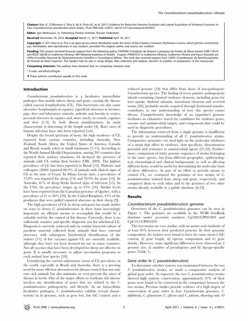
Citation: Ruiz JC, D’Afonseca V, Silva A, Ali A, Pinto AC, et al. (2011) Evidence for Reductive Genome Evolution and Lateral Acquisition of Virulence Functions inTwo Corynebacterium pseudotuberculosis Strains. PLoS ONE 6(4): e18551. doi:10.1371/journal.pone.0018551
Editor: Igor Mokrousov, St. Petersburg Pasteur Institute, Russian Federation
Received November 29, 2010; Accepted March 11, 2011; Published April 18, 2011
Copyright: � 2011 Ruiz et al. This is an open-access article distributed under the terms of the Creative Commons Attribution License, which permits unrestricteduse, distribution, and reproduction in any medium, provided the original author and source are credited.
Funding: This project received financial support from the following grants: FAPEMIG (Fundacao de Amparo a pesquisa do Estado de Minas Gerais) (CBB-1181/0and REDE-186/08 to Guilherme Oliveira), NIH (National Institutes of Health) - Fogarty (TW007012 to Guilherme Oliveira). Guilherme Oliveira and Vasco Azevedo areCNPq (Conselho Nacional de Desenvolvimento Cientıfico e Tecnologico) fellows. The work also received support from CAPES (Coordenacao de Aperfeicoamentode Pessoal de Nıvel Superior). The funders had no role in study design, data collection and analysis, decision to publish, or preparation of the manuscript.
Competing Interests: The authors have declared that no competing interests exist.
* E-mail: [email protected]
. These authors contributed equally to this work.
Introduction
Corynebacterium pseudotuberculosis is a facultative intracellular
pathogen that mainly infects sheep and goats, causing the disease
called caseous lymphadenitis (CL). This bacterium can also cause
ulcerative lymphangitis in equines; superficial abscesses in bovines,
pigs, deer and laboratory animals; arthritis and bursitis in ovines;
pectoral abscesses in equines and, more rarely, in camels, caprines
and deer [1-3]. In both disease manifestations, its main
characteristic is abscessing of the lymph nodes [4]. Rare cases of
human infection have also been reported [5,6].
Despite the broad spectrum of hosts, the high incidence of CL
reported from various countries, including Australia, New
Zealand, South Africa, the United States of America, Canada
and Brazil, mainly refers to small ruminants [7-11]. According to
the World Animal Health Organization, among 201 countries that
reported their sanitary situations, 64 declared the presence of
animals with CL within their borders (OIE, 2009). The highest
prevalence of CL has been reported in Brazil [12]. Pinheiro and
colleagues (2000) reported 66.9% of animals with clinical signs of
CL in the state of Ceara. In Minas Gerais state, a prevalence of
75.8% was reported for sheep [13] and 78.9% for goats [14]. In
Australia, 61% of sheep flocks showed signs of infection [15]. In
the USA, the prevalence ranges up to 43% [16]. Similar levels
have been reported from the Canadian province of Quebec, with a
prevalence of 21 to 36% [10]. In the United Kingdom, 45% of the
producers that were polled reported abscesses in their sheep [9].
The high prevalence of CL in sheep and goats has made studies
on ways to detect C. pseudotuberculosis in these hosts increasingly
important; an efficient means to accomplish this would be a
valuable tool for the control of this disease. Currently, there is no
sufficiently sensitive and specific diagnostic test for subclinical CL.
Diagnosis is currently achieved only by routine bacterial culture of
purulent material collected from animals that have external
abscesses, with subsequent biochemical identification of the
isolates [17]. A few vaccines against CL are currently available,
although they have not been licensed for use in many countries.
Not all vaccines that have been developed for sheep are effective in
goats. It is usually necessary to adjust vaccination programs to
each animal host species [18].
Considering the current unfortunate status of CL prevalence in
the world, especially in Brazil and Australia, there is a pressing
need for more efficient alternatives for disease control that not only
cure sick animals but also minimize or even prevent the onset of
disease in herds. One of the major efforts to eradicate this disease
involves the identification of genes that are related to the C.
pseudotuberculosis pathogenicity and lifestyle. As an intracellular
facultative pathogen, C. pseudotuberculosis exhibits several charac-
teristics in its genome, such as gene loss, low GC content and a
reduced genome [19] that differ from those of non-pathogenic
Corynebacterium species. The finding of seven putative pathogenicity
islands containing classical virulence elements, including genes for
iron uptake, fimbrial subunits, insertional elements and secreted
toxins [20], probably mostly acquired through horizontal transfer,
contributes to our understanding of how this species causes
disease. Comprehensive knowledge of an organism’s genome
facilitates an exhaustive search for candidates for virulence genes,
vaccine and antimicrobial targets, and components that could be
used in diagnostic procedures.
The information retrieved from a single genome is insufficient
to provide an understanding of all C. pseudotuberculosis strains.
Comparative genomics can shed light on the molecular attributes
of a strain that affect its virulence, host specificity, dissemination
potential and resistance to antimicrobial agents [21,22]. Further-
more, comparison of entire genome sequences of strains belonging
to the same species, but from different geographic, epidemiolog-
ical, chronological and clinical backgrounds, as well as affecting
different hosts, would be useful for determining the molecular basis
of these differences. As part of an effort to provide means to
control CL, we examined the genomes of two strains of C.
pseudotuberculosis isolated from sheep and goats, respectively, and
compared them to each other and to the genomes of two other
strains already available in a public database [6,23].
Results
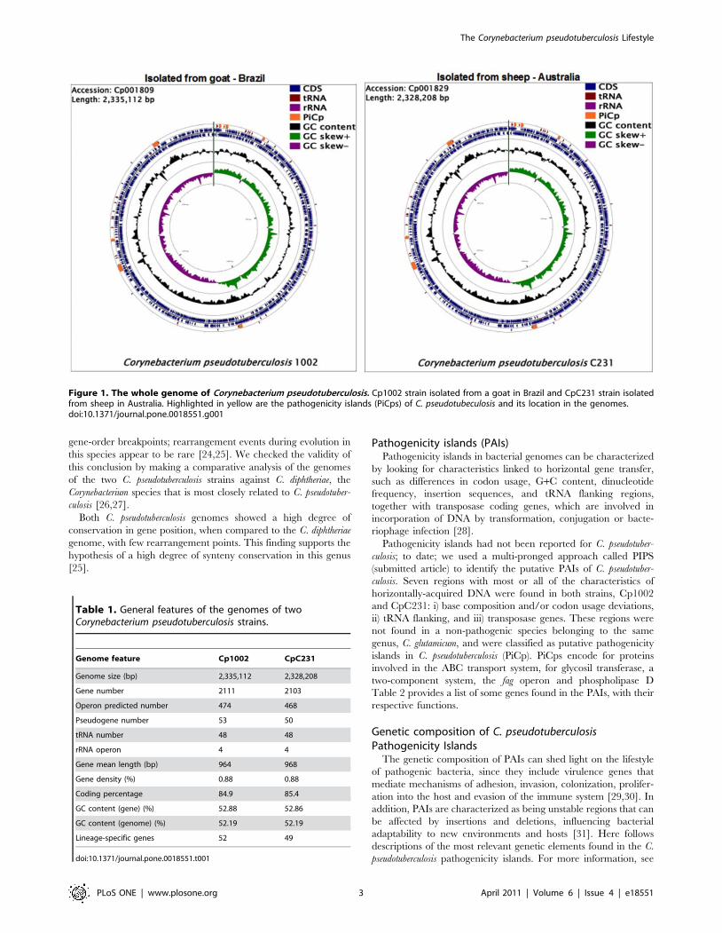
Corynebacterium pseudotuberculosis genomeOverviews of the C. pseudotuberculosis genomes can be seen in
Figure 1. The genomes are available in the NCBI GenBank
database under accession numbers Cp1002:CP001809 and
CpC231:CP001829.
The two strains are very similar, with an amino acid similarity of
at least 95% between their predicted proteins. In their genomic
composition, the isolates were found to have the same mean i) GC
content, ii) gene length, iii) operon composition and iv) gene
density. However, some significant differences were observed in: i)
genome size, ii) number of pseudogenes and iii) lineage-specific
genes (Table 1).
Gene order in C. pseudotuberculosisTo determine whether synteny was maintained between the two
C. pseudotuberculosis strains, we made a comparative analysis of
global gene order. As expected, the two C. pseudotuberculosis strains
showed high synteny conservation; approximately 97% of their
genes were found to be conserved in the comparison between the
two strains. Previous studies provide evidence of a high degree of
conservation of gene order in four Corynebacterium genomes, C.
diphtheriae, C. glutamicum, C. efficiens and C. jeikeium, showing only 10
The Corynebacterium pseudotuberculosis Lifestyle
PLoS ONE | www.plosone.org 2 April 2011 | Volume 6 | Issue 4 | e18551
gene-order breakpoints; rearrangement events during evolution in
this species appear to be rare [24,25]. We checked the validity of
this conclusion by making a comparative analysis of the genomes
of the two C. pseudotuberculosis strains against C. diphtheriae, the
Corynebacterium species that is most closely related to C. pseudotuber-
culosis [26,27].
Both C. pseudotuberculosis genomes showed a high degree of
conservation in gene position, when compared to the C. diphtheriae
genome, with few rearrangement points. This finding supports the
hypothesis of a high degree of synteny conservation in this genus
[25].
Pathogenicity islands (PAIs)Pathogenicity islands in bacterial genomes can be characterized
by looking for characteristics linked to horizontal gene transfer,
such as differences in codon usage, G+C content, dinucleotide
frequency, insertion sequences, and tRNA flanking regions,
together with transposase coding genes, which are involved in
incorporation of DNA by transformation, conjugation or bacte-
riophage infection [28].
Pathogenicity islands had not been reported for C. pseudotuber-
culosis; to date; we used a multi-pronged approach called PIPS
(submitted article) to identify the putative PAIs of C. pseudotuber-
culosis. Seven regions with most or all of the characteristics of
horizontally-acquired DNA were found in both strains, Cp1002
and CpC231: i) base composition and/or codon usage deviations,
ii) tRNA flanking, and iii) transposase genes. These regions were
not found in a non-pathogenic species belonging to the same
genus, C. glutamicum, and were classified as putative pathogenicity
islands in C. pseudotuberculosis (PiCp). PiCps encode for proteins
involved in the ABC transport system, for glycosil transferase, a
two-component system, the fag operon and phospholipase D
Table 2 provides a list of some genes found in the PAIs, with their
respective functions.
Genetic composition of C. pseudotuberculosisPathogenicity Islands
The genetic composition of PAIs can shed light on the lifestyle
of pathogenic bacteria, since they include virulence genes that
mediate mechanisms of adhesion, invasion, colonization, prolifer-
ation into the host and evasion of the immune system [29,30]. In
addition, PAIs are characterized as being unstable regions that can
be affected by insertions and deletions, influencing bacterial
adaptability to new environments and hosts [31]. Here follows
descriptions of the most relevant genetic elements found in the C.
pseudotuberculosis pathogenicity islands. For more information, see
Figure 1. The whole genome of Corynebacterium pseudotuberculosis. Cp1002 strain isolated from a goat in Brazil and CpC231 strain isolatedfrom sheep in Australia. Highlighted in yellow are the pathogenicity islands (PiCps) of C. pseudotubeculosis and its location in the genomes.doi:10.1371/journal.pone.0018551.g001
Table 1. General features of the genomes of twoCorynebacterium pseudotuberculosis strains.
Genome feature Cp1002 CpC231
Genome size (bp) 2,335,112 2,328,208
Gene number 2111 2103
Operon predicted number 474 468
Pseudogene number 53 50
tRNA number 48 48
rRNA operon 4 4
Gene mean length (bp) 964 968
Gene density (%) 0.88 0.88
Coding percentage 84.9 85.4
GC content (gene) (%) 52.88 52.86
GC content (genome) (%) 52.19 52.19
Lineage-specific genes 52 49
doi:10.1371/journal.pone.0018551.t001
The Corynebacterium pseudotuberculosis Lifestyle
PLoS ONE | www.plosone.org 3 April 2011 | Volume 6 | Issue 4 | e18551
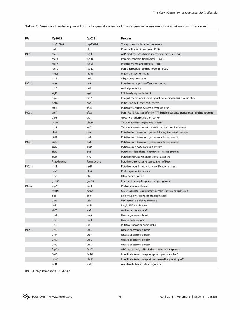
Table 2. Genes and proteins present in pathogenicity islands of the Corynebacterium pseudotuberculosis strain genomes.
PAI Cp1002 CpC231 Protein
tnp7109-9 tnp7109-9 Transposase for insertion sequence
pld pld Phospholipase D precursor (PLD)
PiCp 1 fag C fag C ATP binding cytoplasmic membrane protein - FagC
fag B fag B Iron-enterobactin transporter - FagB
fag A fag A Integral membrane protein - FagA
fag D fag D Iron siderophore binding protein - FagD
mgtE mgtE Mg2+ transporter mgtE
malL malL Oligo-1,6-glucosidase
PiCp 2 tetA tetA Putative tetracycline-efflux transporter
cskE cskE Anti-sigma factor
sigK sigK ECF family sigma factor K
dipZ dipZ Integral membrane C-type cytochrome biogenesis protein DipZ
potG potG Putrescine ABC transport system
afuB afuB Putative transport system permease (iron)
PiCp 3 afuA afuA Iron (Fe3+) ABC superfamily ATP binding cassette transporter, binding protein
glpT glpT Glycerol-3-phosphate transporter
phoB phoB Two-component regulatory protein
lcoS lcoS Two-component sensor protein, sensor histidine kinase
ciuA ciuA Putative iron transport system binding (secreted) protein
ciuB ciuB Putative iron transport system membrane protein
PiCp 4 ciuC ciuC Putative iron transport system membrane protein
ciuD ciuD Putative iron ABC transport system
ciuE ciuE Putative siderophore biosynthesis related protein
s70 s70 Putative RNA polymerase sigma factor 70
Pseudogene Pseudogene Putative chromosome segregation ATPase
PiCp 5 hsdR hsdR Putative type III restriction-modification system
pfoS pfoS PfoR superfamily protein
htaC htaC HtaA family protein
guaB3 guaB3 Inosine 5-monophosphate dehydrogenase
PiCp6 pipA1 pipB Proline iminopeptidase
mfsD1 mfsD1 Major facilitator superfamily domain-containing protein 1
dcd dcd Deoxycytidine triphosphate deaminase
udg udg UDP-glucose 6-dehydrogenase
lysS1 lysS1 Lysyl-tRNA synthetase
alaT alaT Aminotransferase AlaT
ureA ureA Urease gamma subunit
ureB ureB Urease beta subunit
ureC ureC Putative urease subunit alpha
PiCp 7 ureE ureE Urease accessory protein
ureF ureF Urease accessory protein
ureG ureG Urease accessory protein
ureD ureD Urease accessory protein
fepC2 fepC2 ABC superfamily ATP binding cassette transporter
fecD fecD1 Iron(III) dicitrate transport system permease fecD
phuC phuC Iron(III) dicitrate transport permease-like protein yusV
arsR arsR1 ArsR-family transcription regulator
doi:10.1371/journal.pone.0018551.t002
The Corynebacterium pseudotuberculosis Lifestyle
PLoS ONE | www.plosone.org 4 April 2011 | Volume 6 | Issue 4 | e18551

the list of these orthologous genes in other Corynebacterium species in
the Table S1 (online supporting information).
PiCp 1. C. pseudotuberculosis PiCp 1 harbors key genes involved
in virulence and pathogenicity; these include PLD, the major
virulence factor of this organism, which plays a role in spreading
through the host; the fag operon, responsible for extracellular iron
acquisition and, consequently, for survival in hostile environments;
and a transposase gene, probably responsible for insertion of the
island into the C. pseudotuberculosis genome. The finding that C.
ulcerans can produce phospholipase D protein [32] indicates
acquisition of PiCp1 by both C. pseudotuberculosis and C. ulcerans.
PiCp 2. Gene mgtE of island 2 has Mg2+ influx activity [33].
In prokaryotes, Mg2+ has been identified as an important
regulatory signal that is essential for virulence, since it is
involved in thermal adaptation, protecting bacteria from heat
shock caused by fever in warm-blooded mammals [34].
Translation of the mgtE gene is regulated by changes in cytosolic
Mg2+ concentration; loss of MgtE reduces biofilm formation and
motility in the pathogenic bacteria Aeromonas hydrophila [33].
The protein MalL (malL), a maltose-inducible a-glucosidase,
hydrolyzes various disaccharides, such as maltose and isomaltose,
which can serve as carbon and energy sources [35,36].
The tetA gene codes for a tetracycline-efflux transporter protein
that extrudes antibiotics from the cell and confers resistance to
biofilm cells. The tetA gene is often carried by transmissible
elements, such as plasmids, transposons, and integrons [37], thus
explaining its presence in a PAI.
The sigK gene is an extracytoplasmic function sigma factor (sigma
ECF) regulated by cskE, an anti-sigma factor. Another sigma ECF,
sigK, mediates targeted alterations in bacterial transcription via
transduction of extracellular signals. In M. tuberculosis, sigK regulates
several genes (Rv2871, mpt83, dipZ, mpt70, Rv2876, and mpt53).
Also, sigK mutations produce reduced quantities of the antigens
MPT70 and MPT83 in vitro, and only induce strong expression
during infection of macrophages [38–40].
PiCp2 also harbors a dipZ gene, which is regulated by sigK and
seems to play a role in macrophage infection by M. tuberculosis,
although its function is not clearly elucidated. DipZ is found as two
separate proteins in most bacteria: CcdA and TlpA-like. Also, a
full-length dipZ gene, found in the phylum Actinobacteria, is present
exclusively in pathogenic bacteria (C. diphtheriae, C. jeikeium, M.
avium, M. kansasii, M. marinum, M. ulcerans and M. tuberculosis) [40].
PiCp 3. potG gene, of the potFGHI operon, is a membrane-
associated/ATP-binding protein that provides energy for
putrescine (polyamine) uptake from the periplasmic space [41].
Although the potFGHI operon is a putrescine-specific transport
system, potG is downregulated by another polyamine (spermine),
which is produced only by eukaryotes. Carlson et al. (2009)
demonstrated that transcription of the potG gene in Francisella
tularensis decreases with high levels of spermine, while transcription
of IS elements ISFtu1 and ISFtu2 increases in response to high
levels of spermine in macrophages responding to bacterial
infection. Also, many of the upregulated genes of F. tularensis
(pseudogenes and transposase genes) are located near the IS
elements in the chromosome [42].
The gene glpT belongs to the organophosphate:phosphate
antiporter family of the major facilitator superfamily (MFS); it
mediates transport of glycerol 3-phosphate (G3P) across the
membrane in bacteria [43].
The PhoPR system regulates expression of various genes
involved in metabolic, virulence and resistance processes in several
intracellular bacterial pathogens [44]. Based on the information
obtained from the complete genome sequence of C. pseudotubercu-
losis, we found that the PhoPR system is constituted of the phoP
(714 bp) and phoR (1506 bp) genes, separated by a small 39-bp
sequence, suggesting that these two genes are transcribed by a
bicistronic operon. The size and organization of this system in C.
pseudotuberculosis is similar to those of other Gram-positive bacteria
[45]. Live bacteria attenuated via phoP inactivation are also
promising vaccine candidates against tuberculosis. Several studies
have reported the efficacy of attenuated mutant strains of M.
tuberculosis as vaccines [46,47]. Phylogenetic relationships within
the class Actinobacteria strongly suggest correlation of the C.
pseudotuberculosis PhoPR system with virulence mechanisms. The
phoP gene is an important subject for regulation studies; and is also
a probable vaccine candidate against CL.
PiCp4. The operon ciuABCDE (corynebacterium iron uptake) was
described in C. diphtheriae as an iron transport and siderophore
biosynthesis system. Proteins involved in iron acquisition are
recognized as virulence factors, since they help pathogens to
obtain iron from a host by using siderophores to strip iron from
carrier proteins, such as transferrin, lactoferrin, and hemoglobin-
haptoglobin [48,48].
PiCp5. Island 5 harbors a gene (pfoS) related to the pfoR
superfamily. The pfoR gene was previously characterized as
responsible for positive regulation of production of perfringolysin
A (pfoA) and other toxins in Clostridium perfringens [50]. The
virulence factors regulated by pfoR have not been totally
elucidated. However, it is well known that deactivation of this
gene inhibits hemolysis through negative regulation of several C.
perfringens toxins. Clostridium perfringens harbors a phospholipase C
gene (plc) that serves a function similar to that of phospholipase D
[51]. Additionally, PiCp 5 contains a putative sigma 70 factor that
is responsible for transporting the transcription machinery to
specific promoters. Interestingly, the putative sigma 70 factor
presents a nonsense mutation in C. pseudotuberculosis strain C231,
which could be responsible for differential gene expression.
PiCp6. The pipA1 gene, which codes for a proline imin-
opeptidase, may have a role in pathogenesis, since it catalyses the
removal of N-terminal proline residues from peptides; it also has a
role in energy production [52]. In addition, a PIP-type protein is
required for virulence of Xanthomonas campestris pv. campestris [53].
PiCp7. Island 7 harbors a urease operon that is also present in
C. glutamicum; it is flanked, on both sides, by regions that are absent
in the non-pathogenic C. glutamicum. This mosaicism is a common
feature of pathogenicity islands [54]. The ure operon presents a
codon usage deviation in C. glutamicum, as in C. pseudotuberculosis,
indicating that this region is a putative genomic island in C.
glutamicum.
The ure operon is responsible for nitrogen acquisition through
hydrolysis of urea to carbamate and ammonia. Production of
ammonia by uropathogenic and enteropathogenic bacteria causes
cellular damage and compromises the action of the host’s immune
system [55]. Considering this fact, due to the intramacrophagic
location of C. pseudotuberculosis and the finding of this operon in a
non-pathogenic bacterial species, additional studies will be needed
to elucidate how C. pseudotuberculosis obtains urea from the host and
how this operon affects pathogenicity.
PiCp 7 also harbors a lysyl-tRNA synthetase (lysS), responsible
for lysine incorporation into its respective transfer tRNA. The
importance of lysS would normally make its location on a PAI
inviable, since it is essential for cell metabolism. However, it is the
only tRNA synthetase gene that is duplicated in the genome.
Protein classification of C. pseudotuberculosis in thebiological process
Using the controlled vocabulary of functional terms proposed by
the Gene Ontology (GO) Consortium for gene products
The Corynebacterium pseudotuberculosis Lifestyle
PLoS ONE | www.plosone.org 5 April 2011 | Volume 6 | Issue 4 | e18551

classification [56], the predicted proteomes of the two genomes
were analyzed according to the three organizing principles of gene
ontology: cellular component, biological process and molecular
function. The most abundantly represented categories are linked
to metabolic processes in the two strains (cellular metabolic,
biosynthetic, primary and macromolecule processes).
The gene products composition characterized using GO
terminology suggests that C. pseudotuberculosis is a facultative
intracellular pathogen. It is commonly found that pathogens
specialized for an intracellular lifestyle have a high proportion of
proteins linked to the above-mentioned processes. Moreover, the
low proportion of proteins linked to the metabolism of secondary
metabolites is an indication that C. pseudotuberculosis does not
possess the metabolic machinery to deal with secondary
metabolites, because they are supplied by the host.
Sub-cellular localization of C. pseudotuberculosis proteinsPrediction of the sub-cellular localization of C. pseudotuberculosis
proteins was made by in silico analysis, using the SurfG+ tool [57].
Surfg+ is a pipeline for protein sub-cellular prediction, incorpo-
rating commonly used software for motif searches, including
SignalP, LipoP and TMHMM, along with novel HMMSEARCH
profiles to predict protein retention signals. Surfg+ starts by
searching for retention signals, lipoproteins, SEC pathway export
motifs and transmembrane motifs, roughly in this order. If none of
these motifs are found in a protein sequence, then it is
characterized as being cytoplasmic. A novel possibility introduced
by Surfg+ is the ability to distinguish between integral membrane
proteins versus PSE (potentially surface-exposed proteins). This is
done by a parameter that determines the expected cell wall
thickness, expressed in amino acids. Using published information
or electron microscopy, it is possible to estimate cell wall thickness
value for procaryotic organisms. C. pseudotuberculosis proteins were
classified into four different sub-cellular locations: cytoplasmic,
membrane, PSE (potentially surface exposed), or secreted. The C.
pseudotuberculosis genomes were compared to those of other species
of the genus, including C. diphtheriae, C. efficiens, C. glutamicum, C.
jeikeium and C. urealyticum, also predicted by Surfg+, based on
published cell wall thicknesses. Table 3 shows the number of
predicted proteins in each sub-cellular location.
Comparison of the frequencies of subcellular occurrence of the
C. pseudotuberculosis proteins and other Corynebacterium proteomes
was made with Chi-square tests. The ratio between the four
groups (cytoplasmic, membrane anchored, potentially exposed
and secreted proteins) was found to be nearly constant among the
Corynebacterium species. The proportions of the four protein
categories cited above were similar to published data [58,59].
Song and colleagues (2009) showed that approximately 30% of
proteins secreted in gram-positive bacteria are exported through
the Sec pathway. Few proteins (n = 27) were predicted to be
secreted by the Tat pathway in Cp1002. About 2% of the proteins
predicted to be secreted presented tertiary structures. In terms of
proportions of secreted proteins, Cp1002 and CpC231 are at the
higher end of the spectrum. They present 4.61 and 5.21%,
respectively, predicted secreted proteins (Table 3).
Differences in metabolic pathways in the two strains of C.pseudotuberculosis
Automated reconstruction of the C. pseudotuberculosis Cp1002
metabolic pathways identified 156 pathways and 744 enzymatic
reactions. As expected, quite similar results were encountered for
strain CpC231: 154 pathways and 754 reactions (Table 4).
Proteins of predicted functions that did not map to pathways, such
as transport reactions, enzymes, transporters, and compounds,
were also identified. The metabolic pathway database can be
accessed online at http://corynecyc.cebio.org. This database
enabled us to visualize and compare the metabolism of these
two C. pseudotuberculosis strains (Figure 2).
We made a comparative analysis of transport reactions,
pathways, compounds and proteins for C. pseudotuberculosis strains
Cp1002 and CpC231 (Table 5). Despite the high similarity of the
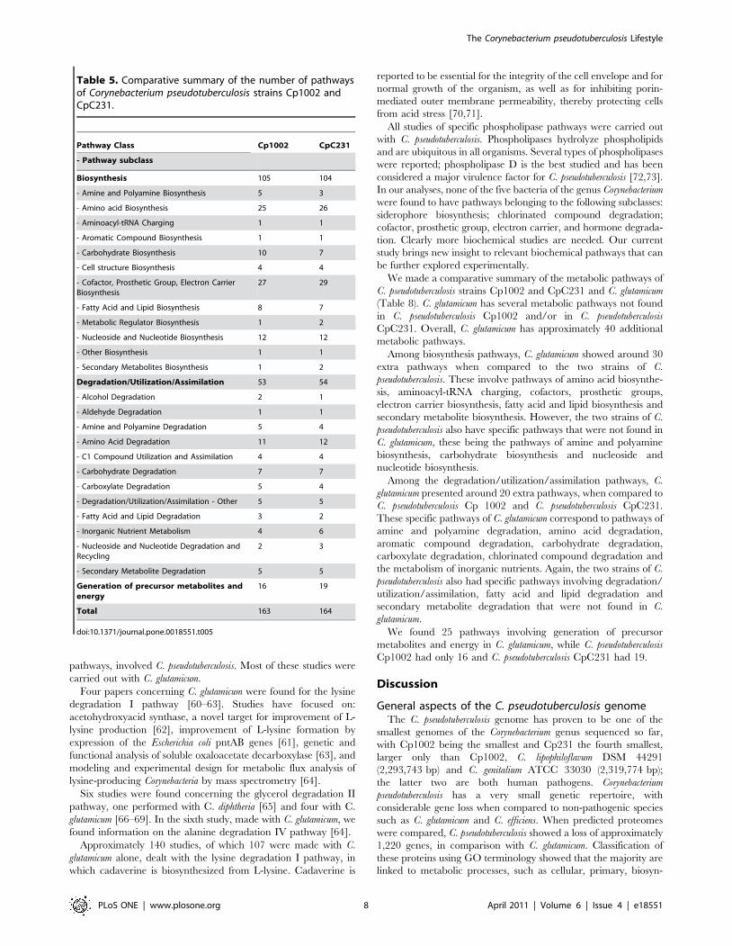
metabolic pathways, some differences were observed.
The metabolic pathways in each of the two bacterial strains
(Cp1002 and CpC231) were classified into several pathway classes;
each pathway class was further broken down to show the
distribution of pathways among the next-level subclasses. Analysis
of the metabolism database of C. pseudotuberculosis strains Cp1002
and CpC231 revealed specific pathway differences between the
two strains. Overall, CpC231 had 13 specific metabolic pathways
Table 3. Subcellular prediction of the protein locations derived from complete genomes of Corynebacterium species.
Category/Species Ce CgB CgK CgR Cj Cd Cu Cp1002 CpC231 Total
Cytoplasm 2,158 2,11 2,082 2,158 1,49 1,594 1,432 1,399 1,389 15,812
Cytoplasm 504 557 541 561 333 375 332 364 356 3,923
PSE 230 254 249 252 197 204 179 201 201 1,967
Secreted 102 136 121 109 100 99 79 95 107 948
Total 2,994 3,057 2,993 3,08 2,12 2,272 2,02 2,059 2,053 22,648
Ce: C. efficiens; CgB: C. glutamicum B; CgK: C. glutamicum K; CgR: C. glutamicum R; Cj: C. jeikeium; Cd: C. diphtheriae; Cu: C. urealyticum; Cp1002: C. pseudotuberculosis1002; CpC231: C. pseudotuberculosis C231. PSE: potential surface exposure.doi:10.1371/journal.pone.0018551.t003
Table 4. Comparative summary of the Corynebacteriumpseudotuberculosis strain gene data types.
Data Type Cp1002 CpC231
Gene products 2,059 2,053
Pathways 156 154
Enzymatic Reactions 744 754
Transport Reactions 8 4
Polypeptides 2,065 2,059
Enzymes 516 506
Transporters 10 10
Compounds 639 651
doi:10.1371/journal.pone.0018551.t004
The Corynebacterium pseudotuberculosis Lifestyle
PLoS ONE | www.plosone.org 6 April 2011 | Volume 6 | Issue 4 | e18551

not found in strain Cp1002, and the latter had 11 metabolic
pathways not found in strain CpC231 (Table 6).
Two amine and polyamine biosynthesis pathways, choline
degradation I and glycine betaine biosynthesis I (Gram-negative
bacteria), were found in strain Cp1002 but not in strain CpC231.
Strain CpC231 was found to have an extra amino acid
biosynthesis pathway, the citrulline-nitric oxide cycle. Strain
Cp1002 was found to have three additional carbohydrate
biosynthesis pathways: gluconeogenesis, trehalose biosynthesis II
and trehalose biosynthesis III. Strain CpC231 showed three
cofactor biosynthesis, prosthetic group and electron carrier
pathways, corresponding to adenosylcobalamin biosynthesis from
cobyrinate a,c-diamide I, heme biosynthesis from uroporphyrin-
ogen II and siroheme biosynthesis. Strain Cp1002 showed only
one unique cofactor biosynthesis pathway, heme biosynthesis from
uroporphyrinogen I. Two extra pathways of fatty acid and lipid
biosynthesis were found in strain Cp1002, cardiolipin biosynthesis
I and fatty acid biosynthesis initiation I. Strain CpC231 showed
only the biotin-carboxyl carrier protein. Among metabolic
regulator biosynthesis genes, strain CpC231 showed the citrul-
line-nitric oxide cycle. Strain CpC231 also showed an extra
pathway, the canavanine biosynthesis pathway, part of secondary
metabolite biosynthesis.
Among degradation/utilization/assimilation pathways, strain
Cp1002 showed an extra pathway: glycerol degradation II, for
alcohol degradation, as well as choline degradation I for amine
and polyamine degradation. Strain CpC231 was found to have
two additional pathways, 2-ketoglutarate dehydrogenase complex
and citrulline-nitric oxide cycle, for amino acid pathways; strain
Cp1002 showed only one extra pathway, valine degradation I.
Among carboxylate degradation pathways, involving fatty acid
and lipid degradation, strain Cp1002 showed two extra pathways:
one corresponding to acetate formation from acetyl-CoA I, and
the second linked to triacylglycerol degradation. Two inorganic
nutrient metabolism pathways were found in strain CpC231 but
not in strain Cp1002: nitrate reduction III (dissimilatory) and
nitrate reduction IV (dissimilatory), and a nucleoside and
nucleotide degradation and purine deoxyribonucleoside recycling
degradation pathway.
Finally, when we analyzed the generation of precursor
metabolites and energy, strain CpC231 showed three extra
pathways: 2-ketoglutarate dehydrogenase complex, nitrate reduc-
tion III (dissimilatory) and nitrate reduction IV (dissimilatory). The
differences are presented in Table 6.
Metabolic pathways in C. pseudotuberculosis comparedto other Corynebacterium species
The web interface enabled us to visually compare the metabolic
pathways of strains Cp1002 and CpC231 reactions (Figure 2) with
those of four other bacteria of the genus Corynebacterium: C.
diphtheriae, C. efficiens, C. glutamicum, and C. jeikeium. Using these
diagrams we were able to easily spot reactions present in C.
pseudotuberculosis and absent in other Corynebacterium species.
A comparative analysis of reactions, pathways, compounds and
proteins was also done for C. pseudotuberculosis and other closely-
related bacteria in the same genus. The list of C. pseudotuberculosis
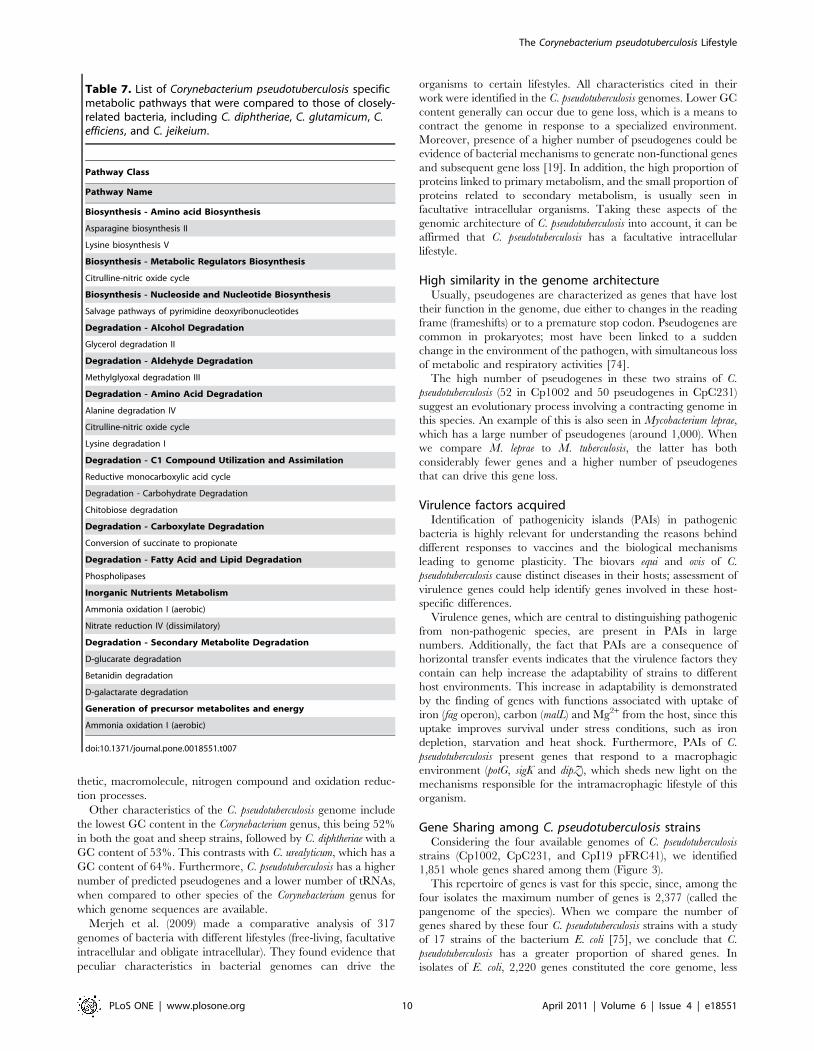
specific pathways is shown in Table 7.
We found that C. pseudotuberculosis has several pathways that are
not found in other species of the genus Corynebacterium. However,
little information is available about these pathways in Corynebac-
terium spp. We found no published information concerning the
following pathways: asparagine biosynthesis II, citrulline-nitric
oxide cycle (amino acid biosynthesis and degradation), pyrimidine
deoxyribonucleotide salvage pathways, methylglyoxal degradation
III, reductive monocarboxylic acid cycle, chitobiose degradation,
conversion of succinate to propionate, ammonia oxidation I
(aerobic), nitrate reduction IV (dissimilatory), D-glucarate degra-
dation, betanidin degradation, D-galactarate degradation, and
ammonia oxidation I (aerobic).
Some studies reported five pathways: lysine biosynthesis V,
glycerol degradation II, alanine degradation IV, lysine degrada-
tion I and phospholipases. However, none of the studies, except
for those concerning lysine degradation I and phospholipase
Figure 2. Corynebacterium glutamicum metabolic pathways overview. C. glutamicum reactions are presented in blue and the reactions sharedwith C. pseudotuberculosis C231 and 1002 in red and green, respectively. By clicking on any compound or reaction, a window pops up showing detailsof each pathway. The fatty acid biosynthesis initiation pathway is the chosen example since computational evidence indicates it is not present only instrain C231.doi:10.1371/journal.pone.0018551.g002
The Corynebacterium pseudotuberculosis Lifestyle
PLoS ONE | www.plosone.org 7 April 2011 | Volume 6 | Issue 4 | e18551
pathways, involved C. pseudotuberculosis. Most of these studies were
carried out with C. glutamicum.
Four papers concerning C. glutamicum were found for the lysine
degradation I pathway [60–63]. Studies have focused on:
acetohydroxyacid synthase, a novel target for improvement of L-
lysine production [62], improvement of L-lysine formation by
expression of the Escherichia coli pntAB genes [61], genetic and
functional analysis of soluble oxaloacetate decarboxylase [63], and
modeling and experimental design for metabolic flux analysis of
lysine-producing Corynebacteria by mass spectrometry [64].
Six studies were found concerning the glycerol degradation II
pathway, one performed with C. diphtheria [65] and four with C.
glutamicum [66–69]. In the sixth study, made with C. glutamicum, we
found information on the alanine degradation IV pathway [64].
Approximately 140 studies, of which 107 were made with C.
glutamicum alone, dealt with the lysine degradation I pathway, in
which cadaverine is biosynthesized from L-lysine. Cadaverine is
reported to be essential for the integrity of the cell envelope and for
normal growth of the organism, as well as for inhibiting porin-
mediated outer membrane permeability, thereby protecting cells
from acid stress [70,71].
All studies of specific phospholipase pathways were carried out
with C. pseudotuberculosis. Phospholipases hydrolyze phospholipids
and are ubiquitous in all organisms. Several types of phospholipases
were reported; phospholipase D is the best studied and has been
considered a major virulence factor for C. pseudotuberculosis [72,73].
In our analyses, none of the five bacteria of the genus Corynebacterium
were found to have pathways belonging to the following subclasses:
siderophore biosynthesis; chlorinated compound degradation;
cofactor, prosthetic group, electron carrier, and hormone degrada-
tion. Clearly more biochemical studies are needed. Our current
study brings new insight to relevant biochemical pathways that can
be further explored experimentally.
We made a comparative summary of the metabolic pathways of
C. pseudotuberculosis strains Cp1002 and CpC231 and C. glutamicum
(Table 8). C. glutamicum has several metabolic pathways not found
in C. pseudotuberculosis Cp1002 and/or in C. pseudotuberculosis
CpC231. Overall, C. glutamicum has approximately 40 additional
metabolic pathways.
Among biosynthesis pathways, C. glutamicum showed around 30
extra pathways when compared to the two strains of C.
pseudotuberculosis. These involve pathways of amino acid biosynthe-
sis, aminoacyl-tRNA charging, cofactors, prosthetic groups,
electron carrier biosynthesis, fatty acid and lipid biosynthesis and
secondary metabolite biosynthesis. However, the two strains of C.
pseudotuberculosis also have specific pathways that were not found in
C. glutamicum, these being the pathways of amine and polyamine
biosynthesis, carbohydrate biosynthesis and nucleoside and
nucleotide biosynthesis.
Among the degradation/utilization/assimilation pathways, C.
glutamicum presented around 20 extra pathways, when compared to
C. pseudotuberculosis Cp 1002 and C. pseudotuberculosis CpC231.
These specific pathways of C. glutamicum correspond to pathways of
amine and polyamine degradation, amino acid degradation,
aromatic compound degradation, carbohydrate degradation,
carboxylate degradation, chlorinated compound degradation and
the metabolism of inorganic nutrients. Again, the two strains of C.
pseudotuberculosis also had specific pathways involving degradation/
utilization/assimilation, fatty acid and lipid degradation and
secondary metabolite degradation that were not found in C.
glutamicum.
We found 25 pathways involving generation of precursor
metabolites and energy in C. glutamicum, while C. pseudotuberculosis
Cp1002 had only 16 and C. pseudotuberculosis CpC231 had 19.
Discussion
General aspects of the C. pseudotuberculosis genomeThe C. pseudotuberculosis genome has proven to be one of the
smallest genomes of the Corynebacterium genus sequenced so far,
with Cp1002 being the smallest and Cp231 the fourth smallest,
larger only than Cp1002, C. lipophiloflavum DSM 44291
(2,293,743 bp) and C. genitalium ATCC 33030 (2,319,774 bp);
the latter two are both human pathogens. Corynebacterium
pseudotuberculosis has a very small genetic repertoire, with
considerable gene loss when compared to non-pathogenic species
such as C. glutamicum and C. efficiens. When predicted proteomes
were compared, C. pseudotuberculosis showed a loss of approximately
1,220 genes, in comparison with C. glutamicum. Classification of
these proteins using GO terminology showed that the majority are
linked to metabolic processes, such as cellular, primary, biosyn-
Table 5. Comparative summary of the number of pathwaysof Corynebacterium pseudotuberculosis strains Cp1002 andCpC231.
Pathway Class Cp1002 CpC231
- Pathway subclass
Biosynthesis 105 104
- Amine and Polyamine Biosynthesis 5 3
- Amino acid Biosynthesis 25 26
- Aminoacyl-tRNA Charging 1 1
- Aromatic Compound Biosynthesis 1 1
- Carbohydrate Biosynthesis 10 7
- Cell structure Biosynthesis 4 4
- Cofactor, Prosthetic Group, Electron CarrierBiosynthesis
27 29
- Fatty Acid and Lipid Biosynthesis 8 7
- Metabolic Regulator Biosynthesis 1 2
- Nucleoside and Nucleotide Biosynthesis 12 12
- Other Biosynthesis 1 1
- Secondary Metabolites Biosynthesis 1 2
Degradation/Utilization/Assimilation 53 54
- Alcohol Degradation 2 1
- Aldehyde Degradation 1 1
- Amine and Polyamine Degradation 5 4
- Amino Acid Degradation 11 12
- C1 Compound Utilization and Assimilation 4 4
- Carbohydrate Degradation 7 7
- Carboxylate Degradation 5 4
- Degradation/Utilization/Assimilation - Other 5 5
- Fatty Acid and Lipid Degradation 3 2
- Inorganic Nutrient Metabolism 4 6
- Nucleoside and Nucleotide Degradation andRecycling
2 3
- Secondary Metabolite Degradation 5 5
Generation of precursor metabolites andenergy
16 19
Total 163 164
doi:10.1371/journal.pone.0018551.t005
The Corynebacterium pseudotuberculosis Lifestyle
PLoS ONE | www.plosone.org 8 April 2011 | Volume 6 | Issue 4 | e18551
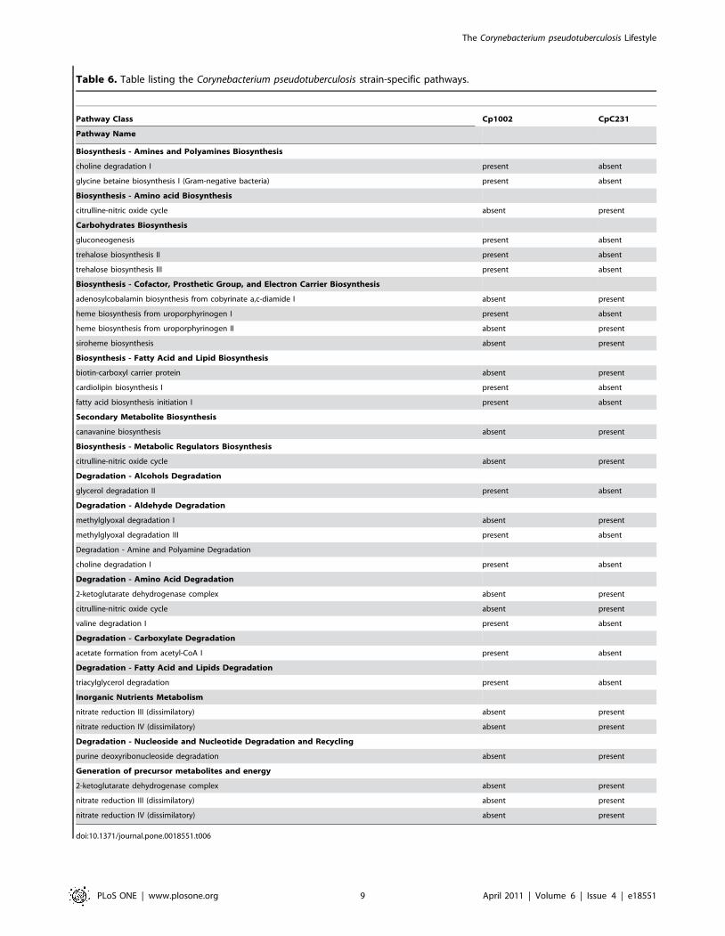
Table 6. Table listing the Corynebacterium pseudotuberculosis strain-specific pathways.
Pathway Class Cp1002 CpC231
Pathway Name
Biosynthesis - Amines and Polyamines Biosynthesis
choline degradation I present absent
glycine betaine biosynthesis I (Gram-negative bacteria) present absent
Biosynthesis - Amino acid Biosynthesis
citrulline-nitric oxide cycle absent present
Carbohydrates Biosynthesis
gluconeogenesis present absent
trehalose biosynthesis II present absent
trehalose biosynthesis III present absent
Biosynthesis - Cofactor, Prosthetic Group, and Electron Carrier Biosynthesis
adenosylcobalamin biosynthesis from cobyrinate a,c-diamide I absent present
heme biosynthesis from uroporphyrinogen I present absent
heme biosynthesis from uroporphyrinogen II absent present
siroheme biosynthesis absent present
Biosynthesis - Fatty Acid and Lipid Biosynthesis
biotin-carboxyl carrier protein absent present
cardiolipin biosynthesis I present absent
fatty acid biosynthesis initiation I present absent
Secondary Metabolite Biosynthesis
canavanine biosynthesis absent present
Biosynthesis - Metabolic Regulators Biosynthesis
citrulline-nitric oxide cycle absent present
Degradation - Alcohols Degradation
glycerol degradation II present absent
Degradation - Aldehyde Degradation
methylglyoxal degradation I absent present
methylglyoxal degradation III present absent
Degradation - Amine and Polyamine Degradation
choline degradation I present absent
Degradation - Amino Acid Degradation
2-ketoglutarate dehydrogenase complex absent present
citrulline-nitric oxide cycle absent present
valine degradation I present absent
Degradation - Carboxylate Degradation
acetate formation from acetyl-CoA I present absent
Degradation - Fatty Acid and Lipids Degradation
triacylglycerol degradation present absent
Inorganic Nutrients Metabolism
nitrate reduction III (dissimilatory) absent present
nitrate reduction IV (dissimilatory) absent present
Degradation - Nucleoside and Nucleotide Degradation and Recycling
purine deoxyribonucleoside degradation absent present
Generation of precursor metabolites and energy
2-ketoglutarate dehydrogenase complex absent present
nitrate reduction III (dissimilatory) absent present
nitrate reduction IV (dissimilatory) absent present
doi:10.1371/journal.pone.0018551.t006
The Corynebacterium pseudotuberculosis Lifestyle
PLoS ONE | www.plosone.org 9 April 2011 | Volume 6 | Issue 4 | e18551
thetic, macromolecule, nitrogen compound and oxidation reduc-
tion processes.
Other characteristics of the C. pseudotuberculosis genome include
the lowest GC content in the Corynebacterium genus, this being 52%
in both the goat and sheep strains, followed by C. diphtheriae with a
GC content of 53%. This contrasts with C. urealyticum, which has a
GC content of 64%. Furthermore, C. pseudotuberculosis has a higher
number of predicted pseudogenes and a lower number of tRNAs,
when compared to other species of the Corynebacterium genus for
which genome sequences are available.
Merjeh et al. (2009) made a comparative analysis of 317
genomes of bacteria with different lifestyles (free-living, facultative
intracellular and obligate intracellular). They found evidence that
peculiar characteristics in bacterial genomes can drive the
organisms to certain lifestyles. All characteristics cited in their
work were identified in the C. pseudotuberculosis genomes. Lower GC
content generally can occur due to gene loss, which is a means to
contract the genome in response to a specialized environment.
Moreover, presence of a higher number of pseudogenes could be
evidence of bacterial mechanisms to generate non-functional genes
and subsequent gene loss [19]. In addition, the high proportion of
proteins linked to primary metabolism, and the small proportion of
proteins related to secondary metabolism, is usually seen in
facultative intracellular organisms. Taking these aspects of the
genomic architecture of C. pseudotuberculosis into account, it can be
affirmed that C. pseudotuberculosis has a facultative intracellular
lifestyle.
High similarity in the genome architectureUsually, pseudogenes are characterized as genes that have lost
their function in the genome, due either to changes in the reading
frame (frameshifts) or to a premature stop codon. Pseudogenes are
common in prokaryotes; most have been linked to a sudden
change in the environment of the pathogen, with simultaneous loss
of metabolic and respiratory activities [74].
The high number of pseudogenes in these two strains of C.
pseudotuberculosis (52 in Cp1002 and 50 pseudogenes in CpC231)
suggest an evolutionary process involving a contracting genome in
this species. An example of this is also seen in Mycobacterium leprae,
which has a large number of pseudogenes (around 1,000). When
we compare M. leprae to M. tuberculosis, the latter has both
considerably fewer genes and a higher number of pseudogenes
that can drive this gene loss.
Virulence factors acquiredIdentification of pathogenicity islands (PAIs) in pathogenic
bacteria is highly relevant for understanding the reasons behind
different responses to vaccines and the biological mechanisms
leading to genome plasticity. The biovars equi and ovis of C.
pseudotuberculosis cause distinct diseases in their hosts; assessment of
virulence genes could help identify genes involved in these host-
specific differences.
Virulence genes, which are central to distinguishing pathogenic
from non-pathogenic species, are present in PAIs in large
numbers. Additionally, the fact that PAIs are a consequence of
horizontal transfer events indicates that the virulence factors they
contain can help increase the adaptability of strains to different
host environments. This increase in adaptability is demonstrated
by the finding of genes with functions associated with uptake of
iron (fag operon), carbon (malL) and Mg2+ from the host, since this
uptake improves survival under stress conditions, such as iron
depletion, starvation and heat shock. Furthermore, PAIs of C.
pseudotuberculosis present genes that respond to a macrophagic
environment (potG, sigK and dipZ), which sheds new light on the
mechanisms responsible for the intramacrophagic lifestyle of this
organism.
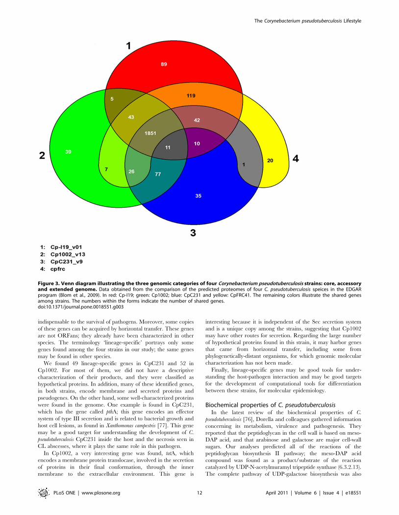
Gene Sharing among C. pseudotuberculosis strainsConsidering the four available genomes of C. pseudotuberculosis
strains (Cp1002, CpC231, and CpI19 pFRC41), we identified
1,851 whole genes shared among them (Figure 3).
This repertoire of genes is vast for this specie, since, among the
four isolates the maximum number of genes is 2,377 (called the
pangenome of the species). When we compare the number of
genes shared by these four C. pseudotuberculosis strains with a study
of 17 strains of the bacterium E. coli [75], we conclude that C.
pseudotuberculosis has a greater proportion of shared genes. In
isolates of E. coli, 2,220 genes constituted the core genome, less
Table 7. List of Corynebacterium pseudotuberculosis specificmetabolic pathways that were compared to those of closely-related bacteria, including C. diphtheriae, C. glutamicum, C.efficiens, and C. jeikeium.
Pathway Class
Pathway Name
Biosynthesis - Amino acid Biosynthesis
Asparagine biosynthesis II
Lysine biosynthesis V
Biosynthesis - Metabolic Regulators Biosynthesis
Citrulline-nitric oxide cycle
Biosynthesis - Nucleoside and Nucleotide Biosynthesis
Salvage pathways of pyrimidine deoxyribonucleotides
Degradation - Alcohol Degradation
Glycerol degradation II
Degradation - Aldehyde Degradation
Methylglyoxal degradation III
Degradation - Amino Acid Degradation
Alanine degradation IV
Citrulline-nitric oxide cycle
Lysine degradation I
Degradation - C1 Compound Utilization and Assimilation
Reductive monocarboxylic acid cycle
Degradation - Carbohydrate Degradation
Chitobiose degradation
Degradation - Carboxylate Degradation
Conversion of succinate to propionate
Degradation - Fatty Acid and Lipid Degradation
Phospholipases
Inorganic Nutrients Metabolism
Ammonia oxidation I (aerobic)
Nitrate reduction IV (dissimilatory)
Degradation - Secondary Metabolite Degradation
D-glucarate degradation
Betanidin degradation
D-galactarate degradation
Generation of precursor metabolites and energy
Ammonia oxidation I (aerobic)
doi:10.1371/journal.pone.0018551.t007
The Corynebacterium pseudotuberculosis Lifestyle
PLoS ONE | www.plosone.org 10 April 2011 | Volume 6 | Issue 4 | e18551

than half of the genes in this species, with a mean of 5,000 genes in
each genome [75]. Other significant information that emerges
from this data is that the C. pseudotuberculosis genomes are extremely
similar, since we found no significant change in the composition of
the repertoire of genes for this species after adding the two new
strains (Figure 3).
Gene Sharing between C. pseudotuberculosis and otherCorynebacterium species
Previous comparative studies of sequences of the rpoB gene of
C. pseudotuberculosis and C. diphtheriae have suggested a close
relationship between them [27,76]. In our current study, we
confirmed this close relationship with several types of evidence: i) a
similar codon bias, ii) high similarity at the amino acid level and iii)
conserved synteny. Synteny analysis of the genomes of the two C.
pseudotuberculosis strains compared to C. diphtheriae indicates that
these genomes are highly conserved; the gene position is conserved
within the species. This observation reinforces the conclusions of
previous research claiming conserved synteny in this genus, which
indicated that few rearrangement events occurred during
evolution [25].
Corynebacterium pseudotuberculosis shares more orthologous genes
with C. glutamicum (1,345 genes), C. efficiens (1,330), C. diphtheriae
(1,263 genes) and C. auricumucosum (1,273 genes); it shares only
1,030 genes with C. jeikeium and C. kroppenstedtii.
The larger number of genes shared between C. pseudotuberculosis,
C. glutamicum and C. efficiens (72%), compared to other species
(pathogenic species, 60%), may be a result not only of their close
relationships, but also because a comparison is made among
species with a larger gene repertoire, such as C. glutamicum and C.
efficiens, which are non-pathogenic microorganisms, thus increasing
the possibility of sharing genes.
Lineage-specific genes in C. pseudotuberculosisMost of the lineage-specific genes are involved in processes of
virulence, pathogenicity, drug resistance and response to certain
types of stress. These factors can increase the adaptability of
microorganisms to the niches they inhabit, but they are not
Table 8. Comparative summary of Corynebacterium pseudotuberculosis strains Cp1002 and CpC231 and C. glutamicum pathways.
Pathway Class Cp1002 CpC231 C. glutamicum
- Pathway subclass
Biosynthesis 105 104 131
- Amine and Polyamine Biosynthesis 5 3 3
- Amino acid Biosynthesis 25 26 29
- Aminoacyl-tRNA Charging 1 1 3
- Aromatic Compound Biosynthesis 1 1 1
- Carbohydrate Biosynthesis 10 7 9
- Cell structure Biosynthesis 4 4 4
- Cofactor, Prosthetic Group, Electron Carrier Biosynthesis 27 29 38
- Fatty Acid and Lipids Biosynthesis 8 7 14
- Metabolic Regulator Biosynthesis 1 2 1
- Nucleoside and Nucleotide Biosynthesis 12 12 10
- Other Biosynthesis 1 1 1
- Secondary Metabolite Biosynthesis 1 2 6
Degradation/Utilization/Assimilation 53 54 72
- Alcohols Degradation 2 1 2
- Aldehyde Degradation 1 1 1
- Amine and Polyamine Degradation 5 4 6
- Amino Acid Degradation 11 12 15
- Aromatic Compound Degradation 0 0 9
- C1 Compound Utilization and Assimilation 4 4 2
- Carbohydrate Degradation 7 7 10
- Carboxylate Degradation 5 4 6
- Chlorinated Compound Degradation 0 0 4
- Degradation/Utilization/Assimilation - Other 5 5 2
- Fatty Acid and Lipid Degradation 3 2 2
- Inorganic Nutrient Metabolism 4 6 9
- Nucleoside and Nucleotide Degradation and Recycling 2 3 1
- Secondary Metabolite Degradation 5 5 4
Generation of precursor metabolites and energy 16 19 25
Total 163 164 206
doi:10.1371/journal.pone.0018551.t008
The Corynebacterium pseudotuberculosis Lifestyle
PLoS ONE | www.plosone.org 11 April 2011 | Volume 6 | Issue 4 | e18551
indispensable to the survival of pathogens. Moreover, some copies
of these genes can be acquired by horizontal transfer. These genes
are not ORFans; they already have been characterized in other
species. The terminology ‘lineage-specific’ portrays only some
genes found among the four strains in our study; the same genes
may be found in other species.
We found 49 lineage-specific genes in CpC231 and 52 in
Cp1002. For most of them, we did not have a descriptive
characterization of their products, and they were classified as
hypothetical proteins. In addition, many of these identified genes,
in both strains, encode membrane and secreted proteins and
pseudogenes. On the other hand, some well-characterized proteins
were found in the genome. One example is found in CpC231,
which has the gene called pthA; this gene encodes an effector
system of type III secretion and is related to bacterial growth and
host cell lesions, as found in Xanthomonas campestris [77]. This gene
may be a good target for understanding the development of C.
pseudotuberculosis CpC231 inside the host and the necrosis seen in
CL abscesses, where it plays the same role in this pathogen.
In Cp1002, a very interesting gene was found, tatA, which
encodes a membrane protein translocase, involved in the secretion
of proteins in their final conformation, through the inner
membrane to the extracellular environment. This gene is
interesting because it is independent of the Sec secretion system
and is a unique copy among the strains, suggesting that Cp1002
may have other routes for secretion. Regarding the large number
of hypothetical proteins found in this strain, it may harbor genes
that came from horizontal transfer, including some from
phylogenetically-distant organisms, for which genomic molecular
characterization has not been made.
Finally, lineage-specific genes may be good tools for under-
standing the host-pathogen interaction and may be good targets
for the development of computational tools for differentiation
between these strains, for molecular epidemiology.
Biochemical properties of C. pseudotuberculosisIn the latest review of the biochemical properties of C.
pseudotuberculosis [76], Dorella and colleagues gathered information
concerning its metabolism, virulence and pathogenesis. They
reported that the peptidoglycan in the cell wall is based on meso-
DAP acid, and that arabinose and galactose are major cell-wall
sugars. Our analyses predicted all of the reactions of the
peptidoglycan biosynthesis II pathway; the meso-DAP acid
compound was found as a product/substrate of the reaction
catalyzed by UDP-N-acetylmuramyl tripeptide synthase (6.3.2.13).
The complete pathway of UDP-galactose biosynthesis was also
Figure 3. Venn diagram illustrating the three genomic categories of four Corynebacterium pseudotuberculosis strains: core, accessoryand extended genome. Data obtained from the comparison of the predicted proteomes of four C. pseudotuberculosis speices in the EDGARprogram (Blom et al., 2009). In red: Cp-I19; green: Cp1002; blue: CpC231 and yellow: CpFRC41. The remaining colors illustrate the shared genesamong strains. The numbers within the forms indicate the number of shared genes.doi:10.1371/journal.pone.0018551.g003
The Corynebacterium pseudotuberculosis Lifestyle
PLoS ONE | www.plosone.org 12 April 2011 | Volume 6 | Issue 4 | e18551

found; although there was no evidence of biosynthesis of
arabinose, we detected a membrane transporter, known as
arabinose efflux permease.
We also found short-chain mycolic acids; 10 variations of acids
of this type were encountered, including 6-O-cis-keto-mycolyl-
trehalose-6-phosphate, and 6-O-mycolyl-trehalose-6-phosphate.
The two strains of C. pseudotuberculosis showed considerable
fermentation ability, with several fermentation pathways, includ-
ing glycolysis III, mixed acid fermentation and pyruvate
fermentation to acetate IV, ethanol I and lactate.
Several sugar degradation pathways were also found in the two
strains of C. pseudotuberculosis, including galactose, lactose, sucrose
and L-and D-arabinose degradation. We confirmed that, as
reported by Dorella et al. (2006), all these pathways produce acids
and no gasses, generating large amounts of energy.
It was also previously reported that C. pseudotuberculosis is
phospholipase D and catalase positive. Our analysis showed that
both phospholipase D and catalase are involved in important
processes. The main molecular functions of phospholipase D are
phospholipase D activity, magnesium ion binding, NAPE-specific
phospholipase D activity and sphingomyelin phosphodiesterase D
activity. Catalase, which is produced by the cat gene, is involved in
response to oxidative stress and oxidation reduction. Although two
enzymes of the denitrification pathway (nitrate reduction I) were
found, absence of the remaining enzymes is probably the
determining factor for the inability of these strains to reduce
nitrate to N2, as reported by Dorella et al. (2006).
We also detected iron acquisition genes (fag) A, B, C and D in
both strains of C. pseudotuberculosis [78]. Genes fagA and fagB
produce the integral membrane proteins FagA, an iron-enter-
obactin transporter, and FagBy; both have important roles,
including ion, transmembrane, organic acid and protein transport.
The ATP binding cytoplasmic membrane protein, FagC,
produced by gene fagC, has two main molecular functions: ATP
binding and ATPase activity. Finally, gene fagD produces the iron
siderophore binding protein, FeAcquisition gene D, which has a
role in iron ion transmembrane transport activity.
Computational reconstruction of the C. pseudotuberculosis path-
ways in our database not only allowed us to better visualize the
metabolism of this bacterium, but also to compare it to closely
related species. The main purpose of this analysis was to describe
C. pseudotuberculosis metabolism by computational means, providing
a predictive tool for ‘‘wet-lab’’ research.
Methods
Bacterial strains and growth conditionsCorynebacterium pseudotuberculosis 1002 biovar ovis (herein referred
to as Cp1002) is a wild strain, isolated from a caprine host in
Brazil. Corynebacterium pseudotuberculosis C231 biovar ovis (herein
referred to as CpC231) is also a wild strain, isolated from an ovine
host in Australia. Both strains were confirmed to be C.
pseudotuberculosis by routine biochemical tests (API CORYNE,
Biomerieux, Marcy l’Etoile, France). These strains were main-
tained in brain-heart-infusion broth (BHI – HiMedia Laboratories
Pvt. Ltda, India) at 37uC, under rotation.
Preparation of high molecular weight DNAChromosomal DNA extraction was performed as follows:
50 mL of 48–72 h cultures of the two strains were centrifuged at
4uC and 2000 x g for 20 min. Cell pellets were re-suspended in
1 mL Tris/EDTA/NaCl [10 mM Tris/HCl (pH 7.0), 10 mM
EDTA (pH 8.0), and 300 mM NaCl] and centrifuged again under
the same conditions. Supernatants were discarded, and the pellets
were re-suspended in 1 mL TE/lysozyme [25 mM Tris/HCl
(pH 8.0), 10 mM EDTA (pH 8.0), 10 mM NaCl, and 10 mg
lysozyme/mL]. Samples were then incubated at 37uC for 30 min.
Thirty milliliters of 30% (w/v) sodium N-lauroyl-sarcosine
(Sarcosyl) were added to each sample and the mixtures were
incubated for 20 min at 65uC, followed by incubation for 5 min at
4uC. DNA was purified using phenol/chloroform/isoamyl alcohol
(25:24:1) and precipitated with ethanol. DNA concentrations were
determined spectrophotometrically, and the DNA was visualized
in ethidium bromide-stained 0.7% agarose gels.
Construction of Corynebacterium pseudotuberculosisgenomic libraries and Sanger sequencing
For the shotgun strategy used to sequence C. pseudotuberculosis
1002, four small fragment libraries were constructed using the
TOPO Shotgun cloning kit and the pCR4 Blunt-TOPO vector
(Invitrogen), according to the manufacturer’s instructions. Sanger
sequencing was carried out using the Minas Gerais Genome
Network (http://rgmg.cpqrr.fiocruz.br). A total of 6,144 forward
and reverse reads were produced using the DYEnamic Dye
Terminator kit and run in a Megabace 1000 automated sequencer
(GE Healthcare).
Genome SequencingCp1002 was sequenced using both Sanger and pyrosequencing
technologies. Pyrosequencing was carried out using 454 Life
Sciences (Branford, CT). A total of 397,147 high quality reads and
86,154,153 high quality bases were obtained, which translates into
approximately 31-fold coverage. The average length of the
sequences was 253 bases. The sequences were delivered after
quality filtering and preassembly with the Newbler assembler (454
Life Sciences).
CpC231 was sequenced with a Roche-454 FLX sequencer at
the Australian Animal Health Laboratory, Geelong, Australia. A
total of 347,361 reads generated 80,336,550 bases, giving 34-fold
coverage of the genome. De novo assembly of the filtered sequence
data was carried out using the Newbler software. This assembly
produced 10 large contigs in four scaffolds. The remaining gaps in
the genomic sequence were closed by PCR walking and Sanger
sequencing of the resulting fragments.
Treatment and assembly dataThe raw Sanger data obtained from sequencing were processed
using the Phred-Phrap-Consed package [75]. Possible contami-
nants (plasmid DNA, sequences with similarity to vectors and
other contaminants) were discarded using the Cross_match
program (www.phrap.org). The quality value used in the base-
calling program was Q = 40 (Probability of incorrect base call 1 in
10,000/base call accuracy 99.99%). An assembly using Phrap
parameters (Force Level: 40 and Gap Length: 10,000) was carried
out.
The 454 data were processed using the Newbler assembler (454
Life Sciences), and the final genomic consensus sequence was
obtained using the Phrap algorithm.
Genome annotationThe annotation procedures involved the use of several
algorithms in a multi-step process. Structural annotation was
performed using the following software: FgenesB: gene predictor
(www.softberry.com); RNAmmer: rRNA predictor [79]; tRNA-
scan-SE: tRNA predictor [80]; and Tandem Repeat Finder:
repetitive DNA predictor (tandem.bu.edu/trf/trf.html). Functional
annotation was performed by similarity analyses, using public
The Corynebacterium pseudotuberculosis Lifestyle
PLoS ONE | www.plosone.org 13 April 2011 | Volume 6 | Issue 4 | e18551
databases and InterProScan analysis [81]. Manual annotation was
performed using Artemis [82].
Identification and confirmation of putative pseudogenes in the
genome was carried out using Consed. Manual analysis was
performed based on the Phred quality of each base in the
frameshift area. This analysis enabled the identification of
erroneous insertions or deletions of bases in the genome
information produced by the sequencing process, and it avoided
identification of false-positive pseudogenes.
Predictions of the cellular locations of Corynebacterium proteins
were made using the program SurfG Plus (version 1.0), with a
minimum protein size of 73 amino acids. Classification of
predicted proteins in functional categories was made using the
BLAST2GO program (www.blast2go.org). The cutoff value used
was 1026 (http://www.blast2go.org/).
In silico Identification of Pathogenicity IslandsIn order to accurately identify and classify putative Pathoge-
nicity Islands (PAIs) in the corynebacterial genomes, we developed
a combined computational approach using several in-house scripts
to integrate the prediction of diverse algorithms and databases,
namely: Colombo-SIGIHMM [83], Artemis [82], tRNAscan-SE
[80]; EMBOSS-geecee [84], ACT: the Artemis Comparison Tool
[85], and mVIRdb [86].
In silico metabolic pathway constructionThe two main data sources used for reconstructing the C.
pseudotuberculosis metabolic pathways were the genome sequence file
in FASTA format and the genome annotation file in GBK format.
Metabolic pathways databases for strains 1002 and C231 were
created using the Pathway tools 13 software, developed by SRI
International [87]. The Pathway tools software contains algo-
rithms that predict metabolic pathways of an organism from its
genome by comparison to a reference pathways database known
as MetaCyc [88]. Construction of a metabolic pathways database
was done using BioCyc [89], in order to compare the different
bacteria, C. diphtheriae NCTC 13129, C. efficiens YS-314, C.
glutamicum ATCC 13032, and C. jeikeium K411, to the deduced
C. pseudotuberculosis pathways.
Comparative analysis of Corynebacteriumpseudotuberculosis strains
Comparative analyses were made for the two C. pseudotuberculosis
strains. Similarity analyses of the two genomes were made using
the BLAST - NCBI [90,91] and InterProScan databases. The
Mauve algorithm (gel.ahabs.wisc.edu/mauve) and the ACT tool
were used to identify whether blocks had undergone gene
rearrangements or remained preserved. The Plotter program of
the MUMMer 3.22 package (mummer.sourceforge.net) was used
for synteny analysis.
Supporting Information
Table S1 Orthologous genes present inside PAIs regions of C.
pseudotuberculosis and their counterparts in other Corynebacterium
species.
(DOC)
Acknowledgments
The authors acknowledge the scientific support of two genomics networks:
Rede Paraense de Genomica e Proteomica and Rede Genoma de Minas
Gerais. In addition, the authors thank for valuable contribution of all
Public Institutions involved and their respective co-authors during the
development of the present work.
Author Contributions
Conceived and designed the experiments: JCR AS MPC RJM AM GCO
VA. Performed the experiments: FAD SB MITF GCO AM VA VD EMC
LMO MCP SRCD AFC JFA. Analyzed the data: JCR AS RJM GCO AM
VA VD ARS FAD LGCP MZT NS TLPC JM AZ SCS SSA VACA
DMR. Contributed reagents/materials/analysis tools: VA GCO GRF
DOL ALP CUV CTG DCB DMO FRS EMR IMC JMO LVP LRG JAF
MITF NPC PRKF SMRT SB SCO. Wrote the paper: JCR AS RJM
GCO AM VA VD ARS FAD LGCP MZT NS TLPC JM AZ SCS SSA
VACA DMR ET JB AT. Obtained permission for use of cell line: RJM
RM AM VA. Bioinformatic support: JCR GCO AD FPL PG.
References
1. Ayers JL (1977) Caseous lymphadenitis in goat and sheep: review of diagnosis,
pathogenesis, and immunity. JAVMA 171: 1251–1254.
2. Brown CC, Olander HJ, Alves SF (1987) Synergistic hemolysis-inhibition titers
associated with caseous lymphadenitis in a slaughterhouse survey of goats andsheep in northeastern Brazil. Can J Vet Res 51: 46–49.
3. Merchant IA, Packer RA (1967) The genus Corynebacterium. In: Merchant IA,
Packer RA, eds. Veterinary Bacteriology and Virology. USA: The Iowa StateUniversity Press. pp 425–440.
4. Piontkowski MD, Shivvers DW (1998) Evaluation of a commercially available
vaccine against Corynebacterium pseudotuberculosis for use in sheep. J Am Vet Med
Assoc 212: 1765–1768.
5. Join-Lambert OF, Ouache M, Canioni D, Beretti JL, Blanche S, et al. (2006)
Corynebacterium pseudotuberculosis necrotizing lymphadenitis in a twelve-year old
patient. Pediatr Infect Dis J 25(9): 848–851.
6. Trost E, Ott L, Schneider J, Schroder J, Jaenicke S, et al. (2010) The complete
genome sequence of Corynebacterium pseudotuberculosis FRC41 isolated from a 12-
year-old girl with necrotizing lymphadenitis reveals insights into gene-regulatory
networks contributing to virulence. BMC Genomics 11(1): 728–745.
7. Connor KM, Quirie MM, Baird G, Donachie W (2000) Characterization of
united kingdom isolates of Corynebacterium pseudotuberculosis using pulsed-field gel
electrophoresis. J Clin.Microbiol 38: 2633–2637.
8. Ben Saıd MS, Ben Maitigue H, Benzarti M, Messadi L, Rejeb A, et al. (2002)
Epidemiological and clinical studies of ovine caseous lymphadenitis. Arch Inst
Pasteur Tunis 79: 51–57.
9. Binns SH, Bailey M, Green LE (2002) Postal survey of ovine caseouslymphadenitis in the United Kingdom between 1990 and 1999. Vet Rec 150:
263–268.
10. Arsenault J, Girard C, Dubreuil P, Daignault D, Galarneau JR, et al. (2003)
Prevalence of and carcass condemnation from maedi-visna, paratuberculosis and
caseous lymphadenitis in culled sheep from Quebec, Canada. Prev Vet Med 59:
67–81.
11. Paton MW, Walker SB, Rose IR, Watt GF (2003) Prevalence of caseous
lymphadenitis and usage of caseous lymphadenitis vaccines in sheep flocks. Aust
Vet J 81: 91–95.
12. Pinheiro RR, Gouveia AMG, Alves FSF, Haddad JP (2000) Aspectos
epidemiologicos da caprinocultura cearense. Arquivo Brasileiro de Medicina
Veterinaria e Zootecnia 52: 534–543.
13. Guimaraes AS, Seyffert N, Portela RWD, Meyer R, Carmo FB, et al. (2009)
Caseous lymphadenitis in sheep flocks of the state of Minas Gerais, Brazil:
prevalence and management surveys. Small Ruminants Research 87(1): 86–91.
14. Seyffert N, Guimaraes AS, Pacheco LGC, Portela RW, Bastos BL, et al. (2010)
High seroprevalence of caseous lymphadenitis in brazilian goat herds revealed
by Corynebacterium pseudotuberculosis secreted proteins-based ELISA. Res Vet Sci
88: 50–55.
15. Eggleton DG, Middleton HD, Doidge CV, Minty DW (1991) Immunisation
against ovine caseous lymphadenitis: comparison of Corynebacterium pseudotuber-
culosis vaccines with and without bacterial cells. Aust Vet J 68: 317–319.
16. Stoops SG, Renshaw HW, Thilsted JP (1984) Ovine caseous lymphadenitis:
disease prevalence, lesion distribution, and thoracic manifestations in a
population of mature culled sheep from western United States. Am J Vet Res
45(3): 557–61.
17. Ribeiro MG, Junior JGD, Paes AC, Barbosa PG, Junior GN, et al. (2001)
Puncao aspirativa com agulha fina no diagnostico de Corynebacterium pseudotuber-
culosis na linfadenite caseosa caprina. Arq Inst Biol 68: 23–28.
18. Dorella FA, Estevam EM, Pacheco LGC, Guimaraes CT, Lana UGP, et al.
(2006) In vivo insertional mutagenesis in Corynebacterium pseudotuberculosis: an
efficient means to identify DNA sequences encoding exported proteins. Appl
Environ Microbiol 72: 7368–7372.
The Corynebacterium pseudotuberculosis Lifestyle
PLoS ONE | www.plosone.org 14 April 2011 | Volume 6 | Issue 4 | e18551

19. Merhej V, Royer-Carenzi M, Pontarotti P, Raoult D (2009) Massivecomparative genomic analysis reveals convergent evolution of specialized
bacteria. Biol Direct 4: 13–37.
20. Webb SAR, Karleh CM (2008) Bench-to-bedside review: Bacterial virulenceand subversion of host defences. Critical Care 12: 234–241.
21. Dobrindt U, Hentschel U, Kaper JB, Hacker J (2002) Genome plasticity inpathogenic and nonpathogenic enterobacteria. Curr Top Microbiol Immunol
264: 157–175.
22. Hall BG, Ehrlich GD, Hu FZ (2010) Pan-genome analysis provides much higherstrain typing resolution than multi-locus sequence typing. Microbiology 156(4):
1060–8.
23. Silva A, Schneider MPC, Cerdeira L, Barbosa MS, Ramos RTJ, et al. (2011)
Complete genome sequence of Corynebacterium pseudotuberculosis I19, a strain
isolated from a cow in Israel with bovine mastitis. J Bacteriol 193(1): 323–324.
24. Nakamura Y, Nishio Y, Ikeo K, Gojobori T (2003) The genome stability in
Corynebacterium species due to lack of the recombinational repair system. Gene317: 149–155.
25. Tauch A, Kaiser O, Hain T, Goesmann A, Weisshaar B, et al. (2005) Complete
genome sequence and analysis of the multiresistant nosocomial pathogenCorynebacterium jeikeium K411, a lipid-requiring bacterium of the human skin flora.
J Bacteriol 187: 4671–4682.
26. Khamis A, Raoult D, La Scola B (2004) rpoB gene sequencing for identificationof Corynebacterium species. J Clin Microbiol 42(9): 3925–31.
27. Khamis A, Raoult D, La Scola B (2005) Comparison between rpoB and 16SrRNA gene sequencing for molecular identification of 168 clinical isolates of
Corynebacterium. J Clin Microbiol 43: 1934–1936.
28. Dobrindt U, Hochhut B, Hentschel U, Hacker J (2004) Genomic islands inpathogenic and environmental microorganisms. Nat Rev Microbiol 2: 414–424.
29. Karaolis DK, Johnson JA, Bailey CC, Boedeker EC, Kaper JB, et al. (1998) AVibrio cholerae pathogenicity island associated with epidemic and pandemic
strains. Proc Natl Acad Sci U.S.A 95: 3134–3139.
30. Schumann W (2007) Thermosensors in eubacteria: role and evolution. J Biosci32: 549–557.
31. Hentschel U, Hacker J (2001) Pathogenicity islands: the tip of the iceberg.
Microbes Infect 3: 545–548.
32. McNamara PJ, Cuevas WA, Songer JG (1995) Toxic phospholipases D of
Corynebacterium pseudotuberculosis, C. ulcerans and Arcanobacterium haemolyticum: cloningand sequence homology. Gene 156: 113–118.
33. Moomaw AS, Maguire ME (2008) The unique nature of Mg2+ channels.
Physiology (Bethesda) 23: 275–285.
34. O’Connor K, Fletcher SA, Csonka LN (2009) Increased expression of Mg(2+)
transport proteins enhances the survival of Salmonella enterica at high temperature.Proc Natl Acad Sci U.S.A 106: 17522–17527.
35. Schonert S, Buder T, Dahl MK (1998) Identification and enzymatic
characterization of the maltose-inducible alpha-glucosidase mall (sucrase-isomaltase-maltase) of Bacillus subtilis. J Bacteriol 180: 2574–2578.
36. Yamamoto H, Serizawa M, Thompson J, Sekiguchi J (2001) Regulation of theglv operon in Bacillus subtilis: yfia (glvR) is a positive regulator of the operon that is
repressed through ccpA and cre. Bacteriol 183: 5110–5121.
37. May T, Ito A, Okabe S (2009) Induction of multidrug resistance mechanism inEscherichia coli biofilms by interplay between tetracycline and ampicillin resistance
genes. Antimicrob Agents Chemother 53: 4628–4639.
38. Smith I (2003) Mycobacterium tuberculosis pathogenesis and molecular determinantsof virulence. Clin Microbiol Rev 16: 463–496.
39. Saıd-Salim B, Mostowy S, Kristof AS, Behr MA (2006) Mutations inMycobacterium tuberculosis RV0444c, the gene encoding anti-sigK, explain high
level expression of mpb70 and mpb83 in Mycobacterium bovis. Mol Microbiol 62:
1251–1263.
40. Veyrier F, Saıd-Salim B, Behr MA (2008) Evolution of the mycobacterial sigK
regulon. J Bacteriol 190: 1891–1899.
41. Vassylyev DG, Tomitori H, Kashiwagi K, Morikawa K, Igarashi K (1998)
Crystal structure and mutational analysis of the Escherichia coli putrescine
receptor: structural basis for substrate specificity. J Biol Chem 273:17604–17609.
42. Carlson PEJ, Horzempa J, O’Dee DM, Robinson CM, Neophytou P, et al.(2009) Global transcriptional response to spermine, a component of the
intramacrophage environment, reveals regulation of Francisella gene expression
through insertion sequence elements. J Bacteriol 191: 6855–6864.
43. Enkavi G, Tajkhorshid E (2010) Simulation of spontaneous substrate binding
revealing the binding pathway and mechanism and initial conformationalresponse of glpT. Biochemistry 49: 1105–1114.
44. Perez E, Samper S, Bordas Y, Guilhot C, Gicquel B, et al. (2001) An essential
role for phoP in Mycobacterium tuberculosis virulence. Mol Microbiol 41(1): 179–87.
45. Soto CY, Menendez MC, Perez E, Samper S, Gomez AB, et al. (2004) IS6110
Mediates Increased Transcription of the phoP Virulence Gene in a Multidrug-Resistant Clinical Isolate Responsible for Tuberculosis Outbreaks. J Clin Microb
42(1): 212–219.
46. Aguilar D, Infante E, Martin C, Gormley E, Gicquel G, Pando RH (2006)Immunological responses and protective immunity against tuberculosis con-
ferred by vaccination of Balb/C mice with the attenuated Mycobacterium
tuberculosis (phoP) SO2 strain. Clin Exper Immunol 147: 330–338.
47. Gonzalo-Asensio J, Mostowy S, Harders-Westerveen J, Huygen K, Hernandez-
Pando R, et al. (2008) PhoP: A Missing Piece in the Intricate Puzzle ofMycobacterium tuberculosis Virulence. PLoS ONE 3(10): 1–11.
48. Carson SD, Klebba PE, Newton SM, Sparling PF (1999) Ferric enterobactinbinding and utilization by Neisseria gonorrhoeae. J Bacteriol 181: 2895–2901.
49. Kunkle CA, Schmitt MP (2005) Analysis of a dtxR-regulated iron transport and
siderophore biosynthesis gene cluster in Corynebacterium diphtheriae. J Bacteriol 187:422–433.
50. Shimizu T, Okabe A, Minami J, Hayashi H (1991) An upstream regulatory
sequence stimulates expression of the perfringolysin o gene of Clostridium
perfringens. Infect Immun 59: 137–142.
51. Urbina P, Flores-Dıaz M, Alape-Giron A, Alonso A, Goni FM (2009)
Phospholipase C and sphingomyelinase activities of the Clostridium perfringens
alpha-toxin. Chem Phys Lipids 159: 51–57.
52. Selby T, Allaker RP, Dymock D (2003) Characterization and expression of
adjacent proline iminopeptidase and aspartase genes from Eikenella corrodens. OralMicrobiol Immunol 18: 256–259.
53. Zhang L, Jia Y, Wang L, Fang R (2007) A proline iminopeptidase gene
upregulated in planta by a luxR homologue is essential for pathogenicity ofXanthomonas campestris pv. campestris. Mol Microbiol 65: 121–136.
54. Boltner D, MacMahon C, Pembroke JT, Strike P, Osborn AM (2002) R391: a
conjugative integrating mosaic comprised of phage, plasmid, and transposonelements. J Bacteriol 184: 5158–5169.
55. Burne RA, Chen YY (2000) Bacterial ureases in infectious diseases. MicrobesInfect 2: 533–542.
56. Huntley RP, Binns D, Dimmer E, Barrell D, O’Donavan C, et al. (2009)
QuickGO: a user tutorial for the web-based Gene Ontology browser. Database10: 1–19.
57. Barinov A, Loux V, Hammani A, Nicolas P, Langella P, et al. (2009) Prediction
of surface exposed proteins in Streptococcus pyogenes, with a potential application toother gram-positive bacteria. Proteomics 9: 61–73.
58. Song C, Kumar A, Saleh M (2009) Bioinformatic comparison of bacterial
secretomes. Genomics Proteomics Bioinformatics 7: 37–46.
59. Wooldridge L, Lissina A, Cole DK, van den Berg HA, Price DA, Sewell AK
(2009) Tricks with tetramers: how to get the most from multimeric peptide-
MHC. Immunology 126: 147–164.
60. Wittmann C, Kiefer P, Zelder O (2004) Metabolic Fluxes in Corynebacterium
glutamicum during Lysine Production with Sucrose as Carbon Source. Applied
and Environmental Microbiology 70: 7277–7287.
61. Kabus A (2007) Expression of the Escherichia coli pntAB genes encoding a
membrane-bound transhydrogenase in Corynebacterium glutamicum improves l-
lysine formation. Appl Microbiol Biotechnol 75: 47–53.
62. Blombach B, Arndt A, Auchter M, Eikmanns BJ (2009) L-Valine production
during growth of pyruvate dehydrogenase complex-deficient Corynebacterium
glutamicum in the presence of ethanol or by inactivation of the transcriptional
regulator SugR. Appl Environ Microbiol 75: 1197–1200.
63. Klaffl S, Eikmanns BJ (2010) Genetic and Functional Analysis of the SolubleOxaloacetate Decarboxylase from Corynebacterium glutamicum. Journal of Bacte-
riology 192: 2604–2612.
64. Wittmann C, Heinzle E (2001) Modeling and experimental design for metabolicflux analysis of lysine-producing Corynebacteria by mass spectrometry. Metab Eng
3(2): 173–91.
65. Parche S, Thomae AW, Schlicht M, Titgemeyer F (2001) Corynebacterium
diphtheriae: a PTS View to the Genome. J Mol Microbiol Biotechnol 3(3):
415–422.
66. Rubenhagen R, Ronsch H, Jung H, Kramer R, Morbach S (2000) Osmosensorand osmoregulator properties of the betaine carrier betP from Corynebacterium
glutamicum in proteoliposomes. J Biol Chem 275: 735–741.
67. Rittmann D, Schaffer S, Wendisch VF, Sahm H (2003) Fructose-1,6-bisphosphatase from Corynebacterium glutamicum: expression and deletion of the
fbp gene and biochemical characterization of the enzyme. Arch Microbiol 180:285–292.
68. Kiefer P, Heinzle E, Zelder O, Wittmann Z (2004) Comparative Metabolic Flux
Analysis of Lysine-Producing Corynebacterium glutamicum Cultured on Glucose orFructose. Applied and Environmental Microbiology 70: 229–239.
69. Rumbold K, Buijsen HJJ, Overkamp KM, Groenestijn JW, Punt PJ, Werf MJ
(2009) Microbial production host selection for converting second-generationfeedstocks into bioproducts. Microbial Cell Factories 8: 1–11.
70. Casalino M, Prosseda G, Barbagallo M, Lacobino A, Ceccarini P, et al. (2010)
Interference of the cadC regulator in the arginine-dependent acid resistancesystem of Shigella and enteroinvasive E. coli. Int J Med Microbiol 300(5): 289–95.
71. Alvarez-Ordonez A, Fernandez A, Bernardo A, Lopez M (2010) Arginine and
lysine decarboxylases and the acid tolerance response of Salmonella typhimurium.Int J Food Microbiol 136: 278–282.
72. Hodgson AL, Carter K, Tachedjian M, Krywult J, Corner LA, et al. (1999)
Efficacy of an ovine caseous lymphadenitis vaccine formulated using agenetically inactive form of the Corynebacterium pseudotuberculosis Phospholipase
D. Vaccine 17: 802–808.
73. D’Afonseca V, Moraes PM, Dorella FA, Pacheco LGC, Meyer R, et al. (2008) A
description of genes of Corynebacterium pseudotuberculosis useful in diagnostics and
vaccine applications. Genet Mol Res 7: 252–260.
74. Cole ST, Eiglmeier K, Parkhill J, James KD, Thomson NR, et al. (2001) Massive
gene decay in the leprosy bacillus. Nature 409: 1007–1011.
75. Rasko DA, Rosovitz MJ, Garry SA, Emmanuel FM, Fricke WF, et al. (2008)The pan-genome structure of Escherichia coli: comparative genomic analysis of E.
coli commensal and pathogenic isolates. Journal of Bacteriology 190(20):
6881–6893.
The Corynebacterium pseudotuberculosis Lifestyle
PLoS ONE | www.plosone.org 15 April 2011 | Volume 6 | Issue 4 | e18551
76. Dorella FA, Pacheco LGC, Oliveira SC, Miyoshi A, Azevedo V (2006)
Corynebacterium pseudotuberculosis: microbiology, biochemical properties, pathogen-esis and molecular studies of virulence. Vet Res 37: 201–218.
77. Shiotani H, Yoshioka T, Yamamoto M, Matsumoto R (2008) Susceptibility to
citrus canker caused by Xanthomonas axonopodis pv. citri depends on the nucleargenome of the host plant. J Gen Plant Pathol 74: 133–137.
78. Ewing B, Green P (1998) Base-calling of automated sequencer traces usingphred. II. Error probabilities. Genome Res 8: 186–194.
79. Lagesen K, Hallin P, Rødland EA, Staerfeldt H, Rognes T, et al. (2007)
Rnammer: consistent and rapid annotation of ribosomal RNA genes. NucleicAcids Res 35: 3100–3108.
80. Lowe TM, Eddy SR (1997) Trnascan-SE: a program for improved detection oftransfer RNA genes in genomic sequence. Nucleic Acids Res 25: 955–964.
81. Zdobnov EM, Apweiler R (2001) Interproscan--an integration platform for thesignature-recognition methods in INTERPRO. Bioinformatics 17: 847–848.
82. Rutherford K, Parkhill J, Crook J, Horsnell T, Rice P, et al. (2000) Artemis:
sequence visualization and annotation. Bioinformatics 16: 944–945.83. Waack S, Keller O, Asper R, Brodag T, Damm C, et al. (2006) Score-based
prediction of genomic islands in prokaryotic genomes using hidden markovmodels. BMC Bioinformatics 7: 142.
84. Rice P, Longden I, Bleasby A (2000) Emboss: the European molecular biology
open software suite. Trends Genet 16: 276–277.
85. Carver TJ, Rutherford KM, Berriman M, Rajandream M, Barrell BG, et al.
(2005) ACT: the Artemis Comparison Tool. Bioinformatics 21: 3422–3423.
86. Zhou CE, Smith J, Lam M, Zemla A, Dyer MD, et al. (2007) Mvirdb--a
microbial database of protein toxins, virulence factors and antibiotic resistance
genes for bio-defence applications. Nucleic Acids Res 35: D391–394.
87. Karp PD, Paley S, Romero P (2002) The pathway tools software. Bioinformatics
18(1): S225–32.
88. Caspi R, Foerster H, Fulcher CA, Kaipa P, Krummenacker M, et al. (2008) The
metacyc database of metabolic pathways and enzymes and the biocyc collection
of pathway/genome databases. Nucleic Acids Res 36: D623–31.
89. Caspi R, Karp PD (2007) Using the metacyc pathway database and the biocyc
database collection. Curr Protoc Bioinformatics Chapter 1: Unit1.17.
90. Altschul SF, Gish W, Miller W, Myers EW, Lipman DJ (1990) Basic local
alignment search tool. J Mol Biol 215: 403–410.
91. Krauthammer M, Rzhetsky A, Morozov P, Friedman C (2000) Using BLAST
for identifying gene and protein names in journal articles. Gene 259: 245–252.
The Corynebacterium pseudotuberculosis Lifestyle
PLoS ONE | www.plosone.org 16 April 2011 | Volume 6 | Issue 4 | e18551