A quick and cost effective method for the diagnosis of Mycobacterium ulcerans infection

10.1101/gr.5942807Access the most recent version at doi: published online Jan 8, 2007; Genome Res.
Laval, Mamadou Daffé, Julian Parkhill and Stewart T. Cole D.R. Johnson, John K. Davies, Grant A. Jenkin, Pamela L.C. Small, Louis M. Jones, Fredj Tekaia, Françoise
PaulMeurice, David Simon, Christiane Bouchier, Laurence Ma, Magali Tichit, Jessica L. Porter, Janine Ryan, Timothy P. Stinear, Torsten Seemann, Sacha Pidot, Wafa Frigui, Gilles Reysset, Thierry Garnier, Guillaume
, the causative agent of Buruli ulcerMycobacterium ulceransof Reductive evolution and niche adaptation inferred from the genome
P<P Published online January 8, 2007 in advance of the print journal.
serviceEmail alerting
click heretop right corner of the article or Receive free email alerts when new articles cite this article - sign up in the box at the
Notes
object identifier (DOIs) and date of initial publication. by PubMed from initial publication. Citations to Advance online articles must include the digital publication). Advance online articles are citable and establish publication priority; they are indexedappeared in the paper journal (edited, typeset versions may be posted when available prior to final Advance online articles have been peer reviewed and accepted for publication but have not yet
http://www.genome.org/subscriptions/ go to: Genome ResearchTo subscribe to
© 2007 Cold Spring Harbor Laboratory Press
on January 9, 2007 www.genome.orgDownloaded from

Reductive evolution and niche adaptation inferredfrom the genome of Mycobacterium ulcerans, thecausative agent of Buruli ulcerTimothy P. Stinear,1,2 Torsten Seemann,3 Sacha Pidot,2 Wafa Frigui,1 Gilles Reysset,1
Thierry Garnier,1 Guillaume Meurice,4 David Simon,4 Christiane Bouchier,5
Laurence Ma,5 Magali Tichit,5 Jessica L. Porter,2 Janine Ryan,2 Paul D.R. Johnson,6
John K. Davies,2 Grant A. Jenkin,2 Pamela L.C. Small,7 Louis M. Jones,8 Fredj Tekaia,9
Françoise Laval,10 Mamadou Daffé,10 Julian Parkhill,11 and Stewart T. Cole1,12
1Unité de Génétique Moléculaire Bactérienne, Institut Pasteur, 75725 Paris Cedex 15, France; 2Department of Microbiology,Monash University, Clayton 3800, Australia; 3Victorian Bioinformatics Consortium, Monash University, Clayton 3800, Australia;4Plate-Forme 4—Intégration et Analyse Génomique, Génopole, Institut Pasteur, 75725 Paris Cedex 15, France;5Plate-Forme—Genopole, Institut Pasteur, 75725 Paris Cedex 15, France; 6Department of Infectious Diseases, Austin Hospital,Heidelberg 3084, Australia; 7Department of Microbiology, University of Tennessee, Knoxville, Tennessee 37996-0845, USA;8Groupe Logiciels et Banques de données, Institut Pasteur, 75725 Paris Cedex 15, France; 9 Unité de Génétique Moléculaire desLevures, Institut Pasteur, 75725 Paris Cedex 15, France; 10Department of Molecular Mechanisms of Mycobacterial InfectionsIPBS-CNRS, 31077 Toulouse Cedex 14, France; 11Wellcome Trust Sanger Institute, Wellcome Trust Genome Campus, Hinxton,Cambridge CB 10 1SA, United Kingdom.
Mycobacterium ulcerans is found in aquatic ecosystems and causes Buruli ulcer in humans, a neglected but devastatingnecrotic disease of subcutaneous tissue that is rampant throughout West and Central Africa. Here, we report thecomplete 5.8-Mb genome sequence of M. ulcerans and show that it comprises two circular replicons, a chromosome of5632 kb and a virulence plasmid of 174 kb. The plasmid is required for production of the polyketide toxinmycolactone, which provokes necrosis. Comparisons with the recently completed 6.6-Mb genome of Mycobacteriummarinum revealed >98% nucleotide sequence identity and genome-wide synteny. However, as well as the plasmid, M.ulcerans has accumulated 213 copies of the insertion sequence IS2404, 91 copies of IS2606, 771 pseudogenes, twobacteriophages, and multiple DNA deletions and rearrangements. These data indicate that M. ulcerans has recentlyevolved via lateral gene transfer and reductive evolution from the generalist, more rapid-growing environmentalspecies M. marinum to become a niche-adapted specialist. Predictions based on genome inspection for the productionof modified mycobacterial virulence factors, such as the highly abundant phthiodiolone lipids, were confirmed bystructural analyses. Similarly, 11 protein-coding sequences identified as M. ulcerans-specific by comparative genomicswere verified as such by PCR screening a diverse collection of 33 strains of M. ulcerans and M. marinum. This workoffers significant insight into the biology and evolution of mycobacterial pathogens and is an important componentof international efforts to counter Buruli ulcer.
[Supplemental material is available online at www.genome.org. The sequence data from this study have beensubmitted to Genbank under accession number CP000325.]
In 1935, two rural medical practitioners reported a series of un-usual indolent skin ulcers from patients within a remote farmingcommunity in the Bairnsdale district of Southeastern Australia(Alsop 1972). Thirteen years later, a team of Australian research-ers discovered the etiologic agent of “Bairnsdale ulcer,” a hithertounknown Mycobacterium that they named Mycobacterium ulcerans(MacCallum et al. 1948). During the 1960s, many cases werereported from the Buruli County in Uganda; hence, the diseasebecame more generally known as Buruli ulcer. The disease occursin other parts of the world, but impoverished rural communitiesof West and Central Africa are worst affected. Since 1989, the
disease burden has steadily increased, and the prevalence of Bu-ruli ulcer now exceeds that of leprosy and, in some instances,tuberculosis (Johnson et al. 2005).
The epidemiology of Buruli ulcer is poorly understood, andoutbreaks are sporadic and unpredictable; however, proximity tostagnant or slow-flowing watercourses is a recognized risk factor.M. ulcerans is associated with aquatic vegetation such as algae(Marsollier et al. 2004b); snails and other organisms that feed onalgae may serve as passive hosts (Marsollier et al. 2004a). Onereport has shown that M. ulcerans can multiply in the salivaryglands of carnivorous water bugs, such as Naucoris cimicoides(Marsollier et al. 2002). It is conceivable that humans becomeinfected through contact with contaminated Naucoridae or bites,as this route of infection has been demonstrated in a murinemodel of the disease (Marsollier et al. 2002).
The extensive subcutaneous necrosis that results in the typi-
12Corresponding author.E-mail [email protected]; fax +33-1-4061-3583Article published online before print. Article and publication date are at http://www.genome.org/cgi/doi/10.1101/gr.5942807.
Letter
17:000–000 ©2007 by Cold Spring Harbor Laboratory Press; ISSN 1088-9051/07; www.genome.org Genome Research 1www.genome.org
on January 9, 2007 www.genome.orgDownloaded from

cal pathology of Buruli ulcer stems from the multiple actions ofa lipophilic macrolide toxin named mycolactone. Recently, avirulence plasmid, pMUM001, was discovered in M. ulcerans(Stinear et al. 2004, 2005b), and this encodes three giantpolyketide synthases (PKS) required for mycolactone synthesis.Injection of purified mycolactone replicates the human diseasein rodent and cellular models. Mycolactone is cytotoxic, has anti-phagocytic activity, induces apoptosis of antigen-presentingcells, and inhibits the pro-inflammatory cytokine response(Coutanceau et al. 2005). As a consequence, from a relativelyearly stage in disease M. ulcerans is found primarily in an extra-cellular location in mammalian tissues, in contrast to otherpathogenic mycobacteria (Coutanceau et al. 2005). Intriguingly,mycolactone is required for lysis of the plasmatocytes that trans-port the bacterium away from the coelomic cavity in N. cimicoides(Marsollier et al. 2005), suggesting that mycolactone may havean important role in the adaptation of M. ulcerans to an arthro-pod niche.
Phylogenetic relationships inferred from limited multi-locussequence comparisons suggest that M. ulcerans has recently di-verged from a Mycobacterium marinum progenitor (Stinear et al.2000). M. marinum does not contain pMUM001 and makes nomycolactone, but causes a tuberculoid-like disease in fish andfrogs and, occasionally, a limited cutaneous infection in humanscharacterized by intracellular multiplication and a granuloma-tous host response. Unlike M. ulcerans, M. marinum replicates in4 not 50 h, produces light-inducible carotenoid pigments, andcan utilize glucose, acetate, succinate, and pyruvate as sole car-bon sources.
Currently, there is no vaccine to prevent Buruli ulcer and,owing to the painless nature of the lesions, patients often refer toclinicians so late that surgical excision followed by skin grafts isthe only effective recourse. Treatment with rifampin and strep-tomycin shows some efficacy but has not yet been validated by acontrolled clinical trial (Etuaful et al. 2005). A recent resolutionadopted by the World Health Assembly called for intensified re-search to develop tools to diagnose, treat, and prevent Buruliulcer (World Health Organization, http://www.who.int/buruli/en/). The genome sequence of M. ulcerans described herein pro-vides an important resource for addressing these aims.
Results
General features
The genome comprises 5,805,761 bp and is composed of twocircular replicons, a 5,631,606-bp chromosome (Fig. 1) and a174,155-bp plasmid, pMUM001. The plasmid, which has an av-erage G+C content of 62.5%, contains 81 protein-coding se-quences (CDS) and encodes the polyketide synthases andpolyketide-modifying enzymes that are required to produce my-colactone (Stinear et al. 2004, 2005a,b).
The chromosome contains 4160 CDS and 771 pseudogenes,and functional information could be attributed to nearly 70% ofthese. There are 45 genes encoding tRNA and a single rRNA op-eron located 1.2 Mb from oriC. The chromosome harbors twoprophage, phiMU01 and phiMU02, and 302 insertion sequenceelements (ISE), including 209 complete or partial copies ofIS2404 and 83 copies of IS2606 (Table 1; Fig. 1; Stinear et al.1999). Cumulative GC skew analysis demonstrated a bias in G+Ccontent between the leading and lagging strands, indicating aprobable origin of replication around dnaA; however, deviations
in this bias were also evident (Fig. 1), suggesting the recent oc-currence of several large chromosomal rearrangements.
Genome comparisons reveal the origin of M. ulcerans
To gain insight into these rearrangements, the recently com-pleted 6,636,827-bp genome sequence of M. marinum, with its5426 CDS, was compared with that of M. ulcerans (Fig. 2). Thisrevealed the existence of many deletions, accounting for 1064kb, and many DNA rearrangements in M. ulcerans; some of theseare asymmetrical around the origin and terminus of replication(Figs. 1, 2). Symmetrical chromosome rearrangements are com-monly seen in bacterial comparisons (Eisen et al. 2000); asym-metric rearrangements that switch genes from the leading to thelagging strands are uncommon, but have been reported in bac-teria with large numbers of ISE. Close inspection reveals that inM. ulcerans at least 12 of the rearrangements shown in Figure 2have resulted in transfer to the lagging strand of 273 CDS withleading strand orthologs in M. marinum and M. tuberculosis, andsome of these CDS are predicted to have key roles in cellularmetabolism. For example, a 71-kb fragment spanning the region5,032,986 to 5,104,623 in M. ulcerans has been relocated from aregion around 945,816 to 1,012,490 in M. marinum (Fig. 2). CDSnow on the lagging strand in this 71-kb region include (1) murB(MUL_4552), encoding a reductase required for peptidoglycanbiosynthesis; (2) icl (MUL_4536), encoding isocitrate lyase, a keyenzyme of the glyoxylate shunt; (3) proC (MUL_4570), encodinga carboxylate reductase that is an essential gene in M. tuberculosis
Figure 1. Circular representation of the Mycobacterium ulcerans chro-mosome. The scale is shown in megabases in the outer black circle.Moving inward, the next two circles show forward and reverse strandCDS, respectively, with colors representing the functional classification(red, replication; light blue, regulation; light green, hypothetical protein;dark green, cell wall and cell processes; orange, conserved hypotheticalprotein; cyan, IS elements; yellow, intermediate metabolism; gray, lipidmetabolism; purple, PE/PPE). The location of each copy of IS2404 andIS2606 is then shown (cyan). The following two circles show forward andreverse strand pseudogenes (colors represent the functional classifica-tion), followed by the G+C content and finally the GC skew (G�C)/(G+C)using a 20-kb window.
Stinear et al.
2 Genome Researchwww.genome.org
on January 9, 2007 www.genome.orgDownloaded from
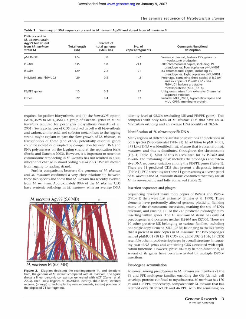
required for proline biosynthesis; and (4) the hemACDB operon(MUL_4598 to MUL_4561), a group of essential genes in M. tu-berculosis required for porphyrin biosynthesis (Sassetti et al.2001). Such exchanges of CDS involved in cell wall biosynthesisand carbon, amino acid, and cofactor metabolism to the laggingstrand might explain in part the slow growth of M. ulcerans, astranscription of these (and other) potentially essential genescould be slowed or disrupted by competition between DNA andRNA polymerases on the lagging strand at the replication forks(Rocha and Danchin 2003). However, it is important to note thatchromosome remodeling in M. ulcerans has not resulted in a sig-nificant net change in strand coding bias as 239 CDS have movedfrom lagging to leading strand.
Further comparisons between the genomes of M. ulceransand M. marinum confirmed a very close relationship betweenthese two species and show that M. ulcerans has recently evolvedfrom M. marinum. Approximately 90% of the M. ulcerans CDShave syntenic orthologs in M. marinum with an average DNA
identity level of 98.3% (excluding ISE and PE/PPE genes). Thiscompares with only 60% of M. ulcerans CDS that have an M.tuberculosis ortholog and an average DNA identity of 78.5%.
Identification of M. ulcerans-specific DNA
Many regions of difference are due to insertions and deletions inboth species (Supplemental Table S1). In addition to pMUM001,475 kb of DNA was identified in M. ulcerans that is absent from M.marinum, and this is distributed throughout the chromosome(Fig. 1; Table 1). Most of this is accounted for by IS2404 andIS2606. The remaining 79 kb includes the prophages and exten-sive DNA sequence variation among the PE/PPE genes (Table 1).There are 11 predicted CDS that present a diagnostic interest(Table 1). PCR screening for these 11 genes among a diverse panelof M. ulcerans and M. marinum strains confirmed that they are allM. ulcerans-specific and fully conserved (Table 2).
Insertion sequences and phages
Sequencing revealed many more copies of IS2404 and IS2606(Table 1) than were first estimated (Stinear et al. 1999). Theseelements have profoundly affected genome plasticity, flankingmany of the chromosome inversions, marking the site of DNAdeletions, and causing 111 of the 743 predicted pseudogenes byinserting within genes. The M. marinum M strain has only 64pseudogenes and possesses neither IS2404 nor IS2606. There are10 other putative ISE belonging to various families, includingone single-copy element (MUL_2378) belonging to the IS3 familythat is present in nine copies in M. marinum. The two prophagesnamed phiMU01 (18 kb, 18 CDS) and phiMU02 (24 kb, 17 CDS)resemble other mycobacteriophages in overall structure, integrat-ing near tRNA genes and containing CDS associated with repli-cation functions. However, phiMU02 may be non-functional, asseveral of its genes have been inactivated by multiple IS2606insertions.
Pseudogene accumulation
Foremost among pseudogenes in M. ulcerans are members of thePE and PPE multigene families encoding the Gly-Ala-rich cellenvelope proteins confined to mycobacteria. M. marinum has 170PE and 105 PPE, respectively, compared with M. ulcerans that hasretained only 70 intact PE and 46 PPE, with the remaining or-
Figure 2. Diagram depicting the rearrangements in, and deletionsfrom, the genome of M. ulcerans compared with M. marinum. The figureshows a linear genomic comparison generated with ACT (Carver et al.2005). (Red lines) Regions of DNA:DNA identity, (blue lines) invertedregions, (orange) strand-displacing rearrangements, (arrow) position ofthe displaced 71-kb fragment.
Table 1. Summary of DNA sequences present in M. ulcerans Agy99 and absent from M. marinum M
DNA present inM. ulcerans strainAgy99 but absentfrom M. marinumstrain M
Total length(kb)
Percent oftotal genome
(5806 kb)No. of
copies/fragmentsComments/functional
description
pMUM001 174 3.0 1–2 Virulence plasmid, harbors PKS genes formycolactone production.
IS2404 335 5.8 213 209 chromosomal copies, including 19pseudogenes. Four copies on pMUM001.
IS2606 129 2.2 91 83 chromosomal copies, including 10pseudogenes. Eight copies on pMUM001.
PhiMU01 and PhiMU02 29 0.5 2 Prophage, containing three copies of IS2404and six copies of IS2606 (12.7 kb).PhiMU01 harbors a putativemetalloprotease (MUL_3218).
PE/PPE genes 15 0.3 97 Uniqueness arises from extensive C-terminalsequence variation.
Other 22 0.4 32 Includes MUL_2832, hypothetical lipase andMUL_0999, membrane protein.
The genome sequence of Mycobacter ium ulcerans
Genome Research 3www.genome.org
on January 9, 2007 www.genome.orgDownloaded from

Tab
le2.
Pres
ence
of11
M.u
lcer
ans-
spec
ific
CD
Sam
ong
dif
fere
nt
stra
ins
ofM
.ulc
eran
san
dM
.mar
inum
asd
eter
min
edb
yPC
R
Stra
ind
etai
ls
Locu
sta
gC
DS
des
crip
tion
Gh
ana
Ag
y99
Ivor
yC
oast
Kob
SEA
ustr
alia
Ch
ant
Nor
thA
ustr
alia
1382
2
Pap
uaN
ewG
uin
ea94
1331
Mal
aysi
a16
15M
alay
sia
9413
28
Peop
le’s
Rep
ublic
ofC
hin
a98
0912
Jap
an75
3M
exic
o51
14Su
rin
am84
2M
.m
arin
uma
MU
L_00
76H
Pb+
++
++
++
�+
++
�M
UL_
0512
HP
phiM
U01
++
�+
++
+�
�+
+�
MU
L_05
13H
Pph
iMU
01+
+�
++
++
��
++
�M
UL_
0515
HP
phiM
U01
++
�+
++
+�
�+
+�
MU
L_05
26H
Pph
iMU
01+
++
++
++
��
�+
�M
UL_
0527
HP
phiM
U01
++
++
++
+�
��
+�
MU
L_09
98H
P+
++
++
++
++
+�
�M
UL_
0999
Mem
bran
epr
otei
n+
++
++
++
++
+�
�M
UL_
1001
HP
++
++
++
++
++
��
MU
L_28
32Li
pase
++
++
++
++
++
��
MU
L_32
18C
HPc
phiM
U02
++
++
++
�+
+�
��
aIn
clud
esM
.m
arin
umM
and
22pr
evio
usly
desc
ribed
M.
mar
inum
stra
ins
(Stin
ear
etal
.20
05a)
.bH
ypot
hetic
alpr
otei
n.c C
onse
rved
hypo
thet
ical
prot
ein.
(CD
S)Pr
otei
n-co
ding
sequ
ence
s.
Stinear et al.
4 Genome Researchwww.genome.org
on January 9, 2007 www.genome.orgDownloaded from

thologs showing widespread sequence variation (Fig. 1; Table 1).These changes suggest a decreased requirement for PE/PPE pro-teins. The degeneration appears symptomatic of an intermediatestage of reductive evolution with M. ulcerans heading toward thehighly contracted genome state of M. leprae, which is nearly de-void of these proteins (Cole et al. 2001).
The loss of paralogous genes and subsequent decrease ingenetic redundancy may also contribute to slow growth via adiminished gene dose effect. For example, the disaccharide tre-halose is essential for mycobacterial cell wall biogenesis and canbe synthesized by three potential pathways (OtsAB, TreYZ, andTreS). As in M. tuberculosis, M. ulcerans has two otsB genes, otsB1and otsB2; however, otsB1 has been disrupted by IS2404. Simi-larly, one of the three gltA genes coding for glutamine synthetase,a central enzyme in the nitrogen regulon, is a pseudogene in M.ulcerans, as are three of four phospholipase C paralogs requiredfor phospholipolysis.
Other gene inactivations suggest divergence of the ecologi-cal niches of M. ulcerans and M. marinum. The crtB locus in M.marinum is responsible for the production of light-inducible ca-rotenoids that protect the bacterium from incident sunlight. M.ulcerans does not produce these pigments, suggesting it is notexposed to direct sunlight in its habitat, yet it contains an iden-tical crtB region. One explanation for the lack of photochromo-genicity may be the introduction of a premature stop codon incrtI, encoding phytoene dehydrogenase, the second committedstep in pigment synthesis.
Protein families
To better understand the biology of M. ulcerans, the predictedproteins were classified into families and compared with thosefound in other mycobacteria. Only one protein family (N > 5),comprising eight metal-dependent hydrolases, was found thathad no counterpart in tubercle and leprosy bacilli. Otherwise, thesame themes were observed (Tekaia et al. 1999), namely an abun-dance of proteins involved in lipid metabolism and prominentPE and PPE families. However, the number of potential transportproteins and enzyme systems predicted to be involved in carbonand energy metabolism was higher. M. marinum has 192 CDSassociated with substrate transport while M. ulcerans has only128.
If M. ulcerans occupies several distinct niches, includingplant biofilms, insects, and humans, then its metabolism may beversatile. In an aquatic environment, M. ulcerans might obtain itsenergy and carbon from degradation of plant saccharides, asthese stimulate growth in vitro (Marsollier et al. 2004b). Little isknown, however, about its metabolism in insect tissues, al-though it may be significant that both M. marinum and M. ulcer-ans encode four potential chitinases/transglycosidases (BuruList/mast/P4.27.html), one of which is attached to a PE domain andthus is expected to be in the cell envelope. These enzymes mightmediate attachment to, or degradation of, the N-acetyl-D-glucosamine polymers that comprise chitin, a major componentof the exoskeletons of insects and crustaceans.
Primary metabolism and respiration
Metabolic pathway reconstruction supported the importance oflipid degradation for central carbon metabolism and uncoveredintact glycolysis and pentose phosphate pathways, but no Ent-ner-Doudoroff pathway. As in M. tuberculosis, M. ulcerans has abifurcated TCA cycle, as it lacks �-ketoglutarate dehydrogenase
(Fig. 3) and thus probably converts �-ketoglutarate to succinatevia �-ketoglutarate decarboxylase (Tian et al. 2005). The TCAcycle incorporates the glyoxylate shunt, with two copies of thegene encoding isocitrate lyase. Thus, like M. tuberculosis, M. ul-cerans may direct the two-carbon degradation products (acetyl-coA) from �-oxidation of host fatty acids into the TCA cycleoperating in biosynthetic mode.
The �-oxidation of fatty acids also generates propionyl-CoA,which can be metabolized in several ways including via themethylcitrate pathway (Fig. 3). M. ulcerans appears to have all themethylcitrate enzymes except methylisocitrate lyase, whose pu-tative gene (MUL_2495) has a frameshift mutation. A reducedcapacity to metabolize propionic acid might be detrimental forgrowth; however, in M. tuberculosis the combination of the twoisocitrate lyases can complement the absence of methylisocitratelyase (Munoz-Elias and McKinney 2005).
Analysis of potential electron transport chains suggests M.ulcerans is capable of growth under aerobic but not anaerobicconditions as it lacks the nitrate and fumarate reductase systems.Based on gene orthology with M. tuberculosis, complete NADHoxidase, ubiquinol cyctochrome c oxidase, and ATP synthasecomplexes indicate ATP generation by oxidative phosphoryla-tion during aerobic growth. M. marinum contains a potentialalternative anaerobic respiratory pathway involving nitritereductase, encoded by nrfD and formate dehydrogenase,where formate oxidation is coupled to nitrite reduction. M. ul-cerans contains an intact nitrite reductase (nrfD), but the linkedformate dehydrogenase system has been inactivated by multi-ple mutations (Fig. 3). Loss of this enzyme, a known seleno-protein, is consistent with deletion of the selenocysteine locusfrom M. ulcerans. M. marinum may be capable of nitrite reduc-tion via the nirBD nitrite reductase and NarK transporter, butwhile M. ulcerans also contains these loci, nirB and narK are pseu-dogenes.
Under microaerophilic conditions, mycobacteria up-regulate expression of a high oxygen affinity cytochrome bdoxidase (cydABCD). This locus is present in both M. ulcerans andM. marinum, but in M. ulcerans the cydA ortholog is a pseudo-gene. A knockout mutation of cydA in Mycobacterium smegmatisleads to a competitive growth disadvantage under microaero-philic conditions (Kana et al. 2001). The above observations sug-gest that M. ulcerans has depleted respiratory versatility. Never-theless, it has maintained >400 putative oxidoreductases, dehy-drogenases, and mono- and dioxygenases (M. marinum has >590),suggesting that a robust and complex respiratory potential re-mains under aerobic conditions. As reported in other bacteriaundergoing reductive evolution, the cytochrome P450 repertoirewas particularly prone to pseudogene formation (Cole et al.2001).
Cell wall lipids and secondary metabolism
The cell envelope of M. ulcerans and the secondary metabolitestherein have been less well studied than those of other mycobac-teria. From the genome sequence several predictions can bemade, as reduction of secondary metabolite loci is widespread,notably through depletion of PKS genes. M. marinum has 27 PKSgenes, but M. ulcerans has reduced its complement to 12. Ten ofthese PKS produce important cell-wall-associated lipids includingmycolic acids (pks13), phthiodiolones (ppsA-E), phenol phthio-diolones (pks15/1), mannosyl-�1-phopholipids (pks12), and my-cobactins (mbtC,D) (Matsunaga et al. 2004; Portevin et al. 2004).
The genome sequence of Mycobacter ium ulcerans
Genome Research 5www.genome.org
on January 9, 2007 www.genome.orgDownloaded from

Neither M. ulcerans nor M. marinum contains the pks2 locus, re-quired for the production of the sulfolipids. A larger PKS operonof unknown function harbors pks9 and pks11, but this operonmay be inactive in M. ulcerans, as its other PKS genes (pks7, pks8,pks10) have become pseudogenes. One predicted consequencefor M. ulcerans of downsizing its PKS complement would be lib-eration of energy (NADP) and substrate (malonyl-CoA and meth-ylmalonyl-CoA) for the production of mycolactone. Contractionof secondary metabolism is also reflected in reduction of theMmpL family of lipid and polyketide transporters from 25 in M.marinum to only six in M. ulcerans.
Despite the loss of many pks, M. ulcerans has retained a sig-nificant anabolic lipid potential highlighting the central impor-tance of particular lipids to the preservation of the mycobacterialcell wall. Structural analyses of some of the signature lipid specieswere undertaken and then correlated with the genome findings.M. ulcerans and M. marinum have been previously shown toproduce diunsaturated, methoxy, and ketomycolic acids (Daffeet al. 1991). Central to synthesis of these mycolates and otherlipids are the fatty acid synthases I and II. All the principal com-ponents of these enzymes are present in M. ulcerans, and the keyproteins share >90% identity with those of M. marinum and M.tuberculosis.
M. ulcerans also produces a highly abundant and apolar cellwall lipid consisting of diesters of phthiodiolone (Daffe et al.1984; Onwueme et al. 2005). The common �-diol backbone ofphthiodiolones in M. ulcerans is produced from C16–C18 fatty
acids by five type I PKS encoded by the genes ppsA–E. Processingof p-hydroxyphenylalkanoate rather than fatty acids by thesePKS results in the production of phenolphthiodiolones, whichcan be further modified by glycosylation to form the virulencefactors and immune modulators, the phenolic glycolipids (PGLs)(Daffe et al. 1992; Reed et al. 2004). In M. tuberculosis, Rv2962encodes the glycosyl transferase that adds the first sugar residueto the highly related lipid, phenolphthiocerol (Perez et al. 2004).The absence of PGLs in M. ulcerans Agy99 is readily explained, asMUL_1998, the Rv2962 ortholog, is a pseudogene. This observa-tion was supported by MALDI-TOF MS detection of the substratefor the MUL_1998 gene product, phenolphthiodiolone diphthio-ceranoate, in M. ulcerans but not in its glycosylated form, whichwas detected in M. marinum (Figs. 3, 4).
M. ulcerans and M. marinum contain both a mevalonate(MVA) and non-mevalonate (DXP) pathway for polyprenoid syn-thesis, the first example of such in the mycobacteria and a rareoccurrence among bacteria (Rohdich et al. 2004). Polyprenylphosphate (Pol-P) forms lipid-linked sugar intermediates that arerequired by bacteria during cell wall biosynthesis. They areformed by sequential condensation of isopentenyl diphosphate(IPP) and dimethylallyl diphosphate (DMAPP). In other myco-bacteria these molecules are synthesized via the DXP pathway;the same appears true for M. ulcerans, as the MVA pathway isprobably inactive because one of the genes in the MVA operon,MUL_3521, encoding a putative hydroxymethylglutaryl-coenzyme A synthase, has been disrupted (Fig. 3).
Figure 3. Some important biochemical pathways. The main biochemical activities of M. ulcerans discussed in the text are depicted; these are allpresent in M. marinum except for mycolactone biosynthesis encoded by pMUM001. Some key genes and pathways that have been inactivated in M.ulcerans are indicated.
Stinear et al.
6 Genome Researchwww.genome.org
on January 9, 2007 www.genome.orgDownloaded from

Implications for pathogenesis from genome reduction
Comparative genomics also shows that the M. ulcerans genome iscontracting and that extensive pseudogene formation has oc-curred. There are 157 regions, accounting for 1232 kb, present inM. marinum but absent from M. ulcerans. These were assigned a M.ulcerans Region of Difference (MURD) number (SupplementalTable S1). While some of these represent insertions in M. mari-num, including seven prophage and 13 ISE, totaling 168 kb, themajority result from the accumulated deletion of 1064 kb of DNAfrom M. ulcerans. In this way many paralogous gene families havebeen lost in a situation reminiscent of M. leprae where consider-able genetic downsizing has occurred (Cole et al. 2001). Mostnotable among these are several secondary metabolism loci andthe PE/PPE genes, the latter accounting for 45% of the MURDs.Other deletions affect genes involved in intermediary and energymetabolism such as potassium transport (MURD9), hydrogenase(MURD17), and production of selenocysteine (MURD142), a rareamino acid found in the active sites of formate dehydrogenaseand many bacterial antioxidant proteins.
Noteworthy deletions in M. ulcerans include the ESX loci.While there are five complete ESX systems in M. marinum,only three remain in M. ulcerans as the other two have incurreddeletions. In M. tuberculosis, the esx-1 locus encodes a novel pro-tein secretion apparatus, which contributes to its virulence, in-tercellular spread, and immunogenicity (Brodin et al. 2004). InM. ulcerans, esx-1 has been disrupted by two deletions (MURD151and MURD152). Effector proteins secreted by ESX systems belongto the ESAT-6 or the EspA families (Fortune et al. 2005), and13 ESAT-6 proteins exist in M. ulcerans. Interestingly, theespA gene, encoding a putative substrate of ESX-1, has beendeleted (MURD111). Loss of these systems, which triggergranuloma formation by M. marinum and M. tuberculosis, maycontribute to the predominantly extracellular location of M. ul-cerans in infected tissue. Supplemental Table S1 also lists 102orthologous M. tuberculosis genes that M. ulcerans has lost bydeletion.
Discussion
Bacteria that have recently passed an evolutionary bottleneckand are now adapting to a new, more stable environment, suchas Mycobacterium leprae, Yersinia pestis, and Bordetella pertussis(Parkhill et al. 2003), have some of the following genomic signa-tures: proliferation of ISE, accumulation of pseudogenes, chro-mosomal rearrangements, genome downsizing, and acquisitionof foreign genes, often via plasmids or bacteriophage, that confera fitness advantage in the new environment. They also show ahigh degree of genetic relatedness or clonality. All six genomicsignatures are shared by M. ulcerans, which appears to be under-going reductive evolution and, as such, may represent a goodmodel for studying this phenomenon and the response to chang-ing from an environmental to host-adapted, possibly arthropodniche.
Our study provides compelling evidence for M. ulcerans be-ing a descendant of an M. marinum progenitor strain that ac-quired the virulence plasmid, pMUM001, from another actino-bacterium. This event then led to divergence of the ecologicalniches of M. ulcerans and M. marinum, although other factors mayhave contributed. For instance, the crtB locus is responsible forthe production of light-inducible carotenoids that protect M.marinum from incident sunlight (Ramakrishnan et al. 1997). M.ulcerans does not produce these pigments, due to a lesion in crtI,so its impaired ability to withstand exposure to direct sunlightwould lead to selection for progeny capable of adapting to, andsettling in, a more protected niche. The strong selective pressureexerted by mycolactone could have enabled M. ulcerans to colo-nize aquatic insects or other animals sharing its ecosystem.
Once within a more stable and protected habitat, loss ofgene function and slower growth would be readily tolerated. Ge-nomic analysis suggests possible mechanisms that have impactedon growth rate including the loss, or reduction in number, ofgenes for transport of potassium and other solutes; enzymes suchas hydrogenase, glutamine synthase, and phospholipase C; andfor trehalose and isoprenoid production, among others; in turn,the capacity for free-living would diminish.
Unlike other pathogenic mycobacteria, M. ulcerans is foundpredominantly in extracellular form in infected human tissue,although it passes via a transient intracellular stage (Coutanceauet al. 2005). In part, this is due to the antiphagocytic properties ofmycolactone, but loss of the protein secretion system, ESX-1,that mediates the export of the proteins ESAT-6 and EspA mayalso contribute. Inactivation of ESX-1 in M. marinum or M. tuber-culosis results in reduced uptake by phagocytes and greatly di-minished intercellular spread (Brodin et al. 2004). It is also con-ceivable that some of the functions encoded by the M. ulcerans-restricted CDS could contribute to the pathology associated withBuruli ulcer. These genes or their products may also find appli-cation in the development of new diagnostic tests, which arerequired to help control the spread of Buruli ulcer, and theirconservation in all isolates of M. ulcerans examined is encourag-ing in this respect.
Methods
M. ulcerans Agy99M. ulcerans Agy99 was isolated from an ulcerative lesion on theright elbow of a female patient from the Ga district of Ghana in1999. Multi-locus sequence typing and inter-IS PCR analysis con-
Figure 4. Partial matrix-assisted laser desorption ionization-time-of-flight (MALDI-TOF) mass spectra of lipids from M. ulcerans and M. mari-num. These spectra show the presence of diesters of phthiocerol andphthiodiolone, and of phenolic glycolipids in M. marinum, whereas onlydiesters of phthiodiolone and phenolphthiodiolone occur in M. ulcerans.
The genome sequence of Mycobacter ium ulcerans
Genome Research 7www.genome.org
on January 9, 2007 www.genome.orgDownloaded from

firmed that this strain belonged to the African epidemic geno-type. DNA was prepared for sequencing four passages after pri-mary isolation.
Whole-genome sequencingA whole-genome shotgun library was prepared in the vectorpCDNA2.1 using Escherichia coli strain XL2-blue (Invitrogen). Ge-nomic DNA was sheared by sonication and size fractionated toproduce plasmids with inserts in the size ranges of 2–3 kb, 3–5 kb,and 5–10 kb. These were used as template in DNA cycle sequenc-ing reactions to obtain 75,415 end-sequence reads using an ABI3700 DNA sequencer (Applied Biosystems), providing theoretical8� coverage. Sequence assembly was performed by PHRAP andGAP4 as described (Cole et al. 1998). Gap closure was facilitatedby scaffolding using a large insert BAC library (Stinear et al.2005a) augmented with primer walking and PCR.
Genome annotation and comparative genomicsAnnotation and database construction was performed withWasabi, an in-house, Web-interfaced MySQL database for man-agement of genome annotation, Artemis, and GenoList. A com-plete database is available at http://genolist.pasteur.fr/BuruList/.CDS were predicted using GenemarkS and GenoStar, and com-pared with public sequence databases using the BLAST suite ofalgorithms. Pseudogenes had one or more mutations that wouldprevent correct translation. Mutations were checked againstoriginal sequence data to exclude sequencing errors. The ArtemisComparison Tool (Carver et al. 2005) was used for comparativegenome analysis.
Lipid analysisLipids were extracted with CHCl3/CH3OH (1:1 v/v) and subjectedto MALDI-TOF mass spectrometry using a Voyager DE-STRMALDI-TOF instrument (PerSeptive Biosystems) equipped with apulse nitrogen laser emitting at 337 nm and analyzed in thereflector mode as previously described (Perez et al. 2004).
AcknowledgmentsWe thank Paul Harrison, Ivan Moszer, and Roland Brosch forsharing expertise and helpful discussions. We gratefully acknowl-edge the financial support of the Génopole program, the Asso-ciation Française Raoul Follereau, the National Health and Medi-cal Research Council of Australia, and the World Health Organi-zation through grants provided by the Nippon Foundation,Tokyo, Japan.
References
Alsop, D. 1972. The Bairnsdale ulcer. Aust. N. Z. J. Surg. 41: 317–319.Brodin, P., Rosenkrands, I., Andersen, P., Cole, S.T., and Brosch, R. 2004.
ESAT-6 proteins: Protective antigens and virulence factors? TrendsMicrobiol. 12: 500–508.
Carver, T.J., Rutherford, K.M., Berriman, M., Rajandream, M.A., Barrell,B.G., and Parkhill, J. 2005. ACT: The Artemis Comparison Tool.Bioinformatics 21: 3422–3423.
Cole, S.T., Brosch, R., Parkhill, J., Garnier, T., Churcher, C., Harris, D.,Gordon, S.V., Eiglmeier, K., Gas, S., Barry III, C.E., et al. 1998.Deciphering the biology of Mycobacterium tuberculosis from thecomplete genome sequence. Nature 393: 537–544.
Cole, S.T., Eiglmeier, K., Parkhill, J., James, K.D., Thomson, N.R.,Wheeler, P.R., Honore, N., Garnier, T., Churcher, C., Harris, D., et al.2001. Massive gene decay in the leprosy bacillus. Nature409: 1007–1011.
Coutanceau, E., Marsollier, L., Brosch, R., Perret, E., Goossens, P.,Tanguy, M., Cole, S.T., Small, P.L., and Demangel, C. 2005.Modulation of the host immune response by a transient intracellular
stage of Mycobacterium ulcerans: The contribution of endogenousmycolactone toxin. Cell. Microbiol. 7: 1187–1196.
Daffe, M., Laneelle, M.A., Roussel, J., and Asselineau, C. 1984. Specificlipids from Mycobacterium ulcerans. Ann. Microbiol. (Paris)135A: 191–201.
Daffe, M., Laneelle, M.A., and Lacave, C. 1991. Structure andstereochemistry of mycolic acids of Mycobacterium marinum andMycobacterium ulcerans. Res. Microbiol. 142: 397–403.
Daffe, M., Varnerot, A., and Levy-Frebault, V.V. 1992. The phenolicmycoside of Mycobacterium ulcerans: Structure and taxonomicimplications. J. Gen. Microbiol. 138: 131–137.
Eisen, J.A., Heidelberg, J.F., White, O., and Salzberg, S.L. 2000. Evidencefor symmetric chromosomal inversions around the replication originin bacteria. Genome Biol 1: research0011.
Etuaful, S., Carbonnelle, B., Grosset, J., Lucas, S., Horsfield, C., Phillips,R., Evans, M., Ofori-Adjei, D., Klustse, E., Owusu-Boateng, J., et al.2005. Efficacy of the combination rifampin–streptomycin inpreventing growth of Mycobacterium ulcerans in early lesions ofBuruli ulcer in humans. Antimicrob. Agents Chemother.49: 3182–3186.
Fortune, S.M., Jaeger, A., Sarracino, D.A., Chase, M.R., Sassetti, C.M.,Sherman, D.R., Bloom, B.R., and Rubin, E.J. 2005. Mutuallydependent secretion of proteins required for mycobacterialvirulence. Proc. Natl. Acad. Sci. 102: 10676–10681.
Johnson, P.D., Stinear, T., Small, P.L., Pluschke, G., Merritt, R.W.,Portaels, F., Huygen, K., Hayman, J.A., and Asiedu, K. 2005. Buruliulcer (M. ulcerans infection): New insights, new hope for diseasecontrol. PLoS Med. 2: e108.
Kana, B.D., Weinstein, E.A., Avarbock, D., Dawes, S.S., Rubin, H., andMizrahi, V. 2001. Characterization of the cydAB-encodedcytochrome bd oxidase from Mycobacterium smegmatis. J. Bacteriol.183: 7076–7086.
MacCallum, P., Tolhurst, J., Buckle, G., and Ha, S. 1948. A newmycobacterial infection in man. J. Pathol. Bacteriol. 60: 93–122.
Marsollier, L., Robert, R., Aubry, J., Saint Andre, J.P., Kouakou, H.,Legras, P., Manceau, A.L., Mahaza, C., and Carbonnelle, B. 2002.Aquatic insects as a vector for Mycobacterium ulcerans. Appl. Environ.Microbiol. 68: 4623–4628.
Marsollier, L., Severin, T., Aubry, J., Merritt, R.W., Saint Andre, J.P.,Legras, P., Manceau, A.L., Chauty, A., Carbonnelle, B., and Cole, S.T.2004a. Aquatic snails, passive hosts of Mycobacterium ulcerans. Appl.Environ. Microbiol. 70: 6296–6298.
Marsollier, L., Stinear, T., Aubry, J., Saint Andre, J.P., Robert, R., Legras,P., Manceau, A.L., Audrain, C., Bourdon, S., Kouakou, H., et al.2004b. Aquatic plants stimulate the growth of and biofilmformation by Mycobacterium ulcerans in axenic culture and harborthese bacteria in the environment. Appl. Environ. Microbiol.70: 1097–1103.
Marsollier, L., Aubry, J., Coutanceau, E., Andre, J.P., Small, P.L., Milon,G., Legras, P., Guadagnini, S., Carbonnelle, B., and Cole, S.T. 2005.Colonization of the salivary glands of Naucoris cimicoides byMycobacterium ulcerans requires host plasmatocytes and a macrolidetoxin, mycolactone. Cell. Microbiol. 7: 935–943.
Matsunaga, I., Bhatt, A., Young, D.C., Cheng, T.Y., Eyles, S.J., Besra,G.S., Briken, V., Porcelli, S.A., Costello, C.E., Jacobs Jr., W.R., et al.2004. Mycobacterium tuberculosis pks12 produces a novel polyketidepresented by CD1c to T cells. J. Exp. Med. 200: 1559–1569.
Munoz-Elias, E.J. and McKinney, J.D. 2005. Mycobacterium tuberculosisisocitrate lyases 1 and 2 are jointly required for in vivo growth andvirulence. Nat. Med. 11: 638–644.
Onwueme, K.C., Vos, C.J., Zurita, J., Soll, C.E., and Quadri, L.E. 2005.Identification of phthiodiolone ketoreductase, an enzyme requiredfor production of mycobacterial diacyl phthiocerol virulence factors.J. Bacteriol. 187: 4760–4766.
Parkhill, J., Sebaihia, M., Preston, A., Murphy, L.D., Thomson, N.,Harris, D.E., Holden, M.T., Churcher, C.M., Bentley, S.D., Mungall,K.L., et al. 2003. Comparative analysis of the genome sequences ofBordetella pertussis, Bordetella parapertussis and Bordetellabronchiseptica. Nat. Genet. 35: 32–40.
Perez, E., Constant, P., Lemassu, A., Laval, F., Daffe, M., and Guilhot, C.2004. Characterization of three glycosyltransferases involved in thebiosynthesis of the phenolic glycolipid antigens from theMycobacterium tuberculosis complex. J. Biol. Chem. 279: 42574–42583.
Portevin, D., De Sousa-D’Auria, C., Houssin, C., Grimaldi, C., Chami,M., Daffe, M., and Guilhot, C. 2004. A polyketide synthase catalyzesthe last condensation step of mycolic acid biosynthesis inmycobacteria and related organisms. Proc. Natl. Acad. Sci.101: 314–319.
Ramakrishnan, L., Tran, H.T., Federspiel, N.A., and Falkow, S. 1997. AcrtB homolog essential for photochromogenicity in Mycobacteriummarinum: Isolation, characterization, and gene disruption via
Stinear et al.
8 Genome Researchwww.genome.org
on January 9, 2007 www.genome.orgDownloaded from

homologous recombination. J. Bacteriol. 179: 5862–5868.Reed, M.B., Domenech, P., Manca, C., Su, H., Barczak, A.K., Kreiswirth,
B.N., Kaplan, G., and Barry III, C.E. 2004. A glycolipid ofhypervirulent tuberculosis strains that inhibits the innate immuneresponse. Nature 431: 84–87.
Rocha, E.P. and Danchin, A. 2003. Essentiality, not expressiveness,drives gene-strand bias in bacteria. Nat. Genet. 34: 377–378.
Rohdich, F., Bacher, A., and Eisenreich, W. 2004. Perspectives inanti-infective drug design. The late steps in the biosynthesis of theuniversal terpenoid precursors, isopentenyl diphosphate anddimethylallyl diphosphate. Bioorg. Chem. 32: 292–308.
Sassetti, C.M., Boyd, D.H., and Rubin, E.J. 2001. Comprehensiveidentification of conditionally essential genes in mycobacteria. Proc.Natl. Acad. Sci. 98: 12712–12717.
Stinear, T., Ross, B.C., Davies, J.K., Marino, L., Robins-Browne, R.M.,Oppedisano, F., Sievers, A., and Johnson, P.D. 1999. Identificationand characterization of IS2404 and IS2606: Two distinct repeatedsequences for detection of Mycobacterium ulcerans by PCR. J. Clin.Microbiol. 37: 1018–1023.
Stinear, T.P., Jenkin, G.A., Johnson, P.D.R., and Davies, J.K. 2000.Comparative genetic analysis of Mycobacterium ulcerans andMycobacterium marinum reveals evidence of recent divergence. J.Bacteriol. 182: 6322–6330.
Stinear, T.P., Mve-Obiang, A., Small, P.L., Frigui, W., Pryor, M.J., Brosch,R., Jenkin, G.A., Johnson, P.D., Davies, J.K., Lee, R.E., et al. 2004.Giant plasmid-encoded polyketide synthases produce the macrolidetoxin of Mycobacterium ulcerans. Proc. Natl. Acad. Sci.101: 1345–1349.
Stinear, T.P., Hong, H., Frigui, W., Pryor, M.J., Brosch, R., Garnier, T.,Leadlay, P.F., and Cole, S.T. 2005a. Common evolutionary origin forthe unstable virulence plasmid pMUM found in geographicallydiverse strains of Mycobacterium ulcerans. J. Bacteriol. 187: 1668–1676.
Stinear, T.P., Pryor, M.J., Porter, J.L., and Cole, S.T. 2005b. Functionalanalysis and annotation of the virulence plasmid pMUM001 fromMycobacterium ulcerans. Microbiol. 151: 683–692.
Tekaia, F., Gordon, S.V., Garnier, T., Brosch, R., Barrell, B.G., and Cole,S.T. 1999. Analysis of the proteome of Mycobacterium tuberculosis insilico. Tuber. Lung Dis. 79: 329–342.
Tian, J., Bryk, R., Itoh, M., Suematsu, M., and Nathan, C. 2005. Varianttricarboxylic acid cycle in Mycobacterium tuberculosis: Identificationof �-ketoglutarate decarboxylase. Proc. Natl. Acad. Sci.102: 10670–10675.
Received September 12, 2006; accepted in revised form November 29, 2006.
The genome sequence of Mycobacter ium ulcerans
Genome Research 9www.genome.org
on January 9, 2007 www.genome.orgDownloaded from
Copyright © 2022 FDOKUMEN