A Genomic View of Sugar Transport in Mycobacterium smegmatis and Mycobacterium tuberculosis
Upload
independentCategory
view
4download
0
RESEARCH ARTICLE Open Access
A quick and cost effective method for thediagnosis of Mycobacterium ulcerans infectionDziedzom K de Souza1*, Charles Quaye1, Lydia Mosi1,2, Phyllis Addo3 and Daniel A Boakye1
Abstract
Background: Buruli ulcer (BU), a neglected tropical skin disease caused by Mycobacterium ulcerans, has beenreported in over 30 countries worldwide and is highly endemic in rural West and Central Africa. The mode oftransmission remains unknown and treatment is the only alternative to disease control. Early and effectivetreatment to prevent the morbid effects of the disease depends on early diagnosis; however, current diagnosisbased on clinical presentation and microscopy has to be confirmed by PCR and other tests in referencelaboratories. As such confirmed BU diagnosis is either late, inefficient, time consuming or very expensive, and thereis the need for an early diagnosis tool at point of care facilities. In this paper we report on a simple, quick andinexpensive diagnostic test that could be used at point of care facilities, in resource-poor settings.
Methods: The methodology employed is based on the loop mediated isothermal amplification (LAMP) technique.Four sets of Primers, targeting the mycolactone encoding plasmid genome sequence of M. ulcerans weredesigned. The BU-LAMP assay was developed and tested on five M. ulcerans strains from patients in Ghana andtwo American Type Culture Control (ATCC) reference isolates; Ghana #970321 (D19F9) and Benin #990826(D27D14). We also tested the assay on other closely related, mycolactone-producing mycobacterial strains; M.marinum 1218, M. marinum DL240490, M. liflandii and M. pseudoshotsii, as well as experimentally infected laboratoryanimal and clinical samples.
Results: The results revealed a high specificity of the BU-LAMP assay for selectively detecting M. ulcerans.Compared to the conventional IS-2404 PCR, the new assay is cheaper and simpler and ten times more sensitive.Test results can be obtained within 1 hour.
Conclusions: This study indicates that the BU-LAMP assay could be suitable for early disease diagnosis andapplication in low-resource health facilities.
Keywords: Buruli ulcer, Mycobacterium ulcerans, Diagnosis, Loop mediated isothermal amplification
BackgroundMycobacterium ulcerans, a bacterium belonging to thesame family as M. tuberculosis and M. leprae, is the cau-sative agent of Buruli ulcer (BU). BU has been describedas a neglected tropical disease and is the third mostcommon mycobacterial infection after tuberculosis andleprosy [1]. It is a necrotizing, painless, cutaneous infec-tion mainly localized on the limbs of affected individualsand causes extensive damage to the skin, its associatedtissues and even the bone. The pathology of the disease
is due to mycolactone, a necrotizing and immunosup-pressive lipid toxin produced by the bacterium. Thegenes that code for the production of the toxin arelocated on a 174 kb plasmid. In endemic countries likeGhana, Cote d’Ivoire and Benin, the disease is prevalentin rural poor communities. In the most endemic districtin Ghana a prevalence of up to 150.8 per 100,000 indivi-duals has been reported [2]. However epidemiologicalstudies suggest underreporting and improper diagnosisas some of the factors hindering the determination ofthe exact disease burden in endemic areas. In all ende-mic areas, the infection burden has long term socio-eco-nomic impacts on infected individuals and theircommunities.
* Correspondence: [email protected] Department, Noguchi Memorial Institute for Medical Research,University of Ghana, P. O. Box, LG 581, Legon-Accra, GhanaFull list of author information is available at the end of the article
de Souza et al. BMC Infectious Diseases 2012, 12:8http://www.biomedcentral.com/1471-2334/12/8
© 2012 de Souza et al; licensee BioMed Central Ltd This is an Open Access article distributed under the terms of the CreativeCommons Attribution License (http://creativecommons.org/licenses/by/2.0), which permits unrestricted use, distribution, andreproduction in any medium, provided the original work is properly cited.
Current reporting of cases of BU infection is based onthe presentation of the symptoms. This represents achallenge due to the vast number of other skin infec-tions or conditions that may exhibit symptoms similarto that of BU [3-5]. The WHO has therefore directedthat all clinically diagnosed or suspected cases of BU beconfirmed. Currently, this can only be done in referencelaboratories, since the current methods are not amen-able to point of care diagnosis.Laboratory confirmation of BU is complex and has
evolved over the years. Mycobacterium ulcerans stainsred (acid-fast bacilli, AFB) in the Ziehl-Neelsen stainingprocedure but this method has a low sensitivity [6].Swabs taken from lesions often do not show AFB bymicroscopic examination. Culturing M. ulcerans fromclinical samples is difficult and has a low sensitivity ofabout 35-60% [6,7]. The bacterium is notoriously slow-growing (6-8 weeks) and culture media are frequentlycontaminated with other faster growing species [8]. PCRmethods have been developed for BU diagnosis basedon the 16S rRNA gene [6], the hsp65 gene [9], or theinsertion sequence IS-2404 [10]. Although the sensitivityof PCR is high (98%), this method is expensive andrequires technical expertise in terms of DNA extractionand equipment needed.Notwithstanding the fact that a combination of these
methods can lead to an accurate diagnosis, the highlytechnical and expensive nature of the techniques con-fines them to reference laboratories. Thus, there is nosimple, rapid test that is appropriate for early point ofcare diagnosis in the low-resourced laboratory settingswhere the disease is most prevalent. This represents ahuge gap between early diagnosis before ulcers anddeformities occur and the critical need for treatmentand prevention of associated deformities. We report thedevelopment of a simple and relatively inexpensive testfor M. ulcerans diagnosis, which could easily be appliedin basic healthcare facilities, without recourse to expen-sive, complex and time-consuming methods. This newmethod is based on a novel DNA amplification method,developed by Notomi and colleagues [11].
MethodsThe methodology used is termed loop mediated isother-mal amplification (LAMP) technique and has beenapplied for the molecular diagnosis of a variety of dis-eases including; P. falciparum malaria, West Nile Virus,Influenza, Tuberculosis, Trypanosomiasis and filarialdetection in mosquitoes [12-16].
Primer designThe mycolactone encoding plasmid genome sequence,pMUM001, of M. ulcerans Agy99 (GenBank accession no.BX649209.1) was compared with M. marinum 1218
(GenBank accession no. EU271967.1); primers weredesigned from regions with the lowest sequence similarity,using the LAMP primer design software, PrimerExplorerV4 http://primerexplorer.jp/e/. Four sets of primers target-ing different regions of pMUM001 were generated.
Preparation of templatesThe bacterial strains used in this study are listed inTable 1. We also used tissue biopsy samples obtainedfrom lesions of mice that had been experimentallyinfected with M. ulcerans. Finally, the assay was testedon archived/stored human samples (Table 2). TheNoguchi Memorial Institute for Medical Research is oneof three reference centers in Ghana where BU samplesfrom patients are sent for confirmation. For this study,no samples were directly taken from patients; the assaywas only tested on stored samples. The DNA templatefrom these samples was prepared by boiling and Qiagenextraction. For the boiling method, each sample wasground in a mortar, transferred to an eppendorf tubeand incubated in a water-bath at 90°C for 20 minutes.The extraction of DNA, using the Qiagen kit, followedthe recommended procedures from the manufacturers.The DNA concentration for the samples extracted usingthe Qiagen kit was quantified by measuring the opticaldensity at 260 nm.
LAMP assayThe BU-LAMP assay protocol was optimized for maxi-mum efficiency, using the Loopamp DNA amplificationkit (Eiken Chemical), and performed according to themanufacturer’s protocol. LAMP assays were performedin 25 μl reactions each containing 20 μM of each innerprimer (FIP and BIP), 5 μM of each outer primer (F3and B3), 20 μM each of the loop primers (FLP andBLP), 12.5 μl of reaction mix, 1 μl of fluorescent detec-tion reagent, 1 μl of Bst DNA polymerase and 2 μl ofprepared DNA templates. The reaction mixture wasincubated at 65°C for 60 min followed by an enzymeinactivation step of 90°C for 10 min. Optimization wascarried out using either a thermal cycler or a waterbath. The use of the latter was to ensure the applicabil-ity of the LAMP assay in poorly resourced health facil-ities. The assay was tested in duplicate on DNAtemplates prepared from all samples as described above.Products were visualized under UV light directly in theeppendorf tubes. A positive and a negative control wereincluded in both conventional PCR and BU-LAMP reac-tions. Sensitivity testing was based on 100-fold serialdilutions up to 10-8. For the purpose of practical applic-ability in low-resourced laboratories, the dilution beyondwhich no discernable differences between the samplesand the negative control could be detected under UVlight (by the naked eye) was considered the sensitivity
de Souza et al. BMC Infectious Diseases 2012, 12:8http://www.biomedcentral.com/1471-2334/12/8
Page 2 of 7
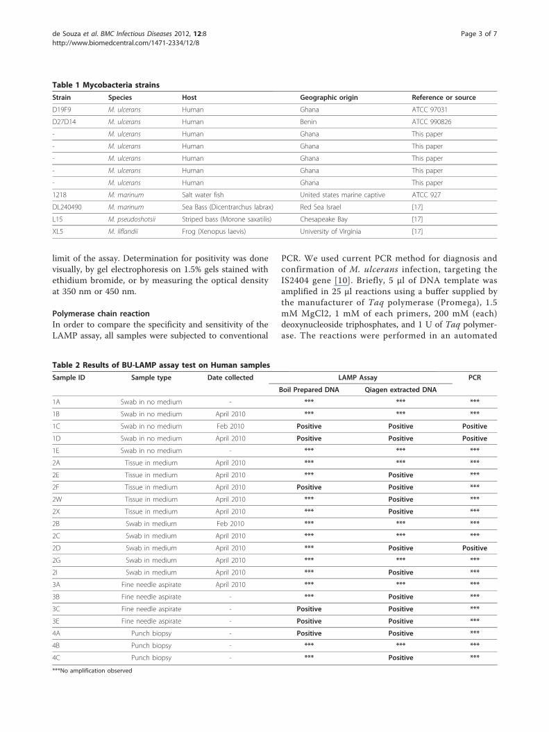
limit of the assay. Determination for positivity was donevisually, by gel electrophoresis on 1.5% gels stained withethidium bromide, or by measuring the optical densityat 350 nm or 450 nm.
Polymerase chain reactionIn order to compare the specificity and sensitivity of theLAMP assay, all samples were subjected to conventional
PCR. We used current PCR method for diagnosis andconfirmation of M. ulcerans infection, targeting theIS2404 gene [10]. Briefly, 5 μl of DNA template wasamplified in 25 μl reactions using a buffer supplied bythe manufacturer of Taq polymerase (Promega), 1.5mM MgCl2, 1 mM of each primers, 200 mM (each)deoxynucleoside triphosphates, and 1 U of Taq polymer-ase. The reactions were performed in an automated
Table 1 Mycobacteria strains
Strain Species Host Geographic origin Reference or source
D19F9 M. ulcerans Human Ghana ATCC 97031
D27D14 M. ulcerans Human Benin ATCC 990826
- M. ulcerans Human Ghana This paper
- M. ulcerans Human Ghana This paper
- M. ulcerans Human Ghana This paper
- M. ulcerans Human Ghana This paper
- M. ulcerans Human Ghana This paper
1218 M. marinum Salt water fish United states marine captive ATCC 927
DL240490 M. marinum Sea Bass (Dicentrarchus labrax) Red Sea Israel [17]
L15 M. pseudoshotsii Striped bass (Morone saxatilis) Chesapeake Bay [17]
XL5 M. liflandii Frog (Xenopus laevis) University of Virginia [17]
Table 2 Results of BU-LAMP assay test on Human samples
Sample ID Sample type Date collected LAMP Assay PCR
Boil Prepared DNA Qiagen extracted DNA
1A Swab in no medium - *** *** ***
1B Swab in no medium April 2010 *** *** ***
1C Swab in no medium Feb 2010 Positive Positive Positive
1D Swab in no medium April 2010 Positive Positive Positive
1E Swab in no medium - *** *** ***
2A Tissue in medium April 2010 *** *** ***
2E Tissue in medium April 2010 *** Positive ***
2F Tissue in medium April 2010 Positive Positive ***
2W Tissue in medium April 2010 *** Positive ***
2X Tissue in medium April 2010 *** Positive ***
2B Swab in medium Feb 2010 *** *** ***
2C Swab in medium April 2010 *** *** ***
2D Swab in medium April 2010 *** Positive Positive
2G Swab in medium April 2010 *** *** ***
2I Swab in medium April 2010 *** Positive ***
3A Fine needle aspirate April 2010 *** *** ***
3B Fine needle aspirate - *** Positive ***
3C Fine needle aspirate - Positive Positive ***
3E Fine needle aspirate - Positive Positive ***
4A Punch biopsy - Positive Positive ***
4B Punch biopsy - *** *** ***
4C Punch biopsy - *** Positive ***
***No amplification observed
de Souza et al. BMC Infectious Diseases 2012, 12:8http://www.biomedcentral.com/1471-2334/12/8
Page 3 of 7

thermal cycler (MJ Research), using an initial denatura-tion of 94°C for 2 min, followed by 35 cycles of 1 min-ute steps at 94°C, 66°C, and 72°C, and a final elongationstep of 72°C for 7 minutes. PCR amplicons were visua-lized by gel electrophoresis on 1.5% gels stained withethidium bromide.
Ethical approvalEthical approval for the use of animal and archived/stored human samples was obtained from the Institu-tional Review Board of the Noguchi Memorial Institutefor Medical Research.
ResultsBU-LAMP primer for M. ulcerans detectionPrimers were designed from positions 7303 to 7483 ofthe M. ulcerans plasmid sequence. A set of 6 primers,consisting of two outer (F3 and B3), two inner (FIP andBIP) and two loop primers (FLP and BLP) weredesigned. The primer sequences and arrangements areshown in Figure 1. Primers FIP and BIP are the combi-nations of 2 sequences consisting F1c and F2 and B1cand B2 respectively. These primers amplify a 180 bpfragment.
BU-LAMP assaysThe BU-LAMP assay was successful, amplifying M.ulcerans at 65°C. Amplification results could also beobserved after incubating for 15 minutes. Trials incu-bated in both a thermal-cycler and the water-bath gavesimilar results.Tests performed on the very closely related M. mari-
num 1218 and other mycolactone producing mycobac-teria; M. marinum DL240490, M. liflandii and M.pseudoshotsii were negative indicating the BU-LAMPprimers to be specific to M. ulcerans. The DNA concen-tration at the visual detection limit was estimated to be
48 pg/μl. However, by measuring the OD value of BU-LAMP products (OD values measured at 350 and 450nm) we were able to detect positivity when using DNAconcentrations as low as 0.5 fg/μl (Figure 2).
Tests on animal and human samples9.5 μg of pure DNA was obtained from approximately50 mg of mice biopsy sample, using the Qiagen DNAextraction method. Both the Qiagen-extracted and boil-prepared DNA produced results, with the former givingthe best results (Figure 3). Raw samples, i.e. maceratedsamples without heat or chemical treatment, used in theassay failed to produce visibly-detectable change in colorintensity when examined directly under UV light (Figure3). However, running the BU-LAMP products from rawsamples on an agarose gel revealed them to produceamplifications (Figure 4).Results of the BU-LAMP test on clinical samples are
shown in Table 2. Samples were tested twice for the
Figure 1 BU-LAMP primer arrangements and sequences.
Figure 2 Absorbance-concentration curve of BU-LAMPproducts.
1 2 3 4 5
Figure 3 Visualization of BU-LAMP products exposed to UVlight. Tube 1 contains DNA extracted using the boil preparationmethod; tube 2 contains Qiagen extracted DNA; tube 3 containsraw/untreated sample; tube 4 is the positive control and tube 5 isthe negative control.
de Souza et al. BMC Infectious Diseases 2012, 12:8http://www.biomedcentral.com/1471-2334/12/8
Page 4 of 7

confirmation of the results. Thirteen of the 22 sampleswere positive for the Qiagen extracted DNA, while only6 samples were positive for the boil- prepared DNA,when tested using the BU-LAMP method. On the otherhand, only 3 of the samples tested positive using theconventional PCR method. Also, samples collected usingfine needle aspirates (3/4), punch biopsy (2/3) and tissuestored in growth medium (4/5), gave the highest positiv-ity compared to swabs stored or not stored in medium.
Discussion and conclusionsThe diagnosis of BU relies primarily on culture [8] and,conventional and real-time PCR methods [6,10,18,19].However, these methods are time consuming, expensiveand limited to well-resourced laboratories. The BU-LAMP assay on the other hand is amenable to point ofcare diagnosis in rural health facilities, due to the ease,simplicity and low technical expertise required.The BU-LAMP assay specifically amplified DNA from
M. ulcerans but not from the closely related M. mari-num or any of the other Mycobacterium species tested.These results clearly demonstrate the high specificity ofthe BU-LAMP assay in detecting M. ulcerans, comparedto other methods, such as the IS-2404 PCR [10,20],which amplifies other environmental mycobacteria,especially those that produce mycolactone [21,22]. Thisnew assay also has a very high sensitivity, as it proved tobe 10 times more sensitive than the most used PCRmethod, based on IS-2404 [10,20]. We noted that thehigh level of sensitivity of the LAMP assay at amplifyingminute amounts of DNA was one of the things thatneeded fine tuning in subsequent research, particularlyfor adaptation to point of care facilities.The evaluation of the BU-LAMP method in animal
biopsy samples showed successful amplification of DNAby the Qiagen extraction and boil preparation methods,and the use of raw samples. However, the failure of raw
samples in producing visibly-detectable results may bebecause there was not enough DNA that could beamplified, or due to the presence of inhibitors, whichmay have been inactivated through heat incubation (boilpreparation) or the pure Qiagen DNA extractionmethod. Nonetheless, the fact that heat-treated samplesproduced visibly detectable results is of significance forthe applicability of this method in resource-poor set-tings. The use of heat-treated samples in low-resourcesettings eliminates the requirements for time-consumingand expensive DNA extraction methods. The timeinvolved from DNA extraction to result was consider-ably lower, since the BU-LAMP results can be obtainedwithin 1.5 hours compared to about 6 hours for theconventional PCR. In addition, costing of this new assaywas estimated at US$ 4 per sample, compared to theconventional PCR method of approximately US$ 6.0 persample. The assay also eliminates the use of thermal-cyclers and expensive UV illuminators. The need for UVlights can be removed completely through the use ofSYBR green dye [23].Of the human samples analyzed, 59% and 27% were
positive using the BU-LAMP method on Qiagen andboil-prepared- DNA respectively. Only 14% of sampleswere positive using the conventional PCR method onQiagen extracted DNA. The discrepancy between BU-LAMP positive and PCR positive results could be attrib-uted to the high sensitivity of the BU-LAMP methodand its ability to detect DNA quantities 10 times lowerthan the sensitivity limit of 0.1 M. ulcerans genomeequivalent established for the IS-2404 method [20].These results may have been affected by various factorssuch as the time from sample collection to processing,the medium of storage and the reliability of the collec-tions. The samples tested had been in storage for atleast 5 months; therefore, future implementationresearch studies should focus on analyzing samplesimmediately after they have been collected. Althoughthe total sample size was small, fine needle aspirates,punch biopsies and tissue in medium provided the bestresults and may be considered as the best sample typesfor field application of the BU-LAMP assay. The extentof disease will also have to be considered in the acquisi-tion of samples. Despite all the possible limitations andthe low number of samples analyzed, the results showthat in our study the LAMP method performed betterthan the conventional PCR method.In conclusion, the BU-LAMP method shows promise
as a diagnostic tool at point of care facilities in BUendemic communities. This new method requires verylittle manipulation of BU samples, and amplifies DNAwith high specificity and rapidity under constant tem-perature conditions (in a water bath). Unlike the con-ventional PCR method that requires the use of a
Figure 4 Gel electrophoregram of BU-LAMP products. M = 100base pair marker, Lane 1 = raw sample, Lane 2 = Boil preparedDNA, Lane 3 = Qiagen extracted DNA, Lane 4 = Positive control,Lane 5 = M. marinum DNA, Lane 6 = Negative control.
de Souza et al. BMC Infectious Diseases 2012, 12:8http://www.biomedcentral.com/1471-2334/12/8
Page 5 of 7

thermal cycler, purified DNA samples are not a require-ment. DNA amplification occurs within 1 hour and theresulting product is a turbid solution, indicative of pro-duct amplification. Sample confirmation can thereforebe done visually with the naked eye, and the intensity ofthe fluorescence observed is indicative of amount ofDNA present. In addition to these characteristics, otherbasic requirements such as cold storage facility forreagents and water bath are usually available at point ofcare facilities and therefore make the BU-LAMP methodfeasible as a point of care test. However, there is theneed for further studies to determine the sensitivity ofthe BU-LAMP assay, on a larger sample size, of freshlycollected sample types (skin biopsy, punch biopsy, swabsand fine needle aspirates), and on the efficacy andapplicability of the assay in low-resourced laboratorysettings.
AcknowledgementsThis project was supported by the Ghana-Michigan Collaborative HealthAlliance for Reshaping Training Education and Research Grant funded by theBill and Melinda Gates Foundation. The funders, however, played no role inany part of this study and in the decision to submit the paper forpublication.We are grateful to the Director of the Noguchi Memorial Institute forMedical Research, University of Ghana, for the permission to publish thiswork.
Author details1Parasitology Department, Noguchi Memorial Institute for Medical Research,University of Ghana, P. O. Box, LG 581, Legon-Accra, Ghana. 2Department ofBiochemistry, University of Ghana, Legon, Ghana. 3Animal ExperimentationDepartment, Noguchi Memorial Institute for Medical Research, University ofGhana, Legon, Ghana.
Authors’ contributionsDKS conceived the study, participated in the study design, the experimentsand drafting of the manuscript. CQ and LM designed and performed theexperiments and drafted the manuscript. PA provided M. ulcerans strains andanimal biopsy samples and drafted the manuscript. DAB designed the studyand drafted the manuscript. All authors read and approved the finalmanuscript.
Competing interestsThe authors declare that they have no competing interests.
Received: 4 January 2012 Accepted: 18 January 2012Published: 18 January 2012
References1. Meyers WM, Tignokpa N, Priuli GB, Portaels F: Mycobacterium ulceran
infection (Buruli ulcer): first reported patients in Togo. Br J Dermatol1996, 134:1116-1121.
2. Amofah G, Bonsu F, Tetteh C, Okrah J, Asamoah K: Buruli ulcer in Ghana:results of a national case search. Emerg Infect Dis 2002, 8:167-170.
3. Coloma JN, Navarrete-Franco G, Iribe P, Lopez-Cepeda LD: Ulcerativecutaneous mycobacteriosis due to Mycobacterium ulceran: report of twoMexican cases. Int J Leprosy 2005, 73:5-12.
4. Semret M, Koromihis G, MacLean JD, Libman M, Ward BJ: Mycobacteriumulceran infection (Buruli ulcer): first reported case in a traveler. AmJTropMed Hyg 1999, 61:689-693.
5. Weenig RH, Davis MDP, Dahl PR, Su WPD: Skin ulcers misdiagnosed aspyoderma gangrenosum. N Engl J Med 2002, 347:1412-1418.
6. Portaels F, Aguiar J, Fissette K, Fonteyne PA, De Beenhouwer H, de Rijk P,Guedenon A, Lemans R, Steunou C, Zinsou C, Dumonceau JM, Meyers WM:Direct detection and identification of Mycobacterium ulceran in clinicalspecimens by PCR and oligonucleotide-specific capture platehybridization. J Clin Microbiol 1997, 35:1097-1100.
7. Phillips R, Horsfield C, Kuijper S, Lartey A, Tetteh I, Etuaful S, Nyamekye B,Awuah P, Nyarko KM, Osei-Sarpong F, Lucas S, Kolk AHJ, Wansbrough-Jones M: Sensitivity of PCR targeting the IS240 Insertion Sequence ofMycobacterium ulceran in an assay using punch biopsy specimens fordiagnosis of Buruli ulcer. J Clin Microbiol 2005, 43(8):3650-3656.
8. Yeboah-Manu D, Bodmer T, Mensah-Quainoo E, Owusu S, Ofori-Adjei D,Pluschke G: Evaluation of decontamination methods and growth mediafor primary isolation of Mycobacterium ulceran from surgical specimens.J Clin Microbiol 2004, 42(12):5875-5876.
9. Roberts B, Hirst R: Immunomagnetic separation and PCR for detection ofMycobacterium ulceran. J Clin Microbiol 1997, 35:2709-2711.
10. Ross BC, Marino L, Oppedisano F, Edwards R, Robins-Browne RM,Johnson PD: Development of a PCR assay for rapid diagnosis ofMycobacterium ulceran infection. J Clin Microbiol 1997, 35:1696-1700.
11. Notomi T, Okayama H, Masubuchi H, Yonekawa T, Watanabe K, Amino N,Hase T: Loop-mediated isothermal amplification of DNA. Nucleic Acids Res2000, 28(12):e63.
12. Aonuma H, Yoshimura A, Perera N, Shinzawa N, Bando H, Oshiro S,Nelson B, Fukumoto S, Kanuka H: Loop-mediated isothermal amplificationapplied to filarial parasites detection in the mosquito vectors Dirofilariaimmiti as a study model. Parasite Vector 2009, 2:15-35.
13. Njiru ZK, Mikosza ASJ, Matovu E, Enyaru JCK, Ouma JO, Kibona SN,Thompson RCA: African trypanosomiasis: Sensitive and rapid detection ofthe sub-genus Trypanozoon by loop-mediated isothermal amplification(LAMP) of parasite DNA. Int J Parasitol 2008, 38:589-599.
14. Pandey BD, Poudel A, Yoda T, Tamaru A, Oda N, Fukushima Y, Lekhak B,Risal B, Acharya B, Sapkota B, Nakajima C, Taniguchi T, Phetsuksiri B,Suzuki Y: Development of an in-house loop-mediated isothermalamplification (LAMP) assay for detection of Mycobacterium tuberculosisand evaluation in sputum samples of Nepalese patients. J Med Microbiol2008, 57:439-443.
15. Parida M, Posadas G, Inoue S, Hasebe F, Morita K: Real-time reversetranscription loop-mediated isothermal amplification for rapid detectionof West Nile virus. J Clin Microbiol 2004, 42(1):257-263.
16. Poon LM, Wong BW, Ma EH, Chan KH, Chow LM, Abeyewickreme W,Tangpukdee N, Yuen KY, Guan Y, Looareesuwan S, Peiris JSM: Sensitive andinexpensive molecular test for Falciparum Malaria: detecting Plasmodiumfalciparu DNA directly from heat-treated blood by loop-mediatedisothermal amplification. Clin Chem 2006, 52(2):303-306.
17. Ranger BS, Mahrous EA, Mosi L, Adusumilli S, Lee RE, Colorni A, Rhodes M,Small PLC: Globally distributed Mycobacterial fish pathogens produce anovel plasmid-encoded toxic Macrolide, Mycolactone F. Infect Immun2006, 74:6037-6045.
18. Ablordey A, Swings J, Hubans C, Chemlal K, Locht C, Portaels F, Supply P:Multilocus variable-number tandem repeat typing of Mycobacteriumulceran. J Clin Microbiol 2005, 43(4):1546-1551.
19. Fyfe JA, Lavender CJ, Johnson PD, Globan M, Sievers A, Azuolas J,Stinear TP: Development and application of two multiplex real-time PCRassays for the detection of Mycobacterium ulceran in clinical andenvironmental samples. Appl Environ Microbiol 2007, 73(15):4733-4740.
20. Stinear T, Ross BC, Davies JK, Marino L, Robins-Browne RM, Oppedisano F,Sievers A, Johnson PDR: Identification and characterization of IS2404 andIS2606: two distinct repeated sequences for detection of Mycobacteriumulceran by PCR. J Clin Microbiol 1999, 37(4):1018-1023.
21. Guimaraes-Peres A, Portaels F, de Rijk P, Fissette K, Pattyn SR, van Vooren J,Fonteyne PA: Comparison of two PCRs for detection of Mycobacteriumulceran. J Clin Microbiol 1999, 37:206-208.
22. Williamson HR, Benbow ME, Nguyen KD, Beachboard DC, Kimbirauskas RK,McIntosh MD, Quaye C, Ampadu EO, Boakye D, Merritt RW, Small PLC:Distribution of Mycobacterium ulceran in Buruli ulcer endemic and non-endemic aquatic sites in Ghana. PLoS Negl Trop Dis 2008, 2(3):e205.
23. Goto M, Honda E, Ogura A, Nomoto A, Hanaki KI: Colorimetric detectionof loopmediated isothermal amplification reaction by using hydroxynaphthol blue. Biotechniques 2009, 46:167-172.
de Souza et al. BMC Infectious Diseases 2012, 12:8http://www.biomedcentral.com/1471-2334/12/8
Page 6 of 7

Pre-publication historyThe pre-publication history for this paper can be accessed here:http://www.biomedcentral.com/1471-2334/12/8/prepub
doi:10.1186/1471-2334-12-8Cite this article as: de Souza et al.: A quick and cost effective methodfor the diagnosis of Mycobacterium ulcerans infection. BMC InfectiousDiseases 2012 12:8.
Submit your next manuscript to BioMed Centraland take full advantage of:
• Convenient online submission
• Thorough peer review
• No space constraints or color figure charges
• Immediate publication on acceptance
• Inclusion in PubMed, CAS, Scopus and Google Scholar
• Research which is freely available for redistribution
Submit your manuscript at www.biomedcentral.com/submit
de Souza et al. BMC Infectious Diseases 2012, 12:8http://www.biomedcentral.com/1471-2334/12/8
Page 7 of 7
Copyright © 2022 FDOKUMEN