Molecular diversity of Mycobacterium tuberculosis complex in ...

ANTIMICROBIAL AGENTS AND CHEMOTHERAPY, Sept. 2010, p. 3678–3685 Vol. 54, No. 90066-4804/10/$12.00 doi:10.1128/AAC.00299-10Copyright © 2010, American Society for Microbiology. All Rights Reserved.
Clinical Efficacy of Combination of Rifampin and Streptomycin forTreatment of Mycobacterium ulcerans Disease�
Fred Stephen Sarfo,1 Richard Phillips,1,2 Kingsley Asiedu,3 Edwin Ampadu,4 Nana Bobi,5 E. Adentwe,5Awuli Lartey,1 Ishmael Tetteh,1 and M. Wansbrough-Jones6*
Komfo Anokye Teaching Hospital, Kumasi, Ghana1; School of Medical Sciences, KNUST, Kumasi, Ghana2; World Health Organization,Geneva, Switzerland3; National Buruli Ulcer Control Programme, Accra, Ghana4; Tepa Government Hospital,
Tepa, Ghana5; and St. George’s, University of London, London, United Kingdom6
Received 2 March 2010/Returned for modification 16 March 2010/Accepted 11 June 2010
We have evaluated the clinical efficacy of the combination of oral rifampin at 10 mg/kg of body weight andintramuscular streptomycin at 15 mg/kg for 8 weeks (RS8), as recommended by the WHO, in 160 PCR-confirmed cases of Mycobacterium ulcerans disease. In 152 patients (95%) with all forms of disease from earlynodules to large ulcers, with or without edema, the lesions healed without recourse to surgery. Eight patientswhose ulcers were healing poorly had skin grafting after completion of antibiotics. There were no recurrencesamong 158 patients reviewed at the 1-year follow-up. The times to complete healing ranged from 2 to 48 weeks,according to the type and size of the lesion, but the average rate of healing (rate of reduction in ulcer diameter)varied widely. Thirteen subjects had positive cultures for M. ulcerans during or after treatment, but all thelesions healed without further antibiotic treatment. Adverse events were rare. These results confirm the efficacyof RS8 delivered in a community setting.
Mycobacterium ulcerans disease, known as Buruli ulcer, is achronic subcutaneous infection which is common in humidrural tropical areas. The majority of patients are children agedless than 15 years living in rural areas remote from a hospital(29). Infection initially manifests as a painless nodule or plaquewhich breaks down centrally to form an ulcer with underminededges (11). In a small proportion of lesions, there is edemaaround the ulcer which spreads rapidly, resulting in a very largeulcer. Significant morbidity results from Mycobacterium ulcer-ans disease when there is extensive scarring or functional lim-itation from contractures at joints, and there is occasionally aneed for amputation of a limb. Ulcers or scars rarely undergomalignant transformation (12).
Until recently, the mainstay of treatment for Buruli ulcerwas excision of lesions with a wide margin to ensure com-plete removal of infected tissue. Recurrence rates after sur-gery varied between 6 and 17%, depending on the type andextent of the lesion and on the experience and skill of thesurgeon (1, 6, 22). Recent evidence that antibiotics are ef-fective has shifted the balance between surgery and antibi-otics. In vitro, M. ulcerans has been shown to be susceptibleto rifampin (13), aminoglycosides (7), macrolides (21), andquinolones (26). Infection of mouse footpads has been usedas a model for susceptibility testing, and after treatmentwith the same antibiotics, the lesions became smaller andthe total number of M. ulcerans organisms in tissue wasreduced (2, 8, 25). The combination of rifampin with amikacin orstreptomycin for 8 weeks has been the most effective in reducing
the bacterial load in a number of studies, and the low relapse rateafter treatment suggested that this combination was bactericidal(2, 15). An important reason for using more than one drug is thatresistant mutants were found after rifampin monotherapy in mice(16).
The bactericidal effect of rifampin at 10 mg/kg of bodyweight orally combined with streptomycin at 15 mg/kg intra-muscularly daily was evaluated in humans with early M. ulcer-ans lesions which were excised for quantitative culture aftertreatment for 0, 2, 4, 8, or 12 weeks (10). Cultures for M.ulcerans were positive at the baseline and after treatment for 2weeks but negative thereafter, and most lesions becamesmaller during treatment. On the basis of these observations,the WHO Advisory Group on Buruli ulcer issued interimguidelines recommending use of the above regimen for 8weeks (RS8). Evidence from an observational study of 219patients offered antibiotic treatment with or without surgery inBenin has subsequently been published (3). A total of 122patients received RS8 without surgery, and 3 of these were lostto follow-up. Seventy-four percent healed after 8 weeks oftreatment, and healing continued after treatment finished, withthe overall success rate being 97.5% and the relapse rate beingonly 1.4%. More recently, a study comparing rifampin andstreptomycin for 8 weeks with the same combination for 4weeks followed by rifampin and clarithromycin for 4 weeksshowed no difference between the two regimens and therewere no recurrences (17).
We report on the clinical efficacy of RS8 in 160 patients withthe full spectrum of M. ulcerans disease, most of whom weretreated without surgical intervention in a community setting inGhana. Post hoc analysis of the data was performed to de-termine the time to complete healing and the rate of healingof M. ulcerans disease while the patients were receiving thistreatment regimen.
* Corresponding author. Mailing address: St. George’s Hospital& Medical School, Department of Cellular and Molecular Medi-cine, Cranmer Terrace, London SW17 0RE, United Kingdom.Phone: 442087255827. Fax: 442087253487. E-mail: [email protected].
� Published ahead of print on 21 June 2010.
3678

MATERIALS AND METHODS
Patients. From September 2005 to December 2007, patients with a clinicaldiagnosis of M. ulcerans disease were actively recruited from villages in the AhafoAno North and Ashanti Akim Districts of the Ashanti Region of Ghana, whereBuruli ulcer is endemic, as well as the Tano North and Asutifi Districts of theBrong Ahafo Region of Ghana. Patients were included if they met the WHOclinical case definition for M. ulcerans disease with a nodule, plaque, edema, orulcer. Patients were excluded if they had tuberculosis, leprosy, or clinical and/orlaboratory evidence of significant renal or hepatic impairment or auditory prob-lems and if they were already receiving treatment with antibiotics or herbalpreparations. Patients were not tested for HIV infection. Informed consent (thethumbprint or signature of the patient, depending on the patient’s literacy) wassought before the patients were included in the study. The study protocol wasapproved by the Ethics Review Committees at the School of Medical Sciences,Kwame Nkrumah University of Science and Technology, Kumasi, Ghana, and St.George’s, University of London, London, United Kingdom.
Treatment protocol. A thorough history and clinical examination were per-formed by an experienced physician, and findings were recorded on the BU01forms issued by the WHO. Lesions were categorized into (i) category I, whichconsisted of a lesion size of �5 cm at the widest diameter; (ii) category II, whichconsisted of a lesion size of between 5 and 15 cm at the widest diameter; and (iii)category III; which consisted of a lesion size �15 cm at the widest diameter. Thepresence and extent of edema were recorded. Lesions involving critical sites,such as the eyes, genitalia, and breasts, were included. Pregnancy tests wereperformed for all females above 15 years of age. Pregnant patients were treatedwith a regular wound dressing 5 to 7 days/week and kept under observation untilafter delivery, when they were offered standard treatment with RS8. Antibiotictreatment with oral rifampin at 10 mg/kg daily and intramuscular streptomycin at15 mg/kg daily for 8 weeks was started on the basis of the clinical diagnosis, andtreatment was continued only if the PCR result was positive (the result wasavailable within 2 weeks). Within each of the four districts in which cases wererecruited, treatment was administered 7 days a week at village health posts,situated less than a kilometer away from the patients, under the direct observa-tion of an appropriately trained health care worker, and compliance was re-corded by ticking on a treatment card. Patients who defaulted were rapidlytraced by health workers. Dressings for ulcers were provided.
A simple assessment of limitation of joint movement adjacent to lesions wasmade, and if any limitation was detected, the patient and/or parent or guardianwas taught some simple self-administered physiotherapy to mobilize the joint.
Diagnostic confirmation. Three different sampling techniques were used toobtain specimens to confirm the presence of M. ulcerans in lesions. (i) Swabswere taken from the undermined edges of ulcerative lesions and analyzed for thepresence of acid-fast bacilli (AFB), culture for M. ulcerans, and PCR for IS2404.(ii) Four-millimeter-diameter punch biopsy specimens were taken from nonul-cerative lesions and from the margin of viable tissue in ulcerative lesions foranalysis for the presence of AFB, semiquantitative culture, and PCR. (iii) Fine-needle aspirates (FNAs) were validated and used for PCR analysis, as describedpreviously (20). This technique was introduced during the study period.
All specimens were processed at the Komfo Anokye Teaching Hospital inKumasi, Ghana. Swabs and homogenized skin tissue specimens were stained forAFB by the Ziehl-Neelsen technique. For semiquantitative culture, 10 ml of skinhomogenate was decontaminated using the N-acetyl cysteine–sodium hydroxidemethod (23) for 10 min. Tenfold dilutions of homogenized tissue up to 10�4 wereinoculated on Lowenstein-Jensen slopes in triplicate, and a volume of 1 ml eachdilution was used.
PCR targeting the IS2404 insertion sequence was performed as describedpreviously (20). For lesions that enlarged or failed to show significant regressionduring antibiotic treatment, further biopsies were performed at 6 and 12weeks to obtain specimens for quantitative culture to assess the in vivo killingof M. ulcerans.
Clinical response to antibiotic therapy. Clinical response was assessed fort-nightly during antibiotic treatment to monitor the time to complete healing andto detect recurrences. Serial photographs were taken until healing was complete,and when lesions could be traced onto acetate paper, the mean diameter wascalculated by measuring the maximum diameter and that at right angles to it. Therate of reduction in diameter per week was calculated as the pretreatment meandiameter divided by the number of weeks to complete healing. After completionof antibiotic treatment, daily dressing with normal saline was continued until theulcer healed and the patients were reviewed fortnightly. Thereafter, they werereviewed monthly for a year to detect recurrences. Patients defaulting the sched-uled follow-up visits during the treatment period were traced by village healthcare workers, who assessed their compliance with therapy. If the patients had
defaulted therapy for 14 or more consecutive days, they were retreated accordingto the WHO recommendations. New lesions occurring during the follow-upperiod were cultured to distinguish recurrent or new infection from sterileinflammatory lesions.
Patients whose lesions enlarged during or after treatment were offered exci-sion surgery. Patients whose lesions had not healed after 8 weeks of antibiotictherapy were managed by daily dressing until healing was complete. Surgicalexcision and grafting were offered to patients whose lesions were not healingwell. An assessment of functional limitations was performed before and aftercompletion of antibiotic therapy. At each fortnightly clinician assessment, thepatients were questioned about side effects from the antibiotic treatment andwere asked to report any problems to the health center between periodic reviews.Further assessments were made as indicated by the history and examination. Sideeffects were judged to be mild (grade 1), moderate (grade 2), severe (grade 3),or life-threatening (grade 4), according to the NIH/NCI Common Toxicity Cri-teria.
Statistical analyses. The median time to complete healing and the rate ofhealing of lesions were compared using the Mann-Whitney U test, and a P valueof less than 0.05 was considered statistically significant.
RESULTS
Patient characteristics. One hundred seventy-one patientswith a clinical diagnosis of M. ulcerans disease were recruited,but diagnostic samples were not available from six patients andfive patients had negative laboratory results. Sixty-seven per-cent, 27%, 5%, and 1% of the subjects were recruited from theAhafo Ano North, Asutifi, Tano North, and Ashanti AkimDistricts, respectively. Table 1 shows the clinical and demo-graphic characteristics of the 160 evaluable patients beforeantibiotic treatment. The median age was 12 years (range, 1 to75 years), and there was a preponderance of females overmales at a ratio of 1.4:1. Fifty-four percent of the patients hadulcers, 22% had nodules, 9% had plaques, and 15% had edem-atous lesions (n � 160). Three percent of the lesions werelocalized on the head and neck, 45% on the upper limb, 8% onthe trunk, and 44% on the lower limb. Before treatment, 30%of the lesions were classified as category I, 35% were categoryII, and 35% were category III. Three patients were pregnant atthe time of recruitment. Nine lesions on the upper limbs andsix on the lower limbs were associated with restricted jointmovements before antibiotic therapy commenced.
Laboratory diagnosis. The diagnosis of M. ulcerans infectionwas confirmed by detection of AFB in 22% of patients tested(n � 120), by culture in 28% (n � 120), and by PCR in 97%(n � 160). The diagnosis was confirmed by one or more ofthese methods in all cases. An analysis of the laboratory resultsis shown in Table 2.
Clinical response to antibiotic treatment. There was a pro-gressive diminution in the sizes of most lesions during and afterantibiotic treatment, although a few lesions enlarged beforethey started to heal (Fig. 1). The median diameter of thelesions which could be measured declined during antibiotictreatment from 6.4 cm (range, 2.1 to 25.9 cm; n � 72) beforetreatment to 2.1 cm (range, 0 to 25.7 cm; n � 41) at 4 weeks(P � 0.0001) and to 1.4 cm (range, 0.0 to 24.5 cm; n � 60)at 8 weeks (P � 0.0001 compared to the baseline).
The median time to complete healing was measured in 144patients who attended for regular follow-up after completingantibiotic treatment. The median time to complete healing ofnodules (8 weeks; range, 2 to 20 weeks) was similar to that ofplaques (8 weeks; range, 4 to 24 weeks) (Fig. 2). The medianhealing time for all ulcers was 12 weeks (range, 2 to 39 weeks);
VOL. 54, 2010 EFFICACY OF RIFAMPIN-STREPTOMYCIN AGAINST M. ULCERANS 3679
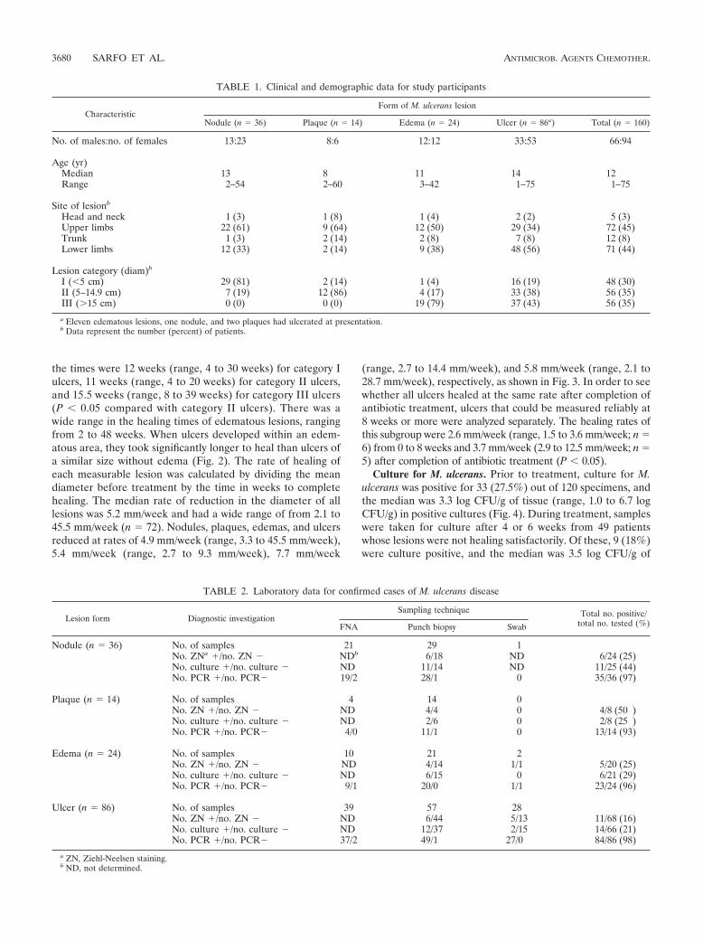
the times were 12 weeks (range, 4 to 30 weeks) for category Iulcers, 11 weeks (range, 4 to 20 weeks) for category II ulcers,and 15.5 weeks (range, 8 to 39 weeks) for category III ulcers(P � 0.05 compared with category II ulcers). There was awide range in the healing times of edematous lesions, rangingfrom 2 to 48 weeks. When ulcers developed within an edem-atous area, they took significantly longer to heal than ulcers ofa similar size without edema (Fig. 2). The rate of healing ofeach measurable lesion was calculated by dividing the meandiameter before treatment by the time in weeks to completehealing. The median rate of reduction in the diameter of alllesions was 5.2 mm/week and had a wide range of from 2.1 to45.5 mm/week (n � 72). Nodules, plaques, edemas, and ulcersreduced at rates of 4.9 mm/week (range, 3.3 to 45.5 mm/week),5.4 mm/week (range, 2.7 to 9.3 mm/week), 7.7 mm/week
(range, 2.7 to 14.4 mm/week), and 5.8 mm/week (range, 2.1 to28.7 mm/week), respectively, as shown in Fig. 3. In order to seewhether all ulcers healed at the same rate after completion ofantibiotic treatment, ulcers that could be measured reliably at8 weeks or more were analyzed separately. The healing rates ofthis subgroup were 2.6 mm/week (range, 1.5 to 3.6 mm/week; n �6) from 0 to 8 weeks and 3.7 mm/week (2.9 to 12.5 mm/week; n �5) after completion of antibiotic treatment (P � 0.05).
Culture for M. ulcerans. Prior to treatment, culture for M.ulcerans was positive for 33 (27.5%) out of 120 specimens, andthe median was 3.3 log CFU/g of tissue (range, 1.0 to 6.7 logCFU/g) in positive cultures (Fig. 4). During treatment, sampleswere taken for culture after 4 or 6 weeks from 49 patientswhose lesions were not healing satisfactorily. Of these, 9 (18%)were culture positive, and the median was 3.5 log CFU/g of
TABLE 1. Clinical and demographic data for study participants
CharacteristicForm of M. ulcerans lesion
Nodule (n � 36) Plaque (n � 14) Edema (n � 24) Ulcer (n � 86a) Total (n � 160)
No. of males:no. of females 13:23 8:6 12:12 33:53 66:94
Age (yr)Median 13 8 11 14 12Range 2–54 2–60 3–42 1–75 1–75
Site of lesionb
Head and neck 1 (3) 1 (8) 1 (4) 2 (2) 5 (3)Upper limbs 22 (61) 9 (64) 12 (50) 29 (34) 72 (45)Trunk 1 (3) 2 (14) 2 (8) 7 (8) 12 (8)Lower limbs 12 (33) 2 (14) 9 (38) 48 (56) 71 (44)
Lesion category (diam)b
I (�5 cm) 29 (81) 2 (14) 1 (4) 16 (19) 48 (30)II (5–14.9 cm) 7 (19) 12 (86) 4 (17) 33 (38) 56 (35)III (�15 cm) 0 (0) 0 (0) 19 (79) 37 (43) 56 (35)
a Eleven edematous lesions, one nodule, and two plaques had ulcerated at presentation.b Data represent the number (percent) of patients.
TABLE 2. Laboratory data for confirmed cases of M. ulcerans disease
Lesion form Diagnostic investigationSampling technique Total no. positive/
total no. tested (%)FNA Punch biopsy Swab
Nodule (n � 36) No. of samples 21 29 1No. ZNa �/no. ZN � NDb 6/18 ND 6/24 (25)No. culture �/no. culture � ND 11/14 ND 11/25 (44)No. PCR �/no. PCR� 19/2 28/1 0 35/36 (97)
Plaque (n � 14) No. of samples 4 14 0No. ZN �/no. ZN � ND 4/4 0 4/8 (50 )No. culture �/no. culture � ND 2/6 0 2/8 (25 )No. PCR �/no. PCR� 4/0 11/1 0 13/14 (93)
Edema (n � 24) No. of samples 10 21 2No. ZN �/no. ZN � ND 4/14 1/1 5/20 (25)No. culture �/no. culture � ND 6/15 0 6/21 (29)No. PCR �/no. PCR� 9/1 20/0 1/1 23/24 (96)
Ulcer (n � 86) No. of samples 39 57 28No. ZN �/no. ZN � ND 6/44 5/13 11/68 (16)No. culture �/no. culture � ND 12/37 2/15 14/66 (21)No. PCR �/no. PCR� 37/2 49/1 27/0 84/86 (98)
a ZN, Ziehl-Neelsen staining.b ND, not determined.
3680 SARFO ET AL. ANTIMICROB. AGENTS CHEMOTHER.

tissue (range, 2.0 to 6.3 log CFU/g). Four of 15 (27%) samplestaken either upon completion of antibiotics (n � 3) or 4 weekslater (n � 1) remained culture positive, and the median was 2.7log CFU/g of tissue (range, 2 to 5.0 log CFU/g). All four ofthese lesions were category III ulcerative lesions that subse-quently healed completely, with the median time to healing
being 20 weeks (range, 12 to 48 weeks) and a diameter regres-sion rate of 3.2 mm/week (range, 1.5 to 7.7 mm/week).
Paradoxical responses. New inflammatory lesions developedclose to or within the vicinity of healing lesions in three pa-tients. They appeared 4, 6, and 12 weeks after the start ofantibiotic treatment, respectively. The new lesions were fluc-
FIG. 1. Changes in the mean diameter of Buruli ulcer lesions in subjects whose lesions could be traced onto acetate sheets during and aftertreatment with rifampin and streptomycin for 8 weeks. The lines connect the data for individual subjects. Category I is lesions �5 cm in diameter,category II is lesions 5 to 15 cm, and category III is lesions �15 cm.
FIG. 2. Time to complete healing. Each dot represents one study subject treated with rifampin and streptomycin for 8 weeks. Horizontal linesrepresent the medians. The results for eight patients who had surgery, seven patients who defaulted from follow-up after completion of antibiotictreatment, and one patient who died are not included. **, P � 0.005; *, P � 0.01.
VOL. 54, 2010 EFFICACY OF RIFAMPIN-STREPTOMYCIN AGAINST M. ULCERANS 3681

tuant, erythematous, and nontender. Pus was aspirated fromall lesions and was sterile after prolonged culture. All lesionshealed after aspiration, and there was no need for furtherantibiotic therapy.
Surgical interventions. Two patients with nodules and onewith a plaque which progressively enlarged and ulcerated dur-ing treatment, five with ulcerated edematous lesions, and twowith large ulcers were offered surgical debridement and graft-
FIG. 3. Rate of healing. The rate of healing was calculated by dividing the diameter of the lesions (in mm) before treatment by the time (inweeks) to complete healing. Horizontal lines represent the median rates of healing, and the broken line shows the overall median healing rate in72 patients whose lesions were measurable before treatment. The circle highlights edematous lesions with ulceration. MU, M. ulcerans.
FIG. 4. Semiquantitative culture for M. ulcerans. Samples were cultured from 120 lesions before treatment with RS for 8 weeks and from 64selected cases responding poorly at 4, 6, 8, or 12 weeks. Negative cultures at each time point are shown as a single point on the baseline. Each linejoins the data for samples taken from the same patient, and dotted lines highlight the results for six patients who had positive cultures at twodifferent time points.
3682 SARFO ET AL. ANTIMICROB. AGENTS CHEMOTHER.

ing after 8 weeks antibiotic treatment because of unsatisfactoryhealing. Eight out of the 10 patients accepted surgery, and splitskin grafting was carried out between weeks 8 and 12. Allexcept one of the patients healed without complications within8 to 13 weeks. In one patient, inflammation persisted in thegraft site and healing was delayed until week 32. This was a24-year-old farmer with a category III ulcerated edema whoresponded poorly to RS8, so a further 4 weeks of RS therapywas administered followed by surgery. A punch biopsy speci-men taken for culture 8 weeks after the skin graft had brokendown was positive. Daily dressings were continued until theulcer healed at week 32.
One patient died 12 weeks after completing treatment. This35-year-old farmer had multiple ulcerated edematous lesions(category III) on the left lower limb which did not improveduring antibiotic therapy. He declined surgery and was lost tofollow-up. A village health care worker from a different districtreported that he died, possibly from sepsis.
Follow-up. Seven patients failed to attend for review of heal-ing after completing antibiotic treatment, so neither the timeto complete healing nor the rate of healing could be calculated.However, of 160 patients treated with RS8, with or withoutsurgery, 158 were reviewed 1 year after the completion oftreatment. The only patient who received RS for 12 weeks wasalso reviewed a year later. All lesions had healed, and therehad been no recurrences.
Functional limitations. Fifteen patients had functional lim-itation of joint movement before antibiotic therapy, and all ofthem were advised to self-administer physiotherapy during an-tibiotic treatment. Eight had category III ulcers and six hadcategory III edema; in three of the patients with edema, thelesions ulcerated at the time of presentation. One patient witha nodule on the left forearm had a contracture at the elbow ofthe affected limb secondary to a previous healed ulcer. Thispatient was excluded from the analysis of Buruli ulcer-associ-ated functional limitation. Eight lesions were situated near theelbow joint, four near the knee joint, and two near the anklejoint. The median time to healing among this subgroup ofpatients was 25 weeks (range, 8 to 48 weeks), and upon healingthe restriction of joint movements had resolved in 10 patients.The remaining four patients had persistent limitation of flexionthat subsequently improved with physiotherapy, and only onepatient still had a minor flexion deformity of the knee jointwith a limp at the 1-year review.
Management of pregnant patients. Three patients werepregnant at the time of presentation with M. ulcerans disease.Two, aged 20 and 26 years, who were in the third trimester hadcategory III ulcers on their lower limbs, and the third, aged 38,was in the second trimester and had a category II ulcer on theupper limb. All three were managed conservatively with dailywound dressings until 8 weeks after delivery, following whichantibiotic treatment with RS8 was administered, according toWHO recommendations.
Adverse events. Three adverse events were attributed toantibiotic therapy. A woman aged 54 years developed dizziness(grade 1) 4 weeks into treatment and a woman aged 63 yearshad protracted vomiting and dizziness (grade 2) 2 weeks intotreatment. These were attributed to streptomycin, which wasstopped, and moxifloxacin at 400 mg daily was substituted. A32-year-old woman with a category II ulcer developed a mac-
ulopapular rash and general malaise (grade 1) 4 weeks intotreatment which were thought to be due to rifampin. Treat-ment was stopped, and the ulcer was managed by daily wounddressing until it healed 24 weeks after the start of treatment.
DISCUSSION
The findings from this study confirm the efficacy of rifampincombined with streptomycin in healing all forms of M. ulceransdisease. In contrast to the Benin study (3), 95% of patients inthe present study were treated without surgery and all lesionshealed within 2 to 48 weeks. There were no recurrences duringa 12-month follow-up period, comparing favorably with the1.4% recurrence rate observed in Benin and the 0% rate ob-served in a recent controlled trial in Ghana (17). Although thenatural history of the disease is poorly documented, the recur-rence rate after surgery has been reported to be between 6 and17% in various studies (1, 6). Therefore, 8 weeks treatmentwith this combination of antibiotics has set a standard of treat-ment against which other antibiotic combinations can be testedin the future.
The aims of treatment can be defined clinically and bacte-riologically. Clinically, the aims were to achieve full healing oflesions with minimal scarring, no functional limitation, and norecurrence. There was complete clinical resolution of all formsand categories of M. ulcerans disease within 1 year, with themedian times to complete healing being 8, 10, and 20 weeks forcategory I, II, and III M. ulcerans disease, respectively (datanot shown). This compares favorably with 18 weeks for cate-gory I and 30 weeks for categories II and III combined re-ported in both arms of the clinical trial in which RS8 wascompared with RS for 4 weeks followed by rifampin and cla-rithromycin for 4 weeks (17). Natural history has not beendocumented except in a small placebo-controlled trial of clo-fazamine treatment, in which 10 patients out of a group of 34with lesions less than 5 cm in diameter healed spontaneously ina median of 14 weeks. The other 24 required surgery. How-ever, there was a high recurrence rate (29.4%) in the group of34 patients (22). The present study did not include any mea-sures of severity of scars. However, there were no scars afterhealing of nonulcerated lesions for a total of 72 patients (34with nodules, 14 with plaques, 24 with edema without ulcer-ation) who would otherwise have had a surgical scar, whichmay be larger than the lesion itself because surgeons leave awide margin.
Another clinical aim is to achieve the maximum rate ofhealing. This is more difficult to measure than the time tocomplete healing, but the latter is dependent on the size ofthe initial lesion. When M. ulcerans infection has been fullytreated, it is expected that Buruli ulcers will heal evenly fromthe outside inwards, provided they are kept clean and infectionfree. The rate of healing was calculated when lesions could bemeasured before treatment by tracing their margins onto ace-tate sheets and the time at which complete healing occurredwas known. The rate of reduction in diameter was used as ameasure of healing rather than the change in surface area,since this is expected to be linear. There was considerablevariation in the healing rates overall (Fig. 2) and also in thesmall subset of ulcers for which the healing rates after treat-
VOL. 54, 2010 EFFICACY OF RIFAMPIN-STREPTOMYCIN AGAINST M. ULCERANS 3683

ment could be calculated. Further study of a larger group ofpatients is required to investigate the causes of slow healing.
Most nodules resolved within 8 weeks, emphasizing the ben-efit of early recognition of M. ulcerans lesions. Large edema-tous lesions could not be measured, but they regressed rapidlyduring treatment, sometimes without developing ulceration.However, when edematous lesions ulcerated they healed at thesame rate or more slowly than ulcers without edema. Thepathogenesis of edema, associated with 15% of Buruli lesionsin the present study, is unknown. Its onset is usually within thefirst few weeks of the lesion appearing, and it sometimesspreads rapidly over a large area, giving rise to some of themost extensive lesions.
Samples taken from individuals whose ulcers were healingslowly showed that there was persistent infection with M.ulcerans in nine patients sampled at 4 or 6 weeks and in fourpatients who had completed treatment. These results weresurprising, since all cultures were negative after treatment withSR for 4 weeks in another study, but only early lesions wereincluded and the number of subjects was small (10). False-negative cultures are found in 40 to 60% of untreated ulcers (9,14, 20) due to sampling error, so it is possible that more thanfour patients in the present study had persistent infection aftercompleting RS8 treatment. We were not able to demonstrate areduction in the quantity of M. ulcerans organisms isolatedfrom tissue samples during antibiotic treatment, and semi-quantitative culture of small punch biopsy specimens may notbe a suitable way to compare the efficacies of new antibioticregimens with the efficacy of RS8 in the future. However, thefact that some cultures remained positive is important, since itmay explain why some lesions healed more slowly after com-pletion of antibiotic therapy. Persisting viable organisms inthese lesions may continue producing mycolactone at low con-centrations, which would inhibit production of growth factorsimportant for wound healing (4, 18, 27).
Despite having positive cultures during or after treatment,all the lesions healed without further antibiotic treatment andwithout recurrences, suggesting that the immune response toM. ulcerans was sufficient to contain the organism once myco-lactone production had been inhibited. Other factors that af-fect the healing rate of wounds include the quality of wounddressing procedures and the nutritional status of the patient.The quality of wound dressing was uniform in this study, andthere were no overt signs of malnutrition, although minordeficiencies cannot be ruled out.
Paradoxical reactions occurred in three patients at weeks 4,6, and 12, respectively. Aspirated pus was sterile on prolongedincubation, suggesting that the reaction was caused by a vig-orous inflammatory response rather than continuing infection.At low concentrations, mycolactone inhibits chemotaxis andsome aspects of the immune response (5, 28), so as the anti-biotic treatment kills the mycobacteria, an intense inflamma-tory response to high concentrations of organisms may beexpected. The implication is that there is no indication toextend antibiotic treatment, and all the lesions in this series ofpatients with paradoxical reactions healed with RS8 treatmentalone, with the median healing time being 12 weeks (range, 8to 16 weeks).
When any limitation of joint flexion was detected at the timeof diagnosis, patients were taught simple techniques for mobi-
lizing the joint which they continued during antibiotic therapyand subsequently, if it was required. As a result, only 1 patientout of 15 in whom it was detected at diagnosis had a persistentfunctional limitation at the end of follow-up. This supports theWHO strategy for management of the functional disabilitiesassociated with Buruli ulcer (24).
Administration of RS8 was accomplished in the presentstudy through a network of village health care workers trainedfor the purpose. They achieved a high rate of compliancethrough a simple system of observation and documentation,and the treatment was popular with the patients. Ten patientswere offered surgery, and two of those declined, possiblythrough fear of consequences such as amputation. In bothcases, the lesion healed without further intervention. Patientsin Western societies usually prefer early closure of wounds, butthe perception of surgery is different in countries where Buruliulcer is endemic, and in reality, it is not available to mostpatients.
The treatment was well tolerated overall, but two adults hadside effects attributed to streptomycin, after treatment for 2 or4 weeks. Moxifloxacin was substituted, and both lesions re-solved after treatment for a total of 8 weeks. There is onereport of a small series of patients treated successfully withrifampin and moxifloxacin for 8 weeks (19). Recently, a clinicaltrial has shown that clarithromycin can be substituted for strep-tomycin during the second 4 weeks of an 8-week regimenwithout any loss of benefit and with no major side effects (17).One patient in the present study had a reaction to rifampinafter 4 weeks. Since the Buruli ulcer lesion was healing well, nofurther antibiotics were given and healing continued unevent-fully. Treatment for 8 weeks was recommended by the WHOTechnical Advisory Group when a study showed that M. ulcer-ans organisms were still viable in human tissue after treatmentwith RS for 2 weeks but not after treatment for 4, 8, or 12weeks (10). It was judged that a safe margin should be allowedby recommending treatment for 8 weeks, but the above caseshows that a shorter duration may be adequate in some in-stances.
In conclusion, RS8 was a highly successful and practicaltreatment for all forms of M. ulcerans disease. Its success wasparticularly notable in edematous disease, which has been as-sociated with development of extensive ulcers. More workneeds to be done to investigate the factors responsible forvariations in the rate of healing and to define the optimalduration of therapy.
REFERENCES
1. Amofah, G., S. Asamoah, and C. Afram-Gyening. 1998. Effectiveness ofexcision of pre-ulcerative Buruli lesions in field situations in a rural districtin Ghana. Trop. Doct. 28:81–83.
2. Bentoucha, A., J. Robert, H. Dega, N. Lounis, V. Jarlier, and J. Grosset.2001. Activities of new macrolides and fluoroquinolones against Mycobacte-rium ulcerans infection in mice. Antimicrob. Agents Chemother. 45:3109–3112.
3. Chauty, A., M. F. Ardant, A. Adeye, H. Euverte, A. Guedenon, C. Johnson, J.Aubry, E. Nuermberger, and J. Grosset. 2007. Promising clinical efficacy ofstreptomycin-rifampin combination for treatment of Buruli ulcer (Mycobac-terium ulcerans disease). Antimicrob. Agents Chemother. 51:4029–4035.
4. Coutanceau, E., J. Decalf, A. Martino, A. Babon, N. Winter, S. T. Cole, M. L.Albert, and C. Demangel. 2007. Selective suppression of dendritic cell func-tions by Mycobacterium ulcerans toxin mycolactone. J. Exp. Med. 204:1395–1403.
5. Coutanceau, E., L. Marsollier, R. Brosch, E. Perret, P. Goossens, M. Tan-guy, S. T. Cole, P. L. Small, and C. Demangel. 2005. Modulation of the hostimmune response by a transient intracellular stage of Mycobacterium ulcer-
3684 SARFO ET AL. ANTIMICROB. AGENTS CHEMOTHER.

ans: the contribution of endogenous mycolactone toxin. Cell. Microbiol.7:1187–1196.
6. Debacker, M., J. Aguiar, C. Steunou, C. Zinsou, W. M. Meyers, and F.Portaels. 2005. Buruli ulcer recurrence, Benin. Emerg. Infect. Dis. 11:584–589.
7. Dega, H., A. Bentoucha, J. Robert, V. Jarlier, and J. Grosset. 2002. Bacte-ricidal activity of rifampin-amikacin against Mycobacterium ulcerans in mice.Antimicrob. Agents Chemother. 46:3193–3196.
8. Dega, H., J. Robert, P. Bonnafous, V. Jarlier, and J. Grosset. 2000. Activitiesof several antimicrobials against Mycobacterium ulcerans infection in mice.Antimicrob. Agents Chemother. 44:2367–2372.
9. Eddyani, M., M. Debacker, A. Martin, J. Aguiar, C. R. Johnson, C. Uwizeye,K. Fissette, and F. Portaels. 2008. Primary culture of Mycobacterium ulceransfrom human tissue specimens after storage in semisolid transport medium.J. Clin. Microbiol. 46:69–72.
10. Etuaful, S., B. Carbonnelle, J. Grosset, S. Lucas, C. Horsfield, R. Phillips,M. Evans, D. Ofori-Adjei, E. Klustse, J. Owusu-Boateng, G. K. Amedofu, P.Awuah, E. Ampadu, G. Amofah, K. Asiedu, and M. Wansbrough-Jones.2005. Efficacy of the combination rifampin-streptomycin in preventinggrowth of Mycobacterium ulcerans in early lesions of Buruli ulcer in humans.Antimicrob. Agents Chemother. 49:3182–3186.
11. Evans, M. R., R. Phillips, S. N. Etuaful, G. Amofah, J. Adomako, O. Adjei,J. Dennis-Antwi, S. B. Lucas, and M. H. Wansbrough-Jones. 2003. Anoutreach education and treatment project in Ghana for the early stage ofMycobacterium ulcerans disease. Trans. R. Soc. Trop. Med. Hyg. 97:159–160.
12. Evans, M. R. W., S. N. Etuaful, G. Amofah, O. Adjei, S. Lucas, and M. H.Wansbrough-Jones. 1999. Squamous cell carcinoma secondary to Buruliulcer. Trans. R. Soc. Trop. Med. Hyg. 93:63–64.
13. Havel, A., and S. R. Pattyn. 1975. Activity of rifampicin on Mycobacteriumulcerans. Ann. Soc. Belg. Med. Trop. 55:105–108.
14. Herbinger, K. H., O. Adjei, N. Y. Awua-Boateng, W. A. Nienhuis, L. Kunaa,V. Siegmund, J. Nitschke, W. Thompson, E. Klutse, P. Agbenorku, A. Schipf,S. Reu, P. Racz, B. Fleischer, M. Beissner, E. Fleischmann, K. Helfrich, T. S.van der Werf, T. Loscher, and G. Bretzel. 2009. Comparative study of thesensitivity of different diagnostic methods for the laboratory diagnosis ofBuruli ulcer disease. Clin. Infect. Dis. 48:1055–1064.
15. Lefrancois, S., J. Robert, A. Chauffour, B. Ji, and V. Jarlier. 2007. CuringMycobacterium ulcerans infection in mice with a combination of rifampin-streptomycin or rifampin-amikacin. Antimicrob. Agents Chemother. 51:645–650.
16. Marsollier, L., N. Honore, P. Legras, A. L. Manceau, H. Kouakou, B. Car-bonnelle, and S. T. Cole. 2003. Isolation of three Mycobacterium ulceransstrains resistant to rifampin after experimental chemotherapy of mice. An-timicrob. Agents Chemother. 47:1228–1232.
17. Nienhuis, W. A., Y. Stienstra, W. A. Thompson, P. C. Awuah, K. M. Abass,W. Tuah, N. Y. Awua-Boateng, E. O. Ampadu, V. Siegmund, J. P. Schouten,O. Adjei, G. Bretzel, and T. S. van der Werf. 2010. Antimicrobial treatmentfor early, limited Mycobacterium ulcerans infection: a randomised controlledtrial. Lancet 375:664–672.
18. Pahlevan, A. A., D. J. M. Wright, C. Andrews, K. M. George, P. L. C. Small,and B. M. Foxwell. 1999. The inhibitory action of Mycobacterium ulceranssoluble factor on monocyte/T cell cytokine production and NF-kappa Bfunction. J. Immunol. 163:3928–3935.
19. Phillips, R., F. S. Sarfo, F. Osei-Sarpong, and M. Wansbrough-Jones. 2006.Clinical response and bacterial killing during antibiotic treatment of Myco-bacterium ulcerans disease, p. 60–61. http://www.who.int/buruli/information/en/Abstracts.
20. Phillips, R. O., F. S. Sarfo, F. Osei-Sarpong, A. Boateng, I. Tetteh, A. Lartey,E. Adentwe, W. Opare, K. B. Asiedu, and M. Wansbrough-Jones. 2009.Sensitivity of PCR targeting Mycobacterium ulcerans by use of fine-needleaspirates for diagnosis of Buruli ulcer. J. Clin. Microbiol. 47:924–926.
21. Portaels, F., H. Traore, K. De Ridder, and W. M. Meyers. 1998. In vitrosusceptibility of Mycobacterium ulcerans to clarithromycin. Antimicrob.Agents Chemother. 42:2070–2073.
22. Revill, W. D., R. H. Morrow, M. C. Pike, and J. Ateng. 1973. A controlledtrial of the treatment of Mycobacterium ulcerans infection with clofazimine.Lancet ii:873–877.
23. Shepard, C. C., and D. H. McRae. 1968. A method for counting acid-fastbacteria. Int. J. Lepr. Other Mycobact. Dis. 36:78–82.
24. Simonet, V. 2008. Prevention of disability in Buruli ulcer: basic rehabil-itation. http://whqlibdoc.who.int/hq/2008/WHO_HTM_NTD_IDM_GBUI_2008.1_eng.pdf.
25. Stanford, J. L., and I. Phillips. 1972. Rifampicin in experimental Mycobac-terium ulcerans infection. J. Med. Microbiol. 5:39–45.
26. Thangaraj, H. S., O. Adjei, B. W. Allen, F. Portaels, M. R. W. Evans, D. K.Banerjee, and M. H. Wansbrough-Jones. 2000. In vitro activity of ciprofloxa-cin, sparfloxacin, ofloxacin, amikacin and rifampicin against Ghanaian iso-lates of Mycobacterium ulcerans. J. Antimicrob. Chemother. 45:231–233.
27. Torrado, E., S. Adusumilli, A. G. Fraga, P. L. Small, A. G. Castro, andJ. Pedrosa. 2007. Mycolactone-mediated inhibition of tumor necrosis factorproduction by macrophages infected with Mycobacterium ulcerans has impli-cations for the control of infection. Infect. Immun. 75:3979–3988.
28. Torrado, E., A. G. Fraga, A. G. Castro, P. Stragier, W. M. Meyers, F.Portaels, M. T. Silva, and J. Pedrosa. 2007. Evidence for an intramacroph-age growth phase of Mycobacterium ulcerans. Infect. Immun. 75:977–987.
29. Wansbrough-Jones, M., and R. Phillips. 2006. Buruli ulcer: emerging fromobscurity. Lancet 367:1849–1858.
VOL. 54, 2010 EFFICACY OF RIFAMPIN-STREPTOMYCIN AGAINST M. ULCERANS 3685
Copyright © 2022 FDOKUMEN