Identification of a major causative agent of - CyberLeninka
10
RESEARCH Open Access Identification of a major causative agent of human cercarial dermatitis, Trichobilharzia franki (Müller and Kimmig 1994), in southern England and its evolutionary relationships with other European populations Scott P Lawton * , Rivka M Lim, Juliet P Dukes, Richard T Cook, Anthony J Walker and Ruth S Kirk Abstract Background: Trichobilharzia is the most species rich and widely distributed genus of schistosomes and is known throughout Europe and North America as an agent of human cercarial dermatitis. The disease is caused by an acute allergic reaction in the skin that develops as a consequence of repeated contact with water containing schistosomatid cercariae. However, despite historical outbreaks of the disease, there are no published records of accurately identified Trichobilharzia species from the UK. Methods: Two hundred Radix auricularia (L.) were sampled from a recreational fishing lake in Hampshire and emerging schistosomatid cercariae were collected for microscopy and DNA extraction. General morphological description of the cercariae was performed, alongside sequencing and phylogenetic analysis of the 28S ribosomal DNA for accurate species identification as well as comparisons of ITS1 in order to identify evolutionary affinities with other European populations. All molecular comparisons were performed using published sequences. Results: The phylogenetic analysis of 28S sequences identified the cercariae as Trichobilharzia franki. Two unique British ITS1 haplotypes were identified which were most closely related to haplotypes of T. franki populations from France. Haplotype network analysis indicated the mixing of T. franki populations throughout Europe. It is suggested that parasite distribution is the probable result of the movement of migratory waterfowl. Conclusions: This is the first accurate record of T. franki in the UK. The movement of T. franki with waterfowl could pose a considerable human health risk, as in mainland Europe, and signifies T. franki-associated human cercarial dermatitis as a re-emerging disease in the UK. Keywords: Trichobilharzia franki, Cercarial dermatitis, UK, Re-emerging disease, 28S ribosomal DNA, ITS1 haplotypes Background The cercariae of many species of avian schistosomatid blood flukes are known to cause human cercarial derma- titis (CD), also known as swimmer’ s itch. Avian schisto- somatids have a truly global distribution with agents of CD being recorded on all major continents except Antarctica. However, throughout Europe and America there has been increased incidence of reported infections and CD is now considered an emerging and re-emerging infectious disease [1-3]. Foci of infection have been identified in several European countries particularly Italy, Germany, Austria, Switzerland, the Netherlands, Czech Republic, France, Poland and Iceland with the disease impacting on local economies that depend on tourism associated with recreational water use [4]. Despite the occurrence of these parasites throughout Europe and North America [1,3], relatively little is known about the occurrence of CD and the identity and diversity of parasites that cause it in the UK, even * Correspondence: [email protected] Molecular Parasitology Laboratory, School of Life Sciences, Kingston University, Kingston upon Thames, Surrey KT1 2EE, UK © 2014 Lawton et al.; licensee BioMed Central Ltd. This is an Open Access article distributed under the terms of the Creative Commons Attribution License (http://creativecommons.org/licenses/by/2.0), which permits unrestricted use, distribution, and reproduction in any medium, provided the original work is properly credited. The Creative Commons Public Domain Dedication waiver (http://creativecommons.org/publicdomain/zero/1.0/) applies to the data made available in this article, unless otherwise stated. Lawton et al. Parasites & Vectors 2014, 7:277 http://www.parasitesandvectors.com/content/7/1/277
-
Upload
khangminh22 -
Category
Documents
-
view
0 -
download
0
Transcript of Identification of a major causative agent of - CyberLeninka

RESEARCH Open Access
Identification of a major causative agent ofhuman cercarial dermatitis, Trichobilharzia franki(Müller and Kimmig 1994), in southern Englandand its evolutionary relationships with otherEuropean populationsScott P Lawton*, Rivka M Lim, Juliet P Dukes, Richard T Cook, Anthony J Walker and Ruth S Kirk
Abstract
Background: Trichobilharzia is the most species rich and widely distributed genus of schistosomes and is knownthroughout Europe and North America as an agent of human cercarial dermatitis. The disease is caused by anacute allergic reaction in the skin that develops as a consequence of repeated contact with water containingschistosomatid cercariae. However, despite historical outbreaks of the disease, there are no published records ofaccurately identified Trichobilharzia species from the UK.
Methods: Two hundred Radix auricularia (L.) were sampled from a recreational fishing lake in Hampshire andemerging schistosomatid cercariae were collected for microscopy and DNA extraction. General morphologicaldescription of the cercariae was performed, alongside sequencing and phylogenetic analysis of the 28S ribosomalDNA for accurate species identification as well as comparisons of ITS1 in order to identify evolutionary affinities withother European populations. All molecular comparisons were performed using published sequences.
Results: The phylogenetic analysis of 28S sequences identified the cercariae as Trichobilharzia franki. Two uniqueBritish ITS1 haplotypes were identified which were most closely related to haplotypes of T. franki populations fromFrance. Haplotype network analysis indicated the mixing of T. franki populations throughout Europe. It is suggestedthat parasite distribution is the probable result of the movement of migratory waterfowl.
Conclusions: This is the first accurate record of T. franki in the UK. The movement of T. franki with waterfowl couldpose a considerable human health risk, as in mainland Europe, and signifies T. franki-associated human cercarialdermatitis as a re-emerging disease in the UK.
Keywords: Trichobilharzia franki, Cercarial dermatitis, UK, Re-emerging disease, 28S ribosomal DNA, ITS1 haplotypes
BackgroundThe cercariae of many species of avian schistosomatidblood flukes are known to cause human cercarial derma-titis (CD), also known as swimmer’s itch. Avian schisto-somatids have a truly global distribution with agentsof CD being recorded on all major continents exceptAntarctica. However, throughout Europe and Americathere has been increased incidence of reported infections
and CD is now considered an emerging and re-emerginginfectious disease [1-3]. Foci of infection have beenidentified in several European countries particularly Italy,Germany, Austria, Switzerland, the Netherlands, CzechRepublic, France, Poland and Iceland with the diseaseimpacting on local economies that depend on tourismassociated with recreational water use [4].Despite the occurrence of these parasites throughout
Europe and North America [1,3], relatively little isknown about the occurrence of CD and the identity anddiversity of parasites that cause it in the UK, even
* Correspondence: [email protected] Parasitology Laboratory, School of Life Sciences, KingstonUniversity, Kingston upon Thames, Surrey KT1 2EE, UK
© 2014 Lawton et al.; licensee BioMed Central Ltd. This is an Open Access article distributed under the terms of the CreativeCommons Attribution License (http://creativecommons.org/licenses/by/2.0), which permits unrestricted use, distribution, andreproduction in any medium, provided the original work is properly credited. The Creative Commons Public DomainDedication waiver (http://creativecommons.org/publicdomain/zero/1.0/) applies to the data made available in this article,unless otherwise stated.
Lawton et al. Parasites & Vectors 2014, 7:277http://www.parasitesandvectors.com/content/7/1/277

though outbreaks of CD have been recorded in LochLochore in Fife in 2006, the Norfolk Broads in 2004 [5],a water sports lake in Suffolk in 1987 [6], a leisure waterpark in Rickmansworth, Hertfordshire in 1970 [7] andRoath Park, Cardiff between 1928–1943 [8].Human CD presents as an acute allergic reaction in
the skin that develops as a consequence of the repeatedpenetration of certain species of schistosomatid cercariaethat emerge from freshwater snail intermediate hosts[3,9]. Maculo-papulo-vesicular eruptions occur after ex-posure, followed by intense itching, fever, swelling of thelymph nodes and eventually erythema and oedema [10].Experimental studies on immunocompetent mammalianmodels show that although the majority of the cercariaedie in the skin, some schistosomula can migrate to thelungs (visceral avian schistosome species) or nervoussystem (Trichobilharzia regenti) [3,9]. Severe pathologiescan develop in immunodeficient mammals due to a highnumber of schistosomula migrating to organs. Therefore,although they are unable to complete their developmentand reproduce, the parasites may cause pathologies inaddition to those associated with the skin (reviewed byKolářová [9]).Trichobilharzia [11] is considered the most species-
rich and widely distributed genus of the Schistosomati-dae, with species parasitizing waterfowl throughout theworld, and several being leading causes of CD [1]. Schis-tosomatids in UK waters have been inadequately studied.Although ocellate schistosomatid cercariae have beenrecorded from snails in the UK [8,12,13], there is nodefinitive identification of a Trichobilharzia species. Inthe last decade, problems relating to the accurate mor-phological identification of Trichobilharzia species asso-ciated with CD outbreak sites in America and Europehave been overcome by using ribosomal DNA (rDNA)markers to identify cercariae to species [1,14]. Such ap-proaches were employed in the current study to identifyschistosomes released by aquatic lymnaeid snails col-lected from Tundry Pond, Hampshire. We report thefirst detailed record/description of T. franki Müller andKimmig [15] in the UK and identify evolutionary rela-tionships with other European populations of this schis-tosome. The public health implications of the findingsare discussed in the context of CD being a re-emerginginfectious disease in the UK.
MethodsSnail and cercariae collectionTwo hundred Radix auricularia (L.) were collectedand identified based on shell morphology from TundryPond, a recreational fishing lake in Hampshire, SouthernEngland, UK (grid reference: SU 775 525, 51.16 N0.53 W), in August 2011 as part of an on-going study onthe diversity of digeneans and their intermediate hosts in
the UK. Ocellate furcocercariae have previously been ob-served at this site (R. Kirk, unpublished observations).Snails were relocated to the laboratory for processing.Each snail was isolated in a 100 ml beaker filled withdechlorinated tap water that had been filtered througha Brimak carbon filter (Silverline UK) and cercarialemergence was stimulated by exposure to natural light.Three R. auricularia shed ocellate furcocercariae. Thesecercariae were collected for molecular work using thetechniques described by Brant and Loker [1] and werepooled in Petri dishes for morphological description.Upon confirmation of species, parasite and snail referencematerial were deposited at the Natural History Museum,London, UK (parasite voucher NHMUK 2014.4.25.1-2;snail voucher NHMUK 20140076).
Morphological description of cercariaeLive cercariae were vitally stained with 0.5% neutral redand examined using light microscopy for initial morpho-logical description. To obtain further morphological in-formation, cercariae were fixed for scanning electronmicroscopy in 2.5% glutaraldehyde in 0.1 M phosphatebuffer (pH 7.4) for 2 h at 4°C. They were then washed infour changes of 0.1 M phosphate buffer, post-fixed in os-mium tetroxide in the same buffer for 1 h, dehydrated ina graded ethanol series and dried in hexamethyldisilazane.Samples were coated with gold-palladium and examinedunder a Zeiss EVO50 scanning electron microscope. Mor-phometric parameters of cercariae were not recorded be-cause of the unreliability of using cercarial dimensionsand flame cell organization for identification of Trichobil-harzia species [10]. Chaetotaxy was not performed due toknown problems with misinterpretation of sensory struc-tures [16] and staining [16,17].
Molecular identification of parasitesLive ocellate furcocercariae were morphologically identi-fied to genus level using light microscopy and werepooled into 1.5 ml microfuge tubes (50 cercariae persnail). Each tube was centrifuged at 10000 rpm for1 min to pellet the cercariae and DNA was extractedwith the Qiagen DNeasy blood and tissue kit (QiagenInc.) using the manufacturer’s protocol. PCR was per-formed to amplify fragments of 28S ribosomal subunit(LSU) and the internal transcribed spacer region (ITS)using primers and cycling conditions specified by Olsonet al. [18] and Wang et al. [19], respectively. PCR reac-tions were performed using 12.5 μl Thermo–StartR PCRmaster mix (0.625 Units of Taq DNA polymerase, 1Xreaction buffer, 0.2 mM of each dNTP and 1.5 mMMgCl2) and 1–2 ng/μl of DNA. Final reactions weremade up to 25 μl with PCR-grade water. Reactions wereperformed using a Veriti 96 well thermal cycler (AppliedBiosystems™) and 5 μl of each amplicon was visualized
Lawton et al. Parasites & Vectors 2014, 7:277 Page 2 of 10http://www.parasitesandvectors.com/content/7/1/277

in 1% agarose gels stained with gel red (Bioline™). Theremaining 20 μl PCR products were sequenced at theDNA sequencing facility of the Natural History Museum,London, using the PCR primers with Fluorescent Dye Ter-minator Sequencing Kits (Applied Biosystems™); sequen-cing reactions were run on an Applied Biosystems™3730XL automated sequencer. Resultant sequences wereassembled using BioEdit [20] and manually corrected forambiguous base calls. It is important to note that despitethe DNA being extracted from pooled cercariae, therewere no polymorphic sites noticed in the chromatographsof both the forward and reverse sequences of LSU andITS. Complete sequences were submitted for BLASTsearch using blastn (http://blast.ncbi.nlm.nih.gov/) to en-able initial identification, indicating that all the sequencesgenerated best-matched T. franki. Novel LSU sequenceswere aligned with previously published sequences of otherspecies of Trichobilharzia and bird schistosomes from dif-ferent genera and Schistosoma sinensium as an out group(Table 1) using MUSCLE (http://www.ebi.ac.uk), giving analignment of comparable data of 556 bp.
Inter- and intra-species phylogenetic analysisTo accurately identify the novel sequences and placethem into a phylogenetic context with other species ofTrichobilharzia, phylogenetic analysis was performed foralignments of LSU using MEGA5 [21]. Several phylo-genetic methods were employed using the neighbourjoining (NJ) approach under the conditions of the Jukes-
Cantor model, maximum likelihood (ML) methods usingthe HKY+G model of evolution also identified inMEGA5 [21]. Maximum parsimony (MP) trees were ob-tained using the close–neighbour–interchange algo-rithm. The model used for the ML tree was identifiedbased on the lowest Bayesian information criterionscores relative to the other models tested and for alltree analysis nodal support was assessed using 1000bootstrap replicates. The pair-wise uncorrected distances(p-distance) were also calculated for the LSU sequencesbetween Trichobilharzia species to estimate geneticdistance and to validate species identification.In order to establish affinities between European and
UK isolates of T. franki, ITS1 fragments were analyseddue to the availability of comparable data from otherEuropean T. franki populations. The three novel se-quences obtained were aligned with previously publishedsequences that represent populations of T. franki fromacross central and northern Europe, producing an align-ment of 878 bp. Sequences were aligned and phylogen-etic analysis performed as described previously, but MLtrees were constructed using the K2+G model as identi-fied by MEGA5 [21]; T. regenti was used as an out-groupin all analyses. Standard population genetic analysis wasperformed using DnaSPv5 [22] to calculate nucleotide di-versity (π) and sequence differentiation within and be-tween sequence sets from different countries and toidentify the occurrence of unique haplotypes (H) andhaplotype diversity (Hd). In order to assess the associationof the UK haplotypes to others from Europe, the mostparsimonious haplotype network was constructed usingTCS version 1.13 [23].
ResultsMorphological description of Trichobilharzia frankiThe morphology of the ocellate furcocercariae partlycorresponded with the previous description of T. frankiby Müller and Kimmig [15]. The ocelli (eye spots) werelocated on the dorsal part of the body, between the acet-abulum and the head organ. The head organ and theacetabulum were spherical. The cercariae were aphar-yngeate, with a subterminal mouth and a bifurcate intes-tine, which were inconspicuous. The body contained fivelarge pairs of penetration glands, two pairs circumace-tabular and three pairs postacetabular, in addition to anapical gland group, with all ducts opening anteriorly.The excretory system progressed through the excretoryjunction at the base of the body and extended into the tailstem to the tip of the furcae. The furcae were surroundedby finfolds and ended in thorns (Figures 1 and 2A). Thedescription of the head organ by Müller and Kimmig [15]as a fused complex of oral sucker and pharynx and thepreacetabular position of two pairs of penetration glandsare suggested to be errors. These authors also reported that
Table 1 Published sequences of schistosomatids used inthe phylogenetic analysis of the LSU to identify UKspecific Trichobilharzia species
Species Host Accession
Trichobilharzia franki Radix auricularia HM131141
T. regenti Anser anser HM439491
T. querquedulae Anas discors FJ174469
T. physellae Physa parkeri FJ174475
T. szidati Lymnaea stagnalis FJ174476
T. stagnicolae Stagnicola emarginata FJ174479
T. ocellata Lymnaea stagnalis AY157243
Anserobilharzia brantae Chen caerulescens FJ174467
Dendritobilharzia pulverulenta Gallus gallus (Lab host) AY157241
Gigantobilharzia huronensis Agelaius phoeniceus AY157242
Heterobilharzia americana Mesocricetus auratus AY157246
Schistosomatium douthitti Mesocricetus auratus AY157247
Ornithobilharzia canaliculata Larus delawarensis AY157248
Austrobilharzia terrigalensis Batillaria australis AY157249
Bilharziella polonica Anas platyrhynchus AF184265
Out group
Schistosoma sinensium Mus musculus (Lab host) AY157251
Lawton et al. Parasites & Vectors 2014, 7:277 Page 3 of 10http://www.parasitesandvectors.com/content/7/1/277

the tegument was covered with small spines. Here, scan-ning electron microscopy revealed that most of the exter-nal surface of the acetabulum was devoid of spines andonly the inner surface had centrally directed curved spineswith rounded tips (Figure 2B). The spines on the tail stemwere large and curved (Figure 2C). The spines on the bodyresembled those on the acetabulum (Figure 2D). All spineswere posteriorly directed. Two types of uniciliate sensory
endings were observed: type 1 sensory papillae (with tegu-mental collar) of different lengths on the body and type 2sensory papillae (without tegumental collar) of differentlengths on the tail stem, furcae and body, but not on thehead organ region (Figure 2D, E), corresponding to thedescription for T. franki by Kock and Böckeler [16]. Thetip of the head organ displayed gland duct openings sur-rounded by type 1 sensory papillae (Figure 2F).
Figure 1 Lateral view of the cercaria of Trichobilharzia franki stained with neutral red. Acetabulum (a), head organ (ho), one of two ocelli(o) circumacetabular penetration glands (cg), postacetabular penetration glands (pg), tail stem (ts) and furcae (f).
Figure 2 Scanning electron micrographs of the cercaria of Trichobilharzia franki. A. Entire cercaria, scale bar = 100 μm. B. Acetabulum, scalebar = 1 μm. C. Tail stem with spines, scale bar = 2 μm. D. Tegumental spines and type 2 sensory papilla on body (arrow), scale bar = 2 μm. E.Anterior of body and type 1 sensory papillae (arrows), scale bar = 10 μm. F. Apical view of body showing head organ, note type 1 sensorypapillae (arrows) scale bar = 2 μm, and gland duct openings (insert, black arrows), scale bar = 1 μm.
Lawton et al. Parasites & Vectors 2014, 7:277 Page 4 of 10http://www.parasitesandvectors.com/content/7/1/277

Molecular identification of Trichobilharzia frankiPhylogenetic analysis of the T. franki LSU fragments with15 other schistosomatids produced a well-supportedTrichobilharzia clade with Dendritobilharzia pulverulentaand Gigantobilharzia huronensis forming a sister clade(Figure 3). Within the Trichobilharzia clade the sequencesgenerated in this current study, Ham Ra6 – 8 (GenBank:KJ775865-67), clustered with T. franki only and formed ashallow, but well supported, clade separate from any ofthe other taxa (Figure 3). This relationship between theUK sequences and T. franki was further supported by thelow p–distance values relative to comparisons with otherspecies (Table 2). However, a noteworthy observation isthat similar levels of genetic divergence seen between theUK specific sequences and T. franki, were also evident be-tween T. franki and T. querquedulae (Table 2). Trichobil-harzia querquedulae appears as a sister species to T.franki, but short branch lengths may indicate that bothspecies are either subspecies or members of a speciescomplex.
Insights into the relationships between populations ofTrichobilharzia franki from the UK and EuropePhylogenetic reconstruction showed the UK-specific se-quences to have close affinity with populations of T.franki from France and clustered within a France-specific clade, but no other country specific clades wereidentified (Figure 4). Two unique haplotypes were iden-tified from the three specimens with Ham Ra6 having itsown individual haplotype and another being shared byHam Ra7 and Ham Ra8 (GenBank: KJ775868-69). Themolecular diversity of T. franki populations from eachcountry appeared high with several different haplotypesbeing found within relatively small sample sizes (Table 3).Diversity within sequences from Switzerland showedconsiderable variation (S = 77; π = 0.02610) and five outof the six sequences analysed were unique haplotypes.This contrasted with sequences from France where onlythree haplotypes were found in the ten sequences sam-pled (S = 2; π = 0.00079). The high number of haplotypeswithin and between populations of T. franki was also
68/67/100
88/71/100
95/90/100
90/52/100
71/66/100
96/95/100
100/99/100
99/95/100
69/63/100
90/86/10072/85/100
70/64/100
100/100/100
Trichobilharzia franki from Hampshire, UK
Trichobilharzia franki
Trichobilharzia querquedulae
Trichobilharzia physellae
Trichobilharzia regenti
Trichobilharzia ocellata
Trichobilharzia szidati
Anserobilharzia brantae
Trichobilharzia stagnicolae
Dendritobilharzia pulverulenta
Gigantobilharzia huronensis
Bilharziella polonica
Ornithobilharzia canaliculata
Austrobilharzia terrigalensis
Heterobilharzia americana
Schistosomatium douthitti
Schistosoma sinesium
0.02
Ham Ra8
Ham Ra7
Ham Ra6
Figure 3 Phylogenetic tree based on partial LSU sequences for avian schistosomes in order to identify ocellate furcocercariae fromHampshire, UK. The samples sequenced from Hampshire, UK have close affinity with T. franki and do not associate with another species. Thetree was constructed using the ML method in MEGA5 using the HKY + G substitution model. The scale shows the number of nucleotidesubstitutions per site between DNA sequences and nodal supports were generated using 1000 bootstrap replicates. The nodal support is given inNJ, ML and MP bootstraps respectively and only values >50 for at least two of the analyses are shown.
Lawton et al. Parasites & Vectors 2014, 7:277 Page 5 of 10http://www.parasitesandvectors.com/content/7/1/277
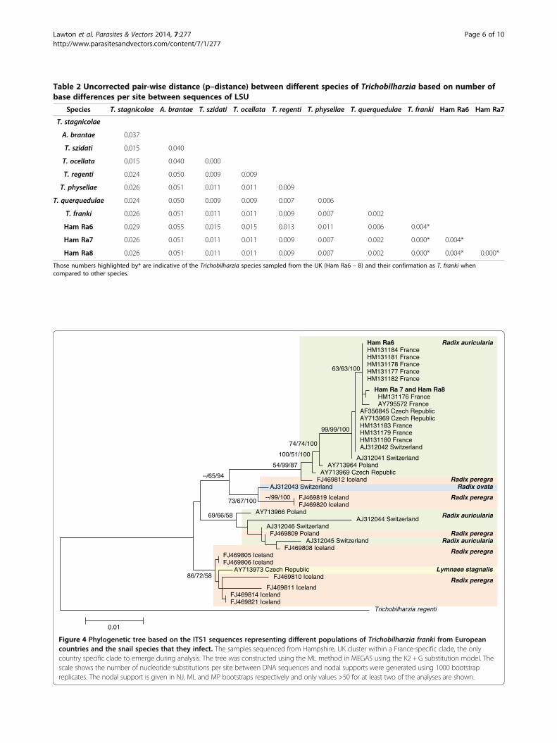
Table 2 Uncorrected pair-wise distance (p–distance) between different species of Trichobilharzia based on number ofbase differences per site between sequences of LSU
Species T. stagnicolae A. brantae T. szidati T. ocellata T. regenti T. physellae T. querquedulae T. franki Ham Ra6 Ham Ra7
T. stagnicolae
A. brantae 0.037
T. szidati 0.015 0.040
T. ocellata 0.015 0.040 0.000
T. regenti 0.024 0.050 0.009 0.009
T. physellae 0.026 0.051 0.011 0.011 0.009
T. querquedulae 0.024 0.050 0.009 0.009 0.007 0.006
T. franki 0.026 0.051 0.011 0.011 0.009 0.007 0.002
Ham Ra6 0.029 0.055 0.015 0.015 0.013 0.011 0.006 0.004*
Ham Ra7 0.026 0.051 0.011 0.011 0.009 0.007 0.002 0.000* 0.004*
Ham Ra8 0.026 0.051 0.011 0.011 0.009 0.007 0.002 0.000* 0.004* 0.000*
Those numbers highlighted by* are indicative of the Trichobilharzia species sampled from the UK (Ham Ra6 – 8) and their confirmation as T. franki whencompared to other species.
Ham Ra6HM131184 FranceHM131181 FranceHM131178 FranceHM131177 FranceHM131182 France
Ham Ra 7 and Ham Ra8HM131176 FranceAY795572 France
AF356845 Czech RepublicAY713969 Czech RepublicHM131183 FranceHM131179 FranceHM131180 FranceAJ312042 Switzerland
AJ312041 SwitzerlandAY713964 Poland
AY713969 Czech Republic
AJ312043 Switzerland
AJ312044 SwitzerlandAJ312046 Switzerland
AJ312045 Switzerland
AY713973 Czech Republic
FJ469812 Iceland
FJ469819 IcelandFJ469820 Iceland
FJ469808 IcelandFJ469805 IcelandFJ469806 Iceland
FJ469810 Iceland
FJ469811 IcelandFJ469814 IcelandFJ469821 Iceland
AY713966 Poland
FJ469809 Poland
Trichobilharzia regenti
0.01
86/72/58
69/66/58
--/99/10073/67/100
--/65/94
54/99/87
100/51/100
74/74/100
99/99/100
63/63/100
Radix auricularia
Radix auricularia
Radix auricularia
Radix peregra
Radix peregra
Radix peregra
Radix peregra
Radix peregra
Radix ovata
Lymnaea stagnalis
Figure 4 Phylogenetic tree based on the ITS1 sequences representing different populations of Trichobilharzia franki from Europeancountries and the snail species that they infect. The samples sequenced from Hampshire, UK cluster within a France-specific clade, the onlycountry specific clade to emerge during analysis. The tree was constructed using the ML method in MEGA5 using the K2 + G substitution model. Thescale shows the number of nucleotide substitutions per site between DNA sequences and nodal supports were generated using 1000 bootstrapreplicates. The nodal support is given in NJ, ML and MP bootstraps respectively and only values >50 for at least two of the analyses are shown.
Lawton et al. Parasites & Vectors 2014, 7:277 Page 6 of 10http://www.parasitesandvectors.com/content/7/1/277

illustrated in the haplotype network (Figure 5). Four dis-tinct clades were revealed in the haplotype network, butno geographical specific lineages emerged as each cladecontained a number of related haplotypes from differentcountries. Clade 1 contained the only haplotype sharedbetween countries with haplotype 1 (H1) being sharedby individuals from France (HM131183, HM131179,HM131180), Switzerland (AJ312041, AJ312042) and theCzech Republic (AY795573, AF356845). The UK-specificsequences were also found in clade 1 with haplotype3 (H3) representing Ham Ra7 and 8 and haplotype 4(H4) representing Ham Ra6. These haplotypes wereunique and were not shared with other populations.However, the UK-specific sequences were most closelyrelated to French sequences in haplotype 5 (H5) that con-tained HM131181, HM131177, HM131184, HM131182,HM131178, HM131176 and haplotype 2 (H2) that con-tained AY795572 (Figure 5). Although these data illustratehigh population variation within and between T. frankipopulations, the sample sizes for each country are small.Further work with larger sample sizes would be requiredto provide in-depth insights into population differentiationand gene flow.
DiscussionUtilising molecular techniques described by Jouet et al.[24], our study has provided the first detailed record of aTrichobilharzia parasite in the UK. The morphologicaldescription of T. franki supported by scanning electronmicroscopy observations, shows that the cercariae haveconsiderable similarity to other Trichobilharzia species,particularly T. regenti. A molecular approach was there-fore considered essential for identification. Phylogeneticanalysis of the LSU and analysis of p–distance demon-strated that the UK-specific sequences corresponded toT. franki, showing the power of this ribosomal markerfor species-specific identification and high levels ofphylogenetic resolution as in other studies of avianschistosomatids [1,24]. However, there is clear evidenceof a close relationship between T. franki and T. querque-dulae, as also highlighted by Brant and Loker [1] and
Jouet et al. [24]. Trichobilharzia querquedulae could beeither a very close sister taxon, part of a species complexin which a number of Trichobilharzia species are veryclosely related and form the “T. franki” complex [24], orsimply be a genetically distinct population of T. frankiand not a species in its own right [24]. Such confoundingfactors on the identity of a species could be problematicfor phylogenetic analysis and subsequent epidemiologicalsurveys. Although the UK parasite was identified as T.franki, other molecular markers could be considered foridentification of other species and genera of non-Schisto-soma schistosomatids, to provide a comprehensive mo-lecular phylogenetic framework for avian schistosomatidparasites [24-26].Both the phylogenetic and haplotype network analysis
of the ITS1 fragment from the UK samples show a closegenetic relationship with French populations (Figures 4and 5). The presence of Trichobilharzia spp. and occur-rence of CD have been known in France for several de-cades [2,24] and, due to close proximity of France to theUK and the movement of migratory waterfowl betweenthe two countries, it could be argued that although somedifferentiation between populations of T. franki has oc-curred, some gene flow is likely to take place. The occur-rence of gene flow between countries was also illustratedin the haplotype network analysis due to H1 beingshared between France, Switzerland and the Czech Re-public (Figure 5). This is a clear indication that the dis-tribution and prevalence of CD is intimately linked withthe movement of waterfowl, and T. franki populationsare moving around Europe, probably as a result of themigratory patterns of their definitive Anseriformes hosts(ducks, geese and swans) [1,9,24]. Brant and Loker [1]discussed the occurrence of genetically related individ-uals of T. physellae and T. querquedulae collected fromsnails along the major avian migratory flyways in theUSA ranging from latitudes as distant as Alaska to Lou-isiana and Florida. This is probably also true for T. frankiin the UK and the rest of Europe as some birds, such asswans and geese, migrate from Iceland to overwinter inthe UK and mainland Europe from October to April,
Table 3 Molecular diversity of ITS1 haplotypes of Trichobilharzia franki from several European countries
Population Number of sequences S H Hd K π
All 35 103 21 0.924 (SD ± 0.03) 33.2546 0.02013
UK (Hampshire) 3 1 2 1 (SD ± 0.5) 1 0.00118
France 10 2 3 0.6 (SD ± 0.131) 0.67 0.00079
Switzerland 6 77 5 0.93 (SD ± 0.122) 39.2 0.02610
Czech Republic 4 56 3 0.83 (SD ± 0.222) 29 0.01574
Poland 3 58 3 1 (SD ± 0.272) 39.67 0.01891
Iceland 10 81 7 0.93 (SD ± 0.062) 24.07 0.01791
Where S is number of polymorphic sites including indels; H is number of haplotypes; Hd is haplotype diversity; K is average number of differences betweensequences and π is nucleotide diversity.
Lawton et al. Parasites & Vectors 2014, 7:277 Page 7 of 10http://www.parasitesandvectors.com/content/7/1/277

which would result in the movement of parasites withinand between countries, reducing the level of genetic dif-ferentiation between T. franki populations [9]. It can beconcluded from the current study that the collected par-asites are closely related to populations in France, and itis highly likely that T. franki in Hampshire was broughtto the UK by waterfowl migrating from France. Al-though the specimens used in this study were collectedfrom a single site, it is likely that there are several differ-ent genetically unique populations of T. franki through-out the UK, particularly given the wide range ofmigratory waterfowl that visit the UK.
Little is known about the distribution of T. franki andother Trichobilharzia species in the UK and further in-vestigation is required to identify the association of mi-gratory birds and parasite distribution in order to fullyunderstand the health risk of this parasite to the UK hu-man population. Cercarial dermatitis is considered a glo-bal risk to human health and several authors haveargued that the disease in Europe should be consideredas re-emerging rather than a new threat [2,3,27]. This ispartly because of changes in human activity, especiallyrecreational use of water bodies over the previous twodecades. There has been a long history and understanding
H5
H3H4
H16
H19
H15
H18
39 changes 38 changes
18 changes
H2
H8 H7
H1
H6
H11
H12H13
H14
H10
H9
H20
H21
H17
France
UK
Switzerland
Czech Republic
Poland
Iceland
Nucleotide change
Key
H Haplotype
Clade 1
Clade 2
Clade 3
Clade 4
Figure 5 Network of ITS1 haplotypes of Trichobilharzia franki from Europe. Each circle represents a single unique haplotype and the size isproportional to the number of sequences with that specific haplotype. The ITS1 shows the UK sequences to have unique haplotypes (H3 and H4),but to be most closely related to haplotypes from France (H2 and H5) with only 1 to 2 nucleotide differences separating them. There is alsosome evidence of gene flow between geographical locations with the same haplotype (H1 which contains HM131183, HM131179, HM131180,AJ312041, AJ312042, AY795573, AF356845) appearing in France, Switzerland and the Czech Republic. All other haplotypes appear to be countryspecific, H2 contains AY795572; H3 contains Ham Ra7 and Ham Ra8; H4 contains Ham Ra 6; H5 contains HM131181, HM131177, HM131184,HM131182, HM131178, HM131176; H6 contains FJ469812; H7 contains AY713969; H8 contains AY713964; H9 contains AJ312044; H10 containsAJ312045; H11 contains AY713966; H12 contains FJ469809; H13 contains FJ469808; H14 contains AJ312046; H15 contains FJ469810; H16 containsFJ469811; H17 contains AY713973; H18 contains FJ469805 and FJ469806; H19 contains FJ469814 and FJ469821; H20 contains AJ312043; H21contains FJ469819 and FJ469820.
Lawton et al. Parasites & Vectors 2014, 7:277 Page 8 of 10http://www.parasitesandvectors.com/content/7/1/277

of CD in mainland Europe and cases are frequentlyreported (reviewed by Soldánová et al. [27]). In the UK,however, the risk of infection and epidemiology of CD arerelatively unknown and the disease has to be considered apotential re-emerging disease. This is mainly due to a dis-tinct lack of knowledge of the occurrence of agents of CDand their abundance, but also to the lack of recorded caseswith only a few cases being reported to physicians becausesymptoms can be pathologically benign and confused withother allergic reactions or insect bites [5,28]. In additionto human factors, it is important to note that recentclimatic changes could also influence the epidemiologyof CD [29]. All fluke infections in snails are climate/temperature sensitive with higher ambient temperaturestending to, not only result in higher cercarial emission,but also lead to higher snail numbers increasing the po-tential number of intermediate hosts and thus the risk ofinfection [29]. Climate change can also affect the distribu-tion of definitive hosts, altering migratory routes and thusenabling infected bird species to visit the UK, which maynot have done so before [29,30]. Although the UK hasmany native species of snail intermediate hosts of avianschistosomatids, it is possible that populations of intro-duced snail vectors could establish themselves in countriessuch as the UK that were previously too cold to sustainthem; this has been observed with snail hosts of otherschistosomatids. For example, Biomphalaria tenagophila,a snail vector of Schistosoma mansoni, which is a majoragent of hepato-intestinal human schistosomiasis in Africa,has recently become established in Romania [31].
ConclusionThis current study describes the first specific and detailedrecord of T. franki in the UK, predominately based on mo-lecular evidence, and illustrates the close relationshipbetween the population sampled in this study and popula-tions found throughout France. The relationship betweenthe parasite populations is likely to be a result of the move-ment of parasites between countries due to migratoryroutes of waterfowl. Therefore, it is crucial to understandthe role of waterfowl in the epidemiology of CD, and toapply appropriate tools to identify Trichobilharzia speciesin order to assess the risk of CD outbreaks and their im-pact on human health in the UK. This is particularly per-tinent in view of the distinct lack of knowledge of thediversity of zoonotic flukes and their snail intermediatehosts within the UK and mainland Europe, especially whenconsidering the potential impact to public health [32,33].
AbbreviationsCD: Cercarial dermatitis; G: Gamma distribution; H: Haplotype; Hd: Haplotypediversity; HKY: Hasegawa-Kishino-Yano substitution model; ITS1: Internaltranscribed region 1; K2: Kimura 2-parameter model; LSU: Large subunit ofribosomal gene; ML: Maximum likelihood; MP: Maximum parsimony;NJ: Neighbour joining; PCR: Polymerase chain reaction; π: Nucleotide diversity.
Competing interestsThe authors declare that they have no competing interests.
Authors’ contributionSPL, JD, RC, AJW, RSK were involved in conceiving the project and wrote themanuscript. SPL, RSK, JD and RC performed the field collection and RCidentified snails. RSK morphologically identified and described thefurcocercariae. SPL, RML and JD performed the molecular laboratory workand phylogenetic analysis. SPL and RML performed the bioinformatics andpopulation level analysis. All authors read and approved the final version ofthe manuscript.
AcknowledgmentsThe authors would like to thank Mrs J. Llewellyn–Hughes and her staff forperforming the DNA sequencing reactions at the Molecular SequencingFacility at the Natural History Museum, London. We would also like to thankKingston University for partial funding of the molecular work through asummer intern studentship to RML and Richard Giddens for assistance withthe scanning electron microscope. The authors are grateful to theanonymous reviewers for their helpful comments.
Received: 7 January 2014 Accepted: 28 May 2014Published: 19 June 2014
References1. Brant SV, Loker ES: Molecular systematics of the avian schistosome genus
Trichobilharzia (Trematoda: Schistosomatidae) in North America.J Parasitol 2009, 95:941–963.
2. Jouet D, Ferté H, Depaquit J, Rudolfová J, Latour P, Zanella D, KaltenbachML, Léger N: Trichobilharzia spp. in natural conditions in Annecy Lake,France. Parasitol Res 2008, 103:51–58.
3. Horák P, Kolářová L: Snails, waterfowl and cercarial dermatitis. FreshwaterBiol 2011, 56:779–790.
4. Chamot E, Toscani L, Rougemont A: Public health importance and riskfactors for cercarial dermatitis associated with swimming in Lake Lemanat Geneva, Switzerland. Epidemiol Infect 1998, 120:305–314.
5. Fraser SJ, Allan SJR, Roworth M, Smith HV, Holme SA: Cercarial dermatitis inthe UK. Clin Exp Dermatol 2008, 34:344–346.
6. PHLS: Report from the PHLS Communicable Disease Surveillance Centre.Br Med J 1988, 296:778–779.
7. Knight R, Worms MJ: An outbreak of cercarial dermatitis in Britain. Trans RSoc Trop Med Hyg 1972, 66:21.
8. Harding JR: Cardiff’s tropical disease: cercarial dermatitis. Med Hist 1978,22:83–88.
9. Kolářová L: Schistosomes causing cercarial dermatitis: a mini–review ofcurrent trends in systematics and of host specificity and pathogenicity.Folia Parasitol 2007, 54:81–87.
10. Horák P, Kolářová L, Adema CM: Biology of the schistosome genusTrichobilharzia. Adv Parasitol 2002, 52:156–233.
11. Skrjabin KI, Zakharov NP: Zwei neue Trematodengattungen aus denBlutgefässen der Vögel. Izv Donsk Vet Inst 1920, 2:1–6.
12. Morley NJ, Lewis JW: Anthropogenic pressure on a molluscan-trematodecommunity over a long-term period in the Basingstoke Canal, UK, andits implications for ecosystem health. EcoHealth 2007, 3:269–280.
13. Khan D: Studies on larval trematodes infecting freshwater snails inLondon (UK) and some adjoining areas. Part IV. Schistosomatidcercariae. J Helminthol 1961, 35:275–284.
14. Dvořák J, Vaňáčová Š, Hampl V, Fleger J, Horák P: Comparison of EuropeanTrichobilharzia species based on ITS1 and ITS2 sequences. Parasitology2002, 124:307–313.
15. Müller V, Kimmig P: Trichobilharzia franki n. sp. a causative agent ofswimmer’s itch in south western Germany. Appl Parasitol 1994, 35:12–31.
16. Kock S, Böckeler W: Observations on cercarial chaetotaxy as a means forthe identification of European species of Trichobilharzia Skrjabin &Zakharow, 1920 (Digenea: Schistosomatidae). Syst Parasitol 1998,43:159–166.
17. Podhorsky M, Hůzova Z, Mikeš L, Horák P: Cercarial dimensions andsurface structures as a tool for species determination of Trichobilharziaspp. Acta Parasitol 2009, 54:28–36.
Lawton et al. Parasites & Vectors 2014, 7:277 Page 9 of 10http://www.parasitesandvectors.com/content/7/1/277

18. Olson PD, Cribb TH, Tkach VV, Bray RA, Littlewood DT: Phylogeny andclassification of the Digenea (Platyhelminthes: Trematoda). Int J Parasitol2003, 33:33–55.
19. Wang CR, Li L, Ni HB, Zhai YQ, Chen AH, Chen J, Zhu XQ: Orientobilharziaturkestanicum is a member of Schistosoma genus based on phylogeneticanalysis using ribosomal DNA sequences. Exp Parasitol 2009, 121:193–197.
20. Hall TA: BioEdit: a user friendly biological sequence alignment programeditor and analysis program for Windows 95/98/NT. Nucleic Acid Symp Ser1999, 41:95–98.
21. Tamura K, Peterson D, Peterson N, Stecher G, Nei M, Kumar S: MEGA5:molecular evolutionary genetics analysis using maximum likelihood,evolutionary distance and maximum parsimony methods. Mol Biol Evol2011, 28:2731–2739.
22. Librado P, Rozas J: DnaSPv5: software for comprehensive analysis of DNApolymorphism data. Bioinformatics 2009, 25:1451–1452.
23. Clement M, Posada D, Crandall KA: TCS : a computer programme toestimate gene genealogies. Mol Ecol 2000, 9:1657–1660.
24. Jouet D, Skírnisson K, Kolářová L, Ferté H: Molecular diversity ofTrichobilharzia franki in two intermediate hosts (Radix auricularia andRadix peregra): a complex of species. Infect Genet Evol 2010, 10:1218–1227.
25. Lockyer AE, Olsen PD, Ostergaard P, Rollinson D, Johnston DA, Attwood SW,Southgate VR, Horák P, Snyder SD, Le TH, Agatsuma T, McManus DP,Carmichael AC, Naem S, Littlewood DTJ: The phylogeny of theSchistosomatidae based on three genes with emphasis on theinterrelationships of Schistosoma Weinland, 1858. Parasitology 2003,126:203–224.
26. Kolárová L, Rudolfová J, Hampl V, Skírnisson K: Allobilharzia visceralis gen.nov., sp. nov. (Schistosomatidae-Trematoda) from Cygnus cygnus (L.)(Anatidae). Parasitol Int 2006, 55:179–186.
27. Soldánová M, Selbach C, Kalbe M, Kostadinova A, Sures B: Swimmer’s itch:etiology, impact, and risk factors in Europe. Trends Parasitol 2013,29:65–74.
28. Lévesque B, Giovenazzo P, Guerrier P, Laverdière D, Prud’Homme H:Investigation of an outbreak of cercarial dermatitis. Epidemiol Infect 2002,129:379–386.
29. Mas-Coma S, Valero MA, Bargues MD: Climate change effects ontrematodiases, with emphasis on zoonotic fascioliasis andschistosomiasis. Vet Parasitol 2009, 163:264–280.
30. Lafferty KD: The ecology of climate change and infectious diseases.Ecology 2009, 90:888–900.
31. Majoros G, Fehér Z, Deli T, Földvári G: Establishment of Biomphalariatenagophila snails in Europe. Emerg Infect Diseases 2008, 14:1812–1813.
32. Soldánová M, Selbach C, Sures B, Kostadinova A, Pérez-del-Olmo A: Larvaltrematode communities in Radix auricularia and Lymnaea stagnalis in areservoir system of the Ruhr River. Parasit Vectors 2010, 3:56.
33. Georgieva S, Selbach C, Faltýnková A, Soldánová M, Sures B, Skírnisson K,Kostadinova A: A New cryptic species of the ‘revolutum’ group ofEchinostoma (Digenea: Echinostomatidae) revealed by molecular andmorphological data. Parasit Vectors 2013, 6:64.
doi:10.1186/1756-3305-7-277Cite this article as: Lawton et al.: Identification of a major causativeagent of human cercarial dermatitis, Trichobilharzia franki (Müller andKimmig 1994), in southern England and its evolutionary relationshipswith other European populations. Parasites & Vectors 2014 7:277.
Submit your next manuscript to BioMed Centraland take full advantage of:
• Convenient online submission
• Thorough peer review
• No space constraints or color figure charges
• Immediate publication on acceptance
• Inclusion in PubMed, CAS, Scopus and Google Scholar
• Research which is freely available for redistribution
Submit your manuscript at www.biomedcentral.com/submit
Lawton et al. Parasites & Vectors 2014, 7:277 Page 10 of 10http://www.parasitesandvectors.com/content/7/1/277