Mechanisms of contrast agent destruction
17
232 IEEE TRANSACTIONS ON ULTRASONICS, FERROELECTRICS, ANT) F'RI.IQUI':NCY CONTHOL, VOL. 48, NO, 1, .JANUARY 2001 Mechanisms of Contrast Agent Destruction James E. Chomas, Paul Dayton, John Allen, Karcn Morgan, and Katherinc W. Ferrara, Senior Member, IEEE Abstract-Various applications of contrast-assisted ul- trasound, including blood vessel detection, perfusion esti- mation, and drug delivery, require controlled destruction of contrast agent microbnbbles. The lifetime of a bub- ble depends on properties of the bubble shell, the gas core, and the acoustic waveform impinging on the bubble. Three mechanisms of microbubble destruction are consid- ered: fragmentation, acoustically driven diffusion,and static diffusion. Fragmentation is responsible for rapid destruction of contrast agents on a time scale of microseconds. The pri- mary characteristics of fragmentation are a very large ex- pansion and subsequent contraction, resulting in instahil- ity of the bubble. Optical studies using a novel pulsed- laser optical system show the expansion and contraction of ultrasound contrast agent microbubbles with the ratio of maximum diameter to minimum diameter greater than 10. Fragmentation is dependent on the transmission pressure, occurring in over 55% of bubbles insonified with a peak negative transmission pressure of 2.4 MPa and in less than 10% of bubbles insonified with a peak negative transmis- sion pressure of 0.8 MPa. The echo received from a bubble decorrelates significantly within two pulses when the bubble is fragmented, creating an opportunity for rapid detection of bubbles via a decorrelation-based analysis. Preliminary findings with a mouse tumor model verify the occurrence of fragmcntation in vivo. A much slower mechanism of bubble destruction is dif- fusion, which is driven by both a concentration gradient between the concentration of gas in tho bubble compared with the concentration of gas in the liquid, as well as con- vective effects of motion of the gas-liquid interface. The rate of diffusion increases during insonation, because of acousti- cally driven diffusion, producing changes in diameter on the time scale of the acoustic pulse length, thus, on the order of microseconds. Gas bubbles diffuse while they are not being insonified, termed static diffusion. An air bubble with ini- tial diameter of 2 pm in water at 37OC is predicted to fully dissolve within 25 ms. Clinical ultrasound contrast agents are often designed with a high molecular weight core in an attempt to decrease the diffusion rate. C3Fs and C4Fla gas bubbles of the same size are predicted to fully dissolve within 400 ms and 4000 ms, respectively. Optical experi- ments involving gas diffusion of a contrast agent support tho theoretical predictions; however, shelled agents diffuse at a much slower rate without insonation, on the order of min- utes to hours. Shell properties play a significant role in the rate of static diffusion by blocking the gas-liquid interface and decreasing the transport of gas into the surrounding liquid. Static diffusion decreases the diameter of albumin- shelled agents to a greater extent than lipid-shelled agents after insonation. Manuscript received Novcnihcr 12, 1999; accepted April 21, 2000. .T. E. Chomas, P. Dayton, J. Allen, and K. W. Fcrrara arc with Division of Uiomedical Enginccsing, Univcrsity of Cdifornia Davis, Davis, CA 95616-5294 (e-mail: jcclionias~ucdavis.e[lii). K. WIorgi~n is with the Departmerit of niorncdical Enginecring, Uni- versity of Virginia, Charlottesville, VA 22908. I. INTRODUCTIOX ESlRUCTION of microbubble contrast agents has been D ohserved to occur with ultrasonic excitation. The mechanisms of dcstriiction may involve fragmentation of the bubble into smaller bnbhles and/or diffusion of the encapsulated gas. Mechanisms responsible for the destruc- tion of the microbubble have not previously been rigor- ously characterized. In this paper, optical and acousti- cal analyscs provide insight into thc underlying physi- cal mechanisms of microbubble destruction and tlic ac- companying received signal decorrelation characteristics. The rapid decorrelation associated with fragmentation is demonstrated in vivo. A vcsscl architecture mapping technique, which uses contrast agents, may be useful in the recognition of malig- nant tumors. Angiogenesis, the forination of new microvcs- sels, has bcen shown to be necessary for tumor growth above 1 to 2 mm3 [1]. In malignant tumor masses, these new rnicrovessels form dcnsc networks of tortuous vessels that exhibit increased flow rcsistaiice and low fluid ve- locity. Baisli ct al. [2] have shown that iiiicrovcssel den- sity in tumors is heterogeneous on thc scale of 500 pm, generally with higher deiisity of vessels in the circumfcr- erice of the tumor and lowcr density near the center. In- crcascd rnicrovessel densily has bccn shown to correspond to metastatic discasc in hrcast and prostate cancer; both of thcsc regions are cmdidates for ultrasound imaging [3]. Thus; a technique that could dctect the presence of mi- crovessels would be usefiil in many radiological applica- tions. Thc task of inapping vessels in tumors has previously bccn difficult because the tumor is highly attenuating to ultrasound propagation, and the acoustic echo from blood may he 34 to 40 dB below lhat of the surrounding walls. The real time, non-invasive, high resolution detection of small blood vessels possible with contrast-as sound makcs it a potentially viable 1,cchniquefor clinical radiology. An iiltrasonic contrast agent contains air or a. high rriolecular wcight gas ciicapsulated in a lipid or albuiriiii shcll. These inicrohiihblcs range in diameter from less than 1 to 10 pm, significantly smaller than the current resolu- tion of clinical ultrasound. Because of their intrinsic com- pressibility (approximatcly 17000 times morc compressible than water), they are very strong scatterers of ultrasound and can be detectcd at typical clinical frequencies of 2 to 13 MHz. Thcsc microbubbles can resonate at specific frequencies; thc linear rc:sonarit frequency anti suhsequent liarinoriics are depcndcrit oil the diamctcr of tlic bubble
Transcript of Mechanisms of contrast agent destruction
232 IEEE TRANSACTIONS ON ULTRASONICS, FERROELECTRICS, ANT) F'RI.IQUI':NCY CONTHOL, VOL. 48, NO, 1, .JANUARY 2001
Mechanisms of Contrast Agent Destruction James E. Chomas, Paul Dayton, John Allen, Karcn Morgan,
and Katherinc W. Ferrara, Senior Member, IEEE
Abstract-Various applications of contrast-assisted ul- trasound, including blood vessel detection, perfusion esti- mation, and drug delivery, require controlled destruction of contrast agent microbnbbles. The lifetime of a bub- ble depends on properties of the bubble shell, the gas core, and the acoustic waveform impinging on the bubble. Three mechanisms of microbubble destruction are consid- ered: fragmentation, acoustically driven diffusion, and static diffusion.
Fragmentation is responsible for rapid destruction of contrast agents on a time scale of microseconds. The pri- mary characteristics of fragmentation are a very large ex- pansion and subsequent contraction, resulting in instahil- ity of the bubble. Optical studies using a novel pulsed- laser optical system show the expansion and contraction of ultrasound contrast agent microbubbles with the ratio of maximum diameter to minimum diameter greater than 10. Fragmentation is dependent on the transmission pressure, occurring in over 55% of bubbles insonified with a peak negative transmission pressure of 2.4 MPa and in less than 10% of bubbles insonified with a peak negative transmis- sion pressure of 0.8 MPa. The echo received from a bubble decorrelates significantly within two pulses when the bubble is fragmented, creating an opportunity for rapid detection of bubbles via a decorrelation-based analysis. Preliminary findings with a mouse tumor model verify the occurrence of fragmcntation in vivo.
A much slower mechanism of bubble destruction is dif- fusion, which is driven by both a concentration gradient between the concentration of gas in tho bubble compared with the concentration of gas in the liquid, as well as con- vective effects of motion of the gas-liquid interface. The rate of diffusion increases during insonation, because of acousti- cally driven diffusion, producing changes in diameter on the time scale of the acoustic pulse length, thus, on the order of microseconds. Gas bubbles diffuse while they are not being insonified, termed static diffusion. An air bubble with ini- tial diameter of 2 pm in water at 37OC is predicted to fully dissolve within 25 ms. Clinical ultrasound contrast agents are often designed with a high molecular weight core in an attempt to decrease the diffusion rate. C3Fs and C4Fla gas bubbles of the same size are predicted to fully dissolve within 400 ms and 4000 ms, respectively. Optical experi- ments involving gas diffusion of a contrast agent support tho theoretical predictions; however, shelled agents diffuse at a much slower rate without insonation, on the order of min- utes to hours. Shell properties play a significant role in the rate of static diffusion by blocking the gas-liquid interface and decreasing the transport of gas into the surrounding liquid. Static diffusion decreases the diameter of albumin- shelled agents to a greater extent than lipid-shelled agents after insonation.
Manuscript received Novcnihcr 12, 1999; accepted April 21, 2000. .T. E. Chomas, P. Dayton, J. Allen, and K. W. Fcrrara arc with
Division of Uiomedical Enginccsing, Univcrsity of Cdifornia Davis, Davis, CA 95616-5294 (e-mail: jcclionias~ucdavis.e[lii).
K. WIorgi~n is with the Departmerit of niorncdical Enginecring, Uni- versity of Virginia, Charlottesville, VA 22908.
I. INTRODUCTIOX
ESlRUCTION of microbubble contrast agents has been D ohserved to occur with ultrasonic excitation. The mechanisms of dcstriiction may involve fragmentation of the bubble into smaller bnbhles and/or diffusion of the encapsulated gas. Mechanisms responsible for the destruc- tion of the microbubble have not previously been rigor- ously characterized. In this paper, optical and acousti- cal analyscs provide insight into thc underlying physi- cal mechanisms of microbubble destruction and tlic ac- companying received signal decorrelation characteristics. The rapid decorrelation associated with fragmentation is demonstrated in vivo.
A vcsscl architecture mapping technique, which uses contrast agents, may be useful in the recognition of malig- nant tumors. Angiogenesis, the forination of new microvcs- sels, has bcen shown to be necessary for tumor growth above 1 to 2 mm3 [1]. In malignant tumor masses, these new rnicrovessels form dcnsc networks of tortuous vessels that exhibit increased flow rcsistaiice and low fluid ve- locity. Baisli c t al. [2] have shown that iiiicrovcssel den- sity in tumors is heterogeneous on thc scale of 500 pm, generally with higher deiisity of vessels in the circumfcr- erice of the tumor and lowcr density near the center. In- crcascd rnicrovessel densily has bccn shown to correspond to metastatic discasc in hrcast and prostate cancer; both of thcsc regions are cmdidates for ultrasound imaging [3]. Thus; a technique that could dctect the presence of mi- crovessels would be usefiil in many radiological applica- tions. Thc task of inapping vessels in tumors has previously bccn difficult because the tumor is highly attenuating to ultrasound propagation, and the acoustic echo from blood may he 34 to 40 dB below lhat of the surrounding walls. The real time, non-invasive, high resolution detection of small blood vessels possible with contrast-as sound makcs it a potentially viable 1,cchnique for clinical radiology.
An iiltrasonic contrast agent contains air or a. high rriolecular wcight gas ciicapsulated in a lipid or albuiriiii shcll. These inicrohiihblcs range in diameter from less than 1 to 10 pm, significantly smaller than the current resolu- tion of clinical ultrasound. Because of their intrinsic com- pressibility (approximatcly 17000 times morc compressible than water), they are very strong scatterers of ultrasound and can be detectcd at typical clinical frequencies of 2 to 13 MHz. Thcsc microbubbles can resonate at specific frequencies; thc linear rc:sonarit frequency anti suhsequent liarinoriics are depcndcrit oil the diamctcr of tlic bubble
CHOMAS et al.: CONTRASI’ AGENT I)I:STRUCTlON
and its shell characteristics. The resonant frequency of ii
niicrobubblc will increase with decreasing diamet,cr if the shell properties remain the same [4]. If the bubble is cx- cited at its resonant frequency, sigiiificarit energy may be distributed into harmonic componcnts. Receiving at twice the center frequency of the niicrobiibblc resonance helps to filter out signals from non-resonant reflectors.
Traditionally, cont,rast agent shells are made of either lipid or albumin. The lipid shells have a wide variety ol material properties siich that both stitf shells and flcxi- blc shells arc possible. Ultrasonic contrast agents cannot he extravasated from the vessel lumen; any echo received from a niicrobubblc inay he inferred to demonstrate the presence of it vessel. Becanse tlic microsphcrcs are gcner- ally smaller than capillaries, which arc 6 to 8 /mi, they can pass through the entire circulation. The c:ffcctivc ves- sel diameter froin which an eclio can be detected is on the order of a capillary, although a capillary itself cannot I,(; resolved.
Previous contrast agent, dctcction schemes that havc heen proposed include harmonic imaging [5] and pliasc inversion [6]. These algorithms pcrhrni optimally with intact microbuhbles. However, a dccorrchtion tleteclioii scherrie may be useful in detecting contrast agent destruc- tion at highcr acoustic pressures and, in conjuiicliori with other techniques, could provide a robust detection scheme. Frinlting et u2. [7] havc demonstrated a detection scheme that uses a low frequency “destructive” pulse to cause re- ceived signal decorrelation. The destruction of microhuh- bles may also he useful in assessing myocardial hloocl flow via a. destruction and wash-in rate technique [8].
Thc receivcd signal from insoiiificd inicrobubhles dccor- relates with time when tlic riiiciwhiihhlc has bccn deformed or destroyed. Rapid decorrelation of the rcccived echoes can occiir if the bubble is destroyed into fragments, which is defined as Gagmentation. Tlic rapid destriictioii ol coii- trast agents has previously been observed in vitro [9] and in vivo [lo], although direct visiialization of the contrast agent during fragmentation has, to our knowledge, not been previoiisly reported.
A slower mechanism for received echo decorrelation is that of static diffnsion. Gas transport from the core of the bubble to tlic surrounding iriediuni is iiiducctl by a con- centration gradient of the gas across the biihble shell. For an unshelled air bubble with diametcr of 2 to 6 pni, tlic rnicrobuhtile dissolves in tiinc intcrvals on the ortlcr ol tens to hundreds ol milliseconds [I I]. At typical pulse rcpeti- tiori frequencies (1 to 5 kHx), full dissolulhi of tlic hnhblc and tlie corresponding decorrclation of the received echoes from subsequent pul ’ with respcct to the first piilsc would occur over hundreds of pulses. Dayton et (11. [12] demonstrated that contrast agent dissolution occnrs on the order of liundreds of rnillisecoi~tls following the disruption of the shell, which is in agreerncnt with theoretical predic- tions for an unoncapsiilatetl buh1)lc. In tlicir cxperirrients, iiiicrohubbles, tetlicrcd to a plate, wcrc viewed optically during insoniiication.
lri this study, tlirce experimc:iit,s arc porforinc:tl to aii-
233
alyzc the iriechanisni of rapid microhuhblc decorrelation. Two sets of iii vitro studies, acoustical and optical, arc condnctcd in analym inicrohubhlcs freely flowing through a celliilose tnbc. Correlation analysis of the received echoes froin insoriified microbiibblcs is performed, arid the effect of pulse repetition frcqiicncy and pressure is characterixed. Iii the optical in vitro experinicrit, images of microblibblc destruction are acqiiircd using a microscope with a high speed camera. Tlirce mecliaiiisms of rriicrobubblc dcstriic- tioii arc observcd: fragmentation, acoustically driven diffii- sioii, and static diffusion. Sl,atic diffusion is the mass trans- fer of gas out of the hubblc into the siirrounding iricdiiirn in the absence of a changing pressure field; tlie trausfcr is driven by a gas coiiccntra1,ion gradicnt out of the buhble. Acoustically driven diffusion, defined as the mass transfer of gas out of the bubble diiring insonation, is also consid- ered.
The paper is organized as follows: all methods arc! in- trotluccd first. The results include a hrief disclission of the thcorctical backgrouiid for each mechanism, arid optical studies relevant to each iricdianism arc considered. Tlxn, in vitro acoustical studies, followed by in vivo acoustical stntlies are discussed and a conclusion follows the results.
The fundamental motivation of the experimental iiicth- ods used in this work is to understand contrast agent de- struction to detect microvasculstiire in vivo. Optical arid acoust,ical experimental methods are designed to gather information rogarding the mechanisms ol hubblc destruc- tion. An optical system is developed to observe the buhble during and after irisonation, including the bubble sBc as a lunction of time arid tlic presence of any fragments. The initial optical system relics oil a digital camera to gcnerate a sliuttcr. Tlic rriinirriurri shutter duration of this carricra is 100 ps. Evidence of fragmentation is observed with this system, but ~ to visualize the niechanisrri of fragmentation in more detail, a laser-based optical system with a shorter shutter duration is developed. The pulsed-laser system is capable of generating a piilse with a duration of 20 ns. The use of a high power microscope in coiljunction with the piilsed-laser strobc provitios high tcmporal and spa- tial rcsolution images of I)ubhlcs during insonation. An in vitro acoustical systcin is developed to capture the rcccived ccliocs from a single biihblc, and the rcsiilts arc cornpared with those oh1,aiiied in tlie optical studies.
The paranicters ~isccl for predicting tlic tinics for disso- lution of a gas in water are showii in Table I. The density ( p ) is in units of g emp3. The diffiisivity ( K ) of the specific gas in water is showii iii nnit,s of em2 sp ’ . The diffusivity valiics of C:$F8 a r i d C ~ F I , , arc obt,aincd from tlic Stoltcs- Einstein approximation, which is based 011 the assiinipt,ioii that thc mo1ec:iilar weight is proportional to r2, where r
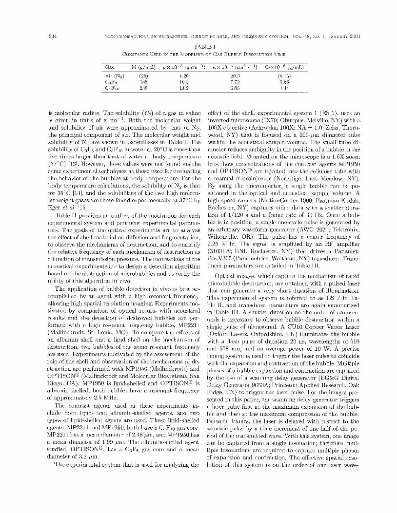
Air (Nz) (28) 1.20 CJF* 188 10.3 C4Flll 238 11.2
20.0 (8.15) 7.72 5.66 6.86 1.44
is molecular radius. The solubility (Cs) of a gas in saline is given in units of g Both the molecular weight and solnbility of air were approximated by that of Nz, the principal componcnt of air. The molecular weight and solubility of N2 arc shown in parentheses in Tablc I. The solubility of C3FB and C4Flo in water ai. 20°C is more than five times largor thaii that of water at body tcnipc:ratiirc: (37°C) [13]. However, thcsc values were not foimd via. the same experimental techniques as (.hose used for evaluating the hehavior of 1,hc bitbblcs at hody 1,cnipcratiirc. For tlic body temperature calculations, the solitbility of Nz is that for 35°C [14], and the solubilities of the two high niolccu- lar weight gases arc tliosc found experimentally at, 37°C by Eger et al. [15].
Tahlc I1 provides an outline of the motivation for ea,ch experimcntal systclrl arld pcrtirlcnt c:xpcrimcnt,al pklrarric- ters. The goals of 1;hc optical cxpcrimcnts arc to ana lpe tile effect of shell material oti diffusion and fragmeIlt;ation, to observe the mechanisms of destruction, antl to quant,ify the relative frequency of each mechanism of destruction as a flinct,ion of t,rarlsrrlissioli press~lre. Tile Iriotivatiolis of the acoilstical cxpcrimcnts arc to desigli a, detectiorl algoritlirri
effect of the shell, experimental system 1 (ES l), uses an inverted microscope (1x70; Olympus, Wklville, NY) with a l00X objective (Achroplan 10OX; S A = 1.0; Zeiss, Thorn- wood, NY) that is focused on a 200-pni diameter tube within the acoustical sample volume. The small tube di- ainctcr reduces ambiguity in tlie position o l a bubble in thc acoustic field. Mounted on the microscope is a 1.6X zoom Icns. Low coricentratioris of the contrast agcnts iVIP1950 antl OPTISONO arc injected into tlie cellulose tube with a niannal microinjcctor (Narisliige, East Meadow, NY). By using the microiiijector, a single bubble can he po- sitioned in the optical arid acoustical sample volume. A high speed camera (MotionCorder 1000; Eastman Kodak, Rochester, NY) captures video data with a sbii1,tcr d im- tion of 1/120 s and a frame rate of 30 Hz. Once a bub- hle is in position, a single one-cycle pulsc is gcncratcd by an arbitrary waveform generator (AWG 2021; Teldronix, Wilsonville, OR,). The pulsc has a center frequency of 2.25 MHz. The signal is ampMed by an R.F amplifier (310OLh; ENI, Roclicstcr, NY) that drives a Panamct- rics v305 (Panainctrics, Waltham, NY) transducer. Trans- duccr pa.rarneters are detailed in Tablc 111.
based on the destruction of microbubbles and to verify tlie Optical images, whicll caI,tllre the mccllanism of rapid niiixhubble destruction, arc ohtaincd with a pulsed laser utility of this algorithm in vivo.
The application of hulhle detection in vivo is best ac- tilat can a. vc!ry dllratiorl of illumination, complished by an agent with a. high rcsonant frcrliicncy, ~ l ~ i ~ systcnl is rcferrctl to as ES 2 in T ~ . allowing high spatial resolution imaging. Experiincnls 1110- hi(. 1 [, arlcl tra.Iis&cer passmeters arc again sulnmarized tivated by comparison of optical results with acoustical in ~ ~ ~ t , l ~ 111, A clltratioa tile of r,anosec. results and the drtection of destroyed b l l b b h arc per- (,l& is necessary to observe bubljlc destruction withirl Ibrmcd with a high resonant freqiicncy bubble, MP2211 Il~trasollrlt~, A C u i 0 copper vapor L~~~~ (IVlallincltrodt, St. Louis, MO). To compare the effects of (oxlord L ~ ~ ~ ~ ~ , oxfordsiIirc, TJK) illuminates the bubble an albumin shell and a lipid shcll on the mcchanisrn of 510 tlcstriiction, two bubbles of the samc resonant frcqiicncy ar,d 578 nm, of 1 ( j W, A precise arc iiscd. Experinieiits motivated by the assessment; of l,hc timirig system is ,lsed lo trigger tile laser pulse to coincicle role of the shell and observation of thc mechanisms of tie- with the expallsioll a,ld contractiorl "fthe bubble, ~ ~ ~ l t i ~ l ~ strnction arc performed with MP1950 (iV~allincltrod1) and phases ()fit ljuhl,lc and corltrae(,ion arc caplnred O P T l S O N ~ (Mallincltrodt and Molcciilar Biosystcrns, SRn by thc llsc of a scallnirlg cleiay generator (EG&G Digital Diego, CA). MP1950 is liPid-shelIe(l arid OI'TISoN@ is Delay Generator 9650A; Princeton Applied R.esearch, Oak allxunin-shelled; both bubblcs have a resonant frequency rtidge, TN) to trigger tkie laser pulse. F~~ tilc images prc. of approximately 2.5 MIIz. scntcd in this paper, the scanning dclay generator triggers
l h e contrast agents used in thcsc cxpcrirncrits in- a laser pulse first at tllc maximllm expansion of tllc but). elude both lipid- and albumin-shelled agents, and two hlc and then at tlie maximunl conlpression of tlle bubble. types of lipid-shelled agents arc usc!d. These lipid-shelled Bei.wecn frarncs, the laser is delayed with respect to the agents, MP2211 antl MP1950,lioth have a CaFjo gas core. acoustic pllse by a time incremcrit of one-half of the pe- MP2211. has a mean diameter of 2.48 pin, and WIP1950 has riod of thc trarlsnlitted wave. With t;1& system, one irrlage a mean diameter of 1.99 pm. The albumin-shcllcd agent can be caphlrcd frorn a. sirigle illsonation; thcrcforc, mul-
plllsc
il flaski pulse of duration 20 ns, wilvelerlgt~ls a,l
r 7
studied, OPTISONO, lias a C3F8 gas core and a inean tlianieter of 3.2 pm.
The experimental system that is used for analying the
tiple irisonations are rcquircd to capture multiple phases of expansion antl coiitraction. The effective spatial rcso- lution of this system is on the order of onc laser wave-

In vitro optical In vilro acoiistical In vivo acoustical
ES' 1 ES 2 ES 3 ES 4 ES 5
Molivatiori
SIn1t.t.cr rirccliaiiisni
'lianstlilcer
1'11lscr
Contrast ilgcnt
Cc111,cr frcqlu!llcg of insonation
ltesonant Srequeiicy of hiiI>hle
Piilse length of transmission at tlrc hiihblr
Effcct os SllCll Mecliariisni OS Itclative dcstriictioii frequency of
incchsnisins
Pulscd-laser High spccd <:mriesa
WO5 V 9 0 0 ~
AWC2021 R.itct: SP-801
MP1950 ~ 1 ' 2 2 1 1 7
2.25 M I I Z 5 MHz
2.5 M H L 2.5 IvIHz 5 MIIR
I 112 cycle 5 cycle 1 112 cycle
Biihhlc dcl.ectioii
5 M H s
5 lViM2
1 ,112 cydc
Unhblc cletectior1 arid verification
of fragmciitatiori
4.4 MHz
5 M H Z
I 112 cyclr
~xperiiiieiital systcrn. 'East,inan Kodak, Roclicstcr, NY. "Panarnctrics, Wali.ham, NY. 4GE Mctlical Systems, Milwaiikec, W1. "'l'cktroriix, Wilsonville, Olt. 'llitcc, Warwick, Ill. 'Mallinckrodt, St. T,ouis, MO. 'Molcciilas Biosystenis, Sari nicgo, CA.
Ccnt.cr hqi ic i icy 6 CID hanrlwirltli Apcrtiisc Focal length Transd i~ccr (h4Ils) (bvilla) size (inches) (inche3)
V305' 2.4 1.6-3.3 0.75 2.34 v:1091 5.6 3.7~ 7.6 0.5 2.19 v31 I1 10.5 7.5 13.(i 0.5 2.07
'l'ariainctrii:s, Wall.liam, N Y .
length or 5J 0 nm. Bccaiise of the laser illiuriinatiori, thc resulting images appcnr to tic liinary and cxliilit tliffwc- (,ion rings around the tiulhlc; Iiowcvcr, thcsc: (lo iiot. iiii- pedc the a~ralysis of nialtirig rc:lal,ivc iiiOnsUi.CiIi(:iit,s of I,hc instantaneous hubblc diameter conip,arcd with the inil,ial diameter. Iridividual biihhlcs ol MPl960 arc again posi- tioned in a ~ o o - / ~ ~ ~ ~ tuhc with a mainial iriicroiiijcctor, a r i d an iiivcrtctl high power r~~ici~oscopc is iiscd that provitlcs 140X magnificatiori. Five-cycle RF pulscs arc gcncmtetl 11y an arbitrary waveform gc~icralor (AWC 2021); t,hc R.F piilscs have a ceiitcr frcqiicncy of 2.25 M H a . Tlic sigrial is amplified by an ItP amplikr (3lOOI,A), which drivcs a Paiiamctrics VBO5 traiisdiiccr.
In vitro optical stutlics, which statistically malyxc the
rclativo frequency o l d uctive mcchanisrris, usc expcr- imciital system ES 3 , s i " x i z c d in Table 11, and, for these cxpcrirnenls, a tratlitioiial microscopc light soiirce is sufficieiit. The lipid-shelled coiitra,st agent MP2211 is iiscd iii tlicsc sliidics mid is compared with in vitro acoiis- tical ancl in vivo acoustical results. A cust;orri-buill, high powcr, non-invortcxi iiiicroscope (IV5OOL; Mikron Tnstru- iiicnts, S a n llicgo, CA) with the l00X objective is fociised on the 200-//,m tuhc within the acoustical sairiplc volume. Moiintcd on the rriicroscopc is :L 2X zoom leas. Images arc acqisircd for offline aiialyHis with the high speed canicra (Kodak MotionCordcr Aiialyzc1 1000). The cairicra frame rate is 240 fmnics/s with a shutter duration of 1/500 s.
111 vitro acoustical c:xpcrinicnls arc: Imforined with the
236 IEEI': TRANSACTIONS ON UI,TRASONICS, FERROELECl'lllCS, AND IIREQUENCY CONI'ROL, VOL. 48, NO. 1, JANUARY 2[)[)1
contrast agent MP2211. Tlie system used is summarizcd in Table I1 as ES 4. Two single piston transducers are mutually focused on a section of 200-pm cellulose t u b ing. The transmission transducer is the Panamctrics V309. Reflected echoes are received by the Pariamctrics V311, a wideband piston transducer with center frequency of 10.5 MHz. The transducers are focused and mounted into a plcxiglass box at an angle of approxirnatcly 15" with re- spect to one another. Thc 200-prn cellulose phantom vessel is placed in the confocal sample volumes of the transduc- ers at an angle of approximately 45" within the plane of the transducers.
In both ES 3 and ES 4, im intermittent pulsing scheme is gcncrated by an arbitrary waveform generator (AWG2021), which triggers a square wave pulscr (SP-801). The transmission prcssnre arnplitudc can be va,ried from 0.8 to 2.6 MPa. The intermit,tcnt pulsing scheme involves a train of 10 pulses with a pulse repetition frequency (PRF) that is varied from 1 to 5 ItHz. The 10-pulse train is fol- lowed by a delay on the order of 1 s before transmission of the next set of 10 pulses. This allows complete washout of the insonified sample volume aftcr each set of pulses, so that each event can be analyzed independent of the pre- vious event. The received echoes from the 10-MHz trans- ducer are amplified by a receive amplifier (Ritec BR-640) and sampled by an oscilloscope (9350L; LeCroy, Chestnut Ridge, NY) a t 100 MSamples/s. The osciloscope is GPIB- controlled in sequence inode by a LabView interface. The data are stored for offline analysis.
The contrast agents are suspended in saline a t a low concentration on the order of 1 sphcrc/l pL. With such low concentrations, a single microbnbble is expected to flow through the acoustical sample volume in each 10-pulse acquisition. The saline-microbubble solution is pumped a t a low flow rate (1 inL/h) that corresponds to a maxirniim velocity in the flow phantom of 1 cm/s.
The transrriission pressures generated in each experi- mental system arc calibrated using a nccdle hydrophone (PZT-Z44-0200; Specialty Engineering Associates, Soquel, CA) and a prearnplificr (Specialty Engineering Associates A1.7dB) connected to an oscilloscope. The tip of thc needle hydrophone is placed in the optical focns or the system.
B. In Vivo Methods
All animal experiments are conducted iintlcr a protocol approved by the institutional review board of the Univer- sity of Virginia using the experimental system siiminarized in Table I1 as ES 5. An athymic mouse is injected with ap- proximately 1 x loG hurrmri LNCaP tumor cells. The cells are injected orthotopically in 1,wo places abovc thc rear legs of the mouse to minimize artifacts from breathing and hcart motion. When the tnmors reach a predeterinincd size (> 5 mrn), the mouse is anaesthetized, arid the jugular vein is cannulated. A low concentration of contrast agent (ap- proximately 10 pL/mL) siispcndcd in saline is injected into the jngular vein, driven by a syringe pump for continnous infusion. The contrast agent used in the in vivo studies is
MP2211; the saline-microbubblc mixture is injected at a rate of 1 mL/hr. The niousc is placed on an isothermal pad (Braintree Scientific, Braintree, MA) to maintain constant body temperature during the experiment .
A modified GE Logiq 700 scanner (GE Medical Sys- tenis, Milwaukee, \VI) is used to acquire raw data. The imaging parameters are a PRF of 7.4 ltHz and a trans- mission pressure of 1.5 MPa. Each acoustic pulse is gcncr- ated by a 1 1/2 cycle sine wave with center frequency of 4.4 MHz. Sixteen pulses arc acquired in each line of sight.
111. RESULTS
The results of this work will be divided into four scc- tions. First, the theoretical background related to the mechanisms of destruction is reviewed. Optical data are then presented that provide visual evidence of thc three mechanisms of destruction. Tlie relative occurrence of a specific destructive event as a function of transmission pressure is discussed. Following the presentation of optical data, the acoustical data collected within the in vitro and in vivo experiments are provided.
A. Theoretical Background
The time scale of events leading to the disappearance of a gas bubble in a liquid medium offers an important cluc to aid in understanding the destruction mechanisms of ultrasound contrast agents. Previous theoretical analysis of iincncapsulated bubbles yields insight into the phenomena that have been observed. The theoretical analysis describes the meclianisms of bubble dcstruction as well as provides an order of magnitude estimate of the time scale required by each mechanism.
1. Fragmentation: Fragmentation of a bubble may arise when an acoustically driven bubble becomes unstable, leading t,o pinch-off of fragments from the original bub- ble. Tlic bubble is most stable when it is spherically sym- metric, and perturbations from spherical symmetry may result in instability. The acoustically driven bnbble may exhibit perturbations from spherical symmetry in corijunc- tion with large radial expansion and contraction. The cx- tent of t,he radial deformation is dependent on the parame- ters of the incident pressure wave and the mechanical prop- erties of tlic liquid and the gas. A stability criterion was developed by Plcsset and Mitchell [lG], [18] that describes a threshold radius below which the undamped collapsing bubble becomes unstable:
A list of symbols used in the equations presented in this paper is given in Table IV. This criterion states that if the bubble radius decreases to less than one-tenth of the maxiinnm radius achieved by the bubble, it will be- come unstable. Instability of the bubble arises when the
CHOMAS et al.: CONTRAST AGENT DESI'RIJO'IION
amplitude of non-spherical dislortioiis bccorue large rel- ative to tlie radius of the bubble. Two non-parametric types of shape instabilitics may arise diiriiig acoustic forc- ing, Rayleigh-Taylor iristabi1it)y and aftorhouncc instabil- ity [17]. R.aylcigh-Taylor instability occurs when the col- lapsing tnibble approaches its minimum radius accompa- nied by a large acceleration of the gas into the fluid, caus- ing violent fragmentation of tihe bubblc in significantly less than one cycle of oscillation. The time scale of Rayleigh- Taylor instability is on the order of nanoseconds. After- bouricc instability occurs when the biibhle wall niolion overcomes stabilizing viscous cirects and surfacc tension, which reduce shape distortion, occurring on the lime scale of lhe period ol the acoustic forcing or rnicroscconds. The result of the two non-parametric shape instabilities or their combination results in the pinch-off of bubble fragnicnts during a single insonation.
2. Static DzfliLsion,: In cases in which fragmentation does not occur, diffusion may be the dominant mechanism of microbubble dcstruction. Static diRusion is the move- ment of gas from the core of tlie microbubble into tlie surroiinding medium neglecting contributions from coii- vection. The rate of change of the radius of an unslielletl bubble in an ovcrsaturated solution is [ll]:
237
2Mn ci s = i f = - BTR<,P(oo) cs
where IF is the coefficient of diffusivity. The equation is
TAR1,F: IV LIS1 OF SYMUOLS
Surface terision Coeficicnt of diffusivity 2n/fu/B7'R,p(oo) Dcnsii.y Density c i f a gas buhblc with gas-
Gas colistarit Initial gas concentration Saturation gas concontration
blolccnla~ wcighb Radius Maxiinurn radiiis of a t>ubblc
during irisonation Thrcshold radius below which
Lhe bublile hecoincs unstable 'I'empcraturc Tirric Non-dirncnsional radiris Non-dirucusional tinic
liquid iritcrface of zero curvaturc
GIG
concentration of gas in the surrounding niediiini may vary from 0 t,o tlic saturation concontration depending on the gas and the concentration of contrast agent that is stud- ied. The quantity f = Ci/C.s may be i i s c d t,o illustrate thc effect of this coriccntration gradient. T ~ C quantity f s p n s the range from 0 (degassed watcr) to 1. (cornplete satura- tion). The time for dissohition of gas bubbles of various ini- tial diameters at room temperaturc is calculated using (2), as shown in Fig. 1. For each gas, the two limits of gas sat- iiration in the surrounding w;tter arc evaluated. The time for dissolution is plotted in millisecorids on a logarithmic scale.
in non-dimcnsional form, with e representing the non- dimensional radius arid r rcpresent,ing non-dimensional tirnc. The noii--diniensioiializntion cc~iialious arc given hc- low (l), where R is the radius of the bubblc and t is time. The constant p ( w ) is the deiisity of the gas in the hub- blc occurring at a zero curvature gas-liquid interface. Tlic
In Pig. 1, the time for dissolution of a bubble in dc- gassed water is shorter thaii that in water saturatcd with gas, but still on the same order of magnitude. The data in Fig. 1 demonstrate a depeiidcncc of diffusion rate on tlic liquid satailration, constitutive gas properties, init,ial diam- eter, arid temperature. Dccaflnorohutanc (CdF1") exhibits
constant U is tlic surface t,cnsion constant of walcr (72 dync/cm), M is the molcciilar weight of the gas, U is the universal gas constant, and T is 1;cmperature in Kelvin. This equation includes surface tension, which seems rea- sonable for thc case of a gas bitbble whose shell has been damaged or dcstroycd by insonation. A bubble with an intact shell may bc affected less by siirfacc knsion, and the coefficient of diffusivity of the gas-liquid or gas-solid interfacc may tic significantly lowcr compared willi an un- encapsulated gas bubble. Howevcr, lhe rolc of thc shell is not incorporated with this approach and will he addressed in future work.
In (2), the rate of diffusion and, hencc, tlie correspond- ing rate of hihblc disintegration is inversely relatcd to the radius of thc bubble and the dcnsily of the gas. The rate is proportional to the cocficieiit of diffusivity and tlie diffcr- ence tictween the initial gas concentra,tioa and the cquilih- rium conccritratioii in the sixrounding medium. The initial
the slowest time for dissohition because of its high density, high rriolcciilar weight, arid low cocfficicnt of diffusivity (see Table 1). For a lnibble with an initial dianietcr of 2 pin arid temperature of 20"C, a typical size of cornrricrcially availaldc agents, perfiuoropropanc requires approximately 90 ms. A deca,Auorobutane agent rcquires approximately 900 ms, nearly a tcrifold increase in tlic time for dissolu- tion. Air dissolves faster than cither of thc high molecular weight gases such that an air bubble at 20°C with an initial diameter of 2 bni dissolves in 8 ms. This result is similar to the tiinc: for air dissolution computed by Frinlcing e% al. [19]. A significant depcndence of dissolution time on the initial radius of the 1)ubblc is observed. For each gas bubble considered, an iricrcnse in the initial diameter by four times rcsults in a more than tenfold increase in thc dissolution time. For air at 20"C, the timc for dissolution increases hom 1 riis for an initial diameter of 1 pm to 12 rris for an initial diarrietcr of 4 pni. Similarly, for CdFio at
I 1.5 2 2.5 3 3.5 4
initial buhble diameter (pm)
Fig. 1. Tirnc for dissolution of an unshcllcd gas bubble in saline at 20°C as prcdictcd by (1). Dashed lincs rcprcscnt f = 1; solid lines rcpresent f = 0, wlicrc f is tlic ratio of the initial gas conccntration of the surrounding mcdiurn to thc saturation conccntration. 'I'riangles are C,iFlo, circlcs are C ~ F R , and lines reprcscnt air (approximated by N2).
20"C, the time for dissolution increases from 0.20 s for an initial diameter of 1 pm to 3.5 s for a bubble with an initial diameter of 4 /mi. There is also a significant dependence of dissolution rate on kmperature, and it is expected that the rate of diffusion will be slower for highcr temperature studies as would be observed in vivo.
To analyze the expected change in bubble diameter for time scales on the order of clinical PRFs, the change of diameter of a gas biibble over a suitably short iiiterval is studied. A bubble containing either of the high molec- ular weight gases studied at room temperature will (le- crease in diameter by less than 0.2 pm in 16 Iris, corre- sponding to a percentage decrease in diameter of less than 10%. For decafluorobulanc ( C ~ F ~ O ) , the core of the lipid- shelled bubble analyzed optically and acoiistically in this paper, tlic inaximum percentage decrease in bubble diarn- eter produced by static diffusion within 16 ms is less than 1%. Malting the assumption that received echo iiiterisity is proportioiial to the sixth power of radius [20], the received acoustic intensity will decrease by less than 3%. Diffusion is predicted to affect the rcccivrd echo lcss in vivo hecause of the decrcasc in diffusion rate corresponding to an in- crease in temperature. As is expected, air bubbles rapidly decrease in diarnetcr, significantly fastcr tliari either high molecular weight gas. Therefore, we conclude that static diffusion may play a major role iii decreasing tlie received echo from an iinshellcd air bubble, nearly dissolving a 2- pm diamcter bubble within a IO-ms time span at body ternperatlire as well as ai, room temperature.
3. Acoustically Driven Di,fusi.on: The rate of diffusion can be affected significantly during insonation because of cliariging pressurc over time and the motion of the gas-
liquid interface. The complete diffusion cquwtion of a gas biibhlc into tlic surrounding liquid is
wlicre C is the gas conccntration, t is time, v is the wall velocity, R is radial distancc, aid r; is the diffusion coefli- cienl; of the gas in tlie liquid. The second term in tlic cqua- tioii is known as the convective term hccausc it describes the cfI'ccl, of the bubble wall moving on gas flux across the hiibble-liquid boundary. To understand the effect of a pressure field on the diffusion of a bitbblc, a more corn- plctc model has been dcvclopcd by Cha arid Heriry [21]. Their modcl of diffusion includes an additiorial lcrin that accounts for tlic transfer of gas &cross the gas-water bound- ary caiisetl by exlcrnal prcssiirc variations. The authors rcporl an increased diffusion rate into the bubble during decompression (rarefaction) of a gas bubhlc; the rate of difhusion out of the bubble increases during compression. With w siiiusoidal pressure field, 1,hc net diameter of a cou- trast agent lias been observed to drcrcase; the iricchanism of this decrease in diameter is termed acoiistically driven diffusion. The net difhsion out of the biibble may he due to the prevailing gas concentration gradient with higher concentration insidc of the biibble. It is difficult to asscss the time scale of diffusion that occurs during insonation. The shell dynaniics may play a critical role iii deterriiinirig the extent to which the increased outward diffusion during compression dominates tlic incrcasctl inward gas diffusion during expansion. T h e initial diamrtcr, the peak transmit- ted pressure, and the shape of tlie iiicident, pressure wavc may also play critical roles in thc diffusion rate caiiscd by insonation. It should be noted that Clia and Henry's result ncglccts the contrilnit,ioiis of convection causcd by the radial oscillations of tlie bubble wall. Convectivc dif- fusion has been stiidicd for acoustically driveii unencap- sulated gas bubbles theoretically (Ellcr arid Flyriii [22]; S a r i [as]), arid experimentally (Cruni [24]), particularly iii the context of "rcctified diffiision" , whcrc forcing over many acoustic cycles is needed. The 1;ransieiit convective diffusion effects are less untlc ood, and, similarly, shell effects have not yet been examined rigorously.
B. h i Vitro Optical Study
Two sets of optical experiments are performed to ob- serve mechanisms of microbubble destruction. Initial op- tical experiments arc perforrried with a high speed digital canicra controlling the shutter timing. The sampling rate of the camera is 240 fraincs/s, giving a time resolution of 4.2 ms between fraines. By using a shutter duration of 1./500 s, each frame holds information of ti finer time res- oliition of 2 ins. An advanced optical method is developed to observe the bubble exparision aiitl contraction elicited by the insonation. In this method, a shutter duration of 20 ns is generated by a pulsed laser, arid data are collected with the high spccd digital camera. The 20-11s shutter is

CIIOMAS e l al.: CON'I'R.4SI' AGlbNT DLS'I'RUCI'ION 230
adequate to simple the size and shape of the Iiubblc during insonation. Thrcc mechanisms of microbubble destruction are observctl optically: fraginentation, acoustically driven diffusion, aiid static diffusion.
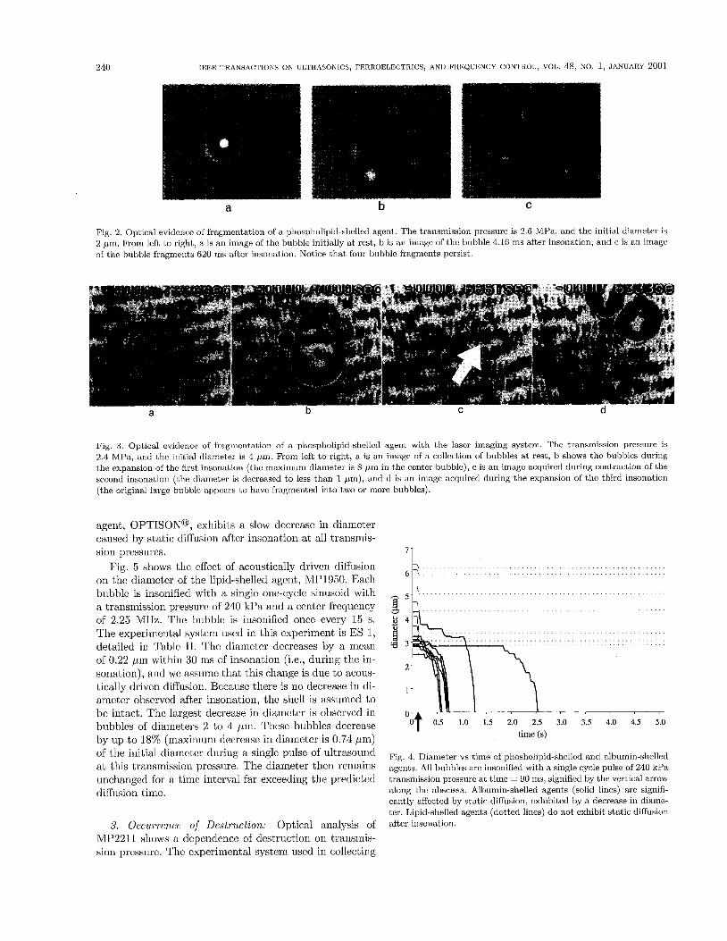
1. E~~,yrrreri , tci t iori: A cornnioii phenomenon observed optically that is produccd by liigli aniplitutlc lmnsinission pressure is the fragrncnt;rtion of tlic original microbubble iiito srnaller bubbles after one pulse. Fig. 2 shows a rep- rcscntativc set of hamcs ol data Iron1 the o p t i d study of ail MP2211 bubble pulsed once bctwccn 1,lir first arid sec- ond video frames. The iniagcs in Fig. 2 arc collcctcd with tlie cainerti shntter-based optical system, ES 3 (a detailed descriptioii can be fount1 ill Table 11), with a. (miismis- siori pressure of 2.6 MPa. In Pig. 2(a), the dianic:t.cr of the original rriicro1,ubblc is approxirna1,cly 2 pni. The six fragments, sliowri in Pig. 2(h), each have a diameter that is approxirriatcly 1.5 /mi. Tlic 1,iInc bctwccn the acquisi- tion of Fig. 2(a ~ i i d 1)) is 4.2 nis. The six fragrrionts are radially distril)iitcd around the original bubble center aritl arc displaccd approximately one bubble dianieter froiri the ccntrr. Four or the bubble fragnients rerriairictl tlic same diairictcr for tlic remainder ol observation. Fig. 2(c) dc- picts the bnbble fragments 620 ins after fragmeii1,tLtiori. Four fragrrients remain visible; tlic other two fragments arc not visible.
Tlie nurnber of fragincrit,s varies from two fragments to ovcr t.cn fragments h i n a single bubble. In gcncral, t,hc fragments tend i;o form a circlc around the position of tlic initial bubble. The largest tlisplacerricnt of fragments ob- served is approxiniatdy 6 /mi fi.0~11 the ccntcr of the ini- tial microbubblc. Thr h g I n ( d s inay c i i h r dissolve after lragmentation or persist. The fragments that persist cx- hibit a sigiiificantly srnaller rate of diff'usion ol gas out ol the biibhlc compared with tlie prediction for uncncnpsu- latcd C / J F ~ ~ ) , suggesting that thc reinainiug slicll retains
To uriderstand the mcchanisnis ol Iragnientatiori fnr- tlicr, a iinique oplical imaging system is iisctl t,o ini- age a bubble tluriiig insonatiori, ES 2 (sec Table I1 for dctails). Pig. 3 shows the expansion m d contraction of WIP1050 during insoiiatioii; tlic irnagcs arc acquircd with the pulsed-laser imaging system. As stated earlier, this sys- tem has a,n effective! sliiil,tcr duration of 20 ns, and a precise tirning system allows thr illuminal,ion of this short time du- ration flash to be aligned with tlic actual insonation of the bubble. The resting bubble of Fig. 3(a) has an iiiitial diairi- eter of approximately 4 pin. The bubble is irisoriificd with a five-cycle, 2.4 MPa excitation with a center frccpency of 2.25 MHz. Pig. 3(h) shows the bubble during tlie rar- efactional phase of tlie first pulse of insonation, where the huhble diameter nearly doublcs Lo approximately 8 p i .
The iiriage in Fig. 3(c) shows the bubble during tlic com- pressional phase of tho soc:ontl :rcoustic pulse. The arrow points toward tlic compressed bubble. The dianictcr of thr buhblc decreases to less than 1 /rm. Fig. 3(d) shows tlie sainc lxihblc in the rarefactional phasc of the third pulse, wlicrc two distinct hul)l)lc fragments arc obscrvcd. Each
bili1,y to rediicc diffusion a,fter excitation.
fragincnt has a diameter olG.5 /m. In terms of tlic Plcsset arid Mitchell stability criterion, instability OCCIII'S when I;he biihblc diameter in the comprrssional phasc is one-tenth of that, of tlic maximinn c?xpandcd phaso cliamctcr. Because tlic cxamplc in Fig. 3 demonstrates a ratio of rnaximurri diametcr to minimum diameter grcatcr than 8, the two large bubble fragments in Fig. 3(d) may bc the result of hagmentation oP an unstable bubble. Repctitiori or this experirricnt dernoiistrates reproducible expaiisiori arid coii- traction of both lipid-shelled and albumin-shelled agcrits on the ordcr of the Plesset arid Mitcliell criterion. A sum- mary of the relative occ:iirrenc:e of fragmentation and other dcstriictivc. mechanisms is presented la1,c:r in this paper.
2. Diffusion: The coiribinatioii of tlie sliell arid gas crc- ate a coniplicated erivironrnent from which to assess tliffii- siori. A sct of oxperinicnts is coriductcd to dcteririiiic t,hc effect. of tlir shell on the clifhision tiinc. In tlicsc cxpcr- inicnt,s, th r microbiibblc is pulsed once with iiltrasound, and tlicii the diameter is recorded as a hinctioii of time by analyzing images acquired evcry 3 0 ins. The cxpcrimental systrm iised in th cxperimenls is ES I, dcscribcd in Ta- hlc 11. Fig. 4 is eprcscnlative sct oL dianictcr versus time ci~rvcs comparing l,hc rc1al;ivo clr lipid- and albumin-shelled agents. The dashed lines rcprc- sent data collcctcd from images of the lipid-shelled agcnt, MPl950, and solid lines rcprcscni; d a h from tlic nlhnmin- shcllcd agcnt, OPTISONO. Both agents arc insoriificd with a siriglc: one-cyclo sine wavc with a peak acoustic p of 240 ltPa arid ceiiter frequeucy of 2.25 MHz. Prior to iii- soriat,ion, diffiisiori is riot apparent on our time scales of ohscrvation. The bubbles arc insonified at a time of 90 ins, as shown in Fig. 4 as aii arrow iri thc tiriic axis. Thc cf- fcct of acoustically driven diffusion is obsei.ved within the first 30 ins after insonation (the tinic interval from 90 to 120 my), appearing as an instantaneous decrease in di- airieter. Tlirougliout the next 5 s of observation, MP1950 retains approxiniately the same resting dialneter, but the diameter of OPTISON@ decreases as a fuiictioii of tinic. NIP1950 contains CL1l.'lo, in which iiiicricapsulatcd gas bub- bles of CIFlo arc prcdictod i,o diKnsc into t,hc snrronnd- irig liquid within 5 s. Becxusc, in this experiment, MP1950 docs not exhibit a significant dccrcasc in diameter, we con- clude the lipid shell retains its ability to significantly in- hibit the diffusion ol gas out or the bubble. Alternatively, the set of OPTISONO bubbles studied fully dissolve in less than 2.6 S. Because OPTISON@ contains C ~ F B , in which uuericapsulated gas bubbles of C3Fe are predicted to fully dissolve in 2 s, we infer that tlic alburniri shell of OPTISONO no longer acts as an ef€ective barrier to diffu- siori after insoriation. The percentage decrease in diarrictcr after insonation ends is analyzed lo characterize the ex- tcxt of static diffusion. The dccrcasc in di,zmcl,cr observed ovcr the interval from 30 ins after irisonation to 60 rns af- ter insonation (i.e., 120 to 150 ms iii Fig. 4) is 17.7% for OPTISONO bubbles, and the iriean percentage decrease in MP1950 during this interval is lcss than 0.1%. The per- centage decrease in diamctcr of OPTISON@ is significantly larger than MP1950 ( P < 0.0002). The all)iirriiri-shclled
240 IEEE TRANSACIIONS ON Ul,rI<ASONICS, rERROELECTRICS, AND FREQUENCY CONTROL, VOL. 48, NO, 1, JANTJARY 2001
a
Fig. 2. Optical evidence of fragmentation of a phospholipid-shcllcd agent. The transmission pressure is 2.6 MPa, and the initial diameter is 2 pm, From left to right, a is an image of the bubble initially at rest, h is an image of the bubble 4.16 ms after insonation, and c is an image of the bubble fragments 620 ins after insonation. Nolice that four bubble fragments persist.
a b C d
Fig. 3. Optical cvidence of fragrncnlation of a phospllolipid-shcllod agent with 1 . 1 ~ laser imaging system. The transmission prossure is 2.4 MPa, and the initial diameter is 4 pni, From lefl. to right, a is an image of a collection of bubbles at rest, b shows thc bubbles during the expansion of thc first insonation (thc mdxinium diameter is 8 piu in the cenler bubble), c is an image acquired during contraction of the second insonation (the diameter is decreased to less than 1 pm), and d is an image acquircd during the expansion of the third insonation (the original large bubble appears to have fragmented into two or more bubbles).
agent, OPTISONO, exhibits a slow decrease in diameter caused by static diffusion after insoriation a t all transniis- sion pressures.
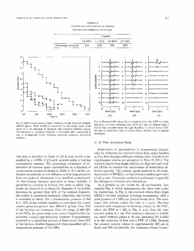
Fig. 5 shows the effect of acoustically driven diffusion on the diameter of the lipid-shelled agent, MP1950. Each bubble is insonified with a single one-cycle siniisoid with a transmission pressure of 240 lcPa and a center frequency of 2.25 MHz. Thc bubble is insoriified once every 15 s. The experirricntal system used in this experiment is ES 1, detailed in Table 11. The diameter decreases by a mean of 0.22 pm within 30 ms of insonation (i.e., during the in- sonation), arid we assume that this change is due to acous- tically driven diffusion. Becaiisc there is no decrease in di- ameter observed after insonation, thc shcll is assumed to be intact. The largest decrease in diamcter is observed in bubbles of diameters 2 to 4 pm. These bubbles decrease by up to 18% (maximum decrease in diameter is 0.74 pm) of the initial diamctcr during a single pulse of ultrasound a t this transmission prcssure. The diameter tlicn remains unchanged for a time interval far exceeding the predicted diffusion time.
3. Occurrence of Destruction: Optical analysis of MP2211 shows a dependence of destruction on transmis- sion pressure. The cxperimental system used in collecting
. . . . . . . . . . . . . . . . . . . . . . . . . . . . . . . . . . . . . .
. . . . . . . . . . . . . . . . . . . . . . . . . . . . . . . . . . . . . . . . . . . . . . .
~ 5E . . . . . . . . . . . . . . . . . . . . . . . . . . . . . . . . . . . . . .
. . . . . . . . . . . . . . . . . . . . . . . . . . . . . . . . . . . . . . . . .s - . . . . . . . . . . . . . . . . . . . . . . . . . . . . . . . . .
1 4
. . . . . . . . . . . . . . . . . . . . . . . . . . . . . . . . . . . . . . . . . . . . . . . . . . . . . . . . . . . . . . . . . . . . . . . . . . . . . . . . . . a 3
2
1
0 .O 3.5 4.0 4.5 5.0
I time (s)
Fig. 4. Diameter vs time of pliosholipid-shelled and albumin-shelled agents. All biibbles arc insonified with a single cycle pulse of 240 kPa transrriission pressure at time = YO ms, signified by the vertical arrow along the abscissa. Albumin-shelled agents (solid lines) are signifi- cantly aficcted by static diffusion, exhibited by a decrease in diame- ter. Lipid-shelled agents (dotted lines) do not exhibit static diffusion after insonation.
CRObIAS et al.: GONI'ILAS'L' A G E N T DESTRUCTION 241
TABLE V PERCENTAGK OCCIJHHENCE or I j u m ~ i ~ D~:S 'CKUcI ' ION OBSERVED OP1.ICALLY.
Pressure
0.8 MPa 1.4 MPa 2 0 MPn 2.6 MPa
Percentage destroyed 10% 80% 70% 80%
5.5 6l
2.5
2
1.5 0.0 50.0 100.0 150.0 200.0 250.0
time (s)
I
2
3
E 4 E
$ 6
e
z 5 r-..
= 7
8
9
10
0 1 2 time (ps)
(a)
0 1 2 time (IS)
(b)
1
5l------
8
9 :E 0 1 2
time (ps)
(c)
clue t o acoustically driven diffusion. Symbols rcpresenf. individual bubbles.
'l'!sLruynu
this data is described in Table 11, ES 3; each bubble is in- sonified by a 5-1\/IHz, 1 1/2-cycle acoustic pulse of varying transmission pressure. Tlic percentage occurrence of dc- struction of contrast agent microbubbles as a function of transmission pressure is shown in Table V. If a bubble un- dcrgocs acoustically drivcn diffusion and/or fragmentation from one pulse of ultrasound, it is classified as destroyed. To discriminate between destroyed or intact bubbles, a quantitative criterion is utilized. For cases in which frag- ments are observcd or in which the diameter of the bubble decreases by greater than 25% of the original diameter, the bubble is considered dcstroycd. Otherwise, the bubble is classified as intact. For a transniission prcssure 01 800 ltPa, 10% of the bubbles studied arc destroyed. For trans- mission pressures greater than 800 kPa, greater than 70% of the bubbles are destroyed. Over the range of 800 ltPa to 2.6 MPa, the percentage occurrence of rapid bubble de- struction remains approximately constant. Fragmentation occurred in a significant portion of these cases. Over 55% of the bubbles studied fragmented when insonified with a transniission pressure of 2.6 MPa.
Destruction of microbubbles is characterized aconsti- cally by analyzing the reccivcd echoes from single bubbles as t,hcy flow through a 200-pm cellulose tube. Details of the cxperirnental system are presented in Table 11, ES 4. The received echoes from single bubbles arc digitized and stud- ied off-line to confirm the incchanisms of destruction ob- served optically. The contrast agent analyzed in all acous- tical studics is MP2211., the lipid-shelled shelled agent with a C4F10 core. Correlation analysis is performed to quantify the changes in a bubble after insonation.
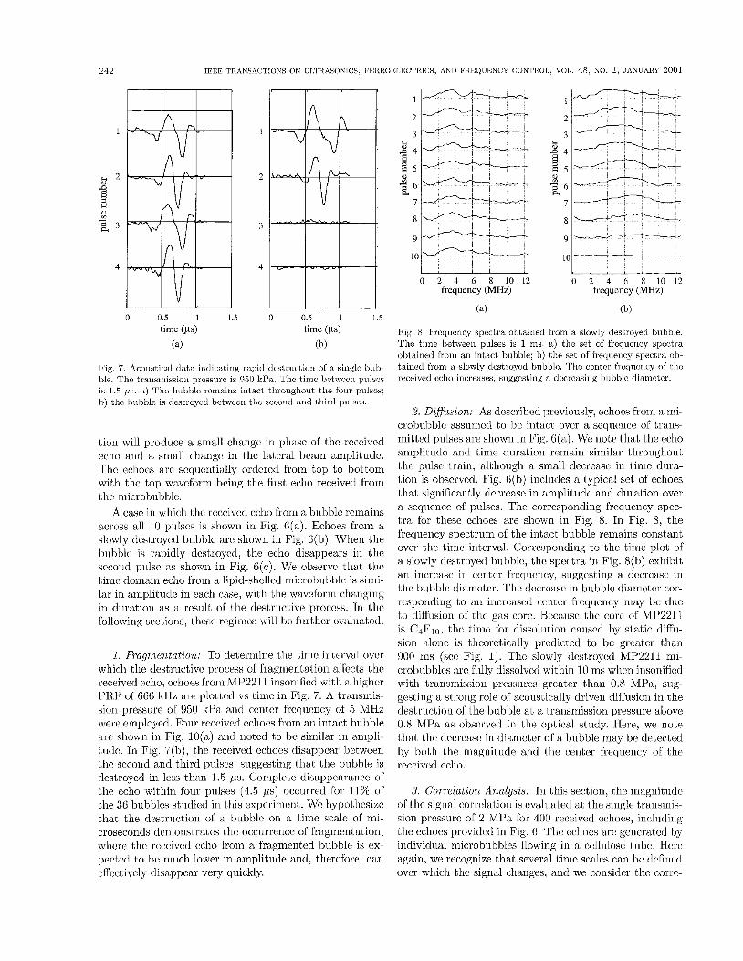
As a prehide to the results for all mechanisms, first consider Fig. 6, which demonstrates the tlircc time scales for destruction. In Fig. 6, the reccivcd cchocs from single iVIP2211 buhblcs obtained following transmission with a peak pressure of 2 NIPa are plotted versus time. The max- imum flow velocity within thc tube is 1 cm/s. The time between each acquisition is inversely related to the PRF, wherc the PRF is 1 ltHz in Fig. 6, and, thus, the time between pulses is 1 ms. The maximum distance a bubble can move between pulses is 10 pm, assuming the bubble is in the centerlinc of flow and a PR.F of 1 1tHz. Because the acoustic sample volunie is approximately 300 pm in depth and 500 pin latcrally, the maxinmrn change in posi-
IEEE TRANSACTIONS ON UI;l IL4SONICS. IXRROELECTRICS, AND FREQUENCY CONTltOl,, VOI,. 48, NO. 1, .JhNlJARY 2001 242
1
B 2 D
! 8 2 3
4
0 0.5 I I .5 n 0.5 I 1.5 time ( ~ s ) time (ps)
(a) (b)
Fig. 7. Acoustical dat,a indicaling rapid dcstructiari of a singla hub- hle. The transrriissiou prcssurc is 960 kPa. 'I'he tirne betwccri pulscs is 1.5 p. a) The bubhlc rcumiris intact throughout the four pulscs; h) the buhhle is destroyed bctwccri ttic second and third pulses.
tiori will produce a small change in phase of tlie received echo and a small change in lhe lateral beam arnplitudc. The ecliocs arc sequentially ordered from t,op to bottom with the top waveform being the first echo received from the microbubble.
A case in which the received echo from a biibhlc rcniains across all 10 pulses is shown in Fig. G(a). Echoes from a slowly destroyed bubble arc shown in Fig. 6(b). When the bubble is rapidly destroyed, the echo disappears in tlie second pulse as shown in Fig. 6(c) . We observc thal the time domain echo from a lipid-shcllcd microbubble is sirni- lar in aniplitiide in each case, with the waveform changing in duration as a result of the destructive procc!ss. In tho following sections, tliesc regimes will be further evaluated.
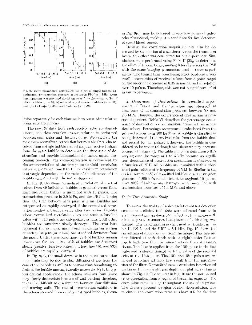
1. fiuqrnentation: To determine the tirnc interval ovcr which tlie destructive process of fragmentation affects thc received echo, echoes froin MP2211 insonificd with a higher PR.F of 666 ItHz arc. plotted vs time in Fig. 7. A transinis- sion pressure of 950 kPa arid center frequency of 5 MHz were employed. Four received echoes from an intact bubble are shown in Fig. 10(a) axid rioted to be similar in ampli- tude. In Fig. 7(h), the received cchocs disappear between tlic second and third pulses, suggesting thal the bubble is destroyed in less than 1.5 p s . Complete disappearance of the echo within four piilses (4.5 ps) occurred for 11% of
tudied in this experiment. We liypotliesim that the destruction of a bubblc on a l,ime scale of mi- croseconds demonstrates tlic occurrence of fragmentation, whcrc the received echo from a fragmented bubble is ex- pected to be much lower in amplitude and, therefore, can effectively disappear very quickly.
11 - I I-- 2
3
2 4 s B S
4 6 a
7
I ----+-- I
0 2 4 6 8 1 0 1 2 0 2 4 6 8 1 0 1 2 frequency (MHz) fiequency (MHL)
(a) (b)
Fig. 8. Rcqucncy spectra obf.ained from a slowly destroyed bubble. The time between pulses is 1 ms. a) the set of frcqucncy spectra obtained from an intact bubble; I?) tlie set of frequency spectra ob- tairied frorn a slowly dcstroycd bubblc. Ttic ccritcr frequency of l.he received echo increases, suggesting a dccrcasirig hubblc diamctcr.
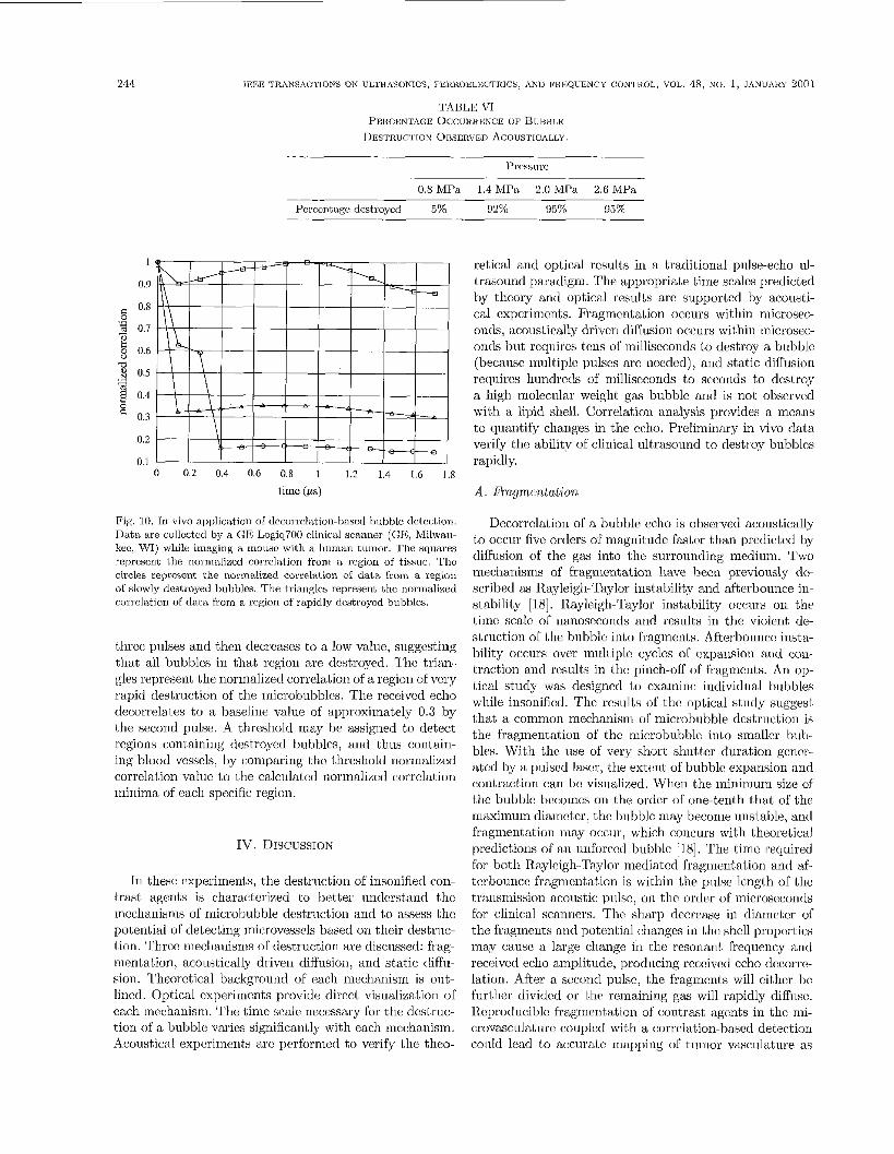
2. Diflusion: As described previously, echoes from a mi- crobuhblc assumed to hc intact ovcr a sequence of trans- milled pulses are shown in Fig. e(.). We note that the echo amplitude and time duration remain similar throughout the pulse train, although a small decrease in time dura- tion is obscrvcd. Fig. G(b) inclndes a lypical set of echoes that significantly decrease in amplitude and duration over a scqiicnce of pulses. The corresponding frequency spec- tra for these echoes are shown in Fig. 8. In Fig. 8, tlie hqueiicy spectrum of the intact bubble remains constant ovcr the time interval. Corresponding to the time plot of a slowly destroyed biibblc, t,lic spectra in Fig. 8(h) exhibit an increase in center frequency, suggesting a decrease in tlie biibble diameler. The decrease in bubble diameter cor- responding to an incre d center frequency may be due to diffusion of the gas core. Because the core of MP2211 is C ~ F J ~ , the time for dissolution canscd by static diffii- siori alone is theoretically predicted to be greater than 900 rns (sec Fig. 1). The slowly destroyed MP2211 mi- crobubblcs are fully dissolved within 10 ms when insonified with lraiismission pressures greater than 0.8 MPa, siig- gcstirig a strong role of acoustically driven diil'usion in the destruction of the bubble a t a transmission pressure above 0.8 WIPa as observed in tlie optical study. Here, we note thal thc decrease in diameter of a bubble may be detected by both the magnitude and tlie center frequency of the received echo.
3. Correlation Anulysis: In this section, the magnitndc of the signal corrolation is evaluated a1 the single transmis- sion prcssurc of 2 MPa for 400 received echoes, including the echoes provided in Fig. 6. The echoes are generatcd by individual inicrobubbles flowing in a cellulose tube. Here again, we recognize that several tinic scales can be defined ovcr which the signal changes, arid we consider the corre-
243
in Fig. 9(c), may he detected in very few pulses of pulsc- cclio ultrasouad, malting it i i candidate for fast detection of small blood vessels.
Becaiisc the: correlation magnitude car1 also bc dc- creased hy tlic motion of a scat,terer across thc traiistiucc:r beam, this cffcct was considered for our expcrinicnl. Sini- illations were pcrfowied using Field I1 [25], to determine t,he effect of a poiut target moving laterally across the PSF with the samc imaging parameters used in these expcri- merits. The transit time kmmdeniiig effect proclucos a very small decorrelation of received echoes from a point target on the order ol a decrease of 0.05 in norrnalized corre1;ltion over 10 pulses. Therefore, this was not a significant cffect in o ~ i r cxperimeiit.
4 . Occwrcince o,f Des/,ruction: In acoustical esper- inients, diffusion ant1 fragmcntatioii arc observed al, lcasl, oilcc at, all transmission pressiires bct,wocri 0.8 and 2.6 hfPa. However, the ocrurrmcc of tlcstruct,ion is pres- s~ i rc dependent. Table VI descrilm tlic percentage OCCUI'-
reilcc of destruction vs trarisinission pressure from acous- tical echoes. Percentage occurrcncc! is calculatcd from the received echoes from 500 hubblcs. A huhble is classified as being destroyed if tlic rcccivcd eclio from tlic buhhlc tlocs noi, persist for l,on pulses. Otherwise, t . 1 ~ buhblc is con- sidered to IJC i n t x t (allhoiigh tlic diarriotcr m a y decrease bccausc of tliirusion), Tlic table combines data froin 1'RFs varying over tlic range of I to 5 ltHz hcausc no signifi- canl, clepcndericc of destruction nicchaiiisrri is obscrved as a fiinctiori of PRF. All huhhlcs are iiisoiiified with a widc- band pulsc with center frccluciicy of 5 M11z. Similar i o the optical results, !15% of insoriificd bubhk!s at a 1;rtiiismission pmssure of 800 ltPa rcirittiri iiit,act (,liroiiglioiit I O pul Over 90% of Inilhles are t1cstroyc:d wlieii insonifictl with i,ransiiiissiori prcssiircs of 1.4 MPa. arid aljovc.
D. In Viiio Acoii,.sl.icel Sbiidy
0.4 0.8 1.2 1.6 2 0.4 0.8 1.2 1.6 2 0.4 0.8 1.2 1.6 2 time (ms) t ime (ms) time (ms)
(a) (b) (C)
Fig. 9. Mcan norrnalized correlation for a set of single hubblc ex- pcriments. 'Ikansmission pressorc is 2.0 MPa; T'RF is I kH8. 1Srror hars rcprescnt one staiidard dcviatioii away from tlio mcan. a) Set of intact hnlitiles (11 T 9), I,) set of slowly clostroya~t bnhhlcs ( t i = I O ) , and c ) sct of rapidly dcstroycrl bubhles ( n = 22).
latiori scparatcly for each time scnlc to assess their rclativc occnrrencc li.eyucncies.
The raw RF data froin each received echo aro deinod- ulat,cd, arid then coniplcx cross-correlation is pcrforincd bctweeri cacli pulse and tlic first piilsc. We calculate lhe niaximiirri iiorrrialixcd corrclation hetwccn tlic first echo rc- ceivcd from a sirigle buhble and sut)scquciit reccivcd echoes from the same bubble to tletcrrniiic llie time sct~le ol dc- struction arid provide information for futiirc signal pro- cessing research. The cross-correlation is norrnalizetl hy tlie aiitocorrclatiori ol the first piilse t,o yield correlation values in the rarigc froiii 0 to 1. The maxirniim correlation is strongly dcperidcnt or1 tlie ratio of the diarnet.cr of tlic buhlAc conipared wit,h the initial diameter.
In Fig. 9, the mean normalized correlation of a sct, OT echoes from 40 iiidividiial 1)ubbles is graphed versus t,iine. Each iridividual hubblc is iiisonificd with 10 pu1sc:s. Tlic transmission pressure is 2.0 MPa, ancl the 1'R.F is 1 ltHz; thus, the time betwccii each pulso is 1 111s. Bubhles arc categorized as rapidly destroyed il tlie normalized corrc- Iatiori reachcs a hasclinci value after t,wo pillscs. Bubbles whose normalized correlai,ion docs not. reach a lmselinc: value within 10 pulses are categorized as intact. All other bubbles are consitlcrcd slowly tlcstroycd. The error bars represent the averaged normalixed maxirniim corrclatioii at each piilse plus (or minus) one st,antlard doviiLtioii froiii the incan. Under these conditions, 22% of bubbles remain intact over blic tcn pulses, 25% of buhl)lcs arc destroyed slo~vly (greator than two piilscs, tnit less tliaii lo) , a r i d 53% of Iiubblcs arc rapidly dcstroycd.
In Fig. 9(a), tlie small decrease in tlic rilean correlation magnit;ude may hc due to slow diffusion of gas froin the core of thc bubble as well as to transit Lime broadening cf- fccts ofthc bubble rnovirig laterally across thc! PSP. In typ- ical cliiiical applications, tlic echoos received from tissue may slowly dccorrelatc hecause of wall mot,ion; therefore, it may he difficult to discriminate between slow diffiision and moving walls. The rate of decorrelation exhihiled in the echoes reccived from rapidly rlcstroycd bubbles, shown
To iisscss the utility of a dccorrclat,iori-ljas(:[i scheiric as a c:liiiica,l tool, (lata wmc c~llc vivo preparation. As dcscribccl iii Scctiori 11, a rriouse with a liiiinaii prostate i,iirnor mll line placed on it,s irnaged. Tlie cxpciimcntal system iised i s d ble 11: ES 5, and t,lic PRF i cormln.t,ioii of diit a ncquirctl 1 first filterod at each depth with aii eig1il.l-ortlcr h t t c r - worth high pass filt,er to I ' R I K ~ O V ~ cc,hoc>s I'rorri sht ionary i.issiic. Tlic riltcr is applied from thc 161,h piilso I.0 the first pulsc arid is stcp-iuitialixctl with tlic valiio of tlic reccivctl echo at, tlic lGtli pulse. Tlic 16th and 15th pulses are rc- moved to rcdiice artifacts that rcsult from tlic initialixa- tion of the filter. Norinalizcd cross-cori~cl~l,iori is performed within cacli liiic:-of-sight and depth i~iid plotted vs time as shown in Fig. 10. Tlic squares in 13g. 10 arc 1,hc normalized cross-correlation from tt rcgioii of tissue. As expected, tlic corrclatiori rcrndris high throughout thc sck of 14 pdses. The circles represent a region of slow decorrelatiou. The received c:clio correlation remains ahovc 0.5 for the first
2/14 IRRX TRANSACTIONS ON UWHASONSCS, PEitHOELRBTRlCS, A N J FRS3QUENCY OONTROli, VOL. 48, NO. 1, JANUARY zo@l
TABLE VI PISI1CENTAC.E ( h X J I t H E N C l i : OF BUHBLE
DESTRUCITON O m m v m ACOUSTICALLY
Pressure
0.8 MPa 1.4 MPa 2.0 MPa 2.6 MPa
Perccritage deslroycd 5%) 92% 95% 95%
1
0.9
0.8 0
'3 0.7
8 0.6
3 0.5
0.4
0.3
0.2
0.1
a 0
0 0.2 0.4 0.6 0.8 1 1.2 1.4 1.6 1.8
timc (ps)
Fig. 10. In vivo application of dccorrelation-based bubble detection. Data are collectcd by a GE Logiq700 cliiiical scanner (GE, Milwaii- let, WI) wliile imaging a irioiisc with a liurrian tumor. The squares rcpresent thc normalized correlation from a region of tissue. Tlic circles represent the normalized correlation of data from a region of slowly destroyed bubbles. The triangles represent thc normalized correlatioii of data from ic rcgion of rapidly destroyed bubbles.
thrcc pulses and then decreases to a low value, suggesting that all bubbles in that region are dcstroyed. The trian- gles represent the norrnalized correlation of a region of very rapid destruction of the microbubblcs. The received echo decorrelates to a baseline value of approximately 0.3 by the secorid pulse. A threshold may be assigned to detect regions containing destroyed bubbles, and lhus contain- ing blood vessels, by comparing tlie threshold normalized correlation value to the calculated normalized correlation minima of cach specific region.
IV. DISCUSSION
In these experiments, the destriiction of insonified con- trast agents is characterized to better understand t,lic mechanisms of microbubble destruction and to assess the potential of detecting microvesscls based on their destriic- tion. Thrcc mechanisms of destruction are discussed: frag- mentation, acoustically driven diffusion, and static diffis- sion. Theoretical background of cach inechanisrn is oiit- lined. Optical experiments provide direct visualization of each mechanism. The time scale necessary for the tlcstruc- tiori of a bubble varies significantly with each mechanism. Acoustical experiments arc performed to verify the theo-
retical and optical results in a traditional pnlse-echo ul- trasound paradigm. The appropriate time scalcs predicted by theory and optical results are supported by acousti- cal experiments. Fragmentation occurs within microsec- onds, acoustically driven diffusion occurs within microsec- onds hut requires tens of milliseconds to destroy a bubble (becaiisc rriultiplc pulses arc needed) , and static diffusion requires hundreds of milliseconds to seconds to destroy a high molecular weight gas bubble arid is not observed with a lipid shell. Correlation analysis provides a means to quantify changes in the ccho. Preliminary in vivo data verify the ability of clinical ultrasound to destroy bubbles rapidly.
A. Fragm.entation
Decorrclation of a bubble ccho is observed acoustically to occur five orders of magnitude faster than predicted by diffusion of the gas into the surrounding medium. Two mechanisms of fragmentation have been previously de- scribed as Rayleigh-Taylor instability and afterbounce in- stability [18]. Rayleigh-Taylor instability occurs on the time scalc of nanoseconds and results in the violent de- struction of tlie bubble into fragments. Afterbouncc insta- bility occurs over multiple cycles of cxpansion and con- 1,raction and results in the pinch-off' of Eraginerits. An op- tical study was designed to examine individual bubbles while insonified. The results of the optical study suggest that a common mechanism of microhiibble destruction is the fragmentation of the microbii1)hle into smaller bulb bles. With the use of very short shutter duration gener- atcd by a pulsed laser, the extent of bubble expansion and contraction can be visualized. When the minimum sim of thc hnhble becomes on the order of one-tenth that of the inaximuiii diameter, the bnbble may become unstable, and fragmentation may occur, which concurs with theoretical predictioris of m unforced bubble [18]. The time required for both R.ay1cigh-Taylor mediated fragmentation and af- terbounce fragmentation is within the pulse length of the transmission acoustic pulse, on the order ol microseconds for clinical scanners. The s h x p decrease in diarnctcr of the fragments and potential changes in the shell properties may cause a large change in the resonant frequency and received echo amplitude, producing receivcd echo decorrc- lation. After a secorid pulse, the fragments will either bc further divided or thc remaining gas will rapidly diffuse. Hcproduciblc fragmentation of contrast agents in the mi- crovasculature coupled with a corrclation-based detection collld lead to accurate mapping of tiunor vasculature as
CIIOh'lhS et al.: CONI'RAST AGENT ~~S ' I ' ILIJCTION 246
well as provide a robust low flow velocity estimator via a destniction and wash-in rate method.
B. Static Dijfu,sion
Static diffusion of gas out of a bubble is motlelcd to quantify the time scale necessary for the destruction of a bubble. Static diffusion of an unshelled bubble oE high molecular weight gas requires himclreds of milliseconds to seconds. Because of the relatively slow time for destruc- tion, diffusion cannot be responsible for the destruction of bubbles within microseconds, and static diffusion may not, bc useful in the design of a rapid clccorrelation-based detector. However, the dependence of diffusion rate on ini- tial diameter, gas core, and shell propertics is important in the design of contrast agents bccaiise it determines their lifetime within the vasculalure.
Analysis of the effect of static diffusion is pcrforiricd ob- serving an albumin-shelled agent, OPTISONO, lor a p(!- riod of seconds after insonation (shown in Fig. 4). The shell of the contrast agent is disrupted, and, thus, tlie gas is able to diffusr at a rate similar to that prcdictcd analyti- cally. Although static diffusion is a continuously occurring phenomenon, thc discreetly clianging diameter apparent in data for OPTISONO in Fig. 4 is due to nori-spherical changes in the bubble over time. Tlic diarriet,cr of t,bc bnb- ble i s approximated by rncasuririg the vcrtical axis of tlic bubblc as a function of time. OPTISON@ oscillates anti dissolves non-spherically, siich that, at times, the vertical axis remains the same size, and tlic horizontal cross-section decreases. Thus, measurcnients such as those prcsentetl in Fig. 4 may appear to indicntc that diffusiori stopped and restarted.
C. A ~ o i ~ ~ t i ~ ~ l l ~ Driven Diflusion,
Acoustically driven diffiision is ohscrvetl cxperimcntally, producing a more rapid decrease in bubble diamc1,cr coin- pared with that predicted by static diffiision. Acoustically driven diffusion may generate a significant increase in the diffusion of gas out of a bubble. Preliminary optical re- sults suggest a dependence of acoustically drivcn diffiision on init,ial diameter, with the largest decrease in diarneter occurring in the range of 2 to 4 pni. The effect of acous- tically driven diffusion is dccreased for bubbles with iiii
initial diameter less than 2 /mi and greater than 4 pm. Increased convective diffusion causcd by increased wnll vc- locity wlicn the hubhle resonarit frequency is t i m e d to tlie center freqiicncy of insonation may be responsible for t,he dependence on initial diameter. Acoustical in vitro echocis from MP2211 suggest a rapid decrease in diameter coinci- dent with a single pulse, which is likely due to acoustically driven diffusion. The destruction of bubbles with low tiiffw sivity gas occurred within 10 ms, significantly fastcr than that acconiplished by static diffusion.
Acoustically driveri diffusion is observed to decrease the diameter of tlic buhblc, implying t,hat the riet inciwsc in diffusion out of the bubblc during coinpressiori is grcntcr
than the riet increase in diffusion into the bitbble during expansion. One physical reason for this phenomenon may be tlie large gas concentration gradient out ol the bubble in the case of high niolcculas weight gas core contrast agents.
D. Role of the Sh,ell in Destiuction,
The cffcct of tlic bubble shell varies over possible mech- anisms of destruction. Large expansion is necessary to pro- duce fragmentation. At high acoustic driving pressures, tlie large (> 100%) fluctuations in diameter and snbsequeiit fragnientation appear to be independent of sliell charac- tcristics. The initial bubble diameter and the center fre- quency and bandwidth of the transmission pulse shoiild play an important role in prohcing fragmentation, These considerations will t ic the subjcct of fiitiirc work.
Tlic shell dynamics are riot iiichitlcd in tlic quantita- tive predic1;ions of diffusion rate for static and acoustically driven diffusion. For static diffusion, the shell may decrease the coefficient of diffusivity of the gas in the liquid, decrcas- irig t,he rate at, which a bubble dissolves. In the cases of acoustically driven diffiision, the prcseiice of t,hc shell niay ail'ect the coefficient ol diffusivity as well as play a role in the dynimiic diifiisioii of gas out of tlic bubble.
Optical experiments relating the diameter of a bubble vs time show differences between lipid-shelled agents and albumin-slicllcd agents. The lipid-shelled agents dcmori- strate a small, rapid decrease iri diameter coincident, with the ultrasolint1 pulse, liltely due to acoustically driven dif- fusion. This rapid decrease in dianieter is greater for lipid- shclled biibhles tlian for albnrniii-shellet1 bubbles. This inay be tlnc to the greater radial expansion and t:ontrac:- tioil of t,he lipid-shelled bubble, which woiild increase con- vective diffusion. Following the acoiislic pulse, the lipid- shelled bubbles rernain the same diameter for an extended period, significantly greater than the time required for static diffusion of the rcmainiiig gas into the siirrouiitling liquid. Thus, it appears that the lipid shell rcniwiris intact lztween pulses. Albiimin-sliellcd agents, however, exhibit static diifnsiori aft,cr insonation yielding dissolution rates on tlie same order as predicted by equations for an un- shelled gas buhble, suggesting the albumin shell becoims ineffective in reducing diffiision after insonation. The vul- nerability of albumin-shelled agents to expansion arid coil- traction may be at1:ributcd to tlie mechanical propertics of the shell. The albumin shell of OPTISONB is relatively tliick (10 to 15 nm), with the albumin forming a stillstruc- talire aronntl the gas core. Lipid-shelled agents may be more stalllc bccausc: of tho Hcxiblc nature of a lipid nionolaycr anti a smallcr shell thickness of 1 to 2 m i . Sprcilic shell properlies appear lo play a hrge role in the regulation of diflusion of gas out of the core of an agent. Moreover, the choice of shell material may be dictated hy the application.
Tho cxtcndcd persisterice of t,he agents after fragrricn- tation niay also be related Lo the slicll. In the opi,ical c1~l.a clcscrihiiig fragmentation 01 a lipitl-shelled hubhlc in Fig. 3, four ol' 1;ho six fragniciits remained the same tliarnelor for at least 620 ms after insonation. The lipid shell or tlic origi-
246 IEEll: ' I ' l tANSACrIONS ON lJI;I'IlASONICS, PERROI:I,IKX'RICS, A N D PREQUHNCY CONTROL, Vol.. 48, NO. 1, .IANUAIlY
rial bubble might have reformed on the fragments, reducing the static diffusion rate of gas from thc fragments.
E. Statistics of Destruction
A significant increase in bubble destruction with in- creasing transmission pressure is cvidcnt in both optical and acoustical experiments. However, other factors con- tribute to the reproducibility of fragmentation and diifu- sion, including the initial bubble diameter, the shell in- tegrity, and the partial fraction of air that might have dif- fused into the bubble. To characterize dominant effects on the destructive niccha,nisms of bubbles: multiple expcri- merits with large sample nnmbcrs have been performed.
Within both the optical and acoustical experiments, a threshold pressure exists betwccn 800 ltPa and 1.4 MPa, above which destruction is more coinmoil. For op1,ical cx- perinients, 10% of the bubbles iiisonified with a traiismis- sion pressure of 800 kPa are destroyed by a single piilsc (multiple fragments were observed or the diameter dc- creases by more than 25%). Similarly, acoustical cxperi- inents show that, 5% of bubblos insonified at a pressure of 800 ltPa are considered to be destroyed by a singlc pulse. With the slightly higher pressure of 950 ltPa applied with a PRP of 666 ItHz, 11% of bubbles were considered tlc- st,royed following a single pulse. Bccanse the echo rcccivcd from these 1)itbbles disappeared wit,hin 1.5 ps, tlic most likely canclitlate of dcstrucliori is fragmentation.
Both aconstic:al aiid optical cxperirrients show a. signifi- cant increase in the percentage of bubbles destroyed when a transmission prcssiirc of 1.4 MPa is iiscd. Optical data show that greater tha,n 70% of bubbles insonified with transmission pressures of 1.4, 2.0, and 2.6 MPa arc dc- stroyeti (again defined as rrinltiplc fragnients observed or diameter decreases grcatcr than 25%) wit,liin 4.2 ins aftcr a singlc pulse. Acoustical data from the same pressures show ovcr 90% of bubbles arc destroyed (again dcterniinotl by the received echoes) within 10 ins sampled by 10 piilscs. Acoustical data collected with a PR.F of 1 ltHz and a trans- mission pressure of 2.0 MPa show that 53% of the bubbles observed were destroyed within the first two pul tliffererice in percentage occurrcncc of ra,pidly dcst,royetl bu1)blcs miry be due to the difference in t,hc critcrin usctl in the optical system arid the acoustical system. In the acousl ical systcrn, if t.lie reccivotl echoes from tlio ln~bhlo rlccorrelatc to a baseline value within two piilscs, the bub- blc is considered rapidly desl.royed. In the optical system, a bubble is considered rapidly destroyed if it is fraginented or if the bubble diameter decreases by more than 25% of the original diameter after one pulse. The criterion for rapid destruction with the acoiistical systcm may be more strin- gent than the optical system because the reccivcd echoes from a bnbble whose diameter has bccn decreased by 25% may remain correlated after two pulses.
The occurrence of fragmentation appears to increase with illcreasing transmission prcssiire, and Ihc net occur- rence of destruction appears to exhibit a threshold char- acteristic:, remaining at a platc:au when nsing transmission
pressures grcatcr than 800 kPa. The fact that some hub- hlcs were not destroyed even at high prcssnrcs is likely to be due to variations in thc initial diameter of the bubble; larger bubbles were less liltcly to be destroycd.
P. Decorrelation-Rased Delection
Correlation analysis of received echoes provides a qnan- titative method of characterizing the dcstructiori of singlc microbubbles. The correlation depends on the transrnis- sion pressurc, similar to the dependence shown optically. When insonifying with transmission pressures eqiial to or grcatcr than 1.4 MPa, the majorit,y of individual bub- bles tested were destroyed. A contimious infusion of low concentrations of contrast agent is necessary to refresh the rriicrovcssels after acoustic pulsing and the subsequent destruction of rriicrobubblcs. The dccorrelation detection scheme succcssfully maps vascular architecture in n ma- lignant prostate tinnor by using data from two or more pulses directed to cacli line of sight.
V. CONCL~JSION
We demonstrate optically that contrast agent mi- crobubhles can be destroyed very rapidly, and that this destruction often involvcs the fragmentation of the bubble. The rragrnentation of a contrast agent during insonation at clinically relevant pressure and frc:quency is demonstrated optically. A novel pulsed-laser imaging system capable of generating a shutter duration of 20 ns is utilized to ob- serve the expansion arid contraction of a bubble during insonation. This system demonstrates that a microbubble can cxpand up to 500% of its resting diameter and that this significant expansion is associated with a rapid de- struction.
Acoustical in vitro studies provide further proof that, fragnientation may result in the very rapid destruction of a microbubble, within the scale of microsccoiids. The a.coustica1 studies also provide insight into the time scale of acoustically driven diffusion. Bubbles that are slowly dc- stroyed exhibit ii decreased recoived echo magnitude and
I center Ircquency, suggesting a decreasing diam- eter. Biibblcs exhibiting tlicsc charactcris1,ics arc observed to disap~icar wit,lliii 20 iris, significantly fasler than pre- dicted b,y static diffusion of mi unencapsulated high inolec- ular weight gas bubblo. h i intact, shell significantly reduces the rate of diffusion, hut, after im initial insonation, tlic: role of the shell may be changed. The constitutive propcr- tics of the shell are important in determining its ability to retain its structural intcgrity and, hence, its ability to re- duce difLiision after insonatioii. A lipid shell inhibits static diffusion after insonation; but an albumin shell does not.
Correlation analysis successfully monitors the change in diameter of a biibblc and is a promising techniquc for in the detection of bubbles in the microvasculature. Pre- liminary in vivo experiments indicate that bubbles can be destroyed rapidly withiri tissue, arid the dcstruct,ion oE a
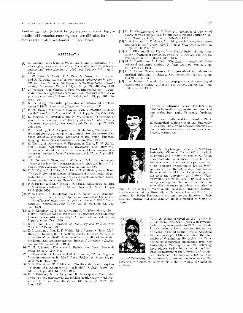
CIlOMA5 et al.: COWI’ICASI‘ ACYN’I’ 1)IISI’RUCTION 247
bubble may he detectcc1 by correlation analysis. Future (2Oj H. D. Van Licw auri M. E. Unrlinnl. “Beha1,ior of bnbl,lcs of sliidies will exaniiic more rigorous gas diffusion forrmila- slowly permeating gas iisctl for nli.msonic imaging contrast,’‘ In-
ucst . Radzol., vol. 311, no. 5, pp. 315-321, 1995. tioiis and thc shell mecliaiiics in more detail. 1211 Y . S. Cha and I?. E. Henry, ‘73ubblc growth during dccoinprcs-
sion of a licinid.” ‘/’runs. ASME J . Heat T r a m f w , vol. 103, no.
REFERENCES
N. Wcidncr, .J. P. Seniple, W. R. Welch, and J . Fiilkman, “Tu- mor angiogcnesis and rnctastasis- Correlation in invasive hreast carcinoma,” Nei~i England J . Med., vol. 324, pp. 1~ 7, Jan. 3, 1999. .1. W. Baish, Y . G a d ; , D. A. Bcrk, M. Nome, L. T. Raxtcr, and A.. K. Jain, “Role of tninor vasciilar architcctnrc i n nirtri- ent and drug dclivcry: An invasion pcrcolation-basctl ilctwork model,’’ Microvasr. Res., vol. 51, no. 3 , pp. 327-34(3, May 19%. N. Weidncr, P. R. Carroll, . I . Flax, W. Blinneiifcld, kind .J, Folk inan, “ ‘ I ~ I ~ I K J ~ ;tngiogenc>sis corro1;~tes with met prostate cart:inonia,” Amer. J . Prrtlrol., vol. Ang. 19!13. N. de Jong, “Acoustic propcrtics of ultrasound contrast agcnts,“ Ph.D. dissertation, Eriisiniis TJnivcrsity; 1993. P. N. Burns, “Harrnoiiic imaging wit11 nl1,rasoinid colits agenl.~,” Clinical Rodiol., vol. 61, Suppl. 1, pp. 60-56, b c b 19 I<. Morganl IVI. Avcrkiou, arid I<. W. Vcrrar phase of transmission on contrast agent cchoi r/ltTfl.son., FerroeleclL, Freq. Cant,.., vol. 45, 11
Jul. 1998. 1’. . I . Vriiiking, E, I,. Ccspedes, a i id N. clc .long, “Tnipsoved ill- 1,rasoinid contrast imaging using a innltipiilse and dccorrclation hased detection strabegy,” presentoil at Int. SyInp Uhsason. lrriaging Tissire Cliaractcrizal.ioii, R.osslyn, VA, 1998. K. Wei, A. R. Jayzzwcera, 9. Firoozan, A. Linla, D. M. Skyha, mid S. Kaiil, “Quantification o l myocardial hlood flow with nltrast,iind-indiice~ dastrni:t,ion of inicrohi~hblcs adminis1crt:d iis a constant vciions inhision,” Circulation, vol. 97> no. 5, p[J. 473 ~
.J. E. Chornas, It. Sikcs, and K. W. Ferrara, “Correbtioii malysis of received echoes frorn contrast. agcnts ill-vitro and in-vivo,” i n PTOC. IEEE Ul1,rason. Sump., Sendai, .Japau~ 1 D Y X . 13. M. Skyba, R.. J . I’ricc, A. Z. L C . Skalak, and S. Kaul, “Direct in vivo visualizat,ion or i ular destriictiori of nii- crobubblcs by illtr;rstiund and its Io<.al elhcts on t,issiir:,” Circu-
1’. S. Epstein and M. S. Plcsset, “On til ability of gas bnlililcs in liqiiicl-gas solatioils,” J. Chcna. Phys., vol. 18, JW. 11, 1~p.
P. A . Ilayton, K. E. Morgan, A. L. Klibanov, Ci. €1. I ~ ian r l cn - burgcr, and K. W. Fcrrara, “Optical and acoiist,ical obscivations of l.he effects of iiltrasourid on contrast agents,” I E E E Trans. Ultroson., b%rroelect., Frcq. Contr., vol. 46, no. 1: p p 220-232, .Jan. 1999. A. S. Kahalnov, I<. N. Maknrov, a id 0. V. Scherbakova, “Solil- bility of Auorocnrbons in water as a kry paranictcr rletcrmining flnnri,carhon emulsion staldiby,” .I. Fluor. Cl?,em., vol. 50, 3 , pp. 271 284. Dec. 1990. D. R.. Lidc, CRC Handbook of (%7lLiStTy a7~d P h ~ ~ s i c s . New Ynrk: CRC Press, 1998. E. I. Egcr, 11, .J. Liii, D. D. Kohlin, Vl. .J. I,aster, S. ’lhcri, M. .1. Ilalscy, 1’. Ionescir, B. S. Cllortkoll, and 7’. Iludlicky, “Molct:iilar propwl,ies OS the ‘ideal’ inhaled ancstlictic Stirdics of flinirinatcd niclhanes, ethanes, propanes, and bnt,anes,” Anesthesia Analgc- sia, vol. 79, pp. 245-251, 1994. T . C,. Leigilton, Tlir Acoilstic Ilubblr. New York: Ar:ailcrriic I’ress, 1994, I J ~ , 335 341. S. Hilgenfeldt, D. Lohse, and M. P. Brenner, “Phasr diagranis Sor sonolumiucscing hrthbles,” P1~g.s. Fliiids, v d . 8 , no. 11, pp. 2808-2826, Nov. 199G. M. S. Plesset aiid T. P. Mitchell, “On I.he stability OS 1.he sphcr- ical shape of a V~IJOIIS cavity i r i a liqiiid,” Q. Appl. Matlr., vol. 13, no. 11, pp. 419-4:3O, Nov. 1966. P. .J. fiinking, N. dc Jong, ancl E. L. Ccspcdes, "Scattering properties of i:ncapsulaI.cd gas blibbks at high ullrasound pres- sures,” .I. Acousl. Soc. Amcr., vol. 105, ria. S, pp. 1989-1 996) Ular. 1999.
.
483, 1W8.
lation, vol. $18, no. 4, pp. 290-293, ID98
1505 1509, 1951).
I, pp. 56-66, Fcb. 1981. [22] A. I. Eller and 14. G. Flyrrn. “Rccl.ificd diflnsioii through non-
lincar pulsations of cavitation bubbles," J . Acoi,d. Soc. Amer., vol. 37, no. 3; pp. 49-503, Ma.r. 1905.
[23] M. h4, Fyrillas and A. .I. Szcri, “Dissolution or growi.h of soluble spherical oscillating bubblc,” J . Fluid Midien., vol. 277, ~ I J .
381-407, Oct. 1994. [24] I,. A. Criim: “Measiirements of tlic growth of air hirhblcs by
rcctificd diffusion,” J . Acoust. Soc. Amer., vol. 08, no. 1, pp, 203-211, .Jul. 1980.
[25] J. A. Jensen, “A model for thc propagation and scattering of iiltrasoiind in tissue,” .J. Acoiist. Soc. Amer., vol. 89, no. 1, pp. 182 191, .Ian. 1991.
James E. Chomas voccived t,hc B.S.1S. i n 1!J9B in hiomcdical cnginccring and electrical cnginccring from Uukc University, Dnrliani, NC.
He is curreutly working toward a 1’h.D. in Iioniedical cnginccriug at tlic University of Virginia. His current research irilcrests in- clnilc conl.rast-assisted ultrasound and bluod vcloci1.y estimation
Paul A. Dayton graduated frorn Villanova University, Vill;rno\n, PA in l!N5 with a 13,s. in physics iind a tiiiiinr i u clmnistry. As an iindcl.gr;rdu;itr:, I ic ctiud~ictad research i n h n r - facc science witti tlic Physics Dcpartinent nnd piilse scqnciicc programming for ail NMR spcctiomctc~r in t,lic Chcrnistry Dcpatt,rnimt. IIc received his M.E. in clc!ctrical enginocr- irig from the Uni\crsity of Virginia. Cbar- lottcsvillc, VA i n Janiiary 1998 and is cur- rcntlv nearing coniplctiou of his 1’1i.D. in bioincrlical cnginccring, which will also he
from tlm University of Virginia. Mr. Dayi.on is ciirrcntly conlhu- ing his research at the University OS California, Ilavis, CA with his advisor, Dr. Kathy Fcrrara.. IIis cnrrent research intcrcst,s include targct,cd imaging and drug ildivcry. He is a inenibcr of Sigma Pi Sigma.
doctoral Fellowship. partiriciit, nf Bioined at Davis.
I i1
. J o h n S. Allen r~ceivcil an A.D. degrco in I>iiysics from I’rincrtoii University i n 1988 and an M.S. degree in physics in 1 9W h m Florida State University, l<’roni 1993 to 1997, Iic was a researcli assistant ai, t,he I’liysical hcoirstics Lab at thc Applied Physics Lab at the TJni- vcrsi1.y of Washington. I IC rcccived his 1’h.D. dcgrec in mcchnnical cngiimcring from the Univcrsi1.y or Washingtori i n 1997. Following his graduat,o stidics, he studied at tlic Third Physical Iristitulc at thc Univcrsi1.y of Gottill- gcri, Goti.ingen, Germany on a DAAU Post-
[ le is currcnbly a research ciigiriccr al; the Dc- :SI E~iginixring al. the University of Califorilia
248 I E E E TRANSACTIONS ON UL'I'HASONICS. FllltROllLRCCRtCS, AND FREQUENCY CONCROL, VOI.. 48, NO. 1 , JANUARY 2001
K a r e n E. Morgan rcccivcd a B.S. degree in 1996 from the Physics Department at the University of Virginia, Charlottesville, VA. As an undergraduate, shc was involved in rc- search in the arcas of medical physics, near field scanning optical microscopy, and atmo- spheric physics. During this time, she became a member of Phi Dcta Kappa and Sigma P i Sigma.
Since June 1996, she has been pursuing a Ph.D. in biomedical engineering in the De- partment of Biomedical Enginccring at the
University of Virginia, Charlottesville, VA. She is currently involved in research in the area of contrast-assisted ultrasound imaging, under the advisement of Dr. Katherinc Ferrara. In 1997, she was awarded a Whitaker Foundation Fellowship and a NSF Graduate Research Fellowship. She is currently supported by a Whitakcr Foundation Fellowship and plans to finish hcr degree in the winter of 2000.
' K a t h e r i n e W h i t t a k e r Ferrara (S'82- M'82-.S'87- M'89 SM'99) received the B.S. degree in physical therapy in 1976 from the University of Pittsburgh, Pittsburgh, PA, the B.S. and M.S. in electrical engineering in 1982 and 1983, respectively, From the California State University, Sacramento, and lhe Ph.D. in electrical engineering and computer sci- ence in 1989 from the University of California, Davis.
From 1976 to 1980, she practiced physical therapy at Akron Children's Hospital and the
Home for Crippled Children, Pittsburgh, PA. From 1983 t,o 1988, she worked for Sound Imaging, Inc., Folsom, CA, and for General Electric Medical Systems, Rancho Cordova, CA, in the areas of mag- netic resonance arid ultrasound imaging. Since 1989, she has been an associatc professor in thc Dcpartrncnt of Elcctrical Engineering at California State University, Sacramento. From 1993 to 1995, she was a principal mcmbcr of the research staff at the Riverside Research Institute, New York, NY, and an adjunct associate professor at Cor- nell University Medical School. Shc is currcntly Profcssor and Chair of thc Division of Biomedical Enginecring at the University of Cal- ifornia, Davis. Her rascarch iritcrcsts arc in the fields of biomcdical signal and image processing and particularly in the use of ultrasound to map blood flow.
Dr. Ferrara is an associate editor of the IEEE 2lonsactions urk Ullrasonics, Ferroelectecs, and Frequency C o n t d in the area of flow measurement techniques and applications.