Gd(III)-EPTPAC16, a new self-assembling potential liver MRI contrast agent: in vitro...
15
Gd(III)-EPTPAC 16 , a new self-assembling potential liver MRI contrast agent: in vitro characterization and in vivo animal imaging studies y Suzana Torres, 1 Maria I. M. Prata, 2 Ana C. Santos, 2 Joa ˜ o P. Andre ´, 1 Jose ´ A. Martins, 1 Lothar Helm, 3 E ´ va To ´ th, 4 Maria L. Garcı´a-Martı´n, 5 Tiago B. Rodrigues, 5,6 Pilar Lo ´ pez-Larrubia, 5 Sebastia ´ n Cerda ´n 5 and Carlos F. G. C. Geraldes 6 * 1 Centro de Quı ´mica, Campus de Gualtar, Universidade do Minho, Braga, Portugal 2 Instituto de Biofı ´sica e Biomatema ´ tica, Faculdade de Medicina, Universidade de Coimbra, Coimbra, Portugal 3 Laboratoire de Chimie Inorganique et Bioinorganique, Ecole Polytechnique Fe ´ de ´ rale de Lausanne, EPFL-BCH, Switzerland 4 Centre de Biophysique Mole ´ culaire, CNRS, Orle ´ ans, France 5 Instituto de Investigaciones Biome ´ dicas ‘‘Alberto Sols’’, CSIC-UAM, Madrid, Spain 6 Departamento de Bioquı ´mica, Centro de RMN e Centro de Neurocie ˆ ncias e Biologia Celular, Faculdade de Cie ˆ ncias e Tecnologia, Universidade de Coimbra, Coimbra, Portugal Received 20 March 2007; Revised 28 May 2007; Accepted 3 June 2007 ABSTRACT: The recently reported amphiphilic chelate, [Gd(EPTPAC 16 )(H 2 O)] 2 , forms supramolecular aggregates in aqueous solution by self-assembly of the monomers with a relaxometrically determined critical micellar concentration (CMC) of 0.34 mM. The effect of sonication on the aggregate size was characterized by dynamic light scattering and relaxometry, indicating the presence of premicellar aggregates and an overall decrease in aggregate size and polydispersity upon sonication, slightly below the CMC. {[ 153 Sm](EPTPAC 16 )(H 2 O)} 2 radiotracer was evaluated in vivo from g scintigraphy and biodistribution in Wistar rats. It was found to depend strongly on the sample concentration, below or above the CMC, and its sonication, in a way that correlates with the effect of the same factors on the size of the aggregates formed in solution. Below CMC, the very large aggregates of the [ 153 Sm] 3þ -labeled chelate were persistently and mainly taken up by the lungs, and also by the macrophage-rich liver and spleen. Sonication of this solution led to loss of the lung uptake. Above CMC, the metal chelate was mainly taken up by the liver, with very little uptake by the spleen and lungs. In vivo, dynamic contrast-enhanced (DCE)-MRI evaluation of the micellar [Gd(EPTPAC 16 )(H 2 O)] 2 compound in Wistar rats showed a persistent hepatic positive-contrast effect in T 1 -weighted images, qualitatively similar to the clinically established Gd III -based hepatobiliary-selective agents. No enhancement effect was observed in the lungs because of the scarcity of mobile protons in this organ, despite the scintigraphic evidence of significant lung retention of the [ 153 Sm] 3þ -labeled chelate at concentrations below the CMC. Copyright # 2007 John Wiley & Sons, Ltd. KEYWORDS: MRI; contrast agents; gadolinium; liver targeting; self-assembly; micelles; dynamic light scattering; g scintigraphy INTRODUCTION Gadolinium(III) chelates have been extensively used as paramagnetic contrast agents (CAs) for MRI (1–6). The first generation of MRI CAs, including [Gd(DOTA) (H 2 O)] (DOTA ¼ 1,4,7,10-tetra-azacyclododecane-1,4, 7,10-tetra-acetate) and [Gd(DTPA)(H 2 O)] 2 (DTPA ¼ diethylenetriamine penta-acetate) among others, are NMR IN BIOMEDICINE NMR Biomed. 2008; 21: 322–336 Published online 10 August 2007 in Wiley InterScience (www.interscience.wiley.com) DOI:10.1002/nbm.1194 *Correspondence to: C. F. G. C. Geraldes, Departamento de Bioquı ´mica, Faculdade de Cie ˆncias e Tecnologia, Universidade de Coimbra, Apartado 3126, 3001-401 Coimbra, Portugal. E-mail: [email protected] y Supplementary Fig. S1 is available online in Supplementary Material. Contract/grant sponsor: Foundation of Science and Technology, Portugal; contract/grant numbers: POCTI/QUI/47005/2002, SFRH/BD/5407/2001. Contract/grant sponsor: FEDER. Contract/grant sponsor: III Instituto de Investigac ¸a ˜o Interdisciplinar University of Coimbra, Portugal; contract/grant number: III/BIO/45/2005. Contract/grant sponsor: Institute of Health Carlos III, Spain; contract/grant number: PIO051845. Contract/grant sponsor: Ministry of Education and Science, Spain; contract/grant numbers: SAF 2004-0391, NAN2004-09125-C07-03. Contract/grant sponsor: European Union; contract/grant number: LSCH-2004-503569. Abbreviations used: CA, contrast agent; CCK, cholecystokinin; CMC, critical micellar concentration; DCE, dynamic contrast enhanced; DLS, dynamic light scattering; DTPA, diethylenetriamine penta-acetate; DOTA, 1,4,7,10-tetra-azacyclododecane-1,4,7,10-tetra-acetate; DOTP, 1,4,7,10- tetra-azacyclododecane-1,4,7,10-tetrakis(methylene phosphinate); EOB-DTPA, ethoxybenzyl-DTPA or S-[3,6,9-triaza-3,6,9-tris(carboxymethyl)- 4-(4-ethoxybenzyl)-undecandicarboxylate]; H 5 EPTPAC 16 , (hydroxymethylhexadecanoyl ester) ethylenepropylene triaminepenta-acetic acid; ID, injected dose; MES, (2-(N-morpholino)ethanesulfonic acid hydrate; PI, polydispersity index; ROI, region of interest. Copyright # 2007 John Wiley & Sons, Ltd. NMR Biomed. 2008; 21: 322–336
Transcript of Gd(III)-EPTPAC16, a new self-assembling potential liver MRI contrast agent: in vitro...
Gd(III)-EPTPAC16, a new self-assembling potential liver MRIcontrast agent: in vitro characterization and in vivo animalimaging studiesy
Suzana Torres,1 Maria I. M. Prata,2 Ana C. Santos,2 Joao P. Andre,1 Jose A. Martins,1 Lothar Helm,3
Eva Toth,4 Maria L. Garcıa-Martın,5 Tiago B. Rodrigues,5,6 Pilar Lopez-Larrubia,5
Sebastian Cerdan5 and Carlos F. G. C. Geraldes6*
1Centro de Quımica, Campus de Gualtar, Universidade do Minho, Braga, Portugal2Instituto de Biofısica e Biomatematica, Faculdade de Medicina, Universidade de Coimbra, Coimbra, Portugal3Laboratoire de Chimie Inorganique et Bioinorganique, Ecole Polytechnique Federale de Lausanne, EPFL-BCH, Switzerland4Centre de Biophysique Moleculaire, CNRS, Orleans, France5Instituto de Investigaciones Biomedicas ‘‘Alberto Sols’’, CSIC-UAM, Madrid, Spain6Departamento de Bioquımica, Centro de RMN e Centro de Neurociencias e Biologia Celular, Faculdade de Ciencias e Tecnologia, Universidade deCoimbra, Coimbra, Portugal
Received 20 March 2007; Revised 28 May 2007; Accepted 3 June 2007
ABSTRACT: The recently reported amphiphilic chelate, [Gd(EPTPAC16)(H2O)]2�, forms supramolecular aggregates in
aqueous solution by self-assembly of the monomers with a relaxometrically determined critical micellar concentration
(CMC) of 0.34mM. The effect of sonication on the aggregate size was characterized by dynamic light scattering and
relaxometry, indicating the presence of premicellar aggregates and an overall decrease in aggregate size and polydispersity
upon sonication, slightly below the CMC. {[153Sm](EPTPAC16)(H2O)}2� radiotracer was evaluated in vivo from g
scintigraphy and biodistribution in Wistar rats. It was found to depend strongly on the sample concentration, below or
above the CMC, and its sonication, in a way that correlates with the effect of the same factors on the size of the aggregates
formed in solution. Below CMC, the very large aggregates of the [153Sm]3þ-labeled chelate were persistently and mainly
taken up by the lungs, and also by the macrophage-rich liver and spleen. Sonication of this solution led to loss of the lung
uptake. Above CMC, the metal chelate was mainly taken up by the liver, with very little uptake by the spleen and lungs.
In vivo, dynamic contrast-enhanced (DCE)-MRI evaluation of the micellar [Gd(EPTPAC16)(H2O)]2� compound in Wistar
rats showed a persistent hepatic positive-contrast effect in T1-weighted images, qualitatively similar to the clinically
established GdIII-based hepatobiliary-selective agents. No enhancement effect was observed in the lungs because of the
scarcity of mobile protons in this organ, despite the scintigraphic evidence of significant lung retention of the
[153Sm]3þ-labeled chelate at concentrations below the CMC. Copyright # 2007 John Wiley & Sons, Ltd.
KEYWORDS: MRI; contrast agents; gadolinium; liver targeting; self-assembly; micelles; dynamic light scattering;
g scintigraphy
INTRODUCTION
Gadolinium(III) chelates have been extensively used asparamagnetic contrast agents (CAs) for MRI (1–6). The
first generation of MRI CAs, including [Gd(DOTA)(H2O)]
� (DOTA¼ 1,4,7,10-tetra-azacyclododecane-1,4,7,10-tetra-acetate) and [Gd(DTPA)(H2O)]
2� (DTPA¼diethylenetriamine penta-acetate) among others, are
NMR IN BIOMEDICINENMR Biomed. 2008; 21: 322–336Published online 10 August 2007 in Wiley InterScience(www.interscience.wiley.com) DOI:10.1002/nbm.1194
*Correspondence to: C. F. G. C. Geraldes, Departamento de Bioquımica, Faculdade de Ciencias e Tecnologia, Universidade de Coimbra, Apartado3126, 3001-401 Coimbra, Portugal.E-mail: [email protected] Fig. S1 is available online in Supplementary Material.Contract/grant sponsor: Foundation of Science and Technology, Portugal; contract/grant numbers: POCTI/QUI/47005/2002, SFRH/BD/5407/2001.Contract/grant sponsor: FEDER.Contract/grant sponsor: III Instituto de Investigacao Interdisciplinar University of Coimbra, Portugal; contract/grant number: III/BIO/45/2005.Contract/grant sponsor: Institute of Health Carlos III, Spain; contract/grant number: PIO051845.Contract/grant sponsor: Ministry of Education and Science, Spain; contract/grant numbers: SAF 2004-0391, NAN2004-09125-C07-03.Contract/grant sponsor: European Union; contract/grant number: LSCH-2004-503569.
Abbreviations used: CA, contrast agent; CCK, cholecystokinin; CMC, critical micellar concentration; DCE, dynamic contrast enhanced; DLS,dynamic light scattering; DTPA, diethylenetriamine penta-acetate; DOTA, 1,4,7,10-tetra-azacyclododecane-1,4,7,10-tetra-acetate; DOTP, 1,4,7,10-tetra-azacyclododecane-1,4,7,10-tetrakis(methylene phosphinate); EOB-DTPA, ethoxybenzyl-DTPA or S-[3,6,9-triaza-3,6,9-tris(carboxymethyl)-4-(4-ethoxybenzyl)-undecandicarboxylate]; H5EPTPAC16, (hydroxymethylhexadecanoyl ester) ethylenepropylene triaminepenta-acetic acid; ID,injected dose; MES, (2-(N-morpholino)ethanesulfonic acid hydrate; PI, polydispersity index; ROI, region of interest.
Copyright # 2007 John Wiley & Sons, Ltd. NMR Biomed. 2008; 21: 322–336

extracellular agents, distributing non-specifically through-out the plasma and interstitial spaces and being rapidlyexcreted via the kidneys (7). In the emerging field ofcellular and molecular imaging using MRI, more efficient(higher relaxivity) and biospecific CAs are required todeliver as many GdIII-containing species as possible to thecellular targets of interest and therefore attain sufficientlyhigh contrast to obtain their images (8).
At the magnetic field strengths currently used in MRI(0.5–3.0 T, 21.29–127.73MHz for protons), the aptitudeof GdIII chelates to enhance the longitudinal water protonrelaxation rate per mmol of CA (relaxivity) is mainlydetermined by the inner-sphere water-exchange rate (kex)and the rotational correlation time of the chelate (tR)(2–4). The charge of the complex and the steric crowdingin the inner coordination sphere have been shown to bethe main factors affecting water exchange in GdIII
poly(aminocarboxylate) complexes (9,10). Chelators thatensure optimal fast water exchange on the GdIII complexhave been developed by inducing steric crowding aroundthe water-binding site, by elongation of the aminebackbone or by elongation of a pendant carboxylate armof the ligand, for both linear (DTPA-type) and macro-cyclic (DOTA-type) chelates (11–13), or by replacing apendant carboxylate by a phosphonate group (14). Thisfacilitates the removal of the water molecule by a dis-sociative mechanism, accelerating the exchange. Severalapproaches have been devised to increase tR throughcovalent attachment of GdIII chelates to slowly tumblingmacromolecules (2–5), such as carbohydrates (15–17),proteins (18) and dendrimers (19,20). The formation ofhost–guest non-covalent interactions between GdIII che-lates and macromolecules (5,21,22) or b-cyclodextrinoligomers (23) has also been explored. An alternativewayto increase tR is through self-assembly of amphiphilicGdIII chelates or through inclusion of lipophilic GdIII
chelates in liposomes or other lipid-based systems.However, when the water-exchange rate in the GdIII
inner sphere is not optimized in these macromolecularagents, their proton relaxivity is still seriously limited.
Lipid-based nanoparticles, such as micelles andliposomes, have been used as colloidal drug carriervehicles (24–26) and as contrast-enhanced MRI andmolecular imaging agents (27,28). Liposomal MRI CAsinclude, among other types, liposomes entrappinghydrophilic paramagnetic agents in the aqueous lumenor carrying amphiphilic agents on the liposomal surfacewith the hydrophobic part non-covalently anchored in thelipid bilayer (27,28). In addition to the increasedrelaxivities, colloidal CAs made up of GdIII-containingsupramolecular assemblies can be efficiently taken up bymacrophage-rich tissue undergoing endocytosis/phago-cytosis (liver and spleen) (29). This provides an efficientway of delivering therapeutic and diagnostic agents tocells with the aid of colloidal-drug-carrier systems.However, the fast sequestration of intravenously injectedcolloidal-drug carriers from the blood by Kupffer cells
(liver/spleen resident macrophages) is a drawback forefficient targeting of drug carriers or diagnostic agents toa specific macrophage population or to non-macrophagesites. As a consequence, there has been increasing interestin the design of colloidal-drug-carrier systems that, byavoiding rapid recognition by Kupffer cells, remain in theblood for long periods. Such carriers have applications invascular drug delivery and release and site-specifictargeting (24). Long-circulating colloidal systems withentrapped radiopharmaceuticals or CAs have beensuccessful in blood-pool imaging (20–32). Micelles pre-pared from poly(ethylene glycol)–lipid conjugates havebeen shown to be useful in the delivery of therapeutic ordiagnostic agents to areas of myocardial infarction (33).
Micelles may themselves be made up of amphiphilicions or molecules or can originate separately and act asvehicles to transport poorly soluble compounds (mixedmicelles). The effectiveness of micelles as delivery agentsdepends, among other factors, on their critical micellarconcentration (CMC), because, on intravenous injection,micelles are often diluted to less than their CMC.
Several gadolinium-based micelles have been relax-ometrically characterized and reported in the literature:linear copolymers of GdIII–DTPA conjugates linked bya,v-alkyldiamides with a varying number of methylenegroups separating the amide function (34,35); GdIII–DTPA-bisamide-(CH2)n copolymers (n¼ 6, 10 or 12)(36); GdIII–DOTA derivatives bearing alkyl or mono-amide-alkyl aliphatic side chains (C10–C18) (37,38); Gd
III
chelates of 1,4,7-tris(carboxymethyl)-10-(2-hydroxyl-alkyl)-1,4,7,10-tetra-azacyclododecane ligands of vary-ing alkyl chain length (39); GdIII chelates of C8-DOTPand C11-DOTP, two fatty acid analogues of DOTP[1,4,7,10-tetra-azacyclododecane-1,4,7,10-tetrakis(met-hylenephosphinate)] (40,41); GdIII–3,6,9,15-tetra-aza-bicyclo[9.3.1]pentadeca-1(15),11,13-triene-3,6,9-triace-tate (42); gadofluorine 8 with a LnIII–DO3A monoamidehydrophilic head (Ln¼Gd or Y) (43).
The formulation for the preparation of stable mixedmicelles based on gadolinium chelates usually includesan amphiphilic GdIII chelate, phospholipid(s) and asurfactant. These micelles should meet the criteria of highcontrast efficiency (high relaxivity), small particle size,high loading, good stability and easy production. Severalmixed systems have been relaxometrically and/or phar-macologically studied: mixed micelles based on GdIII–DTPA derivatives (44); GdIII–HHD-DO3A-cholesterol(HHD¼ hydroxyhexadecyl) (45,46); mixed micelles ofthe GdIII–DTPA–cholesterol conjugate (47); GdIII–DTPAmonoamide and bisamide derivatives with alkyl chains(C12, C14, C16 or C18) (48,49). Target-specific mixedmicelles based on GdIII–C18DTPAGlu/C18CCK8 havealso been reported; their effectiveness is due to thepresence of the bioactive peptide CCK8, which acts astargeting vector for cholecystokinin (CCK) receptorslocalized in the cell membrane, which are overexpressedin many tumors (50).
Copyright # 2007 John Wiley & Sons, Ltd. NMR Biomed. 2008; 21: 322–336DOI: 10.1002/nbm
A NEW POTENTIAL SELF-ASSEMBLING LIVER MRI CONTRAST AGENT 323

We have previously reported the synthesis and charac-terization of the GdIII complex of the chelator, H5EPTPAC16
[(hydroxymethylhexadecanoyl ester) ethylenepropylenetriaminepenta-acetic acid] (Fig. 1) (51). This chelatedisplays an increased water-exchange rate (kex) resultingfrom steric compression around the water-binding site.The attachment of a palmitic ester unit to the ethylenedia-mine bridge yields an amphiphilic conjugate which formssupramolecular aggregates, possibly micelles, in aqueoussolution, with a calculated CMC of 0.34mM at 258C (51).A global analysis of the variable-temperature, multiple-field 17O NMR, electron paramagnetic resonance and1H NMR dispersion data allowed determination of theparameters governing the relaxivity of [Gd(EPTPAC16)(H2O)]
2�. The micelles formed in aqueous solutionshowed considerable internal flexibility, leading to alimited increase in proton relaxivity (51).In the present work, we studied how the self-assembly
of the amphiphilic monomer metal chelates intosupramolecular aggregates dramatically influences theirin vivo behavior and determines their performance aspotential imaging agents. We also report the evaluation ofthe [153Sm]3þ-labeled compound in Wistar rats throughpharmacokinetic studies involving in vivo dynamicg-scintigraphy and biodistribution. The effects of concen-tration and preparationmethod (with and without sonication)of the radiotracer {[153Sm](EPTPAC16) (H2O)}
2� solu-tion on its biodistribution were correlated with the sizedistribution of the aggregates, determined by dynamiclight scattering (DLS). The influence of sonication on[Ln(EPTPAC16)(H2O)]
2� in water was monitored byNMR and relaxometry. An in vivo MRI study of thecontrast effects and pharmacokinetics of the micellar[Gd(EPTPAC16)(H2O)]
2� complex in Wistar rats is also
reported and compared with a typical commercial,low-molecular-mass CA, GdDTPA (Magnevist1).
EXPERIMENTAL
H5EPTPAC16 was synthesized as described previously(51).
Preparation of LnIII–EPTPA complexes
The general procedurewas dissolution of H5EPTPAC16 indistilled water and addition of a molar equivalent ofLnCl3 solution (Ln¼Eu, Gd). The pH was adjusted to5.5 with aqueous KOH (0.1M). The solution was stirredat room temperature for 1 h and then adjusted to pH 7.0with aqueous KOH (0.1M). The solution was concen-trated at reduced pressure giving rise to a white solid.
For the DLS experiments, 0.2mM and 2mM solu-tions were prepared by dissolving the corresponding[Gd(EPTPAC16)(H2O)]
2� complex in 150mM MES[2-(N-morpholino)ethanesulfonic acid hydrate, 99.5%]buffer, pH 6.4. Samples were sonicated using a BandelinSonorex RK 106S sonicator (P¼ 200W, y¼ 35MHz).Aqueous solutions of [Gd(EPTPAC16)(H2O)]
2� wereprepared for the relaxometric experiments, whereasthe NMR experiments used D2O solutions of[Eu(EPTPAC16)(H2O)]
2�.
NMR measurements
1H NMR spectra were recorded in D2O (99.99% 2H) onVarian Unity Plus 300 and Unity 500 NMR spec-trometers, operating at 299.938MHz and 499.80MHz,respectively. Chemical shifts (d) are given in ppm relativeto TSP as internal reference (1H, d 0.0).
Relaxometric measurements
Water 1H relaxivity measurements were performed on aBruker Minispec mq60 (60MHz) at 258C and 378C. Thetemperature was measured by a substitution technique.Longitudinal relaxation rates were measured at fourdifferent concentrations, two below (0.1 and 0.2mM) andtwo above (2.0 and 4.0mM) the CMC before and after 2,4 and 20min of sonication.
DLS measurements
These were performed on a Coulter1 N4 Plus SubmicronParticle Sizer. The N4 Plus contains a 10mW He–Ne
Figure 1. Structure of the metal chelator, H5EPTPA. Therelevant structural features (highlighted) are: substitution ofan ethylenediamine bridge by a propylenediamine bridgeresulting in stereochemical compression around the water-binding site in the Gd3þ complex, and a pendant hydro-xymethyl-linked palmitic acid ester moiety rendering themolecule amphiphilic.
Copyright # 2007 John Wiley & Sons, Ltd. NMR Biomed. 2008; 21: 322–336DOI: 10.1002/nbm
324 S. TORRES ET AL.

laser which emits monochromatic polarized light with awavelength of 632.8 nm. The electric field polarization isperpendicular to the plane formed by the incident anddetected rays (vertical polarization). The particle size iscalculated from the measurement of the sample diffusioncoefficient by photon correlation spectroscopy. The N4Plus is controlled by PC-based software which operates inthe Microsoft Windows environment. Before dataacquisition, the sample was allowed to equilibrate for2min. All the measurements were performed at an angleof 908 and at 208C. The data acquired were analysed byunimodal analysis, which yields the mean intensity-weighted particle size and standard deviation. The meanintensity-weighted particle size was determined for eachsolution concentration over time, without and with 2minof sonication of the solution. Multimodal DLS analysiswas performed with a Zetasizer Nano ZS instrument(Malvern Instruments Ltd, Malvern, UK) equipped with a4mW He–Ne laser at 633 nm with backscatteringdetection at 1738.
Reagents for g imaging andbiodistribution studies
153SmCl3 was produced at the Instituto Tecnologico eNuclear, Lisbon, Portugal by Dr Maria dos Anjos Neves,with a specific radioactivity of >5GBq/mg. For thispurpose, 153Sm2O3 was prepared from a 98% 152Sm-enriched Sm2O3 target, sealed into a quartz vial andwelded into an aluminum can, by neutron irradiationusing a thermal flux of 2.3� 1013 n/cm2 � s. After irradia-tion, the sample was opened, dissolved in 1MHCl, andthe final 153SmCl3 solution was brought to a stockconcentration of 1.9� 10�3M.
Radiotracer preparation
Stock solutions of H5EPTPAC16 were prepared in150mM MES buffer, pH 6.4, and mixed with 153SmCl3.The solutions were left stirring at room temperature for1 h. Solutions with different concentrations of theradiotracer, 0.2mM (below CMC) and 2mM (aboveCMC) [CMC¼ 0.34mM, determined by relaxometry(51)], were prepared for biodistribution and g imagingexperiments. The solution of 2mM concentration ofradiotracer was prepared by adding to a 0.2mM solutionof the {[153Sm](EPTPAC16)(H2O)}
2� radiotracer the appro-priate amount of non-radioactive {[152Sm](EPTPAC16)(H2O)}
2� complex.
In vivo g imaging
A g camera–computer system (GE 400 GenieAcq;General Electric, Milwaukee, WI, USA) was used for
acquisition and preprocessing. Data processing anddisplay were performed on a PC using homemadesoftware developed for the IDL 5.2 computer tool. Awellcounter (DPC-gamma C12, Los Angeles, CA, USA) witha Compaq DeskPro compatible computer was used forradioactivity counting in the biodistribution studies.
g images and biological distribution of [153Sm]3þ
complexes were determined using 200–250 g Wistar rats.All animal studies were carried out in compliance withprocedures approved by the appropriate institutionalreview committees. Conscious rats were allowed freeaccess to food and water ad libitum. Groups of fouranimals (one group for each concentration of theradiotracer with and without sonication) were anesthe-tized with 50mg/ml ketamine/2.5% chlorpromazine(10:3, v/v) and injected into the femoral vein with�200mCi of the [153Sm]3þ complex immediately afterthe solution had been prepared. Two different injecteddose concentrations were studied (0.2mM and 2mM) aswell as the effect of 2min of sonication on the gscintigraphic image. The animals were then positioned indorsal decubitus over the detector. Image acquisition wasinitiated immediately before radiotracer injection.Sequences of 180 images (of 10 s each) were acquiredto 64� 64 matrices. In addition, static data were acquired24 h after the radiotracer injection.
Images were subsequently processed using an IDL-based program (Interactive Data Language, ResearchSystems, Boulder, CO, USA). For analysis of thetransport of radiotracer over time, three regions ofinterest (ROIs) were drawn on the image files,corresponding to the thorax, liver and left kidney. Fromthese regions, time–radioactivity curves were obtained.
Biodistribution studies
Groups of four animals were injected in the tail vein with�100mCi of the [153Sm]3þ tracer (at the concentrationsand conditions stated in the scintigraphic imagingsection) and killed 1 h later. The major organs wereexcised and weighed, and tissue radioactivity wasmeasured in a g well counter. Similar biodistributionstudies were also performed with the animals referred toin the previous section killed at 24 h.
MRI
Sample formulation/preparation of [Gd(EPTPAC16)(H2O)]2� for MRI. The ligand, H5EPTPAC16 (147.4mg;0.218mmol), was dissolved in distilled water. To thissolution was added dropwise an aqueous solution ofGd(NO3)3.6H2O (90.0mg; 0.199mmol). The pH waskept at 5.5–6.0 by the addition of NaOH (aqueoussolution, 0.1M). The reaction mixture was left stirring for
Copyright # 2007 John Wiley & Sons, Ltd. NMR Biomed. 2008; 21: 322–336DOI: 10.1002/nbm
A NEW POTENTIAL SELF-ASSEMBLING LIVER MRI CONTRAST AGENT 325

1 h. The solution was adjusted to pH 7.0 with NaOH(aqueous solution, 0.1M), filtered (Filtropur S filters,0.2mm), and concentrated under reduced pressure. Theabsence of free metal was confirmed by the xylenolorange test.To remove the salts (NaNO3) produced during the
synthesis of the [Gd(EPTPAC16)(H2O)]2� complex, a
sample was purified by silica-gel 100 C8 reverse-phasechromatography with gradient elution, 100% water!100% ethanol. The relevant fractions, identified byreverse-phase C18 thin-layer chromatography and con-ductivity measurement, were pooled, filtered (Filtropur Sfilters, 0.2mm), and concentrated under reduced pressureto give the salt-free complex, probably [Gd(EPTPA-C16)(H2O)]Na/K2, as a vitreous solid.
In vivo MRI studies. The experimental protocolsperformed were approved by the appropriate institutionalreview committees and followed the guidelines of theirresponsible governmental agency. The MRI experimentswere all performed on a Bruker Pharmascan platform(Bruker Medical Gmbh, Ettlingen, Germany) using a7.0 T horizontal-bore superconducting magnet, equippedwith a 1H-selective 60mm birdcage resonator and aBruker gradient insert with 90mm diameter (maximumintensity 300mT/m). Data were acquired using aHewlett-Packard console running Paravision software(Bruker Medical) in a Linux environment.All MRI examinations were carried out on male Wistar
rats (n¼ 4, 250–260 g body weight) anesthetized initiallyby inhalation in an induction box with O2 (1 liter/min)containing 3% isoflurane, and maintained during theexperiment using a mask and 1–2% isoflurane in O2.Animals were taped down into a holder to minimizebreathing-related motion, and then placed in a heatedprobe, which kept the core body temperature at �378C,monitored by a rectal probe. The physiological state of theanimal was monitored throughout the experiment with aBiotrig physiological monitor (Bruker Medical), usingthe respiratory rate and body temperature. Solutions of[Gd(EPTPAC16)(H2O)]
2� and [Gd(DTPA)(H2O)]2�
(Magnevist1; Schering, Berlin, Germany; 100mM) wereprepared in distilled water, and the pH was adjusted to7.2. The solutions were injected into the catheterized tailvein as a bolus of 200mL in 20 s (0.2mmol Gd/kg bodyweight) using an infusion pump (Panlab, Barcelona,Spain).T1-weighted spin-echo anatomical images (TR¼
200ms; TE¼ 11.7ms; field of view¼ 6� 6 cm; acqui-sition matrix¼ 256� 256; number of averages¼ 2; slicethickness¼ 2mm; two packages of four slices each,centered on the liver and on the kidneys) were acquired inaxial orientation. Baseline images were acquired beforethe administration of our CA, [Gd(EPTPAC16)(H2O)]
2�.After injection, regional CA uptake was assessed byacquiring sequential images for 1 h (50 images).
MRI data processing. Data were analyzed withsoftware written in-house using IDL. With the aim ofcomparing the pharmacokinetics obtained from differentanimals, the data were normalized by calculating therelative enhancement (RE):
RE ¼ ðI � I0ÞI0
� 100
where I is the signal intensity at any given time after CAinjection, and I0 is the intensity before injection.Pharmacokinetics were analyzed by calculating theaverage enhancements within different ROIs in eachone of the following regions: liver, kidney medulla,kidney cortex, vena cava and muscle.
RESULTS AND DISCUSSION
DLS studies
The effects of concentration and sonication of aqueous[Gd(EPTPAC16)(H2O)]
2� solutions on the averageparticle size and polydispersity index (PI) were studiedby DLS at 208C (Fig. 2). We assumed that the CMC doesnot change between 258C (51) and 208C, as observedfor [Gd(DOTAC14)(H2O)] between 58C and 258C (38).For [Gd(EPTPAC16)(H2O)]
2� concentrations (0.2mM)below the CMC and without sonication, the meanintensity-weighted particle size was above the workingrange of the instrument (<3000 nm). After 2min ofsonication, it decreased to�500 nm, and a heterogeneouspopulation of aggregates was present (PI �1.0). Theduration of the sonication, 2 or 4min, did not substantiallyaffect either the mean intensity-weighted particle size orthe PI (not shown). The aggregates formed on sonication
Figure 2. DLS studies on the effect of concentration andsonication of the [Gd(EPTPAC16)(H2O)]
2� solution on theaverage particle size (mean� SD) and population distri-bution, as measured by PI.
Copyright # 2007 John Wiley & Sons, Ltd. NMR Biomed. 2008; 21: 322–336DOI: 10.1002/nbm
326 S. TORRES ET AL.

remained stable for at least 4 h with regard to size andpolydispersity (Fig. 2). For [Gd(EPTPAC16)(H2O)]
2�
solutions of concentration 2mM (above the CMC), themean intensity-weighted particle size (�450 nm) was thesame before and after 2min of sonication. However,sonication reduced the heterogeneity of the aggregates, asindicated by a decrease in PI from �1.5 to �1.0.Furthermore, both the mean intensity-weighted particlesize and the PI decreased over time, reaching values of�300 nm and �0.8, respectively, after 24 h (Fig. 2).
For all concentrations studied, both below and abovethe CMC, with and without sonication, a DLSmultimodalanalysis (data not shown) revealed populations of at leasttwo particle sizes: small particles (diameter<10 nm), andvery large particles (diameter >100 nm) of variable size.The existence of large particles for concentrations belowthe CMC suggests premicellar aggregation. This is inagreement with a recent study of micelle formationcaused by surfactants in the presence of a reporting dyeusing fluorescence correlation spectroscopy that demon-strated the formation of aggregates in the concentrationrange (0.3–1.0)�CMC, in increasing number whenapproaching the CMC (52). Premicellar aggregation hasbeen shown to be responsible for anomalies of variousphysical parameters (53,54). The large aggregatesprobably represent only a very small population belowthe CMC. Indeed, the value of the calculated meanintensity-weighted particle size can be dominated by avery small population of large particles with highscattering intensity. This is also in accordance with thefact that, using relaxometry, we did not observe anyslowly rotating large particles below 0.34mM (CMCdetermined by relaxivity measurements) (51). Even ifbelow the CMC the large aggregates represent asignificant proportion of the population of particles, theycan be extremely flexible, which implies that the protonrelaxivities will not be much increased with regard to themonomer state.
In conclusion, at a concentration just below the CMC(0.2mM in our case), the aggregate morphology anddynamics can be quite complicated. DLS data atconcentrations lower than 0.2mM would have certainlybeen more representative of the ‘non-aggregated’ state.
Effect of sonication on the NMR spectraand water relaxivity of[Ln(EPTPAC16)(H2O)]2Ssolutions
The 1H NMR spectrum of a 10mM D2O solution of[Eu(EPTPAC16)(H2O)]
2� contains a large number ofhigh-frequency (e.g. þ25.42, þ17.27, þ14.93, þ13.23,þ12.33, þ11.67, þ7.96 ppm) and low-frequency (�1.58,�2.43,�3.04,�3.55,�4.04,�7.45,�10.45,�10.64ppm)paramagnetically shifted resonances. This spectrum(available online as Fig. 1S in Supplementary Material)is not affected by sonication periods of up to 20min, orafter a subsequent period of 1 day, showing that soni-cation does not affect the stability and structure of thecomplex.
The effect of increasing sonication periods up to 20minon the 60MHz water 1H longitudinal relaxivity (r1) of[Gd(EPTPAC16)(H2O)]
2� solutions was studied at differ-ent concentrations and temperatures (258C and 378C), asshown in Table 1 for 0.2mM and 2.0mM concentrations.At the measurement frequency of 60MHz (close to themaximum of the NMR dispersion curves of the complexin the micellar form), r1 is mainly determined by therotational dynamics of the chelate (51). Hence thevariation in r1 reflects the changes in the rotationaldynamics of the micellar aggregates. As the CMC isexpected to increase by �10% between 258C and 378C(38), at 378C 0.2mM is still below the CMC (51). Abovethe CMC and at any of the temperatures, sonication hadno significant influence on r1. This indicates thatsonication does not affect the average rotational dynamicsof the chelate in the micellar aggregates, as reflected bythe r1 values [more precisely, r1 is determined by theglobal (tg) and segmental (tl) rotational correlation timesof the aggregates (51)]. This is in qualitative agreementwith the DLS observation that the mean intensity-weighted particle size was not affected by sonication atconcentrations above the CMC. Below the CMC, r1values, which are about half the values above the CMC,increased with sonication at both temperatures. At 258C,this increase became larger with sonication time, from 9%(2–4min) to 14% (20min). At 378C, the r1 increase wasonly significant (9%) for 20min of sonication. As
Table 1. Effect of sonication on the 60MHzwater 1H longitudinal relaxivity (r1) of [Gd(EPTPAC16)(H2O)]2� solutionsat different temperatures and concentrations below and above the CMC. The results are mean� SD from threemeasurements
r1 (mM�1s�1)
258C 378C
0.2mM 2.0mM 0.2mM 2.0mM
No sonication 9.94� 0.01 20.55� 0.01 6.46� 0.01 15.63� 0.012min sonication 10.94� 0.04 20.61� 0.01 6.53� 0.02 15.63� 0.014min sonication 10.96� 0.02 20.62� 0.01 6.53� 0.02 15.63� 0.0120min sonication 11.66� 0.01 20.63� 0.01 7.06� 0.04 15.63� 0.01
Copyright # 2007 John Wiley & Sons, Ltd. NMR Biomed. 2008; 21: 322–336DOI: 10.1002/nbm
A NEW POTENTIAL SELF-ASSEMBLING LIVER MRI CONTRAST AGENT 327

discussed above, the situation at concentrations justbelow the CMC is rather complex with regard to theformation of preaggregates. Moreover, these structuresare probably very fluxional and may change on soni-cation. These factors may explain why the significantdecrease in mean intensity-weighted particle size detec-ted by DLS was not observed in the relaxivities. Con-centrations significantly lower than 0.2mM would bemuch more useful for examining the non-aggregated state.
Studies of [153Sm]3R-labeled EPTPAC16
complex
In vivo g-imaging and biodistribution. Scinti-graphic images of Wistar rats were obtained as a functionof time after injection of an aqueous solution of 0.2mM{[153Sm](EPTPAC16)(H2O)}
2� tracer (below CMC).Fig. 3 compares the images obtained 30min afterinjection of the tracer solution without and after 2minof sonication. Without previous sonication, there is amarked high uptake/retention of the tracer in the thorax.Further experiments (dynamic acquisition and biodis-tribution; see below) revealed that the uptake of theradiotracer occurred predominantly in the lungs. In sharpcontrast, 2min of sonication of the 0.2mM radiotracersolution resulted in high liver uptake and virtually nouptake in the lungs, as demonstrated by dynamicacquisition and biodistribution (see below).
Time–radioactivity curves for the 0.2mM {[153Sm](EPTPAC16)(H2O)}
2� radiotracer without and after 2minof sonication were obtained in dynamic acquisitionexperiments (Fig. 4). The curves were smoothedand normalized in relation to the maximum radioacti-vity obtained. The radioactivity of the 0.2mM radiotracerwithout sonication increased sharply in the thoraximmediately after the injection, possibly correspondingto lung uptake/retention of the radiolabeled complex, anddecayed very slowly over the duration of the experiment.After 30min, the radioactivity remaining in the thorax/
Figure 3. Scintigraphic image at 30min of a Wistar ratinjected with a 0.2mM solution of {[153Sm](EPTPAC16)(H2O)}
2� (A) without sonication and (B) after 2min ofsonication.
Figure 4. Time–radioactivity curves at various ROIs (liver, kidney and thorax) in Wistar ratsafter the injection of a 0.2mM solution of the radiotracer {[153Sm](EPTPAC16)(H2O)}
2� after2min of sonication (A) and without sonication (B).
Copyright # 2007 John Wiley & Sons, Ltd. NMR Biomed. 2008; 21: 322–336DOI: 10.1002/nbm
328 S. TORRES ET AL.

lungs was still �80% of the maximum obtained for thisregion. In contrast, the radioactivity in the liver increasedsmoothly over the duration of the experiment, and after30min it was approximately half of that remaining in thelungs. There was virtually no uptake of radioactivity bythe kidney, consistent with the lipophilic nature of theradiotracer and its putative hepatobiliary excretionpathway. Sonication of the radiotracer solution beforeinjection had a dramatic effect on the dynamic acquisitioncurves. The radioactivity in the liver increased sharplyafter the injection and reached its highest value after�1min, which can be ascribed to the liver first-pass
uptake of the radiotracer. From there on, the radioactivityin the liver decayed slowly and smoothly, remaining at�80% after 30min. The radioactivity in the thorax andkidneys increased immediately after the injection, andthen decayed constantly during the experiment. After30min, the radioactivity was located mainly in the liver.
The effect of sonication on the biodistribution patternof {[153Sm](EPTPAC16)(H2O)}
2� in Wistar rats at 1 and24 h after injection was studied for concentrations of theradiotracer below (0.2mM) and above (2mM) the CMC.Fig. 5 and Table 2 show representative data as apercentage of injected dose per gram of tissue (%ID/g).
Figure 5. Biodistribution in Wistar rats (percentage of the injected dose/g of organ) of{[153Sm](EPTPAC16)(H2O)}
2� at 1 h (A) and 24 h (B) after the injection for radiotracerconcentrations of 0.2 and 2mM, with and without sonication. S. Intestine, small intestine;L. Intestine, large intestine. Values are mean� SD.
Copyright # 2007 John Wiley & Sons, Ltd. NMR Biomed. 2008; 21: 322–336DOI: 10.1002/nbm
A NEW POTENTIAL SELF-ASSEMBLING LIVER MRI CONTRAST AGENT 329

Above the CMC, the sonication seems not to have had asignificant effect on the biodistribution pattern. Theradioactivity was located mainly in the liver, with verylow %ID/g found in the blood, kidney, spleen andheart. These results are consistent with the DLS studies,which showed that, for concentrations of the radiotracerabove the CMC, it exists as ‘‘small’’ particles (meanintensity-weighted particle size �450 nm), which areefficiently taken up by the liver-resident macrophages(Kupffer cells). Moreover, the DLS studies showed that,above the CMC, sonication had no significant effect onthe size of aggregates and therefore on the biodistributionpattern.
Below the CMC and without sonication, there was avery high uptake and retention of the radiotracer by thelungs, and significant radioactivity was taken up by theliver (even higher than its value above the CMC) andspleen, and to a much lesser extent by the kidney andblood. These results are consistent with the DLS studies,which showed the existence of very large micellar aggre-gates in solution (diameter >3000 nm) in these exper-imental conditions. These large aggregates were effi-ciently taken up by resident macrophages in lungs, liverand spleen (reticuloendothelial system). The high lunguptake may also be due to entrapment of very largeparticles in the narrow lung capillaries. In fact, pulmonary
Table 2. Biodistribution of {[153Sm](EPTPAC16)(H2O)}2� at 1h and 24h after injection in Wistar rats for radiotracerconcentrations of 0.2 and 2mM without sonication and after sonication for 2min. Results are mean�SD fromgroups of four animals
Organ
%ID/g (1 h) %ID/g (24 h)
0.2mM 2mM 0.2mM 2mM
No sonication Sonicated No sonication Sonicated No sonication Sonicated No sonication
Blood 0.11� 0.07 0.17� 0.06 0.042� 0.016 0.02� 0.01 0.0010� 0.0002 0.0016� 0.0008 0.00007� 0.00003Kidney 0.13� 0.06 0.05� 0.02 0.04� 0.01 0.03� 0.01 0.04� 0.01 0.03� 0.02 0.004� 0.001Liver 1.04� 0.01 0.55� 0.02 0.31� 0.10 0.46� 0.10 0.86� 0.11 0.15� 0.04 0.03� 0.01Spleen 0.72� 0.18 0.15� 0.06 0.02� 0.01 0.02� 0.01 0.76� 0.21 0.03� 0.01 0.003� 0.001Heart 0.02� 0.03 0.06� 0.01 0.03� 0.01 0.11� 0.02 0.012� 0.003 0.01� 0.01 0.0015� 0.0007Lung 4.32� 0.47 0.0090� 0.0003 0.0075� 0.0002 0.007� 0.002 1.68� 0.20 0.008� 0.005 0.0005� 0.0002Femur — 0.018� 0.003 0.03� 0.02 0.008� 0.002 — 0.03� 0.01 0.003� 0.001
Figure 6. Comparison of the biodistribution in Wistar rats (percentage of the injecteddose/g of organ) of {[153Sm](EPTPAC16)(H2O)}
2� at 1 h and 24 h after injection for aradiotracer concentration of 0.2mM without sonication. S. Intestine, small intestine; L.Intestine, large intestine. Values are mean� SD.
Copyright # 2007 John Wiley & Sons, Ltd. NMR Biomed. 2008; 21: 322–336DOI: 10.1002/nbm
330 S. TORRES ET AL.

precapillaries are only 8mm in length and 10–15mm indiameter. Thus, particles with diameters exceeding 10mmcannot pass through the smallest capillaries and are there-fore filtered out of the circulation on arrival at these ves-sels (55). This is the reason for the use of 99mTc-labeledmacro-aggregated albumin to evaluate lung perfusion (56).
The most striking result of the biodistribution studies isthe dramatic effect of sonication on the biodistributionpattern 1 h after the injection at concentrations of theradiotracer below the CMC. After sonication, radio-activity in the lungs virtually disappeared, being mainlytaken up by the liver and to a lesser extent by the spleen.This biodistribution pattern is virtually equivalent to thatfound at radiotracer concentrations above the CMC, theonly difference being some uptake by the spleen. Theseresults are again consistent with the DLS studies, whichrevealed that sonication of the radiotracer solution belowthe CMC significantly reduced the size of the particles(from a diameter of >3000 nm to a mean intensity-weighted particle size of�450 nm). It is also striking that,with the 0.2mM non-sonicated radiotracer (concentrationbelow the CMC), there was still very significantradioactivity in the lungs, liver and spleen after 24 h.In fact, in the 24 h time span, the liver %ID/g decrease innon-sonicated radiotracer was �90% at 2mM comparedwith only �18% at 0.2mM. In the same time interval,this value for the 0.2mM non-sonicated radiotracer wasreduced in the lungs by �40% and increased by �6% inthe spleen. This resulted in a total %ID/g ratio (lung/liverplus spleen) decrease from 2.40 at 1 h to 1.03 at 24 h,because of the large relative mass of the liver. Theseresults suggest that the lungs work as a reservoir of(possibly entrapped) radiotracer, which is slowly releasedand taken up by the liver and spleen reticuloendothelialsystem (Fig. 6). For 0.2mM sonicated radiotracer, the%ID/g reduction in the liver and spleen at 24 h wasrespectively �73% and �80%.
We have interpreted the biodistribution data for{[153Sm](EPTPAC16)(H2O)}
2� on the basis of the CMCdetermined by relaxometry for the corresponding Gd3þ
complex (51) and correlating the data with the sizedistribution of the particles as determined by DLS.However, the physical state and fate of the radiotraceronce injected is uncertain. On injection, the resultingdilution, which depends on the volume of the bloodcompartment, may reduce the concentration of theradiotracer to below the CMC, even when injected atconcentrations above the CMC. The situation is even
Figure 7. T1-weighted spin-echo axial MR images of ratliver before (upper panels) and after (lower panels) theinjection of 0.2mmol/kg [Gd(EPTPAC16)(H2O)]
2� (left) or[Gd(DTPA)(H2O)]
2� (right).
Figure 8. Representative T1-weighted DCE-MRI series of rat liver (above) and kidney/muscle (below)axial images before and after the injection of [Gd(EPTPAC16)(H2O)]
2� (0.2mmol/kg). Each pre-contrastand post-contrast image corresponds to the average of three time points (�4min) or 10 time points(12min), respectively.
Copyright # 2007 John Wiley & Sons, Ltd. NMR Biomed. 2008; 21: 322–336DOI: 10.1002/nbm
A NEW POTENTIAL SELF-ASSEMBLING LIVER MRI CONTRAST AGENT 331

more complex when the concentration of the radiotraceris already below the CMC when injected. The non-covalent association of {[153Sm](EPTPAC16)(H2O)}
2�
with plasma lipoproteins through anchorage of the fattyacid chain at their phospholipid monolayer may occur, asshown for the DTPA–bis(steraylamide) complexesof Gd3þ and [111In]3þ with high-density lipoproteinsand low-density lipoproteins (57–59). This would allowtheir transport and delivery to the liver by receptor-mediated endocytosis, which would modify the tracerbiodistribution.
MRI
Effect of the Gd chelate on vital functions. TheCA was well tolerated by the mice. Their body tem-perature remained stable, but respiration rate increased to120 breaths/min during the first 2–3min after injection,returning to baseline values within 3min.
MRI in vivo. Series of T1-weighted spin-echo axialimages of the DCE MRI experiments were obtained withthe [Gd(EPTPAC16)(H2O)]
2� complex (dose 0.2mmol/kg
Figure 9. Time course of signal intensity (A) and relative enhancement (B) of several ROIsduring DCE-MRI experiments after the injection of [Gd(EPTPAC16)(H2O)]
2� (0.2mmol/kg).(A) Data from an individual animal. (B) Mean� SE from four animals.
Copyright # 2007 John Wiley & Sons, Ltd. NMR Biomed. 2008; 21: 322–336DOI: 10.1002/nbm
332 S. TORRES ET AL.
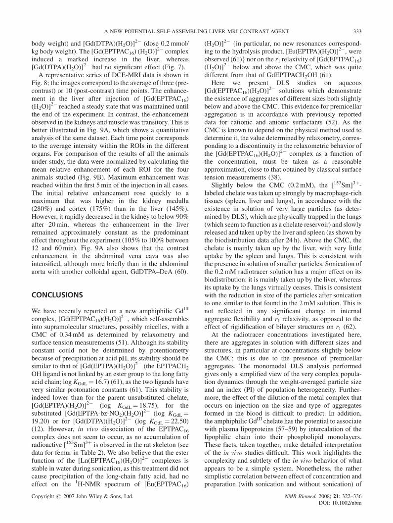
body weight) and [Gd(DTPA)(H2O)]2� (dose 0.2mmol/
kg body weight). The [Gd(EPTPAC16) (H2O)]2� complex
induced a marked increase in the liver, whereas[Gd(DTPA)(H2O)]
2� had no significant effect (Fig. 7).A representative series of DCE-MRI data is shown in
Fig. 8; the images correspond to the average of three (pre-contrast) or 10 (post-contrast) time points. The enhance-ment in the liver after injection of [Gd(EPTPAC16)(H2O)]
2� reached a steady state that was maintained untilthe end of the experiment. In contrast, the enhancementobserved in the kidneys and muscle was transitory. This isbetter illustrated in Fig. 9A, which shows a quantitativeanalysis of the same dataset. Each time point correspondsto the average intensity within the ROIs in the differentorgans. For comparison of the results of all the animalsunder study, the data were normalized by calculating themean relative enhancement of each ROI for the fouranimals studied (Fig. 9B). Maximum enhancement wasreached within the first 5min of the injection in all cases.The initial relative enhancement rose quickly to amaximum that was higher in the kidney medulla(280%) and cortex (175%) than in the liver (145%).However, it rapidly decreased in the kidney to below 90%after 20min, whereas the enhancement in the liverremained approximately constant as the predominanteffect throughout the experiment (105% to 100% between12 and 60min). Fig. 9A also shows that the contrastenhancement in the abdominal vena cava was alsointensified, although more briefly than in the abdominalaorta with another colloidal agent, GdDTPA–DeA (60).
CONCLUSIONS
We have recently reported on a new amphiphilic GdIII
complex, [Gd(EPTPAC16)(H2O)]2�, which self-assembles
into supramolecular structures, possibly micelles, with aCMC of 0.34mM as determined by relaxometry andsurface tension measurements (51). Although its stabilityconstant could not be determined by potentiometrybecause of precipitation at acid pH, its stability should besimilar to that of [Gd(EPTPA)(H2O)]
2� (the EPTPACH2
OH ligand is not linked by an ester group to the long fattyacid chain; log KGdL¼ 16.7) (61), as the two ligands havevery similar protonation constants (61). This stability isindeed lower than for the parent unsubstituted chelate,[Gd(EPTPA)(H2O)]
2� (log KGdL¼ 18.75), for thesubstituted [Gd(EPTPA-bz-NO2)(H2O)]
2� (log KGdL¼19.20) or for [Gd(DTPA)(H2O)]
2� (log KGdL¼ 22.50)(12). However, in vivo dissociation of the EPTPAC16
complex does not seem to occur, as no accumulation ofradioactive [153Sm]3þ is observed in the rat skeleton (seedata for femur in Table 2). We also believe that the esterfunction of the [Ln(EPTPAC16)(H2O)]
2� complexes isstable in water during sonication, as this treatment did notcause precipitation of the long-chain fatty acid, had noeffect on the 1H-NMR spectrum of [Eu(EPTPAC16)
(H2O)]2� {in particular, no new resonances correspond-
ing to the hydrolysis product, [Eu(EPTPA)(H2O)]2�, were
observed (61)} nor on the r1 relaxivity of [Gd(EPTPAC16)(H2O)]
2� below and above the CMC, which was quitedifferent from that of GdEPTPACH2OH (61).
Here we present DLS studies on aqueous[Gd(EPTPAC16)(H2O)]
2� solutions which demonstratethe existence of aggregates of different sizes both slightlybelow and above the CMC. This evidence for premicellaraggregation is in accordance with previously reporteddata for cationic and anionic surfactants (52). As theCMC is known to depend on the physical method used todetermine it, the value determined by relaxometry, corres-ponding to a discontinuity in the relaxometric behavior ofthe [Gd(EPTPAC16)(H2O)]
2� complex as a function ofthe concentration, must be taken as a reasonableapproximation, close to that obtained by classical surfacetension measurements (38).
Slightly below the CMC (0.2mM), the [153Sm]3þ-labeled chelate was taken up strongly bymacrophage-richtissues (spleen, liver and lungs), in accordance with theexistence in solution of very large particles (as deter-mined by DLS), which are physically trapped in the lungs(which seem to function as a chelate reservoir) and slowlyreleased and taken up by the liver and spleen (as shown bythe biodistribution data after 24 h). Above the CMC, thechelate is mainly taken up by the liver, with very littleuptake by the spleen and lungs. This is consistent withthe presence in solution of smaller particles. Sonication ofthe 0.2mM radiotracer solution has a major effect on itsbiodistribution: it is mainly taken up by the liver, whereasits uptake by the lungs virtually ceases. This is consistentwith the reduction in size of the particles after sonicationto one similar to that found in the 2mM solution. This isnot reflected in any significant change in internalaggregate flexibility and r1 relaxivity, as opposed to theeffect of rigidification of bilayer structures on r1 (62).
At the radiotracer concentrations investigated here,there are aggregates in solution with different sizes andstructures, in particular at concentrations slightly belowthe CMC; this is due to the presence of premicellaraggregates. The monomodal DLS analysis performedgives only a simplified view of the very complex popula-tion dynamics through the weight-averaged particle sizeand an index (PI) of population heterogeneity. Further-more, the effect of the dilution of the metal complex thatoccurs on injection on the size and type of aggregatesformed in the blood is difficult to predict. In addition,the amphiphilic GdIII chelate has the potential to associatewith plasma lipoproteins (57–59) by intercalation of thelipophilic chain into their phospholipid monolayers.These facts, taken together, make detailed interpretationof the in vivo studies difficult. This work highlights thecomplexity and subtlety of the in vivo behavior of whatappears to be a simple system. Nonetheless, the rathersimplistic correlation between effect of concentration andpreparation (with sonication and without sonication) of
Copyright # 2007 John Wiley & Sons, Ltd. NMR Biomed. 2008; 21: 322–336DOI: 10.1002/nbm
A NEW POTENTIAL SELF-ASSEMBLING LIVER MRI CONTRAST AGENT 333

the metal chelator on particle size (determined by DLS)and the biodistribution seems to be significant.In contrast with liposomal CAs that carry the para-
magnetic amphiphilic GdIII chelate in the lipid bilayer,which has been reported in various in vivo studies (27,28,63,64), very few targeted or non-targeted GdIII-basedcolloidal CAs have been studied beyond the stage ofbasic physicochemical characterization (29,50,63). Thepresent in vivo DCE-MRI evaluation of the micellar[Gd(EPTPAC16)(H2O)]
2� compound inWistar rats showsa persistent hepatic positive-contrast effect in T1-weightedimages, which is qualitatively similar to that of theclinically-established GdIII-based hepatobiliary agents,Gd-EOB-DTPA (65) and Gd-BOPTA [where BOPTA is4-carboxy-5,8,11-tris(carboxymethyl)-1-phenyl-2-oxa-5,8,11-triazatridecan-13-oate] (66), although a comparativestudy was not undertaken. No enhancement effect wasseen in the lungs because of the scarcity of mobile protonsin this organ, despite the scintigraphic evidence forsignificant lung retention of the [153Sm]3þ-labeled chelateat concentrations below the CMC. The possibility ofusing this type of micellar compound for imaging lungpathologies involving water accumulation, which willdepend on the degree of dilution of the imaging agent inthe body relative to its CMC, is worth pursuing in futurestudies.
Acknowledgments
This work was financially supported by the Foundation ofScience and Technology (FCT), Portugal (project POCTI/QUI/47005/2002 and Ph.D. grant SFRH/BD/5407/2001to T.B.R.), Instituto de Investigacao Interdisciplinar Uni-versity of Coimbra, Portugal (grant III/BIO/45/2005),FEDER, the Institute of Health Carlos III, Spain,PI051845 (to M.L.G.M.), and the Spanish Ministry ofEducation and Science, grants SAF 2004-0391 andNAN2004-09125-C07-03 (to S.C.). We thank Dr Mariados Anjos Neves, at the Instituto Tecnologico e Nuclear,Lisbon, Portugal for providing 153SmCl3. The work wascarried out in the framework of the EC COST ActionsD18 ‘Lanthanide chemistry for diagnosis and therapy’and D38 ‘Metal-Based Systems for Molecular ImagingApplications;, the European-funded EMIL program(LSCH-2004-503569), and the Accao IntegradaLuso-Espanhola E-10/04.
REFERENCES
1. Caravan P, Ellison JJ, McMurry TJ, Lauffer RB. Paramagneticmetal complexes as water proton relaxation agents for NMRimaging. Chem. Rev. 1999; 99: 2293–23525.
2. Toth E, Merbach AE (eds). The Chemistry of Contrast Agents inMedical Magnetic Resonance Imaging. Wiley: Chichester, 2001.
3. Toth E, Helm L, Merbach AE. Relaxivity of MRI contrast agents.Top. Curr Chem. 2002; 221: 61–101.
4. Toth E, Helm L, Merbach AE. MRI contrast enhancement agents.In Comprehensive Coordination Chemistry II, McCleverty JA,Meyer TJ (eds). Publisher: Elsevier Ltd.: Oxford, UK, 2004; 9:841–881.
5. Aime S, Botta M, Fasano M, Terreno E. Lanthanide(III) chelatesfor NMR biomedical applications. Chem. Soc. Rev. 1998; 27:19–29.
6. Comblin V, Gilsoul D, Hermann M, Humbert V, Jacques V,Mesbari M, Sauvage C, Desreux JF. Designing new MRI contrastagents: a coordination chemistry challenge. Coord. Chem. Rev.1999; 185–186: 451–470.
7. Gries H. Extracellular MRI contrast agents based on gadolinium.Top. Curr Chem. 2002; 221: 1–24.
8. Aime S, Cabella C, Colombato S, Crich SG, Gianolo E, MaggioriF. Insights into the use of paramagnetic Gd(III) complexes inMR-molecular imaging investigations. J. Magn. Reson. Imaging2002; 16: 394–406.
9. Toth E, Burai L, Brucher E, Merbach AE. Tuning water-exchangerates on (carboxymethyl)iminobis(ethylenenitrilo)tetraacetate(DTPA)-type gadolinium(III) complexes. J. Chem. Soc. DaltonTrans. 1997; 1587–1594.
10. Andre JP, Maecke HR, Toth E, Merbach AE. Synthesis andphysicochemical characterization of a novel precursor for cova-lently bound macromolecular MRI contrast agents. J. Biol. Inorg.Chem. 1999; 4: 341–347.
11. Ruloff R, Toth E, Tripier R, Handle H, Merbach AE. Acceleratingwater exchange for Gd(III) chelates by steric compression aroundthe water binding site. Chem. Commun. 2002; 22: 2630–2631.
12. Laus S, Ruloff R, Toth E, Merbach AE. GdIII complexes with fastwater exchange and high thermodynamic stability: potential build-ing blocks for high-relaxivity MRI contrast agents. Chemistry2003; 9: 3555–3566.
13. Jaszberenyi Z, Sour A, Toth E, Benmelouka M, Merbach AE.Fine-tuning water exchange on GdIII poly(amino carboxylates) bymodulation of steric crowding. Dalton Trans. 2005; 2713–2719.
14. Kotek J, Lebdukova P, Hermann P, van der Elst L, Muller RN,Geraldes CFGC, Maschmeyer T, Lukes I, Peters JA. Lanthani-de(III) complexes of novel mixed carboxylic-phosphorus acidderivatives of diethylenetriamine: a step towards more efficientMRI contrast agents. Chemistry 2003; 9: 5899–5915.
15. Corsi DM, van der Elst L, Muller RN, van Bekkum H, Peters JA.Inulin as a carrier for contrast agents in magnetic resonanceimaging. Chemistry 2001; 7: 64–71.
16. Andre JP, Geraldes CFGC, Martins JA, Merbach AE, Prata MIM,Santos AC, de Lima JJP, Toth E. Lanthanide(III) complexes ofglycoconjugates for lectin-mediated medical imaging. Chemistry2004; 10: 5804–5816.
17. Baıa P, Andre JP, Geraldes CFGC, Martins JA, Merbach AE, TothE. Lanthanide(III) chelates of DTPA bis(amide) glycoconjugates:potential imaging agents targeted at the asialoglicoprotein recep-tor. Eur. J. Inorg. Chem. 2005; 2110–2119.
18. Aime S, Botta M, Crich SG, Giovenzana G, Palmisano G, Sisti M.Novel paramagnetic macromolecular complexes derived from thelinkage of a macrocyclic Gd(III) complex to polyamino acidsthrough a squaric acid moiety. Bioconjugate Chem. 1999; 10:192–199.
19. Toth E, Pubanz D, Vauthey S, Helm L, Merbach AE. The role ofwater exchange in attaining maximum relaxivities for dendrimericMRI contrast agents. Chemistry 1996; 2: 1607–1915.
20. Laus S, Sour A, Ruloff R, Toth E, Merbach AE. Rotationaldynamics account for pH-dependent relaxivities of PAMAM den-drimeric, Gd-based potential MRI contrast agents. Chemistry2005; 11: 3064–3076.
21. Aime S, Chiaussa M, Digilio G, Gianolio E, Terreno E. Contrastagents for magnetic resonance angiographic applications: 1H and17O NMR relaxometric investigations on two gadolinium(III)DTPA-like chelates endowed with high binding affinity to humanserum albumin. J. Biol. Inorg. Chem. 1999; 4: 766–774.
22. Muller RN, Raduchel B, Laurent S, Platzek J, Pierart C, Mareski P,Vander Elst L. Physicochemical characterization ofMS-325, a newgadolinium complex, by multinuclear relaxometry. Eur. J. Inorg.Chem. 1999; 1949–1955.
23. Aime S, Gianolio E, Terreno E, Giovenzana GB, Pagliarin R, SistiM, Palmisano G, Botta M, Lowe MP, Parker D. TernaryGd(III)L-HSA adducts: evidence for the replacement of inner-
Copyright # 2007 John Wiley & Sons, Ltd. NMR Biomed. 2008; 21: 322–336DOI: 10.1002/nbm
334 S. TORRES ET AL.

sphere water molecules by coordinating groups of the protein.Implications for the design of contrast agents for MRI. J. Biol.Inorg. Chem. 2000; 5: 488–497.
24. Moghini SM, Hunter AC, Murray JC. Long-circulating and tar-get-specific nanoparticles: theory to practice. Pharm. Rev. 2001;53: 283–318.
25. Torchilin VP, Lukyanov AN, Gao ZG, Papahadjopoulos-SternbergB. Immunomicelles: targeted pharmaceutical carriers for poorlysoluble drugs. Proc. Natl. Acad. Sci. USA. 2003; 100: 6039–6044.
26. Wang J, Mongayt D, Torchilin VP. Polymeric micelles for deliveryof poorly soluble drugs: preparation and anticancer activity in vitroof paclitaxel incorporated into mixed micelles based on poly(ethylene glycol)-lipid conjugate and positively charged lipids.J. Drug. Targeting. 2005; 13: 73–80.
27. Mulder WJM, Strijkers GJ, van Tilborg GAF, Griffioen AW,Nicolay K. Lipid-based nanoparticles for contrast-enhancedMRI and molecular imaging. NMR Biomed 2006; 19: 142–164.
28. Lanza GM, Winter PM, Hughes MS, Caruthers SD, Marsh JN,Morawski AM, Schmieder AH, Scott MJ, Fuhrhop RW, Zhang H,Hu G, Lacy EK, Allen JS, Wickline SA. Molecular imaging andtherapy: new paradigms for 21(st) century medicine, polymericdrug delivery I: particulate drug carriers. ACS Symposium Series2006; 923: 295–311.
29. Kostarelos K, Emfietzoglon D. Liposomes as carriers of radio-nuclides: from imaging to therapy. J. Liposome Res. 1999; 9:429–460.
30. Torchilin VP, Kamenetsky MDF, Wolff GL. CT visualization ofblood pool in rats by using long-circulating, iodine-containingmicelles. Acad. Radiol. 1999; 6: 61–65.
31. Goins B, Phillips WT, Klipper R. Blood-pool imaging usingtechnetium-99m-labeled liposomes. J. Nucl. Med. 1996; 37:1374–1379.
32. Phillips WT, Klipper R, Goins B. Novel method of greatlyenhanced delivery of liposomes to lymph nodes. J. Pharm. Exp.Thert. 2000; 295: 309–313.
33. Lukyanov AN, Hartner WC, Torchilin VP. Tumor-targeted lipo-somes: doxorubicin-loaded long-circulating liposomes modifiedwith anti-cancer antibody. J. Control Release 2004; 94: 187–193.
34. Kellar KE, Henricks PM, Hollister R, Koenig SH, Eck J, Wei D.High relaxivity linear Gd(DTPA)-polymer conjugates: the roleof hydrophobic interactions. Magn. Reson. Med. 1997; 38:712–716.
35. Duarte MG, Gil MH, Peters JA, Vander Elst L, Collet JM, MullerRN, Geraldes CFGC. Synthesis, characterization, and relaxivity oftwo linear Gd(DTPA)-polymer conjugates. Bioconj. Chem. 2001;12: 170–177.
36. Toth E, Helm L, Kellar KE, Merbach AE. Gd(DTPA-bisamide)alkyl copolymers: a hint for the formation of MRIcontrast agents with very high relaxivity. Chemistry 1999; 5:1202–1211.
37. Andre JP, Toth E, Fischer H, Seelig A, Macke H, Merbach AE.High relaxivity for monomeric Gd(DOTA)-based MRI contrastagents, thanks to micellar self-organization. Chemistry 1999; 5:2977–2983.
38. Nicolle GM, Toth E, Eisenwiener KP, Macke HR, Merbach AE.From monomers to micelles: investigation of the parametersinfluencing proton relaxivity. J. Biol. Inorg. Chem. 2002; 7:757–769.
39. Gløgard C, Hovland R, Fossheim SL, Aasen AJ, Klaveness J.Synthesis and physicochemical characterisation of new amphiphi-lic gadolinium DO3A complexes as contrast agents for MRI.J. Chem. Soc., Perkin Trans. 2000; 2: 1047–1052.
40. Caravan P, Greenfield MT, Li X, Sherry AD. The Gd3þ complexof a fatty acid analogue of DOTP binds to multiple albumin siteswith variable water relaxivities. Inorg. Chem. 2001; 40:6580–6587.
41. Li X, Zhang S, Zhao P, Kovacs Z, Sherry AD. Synthesis and NMRstudies of new DOTP-like lanthanide(III) complexes containing ahydrophobic substituent on one phosphonate side arm. Inorg.Chem. 2001; 40: 6572–6579.
42. Hovland R, Gløgard C, Aasen AJ, Klaveness J. Preparation andin vitro evaluation of a novel amphiphilic GdPCTA-[12] derivative:a micellar MRI contrast agent. Org. Biomol. Chem. 2003; 1:644–647.
43. Nicolle GM, Helm L, Merbach AE. 8S paramagnetic centres inmolecular assemblies: possible effect of their proximity on thewater proton relaxivity. Magn. Reson. Chem. 2003; 41: 794–799.
44. Tournier H, Hyacinthe R, Schneider M. Gadolinium-containingmixed micelle formulations: a new class of blood pool MRI/MRAcontrast agents. Acad. Radiol. 2002; 202 (Suppl. 1): S20–S28.
45. Gløgard C, Stensrud G, Klaveness J. Novel high relaxivitycolloidal particles based on the specific phase organisation ofamphiphilic gadolinium chelates with cholesterol. Int. J. Pharm.2003; 253: 39–48.
46. Gløgard C. Liposomes as carriers of amphiphilic gadoliniumchelates: the effect of membrane composition on incorporationefficacy and in vitro relaxivity. Int. J. Pharm. 2002; 233: 131–140.
47. Lattuada L, Lux G. Synthesis of Gd-DTPA-cholesterol: a newlipophilic gadolinium complex as a potential MRI contrast agent.Tetrahedron Lett 2003; 44: 3893–3895.
48. Kimpe K, Parac-Vogt TN, Laurent S, Pierart C, Vander Elst L,Muller RN, Binnemans K. Potential MRI contrast agents based onmicellar incorporation of amphiphilic bis(alkylamide) derivativesof [(Gd-DTPA)(H2O)]
2�. Eur. J. Inorg. Chem. 2003; 3021–3027.
49. Parac-Vogt TN, Kimpe K, Laurent S, Pierart C, Vander Elst L,Muller RN, Binnemans K. Gadolinium DTPA-monoamide com-plexes incorporated into mixed micelles as possible MRI contrastagents. Eur. J. Inorg. Chem. 2004; 3538–3543.
50. Accardo A, Tesauro D, Roscigno P, Gianolio E, Paduano L,D’Errico G, Pedone C, Morelli G. Physicochemical propertiesof mixed micellar aggregates containing CCK peptides and Gdcomplexes designed as tumor specific contrast agents in MRI.J. Am. Chem. Soc. 2004; 126: 3097–3107.
51. Torres S, Martins JA, Andre JP, Geraldes CFGC, Merbach AE,Toth E. Supramolecular assembly of an amphiphilic GdIII chelate:tuning the reorientational correlation time and the water exchangerate. Chemistry 2006; 12: 940–948.
52. Zettl H, Portnoy Y, GottliebM, Krausch G. Investigation of micelleformation by fluorescence correlation spectroscopy. J. Phys. Chem.2005; 109: 13397–13401.
53. Perez-RodriguezM, Varela LM, GarciaM,Mosquera V, SarmientoF. Conductivity and relative permittivity of sodium n-dodecylsulfate and n-dodecyl trimethylammonium bromide. Journal ofChemical Engineering Data 1999; 44: 944–947.
54. He X, Liang H, Huang L, Pan C. Complex microstructures ofamphiphilic diblock copolymer in dilute solution. J. Phys. Chem.2004; 108: 1731–1735.
55. Sampson CB. Textbook of Radiopharmacy, Theory and Practice,3rd edition. Gordon and Breach Science Publishers: London, 1999;chapter 23: 283, 294.
56. Ekeh S, Chu R, Ficken VJ, Allen EW, Ryals CJ. Distributions ofperfusion and lung water. Nucl. Med. Biol. 1990; 17: 561–565.
57. Frias JC, Williams KJ, Fischer EW, Fayad ZA. RecombinantHDL-like nanoparticles: a specific contrast agent for MRI ofatherosclerotic plaques. J. Am. Chem. Soc. 2004; 126:16316–16317.
58. Jasanada F, Urizzi P, Suchard JP, LeGaillard F, Fabre G, Nepveu F.Indium-111 labeling of low density lipoproteins with the DTPA-bis(stearylamide): evaluation as a potential radiopharmaceuticalfor tumor localization. Bioconj Chem. 1996; 7: 72–81.
59. Corbin IR, Li H, Chen J, Lund-Katz S, Zhou R, Glickson JD,Zheng G. Low-density lipoprotein nanoparticles as magneticresonance imaging contrast agents. Neoplasia 2006; 8: 488–498.
60. Yoshikawa K, Inoue Y, Akahane M, Shimada M, Itoh S, Seno A,Hayashi S. Phantom and animal studies of a new hepatobiliaryagent for MR imaging: comparison of GdDTPA-DeA withGd-EOB-DTPA. J. Magn. Reson. Imaging. 2003; 18: 204–209.
61. Torres S, Martins JA, Andre JP, Pereira GA, Kiraly R, Brucher E,Helm L, Toth E, Geraldes CFGC. H5EPTPACH2OH: synthesis,relaxometric characterization and 1H NMR studies of the solutiondynamics of its LnIII complexes. Eur. J. Inorg. Chem. 2007; inpress.
62. Accardo A, Tesauro D, Morelli G, Gianolio E, Aime S, Vaccaro M,Mangiapia G, Paduano L, Schillen K. High relaxivity supramole-
Copyright # 2007 John Wiley & Sons, Ltd. NMR Biomed. 2008; 21: 322–336DOI: 10.1002/nbm
A NEW POTENTIAL SELF-ASSEMBLING LIVER MRI CONTRAST AGENT 335

cular aggregates containing peptides and Gd complexes ascontrast agents in MRI. J. Biol. Inorg. Chem. 2007; 12: 267–276.
63. Kabalka GW, Buonocore E, Hubner K, Davis M, Huang L.Gadolinium-labeled liposomes containing paramagnetic amphipa-tic agents: targeted MRI contrast agents for the liver.Magn. Reson.Med. 1988; 8: 89–95.
64. Chu WJ, Simor T, Elgavish G. In vivo characterization ofGd(BME-DTTA), a myocardial MRI contrast agent: tissue distri-
bution of its MRI intensity enhancement, and irs effect on heartfunction. NMR Biomed. 1977; 10: 87–92.
65. Schuhmann-Giampieri G, Schmitt-Willich H, Press W-R, NegishiC, Weinmann H-J, Speck U. Preclinical evaluation ofGd–EOB-DTPA as a contrast agent in MR imaging of the hepa-tobiliary systems. Radiology 1992; 183: 59–64.
66. Vittadini G, Felder E, Musu C, Tirone C. Preclinical profile ofGd-BOPTA: a liver specific MRI contrast agent. Invest. Radiol.1990; 5: S59–S60.
Copyright # 2007 John Wiley & Sons, Ltd. NMR Biomed. 2008; 21: 322–336DOI: 10.1002/nbm
336 S. TORRES ET AL.