Analysis of Tibiofemoral Cartilage Deformation in the Posterior Cruciate Ligament-Deficient Knee
Upload
independentCategory
view
2download
0
Available online http://arthritis-research.com/content/9/6/R121
Open AccessVol 9 No 6Research articleActivation of proteinase-activated receptor 2 in human osteoarthritic cartilage upregulates catabolic and proinflammatory pathways capable of inducing cartilage degradation: a basic science studyChristelle Boileau1, Nathalie Amiable1, Johanne Martel-Pelletier1, Hassan Fahmi1, Nicolas Duval2 and Jean-Pierre Pelletier1
1Osteoarthritis Research Unit, University of Montreal Hospital Centre, Notre-Dame Hospital, 1560 Sherbrooke Street East, Montreal, Quebec, H2L 4M1, Canada2Pavillon des Charmilles, 1487, boul. des Laurentides, Vimont, Quebec, H7M 2Y3, Canada
Corresponding author: Christelle Boileau, [email protected]
Received: 7 Aug 2007 Revisions requested: 1 Oct 2007 Revisions received: 9 Oct 2007 Accepted: 21 Nov 2007 Published: 21 Nov 2007
Arthritis Research & Therapy 2007, 9:R121 (doi:10.1186/ar2329)This article is online at: http://arthritis-research.com/content/9/6/R121© 2007 Boileau et al.; licensee BioMed Central Ltd. This is an open access article distributed under the terms of the Creative Commons Attribution License (http://creativecommons.org/licenses/by/2.0), which permits unrestricted use, distribution, and reproduction in any medium, provided the original work is properly cited.
Abstract
Proteinase-activated receptors (PARs) belong to a family of Gprotein-coupled receptors. PARs are activated by a serine-dependent cleavage generating a tethered activating ligand.PAR-2 was shown to be involved in inflammatory pathways. Weinvestigated the in situ levels and modulation of PAR-2 in humannormal and osteoarthritis (OA) cartilage/chondrocytes.Furthermore, we evaluated the role of PAR-2 on the synthesis ofthe major catabolic factors in OA cartilage, includingmetalloproteinase (MMP)-1 and MMP-13 and the inflammatorymediator cyclooxygenase 2 (COX-2), as well as the PAR-2-activated signalling pathways in OA chondrocytes. PAR-2expression was determined using real-time reversetranscription-polymerase chain reaction and protein levels byimmunohistochemistry in normal and OA cartilage. Proteinmodulation was investigated in OA cartilage explants treatedwith a specific PAR-2-activating peptide (PAR-2-AP), SLIGKV-NH2 (1 to 400 μM), interleukin 1 beta (IL-1β) (100 pg/mL),tumor necrosis factor-alpha (TNF-α) (5 ng/mL), transforminggrowth factor-beta-1 (TGF-β1) (10 ng/mL), or the signallingpathway inhibitors of p38 (SB202190), MEK1/2 (mitogen-activated protein kinase kinase) (PD98059), and nuclear factor-kappa B (NF-κB) (SN50), and PAR-2 levels were determined byimmunohistochemistry. Signalling pathways were analyzed onOA chondrocytes by Western blot using specific phospho-
antibodies against extracellular signal-regulated kinase 1/2(Erk1/2), p38, JNK (c-jun N-terminal kinase), and NF-κB in thepresence or absence of the PAR-2-AP and/or IL-1β. PAR-2-induced MMP and COX-2 levels in cartilage were determined byimmunohistochemistry. PAR-2 is produced by humanchondrocytes and is significantly upregulated in OA comparedwith normal chondrocytes (p < 0.04 and p < 0.03, respectively).The receptor levels were significantly upregulated by IL-1β (p <0.006) and TNF-α (p < 0.002) as well as by the PAR-2-AP at10, 100, and 400 μM (p < 0.02) and were downregulated by theinhibition of p38. After 48 hours of incubation, PAR-2 activationsignificantly induced MMP-1 and COX-2 starting at 10 μM (bothp < 0.005) and MMP-13 at 100 μM (p < 0.02) as well as thephosphorylation of Erk1/2 and p38 within 5 minutes ofincubation (p < 0.03). Though not statistically significant, IL-1βproduced an additional effect on the activation of Erk1/2 andp38. This study documents, for the first time, functionalconsequences of PAR-2 activation in human OA cartilage,identifies p38 as the major signalling pathway regulating itssynthesis, and demonstrates that specific PAR-2 activationinduces Erk1/2 and p38 in OA chondrocytes. These resultssuggest PAR-2 as a potential new therapeutic target for thetreatment of OA.
Page 1 of 10(page number not for citation purposes)
COX-2 = cyclooxygenase 2; CT = threshold cycle; DMEM = Dulbecco's modified Eagle's medium; Erk1/2 = extracellular signal-regulated kinase 1/2; FCS = fetal calf serum; GAPDH = glyceraldehydes-3-phosphate dehydrogenase; IL-1β = interleukin 1 beta; JNK = c-jun N-terminal kinase; MAP = mitogen-activated protein; MEK1/2 = mitogen-activated protein kinase kinase; MMP = matrix metalloproteinase; NF-κB = nuclear factor-kappa B; OA = osteoarthritis; PA = plasminogen activator; PAR = proteinase-activated receptor; PAR-2-AP = proteinase-activated receptor-2-activating pep-tide; PBS = phosphate-buffered saline; PCR = polymerase chain reaction; p-Erk1/2 = phosphorylated form of extracellular signal-regulated kinase 1/2; p-JNK = phosphorylated form of c-jun N-terminal kinase; p-p38 = phosphorylated form of p38; SD = standard deviation; SDS = sodium dodecyl sulfate; TGF-β1 = transforming growth factor-beta-1; TNF-α = tumor necrosis factor-alpha; TTBS 1× = Tris 20 mM, NaCl 150 mM (pH 7.5), and 0.1% Tween 20; uPA = urokinase plasminogen activator.

Arthritis Research & Therapy Vol 9 No 6 Boileau et al.
IntroductionOsteoarthritis (OA) can be defined as a complex degradativeand repair process in cartilage, subchondral bone, and syno-vial membrane. The factors responsible for the appearanceand progression of joint structural changes in OA have beenthe subject of intensive research for a few decades. Althoughsignificant progress has been made in the understanding ofthe pathophysiological pathways responsible for some of thechanges, much remains to be done to establish a therapeuticintervention that can effectively reduce or stop the progressionof the disease.
OA is characterized mainly by degradation of the cartilage. Thealterations in OA cartilage are numerous and involve morpho-logic and synthetic changes in chondrocytes as well as bio-chemical and structural alterations in the extracellular matrixmacromolecules [1]. In OA, the chondrocytes are the firstsource of enzymes responsible for cartilage matrix catabolism,and it is widely accepted that the metalloproteinase (MMP)family has a major involvement in the disease process [2].Moreover, considerable evidence has accumulated indicatingthat the proinflammatory cytokines synthesized and releasedby chondrocytes and synovial membrane are crucial in OA car-tilage catabolic processes and have an important impact in thedevelopment/progression of the disease [1].
In addition to cytokines, other mediators could play a majorrole in the OA pathological process. A member of the newlyidentified cell membrane receptor family, the proteinase-acti-vated receptors (PARs), has been shown to be involved ininflammatory pathways. These receptors belong to a novelfamily of seven-transmembrane G protein-coupled receptorsthat are activated through a unique process. The cleavage byserine proteases of the PAR N-terminal domains unmasks anew N-terminal sequence that acts as a tethered ligand, bind-ing and activating the receptor itself [3,4]. This activation is anirreversible phenomenon: the cleaved receptor is activated,internalized, and degraded. The cell membrane PARs arerestored from the intracellular pool [5].
This receptor family consists of four members, PAR-1 to PAR-4. PAR-1, PAR-3, and PAR-4 are activated by thrombin,whereas PAR-2 is activated mainly by trypsin but also by mastcell tryptase. PARs are expressed by several cell types, includ-ing platelets and endothelial and inflammatory cells, and areimplicated in numerous physiological and pathological proc-esses [3,4]. PAR-2 has also been found to be involved in mul-tiple cellular responses related to hyperalgesia. For example,Kawabata and colleagues [6] showed that the PAR-2 activa-tion by a specific agonist elicited thermal hyperalgesia andnociceptive behavior, and Vergnolle and colleagues [7] dem-onstrated that the thermal and mechanical hyperalgesia werereduced in PAR-2-deficient mice. In addition, PAR-2 is impli-cated in neurogenic inflammation [8] as well as inflammatoryconditions, including those seen in rheumatoid arthritis [9]. In
that regard, an important role for PAR-2 in the mouse adjuvant-induced arthritis model has been shown by using a PAR-2gene knockout mouse in which the appearance of inflamma-tion was significantly delayed [10,11]. Recently, PAR-2expression has been found in chondrocytes and synovialfibroblasts [12,13].
This study aimed to investigate the in situ levels and modula-tion of PAR-2 in human normal and OA cartilage, determine itsfunctional consequences on this tissue, and evaluate thechondrocyte signalling pathways involved in PAR-2 activity.We showed that PAR-2 is present at increased levels inhuman OA cartilage and that its level is modulated by theproinflammatory cytokines interleukin 1 beta (IL-1β) and tumornecrosis factor-alpha (TNF-α). Specific PAR-2 activation stim-ulates major pathophysiological pathways involved in the OAprocess, including MMP-1 and MMP-13 as well as cyclooxy-genase 2 (COX-2), through the activation of extracellular sig-nal-regulated kinase 1/2 (Erk1/2) and p38.
Materials and methodsSpecimen selectionHuman articular cartilage was obtained from femoral condylesor tibial plateaus. Normal knees were obtained within 12 hoursof death (mean age ± standard deviation [SD]: 52 ± 14 years).The cartilage was examined macroscopically and microscopi-cally to ensure that only normal tissue was used. Human OAspecimens were from patients undergoing total knee arthro-plasty (mean age ± SD: 76 ± 5 years). All patients with OAwere evaluated by a certified rheumatologist and were diag-nosed as having OA based on the criteria developed by theAmerican College of Rheumatology Diagnostic Subcommitteefor OA [14]. These specimens represented moderate tosevere OA as defined according to macroscopic criteria. Thisproject and the informed consent form were approved by theinstitutional Ethics Committee Board of the University of Mon-treal Hospital Centre.
Cartilage explant cultureNormal and OA cartilage explants (approximately 150 mg)were dissected and fixed in TissuFix #2 (Chaptec, Montreal,QC, Canada) and processed directly after acquisition from thedonor for immunohistochemistry (basal synthesis) or incu-bated in Dulbecco's modified Eagle's medium (DMEM) sup-plemented with 10% heat-inactivated fetal calf serum (FCS)and an antibiotics mixture (100 units/mL of penicillin base and100 μg/mL of streptomycin base) (Gibco-BRL Life Technolo-gies, now part of Invitrogen Corporation, Burlington, ON, Can-ada) at 37°C in a humidified atmosphere of 5% CO2/95% air.The conditions used were optimal for cartilage explant cul-tures. Cartilage explants were treated for 48 hours by IL-1β(100 pg/mL), TNF-α (5 ng/mL), and transforming growth fac-tor-beta-1 (TGF-β1) (10 ng/mL) (all from R&D Systems, Inc.,Minneapolis, MN, USA) or by the synthetic PAR-2-activatingpeptide (PAR-2-AP), SLIGKV-NH2 (0 to 400 μM) (Bachem
Page 2 of 10(page number not for citation purposes)
Available online http://arthritis-research.com/content/9/6/R121
California, Inc., Torrance, CA, USA), p38 inhibitor (SB202190 at 10 μM) (Tocris Bioscience, Ellisville, MO, USA),and mitogen-activated protein (MAP) kinase kinase (MEK1/2)inhibitor (PD98059 at 10 μM) and nuclear factor-kappa B (NF-κB) inhibitor (SN50 at 50 μg/mL) (both from EMD Bio-sciences, Inc., San Diego, CA, USA). Cartilage explants werethen processed for PAR-2 immunohistochemistry asdescribed below.
Chondrocyte culture and treatmentChondrocytes were released from full-thickness strips of car-tilage followed by sequential enzymatic digestion at 37°C, aspreviously described [15]. Cells were seeded at high density(105 cells/cm2) in tissue culture flasks and were cultured toconfluence in DMEM supplemented with 10% FCS and anantibiotics mixture (Invitrogen Corporation) at 37°C in ahumidified atmosphere. To ensure phenotype, only first-pas-sage cultured chondrocytes were used.
The chondrocytes were harvested with Cell DissociationBuffer (Invitrogen Corporation) (which contains no protease),seeded, and cultured in DMEM containing 10% FCS at 37°Cuntil confluence. Cells were further incubated with DMEMcontaining 2.5% FCS and treated with IL-1β (100 pg/mL),TNF-α (5 ng/mL), and TGF-β1 (10 ng/mL) (all from R&D Sys-tems, Inc.) and the synthetic PAR-2-AP, SLIGKV-NH2 (0 to400 μM) (Bachem California, Inc.), for 72 hours for PAR-2 pro-tein determination and 0 to 60 minutes for signalling pathways.
RNA extraction, reverse transcription, and real-time polymerase chain reactionTotal RNA was extracted from chondrocytes as previouslydescribed [15]. Briefly, total RNA was extracted with TRIzol®
according to the manufacturer's instructions (Invitrogen Cor-poration), and genomic DNA was removed following the man-ufacturer's instructions (Ambion, Inc., Austin, TX, USA). TheRNA was quantified with the RiboGreen® RNA quantificationkit (Molecular Probes Inc., now part of Invitrogen Corporation).cDNA was reverse-transcribed from 1 μg of total RNA purifiedin a 50-μL reaction mixture containing 1 mM each of deoxynu-cleotide triphosphates (Invitrogen Corporation), 0.4 U/μLRNase inhibitor and 2.5 μM of random hexamer (both from GEHealthcare, Baie d'Urfé, QC, Canada), 2.5 U/μL of reversetranscriptase (Invitrogen Corporation), 5 mM of MgCl2, and 1×of polymerase chain reaction (PCR) buffer. The reaction mix-ture was incubated in a DNA thermal cycle at 42°C for 15 min-utes and then stored at -20°C before use. Real-time PCR wasperformed using primers specific for the human PAR-2 and forthe human housekeeping gene glyceraldehydes-3-phosphatedehydrogenase (GAPDH). The primers were 5'-GAAGCCT-TATTGGTAAGGTTG (sense) and 5'-CAGAGAGGAGGT-CAGCCAAG (anti-sense) for PAR-2 and 5'-CAGAACATCATCCCTGCCTCT (sense) and 5'-GCTT-GACAAAGTGGTCGTTGAG (anti-sense) for GAPDH. Inbrief, 10 μL of the cDNA obtained from the reverse transcrip-
tion reactions was amplified in a total volume of 25 μL consist-ing of 1× Quantitect SYBR Green PCR Master Mix (QiagenInc., Mississauga, ON, Canada), 0.5 U/reaction uracil-N-glyc-osylase (Invitrogen Corporation), and gene-specific primersthat were added at a final concentration of 200 nM. Real-timequantitation mRNA was performed in the Rotor-Gene 6® RG-3000A (Corbett Research, Mortlake, NSW, Australia) accord-ing to the manufacturer's instructions. Data were processedwith Rotor-Gene version 6 software and were given a thresh-old cycle (CT) corresponding to the PCR cycle at which anincrease in reporter fluorescence above a baseline signal canfirst be detected. Plasmid DNAs containing target genesequences were used to generate the standard curves. TheCT was converted to number of molecules, and the values foreach sample were calculated as the ratio of the number of mol-ecules of the target gene to the number of molecules ofGAPDH and were expressed as arbitrary units.
ImmunohistochemistryCartilage specimens were processed for immunohistochemi-cal analysis as previously described [16]. Briefly, specimenswere fixed in TissuFix #2 for 24 hours and then embedded inparaffin. Sections (5 μm) of paraffin-embedded specimenswere placed on Superfrost Plus slides (Fisher Scientific,Nepean, ON, Canada), deparaffinized in toluene, rehydrated ina reverse-graded series of ethanol, and preincubated withchondroitinase ABC 0.25 units/mL (Sigma-Aldrich, Oakville,ON, Canada) in phosphate-buffered saline (PBS) (pH 8.0) for60 minutes at 37°C. Subsequently, the specimens werewashed in PBS, incubated in 0.3% Triton X-100 for 20 min-utes, and placed in 3% hydrogen peroxide/PBS for 15 min-utes. Slides were further incubated with a blocking serum(Vectastain ABC assay; Vector Laboratories, Burlingame, CA,USA) for 60 minutes, after which they were blotted and thenoverlaid with the primary antibody against mouse anti-humanPAR-2 (1:50; Zymed Laboratories Inc., now part of InvitrogenCorporation), mouse anti-human COX-2 (1:25; CedarlaneLaboratories Ltd., Burlington, ON, Canada), mouse anti-human MMP-1 (1:40; EMD Biosciences, Inc.), and goat anti-human MMP-13 (1:6; R&D Systems, Inc.) for 18 hours at 4°C.Each slide was washed three times in PBS (pH 7.4) and incu-bated with the second antibody (anti-mouse or anti-goat; Vec-tor Laboratories) for 1 hour at room temperature, followed bya staining with the avidin-biotin-peroxidase complex method(Vectastain ABC assay). The color was developed with 3,3'-diaminobenzidine (DAKO Diagnostics Inc., Mississauga, ON,Canada) containing hydrogen peroxide. Slides were counter-stained with eosin. All incubations were carried out in a humid-ified chamber. Each section was examined under a lightmicroscope (Leitz Orthoplan; Leica Inc., St. Laurent, QC, Can-ada). Two control procedures were performed according tothe same experimental protocol: (a) omission of the primaryantibody and (b) substitution of the primary antibody with anautologous preimmune serum. Controls showed only back-ground staining.
Page 3 of 10(page number not for citation purposes)

Arthritis Research & Therapy Vol 9 No 6 Boileau et al.
Positive cells were quantified as previously described [17]. Inbrief, three sections for each specimen were examined (×40;Leica Orthoplan) from the cartilage superficial zone (whichincluded the superficial and upper intermediate layers). Thesections were scored, and the resulting data were integratedas a mean for each specimen. The total numbers of chondro-cytes and of those staining positive for the specific antigenwere determined. The final results were expressed as the per-centage of chondrocytes staining positive for the antigen (cellscore), with the maximum score being 100%. Each slide wasexamined by two independent readers.
Western blotTotal proteins were extracted with 0.5% sodium dodecyl sul-fate (SDS) (Invitrogen Corporation) supplemented with pro-tease inhibitors. The protein level was determined using thebicinchoninic acid protein assay, and 10 μg of the protein waselectrophoresed on a discontinuous 4% to 12% SDS gel poly-acrylamide. The proteins were transferred electrophoreticallyonto a nitrocellulose membrane (Bio-Rad Laboratories Ltd.,Mississauga, ON, Canada) for 1 hour at 4°C. The efficiency oftransfer was controlled by a brief staining of the membranewith Ponceau Red and destained in water and TTBS 1× (Tris20 mM, NaCl 150 mM [pH 7.5], and 0.1% Tween 20) beforeimmunoblotting.
The membranes were incubated overnight at 4°C with 5%skimmed milk in SuperBlock Blocking Buffer-Blotting in Tris-buffered saline (Pierce, Rockford, IL, USA) or in TTBS 1× only.The membranes were then washed once with TTBS 1× for 10minutes and incubated in SuperBlock Blocking Buffer-Blottingand TTBS 1× (SuperBlock 1:10 with TTBS 1×) or in TTBS 1×(for PAR-2 antibody only) with 0.5% skimmed milk supple-mented with the mouse anti-human PAR-2 (1:1,000; Invitro-gen Corporation) and with mouse anti-human antibodiesagainst the phosphorylated forms of p38 (1:1,000), Erk1/2(1:5,000), c-jun N-terminal kinase (JNK) (1:5,000), and NF-κB(p65) (1:5,000) (all from New England Biolabs Ltd., Pickering,ON, Canada) overnight at 4°C. The membranes were washedwith TTBS 1× and incubated for 1 hour at room temperaturewith the second antibody (1:20,000; anti-mouse immunoglob-ulin G horseradish peroxidase-conjugated; Pierce) andwashed again with TTBS 1×. Detection was performed bychemiluminescence using the Super Signal® ULTRA chemilu-minescent substrate (Pierce) and exposure to Kodak Biomaxphotographic film (GE Healthcare). The band intensity wasmeasured by densitometry using TotalLab TL100 Software(Nonlinear Dynamics Ltd., Newcastle upon Tyne, UK), anddata were expressed as arbitrary units, in which the controlwas assigned a value of 100%.
Statistical analysisValues are expressed as median (range) or as mean ± stand-ard error of the mean (SEM) when appropriate. Statistical anal-ysis was performed using the Mann-Whitney U test.
ResultsPAR-2 expression and synthesisThe levels of PAR-2 mRNA in normal (n = 4) and OA (n = 6)chondrocytes were determined by real-time PCR. As illus-trated at Figure 1a, PAR-2 showed a significantly higher level(mean increase of 6.5-fold; p < 0.04) in OA chondrocytes thanin normal chondrocytes. Similarly, PAR-2 protein levels,detected by immunohistochemistry, were significantly upregu-lated in OA (n = 4) compared with normal (n = 4) cartilage(mean increase of 2.5-fold; p < 0.03) (Figure 1b). Figure 1cillustrates that PAR-2 is localized mainly in the superficial zone(consisting of the superficial and upper intermediate layers) innormal and OA cartilage.
Regulation of PAR-2 synthesisTo explore the mechanism underlying PAR-2 modulation inhuman OA cartilage, we studied the effects of two proinflam-matory cytokines, IL-1β and TNF-α, the growth factor TGF-β1,and a specific PAR-2 activator agonist, the PAR-2-AP, on car-tilage explant cultures. Data showed that, in OA cartilage,PAR-2 levels were significantly upregulated by IL-1β (n = 9; p< 0.006) and TNF-α (n = 9; p < 0.002) but not by TGF-β1 (n= 5) (Figure 2a). The PAR-2-AP (n = 4 to 8) also significantlyincreased the PAR-2 level, starting at 10 μM (p < 0.02) (Figure2a). Interestingly, PAR-2-AP appeared to be more efficient atincreasing the level of PAR-2 protein than the cytokines, andmean increases of 53%, 64%, and 63% were found for PAR-2-AP 10, 100, and 400 μM, respectively, compared with 24%for IL-1β and 29% for TNF-α.
On OA monolayer chondrocytes (n = 3), data obtained for IL-1β and TNF-α (Figure 2b) were similar to those from OA car-tilage explants. However, in contrast to cartilage explants, OAchondrocyte treatment with TGF-β1 (n = 3) yielded a markedPAR-2 protein increase (Figure 2b). As Xiang and colleagues[12] reported that TGF-β decreased the level of PAR-2 in OAchondrocytes, we further validated our findings by investigat-ing the effect of TGF-β1 on PAR-2 expression levels on normal(n = 2) and OA (n = 3) chondrocytes. Data showed that onboth sets this factor markedly increased PAR-2 expression lev-els (20- and 42-fold, respectively; data not shown). In the OAcells as in cartilage, PAR-2-AP treatment (n = 3) also markedlyincreased PAR-2 protein levels (Figure 2b).
To explore the signalling pathways involved in the regulation ofPAR-2 synthesis, human OA cartilage explants were incu-bated for 48 hours with the MAP kinase inhibitor SB 202190,inhibitor of p38; PD 98059, inhibitor of MEK1/2; and SN50,inhibitor of NF-κB. Data (n = 3 to 4) revealed that only the p38inhibitor markedly downregulated (p < 0.06) the PAR-2 pro-duction in OA cartilage (Figure 3). Erk1/2 and NF-κB inhibi-tions had no effect on PAR-2 synthesis.
Page 4 of 10(page number not for citation purposes)

Available online http://arthritis-research.com/content/9/6/R121
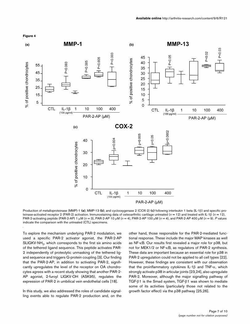
PAR-2 activation and functional consequencesTo determine the functional consequence of PAR-2 activation,we studied some of the major catabolic/inflammatory factorsinvolved in OA pathophysiology, including MMP-1, MMP-13,and COX-2 (Figure 4). PAR-2-AP treatment of OA cartilageexplants (n = 3 to 9) revealed a statistically significant increasestarting at concentrations of 10 μM for MMP-1 (p < 0.005)and COX-2 (p < 0.005) and 100 μM for MMP-13 (p < 0.02).For each factor, the increase was localized at the superficialzone. As expected, IL-1β (n = 12) showed a statistically signif-icant increase for all of the factors examined. Comparisonrevealed that this cytokine had a lower induction level on eachfactor than the PAR-2 activation. IL-1β induced meanincreases for MMP-1, MMP-13, and COX-2 of 29%, 20%, and18% compared with means of 73%, 44%, and 40% for PAR-2-AP.
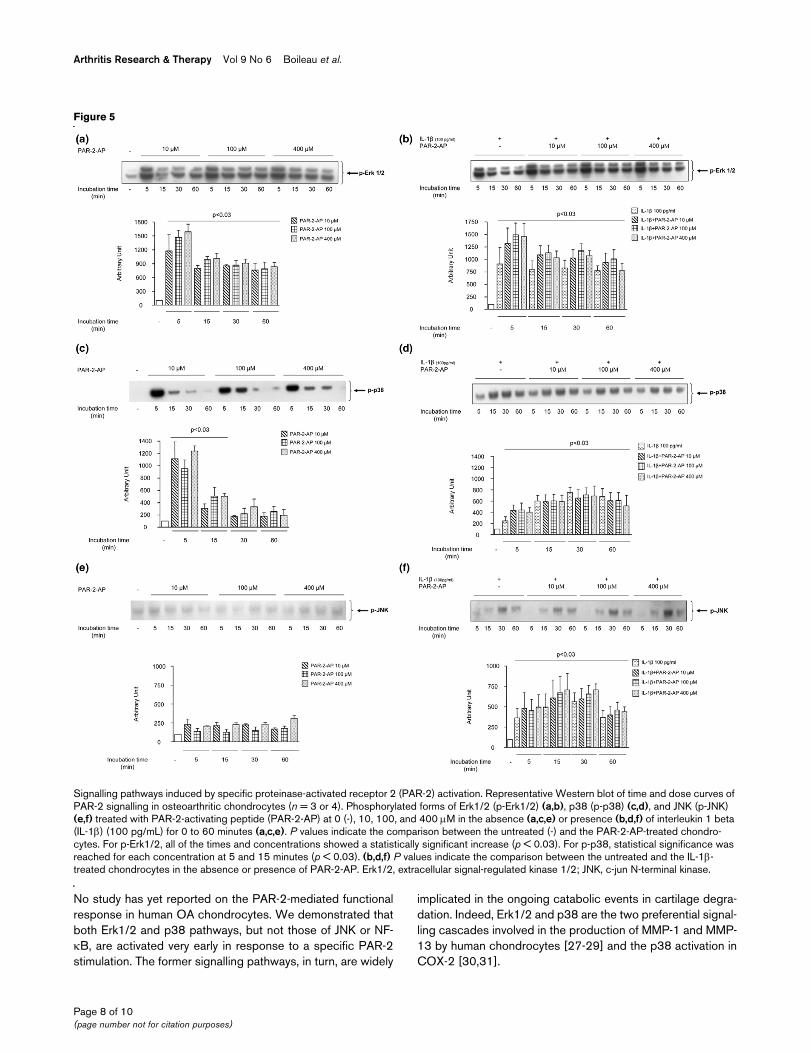
PAR-2-induced signalling pathwaysThe effect of PAR-2 activation on the phosphorylated levels ofthree MAP kinases of the OA chondrocytes (n = 3 to 4),namely Erk1/2, p38, and JNK, and on NF-κB was analyzed byWestern blot using specific antibodies. The activation of PAR-2 in OA chondrocytes using the PAR-2-AP (Figure 5a) yieldedwithin minutes (5 minutes) a sharp phosphorylation of Erk1/2(p-Erk1/2) (p < 0.03). Data also showed that the maximalPAR-2-AP stimulation concentration is 100 μM. At 15 minutesof PAR-2-AP treatment, p-Erk1/2 still showed significantly ele-
vated levels compared with the control (p < 0.03) but to alesser extent than at 5 minutes. PAR-2-AP also induced p38phosphorylation (p-p38) rapidly, with a maximum stimulation at5 minutes (p < 0.03) (Figure 5c), which declined thereafter butwas still statistically significant (p < 0.03) at 15 minutes oftreatment. No true dose-dependent effect was seen for thetransient p38 phosphorylation. The levels of the phosphor-ylated form of JNK (p-JNK) were very low and were notaffected by PAR-2-AP treatment (Figure 5e). Finally, the levelsof the phosphorylated form of NF-κB were barely detectableeven after treatment with PAR-2-AP (data not shown).
IL-1β significantly (p < 0.03) increased the level of the phos-phorylated form of each of the MAP kinases studied, with max-imums reached at 5 minutes of incubation for p-Erk1/2 and 30minutes for p-p38 and p-JNK (Figure 5b,d,f). The treatmentwith PAR-2-AP together with IL-1β yielded an additional effectfor p-Erk1/2, but this was not statistically different from the IL-1β alone (Figure 5b). This was also noticed for p38phosphorylation (Figure 5d) at 5 minutes of incubation. For p-JNK (Figure 5f) and NF-κB (data not shown), the addition ofPAR-2-AP did not modify IL-1β-induced activity.
DiscussionThis study is the first to demonstrate that PAR-2 activation inhuman OA cartilage significantly upregulates the synthesis ofimportant catabolic and proinflammatory mediators involved in
Figure 1
Proteinase-activated receptor 2 (PAR-2) gene expression and protein synthesisProteinase-activated receptor 2 (PAR-2) gene expression and protein synthesis. (a) mRNA levels, as determined by real-time quantitative polymer-ase chain reaction as described in Materials and methods, in normal (n = 4) and osteoarthritis (n = 6) chondrocytes. (b) PAR-2 immunostaining in normal (n = 4) and osteoarthritis (n = 4) cartilage. The percentage of positive chondrocytes represents the number of chondrocytes staining positive for PAR-2 of the total number of chondrocytes. Data are expressed as median and range and are presented as box plots, in which the boxes repre-sent the first and third quartiles, the line within the box represents the median, and the lines outside the box represent the spread of values. P values indicate the comparison of normal to osteoarthritis cartilage using the Mann-Whitney U test. (c) Representative sections showing PAR-2 immunos-taining in normal and osteoarthritis cartilage. The arrows refer to positive chondrocytes.
Page 5 of 10(page number not for citation purposes)

Arthritis Research & Therapy Vol 9 No 6 Boileau et al.
the progression of the disease and that the effect is mediatedby the activation of Erk1/2 and p38 signalling pathways. Here,we showed that PAR-2 expression and protein levels were sig-nificantly increased in OA compared with normal human
chondrocytes and that the levels are upregulated by the proin-flammatory cytokines IL-1β and TNF-α, an effect previouslyobserved on chondrocytes by Xiang and colleagues [12] andon other cell types [13,18,19]. Our data showing that TGF-β1on OA chondrocytes, but not on OA cartilage explants,upregulates PAR-2 levels appear contradictory. A possibleexplanation could be that, in the cartilage explants, largeamounts of TGF-β can be entrapped in the extracellular matrixand consequently only a very low concentration of this factorreaches the cells. Indeed, one of the characteristics of someproteoglycans is their interaction with active TGF-β, whichprovides a tissue reservoir of this factor, thereby modulating itsbioavailability [20,21]. Our finding of an increased level ofPAR-2 induced by TGF-β1 on OA chondrocytes also differed,to some extent, from the results of Xiang and colleagues [12],who reported a differential effect of TGF-β between normaland OA chondrocytes; TGF-β downregulated PAR-2 levels inOA but increased its levels in normal chondrocytes. A reasonfor this discrepancy could be that, in the report by Xiang andcolleagues, the protein measurement was carried out on aspecimen not representative of the heterogeneity of the humansampling; only one normal and one OA chondrocyte wereused. Such a possibility has been underlined by the authors[12], who suggest the possible existence of different cell pop-ulations such as responders and non-responders.
Figure 2
Proteinase-activated receptor 2 (PAR-2) synthesis regulationProteinase-activated receptor 2 (PAR-2) synthesis regulation. (a) PAR-2 immunostaining in osteoarthritis (OA) cartilage explants untreated (n = 16) and treated with interleukin 1 beta (IL-1β) (n = 9), tumor necrosis factor-alpha (TNF-α) (n = 9), transforming growth factor-beta-1 (TGF-β1) (n = 5), PAR-2-activating peptide (PAR-2-AP) 1 μM (n = 3), PAR-2-AP 10 μM (n = 4), PAR-2-AP 100 μM (n = 4), and PAR-2-AP 400 μM (n = 8) for 48 hours in Dulbecco's modified Eagle's medium (DMEM) 10% fetal calf serum (FCS). (b) Representative Western blot of PAR-2 synthesis in OA mon-olayer chondrocytes (n = 3) incubated for 72 hours in DMEM 2.5% FCS in the absence (CTL) or presence of IL-1β, TGF-β1, PAR-2-AP 10 μM, PAR-2-AP 100 μM, and PAR-2-AP 400 μM. P values indicate the comparison with the untreated (CTL) specimens.
Figure 3
Signalling pathways of proteinase-activated receptor 2 (PAR-2) synthesisSignalling pathways of proteinase-activated receptor 2 (PAR-2) synthe-sis. PAR-2 immunostaining in osteoarthritic cartilage untreated (n = 4) and treated with SB 202190 (inhibitor of p38; n = 3), PD 98059 (inhibitor of MEK1/2; n = 4), and SN50 (inhibitor of nuclear factor-kappa B; n = 4). P value indicates the comparison with the untreated (CTL) specimens. MEK1/2, mitogen-activated protein kinase kinase.
Page 6 of 10(page number not for citation purposes)
Available online http://arthritis-research.com/content/9/6/R121
To explore the mechanism underlying PAR-2 modulation, weused a specific PAR-2 activator agonist, the PAR-2-APSLIGKV-NH2, which corresponds to the first six amino acidsof the tethered ligand sequence. This peptide activates PAR-2 independently of proteolytic unmasking of the tethered lig-and sequence and triggers G-protein coupling [3]. Our findingthat the PAR-2-AP, in addition to activating PAR-2, signifi-cantly upregulates the level of the receptor on OA chondro-cytes agrees with a recent study showing that another PAR-2-AP agonist, 2-furoyl LIGKV-OH (ASK95), regulates theexpression of PAR-2 in umbilical vein endothelial cells [18].
In this study, we also addressed the roles of candidate signal-ling events able to regulate PAR-2 production and, on the
other hand, those responsible for the PAR-2-mediated func-tional response. These include the major MAP kinases as wellas NF-κB. Our results first revealed a major role for p38, butnot for MEK1/2 or NF-κB, as regulators of PAR-2 synthesis.These data are important because an essential role for p38 inPAR-2 upregulation could not be applied to all cell types [22].However, these findings are consistent with our observationthat the proinflammatory cytokines IL-1β and TNF-α, whichstrongly activate p38 in articular joints [23,24], also upregulatePAR-2. Moreover, although the major signalling pathway ofTGF-β1 is the Smad system, TGF-β1 was shown to mediatesome of its activities (particularly those not related to thegrowth factor effect) via the p38 pathway [25,26].
Figure 4
Production of metalloproteinase (MMP)-1 (a), MMP-13 (b), and cyclooxygenase 2 (COX-2) (c) following interleukin 1 beta (IL-1β) and specific pro-teinase-activated receptor 2 (PAR-2) activationProduction of metalloproteinase (MMP)-1 (a), MMP-13 (b), and cyclooxygenase 2 (COX-2) (c) following interleukin 1 beta (IL-1β) and specific pro-teinase-activated receptor 2 (PAR-2) activation. Immunostaining data of osteoarthritic cartilage untreated (n = 12) and treated with IL-1β (n = 12), PAR-2-activating peptide (PAR-2-AP) 1 μM (n = 3), PAR-2-AP 10 μM (n = 4), PAR-2-AP 100 μM (n = 4), and PAR-2-AP 400 μM (n = 9). P values indicate the comparison with the untreated (CTL) specimens.
Page 7 of 10(page number not for citation purposes)
Arthritis Research & Therapy Vol 9 No 6 Boileau et al.
No study has yet reported on the PAR-2-mediated functionalresponse in human OA chondrocytes. We demonstrated thatboth Erk1/2 and p38 pathways, but not those of JNK or NF-κB, are activated very early in response to a specific PAR-2stimulation. The former signalling pathways, in turn, are widely
implicated in the ongoing catabolic events in cartilage degra-dation. Indeed, Erk1/2 and p38 are the two preferential signal-ling cascades involved in the production of MMP-1 and MMP-13 by human chondrocytes [27-29] and the p38 activation inCOX-2 [30,31].
Figure 5
Signalling pathways induced by specific proteinase-activated receptor 2 (PAR-2) activationSignalling pathways induced by specific proteinase-activated receptor 2 (PAR-2) activation. Representative Western blot of time and dose curves of PAR-2 signalling in osteoarthritic chondrocytes (n = 3 or 4). Phosphorylated forms of Erk1/2 (p-Erk1/2) (a,b), p38 (p-p38) (c,d), and JNK (p-JNK) (e,f) treated with PAR-2-activating peptide (PAR-2-AP) at 0 (-), 10, 100, and 400 μM in the absence (a,c,e) or presence (b,d,f) of interleukin 1 beta (IL-1β) (100 pg/mL) for 0 to 60 minutes (a,c,e). P values indicate the comparison between the untreated (-) and the PAR-2-AP-treated chondro-cytes. For p-Erk1/2, all of the times and concentrations showed a statistically significant increase (p < 0.03). For p-p38, statistical significance was reached for each concentration at 5 and 15 minutes (p < 0.03). (b,d,f) P values indicate the comparison between the untreated and the IL-1β-treated chondrocytes in the absence or presence of PAR-2-AP. Erk1/2, extracellular signal-regulated kinase 1/2; JNK, c-jun N-terminal kinase.
Page 8 of 10(page number not for citation purposes)
Available online http://arthritis-research.com/content/9/6/R121
Interestingly, co-stimulation of chondrocytes with IL-1β andPAR-2-AP showed an additional stimulatory effect, particularlyon Erk1/2. A possible explanation is that Erk1/2 is not the pref-erential pathway mediating the effects of IL-1β; consequently,stimulation by this cytokine may not have reached maximalactivation of this pathway. This finding thus indicates that, dur-ing the disease process, both PAR-2 and IL-1β could act incooperation at inducing a catabolic cellular response.
The increased level of PAR-2 in OA compared with normalchondrocytes may be related, in addition to the stimulatoryeffect of the cytokines, to an increased level of serine pro-teases in OA cartilage. Indeed, according to the literature, thisenzyme family appears to be responsible for the PAR-2 activa-tion [3,4]. In OA cartilage, one of the most important serineprotease systems is the plasminogen activator (PA) plasmin, inwhich the urokinase PA (uPA) plays a major role [32,33]. Inter-estingly, the uPA/plasmin system, in addition to acting directlyon cartilage macromolecules, has been shown to be responsi-ble for increased levels of other proteases, including colla-genase [32,34]. The specific PAR-2 activation elicitingincreased levels of MMP-1 and MMP-13 strongly suggests thelikely involvement of this serine protease system in in vivoPAR-2 activation. Moreover, interaction between uPA andCOX-2 was also shown in some cancer cells [35,36] and incorneal injury and inflammation [37].
Findings of previous studies have identified a role for PAR-2 inmodulation of inflammation in rodent models, including inflam-matory arthritis [11]. Here, we showed that, in addition toinflammatory factors such as COX-2, PAR-2 activation upreg-ulates two MMPs, providing a critical link between inflamma-tion and tissue destruction and thus contributing to theperpetuation of the altered responses of the chondrocytes.Interestingly, the predominant effect of PAR-2 activation overIL-1β on both MMPs and COX-2 reinforces the suggestionthat PAR-2 is an upstream mediator of catabolic events[38,39]. Hence, data from this study suggest that PAR-2 acti-vation would play a key role in the catabolic and inflammatorypathways that take place during OA by inducing the synthesisof major catabolic and inflammatory mediators via the p38 andp42/44 signalling pathways. Furthermore, PAR-2 upregulationby proinflammatory cytokines would amplify its effect.
ConclusionIn summary, we showed, for the first time, that PAR-2 activa-tion in OA cartilage participates in catabolic and inflammatorypathways induced during OA progression. Moreover, thepresent knowledge points to a possible therapeutic value forPAR-2 antagonists in the treatment of OA, not only as an anti-catabolic and anti-inflammatory but as an analgesic as well.Indeed, the proanalgesic properties of PAR-2 have shown thatits activation plays a pivotal role in pain transmission with adirect effect on nociception and hyperalgesia [7,40-42]. Thismolecule, therefore, is believed to be an attractive target in OA
because reducing its excess production may not only slow thedisease progression, but also likely reduce the symptoms, ena-bling it to reach two targets simultaneously.
Competing interestsThe authors declare that they have no competing interests.
Authors' contributionsCB participated in study design, acquisition of data, analysisand interpretation of data, statistical analysis, and manuscriptpreparation. NA participated in acquisition of data, analysisand interpretation of data, statistical analysis, and manuscriptpreparation. JMP and JPP participated in study design, analy-sis and interpretation of data, and manuscript preparation. NDparticipated in study design and in analysis and interpretationof data. HF participated in study design. All authors read andapproved the final manuscript. CB and NA contributed equallyto this work.
AcknowledgementsWe thank François Mineau, François-Cyril Jolicoeur, Martin Boily, Changshan Geng, and Saranette Cheng for their exceptional technical support and Virginia Wallis and Santa Fiori for their invaluable assist-ance in manuscript preparation. This study was supported by grants from the Groupe de recherche des maladies rhumatismales du Québec, the CIHR/MENTOR training program and by internal funds of the Oste-oarthritis Research Unit, University of Montreal Hospital Centre, Notre-Dame Hospital, Montreal, Quebec, Canada.
References1. Martel-Pelletier J, Lajeunesse D, Pelletier JP: Etiopathogenesis of
osteoarthritis. In Arthritis and Allied Conditions: A Textbook ofRheumatology 15th edition. Edited by: Koopman WJ, MorelandLW. Baltimore: Lippincott, Williams & Wilkins; 2005:2199-2226.
2. Martel-Pelletier J, Welsch DJ, Pelletier JP: Metalloproteases andinhibitors in arthritic diseases. Best Pract Res Clin Rheumatol2001, 15:805-829.
3. Macfarlane SR, Seatter MJ, Kanke T, Hunter GD, Plevin R: Protei-nase-activated receptors. Pharmacol Rev 2001, 53:245-282.
4. Hollenberg MD, Compton SJ: International Union of Pharmacol-ogy. XXVIII. Proteinase-activated receptors. Pharmacol Rev2002, 54:203-217.
5. Trejo J: Protease-activated receptors: new concepts in regula-tion of G protein-coupled receptor signaling and trafficking. JPharmacol Exp Ther 2003, 307:437-442.
6. Kawabata A, Kawao N, Kuroda R, Tanaka A, Itoh H, Nishikawa H:Peripheral PAR-2 triggers thermal hyperalgesia and nocicep-tive responses in rats. Neuroreport 2001, 12:715-719.
7. Vergnolle N, Bunnett NW, Sharkey KA, Brussee V, Compton SJ,Grady EF, Cirino G, Gerard N, Basbaum AI, Andrade-Gordon P, etal.: Proteinase-activated receptor-2 and hyperalgesia: a novelpain pathway. Nat Med 2001, 7:821-826.
8. Steinhoff M, Vergnolle N, Young SH, Tognetto M, Amadesi S,Ennes HS, Trevisani M, Hollenberg MD, Wallace JL, Caughey GH,et al.: Agonists of proteinase-activated receptor 2 induceinflammation by a neurogenic mechanism. Nat Med 2000,6:151-158.
9. Kelso EB, Ferrell WR, Lockhart JC, Elias-Jones I, Hembrough T,Dunning L, Gracie JA, McInnes IB: Expression and proinflamma-tory role of proteinase-activated receptor 2 in rheumatoid syn-ovium: ex vivo studies using a novel proteinase-activatedreceptor 2 antagonist. Arthritis Rheum 2007, 56:765-771.
10. Busso N, Frasnelli M, Feifel R, Cenni B, Steinhoff M, Hamilton J, SoA: Evaluation of protease-activated receptor 2 in murine mod-els of arthritis. Arthritis Rheum 2007, 56:101-107.
Page 9 of 10(page number not for citation purposes)
Arthritis Research & Therapy Vol 9 No 6 Boileau et al.
11. Ferrell WR, Lockhart JC, Kelso EB, Dunning L, Plevin R, Meek SE,Smith AJ, Hunter GD, McLean JS, McGarry F, et al.: Essential rolefor proteinase-activated receptor-2 in arthritis. J Clin Invest2003, 111:35-41.
12. Xiang Y, Masuko-Hongo K, Sekine T, Nakamura H, Yudoh K, Nish-ioka K, Kato T: Expression of proteinase-activated receptors(PAR)-2 in articular chondrocytes is modulated by IL-1beta,TNF-alpha and TGF-beta. Osteoarthritis Cartilage 2006,14:1163-1173.
13. Abe K, Aslam A, Walls AF, Sato T, Inoue H: Up-regulation of pro-tease-activated receptor-2 by bFGF in cultured human syno-vial fibroblasts. Life Sci 2006, 79:898-904.
14. Altman R, Asch E, Bloch D, Bole G, Borenstein D, Brandt K,Christy W, Cooke TD, Greenwald R, Hochberg M, et al.: Develop-ment of criteria for the classification and reporting of osteoar-thritis. Classification of osteoarthritis of the knee. Diagnosticand Therapeutic Criteria Committee of the American Rheuma-tism Association. Arthritis Rheum 1986, 29:1039-1049.
15. Boileau C, Pelletier JP, Tardif G, Fahmi H, Laufer S, Lavigne M,Martel-Pelletier J: The regulation of human MMP-13 bylicofelone, an inhibitor of cyclo-oxygenases and 5-lipoxygen-ase, in human osteoarthritic chondrocytes is mediated by theinhibition of the p38 MAP kinase signalling pathway. AnnRheum Dis 2005, 64:891-898.
16. Boileau C, Martel-Pelletier J, Brunet J, Schrier D, Flory C, Boily M,Pelletier JP: PD-0200347, an alpha2delta ligand of the voltagegated calcium channel, inhibits in vivo activation of the Erk1/2pathway in osteoarthritic chondrocytes: a PKCalpha depend-ent effect. Ann Rheum Dis 2006, 65:573-580.
17. Boileau C, Martel-Pelletier J, Brunet J, Tardif G, Schrier D, Flory C,El-Kattan A, Boily M, Pelletier JP: Oral treatment with PD-0200347, an alpha2delta ligand, reduces the development ofexperimental osteoarthritis by inhibiting metalloproteinasesand inducible nitric oxide synthase gene expression and syn-thesis in cartilage chondrocytes. Arthritis Rheum 2005,52:488-500.
18. Ritchie E, Saka M, Mackenzie C, Drummond R, Wheeler-Jones C,Kanke T, Plevin R: Cytokine upregulation of proteinase-acti-vated-receptors 2 and 4 expression mediated by p38 MAPkinase and inhibitory kappa B kinase beta in human endothe-lial cells. Br J Pharmacol 2007, 150:1044-1054.
19. Nystedt S, Ramakrishnan V, Sundelin J: The proteinase-activatedreceptor 2 is induced by inflammatory mediators in humanendothelial cells. Comparison with the thrombin receptor. JBiol Chem 1996, 271:14910-14915.
20. Yamaguchi Y, Mann DM, Ruoslahti E: Negative regulation oftransforming growth factor-beta by the proteoglycan decorin.Nature 1990, 346:281-284.
21. Droguett R, Cabello-Verrugio C, Riquelme C, Brandan E: Extra-cellular proteoglycans modify TGF-beta bio-availability attenu-ating its signaling during skeletal muscle differentiation.Matrix Biol 2006, 25:332-341.
22. Sabri A, Muske G, Zhang H, Pak E, Darrow A, Andrade-Gordon P,Steinberg SF: Signaling properties and functions of two dis-tinct cardiomyocyte protease-activated receptors. Circ Res2000, 86:1054-1061.
23. Geng Y, Valbracht J, Lotz M: Selective activation of the mitogen-activated protein kinase subgroups c-Jun NH2 terminal kinaseand p38 by IL-1 and TNF in human articular chondrocytes. JClin Invest 1996, 98:2425-2430.
24. Zwerina J, Hayer S, Redlich K, Bobacz K, Kollias G, Smolen JS,Schett G: Activation of p38 MAPK is a key step in tumor necro-sis factor-mediated inflammatory bone destruction. ArthritisRheum 2006, 54:463-472.
25. Niculescu-Duvaz I, Phanish MK, Colville-Nash P, Dockrell ME: TheTGFbeta1-induced fibronectin in human renal proximal tubu-lar epithelial cells is p38 MAP kinase dependent and Smadindependent. Nephron Exp Nephrol 2007, 105:e108-116.
26. Martin MM, Buckenberger JA, Jiang J, Malana GE, Knoell DL, Feld-man DS, Elton TS: TGF-beta1 stimulates human AT1 receptorexpression in lung fibroblasts by cross talk between theSmad, p38 MAPK, JNK, and PI3K signaling pathways. Am JPhysiol Lung Cell Mol Physiol 2007, 293:L790-799.
27. Mengshol JA, Vincenti MP, Coon CI, Barchowsky A, BrinckerhoffCE: Interleukin-1 induction of collagenase 3 (matrix metallo-proteinase 13) gene expression in chondrocytes requires p38,c-Jun N-terminal kinase, and nuclear factor kappaB: differen-
tial regulation of collagenase 1 and collagenase 3. ArthritisRheum 2000, 43:801-811.
28. Pillinger MH, Rosenthal PB, Tolani SN, Apsel B, Dinsell V, Green-berg J, Chan ES, Gomez PF, Abramson SB: Cyclooxygenase-2-derived E prostaglandins down-regulate matrix metalloprotei-nase-1 expression in fibroblast-like synoviocytes via inhibitionof extracellular signal-regulated kinase activation. J Immunol2003, 171:6080-6089.
29. Pei Y, Harvey A, Yu XP, Chandrasekhar S, Thirunavukkarasu K:Differential regulation of cytokine-induced MMP-1 and MMP-13 expression by p38 kinase inhibitors in human chondrosar-coma cells: potential role of Runx2 in mediating p38 effects.Osteoarthritis Cartilage 2006, 14:749-758.
30. Thomas B, Thirion S, Humbert L, Tan L, Goldring MB, Bereziat G,Berenbaum F: Differentiation regulates interleukin-1beta-induced cyclo-oxygenase-2 in human articular chondrocytes:role of p38 mitogen-activated protein kinase. Biochem J 2002,362:367-373.
31. Pelletier JP, Fernandes JC, Jovanovic DV, Reboul P, Martel-Pelle-tier J: Chondrocyte death in experimental osteoarthritis ismediated by MEK 1/2 and p38 pathways: role of cyclooxygen-ase-2 and inducible nitric oxide synthase. J Rheumatol 2001,28:2509-2519.
32. Martel-Pelletier J, Faure MP, McCollum R, Mineau F, Cloutier JM,Pelletier JP: Plasmin, plasminogen activators and inhibitor inhuman osteoarthritic cartilage. J Rheumatol 1991,18:1863-1871.
33. Pap G, Eberhardt R, Rocken C, Nebelung W, Neumann HW,Roessner A: Expression of stromelysin and urokinase typeplasminogen activator protein in resection specimens andbiopsies at different stages of osteoarthritis of the knee.Pathol Res Pract 2000, 196:219-226.
34. Schwab W, Schulze-Tanzil G, Mobasheri A, Dressler J, Kotzsch M,Shakibaei M: Interleukin-1beta-induced expression of theurokinase-type plasminogen activator receptor and its co-localization with MMPs in human articular chondrocytes. His-tol Histopathol 2004, 19:105-112.
35. Minisini AM, Fabbro D, Di Loreto C, Pestrin M, Russo S, CardellinoGG, Andreetta C, Damante G, Puglisi F: Markers of the uPA sys-tem and common prognostic factors in breast cancer. Am JClin Pathol 2007, 128:112-117.
36. Simeone AM, Nieves-Alicea R, McMurtry VC, Colella S, Krahe R,Tari AM: Cyclooxygenase-2 uses the protein kinase C/inter-leukin-8/urokinase-type plasminogen activator pathway toincrease the invasiveness of breast cancer cells. Int J Oncol2007, 30:785-792.
37. Ottino P, Bazan HE: Corneal stimulation of MMP-1, -9 and uPAby platelet-activating factor is mediated by cyclooxygenase-2metabolites. Curr Eye Res 2001, 23:77-85.
38. Wilson SR, Gallagher S, Warpeha K, Hawthorne SJ: Amplifica-tion of MMP-2 and MMP-9 production by prostate cancer celllines via activation of protease-activated receptors. Prostate2004, 60:168-174.
39. Vliagoftis H, Schwingshackl A, Milne CD, Duszyk M, HollenbergMD, Wallace JL, Befus AD, Moqbel R: Proteinase-activatedreceptor-2-mediated matrix metalloproteinase-9 release fromairway epithelial cells. J Allergy Clin Immunol 2000,106:537-545.
40. Kirkup AJ, Jiang W, Bunnett NW, Grundy D: Stimulation of pro-teinase-activated receptor 2 excites jejunal afferent nerves inanaesthetised rats. J Physiol 2003, 552:589-601.
41. Vergnolle N, Hollenberg MD, Sharkey KA, Wallace JL: Character-ization of the inflammatory response to proteinase-activatedreceptor-2 (PAR2)-activating peptides in the rat paw. Br JPharmacol 1999, 127:1083-1090.
42. Vergnolle N, Wallace JL, Bunnett NW, Hollenberg MD: Protease-activated receptors in inflammation, neuronal signaling andpain. Trends Pharmacol Sci 2001, 22:146-152.
Page 10 of 10(page number not for citation purposes)
Copyright © 2022 FDOKUMEN