In Vivo Transfer of Interleukin1 Receptor Antagonist Gene in Osteoarthritic Rabbit Knee Joints
11
In Vivo Transfer of Interleukin-1 Receptor Antagonist Gene in Osteoarthritic Rabbit Knee Joints Prevention of Osteoarthritis Progression Julio Fernandes,* Ginette Tardif,* Johanne Martel-Pelletier,* Viorica Lascau-Coman,* Martine Dupuis,* Florina Moldovan, † Mike Sheppard, † B. Rajendra Krishnan, † and Jean-Pierre Pelletier* From the Osteoarthritis Research Unit,* Centre Hospitalier de l’Universite ´ de Montre ´al, Campus Notre-Dame, Montre ´al, Que ´bec, Canada, and the Animal Health Biological Discovery † Central Research Division, Groton, Connecticut The goal of this study was to determine the efficacy of local IL-1Ra gene therapy by intra-articular plasmid injections on structural changes in the meniscectomy rabbit model of osteoarthritis. A partial meniscec- tomy of the right knee was performed on the rabbits through a medial parapatellar incision. The rabbits were then divided into four experimental groups. Group 1 received no treatment. Group 2 received three consecutive intra-articular injections at 24-hour intervals of 0.9% saline containing a lipid , AP-DL- RIE/DOPE, and a DNA plasmid, VR1012. Group 3 re- ceived three consecutive injections of saline contain- ing 1000 g of canine IL-1Ra plasmid and lipid. The injections were given starting 4 weeks post-surgery. Rabbits from Group 1 were killed 4 weeks post-sur- gery, and all other rabbits 8 weeks post-surgery. The severity of macroscopic and microscopic changes on cartilage on the medial and femoral condyles and tibial plateaus and synovium were graded separately. Specimens were also processed for immunohisto- chemical staining using a rabbit polyclonal antibody against canine IL-1Ra. The level of canine IL-1Ra in synovial fluid was determined using enzyme-linked immunosorbent assay. The presence of the DNA plas- mid in the synovium was tested by polymerase chain reaction. A significant reduction in the width of os- teophytes and size of macroscopic lesions (P < 0.04) was observed, and was dependent on the amount of IL-1Ra plasmid injected. A significant reduction was also noted in the severity of histologic cartilage le- sions (P < 0.01) in the group that received the highest dosage (1000 g) of IL-1Ra plasmid. IL-1Ra was de- tected in synovial fluid by enzyme-linked immunosor- bent assay and by immunohistochemical staining in the synovium and cartilage of rabbits that received injections containing the IL-1Ra plasmid. Polymerase chain reaction analysis of synovial DNA revealed the presence of the cloned cDNA dog IL-1Ra up to 4 weeks after the first intra-articular injection. This study dem- onstrates that direct in vivo transfer of the IL-1Ra gene into osteoarthritis knee cells using intra-articular in- jections of a plasmid vector and lipids can signifi- cantly reduce the progression of experimental osteo- arthritis. This avenue may therefore represent a promising future treatment for osteoarthritis. (Am J Pathol 1999, 154:1159 –1169) Morphological changes observed in osteoarthritis (OA) include cartilage erosion as well as a variable degree of synovial inflammation. 1,2 Current research attributes these changes to a complex network of biochemical fac- tors, including proteolytic enzymes, that lead to a break- down of the cartilage macromolecules. 1 Proinflammatory cytokines such as interleukin-1 (IL-1) and tumor necrosis factor (TNF-), locally produced by the inflamed syno- vium, also likely contribute to these alterations. 2,3 More- over, in OA synovium, a relative deficit in the production of natural IL-1 receptor antagonists (IL-1Ra) has been demonstrated, and could be related to an excess pro- duction of nitric oxide in OA tissues. 4,5 This, coupled with an up-regulation in the receptor level, has been shown to be an additional enhancer of the catabolic effect of IL-1 in this disease. 6,7 These findings, therefore, strongly sup- port the rationale for developing anti-IL-1 therapeutic strategies for the treatment of OA. Several studies illustrate the potential importance of modulating IL-1 activity as a means to reduce the pro- gression of the structural changes in OA. Several in vitro studies have demonstrated that the use of IL-1Ra can reduce the degradation of cartilage induced by IL-1. 8 –10 An in vivo study has shown that intra-articular injections of IL-1Ra can retard the progression of experimental OA. 11 Supported in part by a grant from Pfizer Central Research, Groton, CT. Accepted for publication December 6, 1998. Address reprint requests to Jean-Pierre Pelletier, M.D., Director, Unite ´ des Maladies Rhumatismales, Centre Hospitalier de l’Universite ´ de Mon- tre ´ al, Campus Notre-Dame, 1560 rue Sherbrooke Est, Montre ´ al, Que ´ bec, Canada H2L 4M1. American Journal of Pathology, Vol. 154, No. 4, April 1999 Copyright © American Society for Investigative Pathology 1159
-
Upload
independent -
Category
Documents
-
view
0 -
download
0
Transcript of In Vivo Transfer of Interleukin1 Receptor Antagonist Gene in Osteoarthritic Rabbit Knee Joints
In Vivo Transfer of Interleukin-1 Receptor AntagonistGene in Osteoarthritic Rabbit Knee Joints
Prevention of Osteoarthritis Progression
Julio Fernandes,* Ginette Tardif,*Johanne Martel-Pelletier,* Viorica Lascau-Coman,*Martine Dupuis,* Florina Moldovan,†Mike Sheppard,† B. Rajendra Krishnan,† andJean-Pierre Pelletier*From the Osteoarthritis Research Unit,* Centre Hospitalier de
l’Universite de Montreal, Campus Notre-Dame, Montreal,
Quebec, Canada, and the Animal Health Biological Discovery †
Central Research Division, Groton, Connecticut
The goal of this study was to determine the efficacy oflocal IL-1Ra gene therapy by intra-articular plasmidinjections on structural changes in the meniscectomyrabbit model of osteoarthritis. A partial meniscec-tomy of the right knee was performed on the rabbitsthrough a medial parapatellar incision. The rabbitswere then divided into four experimental groups.Group 1 received no treatment. Group 2 receivedthree consecutive intra-articular injections at 24-hourintervals of 0.9% saline containing a lipid, �AP-DL-RIE/DOPE, and a DNA plasmid, VR1012. Group 3 re-ceived three consecutive injections of saline contain-ing 1000 �g of canine IL-1Ra plasmid and lipid. Theinjections were given starting 4 weeks post-surgery.Rabbits from Group 1 were killed 4 weeks post-sur-gery, and all other rabbits 8 weeks post-surgery. Theseverity of macroscopic and microscopic changes oncartilage on the medial and femoral condyles andtibial plateaus and synovium were graded separately.Specimens were also processed for immunohisto-chemical staining using a rabbit polyclonal antibodyagainst canine IL-1Ra. The level of canine IL-1Ra insynovial fluid was determined using enzyme-linkedimmunosorbent assay. The presence of the DNA plas-mid in the synovium was tested by polymerase chainreaction. A significant reduction in the width of os-teophytes and size of macroscopic lesions (P < 0.04)was observed, and was dependent on the amount ofIL-1Ra plasmid injected. A significant reduction wasalso noted in the severity of histologic cartilage le-sions (P < 0.01) in the group that received the highestdosage (1000 �g) of IL-1Ra plasmid. IL-1Ra was de-tected in synovial fluid by enzyme-linked immunosor-bent assay and by immunohistochemical staining in
the synovium and cartilage of rabbits that receivedinjections containing the IL-1Ra plasmid. Polymerasechain reaction analysis of synovial DNA revealed thepresence of the cloned cDNA dog IL-1Ra up to 4 weeksafter the first intra-articular injection. This study dem-onstrates that direct in vivo transfer of the IL-1Ra geneinto osteoarthritis knee cells using intra-articular in-jections of a plasmid vector and lipids can signifi-cantly reduce the progression of experimental osteo-arthritis. This avenue may therefore represent apromising future treatment for osteoarthritis. (Am JPathol 1999, 154:1159–1169)
Morphological changes observed in osteoarthritis (OA)include cartilage erosion as well as a variable degree ofsynovial inflammation.1,2 Current research attributesthese changes to a complex network of biochemical fac-tors, including proteolytic enzymes, that lead to a break-down of the cartilage macromolecules.1 Proinflammatorycytokines such as interleukin-1 (IL-1) and tumor necrosisfactor � (TNF-�), locally produced by the inflamed syno-vium, also likely contribute to these alterations.2,3 More-over, in OA synovium, a relative deficit in the productionof natural IL-1 receptor antagonists (IL-1Ra) has beendemonstrated, and could be related to an excess pro-duction of nitric oxide in OA tissues.4,5 This, coupled withan up-regulation in the receptor level, has been shown tobe an additional enhancer of the catabolic effect of IL-1 inthis disease.6,7 These findings, therefore, strongly sup-port the rationale for developing anti-IL-1 therapeuticstrategies for the treatment of OA.
Several studies illustrate the potential importance ofmodulating IL-1 activity as a means to reduce the pro-gression of the structural changes in OA. Several in vitrostudies have demonstrated that the use of IL-1Ra canreduce the degradation of cartilage induced by IL-1.8–10
An in vivo study has shown that intra-articular injections ofIL-1Ra can retard the progression of experimental OA.11
Supported in part by a grant from Pfizer Central Research, Groton, CT.
Accepted for publication December 6, 1998.
Address reprint requests to Jean-Pierre Pelletier, M.D., Director, Unitedes Maladies Rhumatismales, Centre Hospitalier de l’Universite de Mon-treal, Campus Notre-Dame, 1560 rue Sherbrooke Est, Montreal, Quebec,Canada H2L 4M1.
American Journal of Pathology, Vol. 154, No. 4, April 1999
Copyright © American Society for Investigative Pathology
1159
More recently, the development of gene therapy hasprovided several new methods to control the activity ofIL-1. The IL-1Ra gene has been transduced in vitro insynovial cells using a retrovirus, MFG.12 This gene hasalso been successfully transduced to articular chondro-cytes using an adenovirus, rendering the cartilage resis-tant to IL-1-induced degradation.13 In the experimentaldog model of OA, we have demonstrated that in vivointraarticular injections of autologous synovial cells trans-duced with the human IL-Ra gene using the MFG retro-virus, or injecting synovial cells transduced ex vivo withthe human IL-1Ra coding sequence of the gene,14 canprevent the progression of structural changes in OA.
In the near future, gene therapy in OA may become thevehicle for intra-articular protein delivery. Traditionalmethods of drug delivery have many pitfalls: targetingdifficulty, side effects, short-lasting efficacy, need for fre-quent administration, and, most importantly, unsuitabilityof delivering proteins as drugs.15–17 Gene therapy, on theother hand, presents no targeting difficulties once thegene is in place and has minimal potential side effectsand long-lasting therapeutic effects. However, use of aviral vector for the transfer of genes in a chronic nonfataldisease such as OA has obvious limitations.15 The de-velopment of a direct in vivo method that uses a nonviralvector would present many advantages.
Our goal was to determine the efficacy of in vivo IL-1Ragene therapy through the direct delivery of a lipoplex(plasmid complexed to a lipid) in a rabbit model of OA, asa new therapeutic approach to control the progression ofthe disease.
Materials and Methods
Surgery
Twenty-eight white New Zealand rabbits weighing 2.5 to3 kg each were used in this study. Partial medial menis-cectomy of the right knee was performed through a me-dial parapatellar incision on all rabbits as previously de-scribed.18–20 Briefly, rabbits were anesthetized withketamine (30 mg/kg, Wyeth-Ayerst, Montreal, QC, Cana-da), zylazine (5 mg/kg, Bayer, Etobicoke, ON, Canada)and acepromazine maleate (1 mg im; Wyeth-Ayerst). Un-der sterile conditions, an anteromedial incision of theright knee was done. The subcutaneous tissue and reti-naculum were incised and retracted, along with the ar-ticular capsule. The medial compartment was visualizedand the peripheral capsular insertion of the medial me-niscus was dissected. The anterior half of the meniscuswas resected and the capsule, the medial retinaculum,and the skin were sutured. All rabbits were housed inregular individual cages and exercised and fed ad libitum.
Experimental Groups
The rabbits were divided into four experimental groups.Rabbits in Group 1 (n � 8) received no treatment. Thosein Group 2 (n � 8) received three intra-articular injectionsat 24-hour intervals of 0.9% saline (0.5 ml), lipid (GAP-
DLRIE/DOPE, a gift of Dr. Carl Wheeler, Vical, San Diego,CA) and VR1012 plasmid (a gift of Dr. Peter Hobart,Vical). Rabbits in Group 3 (n � 8) received three injec-tions at 24-hour intervals of 0.5 ml saline containing 500�g canine IL-1Ra in VR1012 plasmid DNA and lipid.Rabbits in Group 4 (n � 4) received three injections at24-hour intervals of 0.5 ml saline containing 1000 �gcanine IL-1Ra plasmid and lipid. All injections were givenbeginning four weeks postsurgery.
Macroscopic Grading
Immediately after sacrifice, the right knees of the rabbitswere dissected on ice and cartilage was examined forgross morphologic lesions as previously described.11,21
The cartilage changes on the medial and lateral femoralcondyles and tibial plateaus were graded separately un-der a dissecting microscope (Stereozoom, Bausch &Lomb, Rochester, NY). The depth of erosion was gradedon a scale of 0 to 4 as follows: grade 0 � normal surface;grade 1 � minimal fibrillation or a slight yellowish discol-oration of the surface; grade 2 � erosion extending intothe superficial or middle layers only; grade 3 � erosionextending into the deep layers; grade 4 � erosion ex-tending to subchondral bone. The surface area changeswere measured and expressed in mm2. The severity ofOA lesions was graded through independent double-blind observation (JF, JPP).
Histologic Grading
Histologic evaluation was performed on sagittal sectionsof cartilage from the lesional areas of each medial femo-ral condyle and tibial plateau.11,21 Following dissection,specimens were fixed in 10% buffered formalin and em-bedded in paraffin. Serial sections (5 �m) were preparedand stained with safranin-O. The severity of OA lesionswas graded on a scale of 0–14 by through double-blindobservation (JF, JPP), using the histologic-histochemicalscale of Mankin et al.22 This scale evaluates the severityof OA lesions based on loss of safranin-O staining (scale,0–4), cellular changes (scale, 0–3), invasions of tide-mark by blood vessels (scale, 0–1), and structuralchanges (scale, 0–6, where 0 � normal cartilage struc-ture and 6 � cartilage erosion down to the subchondralbone). The scoring system is based on the most severehistologic changes in the multiple sections.
Representative specimens of synovial membrane weredissected from underlying tissues. The specimens werefixed, embedded, sectioned (5 �m) as above, andstained with hematoxylin and eosin. Two synovial mem-brane specimens were examined for scoring purposesand the higher score from each was retained. The aver-age was calculated and considered as a unit for thewhole knee. The severity of synovitis was graded on ascale of 0 to 1023 through double-blind observation (JF,JPP) adding the scores of three histologic criteria: syno-vial lining cell hyperplasia (scale, 0–2), villous hyperpla-
1160 Fernandes et alAJP April 1999, Vol. 154, No. 4
sia (scale, 0–3), and degree of cellular infiltration bymononuclear and polymorphonuclear cells (scale, 0–5),with 0 indicating normal structure.
IL-1Ra ELISA
The level of dog IL-1Ra in synovial fluid was determinedas previously described14 using enzyme-linked immu-nosorbent assay (ELISA) according to the manufacturer’sinstructions (Quantikine, R&D Systems, Minneapolis,MN). For the synovial fluid, 200 �l was tested accordingto the aforementioned instructions. The limit of detectionof the assay is 10 pg/ml, and the one used in this studydoes not recognize rabbit IL-1Ra.
Immunohistochemical Analysis
Cartilage and synovial membrane specimens were pro-cessed for immunohistochemistry as previously de-scribed24,25 with modifications. Sections (5 �m) of paraf-fin-embedded specimens were placed on slides,deparaffinized in benzene, hydrated in a graded series ofethanol, rinsed in phosphate-buffered saline (PBS; 10mmol/L sodium phosphate, pH 7.5, 0.9% saline), incu-bated with chondroitinase ABC (0.25 U/ml in PBS; Sigma
Chemical, St. Louis, MO) for 1 hour at 37°C, and washedin PBS. Incubation with 0.3% Triton X-100 (30 minutes)was followed by a quenching of the peroxidases with 2%H2O2 in PBS for 30 minutes at room temperature. Slideswere further incubated with 3% bovine serum albuminBSA-PBS (Gibco BRL, Life Technologies, Gaithersburg,MD) and 0.3% Triton X-1000 for 3 hours at room temper-ature, then blotted and overlaid with the following primarypolyclonal goat IgG antibodies (mAb) against canineIL-1Ra (R&D Systems). The specificity of the antibodytoward the dog protein was verified by Western immuno-blot and the characteristic bands were found. Moreover,no cross-reactivity of the antibody with the rabbit IL-1Rawas found (personal observation).
Slides were incubated in a humidified chamber for 18hours at 4°C with 5 �g/ml for IL-1Ra, stained using an IgGantigoat antibody (dilution 1:1500; R & D Systems), thenwashed twice in PBS, and the color developed by 3-3�diaminobenzidine tetrahydrochloride (DAB; Dako, Inc.,Mississauga, ON, Canada). Slides were mounted usingGel/Mount (Fisher Scientific, Montreal, QC, Canada) andeach section was examined under light microscopy (LeitzDiaplan, Leitz Canada, Ville St. Laurent, QC, Canada).Photographs were taken with Kodak Ecktachrome ASA60 film.
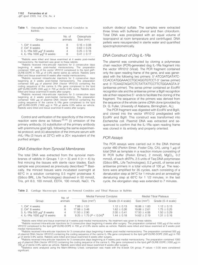
Figure 1. Femoral condyles from (A) a 4-week OA rabbit; (B) an 8-week OA rabbit injected with the control plasmid; (C) an 8-week OA rabbit treated with 500�g of IL-1Ra plasmid; (D) an 8-week OA rabbit treated with 1000 �g of IL-1Ra plasmid. Note the presence of osteophyte formations along the condylar ridge ofOA rabbits killed at either 4 weeks or 8 weeks postsurgery. The osteophytes were smaller in rabbits injected with the IL-1Ra plasmid.
IL-1Ra Gene Therapy in Osteoarthritis 1161AJP April 1999, Vol. 154, No. 4
Control and verification of the specificity of the immunereaction were done as follows:24,25 (i) omission of theprimary antibody; (ii) substitution of the primary antibodywith preimmune goat IgG, following the same experimen-tal protocol; and (iii) absorption of the immune serum withrhIL-1Ra (3 hours at 22°C) with a 20� equivalent of thepurified antigen.
DNA Extraction from Synovial Membranes
The total DNA was extracted from the synovial mem-branes of rabbits in Groups 1 (n � 3) and 4 (n � 4) byfirst mincing the tissues with sterile razor blades. Eachsample was processed as previously described.26 Basi-cally, the minced tissues were incubated overnight at65°C in a solution containing 0.5 mg/ml proteinase K(Gibco BRL, Life Technologies) dissolved in 50 mmol/LTris, pH 8.0, 100 mmol/L EDTA, 100 mmol/L NaCl, 1%
sodium dodecyl sulfate. The samples were extractedthree times with buffered phenol and then chloroform.Total DNA was precipitated with an equal volume ofisopropanol at room temperature and centrifuged. DNApellets were resuspended in sterile water and quantifiedspectrophotometrically.
DNA Construct of Dog IL-1Ra
The plasmid was constructed by cloning a polymerasechain reaction (PCR)-generated dog IL-1Ra fragment intothe vector VR1012 (Vical). The PCR fragment containedonly the open reading frame of the gene, and was gener-ated with the following two primers: 5�-ATCGATGATATC-CCACCATGGAAACCTGCAGGTGTCCT-3� (sense primer)and 5�-TCAAGTAGATCTCTATTATTCCTTCTGGAAGTA-3�(antisense primer). The sense primer contained an EcoRVrecognition site and the antisense primer a BglII recognitionsite at their respective 5�- ends to facilitate the cloning of thefragment. The sequence of the primers was derived fromthe sequence of the whole canine cDNA clone (provided byDr. G. Fuller, University of Alabama, Birmingham, AL).
The PCR fragment was digested with EcoRV and BglII,and cloned into the vector VR1012 predigested withEcoRV and BglII. This construct was transformed intoEscherichia coli. Plasmid DNA was extracted and se-quenced to confirm that the IL-1Ra open reading framewas cloned in its entirety and properly oriented.
PCR Assays
The PCR assays were carried out in the DNA thermalcycler 480 (Perkin Elmer, Foster City, CA), using 1 �g oftotal DNA as template in a reaction mixture consisting ofIX PCR buffer (Perkin Elmer), 2 mmol/L MgCl2, 0.2mmol/L of each dNTPs, 2.5 units of Taq DNA polymerase(Gibco BRL, Life Technologies), 0.2 �mol/L of sense andantisense primers in a total volume of 100 �l. The reac-tions were amplified for 35 cycles, each consisting of adenaturation step at 94°C for 1 minute and an annealing/denaturing step at 60°C for 1 1/2 minutes; in the lastcycle, the elongation step was extended to 7 minutes.
Table 1. Osteophyte Incidence on Femoral Condyles inRabbits
GroupNo. ofanimals
OsteophyteSize (mm)
1, OA* 4 weeks 8 0.16 � 0.082, OA† 8 weeks 8 0.63 � 0.243, IL-1Ra 500 �g‡ 8 weeks 4 0.25 � 0.164, IL-1Ra 1000 �g§ 8 weeks 8 0.41 � 0.17
*Rabbits were killed and tissue examined at 4 weeks post-medialmeniscectomy. No treatment was given to these rabbits.
†Rabbits received intra-articular injections for 3 consecutive daysbeginning 4 weeks post-medial meniscectomy. The preparationcontained 1000 �g of the vector VR1012 complexed to the lipid gAP-DLRIE-DOPE in 700 �l of 0.9% sterile saline as vehicle. Rabbits werekilled and tissue examined 8 weeks after medial meniscectomy.
‡Rabbits received intraarticular injections for 3 consecutive daysbeginning at 4 weeks post-medial meniscectomy. The preparationcontained 500 �g of plasmid DNA (Vector VR1012) containing thecoding sequence of the canine IL-1Ra gene complexed to the lipidgAP-DLRIE-DOPE (500 �g) in 700 �l sterile 0.9% saline. Rabbits werekilled and tissue examined 8 weeks after surgery.
§Rabbits received intra-articular injections for 3 consecutive daysbeginning at 4 weeks post-medial meniscectomy. The preparationcontained 1000 �g of plasmid DNA (Vector VR1012) containing thecoding sequence of the canine IL-1Ra gene complexed to the lipidgAP-DLRIE-DOPE (1000 �g) in 700 �l sterile 0.9% saline as vehicle.Rabbits were killed and tissue examined 8 weeks after surgery.
Table 2. Cartilage Macroscopic Lesions on Femoral Condyles and Tibial Plateaus in Rabbits
GroupNo. ofanimals
Medial Femoral Condyles Medial Tibial Plateaus
Size (mm2) Grade (0–4 scale) Size (mm2) Grade (0–4 scale)
1, OA* 4 weeks 8 7.88 � 1.51 1.12 � 0.13 16.88 � 1.83 1.12 � 0.132, OA† 8 weeks 8 17.88 � 3.48 1.62 � 0.26 19.88 � 2.61 1.75 � 0.253, IL-1Ra 500 �g‡ 8 weeks 4 11.50 � 2.90 2.00 � 0.41 17.50 � 4.06 1.50 � 0.294, IL-1Ra 1000 �g§ 8 weeks 8 9.25 � 1.75 (P � 0.04)¶ 1.44 � 0.18 14.62 � 2.19 1.31 � 0.16
*Rabbits were killed and tissue examined at 4 weeks post-medial meniscectomy. No treatment was given to these rabbits.†Rabbits received intra-articular injections for 3 consecutive days beginning 4 weeks after surgery. The preparation contained 1000 �g of the vector
VR1012 complexed to the lipid gAP-DLRIE-DOPE in 700 �l of 0.9% sterile saline as vehicle. Rabbits were killed and tissue examined at 8 weeks postmedial meniscectomy.
‡Rabbits received intra-articular injections for 3 consecutive days beginning 4 weeks post medial meniscectomy. The preparation contained 500 �gof plasmid DNA (Vector VR1012) containing the coding sequence of the canine IL-1Ra gene complexed to the lipid gAP-DLRIE-DOPE (500 �g) in 700�l of sterile 0.9% saline. Rabbits were killed and tissue examined 8 weeks after surgery.
§Rabbits received intra-articular injections for 3 consecutive days beginning at 4 weeks post medial meniscectomy. The preparation contained 1000�g of plasmid DNA (Vector VR1012) containing the coding sequence of the canine IL-1Ra gene complexed to the lipid gAP-DLRIE-DOPE (1000 �g) in700 �l of sterile 0.9% saline as vehicle. Rabbits were killed and tissue examined 8 weeks after surgery.
¶Statistics were analyzed using the Mann-Whitney U-test; comparison was made with the 8-week OA group. P values � 0.05 were consideredsignificant.
1162 Fernandes et alAJP April 1999, Vol. 154, No. 4
The sequences of the primers (5�-TGCAAGCCTTCA-GAATCTGG-3� sense, 127–146 bp, and 5�-TCGTCCTC-CTGGAAGTAGAA-3� antisense, 540–553 bp) were de-rived from the IL-1Ra cDNA sequence27 and the size ofthe specific amplified fragment was 427 bp. The primersare conserved between the dog and human genes andspan introns, so that amplification from the genomic DNAinstead of the transfected cloned cDNA would be de-tected. The amplified DNA were electrophoresed in 1.2%agarose gels and stained with ethidium bromide. Theidentity of the fragments amplified from the tissues wastested by digestion with enzymes with known sites in theamplified fragment.
Statistical Analysis
Mean values and SE were calculated and statistics wereanalyzed using Student’s t-test with P � 0.05 consideredsignificant.
Results
Macroscopy
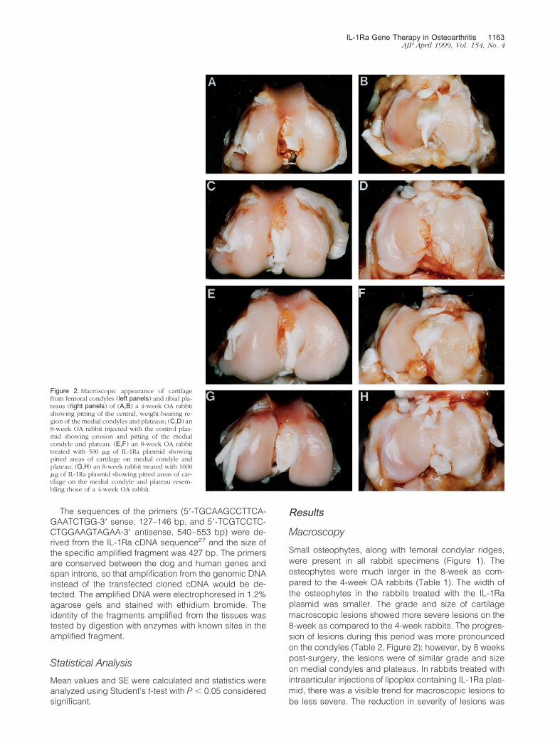
Small osteophytes, along with femoral condylar ridges,were present in all rabbit specimens (Figure 1). Theosteophytes were much larger in the 8-week as com-pared to the 4-week OA rabbits (Table 1). The width ofthe osteophytes in the rabbits treated with the IL-1Raplasmid was smaller. The grade and size of cartilagemacroscopic lesions showed more severe lesions on the8-week as compared to the 4-week rabbits. The progres-sion of lesions during this period was more pronouncedon the condyles (Table 2, Figure 2); however, by 8 weekspost-surgery, the lesions were of similar grade and sizeon medial condyles and plateaus. In rabbits treated withintraarticular injections of lipoplex containing IL-1Ra plas-mid, there was a visible trend for macroscopic lesions tobe less severe. The reduction in severity of lesions was
Figure 2. Macroscopic appearance of cartilagefrom femoral condyles (left panels) and tibial pla-teaus (right panels) of (A,B) a 4-week OA rabbitshowing pitting of the central, weight-bearing re-gion of the medial condyles and plateaus; (C,D) an8-week OA rabbit injected with the control plas-mid showing erosion and pitting of the medialcondyle and plateau; (E,F) an 8-week OA rabbittreated with 500 �g of IL-1Ra plasmid showingpitted areas of cartilage on medial condyle andplateau; (G,H) an 8-week rabbit treated with 1000�g of IL-1Ra plasmid showing pitted areas of car-tilage on the medial condyle and plateau resem-bling those of a 4-week OA rabbit.
IL-1Ra Gene Therapy in Osteoarthritis 1163AJP April 1999, Vol. 154, No. 4
dose-dependent, with the most marked and significant(P � 0.04) decrease observed on the femoral condylesof rabbits treated with the highest dosage of the plasmid(1000 �g; Table 2).
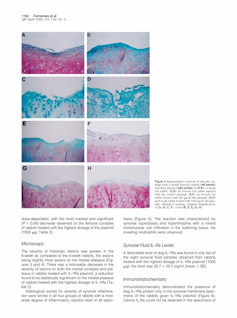
Microscopy
The severity of histologic lesions was greater in the8-week as compared to the 4-week rabbits, the lesionsbeing slightly more severe on the medial plateaus (Fig-ures 3 and 4). There was a noticeable decrease in theseverity of lesions on both the medial condyles and pla-teaus in rabbits treated with IL-1Ra plasmid, a reductionfound to be statistically significant on the medial plateausof rabbits treated with the highest dosage of IL-1Ra (Ta-ble 2).
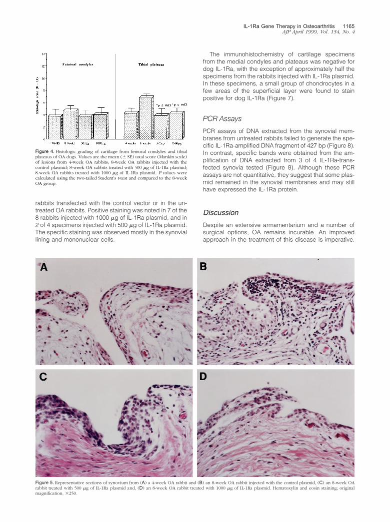
Histological scores for severity of synovial inflamma-tion were similar in all four groups of rabbits with a mod-erate degree of inflammatory reaction seen in all speci-
mens (Figure 5). The reaction was characterized bysynovial hyperplasia and hyperthrophia with a mixedmononuclear cell infiltration in the sublining tissue. Noinvading neutrophils were observed.
Synovial Fluid IL-Ra Levels
A detectable level of dog IL-1Ra was found in only two ofthe eight synovial fluid samples obtained from rabbitstreated with the highest dosage of IL-1Ra plasmid (1000�g); the level was 35.7 � 20.7 pg/ml (mean � SE).
Immunohistochemistry
Immunohistochemistry demonstrated the presence ofdog IL-1Ra protein only in the synovial membrane spec-imens of the rabbits given IL-1Ra plasmid (Figure 6).Canine IL-Ra could not be detected in the specimens of
Figure 3. Representative sections of articular car-tilage from a medial femoral condyle (left panels)and tibial plateau (right panels) of (A,B) a 4-weekOA rabbit; (C,D) an 8-week OA rabbit injectedwith the control plasmid; (E,F) an 8-week OArabbit treated with 500 �g IL-1Ra plasmid; (G,H)an 8-week rabbit treated with 1000 �g IL-1Ra plas-mid. safranin-O staining. Original magnification,�250 (A, C, F), �100 (B, D, E, G, H).
1164 Fernandes et alAJP April 1999, Vol. 154, No. 4
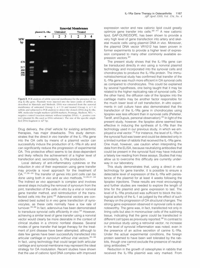
rabbits transfected with the control vector or in the un-treated OA rabbits. Positive staining was noted in 7 of the8 rabbits injected with 1000 �g of IL-1Ra plasmid, and in2 of 4 specimens injected with 500 �g of IL-1Ra plasmid.The specific staining was observed mostly in the synoviallining and mononuclear cells.
The immunohistochemistry of cartilage specimensfrom the medial condyles and plateaus was negative fordog IL-1Ra, with the exception of approximately half thespecimens from the rabbits injected with IL-1Ra plasmid.In these specimens, a small group of chondrocytes in afew areas of the superficial layer were found to stainpositive for dog IL-1Ra (Figure 7).
PCR Assays
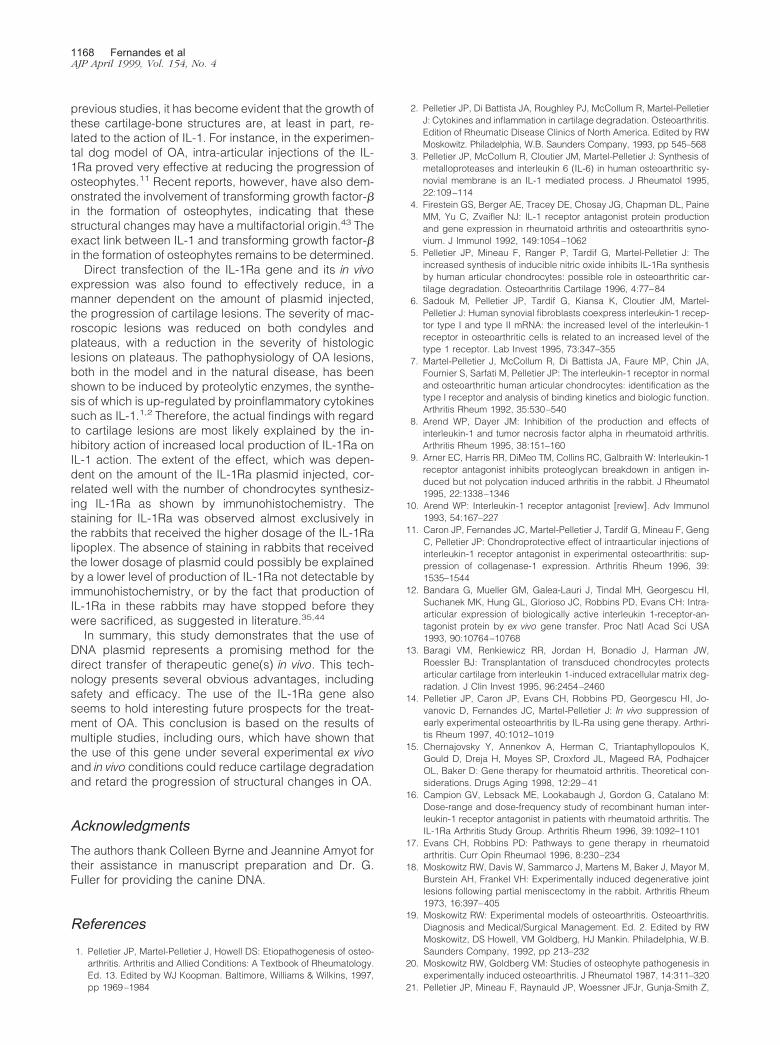
PCR assays of DNA extracted from the synovial mem-branes from untreated rabbits failed to generate the spe-cific IL-1Ra-amplified DNA fragment of 427 bp (Figure 8).In contrast, specific bands were obtained from the am-plification of DNA extracted from 3 of 4 IL-1Ra-trans-fected synovia tested (Figure 8). Although these PCRassays are not quantitative, they suggest that some plas-mid remained in the synovial membranes and may stillhave expressed the IL-1Ra protein.
DiscussionDespite an extensive armamentarium and a number ofsurgical options, OA remains incurable. An improvedapproach in the treatment of this disease is imperative.
Figure 4. Histologic grading of cartilage from femoral condyles and tibialplateaus of OA dogs. Values are the mean (� SE) total score (Mankin scale)of lesions from 4-week OA rabbits; 8-week OA rabbits injected with thecontrol plasmid; 8-week OA rabbits treated with 500 �g of IL-1Ra plasmid;8-week OA rabbits treated with 1000 �g of IL-1Ra plasmid. P values werecalculated using the two-tailed Student’s t-test and compared to the 8-weekOA group.
Figure 5. Representative sections of synovium from (A) a 4-week OA rabbit and (B) an 8-week OA rabbit injected with the control plasmid, (C) an 8-week OArabbit treated with 500 �g of IL-1Ra plasmid and, (D) an 8-week OA rabbit treated with 1000 �g of IL-1Ra plasmid. Hematoxylin and eosin staining; originalmagnification, �250.
IL-1Ra Gene Therapy in Osteoarthritis 1165AJP April 1999, Vol. 154, No. 4
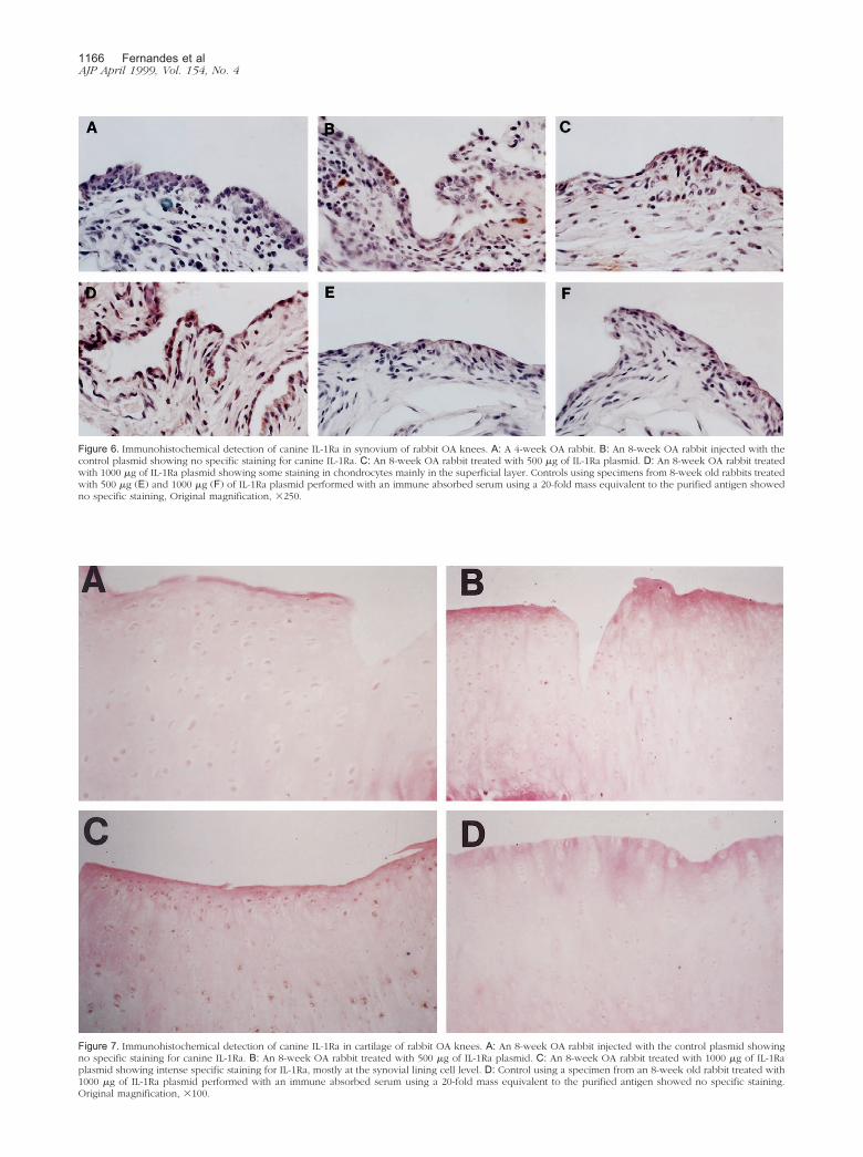
Figure 7. Immunohistochemical detection of canine IL-1Ra in cartilage of rabbit OA knees. A: An 8-week OA rabbit injected with the control plasmid showingno specific staining for canine IL-1Ra. B: An 8-week OA rabbit treated with 500 �g of IL-1Ra plasmid. C: An 8-week OA rabbit treated with 1000 �g of IL-1Raplasmid showing intense specific staining for IL-1Ra, mostly at the synovial lining cell level. D: Control using a specimen from an 8-week old rabbit treated with1000 �g of IL-1Ra plasmid performed with an immune absorbed serum using a 20-fold mass equivalent to the purified antigen showed no specific staining.Original magnification, �100.
Figure 6. Immunohistochemical detection of canine IL-1Ra in synovium of rabbit OA knees. A: A 4-week OA rabbit. B: An 8-week OA rabbit injected with thecontrol plasmid showing no specific staining for canine IL-1Ra. C: An 8-week OA rabbit treated with 500 �g of IL-1Ra plasmid. D: An 8-week OA rabbit treatedwith 1000 �g of IL-1Ra plasmid showing some staining in chondrocytes mainly in the superficial layer. Controls using specimens from 8-week old rabbits treatedwith 500 �g (E) and 1000 �g (F) of IL-1Ra plasmid performed with an immune absorbed serum using a 20-fold mass equivalent to the purified antigen showedno specific staining, Original magnification, �250.
1166 Fernandes et alAJP April 1999, Vol. 154, No. 4
Drug delivery, the chief vehicle for existing antiarthritictherapies, has major drawbacks. This study demon-strates that the direct in vivo transfer of the IL-1Ra geneinto the OA cells by means of a plasmid vector cansuccessfully induce the production of IL-1Ra in situ andcan significantly reduce the progression of experimentalOA. This protective effect seems to be dose-dependentand likely reflects the achievement of a higher level oftransfection and, secondarily, IL-1Ra production.
Local delivery of anti-inflammatory cytokines or theinduction in vivo of their expression using gene transfermay provide a novel approach for the treatment ofOA.17,28–30 The transfer of genes into joint cells can bedone using both in vivo and ex vivo methods.14,28,31–34
The indirect ex vivo approach is complex and involvesseveral steps including the removal of synovium from thejoint, transfection of the cells in vitro by a viral or nonviralgene transfer method, and reintroduction of the trans-fected cells into the joint.29 Adenoviral vectors are con-sidered best suited to in vivo gene transfection of syno-viocytes, as these cells normally have a low rate ofturnover.35,36 In fact, adenovirus has been demonstratedto transduce synovial cells efficiently in vivo.34 However,achieving a similar level of gene transfer using a nonviralvector would clearly be more desirable in the context ofclinical studies in a chronic nonfatal disease. Severalmodes of gene transfer that target therapy for the treat-ment of joint disease have been attempted, although todate few genes have been successfully transfected intoarticular cartilage or synovium without viral vectors.35–38
In fact, using technology that could target both articularcartilage and synovial membrane may represent the idealstrategy for OA modulation. Recent studies have shownthat the use of cationic lipid DNA complex with improved
expression vector and new cationic lipid could greatlyoptimize gene transfer into cells.39–41 A new cationiclipid, GAP-DLRIE/DOPE, has been shown to provide avery high level of gene transfection into artery and skel-etal muscle cells using plasmid DNA in vivo. Moreover,the plasmid DNA vector VR1012 has been proven informer experiments to provide a higher level of expres-sion compared to many other commonly available ex-pression vectors.40
The present study shows that the IL-1Ra gene canbe transduced directly in vivo using a nonviral plasmidtechnology and incorporated into the synovial cells andchondrocytes to produce the IL-1Ra protein. The immu-nohistochemical study has confirmed that transfer of theIL-1Ra gene was much more efficient in OA synovial cellsas compared to chondrocytes. This could be explainedby several hypotheses, one being taught that it may berelated to the higher replicating rate of synovial cells. Onthe other hand, the diffusion rate of the lipoplex into thecartilage matrix may be another factor responsible forthe much lower level of cell transfection. In vitro experi-ments in cell culture have also demonstrated that thetransfection of the IL-1Ra gene in chondrocytes usinglipoplex was less efficient than in synovial cells (Pelletier,Tardif, and Dupuis, personal observation).35 In light of thepresent study, however, the lipoplex alone seemed lesseffective in inducing the synthesis of IL-1Ra than thetechnology used in our previous study, in which we em-ployed a viral vector.14 For instance, the level of IL-1Ra inthe synovial fluid was lower and could be detected only ina limited number of rabbits that received the IL-1Ra gene.One must, however, use caution when interpreting thedata from the ELISA, because neutralizing antibodies thatcould be present in the synovial fluid may have induceda falsely low reading from these assays. Studies that mayallow us to overcome this difficulty are currently under-way in our laboratory.
This study demonstrates that, using a direct in vivotechnology for gene transfer, it is possible to ensure adetectable level of expression of the IL-1Ra with persis-tence of the plasmid for at least 4 weeks following thelipoplex injections. These results are most encouragingand further studies are needed to explore the length oftime for the plasmid and gene expression to last. Thelevel of IL-1Ra produced was sufficient to block the bio-logical activity of the IL-1 as judged by the effect of suchtherapy on the progression of OA structural changes. Thestrong gene expression observed in synovial cells is alsonoteworthy. The gene was, in fact, transfected not only inlining cells but also in mononuclear cells of the subliningtissue, indicating that the gene could be transfected indifferent cell types as previously reported.38 In contrast toour previous study using a retroviral vector, no increasein the level of synovial inflammation was noted, even inthe presence of an active secretion of canine IL-1Ra.Under the actual experimental conditions, the foreignprotein seemed to have been well tolerated by the rab-bits, though one cannot exclude the presence of neutral-izing antibodies.42
Reduction in the growth of osteophytes in rabbits thatreceived the IL-1Ra plasmid was very marked. From
Figure 8. PCR analysis of rabbit synovial membranes for the presence of thedog IL-1Ra gene. Plasmids were injected into the knee joints of rabbits asdescribed in Materials and Methods. DNA was extracted from the synovialmembranes of untreated (Group 1; n � 3) and treated (Group 4; n � 4)rabbits and subjected to PCR analysis to detect the cloned cDNA dog IL-1Ra.MW, molecular weight markers (pGEM markers, Promega, Madison, WI); �,negative control (reaction mixture without template DNA); �, positive con-trol (plasmid IL-1Ra used as DNA substrate). The size of the specific ampli-fied DNA fragment is 427 bp.
IL-1Ra Gene Therapy in Osteoarthritis 1167AJP April 1999, Vol. 154, No. 4
previous studies, it has become evident that the growth ofthese cartilage-bone structures are, at least in part, re-lated to the action of IL-1. For instance, in the experimen-tal dog model of OA, intra-articular injections of the IL-1Ra proved very effective at reducing the progression ofosteophytes.11 Recent reports, however, have also dem-onstrated the involvement of transforming growth factor-�in the formation of osteophytes, indicating that thesestructural changes may have a multifactorial origin.43 Theexact link between IL-1 and transforming growth factor-�in the formation of osteophytes remains to be determined.
Direct transfection of the IL-1Ra gene and its in vivoexpression was also found to effectively reduce, in amanner dependent on the amount of plasmid injected,the progression of cartilage lesions. The severity of mac-roscopic lesions was reduced on both condyles andplateaus, with a reduction in the severity of histologiclesions on plateaus. The pathophysiology of OA lesions,both in the model and in the natural disease, has beenshown to be induced by proteolytic enzymes, the synthe-sis of which is up-regulated by proinflammatory cytokinessuch as IL-1.1,2 Therefore, the actual findings with regardto cartilage lesions are most likely explained by the in-hibitory action of increased local production of IL-1Ra onIL-1 action. The extent of the effect, which was depen-dent on the amount of the IL-1Ra plasmid injected, cor-related well with the number of chondrocytes synthesiz-ing IL-1Ra as shown by immunohistochemistry. Thestaining for IL-1Ra was observed almost exclusively inthe rabbits that received the higher dosage of the IL-1Ralipoplex. The absence of staining in rabbits that receivedthe lower dosage of plasmid could possibly be explainedby a lower level of production of IL-1Ra not detectable byimmunohistochemistry, or by the fact that production ofIL-1Ra in these rabbits may have stopped before theywere sacrificed, as suggested in literature.35,44
In summary, this study demonstrates that the use ofDNA plasmid represents a promising method for thedirect transfer of therapeutic gene(s) in vivo. This tech-nology presents several obvious advantages, includingsafety and efficacy. The use of the IL-1Ra gene alsoseems to hold interesting future prospects for the treat-ment of OA. This conclusion is based on the results ofmultiple studies, including ours, which have shown thatthe use of this gene under several experimental ex vivoand in vivo conditions could reduce cartilage degradationand retard the progression of structural changes in OA.
AcknowledgmentsThe authors thank Colleen Byrne and Jeannine Amyot fortheir assistance in manuscript preparation and Dr. G.Fuller for providing the canine DNA.
References
1. Pelletier JP, Martel-Pelletier J, Howell DS: Etiopathogenesis of osteo-arthritis. Arthritis and Allied Conditions: A Textbook of Rheumatology.Ed. 13. Edited by WJ Koopman. Baltimore, Williams & Wilkins, 1997,pp 1969–1984
2. Pelletier JP, Di Battista JA, Roughley PJ, McCollum R, Martel-PelletierJ: Cytokines and inflammation in cartilage degradation. Osteoarthritis.Edition of Rheumatic Disease Clinics of North America. Edited by RWMoskowitz. Philadelphia, W.B. Saunders Company, 1993, pp 545–568
3. Pelletier JP, McCollum R, Cloutier JM, Martel-Pelletier J: Synthesis ofmetalloproteases and interleukin 6 (IL-6) in human osteoarthritic sy-novial membrane is an IL-1 mediated process. J Rheumatol 1995,22:109–114
4. Firestein GS, Berger AE, Tracey DE, Chosay JG, Chapman DL, PaineMM, Yu C, Zvaifler NJ: IL-1 receptor antagonist protein productionand gene expression in rheumatoid arthritis and osteoarthritis syno-vium. J Immunol 1992, 149:1054–1062
5. Pelletier JP, Mineau F, Ranger P, Tardif G, Martel-Pelletier J: Theincreased synthesis of inducible nitric oxide inhibits IL-1Ra synthesisby human articular chondrocytes: possible role in osteoarthritic car-tilage degradation. Osteoarthritis Cartilage 1996, 4:77–84
6. Sadouk M, Pelletier JP, Tardif G, Kiansa K, Cloutier JM, Martel-Pelletier J: Human synovial fibroblasts coexpress interleukin-1 recep-tor type I and type II mRNA: the increased level of the interleukin-1receptor in osteoarthritic cells is related to an increased level of thetype 1 receptor. Lab Invest 1995, 73:347–355
7. Martel-Pelletier J, McCollum R, Di Battista JA, Faure MP, Chin JA,Fournier S, Sarfati M, Pelletier JP: The interleukin-1 receptor in normaland osteoarthritic human articular chondrocytes: identification as thetype I receptor and analysis of binding kinetics and biologic function.Arthritis Rheum 1992, 35:530–540
8. Arend WP, Dayer JM: Inhibition of the production and effects ofinterleukin-1 and tumor necrosis factor alpha in rheumatoid arthritis.Arthritis Rheum 1995, 38:151–160
9. Arner EC, Harris RR, DiMeo TM, Collins RC, Galbraith W: Interleukin-1receptor antagonist inhibits proteoglycan breakdown in antigen in-duced but not polycation induced arthritis in the rabbit. J Rheumatol1995, 22:1338–1346
10. Arend WP: Interleukin-1 receptor antagonist [review]. Adv Immunol1993, 54:167–227
11. Caron JP, Fernandes JC, Martel-Pelletier J, Tardif G, Mineau F, GengC, Pelletier JP: Chondroprotective effect of intraarticular injections ofinterleukin-1 receptor antagonist in experimental osteoarthritis: sup-pression of collagenase-1 expression. Arthritis Rheum 1996, 39:1535–1544
12. Bandara G, Mueller GM, Galea-Lauri J, Tindal MH, Georgescu HI,Suchanek MK, Hung GL, Glorioso JC, Robbins PD, Evans CH: Intra-articular expression of biologically active interleukin 1-receptor-an-tagonist protein by ex vivo gene transfer. Proc Natl Acad Sci USA1993, 90:10764–10768
13. Baragi VM, Renkiewicz RR, Jordan H, Bonadio J, Harman JW,Roessler BJ: Transplantation of transduced chondrocytes protectsarticular cartilage from interleukin 1-induced extracellular matrix deg-radation. J Clin Invest 1995, 96:2454–2460
14. Pelletier JP, Caron JP, Evans CH, Robbins PD, Georgescu HI, Jo-vanovic D, Fernandes JC, Martel-Pelletier J: In vivo suppression ofearly experimental osteoarthritis by IL-Ra using gene therapy. Arthri-tis Rheum 1997, 40:1012–1019
15. Chernajovsky Y, Annenkov A, Herman C, Triantaphyllopoulos K,Gould D, Dreja H, Moyes SP, Croxford JL, Mageed RA, PodhajcerOL, Baker D: Gene therapy for rheumatoid arthritis. Theoretical con-siderations. Drugs Aging 1998, 12:29–41
16. Campion GV, Lebsack ME, Lookabaugh J, Gordon G, Catalano M:Dose-range and dose-frequency study of recombinant human inter-leukin-1 receptor antagonist in patients with rheumatoid arthritis. TheIL-1Ra Arthritis Study Group. Arthritis Rheum 1996, 39:1092–1101
17. Evans CH, Robbins PD: Pathways to gene therapy in rheumatoidarthritis. Curr Opin Rheumaol 1996, 8:230–234
18. Moskowitz RW, Davis W, Sammarco J, Martens M, Baker J, Mayor M,Burstein AH, Frankel VH: Experimentally induced degenerative jointlesions following partial meniscectomy in the rabbit. Arthritis Rheum1973, 16:397–405
19. Moskowitz RW: Experimental models of osteoarthritis. Osteoarthritis.Diagnosis and Medical/Surgical Management. Ed. 2. Edited by RWMoskowitz, DS Howell, VM Goldberg, HJ Mankin. Philadelphia, W.B.Saunders Company, 1992, pp 213–232
20. Moskowitz RW, Goldberg VM: Studies of osteophyte pathogenesis inexperimentally induced osteoarthritis. J Rheumatol 1987, 14:311–320
21. Pelletier JP, Mineau F, Raynauld JP, Woessner JFJr, Gunja-Smith Z,
1168 Fernandes et alAJP April 1999, Vol. 154, No. 4
Martel-Pelletier J: Intraarticular injections with methylprednisolone ac-etate reduce osteoarthritic lesions in parallel with chondrocytestromelysin synthesis in experimental osteoarthritis. Arthritis Rheum1994, 37:414–423
22. Mankin HJ, Dorfman H, Lippiello L, Zarins A: Biochemical and met-abolic abnormalities in articular cartilage from osteoarthritic humanhips. II. Correlation of morphology with biochemical and metabolicdata. J Bone Joint Surg Am 1971, 53:523–537
23. Pelletier JP, Martel-Pelletier J, Ghandur-Mnaymneh L, Howell DS,Woessner JF Jr: Role of synovial membrane inflammation in cartilagematrix breakdown in the Pond-Nuki dog model of osteoarthritis. Ar-thritis Rheum 1985, 28:554–561
24. Moldovan F, Pelletier JP, Hambor J, Cloutier JM, Martel-Pelletier J:Collagenase-3 (matrix metalloprotease 13) is preferentially localizedin the deep layer of human arthritic cartilage in situ: in vitro mimickingeffect by transforming growth factor �. Arthritis Rheum 1997, 40:1653–1661
25. Pelletier JP, Faure MP, Di Battista JA, Wilhelm S, Visco D, Martel-Pelletier J: Coordinate synthesis of stromelysin, interleukin-1, andoncogene proteins in experimental osteoarthritis: an immunohisto-chemical study. Am J Pathol 1993, 142:95–105
26. Parker SE, Khatibi S, Margalith M, Anderson D, Yankauckas M,Gromkowski SH, Latimer T, Lew D, Marquet M, Manthorpe M, HobartP, Hersh E, Stopeck AT, Norman J: Plasmid DNA gene therapy:studies with the human interleukin-2 gene in tumor cells in vitro and inthe murine B16 melanoma model in vivo. Cancer Gene Ther 1996,3:175–185
27. Eisenberg SP, Evares RJ, Arend WP, Verderber E, Brewer MT, Han-num CH, Thompson RC: Primary structure and functional expressionof complimentary DNA of a human interleukin-1 receptor antagonist.Nature 1990, 343:341–346
28. Evans CH, Robbins PD: Gene therapy for arthritis. GeneTherapeutics: Methods and Applications of Direct Gene Transfer.Edited by JA Wolff. Boston, Birkhauser, 1994, pp 320–343
29. Evans C, Robbins PD: Prospects for treating arthritis by gene therapy[editorial]. J Rheumatol 1994, 21:779–782
30. Bandara G, Robbins PD, Georgescu HI, Mueller GM, Glorioso JC,Evans CH: Gene transfer to synoviocytes: prospects for gene treat-ment for arthritis. DNA Cell Biol 1992, 11:227–231
31. Otani K, Nita IM, Macaulay W, Georgescu HI, Robbins PD, Evans CH:Suppression of antigen-induced arthritis in rabbits by ex vivo genetherapy. J Immunol 1996, 156:3558–3562
32. Makarov SS, Olsen JC, Johnston WN, Schwab JH, Anderle SK, BrownRR, Haskill JS: Retrovirus mediated in vivo gene transfer to synoviumin bacterial cell wall-induced arthritis in rats. Gene Ther 1995, 2:424–428
33. Hung GL, Galea-Lauri J, Mueller GM, Georgescu HI, Larkin LA,Suchanek MK, Tindal MH, Robbins PD, Evans CH: Suppression ofintraarticular responses to interleukin-1 by transfer of the interleukin-1receptor antagonist gene to synovium. Gene Ther 1994, 1:64–69
34. Roessler BJ, Allen ED, Wilson JM, Hartman JW, Davidson BL: Ad-enoviral-mediated gene transfer to rabbit synovium in vivo. J ClinInvest 1993, 92:1085–1092
35. Nita IM, Ghivizzani SC, Galea-Lauri J, Bandara G, Georgescu HI,Robbins PD, Evans CH: Direct gene delivery to synovium. An evalu-ation of potential vectors in vitro and in vivo. Arthritis Rheum 1996,39:820–828
36. Tomita N, Morishita R, Higaki J, Tomita S, Aoki M, Ogihara T, KanedaY: In vivo gene transfer to insulin gene into neonatal rats by theHVJ-liposome method resulted in sustained transgene expression.Gene Ther 1996, 3:477–482
37. Tomita T, Hashimoto H, Tomita N, Morishita R, Lee SB, Hayashida K,Nakamura N, Yonenobu K, Kaneda Y, Ochi T: In vivo direct genetransfer into articular cartilage by intraarticular injection mediatedby HVJ (sendai virus) and liposomes. Arthritis Rheum 1997, 40(5):901–906
38. Yovandich J, O’Malley B, Sikes M, Ledley FD: Gene transfer tosynovial cells by intra-articular administration of plasmid DNA. HumGene Ther 1995, 6:603–610
39. Budker V, Zhang G, Danko I, Williams P, Wolff J: The efficient expres-sion of intravascularly delivered DNA in rat muscle. Gene Ther 1998,5:272–276
40. Hartikka I, Sawdey M, Cornefert-Jensen F, Margalith M, Barnhart K,Nolasco M, Vahlsing HL, Meek J, Marquet M, Hobart P, Norman J,Manthorpe M: An improved plasmid DNA expression vector for directinjection into skeletal muscle. Hum Gene Ther 1996, 7:1205–1217
41. Stephan DJ, Yang ZY, San H, Simari RD, Wheeler CJ, Felgner PL,Gordon D, Nabel GJ, Nabel EG: A new cationic liposome DNAcomplex enhances the efficiency of arterial gene transfer in vivo. HumGene Ther 1996, 7:1803–1812
42. Tripathy SK, Black HB, Goldwasser E, Leiden JM: Immune responsesto transgene-encoded proteins limit the stability of gene expressionafter injection of replication-defective adenovirum vectors. Nat Med1996, 2:545–550
43. Glansbeek HL, Van Beuningen HM, Vitters EL, Van der Kraan PM, vanden Berg WB: Stimulation of articular cartilage repair in establishedarthritis by local administration of transforming growth factor-� intomurine knee joints. Lab Invest 1998, 78:133–142
44. Palmer TD, Rosman GJ, Osborne WR, Miller AD: Genetically modifiedskin fibroblasts persist long after transplantation but gradually inac-tivate introduced genes. Proc Natl Acad Sci USA 1996, 88:1330–1334
IL-1Ra Gene Therapy in Osteoarthritis 1169AJP April 1999, Vol. 154, No. 4