A Three-Dimensional Gel Bioreactor for Assessment of Cardiomyocyte Induction in Skeletal...
12
A Three-Dimensional Gel Bioreactor for Assessment of Cardiomyocyte Induction in Skeletal Muscle–Derived Stem Cells Kelly C. Clause, B.S., 1,2 Joseph P. Tinney, B.S., 1,3 Li J. Liu, M.D., 1,3 Burhan Gharaibeh, Ph.D., 4 Johnny Huard, Ph.D., 4 Jonathan A. Kirk, Ph.D., 2 Sanjeev G. Shroff, Ph.D., 2 Kazuro L. Fujimoto, M.D., 5 William R. Wagner, Ph.D., 2,5 John C. Ralphe, M.D., 1,3 Bradley B. Keller, M.D., 1–3 and Kimimasa Tobita, M.D. 1–3 Skeletal muscle–derived stem cells (MDSCs) are able to differentiate into cardiomyocytes (CMs). However, it remains to be investigated whether differentiated CMs contract similar to native CMs. Here, we developed a three-dimensional collagen gel bioreactor (3DGB) that induces a working CM phenotype from MDSCs, and the contractile properties are directly measured as an engineered cardiac tissue. Neonate rat MDSCs were isolated from hind-leg muscles via the preplate technique. Isolated MDSCs were approximately 60% positive to Sca-1 and negative to CD34, CD45, or c-kit antigens. We sorted Sca-1() MDSCs and constructed MDSC-3DGBs by mixing MDSCs with acid soluble rat tail collagen type-I and matrix factors. MDSC-3DGB exhibited spontaneous cyclic contraction by culture day 7. MDSC-3DGB expressed cardiac-specific genes and proteins. Histological assessment revealed that cardiac-specific troponin-T and -I expressed in a typical striation pattern and connexin- 43 was expressed similar to the native fetal ventricular papillary muscle. b-Adrenergic stimulation increased MDSC-3DGB spontaneous beat frequency. MDSC-3DGB generated contractile force and intracellular calcium ion transients similar to engineered cardiac tissue from native cardiac cells. Results suggest that MDSC-3DGB induces a working CM phenotype in MDSCs and is a useful 3D culture system to directly assess the contractile properties of differentiated CMs in vitro. Introduction L imited cardiomyocyte (CM) proliferative capacity is a major barrier to myocardial regeneration and the resto- ration of contractile function of injured postnatal myocar- dium. Various strategies to restore postnatal CM proliferation and myocardial regeneration are under investigation, in- cluding strategies for the repair and regeneration of damaged myocardium using allogeneic and autologous cell and tissue grafts. 1 Studies suggest that fetal, finitely proliferating CMs display the best cell survival, functional integration, and sustained cardiac recovery, and thus could be an optimal cell type for cardiac repair. 2,3 However, the use of fetal heart cells is contraindicated for clinical use. Stem cells provide an al- ternative solution, and a range of cell types have been em- ployed in cellular cardiomyoplasty strategies, including bone marrow–derived stromal and stem cells, fibroblasts, skeletal myoblasts, mesenchymal stem cells, embryonic stem cells, and resident cardiac stem cells. 4 However, despite some promising results, 5 the rate of CM differentiation from transplanted stem cells remains insufficient to fully recover the recipient myocardial function. 2,4, 6–8 Therefore, a preferred strategy for cellular cardiomyoplasty might be the delivery of progenitor=stem cell–derived CMs, rather than undifferenti- ated cells, into injured myocardial tissue. 9 Skeletal muscle–derived stem cells (MDSCs) are a somatic stem cell population obtained from skeletal muscle speci- mens in animals and humans that can be readily expanded in vitro and then transplanted as an autologous graft. 8,10,11 MDSCs are multipotent and have been shown to differenti- ate along skeletal and smooth muscle, bone, tendon, nerve, endothelial, and hematopoietic lineages. 10,12,13 Previous studies, including our own work, have shown that MDSCs, isolated using variations of a modified preplate technique, can differentiate into CMs or cells with cardiac phenotypes and can facilitate cardiac repair. 8,14–20 However, none of the previous studies investigated whether differentiated MDSC- derived CMs generate contractile force similar to the native CMs. In addition, undifferentiated MDSCs or MDSCs pre- conditioned with chemical reagents were used to evaluate 1 Cardiovascular Development Research Program, Children’s Hospital of Pittsburgh of UPMC, Pittsburgh, Pennsylvania. Departments of 2 Bioengineering, 3 Pediatrics, 4 Orthopedic Surgery, and 5 Surgery, University of Pittsburgh, Pittsburgh, Pennsylvania. TISSUE ENGINEERING: Part C Volume 16, Number 3, 2010 ª Mary Ann Liebert, Inc. DOI: 10.1089=ten.tec.2009.0098 375
Transcript of A Three-Dimensional Gel Bioreactor for Assessment of Cardiomyocyte Induction in Skeletal...
A Three-Dimensional Gel Bioreactorfor Assessment of Cardiomyocyte Induction
in Skeletal Muscle–Derived Stem Cells
Kelly C. Clause, B.S.,1,2 Joseph P. Tinney, B.S.,1,3 Li J. Liu, M.D.,1,3 Burhan Gharaibeh, Ph.D.,4
Johnny Huard, Ph.D.,4 Jonathan A. Kirk, Ph.D.,2 Sanjeev G. Shroff, Ph.D.,2
Kazuro L. Fujimoto, M.D.,5 William R. Wagner, Ph.D.,2,5 John C. Ralphe, M.D.,1,3
Bradley B. Keller, M.D.,1–3 and Kimimasa Tobita, M.D.1–3
Skeletal muscle–derived stem cells (MDSCs) are able to differentiate into cardiomyocytes (CMs). However, itremains to be investigated whether differentiated CMs contract similar to native CMs. Here, we developed athree-dimensional collagen gel bioreactor (3DGB) that induces a working CM phenotype from MDSCs, and thecontractile properties are directly measured as an engineered cardiac tissue. Neonate rat MDSCs were isolatedfrom hind-leg muscles via the preplate technique. Isolated MDSCs were approximately 60% positive to Sca-1and negative to CD34, CD45, or c-kit antigens. We sorted Sca-1(�) MDSCs and constructed MDSC-3DGBs bymixing MDSCs with acid soluble rat tail collagen type-I and matrix factors. MDSC-3DGB exhibited spontaneouscyclic contraction by culture day 7. MDSC-3DGB expressed cardiac-specific genes and proteins. Histologicalassessment revealed that cardiac-specific troponin-T and -I expressed in a typical striation pattern and connexin-43 was expressed similar to the native fetal ventricular papillary muscle. b-Adrenergic stimulation increasedMDSC-3DGB spontaneous beat frequency. MDSC-3DGB generated contractile force and intracellular calciumion transients similar to engineered cardiac tissue from native cardiac cells. Results suggest that MDSC-3DGBinduces a working CM phenotype in MDSCs and is a useful 3D culture system to directly assess the contractileproperties of differentiated CMs in vitro.
Introduction
Limited cardiomyocyte (CM) proliferative capacity is amajor barrier to myocardial regeneration and the resto-
ration of contractile function of injured postnatal myocar-dium. Various strategies to restore postnatal CM proliferationand myocardial regeneration are under investigation, in-cluding strategies for the repair and regeneration of damagedmyocardium using allogeneic and autologous cell and tissuegrafts.1 Studies suggest that fetal, finitely proliferating CMsdisplay the best cell survival, functional integration, andsustained cardiac recovery, and thus could be an optimal celltype for cardiac repair.2,3 However, the use of fetal heart cellsis contraindicated for clinical use. Stem cells provide an al-ternative solution, and a range of cell types have been em-ployed in cellular cardiomyoplasty strategies, including bonemarrow–derived stromal and stem cells, fibroblasts, skeletalmyoblasts, mesenchymal stem cells, embryonic stem cells,and resident cardiac stem cells.4 However, despite somepromising results,5 the rate of CM differentiation from
transplanted stem cells remains insufficient to fully recoverthe recipient myocardial function.2,4, 6–8 Therefore, a preferredstrategy for cellular cardiomyoplasty might be the delivery ofprogenitor=stem cell–derived CMs, rather than undifferenti-ated cells, into injured myocardial tissue.9
Skeletal muscle–derived stem cells (MDSCs) are a somaticstem cell population obtained from skeletal muscle speci-mens in animals and humans that can be readily expandedin vitro and then transplanted as an autologous graft.8,10,11
MDSCs are multipotent and have been shown to differenti-ate along skeletal and smooth muscle, bone, tendon, nerve,endothelial, and hematopoietic lineages.10,12,13 Previousstudies, including our own work, have shown that MDSCs,isolated using variations of a modified preplate technique,can differentiate into CMs or cells with cardiac phenotypesand can facilitate cardiac repair.8,14–20 However, none of theprevious studies investigated whether differentiated MDSC-derived CMs generate contractile force similar to the nativeCMs. In addition, undifferentiated MDSCs or MDSCs pre-conditioned with chemical reagents were used to evaluate
1Cardiovascular Development Research Program, Children’s Hospital of Pittsburgh of UPMC, Pittsburgh, Pennsylvania.Departments of 2Bioengineering, 3Pediatrics, 4Orthopedic Surgery, and 5Surgery, University of Pittsburgh, Pittsburgh, Pennsylvania.
TISSUE ENGINEERING: Part CVolume 16, Number 3, 2010ª Mary Ann Liebert, Inc.DOI: 10.1089=ten.tec.2009.0098
375
in vivo CM differentiation from MDSCs, not transplantedMDSC-derived CMs in animal models of injured myocar-dium.1,5 Our recent studies have also shown that the rate ofCM differentiation of transplanted MDSCs within acutemyocardial infarction model is not sufficient to replace in-jured CMs, and improvement of recipient cardiac function byMDSC transplantation is due to combined effects of myo-genic differentiation of transplanted MDSC, angiogenesis,stimulation of recipient CM proliferation, and reduction ofrecipient CM apoptosis.8,20 Therefore, it remains unclearwhether differentiated CM phenotypic cells replace recipientdead CMs and have contractile function that improves re-cipient cardiac function.
The effects of various biomechanical stimuli on multi-potent stem cells have been investigated to elucidate theirroles in CM induction and differentiation. Previous studieshave shown that cell–cell interactions and specific cultureconditions are often necessary prerequisites for efficient CMdifferentiation.21–23 The culture of multipotent stem cells inaggregate spheres has been shown to facilitate cell–cell cou-pling, increase differentiation capacity, modify cellular me-tabolism, modulate the response to therapeutic agents,23–25
and stimulate the synthesis and release of extracellular ma-trix (ECM) constituents.26 Studies also suggest that three-dimensional (3D) in vitro culture conditions may be optimalfor donor CM preparation,2,3,27 and that tissue-engineeredcardiac tissue constructs provide the requisite 3D environ-ment for efficient cell survival, functional integration, andsustained cardiac recovery.27,28
Thus, the objective of the present study was to develop aculture method that induces CM differentiation from MDSCsand facilitates myocardial tissue formation that enables us todirectly evaluate the contractile properties of MDSC-derivedCMs in vitro. We tested the hypothesis that the combinedMDSC-aggregate formation and 3D collagen gel bioreactorcan induce MDSCs to differentiate into cells with a CMphenotype and that the differentiated CMs form a working3D cardiac like tissue in vitro. We determined that MDSC-aggregate formation followed by 3D collagen gel bioreactor(3DGB) culture succeeded in generating cells with an im-mature CM phenotype mimicking the native fetal myocar-dium. Thus, our results suggest that MDSC-3DGB is a useful3D culture system to directly assess the contractile propertiesof differentiated CMs from MDSCs in vitro.
Materials and Methods
MDSC isolation
MDSCs were isolated from neonatal Lewis rat hind-legmuscles using an established preplate technique.10–12,29–31
Briefly, five postnatal day-3 rat pups were euthanized with5% isoflurane anesthesia followed by cervical truncation.Gastrocnemius muscles were excised, minced in HBSS, andenzymatically digested. Briefly, tissue slurry underwent se-quential incubation of collagnease XI (0.2% collagenase XI),dispase (2.4 U=mL), and 2� trypsin. Isolated cells then un-derwent sequential preplating (24-h interval between eachpreplate) until the sixth preplate phase31 in standard MDSCgrowth medium containing high-glucose Dulbecco’s modi-fied Eagle’s medium w=l-glutamine (Invitrogen, Carlsbad,CA), 10% horse serum (Invitrogen), 10% fetal bovine serum(FBS; Invitrogen), 0.5% chick embryo extract (US Biological,
Swampscott, MA), and 1% antibiotic–antimycotic solution(Invitrogen).26,31,32 Our research protocol followed theNational Institutes of Health (NIH) guidelines for animalcare and was approved by the University of Pittsburgh’sInstitutional Animal Care and Use Committee and theChildren’s Hospital of Pittsburgh Animal Research CareCommittee.
Isolated rat MDSCs were passaged 15 times and expandedto obtain enough cell number (at least 20 million cells permL).12,29 Fluorescent-activated cell sorting (FACS, FACSAria; BD Biosciences, San Jose, CA) revealed that cultured ratMDSCs were approximately 60% positive to Sca-1 antigen(BD Biosciences), and negative to CD34 (BD Biosciences),CD45 (BD Biosciences), and c-kit (BD Biosciences) antigens(Supplemental Fig. S1, available online at www.liebertonline.com=ten; Table 1). Zuba-Surma et al. have shown that Sca-1(�) skeletal muscle stem cells are ‘‘inherently predisposed toundergo cardiac differentiation.15’’ Thus, we sorted Sca-1(�)MDSCs that were expanded in standard two-dimensional(2D) flasks for 48 h to reach a cell number of 3 million. TheseSca-1(�) MDSCs maintained no Sca-1 expression throughoutculture (Supplemental Fig. S2, available online at www.liebertonline.com).
MDSC-3D collagen gel bioreactor(MDSC-3DGB) construction
Expanded MDSCs on the 2D flask were trypsinized usinga 0.05% trypsin–EDTA solution (Invitrogen), and the cellsuspension was cultured on a 100-mm-diameter suspensionculture dish (Corning, Lowell, MA) for 24 h at 378C using agyrating shaker (50 rotations=min) to form 50- to 70-mm-diameter MDSC aggregates (MDSC-aggregate, 330–350cells=aggregate) under standard MDSC growth medium.Acid-soluble rat tail collagen type-I solution (pH 3; Sigma,St. Louis, MO) was neutralized with alkali buffer (0.2 MNaHCO3, 0.2 M HEPES, and 0.1 M NaOH) on ice. Matrigel(13% of total volume; BD Biosciences) was then added, andthe cell suspension and matrix solution mixed to reach afinal collagen type-I concentration of 0.67 mg=mL. Approxi-mately 200mL of the cell=matrix mixture was poured into the20-mm-long�2-mm-wide cylindrical cast of a Flexcell TissueTrain collagen type-I–coated silicone membrane culture plate
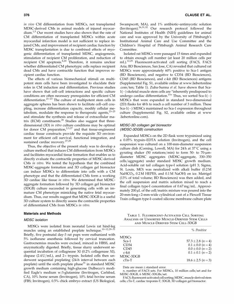
Table 1. Fluorescent-Activated Cell Sorting
Analysis of Unsorted Muscle-Derived Stem Cells
and Muscle-Derived Stem Cell-3DGB
% Positive
MDSCsSca-1 57.3� 2.8 (n¼ 4)CD34 0.1� 0.0 (n¼ 4)CD45 0.0� 0.0 (n¼ 2)c-kit 0.1� 0.1 (n¼ 2)
MDSC-3DGBcTn-T 18.6� 2.5 (n¼ 3)
Data are mean� standard error.n, number of FACS sets. For MDSCs, 10 million cells=set and for
MDSC-3DGB, 6 MDSC-3DGBs=set.FACS, fluorescent-activated cell sorting; MDSC, muscle-derived stem
cells; cTn-T, cardiac troponin-T; 3DGB, 3D collagen gel bioreactor.
376 CLAUSE ET AL.
(FX4000TT; Flexcell International, Hillsborough, NC) andincubated for 120 min (378C, 5% CO2)32 to form a cylindricalMDSC-3DGB. Each MDSC-3DGB was cultured in a 5% FBScontaining growth medium. We compared the efficiency ofCM induction from culture day 7 MDSC-3DGBs to (1) freshlyformed MDSC-aggregates after 24 h rotation culture instandard culture medium; (2) two-dimensional MDSC cul-ture (2D-MDSC) at culture day 7 on rat tail collagen type-I(Sigma)–coated tissue culture plates (Corning) with 5% FBSand Matrigel (17% of total volume, a concentration equiva-lent to 3D culture, dissolved in cell suspension at the be-ginning of culture; BD Biosciences); or (3) three-dimensionalMDSC culture (3D-MDSC) at culture day 7 in which theMDSCs were embedded into collagen gel without MDSC-aggregate formation. We also constructed engineered cardiactissue from gestational day 14 fetal cardiac cells (EFCT) orneonatal day 1 cardiac tissue (ENCT) to investigate whetherMDSC-3DGB contractile properties mimic engineered car-diac tissue from native immature cardiac cells. For EFCTconstruction, pregnant mothers were anesthetized using 3%isoflurane inhalation with 100% oxygen and hysterectomywas performed. Immediately after hysterectomy, the motherwas euthanized by induced asystole under 5% isofluraneanesthesia. The excised uteri were transferred to a sterilizedPetri dish filled with cold phosphate-buffered saline (PBS)buffer and 1% antibiotic–antimycotic solution (Invitrogen),the fetuses were excised by hysterotomy, and the fetal heartswere harvested. Isolated cells were preplated for 1 h and thencultured on a gyratory shaker (50 rotations=min) for 24 h toreaggregate viable CMs for the cell suspension for 3D con-struction. For ENCT construction, neonatal day 1 rat pupswere euthanized by cervical truncation under 5% inhaledisoflurane with 100% oxygen, and the ventricular tissue wasexcised and pooled. Great vessels and atrium were re-moved from each heart, and ventricular tissue was collectedand pooled. Pooled ventricles were then enzymatically di-gested by 2 mg=mL of collagenase type-II followed by 0.05%trypsin–EDTA solution (Invitrogen). Isolated cells werepreplated for 1 h and then cultured on a gyratory shaker (50rotations=min) for 24 h to reaggregate viable CMs for the cellsuspension for each construct. Engineered cardiac tissueconstruction was the same as MDSC-3DGB, and the con-structed EFCT or ENCT was cultured with 10% FBS con-taining growth medium for 7 days.
Real-time–polymerase chain reaction
Total RNA was prepared using Trizol solution (Invitro-gen) and treated with TURBO DNA-free kit (Ambion, Aus-tin, TX). A cardiac a-actin primer was designed usingPrimer-3 (forward 50–30 GCCCTGGATTTTGAGAATGA; re-verse 50–30 CCTTTTGCATACGATCAGCA; product size of289 bp). Other primers whose target genes were Nkx2.5,GATA4, a- and b-cardiac myosin heavy chains (MHCs), andconnexin-43 (Cx-43) were obtained from Qiagen (Valencia,CA) Quanti-Tect Primer Assay with the target fragment sizesapproximately 100 bp. One step real-time (RT) was per-formed with a total volume of 1mg RNA in a total volume of25 mL that used MuLy (Roches, Pleasanton, CA) with thefollowing program: 428C 15 min, 998C 5 min, 58C 5 min, onecycle. cDNA (1 mL) was used for polymerase chain reaction(PCR) that used the following program: 948C 2 min, 958C
50 s, 588C 30 s, 728C 1 min, 35 cycles 728C 7-min extension.For normalization of RT-PCR results, b-actin was used as aninternal control. All PCR products were confirmed by Uni-versity of Pittsburgh DNA Sequence Core Facilities, per-formed by Eppendorf Mastercycles. All RT-PCR assays werecompleted in triplicate (total n¼ 18 MDSC-3DGBs).
Sodium dodecyl sulfate–polyacrylamide gelelectrophoresis and immunoblotting
Whole cell lysates were prepared from native adult andgestational day 20 fetal hearts (n¼ 6) and gastrocnemiusmuscle tissue (n¼ 6), MDSC-3DGB tissue (n¼ 18), 3D-MDSC(n¼ 18), and MDSC-aggregate (n¼ 6 culture plates) pooledpopulations, and separated by sodium dodecyl sulfate–-polyacrylamide gel electrophoresis (7.5% separating gel;Bio-Rad Laboratories, Hercules, CA). Immunoblotting wascarried out using routine protocols. Each lane contained20mg of total protein. Mouse monoclonal b-actin antibody(Abcam, Cambridge, MA), mouse monoclonal cardiactroponin-T (cTn-T; Abcam), mouse monoclonal anti-Cx-43(Abcam), and mouse monoclonal cardiac troponin-I (cTn-I;Abcam) were observed with IR-Dye 800 donkey anti-mousesecondary antibody (Rockland Immunochemicals, Gilberts-ville, PA). All proteins were observed using an infraredWestern blot imaging system (Odyssey; LI-COR Biosciences,Lincoln, NE). Immunoblots were performed in triplicate andquantified using densitometry and an expression ratio wascalculated (Odyssey, LI-COR Biosciences).
Confocal microscopy
Three-dimensional tissue constructs and fetal ventricularsamples were fixed with 4% paraformaldehyde=PBS for15 min and embedded in the 13% polyacrylamide gel. Onehundred and fifty-micrometer-thick sections were made us-ing a vibratory microtome (Vibratome-1000; Vibrotome.com,St. Louis, MO).32 2D-MDSC samples were fixed with 4%paraformaldehyde=PBS for 5 min. Sections or 2D-MDSCsamples were permeabilized with 0.1% Triton X-100 for 30 or5 min, respectively, and stained for mouse monoclonal anti-cTn-T (Abcam), cTn-I (Abcam), a-sarcomeric actinin (Sigma),or Cx-43 (Abcam) primary antibodies and Alexa Fluor 488,Alexa Fluor 647, or Alexa Fluor 594 secondary antibodies(Invitrogen). We reconstructed 3D projection images fromstacks of z-axis optical scans using a standard laser confocalmicroscopy system (FV1000; Olympus, Tokyo, Japan) andScion Image software (Scion, Frederick, MD).32 The com-posite 3D projection images were further processed usingAdobe Photoshop software (Adobe, San Jose, CA).
Spontaneous beating activity
Culture day 7 MDSC-3DGBs (n¼ 8) were imaged at twodifferent regions=construct using a digital video microscopysystem and Scion Image Software with a CG-7 frame-grabberboard (Scion) to determine baseline spontaneous beat fre-quency. MDSC-3DGBs were then treated with 2mM of thenonselective b-adrenergic receptor agonist, isoproterenol(ISP), or 1 mM of the nonselective sodium and calcium ionchannel inhibitor, cadmium chloride. Five minutes aftertreatment, MDSC-3DGBs were imaged again, and were then
3D CARDIAC BIOREACTOR OF MUSCLE STEM CELL 377
incubated with fresh growth media for 15 min and re-imagedto determine a posttreatment baseline.
Mechanical testing
The passive and active force of MDSC-3DGB (n¼ 7), 3D-MDSC (n¼ 6), and EFCT (n¼ 7) constructs was measured aspreviously described.32 In brief, each construct was trans-ferred from the Flexcell culture dish to the perfusion chamberof the muscle testing station containing a cold (258C) calcium-free Ringer solution composed of (in mM): 135 NaCl, 4.0 KCl,10 Trizma-HCl, 8.3 Trizma-base, and 11.0 glucose, and gassedwith 95% O2=5% CO2 (pH 7.4). One end of the construct wasattached to a force transducer (model 403A; Aurora Scienti-fic, Aurora, Canada) and the other end to a length controllermounted on a micromanipulator using 10-0 mono-filament nylon sutures. The buffer within the perfusionchamber (1.5 mL total chamber volume) was then replacedwith a warmed Ringer solution buffer (378C, containing 2 mMCa2þ) and perfused at a rate of 1 mL=min. The construct wasfield-stimulated (1 Hz, 4 ms, 50–100 V, rectangular pulses)using a stimulator (Harvard Apparatus, Holliston, MA). Thelongitudinal length of the construct was increased in 5% in-crements up to a 15% elongation from original length (L0.15).The external diameters of the construct were recorded at eachstretch increment using a digital video microscopy system(Model KPD-50; Hitachi, Tokyo, Japan and Scion ImageSoftware with a CG-7 frame-grabber board; Scion).
Intracellular free calcium ion transient recording
MDSC-3DGBs (n¼ 3) or ENCTs (n¼ 5) were loaded for30 min at 258C with Fura 2-AM (Invitrogen) at a final con-centration of 5 mM within a custom-built muscle chamberequipped for the simultaneous measurement of force (forcetransducer 403A, high-speed length controller model 22Cand 322C, digital control system Series 600A; Aurora Scien-tific) and intracellular free calcium ([Ca2þ]i) recording usingfluorescent probes and IonOptix hardware and software(IonOptix, Milton, MA). The muscle chamber was perfusedwith 378C Ringer solution at a rate of 1 mL=min. MDSC-3DGBs were field-stimulated (1 Hz, 5 ms, 50 V; Harvard Ap-paratus), and Fura-2 fluorescence was recorded at a samplingrate of 100 Hz by alternately illuminating the preparation withlight of 340- and 380-nm wavelength while measuring fluo-rescence at 510 nm. Acquired data were stored for offlineanalysis. To characterize [Ca2þ]i transients, the maximal ratio(F340=F380) and minimal ratio (F340=F380) for 10 successivetransients were calculated and averaged. We also determined[Ca2þ]i transients at L0.15, and pacing frequencies of 1 to 6 Hz.
Statistical analysis
Data are expressed as mean� standard error. One-factoranalysis of variance was used to compare the protein anal-ysis and spontaneous beat frequency among experimentalgroups. Two-factor repeated analysis of variance was per-formed to compare the active stress–length relations amongexperimental groups. We performed a Tukey post hoc test todetermine individual differences between experimentalgroups. Statistical significance was defined by a value ofp< 0.05. All calculations were performed using SigmaStat(Systat Software, Point Richmond, CA).
Results
CM phenotypic cell differentiation within MDSC-3DGB
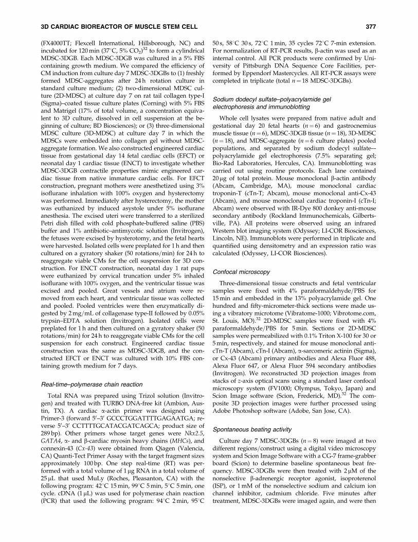
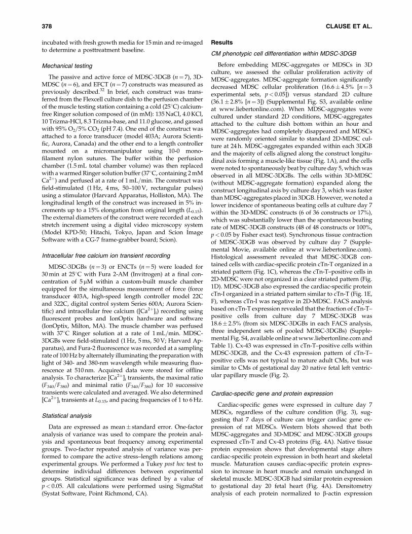
Before embedding MDSC-aggregates or MDSCs in 3Dculture, we assessed the cellular proliferation activity ofMDSC-aggregates. MDSC-aggregate formation significantlydecreased MDSC cellular proliferation (16.6� 4.5% [n¼ 3experimental sets, p< 0.05]) versus standard 2D culture(36.1� 2.8% [n¼ 3]) (Supplemental Fig. S3, available onlineat www.liebertonline.com). When MDSC-aggregates werecultured under standard 2D conditions, MDSC-aggregatesattached to the culture dish bottom within an hour andMDSC-aggregates had completely disappeared and MDSCswere randomly oriented similar to standard 2D-MDSC cul-ture at 24 h. MDSC-aggregates expanded within each 3DGBand the majority of cells aligned along the construct longitu-dinal axis forming a muscle-like tissue (Fig. 1A), and the cellswere noted to spontaneously beat by culture day 5, which wasobserved in all MDSC-3DGBs. The cells within 3D-MDSC(without MDSC-aggregate formation) expanded along theconstruct longitudinal axis by culture day 3, which was fasterthan MDSC-aggregates placed in 3DGB. However, we noted alower incidence of spontaneous beating cells at culture day 7within the 3D-MDSC constructs (6 of 36 constructs or 17%),which was substantially lower than the spontaneous beatingrate of MDSC-3DGB constructs (48 of 48 constructs or 100%,p< 0.05 by Fisher exact test). Synchronous tissue contractionof MDSC-3DGB was observed by culture day 7 (Supple-mental Movie, available online at www.liebertonline.com).Histological assessment revealed that MDSC-3DGB con-tained cells with cardiac-specific protein cTn-T organized in astriated pattern (Fig. 1C), whereas the cTn-T–positive cells in2D-MDSC were not organized in a clear striated pattern (Fig.1D). MDSC-3DGB also expressed the cardiac-specific proteincTn-I organized in a striated pattern similar to cTn-T (Fig. 1E,F), whereas cTn-I was negative in 2D-MDSC. FACS analysisbased on cTn-T expression revealed that the fraction of cTn-T–positive cells from culture day 7 MDSC-3DGB was18.6� 2.5% (from six MDSC-3DGBs in each FACS analysis,three independent sets of pooled MDSC-3DGBs) (Supple-mental Fig. S4, available online at www.liebertonline.com andTable 1). Cx-43 was expressed in cTn-T–positive cells withinMDSC-3DGB, and the Cx-43 expression pattern of cTn-T–positive cells was not typical to mature adult CMs, but wassimilar to CMs of gestational day 20 native fetal left ventric-ular papillary muscle (Fig. 2).
Cardiac-specific gene and protein expression
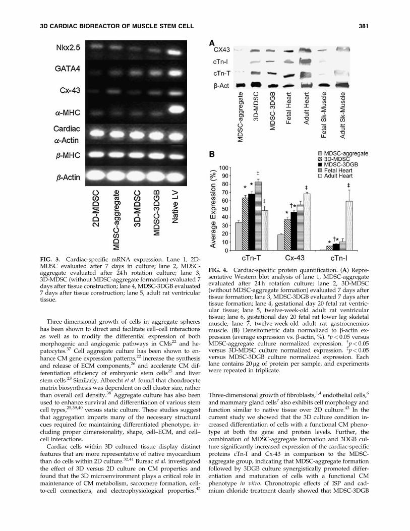
Cardiac-specific genes were expressed in culture day 7MDSCs, regardless of the culture condition (Fig. 3), sug-gesting that 7 days of culture can trigger cardiac gene ex-pression of rat MDSCs. Western blots showed that bothMDSC-aggregates and 3D-MDSC and MDSC-3DGB groupsexpressed cTn-T and Cx-43 proteins (Fig. 4A). Native tissueprotein expression shows that developmental stage alterscardiac-specific protein expression in both heart and skeletalmuscle. Maturation causes cardiac-specific protein expres-sion to increase in heart muscle and remain unchanged inskeletal muscle. MDSC-3DGB had similar protein expressionto gestational day 20 fetal heart (Fig. 4A). Densitometryanalysis of each protein normalized to b-actin expression
378 CLAUSE ET AL.
demonstrated that 3D culture (MDSC-3DGB and 3D-MDSCgroups) was associated with higher cTn-T, Cx-43, and cTn-Iexpression versus MDSC-aggregate culture ( p< 0.05).Within 3D culture groups, we noted that both cTn-I and Cx-43 expression levels were higher in the MDSC-3DGB groupthan in the 3D-MDSC group ( p< 0.05) (Fig. 4B). Notably,MDSC-3DGB had similar cTn-I and Cx-43 expression levelsas well as a cTn-T=cTn-I ratio (9.1� 0.4 [n¼ 3]) to gestationalday 20 fetal heart (9.3� 1.7 [n¼ 3]), which was significantlydifferent compared to adult ventricular tissue (0.8� 0.1[n¼ 4, p< 0.05]) cTn-T=cTn-I ratio as well as cTn-I and Cx-43expression levels ( p< 0.05) (Fig. 4B). These data indicate thatwhile MDSCs express cardiac-specific genes at 7 days inculture, regardless of culture method, the cardiac-specificprotein expression profiles differed with culture method;specifically, MDSC-aggregate formation followed by 3DGBculture synergistically increased induction of cells with animmature CM phenotype.
Chronotropic effects of ISP and cadmium chlorideon MDSC-3DGB
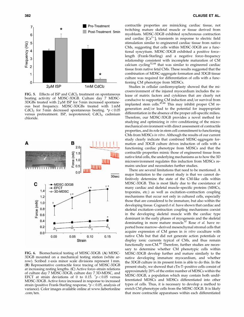
Culture day 7 MDSC-3DGBs were treated with ISP orcadmium chloride. Five minutes after treatment, ISP in-creased MDSC-3DGB spontaneous beat frequency (5.62�0.54 Hz [n¼ 8, p< 0.05]) versus pretreatment (4.05� 0.20 Hz[n¼ 8]), whereas cadmium chloride suppressed spontaneous
beating activity (0.44� 0.16 Hz [n¼ 8, p< 0.05]) compared topretreatment (4.27� 0.44 Hz [n¼ 8]) (Fig. 5). No changes inpretreatment baseline spontaneous beat frequency andposttreatment baseline spontaneous beat frequency werenoted indicating reversible effects. These chronotropic re-sponses do not occur in twitching mature skeletal muscle orskeletal myotubes.16
Contractile properties of 3D MDSC culture
Upon field stimulation, both 3D-MDSC and MDSC-3DGBgenerated contractile force. MDSC-3DGB generated con-tractile force similar to engineered cardiac tissue from nativefetal cardiac cells (EFCT) (Fig. 6). MDSC-3DGB as well asculture day 7 EFCT displayed a positive Frank-Starling re-sponse to increased construct length, whereas 3D-MDSC didnot ( p< 0.05; Fig. 6B, C). MDSC-3DGB generated a greatermaximum active force (0.41� 0.06 mN [n¼ 7, p< 0.05])compared with 3D-MDSC (0.22� 0.04 mN [n¼ 5]), whichwas approximately one-third of EFCT (1.45� 0.39 mN [n¼ 7,p< 0.05]) (Fig. 6C).
[Ca2þ]i transients in MDSC-3DGB
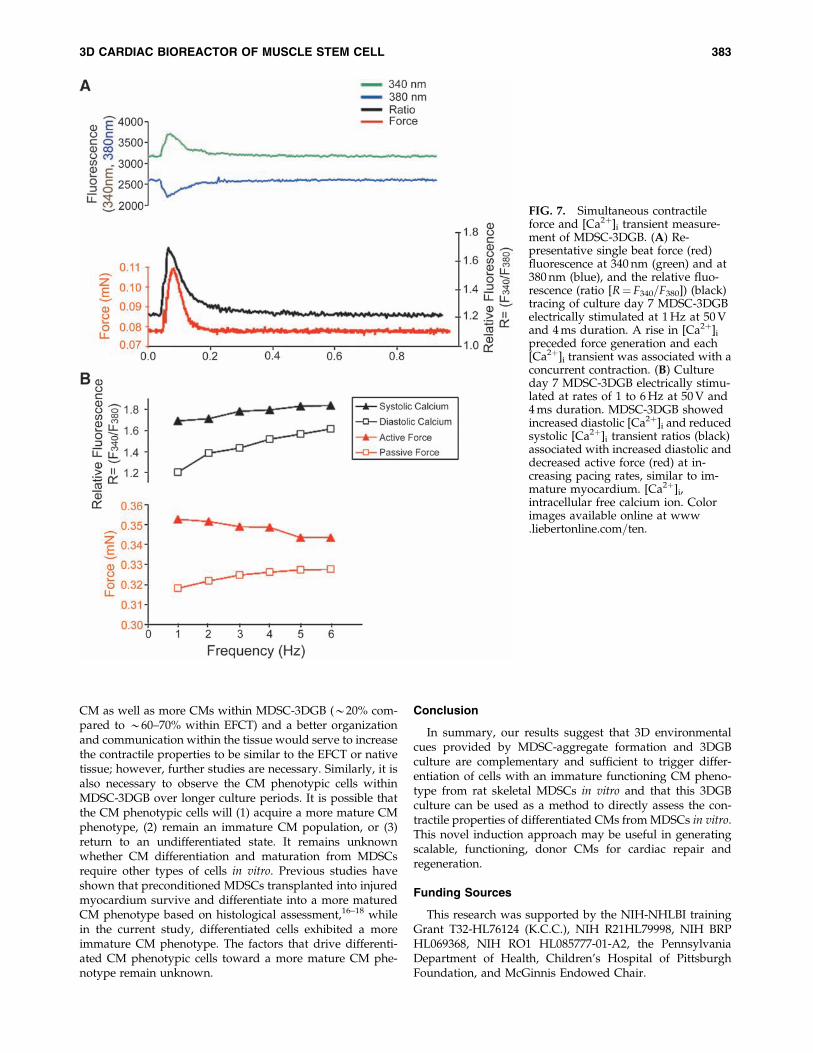
We further investigated calcium handling by simulta-neously recording contractile force and [Ca2þ]i transientsfrom culture day 7 MDSC-3DGB in which we observedspontaneous tissue contraction. A rise in [Ca2þ]i preceded
FIG. 1. Histologic analysis of MDSC-3D collagen gel bioreactor (3DGB). (A) Phase contract image of culture day 7 MDSC-3DGB. Scale bar indicates 500 mm. Red box indicates the area of the tissue where high-magnification images are taken. (B) a-Sarcomeric actinin expression in MDSC-3DGB. Scale bar indicates 500mm. (C) Cardiac troponin-T (cTn-T) expressed inoriented cells of MDSC-3DGB. Blue staining (DAPI) indicates nuclei. Scale bar indicates 50mm. The white double-head arrowindicates MDSC-3DGB longitudinal axis. (D) cTn-T expression of MDSC-2D. Scale bar indicates 20mm. (E) cTn-T expression ofMDSC-3DGB was a typical striated muscle pattern at a higher magnification. Scale bar indicates 20 mm. (F) Cardiac troponin-I(cTn-I) also expressed in a typical striated pattern (white arrowheads). Scale bar indicates 20 mm. 2D, two-dimensional; MDSC,muscle-derived stem cell; DAPI, 40,6-diamidino-2-phenylindole. Color images available online at www.liebertonline.com=ten.
3D CARDIAC BIOREACTOR OF MUSCLE STEM CELL 379
force generation (Fig. 7A) and each [Ca2þ]i transient wasassociated with a concurrent contraction. MDSC-3DGB dis-played a negative force–frequency relationship between 1and 6 Hz based on reduced [Ca2þ]i transients and active forceat increasing pacing rates and was associated with increaseddiastolic ratio (suggesting increased diastolic [Ca2þ]i andpassive force) (Fig. 7B), similar to immature myocardi-um.33,34 MDSC-3DGB force as well as [Ca2þ]i transient de-veloped calcium (ratio¼ F340=F380) (0.48� 0.16 [n¼ 3]) wassimilar to ENCT (engineered cardiac tissue from cardiac cellsisolated from day 1 neonatal rats) (0.56� 0.11 [n¼ 5]). Thesedata indicate that MDSC-3DGB display the [Ca2þ]i transientfeatures of immature CM with incomplete maturation ofexcitation–contraction coupling.
Discussion
Previous studies have shown that stem cells isolated fromskeletal muscle specimens have the ability to differentiateinto CMs;8,14,16–19 however, the efficiency of CM differenti-ation remains unclear and with limited functional charac-terization. In the current study we found that (1) low-serumgrowth medium–treated rat MDSCs expressed cardiac-specific genes similar to native adult myocardium, consis-tent with the findings of others in both human and mouseskeletal muscle–derived cells, and (2) the combination ofMDSC-aggregate formation and 3DGB culture significantly
increased cardiac-specific protein expression, spontaneousbeating cell activity, and contractile properties. Our resultssuggest that 3D microenvironmental cues provided byMDSC-aggregate and 3DGB culture play an important rolein differentiation of cells with a functional CM phenotypefrom MDSCs and the 3DGB system provides the necessaryenvironment to evaluate the contractile properties of thesedifferentiated CMs.
Various experiments have highlighted the importance ofthe microenvironment on stem cell–derived CM induction,differentiation, and survival.35,36 Tamaki et al. have recentlyshown that skeletal muscle–derived multipotent Sk-34 cellscan give rise to CMs and that cell-to-cell relationships andcellular milieu were important for this differentiation.18
However, these results were achieved with coculture withembryonic CMs, and there was no functional characteriza-tion of the SK-34–derived CMs in vitro. Thus, it is unclearhow these cells function in comparison to native CMs ineither a healthy or diseased heart. Rota et al. have shown thatbone marrow stem cells can engraft into the injured myo-cardium and differentiate into functionally competent CMsand vascular structures by establishing a microenvironmentnecessary to adopt the cardiac phenotype.5 However, despitethese positive results there is still limited information on theuse of physical stimuli and unique microenvironments tocontrol CM induction from progenitor=stem cells, CM mat-uration, and contractile function.
FIG. 2. Native fetal left ventricular papillary muscle at gestational day 20 stained for (A) a-sarcomeric actinin (green). (B)Gap junction protein Cx-43 (red), and (C) merged. Culture day 7 MDSC-3DGB stained for (D) a-sarcomeric actinin (green).(E) Gap junction protein Cx-43 (red), and (F) merged. Cx-43 expression of MDSC-3DGB was similar to gestational day 20 fetalleft ventricular papillary muscle Cx-43 expression. Blue staining (DAPI) indicates nuclei. Scale bar indicates 10 mm. Cx-43,connexin-43. Color images available online at www.liebertonline.com=ten.
380 CLAUSE ET AL.
Three-dimensional growth of cells in aggregate sphereshas been shown to direct and facilitate cell–cell interactionsas well as to modify the differential expression of bothmorphogenic and angiogenic pathways in CMs22 and he-patocytes.37 Cell aggregate culture has been shown to en-hance CM gene expression patterns,22 increase the synthesisand release of ECM components,26 and accelerate CM dif-ferentiation efficiency of embryonic stem cells25 and liverstem cells.23 Similarly, Albrecht et al. found that chondrocytematrix biosynthesis was dependent on cell cluster size, ratherthan overall cell density.38 Aggregate culture has also beenused to enhance survival and differentiation of various stemcell types,25,39,40 versus static culture. These studies suggestthat aggregation imparts many of the necessary structuralcues required for maintaining differentiated phenotype, in-cluding proper dimensionality, shape, cell–ECM, and cell–cell interactions.
Cardiac cells within 3D cultured tissue display distinctfeatures that are more representative of native myocardiumthan do cells within 2D culture.32,41 Bursac et al. investigatedthe effect of 3D versus 2D culture on CM properties andfound that the 3D microenvironment plays a critical role inmaintenance of CM metabolism, sarcomere formation, cell-to-cell connections, and electrophysiological properties.42
Three-dimensional growth of fibroblasts,1,4 endothelial cells,6
and mammary gland cells7 also exhibits cell morphology andfunction similar to native tissue over 2D culture.43 In thecurrent study we showed that the 3D culture condition in-creased differentiation of cells with a functional CM pheno-type at both the gene and protein levels. Further, thecombination of MDSC-aggregate formation and 3DGB cul-ture significantly increased expression of the cardiac-specificproteins cTn-I and Cx-43 in comparison to the MDSC-aggregate group, indicating that MDSC-aggregate formationfollowed by 3DGB culture synergistically promoted differ-entiation and maturation of cells with a functional CMphenotype in vitro. Chronotropic effects of ISP and cad-mium chloride treatment clearly showed that MDSC-3DGB
FIG. 3. Cardiac-specific mRNA expression. Lane 1, 2D-MDSC evaluated after 7 days in culture; lane 2, MDSC-aggregate evaluated after 24 h rotation culture; lane 3,3D-MDSC (without MDSC-aggregate formation) evaluated 7days after tissue construction; lane 4, MDSC-3DGB evaluated7 days after tissue construction; lane 5, adult rat ventriculartissue.
FIG. 4. Cardiac-specific protein quantification. (A) Repre-sentative Western blot analysis of lane 1, MDSC-aggregateevaluated after 24 h rotation culture; lane 2, 3D-MDSC(without MDSC-aggregate formation) evaluated 7 days aftertissue formation; lane 3, MDSC-3DGB evaluated 7 days aftertissue formation; lane 4, gestational day 20 fetal rat ventric-ular tissue; lane 5, twelve-week-old adult rat ventriculartissue; lane 6, gestational day 20 fetal rat lower leg skeletalmuscle; lane 7, twelve-week-old adult rat gastrocnemiusmuscle. (B) Densitometric data normalized to b-actin ex-pression (average expression vs. b-actin, %). *p< 0.05 versusMDSC-aggregate culture normalized expression. {p< 0.05versus 3D-MDSC culture normalized expression. {p< 0.05versus MDSC-3DGB culture normalized expression. Eachlane contains 20 mg of protein per sample, and experimentswere repeated in triplicate.
3D CARDIAC BIOREACTOR OF MUSCLE STEM CELL 381
contractile properties are mimicking cardiac tissue, nottwitching mature skeletal muscle or tissue derived frommyoblasts. MDSC-3DGB exhibited synchronous contractionand cardiac [Ca2þ]i transients in response to electric fieldstimulation similar to engineered cardiac tissue from nativeCMs, suggesting that cells within MDSC-3DGB are a func-tional syncytium. MDSC-3DGB exhibited a positive force–length (Frank-Starling) and a negative force–frequencyrelationship consistent with incomplete maturation of CMcalcium cycling32,44 that was similar to engineered cardiactissue from native fetal CMs. These results suggested that thecombination of MDSC-aggregate formation and 3DGB tissueculture was required for differentiation of cells with a func-tioning CM phenotype from MDSCs.
Studies in cellular cardiomyoplasty showed that the mi-croenvironment of the injured myocardium includes the re-lease of matrix factors and cytokines that are clearly notconducive to supporting CM induction and=or survival fromimplanted stem cells.45,46 This may inhibit proper CM re-generation and=or lead to the potential for inappropriatedifferentiation in the absence of the proper cell-specific milieu.Therefore, our MDSC-3DGB provides a novel method forstudying and optimizing in vitro conditioning of the micro-mechanical environment with direct assessment of contractileproperties, and its role in stem cell commitment to functioningCMs from MDSCs in vitro. Although the results of our currentstudy clearly indicate that combined MDSC-aggregate for-mation and 3DGB culture drives induction of cells with afunctioning cardiac phenotype from MDSCs and that thecontractile properties mimic those of engineered tissue fromnative fetal cells, the underlying mechanisms as to how the 3Dmicroenvironment regulates this induction from MDSCs re-mains unclear and necessitates further studies.
There are several limitations that need to be mentioned. Amajor limitation to the current study is that we cannot de-finitively determine the state of the CM-like cells withinMDSC-3DGB. This is most likely due to the coexistance ofmany cardiac and skeletal muscle–specific proteins (MHCs,troponins, etc.) as well as excitation–contraction couplingmechanisms that occur not only in cultured cells, especiallythose that are considered to be immature, but also within thedeveloping tissue. Cognard et al. have shown that cardiac andskeletal excitation–contraction coupling mechanisms coexistin the developing skeletal muscle with the cardiac typedominant in the early phases of myogenesis and the skeletaldominating in more mature muscle.47 Rose et al. have re-ported bone marrow–derived mesenchymal stromal cells thatacquire expression of CM genes in in vitro coculture withnative CMs but that did not generate action potentials ordisplay ionic currents typical of CMs, and thus remainfunctionally non-CM.48 Therefore, further studies are neces-sary to determine whether CM phenotypic cells withinMDSC-3DGB develop further and mature similarly to thenative developing immature myocardium, and whetherthe 3DGB culture in its present form is able to do this. In thepresent study, we showed that cTn-T–positive cells consist ofapproximately 20% of the entire number of MDSCs within theMDSC-3DGB, a population which may contain both undif-ferentiated MDSCs and MDSCs differentiated into othertypes of cells. Thus, it is necessary to develop a method toenrich CM phenotype cells from the MDSC-3DGB. It is likelythat more contractile apparatuses within each differentiated
FIG. 5. Effects of ISP and CdCl2 treatment on spontaneousbeating activity of MDSC-3DGB. Culture day 7 MDSC-3DGBs treated with 2mM ISP for 5 min increased spontane-ous beat frequency. MDSC-3DGBs treated with 1 mMCdCl2 for 5 min decreased spontaneous beating. *p< 0.05versus pretreatment. ISP, isoproterenol; CdCl2, cadmiumchloride.
FIG. 6. Biomechanical testing of MDSC-3DGB. (A) MDSC-3DGB mounted on a mechanical testing station (white ar-row). Scribed x-axis minor scale divisions represent 1 mm.(B) Representative contractile force tracing of MDSC-3DGBat increasing resting lengths. (C) Active force–strain relationsof culture day 7 MDSC-3DGB, culture day 7 3D-MDSC, andEFCT at strain deviations of 0 to 0.15. {p< 0.05 versusMDSC-3DGB. Active force increased in response to increasedstrain (positive Frank-Starling response, *p< 0.05, analysis ofvariance). Color images available online at www.liebertonline.com=ten.
382 CLAUSE ET AL.
CM as well as more CMs within MDSC-3DGB (*20% com-pared to *60–70% within EFCT) and a better organizationand communication within the tissue would serve to increasethe contractile properties to be similar to the EFCT or nativetissue; however, further studies are necessary. Similarly, it isalso necessary to observe the CM phenotypic cells withinMDSC-3DGB over longer culture periods. It is possible thatthe CM phenotypic cells will (1) acquire a more mature CMphenotype, (2) remain an immature CM population, or (3)return to an undifferentiated state. It remains unknownwhether CM differentiation and maturation from MDSCsrequire other types of cells in vitro. Previous studies haveshown that preconditioned MDSCs transplanted into injuredmyocardium survive and differentiate into a more maturedCM phenotype based on histological assessment,16–18 whilein the current study, differentiated cells exhibited a moreimmature CM phenotype. The factors that drive differenti-ated CM phenotypic cells toward a more mature CM phe-notype remain unknown.
Conclusion
In summary, our results suggest that 3D environmentalcues provided by MDSC-aggregate formation and 3DGBculture are complementary and sufficient to trigger differ-entiation of cells with an immature functioning CM pheno-type from rat skeletal MDSCs in vitro and that this 3DGBculture can be used as a method to directly assess the con-tractile properties of differentiated CMs from MDSCs in vitro.This novel induction approach may be useful in generatingscalable, functioning, donor CMs for cardiac repair andregeneration.
Funding Sources
This research was supported by the NIH-NHLBI trainingGrant T32-HL76124 (K.C.C.), NIH R21HL79998, NIH BRPHL069368, NIH RO1 HL085777-01-A2, the PennsylvaniaDepartment of Health, Children’s Hospital of PittsburghFoundation, and McGinnis Endowed Chair.
FIG. 7. Simultaneous contractileforce and [Ca2þ]i transient measure-ment of MDSC-3DGB. (A) Re-presentative single beat force (red)fluorescence at 340 nm (green) and at380 nm (blue), and the relative fluo-rescence (ratio [R¼ F340=F380]) (black)tracing of culture day 7 MDSC-3DGBelectrically stimulated at 1 Hz at 50 Vand 4 ms duration. A rise in [Ca2þ]i
preceded force generation and each[Ca2þ]i transient was associated with aconcurrent contraction. (B) Cultureday 7 MDSC-3DGB electrically stimu-lated at rates of 1 to 6 Hz at 50 V and4 ms duration. MDSC-3DGB showedincreased diastolic [Ca2þ]i and reducedsystolic [Ca2þ]i transient ratios (black)associated with increased diastolic anddecreased active force (red) at in-creasing pacing rates, similar to im-mature myocardium. [Ca2þ]i,intracellular free calcium ion. Colorimages available online at www.liebertonline.com=ten.
3D CARDIAC BIOREACTOR OF MUSCLE STEM CELL 383
Disclosure Statement
No competing financial interests exist.
References
1. Dimmeler, S., Zeiher, A.M., and Schneider, M.D. Unchainmy heart: the scientific foundations of cardiac repair. J ClinInvestig 115, 572, 2005.
2. Dowell, J.D., Rubart, M., Pasumarthi, K.B., Soonpaa, M.H.,and Field, L.J. Myocyte and myogenic stem cell transplan-tation in the heart. Cardiovasc Res 58, 336, 2003.
3. Roell, W., Lu, Z.J., Bloch, W., Siedner, S., Tiemann, K., Xia,Y., Stoecker, E., Fleischmann, M., Bohlen, H., Stehle, R.,Kolossov, E., Brem, G., Addicks, K., Pfitzer, G., Welz, A.,Hescheler, J., and Fleischmann, B.K. Cellular cardiomyo-plasty improves survival after myocardial injury. Circula-tion 105, 2435, 2002.
4. Christoforou, N., and Gearhart, J.D. Stem cells and theirpotential in cell-based cardiac therapies. Prog CardiovascDis 49, 396, 2007.
5. Rota, M., Kajstura, J., Hosoda, T., Bearzi, C., Vitale, S.,Esposito, G., Iaffaldano, G., Padin-Iruegas, M.E., Gonzalex,A., Rizzi, R., Small, N., Muraski, J., Alvarez, R., Chen, X.,Urbanek, K., Bolli, R., Houser, S.R., Leri, A., Sussman, M.A.,and Anversa, P. Bone marrow cells adopt the cardiomyo-genic fate in vivo. PNAS 104, 17783, 2007.
6. Ott, H.C., Davis, B.H., and Taylor, D.A. Cell therapy for heartfailure—muscle, bone marrow, blood, and cardiac-derivedstem cells. Semin Thorac Cardiovasc Surg 17, 348, 2005.
7. Tossios, P., Muller-Ehmsen, J., Schmidt, M., Scheid, C., Unal,N., Moka, D., Schwinger, R.H., and Mehlhorn, U. No evi-dence of myocardial restoration following transplantation ofmononuclear bone marrow cells in coronary bypass graftingsurgery patients based upon cardiac SPECT and 18F-PET.BMC Med Imaging 6, 7, 2006.
8. Okada, M., Payne, T.R., Bo, Z., Oshima, H., Momoi, N., To-bita, K., Keller, B.B., Peault, B., and Huard, J. Identification ofa novel progenitor cell population in human skeletal musclewith a superior ability for myocardial repair in comparison tocommited skeletal myoblast. JACC 52, 1869, 2008.
9. Kolossov, E., Bostani, T., Roell, W., Breitbach, M., Pillekamp,F., Nygren, J.M., Sasse, P., Rubenchik, O., Fries, J.W.,Wenzel, D., Geisen, C., Xia, Y., Lu, Z., Duan, Y., Kettenho-fen, R., Jovinge, S., Bloch, W., Bohlen, H., Welz, A., He-scheler, J., Jacobsen, S.E., and Fleischmann, B.K. Engraftmentof engineered ES cell-derived cardiomyocytes but not BMcells restores contractile function to the infarcted myocar-dium. J Exp Med 203, 2315, 2006.
10. Peng, H., and Huard, J. Muscle-derived stem cells for mus-culoskeletal tissue regeneration and repair. Transplant Im-munol 12, 311, 2004.
11. Lee, J.Y., Qu-Petersen, Z., Cao, B., Kimura, S., Jankowski, R.,Cummins, J., Usas, A., Gates, C., Robbins, P., Wernig, A.,and Huard, J. Clonal isolation of muscle-derived cells ca-pable of enhancing muscle regeneration and bone healing.J Cell Biol 150, 1085, 2000.
12. Jankowski, R.J., Haluszczak, C., Trucco, M., and Huard, J.Flow cytometric characterization of myogenic cell popula-tions obtained via the preplate technique: potential for rapidisolation of muscle-derived stem cells. Hum Gene Ther 12,
619, 2001.13. Zheng, B., Cao, B., Crisan, M., Sun, B., Li, G., Logar, A., Yap,
S., Pollett, J.B., Drowley, L., Cassino, T., Gharaibeh, B.,Deasy, B.M., Huard, J., and Peault, B. Prospective identifi-
cation of myogenic endothelial cells in human skeletalmuscle. Nat Biotechnol 25, 1025, 2007.
14. Oshima, H., Payne, T.R., Urish, K.L., Sakai, T., Ling, Y.,Gharaibeh, B., Tobita, K., Keller, B.B., Cummins, J.H., andHuard, J. Differential myocardial infarct repair with musclestem cells compared to myoblasts. Mol Ther 12, 1130, 2005.
15. Zuba-Surma, E.K., Abdel-Latif, A., Case, J., Tiwari, S., Hunt,G., Kucia, M., Vincent, R.J., Ranjan, S., Ratajszak, M.Z.,Srour, E.F., Bolli, R., and Dawn, B. Sac-1 expression is as-sociated with decreased cardiomyogenic differentiation po-tential of skeletal muscle-derived adult primitive cells. JMCC41, 650, 2006.
16. Winitsky, S.O., Gopal, T.V., Hassanzadeh, S., Takahashi, H.,Gryder, D., Rogawski, M.A., Takeda, K., Yu, Z.X., Xu, Y.H.,and Epstein, N.D. Adult murine skeletal muscle containscells that can differentiate into beating cardiomyocytesin vitro. PLoS Biol 3, e87, 2005.
17. Invernici, G., Cristini, S., Madeddu, P., Brock, S., Spillmann,F., Bernasconi, P., Cappelletti, C., Calatozzolo, C., Fascio, U.,Bisleri, G., Muneretto, C., Alessandri, G., and Parati, E.A.Human adult skeletal muscle stem cells differentiate intocardiomyocyte phenotype in vitro. Exp Cell Res 314, 366,2008.
18. Tamaki, T., Akatsuka, A., Okada, Y., Uchiyama, Y., Tono, K.,Wada, M., Hoshi, A., Iwaguro, H., Iwasaki, H., Oyamada, A.,and Asahara, T. Cardiomyocyte formation by skeletal mus-cle-derived multi-myogenic stem cells after transplantationinto infarcted myocardium. PLoS ONE 3, e1789, 2008.
19. Arsic, N., Mamaeva, D., Lamb, N.J., and Fernandez, A.Muscle-derived stem cells isolated as non-adherent popula-tion give rise to cardiac, skeletal muscle and neural lineages.Exp Cell Res 314, 1266, 2008.
20. Payne, T.R., Oshima, H., Okada, M., Momoi, N., Tobita, K.,Keller, B.B., Peng, H., and Huard, J. A relationship betweenVEGF, angiogenesis, and cardiac repair after muscle stemcell transplantation into ischemic hearts. JACC 23, 1677,2007.
21. Steinberg, M.S. Reconstruction of tissues by dissociated cells.Science 141, 401, 1963.
22. Akins, R.E., Gratton, K., Quezada, E., Rutter, H., Tsuda, T.,and Soteropoulos, P. Gene expression profile of bioreactor-cultured cardiac cells: activation of morphogenetic pathwaysfor tissue engineering. DNA Cell Biol 26, 425, 2007.
23. Anderson, P.A., Muller-Borer, B.J., Esch, G.L., Coleman,W.B., Grisham, J.W., and Malouf, N.N. Calcium signals in-duce liver stem cells to acquire a cardiac phenotype. CellCycle 6, 1565, 2007.
24. Dertinger, H., and Hulser, D.F. Intercellular communicationin spheroids. In: Acker, H., Carlsson, J., Durand, R.,and Sutherland, R.M., eds. Spheroids in Cancer Research:Methods and Perspectives. Berlin: Springer-Verlag, 1984, pp.67–83.
25. Carpenedo, R.L., Sargent, C.Y., and McDevitt, T.C. Rotarysuspension culture enhances the efficiency, yield, and ho-mogeneity of embryoid body differentiation. Stem Cells 25,
2224, 2007.26. Watzka, S.B., Lucien, J., Shimada, M., Edwards, V., Yeger,
H., Hannigan, G., and Coles, J.G. Selection of viable cardi-omyocytes for cell transplantation using three-dimensionaltissue culture. Transplantation 70, 1310, 2000.
27. Zimmermann, W.H., Didie, M., Doker, S., Melnychenko, I.,Naito, H., Rogge, C., Tiburcy, M., and Eschenhagen, T.Heart muscle engineering: an update on cardiac muscle re-placement therapy. Cardiovasc Res 71, 419, 2006.
384 CLAUSE ET AL.
28. Zimmermann, W.H., Melnychenko, I., Wasmeier, G., Didie,M., Naito, H., Nixdorff, U., Hess, A., Budinsky, L., Brune, K.,Michaelis, B., Dhein, S., Schwoerer, A., Ehmke, H., andEschenhagen, T. Engineered heart tissue grafts improve sys-tolic and diastolic function in infarcted rat hearts. Nat Med 12,
452, 2006.29. Jankowski, R.J., Deasy, B.M., and Huard, J. Muscle-derived
stem cells. Gene Ther 9, 642, 2002.30. Deasy, B.M., Gharaibeh, B., Pollett, J.B., Jones, M.M., Lucas,
M.A., Kanda, Y., and Huard, J. Long-term self-renewal ofpostnatal muscle-derived Stem cells. Mol Bio Cell 16, 3323,2005.
31. Gharaibeh, B., Lu, A., Tebbets, J., Zheng, B., Feduska, J.,Crisan, M., Peault, B., Cummins, J., and Huard, J. Isolation ofa slowly adhering cell fraction containing stem cells frommurine skeletal muscle by the preplate technique. Nat Prot3, 1501, 2008.
32. Tobita, K., Liu, L.J., Janczewski, A.M., Tinney, J.P., None-maker, J.M., Augustine, S., Stolz, D.B., Shroff, S.G., andKeller, B.B. Engineered early embryonic cardiac tissue(EEECT) retains proliferative and contractile properties ofdeveloping embryonic myocardium. Am J Physiol HeartCirc Physiol 291, H1829, 2006.
33. Penefsky, Z.J., Wolfe, J., Greengard, O., and Bernstein, J.Mechanical responses of developing Fisher rat heart. Effectsof steroid hormone. J Dev Physiol 8, 333, 1986.
34. Liu, J., Fu, J.D., Siu, C.W., and Li, R.A. Functional sarco-plasmic reticulum for calcium handling of human embry-onic stem cell-derived cardiomyocytes: insights for drivenmaturation. Stem Cells 25, 3038, 2007.
35. Iijima, Y., Nagai, T., Mizukami, M., Matsuura, K., Ogura, T.,Wada, H., Toko, H., Akazawa, H., Takano, H., Nakaya, H.,and Komuro, I. Beating is necessary for transdifferentiationof skeletal muscle-derived cells into cardiomyocytes. FASEBJ 17, 1361, 2003.
36. Rudy-Reil, D., and Lough, J. Avian precardiac endoderm=mesoderm induces cardiac myocyte differentiation in mu-rine embryonic stem cells. Circ Res 94, e107, 2004.
37. Hansen, L.K., Hsiao, C., Friend, J.R., Wu, F.J., Bridge, G.A.,Remmel, R.P., Cerra, F.B., and Hu, W. Enhanced morphol-ogy and function in hepatocyte spheroids: a model of tissueself-assembly. Tissue Eng 4, 65, 1998.
38. Albrecht, D.R., Underhill, G.H., Wassermann, T.B., Sah, R.L.,and Bhatia, S.N. Probing the role of multicellular organiza-tion in three-dimensional microenvironments. Nat Methods3, 369, 2006.
39. Suenaga, H., Furukawa, K.S., Ushida, T., Takato, T., andTateishi, T. Aggregate formation of bone marrow stromalcells by rotation culture. Mat Sci Eng C24, 421, 2004.
40. Kehat, I., Kenyagin-Karsenti, D., Snir, M., Segev, H., Amit,M., Gepstein, A., Livne, E., Binah, O., Itskovitz-Eldor, J., andGepstein, L. Human embryonic stem cells can differentiate
into myocytes with structural and functional properties ofcardiomyocytes. J Clin Invest 108, 407, 2001.
41. Zimmermann, W.H., Schneiderbanger, K., Schubert, P., Di-die, M., Munzel, F., Heubach, J.F., Kostin, S., Neuhuber,W.L., and Eschenhagen, T. Tissue engineering of a differ-entiated cardiac muscle construct. Circ Res 8, 223, 2002.
42. Bursac, N., Papadaki, M., White, J.A., Eisenberg, S.R.,Vunjak-Novakovic, G., and Freed, L.E. Cultivation in rotat-ing bioreactors promotes maintenance of cardiac myocyteelectrophysiology and molecular properties. Tissue Eng 9,
1243, 2003.43. Panorchan, P., Lee, J.S.H., Kole, T.P., Tseng, Y., and Wirtz, D.
Microrheology and ROCK signaling of human endothelialcells embedded in a 3D matrix. Biophys J 91, 3499, 2006.
44. Dolnikov, K., Shilkrut, M., Zeevi-Levin, N., Gerecht-Nir, S.,Amit, M., Danon, A., Itskovitz-Eldor, J., and Binah, O.Functional properties of human embryonic stem cell-derivedcardiomyocytes: intracellular Ca2þ handling and the role ofsarcoplasmic reticulum in the contraction. Stem Cells 24,
236, 2006.45. Frangogiannis, N.G. The mechanistic basis of infarct healing.
Antioxid Redox Signal 8, 1907, 2006.46. Kastrup, J., Ripa, R.S., Wang, Y., and Jorgensen, E. Myocardial
regeneration induced by granulocyte-colony-stimulating fac-tor mobilization of stem cells in patients with acute or chronicischaemic heart disease: a non-invasive alternative for clinicalstem cell therapy? Eur Heart J 27, 2748, 2006.
47. Cognard, C., Rivet-Bastide, M., Constantin, B., and Ray-mond, G. Progressive predominance of ‘‘skeletal’’ versus‘‘cardiac’’ types of excitation-contraction coupling duringin vitro skeletal myogenesis. Eur J Phys 422, 207, 1992.
48. Rose, R.A., Juang, H., Wang, X., Helke, S., Tsoporis, J.N.,Gong, N., Keating, S.C.J., Parker, T.G., Backx, P.H., andKeating, A. Bone marrow-derived mesenchymal stromalcells express cardiac-specific markers, retain the stromalphenotype and do not become functional cardiomyocytesin vitro. Stem Cells 26, 2884, 2008.
Address correspondence to:Kimimasa Tobita, M.D.
Cardiovascular Development Research ProgramChildren’s Hospital of Pittsburgh of UPMC
Rangos Research CenterRoom 8121 One Children’s Hospital Drive 4401 Penn Ave.
Pittsburgh, PA 15224
E-mail: [email protected]
Received: February 12, 2009Accepted: July 13, 2009
Online Publication Date: February 16, 2010
3D CARDIAC BIOREACTOR OF MUSCLE STEM CELL 385